antimicrobial and anti-biofilm properties of polypropylene
TRANSCRIPT
J Mater Sci: Mater Med (2017) 28:97DOI 10.1007/s10856-017-5910-y
BIOMATERIALS SYNTHESIS AND CHARACTERIZATION Original Research
Antimicrobial and anti-biofilm properties of polypropylene meshescoated with metal-containing DLC thin films
Elisa M. Cazalini 1● Walter Miyakawa1 ● Guilherme R. Teodoro2 ●
Argemiro S. S. Sobrinho1 ● José E. Matieli1 ● Marcos Massi1,3 ● Cristiane Y. Koga-Ito2,4
Received: 7 February 2017 / Accepted: 5 May 2017 / Published online: 17 May 2017© Springer Science+Business Media New York 2017
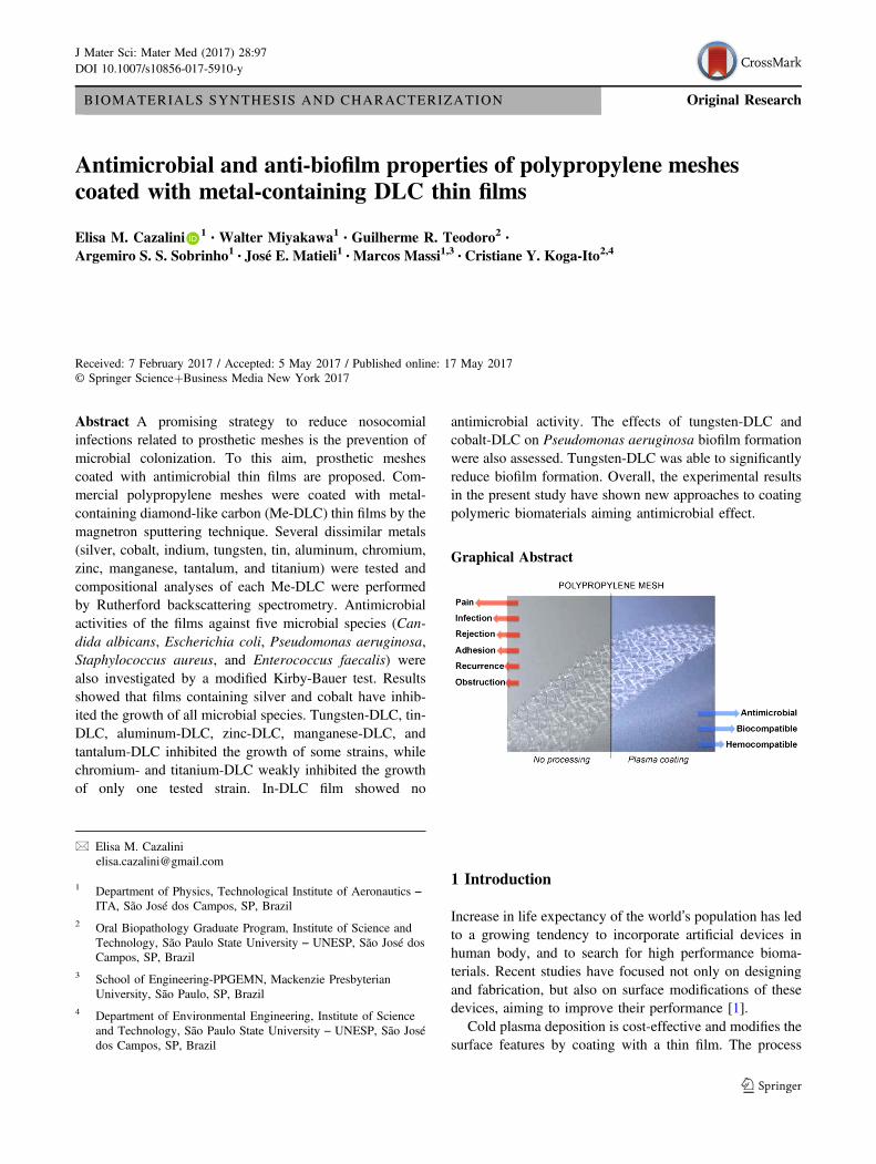
Abstract A promising strategy to reduce nosocomialinfections related to prosthetic meshes is the prevention ofmicrobial colonization. To this aim, prosthetic meshescoated with antimicrobial thin films are proposed. Com-mercial polypropylene meshes were coated with metal-containing diamond-like carbon (Me-DLC) thin films by themagnetron sputtering technique. Several dissimilar metals(silver, cobalt, indium, tungsten, tin, aluminum, chromium,zinc, manganese, tantalum, and titanium) were tested andcompositional analyses of each Me-DLC were performedby Rutherford backscattering spectrometry. Antimicrobialactivities of the films against five microbial species (Can-dida albicans, Escherichia coli, Pseudomonas aeruginosa,Staphylococcus aureus, and Enterococcus faecalis) werealso investigated by a modified Kirby-Bauer test. Resultsshowed that films containing silver and cobalt have inhib-ited the growth of all microbial species. Tungsten-DLC, tin-DLC, aluminum-DLC, zinc-DLC, manganese-DLC, andtantalum-DLC inhibited the growth of some strains, whilechromium- and titanium-DLC weakly inhibited the growthof only one tested strain. In-DLC film showed no
antimicrobial activity. The effects of tungsten-DLC andcobalt-DLC on Pseudomonas aeruginosa biofilm formationwere also assessed. Tungsten-DLC was able to significantlyreduce biofilm formation. Overall, the experimental resultsin the present study have shown new approaches to coatingpolymeric biomaterials aiming antimicrobial effect.
Graphical Abstract
1 Introduction
Increase in life expectancy of the world’s population has ledto a growing tendency to incorporate artificial devices inhuman body, and to search for high performance bioma-terials. Recent studies have focused not only on designingand fabrication, but also on surface modifications of thesedevices, aiming to improve their performance [1].
Cold plasma deposition is cost-effective and modifies thesurface features by coating with a thin film. The process
* Elisa M. [email protected]
1 Department of Physics, Technological Institute of Aeronautics –ITA, São José dos Campos, SP, Brazil
2 Oral Biopathology Graduate Program, Institute of Science andTechnology, São Paulo State University – UNESP, São José dosCampos, SP, Brazil
3 School of Engineering-PPGEMN, Mackenzie PresbyterianUniversity, São Paulo, SP, Brazil
4 Department of Environmental Engineering, Institute of Scienceand Technology, São Paulo State University – UNESP, São Josédos Campos, SP, Brazil

does not cause modifications in the physical and mechanicalproperties of the bulk material [2]. Plasma-based depositionhas promising applications in the biomedical field.According to Cloutier et al. [3], this methodology combines“easy preparation, great versatility, economical and solvent-free processing, compositional control, conformal andpinhole-free coverage, no thermodynamic constraints,sterility upon preparation, and the possibility forcommercial-scale deposition.”
Plasma coatings with nanostructured or nanoparticles-implanted thin films can induce additional characteristics tothe material. Much beyond surface topography and mor-phology, other useful properties such as the antimicrobialeffect can be achieved. Also, increased wear and corrosionresistance, higher durability, and long-term stability inbiological environments can be obtained [3]. All theseproperties are of unquestionable relevance in a material formedical applications.
Diamond-like carbon (DLC) coatings are commonlyused in biomedical field due to their bio- and hemo-compatibility, low surface friction, wear resistance, andchemical inertness [1, 4]. However, increased performanceof multifunctional and bioactive surfaces can be alsoachieved by incorporating appropriate elements [1–6].
Metallic dopants have been historically employed asdisinfectants and antimicrobial agents [7]. Structured in thinfilms as nanoparticles, oxides, or complex nanocomposites;the coated materials exhibit unique optical, electrical, andcatalytic properties. Due to these characteristics, they havebecome very important in medical areas, for water disin-fection, and for food and textile industries [8].
Polypropylene is widely used in medicine, as non-absorbable synthetic suture material, surgical meshes,catheters, syringes, filters, containers, bags, drug-deliverysystems, and others [9]. However, in spite of this con-tinuous dissemination of application, medical poly-propylene is far from ideal. According to the literature,prosthetic meshes for hernia repair may cause infections inabout 8% of the cases, with physical and psychologicalconsequences to the patients [10]. Notwithstanding, pros-thetic meshes have been widely used as urinary incon-tinence slings [11], pelvic organ prolapse suspender [12],and skin tissue carrier [13]. For these reasons, permanentinvestigation and development of safer bioengineered syn-thetic meshes are absolutely necessary.
Besides of antimicrobial activity, the anti-biofilm prop-erty has been considered highly desirable for biomaterials.In fact, bacterial biofilms are more resistant to antibiotics atconcentrations thousand-fold greater when compared to freecells in suspension [14]. For this reason, recent studiesfocused efforts on prevention of biofilm formation [15–17].Bacterial biofilms are commonly found in nature, beingformed by colonies adhered to biotic or abiotic surfaces
representing a reservoir of bacteria that can be shed to thebody, leading to chronic infections [18]. Infections causedby microbial biofilms are a significant socio-economicburden that implicates hospitalization, patient suffering andreduced life quality [19].
The aim of this study was to investigate the antimicrobialand anti-biofilm potentials of commercial polypropylenemeshes coated with several dissimilar metal-containingDLC (Me-DLC) thin films by the magnetron sputteringtechnique.
2 Materials and methods
2.1 Materials
Standard commercial prosthetic meshes (Intracorp®) forhernia repair, polypropylene disks and silicon wafers wereused as substrates. Carbon and high purity metals from KurtJ. Lesker Company listed in Table 1 were used in the metal-containing DLC (Me-DLC) thin film deposition.
2.2 Deposition and characterization of Me-DLC thinfilms
A dual magnetron sputtering plasma system was used fordeposition of the thin films, whereby two solid 4” disks-targets, one of carbon and other of a metal are bombardedby plasma ions. The ejected sputtering material of eachtarget hits the substrate, forming a thin film coating. Para-meters of the plasma process were gathered in Table 2. Carewas taken to keep these parameters strictly constant to eachdeposition, so as the process is highly reproducible andaccurately controlled. Obviously, the percentage of metalinserted in each film will vary according to the target sputteryield [20]. The distinct metallic disk-targets are bombarded
Table 1 Targets and respective purity level
Target Purity level
Carbon (C) 99.99%
Titanium (Ti) 99.95%
Tantalum (Ta) 99.95%
Chromium (Cr) 99.95%
Tungsten (W) 99.95%
Manganese (Mn) 99.95%
Cobalt (Co) 99.99%
Silver (Ag) 99.99%
Zinc (Zn) 99.99%
Aluminum (Al) 99.99%
Indium (In) 99.99%
Tin (Sn) 99.99%
97 Page 2 of 10 J Mater Sci: Mater Med (2017) 28:97
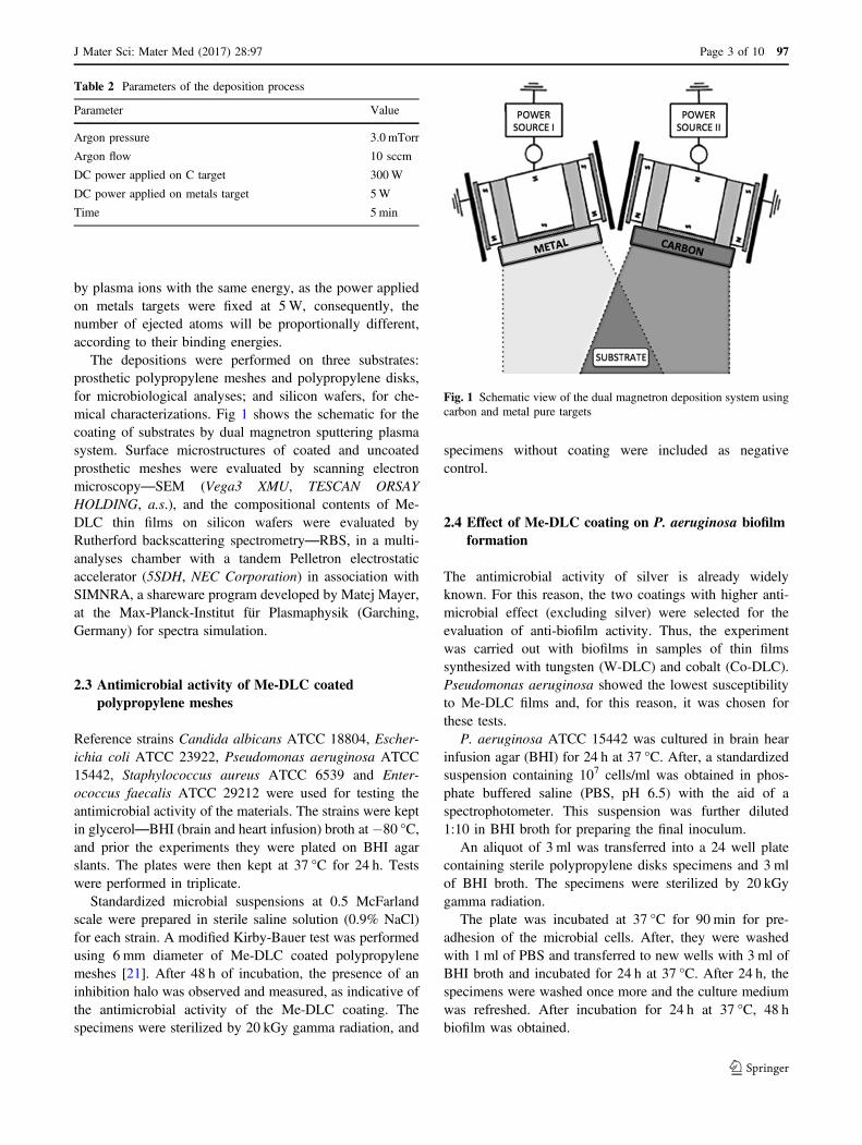
by plasma ions with the same energy, as the power appliedon metals targets were fixed at 5W, consequently, thenumber of ejected atoms will be proportionally different,according to their binding energies.
The depositions were performed on three substrates:prosthetic polypropylene meshes and polypropylene disks,for microbiological analyses; and silicon wafers, for che-mical characterizations. Fig 1 shows the schematic for thecoating of substrates by dual magnetron sputtering plasmasystem. Surface microstructures of coated and uncoatedprosthetic meshes were evaluated by scanning electronmicroscopy—SEM (Vega3 XMU, TESCAN ORSAYHOLDING, a.s.), and the compositional contents of Me-DLC thin films on silicon wafers were evaluated byRutherford backscattering spectrometry—RBS, in a multi-analyses chamber with a tandem Pelletron electrostaticaccelerator (5SDH, NEC Corporation) in association withSIMNRA, a shareware program developed by Matej Mayer,at the Max-Planck-Institut für Plasmaphysik (Garching,Germany) for spectra simulation.
2.3 Antimicrobial activity of Me-DLC coatedpolypropylene meshes
Reference strains Candida albicans ATCC 18804, Escher-ichia coli ATCC 23922, Pseudomonas aeruginosa ATCC15442, Staphylococcus aureus ATCC 6539 and Enter-ococcus faecalis ATCC 29212 were used for testing theantimicrobial activity of the materials. The strains were keptin glycerol—BHI (brain and heart infusion) broth at −80 °C,and prior the experiments they were plated on BHI agarslants. The plates were then kept at 37 °C for 24 h. Testswere performed in triplicate.
Standardized microbial suspensions at 0.5 McFarlandscale were prepared in sterile saline solution (0.9% NaCl)for each strain. A modified Kirby-Bauer test was performedusing 6 mm diameter of Me-DLC coated polypropylenemeshes [21]. After 48 h of incubation, the presence of aninhibition halo was observed and measured, as indicative ofthe antimicrobial activity of the Me-DLC coating. Thespecimens were sterilized by 20 kGy gamma radiation, and
specimens without coating were included as negativecontrol.
2.4 Effect of Me-DLC coating on P. aeruginosa biofilmformation
The antimicrobial activity of silver is already widelyknown. For this reason, the two coatings with higher anti-microbial effect (excluding silver) were selected for theevaluation of anti-biofilm activity. Thus, the experimentwas carried out with biofilms in samples of thin filmssynthesized with tungsten (W-DLC) and cobalt (Co-DLC).Pseudomonas aeruginosa showed the lowest susceptibilityto Me-DLC films and, for this reason, it was chosen forthese tests.
P. aeruginosa ATCC 15442 was cultured in brain hearinfusion agar (BHI) for 24 h at 37 °C. After, a standardizedsuspension containing 107 cells/ml was obtained in phos-phate buffered saline (PBS, pH 6.5) with the aid of aspectrophotometer. This suspension was further diluted1:10 in BHI broth for preparing the final inoculum.
An aliquot of 3 ml was transferred into a 24 well platecontaining sterile polypropylene disks specimens and 3 mlof BHI broth. The specimens were sterilized by 20 kGygamma radiation.
The plate was incubated at 37 °C for 90 min for pre-adhesion of the microbial cells. After, they were washedwith 1 ml of PBS and transferred to new wells with 3 ml ofBHI broth and incubated for 24 h at 37 °C. After 24 h, thespecimens were washed once more and the culture mediumwas refreshed. After incubation for 24 h at 37 °C, 48 hbiofilm was obtained.
Table 2 Parameters of the deposition process
Parameter Value
Argon pressure 3.0 mTorr
Argon flow 10 sccm
DC power applied on C target 300W
DC power applied on metals target 5W
Time 5 min
Fig. 1 Schematic view of the dual magnetron deposition system usingcarbon and metal pure targets
J Mater Sci: Mater Med (2017) 28:97 Page 3 of 10 97
Then, the biofilm formed on the specimens were gentlywashed with PBS and sonicated on ice for 30 s in PBS.Initial suspensions were diluted in PBS and plated on BHIagar to obtain the number of colony forming units perspecimen (CFU/specimen). Uncoated polypropylene sam-ples were used as negative control.
2.5 Analyses of results
The results obtained for the effects of coatings on P. aer-uginosa biofilm formation were statistically compared.Values of colony forming units per specimen (CFU/speci-men) in the groups tungsten (W-DLC), cobalt (Co-DLC)and control (uncoated) were compared by ANOVA andDunnett’s post hoc test. The level of significance wasset as 5%.
3 Results
3.1 Material characterization
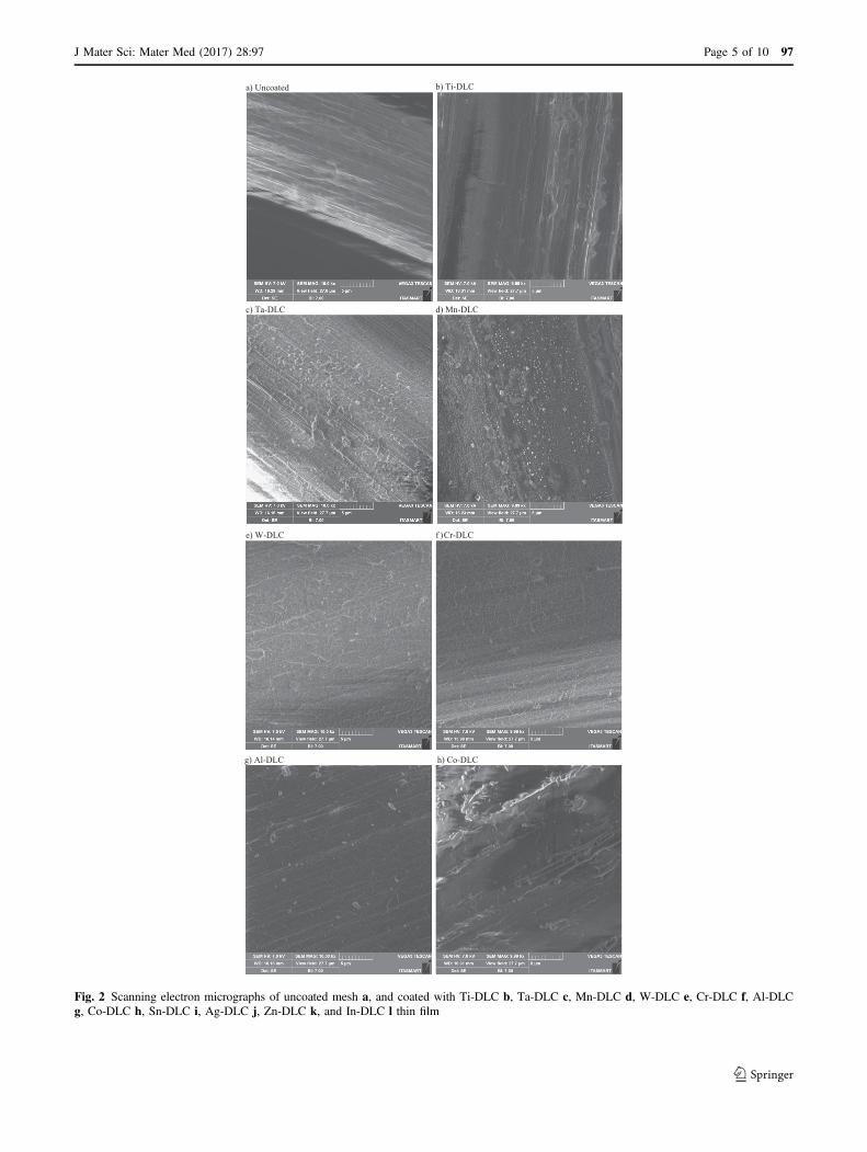
SEM was performed on coated and uncoated prostheticmeshes, and their scanning electron micrographs are shownin Fig 2. The uncoated mesh fibers presented homogeneoussurface with typical manufacturing grooves (Fig. 2a). It alsocan be clearly seen that in all coated meshes (Figs. 2b–l),the deposited thin film did not cover the fiber manufacturinggrooves. However, some contrasting microstructures wereobserved. In Figs. 2e (W-DLC), 2f (Cr-DLC), 2i (Sn-DLC)and 2l (In-DLC), vermicular or worm-shaped micro-structures were formed on the surface. On the other hand, inTi-DLC (Fig. 2b), Mn-DLC (Fig. 2d), Al-DLC (Fig. 2g),Co-DLC (Fig. 2h), Ag-DLC (Fig. 2j), and Zn-DLC(Fig. 2k) coated fiber surfaces, only nanometer-sizedgrains, probably clusters are viewed. The exception wasthe Ta-DLC coated fiber (Fig. 2c), which exhibited both:grains and vermicular microstructures.
3.2 Me-DLC compositional contents by RBS
Obtained elemental contents of Me-DLC coatings are pre-sented in Table 3. Expected variations in metal percentageswere observed, as differences in binding energies lead todistinct sputter yields.
3.3 Me-DLC coated polypropylene meshes antimicrobialactivity
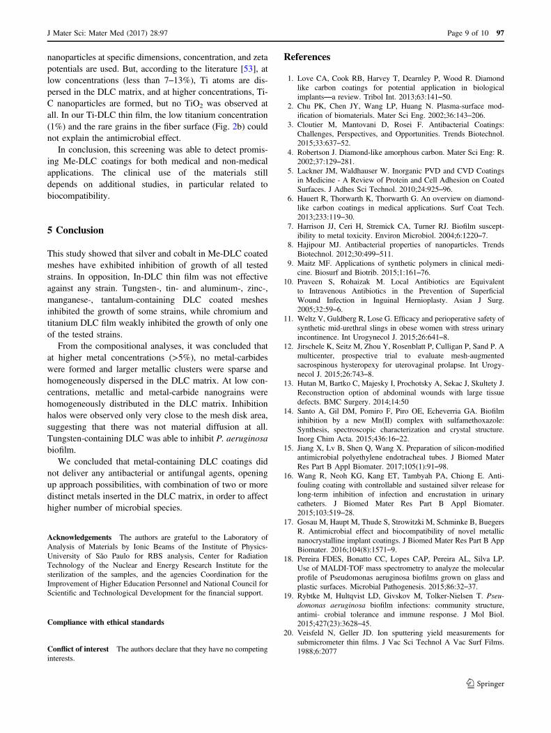
The antimicrobial effect of each metal-doped coatingagainst the microbial species is summarized in Table 4.These effects were estimated by the values of diffusionhalos around the specimens and the microbial growth over
the fibers of meshes. It is important to observe that inconventional Kirby-Bauer protocol, the paper disk isimpregnated with a liquid antibiotic. This disk is placed onthe agar plate and the antibiotic begins to diffuse into thesurrounding agar. That is why this method is also calledKirby-Bauer diffusion test. In our modified Kirby-Bauertest, Me-DLC coated polypropylene mesh disks were used.The Me-DLC thin film is solid and firmely fixed on themesh surface; hence the thin film cannot diffuse into theagar. In our analyses, we set the Ag-DLC halo of 8.0 mm asthe inhibition bias (H). Halos lower than 8.0 mm meanintermediate inhibition (I). (F) means no inhibition halowith no microbial growth on mesh fibers surface, and (N)refers to no inhibition halo and microbial growing over themesh. Our results cannot be compared with conventionalKirby-Bauer protocol results by any means.
Ag-DLC showed inhibition with halos ≥ 8.0 mm for allthe tested microbial species. Co-DLC also had inhibitoryeffects on all the species. Tungsten-DLC, tin-DLC, alumi-num-DLC, zinc-DLC, manganese-DLC, tantalum-DLCinhibited the growth of some strains, while chromium-DLC and titanium-DLC did not show inhibition halosagainst any strain. In-DLC film showed no inhibitionactivity.
3.4 Effect of Me-DLC coating on P. aeruginosa biofilmformation
P. aeruginosa biofilm formation varied among the groups(ANOVA, p= 0.0003) and are shown in Fig. 3. Resultswere expressed by the number of CFU/specimen. The meanvalues and standard deviation of CFU/specimen in thegroup cobalt-containing DLC (Co-DLC) was 73.16±25.51. For group tungsten-containing DLC (W-DLC) thisvalue was 16.16± 10.32. Uncoated control group showed77.33± 15.69.
Data analyses showed that W-DLC film was able tosignificantly reduce the biofilm formation in relation tocontrol (p< 0.05). Conversely, biofilm formation in thesurface of Co-DLC was similar in relation to control (p=0.964).
4 Discussion
The importance of this work relies on the innovative pro-posal of metal-containing thin film deposition over meshes(bundle of soft polypropylene fibers) aiming to contribute toreduction in postoperative microbial infections.
In addition to deposition parameters, the metal content isa relevant issue in Me-DLC thin films, because metalliccomponents are usually grouped in small metallic or metal-carbide clusters (nanometer-sized grains). According to
97 Page 4 of 10 J Mater Sci: Mater Med (2017) 28:97
CLD-iT)bdetaocnU)a
CLD-nM)dCLD-aT)c
CLD-rC)fCLD-W)e
g) Al-DLC h) Co-DLC
Fig. 2 Scanning electron micrographs of uncoated mesh a, and coated with Ti-DLC b, Ta-DLC c, Mn-DLC d, W-DLC e, Cr-DLC f, Al-DLCg, Co-DLC h, Sn-DLC i, Ag-DLC j, Zn-DLC k, and In-DLC l thin film
J Mater Sci: Mater Med (2017) 28:97 Page 5 of 10 97
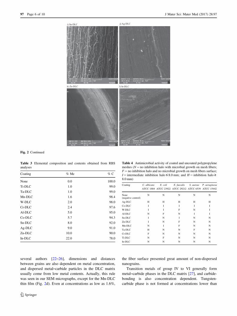
several authors [22–26], dimensions and distancesbetween grains are also dependent on metal concentration,and dispersed metal–carbide particles in the DLC matrixusually come from low metal contents. Actually, this rulewas seen in our SEM micrographs, except for the Mn-DLCthin film (Fig. 2d). Even at concentrations as low as 1.6%,
the fiber surface presented great amount of non-dispersednanograins.
Transition metals of group IV to VI generally formmetal–carbide phases in the DLC matrix [27], and carbide-bonding is also concentration dependent. Tungsten-carbide phase is not formed at concentrations lower than
k) Zn
i) Sn-DLC j) Ag-DLC
-DLC l) In-DLC
Fig. 2 Continued
Table 4 Antimicrobial activity of coated and uncoated polypropylenemeshes (N= no inhibition halo with microbial growth on mesh fibers;F= no inhibition halo and no microbial growth on mesh fibers surface;I= intermediate inhibition halo ≤ 8.0 mm; and H= inhibition halo ≥8.0 mm)
Coating C. albicans E. coli E. faecalis S. aureus P. aeruginosa
ATCC 1884 ATCC 23922 ATCC 29212 ATCC 6539 ATCC 15442
None(negative control)
N N N N N
Ag-DLC H H H H H
Co-DLC I I I I I
W-DLC I I F N I
Al-DLC N F N I I
Sn-DLC I N I N N
Zn-DLC I N F N N
Mn-DLC N I F N N
Ta-DLC H N N F N
Cr-DLC F N N N N
Ti-DLC N F N N N
In-DLC N N N N N
Table 3 Elemental composition and contents obtained from RBSanalyses
Coating % Me % C
None 0.0 100.0
Ti-DLC 1.0 99.0
Ta-DLC 1.0 99.0
Mn-DLC 1.6 98.4
W-DLC 2.0 98.0
Cr-DLC 2.4 97.6
Al-DLC 5.0 95.0
Co-DLC 5.7 94.3
Sn-DLC 8.0 92.0
Ag-DLC 9.0 91.0
Zn-DLC 10.0 90.0
In-DLC 22.0 78.0
97 Page 6 of 10 J Mater Sci: Mater Med (2017) 28:97

2.8 at%, but amorphous and crystalline tungsten-carbideare formed at concentrations higher than 2.8 at% and3.6 at%, respectively [24]. Furthermore, still accordingto Bewilogua et al. [27], metal–carbide particles may not beso strongly cross-linked with the surrounding amorphouscarbon network, and can react more readily with theoxygen.
On the other hand, while aluminum carbide is commer-cially supplied by the market, according to Dai and Wang[23], aluminum does not form carbide when incorporatedinto DLC films.
From the SEM micrographs and Table 3, it can beinferred that, on the one hand, in titanium-DLC, aluminum-DLC, cobalt-DLC, silver-DLC, and zinc-containing DLCfilms, carbides were not formed and larger metallic clusterswere sparse and homogeneously dispersed at the surface,and most probably, in the DLC matrix. On the other, intungsten- and chromium-containing DLC films, metallicand metal-carbide nanograins were possibly embedded intothe DLC matrix bulk.
As well pointed by Dai and Wang [23], the compre-hensive understanding of metal-containing DLC coatingswas still not fully established owing not only to the com-plexity of carbon hybridized bonds, particularly at lowmetal contents, but to the diversity of metal nature, and tothe variety of deposition techniques as well.
The antimicrobial response of Me-DLC coated poly-propylene meshes, the inhibition halo, if any, was observedonly very close to the mesh disk area, suggesting a localpassive mechanism of action, and no diffusion of metals.The Me-DLC thin film is solid and strongly fastened on themesh surface; hence the thin film could not diffuse intothe agar. This is an interesting finding, considering that the
spread of metals to the environment would be negative forbiomedical applications. Future studies on the cyto andgenotoxicity of these materials to human cells are stillneeded.
Predictably, the Ag-DLC coated polypropylene mesh hasexhibited the highest antimicrobial activity against all testedmicroorganisms, with inhibiting halo around 8.0 mm dia-meter. Silver has been used since ancient times [28] and iscurrently the most used antimicrobial metal [29]. Itstoxic effect is well-known [30–32], and Das et al. [30]reported that carboxymethylcellulose gel containing silvernanoparticles had been effective against the growth ofmethicillin-resistant S. aureus (ATCC 6538), E. coli (ATCC8739) and P. aeruginosa (ATCC 9027), even at con-centrations as low as 50 ppm. In Ag-DLC coatings, silver isgrouped in nanometer-sized clusters, which are dispersed inthe Ag-DLC matrix [22], and in the fiber surface, as well(Fig. 2j). Hence, we believe that the antimicrobial effect ofthe Ag-DLC coated mesh should probably be due to thesenanograins on the thin film surface.
On the contrary, indium-containing DLC coated meshdid not have any observable antimicrobial action, even atappreciable contents as 22%. However, some bacteriostaticactivity of In3+ ion had been observed when chelated(attached to an anion group with two or more coordinationsites) to a siderophore (iron-binding protein) [28]. Accord-ing to the literature, this heavy metal ion mimics the Fe3+ inthe protein, but does not replace its function in the cellmetabolism, acting as a toxic substance. In our In-DLCcoated mesh, vermicular formation tendency was noted inFig. 2l, suggesting that metallic-indium clusters should beembedded into the DLC matrix bulk, as observed with otherhigh content Me-DLC films [33]. Indium-carbide cannot befound in In-DLC film, as it is gaseous at ambient tem-perature [34]. Hence, the tested microorganisms could notuptake In3+ ions from the DLC matrix to their metabolism,and probably, metallic-indium clusters were so embeddedinto the DLC matrix that microbial cells have grown overthe In-DLC coated mesh fibers. Despite the low cobaltcontent in the thin film, and the sparse nanosized-grainsdistributed on the mesh fiber surface (Fig. 2h), the Co-DLCcoated polypropylene mesh showed the second most intenseantimicrobial activity. Actually, some authors had alsoobserved cobalt antimicrobial activity, as cobalt complexes[35–38]. Although the antimicrobial mechanism of cobalt isstill not fully elucidated, care should be taken as metalcomplexes effects on microorganisms are far different,compared with both the pure metal and the metal in theDLC coatings.
On the other hand, Icgen and Yilmaz [39] published aninteresting work on the resistance of isolates from a Turkishriver to heavy metals and antibiotic drugs. Due to effluentscontaminated with antibiotics and heavy metals,
Control Co-DLC W-DLC0
10
20
30
40
50
60
70
80
90
100
*
CFU
/Spe
cim
en
Fig. 3 Inhibition of Pseudomonas aeruginosa biofilm formation byplasma coating polymers; histograms represent means and standarddeviations. * statistically significant difference in relation to control(p< 0.05)
J Mater Sci: Mater Med (2017) 28:97 Page 7 of 10 97
microorganisms from this river are becoming drug-resistantand with enhanced heavy metal resistance as well. Not-withstanding, isolates exhibited the least resistance againstcobalt: only 4%, corroborating our results.
Tungsten-containing DLC coating showed activityagainst C. albicans, E. coli, E. faecalis and P. aeruginosastrains, but not against S. aureus. Considering the very lowtungsten content in our W-DLC coating, probably tungsten-carbide clusters should have not been formed, and evenmetallic nanoparticles in the coating surface were notobserved in our SEM micrographs (Fig. 2e). Lemire et al.[28] stated that metals, including tungsten, are either coor-dinated by organic compounds or present as water-solubleoxyanion species in the environment. On the other hand,some authors had also related poor or no antimicrobialactivity of WO3 nanoparticles against peri-implantitis bac-teria [40], and even tungsten oxyanion (WO4
2−) had pre-sented no significant toxicity against E. coli (JM 109), S.aureus (ATCC 29213), and P. aeruginosa (ATCC 27853)[7], emphasizing the low antimicrobial effectiveness oftungsten. Interestingly, tungsten containing DLC coatinghas shown promising anti-biofilm formation effect. Thiseffect was not reported before and deeper investigation ofthe mechanism of action is needed. Besides, recent evi-dences of tungsten toxicity for human cells have beenreported [41]. For this reason, the minimal effective con-centration necessary for microbial biofilm inhibition and thepotential liberation of tungsten for human tissues have stillto be determined before suggesting an application of ourfindings. Notwithstanding, the promising effect of W-DLCcoatings can be useful for the development of severalmaterials with antifouling properties.
Both tin- (Sn) and aluminum- (Al) containing DLCcoatings had antimicrobial action against some of themicroorganisms studied, the first one against C. albicans,and E. faecalis, and the second, against S. aureus, P. aer-uginosa and E. Coli. Icgen and Yilmaz have showed that67% of isolates were resistant to tin, and 79% to aluminum[39]. In other recent publication, SnO2/SnS2 nanocompo-sites have shown more effectiveness against E. coli (ATCC25922) and S. aureus (ATCC 6538) than SnO2 and SnS2nanoparticles. Nanocrystalline SnO2 thin films deposited bysol-gel exhibited antibacterial activity against E. coli andBacillus spp. [42] as well. Fakhri et al. [43] also hadreported antifungal activity of SnO2 and SnS2 nanoparticles,and SnO2/SnS2 nanocomposites against C. albicans. Incontrast, in our SEM micrographs (Fig. 2i), nanoparticleswere not present in the fiber surface. Concerning aluminum,fresh report [44] demonstrated that aluminum oxide (AlO3)nanoparticles had showed concentration dependent inhibi-tion of E. coli growing, probably by generating reactiveoxygen species, in Fig. 2g sparse nanosized-grains can beviewed. Care should be taken, as DLC films containing
incorporated metals are far different from pure metals,metal-oxides (SnO2, AlO3) and metal-compounds (SnS2).
Chromium-containing DLC and zinc-containing DLCcoatings have both revealed antifungal potential against C.albicans. According to Páez et al. [45], some chromiumcomplexes can produce DNA damage and may show anti-bacterial and antifungal activity. However, metal complexestoxicity depends on the coordinated ligand. Han et al. [46]demonstrated that chromium-carbide is formed at the film-substrate interface when the Cr+ ion is implanted on DLCfilms deposited by magnetron sputtering over silicon sub-strate. This might have happened with our Cr-DLC coatingas well (Fig. 2f). On the other hand, Jenilek et al. [47] alsotested antimicrobial activities of Cr-DLC for implants,prepared by laser-magnetron deposition. These authors havecoated silicon, Ti6Al4V and CoCrMo substrates with DLCand Cr-DLC layers. Despite differences in the depositiontechnique and in the film characteristics compared withours, they did not observe antibacterial effects against S.aureus and P. aeruginosa. Concerning zinc, Pop et al. [44]reported that zinc ions exhibit bacteriostatic effect, byinhibiting different physiological pathways, while ZnO hasantimicrobial effect by oxidative stress. And Singh et al.[48] reported that zinc-complexes had exhibited the highestantimicrobial activity against Pseudomonas aeruginosa,Bacillus subtilis, Escherichia coli, and Staphylococcusaureus, and the highest antifungal properties againstAspergillus niger, and Aspergillus flavus.
Manganese-containing DLC film exhibited moderatedantimicrobial activity against E. coli and E. faecalis. Someauthors have reported antibacterial and antifungal activitiesof Mn(II)-complexes [14, 49] and Mn(III)-complex [50].However, to the best of our knowledge, characterizationsand antimicrobial activities of Mn-DLC were not found inthe literature.
Tantalum and titanium are both well-known highly bio-compatible and corrosion resistant metals [51]. To the bestof our knowledge, no antimicrobial effect has been attrib-uted to pure Ta and Ti metals. Tsai et al. [52] have reportedthat in the DLC matrix, crystalline β-Ta and a small fractionof tantalum-carbide is formed, resulting in high bio-compatibility with WS1 human fetal skin fibroblast cells. Inour antimicrobial test, the Ta-DLC coated mesh revealedsome antifungal activity against C. albicans. This is apromising outcome, as C. albicans is one of the mostcommon causes of nosocomial infections, and the Ta-DLCcoated mesh should associate high biocompatibility withantifungal effect. Evidently, more studies should be con-ducted to attest this finding.
Ti-DLC coated meshes have prevented growth of E. colion material surface. Antimicrobial properties of titaniumdioxide had been observed when irradiated by ultravioletlight at appropriate wavelengths [44], or when TiO2
97 Page 8 of 10 J Mater Sci: Mater Med (2017) 28:97
nanoparticles at specific dimensions, concentration, and zetapotentials are used. But, according to the literature [53], atlow concentrations (less than 7–13%), Ti atoms are dis-persed in the DLC matrix, and at higher concentrations, Ti-C nanoparticles are formed, but no TiO2 was observed atall. In our Ti-DLC thin film, the low titanium concentration(1%) and the rare grains in the fiber surface (Fig. 2b) couldnot explain the antimicrobial effect.
In conclusion, this screening was able to detect promis-ing Me-DLC coatings for both medical and non-medicalapplications. The clinical use of the materials stilldepends on additional studies, in particular related tobiocompatibility.
5 Conclusion
This study showed that silver and cobalt in Me-DLC coatedmeshes have exhibited inhibition of growth of all testedstrains. In opposition, In-DLC thin film was not effectiveagainst any strain. Tungsten-, tin- and aluminum-, zinc-,manganese-, tantalum-containing DLC coated meshesinhibited the growth of some strains, while chromium andtitanium DLC film weakly inhibited the growth of only oneof the tested strains.
From the compositional analyses, it was concluded thatat higher metal concentrations (>5%), no metal-carbideswere formed and larger metallic clusters were sparse andhomogeneously dispersed in the DLC matrix. At low con-centrations, metallic and metal-carbide nanograins werehomogeneously distributed in the DLC matrix. Inhibitionhalos were observed only very close to the mesh disk area,suggesting that there was not material diffusion at all.Tungsten-containing DLC was able to inhibit P. aeruginosabiofilm.
We concluded that metal-containing DLC coatings didnot deliver any antibacterial or antifungal agents, openingup approach possibilities, with combination of two or moredistinct metals inserted in the DLC matrix, in order to affecthigher number of microbial species.
Acknowledgements The authors are grateful to the Laboratory ofAnalysis of Materials by Ionic Beams of the Institute of Physics-University of São Paulo for RBS analysis, Center for RadiationTechnology of the Nuclear and Energy Research Institute for thesterilization of the samples, and the agencies Coordination for theImprovement of Higher Education Personnel and National Council forScientific and Technological Development for the financial support.
Compliance with ethical standards
Conflict of interest The authors declare that they have no competinginterests.
References
1. Love CA, Cook RB, Harvey T, Dearnley P, Wood R. Diamondlike carbon coatings for potential application in biologicalimplants—a review. Tribol Int. 2013;63:141–50.
2. Chu PK, Chen JY, Wang LP, Huang N. Plasma-surface mod-ification of biomaterials. Mater Sci Eng. 2002;36:143–206.
3. Cloutier M, Mantovani D, Rosei F. Antibacterial Coatings:Challenges, Perspectives, and Opportunities. Trends Biotechnol.2015;33:637–52.
4. Robertson J. Diamond-like amorphous carbon. Mater Sci Eng: R.2002;37:129–281.
5. Lackner JM, Waldhauser W. Inorganic PVD and CVD Coatingsin Medicine - A Review of Protein and Cell Adhesion on CoatedSurfaces. J Adhes Sci Technol. 2010;24:925–96.
6. Hauert R, Thorwarth K, Thorwarth G. An overview on diamond-like carbon coatings in medical applications. Surf Coat Tech.2013;233:119–30.
7. Harrison JJ, Ceri H, Stremick CA, Turner RJ. Biofilm suscept-ibility to metal toxicity. Environ Microbiol. 2004;6:1220–7.
8. Hajipour MJ. Antibacterial properties of nanoparticles. TrendsBiotechnol. 2012;30:499–511.
9. Maitz MF. Applications of synthetic polymers in clinical medi-cine. Biosurf and Biotrib. 2015;1:161–76.
10. Praveen S, Rohaizak M. Local Antibiotics are Equivalentto Intravenous Antibiotics in the Prevention of SuperficialWound Infection in Inguinal Hernioplasty. Asian J Surg.2005;32:59–6.
11. Weltz V, Guldberg R, Lose G. Efficacy and perioperative safety ofsynthetic mid-urethral slings in obese women with stress urinaryincontinence. Int Urogynecol J. 2015;26:641–8.
12. Jirschele K, Seitz M, Zhou Y, Rosenblatt P, Culligan P, Sand P. Amulticenter, prospective trial to evaluate mesh-augmentedsacrospinous hysteropexy for uterovaginal prolapse. Int Urogy-necol J. 2015;26:743–8.
13. Hutan M, Bartko C, Majesky I, Prochotsky A, Sekac J, Skultety J.Reconstruction option of abdominal wounds with large tissuedefects. BMC Surgery. 2014;14:50
14. Santo A, Gil DM, Pomiro F, Piro OE, Echeverria GA. Biofilminhibition by a new Mn(II) complex with sulfamethoxazole:Synthesis, spectroscopic characterization and crystal structure.Inorg Chim Acta. 2015;436:16–22.
15. Jiang X, Lv B, Shen Q, Wang X. Preparation of silicon-modifiedantimicrobial polyethylene endotracheal tubes. J Biomed MaterRes Part B Appl Biomater. 2017;105(1):91–98.
16. Wang R, Neoh KG, Kang ET, Tambyah PA, Chiong E. Anti-fouling coating with controllable and sustained silver release forlong-term inhibition of infection and encrustation in urinarycatheters. J Biomed Mater Res Part B Appl Biomater.2015;103:519–28.
17. Gosau M, Haupt M, Thude S, Strowitzki M, Schminke B, BuegersR. Antimicrobial effect and biocompatibility of novel metallicnanocrystalline implant coatings. J Biomed Mater Res Part B AppBiomater. 2016;104(8):1571–9.
18. Pereira FDES, Bonatto CC, Lopes CAP, Pereira AL, Silva LP.Use of MALDI-TOF mass spectrometry to analyze the molecularprofile of Pseudomonas aeruginosa biofilms grown on glass andplastic surfaces. Microbial Pathogenesis. 2015;86:32–37.
19. Rybtke M, Hultqvist LD, Givskov M, Tolker-Nielsen T. Pseu-domonas aeruginosa biofilm infections: community structure,antimi- crobial tolerance and immune response. J Mol Biol.2015;427(23):3628–45.
20. Veisfeld N, Geller JD. Ion sputtering yield measurements forsubmicrometer thin films. J Vac Sci Technol A Vac Surf Films.1988;6:2077
J Mater Sci: Mater Med (2017) 28:97 Page 9 of 10 97

21. Hudzicki J. Kirby-Bauer Disk Diffusion Susceptibility Test Pro-tocol. ASM MicrobeLibrary. 2013. http://www.microbelibrary.org/component/resource/laboratory-test/3189-kirby-bauer-disk-diffusion-susceptibility-test-protocol. Accessed 15 Sep 2016.
22. Takeno T, Saito H, Goto M, Fontaine J, et al. Deposition, structureand tribological behavior of silver–carbon nanocomposite coat-ings. Diamond Relat Mater. 2013;39:20–26.
23. Dai W, Wang A. Deposition and properties of Al-containingdiamond-like carbon films by a hybrid ion beam sources. J AlloysCompd. 2011;509:4626–31.
24. Wang AY, Lee KR, Ahn JP, Han JH. Structure and mechanicalproperties of W incorporated diamond-like carbon films preparedby a hybrid ion beam deposition technique. Carbon. 2006;44:1826–32.
25. Zhang S, Bui XL, Jiang J, Li X. Microstructure and tribologicalproperties of magnetron sputtered nc-TiC/a-C nanocomposite.Surf Coat Technol. 2005;198:206–11.
26. Rogers HJ, Woods VE, Synge C. Antibacterial effect of theScandium and Indium complexes of enterochelin on Escherichiacoli. J Gen Microbiol. 1982;128:2389–94.
27. Bewilogua K, Cooper CV, Specht C, Schröder J, Witorff R,Grischke M. Effect of target material on deposition and propertiesof metal-containing DLC (Me-DLC) coatings. Surf Coat Technol.2000;127:224–32.
28. Lemire JA, Harrison JJ, Turner RJ. Antimicrobial activity ofmetals: mechanisms, molecular targets and applications. Nat RevMicrobiol. 2013;11:371–84.
29. Barillo DJ, Marx DE. Silver in medicine: A brief history BC 335to present. Burns. 2014;40:S3–S8.
30. Das A, Kumara A, Patilb NB, Viswanathana C, Ghosh D. Pre-paration and characterization of silver nanoparticle loadedamor-phous hydrogel of carboxymethylcellulose for infected wounds.Carbohyd Polym. 2015;130:254–61.
31. Prabhu S, Poulose EK. Silver nanoparticles: mechanism of anti-microbial action, synthesis, medical applications, and toxicityeffects. Int Nano Lett. 2012;2:1–10.
32. Kim JS, Kuk E, Yu KN, Kim Jong-Ho, et al. Antimicrobial effectsof silver nanoparticles. Nanomed Nanotech Biol Med.2007;3:95–101.
33. Schiffmann KI, Fryda M, Goerigk G, Lauer R, Hinze P, Bulack A.Sizes and distances of metal clusters in Au-, Pt-, W- and Fe-containing diamond-like carbon hard coatings: a comparativestudy by small angle X-ray scattering, wide angle X-ray diffrac-tion, transmission electron microscopy and scanning tunnellingmicroscopy. Thin Solid Films. 1999;347:60–71.
34. Breuer L, Kucher A, Herder M, Wucher A, Winograd N. For-mation of neutral InmCn clusters under C60 ion bombardment ofindium. J Phys Chem A. 2014;118:8542–52.
35. Hu XM, Xue LW, Zhao GQ, Yang WC. Synthesis, structures, andbiological activity of Terbium(III) and Cobalt(III) complexesderived from tripodal Schiff bases. Russ J Coord Chem.2015;41:197–201.
36. Yuoh ACB, Agwara MO, Yufanyi DM, Conde MA, Jagan R,Eyong KO. Synthesis, crystal structure, and antimicrobial prop-erties of a novel 1-D cobalt coordination polymer with dicyana-mide and 2-aminopyridine. Int J Inorg Chem. 2015;2015:106838.http://dx.doi.org/10.1155/2015/106838.
37. Singh K, Kumar Y, Puri P, Kumar M, Sharma C. Cobalt, nickel,copper and zinc complexes with 1,3-diphenyl-1H-pyrazole-4-carboxaldehyde Schiff bases: Antimicrobial, spectroscopic, ther-mal and fluorescence studies. Eur J Med Chem. 2012;52:313–21.
38. Chang EL, Simmers C, Knight DA. Cobalt complexes as antiviraland antibacterial agents. Pharmaceuticals. 2010;3:1711–28.
39. Icgen B, Yilmaz F. Co-occurrence of antibiotic and heavy metalresistance in Kýzýlýrmak river isolates. Bull Environ ContamToxicol. 2014;93:735–43.
40. Vargas-reusa MA, Memarzadeh K, Huang J, Ren GG, Allaker RP.Antimicrobial activity of nanoparticulate metal oxides againstperi-implantitis pathogens. Int J Antimicrob Agents. 2012;40:135–9.
41. Bolt AM, Mann KK. Tungsten: an Emerging Toxicant, Alone orin Combination. Curr Envir Health Rpt. 2016;3:405
42. Henry J, Mohanraj K, Sivakumar G, Umamaheswari S. Electro-chemical and fluorescence properties of SnO2 thin films and itsantibacterial activity. Spectrochim Acta Part A. 2015;143:172–8.
43. Fakhri A, Behrouz S, Pourmand M. Synthesis, photocatalytic andantimicrobial properties of SnO2, SnS2 and SnO2/SnS2 nanos-tructure. J Photochem Photobiol B Biol. 2015;149:45–50.
44. Pop CS, Hussien MD, Popa M, Mares A, et al. Metallic-basedmicro and nanostructures with antimicrobial activity. Curr TopMed Chem. 2015;15(16):1577–82.
45. Paéz PL, Bazán CM, Bongiovanni ME et al. Oxidative stress andantimicrobial activity of Chromium(III) and Ruthenium(II) com-plexes on Staphylococcus aureus and Escherichia coli. BiomedRes Int. 2013;2013.
46. Han X, Yan F, Zhang A, Yan P, et al. Structure and tribologicalbehavior of amorphous carbon films implanted with Cr+ ions.Mater Sci Eng A. 2003;348:319–26.
47. Jenilek M, Kocourek T, Zemek J, et al. Chromium-doped DLC forimplants prepared by laser-magnetron deposition. Mater Sci EngC. 2015;46:381–6.
48. Singh K, Kumar Y, Puri P, Sharma C, Aneja KR. Antimicrobial,spectral and thermal studies of divalent cobalt, nickel, copper andzinc complexes with triazole Schiff bases. Arab J Chem. 2013;10:S978–S987.
49. Bhaskar R, Salunkhe N, Yaul A, Aswar A. Bivalent transitionmetal complexes of ONO donor hydrazone ligand: Synthesis,structural characterization and antimicrobial activity. SpectrochimActa Part A. 2015;151:621–7.
50. Xue L, Deng DN, Xu Y, Wang Q. Synthesis, Crystal Structure,and Antibacterial Activity of a Manganese(III) Complex Derivedfrom N,N’-3,4-Chlorophenylene-Bis(5-Methylsalicylaldimine).Russ J Coord Chem. 2015;41:772–6.
51. Chen Q, Thouas G. Biomaterials: a basic introduction. 1st ed.Boca Raton (FL): CRC Press; 2015.
52. Tsai MT, Chang YY, Huang HL, Chen YC, Wang SP, Lai CH.Reprint of “Biological characteristics of human fetal skin fibro-blasts and MG-63 human osteosarcoma cells on tantalum-dopedcarbon films”. Surf Coat Tech. 2014;259:213–8.
53. Dai W, Ke P, Moon MW, Lee KR, Wang A. Investigation of themicrostructure, mechanical properties and tribological behaviorsof Ti-containing diamond-like carbon films fabricated by a hybridion beam method. Thin Solid Films. 2012;520:6057–63.
97 Page 10 of 10 J Mater Sci: Mater Med (2017) 28:97