relationship between thigmotropism and candida biofilm
TRANSCRIPT
Relationship between thigmotropism and Candida biofilm
formation in vitro.
Abstract
The biofilm formation of the oral fungal pathogen Candida on denture acrylic strips coated
with saliva or serum were examined in relation to the ability to induce hyphae by thigmotropic
reaction, using C. albicans (4 isolates), C. glabrata (3 isolates) and C. tropicalis (3 isolates).
Both the degree of biofilm formation and the amount of hyphae exhibiting thigmotropism
varied depending upon both the species and strains of Candida. Although no signioficant
correlation between the amount of hyphae induced by thigmotropic reaction of fungal isolates
and biofilm formation on uncoated control specimans (r=0.577; p.0.05), the ability of hyphae
induced by thigmotropic reaction significantly correlated with the amount of both saliva- and
serum-admixed biofilms (r=0.734; p<0.05 and r=0.793; p<0.01, respectively).
Taken together our in vitro data suggested that the hyphal induction by thigmotropic reaction
is of importance in candidal biofilm formation on saliva- or serum-coated acrylic surfaces.
Introduction
In Candida-associated denture stomatitis (syn. chronic atrophic candidiasis), a
commoninfection seen elderly denture wearers, the acrylic denture fitting surface acts as a
reservoir of infection (3,4,8)- Although C. albicans is by far the predominant isolate in this
condition other non-albicans species such as C. tropicalis and C. glabrata (syn. Torulopsis
glabrata) are frequently isolated both from the acrylic denture surfaces and the palatal mucosa
(4).
Despite the realization that successful candidal colonization of denture surface is an
important step in the pathogenesis of this condition (22), the role played by saliva or serum
pellicles during the colonization process and subsequent multilayer biofilm formation is
poorly understood. The relationship between the salivary or serum pellicle on denture surfaces
and candidal colonization is a complex subject, since both saliva and serum, which modulate
clearance, aggregation, adherence and nutrition of microorganisms are biological fluids of
immense complexity contributing to specific and non-specific interactions in fungal
colonization. Recently, we developed a bioluminescent adenosine triphosphate (ATP) assay
based on firefly luciferase-luciferin system (1,10,14), and demonstrated the reduced activity of
Candida albicans biofilms on saliva-coated acrylic strips, although on prolonged incubation,
both saliva and serum promoted fungal biofilm activity. (20). There is, however, no data on
the formation of biofilms by non-albicans species such as C. glabrata and C. tropicalis.
Besides the interactions with proteinaceous pellicles, dimorphism and/or germ tube
formation is thought to be one of the most important factors in biofilm formation, since there
are several ways in which germ tube or hyphae formation contributes to candidal persistence
(2,5). Recently, Sherwood et al. (1992) demonstrated thigmotropism (contact sensing) of
hyphae of this fungus (25) and Gow et al. (1994) characterized the nature of their touch-
sensitive responses (ll). The phenomenon should be of importance in the biofilm formation
of fungi on denture acrylic surfaces. However, at present, there is no information available on
the relationship between the biofilm formation and thigmotropism.
Thus the purpose of the present study was to analyze the relationship between ability
of biofilm formation on saliva- or serum-coated acrylics and thigmotropism of 10 Candida
isolates belonging to C. albicans (4 isolates), C. grabrata (3) and C. tropicalis (3) species.
Materials and Methods
Microorganisms and growth conditions
A total of 10 isolates of Candida comprising Candida albicans GDH 16, GDH18,
GDH 19, GDH 20, C. glabrata IFO 0005, GDH 1407, GDH 2269, C. tropicalis IFO 1070,
GDH 1362 and GDH 0462 were used in the study. All GDH isolates were oral isolates
obtained from the routine microbiology services of the Glasgow Dental Hospital and School
and the two IFO isolates were purchased from the Institute for Fermentation, Osaka, Japan.
All the isolates were identified by sugar assimilation test using the API 20C system (API
Products, Biomeroux, Lyon, France) and "germ tube" test (26).
A loopful of the yeast was inoculated in yeast nitrogen base medium (Difco, Detroit,
USA) containing 250mM glucose and grown aerobically at 37°C (19). After overnight culture,
the organisms were harvested in the late exponential growth phase, washed twice with
lOOmM phosphate buffered saline (PBS; pH 6.8) and resuspended to a final concentration of
10s cells/ml by using haemocytometeric counts (18, 19).
Fabrication of acrylic strips
Heat-cured denture acrylic sheets (50 X 50 X 0.7mm) were fabricated according to
conventional prosthodontic techniques (21,24). Briefly, denture acrylic poly
(methylmethacrylate) powder and monomer liquid (Bio Resin, Shofu, Kyoto, Japan) were
mixed according to manufacturer's directions. The mixture was packed into the flask,
processed in water tank at 70°C for 90min and then 100cC for 30min, according to Japan
Industrial Standard (JIS). A smooth surface was obtained by compressing the mixture onto
glass slides. The processed acrylic sheets were cut into 10 X 10 X 0.7mm pieces.
Saliva and serum
Unstimulated whole saliva was collected by expectoration, on ice, from five healthy
adult donors (3 males and 2 females) and an equal amount from each donor was pooled. The
saliva was clarified by centrifugation at 12,000g, for 15min at 4°C (6). Human serum was
purchased from Sigma Chemical Co.(St Louis, MO, USA). Whole saliva and serum were
stored at -25°C before use.
Biofilm assays
The colonization assay was conducted as follows. The acrylic strips were coated with
whole saliva (saliva), or neat serum (serum) by placing them in wells of Multiwell tissue
culture plates (NunclonR Delta, Nunc, Kamstrup, Denmark), into which were dispensed 500 fj,
1 of the protein solution per well, and incubating for 1 hour at 37 °C. In the control wells,
saliva or serum was substituted with an equal volume of sterile distilled water as appropriate.
After incubation the protein solution was aspirated, 50 At 1 of yeast suspension (IX 105
cells/ml) was inoculated into each well and the whole assembly incubated at 37°C for 2 hrs to
promote yeast adherence and colonization. Subsequently, 2.0 ml of Sabouraud broth was
carefully dispensed into each well, and incubated for 72 hrs at 37°C. Afterwards each
specimen was carefully removed, washed thoroughly by rinsing three times for a total of 60
seconds with distilled water to remove loosely adherent organisms, and the ATP content was
measured as described previously (20).
The assays were carried out on two independent occasions, with quadruplicated
samples on each occasion. All the numerical data obtained were analyzed by analysis of
variance (ANOVA) and Tukey's multiple range test at 5 and 1 % levels.
Quantification of thigmotropism
The amount of hyphae induced by thigmotropic reaction was quantified by our
previous method (21). A chemotaxicell (pore size 3um; surface area 0.48 cm2;Kurabo, Osaka,
Japan; 25) was placed on 20 % (v:v) serum agar, made with human serum (Sigma Chemical
Co. St Louis, MO, USA), pre-warmed to 37°C, so that the filter of Chemotaxicell is in contact
with the surface of agar (25). Subsequently, 150 ul of yeast suspension (1 X 10⁷ cells/ml) was
dispensed into each Chemotaxicell and the whole assembly was incubated at 37°C for 72hrs.
Afterwards each filter with penetrant/adherent yeasts was carefully detached from
Chemotaxicell, washed ultrasonically for lOmin and then washed manually further three times
for a total of 60 seconds with distilled water, to remove the loosely attached organisms. The
filter with penetrant hyphal mass was then immersed in 1.0 ml of the extraction-reagent
(benzalkonium; 27) and allowed to react for 15min in an ultrasonicator. The total ATP in
resultant reagent was then quantified using a bioluminescence apparatus (ATPA-100, TOA
Electronics Ltd., Tokyo, Japan) as described previously (1,20).
The assays were carried out on two independent occasions, with quadruplicated
samples on each occasion. The numerical data obtained were analyzed by analysis of variance
(ANOVA) and Tukey's multiple range test at 5 and 1 % levels.
Results
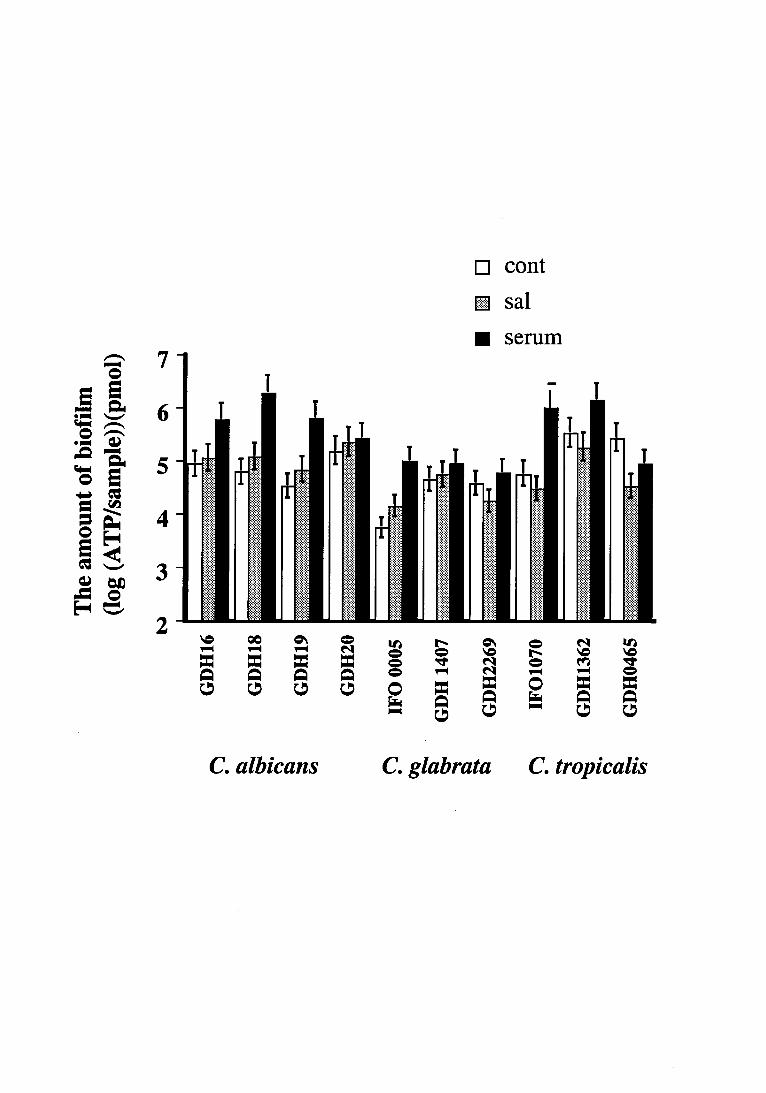
Biofilm form ation
The biofilm activity of each isolate of Candida initially increased, and plateaued
within 48-72h incubation, irrespective of the presence of the pellicles (data not shown). Thus
in the present study, 72h-biofilm activity was used as the mature biofilm activity of all
Candida isolates examined. The results indicated that the extent of biofilm activity varied
depending upon both isolates and the quality of proteinaceous pellicles (Fig. 1). As compared
with the uncoated control, significantly increased biofilm activity was observed with C.
albicans GDH 16, GDH 18, GDH 19 and C. glabrata IFO 0005 on serum coated acrylics.
However no such effect was noted with C. albicans GDH 20 and C. grabrata GDH 1407
isolates despite the protein pellicle. In contrast, the uncoated control specimen exhibited
significantly higher activity than the saliva-admixed biofilm, in the case of C. glabrata GDH
2269 and three isolates of C. tropicalis.
Quantification of thigmotropism
When the property of thigmotropism was evaluated by estimating the degree of hyphal
extension through the filter pores,C. albicans GDH 18 exhibited the highest degree of
thigmotropism followed by C. albicans GDH20, C. albicans GDH16, C. tropicalis GDH
1362, C. albicans GDH 19, C. tropicalis GDH 0465, and lastly C. tropicalis IFO 1070 (Fig.
2). A relatively good correlation was observed between germ tube forming ability and
thigmotropism of seven isolates of C. albicans and C. tropicalis (r=0.854; p<0.05). As to the
isolates of C. glabrata, no hyphal emergence was observed (data not shown). However, little
ATP activity was detected in the filter, and SEM observation revealed this to be due to the
nonspecific adsorption of little number of blastospores on the filter surface (not shown). This
value was therefore the background 'noise' of the assay.
Although no signioficant correlation between the amount of hyphae induced by
thigmotropic reaction of fungal isolates and biofilm formation on uncoated control specimans
(r=0.577; p.0.05), the amount of hyphae induced by thigmotropic reaction significantly
correlated with the activity of both saliva- and serum-admixed biofilms (r=0.734; p<0.05 and r
=0.793; p<0.01, respectively, Fig. 3).
Discussion
A number of experimental approaches have been made to examine the mechanisms of
C. albicans adherence to solid surfaces, such as denture acrylic (7,9,15,16,17,23,24,28). It
should also be noted that the phenomenon of adherence may represent only the first step in the
colonization process (13) which, as time progresses, leads to a formation of a thin biofilm and
then a multilayer, climax community of yeasts. Recently we adapted a bioluminescent ATP
assay to evaluate and quantify biofilm formation in C. albicans, and demonstrated that both
saliva and serum pellicle facilitated the fungal biofilm formation, and scanning electron
microscopic study revealed that this was due to the multilayer, including blastospore-
blastospore co-adhesion, germ tube, hyphal and pseudohyphal emergence and blastospore-
hyphal coadherence, phenomena not observed in the uncoated controls. In addition we also
have shown that the strand-like polymeric adhesive materials obseved on hyphal surface
mediated the hyphal adhesion to acrylic surfaces with saliva- or serum-admixed biofilms.
Hawser & Douglas (1994)(12), reported hyphal induction relative to variations in substrates. It
is therefore likely that during biofilm formation, hyphal emergence, particularly thigmotropic
reaction may be involved and thus a deeper understanding Candida biofilm formation
necessitates clarification of intertwined relationships.
The biofilm activity of each isolate of Candida isolates initially increased, and
plateaued within 48-72h incubation, irrespective of the presence of the saliva or serum
pellicles (data not shown). Nonetheless the extent of biofilm activity varied depending upon
both the isolates and the quality of proteinaceous pellicle (Fig. 1). As compared with the
uncoated control, significantly increased biofilm activity was observed with C. albicans GDH
16, GDH 18, GDH 19 and C. glabrata IFO 0005, on protein coated acrylic strips. These
results tend to concur with our previous observations that both salivary and serum pellicles
facilitate the development of a C. albicans biofilms as compared with acrylic strips devoid of
these biological fluids. Similarly, the protein coats apparently promoted the biofilm formation
of C. albicans GDH 20 and C. grabrata GDH 1407, however the results did not giva a level of
significance (Fig. 1; p>0.05). Although in the case of C. glabrata GDH 2269 and all three
isolates of C. tropicalis, the uncoated control specimen exhibited significantly higher activity
than saliva-admixed biofilm, but the activity of control biofilm did not exceeded that of the
serum-admixed biofilm.
Although no significant correlation between the amount of hyphae induced by
thigmotropic reaction of fungal isolates and biofilm formation on uncoated control specimans
(r=0.577; p.0.05), the ability of hyphae induced by thigmotropic reaction significantly
correlated with the amount of both saliva- and serum-admixed biofilms (r=0.734; p<0.05 and r
=0.793; p<0.01, respectively, Fig. 3). The results were consistent with our previous findings,
that germ tube, hyphal and pseudohyphal emergence and blastospore-hyphal coadherence
were involved in the formation of saliva- or serum-admixed biofilm and the phenomena not
observed in the uncoated controls (20). Thus the results tend to suggest that hyphal emergence
induced by the touch-sensitive reaction against substrates should be of importance in pellicle-
admixed biofilm formation. However, whether this phenomenon simply contributes to fungal
colonization in vivo is still unexplained, because there should be the antagonism against
Candida by bacteria in the oral cavity. Hence further work is required to clarify the
interactions of the fungi with commensal bacteria.
References
l.Berlutti LS, Passariello C, Comodi-Ballanti MR, Thaller MC. Proteolytic enzymes: a new
treatment strategy for prosthetic infections? Antimicrobial Agents and Chemotherapy 1993;
37, 2618-2621.
2.Bouali, A, Robert R, Tronchin G, Senet J-M. Binding of human fibrinogen to Candida
albicans in vitro: A preliminary study. Journal of Medical and Veterinary Mycology 1986;
24, 345-348.
3.Budtz-Jorgensen E. The significance of Candida albicans in denture stomatitis.
Scandinavian Journal of Dental Research (European Journal of Cell Science) 1974; 82, 151-
190.
4.Budtz-Jorgensen E. Candida-associated denture stomatitis and angular cheilitis. In:
Samaranayake LP, MacFarlane TW. edn. Oral Candidosis. Wright, London.1990; 156-183.
5.Bull FG, Turner NR. A serum mannan-binding protein and candidiasis. Sabouraudia
(Journal of Medical and Veterinary Mycology )1984; 22, 347-350.
6.Cannon RD, Nand AK, Jenkinson HF. Adherence of Candida albicans to human salivary
components adsorbed to hydroxylapatite. Microbiology (UK), 1995; 141, 213-219.
7.Critchley IA, Douglas LJ. Differential adhesion of pathogenic Candida species to epithelial
cells and inert surfaces. FEMS Microbiological Letter 1985; 28, 199-203.
8.Davenport JC. The oral distribution of Candida in denture stomatitis. British Dental Journal
1970; 129, 151-156.
9.Edgerton M, Scannapieco FA, Reddy MS, Levine MJ. Human submandibular-sublingual
saliva promotes adhesion of Candida albicans to polymethylmethacrylate. Infection and
Immunity 1993; 61, 2644-2652.
lO.Farber BF, Wolff AG. Salicylic acid prevents the adherence of bacteria and yeast to silastic
catheters. Journal of Biomedical Materials Research 1993; 27, 599-602.
ll.Gow NAR, Perera THS, Sherwood-Higham J, Gooday GW, Gregory DW, Marshall D.
Investigation of touch-sensitive responses by hyphae of the human paththogenic fungus
Candida albicans. Scanning Microscopy 1994; 8: 705-710.
12.Hawser SP, Douglas LJ. Biofilm formation by Candida species on the surface of catheter
materials in vitro. Infection and Immunity 1994; 62, 915-921.
13.Kennedy MJ. Methods for studying the role of fungal attachment in colonization and
pathogenesis. Mycopathologia 1990;109, 123-137.
14.Kricka LJ. Clinical and biochemical application of luciferases and luciferins. Analytical
Biochemistry 1988; 175, 14-21.
15.McCourtie J, Douglas LJ. Relationship between cell surface composition of Candida
albicans and adherence to acrylic after growth on different carbon sources. Infection and
Immunity 1981; 32, 1234-1241.
16.McCourtie J, Douglas LJ. Relationship between cell surface composition, adherence, and
virulence of Candida albicans. Infection and Immunity 1984; 45, 6-12.
17.Minagi S, Miyake Y, Inagaki K, Tsuru H, Suginaka H. Hydrophobic interaction in
Candida albicans and Candida tropicalis adherence to various denture base resin materials.
Infection and Immunity 1985; 47, ll-14.
18.Nikawa H, Hayashi S, Nikawa Y, Hamada T, Samaranayake LP. Interactions between
denture lining material, protein pellicles and Candida albicans. Archives of Oral Biology
1993; 38, 631-634.
19.Nikawa H, Nishimura H, Yamamoto T, Samaranayake LP. A novel method to study the
hyphal phase of Candida albicans and to evaluate its hydrophobicity. Oral Microbiology
Immnology 1995; 10, 110-114.
2O.Nikawa H, Nishimura H, Yamamoto T, Hamada T, Samaranayake LP. The role of salivary
or serum pellicle in the colonization process of Candida albicans on denture acrylic in vitro.
Microbial Ecology in Health and Disease 1996; 9, 35-48.

21.Nikawa, H., Nishimura, H., Hamada, T. and Sadamori, S. Quantification of thigmotropism
(contact sensing) of Candida albicans and Candida tropicalis. Mycopathologia 1997; 138, 13-
19.
22.Rotrosen D, Calderone RA, Edwards Jr JE. Adherence of Candida species to host tissues
and plastic surfaces. Reviews in Infectious Disease 1986; 8, 73-85.
23.Samaranayake LP, MacFarlane TW. An in vitro study of the adherence of Candida
albicans to acrylic surfaces. Archives of Oral Biology 1980; 25, 603-609.
24.Samaranayake LP, McCourtie J, MacFarlane TW. Factors affecting the in-vitro adherence
of Candida albicans to acrylic surfaces. Archives of Oral Biology 1980; 25, 603-609.
25.Sherwood J, Gow NAR, Gooday GW, Gregory DW, Marshall D. Contact sensing in
Candida albicans: a possible aid to epithelial penetration. Journal of Medical and Veterinary
Mycology 1992; 30:461-469.
26.Silverman Jr S, Migliorati CA, Epstein JB, Samaranayake LP. Laboratory diagnosis of oral
candidosis. In: Samaranayake LP, MacFarlane TW. edn. Oral Candidosis. Wright, London,
1990; 213-237.
27.Siro RM, Romar H, Lovgren T. Continuous flow method for extraction and
bioluminescence assay of ATP in baker's yeast. European Journal of Applied Microbiology
and Biotechnology 1982; 15, 258-264.
28.Vasilas A, Molina L, Hoffman M, Haidaris CG. The influence of morphological variation
on Candida albicans adhesion to denture acrylic in vitro. Archives of Oral Biology 1992; 37,
613-622.
Legends to Figures
Fig.1
Three-days biofilm activity of Candida spp. on uncaoted (cont), whole saliva coated (sal), or
neat serum coated (ser) acrylic specimens. The assays were carried out on two independent
occasions, with quadruplicated samples on each occasion.
Fig.2
The ATP content of penetrant hyphae retained on the filter of the Chemotaxicell. This
penetrant was considered a measure of thigmotropism. (The surface area of filter of
Chemotaxicell for culture was 0.48 cm2.) The assays were carried out on two independent
occasions, with quadruplicated samples on each occasion.
Fig.3
Relationship between thigmotropism against the amount of biofilm formation on control,
saliva- and serum-coated acrylics.