relationship between expression of the family of m proteins and lipoteichoic acid to hydrophobicity...
TRANSCRIPT
Relationship between Expression of the Family of MProteins and Lipoteichoic Acid to Hydrophobicity andBiofilm Formation in Streptococcus pyogenesHarry S. Courtney1,4*, Itzhak Ofek1,5, Thomas Penfound1, Victor Nizet6, Morgan A. Pence6, Bernd
Kreikemeyer7, Andreas Podbielbski7, David L. Hasty2,4, James B. Dale1,3,4
1Department of Medicine, University of Tennessee, Memphis, Tennessee, United States of America, 2Department of Anatomy and Neurobiology, University of Tennessee,
Memphis, Tennessee, United States of America, 3Department of Molecular Sciences, University of Tennessee, Memphis, Tennessee, United States of America,
4Department of Veterans Affairs Medical Center, University of Tennessee, Memphis, Tennessee, United States of America, 5Department of Clinical Microbiology and
Immunology, Sackler Faculty of Medicine, Tel Aviv University, Tel Aviv, Israel, 6Department of Pediatrics, University of California San Diego, La Jolla, California, United
States of America, 7Department of Medical Microbiology and Hospital Hygiene, Hospital of Rostock University, Rostock, Germany
Abstract
Background: Hydrophobicity is an important attribute of bacteria that contributes to adhesion and biofilm formation.Hydrophobicity of Streptococcus pyogenes is primarily due to lipoteichoic acid (LTA) on the streptococcal surface but themechanism(s) whereby LTA is retained on the surface is poorly understood. In this study, we sought to determine whethermembers of the M protein family consisting of Emm (M protein), Mrp (M-related protein), Enn (an M-like protein), and thestreptococcal protective antigen (Spa) are involved in anchoring LTA in a manner that contributes to hydrophobicity of thestreptococci and its ability to form biofilms.
Methodology/Principal Findings: Isogenic mutants defective in expression of emm, mrp, enn, and/or spa genes of eightdifferent serotypes and their parental strains were tested for differences in LTA bound to surface proteins, LTA released intothe culture media, and membrane-bound LTA. The effect of these mutations on the ability of streptococci to form ahydrophobic surface and to generate biofilms was also investigated. A recombinant strain overexpressing Emm1 was alsoengineered and similarly tested. The serotypes tested ranged from those that express only a single M protein gene to thosethat express two or three members of the M protein family. Overexpression of Emm1 led to enhanced hydrophobicity andbiofilm formation. Inactivation of emm in those serotypes expressing only a single emm gene reduced biofilm formation,and protein-bound LTA on the surface, but did not alter the levels of membrane-bound LTA. The results were more varied inthose serotypes that express two to three members of the M protein family.
Conclusions/Significance: Our findings suggest that the formation of complexes with members of the M protein family is acommon mechanism for anchoring LTA on the surface in a manner that contributes to hydrophobicity and to biofilmformation in S. pyogenes, but these activities in some serotypes are dependent on a trypsin-sensitive protein(s) that remainsto be identified. The need for interactions between LTA and M proteins may impose functional constraints that limitvariations in the sequence of the M proteins, major virulence factors of S. pyogenes.
Citation: Courtney HS, Ofek I, Penfound T, Nizet V, Pence MA, et al. (2009) Relationship between Expression of the Family of M Proteins and Lipoteichoic Acid toHydrophobicity and Biofilm Formation in Streptococcus pyogenes. PLoS ONE 4(1): e4166. doi:10.1371/journal.pone.0004166
Editor: Matthew E. Falagas, Alfa Institute of Biomedical Sciences (AIBS), Greece
Received July 30, 2008; Accepted December 6, 2008; Published January 9, 2009
Copyright: ! 2009 Courtney et al. This is an open-access article distributed under the terms of the Creative Commons Attribution License, which permitsunrestricted use, distribution, and reproduction in any medium, provided the original author and source are credited.
Funding: These studies were supported by research funds from the US Department of Veterans Affairs (HSC, DLH) and from the US Public Health Service GrantAI-10085 (JBD). The work in the labs of Bernd Kreikemeyer and Andreas Podbielski was funded by the BMBF ‘‘ERANet Pathogenomics’’ program. The funders hadno role in study design, data collection and analysis, decision to publish, or preparation of the manuscript.
Competing Interests: The authors have declared that no competing interests exist.
* E-mail: [email protected]
Introduction
The hydrophobic properties of bacterial surfaces are a majordeterminant in the adhesion of bacteria and in the formation ofbiofilms by bacteria on animate and inanimate surfaces [1].Hydrophobicity is likely due to a complex interplay betweennegatively-charged, positively-charged, hydrophobic and hydro-philic components on the surface of the bacteria. Surfacecomponents that directly contribute to hydrophobicity have beentermed hydrophobins and this contribution can be measured in a
number of ways including the MATH (microbial adhesion tohydrocarbons) test [1]. Lipoteichoic acid (LTA) is a majorhydrophobin that contributes to the hydrophobicity of a varietyof Gram-positive bacteria [1,2,3,4].LTA also plays important roles in bacterial physiology by
chelating metals, maintaining the integrity of the membrane andcontrolling autolytic enzymes [5,6]. Purified LTA has been shownto exhibit a number of biological effects including bone resorptionand activation of cells of the immune system such as macrophagesand T cells [5,7,8,9]. Expression of LTA is vital to Gram-positive
PLoS ONE | www.plosone.org 1 January 2009 | Volume 4 | Issue 1 | e4166
bacteria; bacteria that are completely devoid of LTA cannot grow[10] and bacteria with reduced expression of LTA are deficient inone or more of the functions mentioned above [3].In Streptococcus pyogenes, LTA functions not only as a
hydrophobin but it also mediates adhesion of the organisms toa variety of host cells [2,11]. The hydrophobicity of a number ofserotypes of S. pyogenes is dependent upon the expression ofsurface proteins that form complexes with LTA in a manner thatallows the ester linked fatty acids of LTA to be exposed on thebacterial surface [12]. These fatty acids mediate the binding ofLTA to receptors of host cells and the inhibition of streptococcaladhesion to host cells [11,13]. Thus, LTA complexed on thestreptococcal surface via interactions with proteins are involvedin adhesion to host cells.The M protein family comprises a major group of proteins on
the surface of S. pyogenes that consists of M protein (Emm, notethat the terms M protein and Emm are used interchangeably),M-related protein (Mrp), and Enn (another M-like protein).Some serotypes express only Emm (pattern A, Fig. 1), whileother serotypes express Mrp and/or Enn in addition to Emm(patterns C, D, and E) [14,15,16]. M protein has been previouslyidentified as a major surface protein that was able to formcomplexes with LTA [17]. However, these experiments wereperformed with only one serotype that expressed only a singlemember of the M protein family (i.e., pattern A) and thepossibility that LTA might form complexes with other proteinswas not investigated. Therefore, the present investigation wasundertaken to determine if M protein from other serotypes andother members of the M protein family may also be involved inanchoring LTA to the surface, thereby contributing tohydrophobicity and to the formation of biofilms. For thesestudies we used isogenic mutants lacking one or more ofmembers of the M protein family and another surface proteinthat is involved in resistance to phagocytosis, the streptococcalprotective antigen or Spa [18,19]. Our findings indicate thatmembers of the M protein family are centrally involved in theformation of a hydrophobic cell surface and in biofilm formationin most serotypes. However, we also determined that one ormore additional trypsin-sensitive proteins are involved in these
functions in some serotypes and these proteins remain to beidentified.
Materials and Methods
Bacterial strains and growth conditionsThe sources of the parental strains employed and their isogenic
mutants are listed in Table 1. That inactivation of the targetedgenes resulted in loss of expression of those genes has beendescribed (see references in Table 1). The strains used in thisinvestigation are clinical isolates. The M1 strain used in thisinvestigation was the M1T1 clone that has disseminated globallyand is the single leading cause of group A streptococcal invasiveinfections such as necrotizing fasciitis or toxic shock syndrome,and is also the most common cause of pharyngitis. The strainswere grown overnight at 37uC in Todd-Hewitt broth supplement-ed with 0.5% yeast extract (THY) unless noted otherwise. THYwas supplemented with the protease inhibitor E64 (1 mg/ml)(Sigma, St. Louis, MO) in some experiments to determinehydrophobicity. The bacteria were harvested, washed twice indistilled water and suspended in either water or in PBS (0.01 Mphosphate, 0.14 M NaCl, pH 7.4) to the desired optical densityand kept on ice until further testing. The culture supernatant waspassed through a 0.2 mm pore size filter, heated for 10 minutes at95uC and kept in the cold until used.
Inactivation of the emm gene in M type 5 S. pyogenesThe emm gene was inactivated by allelic replacement in theM type
5 S. pyogenes strain Manfredo. Mutagenic primers were used to createa stop codon at base pair 60 of the sequence encoding the matureM5 protein. The modified gene was ligated into pGHost+9 vectorand electroporated into the streptococci. Procedures for antibioticand temperature selection for single- and double-crossover eventswere as described by Maguin et al. [20]. Confirmation that the wildtype gene was replaced by the mutagenized emm gene was confirmedby PCR and sequencing. Western blot analysis of hot acid extracts ofparent and mutant strains confirmed that Emm5 was not expressedin the mutant. Hot acid treatment is a classical method for theextraction of M proteins [21].
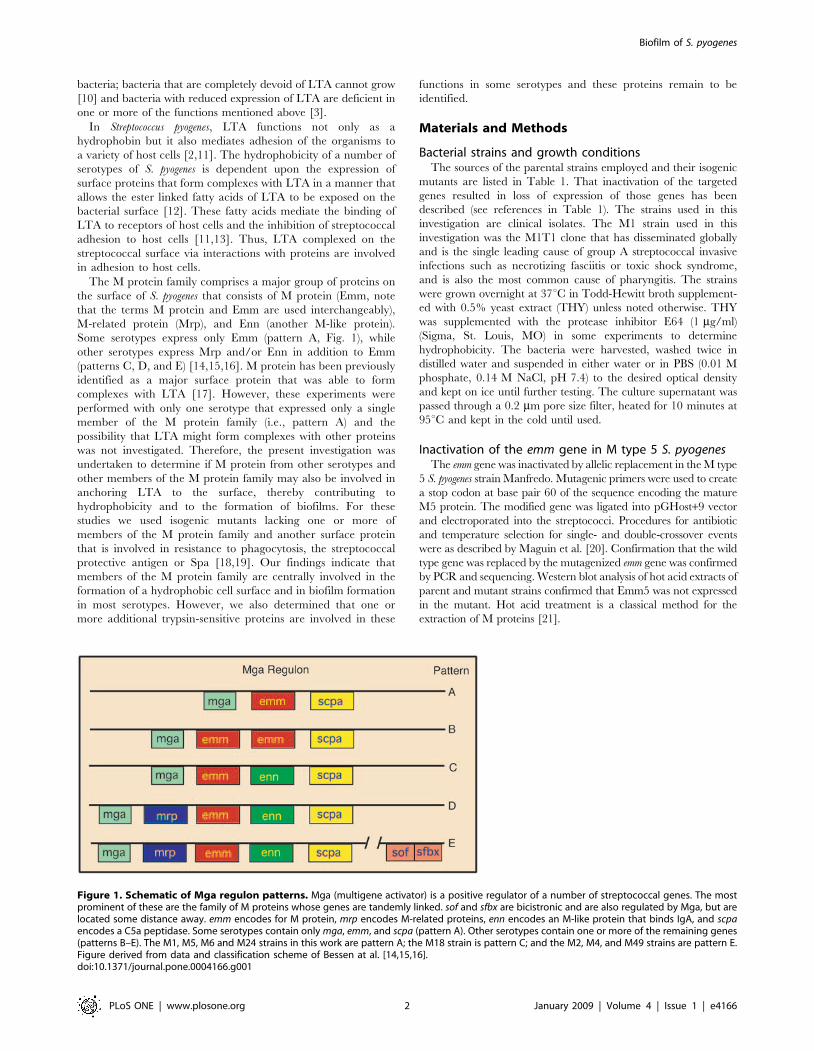
Figure 1. Schematic of Mga regulon patterns. Mga (multigene activator) is a positive regulator of a number of streptococcal genes. The mostprominent of these are the family of M proteins whose genes are tandemly linked. sof and sfbx are bicistronic and are also regulated by Mga, but arelocated some distance away. emm encodes for M protein, mrp encodes M-related proteins, enn encodes an M-like protein that binds IgA, and scpaencodes a C5a peptidase. Some serotypes contain onlymga, emm, and scpa (pattern A). Other serotypes contain one or more of the remaining genes(patterns B–E). The M1, M5, M6 and M24 strains in this work are pattern A; the M18 strain is pattern C; and the M2, M4, and M49 strains are pattern E.Figure derived from data and classification scheme of Bessen at al. [14,15,16].doi:10.1371/journal.pone.0004166.g001
Biofilm of S. pyogenes
PLoS ONE | www.plosone.org 2 January 2009 | Volume 4 | Issue 1 | e4166
Construction of recombinant strain overexpressingEmm1Primers were designed to amply the DNA sequence encoding
Emm1. The PCR product was ligated into the expression vectorpDCerm [22,23] to obtain the plasmid pemm1 that was electropo-rated into the M type 1 strain 5448. An erythromycin resistantcolony was selected for further studies and labeled M1+pemm1.
Enzyme linked immunosorbent assaysThe streptococci were grown overnight in THY at 37uC except
for the recombinant strain M1+pemm1, which was grown in THYcontaining 5 mg/ml erythromycin. The streptococci were washedin PBS, adjusted to an OD530 of 0.4 and 100 ml was added tomicrotiter wells. The plates were incubated at 37uC for 30 minand non-adherent bacteria were removed by washing. The wellswere then blocked with 1% bovine serum albumin (BSA) for30 min at 37uC. A 1:1000 dilution of normal rabbit serum (NRS)or rabbit serum against a synthetic peptide copying amino acidresidues 1–26 of the mature Emm1 was added to the appropriatewells. The production of anti-SM1(1–26) has been described [24].
After a 30 min incubation at 37uC, the wells were washed and100 ml of a 1:2000 dilution of peroxidase-labeled goat anti-rabbitimmunoglobulins was added to all wells and incubated for 30 minat 37uC. The wells were then washed and 100 ml of thetetramethylbenzidine was added. Stop solution was added aftercolor development and the A450 was recorded. Wells coated withBSA served as blanks. The values obtained with NRS wereconsidered non-specific and were subtracted from values obtainedwith anti-SM1(1–26). Assays were done in triplicate.
Biofilm assaysA microtiter method described by several investigators to
measure biofilm formation by S. pyogenes was used with minormodifications [25,26,27]. Briefly, the streptococci were grownovernight in THY (unless stated otherwise), diluted 1:40 in THY,and 100 ml of the suspension added to wells of a polystyrene,microtiter plate (Corning Costar 3595, Fisher Scientific, Pittsburg,PA). In some instances the growth media was supplemented with1 mg/ml of trypsin. The plates were incubated in a humidifiedenvironment for 48 hours at 37uC. Afterwards, the plates weregently washed four times with PBS, any residual fluid was carefully
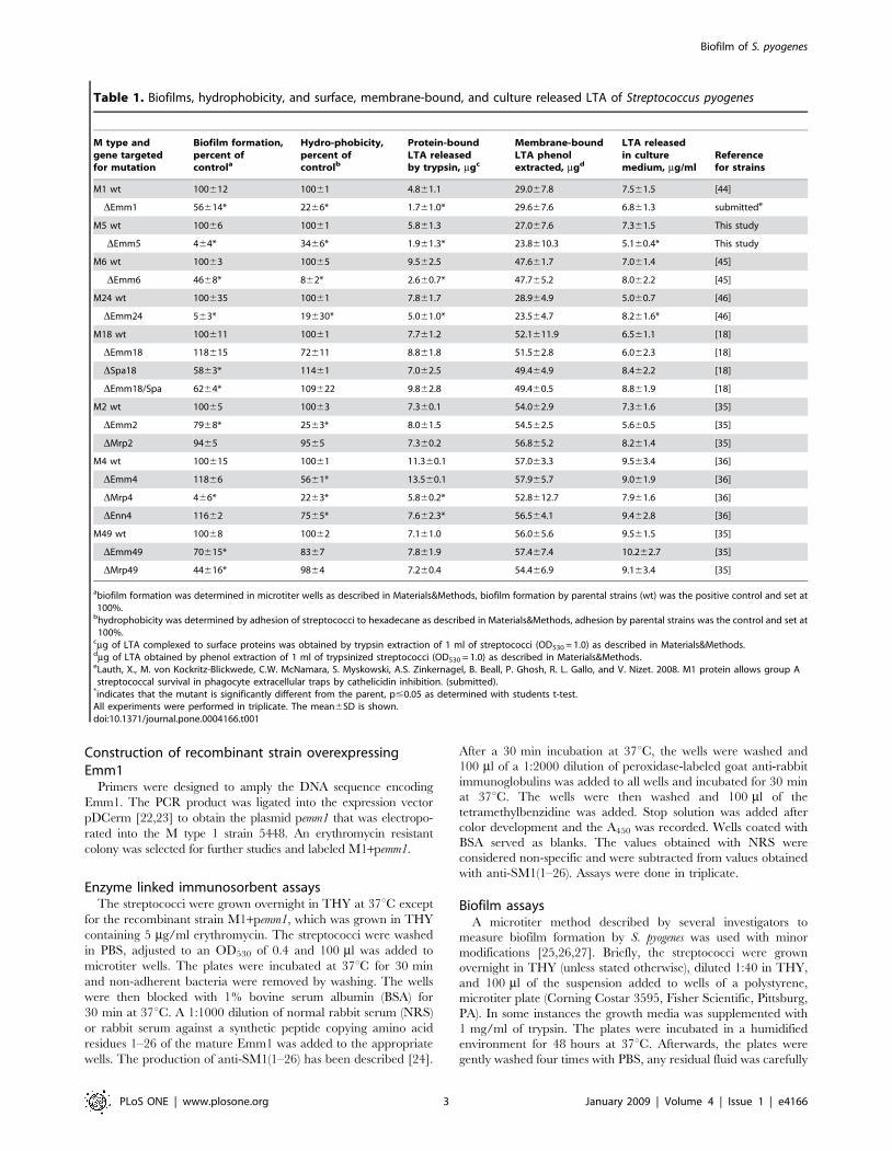
Table 1. Biofilms, hydrophobicity, and surface, membrane-bound, and culture released LTA of Streptococcus pyogenes
M type andgene targetedfor mutation
Biofilm formation,percent ofcontrola
Hydro-phobicity,percent ofcontrolb
Protein-boundLTA releasedby trypsin, mgc
Membrane-boundLTA phenolextracted, mgd
LTA releasedin culturemedium, mg/ml
Referencefor strains
M1 wt 100612 10061 4.861.1 29.067.8 7.561.5 [44]
DEmm1 56614* 2266* 1.761.0* 29.667.6 6.861.3 submittede
M5 wt 10066 10061 5.861.3 27.067.6 7.361.5 This study
DEmm5 464* 3466* 1.961.3* 23.8610.3 5.160.4* This study
M6 wt 10063 10065 9.562.5 47.661.7 7.061.4 [45]
DEmm6 4668* 862* 2.660.7* 47.765.2 8.062.2 [45]
M24 wt 100635 10061 7.861.7 28.964.9 5.060.7 [46]
DEmm24 563* 19630* 5.061.0* 23.564.7 8.261.6* [46]
M18 wt 100611 10061 7.761.2 52.1611.9 6.561.1 [18]
DEmm18 118615 72611 8.861.8 51.562.8 6.062.3 [18]
DSpa18 5863* 11461 7.062.5 49.464.9 8.462.2 [18]
DEmm18/Spa 6264* 109622 9.862.8 49.460.5 8.861.9 [18]
M2 wt 10065 10063 7.360.1 54.062.9 7.361.6 [35]
DEmm2 7968* 2563* 8.061.5 54.562.5 5.660.5 [35]
DMrp2 9465 9565 7.360.2 56.865.2 8.261.4 [35]
M4 wt 100615 10061 11.360.1 57.063.3 9.563.4 [36]
DEmm4 11866 5661* 13.560.1 57.965.7 9.061.9 [36]
DMrp4 466* 2263* 5.860.2* 52.8612.7 7.961.6 [36]
DEnn4 11662 7565* 7.662.3* 56.564.1 9.462.8 [36]
M49 wt 10068 10062 7.161.0 56.065.6 9.561.5 [35]
DEmm49 70615* 8367 7.861.9 57.467.4 10.262.7 [35]
DMrp49 44616* 9864 7.260.4 54.466.9 9.163.4 [35]
abiofilm formation was determined in microtiter wells as described in Materials&Methods, biofilm formation by parental strains (wt) was the positive control and set at100%.bhydrophobicity was determined by adhesion of streptococci to hexadecane as described in Materials&Methods, adhesion by parental strains was the control and set at100%.
cmg of LTA complexed to surface proteins was obtained by trypsin extraction of 1 ml of streptococci (OD530 = 1.0) as described in Materials&Methods.dmg of LTA obtained by phenol extraction of 1 ml of trypsinized streptococci (OD530 = 1.0) as described in Materials&Methods.eLauth, X., M. von Kockritz-Blickwede, C.W. McNamara, S. Myskowski, A.S. Zinkernagel, B. Beall, P. Ghosh, R. L. Gallo, and V. Nizet. 2008. M1 protein allows group Astreptococcal survival in phagocyte extracellular traps by cathelicidin inhibition. (submitted).*indicates that the mutant is significantly different from the parent, p#0.05 as determined with students t-test.All experiments were performed in triplicate. The mean6SD is shown.doi:10.1371/journal.pone.0004166.t001
Biofilm of S. pyogenes
PLoS ONE | www.plosone.org 3 January 2009 | Volume 4 | Issue 1 | e4166
removed, and the plates were heat fixed at 60uC for 1 hour.Hucker’s crystal violet solution (100 ml) was added to each well.After two minutes, the wells were washed with tap water until thewater became clear. Then, 100 ml of destain solution (10%methanol, 7.5% acetic acid in distilled water) was added, the plateswere shaken for 1 minute, and the absorbance at 540 nm wasrecorded. Wells incubated with THY without streptococci wereused as blanks. Additional controls consisted of measuring theOD530 of the streptococci after an overnight growth in THY withor without trypsin to verify that trypsin treatment did not altergrowth.The bacteria were also grown at 37uC in 100% normal rabbit
serum, trypticase soy broth without glucose (TSB), TSB+1%glucose, Mueller Hinton II medium (MHII), Luria-Bertani broth(LB), and THY and then treated as described above to determinethe effect of growth media on biofilm formation. All growth mediawere purchased from Difco Labs except TSB without glucose,which was purchased from Sigma Chem. Co.
Determination of hydrophobicityHydrophobicity was determined by the hexadecane method as
described [12]. Briefly, 1 ml of bacteria (OD530 = 1.0) was placedinto glass tubes and 100 ml of hexadecane (Sigma, St. Louis, MO)was added. The mixtures were vigorously vortexed for 2 minutes,followed by 10 min incubation at ambient temperature to allowphase separation, and then the OD530 of the lower, aqueous phasewas recorded. In some cases, the bacteria were treated with bovinetesticular hyaluronidase (Sigma, St. Louis, MO) at 2 mg/ml at37uC for 15 min and then tested for adhesion to hexadecane. Thepercent hydrophobicity was calculated by the formula: %hydrophobicity = [12(OD530 after vortexing/OD530 before vor-texing)]6100.
Enzymatic treatment and phenol extraction of thebacteriaForty ml of trypsin (1 mg/ml PBS, Sigma, St. Louis, MO) was
added to 0.4 ml bacterial suspension (OD530 = 1.0) in PBS. After30 minutes at 37uC, the bacteria were centrifuged and thesupernatant was removed, heated to 95uC for 5 min to inactivateenzymes, and saved for determinations of LTA content. Thesedimented bacteria were washed twice with distilled water andthen assayed for hydrophobicity.After trypsinization, LTA was extracted from the streptococci
with phenol as previously described [28] by adding an equalvolume of phenol (Sigma, St. Louis, MO) heated to 60uC to astreptococcal suspension that had been adjusted to an OD530 of1.0 in distilled water and then vortexing for 5 min. The mixtureswere then centrifuged at 14,0006g. The aqueous phase wasremoved and dialyzed extensively against distilled water. Thisextract represents membrane-bound LTA.
Quantitation of LTALTA concentrations were determined by a competitive ELISA
as described previously with slight modification [29]. Microtiterwells were coated with commercial LTA (Sigma, St. Louis, MO)by adding 100 ml of LTA (5 mg/ml in carbonate buffer), andincubating the plate for 1 hour at 37uC. The wells were washedwith PBS containing 0.05% Tween-20 (PBST), then blocked with5% v/v hemoglobin in PBST for 30 minutes at 37uC. LTAstandards were made with either commercial LTA or highlypurified LTA extracted from streptococci with butanol, asdescribed previously [8]. 50 ml of serial two-fold dilutions ofLTA standards or two-fold dilutions of culture supernatants,
trypsin-released LTA, or phenol-extracted LTA were added to50 ml of a 1:3200 dilution of a mouse monoclonal antibody againstLTA (Abcam, Cambridge, MA). After incubating for 30 min atambient temperature, the mixtures were added to the LTA-coatedwells and incubated for 60 minutes at 37uC. After washing, 100 mlof HRP-conjugated, goat anti-mouse immunoglobulin (MPBiomedicals, Solon, OH) diluted 1:2500 was added and incubatedfor 1 hour at 37uC. The plates were then washed and the turboTMB substrate (Pierce Chem Co., Rockford, IL) was added. Astop solution (sulfuric acid) was added after color development andthe A450 recorded. The ELISA values of negative control wells notcoated with LTA were subtracted from all experimental values.The amount of LTA in samples was determined from standardcurves. All measurements were performed in triplicate.The amount of LTA needed to achieve 50% inhibition was
3.1 mg/ml for butanol extracted LTA and 5.1 mg/ml ofcommercial LTA. This indicated that about 40% by weight ofthe commercial LTA obtained from Sigma was not immunore-active with the monoclonal antibody against polyglycerol phos-phate. These results are in agreement with those of Morath et al.who also determined the purity of commercially acquired LTA[30]. Therefore, a correction factor of 0.60 was applied to resultsfrom assays that used commercial LTA as standards.
Results
Biofilm formationTo determine the optimal medium for biofilm formation, we
tested the ability of streptococci to grow and form biofilms invarious media (Fig. 2). The addition of glucose to TSB had aminor effect on growth but enhanced biofilm formation byapproximately threefold. THY provided the best medium forgrowth and for biofilm formation and, therefore, was used for allsubsequent experiments.There was significant variation among different M types in their
ability to form biofilms (Fig. 3A). These findings were similar tothose of Lembke et al al. [26] and Baldassarri et al. [27] who alsofound that biofilm formation varied among different serotypes andeven among strains of the same M type. We found that M types 2and 6 exhibited the greatest degree of biofilm formation and Mtype 49 exhibited the least degree. Similar findings have beenreported regarding M types 6 and 49 S. pyogenes [26,27]. Theaddition of trypsin to the growth media blocked the formation ofbiofilms by all of serotypes tested indicating the dependence ofbiofilms on streptococcal surface proteins (Fig. 3A). In controlassays trypsin did not alter the growth of the streptococci. Thisfinding is in agreement with those of others [31,32] who also foundthat trypsin did not alter the viability and growth of group Astreptococci.To investigate the role that various members of the M protein
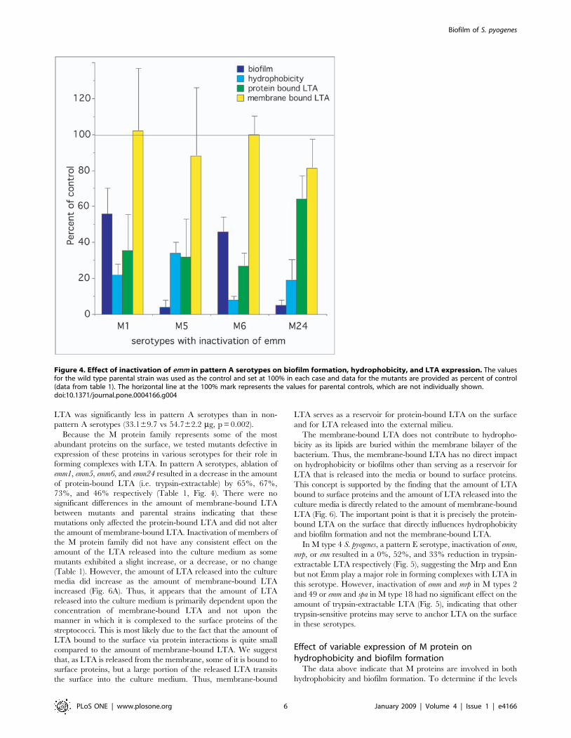
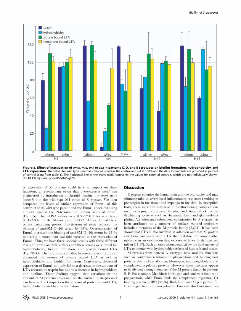
family play in the formation of biofilms, we compared the abilityof parental strains to make biofilms to that of mutants defectivein the expression of Emm, Mrp, Enn, and Spa (Table 1,Fig. 4&5). The data indicated that Emm-negative mutants fromall of the pattern A serotypes (i.e. M1, M5, M6, and M24) weredeficient in their ability to form biofilms (Fig. 4). In M type 2,ablation of emm2 had only a minor effect and ablation of mrp2had no significant effect (Fig. 5). However, in M type 4 ablationof mrp4 reduced biofilms by 96%, whereas disruption of emm4and enn4 was without effect (Fig. 5). In M type 49, inactivation ofEmm49 and Mrp49 reduced biofilms by 30% and 56%respectively (Fig. 5). In the one pattern C serotype tested(M18), Spa seemed to play a significant role as inactivation of sparesulted in a 42% decrease in its biofilm (Fig. 5).
Biofilm of S. pyogenes
PLoS ONE | www.plosone.org 4 January 2009 | Volume 4 | Issue 1 | e4166
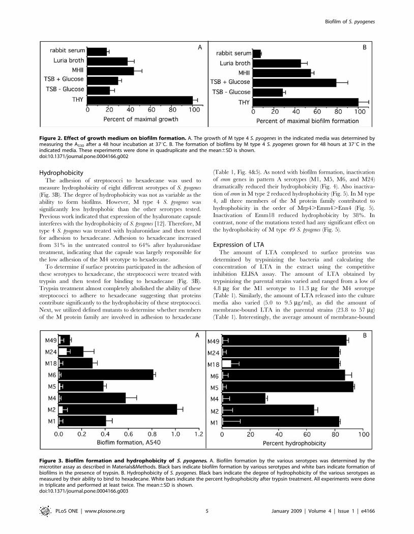
HydrophobicityThe adhesion of streptococci to hexadecane was used to
measure hydrophobicity of eight different serotypes of S. pyogenes(Fig. 3B). The degree of hydrophobicity was not as variable as theability to form biofilms. However, M type 4 S. pyogenes wassignificantly less hydrophobic than the other serotypes tested.Previous work indicated that expression of the hyaluronate capsuleinterferes with the hydrophobicity of S. pyogenes [12]. Therefore, Mtype 4 S. pyogenes was treated with hyaluronidase and then testedfor adhesion to hexadecane. Adhesion to hexadecane increasedfrom 31% in the untreated control to 64% after hyaluronidasetreatment, indicating that the capsule was largely responsible forthe low adhesion of the M4 serotype to hexadecane.To determine if surface proteins participated in the adhesion of
these serotypes to hexadecane, the streptococci were treated withtrypsin and then tested for binding to hexadecane (Fig. 3B).Trypsin treatment almost completely abolished the ability of thesestreptococci to adhere to hexadecane suggesting that proteinscontribute significantly to the hydrophobicity of these streptococci.Next, we utilized defined mutants to determine whether membersof the M protein family are involved in adhesion to hexadecane
(Table 1, Fig. 4&5). As noted with biofilm formation, inactivationof emm genes in pattern A serotypes (M1, M5, M6, and M24)dramatically reduced their hydrophobicity (Fig. 4). Also inactiva-tion of emm in M type 2 reduced hydrophobicity (Fig. 5). In M type4, all three members of the M protein family contributed tohydrophobicity in the order of Mrp4.Emm4.Enn4 (Fig. 5).Inactivation of Emm18 reduced hydrophobicity by 38%. Incontrast, none of the mutations tested had any significant effect onthe hydrophobicity of M type 49 S. pyogenes (Fig. 5).
Expression of LTAThe amount of LTA complexed to surface proteins was
determined by trypsinizing the bacteria and calculating theconcentration of LTA in the extract using the competitiveinhibition ELISA assay. The amount of LTA obtained bytrypsinizing the parental strains varied and ranged from a low of4.8 mg for the M1 serotype to 11.3 mg for the M4 serotype(Table 1). Similarly, the amount of LTA released into the culturemedia also varied (5.0 to 9.5 mg/ml), as did the amount ofmembrane-bound LTA in the parental strains (23.8 to 57 mg)(Table 1). Interestingly, the average amount of membrane-bound
Figure 2. Effect of growth medium on biofilm formation. A. The growth of M type 4 S. pyogenes in the indicated media was determined bymeasuring the A530 after a 48 hour incubation at 37uC. B. The formation of biofilms by M type 4 S. pyogenes grown for 48 hours at 37uC in theindicated media. These experiments were done in quadruplicate and the mean6SD is shown.doi:10.1371/journal.pone.0004166.g002
Figure 3. Biofilm formation and hydrophobicity of S. pyogenes. A. Biofilm formation by the various serotypes was determined by themicrotiter assay as described in Materials&Methods. Black bars indicate biofilm formation by various serotypes and white bars indicate formation ofbiofilms in the presence of trypsin. B. Hydrophobicity of S. pyogenes. Black bars indicate the degree of hydrophobicity of the various serotypes asmeasured by their ability to bind to hexadecane. White bars indicate the percent hydrophobicity after trypsin treatment. All experiments were donein triplicate and performed at least twice. The mean6SD is shown.doi:10.1371/journal.pone.0004166.g003
Biofilm of S. pyogenes
PLoS ONE | www.plosone.org 5 January 2009 | Volume 4 | Issue 1 | e4166
LTA was significantly less in pattern A serotypes than in non-pattern A serotypes (33.169.7 vs 54.762.2 mg, p = 0.002).Because the M protein family represents some of the most
abundant proteins on the surface, we tested mutants defective inexpression of these proteins in various serotypes for their role informing complexes with LTA. In pattern A serotypes, ablation ofemm1, emm5, emm6, and emm24 resulted in a decrease in the amountof protein-bound LTA (i.e. trypsin-extractable) by 65%, 67%,73%, and 46% respectively (Table 1, Fig. 4). There were nosignificant differences in the amount of membrane-bound LTAbetween mutants and parental strains indicating that thesemutations only affected the protein-bound LTA and did not alterthe amount of membrane-bound LTA. Inactivation of members ofthe M protein family did not have any consistent effect on theamount of the LTA released into the culture medium as somemutants exhibited a slight increase, or a decrease, or no change(Table 1). However, the amount of LTA released into the culturemedia did increase as the amount of membrane-bound LTAincreased (Fig. 6A). Thus, it appears that the amount of LTAreleased into the culture medium is primarily dependent upon theconcentration of membrane-bound LTA and not upon themanner in which it is complexed to the surface proteins of thestreptococci. This is most likely due to the fact that the amount ofLTA bound to the surface via protein interactions is quite smallcompared to the amount of membrane-bound LTA. We suggestthat, as LTA is released from the membrane, some of it is bound tosurface proteins, but a large portion of the released LTA transitsthe surface into the culture medium. Thus, membrane-bound
LTA serves as a reservoir for protein-bound LTA on the surfaceand for LTA released into the external milieu.The membrane-bound LTA does not contribute to hydropho-
bicity as its lipids are buried within the membrane bilayer of thebacterium. Thus, the membrane-bound LTA has no direct impacton hydrophobicity or biofilms other than serving as a reservoir forLTA that is released into the media or bound to surface proteins.This concept is supported by the finding that the amount of LTAbound to surface proteins and the amount of LTA released into theculture media is directly related to the amount of membrane-boundLTA (Fig. 6). The important point is that it is precisely the protein-bound LTA on the surface that directly influences hydrophobicityand biofilm formation and not the membrane-bound LTA.In M type 4 S. pyogenes, a pattern E serotype, inactivation of emm,
mrp, or enn resulted in a 0%, 52%, and 33% reduction in trypsin-extractable LTA respectively (Fig. 5), suggesting the Mrp and Ennbut not Emm play a major role in forming complexes with LTA inthis serotype. However, inactivation of emm and mrp in M types 2and 49 or emm and spa in M type 18 had no significant effect on theamount of trypsin-extractable LTA (Fig. 5), indicating that othertrypsin-sensitive proteins may serve to anchor LTA on the surfacein these serotypes.
Effect of variable expression of M protein onhydrophobicity and biofilm formationThe data above indicate that M proteins are involved in both
hydrophobicity and biofilm formation. To determine if the levels
Figure 4. Effect of inactivation of emm in pattern A serotypes on biofilm formation, hydrophobicity, and LTA expression. The valuesfor the wild type parental strain was used as the control and set at 100% in each case and data for the mutants are provided as percent of control(data from table 1). The horizontal line at the 100% mark represents the values for parental controls, which are not individually shown.doi:10.1371/journal.pone.0004166.g004
Biofilm of S. pyogenes
PLoS ONE | www.plosone.org 6 January 2009 | Volume 4 | Issue 1 | e4166
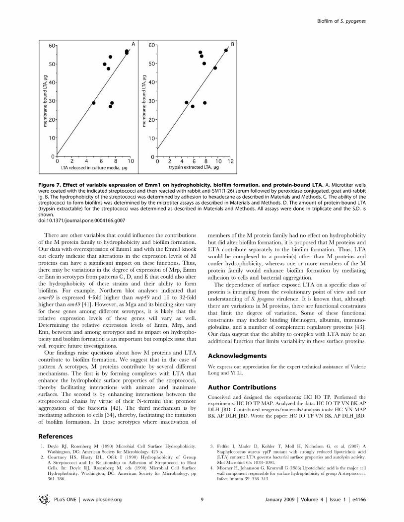
of expression of M proteins could have an impact on thesefunctions, a recombinant strain that overexpresses emm1 wasengineered by introducing a plasmid bearing the emm1 gene(pemm1) into the wild type M1 strain of S. pyogenes. We thencompared the levels of surface expression of Emm1 of thisconstruct to its wild type parent and the Emm1 knock out usingantisera against the N-terminal 26 amino acids of Emm1(Fig. 7A). The ELISA values were 0.3656.011 for wild type,0.04560.16 for the DEmm1, and 0.8456.041 for the wild typeparent containing pemm1. Inactivation of emm1 reduced thebinding of anti-SM1(1–26) serum by 95%. Overexpression ofEmm1 increased the binding of anti-SM1(1–26) serum by 231%indicating a more than two-fold increase in the expression ofEmm1. Thus, we have three isogenic strains with three differentlevels of Emm1 on their surfaces and these strains were tested forhydrophobicity, biofilm formation, and protein bound LTA(Fig. 7B–D). The results indicate that hyper-expression of Emm1enhanced the amount of protein bound LTA as well ashydrophobicity and biofilm formation. Conversely, decreasedexpression of Emm1 not only led to a decrease in the amount ofLTA released by trypsin but also to a decrease in hydrophobicityand biofilms. These findings suggest that variations in theamount of M proteins expressed on the surface of streptococcican have a direct impact on the amount of protein-bound LTA,hydrophobicity and biofilm formation.
Discussion
S. pyogenes colonize the human skin and the oral cavity and maystimulate mild to severe local inflammatory responses resulting inpharyngitis in the throat and impetigo in the skin. In susceptiblehosts, these infections may lead to life-threatening complicationssuch as sepsis, necrotizing fasciitis, and toxic shock, or todebilitating sequelae such as rheumatic fever and glomerulone-phritis. Adhesion and subsequent colonization by S. pyogenes hasbeen attributed to a number of surface exposed moleculesincluding members of the M protein family [33,34]. It has beenshown that LTA is also involved in adhesion and that M proteincan form complexes with LTA that stabilize this amphipathicmolecule in an orientation that exposes its lipids to the externalmilieu [11,17]. Such an orientation would allow the lipid moiety ofLTA to interact with hydrophobic surfaces of host cells and tissues.M proteins from pattern A serotypes have multiple functions
such as conferring resistance to phagocytosis and binding hostproteins that include albumin, fibrinogen, immunoglobulins, andcomplement regulatory proteins. However, these functions appearto be divided among members of the M protein family in patternsB–E. For example, Mrp binds fibrinogen and confers resistance tophagocytosis, while Emm binds the complement regulator, C4binding protein (C4BP) [35,36]. Both Emm and Mrp in pattern B–E serotypes bind immunoglobulins. Enn can also bind immuno-
Figure 5. Effect of inactivation of emm, mrp, enn or spa in patterns C, D, and E serotypes on biofilm formation, hydrophobicity, andLTA expression. The values for wild type parental strain was used as the control and set at 100% and the data for mutants are provided as percentof control (data from table 1). The horizontal line at the 100% mark represents the values for parental controls, which are not individually shown.doi:10.1371/journal.pone.0004166.g005
Biofilm of S. pyogenes
PLoS ONE | www.plosone.org 7 January 2009 | Volume 4 | Issue 1 | e4166

globulins and C4BP [37]. Because M protein from one pattern Aserotype was found to be involved in hydrophobicity, thisinvestigation was undertaken to determine if other members ofthe M protein family may also contribute to hydrophobicity and tothe formation of biofilms.To begin our studies, we trypsinized streptococci from a variety
of serotypes to determine if the hydrophobicity and biofilmformation were dependent upon surface proteins. All of the
parental strains lost the ability to adhere to hexadecane and toform biofilms after trypsinization, indicating that surface proteinswere required for these functions. Next, we used defined mutantsto investigate the role that various surface proteins may have inanchoring LTA and contributing to the hydrophobic surfaceproperties and forming biofilms. The ablation of M proteinexpression in M types 1, 5, 6 and 24 that harbor only a singlemember of the M protein family indicated that M protein iscritically important in conferring hydrophobicity and biofilms inpattern A serotypes. Cho and Caparon [38] also reported thatinactivation of emm in one pattern A serotype resulted in asignificant decrease in biofilm formation. Our finding that theamount of LTA retained on the surface of the mutant strains wasgreatly diminished compared to that retained by the parentstrains suggests that M protein is the major protein in pattern Aserotypes that stabilizes LTA on the bacterial surface. Thus, theformation of LTA-M protein complexes appears to be centrallyinvolved in hydrophobicity and biofilm formation in pattern Aserotypes.Other streptococcal surface antigens besides M protein have
been found to promote biofilm formation in an M type 1 strain ofS. pyogenes. Manettii et al. [39] found that inactivation of either thepilus backbone or its sortase decreased both adhesion to host cellsand biofilm formation. Our data are consistent with this finding asinactivation of emm1 led to a partial decrease in biofilm formation.Thus, it appears that both M protein and pilus proteins have a rolein the formation of biofilms in M type 1 S. pyogenes. This is likely tobe the case for other serotypes of S. pyogenes that also produce pili.In M type 4 S. pyogenes, a pattern E serotype, there was a direct
correlation between hydrophobicity, trypsin-extractable LTA,biofilm formation, and expression of mrp. In other serotypes(M2, M18, and M49) from patterns C–E, there was no correlationbetween hydrophobicity, trypsin-extractable LTA and biofilmformation. The finding that Mrp2 appeared not to be involved inbiofilm formation, whereas both Mrp4 and Mrp49 were involvedin biofilms was somewhat unexpected because the primarystructure of Mrp is highly conserved among serotypes withsequence similarities ranging from 81% to 97% [36]. Whether thelack of involvement by Mrp2 is due to a difference in sequences orto the expression of another protein that is functionally redundantwith Mrp2 remains to be determined.Spa is an antiphagocytic protein and protective antigen that is
expressed by a limited number of serotypes [18,19]. The C-terminus of Spa18 is virtually identical to the C-terminus of Emmfrom S. equi, a horse pathogen [18], suggesting that Spa may alsobe a member of the M protein family. Although Spa had no role inhydrophobicity, it was a major contributor to biofilm formation inan M type 18 serotype. Thus, Spa shares two functions withmembers of the M protein family; an antiphagocytic function anda function in biofilm formation. SilC has also been shown to beinvolved in biofilm formation in M type 18 S. pyogenes, but this ispresumed to be an indirect effect resulting from the role of SilC asa regulator of streptococcal virulence determinants [26,40].These data suggest that the formation of complexes between M
proteins and LTA directly contributes to both hydrophobicity andto biofilm formation in most serotypes of S. pyogenes. However, insome of the serotypes tested, we were not able to demonstrate adirect link to the M protein family. It may be that in theseparticular serotypes inactivation of only a single member of the Mprotein may not be sufficient to alter these functions when anotherfamily member is expressed. Alternatively, these serotypes mayexpress a protein that is better suited than M proteins to formcomplexes with LTA in a manner that allows the lipids of LTA tointeract with environmental surfaces.
Figure 6. Correlation of membrane-bound LTA with trypsinextracted LTA and with LTA released into the culture media.The amount of LTA bound to membranes was compared to the amountof LTA released into the culture media (A) and to the amount of LTAextracted with trypsin (B). There was a significant degree of correlationin each case, r = 0.730.doi:10.1371/journal.pone.0004166.g006
Biofilm of S. pyogenes
PLoS ONE | www.plosone.org 8 January 2009 | Volume 4 | Issue 1 | e4166
There are other variables that could influence the contributionsof the M protein family to hydrophobicity and biofilm formation.Our data with overexpression of Emm1 and with the Emm1 knockout clearly indicate that alterations in the expression levels of Mproteins can have a significant impact on these functions. Thus,there may be variations in the degree of expression of Mrp, Emmor Enn in serotypes from patterns C, D, and E that could also alterthe hydrophobicity of these strains and their ability to formbiofilms. For example, Northern blot analyses indicated thatemm49 is expressed 4-fold higher than mrp49 and 16 to 32-foldhigher than enn49 [41]. However, as Mga and its binding sites varyfor these genes among different serotypes, it is likely that therelative expression levels of these genes will vary as well.Determining the relative expression levels of Emm, Mrp, andEnn, between and among serotypes and its impact on hydropho-bicity and biofilm formation is an important but complex issue thatwill require future investigations.Our findings raise questions about how M proteins and LTA
contribute to biofilm formation. We suggest that in the case ofpattern A serotypes, M proteins contribute by several differentmechanisms. The first is by forming complexes with LTA thatenhance the hydrophobic surface properties of the streptococci,thereby facilitating interactions with animate and inanimatesurfaces. The second is by enhancing interactions between thestreptococcal chains by virtue of their N-termini that promoteaggregation of the bacteria [42]. The third mechanism is bymediating adhesion to cells [34], thereby, facilitating the initiationof biofilm formation. In those serotypes where inactivation of
members of the M protein family had no effect on hydrophobicitybut did alter biofilm formation, it is proposed that M proteins andLTA contribute separately to the biofilm formation. Thus, LTAwould be complexed to a protein(s) other than M proteins andconfer hydrophobicity, whereas one or more members of the Mprotein family would enhance biofilm formation by mediatingadhesion to cells and bacterial aggregation.The dependence of surface exposed LTA on a specific class of
protein is intriguing from the evolutionary point of view and ourunderstanding of S. pyogenes virulence. It is known that, althoughthere are variations in M proteins, there are functional constraintsthat limit the degree of variation. Some of these functionalconstraints may include binding fibrinogen, albumin, immuno-globulins, and a number of complement regulatory proteins [43].Our data suggest that the ability to complex with LTA may be anadditional function that limits variability in these surface proteins.
Acknowledgments
We express our appreciation for the expert technical assistance of ValerieLong and Yi Li.
Author Contributions
Conceived and designed the experiments: HC IO TP. Performed theexperiments: HC IO TP MAP. Analyzed the data: HC IO TP VN BK APDLH JBD. Contributed reagents/materials/analysis tools: HC VN MAPBK AP DLH JBD. Wrote the paper: HC IO TP VN BK AP DLH JBD.
References
1. Doyle RJ, Rosenberg M (1990) Microbial Cell Surface Hydrophobicity.Washington, DC: American Society for Microbiology. 425 p.
2. Courtney HS, Hasty DL, Ofek I (1990) Hydrophobicity of GroupA Streptococci and Its Relationship to Adhesion of Streptococci to HostCells. In: Doyle RJ, Rosenberg M, eds (1990) Microbial Cell SurfaceHydrophobicity. Washington, DC: American Society for Microbiology. pp361–386.
3. Fedtke I, Mader D, Kohler T, Moll H, Nicholson G, et al. (2007) AStaphylococcus aureus ypfP mutant with strongly reduced lipoteichoic acid(LTA) content: LTA governs bacterial surface properties and autolysin activity.Mol Microbiol 65: 1078–1091.
4. Miorner H, Johansson G, Kronvall G (1983) Lipoteichoic acid is the major cellwall component responsible for surface hydrophobicity of group A streptococci.Infect Immun 39: 336–343.
Figure 7. Effect of variable expression of Emm1 on hydrophobicity, biofilm formation, and protein-bound LTA. A. Microtiter wellswere coated with the indicated streptococci and then reacted with rabbit anti-SM1(1-26) serum followed by peroxidase-conjugated, goat anti-rabbitIg. B. The hydrophobicity of the streptococci was determined by adhesion to hexadecane as described in Materials and Methods. C. The ability of thestreptococci to form biofilms was determined by the microtiter assays as described in Materials and Methods. D. The amount of protein-bound LTA(trypsin extractable) for the streptococci was determined as described in Materials and Methods. All assays were done in triplicate and the S.D. isshown.doi:10.1371/journal.pone.0004166.g007
Biofilm of S. pyogenes
PLoS ONE | www.plosone.org 9 January 2009 | Volume 4 | Issue 1 | e4166
5. Fischer W (1988) Physiology of lipoteichoic acids in bacteria. Adv MicrobPhysiol 29: 233–302.
6. Neuhaus FC, Baddiley J (2003) A continuum of anionic charge: structures andfunctions of D-alanyl-teichoic acids in gram-positive bacteria. Microbiol MolBiol Rev 67: 686–723.
7. Ginsburg I (2002) Role of lipoteichoic acid in infection and inflammation.Lancet Infect Dis 2: 171–179.
8. Hasty DL, Meron-Sudai S, Cox KH, Nagorna T, Ruiz-Bustos E, et al. (2006)Monocyte and macrophage activation by lipoteichoic acid is independent ofalanine and is potentiated by hemoglobin. J Immunol 176: 5567–5576.
9. Morath S, Stadelmaier A, Geyer A, Schmidt RR, Hartung T (2002) Syntheticlipoteichoic acid from Staphylococcus aureus is a potent stimulus of cytokinerelease. J Exp Med 195: 1635–1640.
10. Grundling A, Schneewind O (2007) Synthesis of glycerol phosphate lipoteichoicacid in Staphylococcus aureus. Proc Natl Acad Sci U S A 104: 8478–8483.
11. Hasty DL, Ofek I, Courtney HS, Doyle RJ (1992) Multiple adhesins ofstreptococci. Infect Immun 60: 2147–2152.
12. Ofek I, Whitnack E, Beachey EH (1983) Hydrophobic interactions of group Astreptococci with hexadecane droplets. J Bacteriol 154: 139–145.
13. Courtney HS, Simpson WA, Beachey EH (1983) Binding of streptococcallipoteichoic acid to fatty acid-binding sites on human plasma fibronectin.J Bacteriol 153: 763–770.
14. Kalia A, Bessen DE (2004) Natural selection and evolution of streptococcalvirulence genes involved in tissue-specific adaptations. J Bacteriol 186: 110–121.
15. Bessen DE, Sotir CM, Readdy TL, Hollingshead SK (1996) Genetic correlatesof throat and skin isolates of group A streptococci. J Infect Dis 173: 896–900.
16. Bessen DE, Izzo MW, McCabe EJ, Sotir CM (1997) Two-domain motif for IgG-binding activity by group A streptococcal emm gene products. Gene 196: 75–82.
17. Ofek I, Simpson WA, Beachey EH (1982) Formation of molecular complexesbetween a structurally defined M protein and acylated or deacylated lipoteichoicacid of Streptococcus pyogenes. J Bacteriol 149: 426–433.
18. McLellan DG, Chiang EY, Courtney HS, Hasty DL, Wei SC, et al. (2001) Spacontributes to the virulence of type 18 group A streptococci. Infect Immun 69:2943–2949.
19. Dale JB, Chiang EY, Liu S, Courtney HS, Hasty DL (1999) New protectiveantigen of group A streptococci. J Clin Invest 103: 1261–1268.
20. Maguin E, Prevost H, Ehrlich SD, Gruss A (1996) Efficient insertionalmutagenesis in lactococci and other gram-positive bacteria. J Bacteriol 178:931–935.
21. Lancefield RC (1928) The antigenic complex of Streptococcus hemolyticus, IDemonstration of a type-specific substance in extracts of Streptococcushemolyticus. J Exp Med 47: 9–10.
22. Chaffin DO, Rubens CE (1998) Blue/white screening of recombinant plasmidsin Gram-positive bacteria by interruption of alkaline phosphatase gene (phoZ)expression. Gene 219: 91–99.
23. Jeng A, Sakota V, Li Z, Datta V, Beall B, et al. (2003) Molecular genetic analysisof a group A Streptococcus operon encoding serum opacity factor and a novelfibronectin-binding protein, SfbX. J Bacteriol 185: 1208–1217.
24. Kraus W, Haanes-Fritz E, Cleary PP, Seyer JM, Dale JB, et al. (1987) Sequenceand type-specific immunogenicity of the amino-terminal region of type 1streptococcal M protein. J Immunol 139: 3084–3090.
25. Christensen GD, Simpson WA, Younger JJ, Baddour LM, Barrett FF, et al.(1985) Adherence of coagulase-negative staphylococci to plastic tissue cultureplates: a quantitative model for the adherence of staphylococci to medicaldevices. J Clin Microbiol 22: 996–1006.
26. Lembke C, Podbielski A, Hidalgo-Grass C, Jonas L, Hanski E, et al. (2006)Characterization of biofilm formation by clinically relevant serotypes of group Astreptococci. Appl Environ Microbiol 72: 2864–2875.
27. Baldassarri L, Creti R, Recchia S, Imperi M, Facinelli B, et al. (2006)Therapeutic failures of antibiotics used to treat macrolide-susceptible Strepto-
coccus pyogenes infections may be due to biofilm formation. J Clin Microbiol44: 2721–2727.
28. Ofek I, Beachey EH, Jefferson W, Campbell GL (1975) Cell membrane-bindingproperties of group A streptococcal lipoteichoic acid. J Exp Med 141: 990–1003.
29. Hogg SD, Old LA (1993) The wall associated lipoteichoic acid of Streptococcussanguis. Antonie Van Leeuwenhoek 63: 29–34.
30. Morath S, Geyer A, Spreitzer I, Hermann C, Hartung T (2002) Structuraldecomposition and heterogeneity of commercial lipoteichoic Acid preparations.Infect Immun 70: 938–944.
31. Slade HD (1957) Studies on Streptococcus pyogenes. III. The effect of trypsinand a cationic detergent on the structure, permeability, and metabolism of thecell. J Gen Physiol 41: 63–76.
32. Carlsson F, Stalhammar-Carlemalm M, Flardh K, Sandin C, Carlemalm E, etal. (2006) Signal sequence directs localized secretion of bacterial surface proteins.Nature 442: 943–946.
33. Courtney HS, Podbielski A (2004) Group A Streptococcal Invasion of HostCells. In: Lamont RJ, ed (2004) Bacterial Invasion of Host Cells. Cambridge:Cambridge University Press. pp 239–273.
34. Courtney HS, Hasty DL, Dale JB (2002) Molecular mechanisms of adhesion,colonization, and invasion of group A streptococci. Ann Med 34: 77–87.
35. Podbielski A, Schnitzler N, Beyhs P, Boyle MD (1996) M-related protein (Mrp)contributes to group A streptococcal resistance to phagocytosis by humangranulocytes. Mol Microbiol 19: 429–441.
36. Courtney HS, Hasty DL, Dale JB (2006) Anti-phagocytic mechanisms ofStreptococcus pyogenes: binding of fibrinogen to M-related protein. MolMicrobiol 59: 936–947.
37. Perez-Caballero D, Garcia-Laorden I, Cortes G, Wessels MR, de Cordoba SR,et al. (2004) Interaction between complement regulators and Streptococcuspyogenes: binding of C4b-binding protein and factor H/factor H-like protein 1to M18 strains involves two different cell surface molecules. J Immunol 173:6899–6904.
38. Cho KH, Caparon MG (2005) Patterns of virulence gene expression differbetween biofilm and tissue communities of Streptococcus pyogenes. MolMicrobiol 57: 1545–1556.
39. Manetti AG, Zingaretti C, Falugi F, Capo S, Bombaci M, et al. (2007)Streptococcus pyogenes pili promote pharyngeal cell adhesion and biofilmformation. Mol Microbiol 64: 968–983.
40. Eran Y, Getter Y, Baruch M, Belotserkovsky I, Padalon G, et al. (2007)Transcriptional regulation of the sil locus by the SilCR signalling peptide and itsimplications on group A streptococcus virulence. Mol Microbiol 63: 1209–1222.
41. Podbielski A, Flosdorff A, Weber-Heynemann J (1995) The group Astreptococcal virR49 gene controls expression of four structural vir regulongenes. Infect Immun 63: 9–20.
42. Frick IM, Morgelin M, Bjorck L (2000) Virulent aggregates of Streptococcuspyogenes are generated by homophilic protein-protein interactions. MolMicrobiol 37: 1232–1247.
43. Cunningham MW (2000) Pathogenesis of group A streptococcal infections. ClinMicrobiol Rev 13: 470–511.
44. Kansal RG, McGeer A, Low DE, Norrby-Teglund A, Kotb M (2000) Inverserelation between disease severity and expression of the streptococcal cysteineprotease, SpeB, among clonal M1T1 isolates recovered from invasive group Astreptococcal infection cases. Infect Immun 68: 6362–6369.
45. Norgren M, Caparon MG, Scott JR (1989) A method for allelic replacement thatuses the conjugative transposon Tn916: deletion of the emm6.1 allele inStreptococcus pyogenes JRS4. Infect Immun 57: 3846–3850.
46. Courtney HS, Bronze MS, Dale JB, Hasty DL (1994) Analysis of the role of M24protein in group A streptococcal adhesion and colonization by use of omega-interposon mutagenesis. Infect Immun 62: 4868–4873.
Biofilm of S. pyogenes
PLoS ONE | www.plosone.org 10 January 2009 | Volume 4 | Issue 1 | e4166