accepted manuscript (unedited) encapsulation of vitamins using
TRANSCRIPT
Accepted Manuscript (unedited)
The manuscript will undergo copyediting, typesetting, and review of the resulting proof before it is published in its final form.
1 | P a g e
Encapsulation of vitamins using nanoliposome: recent advances and
perspectives
Masoud Aman Mohammadi1ǂ, Parastou Farshi2ǂ, Parisa Ahmadi3, Azam Ahmadi3, Mohammad
Yousefi3, Marjan Ghorbani4*, Seyede Marzieh Hosseini5*
1Student Research Committee, Department of Food Technology, Faculty of Nutrition Science and Food
Technology, Nutritional and Food Technology Research Institute, Shahid Beheshti University of
Medical Sciences, Tehran, Iran. 2Food Science Institute, Kansas State University, Manhattan KS, USA. 3Student Research Committee, Department of Food Sciences and Technology, Faculty of Nutrition and
Food Sciences, Tabriz University of Medical Sciences, Tabriz, Iran. 4Nutrition Research Center, Tabriz University of Medical Sciences, Tabriz, Iran. 5Department of Food Technology, Faculty of Nutrition Sciences and Food Technology/National
Nutrition and Food Technology Research Institute, Shahid Beheshti University of Medical Sciences,
Tehran, Iran.
*Corresponding Author: [email protected]
ǂThese authors contributed equally in this Article
Masoud Aman mohammadi : https://orcid.org/0000-0003-0145-2909
Marjan Ghorbani: https://orcid.org/0000-0002-2500-6930
Abstract
Nowadays the importance of vitamins is clear for everyone. However, many patients are suffering from
insufficient intake of vitamins. Incomplete intake of different vitamins from food sources due to their
destruction during food processing or decrease in their bioavailability when mixing with other food
materials, are factors resulting in vitamin deficiency in the body. Therefore, various lipid based
nanocarriers such as nanoliposomes were developed to increase the bioavailability of bioactive
compounds. Since the function of nanoliposomes containing vitamins on the body has a direct
relationship with the quality of produced nanoliposomes, this review study was planned to investigate
the several aspects of liposomal characteristics such as size, polydispersity index, zeta potential, and
encapsulation efficiency on the quality of synthesized vitamin-loaded nanoliposomes.
Keywords: Encapsulation, Vitamins, Nanoliposomes, Nanoliposome preparation, Nutraceuticals
1. Introduction
Vitamins are essential micronutrients considered as a class of organic compounds which are not
synthesized in the human body (except vitamin D and B2), and are usually deprived of providing
energy, however, they are extremely required for proper performance of body.1 There are thirteen
vitamins which are categorized based on their solubility. Lipophilic vitamins are composed of A, E, K,
and D vitamins along with carotenoids showing the functional traits of vitamin A, and hydrophilic
vitamins are comprised of vitamin C and the group of B vitamins, including thiamin (B1), riboflavin
(B2), nicotinic acid (B3), pyridoxine (B6), pantothenic acid (B5), folate, and cyanocobalamin (B12).2
How to cite this article: Mohammadi MA, Farshi P, Ahmadi P, Ahmadi A, Yousefi M,
Ghorbani M, Hosseini SM. Encapsulation of vitamins using nanoliposome: recent
advances and perspectives. Advanced Pharmaceutical Bulletin, doi: 10.34172/apb.2023.005
Accep
ted M
anus
cript
Accepted Manuscript (unedited)
The manuscript will undergo copyediting, typesetting, and review of the resulting proof before it is published in its final form.
2 | P a g e
The importance of vitamins has been discussed in a separate section in this paper. In addition to the
function of vitamins in the body, these micronutrients are able to prevent many diseases. The most
important example that can be mentioned is COVID-19. Although, there are not adequate data regarding
the function of vitamins against COVID-19, a certain number of recent studies have reported that some
vitamins, particularly vitamin D may have an important role in improving the immune system of the
body to act against the coronavirus.3-6
In spite of many advantages of vitamins, they have poor bio-accessibility and bioavailability, and they
are highly vulnerable to degradation. Different parameters during the process and storage may cause
degradation of vitamins such as oxygen, temperature, light, water activity, moisture content, pH, metal
trace elements especially iron and copper, and degradative enzymes (lipase, proteases, and nucleases).7
Encapsulation techniques can surround vitamins and protect them against sever conditions such as
exposure to oxygen, heat, pro-oxidants, or UV light, throughout the storage and can also enhance their
functional properties like solubility, stability and controlled release. Vitamin D and K have lipophilic
nature with a week solubility in the aqueous media. To improve their stability, solubility, and targeting
feature, some modifications are needed to be made.8 Thus, a wide range of delivery systems have been
developed.
Nanoliposome is considered as one of the lipid-based carriers, which is used for delivery of different
bioactive compounds.9 Due to the presence of hydrophilic and hydrophobic components in the structure
of nanoliposomes, they are considered as suitable encapsulants for loading and delivering different
types of vitamins to target cells.10 Although, there are some studies related to the delivery of vitamins
using lipid carriers such as the work of Hsu,Wang 7, the main purpose of this study is more detailed and
comprehensive investigation of recent literature pertaining to the application and efficacy of
nanoliposomes to encapsulate vitamins, especially in different foods and environmental conditions, is
conducted.
2. An overview of vitamins
Vitamins are considered as one of the essential organic and micronutrient compounds with bioactive
characteristics which are necessary for preserving the normal performance of human body through
adjusting enzymatic and chemical reactions and physiological effects on various biological responses,
including host immunity 11 12. Moreover, they play primary roles in different basic metabolic pathways,
supporting the fundamental function of human cells. In particular, their participation in DNA synthesis,
neuronal functions, energy-yielding metabolism, and oxygen transport makes them essential for
muscular and brain functions. All of these factors can directly or inderictly affect the psychological and
cognitive processes, including physical and mental fatigue.1 They can be extracted from food and
supplements, or in some cases, synthesized by our body or gut microbiome.13
Vitamins are classified into fat-soluble vitamins (vitamins A, D, E, and K) and water-soluble vitamins
(vitamins B and C). Fat-soluble vitamins usually bind to cellular nuclear receptors and have an effect
on the expression of particular genes. 14 In addition, adequate amount of bile flow and micelle formation
enhance the absorption of fat-soluble vitamins. In recent decades, insufficiencies of these vitamins have
been the ground for expanded danger of tumor, type II diabetes mellitus and other various invulnerable
framework issues.15 Water-soluble vitamins are mostly considered as cofactors for enzymes,
influencing the enzymatic activity which can also improve the energy metabolism. Water-soluble
vitamins are not stored in the body and any excess amount of them will be excreted in the urine.16
However, fat-soluble vitamins, can be stored in greasy tissues.17 Moreover, there are several techniques,
such as chromatography and capillary electrophoresis to isolate and purify vitamins.15 Biological
functions of vitamins are included in Table 1.
3. Characterization and role of vitamins in the body
3.1. Vitamin B
The B family vitamins include B1 (thiamine), B2 (riboflavin), B3 (niacin), B4 (Choline), B5
(pantothenic acid), B6 (pyridoxine), B8 (biotin), B9 (folate), and B12 (cobalamin).18 Mammals cannot
synthesize B vitamins on their own; thus, they need to take them up in sufficient amounts from dietary
or microbial sources, such as the intestinal microbiota. Although the majority of them are produced by
plants, yeasts, and bacteria, they can be found in animal‐derived foods such as eggs, meat, and dairy,
Accep
ted M
anus
cript
Accepted Manuscript (unedited)
The manuscript will undergo copyediting, typesetting, and review of the resulting proof before it is published in its final form.
3 | P a g e
through plant consumption by mammals.11 Some main sources of B vitamins are broccoli, bananas,
potatoes, dates, spinach, asparagus, nuts, figs, and dairy products.15 Plants are not able to produce
vitamin B12; however, the bacteria located in the ruminants foregut or humans colon can produce this
vitamin.19 According to the WHO reports, vitamin B12 deficiency will probably be the most prevalent
malnutrition problem in the future.20
The B vitamins aim in digestion, boosting immune system and metabolism, and repairing cells 15.
Vitamins B have an important role as cofactor of enzymes in all tissues, through numerous biochemical
pathways. They can enhance the function of the nervous and immune systems,21 regulate metabolisms,
and improve the cell division and growth. Most of these B vitamins are essential bioactive compounds
which are dependent on the diet supply, excluding niacin that can likewise be produced from tryptophan.
Vitamin B deficiencies can be frequently seen in elderly, children, pregnant women, vegetarians, and
patients with gastrointestinal disorders.22 A higher risk of mood and behavioral disorders, increased
serum homocysteine levels, and heart diseases were recently connected to vitamin B deficiencies.23 B
vitamins, specifically B9, B12, and B6 are involved in removing homocysteine from the body,
pertaining to the dementia via direct vascular or neurotoxic mechanisms.24 For proper neuronal
performances, having an adequate quantity of folic acid, vitamin B12, and vitamin B6 is essential. These
vitamins possess critical roles in donation of methyl group for synthesizing lipids, proteins, nucleic
acids, hormones, and neurotransmitters. However, their deficiencies have been reported to be related to
an increased risk of dementia, neurodevelopmental, and psychiatric diseases. Moreover, improper
absorption, function, and metabolism of such vitamins are attributed to gene polymorphisms related to
the increased occurrence of cognitive disorders.25
3.2. Vitamin C
Vitamin C, also known as ascorbate, is an essential vitamin broadly distributed in many tissues. This
nutrient is plentiful in fruits, vegetables, and animal livers. Vitamin C is comprised of two molecular
forms: the oxidized form (dehydroascorbic acid (DHA)) and the reduced form (ascorbic acid (AA)).
This vitamin is principal for the physiological performance of the nervous system and antioxidative
functions of body through reducing lipid peroxidation, scavenging free radicals, and inhibiting oxidative
stress. Furthermore, it participates in several non-oxidative stress processes, such as production of
cholesterol, collagen, carnitine, amino acids, catecholamines, and some hormones.12 Vitamin C is
necessary for the function of two dioxygenase enzymes responsible for the biosynthesis of carnitine, an
important cofactor that transports long-chain fatty acids into the mitochondria. Therefore, it has an
effect on generating energy via beta-oxidation.26,27 Vitamin C is involved in biochemical reactions
catalyzed by monooxygenases, dioxygenases and mixed function oxygenases. A lack of vitamin C
hampers the activity of a range of enzymes and may lead to scurvy in humans.28
3.3. Vitamin D
Vitamin D is classified in to two main groups: ergocalciferol (vitamin D2) and cholecalciferol (vitamin
D3), which are different in terms of physicochemical traits, molecular structures, and biological
effects.29 Vitamin D3 can be mainly found in animal foods. Vitamin D2 is mostly found in some wild
mushrooms, in which it is converted from a provitamin called ergosterol. Plants utilized as food may
have ergosterol, but it is not transformed to vitamin D2 in nature.30 Vitamin D has significant impact
on brain function and development, mood regulation, dopamine ontogeny, axonal connectivity,
neuronal differentiation, immunological modulation, and transcriptional control of a huge number of
genes. Vitamin D has been proved to protect neurons from inflammation damage via clearance of
gathered amyloid β.24 Its deficiency has been attributed to the pathogenesis of some psychiatric
disorders, such as depression and autism spectrum disorder.31
3.4. Vitamin A
Vitamin A is a collection of organic compounds, including retinoic acid, retinol, retinal, and provitamin
A called carotenoids.13 Vitamin A is a constituent of the pigment rhodopsin situated in the retina,
contributing in visual processes and prevention of blindness. It is also involved in the function of gut
microbiota, plasma retinol, CD38 (cluster of differentiation 38), and RORA (Retinoic acid receptor-
related orphan receptor alpha4) mRNA.1 Insufficient levels of vitamin A can cause decreased CARS
(Childhood Autism Rating Scale) score, increased level of serum 5-hydroxytryptamine (5-HT), and
reduced development of the central nervous system.32 Vitamin A is necessary to maintain epithelial
Accep
ted M
anus
cript
Accepted Manuscript (unedited)
The manuscript will undergo copyediting, typesetting, and review of the resulting proof before it is published in its final form.
4 | P a g e
integrity and cellular differentiation, production of red blood cell, as well as increase in body resistance
against infections. Severe deficiencies of vitamin A lead to vision issues (xerophthalmia). It has been
reported that even moderate deficiency of vitamin A may damage vaccine elicited immunity for some
types of vaccines.13
3.5. Vitamin E
Vitamin E (tocopherols and tocotrienols) exists in all membranes of cells and plasma lipoproteins,
particularly in human red blood cells. Vitamin E can protect DNA, fatty acids, and low-density
lipoproteins from oxidation. It, also, has a role in biosynthesis of hemoglobin, stabilization of the
membranes structure, and modulation of immune responses.28 Fine sources of vitamin E are vegetable
oils, oil seeds, nuts, cheese, egg yolk, margarine, soya beans, oatmeal, wheat germ, avocados, and green
leafy vegetables, etc. Deficiency of vitamin E is rare in humans, though it can be seen in premature
infants as well as in people with chronic fat malabsorption, mild anemia, and ataxia.33
3.6. Vitamin K
Vitamin K exists in two natural forms. Vitamin K1 (phylloquinone) is ample in leafy green vegetables,
such as lettuce, cabbage, and spinach.34 The other natural form, vitamin K2 (menaquinone) is
predominantly from microbial origin.35 Vitamin K2 is mostly present in fermented foodstuff such as
natto and cheese; however, gut microbiota (Escherichia coli, Mycobacterium phlei, and Bacillus
subtilis) is able to produce vitamin K2, as well. 36 There is also a synthetic form of vitamin K, which is
known as vitamin K3 (Menadione).37
Vitamin K has an important promoting role in controlling the bone formation and blood clotting.
Vitamin K deficiency may cause hemorrhagic diseases in babies, in addition to muscle hematomas,
postoperative bleeding, and intracranial hemorrhages in grown person.28 Some food sources containing
vitamin K are liver, meat, egg yolk, whole grain, brussels sprouts, vegetables, parsley, celery, iceberg
lettuce, cabbage, peas, asparagus, broccoli, cucumbers, and soya bean.15
4. Nanoencapsulation Generally, encapsulation is defined as a process to incorporate bioactive compounds into another compound named cover material, shielding them from environmental and gastrointestinal conditions.38 The coated substances (active agents) are also called as fill, core, or internal phase, whereas the coating substances (carrier agents) are known as shell, membrane, wall material, matrix, capsule, or external phase.39 Encapsulation method is widely used to improve the shelf life and bioavailability of bioactive compounds. Encapsulation of food ingredients within nano-capsules, can elevate the stability and bioavailability of bioactive compounds , thus improving food products quality.40 Several food-grade materials such as polysaccharides, lipids, and proteins are used to encapsulate bioactive compounds. However, carbohydrates and proteins are not appropriate for industrial aims because of the utilization of complex chemical materials or heat processing that cannot be completely controlled. On the other hand, lipid-based nanocarriers have advantages such as more loading efficiency, biocompatibility, targeted effect, low toxicity, modified release, and ease of constant production.41 In the following sections, a summary of different lipid-based nanocarrier properties is given. 4.1. Lipid-based nanocarriers for vitamin delivery Lipid-based nanocarriers bear excellent functionality in film formation, emulsification, and encapsulation of a variety of substances. These systems are generally categorized into two groups. First group is liquid lipid nanoparticles (LLNs) including nanoemulsions, colloidosomes, nanoliposomes, and multiple nanoemulsions. The second group is solid lipid-based nanocarriers including solid lipid nanoparticles (SLNs), and nanostructured lipid carriers (NLCs).42 Some important numbers of such delivery systems are briefly discussed in the following sections. 4.2. Nanoemulsion Nanoemulsions are defined as liquid dispersions with droplet sizes of 50 to 500 nm.43 This kind of nanocarriers are formulated by water, oil, and surfactants/biopolymers in several types of single
Accep
ted M
anus
cript
Accepted Manuscript (unedited)
The manuscript will undergo copyediting, typesetting, and review of the resulting proof before it is published in its final form.
5 | P a g e
water-in-oil (W/O) or oil-in-water (O/W) nanoemulsions, double water-in-oil-in-water (W/O/W) or oil-in-water-in-oil (O/W/O) nanoemulsions, Pickering nanoemulsions formed by biopolymer nanoparticles, and structural nanoemulsions covered by one or two layers of biopolymer materials.42 A detailed information pertained to different nanoemulsion structures can be found in different studies (Shaddel et al.44, Salvia-Trujillo et al.45, Naseema et al.46, Nejatian et al.47). Oil-in-water nanoemulsions are particularly appropriate for nanoencapsulation and carrying lipophilic vitamins due to their reliable physicochemical stability along with acceptable oral bioavailability.48 For example, vitamin D3 was encapsulated in O/W nanoemulsions. In this work both in vitro and in vivo studies demonstrated that the nanoemulsion-based delivery system improves the bioavailability of vitamin D3 absorption.49 The capability of W/O/W emulsions to provide an effective delivery system for vitamin B12 into skim milk was evaluated and 88.85% encapsulation efficiency was obtained by this W/O/W emulsion.50 Nanoemulsions are promising carriers owing to their easy preparation, rapid release traits, and high stability. They can be fabricated to meet the particular requirements needed for certain bioactive compounds.51 Nanoemulsions are appropriate for encapsulating, shielding, and carrying both hydrophilic and lipophilic bioactive compounds. Bioavailability and bioaccessibility of bioactive components encapsulated with nanoemulsions are affected by several parameters, such as the type and the physical state of lipid, the size of nanoemulsions, and the nature of surfactants.52 Normally, nanoemulsions are created from GRAS (Generally recognized as safe) compounds. To increase the stability and decrease the toxicity, surfactants or co-surfactants such as peptides, proteins, polysaccharides, phospholipids, or nonionic surfactants (Tween and Span) are being used in the structure of nanoemulsions.51,53 4.3. Solid lipid nanoparticles (SLNs) Solid lipid nanoparticles (SLNs) are often mentioned in texts as the first lipid-based nanocarrier group which were designed at early 1990s as a replacement to emulsions, liposomes, and polymeric nanoparticles.54 Solid lipid nanoparticles are a particular kind of nanoemulsions (with diameter range of 50 to 1000 nm), which are fabricated by substituting the oil phase in an O/W nanoemulsion with a solid lipid or a mixture of solid lipids (such as paraffin, waxes, and triacylglycerol). Contrary to nanoemulsions, lipid droplets in SLN systems have high crystallinity, increasing the stability of encapsulated bioactive and prolonging the release process due to much lower diffusion rate.40,55,56 It has been demonstrated that, poly (vinyl alcohol) films containing SLNs with entrapped α-tocopherol, had a higher control on the release of α-tocopherol compared to the neat films, confirming the more controlled release of bioactive components in these system and the possibility of its usage in active packages for foodstuff conservation.57
Owing to the solid based of SLNs, a sustained release of bioactive compounds can be provided. Nevertheless, the most important issue related to SLNs is their low bioactive compound loading capacity as well as the possibility of expulsion throughout the storage.58 For example in a study by Couto and et al. 59, only 12% encapsulation efficiency was observed for vitamin B2 loaded within SLNs. However, reaching to a higher loading efficiency have also been reported. In this regard, vitamin A-loaded SLNs were successfully prepared using the hot homogenization method with the help of cetyl alcohol and Gelucire 44/14® as carrier materials, which showed more than 90% entrapment efficiency.60 4.4. Nanostructured lipid carriers (NLCs) To overcome some of the problems related to SLNs such as low encapsulation efficiency, nanostructured lipid carriers (NLCs) were suggested as the second generation of SLNs. 61 Nanostructured lipid carriers are spherical shape particles with mean diameters of 50 nm to 500 nm, constituted of a blend of liquid and solid lipids, dispersed within the aqueous media and stabilized using an external layer of surfactants. The NLCs containing vitamin E have been prepared by medium-chain triglycerides, avocado oil or coconut oil as liquid lipids, stearic acid or beeswax as solid lipids,
Accep
ted M
anus
cript
Accepted Manuscript (unedited)
The manuscript will undergo copyediting, typesetting, and review of the resulting proof before it is published in its final form.
6 | P a g e
and some nonionic surfactants.62,63 The incorporation of oil within a solid matrix results in the creation of amorphous nanostructures with numerous imperfections inside its matrix, granting NLCs to have a higher bioactive capacity and a lower degree of expulsion through the storage, compared to SLNs.64 High encapsulation efficiency related to vitamin E (EE:86.6%) 65 and Vitamin D3 (EE:90.4%) 66 has been observed in literatures. Important attributes of NLCs such as size, particle distribution, and stability, depend on the components of NLC and the type of the process that is being used to synthesize the particles.62 4.5. Lipid– polymer hybrid nanoparticle (LPHNPs) Lipomers, lipid– polymer hybrid nanoparticles (LPHNPs), have been developed as promising nanocarriers and have gained considerable interest owing to the corresponding beneficial properties of both polymeric nanoparticles and phospholipid shell. This core-shell kind of nanocarriers, in which a lipid monolayer or a liposomal bilayer envelops the polymeric core, has great structural stability provided by the polymer core rigidity, sustained-release ability, biocompatibility, and surface functionality.67 However, due to the complexity of their structure, designing of lipomers needs more works and accuracy. Thus, lipomers have not been developed in the industrial scale. Contrary to other lipid-based nanocarriers, lipomers offer some exclusive features such as the variety in structural components, controlled bioactive release, higher encapsulation, improved stability profile, enhanced cellular uptake.68 Unlike SLNs and NLCs which are mostly utilized to encapsulate hydrophobic bioactive compounds, LPHNPs are capable of loading several hydrophilic and hydrophobic bioactive compounds due to the coexistence of polymer and lipid providing different material properties, such as hydrophobicity and water-solubility.69 In a research study, a protein-lipid composite lipomers with three layers, including barley protein layer, phospholipid layer, and α-tocopherol layer and an inner aqueous partition was fabricated and successfully entrapped vitamin B12 with encapsulation efficiency of 69% and prolonged release behavior in a simulated gastrointestinal medium.70 Application of different lipid-based nanocarriers for encapsulation of vitamins is summarized in Table 2. 4.6. Nanoliposomes
4.6.1. Characteristics of nanoliposomes
Nanoliposome as a vesicular lipid bilayer nanocarrier, is a developing structure capable of encapsulating
the biologically active ingredients, ensuring their safe delivery. In general, there are similar chemical,
physical and thermodynamic properties between liposomes and nanoliposomes. However, smaller
particle size, which means larger surface-to-volume ratio is the advantage of nanoliposomes over
liposomes. This feature provides other benefits such as improved bioavailability, increased solubility,
exact targeting, and better controlled release of the encapsulated material. 71 A clinical comparison study
on non-liposomal and liposomal vitamin C, demonstrated that the liposomal encapsulated vitamin C
has uniform particle size, and well-organized morphological pattern, providing highly efficient
encapsulation resulting in an improved bioavailability 72. Nanoliposomes are artificial vesicles of
spherical shape with small size that are made of natural non-toxic phospholipids and cholesterol. In
these systems, water-soluble drugs are encapsulated in the aqueouse core which consists of hydrophilic
parts of the phospholipids, and the insoluble agents are entrapped in the hydrophobic domain which
contains lipid part of the phospholipids bilayer.73,74
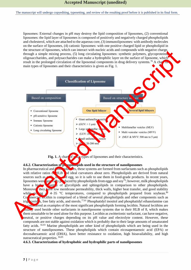
Nanoliposomes have different structural sizes and shapes based on environmental circumstances, their
constituents, and their production techniques.75 Liposomes can be categorized into five types based on
their diameter: 1) multilamellar vesicles (MLV)- 0.5–5 nm with five to twenty lipid bilayers, 2) small
unilamellar one lipid bilayer vesicles (SUV)- 20–200 nm, 3) largeunilamellar vesicles with one lipid
bilayer (LUV)- 200nm, 4) giant unilamellar vesicles with one lipid bilayer (GUV)-1nm, and 5) multi
vesicular vesicles (MVV) with multi lipid bilayer-1 nm.76
Nanoliposomes with a unilamellar (UL) state show a balloon-like structure with a simple monolayer,
whereas liposomes that are multilamellar have onion like structures consisting of several single-layer
cases. UL liposomes can be categorized as small unilamellar vesicles or large unilamellar vesicles, with
diameters below 100 nm and over 100 nm, respectively. Several smaller vesicles are entrapped in a
bigger one in multivesicular vesicles.77Another classification of nanoliposomes is: (1) pH-sensitive
Accep
ted M
anus
cript
Accepted Manuscript (unedited)
The manuscript will undergo copyediting, typesetting, and review of the resulting proof before it is published in its final form.
7 | P a g e
liposomes: External changes in pH may destroy the lipid composition of liposomes, (2) conventional
liposomes: the lipid layer of liposomes is composed of positively and negatively charged phospholipids
and cholesterol, which are attached to the aqueous core, (3) immunoliposomes: with antibody molecules
on the surface of liposomes, (4) cationic liposomes: with one positive charged lipid or phospholipid in
the structure of liposomes, which can interact with nucleic acids and compounds with negative charge,
through a simple mixing process, (5) long circulating liposomes: synthetic polymers, glycoproteins,
oligosaccharides, and polysaccharides can make a hydrophilic layer on the surface of liposome, which
result in the prolonged circulation of the liposomal components in drug delivery systems.78 A chart of
main types of liposomes and their characteristics is given as Fig. 1.
Fig. 1. A chart of main types of liposomes and their characteristics.
4.6.2. Characterization of phospholipids used in the structure of nanoliposomes
In pharmaceutical and food industries, these systems are formed from surfactants such as phospholipids
with relative ratios of HLB and ideal curvatures about zero. Phospholipids are derived from natural
sources such as milk, soy, and egg, so it is safe to use them in food-grade products. In recent years,
liposomes were generally produced by phospholipids from eggs and soy79; however, milk phospholipids
have a higher quantity of glycolipids and sphingolipids in comparison to other phospholipids.
Moreover, they have low membrane permeability, thick walls, higher heat transfer, and good stability
while storing at 4–35 °C temperatures, compared to phospholipids prepared from soybean.80
Commercial lecithin is comprised of a blend of several phospholipids and other components such as
triglycerides, free fatty acids, and sterols.77,81 Phosphatidyl inositol and phosphatidyl ethanolamine can
be considered as examples of the most significant phospholipids forming lecithin. Natural lecithins are
usually used beside other surfactants in nanoliposome systems due to their HLB of 8, which makes
them unsuitable to be used alone for this purpose. Lecithin as zwitterionic surfactant, can have negative,
neutral, or positive charges depending on its pH value and electrolyte content. However, these
compounds are not stable against oxidation which is probably due to their large amounts of unsaturated
fatty acids. 82,83 Marine phospholipids are other kind of phospholipids which are being used in the
structure of nanoliposomes. These phospholipids which contain eicosapentaenoic acid (EPA) or
docosahexaenoic acid (DHA), have better resistance to oxidation, high bioavailability, and high
nutraceutical properties. 84,85
4.6.3. Characterization of hydrophobic and hydrophilic parts of nanoliposomes
Accep
ted M
anus
cript
Accepted Manuscript (unedited)
The manuscript will undergo copyediting, typesetting, and review of the resulting proof before it is published in its final form.
8 | P a g e
Nanoliposomes are able to increase the solubility, bioavailability, and controlled release of the
encapsulated material to a larger extent. Hydrophobic bioactive compounds are entrapped in the lipid
bilayer of these systems, during their formation process. The compounds inside them can be released
when they diffuse through the membranes, or when their membrane is disrupted due to the alterations
in temperature or pH, and etc.86 These systems can entrap different molecules such as amphiphilics,
hydrophilics, and lipophilics which are entrapped into the lipid/water bilayer, into the interior, and
within the hydrophobic bilayer, respectively. These heterogenous particles are obtained by placing
phospholipids (such as lecithin) in the water or organic solutions and applying enough energy which
results in the formation of unilamellar (one bilayer) or multilamellar (series of bilayers) structures.
While phospholipids can spontaneously turn into unilamellars by applying them in the aqueous
mediums, their structure do not show desirable properties and good stability. Suitable production
processes should be applied for production of liposomes to obtain appropriate properties such as smaller
particle size, high loading capacity, and a proper encapsulation entrapment.87-89 Several factors such as
temperature, ionic strength, and pH, determine the physicochemical properties of nanoliposomes. Lipid
and phospholipid vesicles display low permeability to the entrapped or encapsulated material.
Nevertheless, at increased temperatures, a phase transition occurs in them which can have impact on
their permeability characteristics. Phospholipids of nanoliposomes have a substantial thermal
characteristic in which phase transition (Tc) occurs at temperatures lower than their final melting point
(Tm). At Tc, known as gel to liquid crystalline transition temperature, much of the ordered packing
arrangement of phospholipid bilayers are being lost, whereas there is an upsurge in its fluidity 90. By
placing the amphiphilic molecules such as phospholipids in an aqueous phase, they can form aggregated
complexes to protect their hydrophobic parts from water molecules; however, they keep their contact
with the aqueous phase via the hydrophilic head groups. By presence of enough level of energy, the
aggregated phospholipids can organize themselves in the form of closed bilayer vesicles such as
liposomes or nanoliposomes. In this way, liposomes can entrap hydrophilic, lipophilic molecules, or
lipid soluble compounds such as nutrients, drugs, and certain vitamins.91,92 Moreover, there can be other
compounds in the structure of nanoliposomes, such as sterols. Sterol incorporation into bilayers of
nanoliposome can cause major changes in the properties of these carriers. Cholesterol is the most
commonly used sterol in the production of the lipid vesicles. However, it forms bilayer structures by
itself. High concentrations of this compound can be incorporated into phospholipid membranes, to
modulate the fluidity of the lipid bilayer which results in an increase in the stability of vesicles, and
reduced permeability of the lipid membrane to solutes 93.
Other sterols have been scarcely used in the structure of nanoliposomes containing vitamins; however,
in a study by Amiri,Rezazadeh-Bari 10, the effect of cholesterol and phytosterol (Campesterol) powder
was investigated to synthesize a new formulation of nanoliposome for entrapment of vitamin C. The
findings showed that the highest stability of vitamin C in a period of 20 days was obtained in
phospholipids to campesterol ratio of 75:25. A positive impact of cholesterol substitution with
campesterol on control release, encapsulation efficiency, and stability of vitamin C in nanoliposomes
was reported. Regardless of current studies, further researches are needed to be done in this field.
4.6.4. Methods of nanoliposome preparation
Several factors such as concentration of the encapsulated material, nature of the medium,
physicochemical characteristics of the ingredients, polydispersity, size, and shelf life of the liposome
are significant factors to be considered for preparation of liposome or nanoliposome. Preparation
method of these systems can vary depending on their lipid composition.
The preparation methods of liposomes and nanoliposomes usually include the utilization of toxic or
non-food grade solvents such as ether solution, ethanol, hexane, chloroform, methanol and detergents,
such as alkyl glycoside, cholate, alkyl benzene sulfonates substances and triton X-100 for the
solubilization of hydrophobic and/or hydrophilic ingredients.94,95 However, there are several techniques
to reduce or completely eliminate the use of organic solvents for the formation of liposomes, requiring
skilled technical knowledge and high investment for being industrialized such as heating metho .95
Several techniques such as emulsion method, freeze drying double emulsion method, membrane
contactor technology, supercritical fluid technology, solvent-nonsolvent method, high-pressure
homogenization, dual asymmetric centrifugation, cross-flow filtration with detergent depletion method,
Accep
ted M
anus
cript
Accepted Manuscript (unedited)
The manuscript will undergo copyediting, typesetting, and review of the resulting proof before it is published in its final form.
9 | P a g e
thin film hydration, reverse-phase evaporation, solvent/surfactant displacement, heating method,
microfluidization, sonication, and extrusion method, are being used for liposome and nanoliposome
production.
In spite of several techniques being used for production of liposomes and nanoliposomes, due to the
complexity of most of these methods, their application in the industrial scale is difficult and very challenging. For instance, thin film hydration method followed by sonication was used for the
production nanoliposomes to encapsulate different kinds of compounds such as vitamins, previously 96.
In spite of the production of nano-size liposomes with this method, it is impossible to scale up the
process. Some other modifications are needed to overcome the scale up problems of these methods. For
instance, to scale up the sonication assisted homogenization process, different parameters should be
considered. The cycle of sonication time should be kept constant and a 6 mm tip and 100% amplitude
is needed to be used. The power which is delivered to the sample as a result of the number of sonication
cycles is also an important parameter, because the sound wave should be amplified on the whole batch
volume at the same dimensional properties.97 Some of the most important methods of nanoliposome
production are summarized below.
Emulsion and freeze-drying double emulsion method
In the emulsification method, appropriate surfactants are being used to produce oil-in-water and water-
in-oil emulsions. This method is a traditional method; however, freeze-drying double emulsion method
is a novel trend, in which cryoprotectants are added to the liposome formulation and W/O/W emulsion
will be formed followed by a sterilization step.
Membrane contactor technology
In this method, organic phase is placed in the pressurized vessel. A pump directs the aqueous phase to
a module and the nitrogen gas is used to push the oil phase into the system. The aqueous phase is
subsequently pumped through the membrane contactor module, and liposomes will be formed
spontaneously at the time that the lipid and aqueous phases meet. Liposomes prepared using this
technology are homogeneous with small particle size and high encapsulation efficiency for lipophilic
compounds. Moreover, this method is simple to scale up.98,99
Supercritical fluid technology
In supercritical anti-solvent method, lipids are readily dissolved in supercritical carbon dioxide and then
are precipitated, so that ultrafine particles will be formed. In the supercritical reverse phase evaporation
method, which is another supercritical fluid technology, aqueous phase and solid lipid materials are put
into a sealed viewing cell and the pressure and temperature are adjusted and then the CO2 gas is
introduced. CO2 is then removed, and liposomes are formed. These methods of liposome preparation
do not contain any organic solvents showing their higher advantage compared to conventional methods. 100,101
Solvent-nonsolvent method
In the conventional solvent-nonsolvent method, particles are produced due to the lack of solvent. In this
method, firstly solution of the material that is going to be encapsulated is prepared and then the solvent
is removed through diafiltration or an evaporation procedure after dispersion in its nonsolvent, and
liposomes will be produced due to the lack of solvent. 102
Homogenization
In homogenization method of liposome production, the suspension of drug or any compound to be
encapsulated, suddenly passes through a homogenization gap causing high streaming velocity, and in
this way liposomes are being produced. 103 The main drawback of this method is its extremely high
operating pressure. 104
Dual asymmetric centrifugation (DAC)
This method of liposome production includes an additional sample rotation around its own vertical axis
which pushes sample toward the center of the centrifuge, while in the conventional centrifugation
sample is constantly being pushed outwards. Therefore, two overlaying movements of the materials
occur in the centrifugation vial, which provides shear forces and an efficient homogenization for even
Accep
ted M
anus
cript
Accepted Manuscript (unedited)
The manuscript will undergo copyediting, typesetting, and review of the resulting proof before it is published in its final form.
10 | P a g e
a concentrated and viscous blend of lipids 105. This method is easy to operate and no organic solvent is
being used for production of liposomes with small particle size. 105 99
Cross-flow filtration detergent depletion method
One of the production methods of liposomes is based on detergent addition to solubilize lipids which is
being removed through next steps. In this technique membrane can still contain a huge amount of
detergent which is difficult to remove. Therefore, a technique, which can solve this problem and reduce
the preparation time and heterogeneous liposome lamellarity is preferred. Combination of cross-flow
technique and conventional detergent depletion method is an economical technique which is used to
overcome the problems related to the detergent removal.106 This method is consisted of a filtration device, a pump, a starting reservoir, tubing, an integrated rotary slide valve and a manometer for monitoring the pressure of retentate. 99 Thin film hydration method
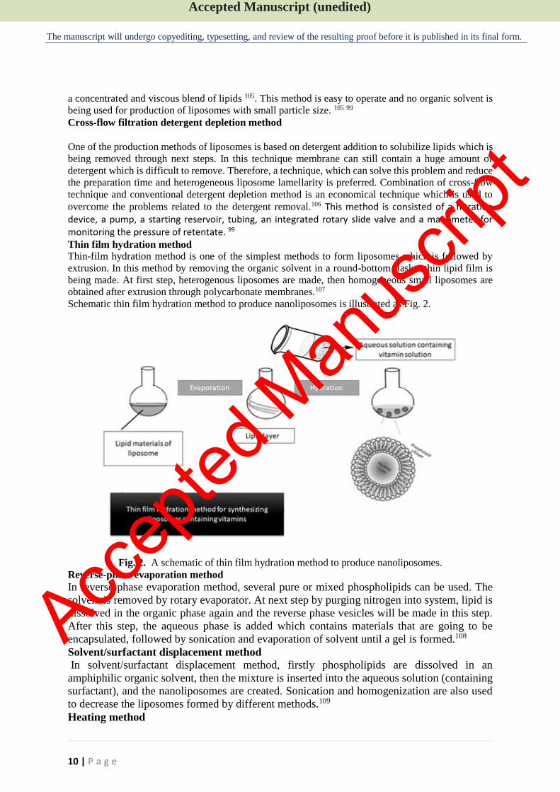
Thin-film hydration method is one of the simplest methods to form liposomes which is followed by
extrusion. In this method by removing the organic solvent in a round-bottom flask a thin lipid film is
being made. At first step, heterogenous liposomes are made, then homogeneous small liposomes are
obtained after extrusion through polycarbonate membranes.107
Schematic thin film hydration method to produce nanoliposomes is illustrated as Fig. 2.
Fig. 2. A schematic of thin film hydration method to produce nanoliposomes.
Reverse-phase evaporation method
In reverse-phase evaporation method, several pure or mixed phospholipids can be used. The
solvent is removed by rotary evaporator. At next step by purging nitrogen into system, lipid is
dissolved in the organic phase again and the reverse phase vesicles will be made in this step.
After this step, the aqueous phase is added which contains materials that are going to be
encapsulated, followed by sonication and evaporation of solvent until a gel is formed.108
Solvent/surfactant displacement method
In solvent/surfactant displacement method, firstly phospholipids are dissolved in an
amphiphilic organic solvent, then the mixture is inserted into the aqueous solution (containing
surfactant), and the nanoliposomes are created. Sonication and homogenization are also used
to decrease the liposomes formed by different methods.109
Heating method
Accep
ted M
anus
cript
Accepted Manuscript (unedited)
The manuscript will undergo copyediting, typesetting, and review of the resulting proof before it is published in its final form.
11 | P a g e
In this method, high pressures or toxic solvents are not used. All of the compounds including
lipids are added to a heating flask and are heated at high temperatures for about 30 min to let
lipids are dissolved. Depending on the liposomal compounds, nanovesicles are formed 90.
Microfluidization
Microfluidization is a common type of homogenization method which is widely used in food
industry. This technique generates high pressures, directing the flow stream to the impingement
area through microchannels. This technique provides acceptable costs for large scale
productions and does not include toxic solvents, which favors food regulatory requirements.
However, there are three drawbacks for it such as contamination, material loss, and hard scale-
up. 110,111
Sonication method
Sonication is a simple way for production of nanoliposomes from liposomes. In this method,
hydrated vesicles are treated by a titanium-tipped probe sonicator for a few minutes with
determined seconds of on- and off- intervals in a temperature-controlled environment. At this
stage, nanoliposomes in the form of small unilamellar vesicles are formed. 90
Extrusion method
In the extrusion method of liposome or nanoliposome preparation, micrometric liposomes are
modified to large unilamellar vesicles or nanoliposomes which depends on the pore-size of
filters.112 Vesicles are extruded by passing through polycarbonate filters with defined pore sizes
for several times. At the end, a homogenous sample (nanoliposome) is produced 90 4.6.5. Advantages and disadvantages of nanoliposomes Generally, liposomes and nanoliposomes are systems contributing to the recent food trend. These
systems can improve the efficiency of food additives and reduce the amounts of required bioactive
compounds. They favor the green chemistry, and they can increase the use of natural compounds instead
of synthetic constituents.
Comparing the lipid based nanocarriers with other encapsulation materials such as carriers based on
polymeric compounds, shows that lipid based nanocarriers have distinctive advantages, such as their
ability to provide a protective cover for biological compounds which protects them from degradation,
their capability of entrapping the compounds with broad range of solubility, and their high potential for
industrial production due to the natural food components being used in their formulations, as well as
their minimum production costs. In addition, they have good biocompatibility and biodegradability.113
According to USFDA (United States Food and Drug Administration) guidance, liposomes are different
than drug-lipid complexes, emulsions, and microemulsions. Thus, developing a liposomal product
needs suitable certification for its chemistry, production, and constituents, beside bioavailability in
human pharmacokinetics and labeling (FDA 2018). Nanoliposomes have several advantageous and
useful features in the formulation, application and delivery; however, they have limited drawbacks such
as poor encapsulation efficiency, lack of enough parameters for continuous industrial production and
stability, excessive cost of food components, and the use of high force pressure, homogenization, and
sonication.114
In contrary to micron-sized particles, nanoliposomes have remarkable properties such as being
metastable and dilatable with water, without any changes in their size distribution. Moreover,
nanoliposomes can encapsulate and liberate two materials with different solubilities such as vitamin C
and vitamin E, simultaneously. Nanoliposomes are able to include and deliver both vitamin C and
vitamin E to an oxidation site, resulting in a synergistic effect. Lipid based vesicles incorporating two
bioactive agents are called bifunctional vesicles.115-117 In other study, Chaves,Oseliero Filho 118 showed
that it is feasible to produce "co-encapsulated liposome" vesicle containing two curcumin and vitamin
D3, which is stable over the storage time of more than 40 days.
One of the most useful characteristics of liposomes and nanoliposomes is their targetability. Directing
bioactive molecules to a specific part in which they can apply their optimum efficacy, is very important.
An appropriate and directed release enhances the efficacy of bioactive material, certifies optimal
dosage, expands the range of their application, and thus increases the cost-effectiveness of the
Accep
ted M
anus
cript
Accepted Manuscript (unedited)
The manuscript will undergo copyediting, typesetting, and review of the resulting proof before it is published in its final form.
12 | P a g e
product.87,119 Moreover, nanoliposomes provide a delivery option for multiple drug molecules at the
same time.120,121
Structural appearance and stability of the nanoliposomes are highly associated with the technology
adopted for their synthesis. Several problems related to physicochemical properties and stability of
conventional nanoliposomes have been reported. Studies showed that liposomes might not be able to
provide good physical and chemical stability resulting in low encapsulation efficiency, lipid oxidation,
aggregation of vesicles or fast release of bioactive compounds.122,123 Moreover, rapid clearance of most of the liposomes from circulation by the reticuloendothelial system, and fast leakage of water-soluble drugs under improper storage conditions, are some other problems related to these formulations. 124 125 Thus, modification of nanoliposomes using methods such as addition of charged substances on their
surface, coating their surfaces with polymers, or using some drying techniques after their formation,
have emerged as efficient approaches to boost their physicochemical properties and stability. 126-128 In addition to the formation of biopolymer-associated liposomes, another promising method to improve the bioactivity, stability, and bioavailability of liposomes is to accomplish additional processes, referred to as “post-processing techniques”, to the aqueous liposome. Some common methods of post-processing include spray drying, freeze drying, and spray freeze drying.129 5. Important properties of vitamin-loaded nanoliposomes
5.1. Particle size, PDI and zeta potential of nanoliposomes
Generally, the average particle size, and polydispersity index (PDI) are parameters that are of high
importance in liposomes and nanoliposomes. PDI, shows the quality of the formulated system with
regard to the size distribution. The suitable application of nanocarriers including nanoliposomes for a
specific route of drug administration is highly dependent on their average particle size, PDI and size
stability. Size variations of nano sized systems are of high importance in the formulation of nanocarriers
and to achieve ideal results, constant and narrow size distribution should be considered. It should be
considered that for nanocarriers, size stability is more important, in comparison to micro size systems,
and this is due to their larger specific surface area 130. In a study on the liposomal formulation of
glucosamine and vitamin D, it was shown that the encapsulated liposomes have average particle size of
840 nm with great stability.131
PDI is known as a parameter indicating uniformity of nanocarriers. When this parameter is more than
1, it means that different sizes of particles exist per volume unite in the solution. Particle aggregation
increases the PDI, so factors causing aggregation can affect PDI. Studies on lipid-based nanoparticles
have shown that several factors such as surfactant type and its concentration, instrumental conditions,
and lipids used in their formulations can have impact on PDI and particle size.132
A study on encapsulation of vitamin E in nanoliposome revealed that the formulation of liposomes by
hydrophobic stabilizers such as gamma oryzanol and lauric acid leads to an increase in the particle size.
Though, hydrophilic stabilizer such as PEG 400 did not have significant effect on mean size. In addition,
it was shown that by using hydrophobic stabilizers, particle size distribution (PDI) decreases in colloidal
system which indicates their stabilizing effect during storage time while PEG had only a slight effect
on PDI.133
Results of a study showed that particle sizes of vitamin C-folic acid liposomes and chitosan coated
vitamin C- folic acid liposomes are 138.58 nm and 249.13 nm, respectively. Also the reported PDIs
were 0.18 and 0.31, respectively, indicating the similarity in the size of nanoliposomes.134 Salama
andGaber 135 could produce nanoliposomes encapsulating ascorbic acid (LEAA) with particle size of
421.6 nm and PDI of 0.539, showing narrow and homogenous particle size distributions.
Zeta potential is known as the electrokinetic potential between particles which are dispersed in a
solution. This parameter shows the surface electrical charges of particles as well as their stability. Zeta
potential and the size distribution are both measured by DLS technique. A large positive or negative
zeta potential of particles present in the suspension results in less tendency of them to aggregate, so they
will have higher stability 136. To broaden the zeta potential of nanoliposomes, providing stronger
repulsion, some researchers use charged substances such as biopolymers as an extra layer covering the
surface of nanoliposomes. The results of a study on chitosan-coated nanoliposomes of vitamin D3
Accep
ted M
anus
cript
Accepted Manuscript (unedited)
The manuscript will undergo copyediting, typesetting, and review of the resulting proof before it is published in its final form.
13 | P a g e
showed that coating the nanoliposomes with 0.01% (w/v) chitosan can improve favorable properties of
nanoliposomes. The PDI of this covered nanoliposome was close to the monodisperse distribution.
Moreover, the increase in size and zeta potential verified the interaction of chitosan and liposome,
showing a successful coverage.137 The increase in zeta potential is due to the more adsorbed cationic
polymers on the surface of nanoliposomes. Since chitosan possess a high positive charge, the adsorption
of chitosan seems to increase the density of positive charge, resulting in a more positive zeta potential.
5.2. Encapsulation Efficiency Encapsulation of vitamins into nanoliposomes provides many advantages, which has been highlighted in this review. One of the most significant benefits of encapsulation of vitamins within liposomes is improving their bioavailability in the human body. The encapsulation efficiency (EE) can be considered as the most important factor to determine the capability of nanoliposomes to encapsulate bioactive compounds. Encapsulation efficiency can also be stated as trapping efficiency, incorporation efficiency, or encapsulation percentage 138. The EE depends on the composition of nanoliposomes and the characteristics and concentration of the encapsulated bioactive compound.139 Dalmoro,Bochicchio 137 reported that the encapsulation efficiency of D3 and K2-loaded in cholesterol and phosphatidylcholine nanoliposomes was 88.4% and 94.7%, respectively. The chitosan coverage for each nanoliposomal formulation of both vitamins, led to up to 98% increase in the entrapment efficiency. Lee,Park 140 have reported that multilamellar vesicle nanoliposomes of retinol, composed of L-α-phosphatidylcholine (PC) and 10% sterol (w/w), could increase the encapsulation efficiency of retinol up to 99%. Since higher EE is correlated with longer shelf-life and better stability of the nanocarrier, it is always favorable to elevate the EE by selecting the best possible mixture of core-wall ratio, processing steps such as drying methods, homogenization process, and other processing variables.141 The encapsulation efficiency of an entrapped bioactive in a nanoliposome depends on its partition coefficient and polarity. If loaded ingredients are hydrophobic in nature, they reside in the hydrocarbon chain of nanoliposome. However, if loaded bioactive compounds are polar, they are likely to be positioned in the aqueous core or next to the water–lipid interface, adjacent to the nanoliposome polar head groups.142 Overall, the encapsulation efficiency (EE) of lipophilic materials is usually higher than hydrophilic ones in nanoliposomes, since they can be tightly positioned in the membrane.143 For instance, in a recent study, two vitamins were simultaneously co-encapsulated within synthesized nanoliposomes: vitamin C as a hydrophilic and vitamin E as a lipophilic bioactive. Vitamin E, and vitamin C displayed an average encapsulation efficiency of 95.1% and 77.8 %, respectively. Thus, vitamin E demonstrated the highest EE, which can be attributed to its high lipid affinity.144 A summarized application of nanoliposome for encapsulation of vitamins is given in Table 3. 5.3. Biocompatibility Biocompatibility shows the ability of nanoliposomes to apply their proposed function without having any negative effect on the targeted tissues. There are several studies mentioning the biocompatibility and biodegradability of nanoliposomes loaded with bioactive compounds. Liposomes are comprised of natural lipids which are biodegradable, biocompatible, and less immunogenic. Preliminary skin toxicity study of oleic acid liposomes showed that no epidermal cell apoptosis occurs in the skin that is treated with these liposomes, indicating good biocompatibility of them with mouse skin. 145 In a study by Al-Ogaidi, I., chitosan (CH) and alginate (Alg) which are biocompatible compounds were used to manufacture biodegradable and biocompatible nanoliposomes of vitamin C. 125 There are several compounds which can be used in the formulation of nanoliposomes to provide biocompatible liposomes. For instance, chitosan has been used in combination with sodium tripolyphosphate to provide core-shell of nanoliposomes for vitamin E encapsulation. 146,147 Chitosan provides an outer hydrophilic barrier by formation of a coating layer onto membrane, which can prevent the interaction between liposomes, increase their drug delivery efficiency, improve their structural properties, and biocompatibility. 148
Accep
ted M
anus
cript
Accepted Manuscript (unedited)
The manuscript will undergo copyediting, typesetting, and review of the resulting proof before it is published in its final form.
14 | P a g e
In other study, L-α-phosphatidylcholine which packages with 1α,25(OH)2D3, and 1,2-distearoyl-sn-glycero-3-phosphoethanolamine-N-[amino-(polyethylene glycol)2000], were used to produce non-toxic and biocompatible vitamin D nanoliposomes. 149 Moreover, in spite of biocompatibility properties of liposomes, these formulations could decrease the toxicity of antimicrobial agent which are potentially toxic. 145 Controlled release The nanoencapsulation systems compared to the direct application of bioactive compounds provide better functional properties such as controlled-release, and higher bioavailability due to their high surface area, in comparison to large particles. 150 151 Encapsulation provides a surrounding for bioactive compounds or drugs, which protects them against environmental stresses and controls their release
over time. 152,153 Several studies have shown the controlled release of different bioactive compounds using nanoliposomes. 154,155 Hydrophilic and hydrophobic compounds can simultaneously and efficiently be trapped in the phospholipids bilayer membrane structure of nanoliposome through various physical and chemical interactios.156 Therefore, nanoliposome is able to extend the residence time of compounds or drugs in the ambient, causing the controlled in vivo release of these compounds, which enables the activity of compounds for a longer time. 157 The control release of loaded compounds in nanoliposomes can be improved by doing some modifications on the surface of conventional nanoliposomes158. Researchers have reported that the surface decoration of nanoliposomes of neohesperidin by chitosan and pectin loaded, improve the controlled release of this compound 159. Chitosan nanoparticles were shown to improve the controlled release vitamin C in several studies. 160,161 In a study by Liu et al., 162 the release behavior of vitamin C was observed for the chitosan and alginate coated nanoliposomes. It was indicated that addition of these polymers onto the surface of anionic nanoliposomes improves the control release of vitamin C during 90 days of storage at 4 °C, by a steric barrier on the surface. Bioavailability of nanoliposomes containing vitamins Bioavailability is a key parameter of pharmacokinetic, which states the proportion of a bioactive
compound or a drug, administered through any non-vascular route which reaches to the systemic
circulation. 163 A number of studies have indicated the increased bioavailability of vitamins by loading
them in nanoliposomes. A review of recent literature shows a growing trend to increase the
bioavailability of vitamins by loading them in nanoliposomes. Łukawski,Dałek 164 conducted a study
with the aim of comparing the profiles of serum concentration of vitamin C in 20 healthy volunteers,
after the oral administration either as an aqueous solution or as a liposomal suspension. Their results
showed that in the nanoliposomal vitamin C treatment group, Cmax of vitamin gets to higher values
compared to the vitamin C solution treatment group (303 µµ compared to 180 µµ). Moreover, for
nanoliposomal formulation, the maximum vitamin C blood concentration delay time (Tmax) is longer by
about 1 h in comparison to the solution (Tmax=180 min vs. 96 min). The incremented half-life (t1/2>6 h
vs. t1/2=4 h) and increased AUC (81 570 µµ* min vs. 45 330 µµ* min) shows that loading vitamin C in
nanoliposomes improve the bioavailability of this vitamin. The same result was reported by Davis,Paris 165, who compared the bioavailability of vitamin C in free and liposomal forms.
Moreover, in another study conducted on the bioavailability of encapsulated and free vitamin C, Gopi
andBalakrishnan 166 indicated that nanoliposomal vitamin C is 1.77 times more bioavailable compared
to the free vitamin C. The nanoliposomal vitamin C showed higher values of Cmax (5.23 vs 2.17 mg/dL),
AUC0-t (55.86 vs 31.53 mg.h/dL), and AUC0-∞ (78.90 vs 57.12 mg.h/dL), compared to the aqueous
solution of vitamin C.
6. Conclusion
In this review, the application of nanoliposomes to encapsulate vitamins was investigated. Regarding to vitamin encapsulation, nanoliposomes are known as the most used lipid based nanocarriers. One of the most principal reasons for the high use of nanoliposomes to deliver vitamins is related to their ability to encapsulate both hydrophilic and hydrophobic vitamins as well as the ease of their production in industrial scale. However, the quality of produced nanoliposomes is very important. The
Accep
ted M
anus
cript
Accepted Manuscript (unedited)
The manuscript will undergo copyediting, typesetting, and review of the resulting proof before it is published in its final form.
15 | P a g e
fabrication of a high quality nanoliposome containing vitamins mainly depends on the production method, utilized materials, and characteristics of liposomes such as particle size, polydispersity index, zeta potential, controlled release, and encapsulation efficiency. Lack of attention to these parameters, while producing nanoliposomes, can easily lead to the fabrication of systems with several drawbacks such as fast release, deposition, low stability, and imperfect protection of vitamins. Recent trends demonstrate that researchers are interested to synthesize nanoliposomes with the highest encapsulation efficiency of vitamins, thus many of the recently published studies have reported reaching to the higher than 70% encapsulation efficiency for different vitamins. In addition to inherent properties of nanoliposomes, the bioavailability of loaded vitamins into nanoliposomes is another significant factor. Increased bioavailability can be considered as the final goal for the application of nanoliposomes to cover different vitamins. As mentioned in this review, there are clear recent evidences showing that nanoliposomes are able to improve the bioavailability of vitamin C. Moreover, based on our knowledge, some variables still need more investigations. For instance, the replacement of cholesterol with other sterols and their effects on different factors of vitamin-loaded nanoliposomes require more studies. Moreover, interaction of vitamins with other compounds that are incorporated into nanoliposomes, and their effects on several parameters of nanoliposomes such as stability, zeta potential, and encapsulation efficiency should be investigated. References
1. Tardy AL, Pouteau E, Marquez D, Yilmaz C, Scholey A. Vitamins and Minerals for
Energy, Fatigue and Cognition: A Narrative Review of the Biochemical and Clinical
Evidence. Nutrients 2020;12(1):228. doi: 10.3390/nu12010228
2. Gupta M, Aggarwal R, Raina N, Khan A. Vitamin-Loaded Nanocarriers as Nutraceuticals
in Healthcare Applications. In: Rahman M, Beg S, Kumar V, Ahmad FJ, editors.
Nanomedicine for Bioactives. Singapore: Springer Singapore; 2020. p. 451-70.
3. Dehghani-Samani A, Kamali M, Hoseinzadeh-Chahkandak F. The Role of Vitamins on the
Prevention and/or Treatment of COVID-19 Infection; a Systematic Review. Mod Care J
2020;17(3). doi: 10.5812/modernc.104740
4. Grant WB, Lahore H, McDonnell SL, Baggerly CA, French CB, Aliano JL, et al. Evidence
that vitamin D supplementation could reduce risk of influenza and COVID-19 infections and
deaths. Nutrients 2020;12(4):988. doi: 10.3390/nu12040988
5. McCartney DM, Byrne DG. Optimisation of Vitamin D Status for Enhanced Immuno-
protection Against Covid-19. Ir Med J 2020;113(4):58.
6. Ali N. Role of vitamin D in preventing of COVID-19 infection, progression and severity. J
Infect Public Health 2020;13(10):1373-80. doi: 10.1016/j.jiph.2020.06.021
7. Hsu C-Y, Wang P-W, Alalaiwe A, Lin Z-C, Fang J-Y. Use of lipid Nanocarriers to
improve Oral delivery of vitamins. Nutrients 2019;11(1):68. doi: 10.3390/nu11010068
8. Glowka E, Stasiak J, Lulek J. Drug Delivery Systems for Vitamin D Supplementation and
Therapy. Pharmaceutics 2019;11(7):347. doi: 10.3390/pharmaceutics11070347
9. Ajeeshkumar KK, Aneesh PA, Raju N, Suseela M, Ravishankar CN, Benjakul S.
Advancements in liposome technology: Preparation techniques and applications in food,
functional foods, and bioactive delivery: A review. Compr Rev Food Sci Food Saf
2021;20(2):1280-306. doi: 10.1111/1541-4337.12725
10. Amiri S, Rezazadeh-Bari M, Alizadeh-Khaledabad M, Amiri S. New formulation of
vitamin C encapsulation by nanoliposomes: production and evaluation of particle size,
stability and control release. Food Sci 2019;28(2):423-32. doi: 10.1007/s10068-018-0493-z
11. Yoshii K, Hosomi K, Sawane K, Kunisawa J. Metabolism of dietary and microbial
vitamin B family in the regulation of host immunity. FRONT NUTR 2019;6:48. doi:
10.3389/fnut.2019.00048/full
Accep
ted M
anus
cript
Accepted Manuscript (unedited)
The manuscript will undergo copyediting, typesetting, and review of the resulting proof before it is published in its final form.
16 | P a g e
12. Zhao X, Zhang M, Li C, Jiang X, Su Y, Zhang Y. Benefits of Vitamins in the Treatment
of Parkinson’s Disease. OXID MED CELL LONGEV 2019;2019. doi: 10.1155/2019/9426867
13. Gossweiler AG, Martinez-Mier EA. Vitamins and Oral Health. The Impact of Nutrition
and Diet on Oral Health: Karger Publishers; 2020. p. 59-67.
14. Sanchez-Hernandez D, Anderson GH, Poon AN, Pannia E, Cho CE, Huot PS, et al.
Maternal fat-soluble vitamins, brain development, and regulation of feeding behavior: an
overview of research. Nutr Res Rev 2016;36(10):1045-54. doi: 10.1016/j.nutres.2016.09.009
15. Kumar PS, Joshiba GJ. Separation and Purification of Vitamins: Vitamins B1, B2, B6, C
and K1. Applications of Ion Exchange Materials in Biomedical Industries: Springer; 2019. p.
177-87.
16. Chawla J, Kvarnberg D. Hydrosoluble vitamins. Handbook of clinical neurology:
Elsevier; 2014. p. 891-914.
17. Cole LA, Kramer PR. Human physiology, biochemistry and basic medicine: Academic
Press; 2015.
18. Calderón‐Ospina CA, Nava‐Mesa MO. B Vitamins in the nervous system: Current
knowledge of the biochemical modes of action and synergies of thiamine, pyridoxine, and
cobalamin. CNS Neurosci Ther 2020;26(1):5-13. doi: 10.1111/cns.13207
19. Kennedy DO. B vitamins and the brain: mechanisms, dose and efficacy—a review.
Nutrients 2016;8(2):68. doi: 10.3390/nu8020068
20. Allen LH, Miller JW, de Groot L, Rosenberg IH, Smith AD, Refsum H, et al. Biomarkers
of Nutrition for Development (BOND): Vitamin B-12 Review. J Nutr
2018;148(suppl_4):1995S-2027S. doi: 10.1093/jn/nxy201
21. Hosomi K, Kunisawa J. The Specific Roles of Vitamins in the Regulation of
Immunosurveillance and Maintenance of Immunologic Homeostasis in the Gut. Immune
Netw 2017;17(1):13-9. doi: 10.4110/in.2017.17.1.13
22. Mikkelsen K, Stojanovska L, Tangalakis K, Bosevski M, Apostolopoulos V. Cognitive
decline: A vitamin B perspective. Maturitas 2016;93:108-13. doi:
10.1016/j.maturitas.2016.08.001
23. Fluegge K. Propionic acid metabolism, ASD, and vitamin B12: Is there a role for
environmental nitrous oxide? Int J Dev Neurosci 2017;57:21-3. doi:
10.1016/j.ijdevneu.2016.12.007
24. Suh SW, Kim HS, Han JH, Bae JB, Oh DJ, Han JW, et al. Efficacy of Vitamins on
Cognitive Function of Non-Demented People: A Systematic Review and Meta-Analysis.
Nutrients 2020;12(4):1168. doi: 10.3390/nu12041168
25. Maillet D, Rajah MN. Age-related differences in brain activity in the subsequent memory
paradigm: a meta-analysis. Neurosci Biobehav Rev 2014;45:246-57. doi:
10.1016/j.neubiorev.2014.06.006
26. Johnston C, Vitamin C, Erdman J, Macdonald I, Zeisel S. Present knowledge in nutrition.
ed JW Erdman Jr, IA Macdonald, and SH Zeisel 2012.
27. Jang S, Han JW, Shin J, Kim TH, Kwak KP, Kim K, et al. Normal-But-Low Serum
Folate Levels and the Risks for Cognitive Impairment. Psychiatry Investig 2019;16(7):532-8.
doi: 10.30773/pi.2019.05.29
28. Shashirekha M, Mallikarjuna S, Rajarathnam S. Status of bioactive compounds in foods,
with focus on fruits and vegetables. Crit Rev Food Sci Nutr 2015;55(10):1324-39. doi:
10.1080/10408398.2012.692736
29. Borel P, Caillaud D, Cano NJ. Vitamin D bioavailability: state of the art. Crit Rev Food
Sci Nutr 2015;55(9):1193-205. doi: 10.1080/10408398.2012.688897
Accep
ted M
anus
cript
Accepted Manuscript (unedited)
The manuscript will undergo copyediting, typesetting, and review of the resulting proof before it is published in its final form.
17 | P a g e
30. Lamberg-Allardt C. Vitamin D in foods and as supplements. Prog Biophys Mol Biol
2006;92(1):33-8. doi: 10.1016/j.pbiomolbio.2006.02.017
31. Naas H, de Oliveira AA, Karpova T, Nunes KP. Toll-like receptor 4 (TLR4) as a possible
pathological mechanism in hyperglycemia-associated testicular dysfunction. Med Hypotheses
2019;127:116-9. doi: 10.1016/j.mehy.2019.04.010
32. Guo M, Zhu J, Yang T, Lai X, Liu X, Liu J, et al. Vitamin A improves the symptoms of
autism spectrum disorders and decreases 5-hydroxytryptamine (5-HT): A pilot study. Brain
Res Bull 2018;137:35-40. doi: 10.1016/j.brainresbull.2017.11.001
33. Colombo ML. An update on vitamin E, tocopherol and tocotrienol—perspectives.
Molecules 2010;15(4):2103-13. doi: 10.3390/molecules15042103
34. L. Booth S. Vitamin K: food composition and dietary intakes. J Food Nutr Res
2012;56(1):5505. doi: 10.3402/fnr.v56i0.5505
35. Walther B, Chollet M. Menaquinones, bacteria, and foods: vitamin K2 in the diet.
Vitamin K2-Vital for Health and Wellbeing 2017:63-82. doi: 10.5772/63712
36. Shahzad S, Ashraf MA, Sajid M, Shahzad A, Rafique A, Mahmood MS. Evaluation of
synergistic antimicrobial effect of vitamins (A, B1, B2, B6, B12, C, D, E and K) with
antibiotics against resistant bacterial strains. J Glob Antimicrob Resist 2018;13:231-6. doi:
10.1016/j.jgar.2018.01.005
37. Tintino SR, de Souza VC, Silva J, Oliveira-Tintino CDdM, Pereira PS, Leal-Balbino TC,
et al. Effect of vitamin K3 inhibiting the function of NorA efflux pump and its gene
expression on Staphylococcus aureus. Membranes 2020;10(6):130. doi:
10.3390/membranes10060130
38. Pimentel-Moral S, Teixeira M, Fernandes A, Arráez-Román D, Martínez-Férez A,
Segura-Carretero A, et al. Lipid nanocarriers for the loading of polyphenols–A
comprehensive review. Adv Colloid Interface Sci 2018;260:85-94. doi:
10.1016/j.cis.2018.08.007
39. Devi N, Sarmah M, Khatun B, Maji TK. Encapsulation of active ingredients in
polysaccharide–protein complex coacervates. Adv Colloid Interface Sci 2017;239:136-45.
doi: 10.1016/j.cis.2016.05.009
40. Katouzian I, Esfanjani AF, Jafari SM, Akhavan S. Formulation and application of a new
generation of lipid nano-carriers for the food bioactive ingredients. Trends Food Sci Technol
2017;68:14-25. doi: 10.1016/j.tifs.2017.07.017
41. Fathi M, Mozafari MR, Mohebbi M. Nanoencapsulation of food ingredients using lipid
based delivery systems. Trends Food Sci Technol 2012;23(1):13-27. doi:
10.1016/j.tifs.2011.08.003
42. Mohammadi M, Assadpour E, Jafari SM. Encapsulation of food ingredients by
nanostructured lipid carriers (NLCs). Lipid-Based Nanostructures for Food Encapsulation
Purposes: Elsevier; 2019. p. 217-70.
43. Aboalnaja KO, Yaghmoor S, Kumosani TA, McClements DJ. Utilization of
nanoemulsions to enhance bioactivity of pharmaceuticals, supplements, and nutraceuticals:
Nanoemulsion delivery systems and nanoemulsion excipient systems. Expert Opin Drug
Deliv 2016;13(9):1327-36. doi: 10.1517/17425247.2016.1162154
44. Shaddel R, Akbari-Alavijeh S, Jafari SM. Encapsulation of food ingredients by Pickering
nanoemulsions. Lipid-Based Nanostructures for Food Encapsulation Purposes: Elsevier;
2019. p. 151-76.
45. Salvia-Trujillo L, Soliva-Fortuny R, Rojas-Graü MA, McClements DJ, Martin-Belloso O.
Edible nanoemulsions as carriers of active ingredients: A review. Annu Rev Food Sci Technol
2017;8:439-66. doi: 10.1146/annurev-food-030216-025908
Accep
ted M
anus
cript
Accepted Manuscript (unedited)
The manuscript will undergo copyediting, typesetting, and review of the resulting proof before it is published in its final form.
18 | P a g e
46. Naseema A, Kovooru L, Behera AK, Kumar KP, Srivastava P. A critical review of
synthesis procedures, applications and future potential of nanoemulsions. Adv Colloid
Interface Sci 2020:102318. doi: 10.1016/j.cis.2020.102318
47. Nejatian M, Saberian H, Jafari SM. Encapsulation of food ingredients by double
nanoemulsions. Lipid-Based Nanostructures for Food Encapsulation Purposes: Elsevier;
2019. p. 89-128.
48. Guttoff M, Saberi AH, McClements DJ. Formation of vitamin D nanoemulsion-based
delivery systems by spontaneous emulsification: factors affecting particle size and stability.
Food Chem 2015;171:117-22. doi: 10.1016/j.foodchem.2014.08.087
49. Kadappan AS, Guo C, Gumus CE, Bessey A, Wood RJ, McClements DJ, et al. The
Efficacy of Nanoemulsion-Based Delivery to Improve Vitamin D Absorption: Comparison of
In Vitro and In Vivo Studies. Mol Nutr Food Res 2018;62(4):1700836. doi:
10.1002/mnfr.201700836
50. Zaghian N, Goli M. Optimization of the production conditions of primary (W 1/O) and
double (W 1/O/W 2) nano-emulsions containing vitamin B 12 in skim milk using ultrasound
wave by response surface methodology. J FOOD MEAS CHARACT 2020:1-11. doi:
10.1007/s11694-020-00567-1
51. Kumar DL, Sarkar P. Encapsulation of bioactive compounds using nanoemulsions.
Environ Chem Lett 2018;16(1):59-70. doi: 10.1007/s10311-017-0663-x
52. Rao J, Decker EA, Xiao H, McClements DJ. Nutraceutical nanoemulsions: influence of
carrier oil composition (digestible versus indigestible oil) on β‐carotene bioavailability. J SCI
FOOD AGR 2013;93(13):3175-83. doi: 10.1002/jsfa.6215
53. Wibowo D, Zhao C-X, Middelberg AP. Emulsion-templated silica nanocapsules formed
using bio-inspired silicification. Chem Commun 2014;50(77):11325-8. doi:
10.1039/C4CC04904G
54. Boskabadi M, Saeedi M, Akbari J, Morteza-Semnani K, Hashemi SMH, Babaei A.
Topical Gel of Vitamin A Solid Lipid Nanoparticles: A Hopeful Promise as a Dermal
Delivery System. Adv Pharm Bull 2020. doi: 10.34172/apb.2021.075
55. AlZahabi S, Sakr OS, Ramadan AA. Nanostructured lipid carriers incorporating prickly
pear seed oil for the encapsulation of vitamin A. J Cosmet Dermatol 2019;18(6):1875-84.
doi: 10.1111/jocd.12891
56. Katouzian I, Jafari SM. Nano-encapsulation as a promising approach for targeted delivery
and controlled release of vitamins. Trends Food Sci Technol 2016;53:34-48. doi:
10.1016/j.tifs.2016.05.002
57. de Carvalho SM, Noronha CM, da Rosa CG, Sganzerla WG, Bellettini IC, Nunes MR, et
al. PVA antioxidant nanocomposite films functionalized with alpha-tocopherol loaded solid
lipid nanoparticles. Colloids Surf A Physicochem Eng Asp 2019;581:123793. doi:
10.1016/j.colsurfa.2019.123793
58. Geszke-Moritz M, Moritz M. Solid lipid nanoparticles as attractive drug vehicles:
Composition, properties and therapeutic strategies. Mater Sci Eng C 2016;68:982-94. doi:
10.1016/j.msec.2016.05.119
59. Couto R, Alvarez V, Temelli F. Encapsulation of Vitamin B2 in solid lipid nanoparticles
using supercritical CO2. J Supercrit Fluids 2017;120:432-42. doi:
10.1016/j.supflu.2016.05.036
60. Argimón M, Romero M, Miranda P, Mombrú ÁW, Miraballes I, Zimet P, et al.
Development and characterization of vitamin a-loaded solid lipid nanoparticles for topical
application. J Braz Chem Soc 2017;28(7):1177-84. doi: 10.21577/0103-5053.20160276
Accep
ted M
anus
cript
Accepted Manuscript (unedited)
The manuscript will undergo copyediting, typesetting, and review of the resulting proof before it is published in its final form.
19 | P a g e
61. Aditya N, Macedo AS, Doktorovova S, Souto EB, Kim S, Chang P-S, et al. Development
and evaluation of lipid nanocarriers for quercetin delivery: a comparative study of solid lipid
nanoparticles (SLN), nanostructured lipid carriers (NLC), and lipid nanoemulsions (LNE).
LWT 2014;59(1):115-21. doi: 10.1016/j.lwt.2014.04.058
62. Souza IDLd, Saez V, de Campos VE, Nascimento MR, Mansur CRE. Multiple Response
Optimization of Beeswax-Based Nanostructured Lipid Carriers for the Controlled Release of
Vitamin E. J Nanosci Nanotechnol 2020;20(1):31-41. doi: 10.1166/jnn.2020.16875
63. de Souza ID, Saez V, de Campos VE, Mansur CR, editors. Size and Vitamin E Release of
Nanostructured Lipid Carriers with Different Liquid Lipids, Surfactants and Preparation
Methods. Macromolecular Symposia; 2019: Wiley Online Library.
64. Pinto F, de Barros DP, Fonseca LP. Design of multifunctional nanostructured lipid
carriers enriched with α-tocopherol using vegetable oils. IND CROP PROD 2018;118:149-
59. doi: 10.1016/j.indcrop.2018.03.042
65. Suner S, Yazici O, Rezaie F, Dizaj EM. New synthesized nanostructured lipid carriers
(NLCs) for the delivery of vitamin E: production and physicochemical characterization. J
Chem Pharm Sci 2019;2(1):85-8.
66. Seo TR, Lee I, Chun YG, Park DJ, Lee SH, Kim BK. Improved Stability of Polyglycerol
Polyricinoleate‐Substituted Nanostructured Lipid Carrier Cholecalciferol Emulsions with
Different Carrier Oils. J Food Sci 2019;84(4):782-91. doi: 10.1111/1750-3841.14423
67. Zhang M, He J, Zhang W, Liu J. Fabrication of TPGS-stabilized liposome-PLGA hybrid
nanoparticle via a new modified nanoprecipitation approach: in vitro and in vivo evaluation.
Pharm Res 2018;35(11):199. doi: 10.1007/s11095-018-2485-3
68. Bose RJ, Ravikumar R, Karuppagounder V, Bennet D, Rangasamy S, Thandavarayan
RA. Lipid–polymer hybrid nanoparticle-mediated therapeutics delivery: advances and
challenges. Drug Discov Today 2017;22(8):1258-65. doi: 10.1016/j.drudis.2017.05.015
69. Wu XY. Strategies for optimizing polymer-lipid hybrid nanoparticle-mediated drug
delivery. Taylor & Francis; 2016.
70. Liu G, Huang W, Babii O, Gong X, Tian Z, Yang J, et al. Novel protein–lipid composite
nanoparticles with an inner aqueous compartment as delivery systems of hydrophilic
nutraceutical compounds. Nanoscale 2018;10(22):10629-40. doi: 10.1039/C8NR01009A
71. Sun Y, Chi J, Ye X, Wang S, Liang J, Yue P, et al. Nanoliposomes as delivery system for
anthocyanins: Physicochemical characterization, cellular uptake, and antioxidant properties.
LWT 2020:110554. doi: 10.1016/j.lwt.2020.110554
72. Gopi S, Balakrishnan P. Evaluation and Clinical Comparison Studies on Liposomal and
Non-Liposomal Ascorbic Acid (Vitamin C) and their Enhanced Bioavailability. J Liposome
Res 2020(just-accepted):1-35. doi: 10.1080/08982104.2020.1820521
73. Deb PK, Al-Attraqchi O, Chandrasekaran B, Paradkar A, Tekade RK. Protein/Peptide
Drug Delivery Systems. In: Tekade RK, editor. Basic Fundamentals of Drug Delivery:
Academic Press; 2019. p. 651-84.
74. Rooijen Nv. Liposomes. In: Delves PJ, editor. Encyclopedia of Immunology (Second
Edition). Oxford: Elsevier; 1998. p. 1588-92.
75. Sarabandi K, Mahoonak AS, Hamishehkar H, Ghorbani M, Jafari SM. Protection of
casein hydrolysates within nanoliposomes: Antioxidant and stability characterization. J Food
Eng 2019;251:19-28. doi: 10.1016/j.jfoodeng.2019.02.004
76. Lasic DD. Novel applications of liposomes. Trends Biotechnol 1998;16(7):307-21. doi:
10.1016/s0167-7799(98)01220-7
Accep
ted M
anus
cript
Accepted Manuscript (unedited)
The manuscript will undergo copyediting, typesetting, and review of the resulting proof before it is published in its final form.
20 | P a g e
77. Taylor TM, Weiss J, Davidson PM, Bruce BD. Liposomal nanocapsules in food science
and agriculture. Crit Rev Food Sci Nutr 2005;45(7-8):587-605. doi:
10.1080/10408390591001135
78. Sharma A, Sharma US. Liposomes in drug delivery: progress and limitations. Int J Pharm
1997;154(2):123-40. doi: 10.1016/S0378-5173(97)00135-X
79. Thompson AK, Hindmarsh JP, Haisman D, Rades T, Singh H. Comparison of the
structure and properties of liposomes prepared from milk fat globule membrane and soy
phospholipids. J Agric Food Chem 2006;54(10):3704-11. doi: 10.1021/jf052859b
80. Farhang B, Kakuda Y, Corredig M. Encapsulation of ascorbic acid in liposomes prepared
with milk fat globule membrane-derived phospholipids. Dairy Sci Technol 2012;92(4):353-
66. doi: 10.1007%2Fs13594-012-0072-7
81. Reza Mozafari M, Johnson C, Hatziantoniou S, Demetzos C. Nanoliposomes and their
applications in food nanotechnology. J Liposome Res 2008;18(4):309-27. doi:
10.1080/08982100802465941
82. Ghorbanzade T, Jafari SM, Akhavan S, Hadavi R. Nano-encapsulation of fish oil in nano-
liposomes and its application in fortification of yogurt. Food Chem 2017;216:146-52. doi:
10.1016/j.foodchem.2016.08.022
83. Tavakoli H, Hosseini O, Jafari SM, Katouzian I. Evaluation of physicochemical and
antioxidant properties of yogurt enriched by olive leaf phenolics within nanoliposomes. J
Agric Food Chem 2018;66(35):9231-40. doi: 10.1021/acs.jafc.8b02759
84. Cansell M, Moussaoui N, Petit AP, Denizot A, Combe N. Feeding rats with liposomes or
fish oil differently affects their lipid metabolism. Eur J Lipid Sci Technol 2006;108(6):459-
67. doi: 10.1002/ejlt.200500337
85. Takahashi K, Inoue Y. Marine by-product phospholipids as booster of medicinal
compounds. Adv Food Nutr Res 2012;65:31-46. doi: 10.1016/B978-0-12-416003-3.00003-2
86. Thompson AK, Couchoud A, Singh H. Comparison of hydrophobic and hydrophilic
encapsulation using liposomes prepared from milk fat globule-derived phospholipids and
soya phospholipids. Dairy Sci Technol 2009;89(1):99-113. doi: 10.1051/dst/2008036
87. Maherani B, Arab-Tehrany E, R Mozafari M, Gaiani C, Linder M. Liposomes: a review
of manufacturing techniques and targeting strategies. Curr Nanosci 2011;7(3):436-52. doi:
10.2174/157341311795542453
88. McClements DJ. Nanoparticle-and microparticle-based delivery systems: Encapsulation,
protection and release of active compounds: CRC press; 2014.
89. Rafiee Z, Jafari S. Application of lipid nanocarriers for the food industry. Bioactive
molecules in food 2018:1-43. doi: 10.1007/978-3-319-78030-6_93
90. Mozafari M. Nanoliposomes: preparation and analysis. Liposomes: Springer; 2010. p.
29-50.
91. Bochicchio S, Barba AA, Grassi G, Lamberti G. Vitamin delivery: Carriers based on
nanoliposomes produced via ultrasonic irradiation. LWT 2016;69:9-16. doi:
10.1016/j.lwt.2016.01.025
92. Barba AA, Bochicchio S, Lamberti G, Dalmoro A. Ultrasonic energy in liposome
production: process modelling and size calculation. Soft Matter 2014;10(15):2574-81. doi:
10.1039/c3sm52879k
93. Reineccius GA. Liposomes for controlled release in the food industry. ACS Publications;
1995.
94. Lakkis JM. Encapsulation and controlled release technologies in food systems: Wiley
Online Library; 2007.
Accep
ted M
anus
cript
Accepted Manuscript (unedited)
The manuscript will undergo copyediting, typesetting, and review of the resulting proof before it is published in its final form.
21 | P a g e
95. Jahadi M, Khosravi-Darani K, Ehsani MR, Mozafari MR, Saboury AA, Pourhosseini PS.
The encapsulation of flavourzyme in nanoliposome by heating method. J Food Sci Technol
2015;52(4):2063-72. doi: 10.1007/s13197-013-1243-0
96. Bochicchio S, Barba AA, Grassi G, Lamberti G. Vitamin delivery: Carriers based on
nanoliposomes produced via ultrasonic irradiation. LWT-Food Science and Technology
2016;69:9-16.
97. Bochicchio S, Dalmoro A, Lamberti G, Barba AA. Advances in Nanoliposomes
Production for Ferrous Sulfate Delivery. Pharmaceutics 2020;12(5):445.
98. Laouini A, Jaafar-Maalej C, Sfar S, Charcosset C, Fessi H. Liposome preparation using a
hollow fiber membrane contactor—application to spironolactone encapsulation. International
journal of pharmaceutics 2011;415(1-2):53-61.
99. Huang Z, Li X, Zhang T, Song Y, She Z, Li J, et al. Progress involving new techniques
for liposome preparation. asian journal of pharmaceutical sciences 2014;9(4):176-82.
100. Lesoin L, Boutin O, Crampon C, Badens E. CO2/water/surfactant ternary systems and
liposome formation using supercritical CO2: a review. Colloids and Surfaces A:
Physicochemical and Engineering Aspects 2011;377(1-3):1-14.
101. Lesoin L, Crampon C, Boutin O, Badens E. Preparation of liposomes using the
supercritical anti-solvent (SAS) process and comparison with a conventional method. The
journal of supercritical fluids 2011;57(2):162-74.
102. Pattnaik A. NANOLIPOSOMES: TOOLS FOR TARGET DRUG DELIVERY
SYSTEM.
103. Perrie Y, Kastner E, Khadke S, Roces CB, Stone P. Manufacturing methods for
liposome adjuvants. Vaccine Adjuvants: Springer; 2017. p. 127-44.
104. Patil YP, Jadhav S. Novel methods for liposome preparation. Chemistry and physics of
lipids 2014;177:8-18.
105. Massing U, Cicko S, Ziroli V. Dual asymmetric centrifugation (DAC)—A new
technique for liposome preparation. Journal of controlled release 2008;125(1):16-24.
106. Schubert R, Beyer K, Wolburg H, Schmidt KH. Structural changes in membranes of
large unilamellar vesicles after binding of sodium cholate. Biochemistry 1986;25(18):5263-9.
107. Zhang H. Thin-film hydration followed by extrusion method for liposome preparation.
Liposomes: Springer; 2017. p. 17-22.
108. Treat J, Huang C, Damjanov N, Walker S, Drobins P, Gokhale P, et al. Phase I trial in
advanced malignancies with liposome encapsulated paclitaxel (LEP). Lung Cancer
2000;29(1):64. doi:
10.1016/S0169-5002(00)80206-8
109. Sarabandi K, Rafiee Z, Khodaei D, Jafari SM. Encapsulation of food ingredients by
nanoliposomes. Lipid-Based Nanostructures for Food Encapsulation Purposes: Elsevier;
2019. p. 347-404.
110. Himanshu A, Sitasharan P, Singhai A. Liposomes as drug carriers. IJPLS
2011;2(7):945-51.
111. Kataria S, Sandhu P, Bilandi A, Akanksha M, Kapoor B. Stealth liposomes: a review.
Int J Ayurveda Res 2011;2(5).
112. Berger N, Sachse A, Bender J, Schubert R, Brandl M. Filter extrusion of liposomes
using different devices: comparison of liposome size, encapsulation efficiency, and process
characteristics. International journal of pharmaceutics 2001;223(1-2):55-68.
113. Hamadou AH, Huang W-C, Xue C, Mao X. Formulation of vitamin C encapsulation in
marine phospholipids nanoliposomes: Characterization and stability evaluation during long
term storage. LWT 2020:109439. doi: 10.1016/j.lwt.2020.109439
Accep
ted M
anus
cript
Accepted Manuscript (unedited)
The manuscript will undergo copyediting, typesetting, and review of the resulting proof before it is published in its final form.
22 | P a g e
114. Khorasani S, Danaei M, Mozafari M. Nanoliposome technology for the food and
nutraceutical industries. Trends Food Sci Technol 2018;79:106-15. doi:
10.1016/j.tifs.2018.07.009
115. Agrawal M, Ajazuddin, Tripathi DK, Saraf S, Saraf S, Antimisiaris SG, et al. Recent
advancements in liposomes targeting strategies to cross blood-brain barrier (BBB) for the
treatment of Alzheimer's disease. J Control Release 2017;260:61-77. doi:
10.1016/j.jconrel.2017.05.019
116. Grumezescu A, Oprea AE. Nanotechnology applications in food: flavor, stability,
nutrition and safety: Academic Press; 2017.
117. Xu H, Zhang W, Li Y, Fei FY, Yin PP, Yu X, et al. The bifunctional liposomes
constructed by poly (2-ethyl-oxazoline)-cholesteryl methyl carbonate: an effectual approach
to enhance liposomal circulation time, pH-sensitivity and endosomal escape. Pharm Res
2014;31(11):3038-50. doi: 10.1007/s11095-014-1397-0
118. Chaves MA, Oseliero Filho PL, Jange CG, Sinigaglia-Coimbra R, Oliveira CLP, Pinho
SC. Structural characterization of multilamellar liposomes coencapsulating curcumin and
vitamin D3. Colloids Surf A Physicochem Eng Asp 2018;549:112-21. doi:
10.1016/j.colsurfa.2018.04.018
119. Mozafari MR. Liposomes: an overview of manufacturing techniques. Cell Mol Biol Lett
2005;10(4):711-9.
120. Aveling E, Zhou J, Lim YF, Mozafari MR. Targeting lipidic nanocarriers: Current
strategies and problems. Pharmakeftiki 2006;19(IV):101-9.
121. Stephenson SM, Low PS, Lee RJ. Folate receptor-mediated targeting of liposomal drugs
to cancer cells. Methods Enzymol 2004;387:33. doi: 10.1016/S0076-6879(04)87003-4
122. F. Gibbs SK, Inteaz Alli, Catherine N. Mulligan, Bernard. Encapsulation in the food
industry: a review. Int J Food Sci Nutr 1999;50(3):213-24. doi: 10.1080/096374899101256
123. Tan C, Xue J, Lou X, Abbas S, Guan Y, Feng B, et al. Liposomes as delivery systems
for carotenoids: comparative studies of loading ability, storage stability and in vitro release.
Food Funct 2014;5(6):1232-40. doi: 10.1039/C3FO60498E
124. Sharma A, Sharma US. Liposomes in drug delivery: progress and limitations.
International journal of pharmaceutics 1997;154(2):123-40.
125. Murata Y, Jinno D, Liu D, Isobe T, Kofuji K, Kawashima S. The drug release profile
from calcium-induced alginate gel beads coated with an alginate hydrolysate. Molecules
2007;12(11):2559-66.
126. Hsieh YF, Chen TL, Wang YT, Chang JH, Chang HM. Properties of liposomes prepared
with various lipids. J Food Sci 2002;67(8):2808-13. doi: 10.1111/j.1365-
2621.2002.tb08820.x
127. Park SN, Jo NR, Jeon SH. Chitosan-coated liposomes for enhanced skin permeation of
resveratrol. J Ind Eng Chem 2014;20(4):1481-5. doi: 10.1016/j.jiec.2013.07.035
128. Takeuchi H, Yamamoto H, Toyoda T, Toyobuku H, Hino T, Kawashima Y. Physical
stability of size controlled small unilameller liposomes coated with a modified polyvinyl
alcohol. Int J Pharm 1998;164(1-2):103-11. doi: 10.1016/S0378-5173(97)00404-3
129. Yu JY, Chuesiang P, Shin GH, Park HJ. Post-Processing Techniques for the
Improvement of Liposome Stability. Pharmaceutics 2021;13(7):1023. doi:
10.3390/pharmaceutics13071023
130. Danaei M, Dehghankhold M, Ataei S, Hasanzadeh Davarani F, Javanmard R, Dokhani
A, et al. Impact of Particle Size and Polydispersity Index on the Clinical Applications of
Lipidic Nanocarrier Systems. Pharmaceutics 2018;10(2):57. doi:
10.3390/pharmaceutics10020057
Accep
ted M
anus
cript
Accepted Manuscript (unedited)
The manuscript will undergo copyediting, typesetting, and review of the resulting proof before it is published in its final form.
23 | P a g e
131. Redondo GM, Zuñiga RV, Rojas MC, Rodríguez SS, Noguera SC. Physicochemical
Characterization of a Liposomal Formulation Based on Glucosamine and Vitamin D,
Commercialized as a Nutritional Supplement. J Pharm Res Int 2018:1-6. doi:
10.9734/JPRI/2018/39746
132. Baviskar A, Hiremath S, Devanna N, Akul M. Influence of various process variables
and formulation excipients on the engineering of sertaconazole solid lipid nanoparticles.
IOSRPHR 2016;6(7):51-63. doi: 10.9790/3013-06715163
133. Amiri S, Ghanbarzadeh B, Hamishehkar H, Hosein M, Babazadeh A, Adun P. Vitamin
E loaded nanoliposomes: effects of gammaoryzanol, polyethylene glycol and lauric acid on
physicochemical properties. COLLOID INTERFAC SCI 2018;26:1-6.
134. Jiao Z, Wang X, Yin Y, Xia J. Preparation and evaluation of vitamin C and folic acid-
coloaded antioxidant liposomes. PARTICUL SCI TECHNOL 2019;37(4):453-9. doi:
10.1080/02726351.2017.1391907
135. Salama A, Gaber MH. The Role of Liposome-Encapsulated Ascorbic Acid on
Hemoglobin Damage by Gamma Irraidation. EJBBE 2018;19(1):13-24. doi:
10.21608/ejbbe.2018.3301.1017
136. Paolino D, Sinha P, Fresta M, Ferrari M. Drug delivery systems. Encyclopedia of
medical devices and instrumentation2006.
137. Dalmoro A, Bochicchio S, Lamberti G, Bertoncin P, Janssens B, Barba AA.
Micronutrients encapsulation in enhanced nanoliposomal carriers by a novel preparative
technology. RSC Advances 2019;9(34):19800-12. doi: 10.1039/c9ra03022k
138. Kanásová M, Nesměrák K. Systematic review of liposomes’ characterization methods.
Monatsh Chem 2017;148(9):1581-93. doi: 10.1007/s00706-017-1994-9
139. Park SJ, Garcia CV, Shin GH, Kim JT. Development of nanostructured lipid carriers for
the encapsulation and controlled release of vitamin D3. Food Chem 2017;225:213-9. doi:
10.1016/j.foodchem.2017.01.015
140. Lee D-U, Park H-W, Lee S-C. Comparing the stability of retinol in liposomes with
cholesterol, β-sitosterol, and stigmasterol. Food Sci 2021;30(3):389-94. doi: 10.1007/s10068-
020-00871-y
141. Timilsena YP, Haque MA, Adhikari B. Encapsulation in the Food Industry: A Brief
Historical Overview to Recent Developments. Food Sci Nutr 2020;11(06):481. doi:
10.4236/fns.2020.116035
142. Mohan A, Narayanan S, Sethuraman S, Krishnan UM. Novel resveratrol and 5-
fluorouracil coencapsulated in PEGylated nanoliposomes improve chemotherapeutic efficacy
of combination against head and neck squamous cell carcinoma. Biomed Res Int
2014;2014:424239. doi: 10.1155/2014/424239
143. Has C, Sunthar P. A comprehensive review on recent preparation techniques of
liposomes. J Liposome Res 2020;30(4):336-65. doi: 10.1080/08982104.2019.1668010
144. Jash A, Hatami T, Rizvi SS. Phosphatidylcholine solubility in supercritical carbon
dioxide: Experimental data, thermodynamic modeling, and application in bioactive-
encapsulated liposome synthesis. J Supercrit Fluids 2020;158:104720. doi:
10.1016/j.supflu.2019.104720
145. Sadrieh N, Davis CD, Snyderwine EG. N-acetyltransferase expression and metabolic
activation of the food-derived heterocyclic amines in the human mammary gland. Cancer
research 1996;56(12):2683-7.
146. Gan Q, Wang T, Cochrane C, McCarron P. Modulation of surface charge, particle size
and morphological properties of chitosan–TPP nanoparticles intended for gene delivery.
Colloids and Surfaces B: Biointerfaces 2005;44(2-3):65-73.
Accep
ted M
anus
cript
Accepted Manuscript (unedited)
The manuscript will undergo copyediting, typesetting, and review of the resulting proof before it is published in its final form.
24 | P a g e
147. Tsai M-L, Chen R-H, Bai S-W, Chen W-Y. The storage stability of
chitosan/tripolyphosphate nanoparticles in a phosphate buffer. Carbohydrate Polymers
2011;84(2):756-61.
148. Takeuchi H, Matsui Y, Sugihara H, Yamamoto H, Kawashima Y. Effectiveness of
submicron-sized, chitosan-coated liposomes in oral administration of peptide drugs.
International journal of pharmaceutics 2005;303(1-2):160-70.
149. Lee Y-C, Huang C-T, Cheng F-Y, Hung S-Y, Lin T-M, Tsai Y-C, et al. The clinical
implication of Vitamin D nanomedicine for peritoneal dialysis-related peritoneal damage.
International journal of nanomedicine 2019;14:9665.
150. Assadpour E, Jafari SM. Nanoencapsulation: Techniques and developments for food
applications. Nanomaterials for food applications: Elsevier; 2019. p. 35-61.
151. Jafari SM, McClements DJ. Nanotechnology approaches for increasing nutrient
bioavailability. Advances in food and nutrition research 2017;81:1-30.
152. Esfanjani AF, Jafari SM. Biopolymer nano-particles and natural nano-carriers for nano-
encapsulation of phenolic compounds. Colloids and Surfaces B: Biointerfaces 2016;146:532-
43.
153. Assadpour E, Mahdi Jafari S. A systematic review on nanoencapsulation of food
bioactive ingredients and nutraceuticals by various nanocarriers. Critical reviews in food
science and nutrition 2019;59(19):3129-51.
154. Bahrami A, Delshadi R, Assadpour E, Jafari SM, Williams L. Antimicrobial-loaded
nanocarriers for food packaging applications. Advances in Colloid and Interface Science
2020;278:102140.
155. Pabast M, Shariatifar N, Beikzadeh S, Jahed G. Effects of chitosan coatings
incorporating with free or nano-encapsulated Satureja plant essential oil on quality
characteristics of lamb meat. Food Control 2018;91:185-92.
156. Ma Q, Kuang Y, Hao X, Gu N. Preparation and characterization of tea polyphenols and
vitamin E loaded nanoscale complex liposome. Journal of nanoscience and nanotechnology
2009;9(2):1379-83.
157. Lee J-S, Chung D, Lee HG. Preparation and characterization of calcium pectinate gel
beads entrapping catechin-loaded liposomes. International journal of biological
macromolecules 2008;42(2):178-84.
158. Aguilar-Pérez K, Avilés-Castrillo J, Medina DI, Parra-Saldivar R, Iqbal H. Insight into
nanoliposomes as smart nanocarriers for greening the twenty-first century biomedical
settings. Frontiers in Bioengineering and Biotechnology 2020;8:1441.
159. Karim N, Shishir MRI, Chen W. Surface decoration of neohesperidin-loaded
nanoliposome using chitosan and pectin for improving stability and controlled release.
International Journal of Biological Macromolecules 2020;164:2903-14.
160. Cho Y, Kim JT, Park HJ. Size-controlled self-aggregated N-acyl chitosan nanoparticles
as a vitamin C carrier. Carbohydrate polymers 2012;88(3):1087-92.
161. Aresta A, Calvano CD, Trapani A, Cellamare S, Zambonin CG, De Giglio E.
Development and analytical characterization of vitamin (s)-loaded chitosan nanoparticles for
potential food packaging applications. Journal of Nanoparticle Research 2013;15(4):1-12.
162. Liu W, Tian M, Kong Y, Lu J, Li N, Han J. Multilayered vitamin C nanoliposomes by
self-assembly of alginate and chitosan: Long-term stability and feasibility application in
mandarin juice. LWT 2017;75:608-15.
163. TOUTAIN PL, BOUSQUET-MÉLOU A. Bioavailability and its assessment. Journal of
Veterinary Pharmacology and Therapeutics 2004;27(6):455-66. doi:
https://doi.org/10.1111/j.1365-2885.2004.00604.x
Accep
ted M
anus
cript
Accepted Manuscript (unedited)
The manuscript will undergo copyediting, typesetting, and review of the resulting proof before it is published in its final form.
25 | P a g e
164. Łukawski M, Dałek P, Borowik T, Foryś A, Langner M, Witkiewicz W, et al. New oral
liposomal vitamin C formulation: Properties and bioavailability. Journal of liposome
research 2020;30(3):227-34.
165. Davis JL, Paris HL, Beals JW, Binns SE, Giordano GR, Scalzo RL, et al. Liposomal-
encapsulated ascorbic acid: Influence on vitamin C bioavailability and capacity to protect
against ischemia–reperfusion injury. Nutrition and metabolic insights 2016;9:NMI. S39764.
166. Gopi S, Balakrishnan P. Evaluation and clinical comparison studies on liposomal and
non-liposomal ascorbic acid (vitamin C) and their enhanced bioavailability. Journal of
Liposome Research 2020:1-9. doi: 10.1080/08982104.2020.1820521
167. He W, Guo X, Feng M, Mao N. In vitro and in vivo studies on ocular vitamin A
palmitate cationic liposomal in situ gels. Int J Pharm 2013;458(2):305-14. doi:
10.1016/j.ijpharm.2013.10.033
168. Moeller H, Martin D, Schrader K, Hoffmann W, Lorenzen PC. Spray-or freeze-drying
of casein micelles loaded with Vitamin D2: Studies on storage stability and in vitro
digestibility. LWT 2018;97:87-93. doi: 10.1016/j.lwt.2018.04.003
169. Tan Y, Liu J, Zhou H, Mundo JM, McClements DJ. Impact of an indigestible oil phase
(mineral oil) on the bioaccessibility of vitamin D3 encapsulated in whey protein-stabilized
nanoemulsions. Int Food Res J 2019;120:264-74. doi: 10.1016/j.foodres.2019.02.031
170. Zai K, Hirota M, Yamada T, Ishihara N, Mori T, Kishimura A, et al. Therapeutic effect
of vitamin D3-containing nanostructured lipid carriers on inflammatory bowel disease. J
Control Release 2018;286:94-102. doi: 10.1016/j.jconrel.2018.07.019
171. Shearer MJ, Okano T. Key pathways and regulators of vitamin K function and
intermediary metabolism. Annu Rev Nutr 2018;38:127-51. doi: 10.1146/annurev-nutr-
082117-051741
172. Jiang Q. Natural forms of vitamin E: metabolism, antioxidant, and anti-inflammatory
activities and their role in disease prevention and therapy. Free Radic Biol Med 2014;72:76-
90. doi: 10.1016/j.freeradbiomed.2014.03.035
173. Cavalcoli F, Zilli A, Conte D, Massironi S. Micronutrient deficiencies in patients with
chronic atrophic autoimmune gastritis: A review. World J Gastroenterol 2017;23(4):563-72.
doi: 10.3748/wjg.v23.i4.563
174. Li R, Yu K, Wang Q, Wang L, Mao J, Qian J. Pellagra Secondary to Medication and
Alcoholism: A Case Report and Review of the Literature. Nutr Clin Pract 2016;31(6):785-9.
doi: 10.1177/0884533616660991
175. Maione-Silva L, de Castro EG, Nascimento TL, Cintra ER, Moreira LC, Cintra BAS, et
al. Ascorbic acid encapsulated into negatively charged liposomes exhibits increased skin
permeation, retention and enhances collagen synthesis by fibroblasts. Sci Rep 2019;9(1):1-14.
doi: 10.1038/s41598-018-36682-9
176. Prantl L, Eigenberger A, Gehmert S, Haerteis S, Aung T, Rachel R, et al. Enhanced
Resorption of Liposomal Packed Vitamin C Monitored by Ultrasound. J Clin Med
2020;9(6):1616. doi: 10.3390/jcm9061616
177. Maurya VK, Aggarwal M. A phase inversion based nanoemulsion fabrication process to
encapsulate vitamin D3 for food applications. J Steroid Biochem Mol Biol 2019;190:88-98.
doi: 10.1016/j.jsbmb.2019.03.021
178. Nasiri F, Faghfouri L, Hamidi M. Preparation, optimization, and in-vitro
characterization of α-tocopherol-loaded solid lipid nanoparticles (SLNs). Drug Dev Ind
Pharm 2020;46(1):159-71. doi: 10.1080/03639045.2019.1711388
Accep
ted M
anus
cript
Accepted Manuscript (unedited)
The manuscript will undergo copyediting, typesetting, and review of the resulting proof before it is published in its final form.
26 | P a g e
179. Gupta S, Wairkar S, Bhatt LK. Isotretinoin and alpha-tocopherol acetate-loaded solid
lipid nanoparticle topical gel for the treatment of acne. J Microencapsul 2020;37(8):557-65.
doi: 10.1080/02652048.2020.1823499
180. Rabelo RS, Oliveira IF, da Silva VM, Prata AS, Hubinger MD. Chitosan coated
nanostructured lipid carriers (NLCs) for loading Vitamin D: A physical stability study. Int J
Biol Macromol 2018;119:902-12. doi: 10.1016/j.ijbiomac.2018.07.174
181. Scopel R, Falcão MA, Cappellari AR, Morrone FB, Guterres SS, Cassel E, et al. Lipid-
polymer hybrid nanoparticles as a targeted drug delivery system for melanoma treatment. INT
J POLYM MATER 2020:1-12. doi: 10.1080/00914037.2020.1809406
182. Jash A, Ubeyitogullari A, Rizvi SS. Synthesis of multivitamin-loaded heat stable
liposomes from milk fat globule membrane phospholipids by using a supercritical-CO 2
based system. Green Chem 2020;22(16):5345-56. doi: 10.1039/D0GC01674H
183. Sharifi F, Jash A, Abbaspourrad A, Rizvi SS. Generation of ironized and multivitamin-
loaded liposomes using venturi-based rapid expansion of a supercritical solution (Vent-
RESS). Green Chem 2020;22(5):1618-29. doi: 10.1039/C9GC04018H
184. Bochicchio S, Dalmoro A, De Simone V, Bertoncin P, Lamberti G, Barba AA. Simil-
Microfluidic Nanotechnology in Manufacturing of Liposomes as Hydrophobic Antioxidants
Skin Release Systems. Cosmetics 2020;7(2):22. doi: 10.3390/cosmetics7020022
185. Łukawski M, Dałek P, Borowik T, Foryś A, Langner M, Witkiewicz W, et al. New oral
liposomal vitamin C formulation: properties and bioavailability. J Liposome Res
2020;30(3):227-34. doi: 10.1080/08982104.2019.1630642
186. Gopi S, Balakrishnan P. Evaluation and clinical comparison studies on liposomal and
non-liposomal ascorbic acid (vitamin C) and their enhanced bioavailability. J Liposome Res
2020:1-9. doi: 10.1080/08982104.2020.1820521
187. Liu X, Wang P, Zou YX, Luo ZG, Tamer TM. Co-encapsulation of Vitamin C and beta-
Carotene in liposomes: Storage stability, antioxidant activity, and in vitro gastrointestinal
digestion. Food Res Int 2020;136:109587. doi: 10.1016/j.foodres.2020.109587
188. Jafari SM, Vakili S, Dehnad D. Production of a functional yogurt powder fortified with
nanoliposomal vitamin D through spray drying. Food Bioproc Tech 2019;12(7):1220-31. doi:
10.1007/s11947-019-02289-9
189. Mahdi Jafari S, Masoudi S, Bahrami A. A Taguchi approach production of spray-dried
whey powder enriched with nanoencapsulated vitamin D3. Dry Technol 2019;37(16):2059-
71. doi: 10.1080/07373937.2018.1552598
190. Rovoli M, Pappas I, Lalas S, Gortzi O, Kontopidis G. In vitro and in vivo assessment of
vitamin A encapsulation in a liposome–protein delivery system. J Liposome Res
2019;29(2):142-52. doi: 10.1080/08982104.2018.1502314
191. Hudiyanti D, Hamidi NI, Anugrah DSB, Salimah SNM, Siahaan P. Encapsulation of
Vitamin C in Sesame Liposomes: Computational and Experimental Studies. Open Chem J
2019;17(1):537-43. doi: 10.1515/chem-2019-0061
192. Jiao Z, Wang X, Yin Y, Xia J, Mei Y. Preparation and evaluation of a chitosan-coated
antioxidant liposome containing vitamin C and folic acid. J Microencapsul 2018;35(3):272-
80. doi: 10.1080/02652048.2018.1467509
193. Chaves MA, Oseliero Filho PL, Jange CG, Sinigaglia-Coimbra R, Oliveira CLP, Pinho
SC. Structural characterization of multilamellar liposomes coencapsulating curcumin and
vitamin D3. Colloids Surf A Physicochem Eng Asp 2018;549:112-21. doi:
10.1016/j.colsurfa.2018.04.018
Accep
ted M
anus
cript
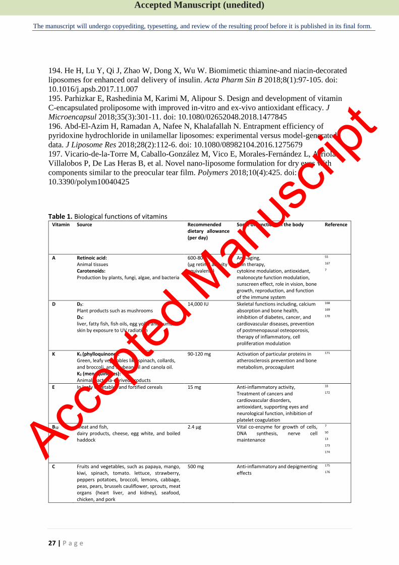
Accepted Manuscript (unedited)
The manuscript will undergo copyediting, typesetting, and review of the resulting proof before it is published in its final form.
27 | P a g e
194. He H, Lu Y, Qi J, Zhao W, Dong X, Wu W. Biomimetic thiamine-and niacin-decorated
liposomes for enhanced oral delivery of insulin. Acta Pharm Sin B 2018;8(1):97-105. doi:
10.1016/j.apsb.2017.11.007
195. Parhizkar E, Rashedinia M, Karimi M, Alipour S. Design and development of vitamin
C-encapsulated proliposome with improved in-vitro and ex-vivo antioxidant efficacy. J
Microencapsul 2018;35(3):301-11. doi: 10.1080/02652048.2018.1477845
196. Abd-El-Azim H, Ramadan A, Nafee N, Khalafallah N. Entrapment efficiency of
pyridoxine hydrochloride in unilamellar liposomes: experimental versus model-generated
data. J Liposome Res 2018;28(2):112-6. doi: 10.1080/08982104.2016.1275679
197. Vicario-de-la-Torre M, Caballo-González M, Vico E, Morales-Fernández L, Arriola-
Villalobos P, De Las Heras B, et al. Novel nano-liposome formulation for dry eyes with
components similar to the preocular tear film. Polymers 2018;10(4):425. doi:
10.3390/polym10040425
Table 1. Biological functions of vitamins Vitamin Source Recommended
dietary allowance (per day)
Some of functions in the body Reference
A Retinoic acid: Animal tissues Carotenoids: Production by plants, fungi, algae, and bacteria
600-800 (μg retinol activity equivalents)
Anti‐aging, Skin therapy, cytokine modulation, antioxidant, malonocyte function modulation, sunscreen effect, role in vision, bone growth, reproduction, and function of the immune system
55 167 7
D D2: Plant products such as mushrooms D3: liver, fatty fish, fish oils, egg yolks and human skin by exposure to UV radiation
14,000 IU Skeletal functions including, calcium absorption and bone health, inhibition of diabetes, cancer, and cardiovascular diseases, prevention of postmenopausal osteoporosis, therapy of inflammatory, cell proliferation modulation
168 169 170
K K1 (phylloquinone): Green, leafy vegetables like spinach, collards, and broccoli, and soybean oil and canola oil. K2 (menaquinones): Animal/bacteria-derived products
90-120 mg Activation of particular proteins in atherosclerosis prevention and bone metabolism, procoagulant
171
E In leafy vegetables and fortified cereals
15 mg Anti-inflammatory activity, Treatment of cancers and cardiovascular disorders, antioxidant, supporting eyes and neurological function, inhibition of platelet coagulation
33 172
B12 Meat and fish, dairy products, cheese, egg white, and boiled haddock
2.4 μg Vital co-enzyme for growth of cells, DNA synthesis, nerve cell maintenance
7 50 13 173 174
C Fruits and vegetables, such as papaya, mango, kiwi, spinach, tomato. lettuce, strawberry, peppers potatoes, broccoli, lemons, cabbage, peas, pears, brussels cauliflower, sprouts, meat organs (heart liver, and kidney), seafood, chicken, and pork
500 mg Anti-inflammatory and depigmenting effects
175 176
Accep
ted M
anus
cript
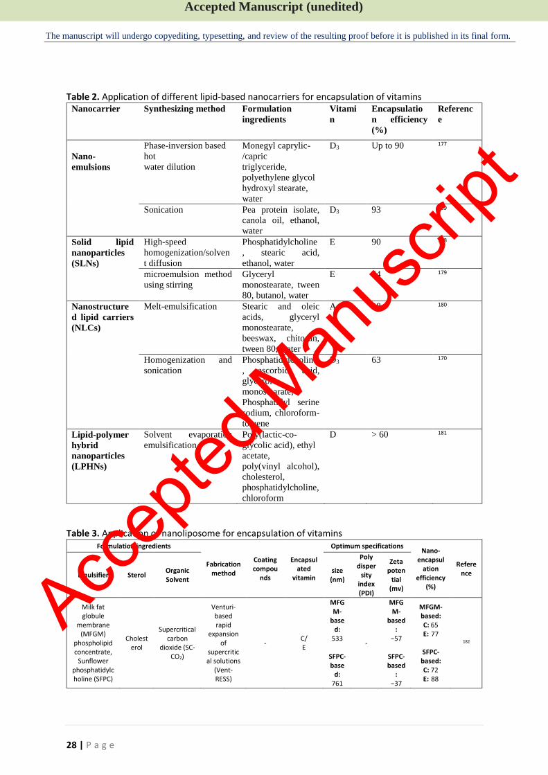
Accepted Manuscript (unedited)
The manuscript will undergo copyediting, typesetting, and review of the resulting proof before it is published in its final form.
28 | P a g e
Table 2. Application of different lipid-based nanocarriers for encapsulation of vitamins Nanocarrier Synthesizing method Formulation
ingredients
Vitami
n
Encapsulatio
n efficiency
(%)
Referenc
e
Nano-
emulsions
Phase-inversion based
hot
water dilution
Monegyl caprylic-
/capric
triglyceride,
polyethylene glycol
hydroxyl stearate,
water
D3 Up to 90 177
Sonication
Pea protein isolate,
canola oil, ethanol,
water
D3 93 169
Solid lipid
nanoparticles (SLNs)
High-speed
homogenization/solven
t diffusion
Phosphatidylcholine
, stearic acid,
ethanol, water
E 90 178
microemulsion method
using stirring
Glyceryl
monostearate, tween
80, butanol, water
E 84 179
Nanostructure
d lipid carriers
(NLCs)
Melt-emulsification Stearic and oleic
acids, glyceryl
monostearate,
beeswax, chitosan,
tween 80, water
A 98 180
Homogenization and
sonication
Phosphatidylcholine
, ascorbic acid,
glycerol
monostearate,
Phosphatidyl serine
sodium, chloroform-
toluene
D3 63 170
Lipid-polymer
hybrid
nanoparticles
(LPHNs)
Solvent evaporation
emulsification
Poly(lactic-co-
glycolic acid), ethyl
acetate,
poly(vinyl alcohol),
cholesterol,
phosphatidylcholine,
chloroform
D > 60 181
Table 3. Application of nanoliposome for encapsulation of vitamins Formulation ingredients
Fabrication method
Coating compou
nds
Encapsulated
vitamin
Optimum specifications Nano-
encapsulation
efficiency (%)
Reference Emulsifier Sterol
Organic Solvent
size (nm)
Poly disper
sity index (PDI)
Zeta poten
tial (mv)
Milk fat globule
membrane (MFGM)
phospholipid concentrate,
Sunflower phosphatidylcholine (SFPC)
Cholesterol
Supercritical carbon
dioxide (SC-CO2)
Venturi-based rapid
expansion of
supercritical solutions
(Vent-RESS)
- C/ E
MFGM-
based:
533
SFPC-base
d: 761
-
MFGM-
based:
−57
SFPC-based
: −37
MFGM-based: C: 65 E: 77
SFPC-based: C: 72 E: 88
182
Accep
ted M
anus
cript
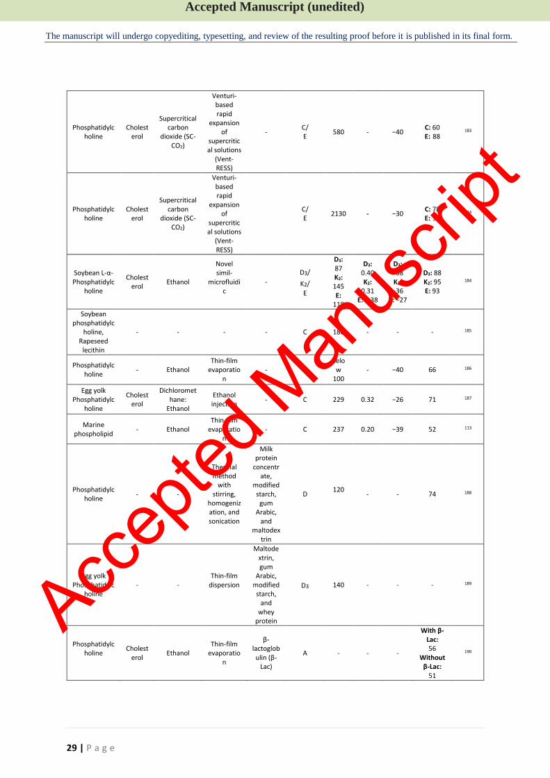
Accepted Manuscript (unedited)
The manuscript will undergo copyediting, typesetting, and review of the resulting proof before it is published in its final form.
29 | P a g e
Phosphatidylcholine
Cholesterol
Supercritical carbon
dioxide (SC-CO2)
Venturi-based rapid
expansion of
supercritical solutions
(Vent-RESS)
- C/ E
580 - −40 C: 60 E: 88
183
Phosphatidylcholine
Cholesterol
Supercritical carbon
dioxide (SC-CO2)
Venturi-based rapid
expansion of
supercritical solutions
(Vent-RESS)
C/ E
2130 - −30 C: 78 E: 95
144
Soybean L-α- Phosphatidylc
holine
Cholesterol
Ethanol
Novel simil-
microfluidic
-
D3/
K2/
E
D3: 87 K2: 145 E:
118
D3: 0.40 K2:
0.31 E: 0.38
D3: −38 K2: −36
E: −27
D3: 88 K2: 95 E: 93
184
Soybean phosphatidylc
holine, Rapeseed
lecithin
- - - - C 180 - - - 185
Phosphatidylcholine
- Ethanol Thin-film
evaporation
- C belo
w 100
- −40 66 186
Egg yolk Phosphatidylc
holine
Cholesterol
Dichloromethane:
Ethanol
Ethanol injection
- C 229 0.32 −26 71 187
Marine phospholipid
- Ethanol Thin-film
evaporation
- C 237 0.20 −39 52 113
Phosphatidylcholine
- -
Thermal method
with stirring,
homogenization, and sonication
Milk protein
concentrate,
modified starch,
gum Arabic,
and maltodex
trin
D 120
- - 74 188
Egg yolk Phosphatidylc
holine - -
Thin-film dispersion
Maltodextrin, gum
Arabic, modified starch,
and whey
protein
D3
140
- - - 189
Phosphatidylcholine
Cholesterol
Ethanol Thin-film
evaporation
β-lactoglobulin (β-
Lac)
A - - -
With β-Lac: 56
Without β-Lac:
51
190
Accep
ted M
anus
cript
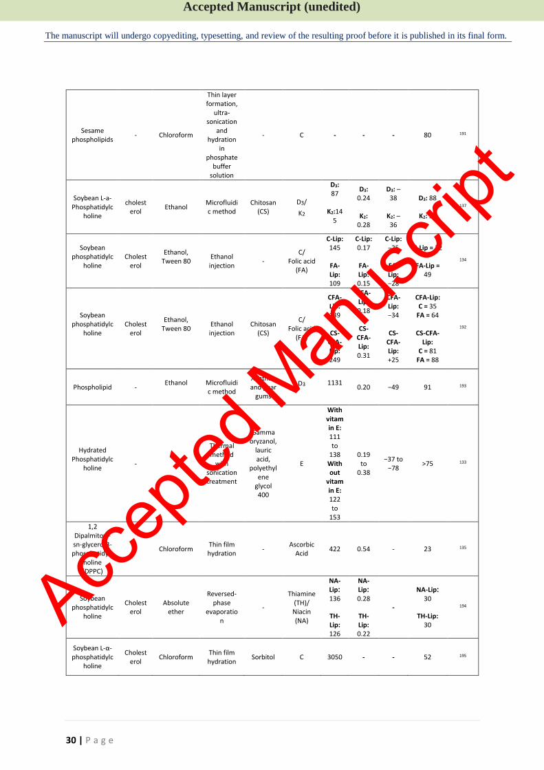
Accepted Manuscript (unedited)
The manuscript will undergo copyediting, typesetting, and review of the resulting proof before it is published in its final form.
30 | P a g e
Sesame phospholipids
- Chloroform
Thin layer formation,
ultra-sonication
and hydration
in phosphate
buffer solution
- C - - - 80 191
Soybean L-a-Phosphatidylc
holine
cholesterol
Ethanol Microfluidic method
Chitosan (CS)
D3/
K2
D3: 87
K2:14
5
D3: 0.24
K2:
0.28
D3: –38
K2: –36
D3: 88
K2: 95
137
Soybean phosphatidylc
holine
Cholesterol
Ethanol, Tween 80
Ethanol injection
- C/
Folic acid (FA)
C-Lip: 145
FA-Lip: 109
C-Lip: 0.17
FA-Lip: 0.15
C-Lip: −25
FA-Lip: −28
C-Lip = 32
FA-Lip = 49
134
Soybean phosphatidylc
holine
Cholesterol
Ethanol, Tween 80
Ethanol injection
Chitosan (CS)
C/ Folic acid
(FA)
CFA-Lip: 139
CS-
CFA-Lip: 249
CFA-Lip: 0.18
CS-
CFA-Lip: 0.31
CFA-Lip: −34
CS-
CFA-Lip: +25
CFA-Lip: C = 35
FA = 64
CS-CFA-Lip:
C = 81 FA = 88
192
Phospholipid - Ethanol
Microfluidic method
Xanthan and guar
gums
D3
1131
0.20
−49 91 193
Hydrated Phosphatidylc
holine
- -
Thermal method
with sonication treatment
Gamma oryzanol,
lauric acid,
polyethylene
glycol 400
E
With vitamin E: 111 to
138 Without
vitamin E: 122 to
153
0.19
to 0.38
−37 to −78
>75
133
1,2 Dipalmitoyl-sn-glycero-3-phosphatidylc
holine (DPPC)
- Chloroform Thin film hydration
- Ascorbic
Acid 422 0.54 - 23 135
Soybean phosphatidylc
holine
Cholesterol
Absolute ether
Reversed-phase
evaporation
-
Thiamine (TH)/ Niacin (NA)
NA-Lip: 136
TH-Lip: 126
NA-Lip: 0.28
TH-Lip: 0.22
-
NA-Lip: 30
TH-Lip: 30
194
Soybean L-α- phosphatidylc
holine
Cholesterol
Chloroform Thin film hydration
Sorbitol C 3050 - - 52 195
Accep
ted M
anus
cript
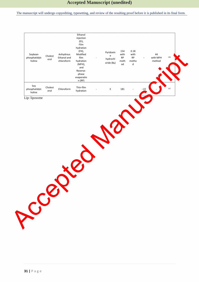
Accepted Manuscript (unedited)
The manuscript will undergo copyediting, typesetting, and review of the resulting proof before it is published in its final form.
31 | P a g e
Soybean phosphatidylc
holine
Cholesterol
Anhydrous Ethanol and chloroform
Ethanol injection
(EI), Film
hydration (FH),
Modified film
hydration (MFH),
and Reverse-
phase evaporatio
n (RP)
-
Pyridoxine
hydrochl
oride (B6)
154 with RP
method
0.18 with RP
method
- 44
with MFH method
196
Soy phosphatidylc
holine
Cholesterol
Chloroform Thin-film hydration
- E 181 - −22 - 197
Lip: liposome
Accep
ted M
anus
cript