the diversity of uses for cellulose sulphate encapsulation
TRANSCRIPT

70 Bioencapsulation of Living Cells for Diverse Medical Applications, 2013, 70-92
Eva Maria Brandtner and John Austin Dangerfield (Eds) All rights reserved-© 2013 Bentham Science Publishers
CHAPTER 3
The Diversity of Uses for Cellulose Sulphate Encapsulation
John A. Dangerfield1,*, Brian Salmons1, Randolph Corteling2, Jean-Pierre Abastado3, John Sinden2, Walter H. Gunzburg1 and Eva M. Brandtner1,4
1SG Austria Pte Ltd/Austrianova Singapore Pte Ltd, Biopolis, Singapore; 2ReNeuron Group Plc, Guildford, Surrey, England; 3Singapore Immunology Network (SIgN), Biopolis, Singapore and 4VIVIT Molecular Biology Laboratory, Dornbirn, Austria
Abstract: In this chapter we propose sodium cellulose sulphate (SCS) as a prime candidate for clinical application of encapsulated cells and present data for uses of SCS encapsulation for the direct delivery of therapeutic antibodies and advanced approaches for stem cell therapy. We also provide a simple lab protocol allowing researchers to make capsules at the bench without the need for expensive machinery.
Keywords: Live cell encapsulation, bioencapsulation, sodium cellulose sulphate, therapeutic antibodies, biomolecule release, long-term release, long-term survival, stem cells, biocompatibility, GMP manufacturing, immune protection, patient safety, storage capability, localisation, removability.
1. INTRODUCTION
The treatment of diseases and disorders by microencapsulation of living cells and the subsequent implantation of these capsules into patients were pioneered 30 years ago [1]. It can be considered as a specialised type of cell therapy and one that is potentially safer since the cells are physically separated from the body as well as more efficacious since the cells are confined to the site at which they are implanted and have the potential to be removed if necessary after treatment is complete.
Alginate was one of the first materials used to encapsulate cells and this seaweed derived material is still in use today, mainly in the development of encapsulated
Address correspondence to John A. Dangerfield: Austrianova Singapore Pte Ltd, 20 Biopolis Way, #05-518 Centros, Singapore; Tel: +65 6779 2932; Fax: +65 6774 5569; E-mail: [email protected]
Send Orders of Reprints at [email protected]

The Diversity of Uses for Cellulose Bioencapsulation of Living Cells for Diverse Medical Applications 71
cell treatments for diabetes (see chapters 1 and 2). More recently, subseive agarose beads have also been used for cell encapsulation (see chapter 6).
We have focussed on the use of sodium cellulose sulphate (SCS) as a cell encapsulation material [2, 3]. Capsules consisting of polymers of SCS and polydiallyldimethyl ammonium chloride (pDADMAC) offer a number of advantages including:
ability to reproducibly source, produce and characterise the SCS starting material.
robustness of the capsules (permitting delivery through needles or catheters without bursting).
good biocompatibility for the cells in the microcapsule.
cells like to grow and survive for extended periods in the capsule but they do not escape due to a three dimensional contact inhibition.
good biocompatibility and inertness of the capsules when implanted at various sites in the body.
lack of an immune or inflammatory response either to the capsule material or to the cells that are protected by it.
lack of fibrous overgrowth.
large scale GMP manufacturing of an encapsulated cell medicinal product has been achieved [4].
ability to freeze the encapsulated cells and thus store and ship them.
We have developed the SCS encapsulation technology originally for the treatment of solid tumours such as pancreatic cancer and breast. These encapsulated cells were tested in clinical trials and shown to be both efficacious and (perhaps more importantly when considering other uses of the technology) safe when they remain in the body for at least two years [5, 6].

72 Bioencapsulation of Living Cells for Diverse Medical Applications Dangerfield et al.
Consequently, and building on the clinical data we already have with SCS encapsulated cells, we have been encapsulating various cells types in order to design therapies for the treatment of a wide variety of diseases with various partners. Some of these approaches to treat diseases such as virus infections, tumours and improved stem cell treatments are discussed in this article.
2. RELEASE OF ANTIBODIES FROM ENCAPSULATED CELLS
2.1. General Considerations: Immune Protection
The main purpose of cell encapsulation is to protect the cells from the immune system. The capsules act as a mechanical barrier between the encapsulated cells and their surroundings. This is accomplished by the capsule wall which has pores that are big enough to allow nutrients to diffuse in and waste products out of the capsules and of course is permissive for the release of the therapeutic molecule of interest. On the other hand, the pores are small enough to deny access to immune cells like macrophages, neutrophils and T-cells.
For release of antibodies from encapsulated cells the membrane pores must, of course, be large enough to allow the passage of antibodies. Therefore, for in-vivo application of this technology the question presents itself whether (a) antibodies from the host can get into the capsules and (b) if this could result in immune rejection of the encapsulated cells.
The answer to the first question (a) is yes, there is no reason why antibodies could not get into the capsules since they can get out and the mechanism of crossing the capsule membrane is a passive one. However, there are several reasons why this does not result in immune rejection of the encapsulated cells. Firstly, the concentration gradient favours the exit of antibodies from the capsules since the production of antibodies by the encapsulated cells leads to a high local concentration of antibodies inside the capsules, much higher than in the surrounding tissue. This means although, in theory, traffic can occur in both directions, antibodies diffuse out of the capsules much more efficiently than into the capsules.
Secondly (b), for an immune rejection to occur, antibodies of the right specificity must be present. Antibodies are secreted by mature B-cells which evolve from

The Diversity of Uses for Cellulose Bioencapsulation of Living Cells for Diverse Medical Applications 73
naive B-cells after contact with the antigen that their receptor is specific for. After activation of the B-cells, a cascade of events happens which involves the expansion of the respective B-cell clone and results in a large number of mature B-cells, so called plasma cells, which are able to secrete antibodies. Since the initial step requires cell to cell contact between the naive B-cell and the surface antigen on the encapsulated cells and the capsules do not allow cell to cell contact across their membrane, activation of the specific B-cells should not happen. This means, although antibodies can in principle enter the capsules, there is no specific humoral immune response against the encapsulated cells.
However, in the unlikely case that a capsule breaks and cells are exposed, as well as in a xenotransplantation scenario where natural antibodies (antibodies which are present in higher primates independent of prior infection or vaccination) come into play, the presence of antibodies with affinity to the cells inside the capsules cannot be ruled out. But even in this latter case the encapsulated cells are still protected. Antibodies do not attack and kill cells as such. They just earmark them for further attention from other players in the immune system. There are two ways in which foreign cells are eliminated from the body once they are marked with antibodies. One is via effector cells and requires cell to cell contact. In the case of encapsulated cells, this is prevented by the capsule wall. The other way is via the classical pathway of complement activation and requires a high concentration of antibodies bound to the surface of the target cell. Proteins of the complement system, prior to activation, are part of large multi protein complexes which cannot enter the capsules. For example, the first step of the classical pathway involves the C1 complex. C1 binds to antibody molecules which are attached to antigens (resulting in immune complexes) and thereby initiates the cascade. With a size of 766 kDa the C1 complex is too large to enter the capsules. The same is true for other complement protein complexes involved in the other steps of complement activation. Therefore, encapsulated antibody producing cells are protected from the action of the complement system as well as from the access of effector cells. The experimental data presented below confirm that, indeed, encapsulated hybridoma cells can survive in vivo for prolonged periods of time.

74 Bioencapsulation of Living Cells for Diverse Medical Applications Dangerfield et al.
2.2. Applications of Antibody Production from Encapsulated Cells
2.2.1. Depletion of Immune Cell Lineages
The biological role of different immune cell lineages can be studied by depleting the cell lineage of interest and looking at the effect in animal models. Depletion of individual lineages can be achieved by using cytotoxic antibodies against surface markers on the target cells. Such antibodies are available for the depletion of CD4+ and CD8+ T cells [7-18], granulocytes [19-21], CD20+ B cells [22, 23] for example.
For sustained depletion standard protocols see the animals being injected with antibodies several times per week. The treatment as such is costly and labour intensive. Therefore, release of the antibodies from encapsulated cells within the body is an attractive alternative. Once implanted into the body, encapsulated cells can survive and produce antibody in constant amounts over sustained periods of time. This avoids the “peaks and troughs” of antibody concentration which occurs as a result of infusion or injection of the large amounts necessary to sustain the effect until the next infusion is made. These large quantities are also associated with negative immune-related stimulation, causing faster clearance of the antibodies and for some antibodies anti-idiotypic responses, and so the effects of the antibodies decrease with time. The constant low amount released from the capsules avoids this scenario. This has been demonstrated in a proof-of concept study in which a rat hybridoma producing a cytotoxic anti-mouse CD8 antibody (2.43) [7] was encapsulated and implanted in mice (Fig. 1). The capsules containing the 2.43 antibody producing cells were retrieved from the mice 8 weeks after implantation and shown to still retain viability. Thus even in a xenogeneic setting, encapsulation protects cells from immune attack (Fig. 2).
These findings indicate that the long-term depletion of immune cell lineages using encapsulated antibody producing cells is feasible and warrants further optimisation.

The Diversity of Uses for Cellulose Bioencapsulation of Living Cells for Diverse Medical Applications 75
Figure 1: CD8+ T-cell depletion in mice implanted with encapsulated cells producing 2.43 antibody. The amount of CD8+ T-cells in the peripheral blood of mice was measured in relation to the amount of CD11b cells, which are not affected by the treatment. CD8+/CD11b+ cell ratios are shown for mice implanted with 0, 20, 40, 80, 400 capsules subcutaneously (s.c.) and 80 capsules intraperitoneally (i.p.). Animal care and experimental procedures were approved by the Institutional Animal Care and Use Committee (IACUC) (Application No. 090425) of the Biological Resource Center, 20 Biopolis Way, Singapore 138668.
Figure 2: Encapsulated rat cells are still alive eight weeks after implantation into the body of immunocompetent mice. Encapsulated cells were removed from the body of a mouse eight weeks after implantation, stained with calcein-AM, an indicator for living cells and visualised by fluorescence microscopy. Animal care and experimental procedures were approved by the Institutional Animal Care and Use Committee (IACUC) (Application No. 090425) of the Biological Resource Center, 20 Biopolis Way, Singapore 138668.
0
0.1
0.2
0.3
0.4
0.5
0.6
0 caps 20 caps s.c. 40 caps s.c. 80 caps s.c. 400 caps s.c. 80 caps i.p.
CD8/
CD11
b ce
ll ra
tio
CD8+T-cell depletion
two weeks after implantation
three weeks after implantation
four weeks after implantation

76 Bioencapsulation of Living Cells for Diverse Medical Applications Dangerfield et al.
2.2.2. Encapsulation of mAb Producing Cells for the Treatment of Infectious Disease
FrCas E is a chimeric virus consisting of mouse ectopic retrovirus CasBrE (clone 15-1) env and 3’ pol sequences in a Friend murine leukemia virus (FMuLV) background. FrCasE virus infection in new-born mice is an ideal animal model of infectious disease since it causes a clear phenotype, i.e. rapidly progressing neurodegenerative disease which is fatal within 6 weeks, with 100% incidence. This model was used to show that neutralising antibody released from encapsulated hybridoma cells is able to rescue a large percentage of the animals even in a non-optimised first experiment [24]. This was the case irrespective of whether the capsules were implanted concurrently with viral infection (80% survival of the treated animals) or 2 days after the infection (65% survival of the treated animals) (Fig. 3) which strongly indicates potential use in a therapeutic rather than just prophylactic setting.
Figure 3: Survival of FrCasE-infected mice treated with 667 cell-containing capsules [24]. (Left) Four-day-old mice were infected by intraperitoneal injection with 5 x 10⁴ focus forming units (FFU). Twenty-nine mice received no further treatment (full triangles). G8P2B5 (control antibody; full circles) and 667 (FrCasE neutralising antibody; open circles) hybridoma cell-containing cellulose sulphate capsules were subcutaneously implanted in 13 and 45 mice, respectively, on the day of infection. (Right) Twenty 4-day-old mice infected with 5 x 10⁴ FrCasE FFU received no further treatment (full triangle) and a further 20 infected mice received 667 cell-containing capsules on day 2 post-infection (open circle), respectively. Figure taken from published article [24].

The Diversity of Uses for Cellulose Bioencapsulation of Living Cells for Diverse Medical Applications 77
This goes to show that encapsulated antibody producing cells are a means to fight infectious disease and potentially protect the body in the critical time window between exposures to a pathogen and evolving of the body’s own adaptive immune response.
Besides in vivo use, encapsulation of antibody or antibody like producing cells can be of great interest for the industrial production of antibodies by the biotech industry. The capsules can act as a pre-filtration device and eliminate cost-intensive steps during downstream processing of antibody products. Such encapsulated products should be seen favourably by industry since they may allow for new IP surrounding old technologies in combination with capsules. This may allow generation of new patents or extension of old patents, something it can be assumed is much needed when looking at the current portfolios of many of the large pharmaceutical companies.
Treatments with therapeutic antibodies come at costs of at least US$ 40,000 per year per patient (Table 1). Worldwide, the market for therapeutic antibodies has a volume of over US$ 50 billion. Patients need frequent injections, sometimes for their entire life. There is tremendous potential in finding more cost-efficient ways of delivery for these therapeutic antibodies. The encapsulation and subsequent implantation of the cells producing these antibodies opens a whole new range of possibilities and makes such therapies more accessible and affordable for a vast number of patients.
Table 1: Therapeutic antibodies for the treatment of cancer
Antibody Trade Name Target Type Indication Approval Rituximab Rituxan®
Mabthera® CD20 chimeric non-Hodgkin
lymphoma 1997
Trastuzumab Herceptin® ErbB2 humanised breast cancer 1998 Gemtuzumab Ozogamicin
Mylotarg® CD33 humanised acute myelogeneous leukemia
2000
Alemtuzumab Campath® CD52 humanised chronic lmphocytic leukemia
2001
Ibritumomab Tiuxetan
Zevalin® CD20 murine non-Hodgkin lymphoma
2002

78 Bioencapsulation of Living Cells for Diverse Medical Applications Dangerfield et al.
Table 1: cont....
Tositumomab Bexxar® CD20 murine non-Hodgkin lymphoma
2003
Bevacizumab Avastin® VEGF humanised colorectal cancer
2004
Panitumumab Vectibix® EGFR human colorectal cancer
2006
Ofatumamab Arzerra® CD20 human chronic lmphocytic leukemia
2009
Brentuximab Vedotin
Adcetris® CD30 chimeric large cell lymphoma and Hodgkin lymphoma
2011
Ipilimumab Yervoy® CTLA-4 human melanoma 2011
Cetuximab Erbitux® EGFR chimeric colorectal cancer head and neck cancer
2004 2006
3. HOW ENCAPSULATION CAN HELP THE BUDDING STEM CELL INDUSTRY
3.1. Is There a Budding Stem Cell Industry?
Before considering the reasons for wanting to encapsulate stem cells, it is firstly important to decide if stem cells have lived up to their expectations in terms of medical potential, i.e. does it makes sense to pursue such applications? Some may consider that stem cell progress has been slower than predicted, attributable most likely to the long lasting media hype, but there are some marked examples which suggest that the answer to this question could indeed be “yes”.
2012 has seen the world’s first, market approved, allogeneic cell therapy products. The product CARTISTEM® from the Korean biotech company Medipost Co. Ltd. was approved for sale in Korea by the Korean FDA in January 2012. Their product is manufactured using mesenchymal stem cells (MSC), which are also known as mesenchymal precursor cells (MPC) which, in Medipost’s case, are extracted from publicly donated umbilical cord blood. Sales are taking off in Korea for the use of CARTISTEM® as an off-the-shelf product to treat degenerative arthritis and knee cartilage defects. It is currently in further clinical

The Diversity of Uses for Cellulose Bioencapsulation of Living Cells for Diverse Medical Applications 79
trials under guidance of the FDA and is expected to be approved for sale in the U.S. before 2014. Medipost also has three other products in human trials based on the same cells which they claim can be used for the treatment of neuro-degenerative disorders such as Alzheimer’s, pulmonary disorders such as bronchopulmonary dysplasia and the transplant engraftment disorder, graft vs. host disease (Table 2). In May 2012 the U.S. based company Osiris Therapeutics Inc. also claimed to be the world's first company to receive market clearance for an approved stem cell drug called ProchymalTM. Osiris also has several other late stage trials going on with their MPC-based products ProchymalTM and ChongrogenTM which offer hope to children suffering from the life threatening graft vs. host disease as well as those with Crohn’s disease and a number of pulmonary related problems such as emphysema and bronchitis (Table 2).
Table 2: World-wide clinical trials using allogeneic stem cell based therapeutics (as of July 2012). Information sources include the U.S. governmental National Institutes of Health website (http://clinicaltrials.gov/ct2/home), the websites of the companies as well as other media sources. In many cases, information between company websites and the government database was conflicting. Mostly, the company websites suggested trials were further along. Although this may be factual, in these cases, the government database information was used. Trials at various enrolment or pre-recruitment stages, trials which were terminated or trials for which information has not been updated within the past two years were not included. Trials are listed alphabetically based on the sponsor’s name.
Sponsor (Company Location)
Indication(s) Technology/Product Status
Advanced Cell Technology Inc. (USA)
Stargardt’s Macular Dystrophy
human embryonic stem cell derived retinal pigmented epithelial (MA09-hRPE) cells
Phase I/II (ongoing)
Advanced Cell Technology (USA)
Dry Age-Related Macular Degeneration
human embryonic stem cell derived retinal pigmented epithelial (MA09-hRPE) cells
Phase I/II (ongoing)
Geron Corp. (USA) Spinal Cord Injury human embryonic stem cells (GRNOPC1)
Phase I (ongoing)
MEDIPOST Co. Ltd. (Korea)
Cartilage Injury
Osteoarthritis human umbilical cord blood derived-mesenchymal stem cells/CARTISTEM®
Phase III (completed)

80 Bioencapsulation of Living Cells for Diverse Medical Applications Dangerfield et al.
Table 2: cont....
MEDIPOST Co. Ltd. (Korea)
Dementia of the Alzheimer's Type
human umbilical cord blood derived-mesenchymal stem cells/NEUROSTEM®
Phase I (completed)
MEDIPOST Co. Ltd. (Korea)
Acute Leukemia human umbilical cord blood derived-mesenchymal stem cells
Phase I/II (completed)
MEDIPOST Co. Ltd. (Korea)
Bronchopulmonary Dysplasia
human umbilical cord blood derived-mesenchymal stem cells/PNEUMOSTEM®
Phase I (completed)
MEDIPOST Co. Ltd. (Korea)
Graft-vs.-Host Disease
human umbilical cord blood-derived mesenchymal stem cells
Phase I/II (recruiting)
Mesoblast Ltd. (Australia)
Degenerative Disc Disease in Subjects with Chronic Discogenic Lumbar Back Pain
adult human mesenchymal precursor cells (MPCs) with hyaluronic acid carrier
Phase II (recruiting)
Mesoblast Ltd. (Australia)
Degenerative Disc Disease in Subjects requiring Lumbar Interbody Infusion
adult human mesenchymal precursor cells (MPCs)/NeoFuse
TM
combined with MasterGraft® matrix fusion granules
Phase II (ongoing)
Mesoblast Ltd. (Australia)
Cervical Degenerative Disc Disease in Subjects Undergoing Multi-Level Anterior Cervical Discectomy
adult human mesenchymal precursor cells (MPCs)/NeoFuse
TM
combined with MasterGraft® matrix fusion granules
Phase II (ongoing)
Mesoblast Ltd. (Australia)
Degenerative Disc Disease in Subjects Requiring Posterolateral Lumbar Fusion (PLF)
adult human mesenchymal precursor cells (MPCs)/NeoFuse
TM
combined with MasterGraft® matrix fusion granules
Phase I/II (ongoing)
Mesoblast Ltd. (Australia)
Anterior Cruciate Ligament Injury/Osteoarthritis
adult human mesenchymal precursor cells (MPCs, MSB-
Phase I/II (recruiting)

The Diversity of Uses for Cellulose Bioencapsulation of Living Cells for Diverse Medical Applications 81
CAR001) with hyaluronic acid carrier
Table 2: cont....
Mesoblast Ltd. (Australia)
Type 2 Diabetes adult human mesenchymal precursor cells (MPCs)
Phase I/II (recruiting)
Osiris Therapeutics Inc. (USA)
Treatment-refractory Crohn’s Disease
adult human mesenchymal stem cells/PROCHYMAL
TM
Phase III (completed)
Osiris Therapeutics Inc. (USA)
Recovery Following Partial Medial Meniscectomy
adult human mesenchymal stem cells/Chondrogen
TM
Phase I/II (completed)
Osiris Therapeutics Inc. (USA)
Moderate to Severe Chronic Pulmonary Disease/Pulmonary Emphysema/Chronic Bronchitis
adult human mesenchymal stem cells/PROCHYMAL
TM
Phase II (completed)
Osiris Therapeutics Inc. (USA)
Treatment-refractory (1) and Newly Diagnosed (2) Acute Graft vs. Host Disease
(1) and (2): varying doses of adult human mesenchymal stem cells/PROCHYMALTM (2): in combination with corticosteroids
(1) Phase II (completed) (2)Phase III (completed)
ReNeuron Limited (UK)
Stable Ischemic Stroke
neural stem cells (CTX0E03)
Phase I (ongoing)
Stempeutics Research Pvt. Ltd. Stempeutics Research Malaysia SDN. BHD. (India/Malaysia)
Osteoarthritis of the Knee Joint
Bone marrow derived adult mesenchymal stem cells/Stempeucel - CLI™
Phase II (recruiting)
Stempeutics Research Pvt. Ltd. (India)
Critical Limb Ischemia due to Buerger’s Disease
Bone marrow derived adult mesenchymal stem cells/Stempeucel - CLI™
Phase II (recruiting)
There are other stem cell companies with allogeneic products in various stages of clinical trials as well. Until recently, the U.S. biotech Geron Corporation has used human embryonic stem cells (HSC) as a platform and has undertaken a Phase I study in patients with thoracic spinal cord injuries (Table 2). They also have evidence for therapeutic benefit of their HSC-based products in the areas of heart muscle regeneration, diabetes, cartilage repair and development of cancer vaccines. ReNeuron Group Plc. in U.K. has a neural based stem cell line in ongoing Phase I for the treatment of stroke (Table 2). But possibly the most

82 Bioencapsulation of Living Cells for Diverse Medical Applications Dangerfield et al.
advanced and financially endowed stem cell company currently is Mesoblast Ltd. in Australia, who has ongoing trials in four disease areas, including a recent successful Phase II outcome and permission to begin a Phase III. Their cell technology platform is based on MPC derived from bone marrow aspirates as well as other tissues of healthy adults and they have good evidence showing potential for the treatment of cardiovascular diseases foremost but also diabetes, bone and cartilage repair and replacement and some forms of eye disease (Table 2).
Advanced Cell Technology (ACT) is performing two trials with HSC aimed at improving the vision of patients with Stargardt’s Macular Dystrophy and Dry Age-Related Macular Degeneration (Table 2). Patients’ eyes were injected with retinal pigmented epithelial cells derived from human embryonic stem cells. The preliminary findings appear to be promising, as judged by the outcomes from two patients treated as part of the trial. During the trial, neither patient’s vision worsened nor were there obvious negative side effects [25].
3.2. Reasons for Encapsulating Stem Cells
When discussing the encapsulation of stem cells, it is often asked, “How can it work because the stem cells are trapped inside the capsule?” This question exists because of the common misconception that implanted stem cells grow, differentiate and/or develop to become the regenerated tissue and that the presence of this tissue itself is necessary to have the therapeutic effect. Although this may be true for a few special cases, it is now generally accepted amongst experts that the vast majority of stem cells react on implantation to their micro-environment to release a wide array of soluble factors that mediate beneficial paracrine effects and may greatly contribute to the therapeutic effect. These include cytokines, growth factors and microvesicles which then act locally to trigger the patient’s own systems into repair [26].
Mesoblast, for example, clearly states this to be the case on their website under the technology section, “The MPCs act as micro-drug factories providing the secretion of trophic factors that then exert multiple mechanisms of action including but not limited to anti-apoptosis (anti-death) of cells, regeneration of damaged tissue, recruitment of the body’s own tissue specific precursor cells and proliferation/inhibition of relevant cells types including blood vessels”. This is
The Diversity of Uses for Cellulose Bioencapsulation of Living Cells for Diverse Medical Applications 83
now accepted in the field of stem cell research, as well as industry, to be the main mechanism of action for stem cells [27].
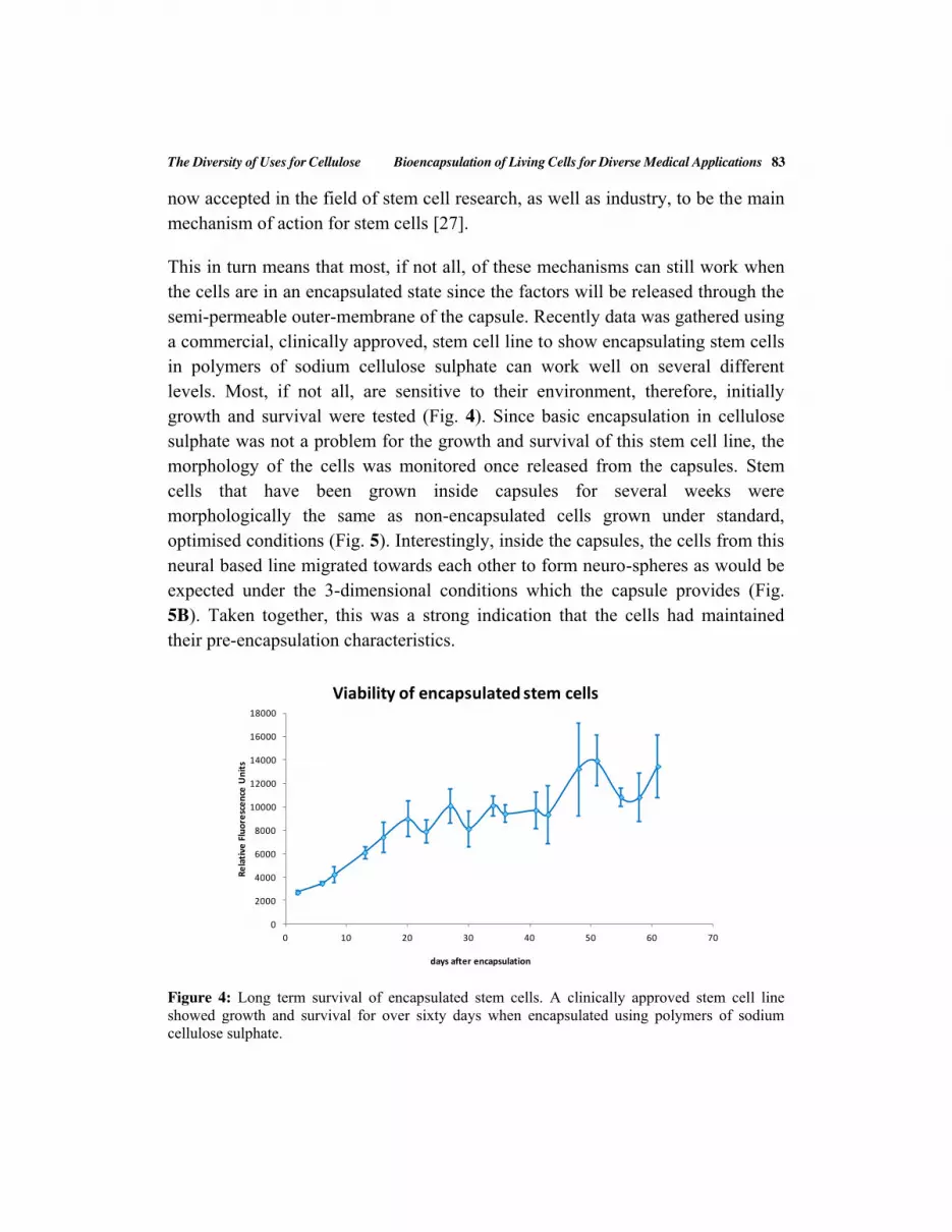
This in turn means that most, if not all, of these mechanisms can still work when the cells are in an encapsulated state since the factors will be released through the semi-permeable outer-membrane of the capsule. Recently data was gathered using a commercial, clinically approved, stem cell line to show encapsulating stem cells in polymers of sodium cellulose sulphate can work well on several different levels. Most, if not all, are sensitive to their environment, therefore, initially growth and survival were tested (Fig. 4). Since basic encapsulation in cellulose sulphate was not a problem for the growth and survival of this stem cell line, the morphology of the cells was monitored once released from the capsules. Stem cells that have been grown inside capsules for several weeks were morphologically the same as non-encapsulated cells grown under standard, optimised conditions (Fig. 5). Interestingly, inside the capsules, the cells from this neural based line migrated towards each other to form neuro-spheres as would be expected under the 3-dimensional conditions which the capsule provides (Fig. 5B). Taken together, this was a strong indication that the cells had maintained their pre-encapsulation characteristics.
Figure 4: Long term survival of encapsulated stem cells. A clinically approved stem cell line showed growth and survival for over sixty days when encapsulated using polymers of sodium cellulose sulphate.
0
2000
4000
6000
8000
10000
12000
14000
16000
18000
0 10 20 30 40 50 60 70
Rela
tive
Flu
ores
cenc
e U
nits
days after encapsulation
Viability of encapsulated stem cells

84 Bioencapsulation of Living Cells for Diverse Medical Applications Dangerfield et al.
Figure 5: Morphology of neural stem cells is maintained after sodium cellulose sulphate encapsulation. (A) Cells cultured under conditions for normal maintenance, viewed with standard light microscopy. (B) Live/dead staining viewed with fluorescence microscopy show as indicated by white arrows that cells grow and migrate towards each other inside the capsule to form neuro-spheres of increasing size. Green indicates living cells and red dead or dying cells. (C) Cells after the capsule has been dissolved away in vitro, viewed with standard light microscopy. A dissolved capsule fragment is indicated by the circle. A neuro-sphere is indicated with the black arrow. Cells growing away from the neuro-sphere have the same morphology as non-encapsulated cells in (A).
Having established that there are many medically interesting stem cells and that they would in principal still function normally when encapsulated, the final and most critical point to address is the reason or benefit to encapsulating them. Despite their multi-tiered use in humans already, many understand there are some serious concerns and challenges still to be overcome. These can be divided into two main categories; one being safety concerns from authorities, regulators, scientists and companies alike about the fate of the cells after implantation (reviewed in [28]) and the other being issues relating to the sensitivity of cell growth, upscaling and general manufacturing procedures (reviewed in [29]). Encapsulation can provide solutions in all these areas as well as provide other benefits which will be discussed below.
Concerning the fate of the cells and the safety implications this has. Some years back it was discovered in animal models that stem cells can differentiate into tissue with tumorigenic capability and from a type of tumour called a teratoma [30]. Since then, similar incidents of malignant outgrowth from stem cells have been recorded by others [31-33]. In addition to the 500 or so companies that can be found in the internet dealing with so called stem cell products (with varying levels of seriousness), many patients have also been treated with autologous stem
A CB

The Diversity of Uses for Cellulose Bioencapsulation of Living Cells for Diverse Medical Applications 85
cells at clinics all round the world. All such treatments are not approved through regulatory bodies although most large hospitals have such autologous programmes running. So the teratoma and other malignant outgrowth findings did not prevent stem cells being implanted into patients and there have been some worrying and notable cases as a result. For example there was a brain tumour in a boy with ataxia telangiectasia that was treated with stem cells. The tumour cells were shown to be of non-host origin, suggesting strongly that it was derived from the transplanted neural stem cells [34], and there have been other cases where adverse effects and growths have been shown after stem cell treatments [35, 36]. If the cells had been implanted in stable capsules, it is likely that none of these incidences would have occurred.
Despite many attempts to study cell fate in animal models, even in the case of the approved product CARTISTEM®, it is not known what happens to the implanted cells in human patients. This is because there are no available technologies allowing stem cells to be marked for tracking purposes without detrimentally affecting the cells. Both external labelling with antibody tags, internal marking with fluorescent particles, or genetic marking by introducing a gene such as the green fluorescent protein for example, all result in various changes to the cell. This can manifest as damage the therapeutic potential since most stem cells are very sensitive to any manipulation causing them to change, usually for the worse. The change could also for example cause an increase in immunogenicity, i.e. making the cells have a higher profile to the patient’s immune system because of the added foreign components, meaning they are cleared before being able to carry out their therapeutic function. It must also be considered that any such changes provide additional hurdles from a regulatory perspective since any new components to the product need to be approved for human use. Taken together, this means that within the entire field, still only theories and speculation exists as to the fate of stem cells once implanted in human patients. For interest’s sake alone, most commonly it’s proposed that they undergo apoptosis (since this would be convenient) or that they migrate away from the site of implantation.
Encapsulation of the cells could have prevented the severe adverse events and could entirely address the above discussed concerns since the cells are not released from the capsules at any stage after implantation. It should also make it

86 Bioencapsulation of Living Cells for Diverse Medical Applications Dangerfield et al.
easier for companies to gain permission for clinical trials and allow companies, hospitals and clinics alike a considerable measure of safety when undertaking such procedures. Another benefit of having cells in capsules is that they hold the cells at the locality of treatment. It is not known how long naked cells stay at the desired site. It is known however that many implanted cells have a high tendency for migration in vivo. There is evidence to suggest that implanted cells wander away from the implantation site quite quickly, between hours and days depending on the cells and site of implantation [37, 38]. Encapsulation clearly allows the possibility to keep the cells at the site of implantation since typically capsules are around 1 mm in size and contain thousands of cells each, so they remain physically fixed at the site. For example, this would be true for sub-cutaneous, intra-muscular, brain, peri-organ or peri-tumoral application but not the case for inter-peritoneal application or injection into the blood system, where the capsules would move around. Having them around at the desired location for longer period may well enhance their therapeutic effect. Encapsulation can also improve treatment efficacy by enhancing the length of time that stem cells can survive in vivo. If capsules are implanted sub-cutaneous, this also offers the possibility of removal of the cells after treatment is complete, since they can be found easily later at the same place they were implanted. This is important since many patients are concerned about having foreign cells in them for the rest of their time.
Finally, although most claim that allogeneic stem cells are non-immunogenic, this is still not fully understood. It certainly seems that they are less immunogenic, than for example a syngeneic somatic cell line would be, however, it may be that immune system plays a role in the fate or pre-mature clearance of implanted stem cells. Again, encapsulation would prevent this, since cells of the patient’s immune system cannot enter the capsules.
Encapsulation also has several potential benefits for manufacturing. The vast majority of stem cells are cultured in monolayer system as opposed to stirred reactors which makes upscaling highly manpower and materials intensive. This is one of the major barriers to making large volume, off-the-shelf stem cell products and is in turn why many of the large pharma companies are holding back on development of stem cell products until these issues are solved. It’s proving very difficult to evolve stem cells into growth in bioreactors since generally speaking

The Diversity of Uses for Cellulose Bioencapsulation of Living Cells for Diverse Medical Applications 87
they are highly sensitive to all kinds of manipulation, including physical stress, meaning that stemness of the cells is not maintained under stirred conditions. Having cells inside capsules can entirely circumvent this problem and allow monolayer dependent growth under stirred conditions since the inside of capsule acts as a substrate for the cells to attach and the outside of the capsule acts as a protective device against collisions with other capsules and sheer stresses of the vortex caused by stirring. Bioreactor benefits, such as increased oxygen availability, reduction in plastic consumables, ease of changing of culture medium and harvesting of the product can be then be gained for stem cells.
Capsules may be dissolved in vitro prior to use in order to release the cells or implanted directly as capsules into the patient, depending on the application. The cellulose sulphate encapsulation technology Cell-in-a-Box® developed by the company SG Austria Pte Ltd/Austrianova Singapore Pte Ltd in Singapore allows for cryopreservation of capsules containing cells [4]. Frozen capsules can be stored for many months without appreciable loss of cell viability; an attribute that makes cellulose sulphate desirable over other more commonly used encapsulation technologies such as alginate which cannot be frozen (Fig. 6). This attribute has a
Figure 6: Viability of cells encapsulated in sodium cellulose sulphate after unfreezing. Cell-in-a-Box® capsules frozen at -80⁰C by means of a propriety process were unfrozen after five and twenty-four months and cultivated under normal conditions. Cell viability was equally high after both time periods and cells recover to full metabolic activity (as measured using an enzymatic assay) after three to four days.
0
2
4
6
8
10
12
14
equi
vale
nt t
o µm
Res
oruf
in
Viability of NovaCaps
day 1
day 3 or 4
5 month old capsules 24 month old capsules
after thawing

88 Bioencapsulation of Living Cells for Diverse Medical Applications Dangerfield et al.
high commercial value since cells can be frozen at -80⁰C for storage in chest freezers or transport on dry ice or at lower temperatures between -150 and -178⁰C which allows storage and/or transport under the gas phase of liquid nitrogen. Both are equally viable using cellulose sulphate. This long-term storage and long-distance transport option makes product validation and distribution possible, which are both necessary for the development of a mass production of a medical product.
4. PROTOCOL FOR THE MANUAL ENCAPSULATION OF CELLS
1) Add 1 ml of sodium cellulose sulphate/NaCl (1-4% SCS*, 0.9% NaCl) solution to your cell pellet and resuspend it by pipetting up and down until the cells are uniformly dispersed. Caution: Avoid bubble formation.
2) Attach the filling needle (18G) to the 1 ml syringe (use luer-lock syringe only) and draw up the cell suspension. Caution: Do not generate any air bubbles.
3) Replace the filling needle with the droplet needle (34G) taking care that the needle is screwed firmly in place. Eliminate air bubbles within the luer-lock syringe. Discard the first few droplets.
4) Hold the syringe/needle vertically, 2 – 3 cm above the pDADMAC/NaCl (1-4% pDADMAC1, 0.9% NaCl) bath. The needle tip must not enter the bath. Dispense droplets at a moderate speed of 1 - 2 drops per second, maintaining the same drop height. You may move the needle around slightly to prevent droplets from landing on the same spot.
5) Make as many capsules as required but do not exceed 1 minute dispensing the droplets. (Use timer)
6) Allow the capsules to harden for 5 minutes, starting the count from the last droplet, under constant stirring. Adjust the stirrer speed to ensure that the capsules are moving continuously in the bath.
1Sodium cellulose sulphate (SCS) and pDADMAC concentrations may vary depending on the viscosity of the material.

The Diversity of Uses for Cellulose Bioencapsulation of Living Cells for Diverse Medical Applications 89
7) Discard 50ml of the bath solution and add 100 ml of sterile PBS into the beaker.
8) Restart the stirring to wash the capsules for 10 minutes.
9) Discard 100 ml of the bath solution and add 100ml of fresh sterile PBS into the beaker. Wash a second time for 5 minutes.
10) Discard remaining PBS. Leave just a little liquid to cover all the capsules.
11) Rinse 3 times with 30ml PBS, and then another 3 times with 30ml cell culture media.
12) Carefully place your capsules into an appropriate cell culture vessel and adjust the amount of cell culture medium.
ACKNOWLEDGEMENTS
Thanks to Tan Wee Jin, Pauline Toa and Myo Myint Aung from the Austrianova team for their contribution in the lab and thanks from the Singapore Immunology Network to Wang Xiaojie for technical help and Benjamin Toh and Jo Eyles for useful discussions.
CONFLICT OF INTEREST
The author(s) confirm that this chapter content has no conflict of interest.
REFERENCES
[1] Lim F, Sun AM. Microencapsulated islets as bioartificial endocrine pancreas. Science 1980; 210(4472): 908-10
[2] Lohr M, Muller P, Karle P, Stange J, Mitzner S, Jesnowski R, et al. Targeted chemotherapy by intratumour injection of encapsulated cells engineered to produce CYP2B1, an ifosfamide activating cytochrome P450. Gene Ther 1998; 5(8): 1070-8.
[3] Dautzenberg H, Schuldt U, Grasnick G, Karle P, Muller P, Lohr M, et al. Development of cellulose sulfate-based polyelectrolyte complex microcapsules for medical applications. Ann N Y Acad Sci 1999; 875: 46-63.

90 Bioencapsulation of Living Cells for Diverse Medical Applications Dangerfield et al.
[4] Salmons B, Hauser, O, Günzburg, WH, Tabotta, W. GMP Production of an Encapsulated Cell Therapy Product: Issues and Considerations. BioProcessing Journal 2007; 6(2): 37-44.
[5] Löhr M KJ, Hoffmeyer A, Freund M, Hain J, Holle A, Knöfel WT, Liebe S, Nizze H, Renner M, Saller R, Müller P, Wagner T, Hauenstein K, Salmons B, and Günzburg WH. Safety, feasibility and clinical benefit of localized chemotherapy using microencapsulated cells for inoperable pancreatic carcinoma in a phase I/II trial. Cancer Ther 2003; 1: 121-31. Epub June 2003.
[6] Lohr M, Hoffmeyer A, Kroger J, Freund M, Hain J, Holle A, et al. Microencapsulated cell-mediated treatment of inoperable pancreatic carcinoma. Lancet 2001; 357(9268): 1591-2.
[7] Sarmiento M, Glasebrook AL, Fitch FW. IgG or IgM monoclonal antibodies reactive with different determinants on the molecular complex bearing Lyt 2 antigen block T cell-mediated cytolysis in the absence of complement. Journal of Immunol 1980; 125(6): 2665-72. Epub 1980/12/01.
[8] Kruisbeek AM. In Vivo Depletion of CD4-‐ and CD8-‐Specific T Cells. Current Protocols in Immunology: John Wiley & Sons, Inc; 2001. p. 4.1.-4.1.5.
[9] Hathcock KS. T cell depletion by cytotoxic elimination. Current protocols in immunology/edited by John E Coligan [et al]. 2001; Chapter 3:Unit 3 4. Epub 2008/04/25.
[10] Wilde DB, Marrack P, Kappler J, Dialynas DP, Fitch FW. Evidence implicating L3T4 in class II MHC antigen reactivity; monoclonal antibody GK1.5 (anti-L3T4a) blocks class II MHC antigen-specific proliferation, release of lymphokines, and binding by cloned murine helper T lymphocyte lines. Journal of Immunol 1983; 131(5): 2178-83. Epub 1983/11/01.
[11] Epstein SL, Stack A, Misplon JA, Lo C-Y, Mostowski H, Bennink J, et al. Vaccination with DNA Encoding Internal Proteins of Influenza Virus Does Not Require CD8+ Cytotoxic T Lymphocytes: Either CD4+ or CD8+ T Cells Can Promote Survival and Recovery After Challenge. International Immunol 2000; 12(1): 91-101.
[12] Lin CC, Chou CW, Shiau AL, Tu CF, Ko TM, Chen YL, et al. Therapeutic HER2/Neu DNA vaccine inhibits mouse tumor naturally overexpressing endogenous neu. Molecular therapy : the journal of the American Society of Gene Ther 2004; 10(2): 290-301. Epub 2004/08/06.
[13] Lengagne R, Graff-Dubois S, Garcette M, Renia L, Kato M, Guillet J-G, et al. Distinct Role for CD8 T Cells Toward Cutaneous Tumors and Visceral Metastases. The Journal of Immunol 2008; 180(1): 130-7.
[14] Atherton SS, Newell CK, Kanter MY, Cousins SW. T cell depletion increases susceptibility to murine cytomegalovirus retinitis. Invest Opthalmol Vis Sci 1992; 33(12): 3353-60. Epub 1992/11/01.
[15] Newell CK, Martin S, Sendele D, Mercadal CM, Rouse BT. Herpes simplex virus-induced stromal keratitis: role of T-lymphocyte subsets in immunopathology. J Virol 1989; 63(2): 769-75. Epub 1989/02/01.
[16] Ribas A, Wargo JA, Comin-Anduix B, Sanetti S, Schumacher LY, McLean C, et al. Enhanced Tumor Responses to Dendritic Cells in the Absence of CD8-Positive Cells. J Immunol 2004; 172(8): 4762-9.
[17] Eguchi J, Hiroishi K, Ishii S, Baba T, Matsumura T, Hiraide A, et al. Interleukin-4 gene transduced tumor cells promote a potent tumor-specific Th1-type response in cooperation with interferon-alpha transduction. Gene Ther 2005; 12(9): 733-41. Epub 2005/03/18.
[18] Liu W, Chen X, Evanoff DP, Luo Y. Urothelial antigen-specific CD4+ T cells function as direct effector cells and induce bladder autoimmune inflammation independent of CD8+ T cells. Muc Immunol 2011; 4(4): 428-37.

The Diversity of Uses for Cellulose Bioencapsulation of Living Cells for Diverse Medical Applications 91
[19] Tacchini-Cottier F, Zweifel C, Belkaid Y, Mukankundiye C, Vasei M, Launois P, et al. An immunomodulatory function for neutrophils during the induction of a CD4+ Th2 response in BALB/c mice infected with Leishmania major. J Immunol 2000; 165(5): 2628-36. Epub 2000/08/18.
[20] Lopez AF, Strath M, Sanderson CJ. Differentiation antigens on mouse eosinophils and neutrophils identified by monoclonal antibodies. Brit J Haem 1984; 57(3): 489-94. Epub 1984/07/01.
[21] de Vries B, Kohl J, Leclercq WK, Wolfs TG, van Bijnen AA, Heeringa P, et al. Complement factor C5a mediates renal ischemia-reperfusion injury independent from neutrophils. J Immunol 2003; 170(7): 3883-9. Epub 2003/03/21.
[22] Ueki I, Abiru N, Kobayashi M, Nakahara M, Ichikawa T, Eguchi K, et al. B cell-targeted therapy with anti-CD20 monoclonal antibody in a mouse model of Graves' hyperthyroidism. Clinical Exp Immunol 2011; 163(3): 309-17. Epub 2011/01/18.
[23] Kim S, Fridlender ZG, Dunn R, Kehry MR, Kapoor V, Blouin A, et al. B-cell Depletion Using an Anti-CD20 Antibody Augments Antitumor Immune Responses and Immunotherapy in Nonhematopoetic Murine Tumor Models. J Immuno Ther 2008; 31(5): 446-57.
[24] Pelegrin M, Marin M, Oates A, Noel D, Saller R, Salmons B, et al. Immunotherapy of a viral disease by in vivo production of therapeutic monoclonal antibodies. Hum Gene Ther 2000; 11(10): 1407-15.
[25] Schwartz SD, Hubschman JP, Heilwell G, Franco-Cardenas V, Pan CK, Ostrick RM, et al. Embryonic stem cell trials for macular degeneration: a preliminary report. Lancet 2012; 379(9817): 713-20. Epub 2012/01/28.
[26] Gnecchi M, Zhang Z, Ni A, Dzau VJ. Paracrine mechanisms in adult stem cell signaling and therapy. Cir Res 2008; 103(11): 1204-19. Epub 2008/11/26.
[27] Ratajczak MZ, Kucia M, Jadczyk T, Greco NJ, Wojakowski W, Tendera M, et al. Pivotal role of paracrine effects in stem cell therapies in regenerative medicine: can we translate stem cell-secreted paracrine factors and microvesicles into better therapeutic strategies? Leuk Off J Leuk Soc U S A Leuk Res Fund UK 2012; 26(6): 1166-73. Epub 2011/12/21.
[28] Gunzburg WH, Salmons B. Stem cell therapies: on track but suffer setback. Current Opp Mol Ther 2009; 11(4): 360-3. Epub 2009/08/04.
[29] Wang Y, Han ZB, Song YP, Han ZC. Safety of mesenchymal stem cells for clinical application. Stem Cell Int 2012; 2012: 652034. Epub 2012/06/12.
[30] Fujikawa T, Oh SH, Pi L, Hatch HM, Shupe T, Petersen BE. Teratoma formation leads to failure of treatment for type I diabetes using embryonic stem cell-derived insulin-producing cells. The Am J Path 2005; 166(6): 1781-91. Epub 2005/05/28.
[31] Zakrzewski JL, Kochman AA, Lu SX, Terwey TH, Kim TD, Hubbard VM, et al. Adoptive transfer of T-cell precursors enhances T-cell reconstitution after allogeneic hematopoietic stem cell transplantation. Nature Med 2006; 12(9): 1039-47. Epub 2006/08/29.
[32] Rosland GV, Svendsen A, Torsvik A, Sobala E, McCormack E, Immervoll H, et al. Long-term cultures of bone marrow-derived human mesenchymal stem cells frequently undergo spontaneous malignant transformation. Cancer Res 2009; 69(13): 5331-9. Epub 2009/06/11.
[33] Mishra PJ, Mishra PJ, Glod JW, Banerjee D. Mesenchymal stem cells: flip side of the coin. Cancer Res 2009; 69(4): 1255-8. Epub 2009/02/12.

92 Bioencapsulation of Living Cells for Diverse Medical Applications Dangerfield et al.
[34] Amariglio N, Hirshberg A, Scheithauer BW, Cohen Y, Loewenthal R, Trakhtenbrot L, et al. Donor-derived brain tumor following neural stem cell transplantation in an ataxia telangiectasia patient. PLoS Med 2009; 6(2): e1000029. Epub 2009/02/20.
[35] Nagy A, Quaggin SE. Stem cell therapy for the kidney: a cautionary tale. J Am Soc Nephr 2010; 21(7): 1070-2. Epub 2010/06/19.
[36] Thirabanjasak D, Tantiwongse K, Thorner PS. Angiomyeloproliferative lesions following autologous stem cell therapy. J Am Soc Nephr 2010; 21(7): 1218-22. Epub 2010/06/19.
[37] Sarkar D, Spencer JA, Phillips JA, Zhao W, Schafer S, Spelke DP, et al. Engineered cell homing. Blood 2011; 118(25): e184-91. Epub 2011/10/29.
[38] Smart N, Riley PR. The stem cell movement. Cir Res 2008; 102(10): 1155-68. Epub 2008/05/24.