development of encapsulation strategies and composite
TRANSCRIPT
materials
Review
Development of Encapsulation Strategies and CompositeEdible Films to Maintain Lactoferrin Bioactivity: A Review
Inés Abad 1,2, Celia Conesa 1 and Lourdes Sánchez 1,2,*
�����������������
Citation: Abad, I.; Conesa, C.;
Sánchez, L. Development of
Encapsulation Strategies and
Composite Edible Films to Maintain
Lactoferrin Bioactivity: A Review.
Materials 2021, 14, 7358. https://
doi.org/10.3390/ma14237358
Academic Editors: Anca Roseanu and
Robert W. Evans
Received: 30 October 2021
Accepted: 23 November 2021
Published: 30 November 2021
Publisher’s Note: MDPI stays neutral
with regard to jurisdictional claims in
published maps and institutional affil-
iations.
Copyright: © 2021 by the authors.
Licensee MDPI, Basel, Switzerland.
This article is an open access article
distributed under the terms and
conditions of the Creative Commons
Attribution (CC BY) license (https://
creativecommons.org/licenses/by/
4.0/).
1 Departamento de Producción Animal y Ciencia de los Alimentos, Facultad de Veterinaria, Universidad deZaragoza, 50013 Zaragoza, Spain; [email protected] (I.A.); [email protected] (C.C.)
2 Instituto Agroalimentario de Aragón (IA2), Universidad de Zaragoza-CITA, 50013 Zaragoza, Spain* Correspondence: [email protected]; Tel.: +34-976-761-585
Abstract: Lactoferrin (LF) is a whey protein with various and valuable biological activities. Forthis reason, LF has been used as a supplement in formula milk and functional products. However,it must be considered that the properties of LF can be affected by technological treatments andgastrointestinal conditions. In this article, we have revised the literature published on the researchdone during the last decades on the development of various technologies, such as encapsulation orcomposite materials, to protect LF and avoid its degradation. Multiple compounds can be used toconduct this protective function, such as proteins, including those from milk, or polysaccharides,like alginate or chitosan. Furthermore, LF can be used as a component in complexes, nanoparticles,hydrogels and emulsions, to encapsulate, protect and deliver other bioactive compounds, such asessential oils or probiotics. Additionally, LF can be part of systems to deliver drugs or to apply certaintherapies to target cells expressing LF receptors. These systems also allow improving the detection ofgliomas and have also been used for treating some pathologies, such as different types of tumours.Finally, the application of LF in edible and active films can be effective against some contaminantsand limit the increase of the natural microbiota present in meat, for example, becoming one of themost interesting research topics in food technology.
Keywords: lactoferrin; nanocarriers; nanoparticles; microparticles; encapsulation; edible films; activefilms; active packaging; milk proteins; polysaccharides; technological treatments; gastrointestinaltract; targeted delivery; food technology
1. Introduction
Lactoferrin (LF) is an iron-binding glycoprotein present in the secretions of mammalianspecies. It was first isolated from milk, and afterwards, it was found in mucosal surfaces,specific granules of leukocytes and several secretory fluids including tears, saliva, seminaland nasal secretions [1]. LF plays important roles, such as modulation of iron uptakeand release by the intestinal mucosal cells in the suckling newborn; antimicrobial activityagainst bacteria, virus, fungus and parasites; antioxidant activity in several systems andregulation of immunity and cellular growth [2]. When LF is intended to be used as aningredient in some functional products, it is essential to know the effect that technologicaltreatments exert on its activity [3].
LF is highly unstable under the conditions of the stomach and intestines due to thegastric low pH and the high protease activity present in both compartments that causeconformational changes and loss of activity to this protein [4–6]. Taking this susceptibilityinto account, it is important to achieve LF stabilization by using some technologies, such asencapsulation or composite materials, to avoid its degradation along the gastrointestinaltract (GIT) [7]. Apart from the interest in encapsulating LF to preserve its beneficialbiological activities, this protein is used as a component in complexes, nanoparticles,hydrogels and emulsions to encapsulate, protect and deliver other bioactive compounds [8].
Materials 2021, 14, 7358. https://doi.org/10.3390/ma14237358 https://www.mdpi.com/journal/materials
Materials 2021, 14, 7358 2 of 25
In addition, LF is also being investigated to be part of systems to deliver drugs or to applycertain therapies to target cells expressing LF receptors on their surface [9].
Encapsulation techniques have been developed in the last decades to preserve bioac-tive compounds incorporated into pharmaceutics, food and cosmetic products [10]. Mi-croencapsulation is defined as a process in which very small particles or droplets aresurrounded by a coating or are embedded in homogeneous or heterogeneous matrices.This process is intended to preserve bioactive compounds from inactivation, which canbe caused by certain conditions and interactions with the environment where those com-pounds are intended to exert their activity [11]. Nanocarrier systems have many advantages,such as improving the bioavailability and solubility of bioactive compounds, enhancingtheir residence time and stability in the GIT and giving them a better ability to enter andpermeate tissues and cells [12].
Nowadays, research in edible films is a relevant emerging area of study because thereduction of plastic waste is of high priority at the global level. Edible films and coatingsavoid the desiccation of the food product, increasing its structure resistance and controllinggas exchange [13,14]. Furthermore, edible films and coatings can contain and deliveractive ingredients that add extra properties to them, such as antioxidant and antimicrobialproperties, by incorporating bioactive compounds like LF [14].
The development of nanotechnology has produced great changes in many areas, suchas the way to administer some therapies or the design of particles that can be directedto specific targets. The present review covers the current emerging technologies that canbe used to preserve the biological activity of LF in different systems. This report is notcomprehensive, as the studies on LF are numerous and it is impossible to cover all of them;however, the articles revised here are representative of the research performed on thisfield in recent years. We have focused on the mechanisms to encapsulate LF to preserveits integrity, the use of LF to decorate particles with the aim to improve the delivery ofcompounds to specific cells and finally, on the use of LF in active films for food preservation.
2. Materials and Methods2.1. Search Strategy
The three authors performed the electronic search from 1995 to September 2021, usingMedline (PubMed) and ScienceDirect. The search strategy applied was a combinationof medical subject headings (MeSH), terms and free text words, including the followingkeywords individually or combined: lactoferrin, nanocarriers, nanoparticles, micropar-ticles, encapsulation, edible films, active films, active packaging, milk proteins, wheyproteins, caseins, polysaccharides, alginate, chitosan, pectin, gums, chondroitin sulphateand galactomannans.
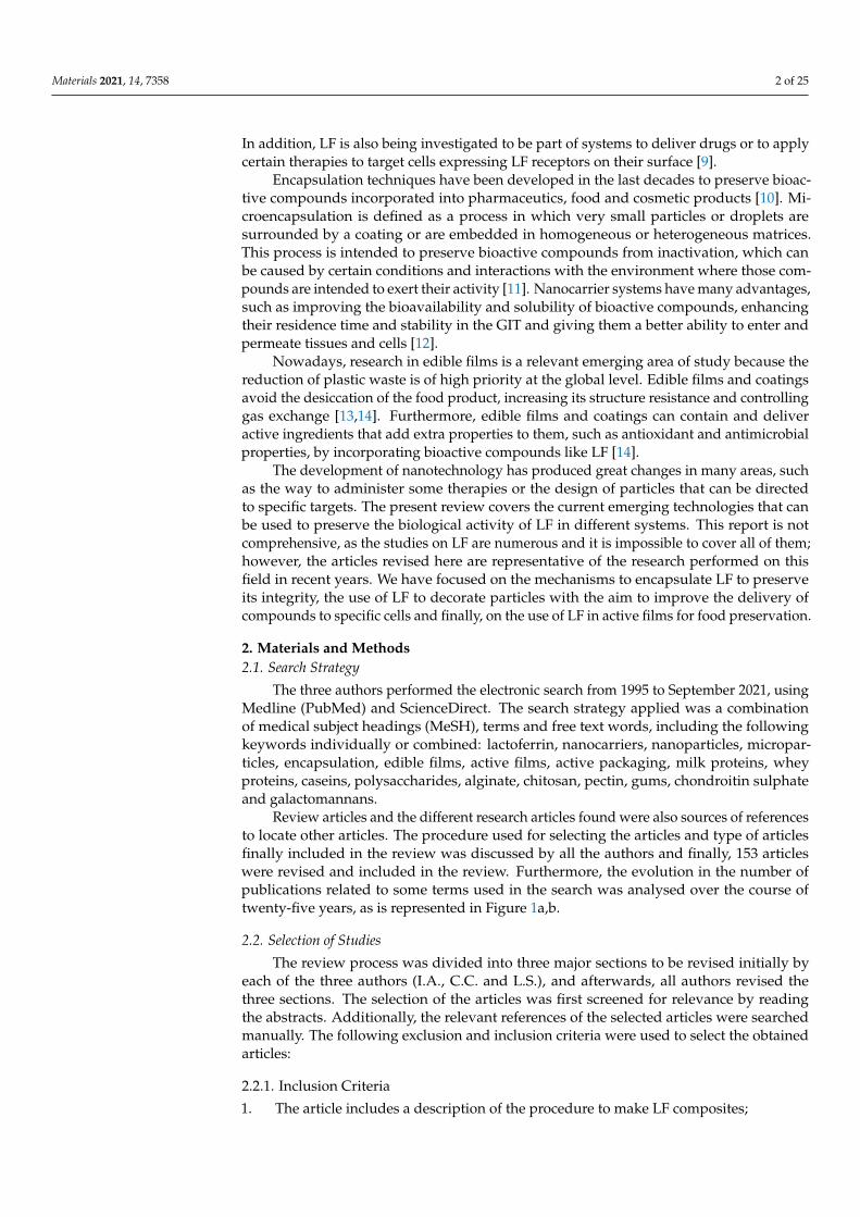
Review articles and the different research articles found were also sources of referencesto locate other articles. The procedure used for selecting the articles and type of articlesfinally included in the review was discussed by all the authors and finally, 153 articleswere revised and included in the review. Furthermore, the evolution in the number ofpublications related to some terms used in the search was analysed over the course oftwenty-five years, as is represented in Figure 1a,b.
2.2. Selection of Studies
The review process was divided into three major sections to be revised initially byeach of the three authors (I.A., C.C. and L.S.), and afterwards, all authors revised thethree sections. The selection of the articles was first screened for relevance by readingthe abstracts. Additionally, the relevant references of the selected articles were searchedmanually. The following exclusion and inclusion criteria were used to select the obtainedarticles:
2.2.1. Inclusion Criteria
1. The article includes a description of the procedure to make LF composites;
Materials 2021, 14, 7358 3 of 25
2. The article includes applications of LF composites related to medicine;3. The article includes applications of LF composites related to foods;4. Review articles that help to identify articles related to the topics for review.
2.2.2. Exclusion Criteria
1. Articles not providing detailed information on the preparation of LF composites;2. Articles not providing detailed information on the applications of LF composites;3. Articles published before the year 1999.
Figure 1. Graphs representing the publications found by combining the term lactoferrin with(a): nanoparticles, microparticles or encapsulation; (b): edible films, active films or active packaging,in PubMed (blue bars) and ScienceDirect (red bars) shown with years.
3. Results3.1. Encapsulation of Lactoferrin
The selection of appropriate materials for encapsulating bioactive compounds isessential. These materials should be chemically compatible, non-reactive with the encapsu-lated substance and are expected to show certain properties, such as strength, flexibility,impermeability and stability, which are required for their application [15,16].
There are various food biopolymers, including polysaccharides, lipids, proteins, andtheir conjugates used to develop a wide range of nanocarriers. These are designed toprotect, entrap, encapsulate and control the delivery of bioactive compounds and nutraceu-ticals [12]. The systems based on lipid carriers can be classified as nanoemulsions, solidlipid nanoparticles, nanostructured lipid carriers, nanoliposomes, micelles and nanosus-pensions [17]. Polysaccharide-based nanocarriers also include polymer nanoparticles,polymeric micelles and inclusion complexes [12]. However, nanocarriers based on foodproteins can be prepared with different structures, including nanoparticles, hollow nanopar-ticles, nanohydrogels, heat-induced nanofibrillar aggregates, electrospun nanofibers andtubular nanostructures [18].
Materials 2021, 14, 7358 4 of 25
Food proteins normally used to construct nanocarriers can derive from animals andplants, such as whey, soy, egg, corn proteins, gelatin and bovine serum albumin (BSA),among others. The nanocarriers produced with those proteins are adequate for the deliveryof both hydrophobic and hydrophilic bioactive ingredients and can be included in productsfor nutrition, drugs or can be directed to apply several therapies [12].
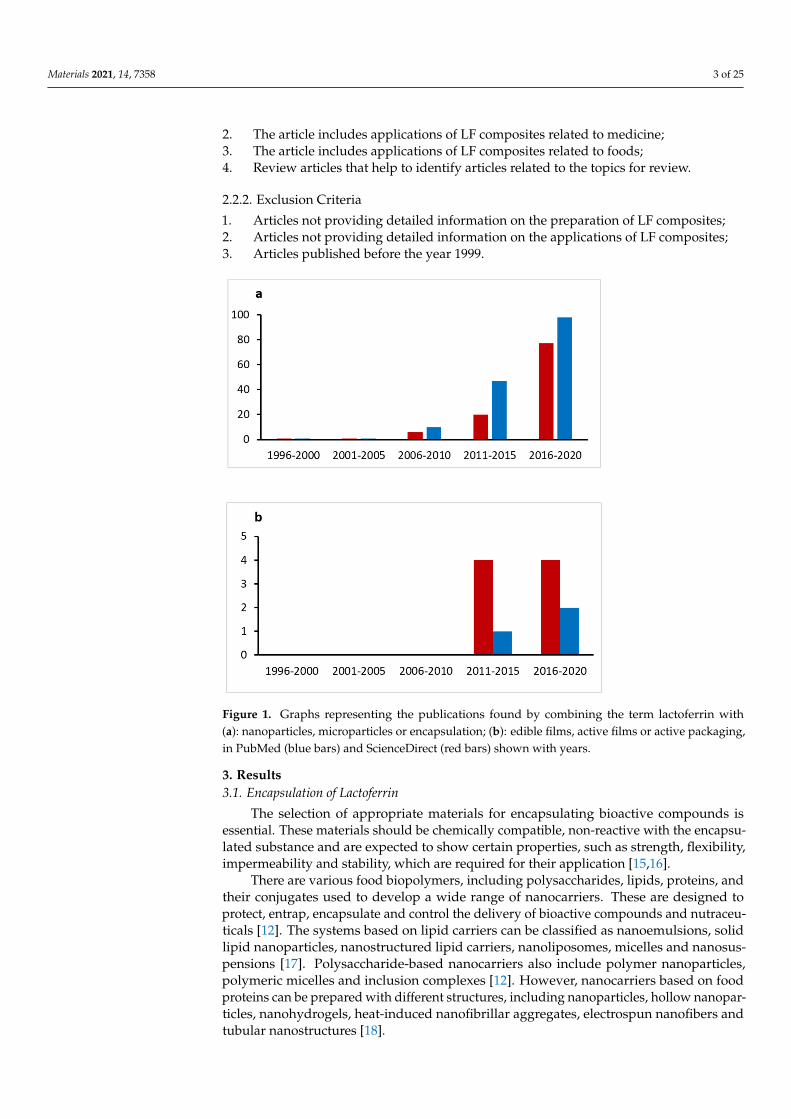
Encapsulation can be conducted by chemical or mechanical procedures, as shownin Figure 2. Chemical encapsulation can be done by simple and complex coacervation,co-crystallization, interfacial polymerization, ionic gelation, entrapment in liposomes,molecular inclusion and ionic gelation plus electrostatic interactions. The mechanicalprocedures are spray-drying, freeze/cold-drying, extrusion and fluidized bed [19].
Figure 2. A schematic illustration of different processes of encapsulation used in food and flavourindustries (adapted from Madene et al. [19]).
The complex coacervates are generated by an encapsulation method that usuallyconsists of three stages: emulsification, coacervation and cross-linking. The complex coac-ervation is a liquid–liquid phase separation that occurs when there is an interaction, mainlyelectrostatic, between biopolymers of opposite charges, the interaction being influenced bypH. The polymer matrix, considered as the wall, mainly made up of positively chargedproteins and negatively charged polymers surrounds the bioactive compound, consideredas the core [20]. The polymer-based coacervates have many applications as food and bio-material ingredients or encapsulants in personal care and functional foods. Recently, therehas been an increase in the interest to study heteroprotein complex coacervates, which area special case of coacervates in which the dense phase is formed by at least two differentproteins with opposite charges [21].
The potential benefits of bioactive compounds, also known as nutraceuticals, arenot optimally achieved because of their low and/or variable bioavailability [22]. Thisavailability is a measure of the bioefficiency of bioactive compounds and is the result ofprocesses that those components undergo within four gastrointestinal segments: the mouth,stomach, small intestine and large intestine [23].
The factors that mainly affect the bioavailability of encapsulated bioactive compoundsare the composition of nanocarriers and loading capacity, size and surface. For example,nanocarriers based on starch and proteins can be hydrolysed by amylases and proteases,while other materials, such as resistant starch, pectin and alginate are resistant to enzymesand pH, and are attacked only by the colonic microbiota [23]. However, the release ofbioactive compounds from lipid-based nanocarriers is favoured by the action of lipase in
Materials 2021, 14, 7358 5 of 25
the mouth, stomach and intestines, which breaks down triacylglycerides into free fattyacids and monoacylglycerides, contributing, with the bile salts, to the solubilisation ofbioactives into mixed micelles [24]. Furthermore, the bioavailability of many bioactivecomponents depends on the food matrix that is coingested with them, in the case that thoseare contained in a food product or in some nutraceuticals.
3.1.1. Encapsulation of Lactoferrin with Milk Proteins
Milk proteins have a rich diversity of physicochemical characteristics and biodegrad-able properties, which make them appealing for different food and pharmaceutical applica-tions [25]. Proteins are natural vehicles for bioactive molecules, thanks to their structuraland physicochemical properties that facilitate their functionality in delivery systems. Fororal delivery applications, the biocompatibility of milk proteins is usually positive [26],although it is necessary to consider that a small portion of the population is allergic tosome milk proteins.
• Whey proteins
Generally, whey proteins and LF are electrostatically attracted to each other overa range of pH values due to their different isoelectric points (pI), and so they can beassembled in various interfacial structures, such as single, double and mixed layers inemulsions [27]. In that sense, Anema and de Kruif [28] assayed the mixture of LF with theanionic protein β-lactoglobulin (β-Lg) at a range of pH and mixing ratios. Complexationwas monitored through turbidity and zeta potential measurements, and the formation ofcomplex coacervates was observed. Complexes were formed in the pH region of 5–7.3,and at the maximum turbidity, the complexes were neutrally charged. Moreover, althoughthe charge ratios of LF/β-Lg varied with pH, LF/β-Lg complexes were found to have aconstant stoichiometry of 1:3 at all pHs due to charge neutralization. With the additionof NaCl, the complexation diminished and disappeared at a salt concentration of about100 mM.
Chapeau et al. [29] showed that electrostatic complexes formed between LF and β-Lgcan be used to successfully encapsulate vitamin B9 at its recommended daily intake levels.They identified two types of B9/LF/β-Lg co-assembly: aggregates (at low and high proteinconcentrations) and heteroprotein coacervates (at intermediate protein concentrations),both exhibiting different kinetics and stability over time. The B9/LF/β-Lg coacervatesshowed high entrapment of B9 (from 6 to 16 mg B9/g protein), which means that afew milligrams of these coacervates would be enough to cover the recommended dailyintake of this vitamin. The same research group also demonstrated the scale-up of thissystem, confirming the efficiency of this type of biocarrier for developing natural functionalfoods [30].
Whey protein isolate (WPI) is obtained from the main by-product of cheese productionand mainly consists of β-Lg and α-lactalbumin (α-La). Irreversible denaturation andaggregation of whey proteins occur at heating temperatures higher than 70 ◦C, and thedenatured proteins interact with each other, forming a gel network. WPI has a strongbinding affinity for LF as it can cross-link to form a stable protein complex with enhancedelasticity and gel strength under various heating conditions, as reported by Li and Zhao [31].These researchers showed that the addition of LF into WPI gels increased the gel strengthat the two pHs tested of 5.8 and 6.7, being stronger and more uniform at pH 6.7 in thepresence of high concentrations of LF (20% and 30%). Those findings revealed both thepractical and theoretical significance for the utilisation of LF in gelled protein products.
Theoretical models and numerical simulations were applied by Delboni and daSilva [25] to gain insight into understanding the fundamental mechanisms responsiblefor the process of milk protein complexation under different conditions. This knowledgecan be essential to use milk proteins in nanoscale encapsulation systems for food andpharmaceutical applications. The interactions between β-Lg and LF and between α-La andLF were investigated by means of Monte Carlo simulations. The comparison between thefree energies associated with the complexation of LF with β-Lg and α-La at different pH
Materials 2021, 14, 7358 6 of 25
and ionic strengths revealed the weaker attraction between α-La and LF in contrast with thestronger attraction measured between β-Lg and LF. The driving force for the complexationbeing studied of β-Lg with LF is due to an association of electrostatic interactions, whereprotein charge plays an important role.
The influence of small ligands on the complex coacervation between β-Lg and LF wasstudied by Tavares et al. [32]. In this work, 8-anilinonaphthalene-1-sulfonic acid (ANS),a fluorescent probe, was used as a model of a small ligand. While ANS did not interactwith β-Lg, it presented two binding sites for LF inducing its self-aggregation. Dependingon its concentration, ANS modulated the shape of the β-Lg-LF macromolecular assembly.Coacervates were observed for ANS/LF molar ratio <25 against amorphous aggregatesfound for higher ANS/LF molar ratios.
In 2020, Darmawan et al. [33] explored a novel in silico approach to study the possibil-ity of employing whey protein components to encapsulate LF. The interaction of apo-LF(the most unstable form of the protein) with β-Lg and α-La was studied at temperatureconditions that are used during the spray drying process (95 and 180 ◦C). This method iscommonly employed in the food industry to encapsulate food ingredients by using hotair drying. It was found that the interaction of β-Lg and α-La with LF allowed maintain-ing the structure of two regions of the molecule corresponding to antibacterial peptides(lactoferricin and lactoferrampin) under the spray drying conditions. In this study, it wasalso demonstrated that apo-LF was most probably readily dispersible during subsequentrehydration due to its tendency to agglomerate under those temperatures. Moreover, theyshowed that the interactions between apo-LF, β-Lg and α-La were due to the acidic andbasic amino acid residues of these proteins.
In the study by De Figueiredo Furtado et al. [34], LF was proposed as a promisingingredient for incorporation into powder infant formulas, because it improved their proper-ties compared with formulas without this protein. The interaction of LF with WPI or wheyprotein hydrolysates used in the formulas, as encapsulating components of the oil mixtureadded, resulted in more stable emulsions. The powders obtained from the emulsionscontaining LF were those with a lower stickiness and consequently, with higher yield,encapsulation efficiency and wettability.
• Caseins
Caseins represent the major protein fraction of milk, making up 80% of the totalproteins. They precipitate at pH 4.6 and individual caseins, including αs1, αs2, β andκ-casein, are unique proteins with respect to their structure and function. They have openand flexible conformations and consist of hydrophilic and hydrophobic segments. Dueto their highly hydrophobic nature, individual caseins are stabilised by the creation of amicellar structure, which is chemically heterogeneous and is composed of the four types ofcaseins and by amorphous calcium phosphate [35]. The stability of the caseins and caseinmicelles to some treatments, such as heating, freezing, and drying, make them valuable indelivering food ingredients and bioactive compounds [36].
Anema and de Kruif investigated the interaction of LF with the casein micelles, asfound in bovine skimmed milk [37], and with the individual caseins [38]. The cationic LF(pI 8.3–8.7) was found to bind to the anionic caseins (pI range from 4.9 to 5.6) and to thecasein micelles (pI 4.6) at intermediate pH values. The binding of LF to the individualcaseins can be described as the formation of complex coacervates. Moreover, Anema andde Kruif [39] assayed the binding of LF to transglutaminase (TGA) cross-linked caseinmicelles. They found that the internal cross-linking of the casein micelles by TGA did notalter the binding mechanism of LF to them. The binding of LF to the untreated micellesswelled the micelles, which was avoided in the TGA-micelles. Additionally, after severalhours in the presence of added LF, the untreated micelles started to disintegrate, and theirsize decreased (increasing transparency of the milk was observed). In contrast, the internalcross-linking of the casein micelles prevented the disintegration of the casein micelles.
In 2016, Anema and de Kruif [40] studied complex formation between some bovinecasein types (α-casein, ACN; β-casein, BCN, and κ-casein, KCN) and LF. They showed
Materials 2021, 14, 7358 7 of 25
that the optimum coacervation occurred at the mixing ratio in which charge neutralityhappened and the turbidity was maximum. The kinetics of complex formation for LF/BCNand LF/KCN were rapid and occurred through a nucleation and coalescence process. How-ever, the kinetics of complex formation for LF/ACN were much slower. They evidencedthat when salt is added the coacervation diminishes, leading eventually to a one-phasesystem. Salt decreases the entropical contribution, and consequently, the coacervation isnot observed. For the LF/BCN or LF/KCN coacervates, the samples were completelytransparent (turbidity of ~0) at an ionic strength of ~25 mM, whereas for the LF/ACN,a much lower ionic strength of only ~10 mM was required for the sample to be com-pletely transparent. It is interesting to read the review article of Anema [41], in whichhe summarises all the results that have been obtained in his research group studying thespontaneous self-association of LF with the casein micelles in milk and with individualisolated caseins.
The complexation behaviour between LF and sodium caseinate (NaCas) before andafter heat treatment was also studied by Li and Zhao [42]. The results show that the denat-uration and aggregation of LF can be inhibited by forming soluble LF-NaCas complexes.The complexes formed at a ratio of 2:1 had an average diameter of 194 nm and exhibiteda higher capacity for lowering the air/water interfacial tension compared to complexeswith a lower proportion of LF. NaCas played a role as a stabiliser to protect LF againstheat-induced precipitation. Therefore, the presence of NaCas is important for maintainingthe structure and functionalities of LF during the production of LF-enriched food products.However, the authors have not demonstrated the biological activity of LF while being partof the complexes formed.
3.1.2. Encapsulation of Lactoferrin with Other Proteins
The ability of LF to form positively charged droplets at neutral pH can have manyimportant practical implications. Recently, there has been an increasing interest in theapplication of whey proteins, and especially LF, as emulsifiers [43]. LF has been usedas an emulsifier in multilayer emulsions for lipophilic nutraceutical delivery of activecompounds, such as curcumin and β-carotene [44]. LF can interact with other proteins orpolysaccharides, providing good stability to oil-in-water emulsions [45].
BSA with tannic acid (TA) has been used to encapsulate LF for oral administration byalternating layers of BSA and TA on porous microparticles of CaCO3 previously preparedwith LF [46]. The authors examined two approaches to load LF into CaCO3 particles, asshown in Figure 3. The first one used co-precipitation in which CaCO3 microparticles weredirectly formed in an LF solution. The second one used post-loading in which CaCO3microparticles were prepared in a water solution and then dispersed in an LF solution. TheCaCO3 was dissolved after covering the microparticles with the layers of BSA and TA. Thepost-loading approach showed lower LF degradation than that of co-precipitation and waschosen as the oral delivery system in a murine model. The microcapsule shells formedconferred LF high stability and protection in gastric conditions, while they were degradedunder intestinal conditions, thus releasing LF. The animals dosed with encapsulated LFshowed a 6.5 times higher concentration of LF in the intestine than the control groupdosed with free LF. Moreover, LF was later detectable in the liver, demonstrating that thisencapsulation system has great potential for oral delivery of bioactive molecules.
Pea protein isolate (PPI) has also been used to form complexes and coacervates withLF by electrostatic interactions under specific pH conditions [47]. The maximum level ofcoacervate formation was observed at charge neutrality. The pH where coacervation wasmost favourable was pH 5.4, and the soluble complexes were maximised at pH 7. Theformation of heteroprotein complexes was studied by Small Angle X-ray Scattering (SAXS),confirming the formation of complexes of around 13 nm at pH 7 and around 80 nm at pH5.4. A predominance of elliptical over spherical shapes for LF-PPI coacervates was found,and rare chain-like aggregations were observed, probably due to PPI-PPI complexes. Thesecoacervates can be of interest in various food applications.

Materials 2021, 14, 7358 8 of 25
Figure 3. Scheme of LF encapsulation, co-precipitation of CaCO3 and LF vs. post-loading of LF in porous CaCo3 mi-croparticles, both followed by Layer-by-Layer (LbL) deposition of bovine serum albumin and tannic acid and finaldissolution of CaCO3 (Redrawn and slightly changed from that contained in the article by Kilic et al. [46], which is anopen access article distributed under the terms and conditions of the Creative Commons Attribution (CC BY) license(http://creativecommons.org/licenses/by/4.0/, accessed on 30 October 2021).
Zheng et al. [21] investigated the conditions, thermodynamic mechanism, and mor-phological structure for the formation of soy protein isolate/lactoferrin (SPI/LF) complexcoacervate. The stoichiometry of two optimal complex coacervates (SPI/LF = 1:3 at pH 6.25and SPI/LF = 1:4 at pH 6.6) was identical to their mixing ratio. Moreover, the SPI/LFcomplex coacervation was exothermic and accompanied by significant entropy gain. Thiscoacervation was established by electrostatic interactions and by hydrogen bonds. It wasshown that the SPI/LF interaction improved the heat-stability of the LF heat-sensitive lobe.When the structure of the complexes was analysed, it was observed that the 1:3 SPI/LFcomplex exhibited distinct granules, whereas the 1:4 SPI/LF complex presented a uniformcross-linking structure. Analysis by atomic force microscopy showed the existence ofsphere complexes with a diameter of 50–150 nm in the 1:3 SPI/LF complex and a chain-likestructure with a length of 50–150 nm and a width of 20–80 nm in the 1:4 SPI/LF one. Thisdifferent structure was hypothesised to be by SPI self-aggregation. It remains unansweredif LF maintains its properties in SPI/LF coacervates, although the increase observed in LFthermal resistance is very positive for the potential applications of those coacervates.
3.1.3. Encapsulation of Lactoferrin with Polysaccharides
Biopolymer nanoparticles have gained popularity thanks to their exceptional physicalcharacteristics, such as biodegradability, biocompatibility, low toxicity and great ability tobind hydrophobic bioactive compounds [48,49]. The protein-ionic polysaccharide electro-static attraction requires a specific pH, in which the charges of these molecules are opposite.The net charge of a protein at its pI is zero, being positive below the pI, and negative at apH higher than the pI. Thus, cationic and anionic polysaccharides can form electrostaticcomplexes with proteins depending on pH. At physiological pH, LF is positively charged(pI 8.3–8.7); therefore, its capacity to create complexes with several anionic polysaccharideshas made possible the extensive research produced in this area [49].
The type and concentration of protein and polysaccharide, as well as the temperature,pH and ionic composition of the solution, determine the functional characteristics of aprotein-polysaccharide complex [50]. Additionally, the formation of complex coacervatesis influenced by the molecular weight, charge density and chemical nature of the polymersused [51].
• Alginate
Alginate is a natural, biodegradable and biocompatible anionic polysaccharide ob-tained from brown algae [52]. It is an organic polymer derived from alginic acid and islocated in the cell wall of the algae. Its structure consists of linear chains composed ofmonosaccharides D-mannuronic and L- guluronic.
Materials 2021, 14, 7358 9 of 25
This polymer owes its polyanionic character to the carboxyl groups that appear alongthe chain. The distribution of monomers in the polymeric chain and the charge and volumeof carboxyl groups give to the formed gel characteristics of flexibility or rigidity dependingon the guluronic content. The higher the number of guluronic blocks, the harder and morerigid and brittle the gel is, while if the mannuronic groups predominate, the gel is softerand more elastic [53]. This polymer is presented as sodium alginate (Na-Alg) when usedas a food additive and is easily available and widely applied in the industry. The maincharacteristics of Na-Alg are its good affinity with water and its highly anionic charge atpH values higher than two [54].
On the other side, alginate combined with calcium (Ca-Alg) forms a gel that is a well-known system used for the elaboration of gel beads. These beads are useful to encapsulatea wide variety of bioactive agents, due to their simplicity, biocompatibility, non-toxicityand low cost [5].
When two chains of the guluronic blocks align, coordination sites are created. Thesechains are in the shape of loops, creating cavities with adequate size to accommodate thecalcium ion. After the addition of calcium, the alginate undergoes conformational changes,producing the well-known “egg box” gelling pattern (Figure 1 in [53]). This pattern isbased on the dimerization of the chain and, later on, further aggregation of the dimers [53].
Furthermore, alginate is a good option for the encapsulation of molecules since ithas unique characteristics that allow a controlled release in the intestine [6]. Additionally,alginate microbeads, reaching a mean diameter of 130 ± 47 µm, are one of the most studiedencapsulation systems [55]. Braim et al. [6] and Kanwar et al. [56] confirmed that theLF-alginate capsule can be coated with chitosan, another polycationic polymer, to improvethe integrity of the capsule in GIT fluids.
Some studies have proven the efficacy of coating LF by alginate capsules to deliverthis protein to the colon [56] and to impede the growth of Clostridiodes [6]. Furthermore,the use of LF as a complement to alginate to encapsulate essential oil particles [52] orprobiotics [55] has also been achieved.
In the study by Raei et al. [5], LF was encapsulated in Ca-Alg capsules, and it wasproven that the effectiveness of these capsules increased with higher concentrations ofCa-Alg and with thermal treatment (61 ◦C for 10 min). However, the stability of thenanoparticles was greater when the polysaccharide concentration was lower (0.2%, w/w)and the thermal treatment was applied. These particles remained intact for 30 min atpH 2, conditions that can be considered equivalent to gastric digestion, which guarantees acontrolled release during the first 30 min and a subsequent gradual release in acidic andneutral conditions. In this way, they confirmed that the encapsulation of LF with Ca-Alg isa technique that can be used for the targeted delivery of LF to the intestine.
Furthermore, Bokkhim et al. [57] encapsulated three different forms of LF (apo-,native- and holo-) in micro-gel particles of alginate, using the aerosol technique. Theyproduced the particles from a 2% (w/w) solution of LF/alginate (1:1), using 0.1 M CaCl2as the cross-linking solution. These authors demonstrated that a major concentration ofCaCl2 decreased the encapsulation efficiency. LF/alginate particles were maintained intact-during simulated gastric digestion of 2 h, with 30% more protein compared to the non-encapsulated LF. The digestion of all forms of LF, pure or encapsulated, under intestinalfluids was rapid. Thus, these authors showed with this study that LF/alginate micro-gelparticles can protect LF from the action of pepsin (the main enzyme of gastric digestion)and release it at the intestine.
Additionally, Wang et al. [54] proved the efficacy of the LF/Na-Alg complex coac-ervates. This study showed that owing to the positive charge of LF at pHs lower than8.5 and the negative charge of Na-Alg in the entire pH range studied (2–10), the elec-trostatic interaction between both molecules was excellent. This electrostatic interactionincreased as the pH decreased from eight to five and remained constant at pH from fiveto four. Therefore, the authors concluded that the optimum pH for the formation of theLF/Na-Alg complex coacervates was 4.5. Additionally, they compared the degradation
Materials 2021, 14, 7358 10 of 25
of the uncoated LF and that of the alginate capsule in the gastric stage. They found that100% of the uncoated protein was degraded under gastric conditions. This percentagedecreased to 70% when LF was protected with Na-Alg, keeping 30% of the protein intactat that stage of digestion. Alginate provided a blockage of the sites of LF for the action ofpepsin, reducing LF proteolysis. Several authors have defended that alginate can interactwith pepsin, decreasing its catalytic mechanism during gastric digestion, while it doesnot have the same effect with trypsin, being degraded by this enzyme in the intestinaldigestion [52,54,57].
The encapsulation of LF with Na-Alg allows the preservation of its properties, suchas its antioxidant activity, increasing by 30% with respect to the uncoated LF under gas-tric conditions (pH 2). After gastric digestion, the antioxidant capacity of uncoated LFdecreased by 12% due to the breakdown of peptides. However, the antioxidant capacity ofLF/Na-Alg coacervates was not modified after this digestion stage [54].
• Chondroitin sulphate
Chondroitin sulphate (ChS) is a polyanionic mucopolysaccharide that is used as afood-grade material and is obtained from animal cartilage. ChS contains strongly acidicsulphate groups and weakly acidic carbonyl groups, so it has a high negative chargedensity and can form ionic complexes with positively charged molecules, such as LF.Furthermore, the ability of ChS as a potential carrier for drug delivery has been investigated,as it possesses numerous attractive properties, such as biosafety, biocompatibility andbiodegradability [48]. Additionally, ChS is considered a possible ligand targeting cancercells, which overexpress CD44 receptors [58] and it is valued as a suitable polysaccharidefor delivering active compounds to target cells [48].
In the same study by Abdelaziz et al. [58], it was proposed to coat the surface ofnanoparticles with ChS and LF through carbodiimide coupling or by electrostatic interac-tions, as a potential approach to attack cancer cells overexpressing LF receptors. Here, theuse of LF and ChS as coating materials demonstrated their efficiency in tumour selection.
• Galactomannans
Galactomannans are industrial polysaccharides obtained from seeds of the Legu-minosae family. They are storage polysaccharides formed by a skeleton of mannosewith galactose branches. They are used to increase viscosity or film production. Galac-tomannans have applications in both food and clinical industries, due to their rheologicalproperties [59]. In increasing the order of mannose:galactose ratio, we can distinguishdifferent types of galactomannans: fenugreek gum (1:1), guar gum (2:1), tara gum (3:1) andlocust bean gum (4:1) [60].
Nanoparticles containing LF have been formed from a soluble polyelectrolyte complexcomposed by LF/N-succinyl chitosan/galactomannan in a 1:3:2 ratio, after heat treatmentbelow the protein heat denaturation temperature. The presence of galactomannan allowsforming the particles, not only by electrostatic interaction but also by hydrophobic interac-tions and hydrogen bonds. Because of those interactions, LF is well integrated into thesecomplexes, due to the hydrophobic interaction between the pyranose chains of chitosan,galactomannan and the hexose residues of the hydrocarbon component of LF [61]. Thesebiopolymer particles have a 250–400 nm size, regular spherical shape and good storagestability. The load of LF in the particles was 76–87% and they retained their characteristicsafter lyophilisation. Therefore, these particles can eventually be used for the targetedtransport of biologically active substances [49].
The immobilization of LF in galactomannan films was proposed by Albuquerqueet al. [59] as an alternative to conventional edible solid dosages, such as capsules, particles,beads or tablets. This alternative can be effective for biotechnological applications in thepharmaceutical and the food industries, for individuals with dermal wounds or withdifficulty in swallowing edible doses, for example.
The tara gum, like the rest of galactomannans, is a neutral polysaccharide; therefore,the insertion of charged groups into its structure allows its interaction with other polymers

Materials 2021, 14, 7358 11 of 25
and proteins. The process of carboxymethylation consists of the insertion of negativelycharged carboxymethyl groups in the main polysaccharide, thus favouring the formation ofcomplex coacervates [62]. Taking the advantage of this property, Santos et al. [63] performedthe microencapsulation of vitamin D3 through complex coacervation in matrices formedby carboxymethyl tara gum and LF. To promote the electrostatic interaction between taragum and LF, the system was shaken for 10 min at 300 rpm and then quickly placed in anice bath to reduce the temperature. The addition of TGA to these structures catalysed theformation of covalent bonds between the proteins, increasing the stability of the particlesformed. It was verified that a ratio of 1:3 (core:wall) was enough for correct encapsulationof the vitamin to ensure that it was protected by the polymeric matrix.
The complex coacervation of LF with biopolymers increases its stability during gastricdigestion since the acidic pH of the stomach maintains both polymers with oppositecharges supporting their interaction. However, intestinal pH (7) has a negative effect oncoacervation since this pH modifies the positive charge of LF necessary for the interactionwith tara gum [63]. Therefore, these complexes improved the stability and facilitated thecontrolled release of the vitamin during GIT digestion.
• Gum arabic
Gum arabic (GA) is an anionic polyelectrolyte that mainly consists of three frac-tions of anionic branched arabinogalactans. These fractions are 80–90% arabinogalactan,10–20% arabinogalactan-protein and 2–4% glycoprotein. The emulsifying properties ofGA are mainly attributed to the arabinogalactan-protein [50]. Furthermore, due to its lowviscosity and high solubility at high concentrations, and its emulsifying and encapsulatingcapacities, GA is extensively used in industry [51].
The protein-polysaccharide electrostatic complexes can be used as stabilisers andemulsifiers in high internal phase emulsions (HIPEs). Some authors determined thatthe LF/GA complexes (1:1) can be used to form HIPEs over an ionic strength range of0–300 mM NaCl at a pH range of 3–6. During the homogenisation applied for this process,GA is not as efficient as proteins at producing small droplets; but it is a better stabiliseronce the droplets have been formed as reported by Cheng et al. [50]. These authors alsoshowed that the antioxidant properties of LF allowed, in these HIPEs of LF/GA complexes,the encapsulation of nutraceutical compounds, such as curcumin, protecting them fromchemical degradation.
• Pectin
Pectin is an anionic polysaccharide present in plants, mainly in fruits and youngtissues. It is made up of galacturonic acid units with free carboxyl groups, which give itan acid tendency. In its native form, pectin is highly methylated. The degree of pectinmethylation influences its ability to form gels. This polysaccharide is easily degradableunder alkaline conditions and, especially, at high temperatures [64]. Pectin is a polysac-charide most used to produce multilayer encapsulation systems with whey proteins, andin particular, it has allowed the encapsulation of LF at pH between three and four with asize of around 700 nm and good characteristics to be used in pharmaceuticals and foodproducts [65]. In another study by Bengoechea et al. [64], electrostatic complexes betweenhigh-methoxyl pectin and LF were formed at pH 7. These complexes were soluble atpH between 7 and 3.5 but underwent aggregation at lower pH. The properties of thesepectin/LF complexes can be modulated through the pH, temperature or biopolymer con-centration. Furthermore, it was observed that at pH 7 the stability of LF/pectin complexesto aggregation during heating was much better than that of LF alone.
Niu et al. [66] also created pectin/LF complexes by mixing the two biopolymers insolution, leading to spontaneous colloid formation due to electrostatic interactions. Theycompared the potential of low (LM) and highly methylated (HM) pectin to interact with LFand determined the loading of LF in the capsules and their encapsulation efficiency. Theyconcluded that LM pectin had a greater capacity to encapsulate LF, with an encapsulationefficiency of 40%, compared to 5% for HM pectin. Additionally, they verified the inhibitory
Materials 2021, 14, 7358 12 of 25
effect on bacterial growth of the LF-pectin complexes at a concentration of 1 mg/mL, aneffect that lasted at least 48 h. Therefore, they stated that LF encapsulation with pectin didnot affect its bioactivity.
Moreover, in the study conducted by Niu et al. [66], some pectin-LF complexeswere coated with chitosan in order to increase their gastric stability and muco-adhesiveproperties. This coating improved the colloidal stability of the complex in simulatedgastric fluid. Furthermore, in an HCl solution at pH 3, both systems, the uncoated andthe chitosan-coated LF/pectin complexes, showed a gradual release of LF. After 2 h underthese conditions, the uncoated complex showed a 30% release of LF, while the chitosan-coated complex only released 10%. However, under gastric simulated conditions (pH 3,no pepsin), the uncoated complex showed a negligible release of LF. This limitation of LFrelease indicates that this protein would not be as exposed to digestive enzymes, such aspepsin. Therefore, these pectin/LF systems can be a good way to incorporate LF in foodsor special products for oral administration, as they decrease the hydrolytic effect of pepsinon LF, protecting this bioactive molecule from gastric digestion up to at least 60 min for agut-targeted delivery [66].
• Chitosan
Chitosan is a useful natural biopolymer, obtained by chemical or enzymatic deacetyla-tion of chitin, a polysaccharide present in nature (insects, crustaceans and fungi) [67]. It hasmultiple reactive groups that can help in the transport of drugs due to its physicochemicalproperties, such as charge and hydrophobicity. Thus, due to these reactive groups, chitosannanoparticles can be complemented with targeting vectors like LF [68]. These authorsshowed that LF and succinyl-chitosan formed stable and high-yield nanoparticles andthat the globular structure of LF allowed the formation of stable nanoparticles withoutchemical conjugation. These nanoparticles were designed to bind to immune cells, such asmacrophages or phagocytic cells, in a few minutes and deliver LF by cross-presentation,the mechanism of antigen-presenting cells (APCs). Thus, the encapsulation of LF intochitosan can change the traffic of proteins inside APCs and modify the immune responses.
LF was found to form nanohydrogels by thermal gelation with the glycomacropeptide(GMP) showing high encapsulation efficiencies for curcumin and caffeine and a controlledrelease of those compounds [69]. Later, the same authors [70] studied the application ofchitosan coating in LF-GMP nanohydrogels. This biopolymer did not affect the shape ofnanohydrogels, maintaining their spherical shape and decreasing their aggregation in thesolution. These authors evaluated the effect of chitosan-coat on release mechanisms ofnanohydrogels at gastric digestion, using caffeine as the encapsulated compound. Theyconcluded that the presence of chitosan improved the stability of LF-GMP nanohydrogels,keeping them intact until 60 min under acidic conditions, compared with the 15 min thatthe uncoated nanohydrogels remained intact. They continued their study and, in 2018,revealed that chitosan coating reduced the protein degradation from 86% to 76% for LFand from 71% to 53% for GMP. Furthermore, the chitosan increased the bioaccessibilityof curcumin (bioactive compound encapsulated in the nanohydrogel) and protected itsantioxidant activity during the GIT digestion, losing only 30% of it compared to the 68% ofactivity loss for free curcumin [71].
Throughout this review, it has been shown that chitosan can be combined with otherpolysaccharides to favour the stability and integrity of the formed particles [6,49,61,66]. Asan example, chitosan together with pectin has been used to stabilise LF-loaded liposomesobtaining solid particles that are more resistant than liposomes to heat and to simulatedintestinal digestion [72].
3.2. Nanocarriers Coated with Lactoferrin
The advances in the design of targeted drug delivery systems have been very relevantin the last few decades. These delivery systems must consider the surface properties andreceptors of the target cell, the properties of the carrier and the suitable ligand to be utilised.Moreover, a targeted drug delivery system should be non-immunogenic, biocompatible,
Materials 2021, 14, 7358 13 of 25
specific to the target site, and show chemical and physical stability, both in vitro andin vivo [73].
There are two strategies to design targeted delivery: passive and active. In the passivestrategy, drugs are loaded into nanocarriers of a size around 200 nm, which can reachthe tissues or organs by their enhanced permeability and retention. In the active strategy,drug-loaded nanocarriers are conjugated with a certain ligand, which can further bind to aspecific receptor on the surface of target cells. This receptor is normally overexpressed inthe affected cells by the disease and consequently, the drug accumulates on them [73].
Receptors for LF have been expressed in many cells, such as brain endothelial cells,liver cells, epithelial respiratory cells, cancer cells, etc. Therefore, LF can be used to covernanocarriers loaded with drugs to increase their delivery to the cells via LF interaction withits receptors. Furthermore, the cationic nature of LF allows its binding to anionic cellularcompounds, such as glycosaminoglycans [73].
In the last few years, inorganic nanoparticles have attracted considerable attention fordrug delivery and imaging applications due to their characteristics. The positive features ofthese nanoparticles are their higher loading capacity, ease of functionalization with severaltypes of ligands, biocompatibility and adaptable degradation rates. There are several typesof nanocarriers coated with LF that have been developed for delivery applications.
Thus, superparamagnetic iron oxide nanocarriers (Fe3O4-LF) with narrow size rangeswere developed via surface functionalization with LF using the EDC(1-ethyl-3-(3-dimethylaminopropyl) carbodiimide hydrochloride) coupling reaction aimingto target the receptors that are highly expressed on the surface of human fibroblasts [74].
Superparamagnetic iron oxide nanoparticles conjugated with LF (LF-SPIONs) [75]and LF-PEG-Fe3O4 [76] were successfully fabricated via the EDC coupling reaction andwere applied as a magnetic resonance imaging contrast agent for improving the detectionof in vivo brain glioma.
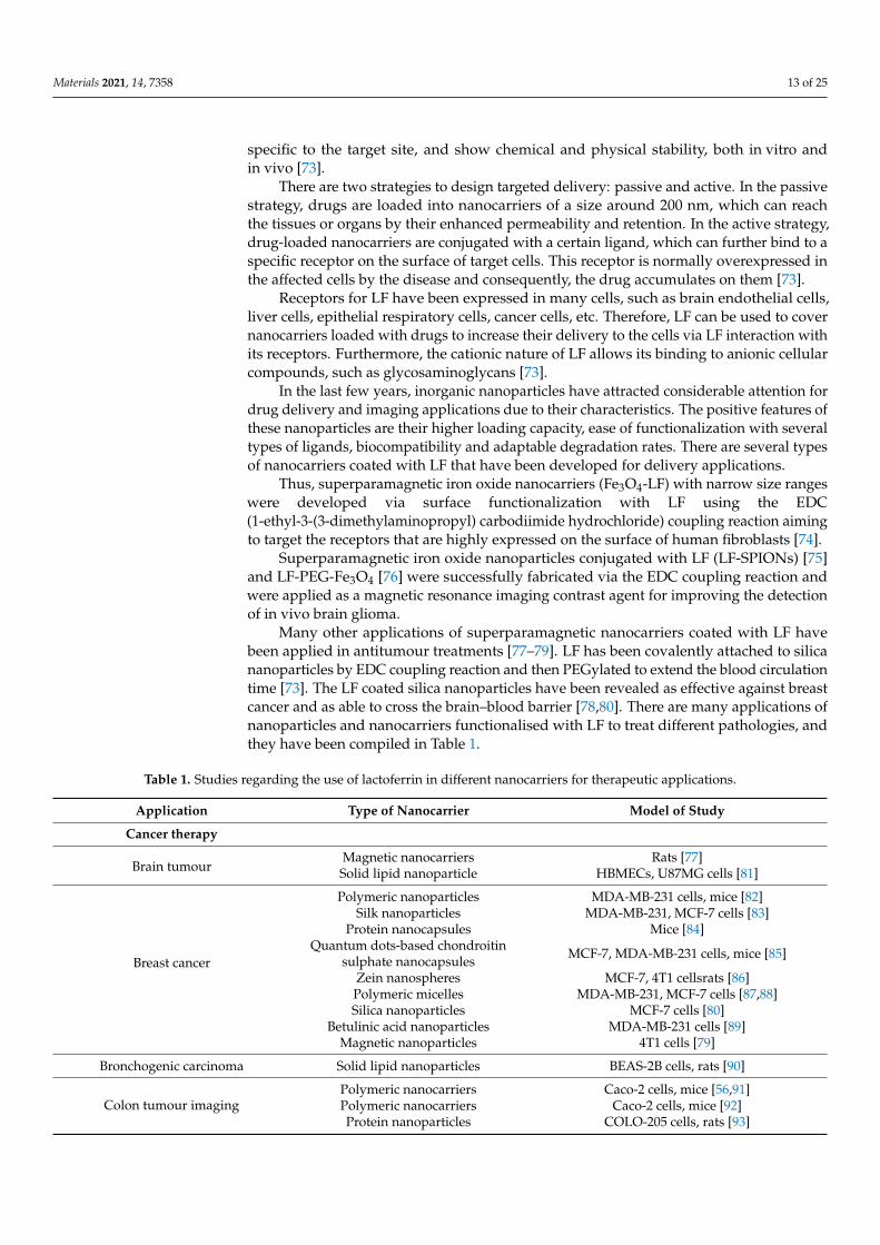
Many other applications of superparamagnetic nanocarriers coated with LF havebeen applied in antitumour treatments [77–79]. LF has been covalently attached to silicananoparticles by EDC coupling reaction and then PEGylated to extend the blood circulationtime [73]. The LF coated silica nanoparticles have been revealed as effective against breastcancer and as able to cross the brain–blood barrier [78,80]. There are many applications ofnanoparticles and nanocarriers functionalised with LF to treat different pathologies, andthey have been compiled in Table 1.
Table 1. Studies regarding the use of lactoferrin in different nanocarriers for therapeutic applications.
Application Type of Nanocarrier Model of Study
Cancer therapy
Brain tumourMagnetic nanocarriers Rats [77]Solid lipid nanoparticle HBMECs, U87MG cells [81]
Breast cancer
Polymeric nanoparticles MDA-MB-231 cells, mice [82]Silk nanoparticles MDA-MB-231, MCF-7 cells [83]
Protein nanocapsules Mice [84]Quantum dots-based chondroitin
sulphate nanocapsules MCF-7, MDA-MB-231 cells, mice [85]
Zein nanospheres MCF-7, 4T1 cellsrats [86]Polymeric micelles MDA-MB-231, MCF-7 cells [87,88]Silica nanoparticles MCF-7 cells [80]
Betulinic acid nanoparticles MDA-MB-231 cells [89]Magnetic nanoparticles 4T1 cells [79]
Bronchogenic carcinoma Solid lipid nanoparticles BEAS-2B cells, rats [90]
Colon tumour imagingPolymeric nanocarriers Caco-2 cells, mice [56,91]Polymeric nanocarriers Caco-2 cells, mice [92]Protein nanoparticles COLO-205 cells, rats [93]
Materials 2021, 14, 7358 14 of 25
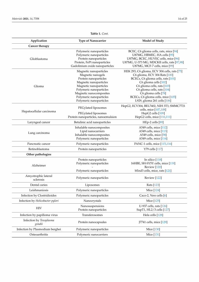
Table 1. Cont.
Application Type of Nanocarrier Model of Study
Cancer therapy
Glioblastoma
Polymeric nanoparticles BCEC, C6 glioma cells, rats, mice [94]Polymeric nanoparticles U87MG, HBMEC, HA cells [95]
Protein nanoparticles U87MG, BCEC, HUVEC cells, mice [96]Protein, FePt nanoparticles U87MG, U-373 MG, MDCKII cells, rats [97,98]
Gadolinium oxide nanoparticles U87MG, MCF-7 cells, mice [99]
Glioma
Magnetic nanoparticles HEK 293, C6 glioma, ECV 304 cells, rats [75]Magnetic nanogels C6 glioma, ECV 304 Rats [100]
Protein nanoparticles BCECs, C6 glioma cells, rats [101]Magnetic nanoparticles C6 glioma cells [102]Magnetic nanoparticles C6 glioma cells, rats [103]Polymeric nanoparticles C6 glioma cells, rats [104]
Magnetic nanocomposites C6 glioma cells [78]Polymeric nanoparticles BCECs, C6 glioma cells, mice [105]Polymeric nanoparticles L929, glioma 261 cells [106]
Hepatocellular carcinomaPEGylated liposomes HepG2, ECV304, BEL7402, NIH 3T3, SMMC7721
cells, mice [107,108]PEGylated liposomes HepG2 cells [109]
Protein nanoparticles, nanoemulsion HepG2 cells, mice [110,111]
Laryngeal cancer Betulinic acid nanoparticles HEp-2 cells [89]
Lung carcinoma
Inhalable nanocomposites A549 cells, mice [112]Lipid nanocarriers A549 cells, mice [113]
Inhalable nanocomposites A549 cells, mice [58]Polymeric nanoparticles A549 cells, mice [114]
Pancreatic cancer Polymeric nanoparticles PANC-1 cells, mice [115,116]
Retinoblastoma Protein nanoparticles Y79 cells [117]
Other pathologies
Alzheimer
Protein nanoparticles In silico [118]Polymeric nanoparticles 16HBE, SH-SY5Y cells, mice [119]
- Review [120]Polymeric nanoparticles bEnd3 cells, mice, rats [121]
Amyotrophic lateralsclerosis Polymeric nanoparticles Review [122]
Dental caries Liposomes Rats [123]
Leishmaniosis Polymeric nanoparticles Mice [124]
Infection by Clostridioides Polymeric nanoparticles Caco-2, Vero cells [6]
Infection by Helicobacter pylori Nanocrystals Mice [125]
HIVNanosuspensions U-937 cells, rats [126]
Protein nanoparticles SupT1, HL2/3 cells [127]
Infection by papilloma virus Transferosomes Hela cells [128]
Infection by Toxoplasmagondii Protein nanocapsules J7741 cells, mice [129]
Infection by Plasmodium berghei Polymeric nanoparticles Mice [130]
Osteoarthritis Polymeric nanocarriers Mice [131]
Materials 2021, 14, 7358 15 of 25
Table 1. Cont.
Application Type of Nanocarrier Model of Study
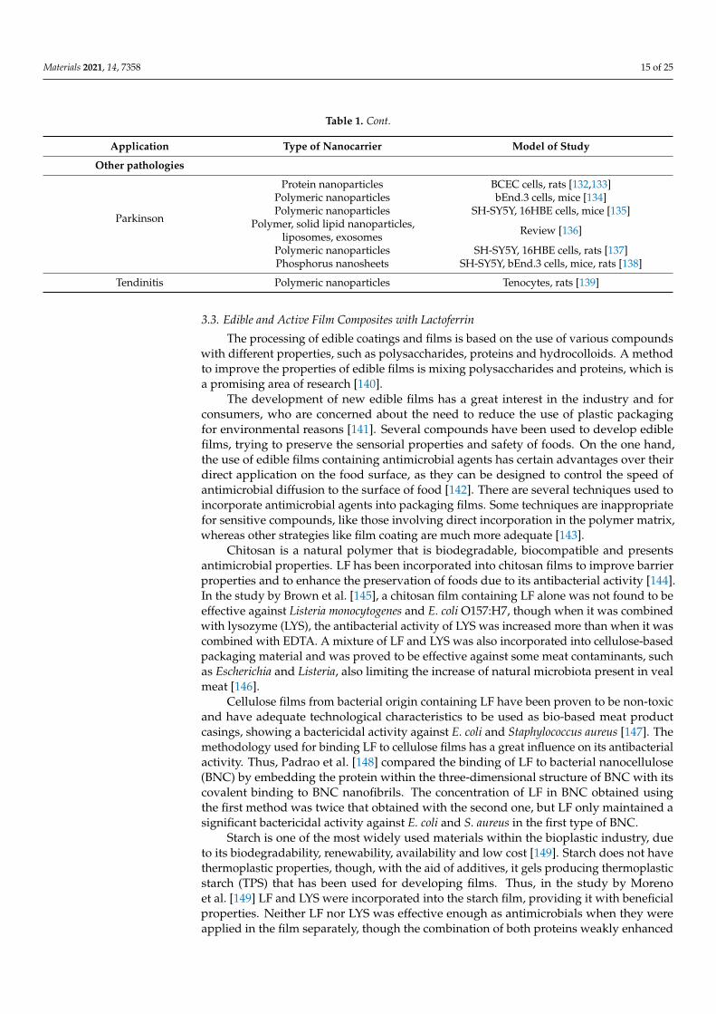
Other pathologies
Parkinson
Protein nanoparticles BCEC cells, rats [132,133]Polymeric nanoparticles bEnd.3 cells, mice [134]Polymeric nanoparticles SH-SY5Y, 16HBE cells, mice [135]
Polymer, solid lipid nanoparticles,liposomes, exosomes Review [136]
Polymeric nanoparticles SH-SY5Y, 16HBE cells, rats [137]Phosphorus nanosheets SH-SY5Y, bEnd.3 cells, mice, rats [138]
Tendinitis Polymeric nanoparticles Tenocytes, rats [139]
3.3. Edible and Active Film Composites with Lactoferrin
The processing of edible coatings and films is based on the use of various compoundswith different properties, such as polysaccharides, proteins and hydrocolloids. A methodto improve the properties of edible films is mixing polysaccharides and proteins, which isa promising area of research [140].
The development of new edible films has a great interest in the industry and forconsumers, who are concerned about the need to reduce the use of plastic packagingfor environmental reasons [141]. Several compounds have been used to develop ediblefilms, trying to preserve the sensorial properties and safety of foods. On the one hand,the use of edible films containing antimicrobial agents has certain advantages over theirdirect application on the food surface, as they can be designed to control the speed ofantimicrobial diffusion to the surface of food [142]. There are several techniques used toincorporate antimicrobial agents into packaging films. Some techniques are inappropriatefor sensitive compounds, like those involving direct incorporation in the polymer matrix,whereas other strategies like film coating are much more adequate [143].
Chitosan is a natural polymer that is biodegradable, biocompatible and presentsantimicrobial properties. LF has been incorporated into chitosan films to improve barrierproperties and to enhance the preservation of foods due to its antibacterial activity [144].In the study by Brown et al. [145], a chitosan film containing LF alone was not found to beeffective against Listeria monocytogenes and E. coli O157:H7, though when it was combinedwith lysozyme (LYS), the antibacterial activity of LYS was increased more than when it wascombined with EDTA. A mixture of LF and LYS was also incorporated into cellulose-basedpackaging material and was proved to be effective against some meat contaminants, suchas Escherichia and Listeria, also limiting the increase of natural microbiota present in vealmeat [146].
Cellulose films from bacterial origin containing LF have been proven to be non-toxicand have adequate technological characteristics to be used as bio-based meat productcasings, showing a bactericidal activity against E. coli and Staphylococcus aureus [147]. Themethodology used for binding LF to cellulose films has a great influence on its antibacterialactivity. Thus, Padrao et al. [148] compared the binding of LF to bacterial nanocellulose(BNC) by embedding the protein within the three-dimensional structure of BNC with itscovalent binding to BNC nanofibrils. The concentration of LF in BNC obtained usingthe first method was twice that obtained with the second one, but LF only maintained asignificant bactericidal activity against E. coli and S. aureus in the first type of BNC.
Starch is one of the most widely used materials within the bioplastic industry, dueto its biodegradability, renewability, availability and low cost [149]. Starch does not havethermoplastic properties, though, with the aid of additives, it gels producing thermoplasticstarch (TPS) that has been used for developing films. Thus, in the study by Morenoet al. [149] LF and LYS were incorporated into the starch film, providing it with beneficialproperties. Neither LF nor LYS was effective enough as antimicrobials when they wereapplied in the film separately, though the combination of both proteins weakly enhanced
Materials 2021, 14, 7358 16 of 25
their antimicrobial activity against E. coli and coliform microbiota of pork-minced meat.The films containing a blend of LF and LYS also reduced lard oxidation after long storagetimes [149].
In the study by Tavassoli et al. [150], multifunctional films were created by embeddingdifferent kinds of functional nanoparticles into a gelatin-based film prepared using a castingmethod. The nanoparticles were prepared by cross-linking cationic chitosan nanofibers,and afterwards, quercetin, LF, or both were introduced into these nanoparticles. Theincorporation of the nanoparticles into the films decreased their mechanical strength andstiffness but increased their flexibility. The presence of LF, quercetin and chitosan in thegelatin-based films increased their antimicrobial and antioxidant activity. Furthermore,the incorporation of the nanoparticles into the films improved their degradation undersimulated environmental conditions.
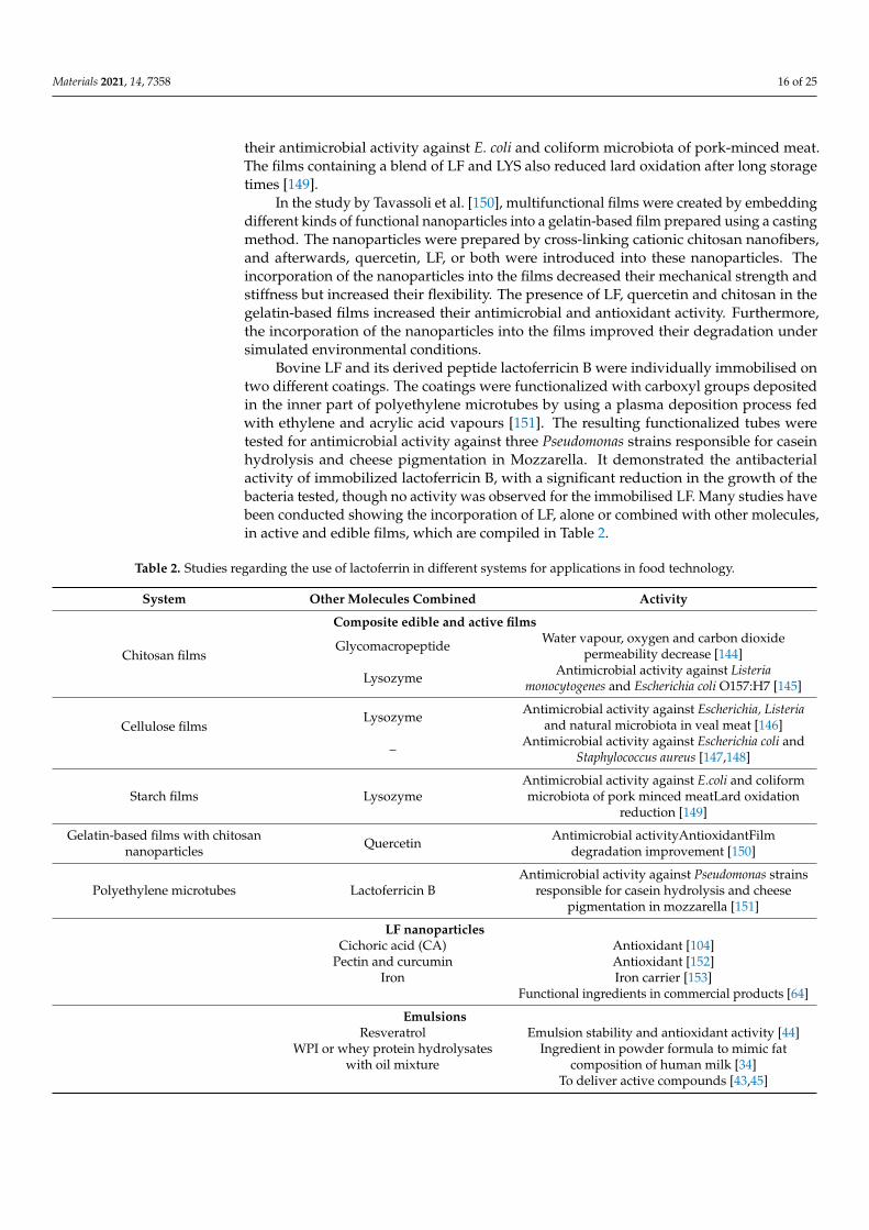
Bovine LF and its derived peptide lactoferricin B were individually immobilised ontwo different coatings. The coatings were functionalized with carboxyl groups depositedin the inner part of polyethylene microtubes by using a plasma deposition process fedwith ethylene and acrylic acid vapours [151]. The resulting functionalized tubes weretested for antimicrobial activity against three Pseudomonas strains responsible for caseinhydrolysis and cheese pigmentation in Mozzarella. It demonstrated the antibacterialactivity of immobilized lactoferricin B, with a significant reduction in the growth of thebacteria tested, though no activity was observed for the immobilised LF. Many studies havebeen conducted showing the incorporation of LF, alone or combined with other molecules,in active and edible films, which are compiled in Table 2.
Table 2. Studies regarding the use of lactoferrin in different systems for applications in food technology.
System Other Molecules Combined Activity
Composite edible and active films
Chitosan filmsGlycomacropeptide Water vapour, oxygen and carbon dioxide
permeability decrease [144]
Lysozyme Antimicrobial activity against Listeriamonocytogenes and Escherichia coli O157:H7 [145]
Cellulose filmsLysozyme Antimicrobial activity against Escherichia, Listeria
and natural microbiota in veal meat [146]
– Antimicrobial activity against Escherichia coli andStaphylococcus aureus [147,148]
Starch films LysozymeAntimicrobial activity against E.coli and coliformmicrobiota of pork minced meatLard oxidation
reduction [149]
Gelatin-based films with chitosannanoparticles Quercetin Antimicrobial activityAntioxidantFilm
degradation improvement [150]
Polyethylene microtubes Lactoferricin BAntimicrobial activity against Pseudomonas strains
responsible for casein hydrolysis and cheesepigmentation in mozzarella [151]
LF nanoparticlesCichoric acid (CA) Antioxidant [104]
Pectin and curcumin Antioxidant [152]Iron Iron carrier [153]
Functional ingredients in commercial products [64]
EmulsionsResveratrol Emulsion stability and antioxidant activity [44]
WPI or whey protein hydrolysateswith oil mixture
Ingredient in powder formula to mimic fatcomposition of human milk [34]
To deliver active compounds [43,45]
Materials 2021, 14, 7358 17 of 25
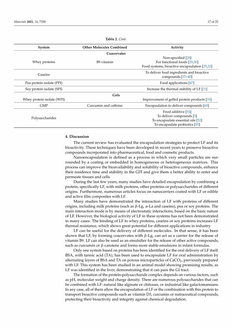
Table 2. Cont.
System Other Molecules Combined Activity
Coacervates
Whey proteinsNon-specified [28]
B9 vitamin For functional foods [29,30]Food systems, bioactive encapsulation [25,32]
Caseins To deliver food ingredients and bioactivecompounds [37–40]
Pea protein isolate (PPI) Food applications [47]
Soy protein isolate (SPI) Increase the thermal stability of LF [21]
GelsWhey protein isolate (WPI) Improvement of gelled protein products [31]
GMP Curcumin and caffeine Encapsulation to deliver compounds [69]
Polysaccharides
Food additive [54]To deliver compounds [5]
To encapsulate essential oils [52]To encapsulate probiotics [55]
4. Discussion
The current review has evaluated the encapsulation strategies to protect LF and itsbioactivity. These techniques have been developed in recent years to preserve bioactivecompounds incorporated into pharmaceutical, food and cosmetic products.
Nanoencapsulation is defined as a process in which very small particles are sur-rounded by a coating or embedded in homogeneous or heterogeneous matrices. Thisprocess can improve the bioavailability and solubility of bioactive compounds, enhancetheir residence time and stability in the GIT and give them a better ability to enter andpermeate tissues and cells.
During the last few years, many studies have detailed encapsulation by combining aprotein, specifically LF, with milk proteins, other proteins or polysaccharides of differentorigins. Furthermore, numerous articles focus on nanocarriers coated with LF or edibleand active film composites with LF.
Many studies have demonstrated the interaction of LF with proteins of differentorigins, including milk proteins (such as β-Lg, α-La and caseins), pea or soy proteins. Themain interaction mode is by means of electrostatic interactions, based on the basic natureof LF. However, the biological activity of LF in these systems has not been demonstratedin many cases. The binding of LF to whey proteins, caseins or soy proteins increases LFthermal resistance, which shows great potential for different applications in industry.
LF can be useful for the delivery of different molecules. In that sense, it has beenshown that LF, by forming coacervates with β-Lg, can act as a carrier for the release ofvitamin B9. LF can also be used as an emulsifier for the release of other active compounds,such as curcumin or β-carotene and forms more stable emulsions in infant formulas.
Only one system based on proteins has been identified for the oral delivery of LF itself.BSA, with tannic acid (TA), has been used to encapsulate LF for oral administration byalternating layers of BSA and TA on porous microparticles of CaCO3, previously preparedwith LF. This system has been studied in an animal model showing promising results, asLF was identified in the liver, demonstrating that it can pass the GI tract.
The formation of the protein-polysaccharide complex depends on various factors, suchas pH, molecular weight and charge density. There are numerous polysaccharides that canbe combined with LF: natural like alginate or chitosan, or industrial like galactomannans.In any case, all of them allow the encapsulation of LF or the combination with this protein totransport bioactive compounds such as vitamin D3, curcumin or nutraceutical compounds,protecting their bioactivity and integrity against chemical degradation.
Materials 2021, 14, 7358 18 of 25
Many studies have focused on the formation of these complexes by electrostaticinteractions, taking advantage of the positive charge of LF and the opposite charge of thepolysaccharides, such as alginate or chondroitin sulphate. This complex coacervation ofLF with biopolymers increases its stability during gastric digestion since the acidic pH ofthe stomach maintains both polymers with opposite charges supporting their interaction.However, intestinal pH has a negative effect on coacervation, modifying the positive chargeof LF necessary for the interaction with the polysaccharide. Therefore, these complexesimproved the stability and facilitated the controlled release of the protein during GITdigestion. Furthermore, galactomannans allow, in addition to electrostatic interaction, theformation of films by hydrogen bonds or by hydrophobic interactions that favour a goodintegration of LF.
In addition to a controlled release in the gut, some polysaccharides allow deliveryto target cells. While chondroitin sulphate combined with LF targets cancer cells, beingeffective for tumour selection, chitosan-LF nanoparticles bind immune cells and deliverLF by cross-presentation to macrophages or phagocytic cells. Several authors have usedchitosan to coat LF-polysaccharide complexes, increasing the integrity of the capsule andits stability under gastric conditions.
The strategy of coating nanoparticles with LF has been developed intensively overthe past few years. The receptors for LF are highly expressed on the surface of many cells;therefore, nanocarriers functionalised with LF have been used to treat tumours of severalorigins, such as mammary, hepatic, pancreatic and colonic, among others, with differentresults. However, there are still some issues to be investigated to know the interaction of LF-nanocarriers with the capillary endothelial cells in the blood–brain barrier and the kineticsof LF interaction with the surface of different types of cells. The use of LF nanocarriers toenhance the imaging analysis of some brain tumours is also an area of great interest forfuture development.
The food industry is currently investing in the development of edible and biodegrad-able films with properties directed to improve food preservation and to avoid the use ofplastic packaging. Furthermore, the incorporation of bioactive molecules into the films hasbeen encouraged over the past few years. These new films are able to maintain food qualityand safety and, at the same time, allow reducing the use of chemical additives. In manystudies, LF has been used in active films because of its antimicrobial properties. However,the results have indicated the existence of some problems in maintaining LF activity in theprocess of combining it with the base materials to build films or packaging. Some studieshave proved that the combination of several active molecules with films, such as LF andLYS, gives better results. Nevertheless, the enrichment of films with bioactive moleculesadds a high cost to the final product, a limitation that should be resolved to make thosefilms applicable to food industry.
5. Conclusions
Lactoferrin is a molecule with many biological properties that must be preservedwhen it is used in products to improve health or is incorporated into active films designedto maintain the quality of food products. The encapsulation of this bioactive proteinwith other proteins or polysaccharides prevents its proteolysis during gastric digestion,allowing a targeted release of LF. This protein can also be used in the active targeted drugdelivery, where drug-loaded nanocarriers are covered with LF, which can further bind toits receptors expressed on the surface of target cells. Future research on all these issuesshould be continued by evaluating the different LF activities, after being encapsulated orcombined with certain composite materials, in in vitro assays and eventually, in humans.
Author Contributions: Conceptualization, L.S.; article reviewing, L.S., I.A. and C.C.; writing—original draft preparation, L.S., I.A. and C.C.; writing—review and editing, L.S., I.A. and C.C. Allauthors have read and agreed to the published version of the manuscript.
Materials 2021, 14, 7358 19 of 25
Funding: This research was funded by the Spanish Ministry of Economy, Industry and Competitive-ness and the European Regional Development Fund (ERDF/FEDER) (AGL2017-82987), EuropeanSocial Found (ESF) and the Aragon Regional Government (A20_20R). I. Abad was supported by aPhD fellowship from Aragon Regional Government.
Institutional Review Board Statement: Not applicable.
Informed Consent Statement: Not applicable.
Data Availability Statement: Data sharing is not applicable to this article.
Conflicts of Interest: The authors declare no conflict of interest.
References1. Farnaud, S.; Evans, R.W. Lactoferrin—A multifunctional protein with antimicrobial properties. Mol. Immunol. 2003, 40, 395–405.
[CrossRef]2. Brock, J.H. The physiology of lactoferrin. Biochem. Cell Biol. 2002, 80, 1–6. [CrossRef] [PubMed]3. Franco, I.; Pérez, M.D.; Conesa, C.; Calvo, M.; Sánchez, L. Effect of technological treatments on bovine lactoferrin: An overview.
Food Res. Int. 2018, 106, 173–182. [CrossRef] [PubMed]4. Takeuchi, T.; Jyonotsuka, T.; Kamemori, N.; Kawano, G.; Shimizu, H.; Ando, K.; Harada, E. Enteric-formulated lactoferrin was
more effectively transported into blood circulation from gastrointestinal tract in adult rats. Exp. Physiol. 2006, 91, 1033–1040.[CrossRef] [PubMed]
5. Raei, M.; Rajabzadeh, G.; Zibaei, S.; Jafari, S.M.; Sani, A.M. Nano-encapsulation of isolated lactoferrin from camel milk by calciumalginate and evaluation of its release. Int. J. Biol. Macromol. 2015, 79, 669–673. [CrossRef]
6. Braim, S.; Spiewak, K.; Brindell, M.; Heeg, D.; Alexander, C.; Monaghan, T. Lactoferrin-Loaded Alginate Microparticles to TargetClostridioides difficile Infection. J. Pharm. Sci. 2019, 108, 2438–2446. [CrossRef] [PubMed]
7. Balcão, V.M.; Costa, C.I.; Matos, C.M.; Moutinho, C.G.; Amorim, M.; Pintado, M.E.; Gomes, A.P.; Vila, M.M.; Teixeira, J.A.Nanoencapsulation of bovine lactoferrin for food and biopharmaceutical applications. Food Hydrocoll. 2013, 32, 425–431.[CrossRef]
8. Liu, W.; Lu, J.; Ye, A.; Xu, Q.; Tian, M.; Kong, Y.; Wei, F.; Han, J. Comparative performances of lactoferrin-loaded liposomes underin vitro adult and infant digestion models. Food Chem. 2018, 258, 366–373. [CrossRef]
9. El-Fakharany, E.M. Nanoformulation of lactoferrin potentiates its activity and enhances novel biotechnological applications. Int.J. Biol. Macromol. 2020, 165, 970–984. [CrossRef]
10. Castro-Rosas, J.; Ferreira-Grosso, C.R.; Gómez-Aldapa, C.A.; Vargas, E.R.; Rodriguez, M.L.R.M.; Guzmán-Ortiz, F.A.;Falfan-Cortes, R.N. Recent advances in microencapsulation of natural sources of antimicrobial compounds used in food—Areview. Food Res. Int. 2017, 102, 575–587. [CrossRef]
11. Gouin, S. Microencapsulation: Industrial appraisal of existing technologies and trends. Trends Food Sci. Technol. 2004, 15, 330–347.[CrossRef]
12. Mohammadian, M.; Waly, M.I.; Moghadam, M.; Emam-Djomeh, Z.; Salami, M.; Moosavi-Movahedi, A.A. Nanostructured foodproteins as efficient systems for the encapsulation of bioactive compounds. Food Sci. Hum. Wellness 2020, 9, 199–213. [CrossRef]
13. Falguera, V.; Quintero, J.P.; Jiménez, A.; Muñoz, J.A.; Ibarz, A. Edible films and coatings: Structures, active functions and trendsin their use. Trends Food Sci. Technol. 2011, 22, 292–303. [CrossRef]
14. Dhumal, C.V.; Sarkar, P. Composite edible films and coatings from food-grade biopolymers. J. Food Sci. Technol. 2018, 55,4369–4383. [CrossRef] [PubMed]
15. Bansode, S.S.; Banarjee, S.K.; Gaikwad, D.D.; Jadhav, S.L.; Thorat, R.M. Microencapsulation: A review. Int. J. Pharm. Sci. Rev. Res.2010, 1, 38–43.
16. Maresca, D.; De Prisco, A.; La Storia, A.; Cirillo, T.; Esposito, F.; Mauriello, G. Microencapsulation of nisin in alginate beads byvibrating technology: Preliminary investigation. LWT Food Sci. Technol. 2016, 66, 436–443. [CrossRef]
17. Shin, G.H.; Kim, J.T.; Park, H.J. Recent developments in nanoformulations of lipophilic functional foods. Trends Food Sci. Technol.2015, 46, 144–157. [CrossRef]
18. Fathi, M.; Donsi, F.; McClements, D.J. Protein-based delivery systems for the nanoencapsulation of food ingredients. Compr. Rev.Food Sci. Food Saf. 2018, 17, 920–936. [CrossRef]
19. Madene, A.; Jacquot, M.; Scher, J.; Desobry, S. Flavour encapsulation and controlled release–a review. Int. J. Food Sci. Technol.2006, 41, 1–21. [CrossRef]
20. Corrêa-Filho, L.C.; Moldão-Martins, M.; Alves, V.D. Advances in the application of microcapsules as carriers of functionalcompounds for food products. Appl. Sci. 2019, 9, 571. [CrossRef]
21. Zheng, J.; Gao, Q.; Tang, C.H.; Ge, G.; Zhao, M.; Sun, W. Heteroprotein complex formation of soy protein isolate and lactoferrin:Thermodynamic formation mechanism and morphologic structure. Food Hydrocoll. 2020, 100, 105415. [CrossRef]
22. McClements, D.J.; Li, F.; Xiao, H. The nutraceutical bioavailability classification scheme: Classifying nutraceuticals according tofactors limiting their oral bioavailability. Annu. Rev. Food Sci. Technol. 2015, 6, 299–327. [CrossRef] [PubMed]
Materials 2021, 14, 7358 20 of 25
23. Dima, C.; Assadpour, E.; Dima, S.; Jafari, S.M. Bioactive-loaded nanocarriers for functional foods: From designing to bioavailability.Curr. Opin. Food Sci. 2020, 33, 21–29. [CrossRef]
24. McClements, D.J. The future of food colloids: Next-generation nanoparticle delivery systems. Curr. Opin. Colloid Interface Sci.2017, 28, 7–14. [CrossRef]
25. Delboni, L.A.; Da Silva, F.L.B. On the complexation of whey proteins. Food Hydrocoll. 2016, 55, 89–99. [CrossRef]26. Livney, Y.D. Milk proteins as vehicles for bioactives. Curr. Opin. Colloid Interface Sci. 2010, 15, 73–83. [CrossRef]27. Teo, A.; Goh, K.K.; Wen, J.; Oey, I.; Ko, S.; Kwak, H.S.; Lee, S.J. Physicochemical properties of whey protein, lactoferrin and Tween
20 stabilised nanoemulsions: Effect of temperature, pH and salt. Food Chem. 2016, 197, 297–306. [CrossRef] [PubMed]28. Anema, S.G.; de Kruif, C.G.K. Complex coacervates of lactotransferrin and α-lactoglobulin. J. Colloid Interface Sci. 2014, 430,
214–220. [CrossRef] [PubMed]29. Chapeau, A.L.; Tavares, G.M.; Hamon, P.; Croguennec, T.; Poncelet, D.; Bouhallab, S. Spontaneous co-assembly of lactoferrin and
β-lactoglobulin as a promising biocarrier for vitamin B9. Food Hydrocoll. 2016, 57, 280–290. [CrossRef]30. Chapeau, A.L.; Hamon, P.; Rousseau, F.; Croguennec, T.; Poncelet, D.; Bouhllab, S. Scale-up production of vitamin loaded
heteroprotein coacervates and their protective property. J. Food Eng. 2017, 206, 67–76. [CrossRef]31. Li, Q.; Zhao, Z. Interaction between lactoferrin and whey proteins and its influence on the heat-induced gelation of whey proteins.
Food Chem. 2018, 252, 92–98. [CrossRef]32. Tavares, G.M.; Croguennec, T.; Hamon, P.; Carvalho, A.F.; Bouhallab, S. How the presence of a small molecule affects the complex
coacervation between lactoferrin and β-lactoglobulin. Int. J. Biol. Macromol. 2017, 102, 192–199. [CrossRef] [PubMed]33. Darmawan, K.K.; Karagiannis, T.C.; Hughes, J.G.; Small, D.M.; Hung, A. High temperature induced structural changes of
apo-lactoferrin and interactions with β-lactoglobulin and α-lactalbumin for potential encapsulation strategies. Food Hydrocoll.2020, 105, 105817. [CrossRef]
34. De Figueiredo Furtado, G.; da Silva Carvalho, A.G.; Hubinger, M.D. Model infant formulas: Influence of types of whey proteinsand oil composition on emulsion and powder properties. J. Food Eng. 2021, 292, 110256. [CrossRef]
35. Holt, C.; Carver, J.A.; Ecroyd, H.; Thorn, D.C. Invited review: Caseins and the casein micelle: Their biological functions, structures,and behavior in foods. J. Dairy Sci. 2013, 96, 6127–6146. [CrossRef]
36. Ranadheera, C.S.; Liyanaarachchi, W.S.; Chandrapala, J.; Dissanayake, M.; Vasiljevic, T. Utilizing unique properties of caseins andthe casein micelle for delivery of sensitive food ingredients and bioactives. Trends Food Sci. Technol. 2016, 57, 178–187. [CrossRef]
37. Anema, S.G.; de Kruif, C.G.K. Interaction of lactoferrin and lysozyme with casein micelles. Biomacromolecules 2011, 12, 3970–3976.[CrossRef]
38. Anema, S.G.; de Kruif, C.G.K. Co-acervates of lactoferrin and caseins. Soft Matter 2012, 8, 4471–4478. [CrossRef]39. Anema, S.G.; de Kruif, C.G.K. Lactoferrin binding to transglutaminase cross-linked casein micelles. Int. Dairy J. 2012, 26, 83–87.
[CrossRef]40. Anema, S.G.; de Kruif, C.G.K. Phase separation and composition of coacervates of lactoferrin and caseins. Food Hydrocoll. 2016,
52, 670–677. [CrossRef]41. Anema, S.G. Spontaneous interaction of lactoferrin with casein micelles or individual caseins. J. R. Soc. N. Z. 2018, 48, 89–110.
[CrossRef]42. Li, Q.; Zhao, Z. Formation of lactoferrin/sodium caseinate complexes and their adsorption behaviour at the air/water interface.
Food Chem. 2017, 232, 697–703. [CrossRef]43. Çelebioglu, H.Y.; Lee, S.; Chronakis, I.S. Interactions of salivary mucins and saliva with food proteins: A review. Crit. Rev. Food
Sci. 2020, 60, 64–83. [CrossRef]44. Acevedo-Fani, A.; Soliva-Fortuny, R.; Martín-Belloso, O. Nanostructured emulsions and nanolaminates for delivery of active
ingredients: Improving food safety and functionality. Trends Food Sci. Technol. 2017, 60, 12–22. [CrossRef]45. Liu, F.; Zhang, S.; Li, J.; McClements, D.J.; Liu, X. Recent development of lactoferrin-based vehicles for the delivery of bioactive
compounds: Complexes, emulsions, and nanoparticles. Trends Food Sci. Technol. 2018, 79, 67–77. [CrossRef]46. Kilic, E.; Novoselova, M.V.; Lim, S.H.; Pyataev, N.A.; Pinyaev, S.I.; Kulikov, O.A.; Sindeeva, O.A.; Mayorova, O.A.; Murney, R.;
Antipina, M.N.; et al. Formulation for oral delivery of lactoferrin based on bovine serum albumin and tannic acid multilayermicrocapsules. Sci. Rep. 2017, 7, 44159. [CrossRef] [PubMed]
47. Adal, E.; Sadeghpour, A.; Connell, S.; Rappolt, M.; Ibanoglu, E.; Sarkar, A. Heteroprotein complex formation of bovine lactoferrinand pea protein isolate: A multiscale structural analysis. Biomacromolecules 2017, 18, 625–635. [CrossRef] [PubMed]
48. Dai, Q.; Zhu, X.; Abbas, S.; Karangwa, E.; Zhang, X.; Xia, S.; Feng, B.; Jia, C. Stable nanoparticles prepared by heating electrostaticcomplexes of whey protein isolate–dextran conjugate and chondroitin sulfate. J. Agric. Food Chem. 2015, 63, 4179–4189. [CrossRef][PubMed]
49. Il’ina, A.V.; Kurek, D.V.; Zubareva, A.A.; Il’in, M.M., Jr.; Mestechkina, N.M.; Varlamov, V.P. Preparation and characterization ofbiopolymer nanoparticles based on lactoferrin-polysaccharide complexes. React. Funct. Polym. 2016, 102, 33–38. [CrossRef]
50. Cheng, C.; Wu, Z.; Wang, Y.; Chen, J.; Zhong, Y.; Liang, R.; Peng, S.; McClements, D.J.; Liu, W. Tunable high internal phaseemulsions (HIPEs) formulated using lactoferrin-gum Arabic complexes. Food Hydrocoll. 2021, 113, 106445. [CrossRef]
51. Gulao, E.D.S.; de Souza, C.J.; da Silva, F.A.; Coimbra, J.S.; Garcia-Rojas, E.E. Complex coacervates obtained from lactoferrin andgum arabic: Formation and characterization. Food Res. Int. 2014, 65, 367–374. [CrossRef]
Materials 2021, 14, 7358 21 of 25
52. Bastos, L.P.H.; Dos Santos, C.H.C.; de Carvalho, M.G.; Garcia-Rojas, E.E. Encapsulation of the black pepper (Piper nigrum L.)essential oil by lactoferrin-sodium alginate complex coacervates: Structural characterization and simulated GIT conditions. FoodChem. 2020, 316, 126345. [CrossRef]
53. Avendaño-Romero, G.; López-Malo, A.; Paolu, E. Propiedades del alginato y aplicaciones en alimentos. Temas Sel. Ing. Aliment.2013, 7, 87–96.
54. Wang, B.; Blanch, E.; Barrow, C.J.; Adhikari, B. Preparation and study of digestion behavior of lactoferrin-sodium alginatecomplex coacervates. J. Funct. Foods 2017, 37, 97–106. [CrossRef]
55. Yucel Falco, C.; Amadei, F.; Dhayal, S.K.; Cárdenas, M.; Tanaka, M.; Risbo, J. Hybrid coating of alginate microbeads based onprotein-biopolymer multilayers for encapsulation of probiotics. Biotechnol. Prog. 2019, 35, e2806. [CrossRef]
56. Kanwar, J.R.; Mahidhara, G.; Kanwar, R.K. Novel alginate-enclosed chitosan–calcium phosphate-loaded iron-saturated bovinelactoferrin nanocarriers for oral delivery in colon cancer therapy. Nanomedicine 2012, 7, 1521–1550. [CrossRef] [PubMed]
57. Bokkhim, H.; Bansal, N.; Grøndahl, L.; Bhandari, B. In-vitro digestion of different forms of bovine lactoferrin encapsulated inalginate micro-gel particles. Food Hydrocoll. 2016, 52, 231–242. [CrossRef]
58. Abdelaziz, H.M.; Elzoghby, A.O.; Helmy, M.W.; Abdelfattah, E.Z.A.; Fang, J.Y.; Samaha, M.W.; Freag, M.S. Inhalablelactoferrin/chondroitin-functionalized monoolein nanocomposites for localized lung cancer targeting. ACS Biomater. Sci. Eng.2020, 6, 1030–1042. [CrossRef]
59. Albuquerque, P.B.; Cerqueira, M.A.; Vicente, A.A.; Teixeira, J.A.; Carneiro-da-Cunha, M.G. Immobilization of bioactive com-pounds in Cassia grandis galactomannan-based films: Influence on physicochemical properties. Int. J. Biol. Macromol. 2017, 96,727–735. [CrossRef]
60. Mathur, V.; Mathur, N.K. Fenugreek and other lesser known legume galactomannan-polysaccharides: Scope for developments. J.Sci. Ind. Res. 2005, 64, 475–481.
61. Il’ina, A.V.; Mestechkina, N.M.; Kurek, D.V.; Levov, A.N.; Semenyuk, P.I.; Orlov, V.N.; Shcherbukhin, V.D.; Varlamov, V.P.Preparing, studying, and prospects of using nanoparticles based on chitosan and galactomannan. Nanotechnol. Russ. 2011, 6,154–160. [CrossRef]
62. Santos, M.B.; Dos Santos, C.H.C.; de Carvalho, M.G.; de Carvalho, C.W.P.; Garcia-Rojas, E.E. Physicochemical, thermal andrheological properties of synthesized carboxymethyl tara gum (Caesalpinia spinosa). Int. J. Biol. Macromol. 2019, 134, 595–603.[CrossRef] [PubMed]
63. Santos, M.B.; de Carvalho, M.G.; Garcia-Rojas, E.E. Carboxymethyl tara gum-lactoferrin complex coacervates as carriers forvitamin D3: Encapsulation and controlled release. Food Hydrocoll. 2021, 112, 106347. [CrossRef]
64. Bengoechea, C.; Jones, O.G.; Guerrero, A.; McClements, D.J. Formation and characterization of lactoferrin/pectin electrostaticcomplexes: Impact of composition, pH and thermal treatment. Food Hydrocoll. 2011, 25, 1227–1232. [CrossRef]
65. Raei, M.; Shahidi, F.; Farhoodi, M.; Jafari, S.M.; Rafe, A. Application of whey protein-pectin nano-complex carriers for loading oflactoferrin. Int. J. Biol. Macromol. 2017, 105, 281–291. [CrossRef]
66. Niu, Z.; Loveday, S.M.; Barbe, V.; Thielen, I.; He, Y.; Singh, H. Protection of native lactoferrin under gastric conditions throughcomplexation with pectin and chitosan. Food Hydrocoll. 2019, 93, 120–130. [CrossRef]
67. Azmana, M.; Mahmood, S.; Hilles, A.R.; Rahman, A.; Arifin, M.A.B.; Ahmed, S. A review on chitosan and chitosan-basedbionanocomposites: Promising material for combatting global issues and its applications. Int. J. Biol. Macromol. 2021, 185, 832–848.[CrossRef]
68. Lyalina, T.; Zubareva, A.; Varlamov, V.; Svirshchevskaya, E. Cross-presentation of lactoferrin encapsulated into chitosan-basednanoparticles. Nanobiomedicine 2016, 3, 1–11. [CrossRef]
69. Bourbon, A.I.; Cerqueira, M.A.; Vicente, A.A. Encapsulation and controlled release of bioactive compounds in lactoferrin-glycomacropeptide nanohydrogels: Curcumin and caffeine as model compounds. J. Food Eng. 2016, 180, 116–119. [CrossRef]
70. Bourbon, A.I.; Pinheiro, A.C.; Cerqueira, M.A.; Vicente, A.A. Influence of chitosan coating on protein-based nanohydrogelsproperties and in vitro gastric digestibility. Food Hydrocoll. 2016, 60, 109–118. [CrossRef]
71. Bourbon, A.I.; Pinheiro, A.C.; Cerqueira, M.A.; Vicente, A.A. In vitro digestion of lactoferrin-glycomacropeptide nanohydrogelsincorporating bioactive compounds: Effect of a chitosan coating. Food Hydrocoll. 2018, 84, 267–275. [CrossRef]
72. Yao, X.; Bunt, C.; Cornish, J.; Quek, S.-Y.; Wen, J. Oral delivery of bovine lactoferrin using pectin- and chitosan-modified liposomesand solid lipid particles: Improve-ment of stability of lactoferrin. Chem. Biol. Drug Des. 2015, 86, 466–475. [CrossRef]
73. Agwa, M.M.; Sabra, S. Lactoferrin coated or conjugated nanomaterials as an active targeting approach in nanomedicine. Int. J.Biol. Macromol. 2020, 167, 1527–1543. [CrossRef] [PubMed]
74. Gupta, A.K.; Curtis, A.S. Lactoferrin and ceruloplasmin derivatized superparamagnetic iron oxide nanoparticles for targeting cellsurface receptors. Biomaterials 2004, 25, 3029–3040. [CrossRef] [PubMed]
75. Xie, H.; Zhu, Y.; Jiang, W.; Zhou, Q.; Yang, H.; Gu, N.; Zhang, Y.; Xu, H.; Xu, H.; Yang, X. Lactoferrin-conjugated superparamagneticiron oxide nanoparticles as a specific MRI contrast agent for detection of brain glioma in vivo. Biomaterials 2011, 32, 495–502.[CrossRef] [PubMed]
76. Qiao, R.; Jia, Q.; Huwel, S.; Xia, R.; Liu, T.; Gao, F.; Galla, H.J.; Gao, M. Receptor-mediated delivery of magnetic nanoparticlesacross the blood–brain barrier. ACS Nano 2012, 6, 3304–3310. [CrossRef]

Materials 2021, 14, 7358 22 of 25
77. Fang, J.H.; Chiu, T.L.; Huang, W.C.; Lai, Y.H.; Hu, S.H.; Chen, Y.Y.; Chen, S.Y. Dual-Targeting Lactoferrin-Conjugated PolymerizedMagnetic Polydiacetylene-Assembled Nanocarriers with Self-Responsive Fluorescence/Magnetic Resonance Imaging for In VivoBrain Tumor Therapy. Adv. Healthc. Mater. 2016, 5, 688–695. [CrossRef]
78. Song, M.M.; Xu, H.L.; Liang, J.X.; Xiang, H.H.; Liu, R.; Shen, Y.X. Lactoferrin modified graphene oxide iron oxide nanocompositefor glioma-targeted drug delivery. Mater. Sci. Eng. C 2017, 77, 904–911. [CrossRef]
79. Sharifi, M.; Rezayat, S.M.; Akhtari, K.; Hasan, A.; Falahati, M. Fabrication and evaluation of anti-cancer efficacy of lactoferrin-coated maghemite and magnetite nanoparticles. J. Biomol. Struct. Dyn. 2020, 38, 2945–2954. [CrossRef]
80. Ali, O.M.; Bekhit, A.A.; Khattab, S.N.; Helmy, M.W.; Abdel-Ghany, Y.S.; Teleb, M.; Elzoghby, A.O. Synthesis of lactoferrinmesoporous silica nanoparticles for pemetrexed/ellagic acid synergistic breast cancer therapy. Colloids Surf. B 2020, 188, 110824.[CrossRef] [PubMed]
81. Kuo, Y.C.; Cheng, S.J. Brain targeted delivery of carmustine using solid lipid nanoparticles modified with tamoxifen andlactoferrin for antitumor proliferation. Int. J. Pharm. 2016, 499, 10–19. [CrossRef] [PubMed]
82. Mahidhara, G.; Kanwar, R.K.; Roy, K.; Kanwar, J.R. Oral administration of iron-saturated bovine lactoferrin–loaded ceramicnanocapsules for breast cancer therapy and influence on iron and calcium metabolism. Int. J. Nanomed. 2015, 10, 4081–4098.
83. Roy, K.; Patel, Y.S.; Kanwar, R.K.; Rajkhowa, R.; Wang, X.; Kanwar, J.R. Biodegradable Eri silk nanoparticles as a delivery vehiclefor bovine lactoferrin against MDA-MB-231 and MCF-7 breast cancer cells. Int. J. Nanomed. 2016, 11, 25.
84. Kanwar, J.R.; Kamalapuram, S.K.; Krishnakumar, S.; Kanwar, R.K. Multimodal iron oxide (Fe3O4)-saturated lactoferrin nanocap-sules as nanotheranostics for real-time imaging and breast cancer therapy of claudin-low, triple-negative (ER-/PR-/HER2).Nanomedicine 2016, 11, 249–268. [CrossRef] [PubMed]
85. AbdElhamid, A.S.; Zayed, D.G.; Helmy, M.W.; Ebrahim, S.M.; Bahey-El-Din, M.; Zein-El-Dein, E.A.; El-Gizawy, S.A.;Elzoghby, A.O. Lactoferrin-tagged quantum dots-based theranostic nanocapsules for combined COX-2 inhibitor/herbal therapyof breast cancer. Nanomedicine 2018, 13, 2637–2656. [CrossRef]
86. El-Lakany, S.A.; Elgindy, N.A.; Helmy, M.W.; Abu-Serie, M.M.; Elzoghby, A.O. Lactoferrin-decorated vs PEGylated zeinnanospheres for combined aromatase inhibitor and herbal therapy of breast cancer. Expert Opin. Drug Deliv. 2018, 15, 835–850.[CrossRef]
87. Sabra, S.A.; Elzoghby, A.O.; Sheweita, S.A.; Haroun, M.; Helmy, M.W.; Eldemellawy, M.A.; Xia, Y.; Goodale, D.; Allan, A.L.;Rohani, S. Self-assembled amphiphilic zein-lactoferrin micelles for tumor targeted co-delivery of rapamycin and wogonin tobreast cancer. Eur. J. Pharm. Biopharm. 2018, 128, 156–169. [CrossRef]
88. Sabra, S.A.; Sheweita, S.A.; Haroun, M.; Ragab, D.; Eldemellawy, M.A.; Xia, Y.; Goodale, D.; Allan, A.L.; Elzoghby, A.O.; Rohani, S.Magnetically guided self-assembled protein micelles for enhanced delivery of dasatinib to human triple-negative breast cancercells. J. Pharm. Sci. 2019, 108, 1713–1725. [CrossRef] [PubMed]
89. Halder, A.; Jethwa, M.; Mukherjee, P.; Ghosh, S.; Das, S.; Helal Uddin, A.B.M.; Mukherjee, A.; Chatterji, U.; Roy, P. Lactoferrin-tethered betulinic acid nanoparticles promote rapid delivery and cell death in triple negative breast and laryngeal cancer cells.Artif. Cells Nanomed. Biotechnol. 2020, 48, 1362–1371. [CrossRef]
90. Pandey, V.; Gajbhiye, K.R.; Soni, V. Lactoferrin-appended solid lipid nanoparticles of paclitaxel for effective management ofbronchogenic carcinoma. Drug Deliv. 2015, 22, 199–205. [CrossRef]
91. Kanwar, J.R.; Mahidhara, G.; Roy, K.; Sasidharan, S.; Krishnakumar, S.; Prasad, N.; Sehgal, R.; Kanwar, R.K. Fe-bLf nanoformula-tion targets survivin to kill colon cancer stem cells and maintains absorption of iron, calcium and zinc. Nanomedicine 2015, 10,35–55. [CrossRef]
92. Roy, K.; Kanwar, R.K.; Kanwar, J.R. LNA aptamer based multi-modal, Fe3O4-saturated lactoferrin (Fe3O4-bLf) nanocarriersfor triple positive (EpCAM, CD133, CD44) colon tumor targeting and NIR, MRI and CT imaging. Biomaterials 2015, 71, 84–99.[CrossRef] [PubMed]
93. Ahmed, F.; Kumari, S.; Kondapi, A.K. Evaluation of antiproliferative activity, safety and biodistribution of oxaliplatin and5-fluorouracil loaded lactoferrin nanoparticles for the management of colon adenocarcinoma: An in vitro and an in vivo study.Pharm. Res. 2018, 35, 178. [CrossRef]
94. Miao, D.; Jiang, M.; Liu, Z.; Gu, G.; Hu, Q.; Kang, T.; Song, Q.; Yao, L.; Li, W.; Gao, X.; et al. Co-administration of dual-targetingnanoparticles with penetration enhancement peptide for antiglioblastoma therapy. Mol. Pharm. 2014, 11, 90–101. [CrossRef][PubMed]
95. Kuo, Y.C.; Chen, Y.C. Targeting delivery of etoposide to inhibit the growth of human glioblastoma multiforme using lactoferrin-and folic acid-grafted poly (lactide-co-glycolide) nanoparticles. Int. J. Pharm. 2015, 479, 138–149. [CrossRef]
96. Mo, X.; Zheng, Z.; He, Y.; Zhong, H.; Kang, X.; Shi, M.; Liu, T.; Jiao, Z.; Huang, Y. Antiglioma via regulating oxidative stressand remodeling tumor-associated macrophage using lactoferrin-mediated biomimetic codelivery of simvastatin/fenretinide. J.Control. Release 2018, 287, 12–23. [CrossRef]
97. Pandey, A.; Singh, K.; Patel, S.; Singh, R.; Patel, K.; Sawant, K. Hyaluronic acid tethered pH-responsive alloy-drug nanoconjugatesfor multimodal therapy of glioblastoma: An intranasal route approach. Mater. Sci. Eng. C 2019, 98, 419–436. [CrossRef]
98. Pandey, A.; Kulkarni, S.; Vincent, A.P.; Nannuri, S.H.; George, S.D.; Mutalik, S. Hyaluronic acid-drug conjugate modifiedcore-shell MOFs as pH responsive nanoplatform for multimodal therapy of glioblastoma. Int. J. Pharm. 2020, 588, 119735.[CrossRef] [PubMed]

Materials 2021, 14, 7358 23 of 25
99. Shen, Z.; Liu, T.; Yang, Z.; Zhou, Z.; Tang, W.; Fan, W.; Liu, Y.; Mu, J.; Li, L.; Bregadze, V.I.; et al. Small-sized gadolinium oxidebased nanoparticles for high-efficiency theranostics of orthotopic glioblastoma. Biomaterials 2020, 235, 119783. [CrossRef]
100. Jiang, L.; Zhou, Q.; Mu, K.; Xie, H.; Zhu, Y.; Zhu, W.; Zhao, Y.; Xu, H.; Yang, X. pH/temperature sensitive magnetic nanogelsconjugated with Cy5. 5-labled lactoferrin for MR and fluorescence imaging of glioma in rats. Biomaterials 2013, 34, 7418–7428.[CrossRef]
101. Su, Z.; Xing, L.; Chen, Y.; Xu, Y.; Yang, F.; Zhang, C.; Ping, Q.; Xiao, Y. Lactoferrin-modified poly (ethylene glycol)-grafted BSAnanoparticles as a dual-targeting carrier for treating brain gliomas. Mol. Pharm. 2014, 11, 1823–1834. [CrossRef]
102. Tomitaka, A.; Arami, H.; Gandhi, S.; Krishnan, K.M. Lactoferrin conjugated iron oxide nanoparticles for targeting brain gliomacells in magnetic particle imaging. Nanoscale 2015, 7, 16890–16898. [CrossRef] [PubMed]
103. Zhou, Q.; Mu, K.; Jiang, L.; Xie, H.; Liu, W.; Li, Z.; Qi, H.; Liang, S.; Xu, H.; Zhu, Y.; et al. Glioma-targeting micelles foroptical/magnetic resonance dual-mode imaging. Int. J. Nanomed. 2015, 10, 1805. [CrossRef] [PubMed]
104. Li, H.; Tong, Y.; Bai, L.; Ye, L.; Zhong, L.; Duan, X.; Zhu, Y. Lactoferrin functionalized PEG-PLGA nanoparticles of shikonin forbrain targeting therapy of glioma. Int. J. Biol. Macromol. 2018, 107, 204–211. [CrossRef] [PubMed]
105. Xu, Y.; Asghar, S.; Yang, L.; Li, H.; Wang, Z.; Ping, Q.; Xiao, Y. Lactoferrin-coated polysaccharide nanoparticles based on chitosanhydrochloride/hyaluronic acid/PEG for treating brain glioma. Carbohydr. Polym. 2017, 157, 419–428. [CrossRef]
106. Tammam, S.N.; Azzazy, H.M.; Lamprecht, A. Nuclear and cytoplasmic delivery of lactoferrin in glioma using chitosan nanoparti-cles: Cellular location dependent-action of lactoferrin. Eur. J. Pharm. Biopharm. 2018, 129, 74–79. [CrossRef] [PubMed]
107. Wei, M.; Xu, Y.; Zou, Q.; Tu, L.; Tang, C.; Xu, T.; Deng, L.; Wu, C. Hepatocellular carcinoma targeting effect of PEGylatedliposomes modified with lactoferrin. Eur. J. Pharm. Sci. 2012, 46, 131–141. [CrossRef]
108. Wei, M.; Guo, X.; Tu, L.; Zou, Q.; Li, Q.; Tang, C.; Chen, B.; Xu, Y.; Wu, C. Lactoferrin-modified PEGylated liposomes loaded withdoxorubicin for targeting delivery to hepatocellular carcinoma. Int. J. Nanomed. 2015, 10, 5123.
109. Wei, M.Y.; Zou, Q.; Wu, C.B.; Xu, Y.H. In vitro targeting effect of lactoferrin modified PEGylated liposomes for hepatoma cells.Acta Pharm. Sin. 2015, 50, 1272–1279.
110. Abdelmoneem, M.A.; Mahmoud, M.; Zaky, A.; Helmy, M.W.; Sallam, M.; Fang, J.Y.; Elkhodairy, K.A.; Elzoghby, A.O. Decoratingprotein nanospheres with lactoferrin enhances oral COX-2 inhibitor/herbal therapy of hepatocellular carcinoma. Nanomedicine2018, 13, 2377–2395. [CrossRef]
111. Abdelmoneem, M.A.; Elnaggar, M.A.; Hammady, R.S.; Kamel, S.M.; Helmy, M.W.; Abdulkader, M.A.; Zaky, A.; Fang, J.Y.;Elkhodairy, K.A.; Elzoghby, A.O. Dual-targeted lactoferrin shell-oily core nanocapsules for synergistic targeted/herbal therapy ofhepatocellular carcinoma. ACS Appl. Mater. Interfaces 2019, 11, 26731–26744. [CrossRef] [PubMed]
112. Abd Elwakil, M.M.; Mabrouk, M.T.; Helmy, M.W.; Abdelfattah, E.Z.A.; Khiste, S.K.; Elkhodairy, K.A.; Elzoghby, A.O. Inhalablelactoferrin–chondroitin nanocomposites for combined delivery of doxorubicin and ellagic acid to lung carcinoma. Nanomedicine2018, 13, 2015–2035. [CrossRef] [PubMed]
113. Kabary, D.M.; Helmy, M.W.; Elkhodairy, K.A.; Fang, J.Y.; Elzoghby, A.O. Hyaluronate/lactoferrin layer-by-layer-coated lipidnanocarriers for targeted co-delivery of rapamycin and berberine to lung carcinoma. Colloids Surf. B 2018, 169, 183–194. [CrossRef][PubMed]
114. Rofeal, M.G.; Elzoghby, A.O.; Helmy, M.W.; Khalil, R.; Khairy, H.; Omar, S. Dual therapeutic targeting of lung infection andcarcinoma using lactoferrin-based green nanomedicine. ACS Biomater. Sci. Eng. 2020, 6, 5685–5699. [CrossRef]
115. Etman, S.M.; Abdallah, O.Y.; Mehanna, R.A.; Elnaggar, Y.S. Lactoferrin/Hyaluronic acid double-coated lignosulfonate nanoparti-cles of quinacrine as a controlled release biodegradable nanomedicine targeting pancreatic cancer. Int. J. Pharm. 2020, 578, 119097.[CrossRef]
116. Etman, S.M.; Abdallah, O.Y.; Elnaggar, Y.S. Novel fucoidan based bioactive targeted nanoparticles from Undaria pinnatifida fortreatment of pancreatic cancer. Int. J. Biol. Macromol. 2020, 145, 390–401. [CrossRef]
117. Ahmed, F.; Ali, M.J.; Kondapi, A.K. Carboplatin loaded protein nanoparticles exhibit improve anti-proliferative activity inretinoblastoma cells. Int. J. Biol. Macromol. 2014, 70, 572–582. [CrossRef]
118. Akilo, O.D.; Kumar, P.; Choonara, Y.E.; Pradeep, P.; du Toit, L.C.; Pillay, V. Hypothesis: Apo-lactoferrin–Galantamine Proteo-alkaloid Conjugate for Alzheimer’s disease Intervention. J. Cell. Mol. Med. 2018, 22, 1957–1963. [CrossRef]
119. Meng, Q.; Wang, A.; Hua, H.; Jiang, Y.; Wang, Y.; Mu, H.; Wu, Z.; Sun, K. Intranasal delivery of Huperzine A to the brain usinglactoferrin-conjugated N-trimethylated chitosan surface-modified PLGA nanoparticles for treatment of Alzheimer’s disease. Int.J. Nanomed. 2018, 13, 705. [CrossRef]
120. Gopalan, D.; Pandey, A.; Udupa, N.; Mutalik, S. Receptor specific, stimuli responsive and subcellular targeted approaches foreffective therapy of Alzheimer: Role of surface engineered nanocarriers. J. Control. Release 2020, 319, 183–200. [CrossRef]
121. Gothwal, A.; Kumar, H.; Nakhate, K.T.; Ajazuddin; Dutta, A.; Borah, A.; Gupta, U. Lactoferrin coupled lower generation PAMAMdendrimers for brain targeted delivery of memantine in aluminum-chloride-induced Alzheimer’s disease in mice. BioconjugateChem. 2019, 30, 2573–2583. [CrossRef] [PubMed]
122. Mazibuko, Z.; Choonara, Y.E.; Kumar, P.; Du Toit, L.C.; Modi, G.; Naidoo, D.; Pillay, V. A review of the potential role of nano-enabled drug delivery technologies in amyotrophic lateral sclerosis: Lessons learned from other neurodegenerative disorders. J.Pharm. Sci. 2015, 104, 1213–1229. [CrossRef] [PubMed]
Materials 2021, 14, 7358 24 of 25
123. Martínez-Gomis, J.; Fernández-Solanas, A.; Vinas, M.; González, P.; Planas, M.E.; Sánchez, S. Effects of topical application of freeand liposome-encapsulated lactoferrin and lactoperoxidase on oral microbiota and dental caries in rats. Arch. Oral Biol. 1999, 44,901–906. [CrossRef]
124. Halder, A.; Shukla, D.; Das, S.; Roy, P.; Mukherjee, A.; Saha, B. Lactoferrin-modified Betulinic Acid-loaded PLGA nanoparticlesare strong anti-leishmanials. Cytokine 2018, 110, 412–415. [CrossRef] [PubMed]
125. Fulgione, A.; Nocerino, N.; Iannaccone, M.; Roperto, S.; Capuano, F.; Roveri, N.; Lelli, M.; Crasto, A.; Calogero, A.; Pilloni, A.P.;et al. Lactoferrin adsorbed onto biomimetic hydroxyapatite nanocrystals controlling-in vivo-the Helicobacter pylori infection.PLoS ONE 2016, 11, e0158646. [CrossRef] [PubMed]
126. Kumar, P.; Lakshmi, Y.S.; Kondapi, A.K. Triple drug combination of zidovudine, efavirenz and lamivudine loaded lactoferrinnanoparticles: An effective nano first-line regimen for HIV therapy. Pharm. Res. 2017, 34, 257–268. [CrossRef]
127. Senapathi, J.; Bommakanti, A.; Mallepalli, S.; Mukhopadhyay, S.; Kondapi, A.K. Sulfonate modified Lactoferrin nanoparticles asdrug carriers with dual activity against HIV-1. Colloids Surf. B 2020, 191, 110979. [CrossRef]
128. Hadidi, N.; Saffari, M.; Faizi, M. Optimized transferosomal bovine lactoferrin (BLF) as a promising novel non-invasive topicaltreatment for genital warts caused by human papiluma virus (HPV). Iran. J. Pharm. Res. 2018, 17, 12.
129. Anand, N.; Sehgal, R.; Kanwar, R.K.; Dubey, M.L.; Vasishta, R.K.; Kanwar, J.R. Oral administration of encapsulated bovinelactoferrin protein nanocapsules against intracellular parasite Toxoplasma gondii. Int. J. Nanomed. 2015, 10, 6355.
130. Anand, N.; Kanwar, R.K.; Sehgal, R.; Kanwar, J.R. Antiparasitic and immunomodulatory potential of oral nanocapsulesencapsulated lactoferrin protein against Plasmodium berghei. Nanomedicine 2016, 11, 47–62. [CrossRef]
131. Samarasinghe, R.M.; Kanwar, R.K.; Kanwar, J.R. The effect of oral administration of iron saturated-bovine lactoferrin encapsulatedchitosan-nanocarriers on osteoarthritis. Biomaterials 2014, 35, 7522–7534. [CrossRef]
132. Huang, R.; Han, L.; Li, J.; Ren, F.; Ke, W.; Jiang, C.; Pei, Y. Neuroprotection in a 6-hydroxydopamine-lesioned Parkinson modelusing lactoferrin-modified nanoparticles. J. Gene Med. 2009, 11, 754–763. [CrossRef]
133. Huang, R.; Ke, W.; Liu, Y.; Wu, D.; Feng, L.; Jiang, C.; Pei, Y. Gene therapy using lactoferrin-modified nanoparticles in arotenone-induced chronic Parkinson model. J. Neurol. Sci. 2010, 290, 123–130. [CrossRef] [PubMed]
134. Hu, K.; Shi, Y.; Jiang, W.; Han, J.; Huang, S.; Jiang, X. Lactoferrin conjugated PEG-PLGA nanoparticles for brain delivery:Preparation, characterization and efficacy in Parkinson’s disease. Int. J. Pharm. 2011, 415, 273–283. [CrossRef] [PubMed]
135. Bi, C.; Wang, A.; Chu, Y.; Liu, S.; Mu, H.; Liu, W.; Wu, Z.; Sun, K.; Li, Y. Intranasal delivery of rotigotine to the brain withlactoferrin-modified PEG-PLGA nanoparticles for Parkinson’s disease treatment. Int. J. Nanomed. 2016, 11, 6547. [CrossRef]
136. Kuo, Y.C.; Rajesh, R. Current development of nanocarrier delivery systems for Parkinson’s disease pharmacotherapy. J. TaiwanInst. Chem. Eng. 2018, 87, 15–25. [CrossRef]
137. Tang, S.; Wang, A.; Yan, X.; Chu, L.; Yang, X.; Song, Y.; Sun, K.; Yu, X.; Liu, R.; Wu, Z.; et al. Brain-targeted intranasal delivery ofdopamine with borneol and lactoferrin co-modified nanoparticles for treating Parkinson’s disease. Drug Deliv. 2019, 26, 700–707.[CrossRef]
138. Xiong, S.; Li, Z.; Liu, Y.; Wang, Q.; Luo, J.; Chen, X.; Xie, Z.; Zhang, Y.; Zhang, H.; Chen, T. Brain-targeted delivery shuttled byblack phosphorus nanostructure to treat Parkinson’s disease. Biomaterials 2020, 260, 120339. [CrossRef]
139. Choi, H.J.; Choi, S.; Kim, J.G.; Song, M.H.; Shim, K.S.; Lim, Y.M.; Kim, H.J.; Park, K.; Kim, S.E. Enhanced tendon restoration effectsof anti-inflammatory, lactoferrin-immobilized, heparin-polymeric nanoparticles in an Achilles tendinitis rat model. Carbohydr.Polym. 2020, 241, 116284. [CrossRef]
140. Coughlan, K.; Shaw, N.B.; Kerry, J.F.; Kerry, J.P. Combined effects of proteins and polysaccharides on physical properties of wheyprotein concentrate-based edible films. J. Food Sci. 2004, 69, E271–E275. [CrossRef]
141. Dinika, I.; Verma, D.K.; Balia, R.; Utama, G.L.; Patel, A.R. Potential of cheese whey bioactive proteins and peptides in thedevelopment of antimicrobial edible film composite: A review of recent trends. Trends Food Sci. Technol. 2020, 103, 57–67.[CrossRef]
142. Min, S.; Krochta, J.M. Inhibition of Penicillium commune by edible whey protein films incorporating lactoferrin, lacto-ferrinhydrolysate, and lactoperoxidase systems. J. Food Sci. 2005, 70, M87–M94. [CrossRef]
143. Barbiroli, A.; Farris, S.; Rollini, M. Combinational approaches for antimicrobial packaging: Lysozyme and lactoferrin. InAntimicrobial Food Packaging, 1st ed.; Barros-Velázquez, J., Ed.; Academic Press: London, UK, 2016; pp. 589–597.
144. Bourbon, A.I.; Pinheiro, A.C.; Cerqueira, M.A.; Rocha, C.M.; Avides, M.C.; Quintas, M.A.; Vicente, A.A. Physico-chemicalcharacterization of chitosan-based edible films incorporating bioactive compounds of different molecular weight. J. Food Eng.2011, 106, 111–118. [CrossRef]
145. Brown, C.A.; Wang, B.; Oh, J.H. Antimicrobial activity of lactoferrin against foodborne pathogenic bacteria incorporated intoedible chitosan film. J. Food Prot. 2008, 71, 319–324. [CrossRef]
146. Barbiroli, A.; Bonomi, F.; Capretti, G.; Iametti, S.; Manzoni, M.; Piergiovanni, L.; Rollini, M. Antimicrobial activity of lysozymeand lactoferrin incorporated in cellulose-based food packaging. Food Control. 2012, 26, 387–392. [CrossRef]
147. Padrao, J.; Gonçalves, S.; Silva, J.P.; Sencadas, V.; Lanceros-Méndez, S.; Pinheiro, A.C.; Vicente, A.A.; Rodrigues, L.R.; Dourado, F.Bacterial cellulose-lactoferrin as an antimicrobial edible packaging. Food Hydrocoll. 2016, 58, 126–140. [CrossRef]
148. Padrao, J.; Ribeiro, S.; Lanceros-Méndez, S.; Rodrigues, L.R.; Dourado, F. Effect of bacterial nanocellulose binding on thebactericidal activity of bovine lactoferrin. Heliyon 2020, 6, e04372. [CrossRef] [PubMed]
Materials 2021, 14, 7358 25 of 25
149. Moreno, O.; Atarés, L.; Chiralt, A. Effect of the incorporation of antimicrobial/antioxidant proteins on the properties of potatostarch films. Carbohydr. Polym. 2015, 133, 353–364. [CrossRef]
150. Tavassoli, M.; Sani, M.A.; Khezerlou, A.; Ehsani, A.; McClements, D.J. Multifunctional nanocomposite active packaging materials:Immobilization of quercetin, lactoferrin, and chitosan nanofiber particles in gelatin films. Food Hydrocoll. 2021, 118, 106747.[CrossRef]
151. Quintieri, L.; Pistillo, B.R.; Caputo, L.; Favia, P.; Baruzzi, F. Bovine lactoferrin and lactoferricin on plasma-deposited coatingagainst spoilage Pseudomonas spp. Innov. Food Sci. Emerg. Technol. 2013, 20, 215–222. [CrossRef]
152. Yan, J.K.; Qiu, W.Y.; Wang, Y.Y.; Wu, J.Y. Biocompatible polyelectrolyte complex nanoparticles from lactoferrin and pectin aspotential vehicles for antioxidative curcumin. J. Agric. Food Chem. 2017, 65, 5720–5730. [CrossRef] [PubMed]
153. Martins, J.T.; Santos, S.F.; Bourbon, A.I.; Pinheiro, A.C.; González-Fernández, A.; Pastrana, L.M.; Cerqueira, M.A.; Vicente, A.A.Lactoferrin-based nanoparticles as a vehicle for iron in food applications–Development and release profile. Food Res. Int. 2016, 90,16–24. [CrossRef] [PubMed]