a new araphid diatom genus psammoneis gen. nov. (plagiogrammaceae, bacillariophyta) with three new...
TRANSCRIPT

A new araphid diatom genus Psammoneis gen. nov.
(Plagiogrammaceae, Bacillariophyta) with three new species based on
SSU and LSU rDNA sequence data and morphology
SHINYA SATO1*, WIEBE H.C.F. KOOISTRA
2, TSUYOSHI WATANABE3, SATOKO MATSUMOTO
4AND LINDA K. MEDLIN
5
1Alfred Wegener Institute for Polar and Marine Research, Am Handelshafen 12, D-27570 Bremerhaven, Germany2Stazione Zoologica ‘Anton Dohrn’, Villa Comunale, 80121 Naples, Italy
3Bunkyo University, 3337 Minamihagishima, Koshigaya-City, 347-8511 Saitama, Japan4Choshi Fisheries High School, 1-1-12 Nagatsuka Cho, Choshi City, Chiba, Japan5Marine Biological Association, Citadel Hill, Plymouth PL1 2PB, United Kingdom
S. SATO, W.H.C.F. KOOISTRA, T. WATANABE, S. MATSUMOTO & L.K. MEDLIN. 2008. A new araphid diatom genusPsammoneis gen. nov. (Plagiogrammaceae, Bacillariophyta) with three new species based on SSU and LSU rDNAsequence data and morphology. Phycologia 47: 510–528. DOI: 10.2216/08-04.1
Five strains of a hitherto undescribed diatom genus, Psammoneis, were established from Japanese and Senegalesemarine benthic samples. The strains were analysed using nuclear-encoded sequence markers and morphometricparameters of their silica frustules. Valves of all strains show a sternum with striae perpendicular thereupon, typical forpennate diatoms. They possess apical pore fields and lack a raphe, typical for araphids, but also rimoportulae. Areolarocclusions are not prominent and often appear to be totally absent. Sequence comparison of both the SSU rDNA andthe D1/D2 region of the LSU rDNA revealed that the strains group into three markedly distinct genotypes. The strainsfrom different genotypes were also distinguishable by a principal component analysis using four morphometric variablesmeasured on the external valve surface. They are described as three new species in a new genus Psammoneis: P. japonica,P. pseudojaponica and P. senegalensis. Molecular phylogenies support placement of the genus Psammoneis as a memberof the Plagiogrammaceae. Our observations on frustule fine structures and the addition of Psammoneis to the familyalter the circumscription of the family Plagiogrammaceae. Therefore, we provide an emended description of the family.
KEY WORDS: Araphid diatom, Phylogeny, Morphometry, Partial LSU rDNA, Psammoneis, P. japonica,Plagiogrammaceae, P. pseudojaponica, P. senegalensis, Secondary structure, SSU rDNA
INTRODUCTION
Benthic diatoms are ubiquitous in shallow coastal environ-
ments and are one of the taxonomically most diverse
groups of organisms in estuarine ecosystems (Sullivan &
Currin 2000). Because of their high primary production
rates, benthic diatoms play an important role in the
functioning of benthic trophic webs in intertidal mudflats
and shallow water ecosystems of temperate to tropical
regions (Cahoon 1999; Underwood & Kromkamp 1999).
The large majority of the benthic diatoms in such unstable
substrata consists of raphid pennates. These diatoms
possess a slit, called a raphe, in their valves, and with this
organelle they can actively move. Araphid pennate diatoms,
which lack a raphe slit in their valves, are largely epiphytic,
epizoic, or epipsammic (Round et al. 1990). Araphid
pennates constitute a paraphyletic assemblage within the
monophyletic pennates (e.g. Alverson et al. 2006; Kooistra
et al. 2007; Sims et al. 2006; Sinninghe-Damste et al. 2004).
In most phylogenetic appraisals, the araphid pennates have
a basal separation in two lineages. One consists of a clade
containing Rhaphoneis Ehrenberg and Delphineis Andrews
and the planktonic taxa Asterionellopsis Round and
Asteroplanus Gardner & Crawford. The other lineage is
vastly more diverse; it contains all other araphid pennate
genera and also is sister to a clade to which all the raphid
pennate diatoms belong (Sims et al. 2006).
The family Plagiogrammaceae is generally placed in the
araphid diatoms (e.g. De Toni 1890; Hustedt 1959;
Simonsen 1979). Although the valve outline of plagio-
grammacean diatoms is a typical araphid one, they lack
some of the distinctive characteristics that can be seen in
most of the araphid diatoms, such as rimoportulae and a
prominent sternum. For these reasons, Round et al. (1990)
placed the Plagiogrammaceae in the centric diatoms.
Kooistra et al. (2004) wrote that ‘such problematic taxa
are often in critical positions in phylogenetic appraisals’.
Results of a SSU rDNA phylogenetic study together with a
reevaluation of morphological characteristics by Kooistra
et al. (2004) revealed that Talaroneis Kooistra & De Stefano
belongs to the Plagiogrammaceae and is a member of the
basal araphid pennate clade that contains Rhaphoneis,
Asterionellopsis and their relatives. Sato et al. (2008a) tested
this hypothesis by examining additional plagiogrammacean
diatoms using morphological and phylogenetic approaches
and described an additional genus, Psammogramma S. Sato
& Medlin, being sister to Talaroneis posidoniae. They also
transferred the genus Neofragilaria Desikachary, Prasad &
Prema, from Fragilariaceae to the Plagiogrammaceae. As a
result, the Plagiogrammaceae currently include six genera:
Dimeregramma Ralfs, Glyphodesmis Greville, Neofragilaria,
Plagiogramma Paddock, Psammogramma and Talaroneis.* Corresponding author ([email protected]).
Phycologia (2008) Volume 47 (5), 510–528 Published 10 September 2008
510

To further address the taxonomy and phylogeny of the
Plagiogrammaceae, we have collected additional samples
from coastal regions of Japan and Senegal. From the
samples, another new genus, Psammoneis, is described as a
member of the Plagiogrammaceae. We assessed sequence
diversity among the specimens using nuclear SSU and
partial LSU rDNA and inferred phylogenetic relationships
from these sequences. In addition, we examined gross
morphological features of living cells and cleaned valves in
light microscopy (LM) and recorded frustule ultrastructural
details utilising scanning electron microscopy (SEM). We
then assessed if specimens could be grouped on the basis of
morphometric features gleaned from the SEM pictures and
if the groups obtained supported clades inferred from the
sequence data. Based on the information obtained, we
describe three new species in Psammoneis: P. japonica, P.
pseudojaponica and P. senegalensis.
MATERIAL AND METHODS
Both natural specimens and clonal cultures were observed
in this study. The details of samples examined in this study
are summarized in Table 1. All vegetative cells of Psammo-
neis spp. were taken from sands. Single cells were collected
to obtain clonal cultures, which were left at c. 20uC with
IMR medium (Eppley et al. 1967) in front of a north-facing
window. Strains were reinoculated approximately once a
month. Abnormally small cells sometimes produced by a
strain s0354a were reisolated to establish a small-celled culture
termed strain s0354b. Culture strains are available on request
from the first author but may not survive long-term in culture
(cf. Chepurnov et al. 2004). Voucher specimens of cleaned
material of the strains were mounted as permanent slides
and have been deposited in the Hustedt Collection, Alfred
Wegener Institute, Bremerhaven, Germany (Table 1).
LM (Axioplan, Zeiss, Oberkochen, Germany) with
bright field (BF), differential interference contrast (DIC)
or phase contrast (PC) optics were used to observe living
cells and cleaned frustules. To photograph live specimens
attaching to the bottom of the culture vessel, an inverted
microscope (Axiovert 35, Zeiss) equipped with a digital
camera (AxioCam MRc, Zeiss) was used. Fixation was
done with 10% glutaraldehyde for 2 h at room temperature
after which the sample was rinsed several times with
distilled water to remove glutaraldehyde. To remove
organic material from frustules, samples were treated as
modified by Nagumo & Kobayashi (1990) as follows: (1)
centrifuge sample and discard the supernatant to make the
pellet; (2) suspend pellet with distilled water and repeat (1)
and (2) several times to complete demineralisation; (3)
recentrifuge and then, for dissolution of organic matter,
add a similar volume of Drano Power-Gel (Johnson Wax
GmbH, Haan, Germany), a strong domestic drain cleaner,
to the pellet; and (4) vortex mixture and leave it at room
temperature for c. 30 min. At the end, repeat (1) and (2)
several times to complete demineralization.
Cleaned frustules were then mounted on glass slides with
Mountmedia (Wako, Osaka, Japan). For SEM examina-
tion, the cleaned material was dropped onto coverslips and
air-dried. Coverslips were fixed onto the SEM stubs by
carbon tape and then coated with gold using SC 500
(Emscope, Ashford, England). A QUANTA 200F (FEI
Company, Eindhoven, The Netherlands) was used for SEM
observation at an accelerating voltage of 3–25 kV and 10–
15-mm working distance. Captured images were adjusted
with Adobe Photoshop. Digitally saved LM images were
used for measurements of valve length using Scion Image
(http://www.scioncorp.com).
For morphometric analyses, all scanning electron micro-
graphs were taken under identical conditions to exclude
potential mechanical errors being involved in SEM: an
accelerating voltage 5 10 kV, working distance 5 10 mm,
magnification 5 80,000 and spot size 5 3 and no tilt.
Exterior views of five valves, which were perpendicular to
the electron beam, were taken from each strain. The
measured parts as variables were V1, width of virgae, which
are silica strips between the striae demarking the punctate
areolae; V2, length of vimines (5 length of areola), which
are cross connections of virgae forming areolae; V3, width
of vimines; and V4, width of the punctate areolae (Fig. 41).
On each valve, measurements were done five times on
different points that were randomly selected from the
middle of the valve. As the areola of the interior end of the
areolae row, thus alongside the sternum, often had an
abnormal shape (see Fig. 41), a second areola from the
sternum was measured. The measurements were made on
five valves of each strain under SEM.
A principal component analysis (PCA) was calculated
from the correlation matrix of the variables. The analysis
was carried out with R 2.5.1 (R Development Core Team
2005). All SEM photographs and data sheets are available
from the first author on request.
Morphological terms were taken from Anonymous
(1975), Cox & Ross (1981) and Round et al. (1990). In
this paper, we use the terms centric and pennate; the latter is
subdivided into araphid and raphid by the absence/presence
of raphe slit because they refer to key morphological
features or their absence. These terms do not reflect diatom
Table 1. Details of samples examined in this study.
Species Strain Voucher Locality Collection Harvest1
Psammoneis japonica s0305 Zu6/44 Iriomote Island, Okinawa, Japan 17 Oct. 2005 24 Jan. 2006P. japonica s0328 Zu6/45 Iriomote Island, Okinawa, Japan 17 Oct. 2005 21 Apr. 2006Psammoneis pseudojaponica s0354a Zu6/46 Iriomote Island, Okinawa, Japan 16 Oct. 2005 16 May 2006P. pseudojaponica s0354b Zu6/47 Iriomote Island, Okinawa, Japan 16 Oct. 2005 20 Jun. 2006P. pseudojaponica s0356 Zu6/48 Iriomote Island, Okinawa, Japan 16 Oct. 2005 20 Jun. 2006Psammoneis senegalensis s0387 Zu6/49 Goree Island, Dakar, Republic of Senegal 08 Sep. 2006 04 Dec. 2006
1 Date of filtration of culture strains.
Sato et al.: A new araphid diatom genus Psammoneis 511

phylogeny as has been pointed out by morphological (e.g.
Simonsen 1972, 1979) and molecular phylogenetic (e.g.
Medlin & Kaczmarska 2004) points of view (for review, see
Sims et al. 2006, p. 366), but they do confer an image of a
diatom valve. Hereafter the term araphid pennate diatom
follows a traditional definition, that is, a diatom that has an
elongate valve with a central or slightly lateral sternum,
apical pore fields and often apical rimoportulae, but the
valves lack a raphe slit. We do not imply that this
corresponds to a mono- (holo-)phyletic group or that it
should be accorded any taxonomic status.
For DNA extraction, samples of c. 500 ml of culture
were filtered through 3-mm-pore-diameter membrane filters
(Millipore SA, Molsheim, France). Filters were immersed
in 500 ml DNA extraction buffer containing 2% (w/v)
CTAB, 1.4 M NaCl, 20 mM EDTA, 100 mM Tris-HCl,
pH 8, 0.2% (w/v) PVP, 0.01% (w/v) SDS and 0.2% b-
mercaptoethanol. Immersed filters were incubated at 65uCfor 5 min, vortexed for a few seconds, and then discarded.
Subsequently, the buffer was cooled briefly on ice. DNA
was extracted with an equal volume of chloroform–isoamyl
alcohol (24:1 [v/v]) and centrifuged in a tabletop Eppendorf
microfuge (Eppendorf AG, Hamburg, Germany) at max-
imum speed (14,000 rpm) for 10 min. The aqueous phase
was collected, reextracted with chloroform–isoamyl alcohol
and centrifuged as described previously. Next, the aqueous
phase was mixed thoroughly with 0.8 volumes of ice-cold
100% isopropanol, left on ice for 5 min and subsequently
centrifuged in a precooled Eppendorf microfuge at
maximum speed for 15 min. DNA pellets were washed in
500 ml 70% (v/v) ethanol, centrifuged for 6 min and then
allowed to air-dry after decanting off the ethanol. DNA
pellets were dissolved overnight in 100 ml of water. The
quantity and quality of DNA were examined by agarose gel
electrophoresis against known standards.
The targeted marker sequence comprised the SSU rDNA
within the nuclear rDNA cistron. The marker was PCR
amplified in 25-ml volumes containing 10 ng DNA, 1 mM
dNTPs, 0.5 mM of forward primer, 0.5 mM of reverse
primer, 13 Roche diagnostics PCR reaction buffer (Roche
Diagnostics, GmbH, Mannheim, Germany), and 1 unit Taq
DNA polymerase (Roche). The PCR cycling comprised an
initial 4-min heating step at 94uC, followed by 35 cycles of
94uC for 2 min, 56uC for 4 min, and 72uC for 2 min and a
final extension at 72uC for 10 min. PCR products were
generated using the forward primer A and a reverse primer
B (Medlin et al. 1988) without the polylinkers. The quantity
and length of the PCR products were examined by agarose
gel electrophoresis against known standards. Excess prim-
ers and dNTPs were removed from the PCR product using
the QIAQuick purification kit (Qiagen, Hilden, Germany),
following the manufacturer’s instructions. Sequencing
reactions took place in a PCR cycler using Big Dye
Terminator v. 31 sequencing chemistry (Applied Biosys-
tems, CA, USA) with sequencing primers described in
Elwood et al. (1985). The sequencing products were then
electrophoresed on an ABI 3100 Avant sequencer (Applied
Biosystems, Foster City, CA, USA) using Big Dye
Terminator v3.1 sequencing chemistry (Applied Biosys-
tems) with the sequencing primers specified by Elwood et
al. (1985).
Besides the Psammoneis spp. sequences, sequences of
Talaroneis posidoniae (Kooistra et al. 2004) and four other
members of the Plagiogrammaceae (Sato et al. 2008a),
Dimeregramma minor var. nana, Neofragilaria nicobarica,
Plagiogramma atomus and Psammogramma vigoensis (only
LSU), were included in this study.
The obtained 18S rDNA sequences were aligned with
publicly available sequences retrieved from GenBank
(Table 2) first using ClustalX (Thompson et al. 1997) and
then refined by referring to the secondary structure model
of the 18S rRNA at the database of the structure of rRNA
(Van de Peer et al. 1998). Finally, ambiguously aligned
positions were excluded using BioEdit 7.0.2 (Hall 1999),
resulting in 1686 positions in the data set. The data set
consisted of 188 operational taxonomic units (OTUs)
including Bolidomonas as outgroup (Guillou et al. 1999).
The alignment examined in this study is available on
TreeBASE (S2109).
PCR and sequencing of LSU, the D1/D2 regions of the
nuclear 28S rDNA, was done following Beszteri et al.
(2005). The obtained partial LSU rDNA sequences were
appended to the alignment of Sato et al. (2008a) and refined
by eye referring to its secondary and tertiary structural
information (below). Then, ambiguously aligned positions
were excluded, resulting in 10 OTUs and 576 positions in
the alignment.
In both SSU and partial LSU rDNA, the sequences of
s0305 were identical to s0328 belonging to P. japonica, and
s0354 were identical to s0356 belonging to P. pseudojapo-
nica, so s0328 and s0356 were excluded from subsequent
phylogenetic analyses.
The secondary structure of the D1–D2 region of LSU
rDNA sequenced from P. japonica s0305 was predicted
from to the secondary structural model of the bryophyte
Funaria hygrometrica Hedwig (X74114) (Capesius & Van de
Peer 1997) as a guideline to aid in the alignment as well as
to check the position of mutations in this molecule within
the species. The thermodynamical folding prediction was
performed by RNA structure 4.5 (Mathews et al. 2004) with
default parameters. The positional numbering of the SSU
rRNA sequence is according to Bacillaria paxillifer
(M87325) from the European Ribosomal RNA data bank
(Van de Peer et al. 1998; http://rrna.uia.ac.be). The
terminology of the secondary structure of the SSU rRNA
followed Van de Peer et al. (1998), and that of the LSU
rRNA followed Capesius & Van de Peer (1997).
To determine which model of sequence evolution best fits
the data, Akaike information criterion (AIC) was per-
formed using Modeltest 3.7 (Posada & Crandall 1998), and
the test selected the TIM + I + G model for the SSU and
GTR + I + G for the partial LSU rDNA. These models had
the following parameter settings: SSU, A: 0. 2718, C: 0.
1775, G: 0. 2527, and T: 0. 2980; substitution rates were A-
C 5 1.0000, A-G 5 2.5768, A-T 5 1.0000, C-G 5 1.0000,
C-T 5 4.5111 and G-T 5 1.0000; the proportion of
invariant sites was 0. 2626, and among-site rate heteroge-
neity was described by a gamma distribution with a shape
parameter of 0.5581; partial LSU, A: 0. 2464, C: 0. 2075, G:
0. 3090, and T: 0. 2371; substitution rates were A-C 5
0.4311, A-G 5 1. 1942, A-T 5 0. 8526, C-G 5 0. 3558, C-T
5 3.7215 and G-T 5 1.0000; the proportion of invariant
512 Phycologia, Vol. 47 (5), 2008

Table 2. List of taxa and GenBank accessions for SSU and LSU rDNA sequences used in this study. Sequences obtained in this study areindicated in bold.
Taxon SSU LSU
Aulacoseira ambigua (Grunow) Simonsen X85404Aulacoseira baicalensis (Meyer) Simonsen AJ535185Aulacoseira baicalensis (Meyer) Simonsen AJ535186Aulacoseira baicalensis (Meyer) Simonsen AY121821Aulacoseira distans (Ehrenberg) Simonsen X85403Aulacoseira islandica (Muller) Simonsen AJ535183Aulacoseira islandica (Muller) Simonsen AY121820Aulacoseira nyassensis (Muller) Simonsen AJ535187Aulacoseira nyassensis (Muller) Simonsen AY121819Aulacoseira skvortzowii Edlund, Stoermer & Taylor AJ535184Aulacoseira subarctica (Muller) Haworth AY121818Actinocyclus curvatulus Janisch X85401Actinoptychus seniarius (Ehrenberg) Heribaud AJ535182Bellerochea malleus (Brightwell) Van Heurck AF525671Biddulphiopsis titiana (Grunow) von Stosch & Simonsen AF525669Chaetoceros curvisetus Cleve AY229895Chaetoceros debilis Cleve AY229896Chaetoceros didymus Ehrenberg X85392Chaetoceros gracilis Schutt AY229897Chaetoceros rostratus Lauder X85391Chaetoceros sp. AF145226Chaetoceros sp. AJ535167Chaetoceros sp. X85390Corethron criophilum Castracane X85400Corethron inerme Karsten AJ535180Corethron hystrix Hensen AJ535179Coscinodiscus radiatus Ehrenberg X77705Cyclotella meneghiniana Kutzing AJ535172Cyclotella meneghiniana Kutzing AY496206Cyclotella meneghiniana Kutzing AY496207Cyclotella meneghiniana Kutzing AY496210Cyclotella meneghiniana Kutzing AY496212Cyclotella cf. scaldensis AY496208Cymatosira belgica Grunow X85387Detonula confervacea (Cleve) Gran AF525672Ditylum brightwellii (West) Grunow in Van Heurck AY188181Ditylum brightwellii (West) Grunow in Van Heurck AY188182Ditylum brightwellii (West) Grunow in Van Heurck X85386Eucampia antarctica (Castracane) Mangin X85389Guinardia delicatula (Cleve) Hasle AJ535192Guinardia flaccida (Castracane) H. Peragallo AJ535191Helicotheca tamesis (Schrubsole) Ricard X85385Lampriscus kittonii Schmidt AF525667Lauderia borealis Cleve X85399Leptocylindrus danicus Cleve AJ535175Leptocylindrus minimus Gran AJ535176Lithodesmium undulatum Ehrenberg Y10569Melosira varians Agardh AJ243065Melosira varians Agardh X85402Odontella sinensis (Greville) Grunow Y10570Papiliocellulus elegans Hasle, von Stosch & Syvertsen X85388Paralia sol (Ehrenberg) Crawford AJ535174Planktoniella sol (Wallich) Schutt AJ535173Pleurosira laevis (Ehrenberg) Compere AF525670Pleurosira cf. laevis AJ535188Porosira pseudodenticulata (Hustedt) Jouse X85398Proboscia alata (Brightwell) Sundstrom AJ535181Rhizosolenia imbricate Brightwell AJ535178Rhizosolenia similoides Cleve-Euler J535177Rhizosolenia setigera Brightwell M87329Skeletonema costatum (Greville) Cleve X52006Skeletonema costatum (Greville) Cleve X85395Skeletonema menzelii Guillard, Carpenter & Reimer AJ535168Skeletonema menzelii Guillard, Carpenter & Reimer AJ536450Skeletonema pseudocostatum Medlin AF462060Skeletonema pseudocostatum Medlin X85393Skeletonema subsalsum (Cleve-Euler) Bethge AJ535166Skeletonema sp. AJ535165Stephanopyxis cf. broschii M87330
Sato et al.: A new araphid diatom genus Psammoneis 513
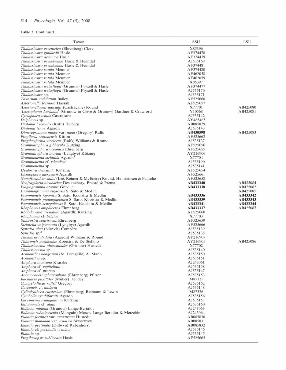
Taxon SSU LSU
Thalassiosira eccentrica (Ehrenberg) Cleve X85396Thalassiosira guillardii Hasle AF374478Thalassiosira oceanica Hasle AF374479Thalassiosira pseudonana Hasle & Heimdal AJ535169Thalassiosira pseudonana Hasle & Heimdal AF374481Thalassiosira rotula Meunier AF374480Thalassiosira rotula Meunier AF462058Thalassiosira rotula Meunier AF462059Thalassiosira rotula Meunier X85397Thalassiosira weissflogii (Grunow) Fryxell & Hasle AF374477Thalassiosira weissflogii (Grunow) Fryxell & Hasle AJ535170Thalassiosira sp. AJ535171Toxarium undulatum Bailey AF525668Asterionella formosa Hassall AF525657Asterionellopsis glacialis (Castracane) Round X77701 AB425080Asteroplanus karianus1 (Grunow in Cleve & Grunow) Gardner & Crawford Y10568 AB425081Cyclophora tenuis Castracane AJ535142Delphineis sp. AY485465Diatoma hyemalis (Roth) Heiberg AB085829Diatoma tenue Agardh AJ535143Dimeregramma minor var. nana (Gregory) Ralfs AB430598 AB425083Fragilaria crotonensis Kitton AF525662Fragilariforma virescens (Ralfs) Williams & Round AJ535137Grammatophora gibberula Kutzing AF525656Grammatophora oceanica Ehrenberg AF525655Grammatophora marina (Lyngbye) Kutzing AY216906Grammonema striatula Agardh1 X77704Grammonema cf. islandica1 AJ535190Grammonema sp.1 AJ535141Hyalosira delicatula Kutzing AF525654Licmophora juergensii Agardh AF525661Nanofrustulum shiloi (Lee, Reimer & McEnery) Round, Hallsteinsen & Paasche AF525658Neofragilaria nicobarica Desikachary, Prasad & Prema AB433340 AB425084Plagiogramma atomus Greville AB433338 AB425082Psammogramma vigoensis S. Sato & Medlin AB425085Psammoneis japonica S. Sato, Kooistra & Medlin AB433336 AB433342Psammoneis pseudojaponica S. Sato, Kooistra & Medlin AB433339 AB433343Psammoneis senegalensis S. Sato, Kooistra & Medlin AB433341 AB433344Rhaphoneis amphiceros Ehrenberg AB433337 AB425087Rhabdonema arcuatum (Agardh) Kutzing AF525660Rhaphoneis cf. belgica X77703Staurosira construens Ehrenberg AF525659Striatella unipunctata (Lyngbye) Agardh AF525666Synedra ulna (Nitzsch) Compere AJ535139Synedra sp.2 AJ535138Tabularia tabulata (Agardh) Williams & Round AY216907Talaroneis posidoniae Kooistra & De Stefano AY216905 AB425086Thalassionema nitzschioides (Grunow) Hustedt X77702Thalassionema sp. AJ535140Achnanthes bongrainii (M. Peragallo) A. Mann AJ535150Achnanthes sp. AJ535151Amphora montana Krasske AJ243061Amphora cf. capitellata AJ535158Amphora cf. proteus AJ535147Anomoeoneis sphaerophora (Ehrenberg) Pfitzer AJ535153Bacillaria paxillifer (Muller) Hendey M87325Campylodiscus ralfsii Gregory AJ535162Cocconeis cf. molesta AJ535148Cylindrotheca closterium (Ehrenberg) Reimann & Lewin M87326Cymbella cymbiformis Agardh AJ535156Encyonema triangulatum Kutzing AJ535157Entomoneis cf. alata AJ535160Eolimna minima (Grunow) Lange-Bertalot AJ243063Eolimna subminuscula (Manguin) Moser, Lange-Bertalot & Metzeltin AJ243064Eunotia formica var. sumatrana Hustedt AB085830Eunotia monodon var. asiatica Skvortzow AB085831Eunotia pectinalis (Dillwyn) Rabenhorst AB085832Eunotia cf. pectinalis f. minor AJ535146Eunotia sp. AJ535145Fragilariopsis sublineata Hasle AF525665
Table 2. Continued
514 Phycologia, Vol. 47 (5), 2008

sites was 0. 5068, and among-site rate heterogeneity was
described by a gamma distribution with a shape parameter
of 0.5931.
Phylogenies were estimated using neighbour joining (NJ),
maximum parsimony (MP), maximum likelihood (ML) and
Bayesian inference (BI).
NJ: For the SSU and the partial LSU rDNA, NJ trees
were inferred with PAUP* 4.0b10 (Swofford 2002) using
NJ of likewise constrained pairwise ML distances using the
model specified by Modeltest 3.7 (above). Nodal support
was estimated using NJ bootstrap analyses using the same
settings (1000 replicates).
MP: Only the partial LSU rDNA data set was analysed
with MP analysis. The MP topology was obtained with an
exhaustive search in PAUP* 4.0b10. Branch-and-bound
search with simple addition sequence option was used for
1000 bootstrap replications.
ML: Because of large data set of SSU rDNA, ML analysis
was performed using the relatively fast program RAxML-
VI-HPC, v2.2.3 (Stamatakis et al. 2005) with the GTRMIX
model. The analyses were performed 100 times to find the
topology receiving the best likelihood using a distinct
random starting MP tree and the rapid hill-climbing
algorithm. Bootstrap values were obtained by 100 replica-
tions with GTRCAT model. For partial LSU rDNA, an
exhaustive search was performed using PAUP* 4.0b10 with
the model selected by Modeltest 3.7 (above). Branch-and-
bound search was used for 1000 bootstrap replications.
Taxon SSU LSU
Gomphonema parvulum Kutzing AJ243062Gomphonema pseudoaugur Lange-Bertalot AB085833Lyrella atlantica (Schmidt) D. G. Mann AJ544659Navicula cryptocephala var. veneta (Kutzing) Grunow AJ297724Navicula diserta Hustedt AJ535159Navicula pelliculosa (Brebisson ex Kutzing) Hilse AJ544657Nitzschia apiculata (Gregory) Grunow M87334Nitzschia frustulum (Kutzing) Grunow AJ535164Pinnularia cf. interrupta AJ544658Pinnularia sp. AJ535154Phaeodactylum tricornutum Bohlin AJ269501Planothidium lanceolatum (Brebisson ex Kutzing) Round & Bukhtiyarova AJ535189Pleurosigma sp. AF525664Pseudogomphonema sp. AF525663Pseudogomphonema sp. AJ535152Pseudo-nitzschia multiseries (Hasle) Hasle U18241Pseudo-nitzschia pungens (Grunow ex Cleve) Hasle U18240Rossia sp. AJ535144Sellaphora capitata Mann & McDonald AJ535155Sellaphora pupula (Kutzing) Mereschkowsky AJ544645Sellaphora pupula (Kutzing) Mereschkowsky AJ544651Sellaphora pupula (Kutzing) Mereschkowsky AJ544647Sellaphora pupula (Kutzing) Mereschkowsky AJ544648Sellaphora pupula (Kutzing) Mereschkowsky AJ544649Sellaphora pupula (Kutzing) Mereschkowsky AJ544650Sellaphora pupula (Kutzing) Mereschkowsky AJ544652Sellaphora pupula (Kutzing) Mereschkowsky AJ544653Sellaphora pupula (Kutzing) Mereschkowsky AJ544654Sellaphora laevissima (Kutzing) D. G. Mann AJ544655Sellaphora laevissima (Kutzing) D. G. Mann AJ544656Surirella fastuosa var. cuneata (Schmidt) H. Peragallo & M. Peragallo AJ535161Thalassiosira antarctica Comber AF374482Undatella sp. AJ535163Bolidomonas mediterranea Guillou & Chretiennot-Dinet AF123596Bolidomonas pacifica Guillou & Chretiennot-Dinet AF123595Bolidomonas pacifica Guillou & Chretiennot-Dinet AF167153Bolidomonas pacifica Guillou & Chretiennot-Dinet AF167154Bolidomonas pacifica Guillou & Chretiennot-Dinet AF167155Bolidomonas pacifica Guillou & Chretiennot-Dinet AF167156Bolidomonas pacifica Guillou & Chretiennot-Dinet AF167157Convoluta convoluta diatom endosymbiont AY345013Peridinium foliaceum endosymbiont Y10567Peridinium balticum endosymbiont Y10566Uncultured diatom AY180014Uncultured diatom AY180015Uncultured diatom AY180016Uncultured diatom AY180020Uncultured eukaryote AY082977Uncultured eukaryote AY082992Uncultured marine diatom AF290085
1 Name change since deposit.2 Likely a new genus collected from a marine habitat (Medlin et al. 2008a).
Table 2. Continued
Sato et al.: A new araphid diatom genus Psammoneis 515

BI: For SSU and partial LSU rDNA, the Message
Passing Interface version of MrBayes 3.1.2 (Huelsenbeck &
Ronquist 2001; Ronquist & Huelsenbeck 2003; Altekar et
al. 2004) was used for BI with the GTR + I + G model to
estimate the posterior probability distribution using Me-
tropolis-coupled Markov chain Monte Carlo (MCMCMC)
(Ronquist & Huelsenbeck 2003). MCMCMC from a
random starting tree was used in these analyses with two
independent runs and one cold and three heated chains.
The Bayesian analyses were run for 20 million and
10 million generations for SSU and LSU data set,
respectively, with trees sampled every 100th generation.
To increase the probability of chain convergence, we
sampled trees after the standard deviation values (ASDSF)
of the two runs were below 0.01 to calculate the posterior
probabilities (i.e. after 10,200,000 and 2,000,000 genera-
tions, for SSU and partial LSU rDNA, respectively). The
remaining phylogenies were discarded as burn-in.
RESULTS
Psammoneis gen. nov. S. Sato, Kooistra & Medlin
Cellulae inter se affixis in coloniis cateniformibus rectis velfractiflexis, in aspectu cingulari rectangularibus, chloroplastisduobus in quoque cellula. Taeniae numerosae sine poris atquecircularia apertae, inter se implexae. Valvae lanceolatae velellipticae. Superficies valvae plana, limbo non profundo. Costacentralis distincta. Striae uniseriatae et interdum in quoque laterecostae alternatim dispositae. Areolae longitudinaliter elongatae.Areae pororum apicalium in extremis ambobus dispositae.Rimiportula carens.
Cells attach to form zigzag or straight chain colonies. Cellsrectangular in girdle view. Two plastids per cell. Bandsnumerous, all are plain and open hoops. Bands interlaced oneanother. Valves lanceolate to elliptic. Valve face flat withshallow mantle. Sternum distinct. Striae uniseriate, and some-times arranged alternately on the each side of sternum. Areolaelongitudinally elongated. Apical pore fields at both ends ofvalve. Rimoportula absent.
This genus can be distinguished from the other Plagio-
grammacean genera by the longitudinally elongated areolae
as well as their placement in a molecular phylogeny. The
absence of rimoportulae are also seen in the other araphid
lineage, which is designated clade 2 sensu Medlin et al.
(2008a) and which includes Staurosira Mereschkowsky and
its relatives. However, Psammoneis differs from these clade
2 genera in having a straight, prominent sternum, to which
parallel striae are arranged perpendicularly. Furthermore,
the valve margin of the Psammoneis lacks any linking
structure that can usually be observed in the members of
the clade 2 genera (Medlin et al. 2008a).
TYPE SPECIES: P. japonica sp. nov. S. Sato, Kooistra &Medlin
Psammoneis japonica sp. nov. S. Sato, Kooistra & Medlin
Figs 1–2, 6–9, 15–17, 22–26, 29–33
Valvae lanceolatae vel ellipticae, 5.5–12.3 mm longae, 2.6–4.3 mmlatae, striis 30–31 per 10 mm, areolis 31.15 (6 4.51) nm latae.Ordines in atomis genericis dictis 18S et 28S rDNA propris/ae.
Figs 1–5. Living cells (LM). Scale bars 5 10 mm.Fig. 1. Psammoneis japonica, strain s0305, showing sequentialplastid division.Fig. 2. P. japonica, strain s0328.Fig. 3. P. pseudojaponica, strain s0354a.Fig. 4. P. pseudojaponica, strain s0356.Fig. 5. P. senegalensis, strain s0387.
Figs 6–11. Cleaned valves (LM). Scale bars 5 2 mm.Figs 6, 7. Psammoneis japonica, strain s0305 (Zu6/44).Fig. 8. P. japonica, strain s0328 (Zu6/45).Fig. 9. P. pseudojaponica, strain s0354a (Zu6/46).Figs 10, 11. P. senegalensis, strain s0387 (Zu6/49).
516 Phycologia, Vol. 47 (5), 2008
Descriptio sequentiae geneticae 18S rDNA AB433336 et 28SrDNA AB433342.
Valves lanceolate to elliptical. Valve length 5.5–12.3 mm, width2.6–4.3 mm. Striae 30–31 per 10 mm. Width of areola 31.15 nm(6 4.51). Nucleotide sequences of 18S (AB433336) and 28SrDNA (AB433342) distinctive.
HOLOTYPE: Zu6/44.
ISOTYPE: TNS-AL-53996.
TYPE STRAIN: s0305.
TYPE LOCALITY: Iriomote Island, Okinawa Pref., Japan.
Psammoneis pseudojaponica sp. nov. S. Sato, Kooistra
& Medlin
Figs 3, 4, 18–20, 27, 34
Valvae ellipticae 2.0–4.8 mm longae 2.3–2.8 mm latae, striis 29–32per 10 mm, areolis 13.89 (6 0.87) nm latae. Cellulae interdum informis fragilis (leniter siliceae). Ordines in atomis genericis dictis18S et 28S rDNA propris/ae. Descriptio sequentiae geneticae 18SrDNA AB433339 et 28S rDNA AB433343.
Valves elliptical. Valve length 2.0– 4.8 mm, width 2.3–2.8 mm.Striae 29–32 per 10 mm. Width of areola 13.89 nm (6 0.87).Cells can produce weakly silicified morphs. Nucleotide
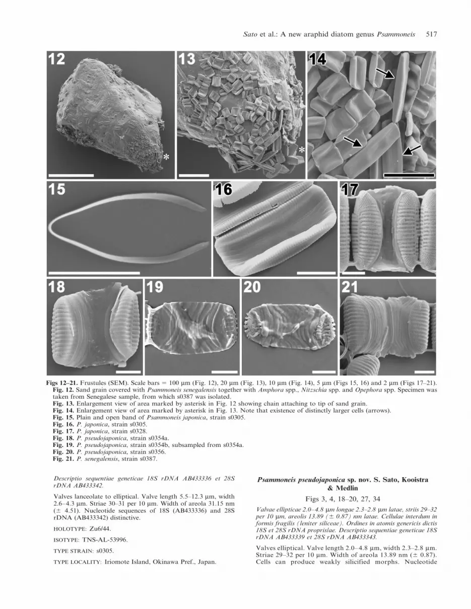
Figs 12–21. Frustules (SEM). Scale bars 5 100 mm (Fig. 12), 20 mm (Fig. 13), 10 mm (Fig. 14), 5 mm (Figs 15, 16) and 2 mm (Figs 17–21).Fig. 12. Sand grain covered with Psammoneis senegalensis together with Amphora spp., Nitzschia spp. and Opephora spp. Specimen wastaken from Senegalese sample, from which s0387 was isolated.Fig. 13. Enlargement view of area marked by asterisk in Fig. 12 showing chain attaching to tip of sand grain.Fig. 14. Enlargement view of area marked by asterisk in Fig. 13. Note that existence of distinctly larger cells (arrows).Fig. 15. Plain and open band of Psammoneis japonica, strain s0305.Fig. 16. P. japonica, strain s0305.Fig. 17. P. japonica, strain s0328.Fig. 18. P. pseudojaponica, strain s0354a.Fig. 19. P. pseudojaponica, strain s0354b, subsampled from s0354a.Fig. 20. P. pseudojaponica, strain s0356.Fig. 21. P. senegalensis, strain s0387.
Sato et al.: A new araphid diatom genus Psammoneis 517

sequences of 18S (AB433339) and 28S rDNA (AB433343)distinctive.
HOLOTYPE: Zu6/46.
ISOTYPE: TNS-AL-53997.
TYPE STRAIN: s0354.
TYPE LOCALITY: Iriomote Island, Okinawa Pref., Japan.
Psammoneis senegalensis sp. nov. S. Sato, Kooistra
& Medlin
Figs 5, 10, 11, 12–14, 21, 28, 35
Valvae ellipticae 4.3–14.0 mm longae 2.7–3.6 mm latae, striis 26per 10 mm, areolis 26.33 (6 3.42) nm latae. Ordines in atomisgenericis dictis 18S et 28S rDNA propris/ae. Descriptio sequentiaegeneticae 18S rDNA AB433341 et 28S rDNA AB433344.
Figs 22–28. External view of valves (SEM). Scale bars 5 2 mm (Figs 22, 23, 26–28) and 0.5 mm (Figs 24, 25).Fig. 22. Psammoneis japonica, strain s0305. Arrow indicates slit-like opening.Fig. 23. P. japonica, strain s0305. Tilted view.Fig. 24. P. japonica, strain s0305. Enlarged view of valve apex showing apical pore field.Fig. 25. P. japonica, strain s0305. Enlarged view of valve edge (arrow). Note simple areolae lacking occlusions.Fig. 26. P. japonica, strain s0328. Arrow indicates bar on middle of striae extending transapical direction. Note remnant of areolarocclusion near sternum.Fig. 27. P. pseudojaponica, strain s0354a. Tilted view.Fig. 28. P. senegalensis, strain s0387. Tilted view.
518 Phycologia, Vol. 47 (5), 2008

Valves elliptical. Valve length 4.3–14.0 mm, width 2.7–3.6 mm.Striae 26 per 10 mm. Width of areola 26.33 nm (6 3.42).Nucleotide sequences of 18S (AB433341) and 28S rDNA(AB433344) distinctive.
HOLOTYPE: Zu6/49.
ISOTYPE: TNS-AL-53998.
TYPE STRAIN: s0387.
TYPE LOCALITY: Goree Island, Dakar, Senegal.
Morphological data
Observations of the living material of Psammoneis japonica
showed sequential movement of the plastids during the cell
cycle; although, not all the stages have been recorded
(Fig. 1). Each cell of Psammoneis spp. contained two
plastids (Figs 1–5). Rectangular cells were attached to each
other usually at both or one end of the valve making
straight or zigzag chains (Figs 1–5, 13, 14).
Figs 29–35. Internal view of valves (SEM). Scale bars 5 2 mm (Figs 29, 30, 33–35) and 0.5 mm (Figs 31, 32).Fig. 29. Psammoneis japonica, strain s0305. Note absence of rimoportula.Fig. 30. P. japonica, strain s0305. Tilted view.Fig. 31. P. japonica, strain s0305. Enlarged view of valve apex showing apical pore field.Fig. 32. P. japonica, strain s0305. Enlarged view of valve edge. Note simple architecture of valve.Fig. 33. P. japonica, strain s0328.Fig. 34. P. pseudojaponica, strain s0354a. Note remnant of areolar occlusion near sternum.Fig. 35. P. senegalensis, strain s0387. Tilted view.
Sato et al.: A new araphid diatom genus Psammoneis 519
No significant difference in gross morphology among the
species was observed in LM except for valve outline, which
was generally a consequence of cell size difference in diatoms.
Valves were lanceolate in P. japonica (Figs 6, 7); whereas,
elliptical valves were found in P. pseudojaponica and P.
senegalensis (Figs 9–11). Parallel striae were arranged
perpendicular to the sternum (Figs 6–11). Valve dimensions
measured on each strain are summarized in Table 3. The
largest cells of P. senegalensis were observed in the natural
sample and were measured only with SEM (in Fig. 14).
Cells of P. senegalensis formed a chain and were attached
to the substratum by secreting mucilaginous substances
from the apical pore fields in their valve apex (Figs 12–14).
Cells belonging to two distinct size classes were found
(Fig. 14). The largest cells bore no remnants of auxospor-
ulation, for example, a fragment of the perizonial bands.
All copulae were plain and open (Fig. 15). Frustules of
Psammoneis spp. had numerous (c. 10) copulae (Figs 16–
21). Weakly silicified frustules were observed in P.
pseudojaponica strain s0356 and s0354b, which was
subsampled from s0354a (compare Figs 18 and 19).
Valves of Psammoneis spp. had a distinct sternum
(Figs 22, 23, 26–28). At times, a slit-like external opening
was observed at one end of the sternum in P. japonica
(Fig. 22). The valve surface of Psammoneis spp. was flat,
gently curving into a shallow valve mantle (Figs 23, 26–28).
Apical pore fields were located at the both ends of valve
(Fig. 24). Areolae were simple, slit-like perforations elon-
gating longitudinally (Fig. 25). Occlusions were difficult to
discern (see below). The valve edge was plain (Fig. 25).
Valve shape was similar across all species (Figs 26–28),
except for the weakly silicified strains of P. pseudojaponica
(Figs 19, 20). In P. pseudojaponica strain s0328, the middle
of the striae sometimes had short bars extending transapi-
cally across the areolae around the valve mantle (Fig. 26).
This valve also showed possible remnants of areolar
occlusions, for example, third and fifth striae from bottom
left near the sternum and in the fifth stria from top right in
the first two areolae next to sternum.
Internally, rimoportulae were absent in all species (Figs
29–35). Valves lacked any layered structure (Fig. 32). No
significant difference was detected in the qualitative
morphology among the species (Figs 29, 33–35). Figure 34
showed a remnant of an areolar occlusion near the sternum.
Molecular data
SEQUENCE COMPARISONS: Three species of Psammoneis, P.
japonica (strains s0305 and s0328), P. pseudojaponica
(strains s0354 and s0356) and P. senegalensis (strain
s0387) were separated by both SSU and partial LSU
rDNA sequences. Strains s0305 and s0328 of P. japonica
shared identical SSU sequences, as did strains s0354 and
s0356 of P. pseudojaponica; whereas, the three SSU and
LSU genotypes obtained were markedly distinct and
corresponded to the three species (Table 4). Most substitu-
tions in the SSU rDNA, including insertions/deletions
(indels) and compensating base pair changes (CBCs) in
stem regions, were concentrated on helices 17 and 49
(Fig. 36). Most changes did not alter secondary structure;
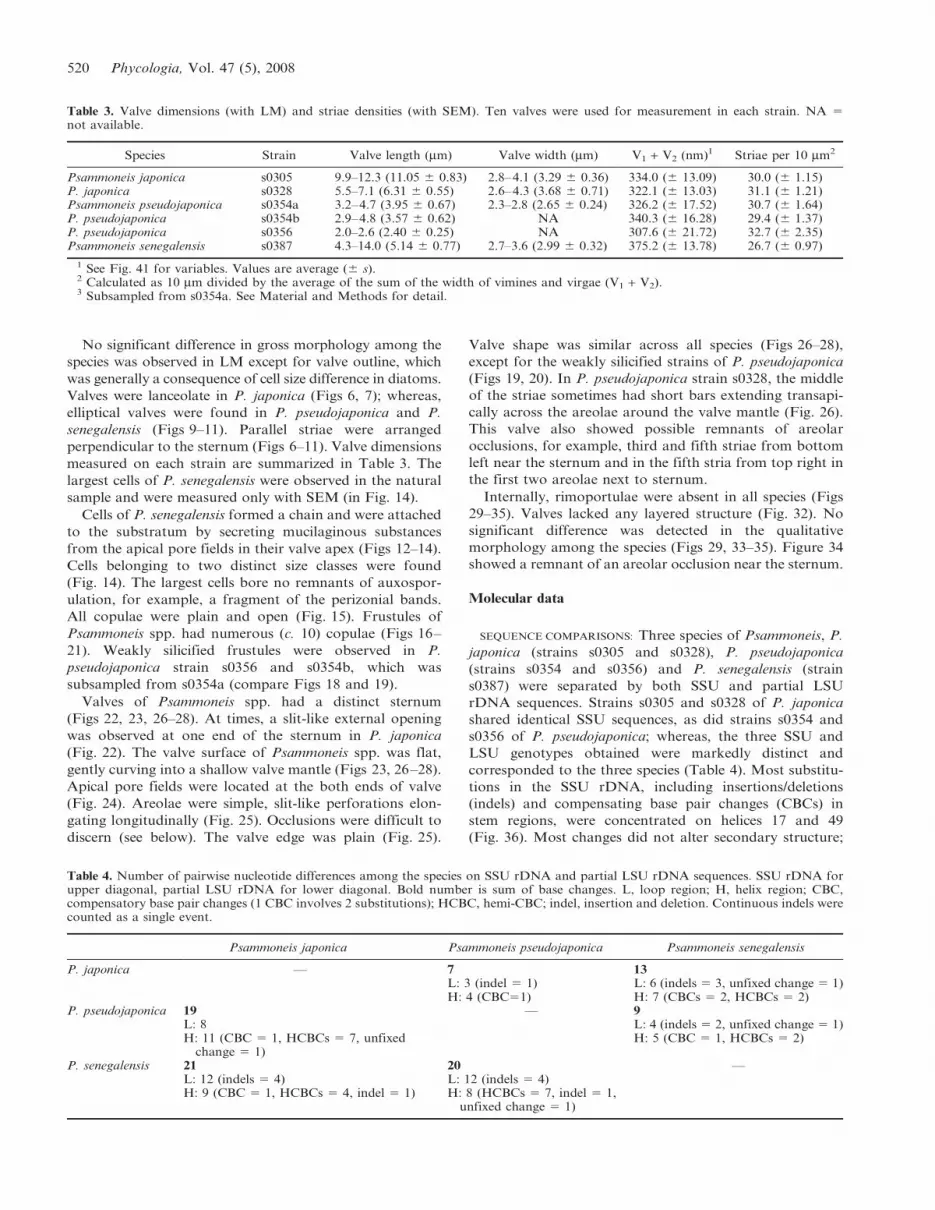
Table 3. Valve dimensions (with LM) and striae densities (with SEM). Ten valves were used for measurement in each strain. NA 5not available.
Species Strain Valve length (mm) Valve width (mm) V1 + V2 (nm)1 Striae per 10 mm2
Psammoneis japonica s0305 9.9–12.3 (11.05 6 0.83) 2.8–4.1 (3.29 6 0.36) 334.0 (6 13.09) 30.0 (6 1.15)P. japonica s0328 5.5–7.1 (6.31 6 0.55) 2.6–4.3 (3.68 6 0.71) 322.1 (6 13.03) 31.1 (6 1.21)Psammoneis pseudojaponica s0354a 3.2–4.7 (3.95 6 0.67) 2.3–2.8 (2.65 6 0.24) 326.2 (6 17.52) 30.7 (6 1.64)P. pseudojaponica s0354b 2.9–4.8 (3.57 6 0.62) NA 340.3 (6 16.28) 29.4 (6 1.37)P. pseudojaponica s0356 2.0–2.6 (2.40 6 0.25) NA 307.6 (6 21.72) 32.7 (6 2.35)Psammoneis senegalensis s0387 4.3–14.0 (5.14 6 0.77) 2.7–3.6 (2.99 6 0.32) 375.2 (6 13.78) 26.7 (6 0.97)
1 See Fig. 41 for variables. Values are average (6 s).2 Calculated as 10 mm divided by the average of the sum of the width of vimines and virgae (V1 + V2).3 Subsampled from s0354a. See Material and Methods for detail.
Table 4. Number of pairwise nucleotide differences among the species on SSU rDNA and partial LSU rDNA sequences. SSU rDNA forupper diagonal, partial LSU rDNA for lower diagonal. Bold number is sum of base changes. L, loop region; H, helix region; CBC,compensatory base pair changes (1 CBC involves 2 substitutions); HCBC, hemi-CBC; indel, insertion and deletion. Continuous indels werecounted as a single event.
Psammoneis japonica Psammoneis pseudojaponica Psammoneis senegalensis
P. japonica — 7 13L: 3 (indel 5 1) L: 6 (indels 5 3, unfixed change 5 1)H: 4 (CBC51) H: 7 (CBCs 5 2, HCBCs 5 2)
P. pseudojaponica 19 — 9L: 8 L: 4 (indels 5 2, unfixed change 5 1)H: 11 (CBC 5 1, HCBCs 5 7, unfixed
change 5 1)H: 5 (CBC 5 1, HCBCs 5 2)
P. senegalensis 21 20 —L: 12 (indels 5 4) L: 12 (indels 5 4)H: 9 (CBC 5 1, HCBCs 5 4, indel 5 1) H: 8 (HCBCs 5 7, indel 5 1,
unfixed change 5 1)
520 Phycologia, Vol. 47 (5), 2008

the same pattern was also observed in the partial LSU
rRNA (Table 4), in which the D1 segment (helixes numbers
indicated by B in Fig. 37) was more conserved than the D2
segment (helixes numbers indicated by C in Fig. 37). All
substitutions in the D1 region were in loops; whereas, one
CBC and many hemi-CBCs (changes on one side only,
sensu Coleman 2000, 2003; Marin et al. 2003) were observed
in the D2 region. Among the species, five multisite gaps,
covering in total 15 positions, were observed among the
species P. senegalensis–P. japonica and P. senegalensis–P.
pseudojaponica.
The three new species can easily be distinguished by their
secondary structural features in SSU rRNA (Fig. 36).
Psammoneis japonica has a C-G base pair in position
470–489 in helix 17 and U-G in position 1678–1705 in helix
49. For P. pseudojaponica, there is a C-G base pair in
position 470–489 in helix 17 and G-U in position 1678–
1705 in helix 49. For P. senegalensis, the base pair in
position 470–489 in helix 17 is A-U, and in position 1678–
1765 in helix 49 it is G-U.
PHYLOGENIES: A Bayesian tree inferred from SSU rDNA
sequences of 181 diatoms and seven Bolidophyceae
(Table 2) confirmed paraphyly of the araphid diatoms
within a robust clade of pennate diatoms (Fig. 38). At the
root of the pennate diatoms, the rhaphoneidacean genera,
Rhaphoneis and Delphineis, diverged, followed by the
divergence of a clade with Asterionellopsis/Asteroplanus
and the family Plagiogrammaceae (Fig. 38). The latter
clade was sister to a clade consisting, in its turn, of a
polytomy composed of a series of araphid pennate clades
and a clade with the araphid diatom Striatella unipunctata
(Lyngby) Agardh and all raphid pennate diatoms. It should
be noted that the node holding the clade of Asterionellopsis/
Asteroplanus/Plagiogrammaceae and the clade with the
remainder of pennates had no support, except in Bayesian
analysis.
Within the Plagiogrammacean clade, Talaroneis posido-
niae diverged first followed by a clade with Neofragilaria
nicobarica and Plagiogramma atomus and then by Dimer-
egramma minor var. nana. The latter was sister to a clade
with the three species of Psammoneis obtained in this study.
Of the latter three, P. senegalensis was the first to diverge
(Fig. 38). The topology within the family was resolved with
significantly high Bayesian posterior probabilities (BPPs)
but not with high bootstrap supports (BSs).
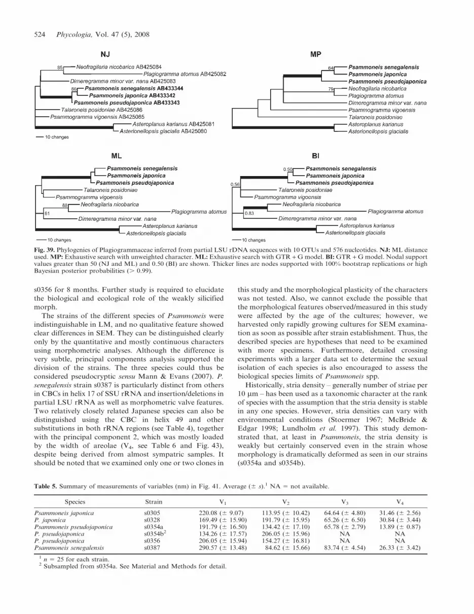
Phylogenetic analyses of partial LSU rDNA sequences
resulted in a topology among the Plagiogrammaceae
different from that obtained with SSU rDNA sequences.
Also, topologies constructed by NJ, MP, and ML analyses
and BI were incongruent (Fig. 39). The results obtained by
ML and BI analyses were substantially similar despite the
fact that the BI tree was not fully resolved. Results of all
analyses recovered the same relationships among the three
species of Psammoneis as well as the sister relationship
between Neofragilaria nicobarica and Plagiogramma atomus
(Fig. 39).
Morphometric data
MORPHOMETRIC COMPARISONS: It should be noted that no
morphological differences were detected between cleaned
(e.g. Fig. 25) and noncleaned (e.g. Fig. 16) specimens even
in high magnification (,3100,000) under SEM, suggesting
that the influence of cleaning procedure is negligible. The
measured parts as variables are illustrated in Figs 40 and 41
and values are presented in Table 5. Because of the weak
silicification of P. pseudojaponica strain s0354b and s0356,
V3 (width of vimines) and V4 (width of areolae) could not
be measured. We assume that this species is genetically
predisposed to weak silicification. The width of areolae, V4
in Fig. 41, appears the only distinguishing character of each
species. Features of striae were examined and summarised
in Table 3. Despite the high degree of diversity seen in the
width of the virgae and the length of the vimines (V1 and V2
in Fig. 41, respectively), the sum of V1 + V2, indicating a
striae density, was not markedly different (Table 3). To see
a variability pattern of the striae density in each strain, the
width of the virgae (V1) was plotted against the length of
vimines (V2) (Fig. 42). This showed that the distribution of
plots of each strain were partly overlapping, making the
feature of the striae density unsuitable as a marker for
distinguishing strains/species. The overall plots were
distributed on the linear approximation (R2 5 0.7728,
Fig. 42), suggesting that stria density is conserved in this
group of diatoms.
PRINCIPAL COMPONENTS ANALYSIS: To summarise the
variation in four recorded variables (width of virgae, striae,
vimines and areolae; see more details in Material and
Methods), a principal components analysis of their
correlation matrix was performed. The first and second
principal components (PCs) accounted for 61.9 and 25.2%
of the total (standardised) variance, respectively (87.1% of
the total variance, Table 6). The third and fourth PCs
Fig. 36. Nuclear SSU rRNA secondary structure diagram ofPsammoneis japonica showing helix 17 and 49. Helix 17: CBC tookplace at position 470 (A)–489 (U) in P. senegalensis. Helix 49: CBCtook place at position 1678 (G)–1705 (U) in P. senegalensis and P.pseudojaponica. Sequence positions in which P. japonica differedfrom the other clones are indicated by a white letter on blackbackground, and the corresponding base of the sequences is shownnext to the sequence position; open square 5 P. senegalensis,shaded square 5 P. pseudojaponica. Base pairing is indicated asfollows: standard canonical pairs by hyphen (C-G, G-C, A-U, U-A); wobble G-U pairs by circle.
Sato et al.: A new araphid diatom genus Psammoneis 521

accounted for 12.9% of the variance. Inclusion of PCs
beyond the second did not contribute substantially to
resolve the different strains. Therefore, only the first two
PCs were included in further analyses.
Component loadings and variance explained for the PCs
are shown in Table 6. V1 describing the width of virgae
(Fig. 41) revealed the heaviest loading on the first PC. The
length and width of vimines (V2 and V3, respectively) were
also heavily loaded on the first PC. Thus, PC1 reflects the
overall dimension of the unperforated area in our sample.
V4, the width of areolae, had the heaviest loading on the
second PC. A plot of these first two PCs for all strains of
the species is shown in Fig. 43. Whereas the plots of two
strains of P. japonica were partly mixed, each species was
distinguished in the PCA plot with no overlap (Fig. 43).
Because the cultures were always harvested when they were
rapidly growing, it is unlikely that silicification differences
played a role in these species differences.
Fig. 37. D1/D2 region of nuclear LSU rRNA secondary structure diagram of Psammoneis japonica. Sequence positions in which P. japonicadiffered from the other clones are indicated by a white letter on black background and the corresponding base of the sequences is shownnext to the sequence position; open square 5 P. senegalensis, shaded square 5 P. pseudojaponica. Base pairing is indicated as follows:standard canonical pairs by hyphen (C-G, G-C, A-U, U-A); wobble G-U pairs by circle. Tertiary interaction is connected by line.
522 Phycologia, Vol. 47 (5), 2008

DISCUSSION
Three species of Psammoneis were described in the present
study based on morphological and SSU and LSU rDNA.
These species are indistinguishable under LM, but molec-
ular and morphometric analysis of ultrastructural features
can separate the taxa. The considerable rDNA sequence
differences among the three species encountered amongst
the strains of Psammoneis (7–13 in SSU and 19–21 in
partial LSU) are surprising given the subtle morphometric
differences among the genus. These differences bear
comparison with those reported in previous studies on
intraspecific and even interspecific differences in diatoms
(see Table 7) supporting the species-level differentiation in
this genus.
Strain-specific characters were detected only in the strain
s0328, which had bars on the middle of the striae around
the valve mantle that extended transapically (indicated by
an arrow in Fig. 26). This structure was not found in s0305,
belonging to the same species, P. japonica, and likely
represents a strain-specific difference. Weakly silicified
frustules were found only in P. pseudojaponica. Notably,
both normal and weakly silicified morphs, observed in
s0354a and b strains, respectively, were derived from a
single cell of P. pseudojaponica (see Material and Methods).
Phenotypic plasticity was not tested for both morphs.
Long-term culture has often been observed to cause
deformities in diatom cell walls (Jaworski et al. 1988;
Round 1993; Estes & Dute 1994). Although the weakly
silicified morph was produced after slightly longer cultiva-
tion compared to the other strains in this study (Table 1), it
might not be the primary factor for induction because there
is no substantial difference in the cultivated period as
follows: normal morph; s0305, s0328, s0354a and s0387 for
c. 3, 6, 7 and 3 months, respectively, after the establishment
of the culture, whereas weakly silicified morph; s0354b and
Fig. 38. Molecular phylogeny of araphid pennate diatoms inferred from 18S rDNA sequence using 1686 aligned positions. The tree shownresulted from Bayesian inference using a GTR + I + G model. Outgroup bolidomonads and centric diatoms were excluded, and a cladecomprising raphid diatoms is collapsed into triangle for clarity. Nodal support values greater than 50 (NJ and ML) and 0.50 (BI) are shown.Nodes with strong supports (bootstrap support . 90 in NJ and ML, and posterior probability . 0.95 in BI) are shown as thick lines. 1Namechange since deposit; 2likely a new genus collected from a marine habitat (Medlin et al. 2008a).
Sato et al.: A new araphid diatom genus Psammoneis 523
s0356 for 8 months. Further study is required to elucidate
the biological and ecological role of the weakly silicified
morph.
The strains of the different species of Psammoneis were
indistinguishable in LM, and no qualitative feature showed
clear differences in SEM. They can be distinguished clearly
only by the quantitative and mostly continuous characters
using morphometric analyses. Although the difference is
very subtle, principal components analysis supported the
division of the strains. The three species could thus be
considered pseudocryptic sensu Mann & Evans (2007). P.
senegalensis strain s0387 is particularly distinct from others
in CBCs in helix 17 of SSU rRNA and insertion/deletions in
partial LSU rRNA as well as morphometric valve features.
Two relatively closely related Japanese species can also be
distinguished using the CBC in helix 49 and other
substitutions in both rRNA regions (see Table 4), together
with the principal component 2, which was mostly loaded
by the width of areolae (V4, see Table 6 and Fig. 43),
despite being derived from almost sympatric samples. It
should be noted that we examined only one or two clones in
this study and the morphological plasticity of the characters
was not tested. Also, we cannot exclude the possible that
the morphological features observed/measured in this study
were affected by the age of the cultures; however, we
harvested only rapidly growing cultures for SEM examina-
tion as soon as possible after strain establishment. Thus, the
described species are hypotheses that need to be examined
with more specimens. Furthermore, detailed crossing
experiments with a larger data set to determine the sexual
isolation of each species is also encouraged to assess the
biological species limits of Psammoneis spp.
Historically, stria density – generally number of striae per
10 mm – has been used as a taxonomic character at the rank
of species with the assumption that the stria density is stable
in any one species. However, stria densities can vary with
environmental conditions (Stoermer 1967; McBride &
Edgar 1998; Lundholm et al. 1997). This study demon-
strated that, at least in Psammoneis, the stria density is
weakly but certainly conserved even in the strain whose
morphology is dramatically deformed as seen in our strains
(s0354a and s0354b).
Fig. 39. Phylogenies of Plagiogrammaceae inferred from partial LSU rDNA sequences with 10 OTUs and 576 nucleotides. NJ: ML distanceused. MP: Exhaustive search with unweighted character. ML: Exhaustive search with GTR + G model. BI: GTR + G model. Nodal supportvalues greater than 50 (NJ and ML) and 0.50 (BI) are shown. Thicker lines are nodes supported with 100% bootstrap replications or highBayesian posterior probabilities (. 0.99).
Table 5. Summary of measurements of variables (nm) in Fig. 41. Average (6 s).1 NA 5 not available.
Species Strain V1 V2 V3 V4
Psammoneis japonica s0305 220.08 (6 9.07) 113.95 (6 10.42) 64.64 (6 4.80) 31.46 (6 2.56)P. japonica s0328 169.49 (6 15.90) 191.79 (6 15.95) 65.26 (6 6.50) 30.84 (6 3.44)Psammoneis pseudojaponica s0354a 191.79 (6 16.50) 134.42 (6 17.10) 65.78 (6 2.79) 13.89 (6 0.87)P. pseudojaponica s0354b2 134.26 (6 17.57) 206.05 (6 15.96) NA NAP. pseudojaponica s0356 206.05 (6 15.94) 154.27 (6 16.81) NA NAPsammoneis senegalensis s0387 290.57 (6 13.48) 84.62 (6 15.66) 83.74 (6 4.54) 26.33 (6 3.42)
1 n 5 25 for each strain.2 Subsampled from s0354a. See Material and Methods for detail.
524 Phycologia, Vol. 47 (5), 2008

THE FAMILY PLAGIOGRAMMACEAE: Given these morpho-
logical and molecular results, we propose that the newly
described genus Psammoneis is a member of the family
Plagiogrammaceae. All other Plagiogrammacean diatoms
have prominent, large areolae, which can easily be viewed
in LM (Round et al. 1990; Sato et al. 2008a). SEM shows
that the areolae of all members of the family are occluded
by a velum (rotae, cribrum or volae). Such prominent
structures, however, were not observed in Psammoneis,
although the existence of a delicate occlusion was suggested
by the possible siliceous remnants (Figs 26, 34). It is likely
that the areolar occlusions of Psammoneis are so delicate or
organic that most of them are lost during the cleaning
process. The areolar structure observed in Psammoneis is
morphologically atypical in the family Plagiogrammaceae.
However, morphological variability of areolar occlusions
have been reported in other closely related diatoms in
pennates, for example, in Staurosira and Punctastriata
(Williams & Round 1986; Medlin et al. 2008a), and even
within single genera Licmophora Agardh (Honeywill 1998),
Opephora (Sabbe & Vyverman 1995) and Tabularia
(Kutzing) Williams & Round (Williams & Round 1986;
Snoeijs & Kuylenstierna 1991; Snoeijs 1992). Medlin et al.
(2008a), in demonstrating the paraphyly of Staurosira,
Staurosirella, Punctastriata, Pseudostauropsis, suggested
that a loss of external valve occlusions would result in the
latter two genera; whereas, the loss of internal valve
occlusions formed the former two genera. They document-
ed fossil taxa containing both the external valve occlusions
of Staurosira and Staurosirella and the internal valve
occlusions of Punctastriata (Medlin et al. 2008a).
The absence of a rimoportula is also typical for this
family. This organelle was probably secondarily lost
because the Rhaphoneidaceae, Asterionellopsis, Asteropla-
nus and the clade with the remainder of the araphid
pennates almost all possess rimoportulae. In araphid
diatoms, the absence of rimoportulae can also be seen in
a clade that comprises Nanofrustulum Round, Hallsteinsen
& Paasche, Opephora Petit, Pseudostaurosira Williams &
Round, Pseudostaurosiropsis Morales, Punctastriata Wil-
liams et Round, Staurosira and Staurosirella Williams &
Round (Medlin et al. 2008a); however, this clade is only
distantly related to Psammoneis in the 18S rDNA
phylogeny (Fig. 38).
The recent observations on frustule fine-structures
(Kooistra et al. 2004; Sato et al. 2008a; this study) and
Figs 40, 41. Variables used for the morphometric analyses.Fig. 40. External valve surface of Psammoneis japonica, strains0305, showing measured part.Fig. 41. Enlargement view of part encircled by line in Fig. 40 atmiddle of valve. V1, width of virgae; V2, length of vimines; V3,width of vimines; V4, width of areolae. See Material andMethods for detail.
Fig. 42. Scatter plot of the width of virgae (V1) against the length ofvimines (V2). Linear approximation was shown as line. R2 5 0.7728.
Table 6. Results of principal components (PC) analysis on variablesmeasured based on Fig. 41 for four strains, s0305, s0328, s0354a,and s0387.
PC1 PC2 PC3 PC4
Component loadings
V1 0.6097035 20.08013043 0.2183755 20.7577288V2 20.5861640 0.02557928 20.5169237 20.6233357V3 0.5333162 0.09045965 20.8209203 0.1829774V4 20.0157263 20.99234161 20.1057914 0.0617980
Percent of total variance explained61.9 25.2 10.6 2.3
Fig. 43. Scatter plot on the first two principal component axis ofthe morphometric data set. Species outliers are connected by lines.
Sato et al.: A new araphid diatom genus Psammoneis 525

the addition of Psammoneis to the family alter the
circumscription of the family Plagiogrammaceae as follows:
Family Plagiogrammaceae De Toni, emend. S. Sato,
Kooistra & Medlin
Elongated valves possess apical pore fields and parallel rows ofstriae, oriented perpendicular to the apical axis; most generapossess a sternum along the apical axis; areolae occluded;absence of rimoportula; marine habitat.
GENERA GROUPING INTO THIS FAMILY ARE: Dimeregramma,
Glyphodesmis, Neofragilaria, Plagiogramma, Psammo-
gramma, Psammoneis, Talaroneis.
GENERAL PHYLOGENETIC FINDINGS ON SSU RDNA TOPOLOGY
OF DIATOMS: As previous phylogenetic studies using SSU
rDNA have already shown, the current taxonomic system
of the araphid diatoms does not represent their phyloge-
netic history (e.g. Medlin et al. 2000, 2008a, b; Kooistra et
al. 2003, 2004; Medlin & Kaczmarska 2004; Alverson et al.
2006; Sims et al. 2006; Sorhannus 2007). For example, the
planktonic fragilariacean diatoms Asterionellopsis/Astero-
planus are distantly related to the nominate genus Fragilaria
Lyngbye. The clade of Asterionellopsis/Asteroplanus formed
a ‘basal araphid’ clade of pennate diatoms with the
Rhaphoneidaceae (only Rhaphoneis and Delphineis had
been used in the past as representatives of the family) and
one Plagiogrammaceae (Talaroneis had been used), and the
rest of ‘core araphid’ pennate diatoms and the raphid
diatoms separated after the divergence of the basal araphid
diatoms (e.g. Kooistra et al. 2004).
In the present study, however, inclusion of additional
sequences into the basal araphid diatoms has changed the
topology, particularly around the root of pennate diatoms.
Now there are three araphid clades. The first one is that of
the Rhaphoneidaceae (Rhaphoneis and Delphineis), which
are the first divergence at the base of the pennate diatoms;
although, the low support of the clade (BPP 0.73 and BSs ,
50) following the divergence of rhaphoneidacean clade
might indicate instability of this relationship. The Plagio-
grammaceae and Asterionellopsis/Asteroplanus then di-
verged from the remaining he clade of the core araphids
sister to the raphid diatoms.
SEXUALITY: In the field material of P. senegalensis, cells
belonging to two distinct size ranges were found. The
absence of cells belonging to the intermediate range might
indicate that auxosporulation occurred; although, no
remnants of the auxospore perizonial band were found.
Therefore, it is also possible that the larger cells were not
formed after sexual reproduction but through vegetative
initial cell formation or vegetative cell size enlargement, as
reported in an araphid diatom Grammatophora marina
(Lyngbye) Kutzing (Sato et al. 2008b). Sexual reproduction
of members in the basal araphid diatoms, including the
Plagiogrammaceae, has never been reported. An initial cell
was observed in Plagiogramma staurophorum (Gregory)
Heiberg by Hasle et al. (1983, fig. 99), but they mentioned no
detail of sexuality in the species. Chepurnov & Mann (2004)
reported that they have grown Rhaphoneis amphiceros
(Ehrenberg) Ehrenberg in culture but have never succeeded
in inducing sexual reproduction, either in monoclonal or in
mixed cultures, even in small-celled clones.
ACKNOWLEDGEMENTS
The authors are grateful to Richard M. Crawford for
correction of the manuscript and discussion, Stephan
Frickenhaus for establishing parallel processing for Bayes-
ian analyses, Paul A. Fryxell for providing the Latin
diagnoses, Friedel Hinz for technical help for LM and SEM
and Alberto Amato for kindly providing us the alignment
of LSU rDNA of Pseudo-nitzschia. We also thank two
anonymous reviewers and the associate editor Koen Sabbe
for their valuable comments and suggestions. This study
was supported by DAAD for doctoral research fellowship
to Shinya Sato.
REFERENCES
ALTEKAR G., DWARKADAS S., HUELSENBECK J.P. & RONQUIST F.2004. Parallel Metropolis-coupled Markov chain Monte Carlofor Bayesian phylogenetic inference. Bioinformatics 20: 407–415.
ALVERSON A.J. & KOLNICK L. 2005. Intragenomic nucleotidepolymorphism among small subunit (18S) rDNA paralogs in thediatom genus Skeletonema (Bacillariophyta). Journal of Phycol-ogy 41: 1248–1257.
ALVERSON A.J., CANNONE J.J., GUTELL R.R. & THERIOT E.C.2006. The evolution of elongate shape in diatoms. Journal ofPhycology 42: 655–668.
AMATO A., KOOISTRA W.H.C.F., LEVIALDI GHIRON J.H., MANN
D.G., PROSCHOLD T. & MONTRESOR M. 2007. Reproductiveisolation among sympatric cryptic species in marine diatoms.Protist 158: 193–207.
ANONYMOUS. 1975. Proposals for a standardization of diatomterminology and diagnoses. Nova Hedwigia, Beiheft 53: 323–354.
Table 7. Summary of intraspecific/genotypic and interspecific/genotypic sequence differences of rDNA in diatom genera. Notethat all sequences were obtained with cloning by Alverson &Kolnick (2005); whereas, the others were sequenced directly.
Genus
Interspecific/genotypic
Intraspecific/genotypic
SSU Partial LSU SSU Partial LSU
Cyclotella 1–111 8–451,2 0–11 1–132
Psammoneis 7–13 19–21 0 0Pseudo-nitzschia — 0–303 — 0–13
Sellaphora 3–84 — 0–25 —Skeletonema . 56 2–657 1–26 (10–32)8 0–257
1 Beszteri et al. (2005).2 Beszteri et al. (2007).3 Amato et al. (2007). The data set used in the study was obtained
from the author, and the numbers of the sequence differences werecounted only on the D1/D2 region; although, the original data setcontains D1–D3 regions.
4 Evans et al. (2007).5 Behnke et al. (2004).6 Sarno et al. (2005).7 Kooistra et al. (2008). The data set used in the study was
retrieved from TreeBASE, and the numbers of the sequencedifferences were counted only on the D1/D2 region; although, theoriginal data set contains D1–D3 regions.
8 Number of intragenomic polymorphisms by Alverson &Kolnick (2005).
526 Phycologia, Vol. 47 (5), 2008

BEHNKE A., FRIEDL T., CHEPURNOV V.A. & MANN D.G. 2004.Reproductive compatibility and rDNA sequence analyses in theSellaphora pupula species complex (Bacillariophyta). Journal ofPhycology 40: 193–208.
BESZTERI B., ACS E. & MEDLIN L.K. 2005. Ribosomal DNAsequence variation among sympatric strains of the Cyclotellameneghiniana complex (Bacillariophyceae) reveals cryptic diver-sity. Protist 156: 317–333.
BESZTERI B., JOHN U. & MEDLIN L.K. 2007. An assessment ofcryptic genetic diversity within the Cyclotella meneghinianaspecies complex (Bacillariophyta) based on nuclear and plastidgenes, and amplified fragment length polymorphisms. EuropeanJournal of Phycology 42: 47–60.
CAHOON L.B. 1999. The role of benthic microalgae in neriticecosystems. Oceanography and Marine Biology. An AnnualReview 37: 47–86.
CAPESIUS I. & VAN DE PEER Y. 1997. Secondary structure of thelarge ribosomal subunit RNA of the moss Funaria hygrometrica.Journal of Plant Physiology 151: 239–241.
CHEPURNOV V.A. & MANN D.G. 2004. Auxosporulation ofLicmophora communis (Bacillariophyta) and a review of matingsystems and sexual reproduction in araphid pennate diatoms.Phycological Research 52: 1–12.
CHEPURNOV V.A., MANN D.G., SABBE K. & VYVERMAN W. 2004.Experimental studies on sexual reproduction in diatoms.International Review of Cytology 237: 91–154.
COLEMAN A.W. 2000. The significance of a coincidence betweenevolutionary landmarks found in mating affinity and a DNAsequence. Protist 151: 1–9.
COLEMAN A.W. 2003. ITS2 is a double-edged tool for eu-karyote evolutionary comparisons. TRENDS in Genetics 19:370–375.
COX E.J. & ROSS R. 1981. The striae of pennate diatoms. In:Proceedings of the 6th International Diatom Symposium on Recentand Fossil Diatoms (Ed. by R. Ross), pp. 267–278. O. Koeltz,Koenigstein, Germany.
DE TONI G.B. 1890. Sulla Navicula aponina Kuetz. e sui due generiBrachysira Kuetz. e Libellus Cleve. Atti del Rale Instituto Venetodi Scienze, Lettere ed Arti 1890: 967–971.
ELWOOD H.J., OLSEN G.J. & SOGIN M.L. 1985. The small subunitribosomal DNA gene sequences from the hypotrichous ciliatesOxytricha nova and Stylonichia pustulata. Molecular Biology andEvolution 2: 399–410.
EPPLEY R.W., HOLMES R.W. & STRICKLAND J.D.H. 1967. Sinkingrates of the marine phytoplankton measured with a fluoroch-rometer. Journal of Experimental Marine Biology and Ecology 1:191–208.
ESTES L. & DUTE R.R. 1994. Valve abnormalities in diatomclones maintained in long-term culture. Diatom Research 9:249–258.
EVANS K.M., WORTLEY A.H. & MANN D.G. 2007. An assessmentof potential diatom ‘‘barcode’’ genes (cox1, rbcL, 18S and ITSrDNA) and their effectiveness in determining relationships inSellaphora (Bacillariophyta). Protist 158: 349–364.
GUILLOU L., CHRETIENNOT-DINET M.-J., MEDLIN L.K., CLAUSTRE
H., LOISEAUX-DE GOER S. & VAULOT D. 1999. Bolidomonas: anew genus with two species belonging to a new algal class, theBolidophyceae class. nov. (Heterokonta). Journal of Phycology35: 368–381.
HALL T.A. 1999. BioEdit: a user-friendly biological sequencealignment editor and analysis program for Windows 95/98/NT.Nucleic Acids Symposium Series 41: 95–98.
HASLE G.R., VON STOSCH H.A. & SYVERTSEN E.E. 1983.Cymatosiraceae, a new diatom family. Bacillaria 6: 9–156.
HONEYWILL C. 1998. A study of British Licmophora species and adiscussion of its morphological features. Diatom Research 13:221–271.
HUELSENBECK J.P. & RONQUIST F. 2001. MRBAYES: Bayesianinference of phylogeny. Bioinformatics 17: 754–755.
HUSTEDT F. 1959. Die Kieselalgen Deutschlands, Osterreichs und derSchweiz unter Berucksichtigung der ubrigen Lander EuropasSowie der angrenzenden Meeresgebiete. Reprint 1977, KoeltzScience Publishers, Koenigstein, Germany. 845 pp.
JAWORSKI G.H.M., WISEMAN S.W. & REYNOLDS C.S. 1988.Variability in sinking rate of the freshwater diatom Asterionellaformosa: the influence of colony morphology. British Phycolog-ical Journal 23: 167–176.
KOOISTRA W., DE STEFANO M., MANN D.G., SALMA N. & MEDLIN
L.K. 2003. Phylogenetic position of Toxarium, a pennate-likelineage within centric diatoms (Bacillariophyceae). Journal ofPhycology 39: 185–197.
KOOISTRA W.H.C.F., FORLANI G., STERRENBURG F.A.S. & DE
STEFANO M. 2004. Molecular phylogeny and morphology of themarine diatom Talaroneis posidoniae gen. et sp. nov. (Bacillar-iophyta) advocate the return of the Plagiogrammaceae to thepennate diatoms. Phycologia 43: 58–67.
KOOISTRA W.H.C.F., GERSONDE R., MEDLIN L.K. & MANN D.G.2007. The origin and evolution of the diatoms: their adaptationto a planktonic existence. In: Evolution of primary producers inthe sea (Ed. by P.G. Falkowski & A.H. Knoll), pp. 207–249.Academic Press, New York.
KOOISTRA W.H.C.F., SARNO D., BALZANO S., GU H., ANDERSEN
R.A. & ZINGONE A. 2008. Global diversity and biogeographyof Skeletonema species (Bacillariophyta). Protist 159: 177–193.
LUNDHOLM N., SKOV J., POCKLINGTON R. & MOESTRUP O. 1997.Studies on the marine planktonic diatom Pseudo-nitzschia. 2.Autoecology of P. pseudodelicatissima based on isolates fromDanish coastal waters. Phycologia 36: 381–388.
MANN D.G. & EVANS K.M. 2007. Molecular genetics and theneglected art of diatomics. In: Unraveling the algae 5 the past,present and future of algal molecular systematics (Ed. by J. Brodieand J.M. Lewis), pp. 232–266. CRC Press, Boca Raton, FL.
MARIN B., PALM A., KLINGBERG M. & MELKONIAN M. 2003.Phylogeny and taxonomic revision of plastid-containing eugle-nophytes based on SSU rDNA sequence comparison andsynapomorphic signatures in the SSU rRNA secondary struc-ture. Protist 154: 99–145.
MATHEWS D.H., DISNEY M.D., CHILDS J.L., SCHROEDER S.J.,ZUKER M. & TURNER D.H. 2004. Incorporating chemicalmodification constraints into a dynamic programming algorithmfor prediction of RNA secondary structure. Proceedings of theNational Academy of Sciences of the United States of America101: 7287–7292.
MCBRIDE S.A. & EDGAR R.K. 1998. Janus cells unveiled: frustularmorphometric variability in Gomphonema angustatum. DiatomResearch 13: 293–310.
MEDLIN L.K. & KACZMARSKA I. 2004. Evolution of the diatoms:V. Morphological and cytological support for the major cladesand a taxonomic revision. Phycologia 43: 245–270.
MEDLIN L., ELWOOD H.J., STICKEL S. & SOGIN M.L. 1988. Thecharacterization of enzymatically amplified eukaryotic 16S-likerRNA coding regions. Gene 71: 491–499.
MEDLIN L.K., KOOISTRA W.H.C.F. & SCHMID A.M.-M. 2000. Areview of the evolution of the diatoms – a total approach usingmolecules, morphology and geology. In: The origin and earlyevolution of the diatoms: fossil, molecular and biogeographicalapproaches (Ed. by A. Witkowski & J. Sieminska), pp. 13–35.Szafer Institute of Botany, Polish Academy of Science, Cracow,Poland.
MEDLIN L.K., JUNG I., BAHULIKAR R., MENDGEN K., KROTH P. &KOOISTRA W.H.C.F. 2008a. Evolution of the diatoms. VI.Assessment of the new genera in the araphids using moleculardata. Nova Hedwigia 133: 81–100.
MEDLIN L.K., SATO S., MANN D.G. & KOOISTRA W.H.C.F. 2008b.Molecular evidence confirms sister relationship of Ardissonea,Climacosphenia and Toxarium within the bipolar centric diatoms.Journal of Phycology. (in press).
NAGUMO T. & KOBAYASHI H. 1990. The bleaching method forgently loosening and cleaning a single diatom frustule. Diatom 5:45–50.
POSADA D. & CRANDALL K.A. 1998. Modeltest: testing the modelof DNA substitution. Bioinformatics 14: 817–818.
R DEVELOPMENT CORE TEAM. 2005. R: A language and environ-ment for statistical computing. R Foundation for StatisticalComputing, Vienna, Austria. Available at: http://www.R-project.org.
Sato et al.: A new araphid diatom genus Psammoneis 527

RONQUIST F. & HUELSENBECK J.P. 2003. MrBayes 3: Bayesianphylogenetic inference under mixed models. Bioinformatics 19:1572–1574.
ROUND F.E. 1993. A Synedra (Bacillariophyta) clone after severalyears in culture. Nova Hedwgia, Beiheft 106: 353–359.
ROUND F.E., CRAWFORD R.M. & MANN D.G. 1990. The diatoms:biology and morphology of the genera. Cambridge UniversityPress, Cambridge, UK. 747 pp.
SABBE K. & VYVERMAN W. 1995. Taxonomy, morphology andecology of some widespread representatives of the diatom genusOpephora. European Journal of Phycology 30: 235–249.
SARNO D., KOOISTRA W.C.H.F., MEDLIN L.K., PERCOPO I. &ZINGONE A. 2005. Diversity in the genus Skeletonema (Bacillar-iophyceae). II. An assessment of the taxonomy S. costatum-likespecies, with the description of four new species. Journal ofPhycology 41: 151–176.
SATO S., WATANABE T., KOOISTRA W.H.C.F. & MEDLIN L.K.2008a. Morphology of four plagiogrammacean diatoms; Dimer-egramma minor var. nana, Neofragilaria nicobarica, Plagio-gramma atomus and Psammogramma vigoensis gen. et sp. nov.,and their phylogenetic relationship inferred from partial 28SrDNA. Phycological Research. (in press).
SATO S., MANN D.G., NAGUMO T., TANAKA J., TADANO T. &MEDLIN L.K. 2008b. Auxospore fine structure and variation inmodes of cell size changes in Grammatophora marina (Bacillar-iophyta). Phycologia 47: 12–27.
SIMONSEN R. 1972. Ideas for a more natural system of the centricdiatoms. Nova Hedwigia, Beiheft 39: 37–54.
SIMONSEN R. 1979. The diatom system: ideas on phylogeny.Bacillaria 2: 9–71.
SIMS P.A., MANN D.G. & MEDLIN L.K. 2006. Evolution of thediatoms: insights from fossil, biological and molecular data.Phycologia 45: 361–402.
SINNINGHE-DAMSTE J.S., MUYZER G., ABBAS B., RAMPEN S.W.,MASSE G., ALLARD W.G., BELT S.T., ROBERT J.-M., ROWLAND
S.J., MOLDOWAN J.M., BARBANTI S.M., FAGO F.J., DENISEVICH
P., DAHL J., TRINDADE L.A.F. & SCHOUTEN S. 2004. The rise ofthe rhizosolenoid diatoms. Science 304: 584–587.
SNOEIJS P. 1992. Studies in the Tabularia fasciculata complex.Diatom Research 7: 313–344.
SNOEIJS P. & KUYLENSTIERNA M. 1991. Two new diatom species inthe genus Tabularia from the Swedish coast. Diatom Research 6:351–365.
SORHANNUS U. 2007. A nuclear-encoded small-subunit ribosomalRNA timescale for diatom evolution. Marine Micropaleontolog65: 1–12.
STAMATAKIS A., LUDWIG T. & MEIER H. 2005. RAxML-III: a fastprogram for maximum likelihood-based inference of largephylogenetic trees. Bioinformatics 21: 456–463.
STOERMER E.F. 1967. Polymorphism in Mastogloia. Journal ofPhycology 3: 73–77.
SWOFFORD D.L. 2002. PAUP*: phylogenetic analysis usingparsimony (*and other methods). Version 4.0b10. SinauerAssociates, Sunderland, MA, USA.
SULLIVAN M.J. & CURRIN C.A. 2000. Community structure andfunctional dynamics of benthic microalgae in salt marshes. In:Concepts and controversies in tidal marsh ecology (Ed. by M.P.Weinstein & D.A. Kreeger), pp. 81–106. Kluwer AcademicPublishers, Dordrecht, Germany.
THOMPSON J.D., GIBSON T.J., PLEWNIAK F., JEANMOUGIN F. &HIGGINS D.G. 1997. The ClustalX windows interface: flexiblestrategies for multiple sequence alignment aided by qualityanalysis tools. Nucleic Acids Research 24: 4876–4882.
UNDERWOOD G.J.C. & KROMKAMP J. 1999. Primary production byphytoplankton and microphytobenthos in estuaries. Advances inEcological Research 29: 93–153.
VAN DE PEER Y., CAERS A., DE RIJK P. & DE WACHTER R. 1998.Database on the structure of small ribosomal subunit RNA.Nucleic Acids Research 26: 179–182.
WILLIAMS D.M. & ROUND F.E. 1986. Revision of the genusSynedra Ehrenb. Diatom Research 1: 313–339.
Received 18 January 2008; accepted 10 June 2008
Associate editor: Koen Sabbe
528 Phycologia, Vol. 47 (5), 2008