anatomy 101- bones. what do bones do? protect vital organs support the body allow the body to move...
TRANSCRIPT
Anatomy 101- Bones

What do bones do?
• Protect vital organs
• Support the body
• Allow the body to move through muscle and tendon attachment.
• Provide attachment points for ligaments
• Store important minerals such as calcium
• Produce red blood cells which transport oxygen and carbon dioxide to and from tissues.

Types of Bones
• Flat bones- examples are the scapula (shoulder blade) and sternum (breast bone)

Types of Bones
• Irregular bones- examples are the talus (ankle bone) and the vertebrae (spinal bones)

Types of Bones
• Long bones- examples are the femur (upper thigh bone) and the ulna (one of the arm bones).

Types of Bones
• Short bones- example are the tarsals (small bones of the foot)

Types of Bones
• Sesamoid bones- an example would be the patella, where the bone floats around freely.

Group Activity
• On a sheet of paper, list as many bones as you can think of in the human body.
• Take a guess. How many bones are there in the human skeleton?
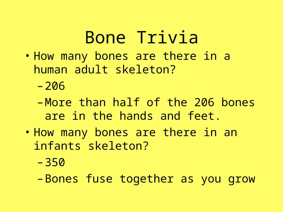
Bone Trivia• How many bones are there in a human adult
skeleton?
– 206
– More than half of the 206 bones are in the hands and feet.
• How many bones are there in an infants skeleton?
– 350
– Bones fuse together as you grow

Bones of the Skull
• The skull is made up of 28 bones.
• 22 form the framework for the head and provide protection for the brain, eyes, and ears.

Bones of the Skull
• Light Blue= Frontal Bone (front)
• Pink= Parietal Bone (upper sides)
• Orange= Temporal Bone (lower sides)
• Green= Occipital Bone (bottom)

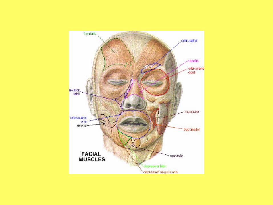
Bones of the Face
• Purple= mandible (lower jaw bone)
• Dark Blue = maxilla (upper jaw bone)

Spinal Column
• The spinal column is made up of the cervical, thoracic, and lumbar vertebrae, and the sacrum.

Cervical Vertebrae
• There are 7 cervical vertebrae labeled C1-C7.
• C1 (atlas) and C2 (axis) are responsible for the head nodding “no”.
• C3-C7 are responsible for the head nodding “yes”.
• C4 no more, C5 still alive- diaphram

Thoracic Vertebrae
• There are 12 thoracic vertebrae labeled T1-T12.
• They increase in size as they move down the spine (Ex: T1 and T2 are smaller than T10 and T11).
• All thoracic vertebrae have facets (small grooves) on the sides of their bodies for attachment of the ribs.

Lumbar Vertebrae• There are 5 lumbar vertebrae labeled
L1-L5.
• Lumbar vertebrae are most frequently involved in back pain.
• They carry the most amount of body weight.
• Are subject to the largest forces and stresses along the spine.

Sacrum and Coccyx
• The sacrum is a large triangular bone at the base of the spine.
• It connects to the L5 vertebrae and ends with the coccyx (tailbone).

Sacrum and Coccyx
• The sacrum is labeled S1.
• The coccyx (tailbone) is the final piece of the vertebral column.

More Bone Trivia
• What is the longest bone in the body?
– Femur. It is almost one quarter of the bodies total height.
• What is the smallest bone in the body?
– The stirrup deep inside the ear. It is hardly larger than a grain of rice.

Sternum and Rib Cage
• The sternum (breastbone) is a long flat bone located in the center of the chest.
• The sternum is connected to the ribs with cartilage.

Sternum and Rib Cage
• The ribcage is made up of 12 ribs.• The first 7 ribs are connected to the sternum
and are called true ribs.• The 8th, 9th, and 10th ribs attach in front to
the cartilage portion of the next rib above, and are called false ribs.
• The 11th and 12th ribs are not attached in front and are called floating ribs.

The Shoulder Complex
• The shoulder complex is made up of the clavicle (collar bone), the scapula (shoulder blade), and the humerus (upper arm bone)

Clavicle• The clavicle (a.k.a.
collar bone) is a long bone that makes up part of the shoulder girdle.
• This bone is clearly visible through the skin.
• The clavicle is the easiest bone in the body to break.

Scapula
• The scapula (a.k.a. shoulder blade) is the bone that connects the humerus and the clavicle.
• It is a flat bone and triangular in shape.

Humerus
• The humerus is a long bone in the arm that runs from the shoulder to the elbow.
• Fits between the scapula and the ulna.
• Called funny bone

Forearm Bones
• The radius is one of two bones in the forearm.
• Extends on the lateral side (outside or thumb side) of the forearm between the elbow and wrist.

Forearm Bones
• The ulna is the stronger and larger of the two forearm bones.
• Extends on the medial side (inside or pinky finger side) of the forearm between the elbow and wrist.

Wrist and Hand Bones
• The 8 bones that make up the wrist are called the carpals.
• Scaphoid
• Lunate
• Triquetrum
• Pisiform
• Trapezium
• Trapezoid
• Capitate
• Hamate


Finger Bones
• The fingers are made up of 5 metacarpals and 14 phalanges.

Pelvis (Hip Bone)
• The 3 bones of the pelvis are the illium, ischium, pubis.
• The pelvis provides the socket for the hip joint.

Bone Trivia
• What is the hardest bone in the body to break?
–Femur. Usually broken in car accidents or a fall from somewhere high.

Thigh and Leg Bones
• The femur (thigh bone) is the longest and strongest bone in the body.
• It forms part of the hip and part of the knee.

Patella
• The patella (knee cap) is a thick triangular shaped seasamoid bone.
• It sits in front of the femur and protects the front of the knee joint.

Tibia
• The tibia (or shin bone) is the larger of the two lower leg bones.
• It is the second longest bone in the body.
• Extends between the femur and the ankle on the medial side of the body.

Fibula
• The fibula extends below the tibia to the ankle on the lateral side.
• It is the skinniest of all long bones in the body.

Foot Bones
• The tarsals are a group of bones that make up the top of the foot.

Foot Bones
• 1= Calcaneus• 2= Talus• 3= Navicular• 4=Medial Cuneiform• 5= Intermediate Cuneiform• 6= Lateral Cuneiform• 7= Cuboid

Foot Bones
• The rest of the foot and toes are made up of 5 metatarsals and 14 phalanges.

Ready for muscles???

Anatomy 101- Muscles

Skeletal Muscles
• Skeletal muscle is a type of striated muscle that is attached to bones.
• Skeletal muscles are used to create movement by applying force to bones and joints.

How Many Muscles Are There In The Body?
• There are just shy of 700 skeletal muscles in the body.
• That includes about 400 that nobody cares about, except specialists

Types of Movement
• Flexion
• Extension
• Abduction
• Adduction
• Rotation– Internal rotation– External rotation
• Inversion
• Eversion
• Dorsi flexion
• Plantar flexion
• Supination
• Pronation

Bet You Didn’t Know• Too much botox injected into the
frontalis muscle leads to drooping eyebrows.
• When someone is unable to close their eyes doctors will sew in tiny gold weights into each eyelid.
• Eye muscles are the busiest muscles in the body. They move more than 100,000 times a day.

Neck Muscles
• The neck can move in 4 different ways:– Flexion
– Extension
– Lateral Flexion
– Rotation

Neck Muscles
• Sternocleidomastoid
• Muscle is responsible for tilting the head laterally, rotating the head, and pulling the back of the head downward.


Back Muscles
• The trapezius is a large superficial muscle of the back.
• The trapezius attaches at the neck, the two shoulders, and down to the 12th thoracic vertebrae.

Back Muscles
• The latissimus dorsi is a large flat muscle of the back.
• The lats cover the lumbar region of the back and the lower half of the thoracic region.

Back Muscles
• The rhomboid major muscle is a muscle on the back that connects the scapula with the vertebrae of the spinal column.

Back Muscles
• The erector spinae muscle is a group of muscles in the back.
• Supports the entire upper body.

Chest Muscles
• Pectoralis major- located on the front of the ribcage.
• Attaches to the humerus near the shoulder joint and to the sternum in the center of the chest.

Chest Muscles
• Pectoralis minor- is located underneath the pectoralis major.
• It attaches to the scapula and the middle ribs.

Abdominal Muscles• Rectus
Abdominus- (abs) are long flat muscles that run vertically on both sides of the abdomen.
• They are two parallel muscles separated by connective tissue.


Abdominal Muscles
• Internal Oblique- the middle muscle of the stomach that lays just underneath the external oblique.

Abdominal Muscles
• External Oblique- is the largest and most superficial of the three stomach muscles.

Shoulder Muscles
• Deltoid- forms the round contour of the shoulder.
• The deltoid assists in shoulder abduction, flexion, and extension.


Shoulder Muscles
• Rotator cuff muscles- four muscles responsible for stabilizing the shoulder joint as well as elevating and rotating the arm.

Shoulder Muscles
• S.I.T.S
• Supraspinatus
• Infraspinatus
• Teres Minor
• Subscapularis

Arm Muscles
• There are 4 muscles responsible for flexion of the arm.– Biceps brachii– Brachialis– Brachioradialis– Pronator teres

Arm Muscles


Arm Muscles
• There are 2 muscles responsible for extension of the arm.
– Triceps brachii
– anconeus

Arm Muscles

Forearm Muscles
• Muscles responsible for flexion of the hand:
• Also known as the Wrist Flexors:– Flexor digitorum profundus– Flexor pollicis longus– Flexor digitorum superficialis– Flexor carpi ulnaris– Palamaris longus– Flexor carpi radiali


Forearm Muscles
• Muscles responsible for extension of the hand:
• Also known as the Wrist Extensors:– Extensor indicis
– Extensor pollicis longus
– Extensor pollicis brevis
– Extensor carpi radialis longus
– Extensor carpi radialis brevis
– Extensor carpi ulnaris
– Extensor digiti minimi
– Extensor digitorum


Muscle Trivia
• What is the largest muscle in the body?– Gluteus Maximus
• What is the smallest muscle in the body?– Stapedius- 1/20th of an inch long– Found in the inner ear
• What is the longest muscle in the body?– Sartorius– Runs from the hip to the knee

Muscles of the Gluteal Region
• Gluteus Maximus• Gluteus Medius• Gluteus Minimus• Gluteal muscles are
responsible for external rotation and extension of the hip joint.


Hip and Thigh Muscles
• Also named by the region and the function of the muscle
- Hip Flexors- muscles acting on the anterior hip/thigh, responsible for hip flexion
Example: Psoas major
-Hip adductors (Groin)- muscles acting on the medial hip/thigh, responsible for hip adduction
Example: Adductor Longus, Gracilis
-Hip abductors- muscles acting on the lateral hip/thigh, responsible for hip abduction
Example: Tensor Fascia Latae, IT Band
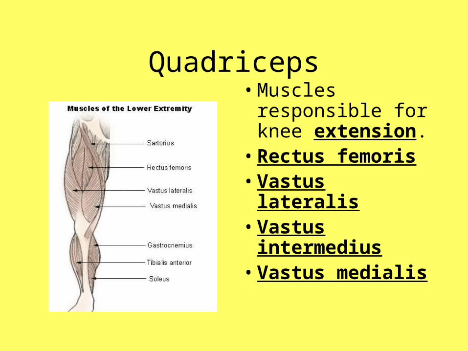
Quadriceps• Muscles
responsible for knee extension.
• Rectus femoris• Vastus lateralis• Vastus
intermedius• Vastus medialis


Hamstrings
• Muscles responsible for knee flexion.
• Semimembranosus
• Semitendinosus
• Biceps Femoris


Muscles of the Anterior/Lateral Leg
• Tibialis Anterior– Dorsiflexion and
inversion of ankle
• Peroneous Longus– Eversion and
plantarflexion of ankle

Muscles of the Posterior Leg
• Gastrocnemius- superficial calf muscle
• Soleus- deep to gastrocnemius
• Tibialis Posterior- assoc. with shin splints


Quick Write
1. List as many muscle as you can that we have talked about so far.
2. Describe an experience you have had with a muscle.– Example: muscle strain, muscle cramps,
muscle soreness, etc.