upper airway assessment in orthodontics: a review
TRANSCRIPT

Upper airway assessment in Orthodontics: a review
Erwin Rojas1, Rodrigo Corvalán1, Eduardo Messen2, Paulo Sandoval3.
DOI: 10.22592/o2017n30a5
Abstract
Introduction: Upper airway assessment is particularly important in the daily work of orthodontists, pediatric dentists, ENT specialists, speech therapists, etc., because of its close connection with the development of craniofacial structures and with other pathologies such as Obstructive Sleep Apnea Syndrome (OSAS). Objective: To review the limits, functions and anomalies of different areas that make up the upper airway, to provide information about specific methods most widely used by specialists for their evaluation, and to describe and evaluate the information level and diagnostic accuracy of methods such as lateral cephalometric analysis and cone beam CT. Materials and Methods: The search was conducted on PubMed, with the following keywords: upper airway and CBCT, upper airway and assessment, evaluation and upper airway; upper airway and orthodontics. Only studies less than 5 years old were selected. A total of 46 papers were read and finally, 38 studios were selected. Conclusions: It is essential to know upper airway assessment methods, which include a clinical examination, a radiographic evaluation and CBCT. These will indicate possible functional changes that could interfere with treatment. Keywords: upper airway, CBCT, clinical assessment, orthodontics. 1 Postgraduate student, Orthodontics and Maxillofacial Orthopedics, Universidad de
La Frontera, Temuco, Chile.
2 Orthodontics and Maxillofacial Orthopedics Specialist. Clinical Professor,
Universidad de La Frontera, Temuco, Chile.
3 Prof. MA. Orthodontics and Maxillofacial Orthopedics Specialist, Universidad de La
Frontera, Temuco, Chile.
Received on: 06 Feb 2017 – Accepted on: 01 Jul 2017

Introduction
Upper airway assessment and its interactions with craniofacial growth and
development have been of interest to ENT specialists, laryngologists, speech
therapists, pediatricians and dentists. Upper airway obstruction tends to alter
breathing, which can have a significant impact on the normal development of
craniofacial structures, causing deficiencies in transverse maxillary growth, as well
as cause the rotational growth of the back of the mandible. These anomalies require
early detection, and it has been shown that the early diagnosis and treatment of
obstructive sleep apnea-hypopnea syndrome allows for an almost complete
normalization of dentofacial morphology(1).
The methods described to assess the airway include: nasal endoscopy,
rhinomanometry, acoustic rhinomanometry(2,3), cephalometry, computed
tomography (CT), magnetic resonance imaging (MRI) and cone-beam computed
tomography (CBCT). When trying to find a connection between subjective and
objective nasal obstruction, researchers found an association only for allergic rhinitis.
However, they have not found a subjective connection with any other alteration such
as asthma, septal deviation, enlarged adenoids or Obstructive Sleep Apnea
Syndrome (OSAS), so it is important to assess the airway beyond the symptoms
described by the patient(4,5).
In orthodontics, upper airway alterations must always be evaluated clinically at the
start of the treatment, as well as through lateral cephalograms or CBCT.
Cephalometry provides a 2D reconstruction of three-dimensional structures, so the
information provided is limited. The CBCT shows 3D structures, the construction of
projections on different planes, and allows us to measure the volume of different
structures, so it provides a large amount of diagnostic information. However, it is not
a routine examination and involves a larger radiation dose.
The aim of this review is to analyze and present the available evidence, from 2008
to date, on upper airway assessment from an interdisciplinary perspective towards
orthodontics.

Development
1) Anatomy and physiology of the upper airway
Breathing allows for a simple exchange of gases between venous blood and
atmospheric air; the air gives part of its oxygen to the blood, and the blood releases
carbonic acid and water vapor into the air. Through the reciprocal effect of this gas
exchange, venous blood recovers all its chemical and biological qualities, and
becomes arterial blood.
The essential organs of the respiratory system are the lungs, located on either side
of the thorax, on each side of the heart, and the great vessels. To reach the lungs,
atmospheric air follows a long passage, the airway, which comprises the nasal cavity
and incidentally the mouth. Then it includes successively the pharynx, larynx,
trachea, and bronchi. The upper airway is formed by the nasal cavity and the
pharynx.
a) Nasal cavity
The normal airway starts, from the functional perspective, in the nostrils. The nasal
cavity includes the nose, the nasal cavities, and extend to the back with the
nasopharynx. In addition to breathing, it has very specific functions, such as smell
and phonation.
A deviated septum, a narrow nasal cavity, and turbinate hypertrophy are some of the
signs that cause mouth breathing and OSAS. In allergic rhinitis, which is also related
to upper airway obstruction, the nasal mucous membrane swell with dust particles,
pollen or even cold, also affecting the eyes and nose and causing a decrease in air
flow(6).
b) Oral cavity
The mouth includes the lips, at the front, up to the oropharyngeal isthmus, at the
back. Functionally, it is a very important structure as the food enters the digestive

system through it, and it is an essential organ for mastication, phonation, taste,
deglutition and breathing. It is formed by the maxillary, palatal and mandible bones,
the tongue, lips and cheeks, and the oropharynx at the back. The palate is the roof
of the oral cavity and the floor of the nasal cavity. It has a bone base, the hard palate,
and fibromuscular tissue, the soft palate.
The tongue is a single, muscular medium-size, symmetrical, highly mobile organ,
located in the curvatures within the dental arches, filling in this space when the mouth
is closed. The tongue is not only the essential organ of taste and deglutition, but it
also plays an important role in mastication, swallowing, suction and the articulation
of sounds. As its muscle tone decreases during sleep, the tongue can block the
upper airway. Jointly with the loss of muscle tone of the pharyngeal walls and the
soft palate, it contributes to the collapse of the airway, one of the main causes of
obstructive sleep apnea syndrome.
The palatine tonsils are two masses of lymphoid tissue located on the side walls of
the oral pharynx, between the palatoglossus and palatopharyngeus muscles. Each
tonsil is covered by mucosa, and its inner side projects into the pharynx. The tonsils
reach their maximum size in the early years of childhood and decrease in size
considerably after puberty.
c) Pharynx
The pharynx is a tube-like structure formed by muscles and membranes (Figure 1).
It measures approximately 12-14 cm and is divided into three parts: nasopharynx,
oropharynx and laringopharynx.
The nasopharynx is the upper part of the respiratory system. It is located behind the
nasal cavity and on the soft palate. The nasopharynx is lined with a mucous
membrane of respiratory epithelium, and becomes transitional epithelium in the
oropharynx. In the roof submucosa there is a collection of lymphoid tissue called
pharyngeal tonsil (adenoids), which, when large in size, is the main obstruction to
the passage of air through the nasopharynx.

The oropharynx extends from the second to the fourth vertebra and opens into the
oral cavity through an isthmus. The upper end is the soft palate, and the lower end
is the lingual side of the epiglottis. The tongue is the main blocking element in the
oropharynx, due to the general decrease in tone of the genioglossus muscle, which
contracts to move the tongue forward during inspiration, and in this way, acts as a
pharyngeal dilator.
The laringopharynx joins the oropharynx at the pharyngoepiglottic fold and hyoid
bone, and continues up to the sixth vertebra. It is behind the opening in the larynx.
The outer wall is formed by the thyroid cartilage and the thyroid membrane.
Fig. 1
2) Most commonly used otorhinolaryngology tests for upper airway
assessment
a) Rhinomanometry
It aims to objectively evaluate nasal obstruction. There are different types of
rhinomanometry (RMM), active anterior RMM being the one most frequently used.
This evaluates nasal airflow in inspiration and expiration by detecting potential
obstructions and/or resistance. This can be done with a face mask or by placing an
olive in each nostril; the first device has the advantage of not deforming the nostrils,
reducing the possibility of leakage. However, it requires full patient cooperation and

cannot be implemented if there is total occlusion of one nostril or a septal perforation.
After placing the mask, airflows are measured with the rhinomanometer, and the
data are analyzed computationally and then graphs are designed in pressure/volume
curves. After a first measurement in basal conditions, the recording is repeated
under the effect of a topical vasoconstrictor, which will differentiate mechanical
obstructions (which do not vary with the vasoconstrictor), vasomotor obstructions
(which fully improve with the vasoconstrictor) and mixed obstructions (which improve
partially with the vasoconstrictor). In general, any cause of obstruction with bone,
cartilage or tissues, with little edema or whose vasoconstriction cannot be affected,
as well as inflammatory etiologies, with edema and tissue susceptible to
vasoconstriction, will yield vasomotor curves. The pathology which best represents
mechanical obstruction is the deviated septum, and the main vasomotor obstruction
is inferior turbinate hypertrophy.
b) Acoustic rhinometry
It is the study of the geometry of the nasal cavity. It is based on the analysis of sound
reflection and provides a calculation of cross-sectional areas of the nasal cavity and
of certain nasal volumes. This is done by generating an audible sound in the nostril
with an adapter, taking care not to deform the nasal vestibule. The sound wave
penetrates the cavities and is reflected on the different nasal structures or their
irregularities. Incident wave signals are measured and reflected according to time,
which makes it possible to determine the distance, from the nostril, where there is a
change in acoustic impedance. The most interesting data are the “minimum cross-
sectional areas 1 and 2 (MCA1 and MCA2)”. MCA1 corresponds anatomically to the
area at the nasal valve level (bounded by the caudal margin of the upper lateral
cartilage and the nasal septum), which has the greatest resistance in the normal
nose. MCA2 corresponds to the area at the level of the head of the inferior turbinate.
As in active anterior RMM, the study can be performed before and after applying a
vasoconstrictor for the same purpose and with a similar interpretation of the results.

c) Nasopharyngolaryngoscopy
This test evaluates the anatomy of the upper airway, as well as the soft palate, the
movement of the vocal cords and the process of deglutition. It is performed with a
flexible fiberscope which is inserted through the nasal cavities to observe both
pharynx and larynx. The patient is usually awake, and topical lidocaine is applied on
the nostrils and, as the case may be, vasoconstrictor (oxymetazoline). During the
test, the patient may be asked to talk, cough or swallow, depending on what is being
evaluated. The following anatomical elements should be evaluated: deviations of the
nasal septum, size of inferior turbinates, presence and size of the adenoid tissue,
quantity and quality of nasal secretion, size of palatine tonsils and of the base of the
tongue and its relationship with the oropharyngeal cavity, abduction of the vocal
cords, subglottic diameter, and presence of masses or pathological deformities at
any of these levels(7).
d) Functional Nasal Permeability (PeNaF):
It is a clinical examination that assesses the independent functional nasal
permeability of each cavity. The performance is recorded as negative (-) when the
patient maintains nasal breathing for six inspirations at rest, and positive (+) when
the patient fails to maintain it for six inspirations. A study validated in Chile
recommends orthodontists implement this simple examination to rule out a possible
nasal obstruction. If this is not the case, they should request an objective
assessment to check the increase in nasal resistance(8).
3) Clinical examination
Physical assessment includes facial morphology, skeletal jaw relationships,
functional assessment of nostrils, the size and function of the tongue and the
anatomy of the soft palate, uvula and tonsils.
Regarding facial morphology, Class II patterns due to mandibular retrusion have
smaller upper airway volumes, which is usually associated with adenoid

hypertrophy(9,10,11), which includes lip hypotonia, with a very short upper lip and a
thick and everted bottom lip.
a) Functional assessment of nostrils (Duran V.)
To do this we observe nostril response to intense inspiration, paying special attention
to the degree of collapse during the maneuver. This is the classification obtained:
Value 0: Dilated nostrils both at rest and in deep inspiration
Value 1: Narrowed nostrils at rest, without functional collapse
Value 2: Functional partial unilateral collapse
Value 3: Functional total unilateral or bilateral partial collapse
Value 4: Functional partial collapse of one nostril and total collapse of the other one
Value 5: Total functional collapse in both nostrils(9)
Fig. 2
b) Intraoral evaluation
The tonsils are assessed according to the degree of obstruction of the oropharynx,
on a scale of 1 to 4. This is a reliable clinical evaluation method (Fig. 3). In Grade 1,
the tonsils are within their cavity; in Grade 2, they do not exceed the midline between
the uvula and the anterior pillar of the soft palate; in Grade 3, they go over the midline
between the uvula and the anterior pillar; and in Grade 4, the tonsils are less than
4 mm between them. A degree of obstruction Grades 3 or 4 represents a decrease
in airway permeability(12).
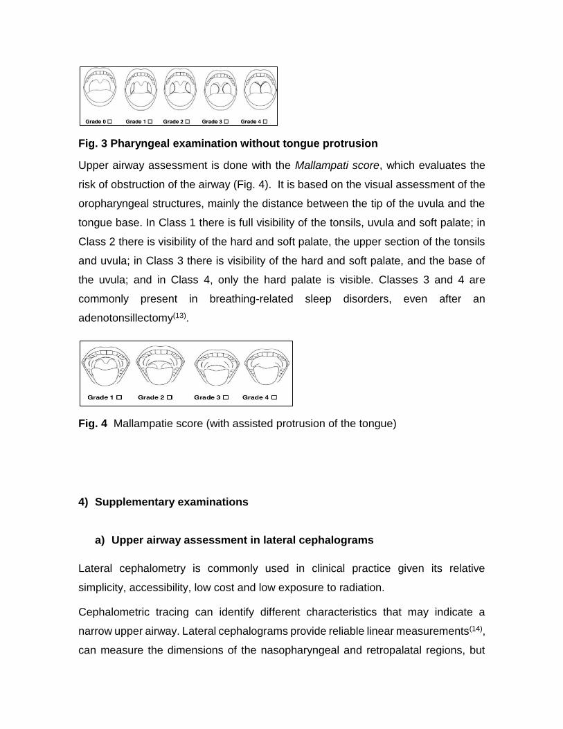
Fig. 3 Pharyngeal examination without tongue protrusion
Upper airway assessment is done with the Mallampati score, which evaluates the
risk of obstruction of the airway (Fig. 4). It is based on the visual assessment of the
oropharyngeal structures, mainly the distance between the tip of the uvula and the
tongue base. In Class 1 there is full visibility of the tonsils, uvula and soft palate; in
Class 2 there is visibility of the hard and soft palate, the upper section of the tonsils
and uvula; in Class 3 there is visibility of the hard and soft palate, and the base of
the uvula; and in Class 4, only the hard palate is visible. Classes 3 and 4 are
commonly present in breathing-related sleep disorders, even after an
adenotonsillectomy(13).
Fig. 4 Mallampatie score (with assisted protrusion of the tongue)
4) Supplementary examinations
a) Upper airway assessment in lateral cephalograms
Lateral cephalometry is commonly used in clinical practice given its relative
simplicity, accessibility, low cost and low exposure to radiation.
Cephalometric tracing can identify different characteristics that may indicate a
narrow upper airway. Lateral cephalograms provide reliable linear measurements(14),
can measure the dimensions of the nasopharyngeal and retropalatal regions, but

have not proven to be reliable to measure the airway in the back of the tongue(15).
However, this is a highly reproducible test using the natural position of the patient’s
head, provided that it is run correctly(16). A 2013 meta-analysis on craniofacial
morphology found a significant relationship between a reduced upper airway at the
pharynx level (mainly adenoid hypertrophy) and pediatric sleep disorders(17).
Figure 5 shows the points and lines most commonly used to assess upper airway
obstruction, as well as the reference airway diameters and the diameters for
individuals with OSAS in Table 1(18).
In 1984, McNamara stated that there is obstruction of the airway if there is a
distance lower than 5 mm. Between the nearest points of the posterior wall of the
nasopharynx and of the soft palate (Figure 5B). In 1979, Fujioka et al. described
the adenoidal-nasopharyngeal ratio (AN ratio), which relates the length of the line
perpendicular to the sphenoid bone (A) by the thickest portion of the adenoids with
the distance between the posterior nasal spine and the anterior edge of the
sphenobasioccipital synchondrosis (N). An AN< 0.8 is considered normal and an
AN > 0.8 is considered enlarged (Fig. 5D). In addition, Feres, Murilo et al. in 2012
found that both parameters had good reproducibility and a variability which was not
clinically significant.

Fig.5
One of the most common reasons for upper airway obstruction is hypertrophic
adenoids, defined as a collection of lymphoid tissues in the posterior wall of the
nasopharynx which increase in volume as the immune activity increases. Before
planning an orthodontic treatment, this area is usually observed in the lateral
cephalometry, therefore, lateral teleradiography is used as a profitable and
reproducible diagnostic method which is easy to interpret when assessing the size
of the adenoids. With the advent of CBCT, 3D images were made available to
orthodontists. Studies have tried to correlate lateral cephalograms and CBCT in
relation to the linear volumes of the airway, but no clear consensus has been
established.
Adenoids develop progressively, with their
highest growth achieved between 4 and 5
years of age, followed by another peak
OSA Reference
Mean SD Mean SD Difference
A B
C D
Table 1: Reference airway diameters and diameters for individuals with OSAS

between 9 and 10, and then the size
decreases progressively until 14 to 15
years of age(19).
A study was conducted to assess whether
adenoidal ratio on lateral cephalograms can
be used to estimate airway volumes, using
CBCT as the validation method. They
concluded that the lateral cephalogram can
provide some information about the
nasopharyngeal space, particularly in
patients over 15. This is due to the stability
reached by the tissue at this age; however,
it cannot be used as a diagnostic procedure
to determine the volume of the total airway,
but rather as an assessment tool to
determine the need for a more
comprehensive ENT examination(20). Fiber endoscopy is the most successful
diagnostic test for adenoid hypertrophy. Of the radiological examinations, only
cephalometry has proven useful for the study of the facial skeleton(21).
b) Upper airway assessment using CBCT
Since its creation in 1990, the CBCT has been well adopted for diagnosis in the
maxillofacial area, as it provides a 3D representation of the structures at a low cost
and with an effective radiation dose which is much lower when compared to
computed tomography (CT)(10,22). Although CBCT is less effective than CT in tissue
discrimination, it defines the boundaries between tissues and empty spaces with
high spatial resolution(21). In addition, several studies have shown that it is accurate
and reliable for upper airway assessment(22,23, 1, 10 ,14).
Volumetric reconstructions that may be obtained from CBCTs help clinicians make
a correct diagnosis and indicate a better treatment plan for some pathologies of the
maxillofacial area, especially those related to the airway(24). Three-dimensional
(mm) (mm)
tu-
ad3 11.10 3.20 9.10 1.85 2.00
pm-
ad2 21.44 3.97 23.15 3.23 -1.71
pm-
ad1 22.82 3.50 25.69 2.90 -2.87
ve-
pve 5.16 2.34 10.09 2.80 -4.93
uv-
puv 9.51 3.09 11.79 2.77 -2.28
rl-
prl 10.17 3.54 9.30 3.06 0.86
va-
pva 17.55 5.23 18.59 2.27 -1.04

images and volumes can be obtained from two-dimensional slices with CBCT after
a complex process, which involves the use of especially designed computer
programs(25). For the volumetric reconstruction and visualization of the upper airway,
these software programs must allow us to find the correct location of the boundaries
of the pharynx and nasal cavity (segmentation) through a process that can be
manual, automatic or semi-automatic. Three commercial software programs for the
study of the airway were analyzed. They were found to have reliable reproducible
and accurate results of linear measurements, but they lost accuracy when
calculating the volume of the airway. This could be due to the automatic
segmentation of the nasal cavity, the nasopharynx and oropharynx. Weissheimer et
al. in 2012(31) had the same results when analyzing six commercial software
programs.
Besides the differences found in the use of different programs, when assessing the
volume of the upper airway we should consider the differences in the anatomical
boundaries of the nasopharynx and oropharynx, reported in different studies. The
upper boundary of the nasopharynx and the lower boundary of the oropharynx have
the greatest variability, followed by the boundary between these two structures. The
oral cavity and the nasal cavity do not show variability in their boundaries(22,1).
Alsufyani et al., in their 2012 review(1), suggest that the protocol proposed by EI and
Palomo in 2010 should be replicated in other studies. The nasopharynx, on the
sagittal plane, was delimited from the last slice before the nasal septum joins the
posterior wall of the pharynx, on the sagittal plane; the lower boundary was
determined by the palatal plane. The upper boundary of the oropharynx is the
nasopharynx, and the lower one is the parallel to the plane that goes through the
lowest anterior point of the second cervical vertebra (Figure 6). These authors
suggest using as lower boundary the section between the oropharynx to C2, and not
a lower sector, such as C3, C4, or the epiglottis, because in this way we can use
smaller windows and reduce the radiation dose patients receive. The segmentation
was performed manually and 30-cm windows (FOV) were used, though a 13-cm
window is acceptable to display the oropharynx or the nasopharynx and the nasal
cavity.

Fig. 6
We must also consider the head position and the position of the patient when the
CBCT is taken to obtain accurate and repeatable upper airway measurements and
volumes. The position of the hyoid bone and tongue, and the dimension of the airway
would be highly reproducible using the natural position of the head when taking
lateral cephalograms(16). In addition, it has been found that individuals would be
approximately 40% more affected by the width of the airway in an upright position(15).
Solow et al.(18) determined that in an upright position or by increasing the cervical
skull angle, there is an increase in upper airway diameters. Alsufyani(1) states that
images must be obtained with the patient in a sitting position so as not to affect
airway diameter.
Two systematic literature reviews(1,22) concluded that although major progress has
been made in the capture and management of CBCT images, there is no optimized
evidence-based protocol to obtain images to analyze the upper airway. Several
obstacles must still be overcome, such as the influence of the position of the tongue,
mandible morphology, the impact of the respiratory phase and the definition of the
anatomical boundaries of the upper airway, as well as the lack of consistency in the
configuration of the equipment and in how images and volumetric reconstructions
are obtained.

McCrillis et al., in a 2009 review, indicate a lack of studies to map the characteristics
shown in the upper airway CBCT with clinical results according to the treatment
modality, so that the various modalities are based on predictable outcomes(27).
5) Airway and skeletal patterns
A study conducted in New Delhi compared the reliability of lateral cephalograms and
computed tomographies to assess airways. They compared three skeletal patterns
determined by the different values of the ANB angle, and related their linear values
taken from the cephalometries to volumetric values delivered by CT, and concluded
that the skeletal pattern had a strong association with pharyngeal volume and its
linear dimensions. They also found sex dimorphism in relation to normal values.
They also noted that the S-shaped soft palate can be considered high-risk for sleep
apnea compared to the leaf shape, which is more common(28).
In contrast, Dalmau et al., found in Spain no statistically significant differences that
correlate airway with skeletal patterns or facial biotypes. However, they did find
correlations, for example, for upper airway measurement. Class II subjects
presented higher measurements than Class I and III patients. Additionally, the
measurements for Class III were higher for lower areas(29). This agrees with the
recent results of Lucas Castro-Silva et al., in Brazil, who also found a positive
correlation of higher values of pharyngeal airway for Class III patients(30). A new
study by El and Palomo found that oropharyngeal airway volumes were lower in
Class II patients compared to Class I and Class III patients. They also state that the
mandibular position with respect to the skull base has a strong impact on
oropharyngeal volume.
All these results are conclusive in the sense that airway volume and shape vary in
patients with different maxillomandibular relationships in the sagittal direction(20).
Conclusions

Upper airway assessment is essential in orthodontics because of the close
interrelation between the correct respiratory function and the normal development of
craniofacial structures.
The clinical examination, especially using Mallampatie’s score, can give us an
indication of the health of our patient’s airway, which, together with the initial
radiographic examination, shows us the need for further studies to rule out, for
example, sleep disorders, which, with the right treatment, can restore our patients’
health and greatly improve their quality of life.
The cephalometric study of the nasopharynx is essential, as it can be easily
assessed and it is a determining factor for the development of pediatric sleep
disorders. The assessment of adenoid tissue with lateral cephalogram is a
reproducible and easy-access exam in our daily work. However, it will never yield an
accurate diagnosis of the airway volume, but rather it will indicate the need for a
referral to an ENT specialist so that more comprehensive tests are run.
CBCT is becoming commonplace in dental practice. It provides 3D images and axial
slices of the airway at low cost and with an acceptable radiation dose for a specific
image quality. However, there are still difficulties to overcome to be able to
extrapolate the results of the scientific evidence on the upper airway to our
population, given the large number of factors that have not been properly
protocolized. Additionally, CBCT is not essential for airway diagnosis, as its
volumetric calculations are static and change significantly depending on patient
position, respiratory phase, etc. Hence the importance of the medical history, and of
tools such as the sleep questionnaire, both for pediatric and adult patients, and not
just the subjective evaluation of a diagnostic image.
References
1. Alsufyani NA, Flores-Mir C, Major PW. Three-dimensional segmentation of the upper airway using cone beam CT: a systematic review. Dentomaxillofac Radiol. 2012; 41 (4): 276–284.
2. Compadretti G, Tasca I, Bonetti G. Nasal airway measurements in children treated by rapid maxillary expansion. Am J Rhinology. 2006; 20 (4): 385–393

3. De Felippe NLO, Bhushan N, Da Silveira AC, Viana G, Smith B. Long-term effects of orthodontic therapy on the maxillary dental arch and nasal cavity. Am J Orthod Dentofacial Orthop. 2009; 136 (4): 490.e1–e8.
4. Isaac A., Major M, Witmans M, Alrajhi Y, Flores-Mir C, Major P, Alsufyani N, Korayem M, El-Hakim H. Correlations between acoustic rhinometry, subjective symptoms, and endoscopic findings in symptomatic children with nasal obstruction. JAMA Otolaryngol Head Neck Surg. 2015; 141 (6): 550-5.
5. Togeiro SM, Chaves CM, Palombini L, Tufik S. Evaluation of the upper airway in obstructive sleep apnoea. In Indian J. Med. Res. 2010; 131 (2): 230–235.
6. McNamara, J. A.: A method of cephalometric evaluation. Am J Orthod. 1984; 86 (6): 449–469.
7. Martín J, Cassade S. Evaluación Funcional de la Vía aérea. Neumol Pediatr. 2012; 7 (2): 61-66.
8. Villanueva P, Zepeda A, Lizana M, Fernández M, Palomino H. Efectividad en la detección de la permeabilidad nasal funcional: Presentación de un método clínico. Rev Ch Ort. 2008; 25 (2): 98-106.
9. Claudino LV, Matoos CT, Ruellas AC, Sant' Anna EF. Pharyngeal airway characterization in adolescents related to facial skeletal pattern: a preliminary study. Am J Orthod Dentofacial Orthop. 2013: 143 (6): 799–809.
10. El H, Palomo MJ. Measuring the airway in 3 dimensions: a reliability and accuracy study. Am J Orthod Dentofacial Orthop. 2010; 137 (4): S50.e1-9.
11. El H, Palomo MJ. Airway volume for different dentofacial skeletal patterns. Am J Orthod Dentofacial Orthop. 2011; 139 (6): e511-21.
12. Pirilä-Parkkinen K, Löppönen H, Nieminen P, Tolonen U, Pääkkö E, Pirttiniemi P: Validity of upper airway assessment in children: a clinical, cephalometric, and MRI study. Angle Orthod 2011; 81 (3): 433–439.
13. Kim JH, Guilleminault C. The nasomaxillary complex, the mandible, and sleep-disordered breathing. Sleep Breath 2011; 15 (2): 185–193.
14. Vizzotto MB, Liedke GS, Delamare EL, Silveira HD, Dutra V. A comparative study of lateral cephalograms and cone-beam computed tomographic images in upper airway assessment. Eur J Orthod 2012: 34 (3): 390–393.
15. Battagel JM. Postural variation in oropharyngeal dimensions in subjects with sleep disordered breathing: a cephalometric study. The European Journal of Orthodontics 2012: 24 (3): 263–276.
16. Malkoc S, Usumez S, Nur M, Donaghy CE. Reproducibility of airway dimensions and tongue and hyoid positions on lateral cephalograms. American Journal of Orthodontics and Dentofacial Orthopedics 2005: 128 (4): 513–516.
17. Katyal V, Pamula Y, Martin AJ, Daynes CN, Kennedy JD, Sampson JW. Craniofacial and upper airway morphology in pediatric sleep-disordered breathing: Systematic review and meta-analysis. Am J Orthod Dentofacial Orthop. 2013; 143 (1): 20-30.e3.
18. Solow B, Skov S, Ovesen J, Norup PW, Wildschiødtz G. Airway dimensions and head posture in obstructive sleep apnoea. Eur J Orthod. 1996; 18 (6): 571–579.

19. Fujioka M, Young L W, Girdany BR. Radiographic evaluation of adenoidal size in children: adenoidal-nasopharyngeal ratio. AJR Am J Roentgenol. 1979; 133 (3): 401–404.
20. Xin-Feng A, Gang-Li. Comparative analysis of upper airway volume with lateral cephalograms and cone-beam computed tomography. Am J Orthod Dentofacial Orthop. 2015; 147 (2): 197-204.
21. Matiñó E, Manel J, Rubert A, Bellet L. Trastornos de respiración obstructivos del sueño en los niños. Acta otorrinolaringológica española. 2010; 61 (1): 40-44.
22. Guijarro MR, Swennen GRJ. Cone-beam computerized tomography imaging and analysis of the upper airway: a systematic review of the literature. International Journal of Oral and Maxillofacial Surgery. 2011; 40 (11): 1227–1237.
23. Ghoneima A, Kula K. Accuracy and reliability of cone-beam computed tomography for airway volume analysis. In Eur J Orthod. 2013; 35 (2): 256–261.
24. Van Vlijmen OJ, Kuijpers MA, Bergé SJ, Schols JG, Matal TJ, Breuning H, Kuijpers-Jagtman AM. Evidence supporting the use of cone-beam computed tomography in orthodontics. In J Am Dent Assoc. 2012; 143 (3): 241–252.
25. Celenk M, Farrell ML, Eren H, Kumar K, Singh G, Lozanoff S. Upper airway detection and visualization from cone beam image slices. In J Xray Sci Technol.2010; 18 (2): 121–135.
26. Echarri P. tratamiento ortodóncico y tratamiento ortopédico/funcional de primera fase. Echarri P. Tratamiento ortodóncico y ortopédico de primera fase en dentición mixta. 2nd ed. Barcelona: Nexus médica, 2009. p55-59.
27. McCrillis JM, Haskell J, Haskell BS, Brammer M, Chenin D, Scarfe WC, Farman AG. Obstructive Sleep Apnea and the Use of Cone Beam Computed Tomography in Airway Imaging: A Review. In Seminars in Orthodontics. 2009; 15 (1): 63–69.
28. Kaur S, Rai M, Kaur M. Comparison of reliability of lateral cephalogram and computed tomography for assessment of airway space. Niger J Clin Pract. 2014; 17 (5): 629-36.
29. Dalmau E, Zamora N, Tarazona B. A comparative study of the pharyngeal airway space, measured with cone beam computed tomography, between patients with different craniofacial morphologies. Journal of Craneo-Maxilofacial surgery. 2015; 43 (8): 1438–1446.
30. Castro SL, Silva M. Cone-beam evaluation of pharyngeal airway space in class I, II, and III patients. Oral Surg Oral Med Oral Pathol Oral Radiol 2015; 120 (6): 679-683.
31. Weissheimer A, Menezes LM, Sameshima GT, Enciso R, Pham J, Grauer D. Imaging software accuracy for 3-dimensional analysis of the upper airway. In Am J Orthod Dentofacial Orthop. 2012; 142 (6): 801–813.
32. El H, Palomo JM. An airway study of different maxillary and mandibular sagittal positions. Eur J Orthod. 2013; 35 (2): 262–270.
33. Feres MFN, De Sousa H, Francisco SM, Pignatari SSN. Reliability of radiographic parameters in adenoid evaluation. In Braz J Otorhinolaryngol. 2012; 78 (4): 80–90.

34. Lenza MG, Lenza MM, Dalstra M, Melsen B, Cattaneo PM. An analysis of different approaches to the assessment of upper airway morphology: a CBCT study. In Orthod Craniofac Res. 2012; 13 (2): 96–105.
35. Lorenzoni DC, Bolognese AM, Garib DG, Guedes FR, Sant’Anna EF. Cone-Beam Computed Tomography and Radiographs in Dentistry: Aspects Related to Radiation Dose. International Journal of Dentistry. 2012; 18 (1): 1–10.
36. Ogawa N, Miyazaki Y, Kubota M, Huang A, Maki K. Application of cone beam CT 3D images to cephalometric analysis. Orthodontic Waves. 2010; 69 (4): 138–150.
37. Raffat A, ul Hamid W. Cephalometric assessment of patients with adenoidal faces. J Pak Med Assoc. 2009; 59 (11): 747–752.
38. Zettergren-Wijk L, Forsberg CM, Linder-Aronson S. Changes in dentofacial morphology after adeno-/tonsillectomy in young children with obstructive sleep apnoea, a 5-year follow-up study. Eur J Orthod. 2006; 28 (4): 319–326.
Paulo Sandoval: [email protected]