the upper airway in sleep: physiology of the · pdf filethe upper airway in sleep: physiology...
TRANSCRIPT
Sleep Medicine Reviews, Vol. 7, No. 1, pp 9±33, 2003
doi:10.1053/smrv.2002.0238
PHYSIOLOGICAL REVIEW
The upper airway in sleep: physiology of thepharynx
Indu Ayappa and David M. Rapoport
Division of Pulmonary and Critical Care Medicine, New York University School of Medicine
Summary The upper airway is the primary conduit for passage of air into the lungs.
Its physiology has been the subject of intensive study: both passive mechanical and
active neural in¯uences contribute to its patency and collapsibility. Different models
can be used to explain behavior of the upper airway, including the `̀ balance of forces''
(airway suction pressure during inspiration versus upper airway dilator tone) and the
Starling resistor mechanical model.
As sleep is the primary state change responsible for sleep disordered breathing(SDB) and the obstructive apnea/hypopnea syndrome (OSAHS), understanding its
effects on the upper airway is critical. These include changes in upper airway muscle
dilator activity and associated changes in mechanics and re¯ex activity of the muscles.
Currently SDB is thought to result from a combination of anatomical upper airway
predisposition and changes in neural activation mechanisms intrinsic to sleep.
Detection of SDB is based on identifying abnormal (high resistance) breaths and
events, but the clinical tools used to detect these events and an understanding of their
impact on symptoms is still evolving. Outcomes research to de®ne which events aremost important, and a better understanding of how events lead to physiologic
consequences of the syndrome, including excessive daytime somnolence (EDS), will
allow physiologic testing to objectively differentiate between `̀ normal'' subjects and
those with disease. & 2002 Elsevier Science Ltd. All rights reserved.
KEYWORDSobstructive sleep
apnea±hypopnea
syndrome, upper airway
physiology, snoring,
sleep disordered
breathing, upper airway
resistance, pharynx
INTRODUCTION
Over the past 20 years, there has been growing interest
in the role of the human upper airway during breathing,
especially during sleep. In large part, this interest has
come from the increased recognition of the entity of
Obstructive Sleep Disordered Breathing. While tech-
nically, sleep disordered breathing is a general term
for all disorders of breathing during sleep, for simpli-
city, we will use the term sleep disordered breathing
1087±0792/02/$ ± see front matter & 2002 Elsevier Science Ltd.
Correspondence should be addressed to: David M. Rapoport,
MD, Department of Medicine, New York University
Medical Center, 550 First Ave., New York, NY 10016, USA.
Tel.:�1-212-263±6407; Fax:�1-212-263±7445;
E-mail: [email protected]
(SDB) in this review to mean only obstructive syn-
dromes of abnormal upper airway resistance, i.e.
a generalization of the Obstructive Sleep Apnea/
Hypopnea Syndrome. This disorder is both frequent
in the general population [1] and has important clinical
implications, ranging from disruption of sleep with
daytime sequellae of excessive sleepiness (2) to sus-
pected long term cardiovascular consequences [3±5].
Because of this greatly increased interest in the
diagnosis of sleep disordered breathing, it has become
clear that a better understanding of normal physiology
of the upper airway, as well as re-examination of the
current de®nitions and measurement techniques for
identifying abnormal respiratory `̀ events'' during sleep
is necessary. The present review deals with these
issues.
All rights reserved.
10 I. AYAPPA AND D. M. RAPOPORT
NORMAL AIRWAY
Upper airway anatomy
The upper airway is a complicated structure that is
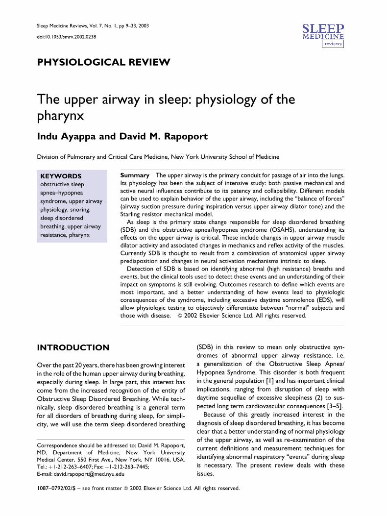
usually divided into 4 anatomical subsegments (see
Fig. 1):
Nasopharynx ± between the nares and hard palate;Velopharynx or retropalatal oropharynx ±between the hard palate and soft palate;Oropharynx ± from the soft palate to the epiglottis;Hypopharynx ± from the base of the tongue to thelarynx.
This total structure forms a passage for movement of
air from the nose to the lungs and also participates in
other physiological functions such as phonation and
deglutition [6]. The properties of the upper airway
are a compromise between these different functions,
which variably require maintenance of patency (during
breathing) or closure of the airway (as in swallowing).
There are 20 or more upper airway muscles surround-
ing the airway that actively constrict and dilate the
upper airway lumen [7, 8]. They can be classi®ed into
four groups ± muscles regulating the position of the
soft palate (alai nasi, tensor palatini, levator palatini),
tongue (genioglossus, geniohyoid, hyoglossus,
Figure 1 Anatomy of the upper airway showingthe main segments: nasopharynx, velopharynx, oro-pharynx and hypopharynx. From Anatomy andPhysiology of Upper Airway Obstruction. Samuel Kunaand John E Remmers 840±858. (From Meir H Kryger,Thomas Roth and WC Dement (Eds). Principles andPractice of Sleep Medicine, 3rd Edn. W.B. SaundersCompany; with permission.)
styloglossus), hyoid apparatus (hyoglossus, genioglos-
sus, digastric, geniohyoid, sternohyoid) and the
posterolateral pharyngeal walls (palatoglossus, pharyn-
geal constrictors). These groups of muscles interact in
a complex fashion to determine the patency of the
airway. Soft tissue structures form the walls of the
upper airway, and include the tonsils, soft palate, uvula,
tongue and lateral pharyngeal walls [9]. The main
craniofacial bony structures that determine the airway
size are the mandible [10] and the hyoid bone [11];
these presumably act by providing the anchoring
structures to which muscles and soft tissue attach.
However, it is clear that complex relationships occur
even in these `̀ ®xed'' structures, as some of these like
the hyoid bone `̀ ¯oat'' without any attachment to
other bony or cartilaginous structure. They act as
complex levers where muscle action, instead of moving
the structure, may tense some of the adjacent soft
tissues (e.g. tracheal pull).
In normal non-obese subjects, the mean minimum
cross-sectional area across multiple segments of the
upper airway has been measured using several techni-
ques: estimates vary from 320±450 mm2 (acoustic
re¯ection) [12±14], 59 mm2 (fast CT at FRC) [15],
64 mm2 (MRI) [9], 144 mm2 [16], 188 mm2 [17] and
138 mm2 [18] (conventional CT). This wide range in
size re¯ects the differences due to individual variability
but is also due to differing locations of measurement,
positional change (sitting/supine), and differences im-
posed by the choice of imaging modality (e.g. mouth
open is required for acoustic re¯ection). There is
substantial overlap in measurements between normal
subjects and those with OSAS. However, it is import-
ant to remember that most of the reported measure-
ments were made during wakefulness ± thus they
combine truly anatomical properties (such as bone
structure, fat deposition) and activation of the upper
airway dilator muscles.
The minimum caliber of the upper airway in the
wake state is primarily in the retropalatal oropha-
rynx [9, 19], which makes it a site of interest as the
potential location of collapse during sleep. The ante-
rior wall of the oropharynx is composed primarily of
the soft palate, tongue and lingual tonsils; and the
posterior wall is bounded by a muscular wall made up
of the superior, middle and inferior constrictor mus-
cles that lie in front of the cervical spine. The lateral
pharyngeal walls are a complex structure made up of
muscles (hypoglossus, styloglossus, stylohyoid, stlylo-
pharyngeus, palatoglossus, palatopharyngeus, the
pharyngeal constrictors), lymphoid tissue and pharyn-
geal mucosa. This complexity of the interactions

UPPER AIRWAY IN SLEEP 11
between thesedifferent muscles makes theoropharynx
an extremely dif®cult structure to evaluate.
Gravity/position
Since the upper airway lacks a ®xed rigid structural
support, shape and size of the airway are dependent
on the position of soft tissue structures like the soft
palate, tongue and the walls of the oropharynx. These
can be in¯uenced by gravity. In the supine position the
tongue and soft palate have been shown to move
posteriorly, reducing the oropharyngeal area [20, 21],
thereby increasing the supraglottic airway impedance
[22] and collapsibility [23]. Clinical correlation for this
anatomic and functional change is demonstrated by the
observation that snoring is much more prominent in
the supine position.
Landmarks: bony, fat, muscle, airway
Upper airway imaging techniques have also been
used to visualize the airway lumen and to de®ne the
surrounding structures. In awake subjects Schwab et al.
[19] have shown that the normal upper airway has
a longer lateral than AP dimension using MRI techni-
ques. In addition, by using fast cine CT, they also
showed that airway size stays fairly constant during
inspiration and reaches a minimum during end expi-
ration, suggesting that muscular stabilization of the
airway lumen during inspiration against the negative
intraluminal pressure is more important than actual
dilatation, as had previously been believed. According
to these authors, most respiratory related changes
(i.e. end-expiratory lossof diameter) arepredominantly
in the lateral dimension [19].
Models of behavior of the airway
In describing the dynamic behavior of the airway
during cyclic respiration, it is useful to employ various
physical and/or mathematical models to reduce this
complex structure to a more simple and understan-
dable one. The most basic approach is to treat the
airway as a rigid tube and analyze its resistance
(i.e. assume a ®xed or `̀ average'' relationship between
driving pressure and ¯ow). An extension of this
model is to consider `̀ resistance'' as varying during
the inspiratory cycle composed of a dynamic inter-
action between ¯ow and pressure. The need for
this more complex model becomes evident when
one observes that negative intrathoracic pressure
transmitted to the passive upper airway during inspira-
tion promotes a reduction in pharyngeal cross sec-
tional area [24]. According to the `̀ balance of
pressures'' concept [25, 26] the size (and thus resis-
tance) of the airway depends on the balance between
collapsing intraluminal pressures generated during
inspiration by subatmospheric pressures in the
thorax and outward contracting forces of the
upper airway dilator muscles. In this analysis, patency
of the airway depends on transmural pressure (Ptm),
which is the difference between the negative intra-
luminal pressure caused by inspiratory efforts and the
positive dilating pressure from the upper airway
musculature.
To help in understanding this `̀ dynamic collapse'' an
alternative complementary approach has been to
describe the upper airway as a collapsible tube. This
model deals better with the dynamic collapse descri-
bed above, and also helps to explain why airway caliber
may increase in association with lung in¯ation [13, 15].
In this collapsible tube model, one can examine
multiple components that in¯uence collapse of the
susceptible airway. These factors include at least the
following:
Respiratory driving pressure across the regionsusceptible to collapse ± determined by thenegative intra-thoracic inspiratory pressure andany ®xed resistances of anatomic structures suchas the nose;Intrinsic properties of the airway wall ± this iscalled the `̀ tube law'' and is determined by thesize, collapsibility and longitudinal tension on thetube;Neural input to the upper airway ± which dictatesthe behavior of the dilating/stabilizing musculature.
A more detailed discussion of these approaches
follows.
Passive static properties of the airwayStatic properties of a tube can be inferred from its
behavior during steady state ¯ow. Thus resistance has
been used to describe the upper airway, but this is
primarily because it is the simplest measure to describe
a pressure/¯ow relationship through a conduit.
However because of collapsibility and the in¯uence
of dilator muscles, the upper airway does not have
a ®xed cross-sectional area, which is a minimum
requirement for having the linear pressure/¯ow
relationship necessary to de®ne `̀ a resistance''. To
overcome this limitation of the `̀ resistance'' concept,
pharyngeal behavior has been described using either
12 I. AYAPPA AND D. M. RAPOPORT
the resistance calculated at a single ®xed ¯ow rate or
resistance at the peak ¯ow rate during a maneuver.
This approach has been used to describe the relative
properties of different segments of the upper airway
under speci®c conditions.
Static properties of the upper airway have also been
evaluated by direct measurement of the compliance of
the pharyngeal wall (slope of the volume to pressure or
cross-sectional area to pressure relationship).
However, to evaluate true compliance of this structure
one must suppress the neuromuscular in¯uences on
the upper airway, as these can ¯uctuate during the
measurement process. In order to remove these
neuromuscular in¯uences studies have been per-
formed in anesthetized and paralyzed subjects or
during sleep with the application of CPAP. The
genioglossus and other pharyngeal muscles are pre-
sumably hypotonic on the ®rst breath following the
removal of CPAP [27]. Using these techniques Isono
et al. evaluated the properties of the pharyngeal wall
plus the surrounding structures in normal subjects and
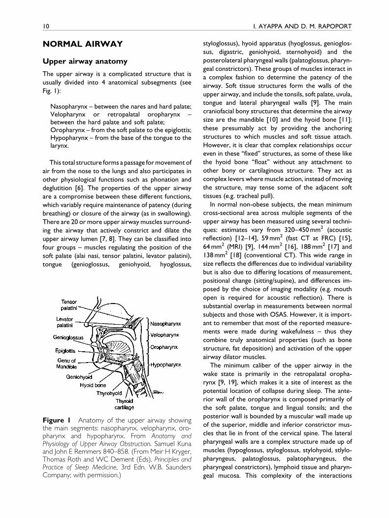
patients with obstructive sleep apnea [24, 28, 29]. The
relationship between pressure and cross-sectional
area of the veloparynx and oropharynx of the passive
airway was described as exponential; thus, the airway
is more collapsible (i.e. the derivative of area to
pressure becomes greater) as the pharyngeal airway
becomes smaller (see Fig. 2). There was a fundamental
difference between the airway of the normal subjects
Figure 2 Static pressure±area curves of the passive vefunction and curves of patients with OSAS lie below andcollapsibility. (From Isono S, Remmers JE, Tanaka A, Showith obstructive sleep apnea and in normal subjects. J Ap
and those with OSAS in that the airway was closed at
atmospheric pressure in subjects with OSAS, while
active negative pressure was required to close the
normal airway, even in the absence of upper airway
muscle activity.
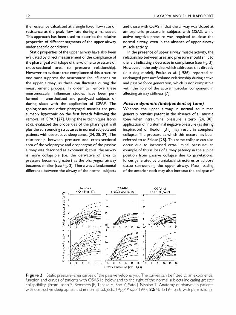
In the presence of upper airway muscle activity, the
relationship between area and pressure should shift to
the left indicating a decrease in compliance (see Fig. 3).
However, in the only data which addresses this directly
(in a dog model), Fouke et al. (1986), reported an
unchanged pressure/volume relationship during active
and passive force generation, which is not compatible
with the role of the active muscular component in
affecting airway stiffness [7].
Passive dynamic (independent of tone)Whereas the upper airway in normal adult man
generally remains patent in the absence of all muscle
tone when intraluminal pressure is zero [24, 30],
application of intraluminal negative pressure (as during
inspiration) or ¯exion [31] may result in complete
collapse. The pressure at which this occurs has been
referred to as Pclose [28]. This same collapse can also
occur due to increased extra-luminal pressure: an
example of this is loss of airway patency in the supine
position from passive collapse due to gravitational
forces generated by craniofacial structures or adipose
tissue surrounding the upper airway. Mass loading
of the anterior neck may also increase the collapse of
lopharynx. The curves can be ®tted to an exponentialto the right of the normal subjects indicating greaterY, Sato J, Nishino T. Anatomy of pharynx in patientspl Physiol 1997; 82(4): 1319±1326; with permission.)
Figure 3 Increasing the transmural pressure (like onapplication of CPAP) can passively increase the crosssectional area (see passive curve). The application ofmuscle activity shifts the curve up and to the left(active curve). (From Anatomy and Physiology of UpperAirway Obstruction. Samuel Kuna and John ERemmers 840±858. From Meir H Kryger, ThomasRoth and WC Dement (Eds). Principles and Practice ofSleep Medicine, 3rd Edn. W.B. Saunders Company;with permission.)
UPPER AIRWAY IN SLEEP 13
the passive airway. This has been shown in anesthe-
tized rabbits with mass loading to simulate excessive
adipose tissue resulted in narrowing of the pharynx
and increase in upper airway resistance [32].
Conversely, negative pressure applied around the
neck of anesthetized dogs decreased upper airway
resistance, indicating passive dilation [33].
Active (i.e. muscle dilator) contributionThe upper airway is rich in neural receptors, which
play a part in controlling baseline tonic genioglossal
EMG. Any loss of this EMG tone, as occurs at sleep
onset, probably contributes to raising pharyngeal
resistance [34]. The existence of topical receptor
mechanisms in the nasopharynx that may in¯uence
dilator muscle activity has been investigated following
the application of upper airway anesthesia [35]. In
addition, a decrease in phasic and tonic genioglossal
(GG) activity has been measured during stomal breath-
ing when compared to nasal breathing in tracheo-
stomized subjects, suggesting the in¯uence of local
upper airway stimuli [36]. During inspiration there is
also phasic inspiratory activity of many dilatory upper
airway muscles, including the genioglossus [37, 38]
and geniohyoid that has been demonstrated in both
human and animal models [39, 40]. This phasic acti-
vation of the muscles of the nose, pharynx and larynx
has been shown to occur before the diaphragm and
intercostals muscle activity suggesting pre-activation
of the upper airway muscles in preparation for the
development of negative pressure [41, 42]. In experi-
mental situations, it does appear that the upper
airway dilator muscles may enlarge the airway by
shortening of the muscle ®bers (shortening is often
assumed to have occurred when increased EMG
activity is seen). The presence of a dilating force
concurrent with upper airway activation during early
inspiration has been shown in the isolated sealed
upper airway in a dog model [8]. A more recent study
[43] examined the relationship between EMG of
the genioglossus and pharyngeal dimensions in laryn-
gectomized patients breathing through a tracheal
stoma. In this study, inspiratory-related muscle acti-
vation was associated with enlargement of the glosso-
pharyngeal airway, all in the absence of ¯ow through
the upper airway because of the tracheostomy.
Despite the above data, it remains unclear whether
the activity of `̀ dilator'' muscles is in fact to dilate
the airway, or whether these increases in muscle
tone act to stabilize the airway and maintain patency
against the collapsing forces present during inspiratory
air¯ow.
Whereas phasic ®ring of the upper airway neural
pathways occurs with inspiration, it may also be
increased by re¯ex activation, independently of
chemoreceptor stimulation. In several studies, the
experimental application of large negative pressure
to the airway produced such a re¯ex and also sug-
gested changes in the timing between upper airway
and diaphragm muscle activation [42, 44±46], and in
one study [47] inspiratory activity of the genioglossus
closely tracked epiglottic negative pressure under a
variety of more physiologic conditions. In the case of
the response to high negative pressures one can
question whether response may be more relevant to
the re¯exes induced by abnormal upper airway ob-
struction than normal upper airway function.
Starling resistor model
As pointed out above, the upper airway has been
shown to collapse variably under conditions of nega-
tive intraluminal pressure, as occurs during inspiration.
This type of behavior has been empirically described by
several approaches described above. However, a pat-
tern of dependence of ¯ow on the driving pressure
occurs in `̀ Starling resistors'', which are a speci®c
model of `̀ collapsible tube'' behavior. This behavior is
characterized by a pattern of ¯ow which initially
increases as driving pressure increases; however,
14 I. AYAPPA AND D. M. RAPOPORT
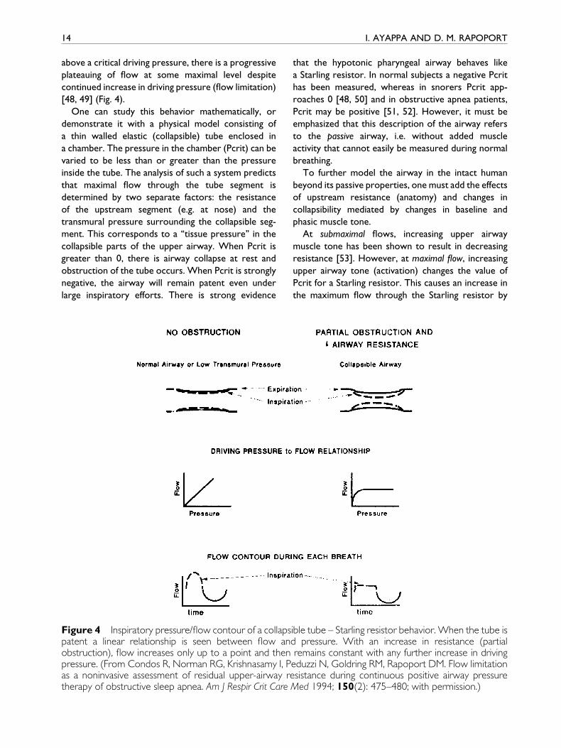
above a critical driving pressure, there is a progressive
plateauing of ¯ow at some maximal level despite
continued increase in driving pressure (¯ow limitation)
[48, 49] (Fig. 4).
One can study this behavior mathematically, or
demonstrate it with a physical model consisting of
a thin walled elastic (collapsible) tube enclosed in
a chamber. The pressure in the chamber (Pcrit) can be
varied to be less than or greater than the pressure
inside the tube. The analysis of such a system predicts
that maximal ¯ow through the tube segment is
determined by two separate factors: the resistance
of the upstream segment (e.g. at nose) and the
transmural pressure surrounding the collapsible seg-
ment. This corresponds to a `̀ tissue pressure'' in the
collapsible parts of the upper airway. When Pcrit is
greater than 0, there is airway collapse at rest and
obstruction of the tube occurs. When Pcrit is strongly
negative, the airway will remain patent even under
large inspiratory efforts. There is strong evidence
Figure 4 Inspiratory pressure/¯ow contour of a collapspatent a linear relationship is seen between ¯ow anobstruction), ¯ow increases only up to a point and thenpressure. (From Condos R, Norman RG, Krishnasamy I, Pas a noninvasive assessment of residual upper-airway rtherapy of obstructive sleep apnea. Am J Respir Crit Care
that the hypotonic pharyngeal airway behaves like
a Starling resistor. In normal subjects a negative Pcrit
has been measured, whereas in snorers Pcrit app-
roaches 0 [48, 50] and in obstructive apnea patients,
Pcrit may be positive [51, 52]. However, it must be
emphasized that this description of the airway refers
to the passive airway, i.e. without added muscle
activity that cannot easily be measured during normal
breathing.
To further model the airway in the intact human
beyond its passive properties, one must add the effects
of upstream resistance (anatomy) and changes in
collapsibility mediated by changes in baseline and
phasic muscle tone.
At submaximal ¯ows, increasing upper airway
muscle tone has been shown to result in decreasing
resistance [53]. However, at maximal ¯ow, increasing
upper airway tone (activation) changes the value of
Pcrit for a Starling resistor. This causes an increase in
the maximum ¯ow through the Starling resistor by
ible tube ± Starling resistor behavior. When the tube isd pressure. With an increase in resistance (partialremains constant with any further increase in drivingeduzzi N, Goldring RM, Rapoport DM. Flow limitationesistance during continuous positive airway pressureMed 1994; 150(2): 475±480; with permission.)
UPPER AIRWAY IN SLEEP 15
decreasing airway collapsibility of the ¯ow-limiting
segment, [54] and can occur even without a change
in the resistance (i.e. both ¯ow and pressure can
increase). Thus the Starling model helps better char-
acterize the changes in ¯ow that occur with changes in
muscle tone.
Factors that in¯uence resistance/collapsibility of the upper airway inhumans
Upstream resistance within thenasal airwayAs pointed out above, the upper airway can be modeled
by the more complex Starling resistor. This leads to the
understanding that not only collapsibility of the critical
segment of the airway needs to be examined, but that
upstream (e.g. nasal and nasopharyngeal) resistance will
affect the behavior of the entire system. Even under
normal circumstances the nose has a relatively high
resistance, which is increased by airway narrowing that
occurs with mucosal congestion.
Gender effectsThe data on the effect of gender on pharyngeal cross-
sectional area and resistance is con¯icting. While
some studies have indicated that differences in pha-
ryngeal size can be explained by differences in body
surface area [13] other studies have reported greater
pharyngeal size in men after normalizing for body
weight [12]. Although larger airways should imply
lower resistance in males, pharyngeal resistance has
been measured to be twice as high in males than
females [55], suggesting that there may be gender
speci®c differences in the airway mechanics. Some of
these discrepancies may be due to the intrinsic limita-
tions of using the `̀ resistance'' concept ± see discussion
above, and may be better addressed by concepts of
collapsibility, i.e. Pcrit. Additionally, it has been shown
that changes in pharyngeal cross-sectional area are
more dependent on lung volume [13], and increases in
pharyngeal resistance in response to load are greater
in men than women [12, 56]. This suggests that the
differences in mechanics (greater stability and less
dependence on lung volume) in women may play
a more important role than size.
Hormonal statusIn addition to gender there is some evidence that
hormonal status might in¯uence the upper airway
dilator activity [57] with premenopausal women
having signi®cantly greater genioglossal muscle activity
compared with post memenopausal women , and age-
matched men [58, 59], during inspiratory resistive
loading.
Age effectsModi®cations in pharyngeal characteristics with age
have been studied but there is no consensus on the
effect. Increases [60] and decreases [12, 23] in pha-
ryngeal cross sectional area with age have been
reported in subjects when studied awake. Age-related
increases in pharyngeal resistance during sleep [61]
and wakefulness [34] have been reported along with
decreases in the activity of genioglossus and tensor
palitini muscles [62]. However, other studies during
wakefulness [60] and sleep have reported that upper
airway resistance and genioglossus activity during quiet
breathing are similar in elderly and young people.
Extrinsic anatomic and static factorsaffecting the upper airway
Neck and Jaw postureClosure of the upper airway has been observed with
neck ¯exion in both the supine and prone positions in
anesthetized spontaneously breathing patients [63]. In
addition, jaw posture has also been documented to
in¯uence the size of the upper airway.
Surface adhesive forcesThe adhesive forces between the airway luminal
surfaces may promote closure of the airway or
impede subsequent opening of the airway [30]. It has
been shown that the positive pressure needed to
open an already closed airway is greater than the
closing pressure in both adults and infants [30, 50].
ObesityObesity can in¯uence the airway properties by increas-
ing the mass loading on the upper airway that could
result in airway compression. Since it is one of the
main risk factors for obstructive sleep apnea, and
weight loss results in improvement of the apnea in
some patients [64], it is a factor to consider. However,
the exact mechanism whereby obesity predisposes to
sleep apnea is still unclear.
Tracheal tugThe cross-sectional area of the upper airway appears
to increase with lung volume, along with a reduction in
the closing pressures and compliance of the upper
airway [65, 66]. No data exist to suggest this is an
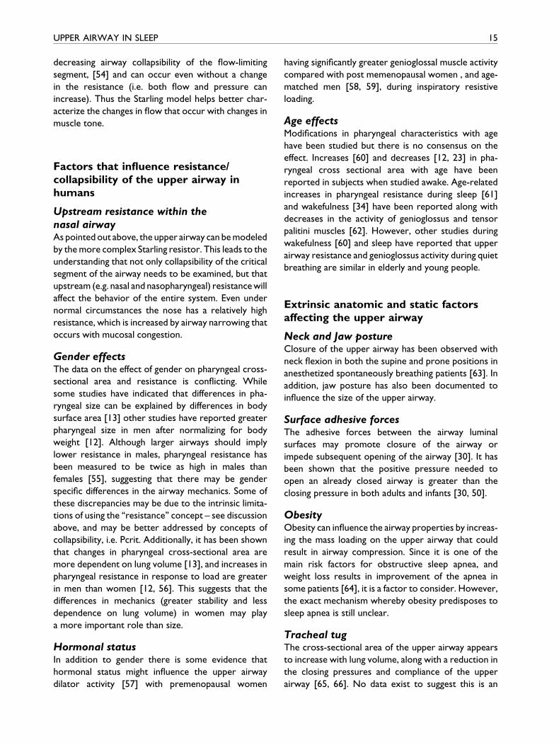
Figure 5 Axial magnetic resonance images of anormal subject during wakefulness and sleep. Theairway is reduced in both the AP and lateral dimen-sions during sleep compared with wakefulness. Thereis also thickening of the lateral pharyngeal walls duringsleep. (From Trudo FJ, Gefter WB, Welch KC,Gupta KB, Maislin G, Schwab RJ. State-related changesin upper airway caliber and surrounding soft-tissuestructures in normal subjects. Am J Respir Crit Care Med1998; 158(4): 1259±1270; with permission.)
16 I. AYAPPA AND D. M. RAPOPORT
active neural re¯ex, and in fact lung in¯ation is
generally inhibitory to respiration (Hering Breur
re¯ex) [67]. The increase in cross-sectional area is
probably a passive mechanical effect that results
from the axial forces transmitted through the
trachea. As the lung volume increases, caudal displace-
ment of the intrathoracic trachea appears to exert
longitudinal forces on the pharynx which stabilize it
and prevent passive collapse. Support for this inter-
pretation of the mechanical effect of lung in¯ation
comes from direct measurements of cross-sectional
area during the entire respiratory cycle measured
using cine CT. These data showed that the cross-
sectional area stays large at end-inspiration (when
neural tone is falling and lung volume is elevated), and
only falls to a minimum at end-expiration (when lung
volume is small) [19].
Effect of sleep on upper airwayresistance/collapsibility
Radiographic measurements have shown that during
wakefulness patency of the upper airway is well main-
tained in different postures, although the re¯exes
which control this seem to be critical [68]. However,
with the onset of sleep there are several modi®cations
that may occur in the factors affecting patency of the
upper airway including changes in neuromuscular
activation, ventilation, chemical and mechanical load
responses.
Signi®cant increases in upper airway resistance
associated with sleep have been shown in animals
[69] and humans [70±72]. Supraglottic resistance has
been shown to increase from low values (1±2 cm H2O/
L/s) [22, 72] to values as high as 5±10 cm H2O/L/s and
to 50 cm H2O/L/s in heavy snorers [73]. Less is known
about changes in airway caliber, but most studies
suggest that it decreases during sleep, with the lateral
pharyngeal walls playing an important role in this
narrowing [74] (Fig. 5) These changes in mechanics
induced by sleep could result in either hypoventilation
(loss of the re¯ex response to increased airway load),
or a re¯ex induced increase in ventilatory output with
maintained ventilation and blood gases. Some degree
of hypoventilation occurs at sleep onset in normals.
That this is a consequence of the mechanics rather
than a change in set-point for CO2 has been shown in
a study where unloading by CPAP or breathing He/O2
mixtures (characterized by reduced density) returned
mildly elevated sleeping PCO2 to awake levels [75].
Although it is likely that sleep in normals induces
increased collapsibility in the airway (decreased tone
to the upper airway dilators), the effect on calculations
of resistance is confounded by re¯ex responses (or
their absence). Thus it has been shown that sleep has
an effect on multiple aspects of upper airway behavior.
Effect of sleep on muscle tone in theupper airwayThe change in muscle activity of the upper airway
with onset of sleep has been investigated directly by
measuring the EMG, by using measured changes in
pharyngeal wall compliance or by using derived values
of ventilation and airway resistance. Many studies
have shown that the phasic inspiratory activity of the
genioglossus [26, 37, 76, 77] and geniohyoid are
maintained during sleep in normals. Conversely,
decreases in both tonic and phasic tone have been
shown in the genioglossus, geniohyoid [78], tensor
palatini [72], levator palatini, palatoglossus [77, 79]
and other respiratory muscles at the onset of sleep.
These have been shown to be associated with transient
decreases in ventilation and increased upper airway
UPPER AIRWAY IN SLEEP 17
resistance [62, 72]. However, in normal subjects these
decreases are short lived and parallel those seen in the
diaphragm [76] and intercostal muscles, which rise
as a response to induced obstructive hypoventilation
and mild hypercapnea (1±2 mmHg) seen during sleep.
The role that both tonic and phasic tone play in
maintaining airway patency is shown by denervation
studies, which in an animal model resulted in collapse
of the airway during sleep [26]. In healthy human
subjects, dense upper airway anesthesia increases
upper airway resistance during sleep and can cause
prolongation of apnea [80]. Despite this, the marked
decrease in EMG seen in all airway muscles during
REM sleep [81] does not result in uniform obstruction.
Some of this last paradox may be due to the protective
effect of the reduced inspiratory airway pressures
generated during REM sleep, again illustrating the
dif®culty in using ¯ow and resistance of the collapsible
airway as indices of function during conditions of
changing effort.
Effect of sleep on load responseWhereas during wakefulness application of a resistive
load to the airway results in increased respiratory
drive, this response may be lost or greatly attenuated
during sleep [71]. There is debate over whether
a similar response to airway loading exists by which
the upper airway muscle tone directly increases in
response to resistive loading of the system. How-
ever, it is dif®cult to separate this possible direct
effect from a non-speci®c increase in ventilatory
drive. Despite numerous studies that have measured
upper airway resistance and genioglossal EMG
during wakefulness, sleep and with resistive loading,
the direct relationship between GG EMG activity and
upper airway resistance during sleep is unclear, with
con¯icting results in multiple studies [71, 78, 82].
Possible reasons for con¯icting observations include
the assumption that muscle activity (EMG) is a sur-
rogate of muscle ®ber shortening, and the fact that
in most experiments muscle activity can only be
measured in a few locations/muscles, which do not
fully re¯ect the total airway muscle response.
Effect of CO2 on muscle activityduring sleepIn the awake state, elevation of CO2 is a powerful
respiratory stimulant. There is a large literature on
the effect of sleep on this response, and the consensus
is that this may be only minimally affected by sleep,
at least in non-REM stages. Less is known about the
effect of sleep on CO2 responses of the upper airway
muscles [75], independent of general respiratory
stimulation. The results from studies examining
changes in GG activation with hypercapnia are variable
[77, 83], and hypercapnia has been shown to decrease
collapsibility of the airway similar to tracheal
displacement [84].
Special human considerations
GenderThe data on gender differences in airway resistance
measured during sleep are con¯icting. While Trinder
et al. [85] measured a greater increase in upper airway
resistance during sleep in healthy men compared with
women Thurnheer et al. [86] concluded that there
were no major age or gender differences in the
changes in airway resistance during sleep.
SNORING
One of the most common consequences of the
changes in the properties of the upper airway that
occur during sleep is the occurrence of snoring.
Whether to call this pathology, if it occurs in the
absence of frank airway obstruction (apnea, hypopnea)
or repetitive arousal, is still debated. The prevalence
of habitual snoring is extremely high and reported
to be roughly 40% in men and 20% in women
[1, 87, 88]. The snoring sound is the result of vibra-
tions of the soft tissues of the pharynx, soft palate
and uvula having speci®c acoustic characteristics
with frequencies ranging from 5 to 136 Hz [89]. Early
cineradiographic recordings made by Lugaresi et al.
[90] showed that snoring was associated with
increased esophageal pressure swings during inspi-
ration with partial pharyngeal obstruction. In studies
comparing snorers and non-snorers during sleep it
has been shown that snorers have more negative
inspiratory pressure [91], greater pulmonary resis-
tance during snoring breaths [73] and ¯ow limitation
[51, 73, 92]. These events are all thought to be
normal consequences of sleep, and not necessarily
pathologic. However, many suggest snoring can be
considered as part of the spectrum of sleep disordered
breathing because the magnitude of these changes
lies between that of the non-snoring (normal) subject
and obstructive sleep apneic.
Anatomical measurements of the upper airway using
multiple techniques like MRI [93], acoustic re¯ections
[14], CT scanning [94] and x-ray cephalometry [95]
18 I. AYAPPA AND D. M. RAPOPORT
have demonstrated differences between snorers
and non-snoring asymptomatic subjects. As discussed
previously, since the airway area depends on body
posture, gender, lung volume, state of consciousness
and body size it is dif®cult to attribute the differences
in airway dimensions to snoring alone. Nevertheless,
it has been reported that snorers have reduced
airway area at the level of velopharynx [96], tongue
base, hyoid bone, have reduced sagittal dimensions,
longer soft palates and longer and wider uvulas than
healthy volunteers [97]. There is also however the
alternate hypothesis that the reduction in airway
cross section is a result of snoring, due to in¯ammation
of the mucosa and edema [98].
The changes in upper airway physiology that occur
during sleep have all been implicated in the develop-
ment of snoring. In addition to anatomy, the functional
properties of the upper airway appear to be different in
snorers and normal subjects. During wakefulness
snorers (non-apneic) have more collapsible upper
airways than non-snoring normals [50]. Also, the
critical pressures needed to collapse the upper
airway lies between that of normal subjects and
patients with OSAS [51]. The reason for the increased
collapsibility in snorers is still unclear. Possible
mechanisms include neuromuscular de®ciencies of
the upper airway dilator muscles, abnormal tissue
properties and abnormal linkage between the pharyn-
geal dilator muscles and pharyngeal tissue due to
fatty in®ltration [99].
The results from studies comparing the upper
airway properties of apneic snorers and non-apneic
snorers have been con¯icting because their classi®ca-
tion is dependent on the technique used to measure
the respiratory events. A subject classi®ed as a non-
apneic snorer by the respiratory index based on
nasal air¯ow measured by thermistry could be clas-
si®ed as an apneic snorer by a more sensitive measure
of ¯ow/effort like nasal cannula or esophageal mano-
metry. Therefore, while some studies showed no
difference in collapsibility [100], snoring frequency,
intensity and nasal resistance [101] or pharyngeal
cross-sectional areas at different lung volumes [14],
others have reported higher pharyngeal distensibility
[102, 103] and sound intensity [100, 104] in apneic
snorers versus non-apneic snorers. The greater sound
intensity appears to be the result of greater negative
pressures on resumption of breathing in apneic
snorers, resulting in high ¯ow rates, turbulent ¯ow
and greater forces on the vibrating structures. More
recently, Series et al. have reported differences in the
properties of the musculus uvulae (an upper airway
dilator located in the uvula) [105] and the genioglossus
[106] in snorers and subjects with OSAHS. The force
generated by these muscles is greater in apneics than
non-apneic snorers during wakefulness and sleep,
possibly as an adaptive response to high-intensity
resistive loading. However, the sleep-induced dec-
rease of this upper airway dilator activity results in
greater alterations in mechanical ef®ciency of apneic
subjects [107].
Several mathematical models of the upper airway
as a collapsible tube have been developed to study
snoring. A tube of given geometry, elastic constant,
resistance, density of gas and ¯ow parameters will
become unstable at a certain location leading to
vibration of the walls and dynamic collapse with the
appearance of sound structures similar to snoring
[108]. Since these parameters are different in individual
subjects the site of snoring sound generation will vary
depending on where the instability occurs. Direct
observations during sleep have con®rmed that the
location of the vibrations along with the narrowing
of the upper airway is variable [92, 109]. Other models
coupling mechanics to neuromuscular physiology
through re¯ex wall stiffening predict that snoring
develops with reduced strength and increased latency
of the re¯ex [110].
Despite the reported greater prevalence of
snoring in men the gender effects on snoring intensity
are unclear. Wilson et al. [104] have reported that
women do not snore as loudly as men, independent
of severity of SDB or BMI, in contrast to Metes et al.
[101] who showed no difference in the maximum
snoring intensity between genders. Both studies
however did not show any age effect on the intensity
of snoring [101, 104]. A recent epidemiological study
has reported chronic nocturnal nasal congestion as
a risk factor for habitual snoring [111].
SLEEP DISORDEREDBREATHING
Since the 1960s, there has been an increasing recogni-
tion of the syndrome of Sleep Disordered Breathing.
This term includes all forms of abnormal breathing
patterns associated with sleep. By far the most
common are the variants in which there is transient
complete (apnea) or partial (hypopnea, snoring) obs-
truction of the upper airway. Early hypotheses to
explain the hypersomnolence seen in the most
severe form of sleep disordered breathing, then
UPPER AIRWAY IN SLEEP 19
called `̀ the Pickwickian Syndrome'', suggested this
symptom was caused by CO2 narcosis. However,
later studies reported that obstructive apneas were
caused by the `̀ obstruction of the upper outlet, by
the backward movement of the tongue'', which then
resulted in frequent awakenings and disruption of
sleep, explaining the hypersomnolence [112]. In
1972, Walsh et al. [113] directly observed the upper
airway during sleep using cine¯uroscopy in obese,
hypersomnolent patients with OSA, and observed
that upper airway obstruction was produced by the
`̀ tongue retracting into apposition with the posterior
pharyngeal wall''. Establishment of an airway with
a tracheostomy or nasopharyngeal tube abolished
the apneas and usually cures the symptoms of the
syndrome. A landmark study by Remmers et al. in 1978
[25] established the location of upper airway collapse
during sleep as the oropharynx in patients with
obstructive sleep apnea syndrome, and proposed
that this resulted from an imbalance between negative
pharyngeal pressure and the opposing force of the
upper airway musculature (genioglossal). These events
(apnea) were shown to resolve with a burst of GG
EMG activation The location of this collapse has
since been shown to be at variable locations in the
upper airway and can occur simultaneously at multiple
sites [27, 114±117]. Thus, presently, abnormal func-
tion of the upper airway is thought to be the main
source of obstruction and symptoms in the syndrome
of sleep disordered breathing.
Anatomy
Much work has been done to address the hypothesis
that abnormal anatomy underlies sleep disordered
breathing. Several imaging techniques have been used
to study the upper airway size and changes in the
airway size and soft tissue structures that surround
the airway in patients with SDB, including acoustic
re¯ection [10, 14, 65, 102], ¯uoroscopy [16],
nasopharyngoscopy [118], cephalometry [119], CT
and MRI imaging [16, 19, 120±122]. Reduced awake
supine upper airway caliber by CT [123] has been
shown in male patients with obstructive sleep apnea
syndrome at the naso, oro and and hypopharyngeal
levels when compared to normal subjects. These
images showed that the narrowest region in both
patients with OSA and normal non-obese controls,
while awake, is the region posterior to the soft palate,
and that the cross sectional area of this retropalatal
region is smaller in patients than controls. No
abnormal collections of fat density were found to
explain these differences. However, a recent series of
observations have pointed out that, independent of
airway caliber, airway con®guration may differ in this
area in OSA patients. Thus, in contrast to normals who
have the major axis of the pharyngeal airway oriented
in the lateral dimension, patients show an axis oriented
anteroposteriorly, which corresponds to lateral nar-
rowing at the critical level [93] (see Fig. 6). Two
structures lateral to the upper airway that could
cause this narrowing, the lateral pharyngeal walls
and the lateral pharyngeal fat pads, have been exam-
ined in these studies. The lateral pharyngeal walls
have been shown to be thicker in apneics [124];
however, the pharyngeal fat pads were not closer
together, and the area and width of the fat pads
were not larger in apneics at the level of the minimum
airway [19]. These unexpected ®ndings have led the
authors to conclude that the thickness of the lateral
pharyngeal walls, and not the actual size of the soft
palate, tongue or fat pads, was the predominant
anatomic factor causing airway narrowing in apneic
patients. The reason for the thicker walls is not
known; in particular, spectroscopic analysis of tissue
in this area shows no differences in fat or water
content between normal subjects and apneics [19]. It
has been speculated that an increase in muscle mass
due to weight gain or the `̀ exercise'' of overcoming
apnea might explain the increase in the size of the
lateral soft-tissues without increase in the direct fat
deposition. However, application of CPAP results in
increase of the airway volume and area in the oro-
pharynx [125], with most of the changes occurring
again in the lateral rather than anteroposterior axis.
This has led some to invoke `̀ folding'' of the mucosa
as the explanation of the lateral wall thickening,
with `̀ unfolding'' during CPAP. In any case, these
consistent observations emphasize the importance of
the lateral structures in contrast to other (tongue,
soft palate) airway structures in the pathogenesis of
obstruction.
Explanations accounting for role ofsleep in causing upper airwayobstruction/increased resistance
While the actual location of collapse in sleep dis-
ordered breathing events is often the oro or hypo-
pharynx where an underlying anatomic change may be
present, it is clear that patients with severe obstruc-
tion during sleep do not have signi®cant functional
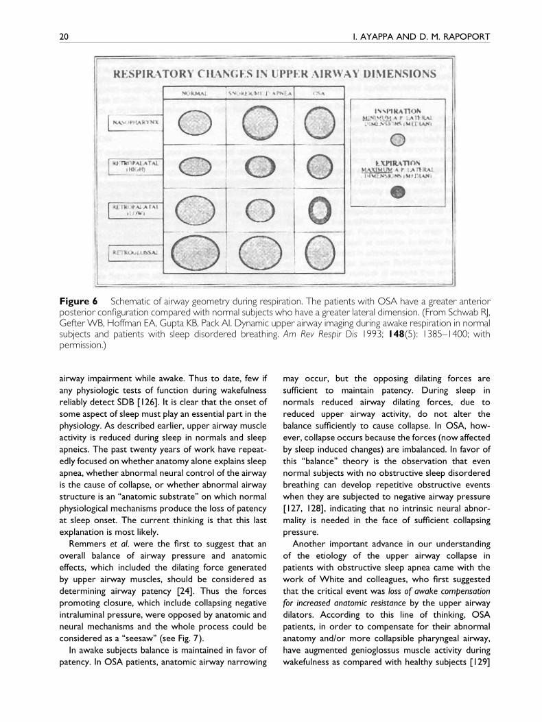
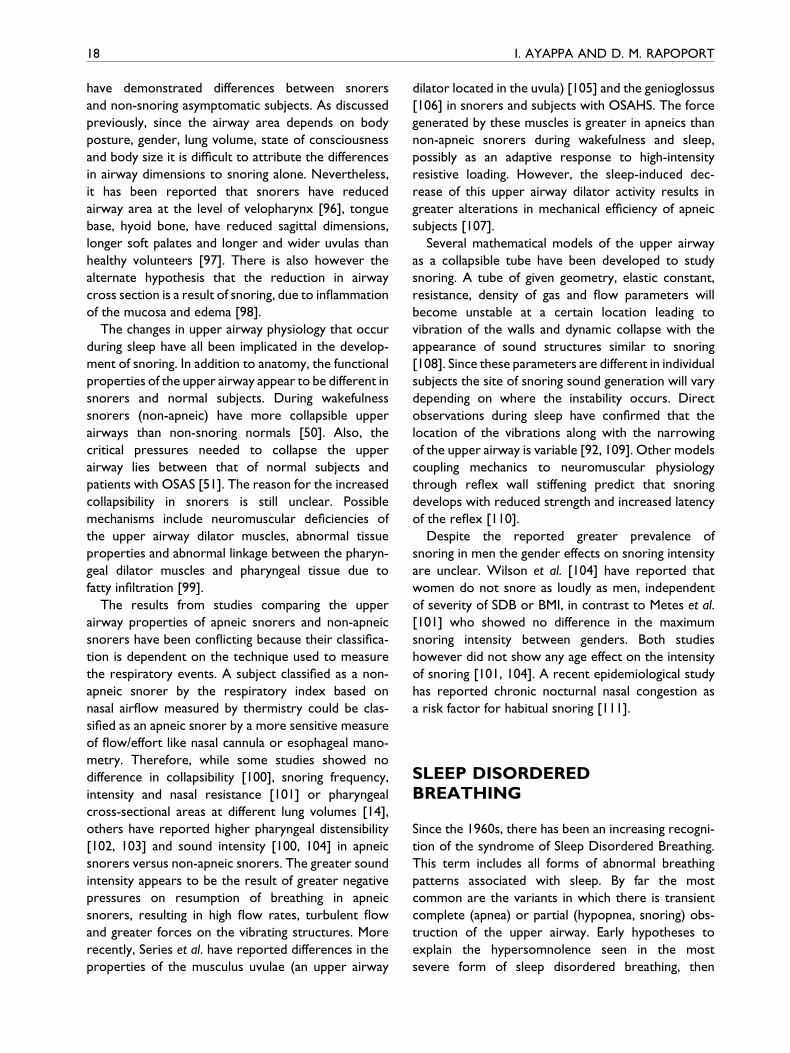
Figure 6 Schematic of airway geometry during respiration. The patients with OSA have a greater anteriorposterior con®guration compared with normal subjects who have a greater lateral dimension. (From Schwab RJ,Gefter WB, Hoffman EA, Gupta KB, Pack AI. Dynamic upper airway imaging during awake respiration in normalsubjects and patients with sleep disordered breathing. Am Rev Respir Dis 1993; 148(5): 1385±1400; withpermission.)
20 I. AYAPPA AND D. M. RAPOPORT
airway impairment while awake. Thus to date, few if
any physiologic tests of function during wakefulness
reliably detect SDB [126]. It is clear that the onset of
some aspect of sleep must play an essential part in the
physiology. As described earlier, upper airway muscle
activity is reduced during sleep in normals and sleep
apneics. The past twenty years of work have repeat-
edly focused on whether anatomy alone explains sleep
apnea, whether abnormal neural control of the airway
is the cause of collapse, or whether abnormal airway
structure is an `̀ anatomic substrate'' on which normal
physiological mechanisms produce the loss of patency
at sleep onset. The current thinking is that this last
explanation is most likely.
Remmers et al. were the ®rst to suggest that an
overall balance of airway pressure and anatomic
effects, which included the dilating force generated
by upper airway muscles, should be considered as
determining airway patency [24]. Thus the forces
promoting closure, which include collapsing negative
intraluminal pressure, were opposed by anatomic and
neural mechanisms and the whole process could be
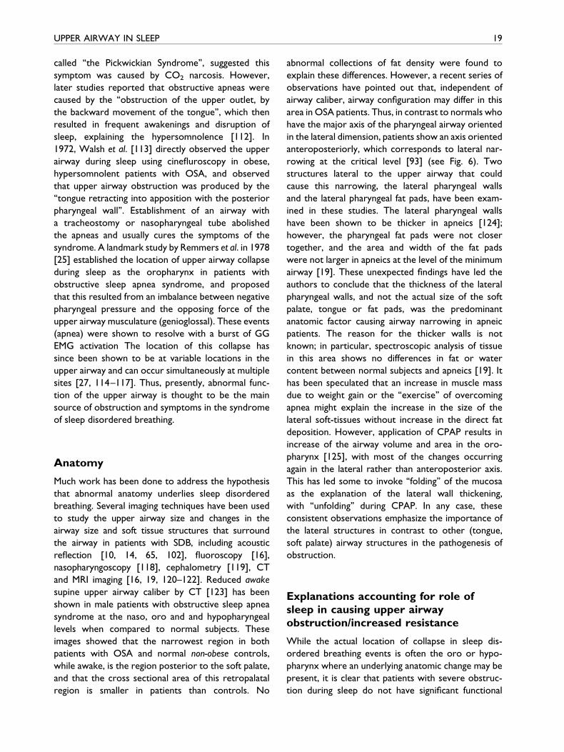
considered as a `̀ seesaw'' (see Fig. 7).
In awake subjects balance is maintained in favor of
patency. In OSA patients, anatomic airway narrowing
may occur, but the opposing dilating forces are
suf®cient to maintain patency. During sleep in
normals reduced airway dilating forces, due to
reduced upper airway activity, do not alter the
balance suf®ciently to cause collapse. In OSA, how-
ever, collapse occurs because the forces (now affected
by sleep induced changes) are imbalanced. In favor of
this `̀ balance'' theory is the observation that even
normal subjects with no obstructive sleep disordered
breathing can develop repetitive obstructive events
when they are subjected to negative airway pressure
[127, 128], indicating that no intrinsic neural abnor-
mality is needed in the face of suf®cient collapsing
pressure.
Another important advance in our understanding
of the etiology of the upper airway collapse in
patients with obstructive sleep apnea came with the
work of White and colleagues, who ®rst suggested
that the critical event was loss of awake compensation
for increased anatomic resistance by the upper airway
dilators. According to this line of thinking, OSA
patients, in order to compensate for their abnormal
anatomy and/or more collapsible pharyngeal airway,
have augmented genioglossus muscle activity during
wakefulness as compared with healthy subjects [129]
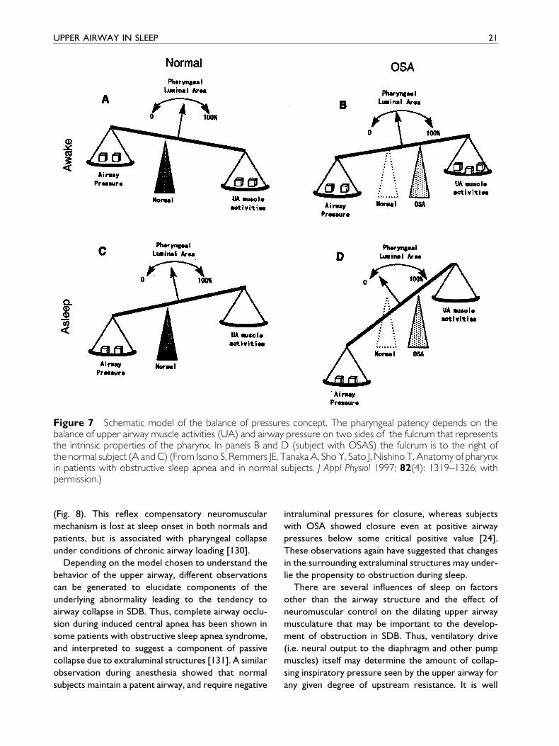
Figure 7 Schematic model of the balance of pressures concept. The pharyngeal patency depends on thebalance of upper airway muscle activities (UA) and airway pressure on two sides of the fulcrum that representsthe intrinsic properties of the pharynx. In panels B and D (subject with OSAS) the fulcrum is to the right ofthe normal subject (A and C) (From Isono S, Remmers JE, Tanaka A, Sho Y, Sato J, Nishino T. Anatomy of pharynxin patients with obstructive sleep apnea and in normal subjects. J Appl Physiol 1997; 82(4): 1319±1326; withpermission.)
UPPER AIRWAY IN SLEEP 21
(Fig. 8). This re¯ex compensatory neuromuscular
mechanism is lost at sleep onset in both normals and
patients, but is associated with pharyngeal collapse
under conditions of chronic airway loading [130].
Depending on the model chosen to understand the
behavior of the upper airway, different observations
can be generated to elucidate components of the
underlying abnormality leading to the tendency to
airway collapse in SDB. Thus, complete airway occlu-
sion during induced central apnea has been shown in
some patients with obstructive sleep apnea syndrome,
and interpreted to suggest a component of passive
collapse due to extraluminal structures [131]. A similar
observation during anesthesia showed that normal
subjects maintain a patent airway, and require negative
intraluminal pressures for closure, whereas subjects
with OSA showed closure even at positive airway
pressures below some critical positive value [24].
These observations again have suggested that changes
in the surrounding extraluminal structures may under-
lie the propensity to obstruction during sleep.
There are several in¯uences of sleep on factors
other than the airway structure and the effect of
neuromuscular control on the dilating upper airway
musculature that may be important to the develop-
ment of obstruction in SDB. Thus, ventilatory drive
(i.e. neural output to the diaphragm and other pump
muscles) itself may determine the amount of collap-
sing inspiratory pressure seen by the upper airway for
any given degree of upstream resistance. It is well
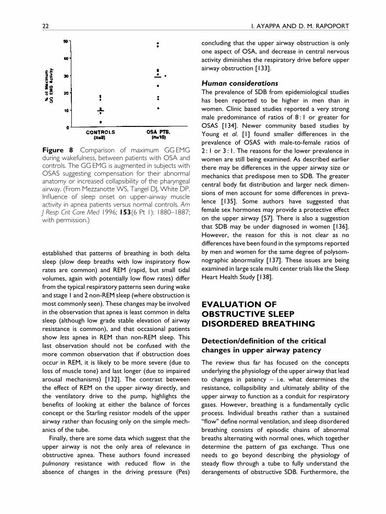
Figure 8 Comparison of maximum GG EMGduring wakefulness, between patients with OSA andcontrols. The GG EMG is augmented in subjects withOSAS suggesting compensation for their abnormalanatomy or increased collapsibility of the pharyngealairway. (From Mezzanotte WS, Tangel DJ, White DP.In¯uence of sleep onset on upper-airway muscleactivity in apnea patients versus normal controls. AmJ Resp Crit Care Med 1996; 153(6 Pt 1): 1880±1887;with permission.)
22 I. AYAPPA AND D. M. RAPOPORT
established that patterns of breathing in both delta
sleep (slow deep breaths with low inspiratory ¯ow
rates are common) and REM (rapid, but small tidal
volumes, again with potentially low ¯ow rates) differ
from the typical respiratory patterns seen during wake
and stage 1 and 2 non-REM sleep (where obstruction is
most commonly seen). These changes may be involved
in the observation that apnea is least common in delta
sleep (although low grade stable elevation of airway
resistance is common), and that occasional patients
show less apnea in REM than non-REM sleep. This
last observation should not be confused with the
more common observation that if obstruction does
occur in REM, it is likely to be more severe (due to
loss of muscle tone) and last longer (due to impaired
arousal mechanisms) [132]. The contrast between
the effect of REM on the upper airway directly, and
the ventilatory drive to the pump, highlights the
bene®ts of looking at either the balance of forces
concept or the Starling resistor models of the upper
airway rather than focusing only on the simple mech-
anics of the tube.
Finally, there are some data which suggest that the
upper airway is not the only area of relevance in
obstructive apnea. These authors found increased
pulmonary resistance with reduced ¯ow in the
absence of changes in the driving pressure (Pes)
concluding that the upper airway obstruction is only
one aspect of OSA, and decrease in central nervous
activity diminishes the respiratory drive before upper
airway obstruction [133].
Human considerationsThe prevalence of SDB from epidemiological studies
has been reported to be higher in men than in
women. Clinic based studies reported a very strong
male predominance of ratios of 8 : 1 or greater for
OSAS [134]. Newer community based studies by
Young et al. [1] found smaller differences in the
prevalence of OSAS with male-to-female ratios of
2 : 1 or 3 : 1. The reasons for the lower prevalence in
women are still being examined. As described earlier
there may be differences in the upper airway size or
mechanics that predispose men to SDB. The greater
central body fat distribution and larger neck dimen-
sions of men account for some differences in preva-
lence [135]. Some authors have suggested that
female sex hormones may provide a protective effect
on the upper airway [57]. There is also a suggestion
that SDB may be under diagnosed in women [136].
However, the reason for this is not clear as no
differences have been found in the symptoms reported
by men and women for the same degree of polysom-
nographic abnormality [137]. These issues are being
examined in large scale multi center trials like the Sleep
Heart Health Study [138].
EVALUATION OFOBSTRUCTIVE SLEEPDISORDERED BREATHING
Detection/de®nition of the criticalchanges in upper airway patency
The review thus far has focused on the concepts
underlying the physiology of the upper airway that lead
to changes in patency ± i.e. what determines the
resistance, collapsibility and ultimately ability of the
upper airway to function as a conduit for respiratory
gases. However, breathing is a fundamentally cyclic
process. Individual breaths rather than a sustained
`̀ ¯ow'' de®ne normal ventilation, and sleep disordered
breathing consists of episodic chains of abnormal
breaths alternating with normal ones, which together
determine the pattern of gas exchange. Thus one
needs to go beyond describing the physiology of
steady ¯ow through a tube to fully understand the
derangements of obstructive SDB. Furthermore, the
UPPER AIRWAY IN SLEEP 23
techniques used to detect and de®ne these abnor-
malities in breathing are intimately involved in our
ability to understand the process.
Abnormal breath detection
Having identi®ed that one wishes to detect that
a change has occurred in the overall behavior of the
upper airway associated with `̀ sleep disordered
breathing'' the next step is to operationalize the
detection of the abnormality that will be the basis of
identifying an `̀ abnormal'' breath. The earliest practical
approach was to identify breaths that did not occur
despite clear evidence of respiratory effort (i.e. obs-
tructive apnea). This is easily done with any detector
of respiratory gas at the nose and mouth; in the
usual form, these detectors rely on the different
characteristics of inspired and expired air as a marker.
Thus temperature oscillation (used by thermistors or
thermocouples) between cooler inhaled room air
temperature and warmer exhaled air (near body
temperature) and oscillations in PCO2 (only present
in exhaled gas) were, and remain, good markers of
the presence of a breath. Alternatively, respiratory
efforts, measured as movement of the chest and
abdomen, can also be calibrated to represent a `̀ tidal
volume''. When these movements are fully out of
phase, this may indicate total obstruction to air¯ow
because there is no net change in the volume of the
chest cavity. However, all of the above are best at
detecting `̀ all or nothing'' air movement. In the case of
incomplete airway obstruction the relationship be-
tween temperature change and true ¯ow is not
straightforward [139, 140].
By de®nition, measurement of airway resistance
must rely on actual measurement of a true index of
both air¯ow and a separate measure of intrathoracic
effort or pressure. It has been suggested that the
reference standard for measurement of respiratory
effort should be esophageal pressure measurement
[141]. This technique consists of inserting a trans-nasal
¯exible pressure catheter to directly measure intra-
thoracic (and presumably pleural) pressure swings
during inspiration as a continuous measure of effort.
However, the determination of airway resistance
actually requires more than effort and should be
based on its ratio to air¯ow (directly measured at
mouth and nose with a snug ®tting mask).
The concept of `̀ hypopnea'' was introduced by Block
et al. as periods of shallow breathing with oxygen
desaturation [142]. The assumption was made that
reductions in ventilation were due to increases in
airway resistance. Ventilation (tidal volume) was thus
the critical variable to be measured, and this was
done by derivation from non-invasive Inductance
Plethysmography (which measures chest/abdominal
movement). Transient reductions in the signal were
con®rmed as being reductions in ventilation (pre-
sumed due to increases in resistance) by looking for
consequences such as a drop in O2 saturation. While
originally successful at de®ning a characteristic
pattern of abnormal breaths, the rapid extrapolation
of these de®nitions to use with other more qualitative
monitors (thermistors, thermocouples and end-tidal
PCO2 devices) led to great non-uniformity in what
was detected and called `̀ hypopnea'' [143, 144]. More
recently, it has been suggested a return is necessary
to measurements which truly measure volume or ¯ow
and correctly quantitate the underlying ventilation.
Use of nasal/full facemasks with pneumotachographs,
although theoretically necessary, have tended to be
reserved for research. In the past several years, we
and others have suggested an attractive surrogate for
true pneumotachography; the use of a combination of
a nasal cannula and pressure transducer, which has
been validated for non-quantitative (relative)
detection of ventilation [139, 140, 145, 146].
The combination of this return to measurement
of actual air¯ow or its closest analogues, and the
recognition that breaths may occur that have high
resistance/abnormal airway collapse without actual
reduction in air¯ow has led to evaluation of techni-
ques to detect airway resistance directly on a breath
by breath basis [147]. In the current approaches to
detecting SDB, the shift has been away from detecting
only actual ventilation, to trying to detect unexpected
increases in respiratory drive (inspiratory EMG or
esophageal pressure [2]) during `̀ normal'' appearing
breaths (high resistance), or to measuring indirect
evidence of a change in airway patency by detecting
consequences (arousal, desaturation, elevation in
markers of sympathetic activity [148], or ®nally to
measuring surrogates of ¯ow/pressure relationships
which may indicate abnormal behavior. The last of
these has been the subject of much recent work by
our laboratory and is based on the `̀ ¯ow limitation''
concept.
Flow limitation
`̀ Flow limitation'', as used in the context of SDB,
refers to the characteristic ¯attened shape of the
24 I. AYAPPA AND D. M. RAPOPORT
pressure-¯ow relationship which has been observed
when increasing effort does not result in increasing
air¯ow in the upper airway. In addition to measuring
amplitude of the ¯ow (useful to detect conventional
apnea and hypopnea), the identi®cation of a plateau on
the inspiratory ¯ow/time waveform correlates with
an elevated upper airway resistance (measured as
pressure divided by ¯ow) during CPAP titration [49]
and during spontaneous breathing [140, 149]. Snoring
generally occurs with ¯ow limitation, but ¯ow limita-
tion appears to be a more sensitive marker of elevated
resistance [150, 151], and treatment aimed at normal-
izing ¯ow limitation has resulted in improvement of
EDS [152].
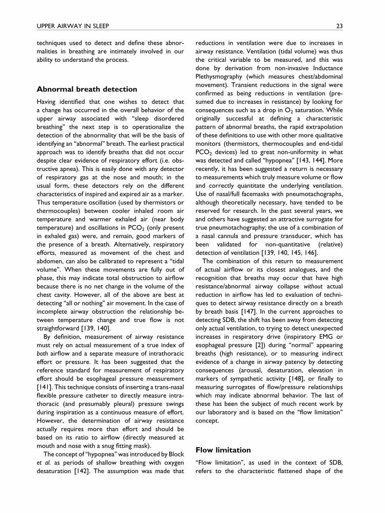
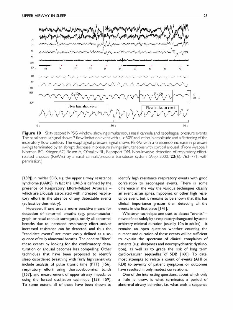
Early studies to identify ¯ow limitation were per-
formed with a conventional pneumotachograph ¯ow
signal but required a tight-®tting face mask, which was
excessively intrusive for routine sleep monitoring. The
nasal cannula/pressure transducer circumvents this
limitation, and provides a simple alternative to the
pneumotachograph that provides a semi-calibrated
signal proportional to inspiratory air¯ow. It detects
the small pressure ¯uctuations caused by inspiration
and expiration inside the nose. We and others have
shown that the signal obtained from this device is
comparable in both shape and amplitude to that of
a conventional pneumotachograph [49, 145, 153, 154].
The relationship between this signal and a simulta-
neous pneumotachographic ¯ow signal is essentially
linear over the relevant range of ¯ow measured,
at least over periods of interest (20±100 breaths)
(Fig. 9). This system has recently been accepted by
Figure 9 Comparison of signal from nasal cannula/pressure transducer system and simultaneouslyobtained pneumotachograph ¯ow from a mask intwo subjects. The relationship is curvilinear overall,but is linear in the range of normal breathing. (FromHosselet JJ, Norman RG, Ayappa I, Rapoport DM.Detection of ¯ow limitation with a nasal cannula/pressure transducer system. Am J Respir Crit CareMed 1998; 157(5 Pt 1): 1461±1467; with permission.)
the general sleep community in a position paper as
one of the two acceptable methods of monitoring
respiration during sleep [141]. The simplicity, low
cost and non-obtrusiveness of the device make it
ideal for quantitative monitoring of respiration during
sleep [155].
Events
Sleep Disordered Breathing is characterized by inter-
mittent sequences of abnormal breaths. Thus, apart
from identifying individual breaths that are abnormal,
it is necessary to de®ne which `̀ sequences'' of
breaths de®ne pathological `̀ events''. Breaths are an
intermittent `̀ sampling'' of the airway behavior and it
is transient abnormalities (i.e. elevations in resistance)
which need to be detected during sleep. Apnea is
easily de®ned and identi®ed as an abnormal respira-
tory event that has a speci®c duration. The logical
extension of the concept of apnea is to describe an
abnormal period lasting more than a given ®xed
duration, now including breaths that have only partial
airway collapse. Thus one must detect (and de®ne)
events composed of a series of breaths with a charac-
teristic (low volume, low ¯ow, high effort, a waveform
indicating ¯ow-limitation) indicating a period of sus-
tained but intermittently elevated instantaneous
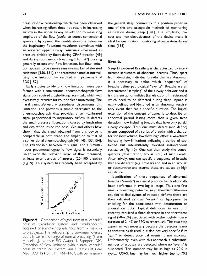
resistance (Fig. 10). One can then study the conse-
quences (desaturation, arousal etc.) of such events.
Alternatively, one can specify a sequence of breaths
that are different (e.g. smaller) and end in an arousal
or desaturation and assume these are caused by high
resistance.
Identi®cation of these sequences of abnormal
breaths (`̀ events'') in clinical practice has traditionally
been performed in two logical steps. Thus one ®rst
uses a breathing detector (e.g. thermistor/thermo-
couple) to ®nd events of reduced air¯ow; these are
then validated as true `̀ events'' or hypopneas by
checking for the coincidence with desaturation or
arousal on EEG. Typical de®nitions in use until
recently required a ®xed decrease in the thermistor
signal (50±75%) associated with oxyhemoglobin desa-
turation of 2±4% or EEG microarousal. This two step
algorithm was necessary because the detector is not
as sensitive as desired, but also not very speci®c if its
`̀ gain'' to detect potential events is not maximal.
Unfortunately, even with this approach, a substantial
number of arousals are detected where no `̀ event'' is
suspected. This number may be as high as 20% in
typical OSAS, but may be much higher (up to 70%
0 s 30 s 60 s
10cmH 2 0
C3
O1
LOC
ROC
CHIN
SaO 2
RIB
ABD
ESOP
PRESS
FLOW
Figure 10 Sixty second NPSG window showing simultaneous nasal cannula and esophageal pressure events.The nasal cannula signal shows 2 ¯ow limitation event with a550% reduction in amplitude and a ¯attening of theinspiratory ¯ow contour. The esophageal pressure signal shows RERAs with a crescendo increase in pressureswings terminated by an abrupt decrease in pressure swings simultaneous with cortical arousal. (From Ayappa I,Norman RG, Krieger AC, Rosen A, O'malley RL, Rapoport DM. Non-Invasive detection of respiratory effort-related arousals (RERAs) by a nasal cannula/pressure transducer system. Sleep 2000; 23(6): 763±771; withpermission.)
UPPER AIRWAY IN SLEEP 25
[139]) in milder SDB, e.g. the upper airway resistance
syndrome (UARS). In fact the UARS is de®ned by the
presence of Respiratory Effort-Related Arousals ±
which are arousals associated with increased respira-
tory effort in the absence of any detectable events
(at least by thermistry).
However, if one uses a more sensitive means for
detection of abnormal breaths (e.g. pneumotacho-
graph or nasal cannula surrogate), nearly all abnormal
breaths due to increased respiratory effort and/or
increased resistance can be detected, and thus the
`̀ candidate events'' are more easily de®ned as a se-
quence of truly abnormal breaths. The need to `̀ ®lter''
these events by looking for the con®rmatory desa-
turation or arousal becomes less compelling. Other
techniques that have been proposed to identify
sleep disordered breathing with fairly high sensitivity
include analysis of pulse transit time (PTT) [156],
respiratory effort using thoracoabdominal bands
[157], and measurement of upper airway impedance
using the forced oscillation technique [158, 159].
To some extent, all of these have been shown to
identify high resistance respiratory events with good
correlation to esophageal events. There is some
difference in the way the various techniques classify
an event as an apnea, hypopnea or other high resis-
tance event, but it remains to be shown that this has
clinical importance greater than detecting all the
events in the ®rst place [141].
Whatever technique one uses to detect `̀ events'' ±
now de®ned solely by a respiratory change and by some
arbitrary minimal duration (usually 10 s in adults) ± it
remains an open question whether counting the
number and duration of these events will be suf®cient
to explain the spectrum of clinical complaints of
patients (e.g. sleepiness and neuropsychiatric dysfunc-
tion), as well as to grade the risk of long term
cardiovascular sequaellae of SDB [160]. To date,
most attempts to relate a count of events (AHI or
RDI) to severity of patient symptoms or outcomes
have resulted in only modest correlations.
One of the interesting questions, about which only
a little is know, is what terminates a period of
abnormal airway behavior, i.e. what ends a sequence
26 I. AYAPPA AND D. M. RAPOPORT
of abnormal breaths. The general consensus is that
some level of brain activation (i.e. arousal) is always
seen at the end of an obstructive event, just as loss
of the state of arousal is the proximate cause of
the onset of airway abnormality/ventilatory instability
in SDB [161]. This is not equally true of `̀ central''
events, where the predominant abnormality is loss of
central neural respiratory `̀ drive'' rather than loss of
airway patency. Thus at the end of an obstructive
event frank EEG arousal (a change in basic frequency
and appearance of waves characterizing the `̀ awake''
state) is usually seen, but these EEG changes may
be absent in up to 20% of obstructive events. Some
of these `̀ missed'' arousals are clearly due to our
inability to de®ne and `̀ rate'' EEG changes which are
visible to the eye, or even to more sophisticated
signal processing techniques. Some may be due to
cortical activation having been present at other sites
of the brain (frontal) and not detected by the
central EEG leads conventionally used in sleep moni-
toring (C3, C4). In rare (approximately 5%) of cases,
no cortical EEG changes are seen at the end of an
obstructive event. However, other markers of brain-
stem activation (change in autonomic tone, heart
rate or BP) are almost invariably detected, indicating
some degree of `̀ arousal'' was present [162, 163].
All of the above begs the question of what causes
the brain to arouse in situations where obstruction
of the airway has occurred. Although suf®cient
chemical (i.e. hypoxic and hypercapnic) stimuli will
arouse most individuals (even those with spinal
injury and thus unable to respond to express the
ventilatory stimulation), it has been suggested that
it is the mechanical effort which generates a common
signal causing arousal under most physiological condi-
tions. Thus Gleeson et al. [164] showed that for a
given individual in a single sleep state, arousal always
occurred at the same level of intrathoracic pressure
swing with a breath ± whether the effort was
generated in response to obstruction, hypoxia or
hyper-capnia. During clinical sleep studies, many
patients tend to show a regular recurrent level of
drive at arousal during periods of obstructive
apnea, and have regular apnea lengths, minimal satura-
tions and similar large breaths after each apnea,
suggesting they have aroused at very similar levels of
stimulation for each episode. To date, little has
been done to use this physiological `̀ signature'' of
an individual's apneas to characterize their clinical
disease. In contrast to the mechanoreceptor hypoth-
esis for arousal, Ayas et al. [165] showed that
hypercapnia alone could induce arousal from sleep in
a study on neurologically complete spinal cord injury
patients.
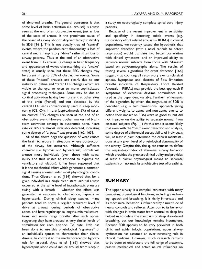
Because of the recent improvement in sensitivity
and speci®city in detecting subtle events (e.g.
Respiratory effort related arousals ± RERAs) in clinical
populations, we recently tested the hypothesis that
improved detection (with a nasal cannula to detect
respiration) would translate into better correlation
with clinical symptoms, and an improved ability to
separate normal subjects from those with `̀ disease''
based on polysomnography alone. The results of
testing several algorithms for event detection [166]
suggest that counting all respiratory events (classical
apneas, hypopneas and clusters of ¯ow limitation
breaths indicative of Respiratory Effort Related
Arousals ± RERAs) may provide the best approach if
symptoms of excessive daytime somnolence are
used as the dependent variable. Further re®nements
of the algorithm by which the magnitude of SDB is
described (e.g. a two dimensional approach giving
different weights to apnea and non-apnea events to
de®ne their impact on EDS) were as good as, but did
not improve on the ability to separate normal from
diseased subjects (Fig. 11) At this time it seems likely
that even with the `̀ best'' event detection and analysis,
some degree of differential susceptibility of individuals
will, at least in part, determine the clinical manifesta-
tions at any given level of physiological abnormality of
the airway. Despite this, the quest remains to de®ne
the respiratory index of abnormal airway behavior
which provides the greatest clinical utility and provides
at least a partial physiological means to separate
patients from normals by an objective test of breathing.
SUMMARY
The upper airway is a complex structure with many
competing physiological functions, including swallow-
ing, speech and breathing. It is richly innervated and
its mechanical behavior is in¯uenced by a multitude of
neural controls and re¯exes. Attention to its behavior
with changes in brain states from arousal to sleep has
helped us to de®ne the spectrum of sleep disordered
breathing, but our knowledge remains incomplete.
Because SDB appears to be very prevalent in both
clinic and epidemiologic populations, upper airway
dysfunction has assumed an ever-increasing role in
clinical medicine. However, much research remains
to be done to understand the full range of anatomic,
passive mechanical and active neural in¯uences on
Figure 11 Plot of apnea index (AI) against non-apnea event index (RDITotal-AI) for 103 test subjects. Opencircles are asymptomatic subjects, open triangles are snorers without excessive daytime somnolence (EDS) andthe closed circles are the subjects with both EDS and snoring. The region bounded by AI52/h and RDITotal -AI516/h was de®ned as identifying the `̀ normal'' region. (From Hosselet JJ, Ayappa I, Norman RG, Krieger AC,Rapoport DM. Classi®cation of sleep-disordered breathing. Am J Respir Crit Care Med 2001; 163(2): 398±405;with permission.)
UPPER AIRWAY IN SLEEP 27
upper airway patency and to fully exploit this
knowledge in the de®nition of disease states and the
optimization of therapies for disease.
Practice Points
1. The upper airway is a collapsible structure
whose patency is dictated by a combination of
passive mechanical properties and active neural
mechanisms.
2. Sleep produces changes in airway physiology,
which, in association with abnormal anatomy,
lead to sleep disordered breathing events and
the syndrome of obstructive sleep apnea/
hypopnea.
3. Formal de®nitions of sleep disordered breathing
events exist, but are changing as technology and
our understanding of the underlying physiology
evolve.
Research Agenda
1. The full range of anatomic, passive mechanical
and active neural in¯uences on upper airway
patency needs to be more fully described.
2. The neural pathways leading to arousal and
their implications for EDS need to be better
understood.
3. This knowledge needs to be used to improve
the de®nition of disease states, ranging from
snoring to obstructive sleep apnea, and to help
develop optimal therapies for disease.
28 I. AYAPPA AND D. M. RAPOPORT
REFERENCES
1. Young T, Palta M, Dempsey J, Skatrud J, Weber S, Badr S.
The occurrence of sleep-disordered breathing among
middle-aged adults. N Engl J Med 1993; 328(17): 1230±1235.
2. Guilleminault C, Stoohs R, Clerk A, Cetel M, Maistros P.
A cause of excessive daytime sleepiness. The upper airway
resistance syndrome. Chest 1993; 104(3): 781±787.
3. Guilleminault C, Stoohs R, Shiomi T, Kushida C,
Schnittger I. Upper airway resistance syndrome, nocturnal
blood pressure monitoring, and borderline hypertension.
Chest 1996; 109(4): 901±908.
4. Peppard PE, Young T, Palta M, Skatrud J. Prospective study
of the association between sleep-disordered breathing and
hypertension. N Engl J Med 2000; 342(19): 1378±1384.
5. Phillipson EA. Sleep apnea ± a major public health problem.
N Engl J Med 1993; 328(17): 1271±1273.
6. Nishino T, Hiraga K. Coordination of swallowing and
respiration in unconscious subjects. J Appl Physiol 1991;
70(3): 988±993.
7. Fouke JM, Teeter JP, Strohl KP. Pressure±volume behavior
of the upper airway. J Appl Physiol 1986; 61(3): 912±918.
8. Strohl KP, Fouke JM. Dilating forces on the upper airway
of anesthetized dogs. J Appl Physiol 1985; 58(2): 452±458.
9. Schwab RJ, Gupta KB, Gefter WB, Metzger LJ, Hoffman EA,
Pack AI. Upper airway and soft tissue anatomy in normal
subjects and patients with sleep-disordered breathing.
Signi®cance of the lateral pharyngeal walls. Am J Respir Crit
Care Med 1995; 152(5 Pt 1): 1673±1689.
10. Rivlin J, Hoffstein V, Kalb¯eisch J, McNicholas W, Zamel N,
Bryan AC. Upper airway morphology in patients with
idiopathic obstructive sleep apnea. Am Rev Respir Dis 1984;
129(3): 355±360.
11. Lyberg T, Krogstad O, Djupesland G. Cephalometric
analysis in patients with obstructive sleep apnoea synd-
rome. I. Skeletal morphology. J Laryngol Otol 1989; 103(3):
287±292.
12. Brooks LJ, Strohl KP. Size and mechanical properties of the
pharynx in healthy men and women. Am Rev Respir Dis
1992; 146(6): 1394 ±1397.
13. Brown IG, Zamel N, Hoffstein V. Pharyngeal cross-
sectional area in normal men and women. J Appl Physiol
1986; 61(3): 890±895.
14. Bradley TD, Brown IG, Grossman RF, Zamel N, Martinez D,
Phillipson EA, Hoffstein V. Pharyngeal size in snorers,
nonsnorers, and patients with obstructive sleep apnea.
N Engl J Med 1986; 315(21): 1327±1331.
15. Burger CD, Stanson AW, Daniels BK, Sheedy PF,
Shepard JW. Fast-CT evaluation of the effect of lung
volume on upper airway size and function in normal men.
Am Rev Respir Dis 1992; 146(2): 335±339.
16. Suratt PM, Dee P, Atkinson RL, Armstrong P, Wilhoit SC.
Fluoroscopic and computed tomographic features of the
pharyngeal airway in obstructive sleep apnea. Am Rev
Respir Dis 1983; 127(4): 487±492.
17. Kuna ST, Bedi DG, Ryckman C. Effect of nasal airway
positive pressure on upper airway size and con®guration.
Am Rev Respir Dis 1988; 138(4): 969±975.
18. Shepard JW, Garrison M, Vas W. Upper airway
distensibility and collapsibility in patients with obstructive
sleep apnea. Chest 1990; 98(1): 84±91.
19. Schwab RJ, Gefter WB, Pack AI, Hoffman EA. Dynamic
imaging of the upper airway during respiration in normal
subjects. J Appl Physiol 1993; 74(4): 1504±1514.
20. Pevernagie DA, Stanson AW, Sheedy PF, Daniels BK,
Shepard JW, Jr. Effects of body position on the upper
airway of patients with obstructive sleep apnea. Am J Respir
Crit Care Med 1995; 152(1): 179±185.
21. Fouke JM, Strohl KP. Effect of position and lung volume
on upper airway geometry. J Appl Physiol 1987; 63(1):
375±380.
22. Anch AM, Remmers JE, Bunce H. Supraglottic airway
resistance in normal subjects and patients with occlusive
sleep apnea. J Appl Physiol 1982; 53(5): 1158±1163.
23. Martin SE, Mathur R, Marshall I, Douglas NJ. The effect of
age, sex, obesity and posture on upper airway size. Eur
Respir J 1997; 10(9): 2087±2090.
24. Isono S, Remmers JE, Tanaka A, Sho Y, Sato J, Nishino T.
Anatomy of pharynx in patients with obstructive sleep
apnea and in normal subjects. J Appl Physiol 1997; 82(4):
1319±1326.
25. Remmers JE, deGroot WJ, Sauerland EK, Anch AM.
Pathogenesis of upper airway occlusion during sleep. J Appl
Physiol 1978; 44(6): 931±938.
26. Brouillette RT, Thach BT. A neuromuscular mechanism
maintaining extrathoracic airway patency. J Appl Physiol
1979; 46(4): 772±779.
27. MorrisonDL, Launois SH, IsonoS, FeroahTR, WhitelawWA,
Remmers JE. Pharyngeal narrowing and closing pressures in
patients with obstructive sleep apnea. Am Rev Respir Dis
1993; 148(3): 606±611.
28. Isono S, Feroah TR, Hajduk EA, Brant R, Whitelaw WA,
Remmers JE. Interaction of cross-sectional area, driving
pressure, and air¯ow of passive velopharynx. J Appl Physiol
1997; 83(3): 851±859.
29. IsonoS,MorrisonDL, Launois SH, FeroahTR, Whitelaw WA,
Remmers JE. Static mechanics of the velopharynx of
patients with obstructive sleep apnea. J Appl Physiol 1993;
75(1): 148±154.
30. Reed WR, Roberts JL, Thach BT. Factors in¯uencing
regional patency and con®guration of the human infant
upper airway. J Appl Physiol 1985; 58(2): 635±644.
31. Wilson SL, Thach BT, Brouillette RT, Abu-Osba YK.
Upper airway patency in the human infant: in¯uence of
airway pressure and posture. J Appl Physiol 1980; 48(3):
500±504.
32. Koenig JS, Thach BT. Effects of mass loading on the upper
airway. J Appl Physiol 1988; 64(6): 2294±2299.
33. Wolin AD, Strohl KP, Acree BN, Fouke JM. Responses to
negative pressure surrounding the neck in anesthetized
animals. J Appl Physiol 1990; 68(1): 154±160.
UPPER AIRWAY IN SLEEP 29
34. White DP, Lombard RM, Cadieux RJ, Zwillich CW.
Pharyngeal resistance in normal humans: in¯uence of
gender, age, and obesity. J Appl Physiol 1985; 58(2):
365±371.
35. Fogel RB, Malhotra A, Shea SA, Edwards JK, White DP.
Reduced genioglossal activity with upper airway anesthesia
in awake patients with OSA. J Appl Physiol 2000; 88(4):
1346±1354.
36. Malhotra A, Fogel RB, Edwards JK, Shea SA, White DP.
Local mechanisms drive genioglossus activation in obstruc-
tive sleep apnea. Am J Respir Crit Care Med 2000; 161(5):
1746±1749.
37. Sauerland EK, Mitchell SP. Electromyographic activity of
intrinsic and extrinsic muscles of the human tongue. Tex
Rep Biol Med 1975; 33(3): 444±455.
38. Malhotra A, Pillar G, Fogel RB, Beauregard J, Edwards JK,
Slamowitz DI, Shea SA, White DP. GG but not
palatal muscle activity relates closely to pharyngeal
pressure. Am J Respir Crit Care Med 2000; 162(3 Pt 1):
1058±1062.
39. Van de Graaff WB, Gottfried SB, Mitra J, van Lunteren E,
Cherniack NS, Strohl KP. Respiratory function of
hyoid muscles and hyoid arch. J Appl Physiol 1984; 57(1):
197±204.
40. Kuna ST, Vanoye CR. Mechanical effects of pharyngeal
constrictor activation on pharyngeal airway function. J Appl
Physiol 1999; 86(1): 411±417.
41. Strohl KP, Hensley MJ, Hallett M, Saunders NA, Ingram
RHJ. Activation of upper airway muscles before onset of
inspiration in normal humans. J Appl Physiol 1980; 49(4):
638±642.
42. van Lunteren E, Van de Graaff WB, Parker DM, Mitra J,
Haxhiu MA, Strohl KP, Cherniack NS. Nasal and laryngeal
re¯ex responses to negative upper airway pressure. J Appl
Physiol 1984; 56(3): 746±752.
43. Kobayashi I, Perry A, Rhymer J et al. Inspiratory coactiva-
tion of the genioglossus enlarges retroglossal space in
laryngectomized humans. J Appl Physiol 1996; 80(5):
1595±1604.
44. Mathew OP, Abu-Osba YK, Thach BT. Genioglossus
muscle responses to upper airway pressure changes:
afferent pathways. J Appl Physiol 1982; 52(2): 445±450.
45. Horner RL, Innes JA, Murphy K, Guz A. Evidence for re¯ex
upper airway dilator muscle activation by sudden negative
airway pressure in man. J Physiol 1991; 436: 15±29.
46. Mathew OP. Upper airway negative-pressure effects on
respiratory activity of upper airway muscles. J Appl Physiol
1984; 56(2): 500±505.
47. Akahoshi T, White DP, Edwards JK, Beauregard J, Shea SA.
Phasic mechanoreceptor stimuli can induce phasic activa-
tion of upper airway muscles in humans. J Physiol 2001;
531(Pt 3): 677±691.
48. Smith PL, Wise RA, Gold AR, Schwartz AR, Permutt S.
Upper airway pressure-¯ow relationships in obstructive
sleep apnea. J Appl Physiol 1988; 64(2): 789±795.
49. Condos R, Norman RG, Krishnasamy I, Peduzzi N,
Goldring RM, Rapoport DM. Flow limitation as a non-
invasive assessment of residual upper-airway resistance
during continuous positive airway pressure therapy of
obstructive sleep apnea. Am J Respir Crit Care Med 1994;
150(2): 475± 480.
50. Issa FG, Sullivan CE. Upper airway closing pressures in
snorers. J Appl Physiol 1984; 57(2): 528±535.
51. Gleadhill IC, Schwartz AR, Schubert N, Wise RA,
Permutt S, Smith PL. Upper airway collapsibility in snorers
and in patients with obstructive hypopnea and apnea. Am
Rev Respir Dis 1991; 143(6): 1300±1303.
52. Issa FG, Sullivan CE. Upper airway closing pressures
in obstructive sleep apnea. J Appl Physiol 1984; 57(2):
520±527.
53. Strohl KP, O'Cain CF, Slutsky AS. Alae nasi activation and
nasal resistance in healthy subjects. J Appl Physiol 1982;
52(6): 1432±1437.
54. Gold AR, Smith PL, Schwartz AR. Effect of alae nasi
activation on maximal nasal inspiratory air¯ow in humans.
J Appl Physiol 1998; 84(6): 2115±2122.
55. White DP, Lombard RM, Cadieux RJ, Zwillich CW.
Pharyngeal resistance in normal humans: in¯uence of
gender, age, and obesity. J Appl Physiol 1985; 58(2):
365±371.
56. Pillar G, Malhotra A, Fogel R, Beauregard J, Schnall R,
White DP. Airway mechanics and ventilation in response
to resistive loading during sleep: in¯uence of gender. Am J
Respir Crit Care Med 2000; 162(5): 1627±1632.
57. Popovic RM, White DP. Upper airway muscle activity in
normal women: in¯uence of hormonal status. J Appl Physiol
1998; 84(3): 1055±1062.
58. Popovic RM, White DP. In¯uence of gender on waking
genioglossal electromyogram and upper airway resistance.
Am J Respir Crit Care Med 1995; 152(2): 725±731.
59. Krol RC, Knuth SL, Bartlett D, Jr. Selective reduction of
genioglossal muscle activity by alcohol in normal human
subjects. Am Rev Respir Dis 1984; 129(2): 247±250.
60. Burger CD, Stanson AW, Sheedy PF, Daniels BK,
Shepard JW. Fast-computed tomography evaluation of
age-related changes in upper airway structure and func-
tion in normal men. Am Rev Respir Dis 1992; 145(4 Pt 1):
846±852.
61. Browne HA, Adams L, Simonds AK, Morrell MJ. Impact of
age on breathing and resistive pressure in people with and
without sleep apnea. J Appl Physiol 2001; 90(3): 1074±1082.
62. Worsnop C, Kay A, Kim Y, Trinder J, Pierce R. Effect of
age on sleep onset-related changes in respiratory pump
and upper airway muscle function. J Appl Physiol 2000;
88(5): 1831±1839.
63. Safar P, Escarraga LA, Chang F. Upper airway obstruction in
the unconscious patient. J Appl Physiol 1959; 14: 760±764.
64. Strobel RJ, Rosen RC. Obesity and weight loss in
obstructive sleep apnea: a critical review. Sleep 1996;
19(2): 104±115.
30 I. AYAPPA AND D. M. RAPOPORT
65. Hoffstein V, Zamel N, Phillipson EA. Lung volume
dependence of pharyngeal cross-sectional area in patients
with obstructive sleep apnea. Am Rev Respir Dis 1984;
130(2): 175±178.
66. Van de Graaff WB. Thoracic in¯uence on upper airway
patency. J Appl Physiol 1988; 65(5): 2124±2131.
67. Breuer J. Die Selbststeurung der Atmung durch den
Nervus vagus. S.B. Math Naturwiss Cl Akad Wiss Wien
1868; 58: 909±937.
68. Shelton RL, Bosma JF. Maintenance of the pharyngeal
airway. J Appl Physiol 1962; 17: 209±214.
69. Orem J, Netick A, Dement WC. Increased upper airway
resistance to breathing during sleep in the cat. Electro-
encephalogr Clin Neurophysiol 1977; 43(1): 14 ±22.
70. Hudgel DW, Martin RJ, Johnson B, Hill P. Mechanics of
the respiratory system and breathing pattern during
sleep in normal humans. J Appl Physiol 1984; 56(1):
133±137.
71. Wiegand L, Zwillich CW, White DP. Collapsibility of the
human upper airway during normal sleep. J Appl Physiol
1989; 66(4): 1800±1808.
72. Tangel DJ, Mezzanotte WS, White DP. In¯uence of sleep
on tensor palatini EMG and upper airway resistance in
normal men. J Appl Physiol 1991; 70(6): 2574±2581.
73. Skatrud JB, Dempsey JA. Airway resistance and respiratory
muscle function in snorers during NREM sleep. J Appl
Physiol 1985; 59(2): 328±335.
74. Trudo FJ, Gefter WB, Welch KC, Gupta KB, Maislin G,
Schwab RJ. State-related changes in upper airway caliber
and surrounding soft-tissue structures in normal subjects.
Am J Respir Crit Care Med 1998; 158(4): 1259±1270.
75. Henke KG, Dempsey JA, Badr MS, Kowitz JM, Skatrud JB.
Effect of sleep-induced increases in upper airway
resistance on respiratory muscle activity. J Appl Physiol
1991; 70(1): 158±168.
76. Tangel DJ, Mezzanotte WS, Sandberg EJ, White DP.
In¯uences of NREM sleep on the activity of tonic vs.
inspiratory phasic muscles in normal men. J Appl Physiol
1992; 73(3): 1058±1066.
77. Pillar G, Malhotra A, Fogel RB, Beauregard J, Slamowitz DI,
Shea SA, White DP. Upper airway muscle responsiveness
to rising PCO(2) during NREM sleep. J Appl Physiol 2000;
89(4): 1275±1282.
78. Wiegand DA, Latz B, Zwillich CW, Wiegand L. Upper
airway resistance and geniohyoid muscle activity in normal
men during wakefulness and sleep. J Appl Physiol 1990;
69(4): 1252±1261.
79. Tangel DJ, Mezzanotte WS, White DP. In¯uences of
NREM sleep on activity of palatoglossus and levator
palatini muscles in normal men. J Appl Physiol 1995; 78(2):
689±695.
80. Berry RB, Gleeson K. Respiratory arousal from sleep:
mechanisms and signi®cance. Sleep 1997; 20(8): 654±675.
81. Sauerland EK, Harper RM. The human tongue during sleep:
electromyographic activity of the genioglossus muscle. Exp
Neurol 1976; 51(1): 160±170.
82. Henke KG. Upper airway muscle activity and upper airway
resistance in young adults during sleep. J Appl Physiol 1998;
84(2): 486±491.
83. Onal E, Lopata M, O'Connor TD. Diaphragmatic and
genioglossal electromyogram responses to CO2 rebreath-
ing in humans. J Appl Physiol 1981; 50(5): 1052±1055.
84. Rowley JA, Williams BC, Smith PL, Schwartz AR.
Neuromuscular activity and upper airway collapsibility.
Mechanisms of action in the decerebrate cat. Am J Respir
Crit Care Med 1997; 156(2 Pt 1): 515±521.
85. Trinder J, Kay A, Kleiman J, Dunai J. Gender differences in
airway resistance during sleep. J Appl Physiol 1997; 83(6):
1986±1997.
86. Thurnheer R, Wraith PK, Douglas NJ. In¯uence of age and
gender on upper airway resistance in NREM and REM
sleep. J Appl Physiol 2001; 90(3): 981±988.
87. Ohayon MM, Guilleminault C, Priest RG, Caulet M.
Snoring and breathing pauses during sleep: telephone
interview survey of a United Kingdom population sample.
BMJ 1997; 314(7084): 860±863.
88. Bearpark H, Elliott L, Grunstein R, Cullen S, Schneider H,
Althaus W, Sullivan C. Snoring and sleep apnea. A
population study in Australian men. Am J Respir Crit Care
Med 1995; 151(5): 1459±1465.
89. Beck R, Odeh M, Oliven A, Gavriely N. The acoustic
properties of snores. Eur Respir J 1995; 8(12): 2120±2128.
90. Cirignotta F, Lugaresi E. Some cineradiographic aspects of
snoringandobstructiveapneas.Sleep1980;3(3±4):225±226.
91. Stoohs R, Guilleminault C. Snoring during NREM sleep:
respiratory timing, esophageal pressure and EEG arousal.
Respir Physiol 1991; 85(2): 151±167.
92. Liistro G, Stanescu DC, Veriter C, Rodenstein DO,
Aubert-Tulkens G. Pattern of snoring in obstructive
sleep apnea patients and in heavy snorers. Sleep 1991;
14(6): 517±525.
93. Rodenstein DO, Dooms G, Thomas Y, Liistro G,
Stanescu DC, Culee C, Aubert-Tulkens G. Pharyngeal
shape and dimensions in healthy subjects, snorers, and
patients with obstructive sleep apnoea. Thorax 1990;
45(10): 722±727.
94. Shepard JWJ, Gefter WB, Guilleminault C, Hoffman EA,
Hoffstein V, Hudgel DW, Suratt PM, White DP. Evaluation
of the upper airway in patients with obstructive sleep
apnea. Sleep 1991; 14(4): 361±371.
95. Zucconi M, Ferini-Strambi L, Palazzi S, Curci C, Cucchi E,
Smirne S. Craniofacial cephalometric evaluation in habitual
snorers with and without obstructive sleep apnea.
Otolaryngol Head Neck Surg 1993; 109(6): 1007±1013.
96. Tsushima Y, Antila J, Svedstrom E, Vetrio A, Laurikainen E,
Polo O, Kormano M. Upper airway size and collapsibility in
snorers: evaluation with digital ¯uoroscopy. Eur Respir
J 1996; 9(8): 1611±1618.
97. Reda M, Sims AJ, Collins MM, McKee GJ, Marshall H,
Kelly PJ, Wilson JA. Morphological assessment of the soft
palate in habitual snoring using image analysis. Laryngoscope
1999; 109(10): 1655±1660.
UPPER AIRWAY IN SLEEP 31
98. Lugaresi E, Cirignotta F, Montagna P. Pathogenic aspects
of snoring and obstructive apnea syndrome. Schweiz Med
Wochenschr 1988; 118(38): 1333±1337.
99. Hoffstein V. Snoring. Chest 1996; 109(1): 201±222.
100. Hoffstein V, Wright S, Zamel N, Bradley TD. Pharyngeal
function and snoring characteristics in apneic and
nonapneic snorers. Am Rev Respir Dis 1991; 143(6):
1294±1299.
101. Metes A, Ohki M, Cole P, Haight JS, Hoffstein V. Snoring,
apnea and nasal resistance in men and women.
J Otolaryngol 1991; 20(1): 57±61.
102. Brown IG, Bradley TD, Phillipson EA, Zamel N,
Hoffstein V. Pharyngeal compliance in snoring subjects
with and without obstructive sleep apnea. Am Rev Respir
Dis 1985; 132(2): 211±215.
103. Steinhart H, Kuhn-Lohmann J, Gewalt K, Constantinidis J,
Mertzlufft F, Iro H. Upper airway collapsibility in habitual
snorers and sleep apneics: evaluation with drug-induced
sleep endoscopy. Acta Otolaryngol 2000; 120(8): 990±994.
104. Wilson K, Stoohs RA, Mulrooney TF, Johnson LJ,
Guilleminault C, Huang Z. The snoring spectrum:
acoustic assessment of snoring sound intensity in 1,139
individuals undergoing polysomnography. Chest 1999;
115(3): 762±770.
105. Series F, Cote C, Simoneau JA, Gelinas Y, St Pierre S,
Leclerc J, Ferland R, Marc I. Physiologic, metabolic, and
muscle ®ber type characteristics of musculus uvulae in
sleep apnea hypopnea syndrome and in snorers. J Clin
Invest 1995; 95(1): 20±25.
106. Series FJ, Simoneau SA, St, Pierre S, Marc I. Character-
istics of the genioglossus and musculus uvulae in sleep
apnea hypopnea syndrome and in snorers. Am J Respir Crit
Care Med 1996; 153(6 Pt 1): 1870±1874.
107. Series F, Cote C, St Pierre S. Dysfunctional mechanical
coupling of upper airway tissues in sleep apnea
syndrome. Am J Respir Crit Care Med 1999; 159(5 Pt 1):
1551±1555.
108. Gavriely N, Jensen O. Theory and measurements of
snores. J Appl Physiol 1993; 74(6): 2828±2837.
109. Skatvedt O. Localization of site of obstruction in snorers
and patients with obstructive sleep apnea syndrome:
a comparison of ®beroptic nasopharyngoscopy and
pressure measurements. Acta Otolaryngol 1993; 113(2):
206±209.
110. Huang L, Williams JE. Neuromechanical interaction in
human snoring and upper airway obstruction. J Appl
Physiol 1999; 86(6): 1759±1763.
111. Young T, Finn L, Palta M. Chronic nasal congestion at
night is a risk factor for snoring in a population-based
cohort study. Arch Intern Med 2001; 161(12): 1514±1519.
112. Gastaut H, Tassinari CA, Duron B. Polygraphic study of
the episodic diurnal and nocturnal (hypnic and respira-
tory) manifestations of the Pickwick syndrome. Brain Res
1966; 1(2): 167±186.
113. Walsh RE, Michaelson ED, Harkleroad LE, Zighelboim A,
Sackner MA. Upper airway obstruction in obese patients
with sleep disturbance and somnolence. Ann Intern Med
1972; 76(2): 185±192.
114. Hudgel DW. Variable site of airway narrowing among
obstructive sleep apnea patients. J Appl Physiol 1986;
61(4): 1403±1409.
115. Shepard JW, Thawley SE. Localization of upper airway
collapse during sleep in patients with obstructive sleep
apnea. Am Rev Respir Dis 1990; 141(5 Pt 1): 1350±1355.
116. Hudgel DW, Hendricks C. Palate and hypopharynx ±
sites of inspiratory narrowing of the upper airway during
sleep. Am Rev Respir Dis 1988; 138(6): 1542±1547.
117. Don GW, Kirjavainen T, Broome C, Seton C, Waters KA.
Site and mechanics of spontaneous, sleep-associated
obstructive apnea in infants. J Appl Physiol 2000; 89(6):
2453±2462.
118. Isono S, Remmers JE, Tanaka A, Sho Y, Nishino T. Static
properties of the passive pharynx in sleep apnea. Sleep
1996; 19(10 Suppl): S175±S177.
119. Sforza E, Bacon W, Weiss T, Thibault A, Petiau C,
Krieger J. Upper airway collapsibility and cephalometric
variables in patients with obstructive sleep apnea. Am J
Respir Crit Care Med 2000; 161(2 Pt 1): 347±352.
120. Shelton KE, Woodson H, Gay S, Suratt PM. Pharyngeal
fat in obstructive sleep apnea. Am Rev Respir Dis 1993;
148(2): 462±466.
121. Ryan CF, Lowe AA, Li D, Fleetham JA. Three-dimensional
upper airway computed tomography in obstructive sleep
apnea. A prospective study in patients treated by
uvulopalatopharyngoplasty. Am Rev Respir Dis 1991;
144(2): 428±432.
122. Schotland HM, Insko EK, Schwab RJ. Quantitative
magnetic resonance imaging demonstrates alterations of
the lingual musculature in obstructive sleep apnea. Sleep
1999; 22(5): 605±613.
123. Haponik EF, Smith PL, Bohlman ME, Allen RP,
Goldman SM, Bleecker ER. Computerized tomography
in obstructive sleep apnea. Correlation of airway size
with physiology during sleep and wakefulness. Am Rev
Respir Dis 1983; 127(2): 221±226.
124. Schellenberg JB, Maislin G, Schwab RJ. Physical ®ndings
and the risk for obstructive sleep apnea. The importance
of oropharyngeal structures. Am J Respir Crit Care Med
2000; 162(2 Pt 1): 740±748.
125. Schwab RJ. Properties of tissues surrounding the upper
airway. Sleep 1996; 19(Suppl. 10): S170±S174.
126. Wasicko MJ, Erlichman JS, Leiter JC. Control of
segmental upper airway resistance in patients with
obstructive sleep apnea. J Appl Physiol 1993; 74(6):
2694±2703.
127. Rapoport DM, Rothenberg SA. An experimental model
for inducing obstructive sleep apnea in normal human
subjects. [Abstract] Am Rev Respir Dis 1986; 133: A309.
128. King ED, O'Donnell CP, Smith PL, Schwartz AR. A model
of obstructive sleep apnea in normal humans. Role of the
upper airway. Am J Respir Crit Care Med 2000; 161(6):
1979±1984.
32 I. AYAPPA AND D. M. RAPOPORT
129. Mezzanotte WS, Tangel DJ, White DP. Waking
genioglossal electromyogram in sleep apnea patients
versus normal controls (a neuromuscular compensatory
mechanism). J Clin Invest 1992; 89(5): 1571±1579.
130. Mezzanotte WS, Tangel DJ, White DP. In¯uence of sleep
onset on upper-airway muscle activity in apnea patients
versus normal controls. Am J Respir Crit Care Med 1996;
153(6 Pt 1): 1880±1887.
131. Badr MS, Toiber F, Skatrud JB, Dempsey J. Pharyngeal
narrowing/occlusion during central sleep apnea. J Appl
Physiol 1995; 78(5): 1806±1815.
132. Phillipson EA, Kozar LF, Rebuck AS, Murphy E. Ventilatory
and waking responses to CO2 in sleeping dogs. American
Review of Respiratory Disease 1977; 115(2): 251±259.
133. Martin RJ, Pennock BE, Orr WC, Sanders MH,
Rogers RM. Respiratory mechanics and timing during
sleep in occlusive sleep apnea. J Appl Physiol 1980; 48(3):
432±437.
134. Redline S, Kump K, Tishler PV, Browner I, Ferrette V.
Gender differences in sleep disordered breathing in a
community-based sample. Am J Respir Crit Care Med 1994;
149(3 Pt 1): 722±726.
135. Millman RP, Carlisle CC, McGarvey ST, Eveloff SE,
Levinson PD. Body fat distribution and sleep apnea
severity in women. Chest 1995; 107(2): 362±366.
136. Young T, Evans L, Finn L, Palta M. Estimation of the
clinically diagnosed proportion of sleep apnea syndrome in
middle-aged men and women. Sleep 1997; 20(9): 705±706.
137. Young T, Hutton R, Finn L, Badr S, Palta M. The gender
bias in sleep apnea diagnosis. Are women missed because
they have different symptoms? Arch Intern Med 1996;
156(21): 2445±2451.
138. Quan SF, Howard BV, Iber C. et al. The sleep heart
health study: design, rationale, and methods. Sleep 1997;
20(12): 1077±1085.
139. Norman RG, Ahmed MM, Walsleben JA, Rapoport DM.
Detection of respiratory events during NPSG: nasal
cannula/pressure sensor versus thermistor. Sleep 1997;
20(12): 1175±1184.
140. Hosselet JJ, Norman RG, Ayappa I, Rapoport DM.
Detection of ¯ow limitation with a nasal cannula/pressure
transducer system. Am J Respir Crit Care Med 1998;
157(5 Pt 1): 1461±1467.
141. American Academy of Sleep Medicine. Sleep-related
breathing disorders in adults: Recommendations for
syndrome de®nition and measurement techniques in
clinical research. Sleep 1999; 22(5): 667±689.
142. Block AJ, Boysen PG, Wynne JW, Hunt LA. Sleep apnea,
hypopnea and oxygen desaturation in normal subjects. A
strong male predominance. N Engl J Med 1979; 300(10):
513±517.
143. Moser NJ, Phillips BA, Berry DT, Harbison L. What is
hypopnea, anyway? Chest 1994; 105(2): 426±428.
144. Redline S, Sanders M. Hypopnea, a ¯oating metric:
implications for prevalence, morbidity estimates, and
case ®nding. Sleep 1997; 20(12): 1209±1217.
145. Montserrat JM, Farre R, Ballester E, Felez MA, Pasto M,
Navajas D. Evaluation of nasal prongs for estimating
nasal ¯ow. Am J Respir Crit Care Med 1997; 155(1):
211±215.
146. Ayappa I, Norman RG, Krieger AC, Rosen A, O'malley RL,
Rapoport DM. Non-Invasive detection of respiratory
effort-related arousals (REras) by a nasal cannula/
pressure transducer system. Sleep 2000; 23(6): 763±771.
147. Tamisier R, Pepin JL, Wuyam B, Smith R, Argod J, Levy P.
Characterization of pharyngeal resistance during sleep in
a spectrum of sleep-disordered breathing. J Appl Physiol
2000; 89(1): 120±130.
148. Stradling JR, Davies RJ, Pitson DJ. New approaches to
monitoring sleep-related breathing disorders. Sleep 1996;
19(9 Suppl): S77±S84.
149. Ballester E, Badia JR, Hernandez L, Farre R, Navajas D,
Montserrat JM. Nasal prongs in the detection of
sleep-related disordered breathing in the sleep
apnoea/hypopnoea syndrome. Eur Respir J 1998; 11(4):
880±883.
150. Ayappa I, Norman RG, Hosselet JJ, Gruenke RA,
Walsleben JA, Rapoport DM. Relative occurrence of
¯ow limitation and snoring during continuous
positive airway pressure titration. Chest 1998; 114(3):
685±690.
151. Meslier N, Boulay V, Meurice JC, et al. Snoring, ¯ow
limitation and airway resistance. [Abstract] Am J Respir
Crit Care Med 1999; 159: (3)A792.
152. Meurice JC, Paquereau J, Denjean A, Patte F, Series F.
In¯uence of correction of ¯ow limitation on continuous
positive airway pressure ef®ciency in sleep apnoea/
hypopnoea syndrome. Eur Respir J 1998; 11(5):
1121±1127.
153. Ballester E, Badia JR, Hernandez L, Farre R, Navajas D,
Montserrat JM. Nasal prongs in the detection of
sleep-related disordered breathing in the sleep
apnoea/hypopnoea syndrome. Eur Respir J 1998; 11(4):
880±883.
154. Guyatt AR, Parker SP, McBride MJ. Measurement of
human nasal ventilation using an oxygen cannula as a pitot
tube. Am Rev Respir Dis 1982; 126(3): 434±438.
155. Clark SA, Wilson CR, Satoh M, Pegelow D, Dempsey JA.
Assessment of inspiratory ¯ow limitation invasively and
noninvasively during sleep. Am J Respir Crit Care Med 1998;
158(3): 713±722.
156. Pitson DJ, Sandell A, van den Hout R, Stradling JR. Use of
pulse transit time as a measure of inspiratory effort in
patients with obstructive sleep apnoea. Eur Respir J 1995;
8(10): 1669±1674.
157. Loube DI, Andrada T, Howard RS. Accuracy of
respiratory inductive plethysmography for the diagnosis
of upper airway resistance syndrome. Chest 1999; 115(5):
1333±1337.
158. Badia JR, Farre R, Montserrat JM, Ballester E, Hernandez L,
Rotger M, Rodriguez-Roisin R, Navajas D. Forced
oscillation technique for the evaluation of severe sleep
UPPER AIRWAY IN SLEEP 33
apnoea/hypopnoea syndrome: a pilot study. Eur Respir J
1998; 11(5): 1128±1134.
159. Yen FC, Behbehani K, Lucas EA, Burk JR, Axe JR. A
noninvasive technique for detecting obstructive and
central sleep apnea. IEEE Trans Biomed Eng 1997;
44(12): 1262±1268.
160. Nieto FJ, Young TB, Lind BK et al. Association of sleep-
disordered breathing, sleep apnea, and hypertension in a
large community-based study. Sleep Heart Health Study.
JAMA 2000; 283(14): 1829±1836.
161. Trinder J, Van Beveren JA, Smith P, Kleiman J, Kay A.
Correlation between ventilation and EEG-de®ned arousal
during sleep onset in young subjects. J Appl Physiol 1997;
83(6): 2005±2011.
162. Pitson DJ, Stradling JR. Autonomic markers of arousal
during sleep in patients undergoing investigation for
obstructive sleep apnoea, their relationship to EEG
arousals, respiratory events and subjective sleepiness.
J Sleep Res 1998; 7(1): 53±59.
163. Pitson D, Chhina N, Knijn S, van Herwaaden M,
Stradling J. Changes in pulse transit time and pulse rate
as markers of arousal from sleep in normal subjects. Clin
Sci (Colch) 1994; 87(2): 269±273.
164. Gleeson K, Zwillich CW, White DP. The in¯uence of
increasing ventilatory effort on arousal from sleep. Am
Rev Respir Dis 1990; 142(2): 295±300.
165. Ayas NT, Brown R, Shea SA. Hypercapnia can induce
arousal from sleep in the absence of altered respiratory
mechanoreception. Am J Respir Crit Care Med 2000;
162(3 Pt 1): 1004±1008.
166. Hosselet JJ, Ayappa I, Norman RG, Krieger AC,
Rapoport DM. Classi®cation of sleep-disordered
breathing. Am J Respir Crit Care Med 2001; 163(2):
398±405.