university of groningen voice restoration after total
TRANSCRIPT
University of Groningen
Voice restoration after total laryngectomyWeissenbruch, Ranny van
IMPORTANT NOTE: You are advised to consult the publisher's version (publisher's PDF) if you wish to cite fromit. Please check the document version below.
Document VersionPublisher's PDF, also known as Version of record
Publication date:1996
Link to publication in University of Groningen/UMCG research database
Citation for published version (APA):Weissenbruch, R. V. (1996). Voice restoration after total laryngectomy. [S.n.].
CopyrightOther than for strictly personal use, it is not permitted to download or to forward/distribute the text or part of it without the consent of theauthor(s) and/or copyright holder(s), unless the work is under an open content license (like Creative Commons).
The publication may also be distributed here under the terms of Article 25fa of the Dutch Copyright Act, indicated by the “Taverne” license.More information can be found on the University of Groningen website: https://www.rug.nl/library/open-access/self-archiving-pure/taverne-amendment.
Take-down policyIf you believe that this document breaches copyright please contact us providing details, and we will remove access to the work immediatelyand investigate your claim.
Downloaded from the University of Groningen/UMCG research database (Pure): http://www.rug.nl/research/portal. For technical reasons thenumber of authors shown on this cover page is limited to 10 maximum.
Download date: 05-04-2022


VOICE RESTORATION AFTER TOTAL LARYNGECTOMY

Stellingen
behorende bij het proefschrift
'Voice restoration after total laryngectomy'
R. van Weissenbruch
Groningen, 20 november 1 996

I . Na een totale laryngectomie blijft het noodzakelijk om naast de tracheo
oesophageale shuntstem ook de klassieke injectie oesophagusstem aan te leren.
2. Indien bij een dysfunctionele stemprothese geen evident beslag rond het klep
mechanisme wordt aangetroffen, moet emstig rekening worden gehouden met
overmatige reiniging.
3 . De siliconen stemprothesen zijn een geschikte voedingsbodem voor schimmels
en gisten.
4. Het profylactisch gebruik van antimycotica wordt in verband met mogelijke
resistentievorming slechts voorbehouden aan gelaryngectomeerden met persi
sterende kolonisatie van de stemprothese.
5. De levensduur van stemprothesen kan door het regelmatig nuttigen van zure
melkprodukten gunstig worden bei"nvloed.
6. Voor een optimaal behoud van de tracheo-oesophageale fistel dient bij het ver
wisselen van stemprothesen tel kens de juiste techniek voor insertie en vervan
ging te worden toegepast.
7. Bij sluiten van het pharyngostoma moet een unilaterale myotomie van het
pharyngo-oesophageale segment plaatsvinden ter voorkoming van pharyngo
spasmen.
8. Het indelen van rhinitis in seizoensgebonden en niet-seizoensgebonden vor
men heeft zonder verdere etiologische differentiatie vrijwel geen praktische
consequenties.
9. Verbale incompetentie leidt tot sociaal isolement.
1 0. Bij fracturen van het aangezicht is een onmiddellijke semi-rigide fixatie met
repositie van de neus noodzakelijk.

II. Gezien de heropleving van tuberculose verdient de 'Requiem for a great killer'
van Williams een herziene uitgave.
Williams H. Requiem for a great killer. London: Health Horizontal, 1973.
12 . Het voor ontwikkelingslanden vee! gepropageerde systeem van ' primary
health care' kan aileen met succes worden toegepast door het privatiseren
van specifieke onderdelen van de gezondheidszorg.
1 3 . Het management van het ondervoedingsprobleem in ontwikkelingslanden
behoort niet primair tot de competentie van de reguliere medische orde.
1 4. In het streven naar onafhankelijkheid kan een gemenebest-verhouding met de
voormalige kolonisator een fundamentele voorwaarde zijn om een stabiele
overgang te realiseren. Het behoud van sociale, economische en strategische
zekerheden voorkomt een progressieve ontaarding van de sociaal-maatschap
pelijke orde.

RIJKSUNIVERSITEIT GRONINGEN
VOICE RESTORATION AFTER TOTAL LARYNGECTOMY
PROEFSCHRIFT
ter verkrijging van het doctoraat in de
Medische Wetenschappen
aan de Rijksuniversiteit Groningen
op gezag van de
Rector Magnificus Dr F. van der Woude
in het openbaar te verdedigen op
woensdag 20 november 1996
des namiddags te 4.15 uur
door
Ranny van Weissenbruch
geboren op 27 november 1962
te Paramaribo
Groningen
1996

Promotores: Prof.dr F.W.J. Albers
Prof.dr P.B. van Cauwenberge

Promotiecommissie: Prof.dr G.J. Hordijk
Prof.dr P.F. Schouwenburg
Prof.dr A. Vermey, FACS

CIP-GEGEVENS KONINKLIJKE BIBLIOTHEEK, DEN HAAG
Van Weissenbruch, R.
Voice restoration after total laryngectomy I R. van Weissenbruch, - [S. I . : s.n.] . -Foto's Proefschrift Groningen. Met lit. opg. - Met samenvatting in het Nederlands. ISBN 90-90 I 0023-7 Trefw.: laryngectomie; stemrevalidatie; tracheo-oesophageale shuntprothese.
Printing of this thesis was financially supported by: Artu Biologicals N.Y., Asta Medica B .V., Astra Pharmaceutica B .V., Electro Medical Instruments B .V., Entermed B.V., Glaxo Wellcome B .V. , GN Danavox Nederland B.V., Hal Allergenen Laboratorium B.V. , Hoechst Marion Roussel BV., Immuno, producent van Tissucol, fibrinelijm voor hemostase, hechting en wondgenezing in de KNO-chirurgie, Janssen-Cilag B.V., Karl Storz Endoscopie Nederland B .V., Medin KNO-instrumenten, Mediprof Holland B.V., Nobel B iocare Benelux B .V., A.C.M. Ooms Allergie B .V., Oticon Nederland B.V., Pfizer B .V., producent van o.a. Zithromax®, Prof.dr Eelco Huizinga Stichting, Resound B .V., Smith & Nephew Nederland B.V., UCB Pharma B .V. , Veenhuis Medical Audio B .V.
ProvoxTM is a registered trademark of Atos Medical, P.O. Box 1 83 , S-242 22, Horby, Sweden.
Copyright © 1 996 R. van Weissenbruch, Groningen
Drukkerij Van Denderen B.V. , Groningen

Aan mijn ouders,
voor Carla en Randolph


CONTENTS
Chapter 1
Chapter 2
Chapter 3
Chapter 4
Chapter 5
Chapter 6
Chapter 7
Chapter 8
Chapter 9
General introduction
Voice rehabilitation after total laryngectomy
Voice rehabilitation after total laryngectomy using the ProvoxTM voice prosthesis
Chemoprophylaxis of fungal deterioration of the Provox™ silicone tracheoesophageal prosthesis in postlaryngectomy patients
Deterioration of the ProvoxTM tracheoesophageal voice prosthesis: microbial aspects and structural changes
Cineradiography of the pharyngoesophageal segment in postlaryngectomy patients
Assessment of the pharyngoesophageal segment after total laryngectomy: alaryngeal speech characteristics
Assessment of the pharyngoesophageal segment after total laryngectomy: swallowing characteristics
Summary and conclusions
Samenvatting
Acknowledgements
Curriculum vitae
7
49
6 1
79
93
109
1 23
1 39
1 45
1 53
1 55


Chapter I
General introduction

General introduction
Treatment of laryngeal carcinoma is always complicated by reluctance to drastically alter the quality of life. The loss of verbal communication is generally considered as a major disabling consequence of surgical removal of the larynx. The evolution of total laryngectomy is parallelled by numerous innovative attempts to restore speech ( I ). The earliest attempt of voice rehabilitation was done by Gussenbauer (2). He developed an artificial larynx that l inked the pharyngostoma and tracheostoma of the original laryngectomy patient described by Billroth in 1 873. A trifurcated tracheotomy tube was created to prevent aspiration of pharyngeal contents, but it allowed free passage of pulmonary air to the pharynx via a musical reed. Reportedly, the patient developed an intelligible though monotonous voice. The advent of primary pharyngeal closure following total laryngectomy, as pioneered by Gluck and Sorensen in 1 894, abolished the troublesome pharyngostoma but at the same time eliminated the artificial larynx as a mean of alaryngeal speech ( 1 ). This one-stage procedure compelled the introduction of esophageal speech as primary method of alaryngeal speech. In this method of speech production, sound is produced by the eructation of air through the pharyngoesophageal segment. However, successful esophageal speech was obtained by only 50-60% of laryngectomees (3). The use of pulmonary air as an energy source for esophageal speech was once more described by Guttman in 1931 (4). By piercing the tracheoesophageal wall with a hot ice pick a patient established a tracheal shunt, which allowed subsequent tracheoesophageal speech. This encouraged several surgical tracheoesophageal fistul ization techniques or neoglottis procedures, which relied upon diversion of expiratory airflow through a dynamic fistula into the pharynx (5-8) . The success of these surgical techniques was limited by the occurrence of breakdown of lined tubes, pharyngocutaneous fistulae, stenosis and aspiration (9). The high failure rate of shunt and neoglottic procedures along with the unacceptable rate of local tumor recurrence stimulated new interest in laryngeal substitutes. The VoiceBak, a valved bypass cannula from tracheostoma to a modified lateral cervical esophagostomy, restored speech production in some laryngectomees ( 1 0). The use of this bulky shunt device, which was not applicable following neck dissection and radiation therapy, was discontinued because of stenosis and aspiration. The technique of a direct tracheoesophageal fistula was revolutionized by Singer and BJorn with their description of endoscopic insertion of a tracheoesophageal one-way duckbill valve following total laryngectomy ( 1 1 ). The advantages of internal shunts were incorporated with the application of a shunt prosthesis with a simple valve mechanism. Fluent speech results were obtained after insertion of voice prostheses as a secondary procedure, as well as with primary placement at the time of laryngectomy ( 1 2- 1 5) . The presence of a voice prosthesis will not interfere with acquisition of injection esophageal voice or use of an artificial larynx. Several types of voice prostheses have been introduced with promising speech results ( 16- 1 8). Specific modifications of the prosthesis were aimed at more effortless and fluent phonation
2

by improving aerodynamic properties of the valve, sufficient retaining properties to allow primary and secondary insertion, acceptable device life, easy maintenance procedures, and simple replacement procedures in the outpatient clinic.
Objectives of this study
The primary goal after total laryngectomy is to efficiently return the patient as close as possible to the preoperative status. Fundamental concern revolves around voice production and the maintenance of good deglutition. Tracheoesophageal puncture incorporating a valved silicone voice prosthesis, injection esophageal speech, and the use of an artificial larynx are the methods most commonly used to obtain voice and speech restoration after total laryngectomy. This study describes the restoration of voice after total laryngectomy by means of both injection esophageal speech as well as shunt esophageal speech by using the low-resistance, indwelling ProvoxTM voice prosthesis system. Several surgical and prosthesis related aspects in the postlaryngectomy patient have been investigated.
In Chapter 2 a historical and contemporary review of the different methods to restore voice and speech after total laryngectomy is described.
In Chapter 3 a prospective evaluation of the acquisition of injection esophageal speech and shunt esophageal speech in postlaryngectomy patients is presented. Alaryngeal speech was assessed according to semi-quantitative criteria ( 1 9, 20). These criteria were discussed during the Third International Congress on Voice Prostheses (Groningen, 1988). The clinical results obtained with the ProvoxTl\1 voice prosthesis are given.
Chapter 4 describes the influence of a buccal, bio-adhesive, slow-release miconazole ni trate tablet on the functioning and device life of the ProvoxTM voice prosthesis in postlaryngectomy patients. This study was conducted as a prospective, placebo controlled, double-blind, randomized trial during a period of 6 months.
In Chapter 5 the microbial colonization of the ProvoxTM voice prosthesis was investigated. During this prospective evaluation structural changes of the silicone elastomers by microbial adhesion and penetration were studied by scanning electron microscopy. The functioning of the valved prostheses has been assessed by measuring the intratracheal phonatory pressures during shunt esophageal phonation.
In Chapter 6 structural and functional changes of the pharyngoesophageal segment in postlaryngectomy patients are described during shunt esophageal speech. The role of structural and functional disturbances of the neopharynx are described in relation to alaryngeal speech failure. The influence of myotomy and neurectomy procedures to prevent pharyngospasms is discussed.
3

In Chapter 7 the influence of a unilateral myotomy with or without a pharyngeal neurectomy on alaryngeal speech acquisition in laryngectomees is discussed. Computer manometric data and cineradiographic findings of the pharyngoesophageal segment are determined and related to alaryngeal speech proficiency.
In Chapter 8 the swallowing function in postlaryngectomy patients is assessed by computer manometry and cineradiography. The influence of additional surgical procedures, which are primarily proposed to facilitate injection and shunt esophageal phonation, on the pharyngoesophageal segment is discussed in relation to swallowing proficiency.
In Chapter 9 the results of this study are summarized and conclusions are presented.
References
I. Holinger PH. A century of progress of laryngectomies i n the northern atmosphere. Laryngoscope 1975; 85: 322-332.
2. Gussenbauer C. Ueber die erste durch Th. Billroth am Menschen ausgefilrhte Kehlkopf-extirpation und die Anwendung eines ktinstlichen Kehlkopfes. Arch Klin Chir 1 874; 17 : 343-356.
3 . Winans CS, Reichbach EJ, Waldrop WF. Esophageal determinants of alaryngeal speech. Arch Otolaryngol 1974; 99: 1 0- 1 4.
4. Guttman MR. Rehabilitation of the voice in laryngectomized patients. Arch Otolaryngol 1 932; 1 5: 478-479.
5. Conley JJ. Vocal rehabilitation by autogenous vein graft. Ann Otol Rhino! Laryngol 1 959; 68: 990-995.
6. Asai R. Laryngoplasty after total laryngectomy. Arch Otolaryngol 1972; 95: 1 1 4- 1 19.
7 . Arslan M, Serafini I . Restoration of laryngeal functions after total laryngectomy. Report on the first 25 cases. Laryngoscope 1972; 82 : 1 349-1 360.
8. Sisson GA, Bytell DE, Becker SP, McConnel FMS, Singer MI. Total laryngectomy and reconstruction of a pseudoglottis: problems and complications. Laryngoscope 1978; 88: 639-650.
9. Leipzig B. Neoglottic reconstruction following total laryngectomy. Ann Otolaryngol 1 980; 89: 534-537.
I 0. Taub S, Spiro RH. Vocal rehabilitation of laryngectomees. Preliminary report of a new technique. Am 1 Surg 1972; 1 24: 87-90.
II. Singer Ml, BJorn ED. An endoscopic technique for restoration of voice after laryngectomy. Ann Otol Rhinal Laryngol 1 980; 89: 529-533.
1 3. Panje WR. Prosthetic vocal rehabilitation following laryngectomy: the voice button. Ann Otol Rhinal Laryngol 1 98 1 ; 90: 1 1 6- 1 20.
14 . Wetmore SJ, Johns ME, Baker SR. The Singer-Blom voice restoration procedure. Arch Otolaryngol 198 1 ; 107: 674-676.
4
15 . Hamaker RC, Singer Ml, BJorn ED, Daniels HA. Primary voice restoration at laryngectomy. Arch Otolaryngol 1985; I l l : 1 82- 1 86.
16. Nijdam HF, Annyas AA, Schutte HK, Leever H. A new prosthesis for voice rehabilitation following laryngectomy. Arch Otorhinolaryngol 1982; 237: 27-33.
17. Weinberg B, Moon JB. Airway resistances of Blom-Singer and Panje low pressure tracheoesophageal puncture prostheses. J Speech Hear Disord 1986; 5 1 : 1 69- 1 72.
1 8. Hilgers FJM, Schouwenburg PF. A new low-resistance, self-retaining prosthesis (ProvoxTM) for voice rehabilitation after total laryngectomy. Laryngoscope 1990; I 00: 1202- 1 207.
19 . Bors EFM, Wicherlink WH, Schutte HK, Mahieu HF. Evaluatie oesophagusstem. Log Fon 1 986; 58: 230-234.
20. Schutte HK, Bors EFM, de Boer GHA, Nieboer GLJ, Annyas AA. Evaluation of speech with and without a Groningen type voice button. In: Herrmann IF, editor. Speech restoration via voice prostheses. Berlin-Heidelberg, Springer Verlag, 1 986: 1 35- 138 .
5


Chapter 2
Voice rehabilitation after total laryngectomy
Van Weissenbruch R, Albers FWJ. Voice rehabi litation after total laryngectomy. Acta Oto-Rhino-laryngol Belg 1992; 46: 22 1 -246.
Introduction
Laryngeal carcinoma threatens not only the existence but also the integrity of life by destroyi ng the fundamental human characteristic of verbal communication. Treatment of this disease has always been complicated by reluctance to drastically alter the quality of l ife with total laryngectomy. The loss of vocal function is generally considered to be the most disabling consequence of laryngectomy. The importance of voice rehabilitation following laryngectomy has been recognized since the original laryngectomy was performed by Watson in 1 866 and Billroth in 1 874. Not surprisingly the evolution of total laryngectomy as definite therapy for ad-vanced laryngeal carcinoma has been parallelled by the development of innovative procedures for voice restoration. In the more than 1 00 years that passed, several methods of substitute voice production have been developed (49,80, 1 30). The earliest efforts to introduce total laryngectomy as a treatment method were accompanied by innovative artificial larynges or prostheses. Gussenbauer devised an artificial larynx in 1 874, which provided communication from the tracheostoma to the pharyngostoma for B illroth's first laryngectomy patient. Aspiration was prevented by a trapdoor flap while sound production was created by airflow passing through a metal reed. The patient developed an intelligible though monotonous voice ( 1 30). In 1 894, Gluck and Sorensen were the pioneers of the primary closure of the pharynx following laryngectomy. This abolished the troublesome pharyngostoma, but at the same time the prosthetic shunt for voice production was eliminated. Primary pharyngeal closure forced the introduction of esophageal speech as the chief means of voice rehabilitation for the laryngectomee. Until 1959 speech rehabilitation of the laryngectomee remained virtually unchanged, when Conley introduced a tracheoesophageal vein graft fistulization procedure (3 1 ,32). In 1 965, Asai modified this approach with a three-stage reconstruction of an internal cervical 'dermal tube'. These procedures relied upon diversion of expiratory airflow into the pharynx. It was limited by aspiration or strictures of the fistula tract. The Staffieri technique was introduced in the United States by S isson and co-workers ( 1 27). The cricoid ring, thyroid perichondrium, hyoid bone, and epiglottic remnant were joined with hypopharyngeal mucosa following narrow field laryngectomy. This procedure was accompanied by a relatively high percentage of recurrent carcinoma. Surgical voice restoration with tracheoesophageal shunt methods results in good voice quality when compared with other methods of substitute voice production. Aspiration through the shunt and stenosis of the fistula have been known as the most important drawbacks (49). Several techniques have been developed to prevent aspiration. The neoglottis procedure was introduced by Serafini in 1972 ( 1 1 4). A primitive valved glottis was created after narrow field laryngectomy. This procedure was accompanied by a high percentage of complications. Montgomery and Lavelle tried to regulate the opening of the tracheopharyngeal shunt with a myoplasty, using the sternocleidomastoid muscle (90). Amatsu used two
8

esophageal muscle flaps in an attempt to gain control over the valve mechanism of the shunt (3). Herrmann created a valve with homogeneous cartilage to close the shunt during swallowing (56). Despite these efforts acceptable voice without aspiration was not always achieved. The high rate of failure of shunt and neoglottis procedures along with unacceptable rate of local tumor recurrence, stimulated new interest in laryngeal substitutes. A valve prosthesis can effectively prevent stenosis of the shunt as well as aspiration due to leakage of esophageal contents through the shunt. By insertion of a valve prosthesis, the shunt has become a 'protected' tracheoesophageal fistula. In 1979, Singer and BJorn revolutionized this field with their description of endoscopic insertion of a tracheoesophageal one-way duckbill valve fol lowing total laryngectomy ( 16). This stimulated new interest in prosthetic voice rehabilitation, including the introduction of the different types of shunt valved prostheses.
Alaryngeal speech rehabilitation
The result of a laryngectomy is not limited to the loss of the larynx and its vocal folds, the lower respiratory tract is separated from the vocal tract as well as from the upper digestive tract (Fig. I a-b).
Fig. la Fig. lb
Figure 1( a). Preoperative status. The larym: is responsible for voice production and for preventing esophageal contents from entering the trachea during deglutition.
Lm)•ngeal phonation is produced by passing exhaled air over the vocal cords result
ing in vibrations which are modified in the oral cavity. (b) Postoperative status. After laryngectomy the pharyngostoma is closed and the trachea is proximally attached to a permanent tracheostoma. Respiration is carried on through the tracheostoma.
9

Breathing is performed through the created tracheostoma and there is no connection between the oral cavity and the airways. After laryngectomy the laryngectomee has to develop a new sound source and a new energy source in order to acquire a substitute voice. In a way both the sound and energy source should be connected to the vocal tract and oral cavity. Basically there are three methods by which alaryngeal speech can be acquainted (49). These methods are: I ) artificial larynges. 2) intrinsic forms of alaryngeal speech. These forms of alaryngeal speech can be realized by the anatomical structures which remain after laryngectomy. These structures enable the production of buccal, pharyngeal, pseudo-whispered, and esophageal speech. 3) extrinsic forms of alaryngeal speech. These forms of alaryngeal speech consist of specially created or surgical-prosthetic methods of speech restoration.
Today, the three most common methods of communication used by laryngectomees are the artificial larynx, esophageal speech, and tracheoesophageal speech ( 1 30).
1. The artificial larynges
The function of an artificial larynx is to replace the voice source, and not to replace the natural larynx. These internally or externally applicable mechanical vibration sources have been developed to set the air in the vocal tract in vibration . The energy sources for these vibrators are either powered by air pressure (pneumatic artificial larynges), or they are electrically (battery) powered (electric artificial larynges). The artificial larynx passes the sound intraorally for articulation (42).
A. Pneumatic artificial larynges
The pneumatic artificial larynx is driven by pulmonary air channeled across a reed vibrator and coupled to the permanent tracheostoma. The different pneumolarynges are available in neck-type and mouth-type models. In the mouth-type devices the patient places a tube in the corner of his or her mouth as exhalatory air from the lungs drives the reed to produce sound that is further processed by the action of articulators in the oral cavity. The first internal pneumolarynx was developed by Leiter in 1 873 and modified by Gussenbauer in 1 874 for Billroth's first laryngectomy patient. Further m�difications were made by Caselli in 1 879 and Roswell Park in 1 886. Around the turn of the century the first external pneumatic larynx was devised by Gluck. Shedd and coworkers created a pharyngocutaneous fistula with the advent of closure of the pharyngostoma, in order to achieve transmission of sound from the pneumolarynx into the pharynx (49,80). On large scale the Western Electric nr. l ( 1 925) and nr. 2 ( 1 929) pneumatic larynges were applied. Currently sold pneumatic larynges are Neher 5000, Osaka, Tokyo, and van Hunen DSP 8 Speech Aid (49). The last mentioned device is
10

equipped with the vibrator within the tubing. The pneumatic devices are rather inexpensive.
B. Electric artificial larynges
In 1 909 Gluck devised the first electric artificial larynx by fitting a receiver connected to a phonograph into a dental prosthesis. The principle of the electro larynx consists of vibrations generated by a electromagnetic mechanism. The devices can be divided into types that can be used transcervically, and mouth-types. The oral devices (e.g. Cooper-Rand Electric Speech Aid) directs the battery-powered sound into the oral cavity via a small tube placed in the mouth. Immediately after surgery the oral devices may be advantageous, because the patient can use it without interfering with neck healing or causing discomfort. The Servox Inton® and the Western Electric® AT&T 5E electrolarynges are examples of widely known neck-held devices. The hand-held sound sources are placed against the neck to direct sound through the skin into the vocal tract. More recent types have variable pitch and loudness adjustments. The disadvantages of these types are the difficult positioning against the neck, especially in edematous swollen necks postop-eratively and in necks with thick scar tissue formation. Some electrolarynges feature an intraoral connector to permit the use of the device immediately after surgery. Another disadvantage is the hand-held feature, which may draw special attention to the disability. This could partially be compensated by using a neckstrap. A more significant drawback of the use of electric artificial larynges is the production of a monotonous and mechanical sound (49).
2. Intrinsic forms of alaryngeal speech
The intrinsic forms of alaryngeal speech make use of the anatomical structures which remain after the laryngectomy procedure (49,80).
A. Buccal speech
The application of buccal voice production is rarely used as a substitute method of vocal rehabilitation for laryngectomy patients. It is considered to be unpractical and undesirable (33). Phonation is created within the oral cavity. A neoglottis is formed within the oral cavity between the upper jaw and the cheek. With this technique only a small part of the upper vocal tract is available for voice production (50, 1 44). Buccal speech is characterized by a croaking voice (36). B. Pharyngeal speech
In pharyngeal speech the tongue is used both as an articulatory and vibratory organ, together with the palate, fauces and pharyngeal walls. The oropharynx serves as an air reservoir. Compared with buccal speech just a larger part of the vocal tract is available for modulation (34, 145). In both buccal and pharyngeal speech the intel-
1 1

ligibility is markedly reduced. These types of alaryngeal speech may not only result in a substitute voice inferior to esophageal speech, but they may also interfere with the development of esophageal speech (36).
C. Pseudowhispered speech
Pseudowhispered speech is produced with buccal air while the floor of mouth is used as a pump. Compared to buccal speech, pseudowhispered speech lacks the typical croaking voice (36). Therefore it is desirable to use different terms for both modes of voice substitution (49).
D. Esophageal speech
At the end of the 19th century esophageal speech was introduced as a substitute voice for laryngectomees. In the early 1 890's Gluck completely altered the technical principles of laryngectomy by closing the open communication between trachea and hypopharynx. The air and food passages were thus separated by a thick pad of soft tissue. Nevertheless patients were able to speak without the help of any artificial larynx ( 1 34). Frankel is credited to be the first in 1 893 to locate the source of the esophageal voice at the opening of the esophagus into the hypopharynx (36). Early radiological studies of the mechanism of esophageal speech (Stern, 1 928) determined that the stomach formed part of the excitor. Subsequent studies by Beck ( 1 93 1 ), Weiss and Grunberg ( 1 939), van Gil sen ( 1 950), Moolenaar-Bijl ( 1 95 1 ), Damste (1958), and Diedrich and Youngstrom ( 1 966) showed that the excitor lays in the esophagus and that the downward passage of air is accompanied by an active
Figure 2. Esophageal speech arises
from vibrations of the pharyngoesoph
ageal segment during erucation of
injected esophageal air.
1 2
Fig. 2

opening of the esophagus and not by swallowing ( 1 20). In belching a new substitute voice was created. Instead of producing esophageal sounds rather involuntary and uncontrolled, voluntary control has to be acquired to use this method of sound production as a substitute voice. Intelligible speech can be achieved by using the acoustical properties of the vocal tract to modulate the esophageal sounds of belching (80). Esophageal speech is considered the best intrinsic form of alaryngeal speech (49). Acquiring esophageal speech successfully after laryngectomy ranges from 43% to 98%, with an average of 64% to 69% (48). To produce sound in esophageal speech an energy source and a sound source are needed (Fig. 2).
D.l. The energy source
To produce sound in esophageal speech air must first be transferred into the esophagus. An air reservoir in the upper part of the esophagus is used as the energy source of esophageal voice production. The small air reservoir available for esophageal speech will limit the esophageal speaker's ability to produce long utterances on a single charge of air. However, this limited air supply need not to be a significant limitation to good sound production. Different methods of air intake have been described to fill this esophageal reservoir (49,80).
D.l. l Swallowing method
This method, described by Gottstein in 1900, uses swallowing actions to fill the esophagus with air. This method makes only use of the first phase of swallowing without initiating esophageal peristaltis or using gastric air as an energy source ( 1 4,33,77). In the literature it is often related to as a 'modified swallowing technique' (39). It is generally considered as an uneconomical and inefficient air intake method (80). The necessary swallowing actions induce a low speech rate ( 1 9) .
D.l.2 Inhalation method
The inhalation method or aspiration method has been described by Seeman in 1 920 and by Burger and Kaiser in 1 925. The air intake is proceeded by a rapid respiratory movement. During inspiration the negative intrathoracic pressure is also transmitted to the esophagus. During simultaneous relaxation of the pharyngoesophageal segment (PE-segment), atmospheric air pressure in the mouth and nose will push air through the pharynx into the esophagus until the air pressure differences are equalized (33,39). The insufflated esophagus is then ready for esophageal sound production ( 1 4). With this method good and acceptable esophageal voice production can be achieved.
1 3

D.l.3 Injection method
The principle of air compression was originally described by Gutzmann in 1 908. This method of air splitting features to promote injection was further described by Moolenaar-Bijl in 195 1 . When the injection method is used, the patient uses movements of the floor of the mouth to increase intraoral and pharyngeal pressure which causes the PE-segment to open and to allow air in the esophagus (49,80). The oropharyngeal cavity is enclosed by the lips and the velopharyngeal closing mechanism. The entrapped air can be sufficiently pressurized to overcome the resistance of the PE-segment. Air injection can be accomplished in two ways. The consonant injection or plosive injection method makes use of pressure buildup in the production of voiceless plosive consonants (lp/,/t/,lk/). The air is injected into the esophagus before the consonant is produced ( 1 4,33). Another method of injecting air into the esophagus is known as the glossal press, glossopharyngeal press, or the tongue pump injection method. It has been described by Diedrich and Youngstrom in 1 966, and by Weinberg and Bosma in 1 970. The method consists of pumping air into the esophagus through coordinated stroking or piston type of actions of the tongue, jaw, and pharynx (49,80).
D.I.4 Shunt method
In the shunt method expired pulmonary air is used, just as in normal laryngeal voice production. The air reservoir in the esophageal segment below the PE-segment is filled by shunting air from the respiratory tract. In all shunting methods the tracheostoma should be occluded during expiration to bring air within the PE-segment. During expiration the air pressure will rise in the trachea, inducing a flow of air through the tracheoesophageal shunt into the PE-segment. The advantages of shunt esophageal voice are the almost identical features of this method compared to the physiological ways of laryngeal voice production. The energy source of air supply is the same, so less respiratory adjustments are necessary which makes the shunt method easy to learn. A longer sustained phonation and a higher speech rate is achieved by less frequent interruptions, due to a larger available air reservoir. Just as for normal laryngeal phonation expired air from the lungs is available for shunt esophageal voice production (80). Leakage of esophageal contents through an unprotected shunt during deglutition and spontaneous closure, may be considered as major disadvantages. Once air has passed into the esophagus through one of these methods, it has to be expelled to achieve phonation. The force of the air expulsion is achieved by several mechanisms which increase the pressure in the esophagus to direct the air cranially through the PE-segment.
D.II The sound source
The actual sound source of esophageal speech has been a topic of intensive discus-
1 4

sian. A structure within the PE segment, called the pseudoglottis, is general ly considered to be the activator of esophageal sound production. The myoelastic theory of voice production (van den Berg 1958, 1 3) and the 'body and cover' voice production theory (Hirano 1977, 63) suggest that voice production in esophageal speech may be alike 'laryngeal ' phonation. Both theories involve the vibration action of esophageal wall structures, which could be adjusted by elasticity and muscular control during phonation (80). Fluoro-stroboscopic examinations of Brankel ( 1958), electroglottographic studies (74,93), radiographic studies, stroboscopic studies of the PE-segment (22,80, 1 39), and ultrasonic pseudoglottis imaging studies (20) demonstrated that the pseudoglottis is the sound source in esophageal speech. The muscular control of the pseudoglottis has been verified by electromyographic studies ( 1 1 8) . The pseudoglottis is located at a level between the fourth and sixth cervical vertebra. It is also identical with the entrance of the esophagus (38, 73, 1 03, 1 05, 1 36). The term pharyngoesophageal segment is used to describe the region where the pseudoglottis is located within the surroundings of the esophageal entrance. The shape and length of the PEsegment varies depending on the exact surgical procedure. The pseudoglottis may be regarded as a sphincter muscle (23,39,47,80, 1 42). The location and shape of the pseudoglottis may be of influence on the quality of the esophageal voice production ( 1 2,33 ,35 ,39,79, 80, 1 05). Massively shaped pseudo glottides are associated with poor esophageal voice (80). The pseudoglottis is formed by the same structures as the upper esophageal sphincter. This may be assumed by the fact that both structures are located at the entrance of the esophagus. The pseudoglottis is composed of musculature which is normally present in the PE-segment, such as the cricopharyngeal muscle. The cricopharyngeal muscle is described to be one of the primary muscles of the pseudoglottis ( 1 4, 1 5,33 , 1 05 , 1 1 3). During the laryngectomy procedure, the anterior fibers of the cricopharyngeus muscle are sutured together, creating a circular muscle sphincter around the upper esophagus. Anatomical and radiological studies performed in non-laryngectomized subjects showed that the sphincter mechanism is not only composed of the cricopharyngeal muscle, but also of the lower pharyngeal muscle and the muscle fibers of the upper esophagus and the mucosal coverings (80, 1 1 8, 1 52). The laryngectomee has to gain voluntary control over the PE-segment in order to achieve sufficient air intake and expulsion of air for esophageal voice production. During esophageal speech there should be an active and coordinated control of the PE-segment musculature. In proficient esophageal speakers there is a better differential contraction of these muscles without a typical muscle pattern, as explained by interindividual variations in voicing methods (80, 1 1 8). In manometric studies incomplete relaxation and coordination are accompanied by inefficient phonation ( 1 43). The importance of proper tonicity control of the PE-segment is stressed by many authors. The degree of tonicity of the PE-segment appears to be a factor in predicting successful acquisition of esophageal speech. Both hypotonicity and hypertonicity influence the development of esophageal voice negatively.
1 5
A hypotonicity within the PE-segment results in easy intake and expulsion of air, but due to a lack of resistance to the air which is expelled from the esophagus during phonation, a poor esophageal voice will be produced. This resistance is considered to be an essential factor in the mechanism which sets the air into vibration in the pseudoglottis (33,80). To increase the loudness and intelligibility of the esophageal voice externally applied pressure on the PE-segment could compensate for the hypotonicity of the pseudoglottis. This could be accomplished digitally (30,36), by special devices around the neck ( 1 1 7) or by surgical reconstruction techniques (90). The external pressure should be applied after the air i ntake to allow easy air expulsion. It could prevent bulging or ballooning of the PE-segment during phonation ( 4 1 ). A hypertonicity, or inability of sphincter relaxation, may result in hindering or preventing the intake or expulsion of air through the PE-segment. A high resistance of the pseudoglottis is associated with decreased vibratory movements of this sphincter (73,80). The hypertonic situation is considered to be the most frequent cause of esophageal speech failure. A myotomy of the PE-segment is advocated by several authors as a functional pharynx surgery procedure to regulate the tonicity of the PEsegment (80). Alternative methods are bouginage of the PE-segment (36) and speech therapeutic approaches to achieve relaxation of the PE-segment (145).
D.III. l Problems associated with non-shunt esophageal speech
Duguay ( 4 1 ) categorized the different causes of disabilities of acquiring esophageal voice: -Physiological and anatomical factors; -Psychological and sociological factors; -Factors related to speech therapy.
The non-shunt esophageal voice rehabilitation method is generally regarded as the most desirable method of non-prosthetic postlaryngectomy voice acquisition (80). In the literature large differences are reported of the number of patients who successfully acquired esophageal voice. The percentage of laryngectomees that failed to achieve satisfactory non-shunt esophageal voice, ranges from 1 4 per cent (64) to 76 per cent ( 1 09). The results from retrospective studies showed an averaged speech failure rate of 30 per cent (80). Besides the differences in research design of the different studies, the causes of the lack of abilities and facilities of the laryngectomee should be taken into account ( 41 ).
Physiological and anatomical factors
The PE-segment, functioning as the sound source, can be associated with major physiological problems causing a dysfunction of the air intake, air expulsion and vibratory movements of the pseudoglottis. The condition of this voice mechanism is dependent of factors related to air intake, i .e. flexibility of the neoglottis ( 1 32), dysfunction of closure of the mouth (80), tongue strength and movements (94), velo-
1 6
pharyngeal dysfunction, denture problems (39), and factors related to air expulsion. Problems concerning the expulsion of air which interfere with the acquisition of esophageal voice occur less frequently than associated with the intake of air (80). Incompetence of the distal esophageal sphincter has been suggested to interfere with the acquisition of esophageal voice ( 46, 148). A dyscoordination of the diaphragmatic ascent and the PE-segment relaxation may be associated with problems of air expulsion ( I 07, 108). Factors related to radiotherapy may have a negative influence on the acquisition of esophageal voice, as a result of the loss of elasticity of the PE-segment and of the walls of the esophageal reservoir. However, studies comparing the acquision of esophageal speech between speakers who received radiotherapy postoperatively with non-irradiated esophageal speakers, are controversial (26,48,80, 1 04, I 09). Anatomical factors may interfere with the acquisition of esophageal voice (33,35,36,37,39, 1 20). The described abnormalities are scar tissue formation, strictures, pouches and diverticula located in or above the level of the PE-segment. Surgery related factors involve the extent of the procedure on the PE segment. A longer time interval between the time of surgery and the onset of speech therapy, is also associated with a negative influence on esophageal voice acquisition.
Psychological and sociological factors
These patient related factors do have an impact on the abilities of the laryngectomee to acquire esophageal voice. Age and gender related factors have shown a controversial influence on the acquisition of esophageal voice (1 3,25,26,27,39,66,67, 1 20). Auditive factors such as impaired hearing may negatively influence the acquisition of esophageal voice (39,85, 1 34). Auditory rehabilitation is suggested for laryngectomees with a hearing impairment (80). Surgical removal of the larynx may have serious emotional consequences for the laryngectomee. This can be accompanied by insufficient psychological adjustments, which are necessary to cope with the traumatizing effect of this surgical procedure. Factors as a higher intelligence, a higher educational level, a favorable self concept, a good body-image, a high achievement level, less depression and lower levels of anxiety are also related to a good esophageal speech development (80).
Speech therapy related factors
The most important factors which will have a major influence on the acquisition of esophageal voice are the available teaching time and the moment at which speech therapy may be started. Speech therapy should start as early as possible after the laryngectomy, to achieve the best rehabilitation results (43,45,66,91 ,92, 1 1 6). A training program for esophageal speech rehabilitation is supposed to be managed by a well trained and motivated speech therapist. There should be sufficient teaching time available and a guaranteed continuation of the voice rehabilitation program (80).
1 7
D.III.2 Problems associated with shunt esophageal voice production
The shunt esophageal methods of voice rehabilitation are reported to have better success rates compared to the non-shunt methods. The voice produced with the shunt methods is better than the esophageal voice produced with the non-shunt methods. The use of the shunt methods was limited due to the high incidence of surgical and deglutition problems. By the introduction of a valve prosthesis the leakage of esophageal contents into the airways can be prevented, while the air passage through the tracheoesophageal shunt is allowed. The average percentage of failed shunt esophageal
speakers with a valve prosthesis is estimated at 1 5 percent in the early reports (80).
3. Extrinsic forms of alaryngeal speech
Extrinsic forms of alaryngeal speech are those forms which rely on surgically made structures created for voice production. The methods are characterized by a surgical connection between the trachea, which was separated from the larynx during the laryngectomy, and the vocal tract. Some methods provide an air-shunt between the energy source, the air in the lungs and upper airways, and the esophagus or the pharynx. Other methods provide both an air connection and a mechanical voice source between trachea and pharynx. In partial or subtotal laryngectomies as much functional tissue as possible is preserved during the surgical procedure to reconstruct a functional neoglottis for voice production. The created neoglottis will function as the sound source. Besides voice production, another objective of these surgical reconstructions is sometimes to avoid the creation of a permanent tracheostoma. By preservation of one arytenoid, the cricoid plate and sometimes the ipsilateral recurrent nerve, postoperative problems with deglutition and aspiration are tried to be reduced. Stenosis of the surgically made air-shunt resulting in poor voice production is frequently described ( 1 ,2,80,99, 1 00, 1 27 , 1 28 , 1 38). After a total laryngectomy no laryngeal structures are left for reconstruction of a neoglottis. The methods which were developed for voice rehabilitation after total laryngectomy are procedures involving creation of an internal shunt with or without a voice prosthesis, a neoglottic reconstruction, a larynx transplantation and implantation of an artificial larynx.
A. Internal shunts
In 1932 Guttman described a procedure by which a fistula between the trachea and esophagus below the level of the pseudo glottis is created under surgical control with a diathermic needle. This procedure allowed fast but temporarily vocal rehabilitation, because of spontaneous closure of the fistula (5 1 ) . Following laryngectomy tracheopharyngeal or tracheoesophageal shunt methods are the most frequently used procedures for voice rehabilitation . In 1 958 Conley and
1 8

coworkers revived the surgical techniques of vocal rehabilitation by procedures that consist of creation of shunts between the airway and the upper digestive tract trying to overcome the problems of an obstructed airway without aspiration of food or saliva into the trachea (3 1 ,98). Besides internal shunts also external shunts have been described located at different levels entering the vocal tract (33,76,80). These external shunts require a connecting tube and a pharyngocutaneous fistula. The complications encountered with these shunts were local inflammation, recurrence of tumor, and leakage of pharyngeal contents through the connecting tube into the tracheostoma. Many authors described the use of valved prostheses in these shunts to overcome the problem of leakage (44, 1 26, 1 37, 1 49). The internal shunts are created inside the body by using skin, tracheal mucosa, hypopharyngeal mucosa, tracheal cartilage, venous grafts, enteric grafts or a direct connection between the trachea and the esophagus (80). In 1959 Conley introduced the tracheoesophageal vein graft fistulization procedure. The esophageal mucosal tube was intraluminal and inferiorly based, and further anastomosed to the skin above the tracheostoma. This internal shunt method was complicated by stenosis and tracheal contamination (3 1 ,32). Asai et al. (9, I 0) proposed a three-stage method in 1965, to establish an internal shunt. A superiorly placed tracheostoma created at the time of laryngectomy was followed with secondary construction of a superior pharyngostoma in the midline hypopharynx. During the final stage, this was connected to the tracheostoma by a cervical skin tube, resulting in a long, vertical , dermal lined internal shunt. Upon closing the tracheostoma with a finger, expired air could be shunted through this tube into the pharyngeal cavity. Asai speech is described to be acquainted faster than esophageal speech. This technique had an overall complication rate of 30 percent. Aspiration problems, shunt disruption, stenosis and troubles of hair growth in the tunnel were encountered with this procedure ( 1 30). In 1 969, Staffieri proposed a tracheopharyngeal shunt termed a neoglottis phonatoria ( 1 23, 1 30, 1 3 1 , 1 35). As been inspired by Guttman ( 1 932), he created a small slit in the esophageal wall of the laryngectomized patient. Afterwards he placed a part of the esophageal wall over the top of the trachea, forming a valve that linked the trachea to the pharynx. The valve was only supposed to be opened during expiration of air, while the tracheostoma was occluded. Compared to the above mentioned techniques no air tube of any kind is utilized. This technique was often accompanied with complications of chronic aspiration, shunt stenosis and recurrence of tumor (75, 1 23, 1 27, 1 28, 1 30). A success rate ranging from 50 to 83 percent of achieved vocalization has been reported (87, 1 23, 1 27). In 1 972, Serafini and Arslan described a procedure to form a neolarynx following narrow field laryngectomy, by preserving the cricoid ring, thyroid perichondrium, hyoid bone, and suprahyoid epiglottic stump ( 1 1 4, 1 1 5). The tracheocricoid unit was joined to the hypopharyngeal mucosal remnant, and the anterior wall was reconstructed by mobilizing the hyoid bone and epiglottis to the superior cricoid cartilage.
1 9

Although some good speech results were obtained with this procedure, it was accompanied with significant percentages of decanulation problems, chronic aspiration, and midline recurrences of tumor ( 1 14, 1 1 5) . A tracheoesophageal shunt was introduced by Amatsu et al . in 1 977, which was modified in 1986 (3,4,5). The technique consists of a tracheal flap, side-to-side anastomosis of the trachea to the esophagus, bilateral esophageal constrictor muscle flaps, construction of the tracheoesophageal shunt, and reapproximation of the esophageal constrictor muscles. A high percentage of the patients developed tracheoesophageal speech, without stenosis or deglutition problems (3,4,5). Other efforts employed in internal shunts are tracheohyoidpexy ( 1 1 4, 1 33), fistula techniques without prosthesis ( 1 3 1 , 1 33), a dermis lined tracheoesophageal tube (89, 1 33), a full-thickness skin tracheoesophageal fistula (28,72, 1 33), extended hemilaryngectomy ( 1 0 I), triangular neoglottis ( 1 28, 1 33), V-shaped neoglottis ( 1 35), and valved tracheoesophageal shunt ( 1 33). All these techniques are based on the same principle of shunting pulmonary air into the esophagus and using a pseudoglottis as a new sound source. A larger shunt diameter is accompanied with increased airflow, but also with a greater risk for aspiration. The critical problem is always to accomplish an effective cancer control with avoidance of aspiration next in priority over the acquisition of voice rehabilitation. Primary neoglottic reconstruction has been stated as a questionable alternative to total laryngectomy and esophageal speech rehabilitation ( 1 30).
B. Larynx transplantation
The first human larynx transplantation was performed by Kluyskens and coworkers in 1969 (68,69). This was another attempt to reconstruct the larynx after laryngectomy, aiming at total restoration of the laryngeal functions as respiration, deglutition and phonation. After transplantation the vocal cords remained in intermediate position and were laterally fixated. Unfortunately, the patient died of recurrent tumor, possibly induced by the immunosuppressive treatment (70, 7 1 , 1 4 1 ). Human larynx transplantations were further rejected for technical, immunological, oncological and ethical reasons.
C. Implantation of an artificial larynx
Since the experiments with human larynx transplantations were abandoned, no further acceptable solutions were found to tackle the limitations experienced by the patient with a permanent tracheostoma. The implantation of an artificial larynx, which enables respiration, deglutition and phonation has not yet been successfully developed. In animal studies attempts have been made to insert changeable artificial larynges, which enabled respiration and deglutition without aspiration (58,80).
20
D. Internal tracheoesophageal shunts with valve prostheses
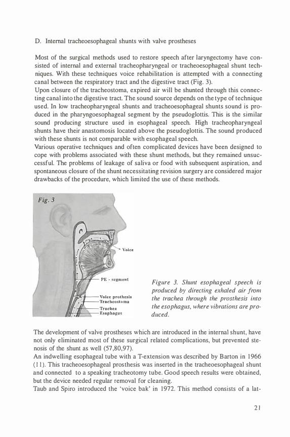
Most of the surgical methods used to restore speech after laryngectomy have consisted of internal and external tracheopharyngeal or tracheoesophageal shunt techniques. With these techniques voice rehabilitation is attempted with a connecting canal between the respiratory tract and the digestive tract (Fig. 3). Upon closure of the tracheostoma, expired air will be shunted through this connecting canal into the digestive tract. The sound source depends on the type of technique used. In low tracheopharyngeal shunts and tracheoesophageal shunts sound is produced in the pharyngoesophageal segment by the pseudoglottis. This is the similar sound producing structure used in esophageal speech. High tracheopharyngeal shunts have their anastomosis located above the pseudoglottis. The sound produced with these shunts is not comparable with esophageal speech. Various operative techniques and often complicated devices have been designed to cope with problems associated with these shunt methods, but they remained unsuccessful. The problems of leakage of saliva or food with subsequent aspiration, and spontaneous closure of the shunt necessitating revision surgery are considered major drawbacks of the procedure, which limited the use of these methods.
Fig. 3
Figure 3. Shunt esophageal speech is produced by directing exhaled air from the trachea through the prosthesis into the esophagus, where vibrations are produced.
The development of valve prostheses which are introduced in the internal shunt, have not only eliminated most of these surgical related complications, but prevented stenosis of the shunt as well (57 ,80,97). An indwelling esophageal tube with a T-extension was described by Barton in 1 966 ( I I ) . This tracheoesophageal prosthesis was inserted in the tracheoesophageal shunt and connected to a speaking tracheotomy tube. Good speech results were obtained, but the device needed regular removal for cleaning. Taub and Spiro introduced the 'voice bak' in 1972. This method consists of a !at-
2 1
erally placed esophagostoma at a preselected cervical level that permitted maximum air flow activation of the pharyngoesophageal mucosa for sound production. The site was selected preoperatively by an insufflation test of the esophagus. The prosthesis was inserted at the fistula site by a flanged silicone tube attached to a one-way saliva valve and regulator worn on the upper chest. During normal breathing this device permitted a two-way air flow, but a one-way flow was obtained to the fistula under increased pressure for speech production. Due to regular mechanical maintenance, high costs and surgical l imitations, the use of this device was limited ( 1 30, 1 37). The first real tracheoesophageal valve prosthesis was introduced by Mozolewski in 1972. Renewed interest for surgical voice rehabilitation was obtained after the introduction of the 'duckbil l ' voice prosthesis by B Jorn and Singer in 1979 ( 1 6). This valved prosthesis could be inserted by a simple endoscopic tracheoesophageal puncture technique. It was constructed of a medical biocompatible grade silicon polymer, resistant against chemical influences. Since than many different silicone-made prostheses have been developed. The non self-retaining prostheses (Bivona, Blom-Singer Duckbill and low pressure devices, Herrmann) are designed for secondary placement some time following laryngectomy. The patient should be able to remove and replace the device for maintenance. The disadvantages of these non self-retaining devices are the attachment of the prosthesis to the skin with glue, reinsertion problems with spontaneous closure of the fistula, irritation of the tracheoesophageal shunt, and shunt migration (97). The selfretaining prostheses (Biom-Singer indwelling, Groningen, Nijdam, Provox, Traissac) need daily maintenance without removal. During voice production these prostheses are self cleaning. They can be placed during laryngectomy, or as a secondary procedure after laryngectomy. Replacement of the prosthesis is performed by a clinician or physician, often as an outpatient procedure. For the introduction and replacement of indwelling prostheses a flexible guide wire with a connector for attachment of the introduction string of the new prosthesis is used. After retrograde introduction of the guide wire through the esophagus and pharynx , the new prosthesis can be inserted transorally. The introduction and replacement of indwelling prostheses can be facil itated by using modified insertion techniques (front loader systems). Just a few complications are reported with the standard procedures which are often limited to dislodgment of the prosthesis, aspiration, external leakage, hypertrophy and granulation of the shunt (97). During shunt esophageal speech the intratracheal air pressure is dependent of the tonicity of the esophagus and the PE-segment, and of the resistance of the prosthesis used. The early developed prostheses (Duckbill, Groningen button, Herrmann, Traissac) are considered to be high resistant prostheses. The newer ones (BiomSinger low pressure, low-resistance Groningen, Provox) are made of low pressure valve designs or are even valveless (N1jdam, 97). The low-pressure prostheses should allow easier passage of air through the shaft, due to improved aerodynamic properties of the valve. By altering the size of the inner diameter of the shaft and by modifying the valve design, the prostheses can improve the efficiency of shunt esophageal
22

speech. However, the size of the diameter of the prosthesis is limited as larger prostheses may interfere with wall strength and give rise to shunt insufficiency (57). The device-life is determined by the normal wear and tear of the silicone rubber, colonization and deterioration of the silicone surface by a mixed biofilm containing fungi, bacteria and food residua. This may lead to stiffening of the valve with secondary leakage of pharyngeal contents into the trachea, and an increased air flow resistance of the valve (57,60,97). A prolonged device life may be expected after surface-coating techniques to prevent fungal and bacterial contamination. The properties of an ideal tracheoesophageal shunt prosthesis are: possible insertions during and following total laryngectomy; self-cleaning; maintenance free; l ow flow resistance; an unlimited device-life ( 1 29).
D.I The different types of shunt valved prostheses
The widely known types of shunt valved prostheses are summarized in Table I. The Blom-Singer prosthesis, the Groningen button, the Low-resistance Groningen button, and the ProvoxTM low-resistance voice prosthesis are described in more detail .
D.l. 1 The Blom-Singer prosthesis
Figure 4. Blom-Singer duckbill voice prosthesis.
The original Blom-Singer prosthesis was introduced in 1 979 (Fig. 4). It consists of a silicone made straight tube with an one-way directional valve, which is introduced in the tracheoesophageal shunt ( 1 6). This hollow tube is open both at the front end and on the side. The valve, formed by a horizontal thin slit attached at the esophageal end of the prosthesis, opens under positive airway pressure and closes by elastic recoil when airflow stops. This mechanism provides a barrier to the reverse flow of esophageal contents. Because of the configuration of the valve, this device is known as the duckbill speech tube or the Blom-Singer voice prosthesis. Blom and Singer develped this prosthesis combined with a tracheoesophageal puncture to overcome the limited usefulness of the Staffieri technique ( 1 6) . This allows air to pass into the esophagus and it keeps fluids from entering the trachea. The original duckbill prosthesis was later modified by lateral flaps for peritracheal
23

Table I. Different types of shunt valved prostheses.
Algaba voice prosthesis Bivona voice prostheses:
Blom-Singer voice prostheses:
Bonelli valve Groningen voice prostheses:
Henley-Cohn voice prosthesis Herrmann ESKA voice prosthesis Panje voice button ProvoxTM voice prosthesis Staffieri voice prosthesis
Duckbill; Low resistance; Ultra low resistance Bivona-Colorado prosthesis Duckbill; Low pressure Indwelling low pressure
Standard button; Low-resistance Ultra-low resistance
Supratracheal semi-permanent valve prosthesis (Mozolewski)
Traissac voice prosthesis
attachment, a retention collar for prevention of tube dislodgement and external leakage, and a low resistance trapdoor valve (Fig. 5) ( 1 24). This low resistance prosthesis is currently used. Initially the prosthesis was only placed during a secondary puncture procedure after the laryngectomy. Under general anaesthesia, the tracheoesophageal wall is reached at the level of the tracheostoma by a rigid endoscope. From the tracheal side a shunt is created towards the endoscope in the esophagus. The secondary puncture procedure was often preferred because of a better preoperative patient selection. A primary procedure was also described (54,86). Some infectious complications were reported after this primary procedure. A separated tracheal and esophageal
Figure 5. Blom-Singer low-pressure voice prosthesis.
24

party wall is a possible reason for this complication, by forming of pockets or false tracts during the puncture (29,88). After the puncture procedure a stent is introduced into the shunt to allow epithelialization of the shunt. The prosthesis is inserted approximately two weeks after the puncture has been performed ( 1 6). When the speaker exhales while closing the tracheostoma with a finger, the air enters the prosthesis through the airflow port and reaches the esophagus. As described before, esophageal voice is created. Deterioration of the silicone material of the prosthesis may give rise to impaired functioning of the valve mechanism and leakage through the prosthesis. The mean device life is approximately 60 days or less ( 1 8, 1 2 1 ). Leakage around the prosthesis is due to shunt dilatation, caused by malfunctioning of the tracheoesophageal shunt (6,53, 1 1 9, 1 46). The Blom-Singer prosthesis requires regular maintenance of the device, by removal from the shunt, valve cleaning and reinsertion a few times weekly (80). Improper reinsertion of the prosthesis by the laryngectomee may result in false routes, dislocation or wandering of the shunt, aspiration of the prosthesis, extrusion of the prosthesis or spontaneous closure of the shunt (6,52,55,56,65, 1 40, 1 4 7). The Blom-Singer indwelling low pressure voice prosthesis was designed for the laryngectomee who is unable to perform the routine removal and insertion necessary for the maintenance of the traditional Blom-Singer prosthesis (Fig. 6a and b). A high success rate (56- 1 00%) of speech rehabilitation by laryngectomees using the Blom-Singer prosthesis, is reported by several authors (40,65, 1 22).
Figure 6a and b. Blom-Singer indwelling type voice prosthesis.
D.I.2 The Groningen button
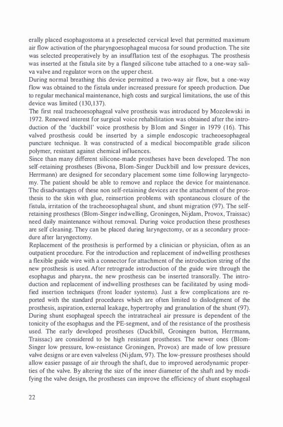
The Groningen button was developed by Nijdam and coworkers in 1 980 (95). This silicone valve prosthesis consists of a tube with two flanges and a valve which is incorporated in the esophageal flange (Fig. 7). The design of this prosthesis was developed to overcome the main disadvantages of the former devices, without inter-
25
fering with the rather good voice rehabilitation results. The Groningen button has self-retaining properties and requires no maintenance. It could stay in place for a considerable time, and it is easily fitted. The prosthesis is less protruding in the esophageal segment. There are no special adhesives necessary to keep the device in place. Also stenting of the newly created tracheoesophageal fistula, was no longer necessary prior to prosthesis insertion, because the prosthesis itself resembles a biflanged tube which is used as a stent (96).
Figure 7. Standard Groningen button.
The tracheal side of the prosthesis is made like a flange with a small string. The string is only used to insert the prosthesis and is removed afterwards. The tracheal side of the prosthesis is open. The esophageal side resembles a combined flange and oneway valve. The large valve outlet was developed for aerodynamic purposes and to stimulate self-cleaning properties of the prosthesis. The Groningen button was initially developed for use during laryngectomy. A primary puncture was favored to keep the period of communicational handicap as short as possible (80). This is probably the first prosthesis that has been described to be placed with a primary procedure, as well as with a secondary endoscopic procedure (7,80,95,96). A well fitted Groningen button allows both shunt and non-shunt injection esophageal speech without dislodgement of the prosthesis (83). Primary prosthetic rehabilitation with the Groningen button is indicated in all patients undergoing total laryngectomy, if the patient decided to accept the minor drawbacks of the procedure. The procedure is contraindicated in the presence of local disturbances as: severe edema of the postlaryngeal area following radiotherapy, perichondritis caused by irradiation, subglottic tumor extension, postcricoid tumor location, and large resections of the pharynx without appropriate reconstruction (80,84). In these cases puncture may promote spread of malignancy or conceal local recurrence of malignancy (80).
Severe pulmonary ventilation disturbances and impaired manual dexterity are considdered general contraindications for prosthetic rehabilitation (84). A relative contraindication for secondary puncture is present in case this procedure should be performed
26

two years or more after the laryngectomy. The laryngectomee may have lost the ability to phonate on expiration with the use of expired air (80). Other relative contraindications for prosthetic voice rehabilitation are certain professions and hobbies, which require both hands. The mean device life is more than 3 months with a range from a few weeks to 2 years (78,8 I ). During this period, the device does not have to be removed for cleaning purposes. The prosthesis replacement can be done as an outpatient procedure (82). The success rate of shunt esophageal voice rehabilitation is higher than that of the non-shunt method. Also a better quality of voice is recognized with the shunt method. A high percentage of patients (75%) were able to produce shunt esophageal speech immediately on the first postoperative day following a secondary procedure or on the 1 2th day after removal of the nasogastric tube following a primary procedure (8 I ,82). The success rate of the secondary puncture group ( 40-79%) is lower than that of the primary puncture group (86-93%) (80,8 I ,82, I 06). Surgery and prosthesis related complications with the Groningen button were few and insignificant in comparison with reports in literature. The uncomplicated surgical procedure for the insertion of the prosthesis was not accompanied with problems of infection (84). The prosthesis-related complications (28%) were only of minor nature and mainly consisted of granulation formation and hypertrophic scarring (84). This is noticed in approximately 7- I 0 % of a selected patients group. It could be caused by a reaction of the body to the foreign material, or by a too short tube shaft of the prosthesis leading to too much tissue pressure (8). Shunt insufficiency and prosthesis dislocation were seen as a late complication in 8 per cent of the cases (80). Leakage through the. prosthesis was due to the increasing stiffness of the silicone material occasionally influenced by overgrowth of Candida species (8). Leakage around the fistula tract could be caused by frequent replacement of the prosthesis and/or manipulation by the patient with the prosthesis ( 1 50). Late infections of the fistula tract were caused by Staphylococcus aureus, sometimes combined with Candida albicans overgrowth. The advantages of the Groningen button as compared to other devices are the selfretaining properties, the sufficient device life, limited maintenance and care by the patient without removal , minimal respiration and deglutition problems, and a high safety but low complication rate. The disadvantages are considered to be device replacement under medical supervision, and hypopharyngeal stenosis which may interfere with the replacement procedure (80).
D.I.3 The Low-Resistance Groningen button
The standard Groningen button has proved to be a satisfactory device i n shunt oesophageal speech, since its introduction in I 980. The relative high airflow resistance of this standard prosthesis compared to low resistance devices, is considered a disadvantage for voice rehabilitation . A modified low resistance device has been developed without changing the successfully tested design of the standard prosthesis (Fig. 8). The advantage of a low airflow resistance in valve prostheses is to facilitate
27

tracheoesophageal shunt phonation. A lower intratracheal air pressure is required for phonation. The intratracheal air pressure depends on a combination of factors. These are the tonicity of the pharyngoesophageal segment, design and condition of the valve of the prosthesis, and the patient skills to produce shunt speech ( 1 5 1 ).
The airflow resistance of a valve prosthesis depends on the design of the valve, the diameter of the shaft, and the materials used. The valve seems to be of major importance on the airflow resistance.
Figure 8. Low-resistance Groningen button.
The low-resistance Groningen button has a semicircular slit of 1 45" in the hat of the esophageal flange, in contrary to a straight slit in the hat of the esophageal flange of the standard type ( 1 5 1 ). Increased intratracheal pressure will force the valve slit to open for air passage. The valve will close automatically by natural recoil of the material. A reduction of 55% in the range of airflow normally used for tracheoesophageal shunt phonation has been achieved at a comfortableloudness level by the design of the low-resistance type. An evident reduction of airflow resistance has also been found with the low-resistance type during in vitro experiments ( 1 5 1 ) . Tracheoesophageal shunt phonation and speech are expected to be less straining. In vivo results indicate improvement of the efficiency of the shunt phonation and speech. Further modifications of the Groningen button by elongating the slit in the esophageal flange up to 200" (Ultra-low resistance prosthesis) resulted in a reduction of the intratracheal phonatory pressure. However, these modifications may increase the risk of internal leakage of esophageal contents. Additional modifications of the valve or the design of the prosthetic tube, are not expected to decrease airflow resistance significantly. Eventually anatomical changes to the PE-segment may lead to further improvement of aerodynamics.
D.I.4 The ProvoxTM voice prosthesis
Further differentiation of the methods and instruments for prosthetic voice rehabilitation has led to the development of a new low resistance, self-retaining voice pros-
28
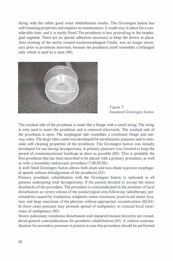
thesis (ProvoxTM) in 1 987 (6 1 ) . This prosthesis was designed to have low airflow resistance for effortless and fluent shunt speech, optimal self-retaining properties, extended device l ifetime, easy outpatient replacement of the device, and simple maintenance procedures ( 1 1 1 ) .
The ProvoxTM prosthesis, i s a biflanged device and made of medical grade silicon rubber (Fig. 9). The esophageal flange is more rigid than the tracheal flange. The valve is molded into one piece with the prosthesis and is supported by a fluoroplastic ring, which is securely fastened in the shaft of the prosthesis. This ring is radiopaque (6 1).
This prosthesis may be used in primary and secondary puncture procedures. The procedure is more or less the same as described by the Groningen prosthesis. The maintenance and care of the prosthesis are limited. The retained device should be cleaned daily with a cotton swab or a specially developed brush. Mucous debris and crusts can be removed easily over the entire length of the prosthesis. The mean device life is estimated to be approximately 6 months, ranging from 5
weeks to more than 2 years (62). Mild leakage through the valve, caused by Candida overgrowth, was the main indication for prosthesis replacement. Increased airflow resistance was Jess often encountered as a reason for replacement. The opening pressure of the ProvoxTM prosthesis (3-5 mm H20) is comparable to the low-resistance Blom-Singer prosthesis, low-resistance Groningen button and Panje prosthesis, but evidently lower than the Blom-Singer duckbill prosthesis and the standard Groningen button. In vivo studies of the Provox™ device compared to the standard Groningen device revealed a 50% reduction of airflow resistance in favor of the ProvoxTM device (6 1 ).
Figure 9. ProvoxTM voice prosthesis.
Good long term speech results (90%) were reported with the ProvoxTM device. Speech failures accounted for 5 per cent of the cases. Also good results were obtained in patients after a gastric pull-up operation (62). Only minor complications were reported, which consist of leakage of fluids around the prosthesis (8,6% ). Other complications ( 1 4,5%) are uncontrollable leakage around and through the prosthesis, hypertrophic scarring and a wrongly placed pros-
29
thesis. The fistula was reduced or closed surgically to overcome these problems. In some patients successful repuncture and voice acquisition were accomplished. Esophageal or hypopharyngeal stenosis was reported in three patients (82).
The Provox™ prosthesis showed to be a useful alternative tool in shunt esophageal voice rehabilitation.
4. General principles of surgical voice rehabilitation
A. Preoperative speech evaluation
Good candidates for tracheoesophageal puncture should be motivated and mentally stable patients ( 1 02). Understanding of the anatomy and function of the prosthesis, as well as manual dexterity and visual acuity are necessary factors in order to guarantee maintenance and care for the stoma and prosthesis. Active and passive air insufflation tests with or without videotluoroscopy should be routinely undertaken to find out if the patient will be able to produce shunt esophageal speech following total laryngectomy. Additional tests via multiple level manometry may be necessary. This allows the examiner to assess the presence or lack of the vibrating pharyngoesophageal segment. Patients should also have a good to moderate pulmonary ventilation and a good cough reflex. Patients with low pulmonary flow rates may have difficulty with the speech fistula (24). Patients who undergo pharyngeal reconstruction with a skin flap or visceral transposition may effectively use tracheoesophageal phonation (61 ,62, 1 30). In these cases the prognosis for esophageal speech acquisition and effective artificial larynx use is relative poor ( 1 30). After total laryngectomy a complex of speech options become possible. The esophageal speech and artificial larynx speech options have already been discussed. All methods have one major communication goal: to accomplish patients satisfaction in meeting his or her communication needs.
B. Surgical techniques
Treatment for laryngeal malignancies is inextricably linked to quality of life after diagnosis. Fundamental concern revolves around voice production, while secondary consideration of the maintenance of good deglutition is of prime importance. It is the dynamic interplay between the intelligibility of speech and the vegetative function of swallowing, especially encountered with extended reconstructive procedures, that dominates the analysis of treatment in voice rehabilitation following total laryngectomy (42). The decision for primary or secondary tracheoesophageal puncture rests with the surgeon and the patient.
B.l Tracheoesophageal puncture at the time of laryngectomy: the primary procedure
The technique of total laryngectomy should be carefully assessed in order to obtain good shunt esophageal speech.
30

The tracheostoma must be of adequate size to accomplish sufficient closure with a finger. Tendency for stenosis of the stoma should also be prevented. The pharyngeal vocal tract must have efficient aerodynamic properties to enable good airflow. A right muscular tonus of the pharyngeal wall is necessary to generate a good pitch ( 1 1 2).
The procedure of total laryngectomy is performed after intubation and creation of a tracheostomy. After resection of the larynx with or without a hemithyroidectomy from the surrounding neck structures, the specimen is taken out after making a horizontal incision at the first tracheal ring. The U-shaped incision length is dependent on the length of the neck and the required elevation of the superior skin flap to expose the hyoid. The tracheostoma is created by suturing the skin to the upper tracheal ring (42).
The tracheoesophageal puncture is placed through the back wall of the trachea from the cervical esophagus. In the methods described for the Groningen and ProvoxTM prostheses, a pharynx protector is used and placed inside the open pharynx wound and positioned just cranially of the tracheostoma. A trocar or sharp pointed wire is used for puncturing the tracheoesophageal wall. The cutting device is placed in the midline of the trachea and between 1 and I ,5 em below the cut edge of the posterior tracheal wall. If the tracheoesophageal wall has been separated to or below this level, a primary puncture should not be performed to avoid possible abscess formation. After puncturing the wall the mandrin of the trocar is removed, and a flexible or metal guide wire is introduced. After the tip of the wire is visible in the pharynx opening, the string of a prosthesis of the proper size is attached to it. By pulling the guide wire with the prosthesis backward, the prosthesis will be inserted into the tracheoesophageal shunt. The tracheal flange should be brought into position to accomplish accurate fitting of the prosthesis. The introduction string of the prosthesis is then cut off. Hypopharyngeal closure is achieved by approximating the mucosal edges in a Y fashion, allowing a low-tension closure adapted to the defect. Running or interrupted sutures are used for mucosal closure. This is followed by submucosal and muscular sutures. The muscular closure may pull the eventually myotomized edges apart. Very tight closure of this layer is not recommended. To prevent fausse routes a nasogastric tube is introduced before mucosal closure. Skin closure is performed after unilateral dissection of pharyngeal plexus branches, and introduction of wound drains for vacuum suction. A cuffed Shiley® canula is introduced into the tracheostoma for 24-48 hours. Prosthetic voice rehabilitation may start I 0- 1 2 days postoperatively, as soon as the nasogastric tube has been removed (95, 1 1 2).
B .II Secondary voice restoration
A number of factors are considered in selecting patients for secondary endoscopic voice restoration following laryngectomy. A barium pharyngoesophagogram is indicated if there is a history of significant deglutition problems, stricture, or pharyngoesophageal reconstruction. Evidently, also the factors described at the primary puncture section are judged.
3 1
The tracheostoma is evaluated with regard to location and diameter. The minimal stomatal diameter to accommodate a voice prosthesis without obstructing respiration is I ,5 em. The small stoma is managed by dilatation with silicone stents or surgical revision. Stoma stents may also be used in combination with the voice prosthesis (42).
The secondary puncture is also performed under general anaesthesia. An esophagoscope is introduced into the hypopharynx, and advanced towards the tracheostoma while the midline of the neck is palpated with a hand. The tip of the esophagoscope is rotated 1 80., to direct the open side of the instrument upwards. After removing the intubation tube out of the trachea, a trocar or sharp pointed wire is used again to puncture the tracheoesophageal wall. A metal or flexible guiding wire is advanced upwards through the esophagoscope into the month, after removal of the mandrin. After attaching the prosthesis to the wire, it is pulled back and inserted into the created tracheoesophageal shunt. Voice rehabilitation may start almost immediately after this procedure (6 1 ,8 1 ,82, 1 1 2) .
B.III Pharyngeal constrictor myotomy
A pharyngeal constrictor myotomy may be a very important single step to facilitate tracheoesophageal puncture speech, as well as esophageal speech. In general constrictor myotomy is created at the time of laryngectomy, especially if the muscles are not reapproximated in the closure (42). Hypertonicity of the PE-segment and constrictor spasm are considered to be major causes of fail ing shunt esophageal speech. A tight PE-segment will prevent the passage of air, while relaxation will facilitate air expulsion and subsequent phonation (80). Pharyngoesophageal myotomies were originally performed as a secondary procedure to improve voice rehabilitation after laryngectomy. A primary myotomy is recommended in cases of a high tension of the upper esophageal sphincter. This can easily be judged by palpating the esophageal inlet musculature. A primary myotomy is preferred over a second stage operation, because after extensive neck surgery and radiation this may be a difficult procedure. During total laryngectomy, it is easily performed. Patients often refuse to undergo a second operation. Myotomies or pharyngeal plexus neurectomy of the PE-segment have been advocated to treat or prevent this hypertonic PE-segment (79,80, 1 22, 1 24).
When too much musculature has been dissected, a hypotonic state of the PE-segment may be caused, which results in a breathy quality of the shunt esophageal speech. Modified procedures have been reported to prevent these problems (80). The present technique is performed in the posterior midline from the level of the tongue base to the puncture site. All the musculature is incised to the depth of the submucosal vasculature. To prevent embarrassment of vertical vasculature, straight myotomy incisions should be made (42). The mucosa should be carefully examined for mucosal lacerations after performing the myotomy. It is advocated to perform a pharyngeal constrictor myotomy, even if the tracheoesophageal puncture has been delayed to a secondary procedure (42).
32

B .IV Pharyngeal plexus neurectomy
During tracheoesophageal speech a critical tension in the muscular waH of the pharynx must exist to permit airflow for speech production. Hypertonicity or spasm of the PE-segment will trap air, forcing it into the stomach with resultant distention. In the majority of cases, a unilateral myotomy of the cricopharyngeus muscle and inferior pharyngeal constrictor muscle is successful in preventing elevated tensions in the PEsegment during phonation (42, 1 24). The resultant tonicity of the PE-segment wilJ affect the pitch in alaryngeal phonation. A unilateral neurectomy of the pharyngeal plexus may have the advantage over a myotomy in that it is less anatomically destructive ( 1 24). Additionally, the vascular supply to the pharyngeal wall is preserved. A neurolytic procedure may result in a finer adjustment of the pharyngeal wall tensions. One of the disadvantages of a myotomy procedure may be a hypotonic state of the PE-segment. A weak or absent PEsegment will develop lower pitches. Vocal intensity and pitch can be improved by applying pressure to the pharynx. The pharyngeal plexus neurectomy can be performed simultaneously with total laryngectomy ( 124). After separating the constrictor pharyngeal muscles from the laryngeal specimen, the posterolateral pharyngeal waH is stretched by grasping the edges of the pharyngostomy. At the level of the middle pharyngeal constrictor muscle, the pharynx is separated from the prevertebral space. The pharyngeal plexus is frequently identified at the level of the superior thyroid artery. After checking the plexus by electrical stimulation, the identified branches are divided and the edges are separated by electrocautery. In the secondary setting the identification of the pharyngeal branches is more difficult or impossible because of scarring and previous dissection in this area. Pharyngeal plexus neurectomy is another way to improve alaryngeal speech acquisition and quality.
5. Troubleshooting voice failure and complications
As described in previous sections, shunt esophageal speech has many advantages above other methods of voice rehabilitation. As foreign bodies, voice prostheses for vocal rehabilitation of the laryngectomee may also be accompanied by frequent minor or severe complications. Primary voice fistulas created at the time of laryngectomy were prone to more severe complications, i .e. infections. Fistula creation required a longer course of treatment in this group, compared to secondary created fistulas. The secondary procedures were often associated with complications as esophageal perforation, peristomal cellulitis or cervical osteomyelitis (6). By modifying the techniques of performing these punctures the complication rate has declined drastically (6 1 ,62,8 1 ,82, 1 06).
Proper fit of the prosthesis is necessary for good quality esophageal speech and to prevent complications. The length of the device and the angle of the prosthesis entering the posterior wall of the trachea into the esophagus are important factors. A long
33

shaft of the prosthesis may injure the posterior pharyngeal wall. When excessive digital pressure is applied in occlusion of the stoma, the stoma may be pushed to the posterior esophageal wall. This may of course cause obstruction of the airflow through the prosthesis. Other described complications encountered with voice prostheses are summarized in table II. Many of the mentioned problems can be solved by simple measures as conservative antibiotic treatment, prosthetic replacement procedures, or surgical revisions.
Table II. Complications with voice prostheses and literature.
Local inflammation and/or infection
Migration of the fistula tract Closure of the fistula tract Dislocation of the prostheses Granulation tissue formation Ftbrous tissue formation Hypertrophic scarring Esophageal stenosis or stricture Swallowing problems Tracheostomatal stenosis Leakage through the prosthesis Leakage around the prosthesis Pulmonary aspiration Increased airflow resistance
Pharyngeal constrictor spasm
6. Tracheostoma valves
(6,8) (6) (6) (88) (6) (8) (83) (6) (6) (6) (79,83) (8,83) (6) (8)
(80)
During shunt esophageal speech with a voice prosthesis the patient is committed to manual occlusion of the airway. Recognizing these limitations tracheal valves were developed that close the trachea for phonation, but remain open during respiration. This allows shunt esophageal speech without digital occlusion of the tracheostoma. During shunt phonation a thin diaphragm responds to the increase in air pressure by closing. These tracheostoma valves should enable a better social and professional reintegration of the laryngectomee. The employment of a flexible valve housing or collar (Blom-Singer tracheostoma valve, Provox™ stomafilter) must be sealed for attachment to the peristomal skin (Fig. lOa and J Ob) (4,5, 1 7, 1 25). This is done with the use of double-faced tape and/or l iquid adhesives. A valve part can easily be inserted in the housing and can be quickly removed. In combination with a heat and moisture exchanger the ease of prosthetic vocal rehabil itation can also be improved by reducing dryness, mucous secretions and coughing. A significant disadvantage of this type of tracheostoma valves is insufficient durability of the adhesive seal attachment (80).
34

The ESKA-Herrmann tracheostoma valve has been developed to eliminate the above mentioned sealing problems (59). The tracheostoma valve consists of a cannula part with 3 different silicone outer flanges to retain the device in the stoma. A similar concept is provided by the Barton-Mayo tube which consists of a customized tracheal cannula for a tight stomal occlusion. Fluent speech can be achieved with this valve when its properly fitted and a functional voice prosthesis is inserted. This tracheostoma valve may interfere with the intensity of voice. The application and use of tracheostoma valves are described to be disappointing, due to application problems. Patients must be selected and fitted appropriately with a valve. Before the valve is attempted, the patients should be able to speak effectively using digital stoma occlusion. Sneezing and coughing are difficult to manage (80). In case of significant respiratory problems, excessive mucous discharge, or secretions, the use of tracheostoma valves may be problematic.
Figure 10. (a) A Blom-Singer adjustable tracheostoma valve attached to a disposable plastic housing ( ProvoxTM). (b) A heat and moister exchanger ( ProvoxTM Stomafilter) can be used for digital stoma occlusion combined with a Barton-Mayo Tracheostoma Button.
7. Postoperative speech evaluation
The quality of shunt esophageal speech can be assessed by using questionnaires, videofluoroscopy, videotaped interviews, and head and neck examinations. The appearance of the reconstructed pharyngoesophageal segment can be examined by videofluoroscopy during swallowing, esophageal phonation, and phonation during air insufflation. The esophageal segment must be examined dynamically ( 1 30). Voice production by means of a voice prosthesis resembles the normal laryngeal voice production. Pulmonary air is used as the energy source. The pseudoglottis is known as the alternative sound source. Voice rehabilitation may start I 0- 12 days after primary puncture.
35
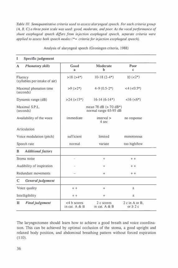
Table Ill. Semiquantitative criteria used to assess alaryngeal speech. For each criteria group
(A, B, C) a three point scale was used: good, moderate, and poor. As the vocal performance of
shunt esophageal speech differs from injection esophageal speech, separate criteria were
applied to assess both speech modes (*= criteria for injection esophageal speech).
Analysis of alaryngeal speech (Groningen criteria, 1 988)
I Specific judgement
A Phonatory skills Good Moderate Poor a b c
Fluency > 1 8 (>4*) I 0- 1 8 (2-4*) 10 (<2*) (syllables per intake of air)
Maximal phonation time >9 (>2*) 4-9 (0.5-2*) <4 (<0.5*) (seconds)
Dynamic range (dB) >24 (> 1 5*) 1 6- 1 4 (6- 1 4*) < 1 6 (<6*)
Maximal S.P.L. mean 78 dB (> 70 dB*) (seconds) normal range 65-95 dB
Availability of the voice immediate interval > no response 4 sec
Articulation
Voice modulation (pitch) sufficient limited monotonous
Speech rate normal variate too high/low
B Additional factors
Stoma noise - + + +
Audibility of inspiration - + + +
Redundant movements - + + +
c General judgement
Voice quality + + + ±
intelligibility + + + ±
II Final judgement <4 b scores 2 c scores 2 c in A or B, in cat. A & B in cat. A & B or � 2 c
The laryngectomee should learn how to achieve a good breath and voice coordination. This can be achieved by optimal occlusion of the stoma, a good upright and relaxed body position, and abdominal breathing pattern without forced expiration ( 1 10).
36

The majority of patients benefits from a brief program of speech therapy provided simultaneously with daily instructions in prosthesis management. Patients are instructed to use a continuous pulmonary airflow to speak fluently and with natural phrasing. By varying expiratory pressure and flow they can influence duration, stress, and intonation of their communication pattern. Articulation therapy should be directed toward consonant voicing, speech intelligibility, and specific phoneme difficulties in case of anatomical alterations by surgery in the mouth or oropharynx. Unsatisfactory voice production may be caused by several problems. Articulation problems are often due to a dysfunction of the tongue, soft palate, jaw or lips. In case of pharyngoesophageal spasm, voice production will be poor. The injection of air into the esophagus is usually sufficient, but regurgitation gives an uncoordinated strained and strangled voice. With a stricture the patient will not be able to inject sufficient air into the esophagus for phonation to occur. This may be solved with the already described myotomy and or neurectomy procedures of this segment. In case of hypotonicity of the pharyngoesophageal segment, the pharynx and esophagus dilate on swallowing, and the voice is weak and whispery. This can be corrected by digital pressure on the pharynx externally (42). Also prosthesis related problems should be checked in case of no voice or poor voice. Insufficient stoma closure and excessive mucus production may also interfere with shunt esophageal voice. The laryngectomee should learn how to effectively clean his self-retaining device daily with a specially developed brush or cotton swab. A temporary decrease of shunt esophageal voice production may occur during radiotherapy due to swelling and rigidity of the pharyngeal wall. Nonetheless speech therapy should continue, and the laryngectomee must be reassured that the voice quality will improve after the radiotherapy sessions. The evaluation of shunt esophageal voice production is standardized conform to the criteria discussed at the 3rd International Congress on Voice Prosthesis (Groningen, 1988) (Table III). The phonatory skills of voice production are judged on the following criteria: fluency (quantity of syllables per intake of air), maximum phonation time, dynamic range, availability of the voice, articulation, voice modulation and speech rate. The fluency of speech measured by the number of syllables produced on one breath, is suggested to determine the quality of perception of shunt esophageal voice (2 1 ,80). Additional factors as stoma noise, audibility of inspiration, and redundant movements of the head and neck are also evaluated. Vocal quality and intelligibility are to be judged subjectively during the entire examination. The voice should be relaxed, pleasantly and easily understood (2 1 ) .
Speech therapy should both consist of injection esophageal speech and shunt esophageal speech. Shunt esophageal speech can be acquired faster than the original nonshunt esophageal voice. Patients with shunt esophageal voice, were also able to acquire the injection esophageal voice easier ( I 06). Eventually the laryngectomee may choose which form of communication is preferable.
37

Discussion
Tumors of the larynx are not only significant because they present treatment dilemmas but also because most treatment modalities have a profound impact on the life of a patient. The loss of the larynx requires social adjustment secondary to changes in respiration, speech, and deglutition. The primary goal of laryngectomy rehabilitation is to efficiently restore these dynamic functions after accomplishing radical tumor extirpation (42). Achieving this goal depends significantly on the patient's adaptation to the loss of normal speech. The most common methods of alaryngeal communication which may be used are the artificial larynx, injection esophageal speech and tracheoesophageal speech. The disadvantages of the hand-held electrolarynx are the monotonous mechanical sounding voice, maintenance and care, and the fact that the user should always carry it with him or her. In comparison, esophageal speech has the advantage of hands-free voice production. Traditionally, injection esophageal speech has been considered the method of choice for vocal rehabilitation after total laryngectomy. However, approximately 50 percent of the patients using esophageal speech fail to acquire functional communication ( 1 30). Among those who do, therapy-time is considerable and the resultant speech proficiency is variable. Characteristically, the quality of acquired esophageal voice is limited with respect to intensity, pitch, and rate (42). Tracheoesophageal phonation produces higher success rates of useful voice, but requires an operative procedure and patient compliance. The fistula techniques, which allow tracheoesophageal speech, have been complicated with a high failure rate due to aspiration, local infection, stenosis, and migration of the tract. In 1979, Singer and Blom revolutionized the speech restoration field with their description of an endoscopic technique in which a tracheoesophageal one-way sil icone valve was placed in a tracheoesophageal fistula after total laryngectomy. Increased amplitude levels of tracheoesophageal voice production are possible because the pulmonary system generates and sustains greater esophageal pressures. The characteristics of tracheoesophageal speech are more similar to normal laryngeal speech because of the powerful advantage of pulmonary supported airflow. Esophageal speakers have a reduced maximum phonation time due to their limited esophageal volume (80 ml). The finding that tracheoesophageal speech production is acoustically more similar to laryngeal speech, encouraged several researchers to develop a more ideal prosthesis for vocal rehabilitation. Compared to the original developed prostheses (i.e. BlomSinger, Panje), the self-retaining prostheses ( i .e. Groningen, ProvoxTM) ensure easier patient's maintenance and care for the prosthesis. This could be the reason for less reported complications concerning the fistula tract. Primary placement of the prosthesis at the time of laryngectomy leads to higher success rates without increased postoperative complications. Also primary introduction of the prosthesis at the time of laryngectomy made stenting of the created fistula unnecessary, which resulted in a reduction of postoperative woundhealing. The replacement of the self-retaining
38

(indwelling) prostheses can be performed as a simple outpatient procedure. This can be further enhanced by the introduction of front loader systems (Blom-Singer Gel Cap Insertion System, Voice master). Preoperative evaluation of the candidates for surgical voice rehabilitation have proved to increase the success rate of shunt esophageal speech. Factors as visual acuity, manual dexterity, sufficient pulmonary reserve and coughing reflexes, and the muscular tonus of the cricopharyngeal segment appears to be important parameters for patient selection. However, the anatomical changes that follow surgical tumor resection will obviously determine which option for communication is adaptable. With the development of self-retaining low-resistance prostheses a reduction of airflow resistance has been achieved. These prostheses are considered comfortable because less effort is needed to produce adequate speech. However, not every laryngectomee may profit from the benefits of low pressure devices. The tonicity of the PE-segment seems to play a major role in the airflow resistance during shunt esophageal speech. The mean device life of the different prostheses ranges from a few weeks to two or more years. Deterioration of the prostheses may lead to increased airflow resistance and leakage of fluids through the device. Device l ife may be influenced by microbial (yeast) colonization of the silicone material, which leads to irreversible damage of the valve mechanism. Early failure of the voice prostheses can be prevented by using antifungal drugs (e.g. Amphotericin-B) lozenges in patients who tend to have rapid shunt valve deterioration (78). The complications encountered with the self-retaining low-resistance prostheses are not significant and mainly consist of hypertrophic scarring, granulation formation and minor leakage around the device. Usually, these problems can easily be overcome with limited measures. The ideal voice prosthesis with self-retaining, self-cleaning, and low pressure properties combined with an unlimited device life, has yet to be developed. As device l ife seems to be limited in most cases, improved techniques should be advocated that reduce the sometimes inconvenient methods for replacement of indwelling prostheses. Considering the quality of life, primary prosthetic voice rehabilitation seems to be an adequate method of alaryngeal communication following laryngectomy.
References
I . Alajmo E . La laryngectomie reconstructive avec crico-hyoidopexie. Rev Laryngol Otol Rhino) (Bord) 1974; 95: 53-57.
2. Alajmo E. Le role de Ia laryngectomie reconstructive dans Ia chirurgie du larynx. Rev Laryngol Otol Rhino) (Bord) 1976; 97: 207-2 1 0.
3 . Amatsu M. A one stage surgical technique for postlaryngectomy voice rehabilitation. Laryngoscope 1980; 90: 1 378- 1 386.
39
4. Amatsu M. Amatsu's technique using the Blom-Singer tracheostoma valve. In: Henmann IF, editor. Speech restoration via voice prostheses. BerlinHeidelberg: Springer Verlag, 1 986: 177- 1 8 1 .
5 . Amatsu M, Makino K, Kinishi M et al. Primary tracheoesophageal shunt operation for postlaryngectomy speech with sphincter mechanism. Ann Otol Rhino! Laryngol 1986; 95: 373-376.
6 . Andrews 1C, Mickel RA , Hanson DG, e t al . Major complications following tracheoesophageal puncture for voice rehabilitation. Laryngoscope 1 987 ; 97 : 562-567.
7 . Annyas AA, Nijdam HF, Escajadillo 1R et al . Groningen prosthesis for voice rehabilitation after laryngectomy. Clin Otolaryngol 1984; 9: 5 1 -54.
8. Annyas AA. Indwelling voice prostheses for voice restoration after total laryngectomy: Complications and Solutions. Teaching Course on Indwelling Voice Prostheses for Voice Restoration after Total Laryngectomy; 1991 Apr 1 2; Amsterdam. Amsterdam: The Netherlands Cancer Institute; 1 99 1 : 36-38.
9. Asai R. Asai 's new voice production method: substitution for voice and speech. In: Ono Y, editor. Proceedings of the Vlllth International Congress of Otorhinolaryngology; 1965; Tokyo. Amsterdam: Excerpta Medica, 1 966: 730.
1 0. Asai R. Laryngoplasty after total laryngectomy. Arch Otolaryngol 1 972; 9 5 : 1 1 4- 1 1 9.
1 1 . Barton RT. A review of attempted physiological restoration of voice following total laryngectomy. In: Ono Y, editor. Proceedings of the VIIIth International Congress of Otorhinolaryngology; 1965; Tokyo. Amsterdam: Exerpta Medica, 1966: 73 1 -734.
1 2. Bentzen N, Guld A, Rasmussen H. X-ray video tape studies of laryngectomized patients. 1 Laryngol Otol 1976; 90: 655-666.
1 3 . Berg 1W van den. Myoelastic-aerodynamic theory of voice production. 1 Speech Hear Res 1958; I : 227-244.
1 4. Berg 1W van den, Moolenaar-Bijl A1, Damste PH. Oesophageal speech. Folia Phoniatr 1958; 10: 65-84.
1 5 . Berg 1W van den, . Moolenaar-Bijl AJ. Cricopharyngeal sphincter pitch intensity and fluency in oesophageal speech. Pract Oto-rhino-Laryngol 1 959; 2 1 : 298-3 1 5.
1 6. B Jorn ED, Singer MI. Surgical-prosthetic approaches for postlaryngectomy voice restoration. In: Keith RL and Darley FL, editors. Laryngectomee rehabilitation. Houston: College-Hill Press, 1979: 25 1 -276.
1 7. BJorn ED, Singer MI, Hamaker RC. Tracheostoma valve for postlaryngectomy voice rehabilitation. Ann Otol Rhino! Laryngol 1982; 9 1 : 576-578.
1 8. B Jorn ED, Singer MI, Hamaker RC. A prospective study of tracheoesophageal speech. Arch Otolaryngol Head Neck Surg 1986; 1 1 2: 440-447.
1 9. Bohme G. Stimm-, Sprech- und Sprachstorungen. Stuttgart: Gustav Fisher Verlag, 1974: 3- 19.
20. Bockler R, Wein B , Klajman S et al . Die Ultraschalluntersuchung der Pseudoglottis bei Kehlkopflosen. HNO 1988; 36: 1 1 5- 1 1 8 .
40
2 1 . Bors EFM. Indwelling voice prostheses for voice restoration after total l aryngectomy: Criteria for evaluation of speech with a voice-button. Teaching Course on Indwelling Voice Prostheses for Voice Restoration after Total Laryngectomy; 1 99 1 Apr 12 ; Amsterdam. Amsterdam: The Netherlands Cancer Institute; 1 99 1 : 50-57.
22. Brandenburg JH, Bless DM, Salinsky B. Speech results of Brandenburg's neoglottic procedure. In: Myers EN, editor. New dimensions in otorhinolaryngology, head and neck surgery. Vol II. Amsterdam: Elsevier Science Publ BY, 1985: 398-399.
23. Brankel 0. Formung und Gestalt der Pseudoglottis Laryngektomierter im Stroboskopischen Rontgenbild. Folia Phoniatr 1 957; 9 : 1 8-3 1 .
24. Brendebach J, Schmidt M, Brugger E. Lung function tests in laryngectomized patients. In: Herrmann IF, editor. Speech restoration via voice prostheses. Berlin-Heidelberg: Springer Verlag, 1986: 1 30- 1 34.
25. Breuninger H. Zur Rehabilitation nach Laryngektomie. Laryngol Rhinal Otol 1982; 6 1 : 267-27 1 .
26. Brouwer B, Snow GB, van Dam FASM. Experiences of patients who undergo laryngectomy. Clin Otolaryngol 1979; 4: 109- 1 1 8 .
27. Brusis T, Schoning A. Wie gut ist die Oesophagusstimme? Laryngol Rhinal Otol 1984; 63: 585-588.
28. Calcaterra TC, Jafec BW. Tracheoesophageal shunt for speech rehabilitation after total laryngectomy. Arch Otolaryngol 197 1 ; 94: 1 24- 128.
29. Casiano RR, Maniglia AJ, Lundy DS. Analysis of primary versus delayed tracheoesophageal puncture. In: Myers EN, editor. New dimensions in otorhinolaryngology, head and neck surgery. Vol II . Amsterdam: Elsevier Science Publ BY, 1 985: 109 1 - 1 092.
30. Cheesman AD, Holden H, Perry A. Assessment and management protocol for trachea-oesophageal puncture procedures. In : Herrmann IF, editor. Speech restoration via voice prostheses. Berlin-Heidelberg: Springer Verlag, 1986: 49-58.
3 1 . Conley JJ, De Amesti F, Pierce MK. A new surgical technique for the vocal rehabilitation of the laryngectomized patient. Ann Otol Rhinal Laryngol 1958; 67: 655-664.
32. Conley JJ. Vocal rehabilitation by autogenous vein graft. Ann Otol Rhino) Laryngol 1959; 68: 990-995 .
33. Damste PH. Oesophageal speech after laryngectomy. [dissertation] Groningen: Publ. Gebr. Hoitsema, 1958.
34. Damste PH, Moolenaar-Bijl A J. Drie bijzondere vormen van spraak bij een patient met larynxstenose. Log Fon 1960; 32: 49-55.
35. Damste PH, Lerman JW. Configuration of the neoglottis: an X-ray study. Folia Phoniatr 1969; 2 1 : 347-358.
36. Damste PH. Methods of restoring the voice after l aryngectomy. Laryngoscope 1975 ; 85: 649-655.
4 1
37. Damste PH. Some obstacles in learning oesophageal speech. In: Keith RL and Darley FL, editors. Laryngectomee rehabilitation. Houston: College-Hill Press, 1 979: 49-62.
38. Decroix G, Libersa Cl, Lattard R. Bases anatomiques et physiologiques de Ia reeducation vocale des laryngectomises. J Fr Otorhinolaryngol 1958; 7 : 549-573.
39. Diedrich W M, Youngstrom KA. Alaryngeal speech. Springfield (IL): CC Thomas Publ, 1 966.
40. Donegan JO, Gluckman JL, S ingh J. Limitations of the Blom-Singer technique for voice restoration. Ann Otol Rhino) Laryngol 1 98 1 ; 90: 495-497.
4 1 . Duguay M . Special problems o f the alaryngeal speaker. In: Keith RL and Darley FL, editors. Laryngectomee rehabilitation. Houston: College-Hill Press, 1979: 423-444.
42. Duguay MJ, Feudo P Jr. The Process of Postlaryngectomy Rehabilitation. In: Fried MP, editor. The Larynx. ,A Multidisciplinary Approach". BostonToronto: Little, Brown and Co, 1988: 603-613 .
43. Edels Y. Pseudo-voice-its theory and practice. In : Edels Y, editor. Laryngectomy, diagnosis to rehabilitation. London-Canberra: Croom Helm, 1 983: 1 07- 1 4 1 .
44. Edwards N. Post-laryngectomy vocal rehabilitation. Preliminary report of a one stage method using expired air and a valved prosthesis. J Laryngol 1 974; 88: 905-9 1 8.
45. Fontaine A, Mitchell J. Oesophageal voice: a factor of readiness. J Laryngol Otol 1960; 74: 870-876.
46. Frith CK, Montague JC Jr. Reported dietary effects on esophageal voice production. Folia Phoniatr 1 985; 37: 238-245.
47. Fumeaux J. Structure de Ia pseudoglotte des laryngectomises etudiee par radiocinematographie. Rev Laryngol Otol Rhino] 1 96 1 ; 82: 24 1 -246.
48. Gates GA, Ryan W, Cooper JC Jr et al . Current status of laryngectomee rehabilitation: I. Results of therapy. Am J Otolaryngol 1 982; 3: 1 -7 .
49. Gee] RC van. Pitch inflection in electrolaryngeal speech [dissertation]. Utrecht: Univ of Utrecht, 1 983.
50. Gilse PHG van. Another method of speech without larynx. Acta Otolaryngol 1 949; 78 Suppl: 1 209- 12 10.
5 1 . Guttman MR. Rehabilitation o f voice i n laryngectomized patients. Arch Otolaryngol 1932; 15 : 478-479.
52. Hall JG, Arnesen AR. Laryngectomia cum neoglottis phoniatoria. Acta Otolaryngol 198 1 ; 91 : 627-628.
53. Hall JG, Dahl T, Arnesen AR. Speech prostheses, failures, problems and success. In: Myers EN, editor. New dimensions in otorhinolaryngology, head and neck surgery. Vol I. Amsterdam: Elsevier Science Publ BV, 1 985: 4 1 8-42 1 .
54. Hamaker RC, Singer MI, BJorn ED et al . Primary voice restoration at laryngectomy. Arch Otolaryngol 1 985; I l l : 1 82- 1 86.
42

55. Hausfeld JN, Henley Cohn JL. A second generation artificial larynx prosthesis. A comparative clinical evaluation. In: Herrmann IF, editor. Speech restoration via voice prostheses. Berlin-Heidelberg: Springer Verlag, 1986: 1 3- 1 9.
56. Herrmann IF, Zenner HP. Erfahrungen mit der Blom-Singer prothese nach Blom-Singer-Punktion und nach funktiongestorter Neoglottis Phonatoria. HNO 1984; 32: 286-293.
57. Herrmann IF. Introduction to speech restoration. In: Herrmann IF, editor. Speech restoration via voice prostheses. Berlin Heidelberg: Springer-Verlag, 1986: 3-4.
58. Herrmann IF, Hammer C, Grevemeyer G. Experimental research to establish an artificial larynx. In: Herrmann IF, editor. Speech restoration via voice prostheses. Berlin-Heidelberg: Springer Verlag, 1986: 1 93-200.
59. Herrmann IF, Koss W. Experience with the ESKA-Herrmann tracheostoma valve. In: Herrmann IF, editor. Speech restoration via voice prostheses. Berlin-Heidelberg: Springer Verlag, 1986: 1 84- 1 86.
60. Herrmann IF, Poschet G, Zohren J. Wear and tear on the silicon of valve prostheses in the upper digestive tract. A study using electron microscope scanning. In: Herrmann IF, editor. Speech restoration via voice prostheses. Berlin-Heidelberg: Springer Verlag, 1986: 69-73.
6 1 . Hilgers FJM, Schouwenburg PF. A new low-resistance self-retaining prosthesis (Pro,.,oxTM) for voice rehabilitation after total laryngectomy. Laryngoscope 1990; 100: 1 202- 1 207.
62. Hilgers FJM. Indwelling voice prostheses for voice restoration after total laryngectomy: The Amsterdam experience. Teaching Course on Indwelling Voice Prostheses for Voice Restoration after Total Laryngectomy; 1 99 1 Apr 12 ; Amsterdam. Amsterdam: The Netherlands Cancer Institute; 199 1 : 22-26.
63 . Hirano M. Structure and vibratory behavior of the vocal folds. In: Sawashima M and Cooper FS, editors. Dynamic aspects of speech production. Tokyo: University of Tokyo Press, 1977: 1 3-30.
64. Hunt RB. Rehabilitation of the laryngectomee. Laryngoscope 1 964; 74: 382-395.
65 . Johns ME, Cantrell RW. Voice restoration of the total laryngectomy patient: The Blom-Singer technique. Otolaryngol Head Neck Surg 1 98 1 ; 89: 82-86.
66. Kelly DR, Adamovich BLB, Roberts TA Jr. Detailed investigation of alaryngeal speech to elucidate etiology of variation in quality. Otolaryngol Head Neck Surg 198 1 ; 89: 6 1 3-623.
67. Kitzing P, Toremalm NG. The situation of the laryngectomized patient. Acta Otolaryngol 1970; 263 : 1 19- 123.
68. Kluyskens P, Boedts D, Dhont D et al. Note preliminaire sur Ia transplantation du larynx. Acta Otorhinolaryngol Belg 1969; 23: 5-8.
69. Kluyskens P. Human larynx homotransplantation. Am Laryngol Ass, 1 970:9 1 . 70. Kluyskens P, Ringoir S. Follow-up of a human larynx transplantation.
Laryngoscope 1970; 80: 1244- 1 250.
43
7 1 . Kluyskens P, Ringoir S , Roels H. A case of larynx homotransplantation in man. In: Bustamante Gurria A and Salmon LFW, editors. Otolaryngology Proceedings of the Ninth Int Congress, Mexico. Amsterdam: Exerpta Medica, 1 970: 302-305.
72. Komom RM. Vocal rehabilitation in the laryngectomized patient with a tracheoesophageal shunt. Ann Otol Rhino! Laryngol 1974; 83 : 445-45 1 .
73. Landaeau M. La voix oesophagienne. J Fr ORL 1 96 1 ; 1 0: 9-45. 74. Lecluse FL, Tiwari RM, Snow GB. Electroglottographic studies of Staffieri
neoglottis. Laryngoscope 1 98 1 ; 9 1 : 97 1 -975. 75. Leipzig B, Griffiths CM, Shea JP. Neoglottic reconstruction following total
laryngectomy. The Galveston experience. Ann Otol Rhino] Laryngol 1 980; 89: 204-208.
76. Lockhart HB. The surgical rehabilitation of the laryngectomized patients. Laryngoscope 1962; 72: 1 585- 1 595.
77. Luchsinger, R, Arnold GE. Handbuch der Stimme- und Sprachheilkunde. Vol . I. Die Stimme und ihre Storungen. X. Stimme ohne Kehlkopf. Vienna: Springer, 1970.
78. Mahieu HF, van Saene HKF, Rosingh HJ et al . Aantasting van siliconrubber stemprothese door Candida-vegetaties. Ned Tijdschr Geneeskd 1986; 1 30: 891 .
79. Mahieu HF. Laryngectomee voice rehabilitation using the Groningen button voice prosthesis. Rev Laryngol Rhino! Otol 1987 ; I 08: 1 1 3- 1 19 .
80. Mahieu HF. Voice and speech rehabil itation fol lowing laryngectomy [dissertation] . Groningen: Groningen University Press, 1 988 .
8 1 . Manni JJ, van den Broek P, de Groot MAH et al. Voice rehabilitation after laryngectomy with the Groningen prosthesis. J Otolaryngol 1 984; 1 3 : 333-336.
82. Manni JJ, van den Broek P, de Groot MAH. Voice rehabilitation after laryngectomy with the Groningen voice prosthesis: Experiences made in Nijmegen. In: Herrmann IF, editor. Speech restoration via voice prostheses. Berlin-Heidelberg: Springer Verlag, 1 986: 33-36.
83. Manni JJ, van den Broek P. Surgical and prosthesis-related complications using the Groningen button voice prosthesis. Clin Otolaryngol 1 990; 15 : 5 1 5-523.
84. Manni JJ. Indwelling voice prostheses for voice restoration after total laryngectomy: The Nijmegen experience. Teaching Course on Indwelling Voice Prostheses for Voice Restoration after Total Laryngectomy; 1 99 1 Apr 1 2; Amsterdam. Amsterdam: The Netherlands Cancer Institute; 1 99 1 : 1 6- 1 9.
85. Martin DE, Hoops HR, Shanks JC. The relationship between esophageal speech proficiency and selected measures of auditory function. J Speech Hear Res 1974; 17: 80-85.
86. Maves MD, Lingeman RE . . Primary vocal rehabilitation using the BlomSinger and Panje voice prostheses. Ann Otol Rhino! Laryngol 1982; 9 1 : 458-460.
87. McConnel FMS, Teichgraber J. Neoglottis reconstruction following total laryngectomy: The Emory experience. Otolaryngol Head Neck Surg 1 982; 90: 569-575.
44
88. McConnel FMS, Duck SW. Indications for tracheoesophageal puncture speech rehabilitation. Laryngoscope 1 986; 96: 1 065- 1 068.
89. Montgomery WW, Toohill RJ. Voice rehabilitation after laryngectomy. Arch Otolaryngol 1968; 88: 499-506.
90. Montgomery WW, Lavelle WG. A technique for improving esophageal and tracheopharyngeal speech. Ann Otol Rhino) Laryngol 1974; 83: 452-46 1 .
9 1 . Moolenaar-Bijl AJ. The importance of certain consonants i n oesophageal voice after laryngectomy. Ann Otol Rhino] Laryngol 1953; 62: 979-989.
92. Moolenaar-Bijl AJ. Het spreken zonder strottehoofd. Log en Fon 1979; 5 1 : 50-68. 93. Motta GA, Cesari U, Iengo M et al. Comparative study by means of electro
glottography and spectrography in voice of patients operated by cordectomy or total laryngectomy. In: Myers EN, editor. New dimensions in otorhinolaryngology, head and neck surgery. Vol . II. Amsterdam: Elsevier Science Publ BV, 1985: 644-645.
94. Noll JD, Torgeson JK. A cinefluorographic observation of the tongue in esophageal speakers. Folia Phoniatr 1967; 19 : 343-350.
95. Nijdam HF, Annyas AA, Schutte HK et al . A new prosthesis for voice rehabilitation after laryngectomy. Arch Otorhinolaryngol 1982; 237: 27-33.
96. Nijdam HF, Annyas AA, Schutte HK et al . The Groningen voice prosthesis for secondary voice rehabilitation after total laryngectomy. In: Herrmann IF, editor. Speech restoration via voice prostheses. Berlin-Heidelberg: S pringer Verlag, 1986: 20-22.
97. Nijdam HF. Indwelling voice prostheses for voice restoration after total laryngectomy: The development of an indwelling voice prosthesis. Teaching Course on Indwelling Voice Prostheses for Voice Restoration after Total laryngectomy; 199 1 Apr 1 2 ; Amsterdam. Amsterdam: The Netherlands Cancer Institute, 199 1 : 8- 1 0.
98. Ossoff RH, Lazarus CL, Sisson GA. Tracheoesophageal puncture for voice restoration: modification of the Blom-Singer technique. Otolaryngol Head Neck Surg 1984; 92: 41 8-423.
99. Pardes P. Ou en est Ia laryngectomie totale reconstructive? Rev Laryngol Rhino) Otol 1974; 95: 49-5 1 .
I 00. Pardes P, Traissac L, Vazel P. Notre experience des laryngectomies avec reconstruction fonctionelle. Rev Laryngol Rhino) Otol 1 974; 95: 59-62.
10 1 . Pearson BW. Subtotal laryngectomy. Laryngoscope 198 1 ; 9 1 : 1904- 1 9 1 2. 1 02. Pfrang H. Social and psycho-social aspects of vocal rehabilitation in laryn
gectomized patients preliminary results. In: Herrmann IF, editor. Speech restoration via voice prostheses. Berlin-Heidelberg: Springer Verlag, 1 986: 1 65- 169.
I 03. Precechtel A. Contribution a Ia discussion de conferences concernant Ia voice oesophagienne au XXe Congress de Societe Francais de Phoniatrie. J Fr Otorhinolaryngol 1961 ; 10: 69-73.
I 04. Richardson JL. Surgical and radiological effects upon the development of speech after total laryngectomy. Ann Otol Rhino) Laryngol 198 1 ; 90: 294-297.
45

105. Robe E, Moore P, Andrews AHJ et al. A study of the role of certain factors in the development of speech after laryngectomy, l ) type of operation, 2) site of pseudoglottis, 3) coordination of speech with respiration. Laryngoscope 1956; l ) 66: 1 73- 1 86, 2) 66: 382-401
' 3) 66: 48 1 -499.
106. Rosingh HJ, Mahieu HF, Annyas AA et al . Stemrevalidatie na larynxextirpatie met behulp van de Groningen button. Ned Tijdschr Geneeskd 1 99 1 ; 1 35 : 1 3 1 5- 1 3 1 8.
107. Samuel P, Adams FG. The effect of oesophageal speech on dyspeptic symptoms. J Laryngol Otol 1976; 90: 1 099- 1 103.
I 08. Samuel P, Adams FG. The role of oesophageal and diaphragmatic movements in a laryngeal speech. J Laryngol Otol 1976; 90: I I 05- l l l l .
109. Schaefer SD, Johns DF. Attaining functional esophageal speech. Arch Otolaryngol 1982; 108: 647-649.
1 1 0. Scholtens BEGM. Indwelling voice prostheses for voice restoration after total laryngectomy: Speech therapy. Teaching Course on Indwelling Voice Prostheses for Voice Restoration after Total Laryngectomy; 1 99 1 Apr 12 ; Amsterdam. Amsterdam: The Netherlands Cancer Institute, 199 1 : 42-49.
I l l . Schouwenburg PF, Hilgers FJM. Modifications in design and use of the Groningen voice prosthesis. Proceedings of the Third International Congress on Voice Prosthesis; 1988 Sep; Groningen. Groningen, 1 988.
1 1 2. Schouwenburg PF. Indwelling voice prostheses for voice restoration after total laryngectomy: Technical aspects of laryngectomy and primary and secondary TE puncture. Teaching Course on Indwelling Voice Prostheses for Voice Restoration after Total Laryngectomy; 1 99 1 Apr 1 2; Amsterdam. Amsterdam: The Netherlands Cancer Institute, 1 99 1 : 28-33.
1 1 3 . Seeman M. Nouveaux resultats des recherches experimentales dans Ia reeducation de Ia voix oesophagienne. J Fr Otorhinolaryngol 1 96 1 ; 10:47-56.
1 14. Serafini I, Arslan M. Reconstructive laryngectomy: Report of the first 35 cases. Ann Otol Rhino! Laryngol 1 972; 8 1 : 479-486.
1 1 5. Serafini I, Arslan M. Restoration of laryngeal function after total laryngectomy. Report of first 25 cases. Laryngoscope 1 972; 82: 1 349- 1 354.
1 1 6. Shames GH, Font J, Matthews J. Factors related to speech proficiency of the laryngectomized. J Speech Hear Disord 1 963 ; 28: 273-287.
1 17 . Shanks JC. Improving oesophageal communication. In: Edels Y, editor. Laryngectomy, diagnosis to rehabilitation. London-Canberra: Croom Helm, 1983 : 1 63- 176.
1 1 8 . Shipp T. EMG of pharyngoesophageal musculature during alaryngeal voice production. J Speech Hear Res 1970; 1 3 : 1 84- 1 92.
1 1 9. Si lver FM, Gluckman JL, Donegan JO. Operative complications of tracheoesophageal puncture. Laryngoscope 1 985; 95 : 1 360- 1 362.
1 20. Simpson IC, Smith JCS, Gordon MT. Laryngectomy: the influence of muscle reconstruction on the mechanism of oesophageal voice production. J Laryngol Otol 1972; 86: 96 1 -990.
1 2 1 . Singer Ml, BJorn ED. An endoscopic technique for restoration of voice after laryngectomy. Ann Otol Rhino! Laryngol 1 980; 89: 529-533 .
46
122. Singer Ml, BJorn ED, Hamaker RC. Further experience with voice restoration after total laryngectomy. Ann Otol Rhino) Laryngol 198 1 ; 90: 498-502.
1 23. Singer MI, BJorn ED, Hamaker RC. Voice rehabilitation after total laryngectomy. J Otolaryngol 1983; 12: 329-334.
124. Singer MI, BJorn ED, Hamaker RC. Pharyngeal plexus neurectomy for alaryngeal speech rehabilitation. Laryngoscope 1 986; 96: 50-53.
125. Singh W. New tracheostoma flap valve for surgical speech reconstruction. In: Myers EN, editor. New dimensions in otorhinolaryngology, head and neck surgery. Vol II. Amsterdam: Elsevier Science Pub! BV, 1985: 480-483.
1 26. Sisson GA, McConnel FMS, Logemann JA et al. Voice rehabilitation after laryngectomy. Results with the use of a hypopharyngeal prosthesis. Arch Otolaryngol 1975; 1 0 1 : 178- 1 8 1 .
1 27. Sisson GA, Bytell DE, Becker SP et al. Total laryngectomy and reconstruction of a pseudoglottis: Problems and complications. Laryngoscope 1 978; 88: 639-650.
1 28. Sisson GA, Goldman ME. Pseudoglottis procedure: update and secondary reconstruction techniques. Laryngoscope 1980; 90: 1 1 20- 1 1 29.
129. Smith JK, Rise EN, Gralnek DE. Speech recovery in laryngectomized patients. Laryngoscope 1 966; 76: 1540- 1 546.
1 30. Snyderman NL. Surgical Vocal Rehabilitation. In: Paparella MM, Schumrick DA, Gluckman JL, Meyerhoff WL, editors. Otolaryngology. Vol III. Philadelphia-London-Toronto: W.B. Saunders Company, Harcourt Brace Jovanovich Inc, 199 1 : 237 1 -2378.
1 3 1 . Staffieri M , Procaccini A, Steiner W et al. Chirurgische rehabilitation der stimme nach laryngectomie. Laryngol Rhino! Otol 1979; 57: 477-488.
1 32. Stetson RH. Esophageal speech for any laryngectomized person. Arch Otolaryng (Chicago) 1937; 26: 1 32- 1 42.
1 33. Strome M, Mustoe TA, Kelly JH. Voice rehabilitation following laryngectomy myomucosal tracheoesophageal shunt. Arch Otolaryngol 1986; 1 1 2: 1 1 68- 1 1 7 1 .
1 34. Svane-Knudsen V. The substitute voice of the laryngectomized patient. Acta Otolaryngol 1960; 52: 85-93 .
1 35 . Tanabe M, Honjo T, Isshiki N. Neoglottic reconstruction following total laryngectomy. Arch Otolaryngol 1985; I l l : 39-42.
1 36. Taptapova SL. Methode d'enseignement de Ia voix oesophagienne. Rev Laryngol Otol 1 979; 100: 1 89- 199.
1 37. Taub S, Spiro RH. Vocal rehabilitation of laryngectomees : preliminary report of a new technique. Am J Surg 1972; 1 24: 87-90.
1 38. Traissac L, Pardes P, Vazel P et al. A propos de plusieurs techniques de Ia laryngectomie reconstructive. Rev Laryngol Otol Rhino! (Bord) 1 973; 94: 543-557.
1 39. Traissac L, Petit J, Lecoq M et al. Function of the pharynx and Killian mouth in voice production with our prosthesis. In: Herrmann IF, editor. Speech restoration via voice prostheses. Berlin-Heidelberg: Springer Verlag, 1986: 97-99.
47

1 40. Trudeau MD, Hirsch SM, Schuller DE. Vocal restorative surgery : why wait? Laryngoscope 1 986; 96: 975-977.
1 4 1 . Tucker HM. Laryngeal transplantation: current status 1 974. Laryngoscope 1 975; 85: 787-796.
1 42. Vrticka K, Svoboda M. A clinical and X-ray study of 1 00 laryngectomized speakers. Fol Phoniatr 1 96 1 ; 1 3: 1 74- 1 86.
1 43. Vuyk HD, Klinkenberg-Knol E, Tiwari RM. The role of the upper esophageal sphincter in voice rehabilitation after laryngectomy and Staffieri 's procedure. J Laryngol Otol 1 986; 100: 59-68.
1 44. Weinberg B, Westerhouse J. A study of buccal speech. J Speech Hear Res 1 97 1 ; 1 4: 652-658.
145. Weinberg B, Westerhouse J. A study of pharyngeal speech. J Speech Hear Disord 1 973; 38: 1 1 1 - 1 1 8.
1 46. Wetmore SJ, Johns ME, Baker SR. The Singer-Blom voice restoration procedure. Arch Otolaryngol 198 1 ; 1 07 : 674-676.
1 47. Wetmore SJ, Krueger K, Wesson K. The Blom-Singer speech rehabilitation procedure. Laryngoscope 198 1 ; 9 1 : I I 09- 1 1 1 7.
1 48. Wolfe RD, Olson JE, Goldenberg DB. Rehabilitation of the laryngectomee: the role of the distal esophageal sphincter. Laryngoscope 1 97 1 ; 8 1 : 197 1 - 1 978.
149. Zeitzer LD, Hays LL. Tracheoesophageal shunt. Arch Otolaryngol 1 972; 96: 27-30.
I 50. Zenner HP, Herrmann IF, Pfrang H. Voice prostheses: surgery, evaluation and results. In: Myers EN, editor. New dimensions in otorhinolaryngology, head and neck surgery. Vol II. Amsterdam: Elsevier Science Publ BV, 1985: 482-483.
1 5 1 . Zijlstra RJ, Mahieu HF, van Lith-Bijl JT et al. Aerodynamic properties of the low resistance Groningen button. Arch Otolaryngol Head Neck Surg 1 99 1 ; 1 1 7: 657-66 1 .
1 52. Zwitman DH. The function of the pharyngoesophageal segment during speech. In: Keith RL and Darley FL, editors. Laryngectomee rehabilitation. Houston: College-Hill Press, 1 979: 299-3 1 6.
48
Chapter 3
Voice rehabilitation after total laryngectomy using the Provox™
voice prosthesis
Van Weissenbruch R, Albers FWJ. Voice rehabilitation after total laryngectomy using the ProvoxTM voice prosthesis. Clin Otolaryngol 1993; 1 8: 359-364.

Introduction
Since the first laryngectomies performed by Watson ( 1 866) and Bill roth ( 1 873) the loss of the natural vocal production mechanism has been considered as a major disabling consequence of this procedure besides changes in respiration and deglutition (I ,2). Several methods of substitute voice production have been developed with variable success rates. Traditionally the rehabilitation efforts to establish these goals have been centered on the use of artificial larynges, esophageal speech or a combination of these methods. A1aryngeal communication through artificial larynges (e.g. Servox®) has the disadvantages of mechanical sound production and fine-motor control problems of the device with one or both hands. Also visual distractions to the l istener while the device is being used may be considered as a disturbance for interactive communication (3). Esophageal speech is generally considered as the method of choice for speech acquisition after total laryngectomy. It is estimated that 50-70% of laryngectomees use esophageal speech as their primary means of communication (4,5). Other reports have indicated that only about 25% of the laryngectomees achieved functional esoph-ageal speech proficiency (6). Esophageal speech has the advantage of not necessitating finger closure, and the disadvantage of a long-lasting, complicated learning process. Compared to normal laryngeal speech , esophageal speech has the inherent limitations of slower rate, decreased intensity, lower pitch, stoma blast and clucking sounds on air intake (7,8). Tracheoesophageal speech with the use of a valve prosthesis has become an important method of surgical speech rehabilitation after total laryngectomy since the introduction of the Blom-Singer Duckbill prosthesis (9) and the Panje prosthesis ( 1 0) . Several prostheses have been developed for tracheoesophageal puncture performed as a primary puncture at the time of laryngectomy or as a secondary procedure at a later stage ( I I ). It seemed that prosthetic shunt esophageal speech is a more accepttable method of voice restoration with better intelligibility and fluency, and easier to acquire than the traditional esophageal speech or artificial l aryngeal speech ( 1 2) . The problems encountered during routine use of tracheoesophageal prostheses were determined by the high airflow resistance through the prosthesis and the pharyngoesophageal segment, and the limited device life. The development of lowresistance valve prostheses by modifications especially to the valve part of the prosthesis has proven to facilitate tracheoesophageal shunt phonation ( 1 3 , 14 ). A unilateral myotomy of the pharyngeal constrictor muscles could additionally reduce the mean intratracheal phonatory pressure necessary to facilitate airflow through the pharyn-goesophageal segment ( 1 5, 1 6). This may also be accomplished by a selective unilateral neurectomy of the pharyngeal plexus to reduce the remaining pharyngeal muscle tonus ( 1 7). Deterioration of the silicone voice prostheses by fungal colonization and erosion may cause valve dysfunction. The use of local antimycotic drugs has proven to extent device life by preventing early valve deterioration ( 1 8, 1 9) . Since February 199 1 we have been using the Provoxn.r self-retaining, low-resistance
50

prosthesis for voice rehabilitation after total laryngectomy. The experiences made with this type of prosthesis have shown high success rates for voice acquisition, a low complication rate, an acceptable device lifetime with rather easy replacement and maintenance procedures ( 1 4). We have prospectively studied a group of total laryngectomees operated in our department to assess the efficacy of their voice rehabilitation and to evaluate our clinical experiences made with surgical voice rehabilitation.
Patients and methods
Figure 1. The low-resistance ProvoxTM voice prosthesis.
Between February 199 1 and March 1993, a total of 37 patients underwent primary or secondary tracheoesophageal puncture with insertion of a Provox1M voice prosthesis (Figure I). This study included 33 males (mean age: 62 years) and 4 females (mean age: 60 years). A primary puncture was performed in 28 patients (26 males and 2 females), whereas a secondary puncture was done in 9 patients (6 males and 3 females). The time-interval from total laryngectomy to secondary tracheoesophageal puncture ranged from 6-92 months (mean: 14 months). All 37 patients received preoperative or postoperative radiotherapy (50-70 Gy in 5-7 wks). In 17 patients ( 1 4 males and 3 females) a total laryngectomy was performed in association with a unilateral radical neck dissection. The voice button had been removed in 2 male patients because of local tumor recurrence. During the follow-up period, one deceased due to distal tumor metastasis and the other due to pneumonia during irradiation. In all participating patients a Provox1M self-retaining, low-resistance prosthesis was inserted ( 1 4 ) . In total, 72 prostheses were inserted in this group of 37 patients. A primary puncture was performed during total laryngectomy in combination with a unilateral myotomy of the cricopharyngeal muscle and the inferior pharyngeal constrictor muscle to prevent hypertonicity of the pharyngoesophageal segment. This myotomy was performed in 28 laryngectomees in combination with a unilateral pharyngeal plexus neurectomy. The secondary puncture procedures were performed on
5 1
patients with poor acquisition of esophageal speech . These patients did not receive a primary myotomy at the time of total laryngectomy. The function of the pharyngoesophageal segment was evaluated in all patients before secondary puncture by videofluoroscopy and an insufflation test. A secondary myotomy was performed in two patients simultaneously with the secondary puncture procedure. The secondary tracheoesophageal puncture was performed endoscopically with a rigid short esophagoscope with a Haslinger handle as described by Blom-Singer (9). Replacement of the prosthesis was performed as an outpatient procedure. Contraindications for tracheoesophageal puncture were selected for non-motivated patients, patients with subglottic laryngeal tumor spread, general bad health, severely impaired pulmonary function with abundant secretions, manual dexterity problems, high visual acuity loss, low intelligence or mental disabilities. All patients received intensive preoperative and postoperative in-hospital and postdischarge information. The vocal rehabilitation was performed by qualified speech therapists and a surgical team. The patients were trained to acquire both prosthetic shunt esophageal speech and traditional non-shunt esophageal speech. An electrolarynx was only used as a complementary tool when primary voice restoration failed. Criteria for evaluation of tracheoesophageal speech were adopted from the uniform criteria assessed during the Third International Congress on Voice Prosthesis held in Groningen ( 1988) (20,2 1 ) . The evaluation procedure distinguished the following main categories: phonatory skills, additional factors, and general judgment (Table I). Judging was performed on a three point scale: good, moderate and poor. Further details are described in previous literature (20,2 1 ). The voice was judged good if no more than 3 moderate evaluations have been recorded in the main category groups. A moderate judgement was given if no more than one aspect was considered poor in the main groups. The shunt esophageal speech was rated poor if more than twice a category was judged poor. The evaluation of injection-esophageal speech performance was also done on a three point scale. Three main category groups were adapted from the shunt esophageal speech evaluation. The phonatory measures were distinguished in phonatory skills (fluency, dynamic range, articulation, voice modulation, speech rate), additional factors (stoma noises, clucking), and general judgements (voice quality, intelligibility). These criteria for esophageal speech were evaluated less rigorously compared to those for shunt esophageal speech. The first year after insertion the patients were seen monthly in our outpatients department, and were evaluated by a surgical team and speech therapists. Early voice acquisition was evaluated I month after primary insertion and two weeks after secondary insertion of the ProvoxTM voice prosthesis. The primary mode of voice restoration was noted in all patients. After the initial year the patients were still seen on a regular basis every 2-3 months. During every visit a culture of the valve part of the prosthesis was taken for identification of specific bacterial or fungal growth on the silicone material. No prophylactic measures were taken to prevent any fungal colonization. During the postlaryngectomy follow-up period all complications related to the insertion, use or replacement of the valve prosthesis were registered.
52
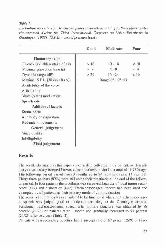
Table I. Evaluation procedure for tracheoesophageal speech according to the uniform criteria assessed during the Third International Congress on Voice Prosthesis in Groningen (1988). (S.P.L. = sound pressure level).
Phonatory skills
Fluency (syllables/intake of air)
Maximal phonation time (s)
Dynamic range (dB)
Maximal S.P.L. [30 em dB (A))
Availability of the voice
Articulation
Voice (pitch) modulation
Speech rate
Additional factors
Stoma noise
Audibility of inspiration
Redundant movements
General judgement
Voice quality
Intelligibility
Final judgement
Results
Good Moderate
> 1 8 10 - 1 8
> 9 4 - 9
> 24 16 - 24
Range 65 - 95 dB
Poor
< 1 0
< 4
< 1 6
The results discussed in this paper concern data collected in 37 patients with a primary or secondary inserted Provox voice prosthesis in situ for a total of 1 1 .730 days. The follow-up period varied from 3 months up to 24 months (mean: 1 4 months). Thirty three patients (89%) were still using their prosthesis at the end of the followup period. In four patients the prosthesis was removed, because of local tumor recurrence (n=2) and dislocation (n=2). Tracheoesophageal speech had been used and attempted by all patients as their primary mode of communication. The voice rehabilitation was considered to be functional when the tracheoesophageal speech was judged good or moderate according to the Groningen criteria. Functional tracheoesophageal speech after primary puncture was obtained by 79 percent (22/28) of patients after I month and gradually increased to 95 percent (24/25) after one year (Table II). Patients with a secondary puncture had a success rate of 67 percent (6/9) of func-
53
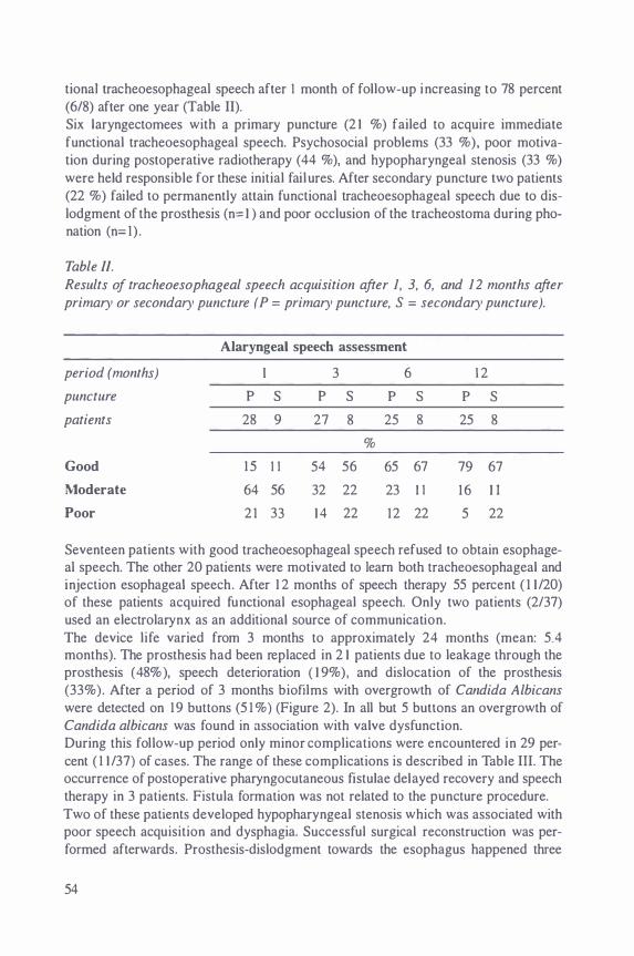
tiona! tracheoesophageal speech after I month of follow-up increasing to 78 percent (6/8) after one year (Table II). Six laryngectomees with a primary puncture (2 1 %) failed to acquire immediate functional tracheoesophageal speech. Psychosocial problems (33 %), poor motivation during postoperative radiotherapy (44 %), and hypopharyngeal stenosis (33 %) were held responsible for these initial failures. After secondary puncture two patients (22 %) failed to permanently attain functional tracheoesophageal speech due to dislodgment of the prosthesis (n= I ) and poor occlusion of the tracheostoma during phonation (n= I) .
Table II. Results of tracheoesophageal speech acquisition after 1, 3, 6, and 12 months after primary or secondary puncture ( P = primary puncture, S = secondary puncture).
Alaryngeal speech assessment
period (months) 3 6 1 2
puncture p s p s p s p s
patients 28 9 27 8 25 8 25 8
%
Good 1 5 I I 54 56 65 67 79 67
Moderate 64 56 32 22 23 I I 16 I I
Poor 2 1 33 1 4 22 12 22 5 22
Seventeen patients with good tracheoesophageal speech refused to obtain esophageal speech. The other 20 patients were motivated to Jearn both tracheoesophageal and injection esophageal speech. After 1 2 months of speech therapy 55 percent ( 1 1 /20) of these patients acquired functional esophageal speech. Only two patients (2/37) used an electrolarynx as an additional source of communication. The device life varied from 3 months to approximately 24 months (mean: 5.4 months). The prosthesis had been replaced in 2 1 patients due to leakage through the prosthesis ( 48% ), speech deterioration ( I 9% ) , and dislocation of the prosthesis (33% ) . After a period of 3 months biofilms with overgrowth of Candida Albicans were detected on 19 buttons (5 1 %) (Figure 2). In all but 5 buttons an overgrowth of Candida albicans was found in association with valve dysfunction. During this follow-up period only minor complications were encountered in 29 percent ( 1 1 /37) of cases. The range of these complications is described in Table III. The occurrence of postoperative pharyngocutaneous fistulae delayed recovery and speech therapy in 3 patients. Fistula formation was not related to the puncture procedure. Two of these patients developed hypopharyngeal stenosis which was associated with poor speech acquisition and dysphagia. Successful surgical reconstruction was performed afterwards. Prosthesis-dislodgment towards the esophagus happened three
54

times after puncture. This was solved adequately by replacing the prosthesis with a larger device.
Table Jll.
Range of all surgery and prosthesis related problems during the follow-up period. Numbers and percentages are given for all laryngectomees equipped with their initial button. In 29 percent ( l/137) of these cases, problems were encountered which were not related to the nomzal wear and tear of the prostheses (i.e. valve incompetence).
Complications n %
Leakage through prosthesis 30 8 1 Leakage around prosthesis 5 14 Granulation tissue 3 8 Dislocation of prosthesis 4 I I Postoperative fistula 3 8 Fungal colonization 25 68 Obstruction valve part 6 1 6 Hypopharyngeal stenosis 2 5 Tracheostomatal stenosis 2 5 Dysphagia 5 14 Gastric reflux complaints 2 5
Leakage around the prosthesis occurred temporarily in 5 patients after frequent replacements of the prosthesis because of valve incompetence. The prosthesis had been removed in one case for recurrent external leakage for a period of 5 days to allow sufficient narrowing of the fistula tract. The fistula tract was closed surgically in two patients with tumor recurrence without spontaneous closure of the fistula tract after prosthesis removal. The development of granulation tissue around the fistula tract occurred in three patients after a period of 3 to 6 months. Recurrent granulation tissue was effectively treated by surgical or chemical cauterization (silver nitrate). Complaints of dysphagia were in two cases associated with hypopharyngeal stenosis, and in three other cases caused by a long shaft of the prosthesis with the valve part protruding into the esophageal lumen. Frequent maintenance of the prosthesis was necessary in 6 patients suffering from bronchial hypersecretion and crust formation. A stomaplasty was performed in two patients with complaints of a narrowed tracheostoma. One infected fistula tract (Staphylococcus aureus) was treated with antibiotics after secondary puncture.
55
Figure 2. Deposits contammg a mixed biofilm of fungi and bacteria on the esophageal part of the Provox™ prosthesis
after 7 months of use.
Discussion
Voice rehabilitation after total laryngectomy using a self-retaining, low resistance prosthesis has become a preferred method of speech restoration in several centers ( I I ). Patients are able to acquire alaryngeal shunt speech early after primary or secondary prosthesis placement. The several psychosocial advantages of immediate voice restoration have already been stated clearly in other studies. In our study functional speech rehabilitation, defined as good or moderate according to the Groningen criteria (20,2 1 ), was obtained after I month in 79 percent of cases. However, postoperative radiotherapy delayed fluent speech acquisition in a minority of our patients. These results are comparable with results in other studies (9, 1 0, 1 8,22,23). Our results of speech acquisition after one year of follow-up are also similar with results obtained with comparable prostheses (80-95%) ( 14,23,24). The low rate of speech failures in the group of primary punctures may be ascribed to the additional constrictor myotomy and pharyngeal plexus neurectomy procedures. We did not experience any disadvantage of this approach (i.e. hypotonicity of the pharyngoesophageal segment). The success rate for secondary punctures was relatively good (78%) which may be attributed to the preoperative assessment of the pharyngoesophageal segment by cineradiography and insufflation testing. An additional myotomy was performed in two cases in association with a secondary puncture. Improved analysis of the several pharyngeal relaxation procedures and operative techniques can contribute to a continuous decrease of speech failure rates (25,26,27). To predict the outcome of alaryngeal speech several authors have discussed the importance of several parameters such as clinician expertise, patient age and general health, and preoperative criteria for patient selection ( 4,28). In comparing several phonatory aspects of tracheoesophageal speech with esophageal speech, we found a better fundamental voice frequency, greater intensity, and intelligibility in favor of tracheoesophageal speech. In our series 46 percent ( 1 7 /37) of the patients with moderate to good tracheoesophageal speech acquisition, refused further training in esophageal speech.
56

The morbidity and complications due to tracheoesophageal puncture are relatively low in our series. In 29 percent of cases we had to deal with minor but manageable problems. Leakage through the prosthesis was the most frequent reason for prosthesis replacement. However, leakage around the prosthesis was the most distressing side effect after prosthesis replacement. During the follow-up period no major complications were encountered. This may be attributed to the self-retaining properties of the prosthesis without prosthesis removal for maintenance procedures (24,29,30). The device life (mean: 5.4 months) also corresponds to the results obtained with similar prostheses ( 14, 1 8,24). Valve dysfunction was often related to fungal colonization of the prosthesis ( 1 8,3 1 ,32). Anti-fungal drugs (amphotericin-B . nystatin, miconazole) are known to extent the device life by preventing early valve deterioration ( 1 8,3 1 ). Postlaryngectomy speech rehabilitation using a ProvoxTM self-retaining voice prosthesis can be realized after a primary or secondary puncture procedure. Our results of voice restoration are comparable to the outcome of several other studies with only minor complications. Tracheoesophageal speech can be considered as an appropriate alternative to other forms of alaryngeal speech. However, postlaryngectomy speech therapy should both consist of tracheoesophageal speech and injection esophageal speech. Patient selection and postoperative follow-up are critical parts of postlaryngectomy speech counselling. By maintaining a team approach to voice restoration most laryngectomees should be able to achieve an effective method of communication.
References
I . Alberti PW. The evolution of laryngology and laryngectomy in the mid 19th century. Laryngoscope 1975; 85: 288-298.
2. Gussenbauer C. Ueber die erste durch Th. Billroth am Menschen ausgefi.ihrte Kehlkopf-Extirpation und die Anwendung eines Kiinstlichen Kehlkopfes. Arch Klin Chir 1 874; 17 : 343-356.
3. Duguay MJ, Feudo P Jr. The process of postlaryngectomy rehabilitation. In: Fried MP, editor. The Larynx. 'A multidisciplinary approach' . BostonToronto: Little, Brown and Co, I 988: 603-6 1 3 .
4. Gates GA, Hearne EM. Predicting esophageal speech. Ann Otol Rhino! Laryngol 1982; 9 1 : 454-457.
5. Schaefer SD, Johns DF. Attaining functional esophageal speech. Arch Otolaryngol 1982; 108 : 646-649.
6. Zanoff DJ, Wold D, Montague JC, Krueger K, Drummond S . Tracheoesophageal speech: with and without tracheostoma valve. Laryngoscope 1990; 100: 498-502.
7. Snidecor JC, Isshiki N. Air volume and airflow relationships of six male esophageal speakers. J Speech Hear Disord 1 965; 30: 205-2 1 6.
8. Goldstein LP, Salmon SJ. The relationship between adience-abience scale scores and judged communication proficiency of alaryngeal speakers. Laryngoscope 1978; 88: 1 855- 1 860.
57

9. Singer Ml, BJorn ED. An endoscopic technique for restoration of voice after laryngectomy. Ann Otol Rhino! Laryngol 1 980; 89: 529-533.
1 0. Panje WR. Prosthetic vocal rehabilitation following laryngectomy: the voice button. Ann Otol Rhino! Laryngol 1 98 1 ; 90: 1 1 6- 1 20.
1 1 . Van Weissenbruch R, Albers FWJ. Voice rehabilitation after total laryngectomy. Acta Otorhinolaryngol Belg 1 992; 46: 22 1 -246.
1 2. Pindzola RH, Cain BH. Acceptability ratings of tracheoesophageal speech. Laryngoscope 1988; 98: 394-397.
1 3 . Zijlstra RJ, Mahieu HF, van Lith-Bijl JT, Schutte HK. Aerodynamic properties of the low-resistance Groningen Button. Arch Otolaryngol Head Neck Surg 199 1 ; 1 7: 657-661 .
1 4. Hilgers FJM, Schouwenburg PF. A new low-resistance, self-retaining prosthesis (Provox™) for voice rehabi l i tation after total laryngectomy. Laryngoscope 1990; 100: 1202- 1 207.
1 5 . Mahieu HF, Annyas AA, Schutte HK, van der Jagt EJ. Pharyngoesophageal myotomy for vocal rehabi litation of laryngectomees. Laryngoscope 1 987; 97: 45 1 -457.
1 6. Singer Ml, BJorn ED. Selective myotomy for voice restoration after total laryngectomy. Arch Otolaryngol Head Neck Surg 1 98 1 ; 107: 670-673.
17 . Singer Ml, BJorn ED, Hamaker RC. Pharyngeal plexus neurectomy for alaryngeal speech rehabilitation. Laryngoscope 1 986; 6:50-53.
1 8. Mahieu HF. Voice and speech rehabilitation following laryngectomy [dissertation]. Groningen: University of Groningen, 1 988.
19 . Van Lith-B ijl JT, Mahieu HF, Patel P, Zijlstra RJ. Clinical experience with the low-resistance Groningen button. Eur Arch Otolaryngol 1 992; 249: 354-357.
20. Schutte HK, Bors EFM, de Boer GHA, Nieboer GLJ, Annyas AA. Evaluation of speech with and without a Groningen type voice button. In: Herrmann IF, editor. Speech restoration via voice prostheses. Berlin-Heidelberg: SpringerVerlag, 1 986: 1 35- 1 38.
2 1 . Bors EFM. Indwelling voice prosthesis for voice restoration after total laryngectomy : criteria for evaluation of speech with a voice button. Teaching course on indwelling voice prostheses for voice restoration after total laryngectomy; 1 99 1 Apr 12; Amsterdam. Amsterdam: The Netherlands Cancer Institute, 1 99 1 : 50-56.
22. Nijdam HF, Annyas AA, Schutte HK, Leever A. A new prosthesis for voice rehabilitation following laryngectomy. Arch Otolaryngol 1 982; 237: 27
23. Manni JJ, van den Broek P, de Groot MAH, Berends E. Voice rehabilitation after laryngectomy with the Groningen prosthesis. J Otolaryngol 1 984; 1 3 : 333-336.
24. Rosingh HJ, Mahieu HF, Annyas AA, Schutte HK, Goorhuis-Brouwer SM.
58
Stemrevalidatie na larynxextirpatie met behulp van de Groningen button. Ned Tijdschr Geneesk 199 1 ; 1 35 : 1 3 1 5- 1 3 1 8.
25. Henley J, Souleire Ch. Tracheoesophageal speech failure in the laryngectomee: the role of the constrictor myotomy. Laryngoscope 1 986; 96: 1 0 1 6-1 020.
26. Baugh RF, Baker SR, Lewin JS. Surgical treatment of pharyngoesophageal spasm. Laryngoscope 1988; 98: 1 1 24- 1 1 26.
27. Medina JE, Reiner SA. Pharyngeal myotomy for voice restoration: a midline approach. Laryngoscope 1990; 1 00: 309-3 12.
28. Robillard Schultz J, Harrison J. Defining and predicting tracheoesophageal puncture success. Arch Otolaryngol Head Neck Surg 1 992; 1 1 8: 8 1 1 -8 1 6.
29. Andrews JC, Michel RA, Hanson DG, Monahan GP, Ward PH. Major complications following tracheoesophageal puncture for voice rehabilitation. Laryngoscope 1987; 97: 562-567.
30. Manni JJ, Van den Broek P. Surgical and prosthesis-related complications using the Groningen button voice prosthesis. Clin Otolaryngol 1990; 1 5 : 5 1 5-523.
3 1 . Mahieu HF, van Saene HKJ, Rosingh HJ, Schutte HK. Candida vegetations in sil icone voice prosthesis. Arch Otolaryngol 1986; 2 1 2: 32 1 -325.
32. Izdebski K, Ross JC, Lee S. Fungal colonization of tracheoesophageal voice prosthesis. Laryngoscope 1987; 97: 594.
59


Chapter 4
Chemoprophylaxis of fungal deterioration of the Provox™ tra
cheoesophageal voice prosthesis in post-laryngectomy patients
Van Weissenbruch R, Bouckaert S, Remon JP, Nelis HJ, Aerts R, Albers FWJ. Chemoprophylaxis of fungal deterioration of the ProvoxTM tracheoesophageal voice prosthesis in post-laryngectomy patients. Ann Otol Rhino! Laryngol. In press.

Introduction
Restoration of speech after total laryngectomy is generally considered to be a primary task in the rehabilitation of the laryngectomee. Since the first laryngectomies performed by Watson ( 1 866) and Billroth ( 1 873) several methods to restore speech have been attempted ( 1 ,2). Alaryngeal speech production requires the establishment of an alternative vibratory source in the reconstructed pharyngoesophageal region. Speech restoration can be achieved by injection and/or shunt esophageal techniques, or by means of an artificial larynx which as an electric pharyngeal speech vibrator can be externally applied to the neck. The method of shunt esophageal speech by means of silicone tracheoesophageal valve prostheses is further developed after the introduction of the Blom-Singer Duckbill prosthesis in 1 979 (3). Compared to the other methods of voice rehabilitation, consistent high success rates of shunt esophageal speech were obtained with several types of tracheoesophageal voice prostheses (4). Further research was directed towards the development of a device which could be easily applied after primary or secondary puncture, which enables short-term acquisition of functional alaryngeal speech, and which requires only simple maintenance for optimal performance. This resulted in the development of indwelling, biocompatible silicone prostheses with a low-resistance one-way valve mechanism. Rational patient selection and additional surgical management of the pharyngoesophageal segment had already learned how to improve prosthetic voice rehabilitation. Device l ife is known to be limited due to dysfunction of the valve mechanism. Colonization and invasion of silicone material with Candida species (spp) has been reported to correlate with valve dysfunction (5,6). The latter may be determined by leakage of esophageal contents through the prosthesis into the trachea as a result of insufficient valve-closure, or by an increased expiratory airflow resistance during phonation. This will gradually lead to increased efforts to produce fluent shunt esophageal speech. Selective oropharyngeal yeast decontamination may prevent early deterioration of valve prostheses (7,8). Systemic and topical antimycotics are currently used to treat oropharyngeal fungal infections (9). Successful decontamination of the oropharyngeal yeast flora has been establi shed with several antifungal drugs in immunocompromized and intensive care patients ( 10, 1 1 ). Topical agents (i.e. lozenges and semi-solid formulations) might adequately resolve superficial fungal infections, but must be applied frequently to maintain effective concentrations ( 1 2) . A rapid decrease below the minimal inhibitory concentration (MIC) is usually noticed after local application of these formulations. Application of a bioadhesive slow-release tablet has proven to maintain effective salivary drug concentrations during 1 0- 1 2 hours after application on the gingival mucosa ( 1 2). This could practically diminish the need for frequent drug applications with lower daily dosages. Fewer daily applications might improve patient compliance, while less drug is needed for long-term prevention of deterioration of silicone voice prostheses. A double-blind randomized study was conducted to assess the efficacy of bioadhesive slow-release tablets containing miconazole nitrate on the device l ife and function of the ProvoxTM tracheoesophageal voice prosthesis.
62
Patients and Methods
Patients
Thirty-six patients who underwent total laryngectomy (34 men, 2 women; age varying from 41 to 87 years; mean 63. 1 years) were selected to participate in this prospective study. In our department all laryngectomees who received a voice prosthesis, were selected according to specific criteria as described in a previous report ( 1 3). All, but two patients had a primary prosthesis insertion at the time of laryngectomy. Both secondary insertions were performed after a period of approximately 7 months. All participants received radiotherapy pre- or postlaryngectomy (60-70 Gy in 6-7 wks). According to uniform criteria discussed at the Third International Congress on Voice Prostheses (Groningen, 1988), all participants were able to produce functional shunt esophageal speech for at least 3 months. The patients were selected to start participation in the trial at the time when replacement of their voice prosthesis was required due to leakage or increased efforts to phonate properly. In all subjects a new Provox™ indwelling, low-resistance voice prosthesis was inserted as a standard outpatient procedure. They were included in the study after appropriate explanation of the trial procedures and signature of an informed consent document. Participants were allowed to prematurely drop out on individual request . Patients known to have allergic or other adverse reactions to miconazole nitrate, were excluded from participation. No other antimicrobial agents were used simultaneously.
Study design
A prospective double-blind randomized placebo controlled study was designed to determine the efficacy of a bioadhesive, slow-release tablet containing 1 0 mg of miconazole nitrate on fungal colonization and dysfunction of the valve mechanism of the ProvoxTM voice prosthesis. Trial randomization, as well as production and supply of the pharmaceutical agents, was directed by the Laboratory of Pharmaceutical Technology (University of Ghent, Belgium). Tablets containing miconazole nitrate and placebo were equally distributed between the 36 participants ( 1 8 versus 1 8). Patients were instructed to administer the bioadhesive tablets twice daily alternating between both sides of the buccal mucosa. At the start of the study culture samples were taken from the oropharynx (dorsum of tongue), the tracheoesophageal shunt, and the esophageal flange (valve part) of the prosthesis. Immediately following prosthesis replacement, intratracheal phonatory pressures were assessed at three voice intensity levels. The efficacy of shunt esophageal speech could readily be assessed by estimating the intratracheal air pressure during phonation ( 1 4). The trial was conducted to last 6 months. Participants were evaluated monthly at the outpatient department (Dept. Otorhinolaryngology, University Hospital Ghent, Belgium) for intratracheal phonatory pressure measurements, prosthesis inspection, and microbiological assessment. Culture swabs were taken from the oropharynx, prosthesis (valve part) and tracheoesophageal shunt.
63

Swabs were taken with suitable cotton-tips and preserved in vials without any transport medium. Daily maintenance of the prosthesis was performed using specifically manufactured brushes without using any antiseptic solution. High performance liquid chromatography (HPLC) was used to assess the concentration of miconazole nitrate in saliva samples ( 1 2). Samples were taken at regular intervals to determine the patient compliance. The trial was considered terminated after a follow-up of 6 months, or sooner in case prosthesis replacement was required. The participants, however, were allowed to continue the use of the tablets. Any occurring adverse effect or complication was recorded. A questionnaire with possible side effects was included for the patients to complete at each visit. In order to assess long-term device life, all patients were regularly seen up to one year after participation. The study proceeded according to the declaration of Helsinki. It has been approved by the Committee for Medical Ethics of the Medical Faculty, University of Ghent on July 5, 1 993 (project 93/84).
Bioadhesive slow-release tablets
After simple application on the buccal mucosa in the region of the right or left upper canine, the tablet is fixed for 1 minute by slight manual pressure on the lip. Sticking of the tablet to the lip is prevented by moistening the tablet with the tongue. The bioadhesive slow-release tablets are sufficiently attached for 1 0 to 1 2 hours (fig. l ). As described in a previous study, no dislodgement of the tablets occured during meals ( 1 2). Also no complaints of a disturbing sensation or dysgeusia were mentioned. The bioadhesive slow-release tablets (total mass 100 mg, 7 mm diameter, 2 mm thick) are compounded of starch, polyacrylic acid 5%, and 1 0 mg of miconazole nitrate ( 1 2). Miconazole nitrate is a synthetic imidazole derivative that has broadspectrum activity against almost all pathogenic fungi except Aspergillus spp and Zygomycetes. It is considered to be the drug of choice in the treatment of superficial fungal infections ( 1 5). In the literature side effects due to the use of miconazole nitrate after local application are reported to be variable (9). In preliminary trials no side effects were evident ( 1 2). During therapy, resistance to this drug is rare. The mycelia are less susceptible than the parasitic forms of dimorphic fungi ( 1 6). Its mode of action consists of ergosterol synthesis blockage in fungi, leading to the production of a defective fungal cell membrane with altered permeability ( 1 5).
Fig. Ja
64

Fig. Jb
Figure 1 a and b. A bio-adhesive tablet attached to the buccal mucosa; (a) immediately after application; (b) after 6-8 hours.
Intratracheal pressure measurements during phonation
At the start of the study and at each monthly visit, the participants were requested to phonate at three intensity levels during at least l 0 seconds. At low (30 to 40 dB), normal (50 to 60 dB), and high intensity levels (70 to 80 dB) the intratracheal pressure was recorded during phonation of a verb {Ia/). Sound intensity measurements were performed by the Computerized Speech Lab (Model 4300, Kay Elemetrics Corporation, Pine Brook, NJ), with a boom-mounted microphone to standardize microphone-to-mouth distance (30 em). A portable manometer with an electromagnetic pressure sensor (type EMA 1 50, Range 0-300 mmHg, Dimed S.A. Electronic Engineering, Antwerp, Belgium) was fitted in an adapted Shiley111 cuffed cannula, which enclosed the tracheostoma sufficiently with silicone adhesives. This method was not likely to interfere with shunt esophageal speech, since the voice prosthesis was located at I to 2 centimeters below the posterior tracheocutaneous border.
Mycological assessment
All culture swabs were assessed on a selective medium in a qualitative and semiquantitative way. Each sample was inoculated on a yeast isolation plate with Sabouraud dextrose agar and Sabouraud dextrose agar containing chloramphenicol. Following inoculation, the swab was spread out over one half of the isolation plate, and over the other half successively after rotation of 1 80" . Next, the culture swabs were put into brain-heart infusion broth. Both plates and broth were then incubated at 25 degrees Celsius for 48 hours. A semiquantitative scoring system was used to determine the growth densities on the isolation plates. In accordance with this scoring system, the results were represented by colonization indices: ( - ) No growth or less than 100 colonies on one half of the plate, ( I+) Isolated colonies (200-250) on only one half of the plate,
65
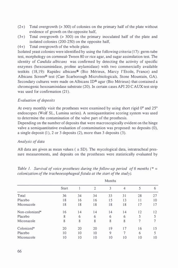
(2+) Total overgrowth (> 300) of colonies on the primary half of the plate without evidence of growth on the opposite half,
(3+) Total overgrowth (> 300) on the primary inoculated half of the plate and isolated colonies (200-250) on the opposite half,
( 4+) Total overgrowth of the whole plate. Isolated yeast colonies were identified by using the following criteria ( 1 7) : germ-tube test, morphology on cornmeal-Tween 80 or rice agar, and sugar assimilation test. The identity of Candida albicans was confirmed by detecting the activity of specific enzymes (hexosaminidase, proline arylamidase) with two commercially available testkits ( 1 8, 1 9): Rapidec albicans® (Bio Merieux, Marcy ! 'Etoile, France) and Albicans Screen® test (Carr Scarborough Microbiologicals, Stone Mountain, GA). Secondary cultures were made on Albicans ID® agar (Bio Merieux) that contained a chromogenic hexosaminidase substrate (20). In certain cases API 20 C AUX test strip was used for confirmation (2 1 ) .
Evaluation of deposits
At every monthly visit the prostheses were examined by using short rigid 0° and 2SO endoscopes (Wolf SL, Lumina series). A semiquantitative scoring system was used to determine the contamination of the valve part of the prosthesis. Depending on the number of deposits that were macroscopically evident on the hinge valve a semiquantitative evaluation of contamination was proposed: no deposits (0), a single deposit (1 ), 2 or 3 deposits (2), more than 3 deposits (3).
Analysis of data
All data are given as mean values ( ± SD). The mycological data, intratracheal pressure measurements, and deposits on the prostheses were statistically evaluated by
Table 1 . Survival of voice prostheses during the follow-up period of 6 months (* =
colonization of the tracheoesophageal fistula at the start of the study).
Months
Start 2 3 4 5 6
Total 36 34 34 33 3 1 28 27 Placebo 1 8 1 6 1 6 1 5 1 3 I I 1 0 Miconazole 1 8 1 8 1 8 1 8 1 8 1 7 1 7
Non-colonized* 1 6 1 4 1 4 1 4 1 4 1 2 1 2 Placebo 8 6 6 6 6 5 5 Miconazole 8 8 8 8 8 7 7
Colonized* 20 20 20 1 9 1 7 1 6 1 5 Placebo 1 0 1 0 1 0 9 7 6 5 Miconazo1e 1 0 1 0 1 0 1 0 1 0 1 0 1 0
66
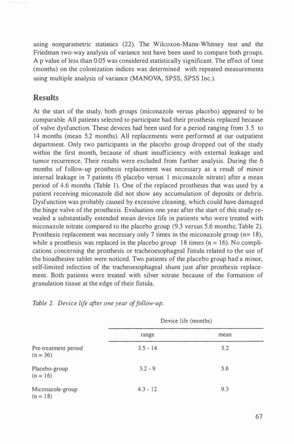
using nonparametric statiStics (22). The Wilcoxon-Mann-Whitney test and the Friedman two-way analysis of variance test have been used to compare both groups. A p value of less than 0.05 was considered statistically significant. The effect of time (months) on the colonization indices was determined with repeated measurements
using multiple analysis of variance (MAN OVA, SPSS, SPSS Inc.).
Results
At the start of the study, both groups (miconazole versus placebo) appeared to be comparable. All patients selected to participate had their prosthesis replaced because of valve dysfunction. These devices had been used for a period ranging from 3 .5 to I 4 months (mean 5.2 months). All replacements were performed at our outpatient department. Only two participants in the placebo group dropped out of the study within the first month, because of shunt insufficiency with external leakage and tumor recurrence. Their results were excluded from further analysis. During the 6 months of follow-up prosthesis replacement was necessary as a result of minor internal leakage in 7 patients (6 placebo versus I miconazole nitrate) after a mean period of 4.6 months (Table I ). One of the replaced prostheses that was used by a patient receiving miconazole did not show any accumulation of deposits or debris. Dysfunction was probably caused by excessive cleaning, which could have damaged the hinge valve of the prosthesis. Evaluation one year after the start of this study revealed a substantially extended mean device life in patients who were treated with miconazole nitrate compared to the placebo group (9.3 versus 5.6 months; Table 2). Prosthesis replacement was necessary only 7 times in the miconazole group (n= I S), while a prosthesis was replaced in the placebo group I 8 times (n = I6). No complications concerning the prosthesis or tracheoesophageal fistula related to the use of the bioadhesive tablet were noticed. Two patients of the placebo group had a minor, self-limited infection of the tracheoesophageal shunt just after prosthesis replacement. Both patients were treated with silver nitrate because of the formation of granulation tissue at the edge of their fistula.
Table 2. Device life after one year of follow-up.
Pre-treatment period (n = 36)
Placebo-group (n = 1 6)
Miconazole-group (n = 1 8)
Device l ife (months)
range
3.5 - 1 4
3.2 - 9
4.3 - 1 2
mean
5.2
5.6
9.3
67
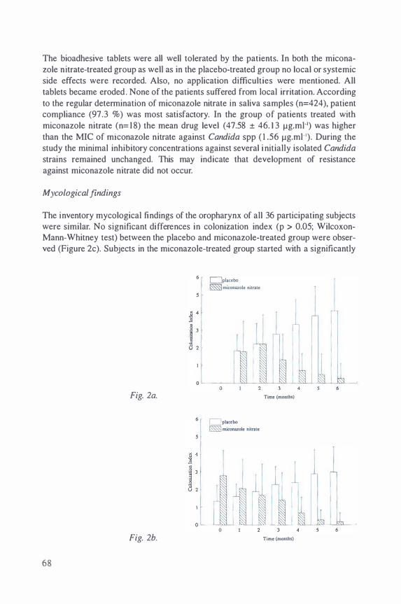
The bioadhesive tablets were all well tolerated by the patients. In both the miconazole nitrate-treated group as well as in the placebo-treated group no local or systemic side effects were recorded. Also, no application difficulties were mentioned. All tablets became eroded . None of the patients suffered from local irritation. According to the regular determination of miconazole nitrate in saliva samples (n=424), patient compliance (97.3 %) was most satisfactory. In the group of patients treated with miconazole nitrate (n= l 8) the mean drug level (47.58 ± 46. 1 3 j.lg.ml-1) was higher than the MIC of miconazole nitrate against Candida spp ( 1 .56 j.lg.ml- 1). During the study the minimal inhibitory concentrations against several i nitially isolated Candida strains remained unchanged. This may indicate that development of resistance against miconazole nitrate did not occur.
Mycological findings
The inventory mycological findings of the oropharynx of all 36 participating subjects were similar. No significant differences in colonization index (p > 0.05; WilcoxonMann-Whitney test) between the placebo and miconazole-treated group were observed (Figure 2c). Subjects in the miconazole-treated group started with a significantly
6 C]placcbo
s � �maconazolc nitrate
4 I l ' 3
I
2 l -�
I [ 0
0 Fig. 2a. Time (months)
6 r C]placcbo � miconazole nitrate
s
4 1 3 1
ill 2
I � '
0 � Fig. 2b. Time (months)
68
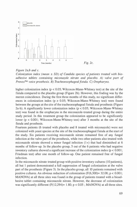
Uplacebo �miconazole nitrate
Figure 2a,b and c.
Time {montlu) 6
Fig. 2c.
Colonization index (mean ± SD) of Candida species of patients treated with bioadhesive tablets containing miconazole nitrate and placebo. A) valve part of ProvoxTM voice prosthesis. B) Tracheoesophageal fistula. C) Oropharynx.
higher colonization index (p < 0.05; Wilcoxon-Mann-Whitney test) at the site of the fistula compared to the placebo group (Figure 2b). However, this finding was by the merest coincidence. During the first three months of this study, no significant differences in colonization index (p > 0.05; Wilcoxon-Mann-Whitney test) were found between the groups at the site of the tracheoesophageal fistula and prosthesis (Figure 2a-b). A significantly lower colonization index (p < 0.05 ; Wilcoxon-Mann-Whitney test) was found in the oropharynx in the miconazole-treated group during the entire study period. In this treatment group the colonization appeared to be significantly lower (p < 0.00 I ; Wilcoxon-Mann-Whitney test) after 3 months at the site of the fistula and prosthesis. Fourteen patients (6 treated with placebo and 8 treated with miconazole) were not colonized with yeast species at the site of the tracheoesophageal fistula at the start of the study. Six patients receiving miconazole nitrate remained free of any fungal infection at the valve part of the prosthesis, while two other patients also treated with miconazole nitrate showed a minor fungal infection ( I +) that had diminished at 6 months of follow-up. In the placebo group, 5 out of the 6 patients who had negative inventory cultures showed a significant increase of the colonization index (p < 0.00 I ; Friedman test) after one month of follow-up. One patient remained free of fungal infection. In the miconazole nitrate treated group with positive inventory cultures ( 10 patients), all but 1 patient demonstrated a full suppression of fungal colonization at the valve part of the prosthesis (Figure 3). In the placebo group, all 10 patients remained with positive cultures. An obvious reduction of colonization (F(6,300)= 32.08 ; p < 0.00 1 ; MAN OVA) at all three sites was found i n the group of patients treated with a bioadhesive tablet containing miconazole nitrate. However, the decrease of colonization was significantly different (F( l 2,294)= 1 .80; p < 0.05 ; MANOVA) at all three sites.
69
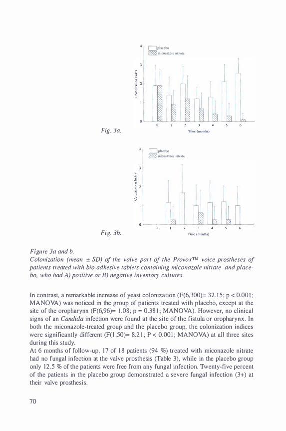
4 1 c=Jplacebo �miconazole nitrate
3 1 I r l l l
: 1 ill� I
ihll
I
I J 0 6
Fig. 3a. Ttme (months)
4 1 Uplncebo �mtconnole rutrate
o '--
Fig. 3b. Time (months)
Figure 3a and b. Colonization (mean ± SD) of the valve part of the ProvoxTM voice prostheses of patiellts treated with bio-adhesive tablets containing miconazole nitrate and placebo, who had A) positive or B) negative inventory cultures.
In contrast, a remarkable increase of yeast colonization (F(6,300)= 32. 15 ; p < 0.00 I ; MANOVA) was noticed in the group of patients treated with placebo, except at the site of the oropharynx (F(6,96)= I .08; p = 0.38 I ; MANOVA). However, no clinical signs of an Candida infection were found at the site of the fistula or oropharynx. In both the miconazole-treated group and the placebo group, the colonization indices were significantly different (F( l ,50)= 8.2 1 ; P < 0.001 ; MANOVA) at all three sites during this study. At 6 months of follow-up, 17 of 1 8 patients (94 %) treated with miconazole nitrate had no fungal infection at the valve prosthesis (Table 3), while in the placebo group only 12.5 % of the patients were free from any fungal infection. Twenty-five percent of the patients in the placebo group demonstrated a severe fungal infection (3+) at their valve prosthesis.
70
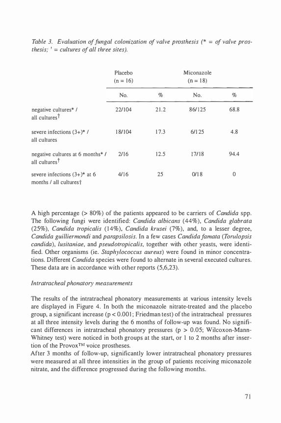
Table 3. Evaluation of fungal colonization of valve prosthesis (* = of valve prosthesis; 1 = cultures of all three sites).
Placebo Miconazole
(n = 1 6) (n = 1 8)
No. % No. %
negative cultures* I 22/104 2 1 .2 86/ 1 25 68.8
all cultures t
severe infections (3+ )* I 1 81 1 04 1 7.3 6/1 25 4.8
all cultures
negative cultures at 6 months* I 2/1 6 1 2.5 1 7/ 1 8 94.4
all cultures t
severe infections (3+ )* at 6 4/1 6 25 0/1 8 0
months I all culturest
A high percentage (> 80%) of the patients appeared to be carriers of Candida spp. The following fungi were identified: Candida albicans (44%), Candida glabrata (25%), Candida tropicalis ( 1 4%), Candida krusei (7%), and, to a lesser degree, Candida guilliermondi and parapsilosis. In a few cases Candida fa mat a (Torulopsis candida), lusitaniae, and pseudotropicalis, together with other yeasts, were identified. Other organisms (ie. Staphylococcus aureus) were found in minor concentrations. Different Candida species were found to alternate in several executed cultures. These data are in accordance with other reports (5,6,23).
Intratracheal phonatory measurements
The results of the intratracheal phonatory measurements at various intensity levels are displayed in Figure 4. In both the miconazole nitrate-treated and the placebo group, a significant increase (p < 0.00 I ; Friedman test) of the intratracheal pressures at all three intensity levels during the 6 months of follow-up was found. No significant differences in intratracheal phonatory pressures (p > 0.05; Wilcoxon-MannWhitney test) were noticed in both groups at the start, or 1 to 2 months after insertion of the ProvoxTM voice prostheses. After 3 months of follow-up, significantly lower intratracheal phonatory pressures were measured at all three intensities in the group of patients receiving miconazole nitrate, and the difference progressed during the following months.
7 1

a placebo • micanazole mtrate
5 I
0 Fig. 4a. T•me (moaths)
40 pljtcebo • m1conazole mtratc
r 35 1 1 -<> "" 30 6
� 25 b T Lo l ..
: 15 10 1
0 0
Fig. 4b. Time {months)
70 plilC;ebo • m1conazole nnrate J 65
l 60
l 1 55
I "'
.--·--� -· 'li 50 ·--r e 1 I - 45 ..... I � 40 1 r 35
30
� l Fig. 4c. T1me (months)
Figure 4a,b and c. Mean (± SD) intra-tracheal phonatory pressures (ITP) at three different intensity levels in patients treated with miconazole nitrate and placebo. A) At 30 to 40 dB. B) At 50 to 60 dB. C) At 70 to 80 dB.
Assessment of deposits on the voice prostheses
Only moderate deposits ( I +) were macroscopically found on the voice prostheses of patients using bioadhesive tablets containing miconazole nitrate. One patient showed
72

deposits ( I +) on the valve part of the prosthesis after 5 months of insertion, while 3 other patients had similar deposits at the end of the study. After 4 months of insertion, a significant increase of deposits (p < 0.00 I ; Friedman test) was demonstrated on the prostheses in patients treated with placebo (Figure 5). At the end of the study all patients receiving placebo, except I , had moderate to severe deposits ( I to 3+) on their valve prostheses (Figure 6). The deposits were mainly located in the shaft, on the valve, and on the esophageal flange surrounding the hinged valve. Six of the valve prostheses that were replaced during this trial due to minor internal leakage, showed heavy deposits and various debris around the esophageal flange. All deposits appeared to be firmly attached to the silicone material. Removal of the deposits was not possible without damaging the surface of the silicone prostheses. Scanning electron microscopic imaging of the esophageal part of dysfunctional prostheses showed both superficial defects and penetration of hyphae of Candida spp in the deeper layers of the silicone material, surrounded by other microbes (Figure 7).
C placebo � mtconazolc: nttratc
' I 0
0
Figure 5.
c Time (months}
6
Evaluation of deposits (mean ± SD) on ProvoxTM voice prostheses in patients treated with miconazole nitrate and placebo.
Discussion
Voice and speech rehabilitation after total laryngectomy can be successfully achieved by using several types of prosthetic devices. Application of indwel ling, low-resistance voice prostheses has further improved the quality and fluency of shunt esophageal speech ( 1 3). The main advantage of self-retaining devices might be the limited maintenance procedures, which could be responsible for the low complication rates as reported ( 1 3 ,24). The device life may be influenced by regular use and maintenance, radiotherapy, and colonization with yeast species. Deterioration of the silicone material will finally result in dysfunction of the one-way valve mechanism, lead-
73

ing to improper closure and increased airflow. The oral cavity and oropharynx are believed to be the continuous suppliers of yeast species and other micro-organisms to the prosthesis located in the tracheoesophageal shunt (5). Certain foods and beverages are also considered to be contributors of several fungi. The prevalence of yeast in the oral cavity is estimated to vary between 20% and 40% in healthy individuals (25). Radiation to the head and neck regions, combined with surgical therapy, may further alter the oral flora and saliva composition to favor oral yeast colonization (26). In studies with the standard and low-resistance Groningen button, successful decontamination of the oropharynx with amphotericin-B lozenges ( 1 0 mg) four times daily was also associated with a prolonged device l ife and lower intratracheal phonatory pressures (8,27). One of the drawbacks encountered in the use of this agent is certainly the frequent daily applications, leading to poor compliance by the patients. Qrug concentrations in saliva are important for efficacy in oropharyngeal candidiasis.
•
Fig. 6a.
Fig. 6b.
Figure 6a and b. Stereomicroscopic view of A) esophageal end and B) valve part of a ProvoxTM voice prosthesis with deposits (original 12x).
74

Application of bioadhesive tablets containing 1 0 mg of miconazole nitrate, demonstrated a significant reduction of yeast colonization (p < 0.00 l ; Friedman test) in the oropharynx and on the Provox™ voice prostheses. At the end of this study, 94% of the patients in the active substance group had negative cultures. In contrast, 80% of the patients treated with placebo were found to have significant higher colonization indexes (p < 0.00 l ; Friedman test) of Candida spp at the valve prostheses. The majority of non-colonized patients receiving miconazole nitrate remained free of fungal colonization during this trial. In 83% of non-colonized patients who received placebo, a positive yeast culture was obtained after one month of prosthesis replacement. Unless high concentrations of yeast organisms were demonstrated in the tracheoesophageal shunt and oral cavity, no clinical symptoms were manifested. Adherence of Candida spp is preferably found on silicone materials, as the surrounding tissues are most often not involved (5). Remarkable higher colonization indices in the placebo group were found at the valve prostheses at 6 months compared to the fistula and oropharynx. Hence, microbial colonization of ProvoxTM prostheses with Candida spp may be prevented by using miconazole nitrate in a bio-adhesive slowrelease system.
Figure 7. Electron microscopic details of fungal deterioration of silicone material of Provox™ voice prosthesis at the esophageal end (scale, 1 em = 5.4 )Jm).
Measurements of the intratracheal phonatory pressure at various intensity levels showed a significant increase (p < 0.00 I ; Friedman test) in both groups. Stabilization of the intratracheal pressure increase was noticed in the miconazole group after 2 months of follow-up. However, a continuous increase of intratracheal phonatory pressure was recorded in patients receiving placebo. Initial pressure rises in both groups may be provoked by influences of esophageal contents on the valve prosthesis. The pressure recordings correlated well with the gradual accumulation of deposits on the surface of the prostheses. In the placebo group a remarkable increase of nonremovable deposits was observed after 4 months of use. This was associated with higher colonization indexes and higher intratracheal phonatory pressures. The degree of fungal colonization of the sili-
75

cone material seems to be correlated with the expiratory airflow resistance of the valve mechanism of the voice prosthesis (8). This may gradually diminish the advantages of indwelling voice prostheses designed with a low-resistance valve mechanism to favor shunt speech production (28,29). High colonization indexes of yeast are associated with progressive dysfunction of valve prostheses. All patients treated with bioadhesive tablets containing miconazole nitrate demonstrated a reduction of valve dysfunction and subsequent prosthesis replacements. After I year of follow-up, a remarkably longer device life was noticed in patients who received chemoprophylaxis. Compliance of the patients was satisfactory. This may be attributed to the absence of local adverse reactions to the bioadhesive system, and the l imited daily dosages. Long-term intermittent or continuous use of antifungal drugs may induce antimicrobial resistance (30). Resistance to oral azoles has been found in intensive care patients and other immunocompromized patients. Selective pressure exerted by antifungal agents on the microbiological flora of patients may influence the prevalence and spectrum of fungal pathogens. However, during this study no evidence for development of resistance to miconazole nitrate was noticed. Intermittent short-term chemoprophylaxis may be reserved for the laryngectomee who is disturbed by the frequent prosthesis replacement due to fungal deterioration. Candida albicans is the one species most often associated with deterioration of valve prostheses. However, several other yeast species and other microorganisms can be cultured in the same individual over time (23). Reduction of oropharyngeal yeast concentrations and inhibition of fungal adherence to silicone devices should be further investigated. Inclusion of antimycotics in the devices during manufacturing, and application of specific coatings, may be considered as possible options. Also, an altered polarization of the polymers may eventually reduce the affinity of Candida spp for these materials.
References
l . Alberti PW. The evolution of laryngology and laryngectomy in the mid 1 9th century. Laryngoscope 1 975; 85: 288-298.
2. Gussenbauer C . Ueber die erste durch Th. Billroth am Menschen ausgefiihrte Kehlkopf-Extirpation und die Anwendung eines Kiinstlichen Kehlkopfes. Arch Klin Chir 1 874; 17 : 343-356.
3. Singer MI, BJorn ED. An endoscopic technique for restoration of voice after laryngectomy. Ann Otol Rhino! Laryngol 1980; 89: 1 1 6- 1 20.
4 . Van Weissenbruch R, Albers FWJ. Voice rehabilitation after total laryngectomy. Acta Otorhinolaryngol Belg 1 992; 46: 22 1 -246.
5 . Mahieu HF, Van Saene HKF, Rosingh HJ, Schutte HK. Candida vegetations on sil icone voice prostheses. Arch Otolaryngol Head Neck Surg 1 986; 1 1 2: 32 1 -325.
6. Izdebski K, Ross JC, Lee S . Fungal colonization of tracheoesophageal voice prosthesis. Laryngoscope 1 987 ; 97: 594-597.
76
7. Mahieu HF, Van Saene JJM, Den Besten J, Van Saene HKF. Oropharynx decontamination preventing Candida vegetation on voice prostheses. Arch Otolaryngol Head Neck Surg 1986; 1 1 2: 1090- 1 092.
8. Mahieu HF. Voice and speech rehabilitation following total laryngectomy [dissertation]. Groningen: University of Groningen, 1 988.
9. Bodey GP. Topical and systemic antifungal agents. Med Clin North Am 1988 ; 72 : 637-659.
10. Lode H, Hoffken G. Oral candidosis and its role in immunocompromised patients. Mycoses 1 989; 32 Suppl 2: 30-33.
1 I . Bodey GP, Anaissie EJ. Opportunistic fungal infections: A major problem i n immunocompromised patients. In: Richardson RG editor. Opportunistic Fungal Infections: Focus on Fluconazole. London: Royal Society of Medicine Services, 1989: 1 - 1 6.
1 2. Bouckaert S, Schautteet H, Lefebvre RA, Remon JP, Van Clooster R. Comparison of salivary miconazole concentrations after administration of a bioadhesive slow-release tablet and an oral gel. Eur J Clin Pharmacol 1 992; 43: 1 37- 1 40.
1 3 . Van Weissenbruch R , Albers FWJ. Vocal rehabilitation after total laryngectomy using the Provox™ voice prosthesis. Clin Otolaryngol l 993; 1 8 : 359-364.
1 4. Nieboer GJL, Schutte HK. The Groningen Button: results of in vivo measurements. Rev Laryngol Otol Rhino! (Bord) l987; 108: 1 2 1 - 1 22.
1 5. Bodey GP. Fungal infections in cancer patients. Ann NY Acad Sci 1988 ; 544: 43 1 -442.
16. Jordan WM , Bodey GP, Rodriguez V, Ketchel SJ, Henney J. Miconazole therapy for treatment of fungal infections in cancer patients. Antimicrob Agents Chemother 1979; 1 6: 792-797.
17 . Larone DH. Medically important fungi. New York, NY: Elsevier, 1 987. 1 8. Dalton MT, Haldane DJM, MacDonald J. Rapid identification of Candida
albicans using 4-methylumbelliferyl-N-acetyi-B-galactosaminide. Diagn Microbiol Infect Dis 1989; 12: 52 1 -523.
19. Spicer AD, Hazen KC. Evaluation of the Candida albicans screen and germ tube test to confirm the identification of Candida albicans. Poster presented at the 92nd General meeting of the American Society for Microbiology. New Orleans, 1992.
20. Orenga S, Berenguer L, Doleans F, Doucet M. Albicans ID®: A new readyto-use medium for the isolation and detection of Candida albicans. Poster presented at the 7th Int Congress on Rapid Methods and Automation in Microbiology and Immunology. London, 1 993.
2 1 . Schuffenecker I , Freydiere A , De Montclos H, Gille Y. Evaluation of four commercial systems for identification of medically important yeasts. Eur J Clin Microb Infect Dis I 993; 12 : 255-260.
22. Siegel S, Castellan N, editors. Nonparametric statistics for the behavioral sciences. 2nd ed. Singapore: McGraw-Hill, 1988: 1 28- 1 82.
77

23. Palmer MD, Johnson AP, Elliott TSJ. Microbial colonization of Blom-Singer prostheses in Postlaryngectomy patients. Laryngoscope 1 993; 1 03: 9 1 0-91 4.
24. Hilgers JM, Balm AJM. Long-term results of vocal rehabilitation after total laryngectomy with the low-resistance, indwelling Provox1M voice prosthesis system. Clin Otolaryngol 1 993; 1 8: 5 1 7-523.
25. Burford-Mason AP, Weber JCP, Willoughby JMT. Oral carriage of Candida albicans, ABO blood group and secretor status in healthy subjects. J Med Vet Mycol 1 988; 26: 49-56.
26. Martin MV, AI-Tikriti U, Bramley PA. Yeast flora of the mouth and skin during and after irradiation for oral and laryngeal cancer. J Med Microbiol 1 98 1 ; 1 4: 457-467.
27. Van Lith-Bijl JT, Mahieu HF, Patel P, Zijlstra RJ. Clinical experience with the low-resistance Groningen button. Eur Arch Otorhinolaryngol 1992; 249: 354-357.
28. Zijlstra RJ, Mahieu HF, van Lith-Bijl JT. Aerodynamic properties of the low-resistance Groningen button. Arch Otolaryngol Head Neck Surg 1 99 1 ; 1 1 7: 657-661 .
29. Hilgers FJM, Cornelissen MW, Balm AJM. Aerodynamic characteristics of the ProvoxTM low-resistance indwell ing voice prosthesis . Eur Arch Otorhinolaryngol 1 993; 250: 375-378.
30. Denning DW. Can we prevent azole resistance in fungi? Lancet 1 995 ; 346: 454-455.
78

Chapter 5
Deterioration of the Pro vox TM silicone tracheoesophageal voice
prosthesis: microbial aspects and structural changes
Van Weissenbruch R, Bouckaert S, Albers FWJ, Nelis HJ, Criel G, Remon JP, Suiter AM. Deterioration of the ProvoxTM silicone tracheoesophageal voice prosthesis: microbial aspects and structural changes. Acta Otolaryngol (Stockh). In press.

Introduction
Throughout the years, various alaryngeal voice techniques have been advocated to restore speech and voice following total laryngectomy. Esophageal speech and artificial larynges are known as primary methods of alaryngeal communication. Since the introduction of the Blom-Singer duckbill prosthesis ( 1 979), several tracheoesophageal valve prostheses have been developed with promising results in voice rehabilitation after total laryngectomy ( 1 ,2,3). These one-way valve prostheses allow sufficient shunting of expiratory air to the esophagus without leakage of esophageal contents into the trachea. Tracheoesophageal speech with a primarily inserted silicone prosthesis may be considered the surgical method of choice for voice restoration after total laryngectomy. Further experience with prosthetic voice rehabilitation has learned that proper acquisition of fluent shunt esophageal speech might be influenced by both intrinsic and extrinsic factors. Psychosocial adjustments and dexterity may determine the patients capability to acquire alaryngeal speech. Adequate stoma occlusion, tonicity of the pharyngoesophageal segment, and flow characteristics of the prosthesis may influence the quality of shunt esophageal speech (4,5). Minor, but easy manageable complications are described with the use of indwelling voice prostheses, which are not removed for regular maintenance by the patients (5,6). One of the drawbacks of voice prostheses is their limited device life due to deterioration of the silicone valve. This is marked by leakage of esophageal contents into the trachea and (or) increasing efforts to phonate. Examination of dysfunctional devices has learned that deterioration of the polymers may be caused by colonization and invasion of Candida species in association with other micro-organisms (7,8,9). Other plastic devices in vivo, i.e. indwelling intravascular, urinary, and peritoneal dialysis catheters, are known to be susceptible for colonization with Staphylococci and fungi, providing a port of entry for invading micro-organisms ( I 0, I I , 1 2, 1 3). These microorganisms are found to adhere especially towards the surface of plastic materials. The present report describes the investigation of microbial colonization, structural and functional changes of the Provox™ indwelling, low-resistance prosthesis in postlaryngectomy patients.
Patients and Methods
Patients
Fifty-five patients (50 men, 5 women, mean age 6 1 .5 years, standard deviation [SD] 24.5) who underwent total laryngectomy and prosthesis i nsertion were prospectively examined during a period of 6 months. The participating patients were irradiated pre- or postoperatively. All patients received a new Provox™ voice prosthesis (46 primary insertions vs 9 secondary i nsertions). They were all able to produce both functional shunt and injection esophageal speech during at least 9 months. During the
80
follow-up period 37 patients had their prosthesis replaced because of internal leakage of esophageal contents and (or) increased phonatory efforts to produce fluent shunt esophageal speech. The remaining patients were evaluated up to one year after initiation.
Table I. Criteria for assessment of deposits and microbiological colonization A semiquantitative evaluation of deposits on the esophageal part and tracheal flange of the voice prosthesis was performed by using a scoring system (0-3). According to the growth densities on the isolation plates a scoring system (0-4) was used to determine the colonization indices.
Index Deposits
0 no deposits
1 deposit
2 2 deposits
3 3 or more deposits
4
Evaluation of deposits
Colonization
no growth or less than 1 00 colonies on one half of the plate
isolated colonies (200-250) on only one half of the plate
total overgrowth (> 300) of colonies on the primary half of the plate without evidence of growth on the opposite half
total overgrowth (> 300) on the primary inoculated half of the plate and isolated colonies (200-250) on the opposite half
total overgrowth of the whole plate
Monthly all prostheses were macroscopically examined by using short rigid o· and 25" endoscopes (Wolf SL, Lumina series). A semiquantitative evaluation of the deposits on the tracheal flange and esophageal part of the prostheses was performed (Table 1). After removal the prostheses were randomly selected for stereomicroscopy and scanning electron microscopy (type ISI-SR--50, International Scientific Instrument, Japan). After localizing the affected parts by stereomicroscopy, the prostheses were cut longitudinally before processing for scanning electron microscopy. All parts were immersed in 70% ethanol ( I hour) followed by step dehydration in 100% ethanol. After dehydration the specimens were critical point dried (type CPD 030, B alzers, Liechtenstein) with C02. This procedure was followed by coating the material with a thin layer of Gold (Sputter Coater, type SCD 050, Balzers, Liechtenstein). The
8 1
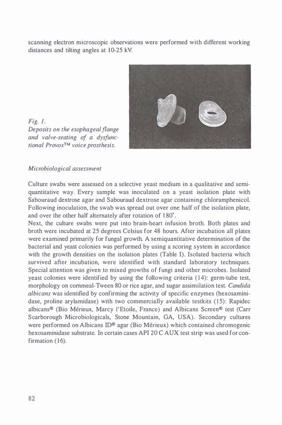
scanning electron microscopic observations were performed with different working distances and tilting angles at 1 0-25 kV.
Fig. 1. Deposits on the esophageal flange
and valve-seating of a dysfunctional Provox™ voice prosthesis.
Microbiological assessment
Culture swabs were assessed on a selective yeast medium in a qualitative and semiquantitative way. Every sample was inoculated on a yeast isolation plate with Sabouraud dextrose agar and Sabouraud dextrose agar containing chloramphenicol. Following inoculation, the swab was spread out over one half of the isolation plate, and over the other half alternately after rotation of 1 80" . Next, the culture swabs were put into brain-heart infusion broth. Both plates and broth were incubated at 25 degrees Celsius for 48 hours. After incubation all plates were examined primarily for fungal growth. A semiquantitative determination of the bacterial and yeast colonies was performed by using a scoring system in accordance with the growth densities on the isolation plates (Table I). Isolated bacteria which survived after incubation, were identified with standard laboratory techniques. Special attention was given to mixed growths of fungi and other microbes. Isolated yeast colonies were identified by using the following criteria ( 1 4) : germ-tube test, morphology on cornmeal-Tween 80 or rice agar, and sugar assimilation test. Candida albicans was identified by confirming the activity of specific enzymes (hexosaminidase, proline arylamidase) with two commercially available testkits ( 1 5) : Rapidec albicans® (Bio Merieux, Marcy ! 'Etoile, France) and Albicans Screen® test (Carr Scarborough Microbiologicals, Stone Mountain, GA, USA). Secondary cultures were performed on Albicans ID® agar (Bio Merieux) which contained chromogenic hexosaminidase substrate. In certain cases API 20 C AUX test strip was used for confirmation ( 1 6).
82
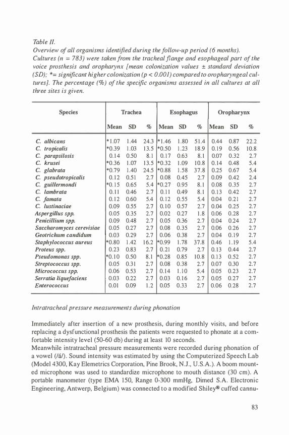
Table II. Overview of all organisms identified during the follow-up period (6 months). Cultures ( n = 783) were taken from the tracheal flange and esophageal part of the voice prosthesis and oropharynx [mean colonization values ± standard deviation (SD ); *= significant higher colonization (p < 0.001) compared to oropharyngeal cultures]. The percentage (%) of the specific organisms assessed in all cultures at all three sites is given.
Species Trachea Esophagus Oropharynx
Mean SD % Mean SD % Mean SD %
C. a/bicans * 1 .07 1 .44 24.3 * 1 .46 1 .80 5 1 .4 0.44 0.87 22.2
C. tropica/is *0.39 1 .03 1 3.5 *0.50 1 .23 1 8 .9 0. 1 9 0.56 1 0.8
C. parapsilosis 0. 1 4 0.50 8. 1 0. 1 7 0.63 8. 1 0.07 0.32 2.7
C. krusei *0.36 1 .07 1 3.5 *0.32 1 .09 1 0.8 0. 14 0.48 5.4
C. g/abrata *0.79 1 .40 24.5 *0.88 1 .58 37.8 0.25 0.67 5.4
C. pseudotropicalis 0. 1 2 0.5 1 2.7 0.08 0.45 2.7 0.09 0.42 2.4 C. guillermondi *0. 1 5 0.65 5.4 *0.27 0.95 8. 1 0.08 0.35 2.7 C. lambrata 0. 1 1 0.46 2.7 0. 1 1 0.49 8. 1 0. 1 3 0.42 2.7 C. famata 0. 1 2 0.60 5.4 0. 1 2 0.55 5.4 0.04 0.2 1 2.7 C. lustinaciae 0.09 0.55 2.7 0. 1 0 0.57 2.7 0.04 0.25 2.7 Aspergillus spp. 0.05 0.35 2.7 0.02 0.27 1 .8 0.06 0.28 2.7 Penicillium spp. 0.09 0.48 2.7 0.05 0.36 2.7 0.04 0.24 2.7 Saccharomyces cerevisiae 0.05 0.27 2.7 0.08 0.35 2.7 0.06 0.26 2.7 Geotricllum candidum 0.03 0.29 2.7 0.06 0.38 2.7 0.04 0. 1 9 2.7 Staphylococcus aureus *0.80 1 .42 1 6.2 *0.99 1 .78 37.8 0.46 1 . 1 9 5.4 Proteus spp. 0.23 0.83 2.7 0.21 0.79 2.7 0. 1 3 0.44 2.7 Pseudomonas spp. *0. 1 0 0.50 8. 1 *0.28 0.85 1 0.8 0. 1 3 0.52 2.7 Streptococcus spp. 0.05 0.3 1 2.7 0.08 0.38 2.7 0.07 0.30 2.7 Micrococcus spp. 0.06 0.53 2.7 0. 1 4 1 . 1 0 5.4 0.05 0.23 2.7 Serratia liquefaciens 0.03 0.22 2.7 0.03 0. 1 6 2.7 0.05 0.27 2.7 Enterococcus 0.01 0.09 1 .2 0.05 0.33 2.7 0.06 0.28 2.7
Intratracheal pressure measurements during phonation
Immediately after insertion of a new prosthesis, during monthly visits, and before replacing a dysfunctional prosthesis the patients were requested to phonate at a comfortable intensity level (50-60 db) during at least 10 seconds. Meanwhile intratracheal pressure measurements were recorded during phonation of a vowel (/a/). Sound intensity was estimated by using the Computerized Speech Lab (Model 4300, Kay Elemetrics Corporation, Pine Brook, N.J., U.S.A.). A boom mounted microphone was used to standardize microphone to mouth distance (30 em). A portable manometer (type EMA 150, Range 0-300 mmHg, Dimed S.A. Electronic Engineering, Antwerp, Belgium) was connected to a modified Shiley® cuffed cannu-
83
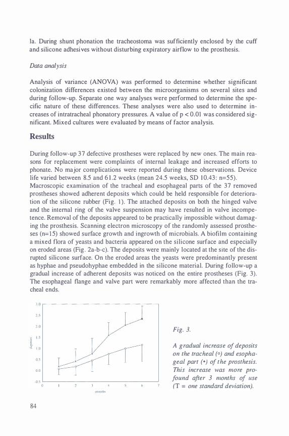
Ia. During shunt phonation the tracheostoma was sufficiently enclosed by the cuff and silicone adhesives without disturbing expiratory airflow to the prosthesis.
Data analysis
Analysis of variance (ANOVA) was performed to determine whether significant colonization differences existed between the microorganisms on several sites and during follow-up. Separate one way analyses were performed to determine the specific nature of these differences. These analyses were also used to determine increases of intratracheal phonatory pressures. A value of p < 0.01 was considered significant. Mixed cultures were evaluated by means of factor analysis.
Results
During follow-up 37 defective prostheses were replaced by new ones. The main reasons for replacement were complaints of internal leakage and increased efforts to phonate. No major complications were reported during these observations. Device life varied between 8.5 and 6 1 .2 weeks (mean 24.5 weeks, SD 1 0.43 : n=55). Macroscopic examination of the tracheal and esophageal parts of the 37 removed prostheses showed adherent deposits which could be held responsible for deterioration of the silicone rubber (Fig. I ) . The attached deposits on both the hinged valve and the internal ring of the valve suspension may have resulted in valve incompetence. Removal of the deposits appeared to be practically impossible without damaging the prosthesis. Scanning electron microscopy of the randomly assessed prostheses (n= l5) showed surface growth and ingrowth of microbials. A biofilm containing a mixed flora of yeasts and bacteria appeared on the silicone surface and especially on eroded areas (Fig . 2a-b-c). The deposits were mainly located at the site of the disrupted silicone surface. On the eroded areas the yeasts were predominantly present as hyphae and pseudohyphae embedded in the silicone material. During follow-up a gradual increase of adherent deposits was noticed on the entire prostheses (Fig. 3). The esophageal flange and valve part were remarkably more affected than the tracheal ends.
1.0
2 5
2 0
I 5
l 1 .0
0.1
0.0
0.)
84
0 1111111111 ..
Fig. 3.
A gradual increase of deposits on the tracheal (0) and esophageal part ( •) of the prosthesis. This increase was more profound after 3 months of 11se (T = one standard deviation).

Fig. 2a.
Fig. 2b.
Fig. 2c.
Fig. 2. (a) Scanning electron microscopy of a dysfunctional prosthesis showing an eroded spot (arrow) on the esophageal surface of the valve-seating. The bar (1 em) equals l03 1.J m. (b) Transversal section ofthe valve-seating showing superficial and deeper erosions (arrow). The bar ( 1 em) equals 15. 3 11 m. (c) Plaques of coccal bacteria and yeast species (arrow) attached to deteriorated silicone rubber of a dys
functional voice prosthesis. The bar ( 1 em) equals 7. 1 11 m.
85
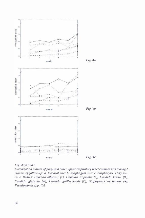
. ,
l l >< <> "0
.5 "
. S? :0 .!::! " 0
0 " . .
• •
. ,
" >< <> "0 5 " c
.S? l3
·;: 0 0 "
"
..
. ,
" >< " "0 5 1 .1 " .S? :0 N
·;: " 0
0 u
.,
- - . -
•
months
. months
. .. - - . .... � - - • 7·:·_
.---------------
- . . - - - . - -
months
Fig. 4a,b and c.
Fig. 4a.
Fig. 4b.
Fig. 4c.
Colonization indices of fungi and other upper respiratory tract commensals during 6 months of follow-up: a. tracheal site; b. esophageal site; c. oropharynx. Only mi-. (p < 0.001 ); Candida albicans (0 ), Candida tropicalis (• ), Candida krusei ('V ), Candida glabrata (T ), Candida guillermondi (0), Staphylococcus au reus (•). Pseudomonas spp. ( ll).
86

During follow-up 1 4 different yeast species were identified on the culture samples taken from 55 voice prostheses (Table II). Isolation of several yeast species varied from patient to patient and between the esophageal and tracheal flanges. During the overall period Candida albicans, glabrata and tropicalis were identified as major colonizers of the prosthesis. Staphylococcus aureus and species of Proteus, Pseudomonas and Streptococcus were also identified with significant colonization indices at both sites of the prosthesis. Associative growths were assessed in 64.2% of cultures a t the tracheal site, and in 85.2% a t the esophageal site. The majority of mixed growths consisted of fungi and upper respiratory bacteria (47-60%), while other associative growths were based on mixed cultures of fungi. Factor analysis (varimax rotation) resulted in 4 factors with a latent root larger than 1 .0. Together these factors could explain 60% of the associative growths. At all sites the first factor showed associations between C. tropicalis, Staphylococcus aureus, and Pseudomonas ssp. Associations between C. albicans, C. tropicalis, C. guillermondi, and Staphylococcus aureus were determined by factor 2. Factor 3 could determine associations between C. krusei and C. glabrata, while associations between C. guillermondi, and Proteus species were determined by factor 4. In the whole group a gradual increase of colonization was demonstrated during the following 6 months at the tracheal and esophageal sites only (Fig. 4a-b). No change of colonization was assessed in the oropharynx (Fig. 4c).
Fig. 5a and b.
The colonization of 37 dysfimctional prostheses which are standardized for the moment of removal (month 0): a. tracheal site; b. esophageal site. Only microbes are included with a significallt increase of colonization during follow-up (p < 0.001 ); Candida albicans (0),
Candida tropicalis (•), Candida krusei ('V), Candida
glabrata (T), Candida guiller
mondi (D), Staphylococcus
aureus (•}, Pseudomonas spp.
(b.}.
• >.-------------------------------.
" "
2 0
] 1 5 " 0 -� ·c 1 0 0 8
"
'·'
"' , . " ., .5 " " 0 -� 1 0
N ·a " 0 0
u
"
- -:· . - .··
.. ·· .. .. ... .. ·
months
.......
months
87
Colonization of the esophageal site was profoundly more apparent compared to the tracheal flange. Only after 6 months of follow-up significant increases (F(5, 240)= 25.40, p< 0.0 1 ) were assessed for Candida guillermondi, /ambrata, andfamata, besides Aspergillus species at both sites of the prosthesis. In comparison to the oropharynx, significant higher colonization indices (F(2, 822)= 38.58, p < 0.0001 ) were assessed for certain yeast species at both sites of the prosthesis. This was true for Candida albicans, tropicalis, krusei, glabrata, and guillermondi. The colonization of 37 dysfunctional prostheses was also analyzed in a different way by standardizing for the moment of removal. A more remarkable change of colonization of several yeast species was demonstrated (Fig. 5a-b). A gradual but significant increase of colonization at the tracheal and esophageal site (p < 0.0 1 ) was demonstrated for almost alJ yeast species approximately one to three months before replacement. In accordance with the yeast species, an increase of colonization of Staphylococcus aureus was assessed. Cultures of the oropharynx did not change during this interval. During follow-up a gradual change of intratracheal phonatory pressures (F(6,306)== 76.4 1 9; p< 0.001 ) was demonstrated (Fig. 6). A significant increase of intratracheal phonatory pressure (p < 0.0 l ) was assessed from the second month. The mean pressures varied from 1 4.9, SD 3.9 (month 0) to 32.9, SD 6.2 mmHg (month 6).
Discussion
The use of medical grade biocompatible silicone rubber seems to be an ideal material for the manufacturing of several implantable prosthetic devices. Experience with indwelling prostheses for speech and voice rehabilitation, e.g. Provox™ voice prosthesis, has learned that primary and secondary insertions are feasible without major local or systemic interactions (5,6). As with other prosthetic devices various organisms are found to adhere and colonize the surface of voice prostheses, which may influence device life. Disruption of the sil icone material at the esophageal end of the prosthesis may interfere with adequate closure and opening of the one-way valve mechanism. Frequent replacements are known to be necessary in patients with rapid colonization of prosthetic devices. Although indwelling voice prostheses are easily replaced as an outpatient procedure, regular changing might be a distressing act interfering with rehabilitation of the post-laryngectomy patient and eventually leading to irreversible damage of the tracheoesophageal fistula. In order to develop more appropriate devices which enable shunt esophageal speech, an investigation of microorganisms which are involved in sil icone deterioration is needed. As described in other reports several yeast species and bacteria originating from the oral cavity and skin flora could be identified as colonizing microorganisms ( 17 , 1 8, 19). Biofilms containing mixed flora are demonstrated on the eroded surface of plastic prosthetic devices. In our study 14 different yeast species were identified in association with other commensals of the oral flora. The yeast strains were the most distinctive colonizers of the prostheses (72.9 %). Candida albicans, and Candida glabrata, were most often isolated. These findings are in accordance with
88

other observations concerning microbial colonization of voice prostheses (7,9). Although both yeast species appeared to be dominant colonizing microbes during the follow-up period of 6 months, Candida albicans, was isolated most frequently as the primary colonizing yeast species in the first month after insertion of a new prosthesis. Afterwards other yeast species e.g. Candida glabrata, krusei, tropicalis, and guillermondi were more distinctly isolated. Staphylococcus aureus, was found to be another dominant microorganism in all cultures. It was often isolated in association with Candida albicans, and glabrata. Erosion of surfaces of several plastic devices is also known to be related to adherence and colonization by Staphylococcus aureus ( 1 1 , 1 3). Considering adhesive cell surface properties Candida albicans is known to be hydrophilic compared to the more hydrophobic other Candida strains. Association of hydrophobic and hydrophilic microorganisms could be essential for adherence to relative hydrophobic plastic surfaces (20). It has been suggested that initial adherence of Candida species, is preceded by hydrophobic bacteria i .e. Staphylococci which might create a conditioning biofilm on the plastic surface of these prostheses (2 1 ). This mechanism may be operational in the adherence and colonization of these devices. A mixed microbial flora as isolated on voice prostheses might be considered as a synergistic infection. Laryngectomees are known to be profound carriers of yeast species after receiving radiotherapy ( 1 8, 19). The oral cavity and certain foods are considered to be the continuous suppliers of yeast species and other microorganisms, which may colonize the silicone voice prostheses. Although most colonizing species are part of the oral flora, significant more Candida species were demonstrated over time at the esophageal and tracheal part as compared to the oral cavity. The esophageal parts were more distinctly affected by deposits than the tracheal flange or shaft. The yeasts are most probably directed towards the silicone material as no clinically suspected fungal infections around the tracheoesophageal fistula were found. Slight intermittent leakage of esophageal contents through the valve is believed to be responsible for colonization of the tracheal flange. The increase of intratracheal phonatory pressure over time might be indicative for progressive deterioration of the silicone valve mechanism. This may be an option for monitoring the function of tracheoesophageal voice prostheses during shunt esophageal speech.
Fig. 6.
Gradual increase of intratracheal phonatory pressure measured at comfortable phonatory levels during follow up (6 months) (T = standard deviation; itp = intratracheal phonatory pressure in mmHg; 1 mmHg = 133,3 Pascal).
"
"
months
89

Scanning electron microscopy showed disruption and irregularities of the surface of the prostheses in association with a mixed biofilm of yeasts and other microorganisms. These microbes seemed to be involved i n the process of deterioration of the prostheses. At the site of the eroded areas fungal pseudo hyphae and hyphae appeared i n a biofilm embedded within the silicone. These observations are in accordance with the results of other reports mentioning the deterioration of silicone prostheses by invasive growth of fungal hyphae into the silicone rubber (7,22,23). The penetration of silicone rubber by filamentous growing yeasts is possibly mediated by adhesive forces as well as by enzymatic degradation of the silicone. The degradation products or silicone rubber may be used as a nutrient source for the yeasts (22). In association with other upper respiratory tract commensals (i.e. Staphylococci) these disruptions of the polymerized material may promote su�sequent adherence and colonization by yeast species (9). The esophageal ends were more affected than the other parts of the prostheses. Early colonization may lead to i rreversible damage as the silicone material is not only superficially damaged, but also penetrated and disrupted by these microbes. A biofilm of mixed microorganisms might further prevent effective inhibition or clearance of these damaging yeast species. As described in previous reports, prevention of silicone deterioration of voice prostheses may be accomplished by decontamination of the oral flora with the use of local antimycotics (24,25). Inhibition of colonization may extend the device life significantly. This may contribute to appreciable reduction of costly replacements. The role of other upper respiratory tract commensals ( i .e. Staphylococci) should be taken into account when considering prevention of deterioration of tracheoesophageal voice prostheses. Further research should be directed towards prevention of deterioration of silicone materials by the use of antimicrobial prophylaxes or modification of surface properties of materials used for manufacturing voice prostheses in order to inhibit adherence of microorganisms.
References
I . Singer MI, BJorn ED. An endoscopic technique for restoration of voice after laryngectomy. Ann Otol Rhino! Laryngol 1 980; 89: 529-533.
2. Panje WR. Prosthetic voice rehabilitation following laryngectomy: The voice button. Ann Otol Rhino! Laryngol 1 98 1 ; 90: 1 1 6- 1 20.
3 . Annyas AA , Nijdam HF, Escadillo JR, e t al . Groningen prosthesis for voice rehabilitation after laryngectomy. Clin Otolaryngol 1 984; 9: 5 1 -54.
4. Zij1stra RJ, Mahieu HF, van Lith-Bijl JT, Schutte HK. Aerodynamic properties of the low-resistance Groningen button. Arch Otolaryngol Head Neck Surg 1 99 1 ; 1 1 7: 657-66 1 .
5. Van Weissenbruch R, Albers FWJ. Vocal rehabilitation after total laryngectomy using the Provox™ voice prosthesis. Clin Otolaryngol 1 993; 1 8: 359-364.
6. Manni JJ, Van den Broek P. Surgical and prosthesis-related complications using the Groningen button voice prosthesis. Clin Otolaryngol 1990; 15 : 5 1 5-523.
90
7. Mahieu HF, van Saene HKF, Rosingh HJ, Schutte HK. Candida vegetations on silicone voice prostheses. Arch Otolaryngol Head Neck Surg 1986; 1 12: 321-325.
8. Izdebski K, Ross JC, Lee S. Fungal colonization of tracheoesophageal voice prosthesis. Laryngoscope 1987; 97: 594-597.
9. Palmer MD, Johnson AP, Elliott TSJ. Microbial colonization of Blom-Singer prostheses in postlaryngectomy patients. Laryngoscope 1993; 1 03: 9 1 0-9 1 4.
10. Elliott TSJ. Plastic devices: new fields for old microbes. Lancet 1 988; i i : 30-3 1 . 1 1 . Sanger JR, Sheth NK, Franson TR. Adherence of microorganisms to breast
prostheses: an in vitro study. Ann Plast Surg 1 989; 22: 337-342. 12 . Gristina AG, Costerton JW. Bacterial adherence to biomaterials and tissues.
The significance of its role in clinical sepsis. J Bone Joint Surg (Am) 1985; 67: 264-273. 13 . Peters G, Locci R , Pulverer G. Adherence and growth of coagulase-negative
Staphylococci on surfaces of intravenous catheters. J Infect Dis 1 982; 1 46: 479-482.
14. Larone DH. Medically important fungi. New York: Elsevier, 1 987. 15 . Dalton MT, Haldane DJM, MacDonald J. Rapid identification of Candida
albicans using 4-me thy llumbellifery 1-N-acety 1-B-galactosaminide. Diagn Microbial Infect 1 989; 12: 521 -523.
16. Schuffenecker I, Freydiere A, De Montclos H, Giiie Y. Evaluation of four commercial systems for identification of medically important yeasts. Eur J Clin Microb Infect Dis 1993; 1 2: 255-260.
17. Dreizen S . Ora!. candidiasis. Am J Med 1984; 77: 28-33. 1 8. Martin MV, Al-Tikriti U, Bramley PA. Yeast flora of the mouth and skin
during and after irradiation for oral and laryngeal cancer. J Med Microbial 1 98 1 ; 14: 457-467.
19 . Chen TY, Webster JH. Oral monilia study on patients with head and neck cancer during radiotherapy. Cancer 1974; 34: 246-249.
20. Neu TR, Verkerke GJ, Herrmann IF, Schutte HK, Van der Mei HC, B usscher HJ. Microflora on explanted silicone rubber voice prostheses: taxonomy, hydrophobicity and electrophoretic mobility. J Appl Bact 1994; 76: 52 1 -528.
2 1 . Neu TR, De Boer CE, Verkerke GJ, Schutte HK, Rakhorst G , Van der Mei HC, Busscher HJ. Biofilm development in time on a silicone voice prosthesis - a case study. Microb Ecol Health Dis 1 994; 7 : 27-33.
22. Busscher HJ, de Boer CE, Verkerke GJ, Kalicharan R, Schutte HK, van der Mei HC. In vitro ingrowth of yeasts into medical grade silicone rubber. International Biodeterioration & Biodegradation 1 994: 383-390.
23. Ell SR. Candida-'the cancer of silastic ' . J Laryngol Otol 1996; 1 1 0: 240-242. 24. Mahieu HF, van Saene JJM, den Besten J, van Saene HKF. Oropharynx
decontamination preventing Candida vegetation on voice prostheses. Arch Otolaryngol Head Neck Surg 1986; 1 1 2: I 090- 1 092.
25. Van Weissenbruch R, Bouckaert S, Remon JP, Nelis HJ, Aerts R, Albers FWJ. Chemoprophylaxis of fungal deterioration of the ProvoxTM silicone tracheoesophageal prosthesis in post-laryngectomy patients. Ann Otol Rhino! Laryngol (in press).
9 1

Chapter 6
Cineradiography of th� pharyngoesophageal segment in
postlaryngectomy patients
Van Weissenbruch R, Kunnen M, Albers FWJ, Van Cauwenberge PB, Suiter AM. Cineradiography of the pharyngoesophageal segment in postlaryngectomy patients. Submitted.

Introduction
Although the survival rate after treatment of laryngeal cancer is relatively high, total surgical removal of the larynx will substantially alter speech, swallowing, and respiration. Rehabilitation of vocal functions following laryngectomy is still considered to be a major challenge. Traditionally, injection esophageal speech has been the primary method of alaryngeal voice rehabilitation. Injection esophageal speech uses air which is injected into the esophagus together with explosive consonants in normal speech ( 1 ). Sound is produced after passing this esophageal air through the pharyngoesophageal (PE) segment. However, the limited volume of the esophageal reservoir determines the short, quiet, low pitched speech produced with this method (2). In addition, only 50% to 60% of the laryngectomees are able to develop injection esophageal speech after appropriate speech training (3,4). The use of one-way valved prostheses in surgically created tracheoesophageal fistulae enabling exhaled air to be diverted from the trachea to the esophagus, have resulted in a more promising method of speech restoration (5). A larger air reservoir is l inked to the basically similar sound source as known for injection esophageal speech (6). Successful acquisition of alaryngeal speech has been reported with both primary and secondary puncture techniques using different types of voice prostheses (7,8). A louder and more sustained speech may be produced. However, good shunt esophageal speech is not invariably attained by all patients (9). Several internal and external factors (i.e. anatomical, physiological, psychological, social and educational factors) may determine successful alaryngeal speech acquisition (3). Altered surgical techniques and modified prosthetic devices were introduced to facilitate shunt esophageal speech. With the introduction of low resistance voice prostheses decreased intratracheal pressures during phonation were demonstrated, which have been paralleled with more effortless expiration ( 10, 1 1 ). Prevention of spasm and hypertonicity of the pharyngoesophageal segment by performing a unilateral myotomy and/or neurectomy seemed to improve shunt and injection esophageal speech (9, 1 2, 1 3, 1 4). The function and structure of the pharyngoesophageal segment has been assessed by using air insufflation tests or radiographic contrast studies { 1 5, 1 6, 17, 1 8). These dynamic studies may reveal considerable functional abnormalities such as spasm or hypertonicity of the cricopharyngeus muscle, and structural abnormalities such as strictures or even tumor recurrence. These disorders could interfere with successful voice rehabilitation of the postlaryngectomy patient. The purpose of this study is to evaluate cineradiographic findings in three surgical groups of laryngectomees in relation to alaryngeal speech acquisition. These findings may, in addition, direct further diagnostic procedures or therapeutic interventions.
94

Patients and Methods
Patients
Sixty patients (56 men and 4 women) were prospectively evaluated after total laryngectomy to assess their acquisition of injection esophageal speech and shunt esophageal speech. Their age varied from 4 1 to 75 years (mean 6 1 .2 years). The follow-up period varied from 8 to 1 7 months (mean 1 1 .4 months). All patients were treated for squamous cell carcinoma of the larynx and were irradiated pre- or postoperatively (60-70 Gy in 6-7 wks). A unilateral radical neck dissection was performed in 24 cases. After the total laryngectomy and primary tracheoesophageal puncture, the participating patients were randomized into three groups for evaluation of their alaryngeal speech proficiency. Nineteen patients (group 1) did not receive an additional surgical procedure. A unilateral myotomy of the cricopharyngeal and inferior constrictor pharyngeal muscles was performed in 20 patients (group 2). In 21 patients an additional unilateral neurectomy of the pharyngeal plexus was combined with a myotomy procedure (group 3). After entering the retropharyngeal plane the unilateral myotomy was carried out on the midline of the posterior surface of the constrictor pharyngeal muscles. The myotomy was limited to the cricopharyngeal and inferior constrictor pharyngeal muscles. The vertical muscle incision was completed down to the submucosal vessels. The pharyngeal neurectomy was performed after identifying the pharyngeal plexus by electrical stimulation. The nerve plexus was approached at the level of the superior thyroid artery and the middle pharyngeal constrictor muscle. The branches were cut and separated by applying electrocautery to the edges. In all patients a threelayer closure of the pharyngeal defect was used with two layers of inverting stitches in the mucosa and submucosa (T-shaped) and a layer of sutures in the constrictor muscle complex. All participating patients attended our outpatient department monthly during the follow-up period. A standardized evaluation procedure was used to compare the speech results of the laryngectomees using injection esophageal speech and I or shunt esophageal speech ( I 9,20). This procedure was discussed at the Third International Congress on Voice Prostheses (Groningen, 1 988). Semiquantitative criteria were determined for obtaining the data of the distinguished categories (phonatory skills, additional factors, general judgement). This assessment could differentiate between good, moderate, and poor speakers. As the vocal performance of shunt esophageal speech differs from injection esophageal speech, separate criteria were applied to assess both speech modes (20). A ProvoxTM voice prosthesis (Atos Medical, Horby, Sweden) was inserted in all 60 patients (1 0). The laryngectomees were simultaneously trained to acquire both injection esophageal speech and shunt esophageal speech. At the time of this study all patients were equipped with a functional voice prosthesis.
95
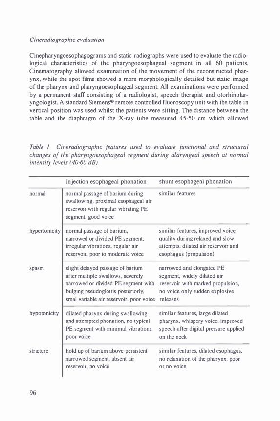
Cineradiographic evaluation
Cinepharyngoesophagograms and static radiographs were used to evaluate the radiological characteristics of the pharyngoesophageal segment in all 60 patients. Cinematography allowed examination of the movement of the reconstructed pharynx, while the spot films showed a more morphologically detailed but static image of the pharynx and pharyngoesophageal segment. All examinations were performed by a permanent staff consisting of a radiologist, speech therapist and otorhinolaryngologist. A standard Siemens® remote controlled fluoroscopy unit with the table in vertical position was used whilst the patients were sitting. The distance between the table and the diaphragm of the X-ray tube measured 45-50 em which allowed
Table I Cineradiographic features used to evaluate Junctional and structural changes of the pharyngoesophageal segment during alaryngeal speech at normal intensity levels (40-60 dB).
normal
injection esophageal phonation
normal passage of barium during
swallowing, proximal esophageal air
reservoir with regular vibrating PE
segment, good voice
shunt esophageal phonation
similar features
hypertonicity normal passage of barium,
narrowed or divided PE segment,
irregular vibrations, regular air
reservoir, poor to moderate voice
similar features, improved voice
quality during relaxed and slow
attempts, dilated air reservoir and
esophagus (propulsion)
spasm slight delayed passage of barium
after multiple swallows, severely
narrowed and elongated PE
segment, widely dilated air
narrowed or divided PE segment with reservoir with marked propulsion,
bulging pseudoglottis posteriorly, no voice only sudden explosive
smal variable air reservoir, poor voice releases
hypotonicity dilated pharynx during swallowing similar features, large dilated
stricture
96
and attempted phonation, no typical pharynx, whispery voice, improved
PE segment with minimal vibrations, speech after digital pressure applied
poor voice on the neck
hold up of barium above persistent
narrowed segment, absent air
reservoir, no voice
similar features, dilated esophagus,
no relaxation of the pharynx, poor
or no voice
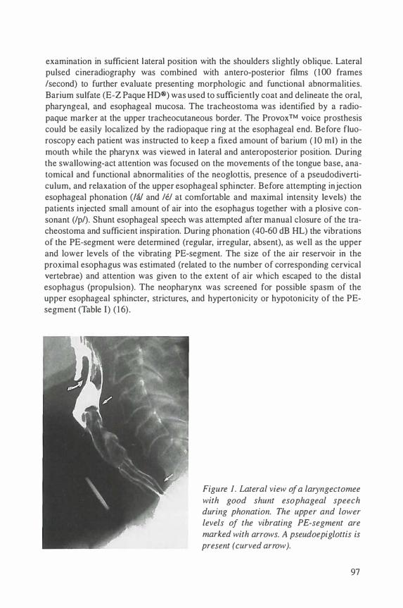
examination in sufficient lateral position with the shoulders slightly oblique. Lateral pulsed cineradiography was combined with antero-posterior films ( 1 00 frames /second) to further evaluate presenting morphologic and functional abnormalities. Barium sulfate (E-Z Paque HD®) was used to sufficiently coat and delineate the oral, pharyngeal, and esophageal mucosa. The tracheostoma was identified by a radiopaque marker at the upper tracheocutaneous border. The Provox™ voice prosthesis could be easily localized by the radiopaque ring at the esophageal end. Before fluoroscopy each patient was instructed to keep a fixed amount of barium ( I 0 ml) in the mouth while the pharynx was viewed in lateral and anteroposterior position. During the swallowing-act attention was focused on the movements of the tongue base, anatomical and functional abnormalities of the neoglottis, presence of a pseudodiverticulum, and relaxation of the upper esophageal sphincter. Before attempting injection esophageal phonation (/a/ and /e/ at comfortable and maximal intensity levels) the patients injected small amount of air into the esophagus together with a plosive consonant (/p/). Shunt esophageal speech was attempted after manual closure of the tracheostoma and sufficient inspiration. During phonation (40-60 dB HL) the vibrations of the PE-segment were determined (regular, irregular, absent), as well as the upper and lower levels of the vibrating PE-segment. The size of the air reservoir in the proximal esophagus was estimated (related to the number of corresponding cervical vertebrae) and attention was given to the extent of air which escaped to the distal esophagus (propulsion). The neopharynx was screened for possible spasm of the upper esophageal sphincter, strictures, and hypertonicity or hypotonicity of the PEsegment (Table I) ( 1 6).
Figure 1. Lateral view of a laryngectomee
with good shunt esophageal speech during phonation. The upper and lower levels of the vibrating PE-segment are marked with arrows. A pseudoepiglottis is present (curved arrow).
97

Data analysis
The speech and PE-segment data were analyzed statistically using repeated measures analysis of variance (ANOVA) to identify significant differences between the three groups and the alaryngeal speech method. All possible interactions among the surgical groups, speech outcome, and PE-segment characteristics were tested separately. Chi-squared comparisons were performed to demonstrate the source of significance for all variables with main-effect ANOVA results that had p values of less than 0.005. To verify the reliability of measures used in this study, speech and cinematography data for 20 patients were randomly reanalyzed. Inter- and intrajudge differences were distinguished by assessing respective kappa values of the specific variables. Analyses were performed with the Scientific Package for Social Sciences (SPSS®).
Results
Speech results
The three groups of laryngectomees were comparable considering age, sex, and additional neck dissection. No significant interactions were assessed for injection esophageal speech or shunt esophageal speech on age and sex differences (p> 0.05). The results of shunt and injection esophageal speech are given in Table II. Overall injection esophageal speech was rated good in 20 percent (n= 1 2) of all cases. Moderate injection esophageal speech was assessed in 57 percent (n= 34), while unsuccessful esophageal speech was acquired in 23 percent (n= l 4) of the laryngectomees. The injection esophageal speech results differed significantly between the three groups (p= 0.002). The majority of patients in group I (85 %) did not acquire fluent injection esophageal speech. Functional injection esophageal speech results were obtained in respectively 85 and 95 percent of patients in the surgical groups with additional myotomy and combined myotomy and neurectomy procedures, as compared to 47 percent in the group of laryngectomees in which these procedures were not performed. The results of shunt esophageal speech were better in all surgical groups as compared to injection esophageal speech (Table II). Shunt esophageal speech was rated good in 58 percent, while 30 percent of the laryngectomees obtained moderate results. Laryngectomees with primary myotomy and neurectomy procedures {group 3) were most successful in obtaining good shunt esophageal speech (8 1 % ). However, functional shunt esophageal speech was obtained in the majority of patients (95%) in group 2 and 3. Most shunt esophageal speech fai lures were assessed in group I
(26% ). These differences appeared to be significant (p< 0.000 I ) across the surgical groups.
Cineradiographic Characteristics
Radiographic contrast studies in the early postoperative period demonstrated pharyngocutaneous fistulae in 9 patients ( 15 %) (Table III). The majority of these
98

Table 2. Results of injection esophageal speech and shunt esophageal speech among
the three surgical groups.
speech quality
poor moderate good
group n n % n % n %
injection method
1 9 1 0 52.6 6 3 1 .5 3 1 5 .8
2 20 3 1 5.0 1 5 75.0 2 1 0.0
3 2 1 4.8 1 3 6 1 .9 7 33.3
all 60 14 23.3 34 56.7 1 2 20.0
shunt method
19 5 26.3 8 42. 1 6 3 1 .6
2 20 5.0 7 35.0 1 2 60.0
3 2 1 I 4.8 3 1 4.3 17 80.9
all 60 7 1 1 .7 1 8 30.0 35 58.3
fistulae (n=5, 26%) occurred in laryngectomees from group I . No significant differences (p=0.526) were apparent between the surgical groups. Six patients developed a pharyngeal stricture at the level of the former fistula tract. A pseudoepiglottis was noticed in 93 percent of all patients during attempted phonation at the anterior wall of the PE-segment just inferior to the base of the tongue (Fig. 1 ) . No significant difference (p=0.749) in occurrence was assessed among good or poor a1aryngeal speakers. This structure may not be related to voice quality. A broad pseudoglottis (width > 1 cervical vertebra) with evidence for spasm or hypertonicity of the upper esophageal sphincter was found in 19 cases. This feature appeared to be significant (p=0.0002) for voice failures in group I. A significant correlation (p< 0.00 1 ) between poor esophageal speech and a delayed opening of upper esophageal sphincter was demonstrated. A bilobular aspect of the PE-segment was associated with functional esophageal speech among all surgical groups. The ProvoxTM voice prosthesis was located inferior to the pseudoglottis in all cases. Shunt esophageal speech failure was not associated with obstruction of the esophageal part of the prosthesis. On average the inferior level of the vibrating PE-segment during phonation was situated at C6 (Table IV). No significant (p> 0.005) differences concerning the quality of injection esophageal or shunt esophageal speech were obtained for this inferior level.
99
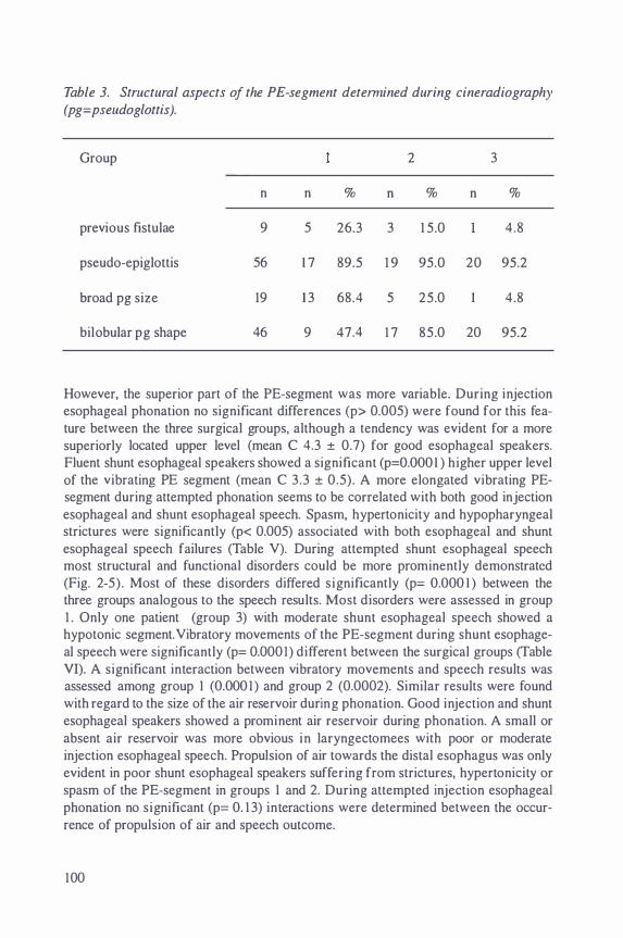
Table 3. Structural aspects of the PE-segment detemzined during cineradiography (pg=pseudoglottis).
Group 2 3
n n % n % n %
previous fistulae 9 5 26.3 3 1 5.0 4.8
pseudo-epiglottis 56 I 7 89.5 I 9 95.0 20 95.2
broad pg size 19 1 3 68.4 5 25.0 4.8
bilobular pg shape 46 9 47.4 I 7 85.0 20 95.2
However, the superior part of the PE-segment was more variable. During injection esophageal phonation no significant differences (p> 0.005) were found for this feature between the three surgical groups, although a tendency was evident for a more superiorly located upper level (mean C 4.3 ± 0.7) for good esophageal speakers. Fluent shunt esophageal speakers showed a significant (p=O.OOOI ) higher upper level of the vibrating PE segment (mean C 3.3 ± 0.5). A more elongated vibrating PEsegment during attempted phonation seems to be correlated with both good injection esophageal and shunt esophageal speech. Spasm, hypertonicity and hypopharyngeal strictures were significantly (p< 0.005) associated with both esophageal and shunt esophageal speech failures (Table V). During attempted shunt esophageal speech most structural and functional disorders could be more prominently demonstrated (Fig. 2-5) . Most of these disorders differed significantly (p= O.OOO I ) between the three groups analogous to the speech results. Most disorders were assessed in group I . Only one patient (group 3) with moderate shunt esophageal speech showed a hypotonic segment. Vibratory movements of the PE-segment during shunt esophageal speech were significantly (p= 0.0001) different between the surgical groups (Table VI). A significant interaction between vibratory movements and speech results was assessed among group I (O.OOOI ) and group 2 (0.0002). Similar results were found with regard to the size of the air reservoir during phonation. Good injection and shunt esophageal speakers showed a prominent air reservoir during phonation. A small or absent air reservoir was more obvious in laryngectomees with poor or moderate injection esophageal speech. Propulsion of air towards the distal esophagus was only evident in poor shunt esophageal speakers suffering from strictures, hypertonicity or spasm of the PE-segment in groups I and 2. During attempted injection esophageal phonation no significant (p= O. I 3) interactions were determined between the occurrence of propulsion of air and speech outcome.
100

The voice and cineradiography data of 20 patients were randomly reanalyzed to verify the reliability of measures. Intra judge reliability between the measures ranged from 0.46 to 1 .0 (mean 0.66). The interjudge reliability between the measures ranged from 0.5 1 to 1 .0 (mean 0.68). Intrajudge and interjudge measures were found to be satisfactory.
Discussion
Tracheoesophageal speech has become an established method of alaryngeal voice rehabilitation in combination with injection esophageal speech. Most patients acquire fluent shunt esophageal speech for effective daily communication without specific intervention (5, 7 ,8). However, a distinctive group of patients still fails to achieve effective injection esophageal or shunt esophageal speech. Several authors have reported failure rates varying from 10 to 80 percent {3,9,2 1 ). In this study laryngectomees who had a primary myotomy procedure or a combined myotomy and
Table 4. Evaluation of the upper and lower levels of the vibrating PE-segment during injection esophageal speech and shunt esophageal speech. The levels are classified by surgical group and speech proficiency (PE- level ± SD= level of the PEsegment and standard deviation; lower= lower level of the PE-segment; upper= upper level of the PE-segment).
speech quality
poor moderate good
PE-level ± SD lower upper lower upper lower upper
injection esophageal speech
group I 5 .9±0.3 4.8±0.4 5 .7±0.5 4.7±0.5 6.0±0. 1 4 .2±0.4
group 2 6.0±0.2 4.7±0.5 5.9±0.2 4.6±0.4 6.0±0. 1 4.3±0.4
group 3 6.0±0.2 5.0±0. 1 6.0±0.3 4.7±0.4 6.2±0.2 4.2±0.5
shunt esophageal speech
group 1 6. 1 ±0.5 5.0±0.4 6.3±0.7 4.2±0.4 5.8±0.3 4. 1 ±0.6
group 2 6.0±0. 1 5 .0±0.3 5.9±0.4 4.4±0.5 6. 1 ±0.4 3 .4±0.5
group 3 6.0±0.3 4. 1 ±0.2 6.0±0.3 3.5±0.4 6.4±0.8 3 .3±0.4
10 1

neurectomy procedure at the time of laryngectomy demonstrated both better shunt esophageal and injection esophageal speech proficiencies (60-80% resp. 1 0-33%) compared to l aryngectomees lacking these additional procedures (32% resp. 1 6%). As removal of the larynx may be considered as a major disabling procedure, failure of voice rehabilitation is often related to psychosocial problems. These problems appear to be susceptible to active intervention ( 4 ). In case of shunt esophageal speech failure, device-related causes should be excluded before further investigations are indicated (22). In all patients the Provox™ voice prosthesis appeared to be localized below the pharyngoesophageal segment. No obstruction of airflow due to devicerelated problems or to blockage of the valve part by the posterior wall of the esophagus was apparent.
Table 5. Evaluation of functional and structural abnormalities of the PE-segment during injection esophageal speech and shunt esophageal speech. Statistical analy-ses (Chi-square) were performed to determine any interaction with speech failure ( 1-3= interaction between the surgical groups).
groups
1 -3 2 3
p n p n p n p
injection method
spasm 0.001 4 0.001 2 0.082 0
hypertonicity 0.0002 9 0.0007 8 0.001 0
hypotonicity 0.5832 0 0 0.349
stricture 0.0089 3 0.002 2 0.03 0.008
shunt method
spasm 0.0001 6 0.0001 4 0.0006 0
hypertonicity 0.0001 7 0.0001 3 0.0006 0.8 1 8
hypotonicity 0.022 0 0 0.8 1 8
stricture 0.0001 3 0.0001 2 0.0001 0.0001
102

Physical factors influencing vibratory characteristics of the PE-segment seem to affect esophageal speech acquisition directly. In assessing quality of speech, the reconstructed pharyngoesophageal segment should be examined dynamically (23). Fluoroscopy can be used to detect functional or morphologic abnormalities within the oral, pharyngeal, or esophageal segments. Patients with functional shunt esophageal speech showed a regular vibrating pharyngoesophageal segment during phonation. The vibrating segment appeared to be more elongated in good shunt esophageal speakers. The majority of examined patients revealed to have their 'pseudoglottis' at the junction of the neopharynx and upper esophagus at the level of the fifth and sixth cervical vertebrae. In fluoroscopic and strobofiberscopic studies the 'pseudoglottis' has been recognized as a protrusion of the posterior wall at the lower PE-segment ( 1 8). The inferior level of the PE-segment did not differ among poor or proficient shunt and injection esophageal speakers. The upper part of the pseudoglottis is considered to be the sound producing part as during phonation a regular vibration could be noticed in conjunction with the proximal part of the PE-segment, while the lower part showed no regular vibration (23).
Table 6. Assessment of specific characteristics of the PE-segment during injection esophageal speech and shunt esophageal speech. Interactions (p value) between specific characteristics and alary•ngeal speech proficiency are classified by surgical group.
injection esophageal speech shunt esophageal speech
group 2 3 2 3
vibration 0.0009 0.0001 0. 1 539 0.0001 0.0002 0.2825
reservoir 0.0047 0.0820 0.68 10 0.0001 0.0007 0.8838
propulsion 0.2461 0.88 1 9 0.4076 0.0001 0.0003 0.7395
After total laryngectomy the size and shape of the pharynx has been changed to a more straight and tubular structure. At the upper surgical margin the neopharynx may appear narrower with formation of a pseudodiverticulum or pseudoepiglottis at the anterior wall. This sharp-angled anterior outpouching is located at the upper suture line of the neopharynx. Widening below this junction may produce the impression of an 'epiglottis ' . Approximately 90% of all patients showed such a pseudodiverticulum. The existence might be related to altered pharyngeal physiology postoperative-
103
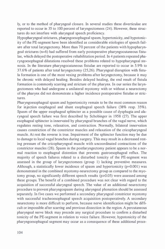
ly, or to the method of pharyngeal closure. In several studies these diverticulae are reported to occur in 35 to 1 00 percent of l aryngectomees (24). However, these structures do not interfere with alaryngeal speech proficiency. Hypopharyngeal strictures, pharyngoesophageal spasm, hypertonicity, and hypotonicity of the PE-segment have been identified as considerable etiologies of speech failure after total laryngectomy. More than 70 percent of the patients with hypopharyngeal strictures (n=6) had suffered from early postoperative pharyngocutaneous fistulae, which delayed the postoperative rehabilitation period. In 4 patients repeated pharyngoesophageal dilatations resolved these problems related to hypopharyngeal stenosis. In the literature pharyngocutaneous fistulae are reported to occur in 5.9% to 37.6% of patients after total laryngectomy (25,26). Pharyngeal disruption with fistula formation is one of the most vexing problems after laryngectomy, because it may be chronic with delayed healing. Besides delayed healing, the end result of fistula formation is commonly scarring and stricture of the pharynx. In our series the laryngectomees who had undergone a unilateral myotomy with or without a neurectomy of the pharynx did not demonstrate a higher incidence postoperative fistulae or strictures. Pharyngoesophageal spasm and hypertonicity remain to be the most common reason for injection esophageal and shunt esophageal speech failure (38% resp. 35%). Spasm of the upper esophageal sphincter as a possible cause of dysphagia and alaryngeal speech fai lure was first described by Schobinger in 1 958 (27). The upper esophageal sphincter is innervated by pharyngeal branches of the vagal nerve, which regulates resting tone, relaxation, and contraction. Normally, bilateral innervation causes constriction of the constrictor muscles and relaxation of the cricopharyngeal muscle. At rest the reverse is true. Impairment of the sphincter function may be due to damage to local vagal branches during surgery. This may result in a decreased resting pressure of the cricopharyngeal muscle with uncoordinated contractions of the constrictor muscles (28). Spasm in the postlaryngectomy patient appears to be a normal reaction to esophageal distention that prevents gastroesophageal reflux. The majority of speech failures related to a disturbed tonicity of the PE-segment was assessed in the group of laryngectomees (group 1 ) lacking preventive measures. Although, a statistically lower incidence of spasm and hypertonicity (p=O.OOl ) was demonstrated in the combined myotomy-neurectomy group as compared to the myotomy group, no significantly different speech results (p>0.05) were assessed among these groups. The benefit of a combined procedure was not clear with regard to the acquisition of successful alaryngeal speech. The value of an additional neurectomy procedure to prevent pharyngospasm during alaryngeal phonation should be assessed separately. In five cases we performed a secondary pharyngeal constrictor myotomy with successful tracheoesophageal speech acquisition postoperatively. A secondary neurectomy is more difficult to perform, because nerve identification might be difficult or impossible after scarring and previous dissection in the region. A percutaneous pharyngeal nerve block may precede any surgical procedure to confirm a disturbed tonicity of the PE-segment in relation to voice failure. However, hypotonicity of the pharyngoesophageal segment may occur as a consequence of these additional proce-
104
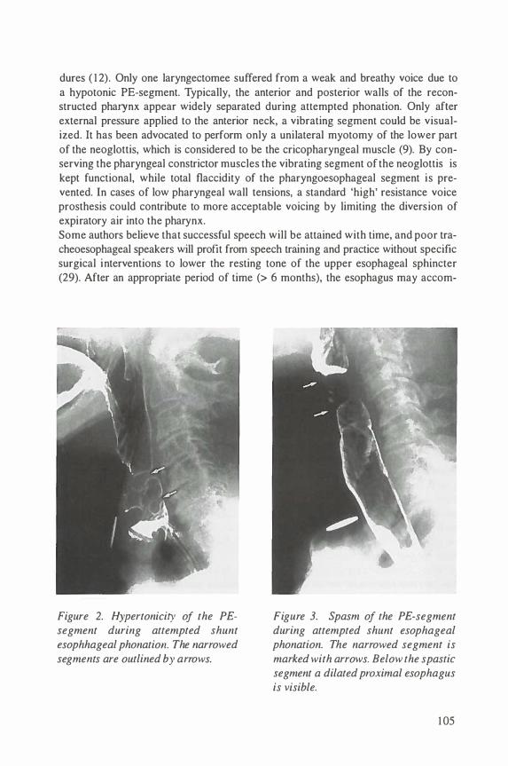
dures ( 1 2). Only one laryngectomee suffered from a weak and breathy voice due to a hypotonic PE-segment. Typically, the anterior and posterior walls of the reconstructed pharynx appear widely separated during attempted phonation. Only after external pressure applied to the anterior neck, a vibrating segment could be visualized. It has been advocated to perform only a unilateral myotomy of the lower part of the neoglottis, which is considered to be the cricopharyngeal muscle (9). By conserving the pharyngeal constrictor muscles the vibrating segment of the neoglottis is kept functional, while total flaccidity of the pharyngoesophageal segment is prevented. In cases of low pharyngeal wall tensions, a standard 'high' resistance voice prosthesis could contribute to more acceptable voicing by limiting the diversion of expiratory air into the pharynx. Some authors believe that successful speech will be attained with time, and poor tracheoesophageal speakers will profit from speech training and practice without specific surgical interventions to lower the resting tone of the upper esophageal sphincter (29). After an appropriate period of time (> 6 months), the esophagus may accom-
Figure 2. Hypertonicity of the PEsegment during attempted shunt esophhageal phonation The narrowed segments are outlined by arrows.
Figure 3. Spasm of the PE-segment during attempted shunt esophageal phonation. The narrowed segment is marked with arrows. Below the spastic segment a dilated proximal esophagus is visible.
1 05
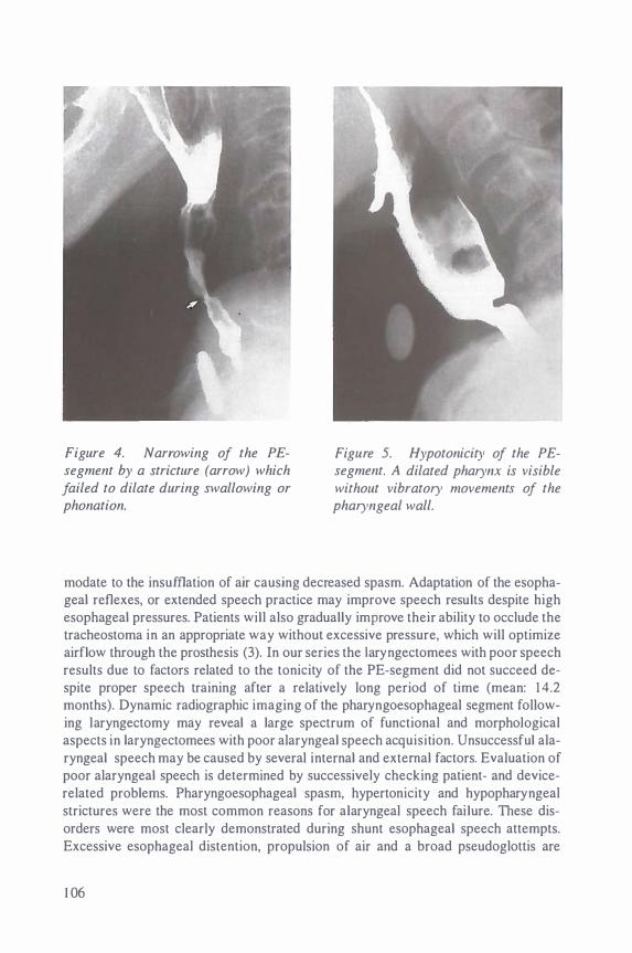
Figure 4. Narrowing of the PEsegment by a stricture (arrow) which failed to dilate during swallowing or phonation.
Figure 5. Hypotonicity of the PEsegment. A dilated pharynx is visible without vibratory movements of the pharyngeal wall.
modate to the insufflation of air causing decreased spasm. Adaptation of the esophageal reflexes, or extended speech practice may improve speech results despite high esophageal pressures. Patients will also gradually improve their ability to occlude the tracheostoma in an appropriate way without excessive pressure, which will optimize airflow through the prosthesis (3). In our series the laryngectomees with poor speech results due to factors related to the tonicity of the PE-segment did not succeed despite proper speech training after a relatively long period of time (mean: 1 4.2 months). Dynamic radiographic imaging of the pharyngoesophageal segment following laryngectomy may reveal a large spectrum of functional and morphological aspects in laryngectomees with poor alaryngeal speech acquisition. Unsuccessful alaryngeal speech may be caused by several internal and external factors. Evaluation of poor alaryngeal speech is determined by successively checking patient- and devicerelated problems. Pharyngoesophageal spasm, hypertonicity and hypopharyngeal strictures were the most common reasons for alaryngeal speech failure. These disorders were most clearly demonstrated during shunt esophageal speech attempts. Excessive esophageal distention, propulsion of air and a broad pseudoglottis are
1 06

typical characteristics of an increased tonicity the PE-segment during alaryngeal speech attempts. A unilateral cricopharyngeal and inferior pharyngeal constrictor myotomy seems to be appropriate to prevent muscle spasm and hypertonicity of the neoglottis. Cineradiography seems to be a useful method for demonstratin g functional and structural disorders which may be responsible for swallowing difficulties or alaryngeal speech failures. These imaging studies do allow proper management of the postlaryngectomy patient.
References
I . Moolenaar-Bijl AJ. The importance o f certain consonants i n oesophageal voice after laryngectomy. Ann Otol Rhino! Laryngol 1 953; 62: 979-989.
2. Winans CS, Reichbach EJ, Waldrop WF. Esophageal determinants of alaryngeal speech. Arch Otolaryngol 1974; 99: 1 0- 14.
3. Gates GA, Hearne III EM. Predicting esophageal speech. Ann Otol Rhino) Laryngol 1 982; 9 1 : 454-457.
4. Gates GA, Ryan W, Cantu E. Current status of laryngectomee rehabil itation: II. Causes of failure. Am J Otolaryngol 1982; 3 : 8- 14.
5. Singer MI, BJorn ED. An endoscopic technique for restoration of voice after laryngectomy. Ann Otol Rhino! Laryngol 1 980; 89: 529-533.
6 . Wetmore SJ, Ry�tn SP, Montague JC. Location of the vibratory segment in tracheoesophageal speakers. Otolaryngol Head Neck Surg 1985; 93: 355-36 1 .
7. Hamaker RC, Singer MI, BJorn ED. Primary voice restoration at laryngectomy. Arch Otolaryngol 1985; I l l : 1 82- 1 86.
8. Panje WR. Prosthetic vocal rehabilitation following laryngectomy. The voice button. Ann Otol Rhino[ Laryngol 198 1 ; 90: 1 1 6- 1 20.
9. Singer MI, BJorn ED. Selective myotomy for voice restoration after total laryngectomy. Arch Otolaryngol 198 1 ; 1 07: 670-673.
I 0. Hilgers FJM, Schouwen burg PF. A new low-resistance, self-retaining prosthesis (ProvoxTM) for voice rehabilitation after total laryngectomy. Laryngoscope 1990; I 00: 1 202- 1 207.
I I . Zijlstra RJ, Mahieu HF, Van Lith-Bijl JT, Schutte HK. Aerodynamic properties of the low-resistance Groningen button. Arch Otolaryngol Head Neck Surg 199 1 ; 1 1 7: 657-66 1 .
12 . Baugh RF, Baker SR, Lewin JS . Surgical treatment of pharyngoesophageal spasm. Laryngoscope 1988; 98: 1 1 24- 1 1 26.
1 3. Medina JE, Reiner SA. Pharyngeal myotomy for voice restoration: a midline approach. Laryngoscope 1990; I 00: 309-3 1 2.
1 4. Singer MI, BJorn ED, Hamaker RC. Pharyngeal plexus neurectomy for alaryngeal speech rehabilitation. Laryngoscope 1986; 96: 50-54.
1 5. Callaway E, Truelson JM, Wolf GT, Thomas-Kincaid T and Cannon S . Predictive value of objective esophageal insufflation testing for acquisition of tracheoesophageal speech. Laryngoscope 1992; 1 02: 704-708.
1 07
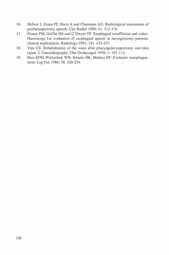
1 6. Mcivor J, Evans PF, Perry A and Cheesman AD. Radiological assessment of postlaryngectomy speech. Clin Radio) 1990; 4 1 : 3 1 2-3 1 6.
1 7. Sloane PM, Griffin JM and O'Dwyer TP. Esophageal insufflation and videofluoroscopy for evaluation of esophageal speech in laryngectomy patients: clinical implications. Radiology 1 99 1 ; 1 8 1 : 433-437.
1 8. Vize CE. Rehabilitation of the voice after pharyngolaryngectomy and skin repair, 1 : Cineradiography. Clin Otolaryngol 1 976; 1 : 1 07- 1 1 3.
19. Bors EFM, Wicherlink WH, Schutte HK, Mahieu HF. Evaluatie oesophagusstem. Log Fon 1 986; 58: 230-234.
108

Chapter 7
Assessment of the pharyngoesophageal segment after total laryn
gectomy: alaryngeal speech characteristics
Van Weissenbruch R, De Looze D, Kunnen M, Albers FWJ, Van Cauwenberge PB. Assessment of the pharygoesophageal segment after total laryngectomy: alaryngeal speech characteristics. Submitted.
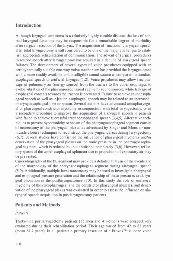
Introduction
Although laryngeal carcinoma is a relatively highly curable disease, the loss of normal laryngeal functions may be responsible for a remarkable degree of morbidity after surgical resection of the larynx. The acquisition of functional alaryngeal speech after total laryngectomy is still considered to be one of the major challenges to establ ish appropriate rehabilitation of communication. The advent of surgical procedures to restore speech after laryngectomy has resulted in a decline of alaryngeal speech failures. The development of several types of voice prostheses equipped with an aerodynamically suitable one-way valve mechanism has provided the laryngectomee with a more readily available and intelligible sound source as compared to standard esophageal speech or artificial larynges ( 1 ,2). Voice prostheses may allow free passage of pulmonary air (energy source) from the trachea to the upper esophagus to evoke vibration of the pharyngoesophageal segment (sound source), while leakage of esophageal contents towards the trachea is prevented. Failure to achieve shunt esophageal speech as well as injection esophageal speech may be related to an increased pharyngoesophageal tone or spasm. Several authors have advocated cricopharyngeal or pharyngeal constrictor myotomy in conjunction with total laryngectomy, or as a secondary procedure to improve the acquisition of alaryngeal speech in patients who failed to achieve successful tracheoesophageal speech (3,4,5). Alternative techniques to prevent hypertonicity or spasm of the pharyngoesophageal segment consist of neurectomy of the pharyngeal plexus as advocated by Singer and BJorn, or nonmuscle closure techniques to reconstruct the pharyngeal defect during laryngectomy (6,7). Several studies have confirmed the influence of pharyngeal myotomy and/or denervation of the pharyngeal plexus on the tonic pressure in the pharyngoesophageal segment, which is reduced but not abolished completely (3,6). However, reflectory spasm of the upper esophageal sphincter due to propulsion of expiratory air may be prevented. Cineradiography of the PE-segment may provide a detailed analysis of the events and of the morphology of the pharyngoesophageal segment during alaryngeal speech (8,9). Additionally, multiple level manometry may be used to investigate pharyngeal and esophageal pressure generation and the relationship of these pressures to alaryngeal phonation in the postlaryngectomee ( I 0). In this study the role of unilateral myotomy of the cricopharyngeal and the constrictor pharyngeal muscles, and denervation of the pharyngeal plexus was evaluated in order to assess the influence on alaryngeal speech acquisition in postlaryngectomy patients.
Patients and Methods
Patients
Thirty-nine postlaryngectomy patients (35 men and 4 women) were prospectively evaluated during their rehabilitation period. Their age varied from 42 to 82 years (mean 6 1 .2 years). In all patients a primary insertion of a ProvoxTM silicone voice
1 1 0
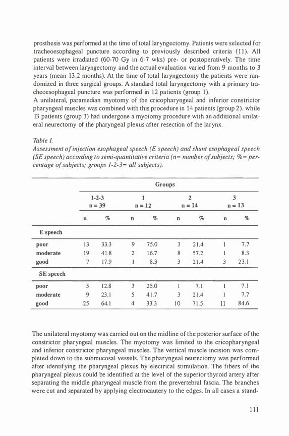
prosthesis was perfonned at the time of total laryngectomy. Patients were selected for tracheoesophageal puncture according to previously described criteria ( I I ). All patients were irradiated (60-70 Gy in 6-7 wks) pre- or postoperatively. The time interval between laryngectomy and the actual evaluation varied from 9 months to 3 years (mean I3 .2 months). At the time of total laryngectomy the patients were randomized in three surgical groups. A standard total laryngectomy with a primary tracheoesophageal puncture was perfonned in I 2 patients (group I ). A unilateral, paramedian myotomy of the cricopharyngeal and inferior constrictor pharyngeal muscles was combined with this procedure in I 4 patients (group 2), while 13 patients (group 3) had undergone a myotomy procedure with an additional unilateral neurectomy of the pharyngeal plexus after resection of the larynx.
Table I. Assessment of injection esophageal speech ( E speech) and shunt esophageal speech (SE speech) according to semi-qualltitative criteria (n= number of subjects; %= percelltage of subjects; groups 1-2-3= all subjects).
1-2-3
n = 39
n % n
E speech
poor 1 3 33.3 9
moderate 1 9 4 1 .8 2
good 7 1 7.9
SE speech
poor 5 1 2.8 3
moderate 9 23. 1 5
good 25 64. 1 4
1
n = 1 2
Groups
%
75.0
1 6.7
8.3
25.0
4 1 .7
33.3
n
3
8
3
I
3
1 0
2
n = 14
%
2 1 .4
57.2
2 1 .4
7. 1
2 1 .4
7 1 .5
n
3
I I I
3
n = 1 3
%
7.7
8.3
23 . 1
7. 1
7.7
84.6
The unilateral myotomy was carried out on the midline of the posterior surface of the constrictor pharyngeal muscles. The myotomy was limited to the cricopharyngeal and inferior constrictor pharyngeal muscles. The vertical muscle incision was completed down to the submucosal vessels. The pharyngeal neurectomy was perfonned after identifying the pharyngeal plexus by electrical stimulation. The fibers of the pharyngeal plexus could be identified at the level of the superior thyroid artery after separating the middle pharyngeal muscle from the prevertebral fascia. The branches were cut and separated by applying electrocautery to the edges. In all cases a stand-
I l l

ard three layer closure of the pharynx was performed in a T-shaped fashion. A standard pre- and postoperative protocol for instruction and rehabilitation of speech was used in all participants. A semiquantitative evaluation of the alaryngeal speech acquisition was made using uniform criteria, which were reported previously ( I I ) . The criteria were discussed at the Third International Congress on Voice Prostheses held in Groningen ( 1988). These criteria were found suitable to assess both shunt esophageal speech as well as injection esophageal speech. Several phoniatric parameters, additional features of voice production, and quality of speech were evaluated. Alaryngeal speech was ultimately evaluated in a three-grade scale as poor, moderate or good.
Computer Manometry
During shunt and injection esophageal speech the pressure generation in the esophagus, pharyngoesophageal segment, and hypopharynx was assessed in all patients. A motility probe, consisting a 4.65 mm diameter, flexible tube which incorporates three miniature pressure transducers near its distal end, was used for direct esophageal pressure measurements (P3 1 -322D, Konigsberg Instruments, Inc., Pasadena, CA 9 1 107, USA). Inter-sensor spacing was 5 em, with the distal sensor 5 em from the tip. Each sensor element (output of 7.5 mV at 300 mmHg, at 5.0 V) was mounted in a titanium carrier and coated with silastic to protect the sensors. All pressure transducers were calibrated to 300 mm Hg (maximal 1 200 mm Hg) and consisted of dynamic temperature compensation. The 1 25 em long, medical grade probe was attached through a compensation module (fJ Digitrapper 2 MB, Synectics Medical, Brussels, Belgium) to a personal computer (Tulip® Tr 386 SX). Software (Polygram version 5 .00C4, Upper GI Edition, Gastrosoft Inc. , USA) capable of measuring pressures in mmHg ( 1 mmHg= 1 33.3 Pascal), and calculating mean and maximal values, duration of activity, and resting pressures was used to evaluate pressure generation. Direct measurement of pressures directly at, above, and below the specific site of interest, eliminates the problems and complexity of fluid columns and infusion pumps. Pressures could be measured continuously, at rest, and dynamically. The subjects were monitored in sitting position. The sensors, oriented orthogonally to the probe center-line permitted measurement of non-symmetric pressures while withdrawing the probe. The probe was clearly marked in I em increments to assist in identifying transducer position. Verification of sensor location was also permitted by its radiopaque tip and sensors. The slightly stiffened probe was easily introduced transnasally in all subjects after local application of Lidocaine® 2%. Intratracheal pressure measurements during shunt esophageal phonation were carried out with a portable manometer provided with a Gaeltec® microtip pressure transducer (EMA 1 50, operating pressure range ± 300 mmHg, Dimed S .A., Electronic Engineering, Antwerp, Belgium). The pressure transducer had an compensated temperature range ( I o· -40"C). Appropriate closure of the tracheostoma during phonation was accomplished with an adapted cuffed Shiley® tracheal canula adhered to the skin with silicone adhesives. The tracheal canula was directly connected to the pressure
1 1 2
transducer. Patients were requested to phonate continuously {Ia/) for at least 10 seconds during intratracheal pressure measurements. Every measurement was repeated 5 times. Before testing all subjects were provided with a new Provox™ voice prosthesis. Intratracheal pressure recordings were evaluated in accordance with intraesophageal pressure measurements. The generation of pressure in the body of the esophagus, the subpseudoglottis (proximal of the esophagus), and the pharyngoesophageal segment were recorded, during soft (30-40 dB), normal (50-60 dB), and loud (60-70 dB) vocalization. Sound intensity was monitored with the Computerized Speech Lab (model 4300, Kay Elemetrics Corp., Pine Brook, NJ, USA).
Cineradiography
Cineradiographic imaging of the pharyngoesophageal segment and esophagus was performed in all subjects during alaryngeal phonation. Functional and morphological aspects of the reconstructed pharynx were assessed and related to pressure generation at specific sites. Frontal and lateral recordings at 100 frames/second with a standard Siemens® remote controlled fluoroscopy unit were made while the subjects were sitting. An external stoma site marker was used to ensure accuracy. Satisfactory coating of the neopharynx and esophagus (E-Z paque® HD: barium sulphate 98, excipiens qs ad 340 gr) was achieved for proper definition of the anatomy and any area of functional or morphological discrepancy. Vibratory movements of the pharyngoesophageal segment (i.e. pseudoglottis) during phonation were assessed. The PE-segment was screened for functional and morphological abnormalities, e.g. spasm, hypertonicity, hypotonicity, strictures, or pseudodiverticulae.
Statistical analysis
All data were evaluated by using repeated measures analysis of variance (ANOVA). Interactions among surgical groups, phonatory results, and computer manometry were tested separately. Chi-squared comparisons were performed to assess the source of significance for all variables with main-effect ANOVA results that had p values of less than 0.0 1 . Analyses were performed with the Scientific Package for Social Sciences (SPSS").
Results
According to the semi-quantitative analysis of alaryngeal speech acquisition, subjects who had complementary surgical procedures (i.e. unilateral myotomy and/or neurectomy) were found to be superior shunt and injection esophageal speakers (table I). Significant better injection esophageal speech results were obtained in both group 2 (p = 0.0016) and group 3 (p = 0.0001 ) compared to group I . Similar results were assessed for shunt esophageal speech in group 2 (p = 0.0047) and group 3 (p = 0.000 1 ). No significant differences considering shunt esophageal speech (p = 0.076) and injection esophageal speech (p= 0.59) were evident between laryngectomees in group 2 and 3.
1 1 3
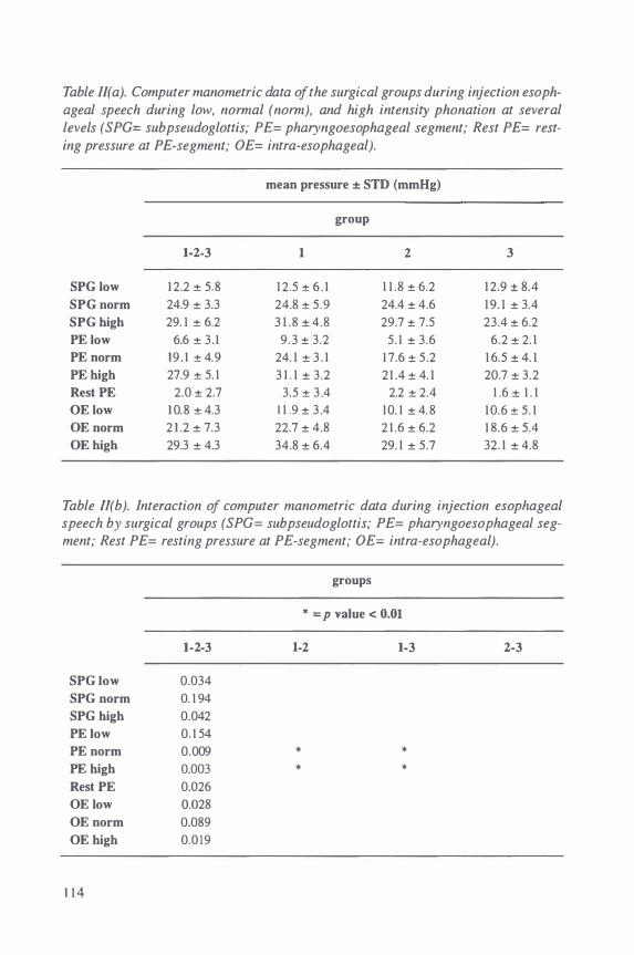
Table II( a). Computer manometric data of the surgical groups during injection esophageal speech during low, normal (nomz), and high intensity phonation at several levels (SPG= subpseudoglottis; PE= pharyngoesophageal segment; Rest PE= resting pressure at PE-segment; OE= intra-esophageal).
mean pressure ± STD (mmHg)
group
1-2-3 1 2 3
SPG low 1 2.2 ± 5.8 1 2 .5 ± 6 . 1 1 1 .8 ± 6.2 1 2.9 ± 8.4
SPG norm 24.9 ± 3.3 24.8 ± 5 .9 24.4 ± 4.6 1 9. 1 ± 3.4
SPG high 29. 1 ± 6.2 3 1 .8 ± 4.8 29.7 ± 7.5 23.4 ± 6.2
PE iow 6.6 ± 3 . 1 9 .3 ± 3.2 5. 1 ± 3.6 6.2 ± 2. 1
PE norm 1 9 . 1 ± 4.9 24. 1 ± 3 . 1 1 7.6 ± 5.2 1 6.5 ± 4. 1
PE high 27.9 ± 5. 1 3 1 . 1 ± 3.2 21 .4 ± 4. 1 20.7 ± 3.2
Rest PE 2.0 ± 2.7 3.5 ± 3 .4 2.2 ± 2.4 1 .6 ± 1 . 1
OE low 1 0.8 ± 4.3 1 1 .9 ± 3 .4 1 0. 1 ± 4. 8 1 0.6 ± 5. 1
OE norm 2 1 .2 ± 7.3 22.7 ± 4.8 2 1 .6 ± 6.2 1 8.6 ± 5.4
OE high 29.3 ± 4.3 34.8 ± 6.4 29. 1 ± 5.7 32. 1 ± 4.8
Table Il(b). Interaction of computer manometric data during injection esophageal speech by surgical groups (SPG= subpseudoglottis; PE= pharyngoesophageal segment; Rest PE= resting pressure at PE-segment; OE= intra-esophageal).
groups
* = p value < 0.01
1-2-3 1-2 1-3 2-3
SPG low 0.034
SPG norm 0. 1 94
SPG high 0.042
PE iow 0. 1 54
PE norm 0.009 * *
PE high 0.003 * *
Rest PE 0.026
OE iow O.D28
OE norm 0.089
OE high 0.0 1 9
1 1 4
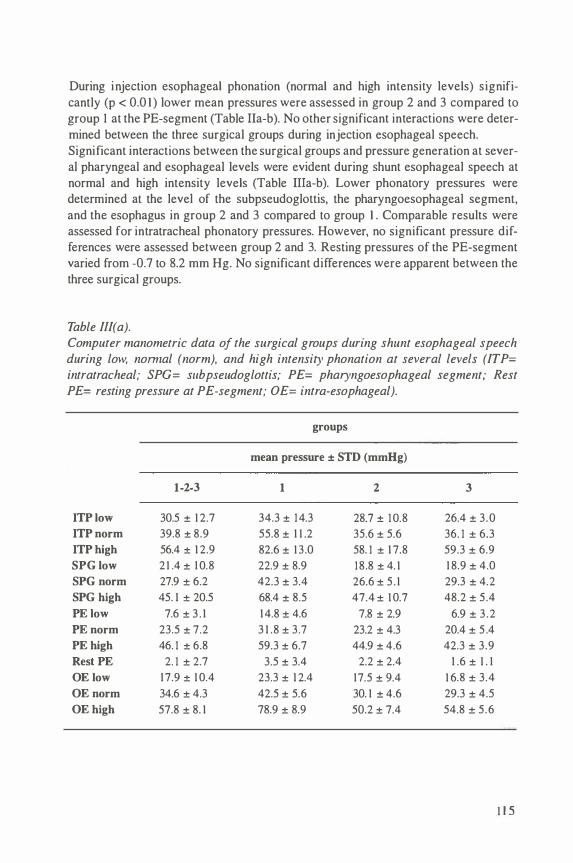
During injection esophageal phonation (normal and high intensity levels) significantly (p < 0.0 1 ) lower mean pressures were assessed in group 2 and 3 compared to group 1 at the PE-segment (Table Ila-b). No other significant interactions were determined between the three surgical groups during injection esophageal speech. Significant interactions between the surgical groups and pressure generation at several pharyngeal and esophageal levels were evident during shunt esophageal speech at normal and high intensity levels (Table Ilia-b). Lower phonatory pressures were determined at the level of the subpseudoglottis, the pharyngoesophageal segment, and the esophagus in group 2 and 3 compared to group 1 . Comparable results were assessed for intratracheal phonatory pressures. However, no significant pressure differences were assessed between group 2 and 3. Resting pressures of the PE-segment varied from -0.7 to 8.2 mm Hg. No significant differences were apparent between the three surgical groups.
Table Ill( a). Computer manometric data of the surgical groups during shunt esophageal speech during low, nomzal (norm), and high intensity phonation at several levels (ITP= intratracheal; SPG= s11bpseudoglottis; PE= pharyngoesophageal segment; Rest PE= resting pressure at PE-segment; OE= intra-esophageal).
groups
mean pressure ± STD (mmHg)
1-2-3 1 2 3
ITP iow 30.5 ± 1 2.7 34.3 ± 1 4.3 28.7 ± 1 0.8 26.4 ± 3 .0
ITP norm 39.8 ± 8.9 55.8 ± 1 1 .2 35.6 ± 5.6 36. 1 ± 6.3
ITP high 56.4 ± 1 2.9 82.6 ± 1 3.0 58. 1 ± 1 7.8 59.3 ± 6.9
SPG low 2 1 .4 ± 1 0.8 22.9 ± 8.9 1 8.8 ± 4. 1 1 8.9 ± 4.0
SPG norm 27.9 ± 6.2 42.3 ± 3.4 26.6 ± 5 . 1 29.3 ± 4.2
SPG high 45. 1 ± 20.5 68.4 ± 8.5 47.4 ± 1 0.7 48.2 ± 5 .4
PE iow 7.6 ± 3 . 1 1 4.8 ± 4.6 7.8 ± 2.9 6.9 ± 3 .2
PE norm 23.5 ± 7.2 3 1 .8 ± 3.7 23.2 ± 4.3 20.4 ± 5 .4
PE high 46. 1 ± 6.8 59.3 ± 6.7 44.9 ± 4.6 42.3 ± 3.9
Rest PE 2. 1 ± 2.7 3.5 ± 3.4 2.2 ± 2.4 1 .6 ± 1 . 1
OE iow 1 7.9 ± 1 0.4 23.3 ± 1 2.4 17.5 ± 9.4 1 6.8 ± 3.4
OE norm 34.6 ± 4.3 42.5 ± 5.6 30. 1 ± 4.6 29.3 ± 4.5
OE high 57.8 ± 8. 1 78.9 ± 8.9 50.2 ± 7.4 54.8 ± 5 .6
1 1 5
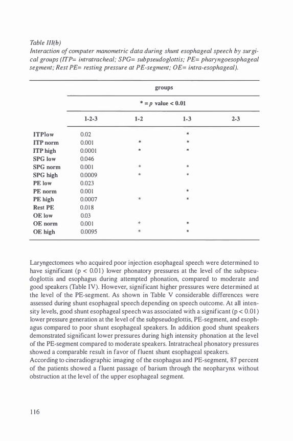
Table lll(b) Interaction of computer manometric data during shunt esophageal speech by surgi
cal groups (ITP= intratracheal; SPG= subpseudoglottis; PE= pharyngoesophageal segment; Rest PE= resting pressure at PE-segment; OE= intra-esophageal).
groups
* = p value < 0.01
1-2-3 1-2 1-3 2-3
ITPlow 0.02
ITP norm 0.001
ITP high 0.0001
SPG low 0.046
SPG norm 0.001 * *
SPG high 0.0009 * *
PE low 0.023
PE norm 0.001 *
PE high 0.0007 * *
Rest PE 0.01 8
OE low 0.03
OE norm 0.001 * *
OE high 0.0095 * *
Laryngectomees who acquired poor injection esophageal speech were determined to have significant (p < 0.0 I ) lower phonatory pressures at the level of the subpseudoglottis and esophagus during attempted phonation, compared to moderate and good speakers (Table IV). However, significant higher pressures were determined at the level of the PE-segment. As shown in Table V considerable differences were assessed during shunt esophageal speech depending on speech outcome. At all intensity levels, good shunt esophageal speech was associated with a significant (p < 0.0 I ) lower pressure generation at the level of the subpseudoglottis, PE-segment, and esophagus compared to poor shunt esophageal speakers. In addition good shunt speakers demonstrated significant lower pressures during high intensity phonation at the level of the PE-segment compared to moderate speakers. Intratracheal phonatory pressures showed a comparable result in favor of fluent shunt esophageal speakers. According to cineradiographic imaging of the esophagus and PE-segment, 87 percent of the patients showed a fluent passage of barium through the neopharynx without obstruction at the level of the upper esophageal segment.
1 1 6

Table IV. Interaction of computer manometric data during injection esophageal speech by semi-quantitative speech results (SPG= subpseudoglottis; PE= pharyngoesophageal segment; Rest PE= resting pressure at PE-segment; OE= intraesophageal; 1= significant difference between poor and moderate speakers; 2= significant difference between poor and good speakers).
SPG low
SPG norm
SPG high
PE iow
PE norm
PE high
Rest PE
OE iow
OE norm
OE high
injection esophageal speech
poor moderate good
mean pressure ± STD (mmHg)
5 . 1 ± 5 .9 7 .8 ± 6 .2 5.3 ± 4.4
1 0.2 ± 4.2 1 5.7 ± 4.3 1 9.6 ± 5.2
9.2 ± 6.4'- ' 27.7 ± 7.3 25.9 ± 5 . 1
1 4.6 ± 2.7 1 1 .9 ± 4. 1 7.7 ± 3.2
20.2 ± 5.2 1 5.4 ± 4.3 1 3 .6 ± 5 . 1
32.5 ± 6.9' ' 1 7.6 ± 4.6 1 5 .3 ± 7.6
3.5 ± 2.3 3.7 ± 3.8 1 .3 ± 1 .8
5.7 ± 2.6 4.9 ± 3.5 6.2 ± 2.8
1 4. 1 ± 5.6 2 1 .5 ± 2.3 1 9.9 ± 4.6
9. 1 ± 5.9 1 2 29.9 ± 9.6 24.6 ± 6.5
Laryngectomees with functional shunt and injection esophageal speech showed a regular vibrating pharyngoesophageal segment during phonation. The segment was estimated to be varying from 3 to 4 em in length. All patients revealed to have their 'pseudoglottis' at the junction of the neopharynx and upper esophagus at the level of the fifth and sixth cervical vertebrae. During phonation narrowing of the vibrating pharyngoesophageal segment may be evident, but dilatation appears to be maximal during swallowing. In 5 laryngectomees narrowing of the PE-segment was responsible for delayed passage of fluids and interfered with fluent alaryngeal speech (Table VI). Spasm of the PE-segment was apparent in 4 patients (33%) in group l , while hypertonicity of the upper esophageal sphincter was assessed in both group 1 (25%) and 2 (2 1 % ). Spasm was associated with slower yielding of the upper esophageal sphincter. In laryngectomees suffering from spasm, hypertonicity or strictures at the level of the PE-segment, no remarkable differences in pressure generation during injection esophageal speech were assessed (Table VII). During shunt esophageal speech these pressure differences appeared to be more profound at normal intensity levels. In case of hypopharyngeal strictures increased phonatory pressures were only assessed during shunt esophageal speech below the level of the PE-segment. Pharyngoesophageal resting pressures were not significantly different between the specific groups.
1 1 7

Table V. Interaction of computer manometric data during shunt esophageal speech by semiquantitative speech results (ITP= intratracheal; SPG= subpseudoglottis; PE= pharyngoesophageal segment; Rest PE= resting pressure at PE-segment; OE= intra
esophageal; I = significant difference between poor and moderate speakers; 2= sig
nificant difference between poor and good speakers; 3= significant difference between moderate and good speakers).
ITP iow
ITP norm
ITP high
SPG low
SPG norm
SPG high
PE iow
PE norm
PE high
Rest PE
OE low
OE norm
OE high
Discussion
shunt esophageal speech
poor moderate good
mean pressure ± STD (mmHg)
38.3 ± 7.41 2 28.7 ± 7.5' 1 4.5 ± 6.2
45.8 ± 6.32 37.8 ± 8.9 26.6 ± 4.6
93. 1 ± 1 7. 8' 72.5 ± 1 8.5' 45.9 ± 1 6.3
25. 1 ± 9.21.2 1 5.5 ± 8.3 6.2 ± 3.2
39.6 ± 6.72 29.9 ± 6.3 23.9 ± 5 .8
70.2 ± 1 2 .42 64. 1 ± 17 .3 46.9 ± 1 3.9
26.4 ± 3.4' 1 7.5 ± 4.9 7.8 ± 3.4
3 1 .6 ± 4.42 24.4 ± 4.6 20.4 ± 3.2
66.9 ± 1 3.7" 53.4 ± 1 0.8' 42. 1 ± 7.8
3.5 ± 2.3 3.7 ± 3.8 1 .3 ± 1 .8
26.4 ± 6.0' 1 8.3 ± 4.8 1 0.9 ± 7.3
43.5 ± 9.42 3 1 .9 ± 6.7 30. 1 ± 5.2
78.3 ± 1 8.9' 69.7 ± 1 0.4 44.9 ± 1 4. 2
A distinctive group of patients fails to develop functional alaryngeal speech. Several factors may be responsible for poor acquisition of speech after total laryngectomy: psychosocial factors, device-related factors and factors related to the PEsegment ( 1 2). In our study psychosocial and device-related factors were excluded in order to study functional and structural aspects in postlaryngectomy speech failure. In assessing the development of adequate alaryngeal speech, the reconstructed pharyngoesophageal segment must be examined dynamically by manometry and cineradiography (9). Fluoroscopy can be used to detect functional or morphologic abnormalities within the oral, pharyngeal, or esophageal segments. Recent fluoroscopic and strobofiberscopic studies report the neoglottis to be considered as a protrusion of the posterior wall in the pharyngoesophageal segment ( 1 3) . The upper part of the neoglottis is considered to be the sound producing part as during phonation a regular
1 1 8
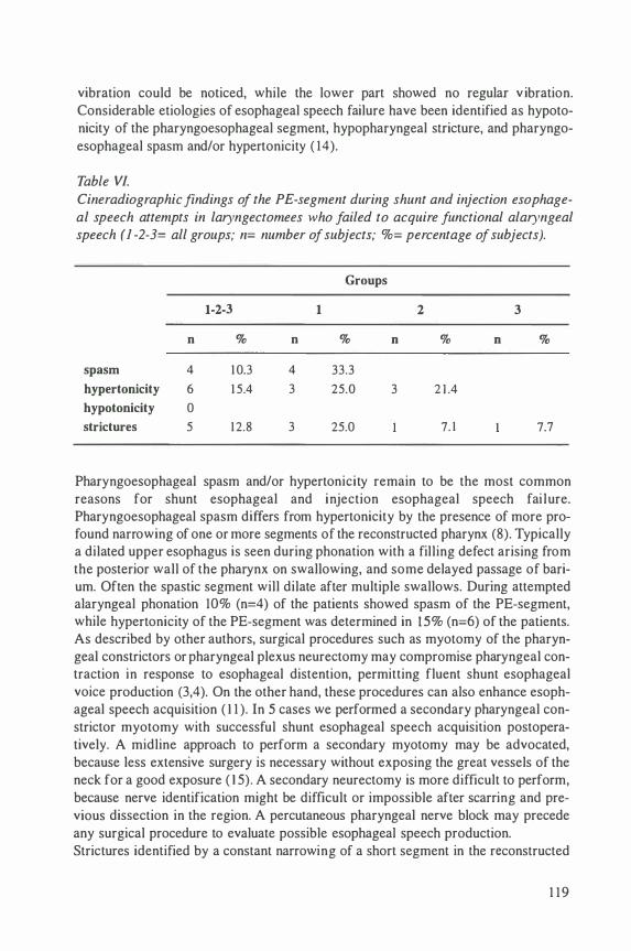
vibration could be noticed, while the lower part showed no regular v ibration. Considerable etiologies of esophageal speech failure have been identified as hypotonicity of the pharyngoesophageal segment, hypopharyngeal stricture, and pharyngoesophageal spasm and/or hypertonicity ( 14).
Table VI.
Cine radiographic findings of the PE-segment during shunt and injection esophageal speech attempts in laryngectomees who failed to acquire functional alaryngeal speech ( 1 -2-3= all groups; n= number of subjects; %= percentage of subjects).
1-2-3
n % n
spasm 4 1 0.3 4
hypertonicity 6 1 5.4 3
hypotonicity 0
strictures 5 1 2.8 3
Groups
1
% n
33.3
25.0 3
25.0
2
%
2 1 .4
7. 1
3
n %
7.7
Pharyngoesophageal spasm and/or hypertonicity remain to be the most common reasons for shunt esophageal and injection esophageal speech fai lure. Pharyngoesophageal spasm differs from hypertonicity by the presence of more profound narrowing of one or more segments of the reconstructed pharynx (8). Typically a dilated upper esophagus is seen during phonation with a filling defect arising from the posterior wall of the pharynx on swallowing, and some delayed passage of barium. Often the spastic segment will dilate after multiple swallows. During attempted alaryngeal phonation 10% (n=4) of the patients showed spasm of the PE-segment, while hypertonicity of the PE-segment was determined in 1 5% (n=6) of the patients. As described by other authors, surgical procedures such as myotomy of the pharyngeal constrictors or pharyngeal plexus neurectomy may compromise pharyngeal contraction in response to esophageal distention, permitting fluent shunt esophageal voice production (3,4). On the other hand, these procedures can also enhance esophageal speech acquisition ( I I ). In 5 cases we performed a secondary pharyngeal constrictor myotomy with successful shunt esophageal speech acquisition postoperatively. A midline approach to perform a secondary myotomy may be advocated, because less extensive surgery is necessary without exposing the great vessels of the neck for a good exposure ( 1 5). A secondary neurectomy is more difficult to perform, because nerve identification might be difficult or impossible after scarring and previous dissection in the region. A percutaneous pharyngeal nerve block may precede any surgical procedure to evaluate possible esophageal speech production. Strictures identified by a constant narrowing of a short segment in the reconstructed
1 1 9

Table VII. Computer manometric data according to cineradiographic findings of the PEsegment (ITP= intratracheal; SPG= subpseudoglottis; PE= pharyngoesophageal segment; Rest PE= resting pressure at PE-segment; OE= intra-esophageal; ie= injection esophageal speech; se= shunt esophageal speech).
spasm
ITP 93. 1 ± 22.9
SPG ie 1 1 .8 ± 6.8
se 82. 1 ± 1 8.9
PE ie 23.2 ± 6.2
se 7 1 .5 ± 1 2.3
OE ie 1 3.8 ± 4.8
se 84.2 ± 1 5.7
Rest PE 3.4 ± 1 .6
hypertonicity hypotonicity
mean pressure ± SD (mmHg)
72.2 ± 1 2.8
1 9.7 ± 7.6
64.6 ± 1 3.5
1 9.4 ± 7. 1
59.2 ± 9.7
1 5 .6 ± 4.3
69.4 ± 14 . 1
2.3 ± 1 . 1
structure
1 26. 1 ± 5.4
7.9 ± 3.2
84.2 ± 1 1 .3
6.9 ± 2.8
1 0.6 ± 3.5
1 1 .2 ± 4.7
1 09.5 ± 23.8
1 .5 ± 1 .3
pharynx with a small amount of residual barium above the segment were found in 5 laryngectomees. The narrowed segment shows the same appearance on swallowing and phonation, and it never dilates. In severe cases no vibrating pharyngoesophageal segment is visible and the patient remains aphonic (8). Some patients were able to produce shunt esophageal speech only on an intermittent basis. In these cases passage of tubes and catheters will be difficult and replacement of indwelling voice prostheses may be impossible or extremely difficult to perform in the ambulatory patient. Structural and functional abnormalities of the neopharynx were clearly associated with poor speech results. In these cases higher phonatory pressures were evident during shunt esophageal speech at the level of the subpseudoglottis, PEsegment, esophagus, and trachea. In case of unsuccessful injection esophageal speech attempts, lower subpseudoglottal pressures were assessed in association with higher pressures at the level of the PE-segment. As the resting pressures did not differ considerably, pressure differences are only obvious during phonation attempts in association with functional and/or structural abnormalities. In our series additional myotomy procedures and combined myotomy and neurectomy procedures were equally effective in preventing pharyngoesophageal spasm. Lower intratracheal and pharyngoesophageal pressures were assessed during shunt esophageal speech in the surgical groups which included the additional procedures. These differences were less obvious during injection esophageal speech. Only higher pressures at the level of the PE-segment were significant in the surgical group lacking these preventive measures. The influence of additional surgical procedures considering the pharyngoesophageal tone is best assessed during shunt esophageal phonation. These results are comparable with effects of esophageal insufflation in postlaryngectomy patients
1 20
lacking a tracheoesophageal fistula ( 1 4). According to computer manometry and cineradiography no significant differences were determined between the surgical group with a unilateral myotomy and the surgical group with a unilateral myotomy and neurectomy procedure. Reconstruction of the pharynx after laryngectomy may affect the success of speech rehabilitation. Together with a standard 3-layer pharyngeal closure, hypertonicity or spasm of the upper esophageal sphincter may develop. A unilateral posteropharyngeal myotomy and/or neurectomy of the pharyngeal plexus should be performed to prevent increased tonicity of the neopharynx. Meticulous closure of the pharynx has always been recommended to prevent pharyngocutaneous fistulas. However, some authors advocate nonmuscle closure of the pharynx as a safe technique, which in their hands is not associated with higher operative complications compared to a 3 layer closure (7). This technique may be used as an alternative for procedures such as pharyngeal constrictor myotomy or pharyngeal plexus neurectomy to avoid hypertonicity or spasm of the PE-segment. Unsuccessful alaryngeal speech may be caused be several internal and external factors. Evaluation of poor alaryngeal speech is determined by successively checking patient- and device-related problems. Hypertonicity and pharyngoesophageal spasm followed by hypopharyngeal stricture were the most common patient-related reasons for esophageal speech failure. Prevention of muscle spasm and hypertonicity of the neoglottis by myotomy and neurectomy procedures, may promote successful acquisition of injection esophageal and shunt esophageal speech. Nevertheless the additional benefit of a unilateral neurectomy of the pharyngeal plexus was not demonstrated in combination with a unilateral myotomy. The effect of a unilateral pharyngeal plexus neurectomy should be determined separately. Analysis of pressure generation by computer manometry is most obvious during shunt esophageal phonation. This could be related to the greater flow and pressure changes during shunt esophageal speech. Computer manometry may be useful in determining voice failures and monitoring surgical techniques. Cineradiography seems to be a useful method for demonstrating functional and structural disorders which may be responsible for alaryngeal speech failures. These imaging studies may further allow proper management of the postlaryngectomy patient.
References
I . Merwin GE, Goldstein LP, Rothman HB. A comparison o f speech using artificial larynx and tracheoesophageal puncture in the same speaker. Laryngoscope 1985; 95: 730-734.
2. Pindzola RH, Cain BH. Acceptability ratings of tracheoesophageal speech. Laryngoscope 1988; 98: 394-397.
3 . Singer MI, Blom ED. Selective myotomy for voice restoration after total laryngectomy. Arch Otolaryngol 198 1 ; I 07: 670-673.
4. Henley J, Souliere C. Tracheoesophageal speech failure in the laryngectomee: the role of constrictor myotomy. Laryngoscope 1 986; 96: I 0 16- 1 020.
12 1
5. Baugh RF, Baker SR, Lewin JS. Surgical treatment of pharyngoesophageal spasm. Laryngoscope 1 988; 98: 1 1 24- 1 1 26.
6. Singer Ml, BJorn ED, Hamaker RC. Pharyngeal plexus neurectomy for alaryngeal speech rehabil itation. Laryngoscope 1 986; 96: 50-54.
7. Olson NR, Callaway E. Nonclosure of the pharyngeal muscle after laryngectomy. Ann Otol Rhino} Laryngol 1990; 99: 507-508.
8. Mcivor J, Evans PF, Perry A, et al. Radiologic assessment of post laryngectomy speech. Clin Radiol l 990; 4 1 : 3 1 2-3 1 6.
9. Muller-Miny H, Eisele DW, Jones B. Dynamic radiographic imaging following total laryngectomy. Head Neck 1 993; 1 5 : 342-347.
10. Walther EK. Computer manometry in site-related reconstruction of the pharynx and pharyngoesophageal segment. Laryngo-Rhino-Otol 1 995; 74: 437-443.
I I . Van Weissenbruch R, Albers FWJ. Voice rehabilitation after total laryngectomy using the Provox™ voice prosthesis. Clin Otolaryngol 1 993; 1 8 :359-364.
1 2. Gates GA, Hearne III EM. Predicting esophageal speech. Ann Otol Rhino! Laryngol 1982; 9 1 : 454-457.
1 3 . Omori K, Kojima H, Nonomura M, et al. Mechanism of tracheoesophageal shunt phonation. Arch Otolaryngol Head Neck Surg 1 994; 1 20: 648-652.
14. Callaway E, Truelson JM, Wolf GT, et al. Predictive value of objective esophageal insufflation testing for acquisition of tracheoesophageal speech. Laryngoscope 1992; I 02: 704-708.
I 5. Medina JE, Reiner SA. Pharyngeal myotomy for voice restoration: a midline approach. Laryngoscope I990; I 00: 309-3 1 2.
122

Chapter 8
Assessment of the pharyngoesophageal segment after total
laryngectomy: swallowing characteristics
Van Weissenbruch R, De Looze D, Kunnen M, Albers FWJ, Van Cauwenberge PB, Suiter AM. Assessment of the pharygoesophageal segment after total laryngectomy: swallowing characteristics. Submitted.

Introduction
The loss of normal laryngopharyngeal functions may be responsible for a remarkable degree of morbidity after surgical removal of the larynx. The acquisition of functional alaryngeal speech after total l aryngectomy is still considered to be one of the major challenges to establish appropriate rehabilitation of communication. The advent of surgical procedures to restore speech after laryngectomy has resulted in a decline of alaryngeal speech failures. Failure to achieve shunt as well as injection esophageal speech is related to an increased pharyngoesophageal tone or spasm ( 1 ). Oicopharyngeal and pharyngeal constrictor myotomy have been advocated in conjunction with total laryngectomy, or as a secondary procedure to improve the acquisition of alaryngeal speech ( 1 ,2). Alternative techniques to prevent hypertonicity or spasm of the pharyngoesophageal segment consist of a unilateral neurectomy of the pharyngeal plexus, or non-muscle closure of the pharynx during laryngectomy (3,4). Dysphagia is considered to be another disturbing side-effect of surgical treatment of laryngeal cancer. The incidence of swallowing difficulties vary considerably from 16% to 62% (5,6). Diminished relaxation of the pharyngoesophageal segment due to absent laryngeal elevation, and a narrowed lumen of the neopharynx in irradiated patients with an altered coordination of peristalsis, may interfere with fluent swallowing in the postlaryngectomy patient (7,8,9). Swallowing problems are consistently related to hypopharyngeal stenosis and a high incidence of fistulas (6,7,9). Higher swallowing pressures after total laryngectomy necessitating increased tongue driving forces are possibly related to functional and morphological changes of the pharyngoesophageal segment (9). Manometric evaluations of laryngectomized patients with postoperative dysphagia have shown significant motility changes in the upper esophhageal sphincter (UES) and esophagus ( 1 0). Radiographic evidence for cricopharyngeal spasm after laryngectomy which may be related to postoperative dysphagia was presented by Schobinger in 1 958 ( I I ). The role of cricopharyngeal muscle dysfunction in postoperative dysphagia has also been recognized to be associated with major oropharyngeal resections ( 1 2) . Cineradiography may provide a detailed analysis of the events of pharyngeal deglutition and morphology of the pharyngoesophageal segment. Additionally, multiple level manometry may be used to investigate pharyngeal and esophageal pressure generation and the relationship of these pressures to bolus transit ( 1 3, 1 4). In this study the swallowing function was analyzed by using computer manometry and cineradiography. The impact of concurrent myotomy and myotomy and neurectomy on the postlaryngectomy pharyngoesophageal segment was determined during deglutition. Additional surgical procedures for prevention of pharyngospasm during alaryngeal speech may alter pharyngeal structures and increase the efficiency of swallowing.
1 24
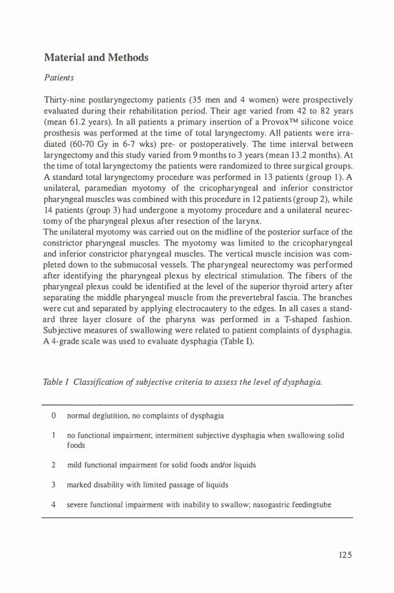
Material and Methods
Patients
Thirty-nine postlaryngectomy patients (35 men and 4 women) were prospectively evaluated during their rehabilitation period. Their age varied from 42 to 82 years (mean 6 1 .2 years). In all patients a primary insertion of a Provox™ silicone voice prosthesis was performed at the time of total laryngectomy. All patients were irradiated (60-70 Gy in 6-7 wks) pre- or postoperatively. The time interval between laryngectomy and this study varied from 9 months to 3 years (mean 1 3 .2 months). At the time of total laryngectomy the patients were randomized to three surgical groups. A standard total laryngectomy procedure was performed in 1 3 patients (group 1 ). A unilateral, paramedian myotomy of the cricopharyngeal and inferior constrictor pharyngeal muscles was combined with this procedure in 1 2 patients (group 2), while 14 patients (group 3) had undergone a myotomy procedure and a unilateral neurectomy of the pharyngeal plexus after resection of the larynx. The unilateral myotomy was carried out on the midline of the posterior surface of the constrictor pharyngeal muscles. The myotomy was limited to the cricopharyngeal and inferior constrictor pharyngeal muscles. The vertical muscle incision was completed down to the submucosal vessels. The pharyngeal neurectomy was performed after identifying the pharyngeal plexus by electrical stimulation. The fibers of the pharyngeal plexus could be identified at the level of the superior thyroid artery after separating the middle pharyngeal muscle from the prevertebral fascia. The branches were cut and separated by applying electrocautery to the edges. In all cases a standard three layer closure of the pharynx was performed in a T-shaped fashion. Subjective measures of swallowing were related to patient complaints of dysphagia. A 4-grade scale was used to evaluate dysphagia (Table I).
Table I Classification of subjective criteria to assess the level of dysphagia.
0 normal deglutition, no complaints of dysphagia
no functional impairment; intermittent subjective dysphagia when swallowing solid foods
2 mild functional impairment for solid foods ancVor liquids
3 marked disability with limited passage of liquids
4 severe functional impairment with inability to swallow; nasogastric feedingtube
125

Computer Manometry
By using multiple level manometry pressure generation in the hypopharynx, the pharyngoesophageal segment (PE-segment), and the proximal esophagus was assessed in all patients. A motility probe consisting of a 4.65 mm diameter flexible tube, which incorporates three miniature pressure transducers near its distal end, was used for direct esophageal pressure measurements (P3 1 -322D, Konigsberg Instruments, Inc., Pasadena, CA 9 1 1 07, USA). Inter-sensor spacing was 5 em, with the distal sensor 5 em from the tip. Each sensor element (output of 7.5 mV at 300 mm Hg, at 5.0 V) was mounted in a titanium carrier and coated with silas tic to protect the sensors. All pressure transducers were calibrated to 300 mm Hg (maximal 1 200 mm Hg) and consisted of dynamic temperature compensation. The 1 25 em long, medical grade probe was attached through a compensation module (fJ Digitrapper 2 MB, Synectics Medical, Brussels, Belgium) to a personal computer (Tulip® Tr 386 SX). Software (Polygram version 5.00C4, Upper GI Edition, Gastrosoft Inc., USA) capable of measuring pressures in mmHg (I mm Hg= 1 33.3 Pascal), and calculating mean and maximal values, resting pressures, duration of activity, and relaxation was used to evaluate pressure generation. Pressures could be measured continuously at rest, and dynamically during deglutition. The generation of pressure at the base of tongue (BTCP) over time represents the force produced by the tongue during bolus transfer in the oropharynx (BTCF) , while the pharyngeal constrictor pressure (PCP) over time represents the force applied to the bolus in the PE-segment (PCF). The transpharyngeal transit time (Ph transit) is defined as the time the bolus takes to pass over a 1 0 em distance from the moment the sensor is touched in the oropharynx until the contraction starts at the proximal esophagus. The average contraction velocity (Ph prop V) is based on the transpharyngeal transit time measured over the 10 em distance. The degree of relaxation after pharyngeal contraction pressure generation is determined (PE relax). The sensors, oriented orthogonally to the probe center-line permitted measurement of non-symmetric pressures while withdrawing the probe. The probe was clearly marked in I em increments to assist in identifying transducer position. Verification of sensor location was also permitted by its radiopaque tip and sensors. The slightly stiffened probe was easily introduced transnasally in all subjects after local application of Lidocaine® 2%. Prior to manometry the several sites of measurement (base of tongue, PE-segment, proximal esophagus) were localized by using a flexible fiberoptic endoscope (Olympus® ENF-2) transnasally. The subjects were monitored in sitting position. Before swallowing a bolus of water (30 ml) was kept in the oral cavity.
Cineradiography
Cineradiographic imaging of the pharyngoesophageal segment and proximal esophagus was performed in all subjects during swallowing. Functional and morphological aspects of the reconstructed p�aryngoesophageal segment could be evaluated and related to pressure generation at specific sites. Frontal and lateral recordings with a standard Siemens® remote control fluoroscopy unit were made while the subjects
1 26

Table II Radiological features of the neopharynx after total laryngectomy to determine functional and structural abnormalities.
normal
hypotonic
hypertonic
spasm
stricture
normal passage of barium; full dilatation of pharynx; single vibrating segment during phonation
similar dilatation of pharynx: no hold up of barium; no vibrating segment during attempted phonation
normal dilatation of pharynx; no hold up of barium; multiple narrowed segment during phonation, cricopharyngeal (CP) muscle bar
typical CP bar (filling defect); hold up of barium; dilatation of pharynx after multiple swallows; narrowed pharyngeal segment during phonation with large esophageal air reservoir
constant narrowing of a short pharyngeal segment with hold up of barium, never dilates after swallowing or phonation; no vibrating segment; dysphagia worse for solid foods
were sitting. An external stoma site marker was used to ensure accuracy. Satisfactory coating of the neopharynx and esophagus (E-Z paque® HD: barium sulphate 98, excipiens qs ad 340 gr) was achieved for proper definition of the anatomy and any area of functional or morphological discrepancy. Prior to each swallowing act a bolus of 30 ml was held in the oral cavity. The PE-segment was screened for functional and morphological abnormalities, e.g. spasm, hypertonicity, hypotonicity, strictures (Table II). The shape and the lower level of the PE-segment were determined and related to the efficiency of swallowing. The PE-segment was also screened for other morphological aspects considering dysphagia (pseudoepiglottis, pouches, prevertebral thickness, fistulas). During swallowing functional aspects related to the specific sites were assessed (passage of barium, percentage of barium residue, base of tongue movements, pharyngeal contraction and relaxation).
Statistical analysis
All data were evaluated by using repeated measures analysis of variance (ANOVA). Interactions among surgical groups, phonatory results, and manometry were tested separately. Chi-squared comparisons were performed to assess the source of signifi-
1 27

cance for all variables with main-effect AN OVA results having p values of less than 0.05. Analyses were performed with the Scientific Package for Social Sciences (SPSS®).
Table III. Classification of dysphagia according to the defined surgical groups.
groups
all 2 3
dysphagia n % n % n % n %
0 23 59 5 39 7 58 1 1 79
1 3 33 6 46 4 33 3 2 1
2 3 8 2 1 5 8
3
4
Results
According to subjective criteria to assess dysphagia complaints, 33% of all laryngectomees in this population suffered from slight (grade I ) dysphagia, while only 8% had moderate (grade 2) swallowing problems (Table III). Intermittent, slight swallowing disturbances (multiple swallows) were assessed predominantly in group 1 (46%) and group 2 (33%). Moderate dysphagia complaints were not observed in group 3. Cineradiographic findings are shown in Table IV-V. In group I , a prominent pseudoglottis (PG shape) at the lower PE-segment was significantly correlation with the degree of dysphagia (Table IV). No significant interactions were assessed for dysphagia among the surgical groups for other structural images. The occurrence of previous fistulas was predominant in group I , but did not differ significantly between the three groups. Interrupted passage of barium (n= 1 5) and delayed relaxation of the upper esophageal sphincter (n= I 0) demonstrated a significant 3-way interaction by surgical group (Table IV). In group I a significant 2-way interaction was found con-
128
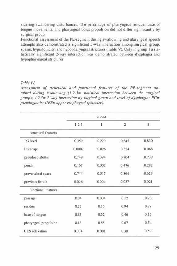
sidering swallowing disturbances. The percentage of pharyngeal residue, base of tongue movements, and pharyngeal bolus propulsion did not differ significantly by surgical group. Functional assessment of the PE-segment during swallowing and alaryngeal speech attempts also demonstrated a significant 3-way interaction among surgical group, spasm, hypertonicity, and hypopharyngeal strictures (Table V). Only in group 1 a statistically significant 2-way interaction was demonstrated between dysphagia and hypopharyngeal strictures.
Table IV. Assessment of structural and functional features of the PE-segment ob-rained during swallowing (1-2-3= statistical interaction between the surgical groups; 1,2,3= 2-way interaction by surgical group and level of dysphagia; PG= pseudoglottis; UES= upper esophageal sphincter).
groups
1 -2-3 2 3
structural features
PG level 0.359 0.229 0.645 0.830
PG shape 0.0002 0.026 0.324 0.068
pseudoepiglottis 0.749 0.394 0.704 0.739
pouch 0. 1 67 0.007 0.476 0.282
prevertebral space 0.744 0.5 1 7 0.864 0.629
previous fistula 0.026 0.004 0.037 0.02 1
functional features
passage 0.04 0.004 0. 1 2 0.23
residue 0.27 0. 1 5 0.94 0.77
base of tongue 0.63 0.32 0.46 0. 1 5
pharyngeal propulsion 0. 1 3 0.55 0.67 0.54
UES relaxation 0.004 0.001 0.30 0.59
1 29

Table V. Assessment of relaxation of the PE-segment during swallowing ( 1 -2-3= 3-way interaction by surgical group; 1 ,2,3= 2-way interaction by surgical group and level of dysphagia).
group
1 -2-3 2 3
spasm n 4 4 0 0
p 0.01 4 0.056
hypertony n 6 3 3 0
p O.o t 8 0.3 1 0.27
hypotony n 0 0 0 0
p
stricture n 5 3
p 0.005 0.002 0.069 0.092
Table VI.A-B summarizes the computer manometric data obtained during deglutition. A wide range of pharyngeal pressures have been assessed among the surgical groups. However, no significant interactions were determined between the surgical groups, oropharyngeal pressures, or pressure generations related to the PE-segment. When considering the pressure generation over time at the level of the base of tongue or PE-segment, no significant differences were determined among the groups. Resting pressure (PE-segment) or relaxation (%) did not show any interactions among the surgical groups. When considering pharyngeal transit times and pharyngeal contraction velocity, both demonstrated a significant 3-way interaction among the surgical groups. These parameters also showed significant differences (p< 0.05) between the individual surgical groups. Pharyngeal transit times appeared to be prolonged in group I compared to group 2 and 3. Contraction velocity measures demonstrated lower values in group I and 2, compared to group 3. Specific cineradiographic findings (Table IV) and computer manometric data (Table VI.A-B) showed some profound differences when comparing these to mean group data (Table VII). Higher pressures were determined at the base of tongue and PEsegment in case of spasm, hypertonicity or strictures. Delayed pharyngeal transit
1 30

Table V/.A Classification of computer manometric data by surgical group ( 1 -2-3= all patients; BTCP= base of tongue contraction pressure; BTCF= base of tongue contraction force; PCP= pharyngeal contraction pressure; PCF= pharyngeal contraction force; PE relax= relaxation of PE-segment after colltraction; Rest PE= resting pressure of PE-segment; PH transit= transpharyngeal transit time; Ph prop V= pharyngeal propagation velocity).
group
1 -2-3 2 3
BTCP [peak] 92.6 98.7 1 04. 1 77.0 (mmHg) (32.4- 1 96.5) (34.7- 1 9 1 .6) (44.8- 1 96.5) (32.4- 1 75.5)
BTCP [mean] 48. 1 53.6 52.2 39.5 (mmHg) ( 1 6.9- 1 1 7.7) ( 1 9.5- 1 1 7.7) (23 .6-8 1 . 9) ( 1 6.9-87.7)
BTCF 32.0 40.2 32. 1 24.4 (mmHg*s) (5.2- 147.2) (5.6- 1 47.2) (5.2-60.2) (6.3-82.3)
PCP [peak] 2 1 .2 24.2 22.8 1 6.9 (mmHg) (7. 8-50.0) (7.8-38.7) (9.5-50.0) (9.2-43 . 1 )
PCP [mean] 1 2.8 1 4.9 13.7 1 0. 1 (mmHg) (3.5-29.6) (3.5-24.9) (5.4-29.6) (4.7-25.3)
PCF 9. 1 1 0. 1 8.9 8.4 (mmHg*s) (2.4-20.8) (2.4- 1 9.8) (3.4-20.8) (3.8- 1 7.7)
PE relax 99.9 99.6 1 0 1 .2 99.0 (%) (7 1 .2- 1 27.3) (8 1 .0- 1 25.0) (83.3-1 27.3) (7 1 .2- 1 06.3)
Rest PE 2.9 4.3 2.3 2.4 (mmHg) (0.0-9.2) (0.0-9.2) (0.6-4.6) (0. 1 -3.9)
Ph Transit 1 .7 2. 1 1 .7 1 .5 (s) ( 1 . 1 -3.6) ( 1 .6-3.6) ( 1 .5- 1 .9) ( 1 . 1 - 1 .8)
Ph prop V 2.9 2.5 3.0 3 .4 (cm/s) ( 1 .4-4.5) ( 1 .4-3. 1 ) (2.6-3.5) (2.8-4.5)
times and slower contraction propagation were also demonstrated. In case of spasm or strictures, the pressure generation over time (BTCF and PCF) appeared to be increased.
1 3 1
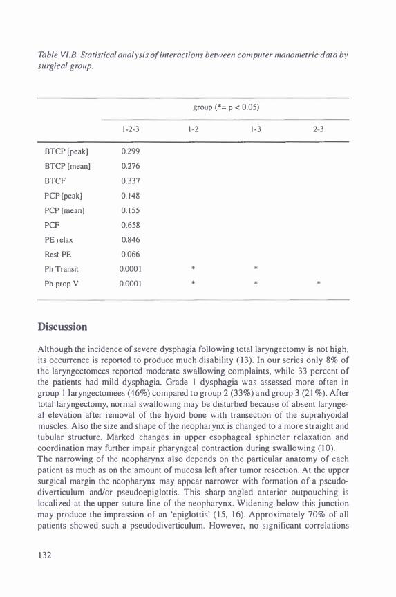
Table VI.B Statistical analysis of interactions between computer manometric data by surgical group.
group (*= p < 0.05)
1 -2-3 1 -2 1 -3 2-3
BTCP [peak] 0.299
BTCP [mean] 0.276
BTCF 0.337
PCP [peak] 0. 1 48
PCP [mean] 0. 1 55
PCF 0.658
PE relax 0.846
Rest PE 0.066
Ph Transit 0.000 1 * *
Ph prop V 0.0001 * * *
Discussion
Although the incidence of severe dysphagia following total laryngectomy is not high, its occurrence is reported to produce much disability ( 1 3). In our series only 8% of the laryngectomees reported moderate swallowing complaints, while 33 percent of the patients had mild dysphagia. Grade 1 dysphagia was assessed more often in group I laryngectomees ( 46%) compared to group 2 (33%) and group 3 (2 1 % ). After total laryngectomy, normal swallowing may be disturbed because of absent laryngeal elevation after removal of the hyoid bone with transection of the suprahyoidal muscles. Also the size and shape of the neopharynx is changed to a more straight and tubular structure. Marked changes in upper esophageal sphincter relaxation and coordination may further impair pharyngeal contraction during swallowing ( 1 0). The narrowing of the neopharynx also depends on the particular anatomy of each patient as much as on the amount of mucosa left after tumor resection. At the upper surgical margin the neopharynx may appear narrower with formation of a pseudodiverticulum and/or pseudoepiglottis. This sharp-angled anterior outpouching is localized at the upper suture line of the neopharynx. Widening below this junction may produce the impression of an 'epiglottis' ( 1 5, 1 6). Approximately 70% of all patients showed such a pseudodiverticulum. However, no significant correlations
1 32

Table V/I. Classification of computer manometric data by specific functional and structural aspects of the PE-segment ( 1-2-3= all patients; BTCP= base of tongue contraction pressure; BTCF= base of tongue contraction force; PCP= pharyngeal contraction pressure; PCF= pharyngeal contraction force; PE relax= relaxation of PE-segment after contraction; Rest PE= resting pressure of PE-segment; PH transit= transpharyngeal transit time; Ph prop V= pharyngeal propagation velocity).
all groups spasm hypertony stricture
BTCP [peak] 92.6 1 62.6 1 0 1 .5 1 63.5 (mmHg) (32.4-1 96.5) ( 1 25. 1 - 1 9 1 .6) (76.6- 1 1 4.5) ( 1 1 2.3- 1 96.5)
BTCP [mean] 48. 1 84.7 55.5 76.9 (mmHg) ( 1 6.9-1 1 7.7) (69.9- 1 1 7.7) (44.6-72.3) (68.8-87.7)
BTCF 32. 1 62.8 3 1 .5 47.9 (mmHg*s) (5.2- 147.2) (5.6- 1 47.2) (28.5-35.6) (5.6-82.3)
PCP [peak] 2 1 .2 32 34.7 32.6 (mmHg) (7.8-50) (21 6.6-50) (30.3-38.7) (26.6-43 . 1 )
PCP [mean] 1 2.8 23. 1 1 4.3 19 . 1 (mmHg) (3.5-29.6) ( 1 6. 1 -29.6) (9.9-20.4) (9.9-25.3)
PCF 9. 1 1 4.7 7.2 1 5 . 1 (mmHg*s) (2.4-20.8) (5.4-20.8) (2.4- 1 1 .3) (6.8- 1 8.2)
PE relax 99.9 99.2 98.9 96.6 (%) (7 1 .2-1 27.3) (8 1 - 1 25) (96.8-1 00) (8 1 - 1 25)
Rest PE 2.9 4.3 4. 1 5.3 (mmHg) (0.0-9.2) (2.4-8. 1 ) (2.3-6.3) (3.4-8. 1 )
Ph Transit 1 .7 2. 1 2.3 2.4 (s) ( 1 . 1-3.6) ( 1 .92-2.3) ( I . 7-3.6) ( 1 .8-3.6)
Ph prop V 2.9 2.2 2.4 2.3 (cm/s) ( 1 .4-4.5) ( 1 .4-2.8) (2.2-2.6) ( 1 .4-2.9)
were obtained between the presence of diverticulae or pseudoglottides and dysphagia. The diverticulae may be related to pouches resulting from postoperative fistulae ( 1 7 , 1 8). In postlaryngeotomy patients the absence of a negative pressure within the pharyngoesophageal segment may result in higher propulsive pressures by using the tongue as a piston. Any functional or morphologic abnormality which may increase
133
the resistance of the pharyngoesophageal segment, may further promote the development of such pouches. Increased thickness of the retropharyngeal soft tissue compartment was evident in 8 1 .6% of our patients. A correlation between increased thickness of the retropharyngeal space with recurrent tumor and other causes of dysphagia has been described ( 1 6, 1 9). No apparent relationship between a wider retropharyngeal compartment and a suspected recurrent carcinoma was evident in our population. The pharyngeal myotomy and neurectomy procedures did not show any relationship with an increased soft tissue mass as compared to the group of patients lacking these procedures. A prominent dorsal impression of the upper esophageal sphincter which changes in shape during swallowing, was seen in half of our patients. This phenomenon may be due to detachment of the pharyngeal muscles ( 1 6, 1 9). Assessment of the retropharyngeal space after laryngectomy may not be considered as a sensitive marker for evaluation of swallowing disturbances or recurrent carcinoma in postlaryngectomy patients (20). Strictures, spasm, and hypertonicity of the upper esophageal sphincter were more frequently demonstrated in laryngectomees without myotomy or myotomy and neurectomy procedures. However, significant interactions were only correlated with the level of swallowing difficulties for patients suffering from hypopharyngeal stenosis. More than 70 percent of the patients with strictures of the pharynx (n=5) had suffered from early postoperative pharyngocutaneous fistulae, which delayed the postoperative rehabilitation period. Fistulae did not occur in the group of postlaryngectomy patients with concurrent pharyngeal myotomy and neurectomy procedures. Pharyngeal disruption with fistula formation is one of the most vexing problems after laryngectomy, which may be chronic with delayed healing. Frequently, the end result of fistula formation is scarring and stricture of the pharynx. Stricture formation as result of trauma from the tracheoesophageal valve prosthesis is also documented (2 1 ). However, in our series esophageal stenosis at the lower pharyngeal border was unrelated to the voice prosthesis. Esophageal stenosis may cause deglutition delay especially for solid foods, and difficulty with alaryngeal speech. During the barium swallow test, strictures were distinguished by a constant narrowing of a short segment in the reconstructed pharynx with a small amount of residual barium above the segment. The narrowed segment shows the same appearance on swallowing and phonation, and it never dilates. A constant narrowing of the pharynx which does not change during swallowing is suspect for benign strictures or recurrent malignancy. In 4 patients repeated pharyngoesophageal dilatations resolved these deglutition problems. Spasm of the upper esophageal sphincter as a possible cause for dysphagia was described by Schobinger in 1 958 ( I I ). Impairment of the sphincter function may be due to damage of local vagal branches during surgery. This may result in a decreased resting pressure of the cricopharyngeal muscle with uncoordinated contractions of the constrictor muscles (22). Spasm in the postlaryngectomy patient appears to be a normal reaction to esophageal distention that prevents gastroesophageal reflux.
1 34

Spasm only occurred in the group of laryngectomees without an additional myotomy or myotomy and neurectomy procedure. Pharyngoesophageal spasm differs from hypertonicity by the presence of more profound narrowing of one or more segments of the reconstructed pharynx. However, no significant interactions were assessed for spasm or hypertonicity of the PE-segment and dysphagia. Typically a dilated upper esophagus is seen during phonation with a filling defect arising from the posterior wall of the pharynx on swallowing, and some hold up to the passage of barium. Often the spastic segment will dilate after multiple swallows. Hypertonicity, which was more easily recognized during alaryngeal phonation attempts, was demonstrated both in laryngectomees without additional procedures (n=3) as well as in laryngectomees with a myotomy procedure (n=3). As described by other authors, surgical procedures such as myotomy of the pharyngeal constrictors or pharyngeal plexus neurectomy may compromise pharyngeal contraction in response to esophageal distention (2,3). According to computer manometric assessment of pharyngeal pressure generation, no significant differences were assessed between the three surgical groups. Alterations to the PE-segment by myotomy or myotomy and neurectomy did not seem to influence the pressure generated by the lingual pharyngeal component. In laryngectomees higher tongue driving forces are demonstrated due to absent laryngeal elevation which assists in the opening of the PE-segment ( 1 3) . The resting pressures did not differ between the swallows. However, group I laryngectomees (without myotomy or myotomy and neurectomy) demonstrated longer transpharyngeal transit times and a slower pharyngeal contraction propagation as compared to the other surgical groups. In the non-myotomized or neurectomized group the PEsegment may be less compliant due to a greater residual muscle tone during swallowing. In laryngectomees generation of negative pressure in the PE-segment is absent due to lack of active opening of the pharynx secondary to laryngeal elevation. The higher resistance in the PE-segment may explain the longer transit times in this group (23). In case of functional (spasm, hypertony) or structural (strictures) disturbances of the PE-segment more profound manometric differences were obtained compared to data of laryngectomees without these problems. The pressure generation in the upper pharynx and the concurrent forces on the pharyngeal walls are increased to compensate for the higher resistance of bolus flow through the PE-segment. Increased swallowing pressures were obtained at the level of the base of tongue and PE-segment in case of pharyngospasm. Increased pharyngeal transit times may be due to obstruction of the PE-segment. Dynamic radiographic imaging of the pharyngoesophageal segment following laryngectomy may reveal a large spectrum of functional and morphological aspects. Swallowing difficulties may be related to hypopharyngeal stenosis, as well as increased resistance of the upper esophageal sphincter. Pharyngeal strictures are likely to be secondary to scarring after early postoperative pharyngocutaneous fistulas or narrow closure of the pharyngostoma. Treatment of hypopharyngeal strictures may not only resolve dysphagia, but may allow alaryngeal speech to be achieved.
1 35

Prevention of spasm and hypertonicity of the neoglottis by pharyngeal constrictor myotomy and pharyngeal plexus neurectomy procedures, may promote successful acquisition of alaryngeal speech and prevent dysphagia. Cineradiography seems to be a useful method to demonstrate functional and structural disorders which may be responsible for swallowing difficulties or alaryngeal speech failures. Manometry of the pharyngoesophageal segment in laryngectomees may reveal several mechanisms of dysfunction. The base of the tongue and PEsegment are important topographic levels which may determine swallowing function after total laryngectomy. Multiple level manometry with concurrent cineradiography may be used to evaluate surgical procedures involving the PE-segment. These studies may further allow proper management of the postlaryngectomy patient.
References
I . Singer MI, BJorn ED. Selective myotomy for voice restoration after total laryngectomy. Arch Otolaryngol 198 1 ; 107: 670-673.
2. Baugh RF, Baker SR, Lewin JS. Surgical treatment of pharyngoesophageal spasm. Laryngoscope 1988; 98: 1 124- 1 1 26.
3 . Singer Ml, BJorn ED, Hamaker RC. Pharyngeal plexus neurectomy for alaryngeal speech rehabilitation. Laryngoscope 1 986; 96: 50-54.
4. Clevens RA, Esclamado RM, Hartshorn DO, Lewin JS. Voice rehabilitation after total laryngectomy and tracheoesophageal puncture using nonmuscle closure. Ann Otol Rhino I Laryngol 1 993; l 02: 792-796.
5 . Boyce SE, Meyers AD. Oral feeding after total laryngectomy. Head Neck 1989; 1 1 : 269-273.
6. Gates GA, Hearne EM. Predicting esophageal speech. Ann Otol Rhinal Laryngol 1982; 9 1 : 454-457.
7 . Kaplan JN. The incidence of hypopharyngeal stenosis after surgery for laryngeal cancer. Otolaryngol Head Neck Surg 198 1 ; 89: 956-959.
8 . Hanks JB, Fisher SR, Meyers WC, Christian KC, Postlethwait RW, Jones RS. Effect of total laryngectomy on esophageal motility. Ann Otol Rhinal Laryngol 1 98 1 ; 90: 33 1 -334.
9. McConnel FMS. Analysis of pressure generation and bolus transit during pharyngeal swallowing. Laryngoscope 1 988; 98: 7 1 -78.
1 0. Duranceau A, Jamieson G, Hurwitz AL, Scott Jones R, Postlethwaith RW. Alterations in esophageal motility after laryngectomy. Am J Surg 1 976; 1 3 1 : 30-35.
1 1 . Schobinger R. Spasm of the cricopharyngeal muscle as cause of dysphagia after total laryngectomy. Arch Otolaryngol 1958; 67: 27 1 -275.
1 2. Mlladick RA, Horton CE, Adamson JE. Cricopharyngeal myotomy.
1 36
Application and technique in major oral-pharyngeal resections. Arch Surg 197 1 ; 102: 1 -5.

1 3. McConnel FM, Cerenko D, Mendelsohn MS. Dysphagia after total laryngectomy. Otolaryngol Clin North Am 1 988; 2 1 : 72 1 -726.
14. Walther EK. Computer manometry in site-related reconstruction of the pharynx and pharyngoesophageal segment. Laryngo-Rhino-Otol 1 995; 74: 437-443.
15 . Mcivor J, Evans PF, Perry A , Cheesman AD. Radiologic assessment of postlaryngectomy speech. Clin Radio! 1990; 4 1 : 3 1 2-3 16.
16. Balfe DM, Koehler RE, Setzen M. Barium examination of the esophagus after total laryngectomy. Radiology 1982; 143: 501 -508.
17 . Kirchner JA, Scatliff JH, Dey FL, Shedd DP. The pharynx after laryngectomy. Changes in its structure and function. Laryngoscope 1 963 ; 73: 1 8-33.
1 8. Kirchner JA, Scatliff JH. Disabilities resulting from healed salivary fistula. Arch Otolaryngol 1 962; 75: 60-68.
19. Jung TK, Adams GL. Dysphagia in laryngectomized patients. Otolaryngol Head Neck Surg 1 980; 88: 25-33.
20. Muller-Miny H, Eisele OW, Jones B. Dynamic radiographic imagin g following total laryngectomy. Head Neck 1993; 15: 342-347.
2 1 . Singer Ml, Hamaker RC, BJorn ED, Yoshida GY. Application of the voice prosthesis during laryngectomy. Ann Otol Rhino! Laryngol 1 989; 98: 92 1 -925.
22. Ellis RH Jr. Upper esophageal sphincter in health and disease. Surg Clin North Am 1 97 1 ; 5 1 : 553-565.
23. Pauloski BR, BJorn ED, Logemann JA, Hamaker RC. Functional outcome after surgery for prevention of pharyngospasms in tracheoesophageal speakers. Part II: Swallow characteristics. Laryngoscope 1 995; 105 : 1 1 04- 1 1 1 0.
1 37


Chapter 9
Summary and Conclusions
Summary
Laryngeal cancer necessitating total laryngectomy substantially alters speech, swallowing, and respiration. Analysis of treatment in rehabilitation for laryngeal cancer is dominated by the intelligibility of speech and the vegetative function of swallowing. Unsatisfactory rehabilitation of voice will disrupt routine interaction and will have negative economic, social, and psychologic consequences. Current methods of voice restoration after total laryngectomy include the use of artificial larynges, injection esophageal speech, and shunt esophageal speech. Shunt esophageal speech aided with valved silicone voice prostheses, has evolved worldwide as a preferred form of voice restoration following total laryngectomy.
In chapter 2 the different forms of voice and speech restoration following total laryngectomy are reviewed. There are essentially three methods of alaryngeal speech acquisition. The first is an artificial larynx, an electric pharyngeal speech vibrator, which is applied externally to the neck. This method is less popular because of its monotonous, mechanical sound production. Secondly, development of injection esophageal speech by injecting small amounts of air from the mouth into the esophagus together with explosive consonants in normal speech. After passing through the pharyngoesophageal segment, this column of air produces sound. Injection esophageal phonation tends to be limited in its duration, because of the small capacity of the esophageal air reservoir. The third method of voice production uses a tracheoesophageal shunt enabling exhaled air to be diverted from the trachea to the esophagus. The result is a more fluent, effortless voice and intelligible speech compared to injection esophageal speech. The experience with tracheoesophageal speech has resulted in improved surgical techniques and better appreciation of the influence of the pharynx in the acquisition of an alaryngeal voice. The introduction of modified voice prostheses, tracheostoma valves, and airway humidification devices have further innovated this surgical method of vocal rehabilitation.
Chapter 3 presents the results of a prospective assessment of voice restoration in postlaryngectomy patients equipped with a Provox™ voice prosthesis. The majority of patients had undergone a primary puncture in combination with a unilateral myotomy of the cricopharyngeal and inferior constrictor pharyngeal muscles. This procedure was combined with a homolateral neurectomy of the pharyngeal plexus. The patients were trained to acquire both injection and shunt esophageal speech. Semiquantitative criteria have been used to evaluate alaryngeal speech proficiency. The mean follow-up period lasted 1 4 months. Functional shunt esophageal speech was obtained by the majority of laryngectomees. This speech form was also more easily obtained as compared to traditional injection esophageal speech. When comparing both forms of esophageal speech, shunt esophageal speech appeared to be more fluent and intelligible. During the follow-up period no serious complications were observed, which could be related to the use or inser-
140
tion of this indwelling type of voice prosthesis. Device l ife was acceptable, while maintenance and replacement of the voice prostheses appeared to be uncomplicated. Both primary as well as secondary prosthetic voice rehabilitation seemed to be an adequate method of alaryngeal speech restoration.
In chapter 4 the results are described of a randomized, double-blind, placebo controlled trial on the influence of buccal, bio-adhesive, slow-release tablets containing miconazole nitrate on the device life and functioning of silicone voice prostheses (ProvoxTM). Internal leakage of esophageal contents through the voice prosthesis or increased phonatory efforts to produce tracheoesophageal speech are the main reasons for prostheses replacement. Although dysfunction of the valve mechanism may be due to normal wear and tear of the silicone elastomers, deterioration of the material is predominantly determined by colonization and penetration of yeasts. A reduction of the oral yeast load may decrease or delay adhesion and colonization of the esophageal flange of the prosthesis. Application of oral gels or lozenges wiii only provide effective antimycotic drug levels shortly after application. In this study slowrelease tablets, which were sufficiently applied on the buccal mucosa, extended device life significantly as compared to non-users. Fungal colonization was decreased or delayed in the majority of participants. By monitoring the functioning of the prostheses during tracheoesophageal speech, the increase of intratracheal phonatory pressure was less pronounced as compared to the control group. The use of a buccal, bioadhesive slow-release tablet containing miconazole nitrate significantly prolonged proper functioning of silicone voice prostheses. Chemoprophylaxis may be indicated in laryngectomees suffering from frequent prosthesis replacements.
In Chapter 5 the colonization of ProvoxTM silicone voice prostheses is studied. Device life of tracheoesophageal voice prostheses is limited due to deterioration of the polymers. A group of 55 postlaryngectomy patients fitted with a Provox1M voice prosthesis was prospectively studied during 6 months. Thirty-seven prostheses were replaced due to a dysfunctional valve mechanism. Although colonization with Candida species was associated with dysfunction of the voice prostheses, other upper respiratory tract commensals, i .e. Staphylococcus aureus were also demonstrated. Electron microscopy of the contaminated devices showed disrupted areas which were colonized by a mixed biofilm of upper respiratory tract commensals. Filamentous growing yeasts were found to be embedded within the silicone. Deposits of mixed biofilms were predominantly observed at the esophageal flange and valve of the voice prostheses. During the study a remarkable increase of intratracheal phonatory pressures was assessed with progressive colonization of the prostheses. Prevention of early deterioration of silicone voice prostheses used in tracheoesophageal fistulas for voice rehabilitation may be accomplished by the use of local antimycotics. However, extended use of azole antifungal drugs may be associated by gradual development of resistance of yeasts against these drugs. Inhibition of adherence and colonization of microbes towards the silicone surfaces should be realized by changing the surface properties of these materials.
1 4 1
In Chapter 6 the results of cinematographic imaging of the pharyngoesophageal segment in postlaryngectomy patients is discussed. Although the use of tracheoesophageal voice prostheses has gained wide acceptance for vocal rehabilitation after total laryngectomy, a limited number of laryngectomees will still fail to achieve functional shunt esophageal speech. In a prospective study, alaryngeal speech proficiency was assessed in 60 postlaryngectomy patients. Poor shunt esophageal speech was assessed in 1 2% of the laryngectomees, while 29% had poor injection esophageal speech. Twenty patients had undergone a primary unilateral pharyngeal myotomy, while 2 1 patients underwent a unilateral neurectomy of the pharyngeal plexus with a pharyngeal myotomy. Radiological evaluation of the reconstructed pharyngoesophageal (PE) segment was performed by means of barium-enhanced cinematography and spot radiography. Pharyngoesophageal dynamics were examined during shunt and injection esophageal speech. A single vibrating segment was seen in good alaryngeal speakers. The vibrating segment during alaryngeal phonation seemed to be more cranially extended in fluent speakers. Hypertonicity, spasm, strictures, and hypotonicity of the PE-segment were significantly correlated with poor or moderate alaryngeal speech. Treatment and prevention of these disorders will help to achieve effective alaryngeal speech.
In Chapter 7 the effects of additional surgical procedures on the pharyngoesophageal segment to prevent pharyngoesophageal spasms are discussed in postlaryngectomy patients. During injection and shunt esophageal phonation the generation of pressure in the esophagus, neoglottis and trachea was determined. Cineradiographic examinations were performed to evaluate the dynamics of the neopharynx during phonation. In 39 laryngectomees the injection and shunt esophageal speech results were characterized according to their surgical group. A unilateral myotomy of the cricopharyngeal and constrictor pharyngeal muscles was performed in 1 4 laryngectomees, while in 13 laryngectomees this procedure was combined with a neurectomy of the pharyngeal plexus. Voice failures were profoundly associated with structural or functional abnormalities of the PE-segment. Significantly lower pressures were assessed in the surgical groups with these additional surgical procedures, in which a significant lower incidence of spasms and hypertonicity was demonstrated. These differences were most obvious during shunt esophageal speech. No significant phonatory pressure differences were determined between the myotomized laryngectomees provided with or without a unilateral neurectomy procedure. Multiple level manometry and cinematography of the PE-segment may be used to determine structural and functional disturbances, or to determine the influence of surgical modifications on the neopharynx.
In Chapter 8 the pharyngoesophageal segment was examined in 39 postlaryngectomy patients for swallowing disturbances by using cineradiography and multiple level computer manometry. The maintenance of good deglutition is often considered as a secondary consideration after surgical treatment for laryngeal cancer. However, it
142

becomes of prime importance besides voice restoration when deglutition fai l s with extended surgical procedures. According to subjective criteria, mild to moderate dysphagia was determined in 4 1 % of patients. According to cineradiographic findings functional and structural disturbances may interfere with the swallowing act after total laryngectomy. However, no significant interaction was determined with the level of dysphagia. These disturbances were often related to a higher generation of pharyngeal pressures during swallowing. No significant manometric differences were obtained between laryngectomees with or without an additional unilateral myotomy with or without a neurectomy procedure. Only increased transpharyngeal transit times were obtained in laryngectomees without additional surgical procedures. Additional surgical procedures which may prevent hypertonicity or spasm of the pharyngoesophageal segment during alaryngeal speech practise, may also promote passage of foods through the narrowed neopharynx. Sufficient piston-like movements of the base of tongue seem to be crucial to generate sufficient but higher pharyngeal pressures in postlaryngectomy patients.
Conclusions
The primary goal after total laryngectomy is to efficiently restore speech, deglutition, and respiration. Voice restoration using sil icone tracheoesophageal voice prostheses has become the preferred form of speech rehabilitation. The majority of laryngectomees seems to benefit from primary or secondary tracheoesophageal puncture with incorporation of a valved prosthesis. Proper preoperative counseling may be important to assess possible aspects which may interfere with functional shunt esophageal speech. Besides general health aspects, motivation, manual dexterity, and visual acuity seem to be important factors, which may determine satisfactory speech restoration and proper maintenance of the shunt devices. Parallel use of other alaryngeal speech forms should remain accessable. Early restoration of shunt esophageal speech may promote the acquisition of injection esophageal speech. The limited device life of silicone prostheses may be a disturbing factor in the early postoperative phase. Laryngectomees suffering from frequent prosthesis replacements should be at least temporarily provided with antimycotics to decrease fungal deterioration of the valve prosthesis. The use of the low-resistance voice prosthesis facilitates fluent and effortless phonation. However, speech failure may be due to structural or functional problems at the site of the PE-segment. Multiple level manometry with or without cineradiography can be used to diagnose specific abnormalities, which may interfere with appropriate speech acquisition. A unilateral neurectomy or myotomy are most efficiently to prevent pharyngospasm during alaryngeal phonation. These procedures are not accompanied with a significant morbidity. Future developments should be directed towards modified materials to prevent early colonization by mixed microbes. Further changes to the devices are necessary to
1 43

simplify insertion and replacement techniques. Every artificial larynx should be easily removable or replaceable. The use of indwelling voice prostheses seems to be a safe and effective procedure that can primarily be used in conjunction with laryngectomy, as well as following laryngectomy. Proper selection of patients and preventive measures are prerequisite to obtain successful shunt esophageal speech.
144

Samenvatting
Samenvatting
Een totale laryngectomie is als chirurgische therapie aangewezen bij maligniteiten van de larynx, die qua omvang en volume niet meer in aanmerking komen voor radiotherapeutische behandeling of gerecidiveerd zijn na irradiatie of microchirurgische verwijdering van de tumor. Het gevolg van een totale laryngectomie is niet aileen beperkt tot het verlies van het stem- en spraakvormend vermogen, maar heeft ook consequenties voor de voedselpassage en de ademhaling. Door het verlies van het strottehoofd inclusief de bijbehorende 'stemplooien' is de fysiologische bron voor het basisgeluid niet meer aanwezig. Tevens kan het normale slikproces als gevolg van de veranderde vorm en functie van het pharyngo-oesophageale segment negatief worden bei"nvloed. De voedselpassage kan worden belemmerd door de c irculair vernauwde 'neopharynx ' , die zich niet meer gecoordineerd opent tijdens het slikken. De bijdrage van de neus als filter en warmte- en vochtwisselaar tijdens de respiratie, is als gevolg van het permanente tracheostoma komen te vervallen. Deze veranderingen kunnen een significante invloed hebben op zowel het lichamelijk als het psychosociaal functioneren van het individu. Het belang van een adequate stem- en spraakrevalidatie na een totale laryngectomie is a! sinds de eerste chirurgische verwijdering van het strottehoofd door Watson in 1 866 en Billroth in 1 873 onderkend. De momenteel beschikbare methoden voor vervangende stemgeving na een totale laryngectomie bestaan uit de toepassing van een kunstmatige larynx, injectie oesophagusstem en tracheo-oesophageale shuntstem. De laatstgenoemde methode heeft zich mede door de toepassing van siliconenprothesen ontwikkeld tot de meest succesvolle vorm van spraakrevalidatie na een totale laryngectomie.
In dit proefschrift worden diverse aspecten van de revalidatie van het stem- en spraakvormend vermogen na een totale laryngectomie belicht. De nadruk wordt gelegd op zowel klinische als prothetische aspecten die de alaryngeale stemgeving kunnen bei"nvloeden.
In hoofdstuk 2 wordt een historisch en recent overzicht gegeven van diverse methoden van stemrevalidatie na een totale laryngectomie. De diverse methoden van alaryngeale stemgeving hebben zich veelal parallel aan de chirurgische mogelijkheden ontwikkeld. De diverse methoden van alaryngeale stemgeving kunnen in principe worden onderscheiden aan de hand van de gebruikte alternatieve geluidsbron en de bijbehorende energievoorziening. Er kan een onderscheid worden gemaakt tussen de volgende revalidatiemethoden:
1 46
alaryngeale stemrevalidatie d.m.v. een kunstmatige larynx intrinsieke vormen van alaryngeale stemrevalidatie. Hierbij wordt gebruik gemaakt van de anatomische en functionele structuren, die aanwezig zijn na een totale laryngectomie. extrinsieke vormen van alaryngeale stemrevalidatie. Door middel van een aanvullende chirurgische verbinding tussen de geluids- en energiebron kan met een eventuele prothese alaryngeale stem worden gerealiseerd.
Van de zogenaamde kunstlarynx bestaan er pneumatisch en electrisch aangedreven typen, die de geluidstrillingen via een verbinding direct met de mond of via de hals kunnen overbrengen. De meest toegepaste kunstlarynx is de exteme halsvibrator, waarbij een trilplaatje electrisch wordt aangedreven (Servox�', Western Electricc). Deze methode kan reeds tijdens de vroege post-operatieve periode met succes worden toegepast. De nadelen van deze methode zijn de handmatige bediening en de zichtbaarheid van het apparaat. Ondanks de diverse modificaties wordt de geluidsproductie van de electrolarynx als monotoon en mechanisch ervaren. Het gebruik is met name gei'ndiceerd bij patienten bij wie andere vormen van stemrevalidatie niet succesvol of mogelijk zijn. De intrinsieke vormen van stemrevalidatie kunnen worden onderverdeeld in b1,1ccale stem, pseudo-fluisterstem, pharyngeale stem en oesophagusstem. Bij de eerste drie vormen wordt slechts gebruik gemaakt van een klein luchtreservoir. Dit resulteert veelal in een onderbroken geluidsproductie die ten opzichte van andere stemvormen als inferieur mag worden beschouwd. De oesophagusstem wordt beschouwd als de meest optimale intrinsieke vorm van alaryngeale sternrevalidatie met een gemiddeld succespercentage van 60-70%. De energiebron is gelokaliseerd in het bovenste gedeelte van de slokdarm, waarbij periodiek Iucht langs de neoglottis (geluidsbron) wordt gebracht. De energiebron kan zich vormen door toepassing van diverse manoeuvres om Iucht in de slokdarm te krijgen. De injectiemethode wordt beschouwd als de meest efficiente methode om Iucht in de slokdarm te krijgen door middel van verhoging yan de oropharyngeale druk. Het aanleren van geavanceerde oesophagusstem kost meestal veel geduld en tijd. De geluidsbron, gevormd door de pseudoglottis, is gelokaliseerd in het pharyngo-oesophageale segment en wordt gevormd door de musculus cricopharyngeus en de musculus constrictor pharyngis inferior, de musculatuur van de oesophagusingang en de lokale mucosa. De extrinsieke vormen van alaryngeale stemgeving worden met name gedomineerd door de inwendige en uitwendige shunttechnieken, waarbij een chirurgische verbinding wordt aangelegd tussen de trachea en pharynx of de trachea en oesophagus. Sedert de beschrijving van Guttman in 1 932, waarbij met behulp van een diathermische naa1d een tracheo-oesophagea1e verbinding werd gemaakt, zijn er diverse technieken ontwikkeld voor de vervaardiging van een dergelijke verbinding. Chronische aspiratie, shuntstenose en tumorrecidief, als gevolg van de hierbij toegepaste weefselsparende technieken, zijn als belangrijke complicaties beschreven. In de afgelopen twee decennia is er groeiende belangstelling ontstaan voor de ontwikkeling van de tracheo-oesophageale shuntprothesen. Deze prothesen worden toegepast ter voorkoming van stenose en voedselaspiratie door de shunt. Sinds de introductie van de 'duckbill ' Blom-Singer prothese in 1 979 zijn er diverse shuntprothesen van siliconenmateriaal ontwikkeld. Hierbij kan een onderscheid worden gemaakt tussen de zogenaamde 'non self-retaining' prothesen (dagelijkse verwijdering voor onderhoud) en de 'self-retaining' prothesen (onderhoud zonder verwijdering). Naar aanleiding van de goede ervaringen opgedaan met dergelijke prothesen zijn er verdere verbeteringen doorgevoerd ten aanzien van het klepmechanisme en het gebruik van tracheostomakleppen. Momenteel wordt de voorkeur gegeven aan het gebruik van 'self-
1 47

retaining, low-resistance' shuntprothesen, die zowel tijdens een primaire als secundaire procedure na een totale laryngectomie kunnen worden geplaatst. Het voordeel van deze techniek betreft een efficiente stemrevalidatie die vroeg na de chirurgische behandeling kan worden gestart. Tot de beperk.ingen van deze techniek behoren de manuele afsluiting van het tracheostoma tijdens fonatie en de beperkte levensduur.
In hoofdstuk 3 zijn de resultaten van een prospectief onderzoek betreffende het gebruik van de ProvoxTM stemprothese beschreven. Bij 29 patienten voorzien van een dergelijke prothese zijn zowel de klinische als de logopedische ervaringen geanalyseerd. Na een zorgvuldige pre-operatieve selectie werd bij deze groep patienten tijdens een primaire (n=23) of secundaire procedure (n=6) een prothese geplaatst. Gemiddeld 1 2 dagen na de ingreep kon bij vrijwel alle patienten worden gestart met de stemrevalidatie. Deze bestond zowel uit het aanleren van de injectie oesophagusstem als de shunt oesophagusstem. Tijdens polikliniekbezoeken werden de prothesen gecontroleerd, gereinigd en zonodig vervangen. Eventuele complicaties werden geregistreerd en behandeld. Op vaste tijdstippen ( 1 ,3,6 maanden) werd een spraakevaluatietest afgenomen conform de richtlijnen van het Third International Congress on Voice Prostheses (Groningen, 1988). De toepassing van de ProvoxTM prothese bij deze groep patienten leidde tot zeer goede resultaten ten aanzien van de stem- en spraakrevalidatie. Ruim 95% van de participanten verkreeg functionele shuntstem, terwijl 80% in staat was functionele injectie oesophagusstem te produceren. De complicaties waren doorgaans beperkt van aard en relatief eenvoudig te verhelpen. De levensduur van de prothesen (gemiddeld 5 maanden) was vergelijkbaar met de resultaten uit andere studies. Slechts in een selecte groep patienten was frequente vervanging van de prothese noodzakelijk in verband met interne lekkage en/of moeizame shuntspraak.
Hoofdstuk 4 beschrijft de resultaten van een prospectief, placebo gecontroleerd onderzoek naar de preventieve werking van miconazolnitraat op de levensduur en functie van de ProvoxTM stemprothese. Het is bekend dat diverse schimmels en gisten het siliconen materiaal van endoprothesen door kolonisatie en penetratie van het oppervlak kunnen aantasten. Interne lekkage en een verhoogde intratracheale drukbelasting tijdens fonatie blijken in hoge mate te worden bepaald door de neerslag van deze microflora op het ventielmechanisme van stemprothesen. Door het gebruik van een buccaal, bio-adhesief tablet voorzien van miconazolnitraat kan gedurende 1 0- 1 2 uur een effectieve speekselconcentratie worden verkregen. Dit impliceert een minder frequente applicatie van het geneesmiddel vergeleken bij het gebruik van gels en zuigtabletten, die een grote lokale klaring hebben. Na afloop van de 6 maanden durende studie bleek de levensduur.van de prothesen in de groep van patienten die waren behandeld met miconazolnitraat duidelijk te zijn toegenomen vergeleken met de onbehandelde groep (9 versus 5 maanden). In de groep patienten behandeld met miconazolnitraat was er na een periode van 2 maanden eveneens sprake van een significant lagere kolonisatie ter plaatse van de prothese, de tracheo-oesophageale fistel en de oropharynx. Parallel hieraan werd in de actief behandelde groep bij een drietal
1 48

intensttetten een significant geringere st!Jgmg van de intratracheale fonatiedruk waargenomen. De neerslag op het oesophageale gedeelte van de prothese bleek in de met miconazolnitraat behandelde groep eveneens lager te zijn. De therapietrouw was gelet op de bepalingen van de concentratie van miconazolnitraat in het speeksel zeer bevredigend. Er werden geen noemenswaardige lokale of algemene bijwerkingen gerapporteerd. Uit deze bevindingen blijkt het gebruik van miconazolnitraat in een buccaal, bioadhesief systeem met een vertraagde afgifte de levensduur en functie van ProvoxTM stemprothesen gunstig te bei'nvloeden. Met het oog op eventuele resistentievorming blijft het gebruik van lokale antimycotica voorbehouden aan patienten met prothesen waarvan de levensduur beperkt is (< 3-4 weken).
Hoofdstuk 5 beschouwt de structurele en microbiologische aspecten van de aantasting van ProvoxTM stemprothesen tijdens het gebruik en na verwijdering in verband met een falend klepmechanisme. Tijdens de postoperatieve peri ode werd bij een aantal gelaryngectomeerden maandelijks microbiologisch onderzoek uitgevoerd van beide zijden van de prothese en van de oropharynx. Ook werden tijdens het polikliniekbezoek de intratracheale fonatiedruk en de neerslag van biofilms geregistreerd. Door middel van stereo-microscopische analyse en gerichte scanning elektronenmicroscopie werden de verwijderde prothesen nader onderzocht. Uit de microbiologische bevindingen mag worden geconcludeerd dat diverse schimmels en gisten betrokken zijn bij de aantasting van het siliconenmateriaal. Een significante correlatie werd gevonden tussen diverse Candida species en andere oropharyngeale microorganismen. Vroeg na plaatsing van de prothese werd in hoge mate Staphylococcus aureus gei'soleerd in associatie met Candida species. Kort voor vervanging van de prothesen werd een duidelijke stijging van de kolonisatie met mengculturen geconstateerd. Tijdens deze prospectieve follow-up periode was er eveneens sprake van een significante toename van de fonatiedrukken en de neerslag van debris met name aan de oesophageale zijde van de prothese. Bij de nader onderzochte prothesen bleek er sprake te zijn van zowel oppervlakkige als diepere aantasting van het siliconen rubber. De aangetaste plekken vertoonden een duidelijke vegetatieve groei van schimmels en gisten.
In hoofdstuk 6 wordt verslag gedaan van de radiologische bevindingen van het pharyngo-oesophageaal (PO) segment tijdens alaryngeale stemgeving. Van 60 patienten werden zowel de injectie oesophagusstem als de shunt oesophagusstem na een totale laryngectomie beoordeeld. Bij 19 patienten werd een traditionele totale laryngectomie met T-sluiting van het pharyngostoma in 3 lagen verricht (groep 1 ). Negentien patienten uit deze groep werden eveneens primair voorzien van een Provox™ stemprothese. Bij 20 patienten (groep 2) werd aanvullend een unilaterale myotomie van de musculus cricopharyngeus en de musculus constrictor pharyngis inferior verricht, terwijl 2 I patienten naast een soortgelijke myotomie ook een unilaterale neurectomie van de pharyngeale plexus ondergingen (groep 3). Bij aile patienten uit groep 2 en 3 werd een stemprothese geplaatst. Diverse functionele en structurele aspecten van het
1 49

PO-segment werden tijdens alaryngeale fonatie beoordeeld. In het bijzonder werd het preventieve aspect van de bovengenoemde aanvullende chirurgische ingrepen beoordeeld op het voorkomen van spasme en hypertoniciteit. Functionele injectie en shunt oesophagusstem werd in een duidelijk boger percentage waargenomen in de groepen 2 en 3. Spasme, hypertoniciteit en stenosen van het PO-segment waren in de meerderheid van de gevallen verantwoordelijk voor de slechte alaryngeale stemgeving bij patienten uit groep 1 . Een gelijkrnatige vibratie van het PO-segment tijdens alaryngeale fonatie werd waargenomen bij patienten met een bevredigende revalidatie van de stem en spraak. Een continue, ononderbroken, vibrerend PE-segment was vooral evident bij goede shunt oesophagussprekers.
Het effect van additionele chirurgische procedures tijdens een totale laryngectomie is nader onderzocht en beschreven in hoofdstuk 7. Met behulp van manometrisch onderzoek op meerdere niveaus in de hypopharynx, het PO-segment en de oesophagus zijn de fonatiedrukken bij 39 patienten onderzocht. Tijdens shunt oesophagusstem werd eveneens de intra-tracheale druk bepaald. Deze onderzoeken werden gecombineerd uitgevoerd met cinematografisch onderzoek van het PO-segment ter uitsluiting van specifieke functionele en structurele afwijkingen. Deze afwijkingen werden vaker waargenomen bij patienten met een slechte injectie en/of shunt oesophagusstem. In de groepen met een primaire myotomie en/of neurectomie van het PO-segment waren de gemiddelde fonatiedrukken in de trachea en caudaal van de neoglottis lager tijdens shunt oesophagusstem. In geval van specifieke afwijkingen van het PO-segment konden beduidend hogere fonatiedrukken worden waargenomen. Echter konden geen significante verschillen tussen de groepen met een gecombineerde myotomie en neurectomie worden aangetoond in vergelijking met een enkele unilaterale myotomie.
In hoofdstuk 8 worden de resultaten van gecombineerde manometrische en cinematografische onderzoeken in relatie met de slikfunktie van gelaryngectomeerden besproken. De invloed van preventieve chirurgische technieken, die worden aangewend ter bevordering van alaryngeale stemgeving, is onderzocht. Lichte en matige klachten van dysfagie werden in 4 1 % van de gevallen geconstateerd. Structurele en functionele afwijkingen van het PO-segment werden gecorreleerd aan slikklachten. Er kon echter geen duidelijke relatie worden aangetoond tussen de mate van dysfagie en de specifieke afwijkingen op dit niveau. Een verhoogde drukopbouw in de hypopharynx tijdens het slikken was evident bij specifieke vernauwingen of spasmen van het PO-segment. Tussen de diverse chirurgische groepen konden geen significante drukverschillen tijdens het slikken worden aangetoond. Een hogere gemiddelde pharyngeale contractiesnelheid werd gevonden in de groepen met een additionele procedure. Het voorkomen van structurele en/of functionele belemmeringen van het PO-segment schijnt ook de slikfunktie van gelaryngectomeerden gunstig te be"invloeden. Een goede drukopbouw op het niveau van de tongbasis lijkt essentieel voor een adequate propulsie van de bolus door de neopharynx.
1 50
De tracheo-oesophageale shuntstem met behulp van een 'self-retaining, low-resistance' prothese is momenteel de meest adequate methode voor de revalidatie van het stem- en spraakvormend vermogen na een totale laryngectomie. Deze techniek is een goede aanvulling c.q. ondersteuning van de traditionele injectie oesophagusstem. De toepassing van de shuntmethode is eenvoudig en snel aan te leren. Tijdens het gebruik worden normaliter weinig complicaties gemeld. De levensduur is over het algemeen redelijk. In een selecte groep patienten is frequente vervanging van de prothese noodzakelijk omwille van een dysfunctionerend ventielmechanisme. Door het gebruik van antimycotica kan de kolonisatie van de prothesen worden afgeremd. Dit kan de levensduur van de prothesen aanzienlijk verlengen (een mengcultuur van oropharyngeale flora). Op de prothesen kan reeds korte tijd na applicatie een mengcultuur van oropharyngeale flora worden aangetoond. Op de aangetaste plekken vertoont het siliconenmateriaal veelal defecten met vegetatief groeiende schimmels en gisten. Een unilaterale myotomie van de musculus cricopharyngeus en de musculus constrictor pharyngis inferior vervult naast een aanvullende unilaterale neurectomie van de pharyngeale plexus een preventieve rol aangaande de preventie van hypertoniciteit c.q. spasme van het pharyngo-oesophageaal segment. Dergelijke aanvullende maatregelen bevorderen zowel de kwaliteit van de alaryngeale stemgeving als de slikfunctie.
1 5 1


Acknowledgements
The majority of the studies presented in this thesis were performed in the Department of Otorhinolaryngology at the University Hospital of Ghent (Belgium). This book is the outcome of all who have made valuable contributions to the several studies and the realization of this manuscript. Grateful acknowledgement is made to several people without whose help this book would not have been possible.
First I would like to express my deepest gratitude to Prof. dr F.W.J. Albers, Chairman of the Department of Otorhinolaryngology, University Hospital Groningen, for his support and encouragement and for creating an environment so highly conducive to such academic endeavors. Grateful acknowledgement is made to Prof. dr P.B . van Cauwenberge, Chairman of the Department of Otorhinolaryngology, University Hospital Ghent, who as a teacher gave valuable advice and support through the years of my residency training and postgraduate years to accomplish this work. I would like to convey my thanks to Prof. dr J.P. Remon (Laboratory of Pharmaceutical Technology, Faculty of Pharmaceutical Sciences, University of Ghent), dr S. Bouckaert (former research fellow at the Laboratory of Pharmaceutical Technology, Faculty of Pharmaceutical Sciences, University of Ghent), Prof. dr H.J. Nelis (Laboratory for Pharmaceutical Microbiology, Faculty of Pharmaceutical Sciences, University of Ghent), Prof. dr R.A. Lefebvre (Heymans Institute of Pharmacology, Medical School , University of Ghent), and Mrs. R. Aerts (Laboratory for Pharmaceutical Microbiology, Faculty of Pharmaceutical Sciences, University of Ghent) for providing the support and the opportunity to conduct the studies on the chemoprophylaxis and the deterioration of si l icone voice prostheses. Acknowledgement is also made to Prof. dr G. Criel (Department of Anatomy and Embryology, University of Ghent) for her scanning electron microscopic illustrations on the deterioration of the silicone rubber of the voice prosthesis. My appreciation is extended to Prof. dr M. Kunnen, Chairman of the Department of Radiology, University Hospital Ghent, who supported and assisted on the fluoroscopic studies and always strived to obtain the best images as possible. I am very grateful to dr D. De Looze and Mr. C. Beke (Department of Gastroenterology, University Hospital Ghent) for their assistance and help in conducting the multiple level manometry of the pharyngoesophageal segment in postlaryngectomy patients. My special thanks to Mrs. L. Bernaert (Department of Otorhinolaryngology, University Hospital Ghent) for her invaluable assistance in the assessment of the alaryngeal voice parameters. Gratitude is expressed to dr A. Suiter (Department of Otorhinolaryngology, University Hospital Groningen) for his assistance in preparing the final data for statistical analysis, and sharing his expertise. I would like to acknowledge with thanks Mr. F. Leemhuis (Department of Otorhinolaryngology, University Hospital Groningen) for his assistance in preparing graphs and illustrations.
1 53

Mrs. J.M. Wilkens-Gietema, Mrs. M.G. Kuiper-Oostema and Mrs. E.M. van der School (Department of Otorhinolaryngology, University Hospital Groningen) are a11 weB acknowledged for providing technical assistance during the preparation of this manuscript. I wish to convey my sincere thanks to my parents and relatives, who enabled and encouraged me to finish both my medical studies and residency training. Fina11y, I wish to express my deepest gratitude to my wife for her patience and moral support notably during the last episode of this work.
1 54
Curriculum vitae
Ranny van Weissenbruch was born November 27, 1 962 in Paramaribo, Suriname. After obtaining his Atheneum-B diploma at the Mr dr J.C. de Miranda College in 1980, he enrolled to study medicine at the University of Suriname. Due to social and political turbulences his medical training was interrupted in December 1982. The following year he continued his studies at the Medical Faculty of the University of Utrecht, where he graduated in 1988 (M.D.). From 1988 until 1989 he served an internship in the Department of Cardiology (Head : Dr S. Sedlis), Veterans Administration, New York University, New York, USA. From 1989 until 1990 he was employed as house officer in the general surgery department of the Dr H. Oduber Hospital in Aruba, and in the St Elisabeth Hospital in Cura<rao, the Netherlands Antilles. In 1990 he started his residency in Otorhinolaryngology in the Department of Otorhinolaryngology, University Hospital Ghent, Belgium (Head: Prof. dr P.B . van Cauwenberge ). From 1993 to 1994 he completed his residency in the Department of Otorhinolaryngology, University Hospital Groningen, the Netherlands (Head: Prof. dr F.W.J. Albers). Since 1994 he is a member of the Medical Staff at the same department. The author is married to Carla van Goor and has one son, Randolph.
R. van Weissenbruch, M.D. Department of Otorhinolaryngology University Hospital Groningen P.O. Box 30.001 9700 RB Groningen The Netherlands Tel. : +3 1-50-36 16161 Fax : +3 1-50-36 1 1 698 E-mail : r.van.weissenbruch @ med.rug.nl
1 55
