the treatment of melasma with fractional … treatment of melasma with fractional photothermolysis:...
TRANSCRIPT
The Treatment of Melasma with Fractional Photothermolysis:A Pilot StudyCAMERON K. ROKHSAR, MD,✽ AND RICHARD E. FITZPATRICK, MD†
✽Department of Dermatology and The New York Aestetic Center, Albert Einstein College of Medicine, Bronx, NewYork, New York; †Dermatology Associates of San Diego County, Encinitas, California
BACKGROUND. Melasma is a common pigmentary disorder thatremains resistant to available therapies. Facial resurfacing withthe pulsed CO2 laser has been reported successful but requiressignificant downtime, and there is a risk of adverse sequelae.OBJECTIVE. To determine if melasma will respond to a new treat-ment paradigm, fractional resurfacing.METHODS. Ten female patients (Fitzpatrick skin types III–V) whowere unresponsive to previous treatment were treated at 1- to 2-week intervals with the Fraxel laser (Reliant Technologies, PaloAlto, CA, USA). Wavelengths of 1,535 and 1,550 nm were bothused, and 6 to 12 mJ per microthermal zone with 2,000 to 3,500mtz/cm2 were the treatment parameters. Four to six treatmentsessions were performed. Responses were evaluated according tothe percentage of lightening of original pigmentation. Two physi-
cians evaluated the photographs, and each patient evaluated herown response.RESULTS. The physician evaluation was that 60% of patientsachieved 75 to 100% clearing and 30% had less than 25%improvement. The patients’ evaluations agreed, except for onepatient, who graded herself as 50 to 75% improved as opposedto the physician grading of over 75%. There was one patientwith postinflammatory hyperpigmentation and no patient withhypopigmentation. No downtime was necessary for wound heal-ing.CONCLUSIONS. Fractional resurfacing affords a new treatmentalgorithm for the treatment of melasma that combines decreasedrisk and downtime with significant efficacy. This treatmentmodality deserves further exploration to maximize benefits.
© 2005 by the American Society for Dermatologic Surgery, Inc. • Published by BC Decker IncISSN: 1076–0512 • Dermatol Surg 2005;31:1645–1650.
RELIANT TECHNOLOGIES LOANED THE FRAXEL LASER FOR THE STUDY. RICHARD E. FITZPATRICK,MD, IS A PAID CONSULTANT FOR RELIANT AND A STOCKHOLDER.
MELASMA IS a common pigmentary condition charac-terized by ill-defined brown macules of the face. Themajority of cases are seen in the sun-exposed portion ofthe face in women, although 10% of the cases are seen inmen. A variety of etiologies, including genetic factors, sunexposure, oral contraceptives, pregnancy, and phototoxicdrugs, may play a role in the pathogenesis of this condi-tion.1 Histologically, three patterns of pigmentation arerecognized: an epidermal type, in which the pigment isdeposited in the basal or suprabasal layer; a dermal type,in which melanin-laden macrophages are in the superficialand mid-dermis; and the mixed type, characterized by fea-tures of both the epidermal and the dermal type.1
Traditional therapies for melasma include the judicioususe of sunscreens, discontinuation of hormonal therapy,and the use of topical agents such as hydroquinone, topi-cal retinoids, and topical steroids. Although the use of gly-colic acid peels as an adjunct therapy has gained muchpopularity in the past few years, there are conflictingreports as to the efficacy of this modality.2,3
The use of lasers and intense pulsed light source in thetreatment of melasma has also been controversial. The use
of a 510 nm pigmented lesion dye laser in the treatment ofmelasma was disappointing.4 Taylor and Anderson reportedon the ineffective treatment of melasma and postinflamma-tory hyperpigmentation in eight subjects using the Q-switched ruby laser.5 The use of intense pulsed light sourcefor the treatment of melasma was recently investigated byWang and colleagues.6 These investigators treated 17patients with the intense pulsed light source (Vasculight,ESC Santa Clara, CA, USA) during four sessions at 4-weekintervals using various filters along with hydroquinone.Only the control group, composed of 16 patients, receivedhydroquinone therapy. They reported a 39.8% improve-ment in the relative melanin index in the treatment groupcompared with 11.6% improvement in the control group atweek 16. However, the authors reported excellent to good(51–100%) improvement in only 35% of the patients.
Recently, there have been a few reports on the success-ful treatment of melasma by using resurfacing lasers. Nouriand colleagues used a combination of pulsed CO2 laseralone versus in conjunction with the Q-switched alexan-drite laser in limited test spot areas in a small number ofpatients and reported on the resolution of the pigment inthe test areas in both groups.7 However, certain patientsdeveloped a ring of hyperpigmentation around the areas ofclearing. Angsuwarangsee and Polnikorn conducted a split-face study comparing the combined ultrapulse CO2 laserand Q-switched alexandrite laser versus the alexandrite
Address correspondence and reprint requests to: Cameron K.Rokhsar, MD, 260 E. 66th Street, New York, NY 10021; or e-mail:[email protected].
laser alone in the treatment of refractory melasma in sixAsian subjects and followed the patients for 6 months.8
They reported a statistically significant reduction inMelasma Area and Severity Index (MASI) scores on theside treated with combination therapy and an insignificantreduction on the side treated with the Q-switched alexan-drite laser.7 There have also been anecdotal reports of suc-cessful treatment of melasma using the ultrapulse CO2laser, as well as the observation of resolution of melasmawhen the primary indication for CO2 resurfacing has beenphotodamage. As a result of these observations in our ownexperience, several patients with melasma as the sole indi-cation have been treated successfully with the ultrapulseCO2 laser, with excellent long-term results. The primarydownside to this treatment, however, has been the down-time for initial healing and the universal occurrence ofpostinflammatory hyperpigmentation lasting as long as 9months. Similarly, erbium resurfacing may also causeimprovement in melasma but was associated with signifi-cant postinflammatory hyperpigmentation.9
Fractional photothermolysis (Fraxel laser treatment,Reliant Technologies, Palo Alto, CA, USA) is a new conceptin laser resurfacing in which the skin is resurfaced fraction-ally at one time. This is accomplished by the placement ofnumerous microscopic zones of thermal damage in the epi-dermis and dermis surrounded by islands of normal tissue.10
Since this laser system resurfaces the skin 15 to 20% at onetime and does not cause full epidermal wounds, healing timeis minimized. We postulated that the new treatment algo-rithm of fractional treatments offered by the Fraxel laser maybe successful in avoidance of adverse sequelae associated withCO2 resurfacing and may improve melasma in the same man-ner as the CO2 laser. Thus, we investigated the use of Fraxellaser technology in the treatment of melasma as a pilot study.
Methods
Ten female patients (Fitzpatrick skin types III–V) with theclinical diagnosis of melasma were enrolled in the study
(Table 1). An explanation including the risks, benefits, andpotential complications was given to the patients, andwritten informed consent was obtained. All patients hadbeen previously treated with at least topical agents. A sig-nificant number of patients also had undergone peels andother laser treatments (see Table 1).
Anesthesia was achieved with a topical 30% lidocainegel for 1 hour prior to each treatment. A blue dye (FD&CNo. 1), which serves as a guide marker for the intelligentoptical tracking device of the laser handpiece, was used todemarcate the areas of laser treatment. The patients weretreated with the Fraxel laser at four to six sessions, andeach session was 1 to 2 weeks apart. The settings of 2,000to 3,500 mctz/cm2 were used at energy levels ranging from6 to 12 mJ per microthermal zone. Initially, a prototypelaser with a wavelength of 1,535 nm was used, which waslater substituted with the current commercially availablelaser with a wavelength of 1,550 nm. Patients wereinstructed to apply Aquaphor ointment immediately post-treatment followed by a few days of the application of abland moisturizer.
Patients were instructed to avoid the use of bleachingagents during the course of treatment. They were alsoinstructed to exercise sun protection and wear a broad-spectrum sunscreen. Patients who had active labial herpessimplex virus did not receive laser treatment until theirlesions had resolved.
The patients were evaluated by two assessors (C.K.R.and R.E.F.), who compared detailed before and after pic-tures. The patients’ improvement was rated according tofour categories: 0%, 1 to 25%, 26 to 50%, 51 to 75%,and 76 to 100%. At the 3-month follow-up, the patientswere asked to report on their level of improvement accord-ing to the same scale. The patients also rated their overalllevel of satisfaction with the laser treatment according tothe following scale: very satisfied, satisfied, slightly satis-fied, and unsatisfied. They also reported on average painscores from a scale of 1 to 10, with 1 describing mild painand 10 describing severe bee sting–like pain.
1646 ROKHSAR AND FITZPATRICK: MELASMA WITH FRACTIONAL RESURFACING Dermatol Surg 31:12:December 2005
Table 1. Patient Demographics
Patient Sex Age (yr) Location Skin Type Previous Therapy Duration (yr)
E.S. F 42 Cheeks IV OTC HQ 12S.S. F 51 Cheeks, forehead V HQ, peels, RA 20G.M. F 56 Forehead, cheeks, chin V OTC HQ 6K.H. F 36 Cheeks III Microdermabrasion, IPL, HQ 5J.M. F 36 Forehead, cheeks III HQ 15%, IPL 10S.C. F 44 Entire face V HQ 2C.F. F 53 Forehead, cheeks V HQ 6M.S. F 58 Mandible, neck III UPCO2 laser, alexandrite laser, 38
IPL, peels, HQ, RAM.A. F 60 Cheeks IV Triluma, Sciton laser 25S.R. F 47 Jawline, neck III HQ, RA 16
HQ = hydroquinone; IPL = intense pulsed light; OTC = over the counter; RA = Retin A; UPCO2 = ultrapulse carbon dioxide.

ResultsAll 10 patients completed the study. All participants werefemales with a mean disease duration of 14.5 years. Theaverage age of the study population was 45 years. Therewere four patients with skin type III, two patients withskin type IV, and four patients with skin type V (seeTable 1). Treatment areas included the cheeks, forehead,chin, and neck.
Side effects were limited to sunburn-like erythema,which lasted 1 to 3 days. Rarely, at high densities, thepatients had small linear abrasions each about 2 to 3 � 8to 16 mm, which healed uneventfully. In a few cases, theseabrasions or crusts appeared 3 to 5 days after treatment.By 7 days, the patients had returned to normal, exceptsome rare residual erythema. Although facial edema is arare side effect that we have encountered while treating ataggressive settings, no patient had significant edema at theabove settings. Patients reported an average pain score of6.3 on a scale of 1 to 10, with 10 signifying pain similar toa bee sting. Patients described the pain as a hot brush orrod against their skin. There was no incidence of scarring.We did observe one case of hyperpigmentation in onepatient who was of Hispanic origin (type V). Interestingly,four other Hispanic patients who were also treated did notdevelop any hyperpigmentation. One such patient of His-panic origin (type V) had 75 to 100% clearing accordingto both investigator and patient assessments. There was noincidence of hypopigmentation. All patients returned towork or normal activity immediately, and no patientneeded to take time off from work. Some patients had dif-ficulty with complete removal of the blue dye after thetreatment. We discovered that baby wipes do an effectivejob of removing most of the residual blue dye. If any resid-ual dye was left on the skin, the patients reported that itwould usually disappear by the next day.
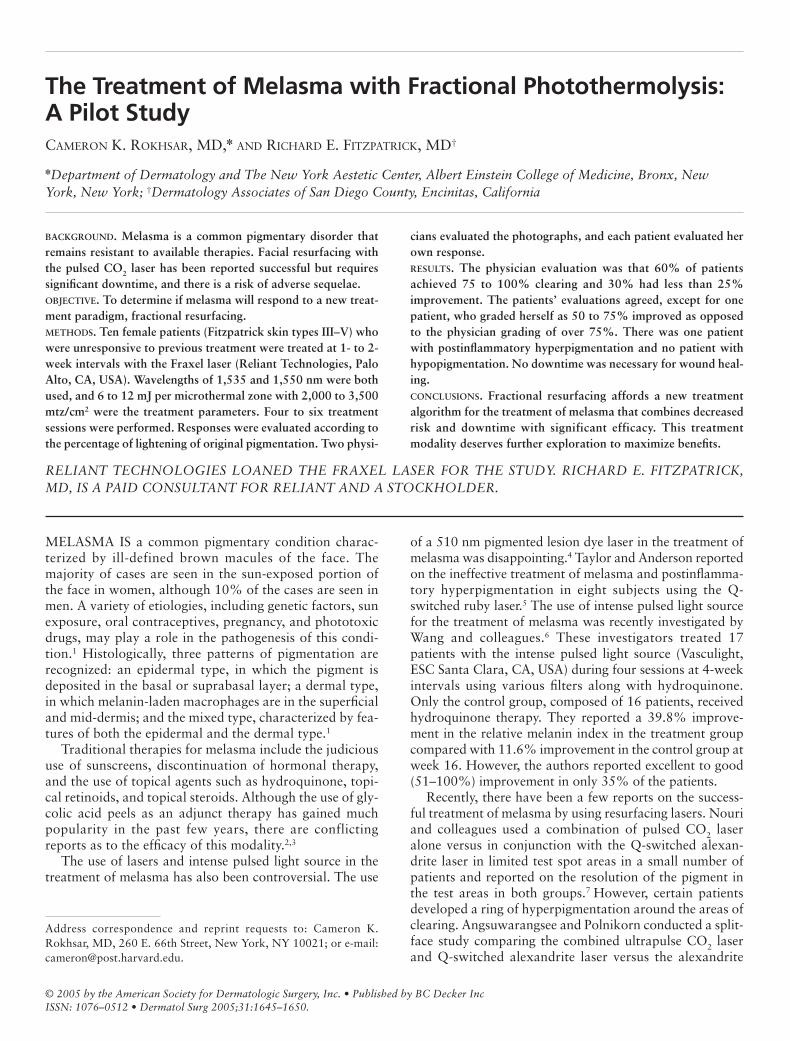
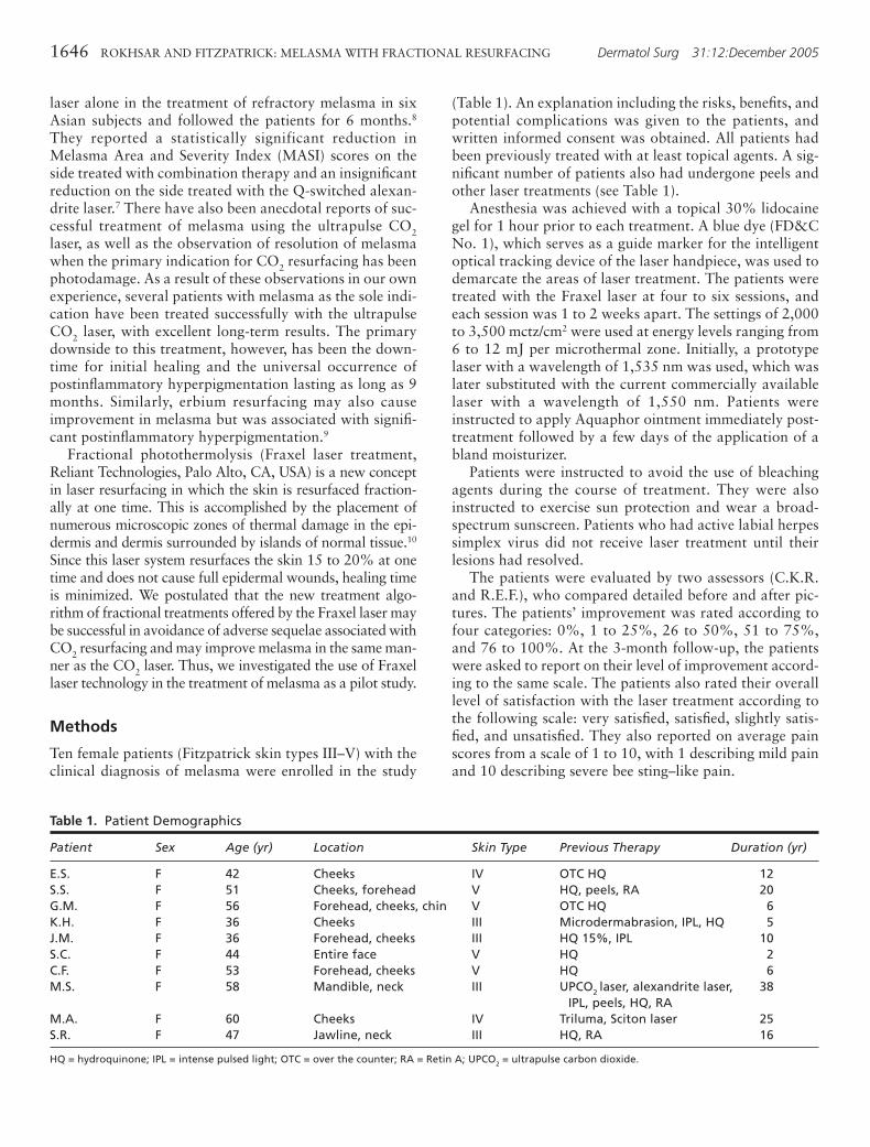
Sixty percent of patients were assessed to have 75 to100% clearing of their melasma according to the evalua-tor assessment (Figure 1). Fifty percent of patients alsorated themselves as having 75 to 100% improvement (Fig-ure 2). Of note was 100% clearing of melasma on bilateralcheeks in an Asian patient with type IV skin without anyevidence of hyperpigmentation or hypopigmentation (Fig-ures 3 and 4). There was also one Hispanic patient (skintype V) who had 75 to 100% improvement according toboth the evaluator and the patient assessments (Figure 5).All patients had at least been treated with topical agents.It is noteworthy that three patients were previously treatedwith intense pulsed light, without significant improve-ment. One patient whose facial melasma had cleared withCO2 resurfacing but still had residual melasma on the neckhad 50 to 75% improvement according to the patient aftersix treatment sessions with the Fraxel laser. The onepatient who did not respond to Fraxel laser treatment alsohad not responded to multiple sessions of 10- to 20-micron erbium laser peels (Sciton, Inc., Palo Alto, CA,
USA), which, in our experience, is somewhat helpful fortreatment-resistant melasma. The patients were also askedto rate their level of satisfaction with the laser treatmentaccording to the following four categories: very satisfied,satisfied, slightly satisfied, and unsatisfied. Sixty percent (6of 10) were very satisfied, 30% were slightly satisfied, and10% were unsatisfied.
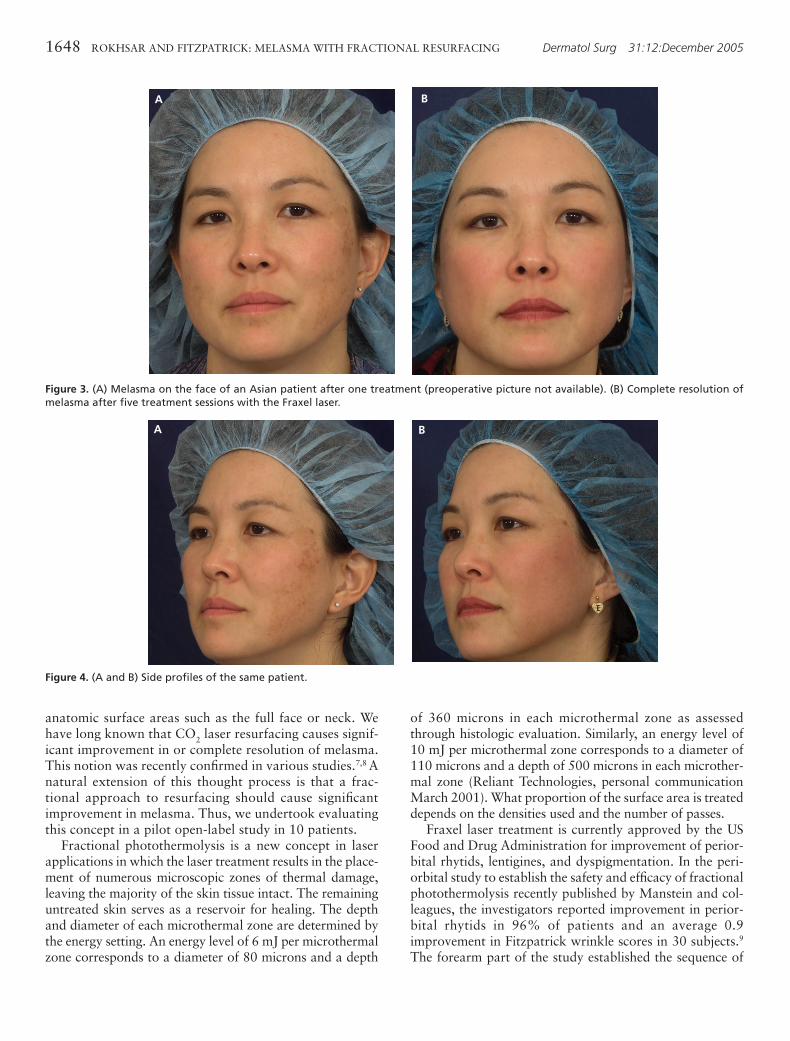
Of the nonresponder (n = 1) or the ones with minimalimprovement (n = 2) (1–25%), all were of Hispanic origin.However, one Hispanic patient had 75 to 100% clearanceafter five sessions, without any evidence of hyperpigmen-tation (see Figure 5).
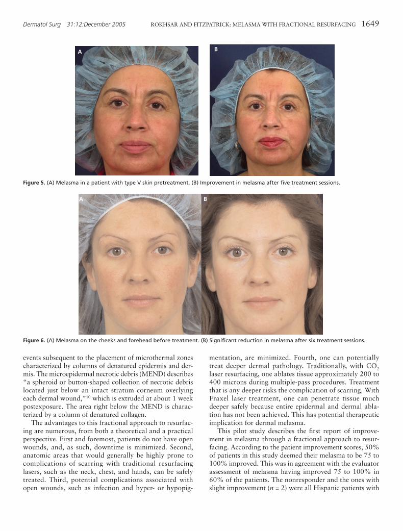
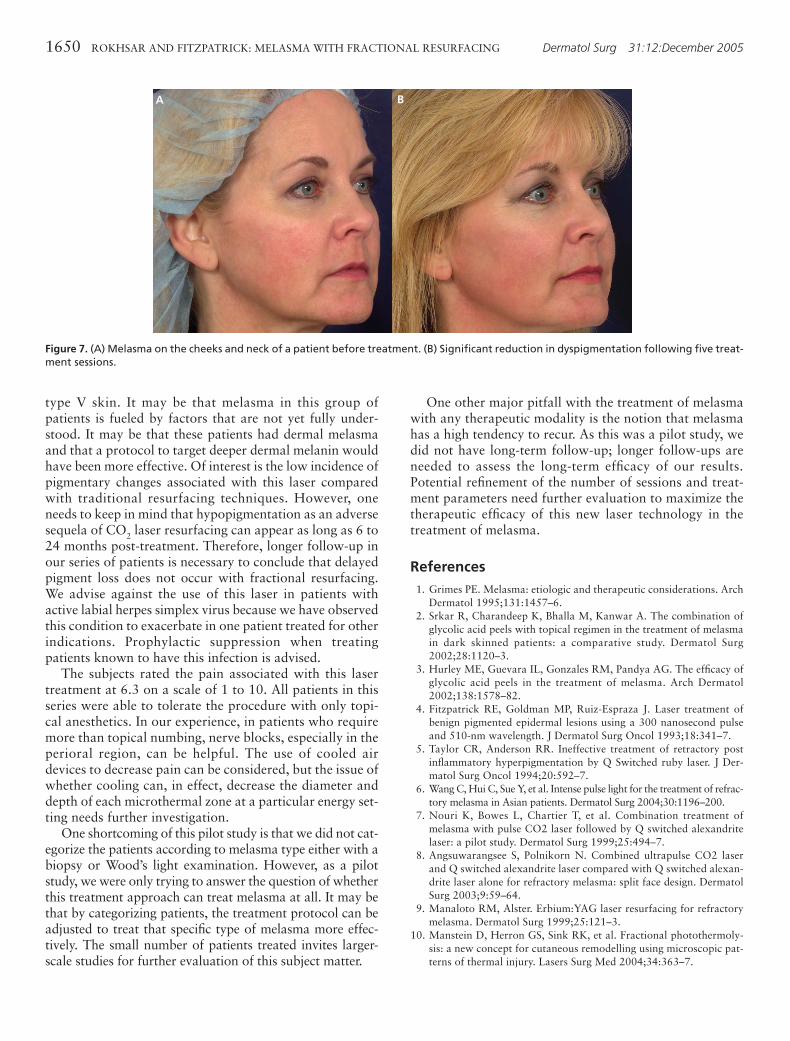
Coincidentally, all patients expressed improvement inskin texture even after one to two treatments (Figure 6).This, however, was not assessed formally by the investiga-tors. We treated patients’ melasma on the neck in twopatients without any adverse sequelae (see Figure 7).
Discussion
This study is the first report of fractional photothermoly-sis in the treatment of melasma and treatment of large
Dermatol Surg 31:12:December 2005 ROKHSAR AND FITZPATRICK: MELASMA WITH FRACTIONAL RESURFACING 1647
Figure 1. Percentile improvement in melasma as assessed by theevaluators.
Figure 2. Percentile improvement in melasma as assessed by thesubjects.
anatomic surface areas such as the full face or neck. Wehave long known that CO2 laser resurfacing causes signif-icant improvement in or complete resolution of melasma.This notion was recently confirmed in various studies.7,8 Anatural extension of this thought process is that a frac-tional approach to resurfacing should cause significantimprovement in melasma. Thus, we undertook evaluatingthis concept in a pilot open-label study in 10 patients.
Fractional photothermolysis is a new concept in laserapplications in which the laser treatment results in the place-ment of numerous microscopic zones of thermal damage,leaving the majority of the skin tissue intact. The remaininguntreated skin serves as a reservoir for healing. The depthand diameter of each microthermal zone are determined bythe energy setting. An energy level of 6 mJ per microthermalzone corresponds to a diameter of 80 microns and a depth
of 360 microns in each microthermal zone as assessedthrough histologic evaluation. Similarly, an energy level of10 mJ per microthermal zone corresponds to a diameter of110 microns and a depth of 500 microns in each microther-mal zone (Reliant Technologies, personal communicationMarch 2001). What proportion of the surface area is treateddepends on the densities used and the number of passes.
Fraxel laser treatment is currently approved by the USFood and Drug Administration for improvement of perior-bital rhytids, lentigines, and dyspigmentation. In the peri-orbital study to establish the safety and efficacy of fractionalphotothermolysis recently published by Manstein and col-leagues, the investigators reported improvement in perior-bital rhytids in 96% of patients and an average 0.9improvement in Fitzpatrick wrinkle scores in 30 subjects.9
The forearm part of the study established the sequence of
1648 ROKHSAR AND FITZPATRICK: MELASMA WITH FRACTIONAL RESURFACING Dermatol Surg 31:12:December 2005
Figure 4. (A and B) Side profiles of the same patient.
BA
Figure 3. (A) Melasma on the face of an Asian patient after one treatment (preoperative picture not available). (B) Complete resolution ofmelasma after five treatment sessions with the Fraxel laser.
A B
events subsequent to the placement of microthermal zonescharacterized by columns of denatured epidermis and der-mis. The microepidermal necrotic debris (MEND) describes“a spheroid or button-shaped collection of necrotic debrislocated just below an intact stratum corneum overlyingeach dermal wound,”10 which is extruded at about 1 weekpostexposure. The area right below the MEND is charac-terized by a column of denatured collagen.
The advantages to this fractional approach to resurfac-ing are numerous, from both a theoretical and a practicalperspective. First and foremost, patients do not have openwounds, and, as such, downtime is minimized. Second,anatomic areas that would generally be highly prone tocomplications of scarring with traditional resurfacinglasers, such as the neck, chest, and hands, can be safelytreated. Third, potential complications associated withopen wounds, such as infection and hyper- or hypopig-
mentation, are minimized. Fourth, one can potentiallytreat deeper dermal pathology. Traditionally, with CO2laser resurfacing, one ablates tissue approximately 200 to400 microns during multiple-pass procedures. Treatmentthat is any deeper risks the complication of scarring. WithFraxel laser treatment, one can penetrate tissue muchdeeper safely because entire epidermal and dermal abla-tion has not been achieved. This has potential therapeuticimplication for dermal melasma.
This pilot study describes the first report of improve-ment in melasma through a fractional approach to resur-facing. According to the patient improvement scores, 50%of patients in this study deemed their melasma to be 75 to100% improved. This was in agreement with the evaluatorassessment of melasma having improved 75 to 100% in60% of the patients. The nonresponder and the ones withslight improvement (n = 2) were all Hispanic patients with
Dermatol Surg 31:12:December 2005 ROKHSAR AND FITZPATRICK: MELASMA WITH FRACTIONAL RESURFACING 1649
Figure 5. (A) Melasma in a patient with type V skin pretreatment. (B) Improvement in melasma after five treatment sessions.
A B
Figure 6. (A) Melasma on the cheeks and forehead before treatment. (B) Significant reduction in melasma after six treatment sessions.
A B
type V skin. It may be that melasma in this group ofpatients is fueled by factors that are not yet fully under-stood. It may be that these patients had dermal melasmaand that a protocol to target deeper dermal melanin wouldhave been more effective. Of interest is the low incidence ofpigmentary changes associated with this laser comparedwith traditional resurfacing techniques. However, oneneeds to keep in mind that hypopigmentation as an adversesequela of CO2 laser resurfacing can appear as long as 6 to24 months post-treatment. Therefore, longer follow-up inour series of patients is necessary to conclude that delayedpigment loss does not occur with fractional resurfacing.We advise against the use of this laser in patients withactive labial herpes simplex virus because we have observedthis condition to exacerbate in one patient treated for otherindications. Prophylactic suppression when treatingpatients known to have this infection is advised.
The subjects rated the pain associated with this lasertreatment at 6.3 on a scale of 1 to 10. All patients in thisseries were able to tolerate the procedure with only topi-cal anesthetics. In our experience, in patients who requiremore than topical numbing, nerve blocks, especially in theperioral region, can be helpful. The use of cooled airdevices to decrease pain can be considered, but the issue ofwhether cooling can, in effect, decrease the diameter anddepth of each microthermal zone at a particular energy set-ting needs further investigation.
One shortcoming of this pilot study is that we did not cat-egorize the patients according to melasma type either with abiopsy or Wood’s light examination. However, as a pilotstudy, we were only trying to answer the question of whetherthis treatment approach can treat melasma at all. It may bethat by categorizing patients, the treatment protocol can beadjusted to treat that specific type of melasma more effec-tively. The small number of patients treated invites larger-scale studies for further evaluation of this subject matter.
One other major pitfall with the treatment of melasmawith any therapeutic modality is the notion that melasmahas a high tendency to recur. As this was a pilot study, wedid not have long-term follow-up; longer follow-ups areneeded to assess the long-term efficacy of our results.Potential refinement of the number of sessions and treat-ment parameters need further evaluation to maximize thetherapeutic efficacy of this new laser technology in thetreatment of melasma.
References1. Grimes PE. Melasma: etiologic and therapeutic considerations. Arch
Dermatol 1995;131:1457–6.2. Srkar R, Charandeep K, Bhalla M, Kanwar A. The combination of
glycolic acid peels with topical regimen in the treatment of melasmain dark skinned patients: a comparative study. Dermatol Surg2002;28:1120–3.
3. Hurley ME, Guevara IL, Gonzales RM, Pandya AG. The efficacy ofglycolic acid peels in the treatment of melasma. Arch Dermatol2002;138:1578–82.
4. Fitzpatrick RE, Goldman MP, Ruiz-Espraza J. Laser treatment ofbenign pigmented epidermal lesions using a 300 nanosecond pulseand 510-nm wavelength. J Dermatol Surg Oncol 1993;18:341–7.
5. Taylor CR, Anderson RR. Ineffective treatment of retractory postinflammatory hyperpigmentation by Q Switched ruby laser. J Der-matol Surg Oncol 1994;20:592–7.
6. Wang C, Hui C, Sue Y, et al. Intense pulse light for the treatment of refrac-tory melasma in Asian patients. Dermatol Surg 2004;30:1196–200.
7. Nouri K, Bowes L, Chartier T, et al. Combination treatment ofmelasma with pulse CO2 laser followed by Q switched alexandritelaser: a pilot study. Dermatol Surg 1999;25:494–7.
8. Angsuwarangsee S, Polnikorn N. Combined ultrapulse CO2 laserand Q switched alexandrite laser compared with Q switched alexan-drite laser alone for refractory melasma: split face design. DermatolSurg 2003;9:59–64.
9. Manaloto RM, Alster. Erbium:YAG laser resurfacing for refractorymelasma. Dermatol Surg 1999;25:121–3.
10. Manstein D, Herron GS, Sink RK, et al. Fractional photothermoly-sis: a new concept for cutaneous remodelling using microscopic pat-terns of thermal injury. Lasers Surg Med 2004;34:363–7.
1650 ROKHSAR AND FITZPATRICK: MELASMA WITH FRACTIONAL RESURFACING Dermatol Surg 31:12:December 2005
Figure 7. (A) Melasma on the cheeks and neck of a patient before treatment. (B) Significant reduction in dyspigmentation following five treat-ment sessions.
A B