pigmented villonodular synovitis of a lumbar facet … · pigmented villonodular synovitis of a...
TRANSCRIPT
Pigmented Villonodular Synovitis of a Lumbar Facet Joint
DavidS. Titelbaum, 1•2 C. Harker Rhodes,3 JohnS. J . Brooks,4 and Herbert I. Goldberg 1
Summary: We describe the CT appearance of suspected pigmented villonodular synovitis involving a lumbar facet in a 51 -year-old woman, and discuss how the histologic and radiologic appearances may differ from those of synovial cyts.
Index terms: Spine, facet joints; Neuropathology; Spine, computed tomography
Pigmented villonodular synovitis (PVNS) is a rare disorder of synovial joints, most commonly occurring in the knee, hip, ankle, and elbow (1-4). Few reports have demonstrated involvement of lumbar facet joints (2, 4). Computed tomography (CT) detection of high attenuation from hemosiderin deposition has been described in PVNS of the knee (5). This report describes an example where CT high attenuation was detected within a lateral extradural defect from lumbar facet joint PVNS. Preoperative distinction of PVNS from other extradural lesions is important because surgical treatment may require synovectomy (6).
Case Report
A 51-year-old woman presented with back pain and right leg radiculopathy following aerobic exercises. Her pain had kept her awake at night, had been particularly severe in the morning , and was somewhat relieved upon standing. Following failure of 6 months of physical therapy and nonsteroidal anti-inflammatory medications, she was referred for radiologic evaluation of possible disk herniation.
Plain films of the lumbar spine showed a mild anterolisthesis of L4 and L5, and L4-L5 facet joint sclerosis. Water soluble contrast myelography revealed a large right lateral extradural defect extending from slightly above the L4-L5 interspace down to just above the level of the L5 pedicle (Figs. 1A and 18). On post-myelogram CT, a large right
lateral L4-L5 extradural defect was confirmed that revealed increased attenuation (Fig. 1 C) . This was felt to represent a partly calcified extruded disk . Bone windows revealed slight widening of the medial aspect of the right facet joint (Fig. lD), not appreciated at the time.
A posterior laminectomy was performed at the L4-L5 level. A large solid right lateral extradural mass was removed that was felt to represent an extruded herniated nucleus pulposus at the time of surgery.
Pathologic examination of the extradural mass demonstrated solid-tissue fragments with features typical of PVNS, including osteoclast-like giant cells accompanied by hemosiderin-laden macrophages, polygonal stromal cells, scattered neutrophils, areas of collagen formation with fibroblasts , and free hemosiderin granules (Fig. lE). No deus pulposus fragment was identified in the specimen. No synovial lining and no evidence of a fibrous cyst wall was identified in any of the specimen fragments .
Discussion
PVNS is an uncommon disorder and primarily involves large joints (2, 3). Also known as "tenosynovial giant cell tumor, diffuse type," PVNS is closely related to nodular tenosynovitis (tenosynovial giant cell tumor, localized type) which is a common tumor of the hands. While the lesion of nodular tenosynovitis is usually a single nodule, PYNS tends to produce a diffuse lesion with joint destruction. The differences between these tumors are probably primarily a consequence of their locations (7).
Gross examination or aspiration of PVNS lesions typically yields yellowish brown material. Histologic examination reveals fibrous trabeculae separating cellular aggregates of histiocytic cells with scattered giant cells and hemosiderin-laden macrophages ( 1, 6). Radiographic findings include
Received March 7, 1991; revision requested April 10; revision received June 4; f inal acceptance June 21. 1 Department of Radiology, Section of Neuroradiology, Hospital of the University of Pennsy lvania, 3400 Spruce St. , Philadelphia, PA 19104. 2 Present address: MRI Diagnostic Center, 420 Libbey Parkway, Weymouth, MA 02189. 3 Department of Pathology, Dartmouth-Hitchcock Medical Center, Hanover, NH 03756. 4 Department of Pathology and Laboratory Medicine, Surgical Pathology Section, Hospita l of the University of Pennsylvania, Philadelphia, PA 19104.
Address reprint requests to H. I. Goldberg.
AJNR 13:1 64- 166, Jan/ Feb 1992 0195-6108/ 92/ 1301-0164 © American Society of Neuroradiology
164

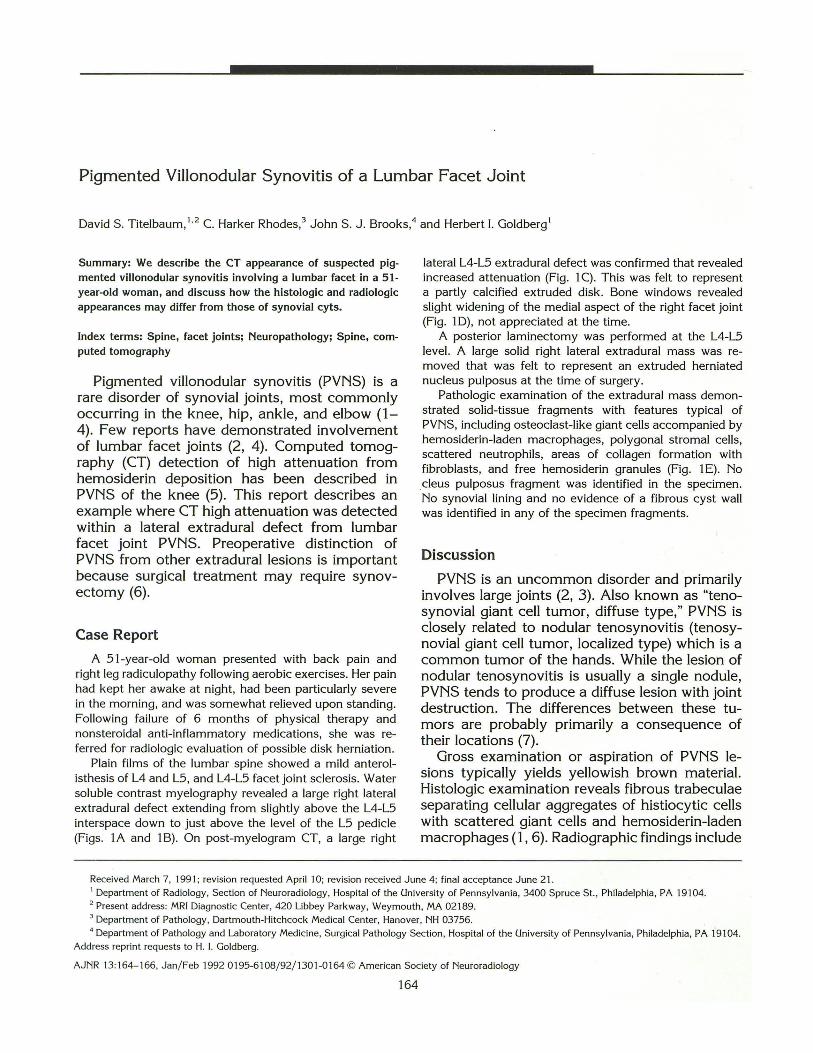
A B
c D
E
Fig. 1. A and B, Lateral (A) and right posterior oblique (B) views from water soluble contrast myelography demonstrate a large right lateral extradural defect at the L4-L5 level, near the facet joint, and a slight anterolisthesis of L4 on L5.
C, Post-myelography CT shows increased attenuation (arrow) within the extradural mass.
D, Bone windows from the CT show a small erosion along the medial aspect of the right L4-L5 facet joint (arrow).
E, Photomicrograph from histologic section of the resected mass shows solid sheets of polygonal stromal cells, macrophages, and clumps of hemosiderin granules (arrows). In adjacent fragments, osteoclast-like giant cells are also present (inset). No fibrous wall or synovial lining was identified. (Hematoxylin and eosin, X400).

effusion, cystic bony erosion , and increased attenuation presumably related to iron within hemosiderin (3) . Calcification is not a feature of PVNS. Rosenthal et al (5) have demonstrated increased CT attenuation in PVNS of the knee, presumably related to iron content. On magnetic resonance (MR) imaging, hemosiderin is known to cause T2 proton relaxation enhancement on long TR/TE images (8) , and areas of hypointensity have been demonstrated within lesions of PVNS involving the knee and hip (3, 9). Preoperative diagnosis may be useful in operative planning because treatment may require synovectomy and possible pre- or postoperative radiotherapy (6) .
PVNS involvement of the lumbar or cervical facet joints is rare, and can present as a large lateral extradural mass ( 1, 2, 4, 1 0). In addition to being a rare finding, the current case is interesting in that increased attenuation was visualized on CT. This finding was initially interpreted as due to calcification within a laterally extruded disk. Regrettably, MR imaging was not performed in this case, and the small medial facet joint erosion was overlooked.
Synovial cysts may also cause posterolateral extradural defects adjacent to abnormal facet joints, most commonly L4-L5, and may cause radiculopathy ( 11, 12). These lesions are typically round, well-circumscribed, and on CT may demonstrate a cyst wall ( 12, 13). Increased attenuation along the rim of the cyst may occur, and may be due to either calcification or hemorrhage (11 , 13, 14). Fluid density material or gas may be seen in the center of the cyst on CT (13-15). Central signal hyperintensity similar to that of cerebrospinal fluid may be seen on long TR MR imaging (11), although central hypointensity can be seen if the center is fibrous or gas ( 11 , 12). Histologically, a synovial cyst is a cavity lined by synovial cells oriented toward the lumen. Uncomplicated cysts have only fibrous tissue in the cyst wall outside of the synovial cells. Sheets of stromal and giant cells are not found. Fragments of a synovial cyst thus appear as tissue strips with cells on one side and acellular fibrous tissue on the other ( 16). No such appearance was present in this case. The current case of PVNS, therefore, is distinguished from a synovial cyst by the radiographic findings of a poorly circumscribed soft tissue mass with central increased attenuation and no evidence of fluid , the surgical finding of a solid mass, and by the pathologic finding of a solid sheet of cells, including histio-
cytic and giant cells, and no evidence of a fibrous synovial-lined cyst wall.
The differentia·( diagnosis of a lateral extradural soft-tissue mass with increased attenuation on CT should include PVNS, synovial cyst, as well as disk fragment and possible tumor. A poorly circumscribed soft-tissue mass with central increased attenuation lateralized near an eroded facet joint should strongly suggest PVNS as a likely diagnosis. MR imaging may then prove useful in helping to confirm the diagnosis by identifying central hemosiderin deposition and lack of cyst fluid.
References
1. Pulitzer DR, Reed RJ . Localized pigmented villonodular synovitis of
the vertebral column. Arch Pat hoi Lab Med 1984; 108:228- 230
2. Retrum ER, Schmidlin TM, Tay lor WK, Pepe RG. CT myelography
of extradura l pigmented villonodular synovitis. AJNR 1987;8:727-
729
3. Spritzer CE, Dalinka MK, Kressel HY. Magnetic resonance imaging of
pigmented villonodular synovitis: a report of two cases. Skeletal Radio!
1987; 16:31 6- 319
4. Weidner N, Challa VR, Bonsib SM, Davis CH Jr. Carroll T J J r. Giant
cell tumors of synovium (pigmented villonodular synovitis) involving
the vertebral column. Cancer 1986;57:2030-2036
5. Rosenthal Dl, Aronow S, Murray WT. Iron content of pigmented
villonodular synovitis detected by computed tomography . Radiology
1979; 133:409- 4 11
6. Byers PD, Cotton RE, Deacon OW, et al. The diagnosis and treatment
of pigmented villonodular synovitis . J Bone Joint Surg (Am)
1968;50B:290-305
7. Enzinger FM, Weiss SW. Soft tissue tumors, 2nd ed. St. Louis: C. V.
Mosby, 1988:638- 658
8. Gomori JM, Grossman Rl , Goldberg HI , Zimmerman RA, Bilaniuk LT.
Intracranial hematomas: imaging by high f ield MR. Radiology
1985; 157:87-93
9. Kottal RA, Vogler JB Ill , Matamoros A , Alexander AH, Cookson JL.
Pigmented v illonodular synovitis: a report of MR imaging of two
cases. Radiology 1987;163:551-553
10. Campbell AJ , Wells IP. Pigmented villonodular synovitis of a lumbar
facet joint. J Bone Joint Surg (Am) 1982;64A :1 45- 146
11 . Jackson DE Jr, Atlas SW, Mani JR, Norman D. Intraspinal synovial
cysts: MR imaging. Radiology 1989; 170:527-530
12. Silbergleit R, Gebarsk i SS, Brunberg JA, McGillicudy J , Blaivas M.
Lumbar synovial cysts: correlat ion of m yelographic, CT , MR, and
pathologic findings. A JNR 1990; 11:777-779
13. Mercader J, Gomez JM, Cardenal C. Intraspinal synovial cyst: diag
nosis by CT. Neuroradiology 1985;27:346-348
14. Hemminghytt S, Daniels DL, Williams AL, Haughton VM. Intraspinal
synovial cysts : natural history and diagnosis by CT . Radiology
1982; 145:375- 376
15. Schulz EE, West WL, Hinshaw DB, Johnson DR. Gas in a lumbar
ex tradural juxtaarticular cyst: sign of synovial origin. AJR
1984; 143:875-876
16. A lguacii-Garcia A . Spinal synovial cyst (ganglion): review and report
of a case presenting as a retropharyngea l mass. A m J Surg Pathol
1987;1 1 :732-735
Please see the Commentary by Bullough on page 167 in this issue.