case report pigmented villonodular synovitis of the...
TRANSCRIPT
Hindawi Publishing CorporationCase Reports in OrthopedicsVolume 2013, Article ID 870324, 4 pageshttp://dx.doi.org/10.1155/2013/870324
Case ReportPigmented Villonodular Synovitis of the Thoracic VertebraPresenting with Progressive Spastic Paraparesis
Mustafa Celiktas,1 Mehmet Ozan Asik,2 Yurdal Gezercan,3 and Mahir Gulsen1
1 Ortopedia Private Hospital, Orthopaedics and Traumatology Clinic, 01130 Adana, Turkey2Hospital Park Darica Hastanesi, Emek Mah, Fatih Sultan Mehmet Cad, No. 131, Bayramoglu Darıca/Kocaeli, Turkey3 Adana Numune Education and Research Hospital, Neurosurgery Clinic, 01240 Adana, Turkey
Correspondence should be addressed to Mustafa Celiktas; [email protected]
Received 27 July 2013; Accepted 15 August 2013
Academic Editors: Y. Kasai, C. C. Liao, C. W. Muller, and G. Singer
Copyright © 2013 Mustafa Celiktas et al. This is an open access article distributed under the Creative Commons AttributionLicense, which permits unrestricted use, distribution, and reproduction in any medium, provided the original work is properlycited.
Pigmented villonodular synovitis (PVNS) is a proliferative benign lesion originating from the synovium and commonly affectslarge joints of the extremities. PVNS can arise from any synovium in the whole body and rarely affects the zygapophyseal joints ofthe spine. Spinal PVNS is diagnosed mostly after resection of the mass. In our case we present a 22-year-old male patient showingprogressive spastic paraparesis with insidious onset of back pain and difficulty of walking in a relatively short period of 1 month.After gross excision of the mass, diagnosis was established through histopathology. Two years of follow-up period reveals completeresolution of the patient’s complaints and no recurrence on radiologic images.
1. Introduction
Pigmented villonodular synovitis (PVNS) is a benign pro-liferative and locally aggressive tumor of the synovium[1, 2]. PVNS may occur in a localized or diffuse form.The localized form is identical histologically to giant celltumor of tendon sheath. The diffuse form also appears tobe identical histologically to the localized form but involvesthe entire synovium. It commonly affects the synovium ofthe large joints of the extremities such as knee, hip, andshoulder and theoretically may arise from any synovium inthe body [3, 4]. PVNS is characterized as villous or nodularproliferation of the synovial tissue and due to presence ofhemosiderin pigment presents yellow to brownish color ongross appearance [4–6]. Only rarely does PVNS affect theaxial skeleton,where it arises from the vertebral articular facetjoint. Their occurrence in the thoracic spine is a very rareentity; nevertheless, it should be considered in the differentialdiagnosis [6]. The tumor represents itself with back pain andacute progressive neurological deficit [4–7]. Treatment of thePVNS includes gross resection of themass and close followupfor recurrences. To our knowledge only 54 articles relatedwith PVNS in the spine published in the English literature
and most of them are case reports [7]. In this report acutespastic paraparesis with one month of back pain history andPVNS of thoracic 7th vertebra are reported.
2. Case Report
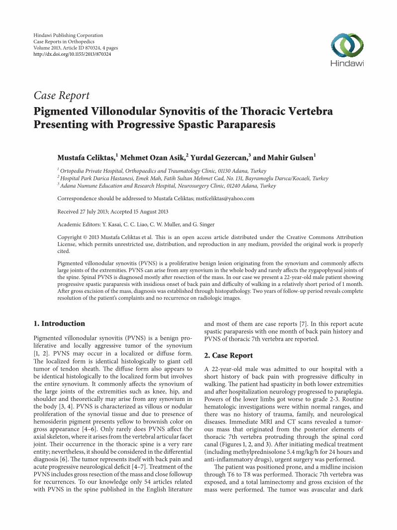
A 22-year-old male was admitted to our hospital with ashort history of back pain with progressive difficulty inwalking. The patient had spasticity in both lower extremitiesand after hospitalization neurology progressed to paraplegia.Powers of the lower limbs got worse to grade 2-3. Routinehematologic investigations were within normal ranges, andthere was no history of trauma, family, and neurologicaldiseases. Immediate MRI and CT scans revealed a tumor-ous mass that originated from the posterior elements ofthoracic 7th vertebra protruding through the spinal cordcanal (Figures 1, 2, and 3). After initiating medical treatment(including methylprednisolone 5.4mg/kg/h for 24 hours andanti-inflammatory drugs), urgent surgery was performed.
The patient was positioned prone, and a midline incisionthrough T6 to T8 was performed. Thoracic 7th vertebra wasexposed, and a total laminectomy and gross excision of themass were performed. The tumor was avascular and dark
2 Case Reports in Orthopedics
Figure 1: Sagittal T2-weighted images show expansive mass lesioninvolving the posterior elements of T6 and T7 vertebrae.
Figure 2: Coronal reformatted CT: eroding areas on the posteriorelements of T6 and T7 vertebrae.
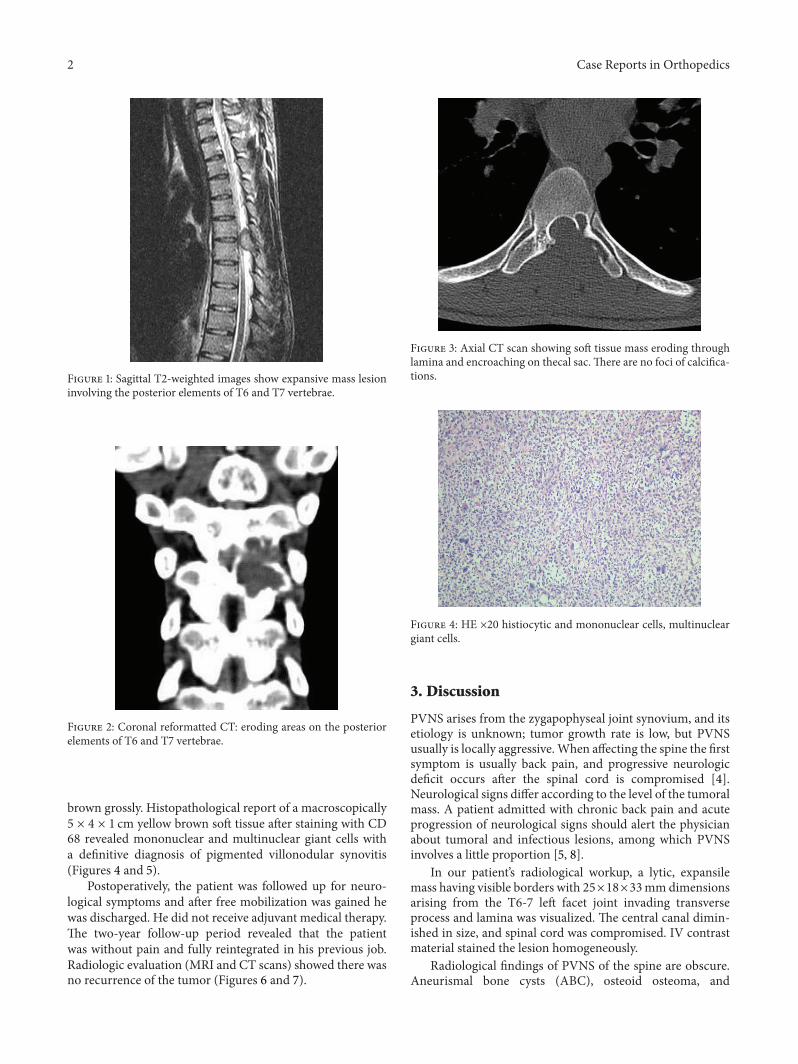
brown grossly. Histopathological report of a macroscopically5 × 4 × 1 cm yellow brown soft tissue after staining with CD68 revealed mononuclear and multinuclear giant cells witha definitive diagnosis of pigmented villonodular synovitis(Figures 4 and 5).
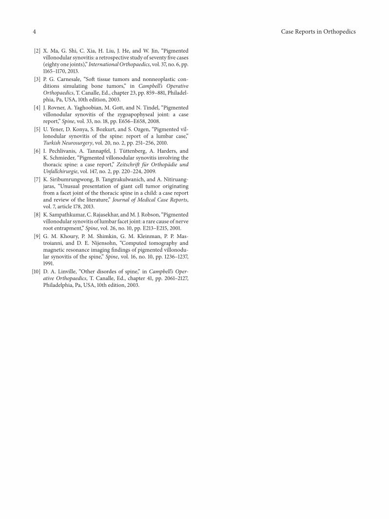
Postoperatively, the patient was followed up for neuro-logical symptoms and after free mobilization was gained hewas discharged. He did not receive adjuvant medical therapy.The two-year follow-up period revealed that the patientwas without pain and fully reintegrated in his previous job.Radiologic evaluation (MRI and CT scans) showed there wasno recurrence of the tumor (Figures 6 and 7).
Figure 3: Axial CT scan showing soft tissue mass eroding throughlamina and encroaching on thecal sac. There are no foci of calcifica-tions.
Figure 4: HE ×20 histiocytic and mononuclear cells, multinucleargiant cells.
3. Discussion
PVNS arises from the zygapophyseal joint synovium, and itsetiology is unknown; tumor growth rate is low, but PVNSusually is locally aggressive.When affecting the spine the firstsymptom is usually back pain, and progressive neurologicdeficit occurs after the spinal cord is compromised [4].Neurological signs differ according to the level of the tumoralmass. A patient admitted with chronic back pain and acuteprogression of neurological signs should alert the physicianabout tumoral and infectious lesions, among which PVNSinvolves a little proportion [5, 8].
In our patient’s radiological workup, a lytic, expansilemass having visible borders with 25×18×33mmdimensionsarising from the T6-7 left facet joint invading transverseprocess and lamina was visualized. The central canal dimin-ished in size, and spinal cord was compromised. IV contrastmaterial stained the lesion homogeneously.
Radiological findings of PVNS of the spine are obscure.Aneurismal bone cysts (ABC), osteoid osteoma, and
Case Reports in Orthopedics 3
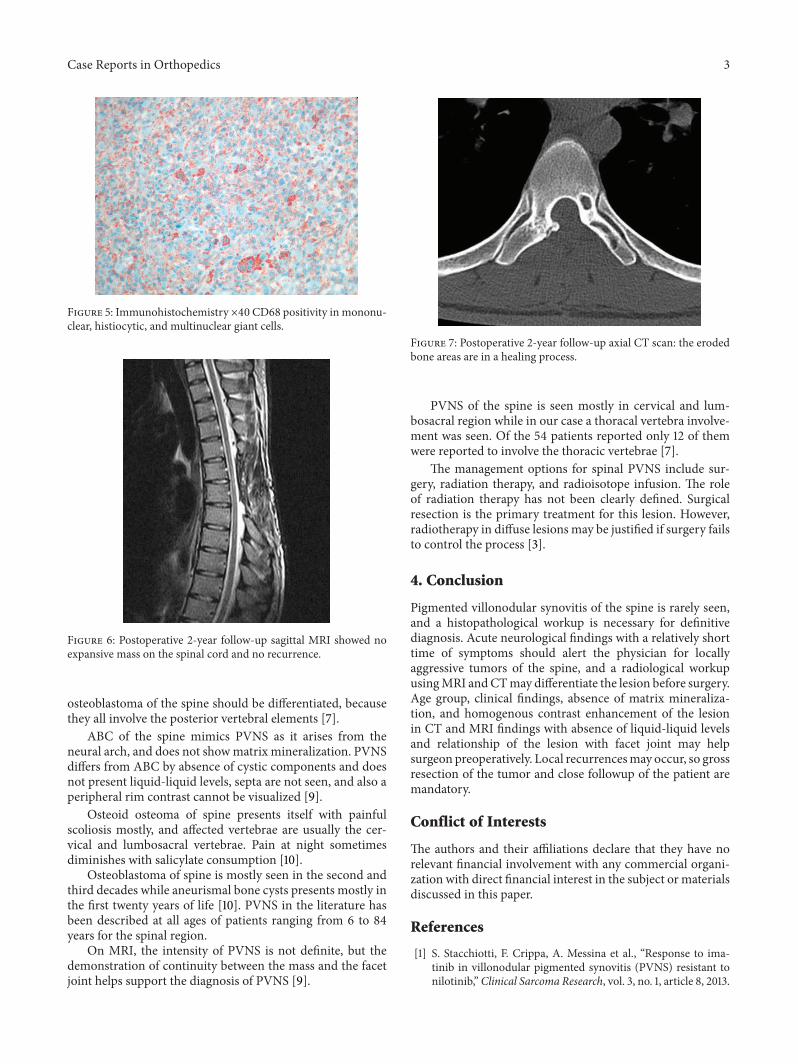
Figure 5: Immunohistochemistry ×40 CD68 positivity in mononu-clear, histiocytic, and multinuclear giant cells.
Figure 6: Postoperative 2-year follow-up sagittal MRI showed noexpansive mass on the spinal cord and no recurrence.
osteoblastoma of the spine should be differentiated, becausethey all involve the posterior vertebral elements [7].
ABC of the spine mimics PVNS as it arises from theneural arch, and does not showmatrix mineralization. PVNSdiffers from ABC by absence of cystic components and doesnot present liquid-liquid levels, septa are not seen, and also aperipheral rim contrast cannot be visualized [9].
Osteoid osteoma of spine presents itself with painfulscoliosis mostly, and affected vertebrae are usually the cer-vical and lumbosacral vertebrae. Pain at night sometimesdiminishes with salicylate consumption [10].
Osteoblastoma of spine is mostly seen in the second andthird decades while aneurismal bone cysts presents mostly inthe first twenty years of life [10]. PVNS in the literature hasbeen described at all ages of patients ranging from 6 to 84years for the spinal region.
On MRI, the intensity of PVNS is not definite, but thedemonstration of continuity between the mass and the facetjoint helps support the diagnosis of PVNS [9].
Figure 7: Postoperative 2-year follow-up axial CT scan: the erodedbone areas are in a healing process.
PVNS of the spine is seen mostly in cervical and lum-bosacral region while in our case a thoracal vertebra involve-ment was seen. Of the 54 patients reported only 12 of themwere reported to involve the thoracic vertebrae [7].
The management options for spinal PVNS include sur-gery, radiation therapy, and radioisotope infusion. The roleof radiation therapy has not been clearly defined. Surgicalresection is the primary treatment for this lesion. However,radiotherapy in diffuse lesions may be justified if surgery failsto control the process [3].
4. Conclusion
Pigmented villonodular synovitis of the spine is rarely seen,and a histopathological workup is necessary for definitivediagnosis. Acute neurological findings with a relatively shorttime of symptoms should alert the physician for locallyaggressive tumors of the spine, and a radiological workupusingMRI andCTmay differentiate the lesion before surgery.Age group, clinical findings, absence of matrix mineraliza-tion, and homogenous contrast enhancement of the lesionin CT and MRI findings with absence of liquid-liquid levelsand relationship of the lesion with facet joint may helpsurgeon preoperatively. Local recurrencesmay occur, so grossresection of the tumor and close followup of the patient aremandatory.
Conflict of Interests
The authors and their affiliations declare that they have norelevant financial involvement with any commercial organi-zation with direct financial interest in the subject or materialsdiscussed in this paper.
References
[1] S. Stacchiotti, F. Crippa, A. Messina et al., “Response to ima-tinib in villonodular pigmented synovitis (PVNS) resistant tonilotinib,”Clinical Sarcoma Research, vol. 3, no. 1, article 8, 2013.
4 Case Reports in Orthopedics
[2] X. Ma, G. Shi, C. Xia, H. Liu, J. He, and W. Jin, “Pigmentedvillonodular synovitis: a retrospective study of seventy five cases(eighty one joints),” InternationalOrthopaedics, vol. 37, no. 6, pp.1165–1170, 2013.
[3] P. G. Carnesale, “Soft tissue tumors and nonneoplastic con-ditions simulating bone tumors,” in Campbell’s OperativeOrthopaedics, T. Canalle, Ed., chapter 23, pp. 859–881, Philadel-phia, Pa, USA, 10th edition, 2003.
[4] J. Rovner, A. Yaghoobian, M. Gott, and N. Tindel, “Pigmentedvillonodular synovitis of the zygoapophyseal joint: a casereport,” Spine, vol. 33, no. 18, pp. E656–E658, 2008.
[5] U. Yener, D. Konya, S. Bozkurt, and S. Ozgen, “Pigmented vil-lonodular synovitis of the spine: report of a lumbar case,”Turkish Neurosurgery, vol. 20, no. 2, pp. 251–256, 2010.
[6] I. Pechlivanis, A. Tannapfel, J. Tuttenberg, A. Harders, andK. Schmieder, “Pigmented villonodular synovitis involving thethoracic spine: a case report,” Zeitschrift fur Orthopadie undUnfallchirurgie, vol. 147, no. 2, pp. 220–224, 2009.
[7] K. Siribumrungwong, B. Tangtrakulwanich, and A. Nitiruang-jaras, “Unusual presentation of giant cell tumor originatingfrom a facet joint of the thoracic spine in a child: a case reportand review of the literature,” Journal of Medical Case Reports,vol. 7, article 178, 2013.
[8] K. Sampathkumar, C. Rajasekhar, andM. J. Robson, “Pigmentedvillonodular synovitis of lumbar facet joint: a rare cause of nerveroot entrapment,” Spine, vol. 26, no. 10, pp. E213–E215, 2001.
[9] G. M. Khoury, P. M. Shimkin, G. M. Kleinman, P. P. Mas-troianni, and D. E. Nijensohn, “Computed tomography andmagnetic resonance imaging findings of pigmented villonodu-lar synovitis of the spine,” Spine, vol. 16, no. 10, pp. 1236–1237,1991.
[10] D. A. Linville, “Other disordes of spine,” in Campbell’s Oper-ative Orthopaedics, T. Canalle, Ed., chapter 41, pp. 2061–2127,Philadelphia, Pa, USA, 10th edition, 2003.

Submit your manuscripts athttp://www.hindawi.com
Stem CellsInternational
Hindawi Publishing Corporationhttp://www.hindawi.com Volume 2014
Hindawi Publishing Corporationhttp://www.hindawi.com Volume 2014
MEDIATORSINFLAMMATION
of
Hindawi Publishing Corporationhttp://www.hindawi.com Volume 2014
Behavioural Neurology
EndocrinologyInternational Journal of
Hindawi Publishing Corporationhttp://www.hindawi.com Volume 2014
Hindawi Publishing Corporationhttp://www.hindawi.com Volume 2014
Disease Markers
Hindawi Publishing Corporationhttp://www.hindawi.com Volume 2014
BioMed Research International
OncologyJournal of
Hindawi Publishing Corporationhttp://www.hindawi.com Volume 2014
Hindawi Publishing Corporationhttp://www.hindawi.com Volume 2014
Oxidative Medicine and Cellular Longevity
Hindawi Publishing Corporationhttp://www.hindawi.com Volume 2014
PPAR Research
The Scientific World JournalHindawi Publishing Corporation http://www.hindawi.com Volume 2014
Immunology ResearchHindawi Publishing Corporationhttp://www.hindawi.com Volume 2014
Journal of
ObesityJournal of
Hindawi Publishing Corporationhttp://www.hindawi.com Volume 2014
Hindawi Publishing Corporationhttp://www.hindawi.com Volume 2014
Computational and Mathematical Methods in Medicine
OphthalmologyJournal of
Hindawi Publishing Corporationhttp://www.hindawi.com Volume 2014
Diabetes ResearchJournal of
Hindawi Publishing Corporationhttp://www.hindawi.com Volume 2014
Hindawi Publishing Corporationhttp://www.hindawi.com Volume 2014
Research and TreatmentAIDS
Hindawi Publishing Corporationhttp://www.hindawi.com Volume 2014
Gastroenterology Research and Practice
Hindawi Publishing Corporationhttp://www.hindawi.com Volume 2014
Parkinson’s Disease
Evidence-Based Complementary and Alternative Medicine
Volume 2014Hindawi Publishing Corporationhttp://www.hindawi.com