fibroblast growth factor receptor 3 mutations in epidermal nevi and associated low grade l bladder...
TRANSCRIPT
Fibroblast Growth Factor receptor 3 Mutations in Epidermal Nevi and
Associated Low Grade l Bladder Tumors.
Hernandez S, Toll A et al
JID (2007, July), Volume 127

INTRODUCTION
•Epidermal Nevi are benign hamartomas in childhood. They present as verrucoid scaly plaques following Blaschko’s lines.
•Histologically these tumors are similar to seborrheic keratosis (SK).
•FGFR3 mutations are found in SK (39% incidence in a study) (Hafner, 2007) and 86% of adenoid seborrheic keratosis, but the highest incidence is found in Urothelial Carcinomas and are then associated with low grade tumors (good prognosis).
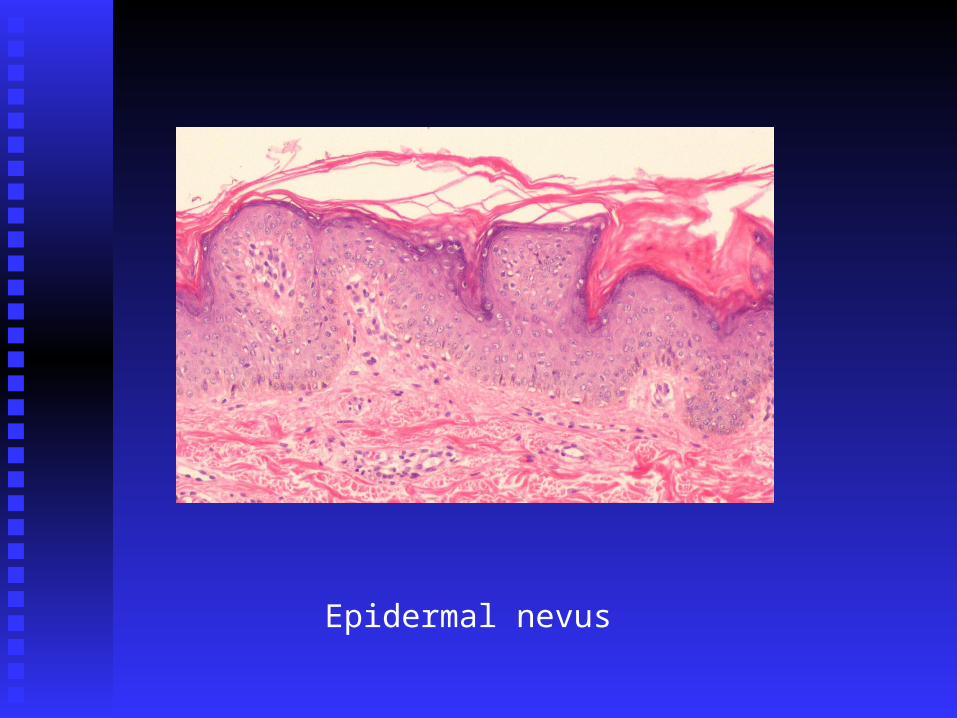
Epidermal nevus
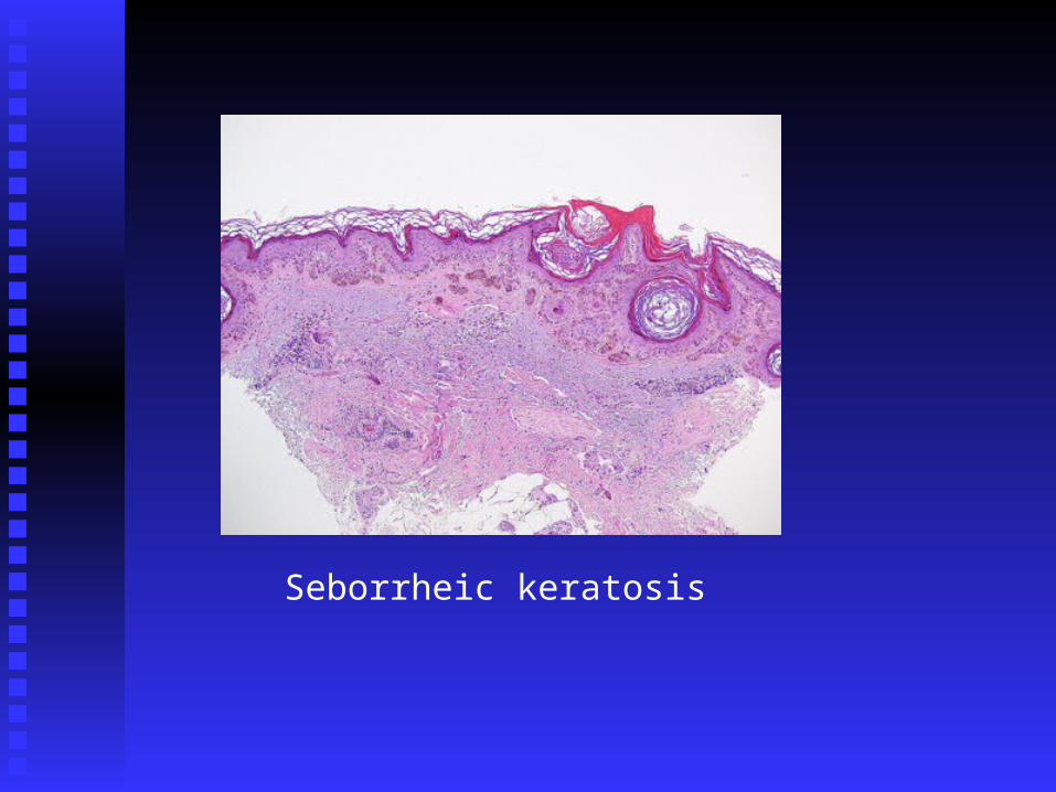
Seborrheic keratosis
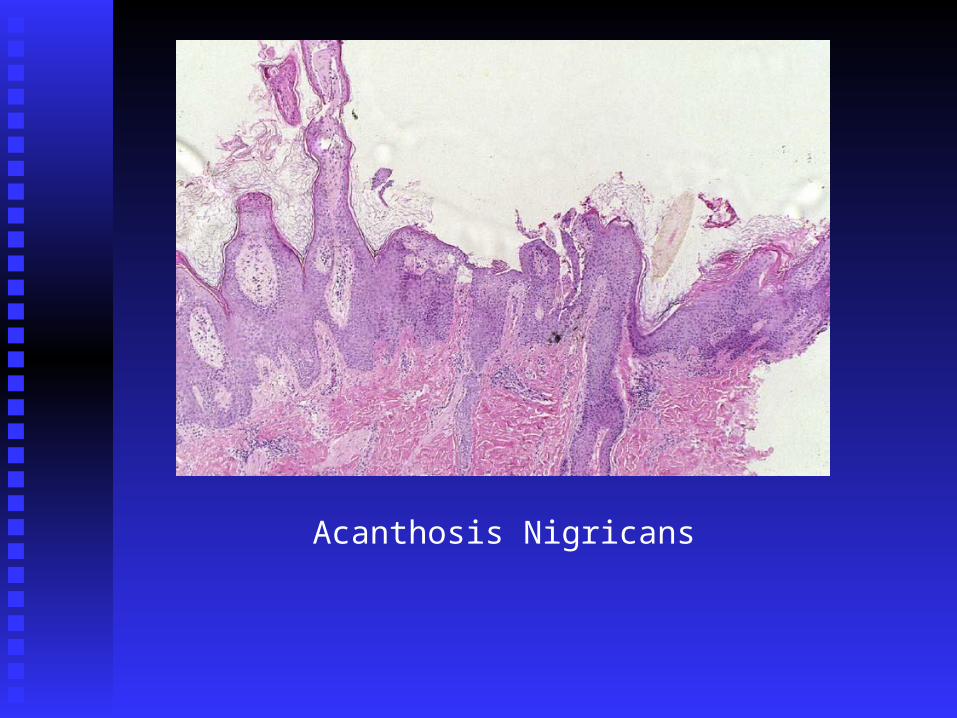
Acanthosis Nigricans
•FGFR3 DNA mutations are found in Skeletal dysplasias (exons 7, 10, 15), and the resultant constitutive kinase activity correlates with disease severity.
•The authors found a case of a 41 year old patient with a congenital widespread non epidermolytic keratinocytic Epidermal Nevi(in addition to 3 other literature reports). Patient had a history of low grade urothelial carcinoma at age 19. Based on three previous reports of associated UC and EN and the histological resemblances with SK, the authors hypothesize that EN might be caused by FGFR3 mutations.
To search for FGFR3 mutations in epidermal nevus
Goal
MATERIALS AND METHODS
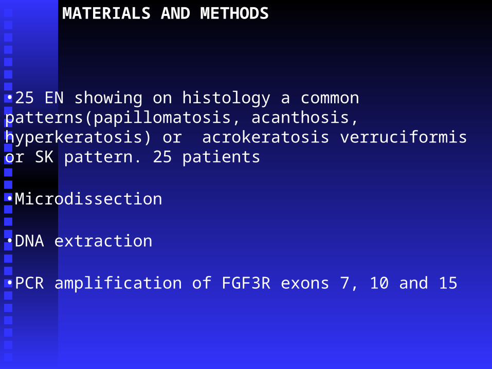
•25 EN showing on histology a common patterns(papillomatosis, acanthosis, hyperkeratosis) or acrokeratosis verruciformis or SK pattern. 25 patients
•Microdissection
•DNA extraction
•PCR amplification of FGF3R exons 7, 10 and 15
RESULTS
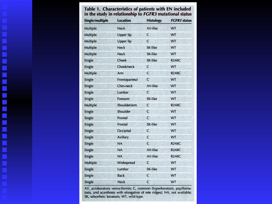
•No statistical difference in gender, age at diagnosis, location, number of EN, histological subtype.•6 patients: R248C mutation in exon 7 found•Exons 10 and 15 normal in all cases•The two specimens of normal skin showed FGFR3 wildtype sequences
•FGFR3 was positively staining (although weakly) on immunohistochemistry in the basal cell layer in 2/6 FGFR3 mutated samples and 9/13 FGFR3 wild type samples. This is similar to the weak detection in the normal epidermis
DISCUSSION
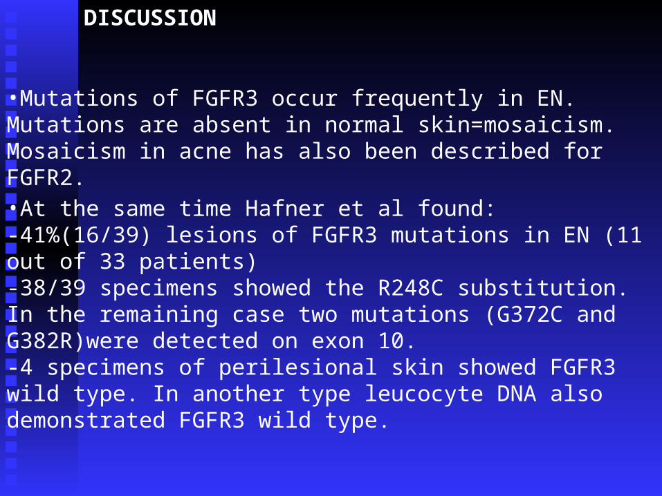
•Mutations of FGFR3 occur frequently in EN. Mutations are absent in normal skin=mosaicism. Mosaicism in acne has also been described for FGFR2.
•At the same time Hafner et al found:-41%(16/39) lesions of FGFR3 mutations in EN (11 out of 33 patients)-38/39 specimens showed the R248C substitution. In the remaining case two mutations (G372C and G382R)were detected on exon 10.-4 specimens of perilesional skin showed FGFR3 wild type. In another type leucocyte DNA also demonstrated FGFR3 wild type.
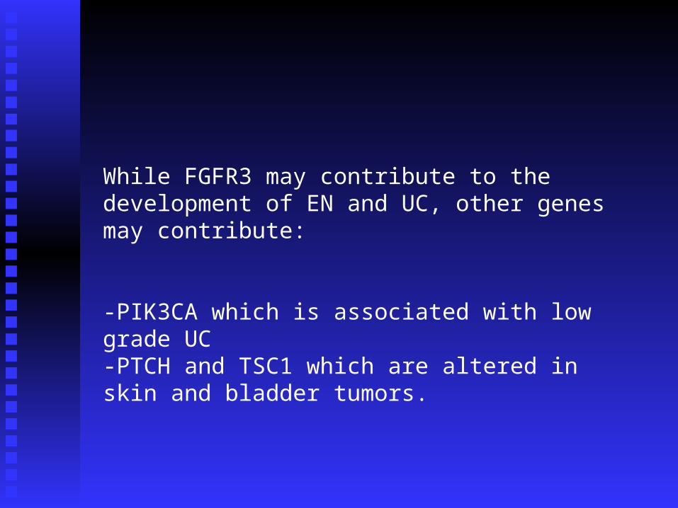
While FGFR3 may contribute to the development of EN and UC, other genes may contribute:
-PIK3CA which is associated with low grade UC-PTCH and TSC1 which are altered in skin and bladder tumors.

COMMENTARY (Hafner et al.) (1572-1573)
•FGFR3 mutations are described in Syndromes provoking skeletal dysplasia and acanthosis nigricans(AN) such as, thanatophoris dysplasia, SADDAN and Crouzon syndrome.
•Saddan syndrome is associated with the development of acanthosis nigricans. AN, EN and SK share papillomatosis, acanthosis and hyperkeratosis(…even though only AN is intertriginous).
•Achondroplasia also a skeletal dysplasia syndrome is characterised by G1144A mutation, and this mutation has not been detected in SK and EN, which hypothesizes that FGFR3 is important in the development of AN. Further studies are needed though.
•Somatic mutations also described in UC and Multiple myeloma
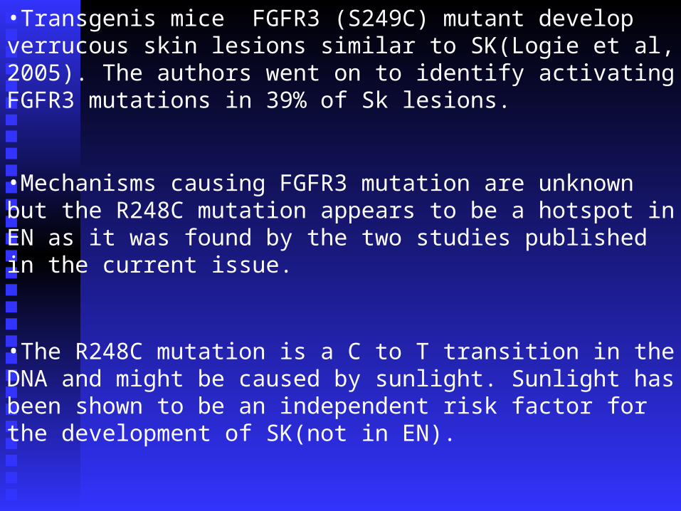
•Transgenis mice FGFR3 (S249C) mutant develop verrucous skin lesions similar to SK(Logie et al, 2005). The authors went on to identify activating FGFR3 mutations in 39% of Sk lesions.
•Mechanisms causing FGFR3 mutation are unknown but the R248C mutation appears to be a hotspot in EN as it was found by the two studies published in the current issue.
•The R248C mutation is a C to T transition in the DNA and might be caused by sunlight. Sunlight has been shown to be an independent risk factor for the development of SK(not in EN).
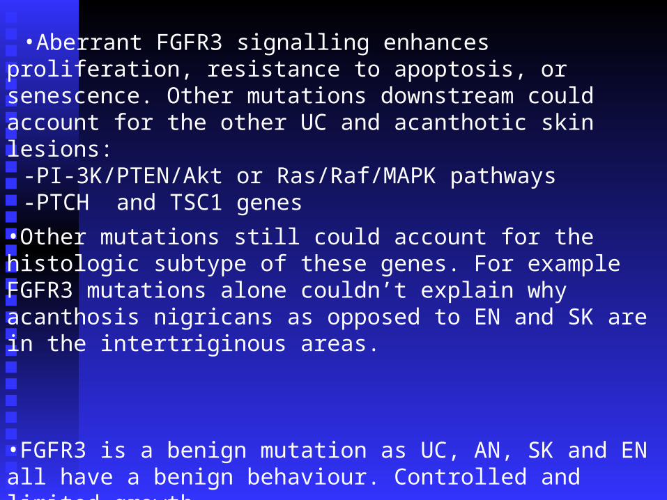
•Aberrant FGFR3 signalling enhances proliferation, resistance to apoptosis, or senescence. Other mutations downstream could account for the other UC and acanthotic skin lesions:
-PI-3K/PTEN/Akt or Ras/Raf/MAPK pathways-PTCH and TSC1 genes
•Other mutations still could account for the histologic subtype of these genes. For example FGFR3 mutations alone couldn’t explain why acanthosis nigricans as opposed to EN and SK are in the intertriginous areas.
•FGFR3 is a benign mutation as UC, AN, SK and EN all have a benign behaviour. Controlled and limited growth.

Conclusion of the commentary
[Several] specific small molecule [direct]inhibitors of FGFR3 [Chir 258, Pro 001] are already available and are currently in evaluation studies in leukaemia.
Topical application of these drugs seems possible for non-invasive treatment of acanthotic skin tumors.