endometrio ed endometriosi: the same tissue? - sichig · endometrio ed endometriosi: clinica...
TRANSCRIPT
Endometrio ed endometriosi:
Clinica Ostetrica e Ginecologica – IRCCS Azienda Ospedaliera Universitaria San Martino IST – Istituto Nazionale per la Ricerca sul Cancro, Università degli
studi di Genova
Valentino Remorgida
the same tissue?
Why does endometriosis developonly in some women?

the endometriotic
cell
A) RETROGRADE MENSTRUATION
B) Blood and linfatic
disseminatiom
C) STEM CELLS
D) Metaplasia of coelomicepithelium
Endometrialfragments migrate through Fallopian
Tubes duringmenstruation( Sampson, 1927)
In a similarmanner to the metastasis oftumor cells.
Stem cells residingin the basal layer
are shed intoperitoneal cavity
and implant.
Peritonealmesothelial cellsdifferentiate intoendometrium-like
tissue under the sex hormones control(mostly estrogen)
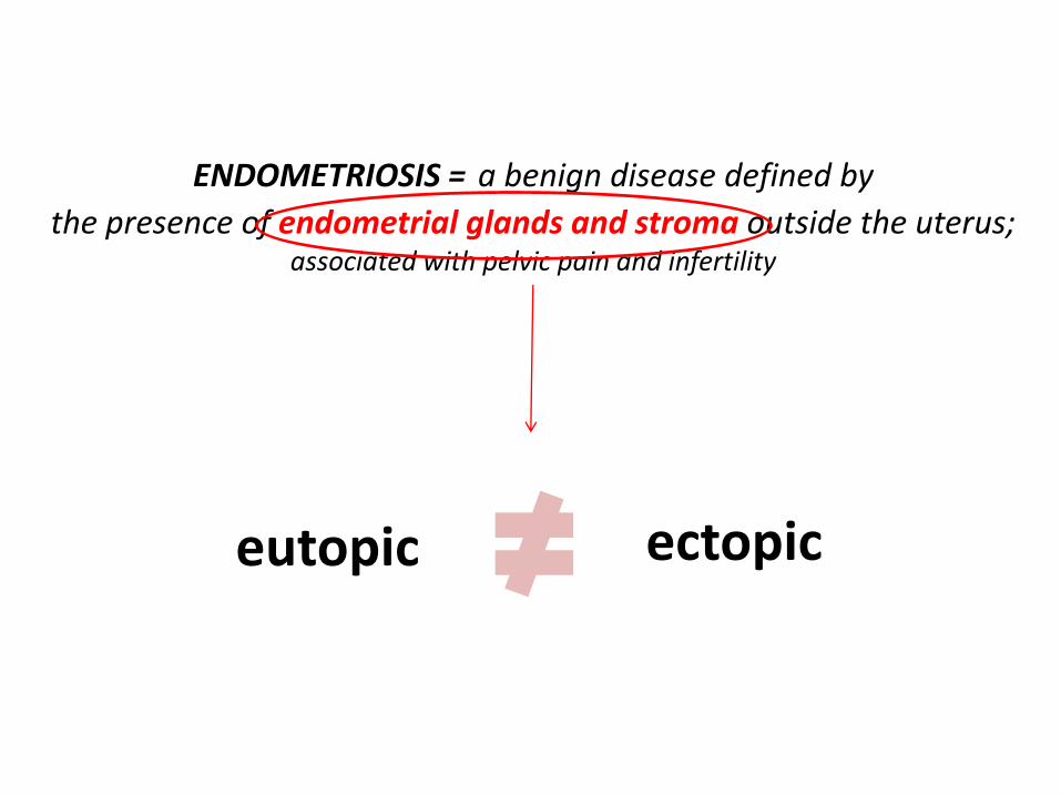
ENDOMETRIOSIS = a benign disease defined by
the presence of endometrial glands and stroma outside the uterus;associated with pelvic pain and infertility
eutopic ectopic

BUT WITHIN THE SAME
ECTOPIC LESION WE CAN
DISTINGUISH 3 DIFFERENTS
TYPE OF DISEASE
a)PERITONEAL IMPLANT
b)OVARIAN ENDOMETRIOMA
c)DIE
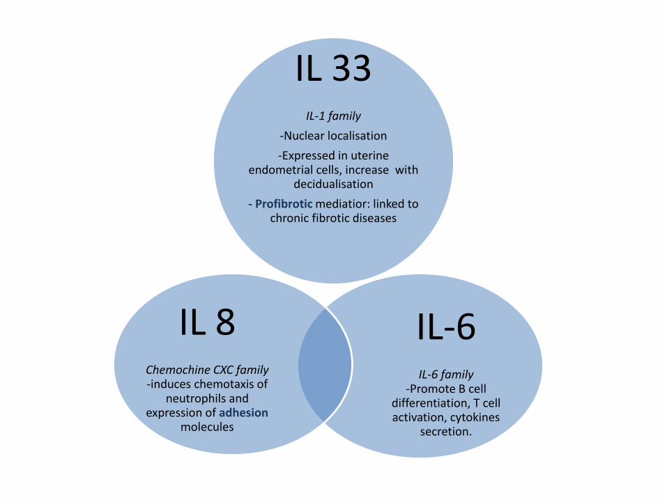
IL 33IL-1 family
-Nuclear localisation
-Expressed in uterine endometrial cells, increase with
decidualisation
- Profibrotic mediatior: linked tochronic fibrotic diseases
IL-6IL-6 family
-Promote B celldifferentiation, T cellactivation, cytokines
secretion.
IL 8Chemochine CXC family-induces chemotaxis of
neutrophils and expression of adhesion
molecules

Molecular mechanisms
Immunobiologic characteristics
Genetic foundation
The endometriotic cell, ectopic endometrium, with peculiar:

Molecular mechanisms
• high local estrogens production• high local prostaglandin production• resistance to the action of progesterone

Immunobiologic characteristics
Changes in cellular and humoral immunity
• activated macrophages and circulatingmonocytes secrete growth factors and cytochines
• NK cells: over expression of killer-hinibiting receptors
ProliferativeAnti-apoptotic
• increased T cells in stroma of ectopicendometrium and peritoneal fluid
• increased cytokines production (IL-1, IL-8, TNFalpha) and growth factors(VEGF)
ProliferativeAngiogenic
Mediate adhesion


Genetic foundation
Endometriosis is six to sever times more prevalentamong first-degree relatives of affected women
Cluster within familesMore common in mono and dizygotic twinsSimilar age of onset in non-twins sisters
Predisposition inherited as a complex genetic trait

Epigenetic changes
Different gene expression in endometriotic tissue.
i.e. ERK, FAS and FAS ligando, SF-1
Promote the survival, attachment and proliferationof the endometriotic cell

Epigenetics refers to heritable changes in DNA and chromatin that impact gene expression withoutchanges in DNA sequence . There are 2 basic epigeneticregulatory mechanisms : DNA methylation and histonemodifications. DNA methylation is the best understoodand most extensively studied epigenetic mechanismand refers to the covalent modification of post-replicative DNA, when a methyl group is added to the cytosine ring to form methyl cytosine . DNA methylation serves a critical role in the regulation of gene expression in development, differentiation, and complex diseases, cancer being the most prominentexample


In most placental mammals, the differentiation (decidualization) of the endometrial stroma into the decidual cells of pregnancy is induced by the implanting blastocyst ;however, primates that menstruate initiate decidualization through an evolutionarily unique mechanism: the post-ovulatory rise in maternal progesterone . Consequently, decidualization is triggered in women with every ovulatory cycle independent of pregnancy.
DNA methylation serves as a critical regulator of gene expression, and global differences in DNA methylation affect multiple aspects of development and disease. Endometriotic cells express variable levels of the DNA methyltransferase enzymes(DNMTs), which introduce and maintain DNA methylation on the C5 position of cytosine in CpG dinucleotides . Abnormal DNA methylation in endometriosis affects the expression of several genes, including homeobox A10 (HOXA10), estrogen receptor beta (ESR2), steroidogenic factor 1 (NR5A1), and aromatase (CYP19A1),which alter steroid signaling and responsiveness, and are criticallyinvolved in development and decidualization.
By comparing healthy and diseased cells treated with or without hormones to mimic part of the menstrual cycle, we uncovered many differentially methylated genes with defective expression in endometriosis that also regulate the hormone-dependent aspects of menstruation. In addition to expanding our understanding of how methylation affects endometriosis many fold, this also led us to propose an epigenetic switch that permits GATA6 expression in endometriosis instead of GATA2, and this switch promotes the aberrant expression of many of the genes seen in endometriosis. Our work provides novel unifying insight into the cause and development of endometriosis


Fertil Steril. 2016 Nov;106(6):1420-1431.e7. doi: 10.1016/j.fertnstert.2016.07.005. Epub 2016 Jul 27.

• Heat Map Analysis Shows that Ectopic Tissue is Uniquely Different from Matched EutopicEndometrium and from Control Endometrium
• Genes Involved in Cytokine–Cytokine ReceptorInteraction, Cellular Adhesion, Immune Cell Recruitment, and Apoptosis are SignificantlyIncreased in Endometriotic Lesions
• Genes Involved in Natural Killer and T-Cell Cytotoxicity, Cell Signaling, and Regulation of Inflammatory Responses are Decreased in Expression in Endometriotic Lesions

• Genes Involved in Both Classic and Alternate Complement Pathways are Aberrantly Expressed in Ectopic Tissues Compared with Control Endometrium Samples
• Eutopic Endometrium Shows DysregulatedImmune Activation Compared with Control Endometrium
• Eutopic Endometrium of Patients AberrantlyExpress Genes Involved in Regulation of Decidualization, Cellular Adhesion, Cytokine–Cytokine Receptor Interaction, and Apoptosis

We show aberrant transcription of genes involved in decidualization of endometrium, which may explain why our endometriosis patients were referred for infertility-related problems.Indeed, our data validate the presence of immune dysregulation often observed in the peritoneal fluid of women with endometriosis, and further emphasize the importance of using immune gene expression profiles as a guide to better define all the facets that contribute to the pathogenesis of endometriosis.

(J Clin EndocrinolMetab100:E955–E963, 2015)
Ridotta apoptosi

Resistenza al P4

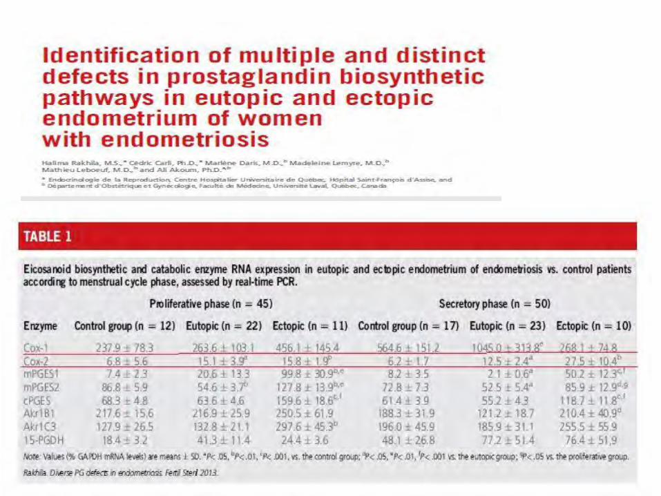

Cox2


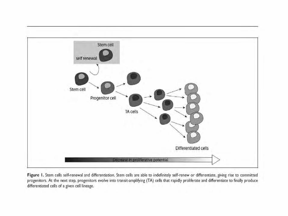
Stem cells and endometriosis


Stem Cell Migration between Endometriosis and Endometrium
Although it is clear that BMDSCs migrate to the uterus and endometriosis, stem cells are also capable of tremendous trafficking between locations. Our laboratory has identified the presence of a cell population that migrates from the endometriotic lesion into the uterus; these cells produce factors capable of altering uterine receptivity. ……….This is a process by which epithelial cells lose their polarity and are converted to a mesenchymal phenotype. These cells migrate as mesenchymal stem cells. After engraftment in the uterine stroma, these cells, all derived from the endometriosis, displayed activation of the Wnt signaling pathway indicating that they had taken on an epithelial identity; however, these cells were not located in the epithelium. These endometriosis-derived stem cells do not home to the appropriate location in the uterus. The cells were inappropriately localized to the stroma yet secreted signals typical of epithelial cells. Proper establishment of a gradient of signaling molecules between epithelium and stroma is essential for normal endometrial receptivity . The disincorporation of stem cells in the eutopicendometrium disrupts stromal-epithelial signaling and leads to decreased endometrial receptivity in endometriosis patients.


In summary, we identified new genes (EPHB4 and NRP1) thatmay contribute to angiogenesis in endometriosis beside knownfactors (VEGFA, VEGFR2, HIF1A, HGF, and PDGFB). Correlationstudies revealed the putative importance of EBHB4 in associationwith endometriosis. Our analyses support preliminary reports thatangiogenic factors seem to be differently expressed in peritonealimplant, ovarian endometriomas and DIE. Our observation thatangiogenic factors are differently expressed in the unaffectedperitoneum of women with endometriosis compared to womenwithout endometriosis underlines the importance of the peritoneumin the establishment of endometriosis.