transferrin-conjugated lipid-coated plga nanoparticles for targeted delivery of aromatase inhibitor...
TRANSCRIPT
P
Ta
YRYa
b
c
d
e
t
a
ARRAA
KAPT7D
1
teb1tf7abnK
4
0d
International Journal of Pharmaceutics 390 (2010) 234–241
Contents lists available at ScienceDirect
International Journal of Pharmaceutics
journa l homepage: www.e lsev ier .com/ locate / i jpharm
harmaceutical Nanotechnology
ransferrin-conjugated lipid-coated PLGA nanoparticles for targeted delivery ofromatase inhibitor 7�-APTADD to breast cancer cells
u Zhenga,b, Bo Yud,e, Wanlop Weecharangsana, Longzhu Piaoa, Michael Darbyc, Yicheng Maoa,e,umiana Koynovaa, Xiaojuan Yanga, Hong Lia, Songlin Xua, L. James Leed,e,asuro Sugimotoc, Robert W. Brueggemeierc, Robert J. Leea,e,∗
Division of Pharmaceutics, College of Pharmacy, the Ohio State University, Columbus, OH 43210, USAState Key Laboratory of Biotherapy, West China Hospital, the Sichuan University, Chengdu, Sichuan 610041, PR ChinaDivision of Medicinal Chemistry and Pharmacognosy, the Ohio State University, Columbus, OH 43210, USADepartment of Chemical and Biomolecular Engineering, the Ohio State University, Columbus, OH 43210, USANSF Nanoscale Science and Engineering Center (NSEC) for Affordable Nanoengineering of Polymeric Biomedical Devices (CANPBD),he Ohio State University, Columbus, OH 43210, USA
r t i c l e i n f o
rticle history:eceived 6 October 2009eceived in revised form 29 January 2010ccepted 8 February 2010vailable online 13 February 2010
eywords:
a b s t r a c t
Transferrin (Tf)-conjugated lipid-coated poly(d,l-lactide-co-glycolide) (PLGA) nanoparticles carrying thearomatase inhibitor, 7�-(4′-amino)phenylthio-1,4-androstadiene-3,17-dione (7�-APTADD), were syn-thesized by a solvent injection method. Formulation parameters including PLGA-to-lipid, egg PC-to-TPGS,and drug-to-PLGA ratios and aqueous-to-organic phase ratio at the point of synthesis were optimized toobtain nanoparticles with desired sizes and drug loading efficiency. The optimal formulation had a drugloading efficiency of 36.3 ± 3.4%, mean diameter of 170.3 ± 7.6 nm and zeta potential of −18.9 ± 1.5 mV.
romatase inhibitorLGA nanoparticleransferrin receptor�-APTADDrug targeting
The aromatase inhibition activity of the nanoparticles was evaluated in SKBR-3 breast cancer cells. IC50
value of the Tf-nanoparticles was ranging from 0.77 to 1.21 nM, and IC50 value of the nanoparticles wasranging from 1.90 to 3.41 nM (n = 3). The former is significantly lower than the latter (p < 0.05). Theseresults suggested that the aromatase inhibition activity of the Tf-nanoparticles was enhanced relativeto that of the non-targeted nanoparticles, which was attributable to Tf receptor (TfR) mediated uptake.In conclusion, Tf-conjugated lipid-coated PLGA nanoparticles are potential vehicles for improving theefficiency and specificity of therapeutic delivery of aromatase inhibitors.
. Introduction
Aromatase is a part of the cytochrome P450 enzyme complexhat catalyzes the conversion of androgen to estradiol, and itsxpression is elevated in breast cancer tissues relative to normalreast tissues (James et al., 1987; Reed et al., 1989; Bulun et al.,993; Miller et al., 1997). Currently, aromatase inhibitors anas-rozole, letrozole and exemestane are first line therapeutic agentsor estrogen responsive breast cancer (Brueggemeier et al., 2005).�-APTADD is a potent irreversible aromatase inhibitor with high
ffinity to its target (Snider and Brueggemeier, 1987). Its activity haseen shown in many cell lines including human mammary carci-oma MCF-7 cells and choriocarcinoma JAr cells (Brueggemeier andatlic, 1990). In addition, therapeutic effect of this drug has been∗ Corresponding author at: College of Pharmacy, 500 W. 12th Ave., Columbus, OH3210, USA. Tel.: +1 614 292 4172; fax: +1 614 292 7766.
E-mail address: [email protected] (R.J. Lee).
378-5173/$ – see front matter © 2010 Elsevier B.V. All rights reserved.oi:10.1016/j.ijpharm.2010.02.008
© 2010 Elsevier B.V. All rights reserved.
shown in vivo in a rat mammary carcinoma model (Brueggemeieret al., 1997).
7�-APTADD is insoluble in water, which may adversely affectits oral bioavailability and requires a solubilization vehicle for sys-temic delivery. Nanoparticles, including liposomes, can facilitatesolubilization of hydrophobic drugs and increase drug accumula-tion in the tumor through the enhanced permeability and retention(EPR) effect (Minko, 2006). In addition, it is possible to target thedelivery of nanoparticles to tumor cells via conjugation to a target-ing ligand. Formulation of anticancer drugs such as doxorubicin intoliposomes has been shown to enhance their therapeutic efficacyand reduce certain toxicities (Lu et al., 2007). However, accordingto our preliminary experiment, 7�-APTADD cannot be efficientlyentrapped into liposomes. Therefore, a PLGA nanoparticle based
strategy was developed in this study.PLGA is a biodegradable and biocompatible polymer frequentlyused in drug delivery (Jain, 2000). PLGA nanoparticles can read-ily incorporate hydrophobic drugs (Budhian et al., 2007). PLGA, innanosize, has a relatively rapid rate of hydrolysis (Duncanson et
al of P
aoowcledteahlatrhnae2pce
2
2
aappggs3((cpc(J(a(r(Icw
2
ahaelrat
Y. Zheng et al. / International Journ
l., 2007). It has been established in previous studies that coatingf a PLGA core with amphiphilic lipids can possibly reduce accessf the polymer to H2O (Bershteyn et al., 2008; Chan et al., 2009),hich in turn decreases the rate of PLGA hydrolysis and the asso-
iated drug release (Chan et al., 2009). Therefore, in this study,ipid-coated PLGA nanoparticles were investigated as drug carri-rs. In addition, Tf was conjugated to the nanoparticles. TfR is aimeric transmembrane glycoprotein (180 kDa) (Yang et al., 2009)hat is over-expressed in many types of tumor tissues (Shindelmant al., 1981; Faulk et al., 1980; Savellano et al., 2003; Rossiello etl., 1984). Besides, neoplastic cells with high metastatic potentialave been shown to express higher levels of the TfR than those with
ow metastatic potential (Inoue et al., 1993). These make the TfR anttractive marker for tumor cell targeting. Tf, an 80 kDa glycopro-ein, is the ligand for TfR (Yang et al., 2009), which is available inecombinant version (Novozymes, 2007) and, as a human protein,as low immunogenicity (Ali et al., 1999). In addition, Tf-conjugatedanoparticles have been shown to selectively deliver therapeuticgents including doxorubicin and cisplatin to tumor cells over-xpressing TfR through TfR-mediated endocytosis (Eavarone et al.,000; Iinuma et al., 2002). TfR-targeted drug delivery systems areotential candidates for clinical translation. In this study, Tf wasonjugated to 7�-APTADD loaded nanoparticles to increase thefficiency and selectivity of delivery to breast cancer cells.
. Materials and methods
.1. Materials
7�-APTADD was synthesized as described previously (Snidernd Brueggemeier, 1987). Egg phosphatidylcholine (egg PC)nd 1,2-dioleoyl-sn-glycero-3-phosphoethanolamine (DOPE) wereurchased from Avanti Polar Lipids (Alabaster, AL). D-�-tocopherololyethylene glycol 1000 succinate (TPGS) was obtained as aift from Eastman Co. Ltd. (Gwynedd, UK). Poly(d, l-lactide-co-lycolide), holo-human Tf, triethylamine, bovine serum albumin,epharose CL-4B chromatography media, calcein, androst-4-ene-,17-dione, bisbenzimide (Hoechst 33258), deoxyribonucleic acidDNA), 3-maleimidobenzoic acid N-hydroxysuccinimide esterMBS), phenazine methosulfate (PMS) and solvents were pur-hased from Sigma–Aldrich (St. Louis, MO, USA). DRAQ 5 wasurchased from Biostatus Limited Inc. (Leicestershire, UK). SKBR-3ells were obtained from American Type Culture Collection (ATCC)Manassas, VA, USA). Fetal bovine serum was purchased fromHR Bioscience (Lenexa, KS, USA). Phenol red-free custom mediaMEM, Earle’s salts, 1.5× amino acids, 2× non-essential aminocids, L-glutamine, 1.5× vitamins), octadecyl-rhodamine B chlorideR18) and gentamicin were purchased from Invitrogen Corpo-ation (Carlsbad, CA, USA). [1�-3H(N)]-androst-4-ene-3,17-dionespecific activity 23.5 Ci/mmol) was purchase from PerkinElmernc. (Waltham, MA, USA). 3-(4,5-Dimethyl-thiazol-2yl)-5-(3-arboxymethoxyphenyl)-2-(4-sulfophenyl)-2H-tetrazolium (MTS)as purchased from Promega Corporation (Madison, WI, USA).
.2. Preparation of 7˛-APTADD loaded nanoparticles
PLGA, egg PC, TPGS and 7�-APTADD were co-dissolved inn acetone–ethanol solvent mixture (1:1, v/v). The solution waseated to 60 ◦C and rapidly injected into preheated deionized watert 60 ◦C using a 1 ml syringe with a 28 gauge needle. In this study,
ffects of four formulation parameters on particle size and drugoading efficiency were investigated, including: (1) PLGA-to-lipidatio, (2) egg PC-to-TPGS ratio, (3) drug-to-PLGA ratio and (4)queous-to-organic phase ratio at the time of synthesis. Single fac-or test was used to optimize each parameter. The initial standardharmaceutics 390 (2010) 234–241 235
formulation used was: 13 mg egg PC, 2 mg TPGS, 15 �g of 7�-APTADD and 1 mg PLGA in 0.2 ml of acetone–ethanol mixture asthe organic phase, and with 1 ml deionized water as the aqueousphase. All experiments were performed for three times.
2.3. Determination of loading efficiency
Drug loaded nanoparticles were separated from the free drugby size-exclusion chromatography on a Sepharose CL-4B column.To analyze drug concentration, 1.9 ml of acetonitrile was addedinto 0.1 ml of the nanoparticle suspension from the column frac-tions and vortexed for 2 min to dissolve all components of thenanoparticles. The 7�-APTADD content was then determined onan Agilent 1100 HPLC equipped with a Hypersil reverse phase C-18column at 25 ◦C (250 mm × 4.6 mm, 5 �m; Thermo Scientific, MA,USA). 7�-APTADD absorbance was measured by an UV–vis detec-tor at 243 nm and had a retention time of 6 min when eluted with70:30 acetonitrile/water mobile phase at a flow rate of 1 ml/min.The loading efficiency of 7�-APTADD was calculated by dividingdrug content in the nanoparticle fractions by the amount of drugadded initially. All experiments were performed for three times.
2.4. Particle size and zeta potential
Three batches of nanoparticles were prepared using the optimalformulation. The nanoparticle size distribution was determinedby dynamic light scattering on a model 370 Nicomp SubmicronParticle Sizer (Particle Sizing Systems, Santa Barbara, CA) at roomtemperature. The zeta potential was determined on a ZetaPALSinstrument at room temperature (Brookhaven Instruments Corp.,Worcestershire, NY).
2.5. Preparation and characterization of 7˛-APTADD loadedTf-nanoparticles
A post-insertion method (Yang et al., 2009; Chiu et al., 2006) wasadopted to incorporate Tf-DOPE ligand into the lipid bilayers of the7�-APTADD loaded nanoparticles. First, maleimidobenzoyl-DOPE(MB-DOPE) was synthesized. MBS (15 �M, 4.7 mg) was dissolved in1 ml chloroform and added to a 2 ml chloroform solution of DOPE(10 �M, 7.44 mg) containing 50 �l triethylamine. The solution wasstirred overnight at room temperature, and then the solvent wasevaporated and ethanol added to dissolve the product. The ethanolsolution of MB-DOPE was rapidly injected into water and the MB-DOPE micelles were thus obtained. Meanwhile, Tf in HEPES buffer(pH 8) was activated with 5× Traut’s reagent to yield Tf-SH. Then,the Tf-SH was mixed with micelles of MB-DOPE at a protein-to-lipid molar ratio of 1:10 and the mixture was incubated at 37 ◦Cfor 1 h. The resulting Tf-DOPE micelles were then incubated with7�-APTADD loaded nanoparticle for 1 h at 37 ◦C to complete thepost-insertion of Tf.
2.6. Cryogenic transmission electron microscopy (cryo-TEM)
7�-APTADD loaded nanoparticles and Tf-nanoparticles wereprepared as described in Sections 2.2 and 2.3. Tf-liposomes wereprepared as described in Sections 2.2 and 2.3 except withoutaddition of PLGA. The morphology of the nanoparticles and theliposomes was examined by cryo-TEM. Samples for cryo-TEM wereprepared as described previously (Yang et al., 2009). Briefly, a dropof the nanoparticles was applied on a perforated carbon film, sup-
ported by a copper grid and held by the controlled environmentvitrification system. Then the grid was immediately plunged intoliquid ethane at its melting point (−183 ◦C) and stored in liquidnitrogen (−196 ◦C). The vitrified sample was examined in a TecnaiG2 Transmission Electron Microscope (FEI Company, Oregon, USA)
2 al of P
ocbD
2
7cpsap
2
ffa3
2
flacc0rtflw
2c
aaaCmiicCpr
ilttE
2l
uam
36 Y. Zheng et al. / International Journ
perated at 120 kV using a Gatan HC3500 Tilt heating/Nitrogenooling holder (Pleasanton, CA, USA). Digital images were capturedy a Gatan 791 MultiScan CCD camera and processed using theigital Micrograph 3.1 software package.
.7. Colloidal stability of the nanoparticles
The colloidal stability of 7�-APTADD loaded nanoparticles and�-APTADD loaded Tf-nanoparticles was studied by monitoring thehanges in the particle size during storage at 4 ◦C. At various timeoints, aliquots of each sample were withdrawn and the particleize distribution was determined by dynamic light scattering onmodel 370 Nicomp Submicron Particle Sizer. Experiments wereerformed for three times.
.8. Cell culture
A breast cancer cell line, SKBR-3, was maintained in phenol red-ree custom media supplemented with glutamine (2 mM), 10% (v/v)etal bovine serum and gentamicin (20 mg/l). Cells were cultureds a monolayer in a humidified atmosphere containing 5% CO2 at7 ◦C.
.9. TfR expression on cell surface
TfR expression levels in breast cancer cells were measured by auorescein isothiocyanate (FITC) labeled Tf (FITC-Tf) binding assay,s described previously (Yang et al., 2009). For the study, 4 × 10⁵
ells were incubated with 200 �g/ml FITC-Tf at 4 ◦C for 30 min. Theells were then washed twice with cold PBS (pH 7.4) containing.1% BSA, pelleted by centrifugation at 1500 RPM for 3 min and thenesuspended in PBS. Cells without treatment were used as a nega-ive control. Cellular fluorescence was measured by a FACSCaliburow cytometer (Becton Dickinson, Franklin Lakes, NJ). Experimentsere carried out in triplicate.
.10. Cellular uptake of calcein loaded nanoparticles by flowytometry
Calcein is a water soluble fluorescent dye with an excitationnd emission wavelengths of 495 and 515 nm, respectively. Under488 nm excitation that is used by flow cytometry, calcein emitsstrong signal in the typical cytometric FL1 channel (525 nm).
alcein loaded nanoparticles were prepared using the optimal for-ulation as described above except that the drug was not added
nto the organic phase. In addition, the organic phase was injectednto preheated calcein aqueous solution (50 mM, pH 8). Then, freealcein was removed by passing the sample through a SepharoseL-4B column. Calcein loaded Tf-nanoparticles were prepared byost-insertion, as described in Section 2.5, at Tf-DOPE-to-total lipidatios of 0.3:100, 0.6:100, 1.5:100 and 3:100.
For cellular uptake studies, 4 × 10⁵ cells were incubated at 37 ◦Cn 1 ml complete culture media with or without 20 �M Tf (at aipid concentration 72 �g/ml) for 2 h. Then, the cells were washedhree times with PBS and fixed in 1% para-formaldehyde solu-ion. Nanoparticle uptake in cells was analyzed by flow cytometry.xperiments were carried out in triplicate.
.11. Cellular uptake of R18 labeled nanoparticles by confocalaser scanning microscopy
Octadecyl-rhodamine B chloride (R18), a lipophilic dye, wassed to track the nanoparticles, as described previously (Chiu etl., 2006). R18 labeled nanoparticles were prepared using the opti-al formulation by replacing 7�-APTADD with R18. Molar ratio of
harmaceutics 390 (2010) 234–241
R18 to lipids used was 0.05:100. R18 labeled Tf-nanoparticles with0.6 mol% of Tf-DOPE were prepared as described in Section 2.5.
For cellular uptake experiments, 4 × 10⁵ cells were incubatedat 37 ◦C in 1 ml complete culture media with or without 20 �MTf (at a lipid concentration 72 �g/ml) for 2 h. Then, the cells werewashed three times with PBS and fixed in 1% para-formaldehydesolution. The cells were stained by DRAQ 5 (a nuclear counterstain)and examined on a Zeiss LSM 510 META laser scanning confocalmicroscope (Zeiss, Oberkochen, Germany).
2.12. Cytoxicity assay for free drug and Tf-nanoparticles
The cytotoxicity of the free drug and the drug loadednanoparticles was evaluated by MTS assay. 7�-APTADD loaded Tf-nanoparticles were prepared as described in Sections 2.2 and 2.3.Empty nanoparticles were prepared similarly except without addi-tion of the drug. The SKBR-3 cells were seeded in 96-well plates ata density of 2 × 10³ cells/well. After 24 h of incubation, the mediumwas replaced with fresh medium containing varying concentra-tions of 7�-APTADD dissolved in DMSO or 7�-APTADD loadedTf-nanoparticles prepared by serial dilution. Empty nanoparticlesof matching lipid concentration were diluted similarly. Cells with-out treatment or cultured in the same media containing eitherDMSO or empty nanoparticles were used as controls. Experimentswere carried out in quadruplicate. After 24 h, the medium was aspi-rated. Twenty �l reagent composed of MTS and PMS at a volumeratio of 20:1 was added to each well containing 100 �l fresh media.After 1.5 h incubation at 37 ◦C, absorbance at 490 nm for formazanproduct of MTS conversion was detected on a Spectra MAX 340plate reader (Molecular Devices Corp., Sunnyvale, CA, USA). Therelative cell viability was calculated by dividing the absorbance inthe samples treated with the test agents by the absorbance in thecontrol samples.
2.13. Inhibition of aromatase in SKBR-3 cells
Drug loaded nanoparticles were prepared using the optimalformulation as described in Section 2.1. For the study, 1 × 10⁵
SKBR-3 cells were seeded in 6-well plates. After 24 h of incubation,the medium was replaced with fresh medium containing varyingconcentrations of 7�-APTADD, 7�-APTADD loaded nanoparticlesand 7�-APTADD loaded Tf-nanoparticles. Experiments were car-ried out in triplicate. After 2 h, the media with the test agentswere removed and cells were washed with PBS. Then, 1 ml freshmedium containing [1�-3H] androstenedione (8.2 pmol, 0.17 �Ci)and androstenedione (193 pmol), as the substrates for aromatase,was added to each well. Cells cultured in the same media con-taining DMSO or empty nanoparticles were used as controls. After3 h, the media were transferred into centrifuge tubes. The remain-ing substrate [1�-3H] androstenedione in the media was extractedby 3 ml diethyl ether for 2 times, and the aqueous media wasthen treated by addition of 3% dextran-coated charcoal suspen-sion (200 �l). After centrifugation, aliquot (0.6 ml) of the media wasthen added to complete scintillation cocktail (5.0 ml) and the [3H]concentration was quantitatively determined by liquid scintillationcounting. A modified Hoechst dye assay method was conducted toanalyze DNA content (Rago et al., 1990). A NaOH (8 mM) solution(980 �l) was added to each well to lyse cells at 60 ◦C for 5 min andthen a HEPES solution (1 M, 20 �l) was added to neutralize NaOH.Cell lysate (100 �l) was placed into each well of a 96-well plateand the Hoechst dye (20 �g/ml) was added to each well on the
plate. The relative fluorescence units (RFU) were determined by aTecan GENios reader (Tecan Systems, Inc., CA, USA). The excitationand emission wavelengths used were 360 and 465 nm, respec-tively. DNA in each sample was quantified using a standard curveof calfthymus DNA determined using the same method. The aro-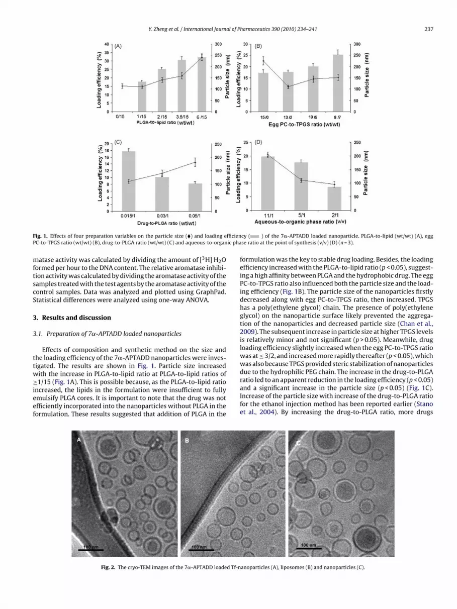
Y. Zheng et al. / International Journal of Pharmaceutics 390 (2010) 234–241 237
F fficienP ic pha
mftscS
3
3
ttw≥ieef
ig. 1. Effects of four preparation variables on the particle size (�) and loading eC-to-TPGS ratio (wt/wt) (B), drug-to-PLGA ratio (wt/wt) (C) and aqueous-to-organ
atase activity was calculated by dividing the amount of [3H] H2Oormed per hour to the DNA content. The relative aromatase inhibi-ion activity was calculated by dividing the aromatase activity of theamples treated with the test agents by the aromatase activity of theontrol samples. Data was analyzed and plotted using GraphPad.tatistical differences were analyzed using one-way ANOVA.
. Results and discussion
.1. Preparation of 7˛-APTADD loaded nanoparticles
Effects of composition and synthetic method on the size andhe loading efficiency of the 7�-APTADD nanoparticles were inves-igated. The results are shown in Fig. 1. Particle size increasedith the increase in PLGA-to-lipid ratio at PLGA-to-lipid ratios of
1/15 (Fig. 1A). This is possible because, as the PLGA-to-lipid rationcreased, the lipids in the formulation were insufficient to fullymulsify PLGA cores. It is important to note that the drug was notfficiently incorporated into the nanoparticles without PLGA in theormulation. These results suggested that addition of PLGA in the
Fig. 2. The cryo-TEM images of the 7�-APTADD loaded Tf-n
cy ( ) of the 7�-APTADD loaded nanoparticle. PLGA-to-lipid (wt/wt) (A), eggse ratio at the point of synthesis (v/v) (D) (n = 3).
formulation was the key to stable drug loading. Besides, the loadingefficiency increased with the PLGA-to-lipid ratio (p < 0.05), suggest-ing a high affinity between PLGA and the hydrophobic drug. The eggPC-to-TPGS ratio also influenced both the particle size and the load-ing efficiency (Fig. 1B). The particle size of the nanoparticles firstlydecreased along with egg PC-to-TPGS ratio, then increased. TPGShas a poly(ethylene glycol) chain. The presence of poly(ethyleneglycol) on the nanoparticle surface likely prevented the aggrega-tion of the nanoparticles and decreased particle size (Chan et al.,2009). The subsequent increase in particle size at higher TPGS levelsis relatively minor and not significant (p > 0.05). Meanwhile, drugloading efficiency slightly increased when the egg PC-to-TPGS ratiowas at ≤ 3/2, and increased more rapidly thereafter (p < 0.05), whichwas also because TPGS provided steric stabilization of nanoparticlesdue to the hydrophilic PEG chain. The increase in the drug-to-PLGA
ratio led to an apparent reduction in the loading efficiency (p < 0.05)and a significant increase in the particle size (p < 0.05) (Fig. 1C).Increase of the particle size with increase of the drug-to-PLGA ratiofor the ethanol injection method has been reported earlier (Stanoet al., 2004). By increasing the drug-to-PLGA ratio, more drugsanoparticles (A), liposomes (B) and nanoparticles (C).
2 al of Pharmaceutics 390 (2010) 234–241
ctocttaao
3pnuztw
3
ncTsccdT
3
Trsn
3n
be
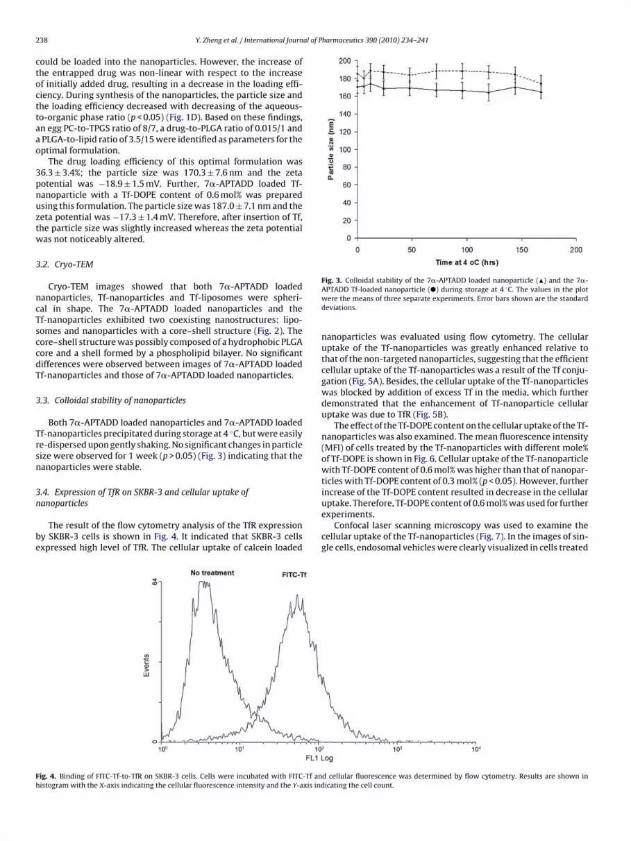
Fig. 3. Colloidal stability of the 7�-APTADD loaded nanoparticle (�) and the 7�-
Fh
38 Y. Zheng et al. / International Journ
ould be loaded into the nanoparticles. However, the increase ofhe entrapped drug was non-linear with respect to the increasef initially added drug, resulting in a decrease in the loading effi-iency. During synthesis of the nanoparticles, the particle size andhe loading efficiency decreased with decreasing of the aqueous-o-organic phase ratio (p < 0.05) (Fig. 1D). Based on these findings,n egg PC-to-TPGS ratio of 8/7, a drug-to-PLGA ratio of 0.015/1 andPLGA-to-lipid ratio of 3.5/15 were identified as parameters for theptimal formulation.
The drug loading efficiency of this optimal formulation was6.3 ± 3.4%; the particle size was 170.3 ± 7.6 nm and the zetaotential was −18.9 ± 1.5 mV. Further, 7�-APTADD loaded Tf-anoparticle with a Tf-DOPE content of 0.6 mol% was preparedsing this formulation. The particle size was 187.0 ± 7.1 nm and theeta potential was −17.3 ± 1.4 mV. Therefore, after insertion of Tf,he particle size was slightly increased whereas the zeta potentialas not noticeably altered.
.2. Cryo-TEM
Cryo-TEM images showed that both 7�-APTADD loadedanoparticles, Tf-nanoparticles and Tf-liposomes were spheri-al in shape. The 7�-APTADD loaded nanoparticles and thef-nanoparticles exhibited two coexisting nanostructures: lipo-omes and nanoparticles with a core–shell structure (Fig. 2). Theore–shell structure was possibly composed of a hydrophobic PLGAore and a shell formed by a phospholipid bilayer. No significantifferences were observed between images of 7�-APTADD loadedf-nanoparticles and those of 7�-APTADD loaded nanoparticles.
.3. Colloidal stability of nanoparticles
Both 7�-APTADD loaded nanoparticles and 7�-APTADD loadedf-nanoparticles precipitated during storage at 4 ◦C, but were easilye-dispersed upon gently shaking. No significant changes in particleize were observed for 1 week (p > 0.05) (Fig. 3) indicating that theanoparticles were stable.
.4. Expression of TfR on SKBR-3 and cellular uptake of
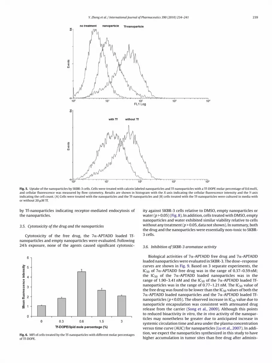
anoparticlesThe result of the flow cytometry analysis of the TfR expressiony SKBR-3 cells is shown in Fig. 4. It indicated that SKBR-3 cellsxpressed high level of TfR. The cellular uptake of calcein loaded
ig. 4. Binding of FITC-Tf-to-TfR on SKBR-3 cells. Cells were incubated with FITC-Tf anistogram with the X-axis indicating the cellular fluorescence intensity and the Y-axis ind
APTADD Tf-loaded nanoparticle (�) during storage at 4 ◦C. The values in the plotwere the means of three separate experiments. Error bars shown are the standarddeviations.
nanoparticles was evaluated using flow cytometry. The cellularuptake of the Tf-nanoparticles was greatly enhanced relative tothat of the non-targeted nanoparticles, suggesting that the efficientcellular uptake of the Tf-nanoparticles was a result of the Tf conju-gation (Fig. 5A). Besides, the cellular uptake of the Tf-nanoparticleswas blocked by addition of excess Tf in the media, which furtherdemonstrated that the enhancement of Tf-nanoparticle cellularuptake was due to TfR (Fig. 5B).
The effect of the Tf-DOPE content on the cellular uptake of the Tf-nanoparticles was also examined. The mean fluorescence intensity(MFI) of cells treated by the Tf-nanoparticles with different mole%of Tf-DOPE is shown in Fig. 6. Cellular uptake of the Tf-nanoparticlewith Tf-DOPE content of 0.6 mol% was higher than that of nanopar-ticles with Tf-DOPE content of 0.3 mol% (p < 0.05). However, furtherincrease of the Tf-DOPE content resulted in decrease in the cellularuptake. Therefore, Tf-DOPE content of 0.6 mol% was used for further
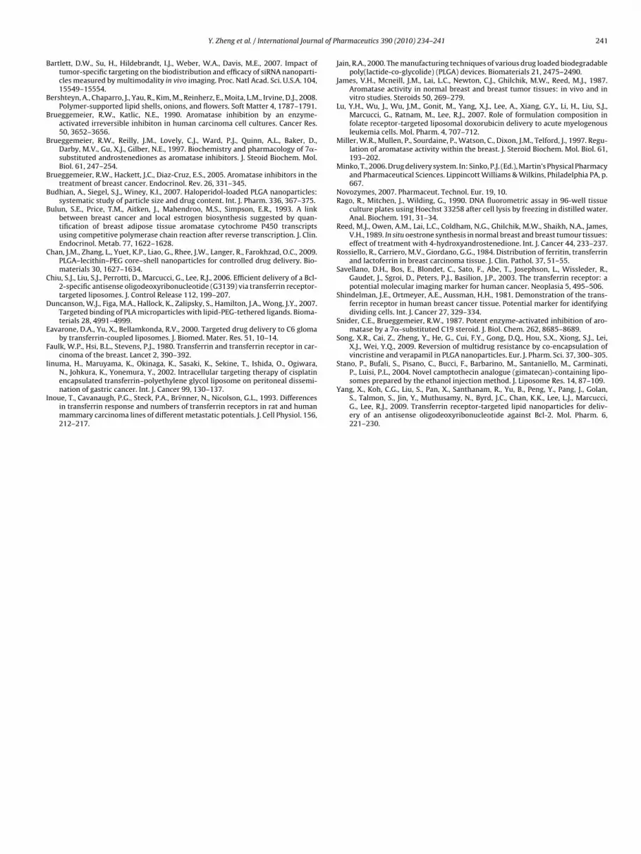
experiments.Confocal laser scanning microscopy was used to examine thecellular uptake of the Tf-nanoparticles (Fig. 7). In the images of sin-gle cells, endosomal vehicles were clearly visualized in cells treated
d cellular fluorescence was determined by flow cytometry. Results are shown inicating the cell count.
Y. Zheng et al. / International Journal of Pharmaceutics 390 (2010) 234–241 239
F labelea histogi anopao
bt
3
n2
Fo
ig. 5. Uptake of the nanoparticles by SKBR-3 cells. Cells were treated with calceinnd cellular fluorescence was measured by flow cytometry. Results are shown inndicating the cell count. (A) Cells were treated with the nanoparticles and the Tf-nr without 20 �M Tf.
y Tf-nanoparticles indicating receptor-mediated endocytosis ofhe nanoparticles.
.5. Cytotoxicity of the drug and the nanoparticles
Cytotoxicity of the free drug, the 7�-APTADD loaded Tf-anoparticles and empty nanoparticles were evaluated. Following4 h exposure, none of the agents caused significant cytotoxic-
ig. 6. MFI of cells treated by the Tf-nanoparticles with different molar percentagesf Tf-DOPE.
d nanoparticles and Tf-nanoparticles with a Tf-DOPE molar percentage of 0.6 mol%,ram with the X-axis indicating the cellular fluorescence intensity and the Y-axisrticles and (B) cells treated with the Tf-nanoparticles were cultured in media with
ity against SKBR-3 cells relative to DMSO, empty nanoparticles orwater (p > 0.05) (Fig. 8). In addition, cells treated with DMSO, emptynanoparticles and water exhibited similar viability relative to cellswithout any treatment (p > 0.05, data not shown). In summary, boththe drug and the nanoparticles were essentially non-toxic to SKBR-3 cells.
3.6. Inhibition of SKBR-3 aromatase activity
Biological activities of 7�-APTADD free drug and 7�-APTADDloaded nanoparticles were evaluated in SKBR-3. The dose–responsecurves are shown in Fig. 9. Based on 3 separate experiments, theIC50 of 7�-APTADD free drug was in the range of 0.37–0.59 nM;the IC50 of the 7�-APTADD loaded nanoparticles was in therange of 1.90–3.41 nM and the IC50 of the 7�-APTADD loaded Tf-nanoparticles was in the range of 0.77–1.21 nM. The IC50 value ofthe free drug was found to be lower than the IC50 values of both the7�-APTADD loaded nanoparticles and the 7�-APTADD loaded Tf-nanoparticles (p < 0.05). The observed increase in IC50 value due tonanoparticle encapsulation was consistent with attenuated drugrelease from the carrier (Song et al., 2009). Although this pointsto reduced bioactivity in vitro, the in vivo activity of the nanopar-
ticles may nonetheless be greater due to anticipated increase insystemic circulation time and area under the plasma concentrationversus time curve (AUC) for nanoparticles (Lu et al., 2007). In addi-tion, we expect the nanoparticles synthesized in this study to havehigher accumulation in tumor sites than free drug after adminis-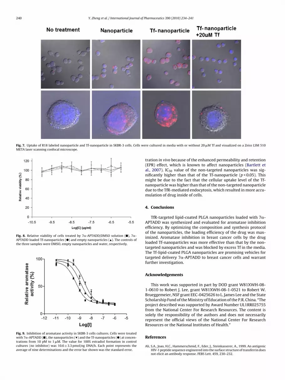
240 Y. Zheng et al. / International Journal of Pharmaceutics 390 (2010) 234–241
Fig. 7. Uptake of R18 labeled nanoparticle and Tf-nanoparticle in SKBR-3 cells. Cells weMETA laser scanning confocal microscope.
Fig. 8. Relative viability of cells treated by 7�-APTADD/DMSO solution (�), 7�-APTADD loaded Tf-nanoparticles (�) and empty nanoparticles (�). The controls ofthe three samples were DMSO, empty nanoparticles and water, respectively.
Fig. 9. Inhibition of aromatase activity in SKBR-3 cells cultures. Cells were treatedwith 7�-APTADD (�), the nanoparticles (�) and the Tf-nanoparticles (�) at concen-trations from 10 pM to 1 �M. The value for 100% estradiol formation in controlcultures (no inhibitor) was 10.6 ± 3.3 pmol/ng DNA/h. Each point represents theaverage of nine determinations and the error bar shown was the standard error.
re cultured in media with or without 20 �M Tf and visualized on a Zeiss LSM 510
tration in vivo because of the enhanced permeability and retention(EPR) effect, which is known to affect nanoparticles (Bartlett etal., 2007). IC50 value of the non-targeted nanoparticles was sig-nificantly higher than that of the Tf-nanoparticle (p < 0.05). Thismight be due to the fact that the cellular uptake level of the Tf-nanoparticle was higher than that of the non-targeted nanoparticledue to the TfR-mediated endocytosis, which resulted in more accu-mulation of drug inside of cells.
4. Conclusions
TfR-targeted lipid-coated PLGA nanoparticles loaded with 7�-APTADD was synthesized and evaluated for aromatase inhibitionefficiency. By optimizing the composition and synthesis protocolof the nanoparticles, the loading efficiency of the drug was max-imized. Aromatase inhibition in breast cancer cells by the drugloaded Tf-nanoparticles was more effective than that by the non-targeted nanoparticles and was blocked by excess Tf in the media.The Tf-lipid-coated PLGA nanoparticles are promising vehicles fortargeted delivery 7�-APTADD to breast cancer cells and warrantfurther investigation.
Acknowledgements
This work was supported in part by DOD grant W81XWH-08-1-0610 to Robert J. Lee, grant W81XWH-08-1-0521 to Robert W.Brueggemeier, NSF grant EEC-0425626 to L. James Lee and the StateScholarship Fund of the Ministry of Education of the P.R. China. “Theproject described was supported by Award Number UL1RR025755from the National Center For Research Resources. The content issolely the responsibility of the authors and does not necessarilyrepresent the official views of the National Center For ResearchResources or the National Institutes of Health.”
References
Ali, S.A., Joao, H.C., Hammerschmid, F., Eder, J., Steinkasserer, A., 1999. An antigenicHIV-1 peptide sequence engineered into the surface structure of transferrin doesnot elicit an antibody response. FEBS Lett. 459, 230–232.
al of P
B
B
B
B
B
B
B
C
C
D
E
F
I
I
Y. Zheng et al. / International Journ
artlett, D.W., Su, H., Hildebrandt, I.J., Weber, W.A., Davis, M.E., 2007. Impact oftumor-specific targeting on the biodistribution and efficacy of siRNA nanoparti-cles measured by multimodality in vivo imaging. Proc. Natl Acad. Sci. U.S.A. 104,15549–15554.
ershteyn, A., Chaparro, J., Yau, R., Kim, M., Reinherz, E., Moita, L.M., Irvine, D.J., 2008.Polymer-supported lipid shells, onions, and flowers. Soft Matter 4, 1787–1791.
rueggemeier, R.W., Katlic, N.E., 1990. Aromatase inhibition by an enzyme-activated irreversible inhibiton in human carcinoma cell cultures. Cancer Res.50, 3652–3656.
rueggemeier, R.W., Reilly, J.M., Lovely, C.J., Ward, P.J., Quinn, A.L., Baker, D.,Darby, M.V., Gu, X.J., Gilber, N.E., 1997. Biochemistry and pharmacology of 7�-substituted androstenediones as aromatase inhibitors. J. Steoid Biochem. Mol.Biol. 61, 247–254.
rueggemeier, R.W., Hackett, J.C., Diaz-Cruz, E.S., 2005. Aromatase inhibitors in thetreatment of breast cancer. Endocrinol. Rev. 26, 331–345.
udhian, A., Siegel, S.J., Winey, K.I., 2007. Haloperidol-loaded PLGA nanoparticles:systematic study of particle size and drug content. Int. J. Pharm. 336, 367–375.
ulun, S.E., Price, T.M., Aitken, J., Mahendroo, M.S., Simpson, E.R., 1993. A linkbetween breast cancer and local estrogen biosynthesis suggested by quan-tification of breast adipose tissue aromatase cytochrome P450 transcriptsusing competitive polymerase chain reaction after reverse transcription. J. Clin.Endocrinol. Metab. 77, 1622–1628.
han, J.M., Zhang, L., Yuet, K.P., Liao, G., Rhee, J.W., Langer, R., Farokhzad, O.C., 2009.PLGA–lecithin–PEG core–shell nanoparticles for controlled drug delivery. Bio-materials 30, 1627–1634.
hiu, S.J., Liu, S.J., Perrotti, D., Marcucci, G., Lee, R.J., 2006. Efficient delivery of a Bcl-2-specific antisense oligodeoxyribonucleotide (G3139) via transferrin receptor-targeted liposomes. J. Control Release 112, 199–207.
uncanson, W.J., Figa, M.A., Hallock, K., Zalipsky, S., Hamilton, J.A., Wong, J.Y., 2007.Targeted binding of PLA microparticles with lipid-PEG-tethered ligands. Bioma-terials 28, 4991–4999.
avarone, D.A., Yu, X., Bellamkonda, R.V., 2000. Targeted drug delivery to C6 glomaby transferrin-coupled liposomes. J. Biomed. Mater. Res. 51, 10–14.
aulk, W.P., Hsi, B.L., Stevens, P.J., 1980. Transferrin and transferrin receptor in car-cinoma of the breast. Lancet 2, 390–392.
inuma, H., Maruyama, K., Okinaga, K., Sasaki, K., Sekine, T., Ishida, O., Ogiwara,N., Johkura, K., Yonemura, Y., 2002. Intracellular targeting therapy of cisplatin
encapsulated transferrin–polyethylene glycol liposome on peritoneal dissemi-nation of gastric cancer. Int. J. Cancer 99, 130–137.noue, T., Cavanaugh, P.G., Steck, P.A., Brv̈nner, N., Nicolson, G.L., 1993. Differencesin transferrin response and numbers of transferrin receptors in rat and humanmammary carcinoma lines of different metastatic potentials. J. Cell Physiol. 156,212–217.
harmaceutics 390 (2010) 234–241 241
Jain, R.A., 2000. The manufacturing techniques of various drug loaded biodegradablepoly(lactide-co-glycolide) (PLGA) devices. Biomaterials 21, 2475–2490.
James, V.H., Mcneill, J.M., Lai, L.C., Newton, C.J., Ghilchik, M.W., Reed, M.J., 1987.Aromatase activity in normal breast and breast tumor tissues: in vivo and invitro studies. Steroids 50, 269–279.
Lu, Y.H., Wu, J., Wu, J.M., Gonit, M., Yang, X.J., Lee, A., Xiang, G.Y., Li, H., Liu, S.J.,Marcucci, G., Ratnam, M., Lee, R.J., 2007. Role of formulation composition infolate receptor-targeted liposomal doxorubicin delivery to acute myelogenousleukemia cells. Mol. Pharm. 4, 707–712.
Miller, W.R., Mullen, P., Sourdaine, P., Watson, C., Dixon, J.M., Telford, J., 1997. Regu-lation of aromatase activity within the breast. J. Steroid Biochem. Mol. Biol. 61,193–202.
Minko, T., 2006. Drug delivery system. In: Sinko, P.J. (Ed.), Martin’s Physical Pharmacyand Pharmaceutical Sciences. Lippincott Williams & Wilkins, Philadelphia PA, p.667.
Novozymes, 2007. Pharmaceut. Technol. Eur. 19, 10.Rago, R., Mitchen, J., Wilding, G., 1990. DNA fluorometric assay in 96-well tissue
culture plates using Hoechst 33258 after cell lysis by freezing in distilled water.Anal. Biochem. 191, 31–34.
Reed, M.J., Owen, A.M., Lai, L.C., Coldham, N.G., Ghilchik, M.W., Shaikh, N.A., James,V.H., 1989. In situ oestrone synthesis in normal breast and breast tumour tissues:effect of treatment with 4-hydroxyandrostenedione. Int. J. Cancer 44, 233–237.
Rossiello, R., Carriero, M.V., Giordano, G.G., 1984. Distribution of ferritin, transferrinand lactoferrin in breast carcinoma tissue. J. Clin. Pathol. 37, 51–55.
Savellano, D.H., Bos, E., Blondet, C., Sato, F., Abe, T., Josephson, L., Wissleder, R.,Gaudet, J., Sgroi, D., Peters, P.J., Basilion, J.P., 2003. The transferrin receptor: apotential molecular imaging marker for human cancer. Neoplasia 5, 495–506.
Shindelman, J.E., Ortmeyer, A.E., Aussman, H.H., 1981. Demonstration of the trans-ferrin receptor in human breast cancer tissue. Potential marker for identifyingdividing cells. Int. J. Cancer 27, 329–334.
Snider, C.E., Brueggemeier, R.W., 1987. Potent enzyme-activated inhibition of aro-matase by a 7�-substituted C19 steroid. J. Biol. Chem. 262, 8685–8689.
Song, X.R., Cai, Z., Zheng, Y., He, G., Cui, F.Y., Gong, D.Q., Hou, S.X., Xiong, S.J., Lei,X.J., Wei, Y.Q., 2009. Reversion of multidrug resistance by co-encapsulation ofvincristine and verapamil in PLGA nanoparticles. Eur. J. Pharm. Sci. 37, 300–305.
Stano, P., Bufali, S., Pisano, C., Bucci, F., Barbarino, M., Santaniello, M., Carminati,P., Luisi, P.L., 2004. Novel camptothecin analogue (gimatecan)-containing lipo-
somes prepared by the ethanol injection method. J. Liposome Res. 14, 87–109.Yang, X., Koh, C.G., Liu, S., Pan, X., Santhanam, R., Yu, B., Peng, Y., Pang, J., Golan,S., Talmon, S., Jin, Y., Muthusamy, N., Byrd, J.C., Chan, K.K., Lee, L.J., Marcucci,G., Lee, R.J., 2009. Transferrin receptor-targeted lipid nanoparticles for deliv-ery of an antisense oligodeoxyribonucleotide against Bcl-2. Mol. Pharm. 6,221–230.