cytochrome p450 aromatase expression in human seminoma
TRANSCRIPT

BioMed Central
Reproductive Biology and Endocrinology
ss
Open AcceResearchCytochrome P450 aromatase expression in human seminomaVittoria Rago1, Francesco Romeo2, Saveria Aquila3, Daniela Montanaro2, Sebastiano Andò1 and Amalia Carpino*1Address: 1Cell Biology Department, Faculty of Pharmacy, University of Calabria, Italy, 2Pathologic Anatomy Unit, Annunziata Hospital, Cosenza, Italy and 3Pharmaco-Biology Departments, Faculty of Pharmacy, University of Calabria, Italy
Email: Vittoria Rago - [email protected]; Francesco Romeo - [email protected]; Saveria Aquila - [email protected]; Daniela Montanaro - [email protected]; Sebastiano Andò - [email protected]; Amalia Carpino* - [email protected]
* Corresponding author
AbstractBackground: The enzyme cytochrome P450 aromatase, catalysing the conversion of androgensinto estrogens, has been detected in normal human testicular cells suggesting a physiological roleof local estrogen biosynthesis on spermatogenesis control. Estrogens, regulating cell growth andapoptosis, can also be involved in tumorigenesis process, but the possible link between estrogensand testicular neoplastic process is, up to now, scarcely known. This study examined aromataseexpression in human seminoma, which is the most common germ cell tumour of the testis.
Methods: The tumour-bearing testes were obtained from 20 patients with classic seminomaundergoing to therapeutic orchidectomy. Paraffin embedded tissues were processed forimmunohistochemistry using a mouse monoclonal antibody generated against human placentalcytochrome P450 arom, as primary antibody, and a biotinylated goat-anti-mouse IgG, as secondaryantibody. Furthermore, Western blot analysis of seminoma extracts was carried out.
Results: Intense P450 arom immunoreactivity was observed in the seminoma cells and Westernblot analysis confirmed the immunodetection. A strong immunostaining was also detected in cellsof intratubular germ cell neoplasia (IGCN), adjacent to seminoma.
Conclusion: The present study demonstrated, for the first time in human, aromatase expressionin neoplastic cells of seminoma suggesting a relation between local estrogen biosynthesis and germcell tumorigenesis. The P450 arom immunolocalization in the cells of IGCN, representing thecommon precursor of most germ cell tumors, seems to support these findings.
BackgroundSomatic and germ cell tumors are typical testicular neo-plasms which can affect young, adult and elderly men. Inthe last years, testis tumor management has improved bynew diagnostic and surgical tools, as well as by innovativetherapy options to preserve fertility against cytotoxic treat-ments [1]. However, pathogenesis of male gonad malig-nancies is often still unknown.
Somatic and germ cells of normal human testis haverevealed the expression of estrogens receptor-β and /oraromatase, which is the microsomial enzyme catalysingthe conversion of androgens into estrogens [2-8]. There-fore, testicular cells are considered targets and in situ bio-synthesis sites of estrogens, suggesting a physiological roleof these hormones in steroidogenesis control and spermmaturation.
Published: 22 December 2005
Reproductive Biology and Endocrinology 2005, 3:72 doi:10.1186/1477-7827-3-72
Received: 18 November 2005Accepted: 22 December 2005
This article is available from: http://www.rbej.com/content/3/1/72
© 2005 Rago et al; licensee BioMed Central Ltd. This is an Open Access article distributed under the terms of the Creative Commons Attribution License (http://creativecommons.org/licenses/by/2.0), which permits unrestricted use, distribution, and reproduction in any medium, provided the original work is properly cited.
Page 1 of 6(page number not for citation purposes)
Reproductive Biology and Endocrinology 2005, 3:72 http://www.rbej.com/content/3/1/72
Estrogens are also known to regulate cell growth andapoptosis, therefore they can be involved in tumorigene-sis process [9,10] but, up to now, their possible link withtesticular neoplastic process is scarcely known.
Germ cell tumors, comprising seminoma and non-semi-noma, are the most common malignancies amonghuman male aged 15–40 years. Aim of this work was toinvestigate aromatase expression in seminoma which rep-resents approximatively the 40% of these testicular neo-plasms.
MethodsSamples and histopathological studiesThe archival cases (collected during the last 3 years) wereprovided by the Pathologic Anatomy Unit (AnnunziataHospital). Tumoral testicular tissues were obtained from20 patients (ages from 27 to 38 years) with unilateral clas-sic seminoma undergoing to therapeutic orchidectomy.Normal testicular tissues were obtained from 2 patientsshowing testes with a granulomatous lesion. The ethicalcommittee members of the University of Calabriaapproved the investigation programme.
Tissues were fixed in formalin (4%), dehydrated with aseries of ethanol solutions and paraffin-embedded. Paraf-fin sections, 5 µm thick, were mounted on slides pre-coated with polylysine, deparaffinized and rehydrated (7–8 serial sections for each sample). These sections wereused for morphological and immunohistochemical anal-yses.
Histopathological studies were carried out by Haematox-ylin-Eosin staining. Seminoma samples were also investi-gated by placental-like alkaline phosphatase (PLAP)immunohistochemistry (monoclonal mouse anti humanPLAP, clone 8A9, DAKO-Cytomation, Milan, Italy)
Furthermore, 50 µm thick [11] paraffin serial sections ofseminoma specimens were cut for protein extraction andmounted on glass slides.
P450 arom immunohistochemistryImmunohistochemistry was performed after heat-medi-ated antigen retrieval [12]. Hydrogen peroxide (3% in dis-tillate water) was used, for 30 minutes, to inhibitendogenous peroxidase activity while normal goat serum(10%) was utilised, for 30 minutes, to block the non-spe-cific binding sites. P450 arom immunodetection was car-ried out using a monoclonal mouse anti-humancytochrome P450 aromatase (MCA 2077, Serotec,Oxford,UK) (1:50 in TBS) at 4°C overnight. Then, a bioti-nylated horse-anti-mouse IgG (Vector Laboratories, CA,USA) was applied (1:600 in TBS) for 1 hour at RT, fol-lowed by the avidin-biotin-horseradish peroxidase com-
plex (ABC/HRP) (Vector, Laboratories, CA, USA).Immunoreactivity was visualized by using the diami-nobenzidine chromogen (DAB) (Zymed Laboratories,CA, USA) and, finally, sections were counterstained withhaematoxylin. The primary antibody was replaced by nor-mal rabbit serum in negative control, while the absorp-tion control has utilised a primary antibody preabsorbedwith an excess (5 nmol/ml) of purified human placentalaromatase protein (4°C for 48 hours). Rat ovary sectionswere used as positive control. 6–7 serial sections wereprocessed for each sample.
Protein extractionProtein extraction from formalin-fixed paraffin-embed-ded sections has been carried out according to Ikeda [11].Briefly, 50 µm testis sections from seminoma regions weredeparaffinized in xylene, reydrated in graded ethanol,immersed in distilled water, and air dried. Then, the neo-plastic area was recovered from the glass slides, further itwas cut into small pieces and placed in Eppendorf tubes.Two hundred µl of RIPA buffer, pH 7,6 (1 M NaH2PO4, 10mM Na2HPO4,154 mM NaCl, 1% Triton X-100, 12 mMC24H39O4Na, 0.2% NaN3, 0.95 mM NaF, 2 mM PMSF, 50mg/ml aprotinin, 50 mM leupeptin) (Sigma Chemical,Saint Louis, MO,USA) containing 0.2% SDS, was added toeach tube and the contents were incubated at 100°C for20 minutes, followed by incubation at 60°C for 2 hours.After incubation, tissue lysates were centrifuged at 15.000× g for 20 minutes at 4°C and the supernatants werestored at -80°C until biochemical analysis.
Western blot analysisTissue lysates were quantified using Bradford proteinassay reagent [13]. Equal amounts of protein (50 µg) wereboiled for 5 minutes, separated under denaturing condi-tions by SDS-PAGE on 10% polyacrylamide Tris-glycinegels and electroblotted to nitrocellulose membrane. Non-specific sites were blocked with 5% non fat dry milk in0.2% Tween-20 in Tris-buffered saline (TBS-T) for 1 hourat room temperature and then probed with a rabbit poly-clonal antiserum generated against human P450 aro-matase(Hauptman-Woodward Medical ResearchInstitute, Buffalo, NY, USA) (1:750). After extensive wash-ings (three times for 15 minutes each time in TBS-T), agoat-antirabbit (1:7000) horseradish peroxidase-conju-gated antibody (Vector Laboratories, CA, USA) was addedfor 1 hour at 22°C. Blots were again washed three timesfor 15 minutes in TBS-T and the bound of secondary anti-body was located with the ECL Plus Western blottingdetection system according to the manufacturer's instruc-tions. Each membrane was exposed to the film for 2 min-utes. P450 arom protein, isolated from human placenta,was used as positive control. Negative control was pre-pared using tissue lysate, where P450 arom was previouslyremoved by preincubation with P450 arom antibody (1
Page 2 of 6(page number not for citation purposes)
Reproductive Biology and Endocrinology 2005, 3:72 http://www.rbej.com/content/3/1/72
hour at room temperature) and subsequently immuno-precipitated with protein A/G – agarose.
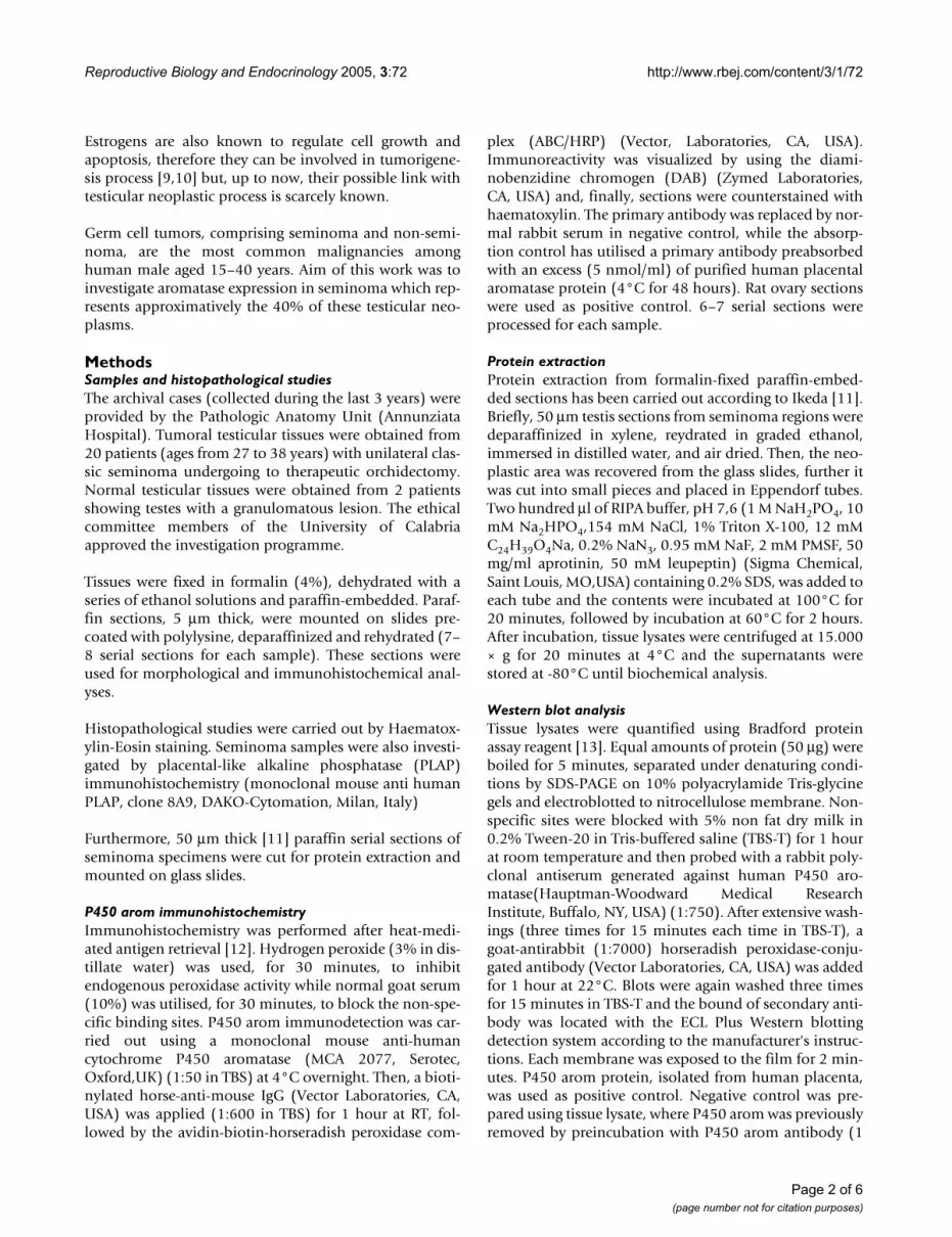
ResultsHistopathological studyTumoral regions of all specimens have revealed the pres-ence of classic seminoma with a uniform population oflarge neoplastic cells and extensive leukocytic infiltration(Fig 1A,B). All seminoma specimens showed tumor stageI.
The region adjacent to seminoma (60% of samples) hasshown the presence of abnormal seminiferous tubules
with decreased tubular diameters and lacking sperma-togenesis, which were identified as intratubular germ cellneoplasia (IGCN). These intratubular lesions were charac-terised by the basal proliferation of undifferentiated atyp-ical enlarged germ cells, with big nuclei, and by thepresence of Sertoli cells in luminal displacement.
The IGCN diagnosis was confirmed by PLAP immunohis-tochemistry. In fact, cytoplasmic and membranous darkstaining have revealed the PLAP immunopositivity in themalignant cells aligned along the basal portion of sem-iniferous tubules (Fig 2B), indicating their germ cell ori-gin.
Morphology and P450 arom immunoreactivity of tumoral region in human testis with seminomaFigure 1Morphology and P450 arom immunoreactivity of tumoral region in human testis with seminoma. A-B: Haematoxylin-eosin staining. C-D: Strong P450 arom immunoreactivity in cytoplasm of neoplastic cells (Nc) and unstained lymphocytes (L). Insert: absorption control. Scale bars: A, 20 µm; B-C, 12.5 µm; D, 5 µm.
Page 3 of 6(page number not for citation purposes)

Reproductive Biology and Endocrinology 2005, 3:72 http://www.rbej.com/content/3/1/72
Furthermore, normal testes have displayed typical normalseminiferous tubules, with active spermatogenesis.
P450 arom immunolocalizationP450 arom immunoreactivity in testicular cells wasdetected as cytoplasm staining while the nuclei wereimmunonegative.
In tumoral regions, neoplastic cell cords have revealed astrong immunoreactivity, while the surrounding abun-dant lymphocytes and connective cells were all immu-nonegative. The immunostaining pattern was similar inall the 20 examined samples and pictures in the figuresshow representative specimens (Fig 1C,D). In all the 12testis samples with intratubular germ cell neoplasia, anintense immunostaining has been observed in the cells ofIGCN, adjacent to seminoma, either in the abnormalgerm cells or in Sertoli cells (Fig 2A). Leydig cells, clus-tered in interstitial tissue among IGCN, were stronglyimmunoreactive, representing an internal positive control(data not shown). Furthermore, the 2 normal testes haverevealed an intense immunoreactivity in Leydig cells,while tubular compartments were all completely immu-nonegative (Fig 2C).
Negative (data not shown) and absorption controls(insert of figure 1C) were all immunonegative. Asexpected, ovarian tissue has shown a strong immunoreac-tivity in luteal cells (positive control) (Fig 2D).
Western blot analysisLysates of all the 20 formalin-fixed paraffin-embeddedtestis sections from seminoma tumoral regions have beensubmitted to Western blot analysis. All specimens haveshown a single band corresponding to a molecular massof 55 kDa, which is the correct size of human P450 aromprotein (3 representative samples are shown in Fig 3). Theband migrated at the same mobility as purified humanplacental P450 arom protein which was used as positivecontrol, while the immunoreactive P450 arom band waslacking in the negative control (Fig 3)
DiscussionThe present study has demonstrated, for the first time,aromatase expression in neoplastic cells of human semi-noma, which is the most common testicular germ celltumor of men in reproductive age.
Aromatase detection in Leydig cells, Sertoli cells and elon-gated spermatids of normal human testis has suggestedthe involvement of local estrogen production in gonadalphysiology [3,4,7]. At the same time, the mitogenic estro-gen action could be related to the testicular tumor etiol-ogy, as hypothesized for human breast, ovarian,endometrial and epathic malignancies [14,15].
Epidemiological studies have indicated a higher incidenceof testicular cancers after prenatal estrogen exposure [16-19]. Furthermore, a link between exposure to estrogens/estrogen mimics and testicular cancer risk has beenhypothesized [20,21].
Few studies have investigated aromatase in testiculartumors. Among somatic testis tumors, a large cell calcify-
Immunoblot of aromatase from seminoma extractsFigure 3Immunoblot of aromatase from seminoma extracts. Lane 1: purified P450 arom protein from human placenta (positive control). Lane 2: negative control prepared as described in Material and Methods. Lanes 3, 4, 5: 55 kDa P450 arom immunoreactive bands from lysates of three different semi-noma extracts. Numbers on the left correspond to molecular weights of marker proteins.
P450 arom immunoreactivity in testicular region adjacent to seminoma and in controls A: Intense aromatase immunos-taining in IGCN cellsFigure 2P450 arom immunoreactivity in testicular region adjacent to seminoma and in controls A: Intense aromatase immunos-taining in IGCN cells. B: Placental-like alkaline phosphatase staining of IGCN basal cells C: Strong aromatase immunore-activity of interstitial Leydig cells in normal testis (Lc) D: Intense immunostaining of luteal cells (Luc) in ovarian tissue Scale bars: A-B, 8 µm; C,12.5 µm; D, 5 µm.
Page 4 of 6(page number not for citation purposes)

Reproductive Biology and Endocrinology 2005, 3:72 http://www.rbej.com/content/3/1/72
ing Sertoli cell tumor [22] and Sertoli cell tumors of Peutz-Jeghers syndrome have shown an enhanced expression ofthis enzyme [23,24]. Furthermore, the single Nakazumi'sreport on testicular germ cell tumors has not detected aro-matase expression in neoplastic cells of seminomas andnon-seminomas but it revealed the enzyme presence onlyin stromal cells of non seminomas [25].
The present study has shown aromatase immunolocaliza-tion in seminoma cells and Western blot analysis has con-firmed this result. Our data disagree with Nakazumi'sreports, but probably, the recent antibodies, used in ourinvestigation, have improved aromatase immunodetec-tion. In addition, our findings agree to a very recent workdemonstrating the aromatase expression in human testic-ular seminoma cell line, JKT-1 [26].
Furthermore, a strong P450 arom immunoreactivity hasbeen observed inside the abnormal tubules of IGCN,which represents the common precursor of most germ celltumours. Invasion of the tubular wall by malignant germcells or intratubular proliferation of tumour cells are thetwo theories proposed to understand the switch fromIGCN to testicular neoplasia [27,28]. However, to date,the mechanisms of invasive tumour development frompre- invasive lesion are still to be elucidated. P450 arompresence in malignant germ cells and in Sertoli cells ofIGCN is a new finding which supports aromatase expres-sion during seminoma development and suggests a possi-ble role of local estrogen biosynthesis in tumorigenesisprocess. The aromatase absence in germ and Sertoli cellsinside seminiferous tubules of normal testes supports thishypothesis. Concerning normal testes, aromatase detec-tion in Leydig cells agrees with previous reports [3,7], butits absence in seminiferous tubule cells could disagreewith Turner's paper [7] indicating a "possible" P450 aromexpression in elongated spermatids and a "faint immuno-positive reaction in Sertoli cells". However, the sameauthors claimed the immunostaining "less convincinglyspecific in human" than in marmoset and rat.
ConclusionFinally, the present study is the first report of aromataseexpression in human testicular germ cells after malignanttransformation, indicating that estrogens, from in situaromatization, could act as autocrine mitogenic factorspromoting neoplastic growth. These findings could repre-sent a cue for further studies on estrogen involvement intesticular germ cell tumorigenesis and new pharmacolog-ical treatment.
This work was supported by "Ministero dell'Universitàedella Ricerca Scientifica e Tecnologica" (Murst 60%) andby PRIN 2004 prot.n00067227.
Authors' contributionsCA: the author responsible for conception, design, analy-sis and interpretation of data as well as of drafting manu-script.
RV: the author responsible for performing the immuno-histochemical expriments and participating in the analy-sis and interpretation of data.
RF: the author responsible for histoplathological diagno-sis and sample collection
AS and MD: the authors responsible for performing West-ern blot analysis and protein extraction as well as for par-ticipating to the interpretation of data.
ANS: the author responsible for a critical revision of themanuscript.
AcknowledgementsThe authors thank prof. Antonietta Martire for the English reviewing of this manuscript.
References1. Schrader M, Muller M, Straub B, Miller K: Testicular sperm extrac-
tion in azoospermic patients with gonadal germ cell tumorsprior to chemotherapy-a new therapy option. Asian J Androl2002, 4:9-15.
2. Simpson ER, Mahendroo MS, Means GD, Kilgore MW, HinshelwoodMM, Graham-Lorence S, Amarneh B, It Y, Fisher CR, Dodson MM,Mendelson CR, Bulun SE: Aromatase cytochrome P450, theenzyme responsible for estrogen biosynthesis. Endocr Rev1994, 15:342-355.
3. Inkster S, Yue W, Brodie AM: Human testicular aromatase:Immunocytochemical and biochemical studies. J Clin Endocri-nol Metab 1995, 80:1941-1947.
4. Akingbemi BT: Estrogen regulation of testicular function.Reprod Biol Endocrinol 2005, 3:51.
5. Makinen S, Makela S, Weihua Z, Warner M, Roseulund B, Salmi S,Hovatta O, Gustafsson JK: Localization of oestrogen receptorsα and β in human testis. Mol Hum Reprod 2001, 7:497-503.
6. Saunders PTK, Millar MR, Macpherson S, Irvine DS, Groome NP,Evans LR, Sharpe RM, Scobie GA: ERβ1 and ERβ2 splice variant(ERβcx/β2) are expressed in distinct cell populations in theadult human testis. J Clin Endocrinol Metab 2002, 87:2706-2715.
7. Turner KJ, Macpherson S, Millar MR, McNeilly AS, Williams K, Cran-field M, Groome NP, Sharpe RM, Fraser HM, Saunders PTK: Devel-opment and validation of a new monoclonal antibody tomammalian aromatase. J Endocrinol 2002, 172:21-30.
8. Aschim EL, Saether T, Wiger R, Grotmol T, Haugen TB: Differentialdistribution of splice variants of estrogen receptor beta inhuman testicular cells suggests specific functions in sperma-togenesis. J Steroid Biochem Mol Biol 2004, 92:97-106.
9. Blobel GA, Orkin SH: Estrogen induced apoptosis by inhibitionof the erythroid transcription factor GATA-1. Mol Cell Biol1996, 16:1687-1694.
10. Mabuchi S, Ohmichi M, Kimura A, Nishio Y, Arimoto-Ishida E, Yada-Hashimoto N, Tasaka K, Murata Y: Estrogen inhibits paclitaxelinduced apoptosis via the phosphorylation of apoptosis signalregulating kinase 1 in human ovary cancer cell lines. Endo-crinology 2004, 145:49-58.
11. Ikeda K, Monden T, Kanoh T, Tsujie M, Izawa H, Haba A, Ohnishi T,Sekimoto M, Tomita N, Shiozaki H, Monden M: Extraction andanalysis of diagnostically useful proteins from formalin-fixed,paraffin-embedded tissue sections. J Histochem Cytochem 1998,4:397-403.
12. Shi SR, Key ME, Kalra KL: Antigen retrieval in formalin fixed,paraffin-embedded tissues: an enhancement method for
Page 5 of 6(page number not for citation purposes)

Reproductive Biology and Endocrinology 2005, 3:72 http://www.rbej.com/content/3/1/72
Publish with BioMed Central and every scientist can read your work free of charge
"BioMed Central will be the most significant development for disseminating the results of biomedical research in our lifetime."
Sir Paul Nurse, Cancer Research UK
Your research papers will be:
available free of charge to the entire biomedical community
peer reviewed and published immediately upon acceptance
cited in PubMed and archived on PubMed Central
yours — you keep the copyright
Submit your manuscript here:http://www.biomedcentral.com/info/publishing_adv.asp
BioMedcentral
immunocytochemical staining based on microwave ovenheating of tissue section. J Histochem Cytochem 1991, 39:741-748.
13. Bradford MM: A rapid and sensitive method for the quantiza-tion of microgram quantities of protein utilizing the princi-ple of protein-dye binding. Analytical Biochemistry 1976,72:248-254.
14. Sasano H, Harada N: Intratumoral aromatase in human breast,endometrial and ovarian malignancies. Endocr Rev 1998,19:593-607.
15. Harada N, Ota H, Yoshimura N, Katsuyama T, Takagi Y: Localizedaberrant expression of cytochrome P450 aromatase in pri-mary and metastatic malignant tumors of human liver. J ClinEndocrinol Metab 1998, 83:697-702.
16. Depue RH, Pike MC, Henderson BE: Estrogen exposure duringgestation and risk of testicular cancer. J Natl Cancer Inst 1983,71:1151-1155.
17. Weir HK, Marrett LD, Kreiger N, Darlington GA, Sugar L: Pre-nataland peri-natal exposure and risk of testicular germ cell can-cer. Int J Cancer 2000, 87:438-443.
18. Strohsnitter WC, Noller KL, Hoover RN, Robboy SJ, Palmer JR,Titus-Ernstoff L, Kaufman RH, Adam E, Herbst AL, Hatch EE: Cancerrisk in men exposed in utero to diethylstilbestrol. J Natl CancerInst 2001, 93:545-551.
19. Dieckmann KP, Endsin G, Pichlmeier U: How valid is the prenatalestrogen excess hypothesis of testicular germ cell cancer? Acase control study on hormone-related factors. Eur Urol 2001,40:677-683.
20. Hardell L, Ohlson CG, Fredrikson M: Occupational exposure topolyvinyl chloride as a risk factor for testicular cancer evalu-ated in a case-control study. Int J Cancer 1999, 82:911-912.
21. Pais V, Leav I, Lau KM, Jiang Z, Ho SM: Estrogen receptor-βexpression in human testicular germ cell tumor. Clin CancerRes 2003, 9:4475-4482.
22. Berensztein E, Belgorosky A, de Davila MTG, Rivarola MA: Testicu-lar steroid biosynthesis in a boy with a large cell calcifyingSertoli cell tumor producing prepubertal gynecomastia. Ster-oids 1995, 60:220-225.
23. Coen P, Kulin H, Ballantine T, Zaino R, Frauenhoffer E, Boal D, InksterS, Brodie A, Santen R: An aromatase-producing sex-cord tumorresulting in prepubertal gynecomastia. New Engl J Med 1991,324:317-321.
24. Bulun SE, Noble LS, Takayama K, Michael MD, Agarwal V, Fisher C,Zhao Y, Hinshelwood MM, Ito Y, Simpson ER: Endocrine disordersassociated with inappropriately high aromatase expression.J Steroid Biochem Mol Biol 1997, 61:133-139.
25. Nakazumi H, Sasano H, Maehara I, Ozaki M, Tezuka F, Orikasa S:Estrogen metabolism and impaired spermatogenesis ingerm cell tumors of the testis. J Clin Endocrinol Metab 1996,81:1289-1295.
26. Roger C, Lambard S, Bouskine A, Mograbi B, Chevallier D, Nebout M,Pointis G, Carreau S, Fenichel P: Estrogen-induced growth inhi-bition of human seminoma cells expressing estrogen recep-tor β and aromatase. J Mol Endocrinol 2005, 35:191-199.
27. Donner J, Kliesch S, Brehm R, Bergmann M: From carcinoma insitu to testicular germ cell tumor. APMIS 2004, 112:79-88.
28. Krag Jacobsen G, Talerman A: Atlas of germ cell tumours.Munksgaard, Copenhagen; 1989.
Page 6 of 6(page number not for citation purposes)