effect of low dose exposure to the herbicide atrazine and its metabolite on cytochrome p450...
TRANSCRIPT
Effect of low dose exposure to the herbicide atrazine and itsmetabolite on cytochrome P450 aromatase and steroidogenicfactor-1 mRNA levels in the brain of premetamorphic bullfrogtadpoles (Rana catesbeiana)
Mark P. Gunderson1,3, Nik Veldhoen1, Rachel C. Skirrow2, Magnus K. Macnab1, WeiDing1, Graham van Aggelen2, and Caren C. Helbing1,41 Department of Biochemistry and Microbiology, P.O. Box 3055, Stn. CSC, University of Victoria,Victoria, British Columbia, V8W 3P6, Canada2 Pacific Environmental Science Centre, Environment Canada, 2645 Dollarton Highway, NorthVancouver, British Columbia, V7H 1V2, Canada
AbstractThe transcriptional regulator steroidogenic factor 1 (SF-1) and the enzyme cytochrome P450aromatase (CYP19) play a central role in modulation of a broad range of tissue-specificdevelopmental processes associated with hormone homeostasis that includes differentiation of thecentral nervous system. SF-1 and CYP19 expression may be targeted by a variety of endocrinedisruptive agents prevalent within the environment. In the present study, we cloned andcharacterized partial sequences for bullfrog (Rana catesbeiana) SF-1 and CYP19 and examinedthe effects of a 48 h exposure to 1 and 100 μg/L of the herbicide atrazine (ATZ) and its majormetabolite desethylatrazine (DEA), as well as 5 ng/L of the estrogenic chemical, 17α-ethynylestradiol (EE2), and 673 ng/L of the thyroid hormone, 3,5, 3′-triiodothyronine (T3), onSF-1 and CYP19 mRNA abundance in the brains of premetamorphic bullfrog tadpoles.Quantitative RT-PCR analysis showed an increase in CYP19 mRNA following a 48 h exposure toEE2 but not T3 while no significant changes in SF-1 transcript levels occurred. We observed astrong positive correlation between CYP19 and SF-1 transcript abundance in the ATZ-exposedanimals which was not evident with DEA- or hormone-exposed tadpoles. Our results areintriguing in light of reported behavioral changes in ATZ-exposed frogs and suggest that furtherresearch is warranted to examine the relationship and role of CYP19 and SF-1 in amphibian braindevelopment.
Keywordsatrazine; desethylatrazine; aromatase; steroidogenic factor 1; endocrine disruption; amphibiandevelopment; pesticides; gene expression; estrogen; thyroid hormone; low dose
4Corresponding author: C.C. Helbing, [email protected], Ph. (250) 721-6146, Fax (250) 721-8855.3Current address: Department of Biology, The College of Idaho, Caldwell ID 83605Publisher's Disclaimer: This is a PDF file of an unedited manuscript that has been accepted for publication. As a service to ourcustomers we are providing this early version of the manuscript. The manuscript will undergo copyediting, typesetting, and review ofthe resulting proof before it is published in its final citable form. Please note that during the production process errors may bediscovered which could affect the content, and all legal disclaimers that apply to the journal pertain.
NIH Public AccessAuthor ManuscriptAquat Toxicol. Author manuscript; available in PMC 2012 March 1.
Published in final edited form as:Aquat Toxicol. 2011 March ; 102(1-2): 31–38. doi:10.1016/j.aquatox.2010.12.019.
NIH
-PA Author Manuscript
NIH
-PA Author Manuscript
NIH
-PA Author Manuscript

IntroductionThe brain functions as an endocrine organ producing hormones that act directly on thecentral nervous system (CNS) as well as on peripheral tissues (Altman, 2004). Sex steroidsalong with other signaling molecules such as thyroid hormones (THs) play significant rolesin differentiation of the CNS and perturbation of these processes can lead to permanentchanges in brain function. Mechanistically, estrogens and THs influence each other leadingto complex regulatory possibilities (Vasudevan et al., 2002). A pivotal enzyme required inestrogen production is cytochrome P450 aromatase (CYP19) which catalyzes conversion oftestosterone to estradiol thereby contributing to a variety of hormone-dependent processesthat include sexual differentiation, behavior, neural development, neuroprotection, andregulation of neurotransmitter receptor function (Hutchison et al., 1997; Li and Shen, 2005;Piferrer and Blazquez, 2005).
The nuclear transcriptional regulatory factor steroidogenic factor-1 (SF-1), also known asadrenal 4 binding protein (Ad4BP), regulates CYP19 expression, and both are expressed inamphibian brain tissue (Mayer et al., 2002; Kuntz et al., 2004; Kloas et al., 2009).Furthermore, potential SF-1 binding sites have been identified in the promoter regions of anumber of genes encoding steroidogenic enzymes in a range of taxa which suggests abroader importance of SF-1 in regulating endocrine processes (Morohashi et al., 1992;Honda et al., 1993; Lynch et al., 1993; Morohashi et al., 1993; Tanaka et al., 1995;Morohashi and Omura, 1996; Sugawara et al., 1997; Stocco et al., 2001). For example, SF-1contributes to the development of the ventromedial hypothalamic nucleus structure (VMH)in rodents and studies in which SF-1 expression was disrupted report impairedgonadotrophs, abnormalities or absence of the VMH, and abnormal expression of VMHmarkers which include NKX2-1 and BDNF neurotrophin (Ikeda et al., 1995; Dellovade etal., 2000; Stallings et al., 2002; Tran et al., 2003; Davis et al., 2004). The VMH region of thebrain is highly conserved across species, plays a critical role in regulating sexual behavior,feeding, energy balance, and cardiovascular function, and contains regions with a high levelof estrogen receptor expression (McClellan et al., 2006).
Thus, the developing brain represents a potential target for endocrine disruption byenvironmental contaminants that interfere with the production or actions of estrogen. Onechemical of concern is the herbicide atrazine (ATZ). Registered for commercial use in theUnited States in 1957, ATZ is extensively used in North America, the majority beingapplied to corn crops in the mid-western United States and southern Ontario, Canada. ATZand its metabolites can be measured in rivers and streams at concentrations that rarelyexceed 20 μg/L, and in reservoirs at concentrations up to 12 μg/L although higherconcentrations have been reported in some instances (Solomon et al., 1996). Mountingevidence of the disruptive effects of ATZ on amphibian development can be found for suchdiverse processes as gonadal differentiation and laryngeal muscle development as well asmaintenance of plasma steroid hormone concentrations (Hayes et al., 2002, 2003). Acommon mechanistic theme for ATZ appears to be potential alteration of CYP19 expressionand function.
Several studies have identified ATZ as an endocrine disruptive agent in frogs with theability to increase estrogen-regulated CYP19 mRNA expression in gonads, althoughcontradictory results have been reported in the literature (Crain et al., 1997; Sanderson et al.,2000; Sanderson et al., 2001; Kazeto et al., 2004; Keller and McClellan-Green, 2004; Coadyet al., 2005; Hecker et al., 2005b; Murphy et al., 2006a; Oka et al., 2008; Kloas et al., 2009).The disparity in ATZ effects could be due to different species or strains examined, thedevelopmental timing of exposure, biological variability of control or reference animals, anddifferences in the chemical concentrations examined. Interestingly, recent studies have
Gunderson et al. Page 2
Aquat Toxicol. Author manuscript; available in PMC 2012 March 1.
NIH
-PA Author Manuscript
NIH
-PA Author Manuscript
NIH
-PA Author Manuscript

shown that the induction of CYP19 by ATZ is dependent upon SF-1 related regulatorypathways (Fan et al., 2007a, 2007b; Suzawa and Ingraham, 2008).
The present study describes the cloning of partial SF-1 and CYP19 cDNA sequences fromthe North American bullfrog, Rana catesbeiana, and investigates whether short-termexposure to ATZ, or its prominent environmental metabolite, desethylatrazine (DEA)(Solomon et al., 1996), alters the steady-state levels of these transcripts in the brain ofpremetamorphic bullfrog tadpoles. The present study sets a critical foundation forunderstanding the transcript responses of two important regulators in the brain and suggeststhat alterations to this organ should be taken into account when considering ATZ exposureeffects.
Materials and MethodsExperimental animals and chemical exposures
The care and treatment of the animals used in this study were in accordance with theguidelines of the Canadian Council on Animal Care. Wild-caught Taylor and Kollros stageVI-VIII bullfrog tadpoles (Rana catesbieana) (Taylor and Kollros, 1946) were obtainedfrom Ward’s Natural Science (St. Catharines, ON, Canada) and housed at the EnvironmentCanada Pacific Environmental Science Centre (PESC; North Vancouver, BC, Canada).Tadpoles were maintained in outdoor tanks containing well water under a natural light cycleat approximately 9°C and were acclimatized for a minimum of 48 h in the environmentalcontrol rooms at 22°C with a light:dark cycle of 16:8 h prior to chemical exposure. ATZ,DEA, EE2 and T3 were purchased from Sigma-Aldrich Canada Ltd. (Oakville, ON). ATZ,DEA, and EE2 stock solutions were all prepared in 100% isopropyl alcohol (IPA;Fisherbrand, Ottawa, ON, Purity 99.9%), to limit the number of solvents used in theexperiments, and chemical analyses were performed to confirm accurate nominal stockconcentrations. Specifically, the nominal stock concentrations were: ATZ: 10,000 mg/L and100 mg/L; DEA: 10,000 mg/L and 100 mg/L; and EE2: 5 mg/L. The confirmed stockconcentrations were ATZ: 10,137 mg/L and 101 mg/L; DEA: 6,400 mg/L and 64 mg/L; andEE2: 3.29 mg/L and we will refer to the nominal concentrations in the manuscript. T3 wasprepared as previously described (Veldhoen et al., 2006) and the biological activity of thischemical was confirmed through measurement of TRβ transcript level induction as waspreviously described (Helbing et al., 2010). All exposures had the same volume of stocksolution applied and contained 0.001% (vol/vol) IPA vehicle control (a 100,000X dilution).Thus the final nominal treatment concentrations were 673 ng/L T3, 50 ng/L EE2, 1 μg/L and100 μg/L DEA, and 1 μg/L and 100 μg/L ATZ.
Two separate exposure experiments were performed. The first experiment was done inDecember 2005 and included IPA, T3, and the two concentrations of DEA. The secondexperiment was done in March 2006 and included IPA, T3, EE2, and the two concentrationsof ATZ. Each treatment group contained 10 animals that were equally divided between tworeplicate 20 gallon glass aquarium tanks containing 50 L of aerated well water. Dissolvedoxygen, temperature, and pH were recorded at the outset of the experiment and attermination. The tadpoles were not fed 24 h prior to treatment as well as throughout the 48 hexposure period. The average tadpole weight was 5.09 ± 0.24 g for experiment 1 and 7.35 ±0.28 g for experiment 2. Tadpoles lost weight over the 2 day exposure period (4.21 ± 0.98%and 5.46 ± 0.60%, experiment 1 and 2, respectively), but this was not significant betweentreatments (p=0.924, n=40 experiment 1; p=0.151, n=50 experiment 2, ANOVA). Theaverage tadpole loading density was 0.63 ± 0.03 g/L. No mortalities were observed in anytreatment.
Gunderson et al. Page 3
Aquat Toxicol. Author manuscript; available in PMC 2012 March 1.
NIH
-PA Author Manuscript
NIH
-PA Author Manuscript
NIH
-PA Author Manuscript

Following the 48 h exposure period, animals were euthanized by immersion in 0.1 % w/vtricaine methylsulfonate (MS-222) (Syndel Laboratories Ltd, Vancouver, BC) buffered with25 mM NaHCO3 and tissue samples were collected. Whole brains were isolated and placedin 1 ml RNAlater (Applied Biosystems Inc., Foster City, CA). Samples were kept at 4°C forone week and then stored at −20°C until RNA was isolated from the tissue according tostandard protocols recommended by the manufacturer.
Total RNA isolation and cDNA synthesisTotal RNA was isolated from whole brain tissue using TRIzol Reagent as outlined by themanufacturer (Invitrogen Canada Inc., Burlington, ON). Each tissue sample wasindividually homogenized in a 1.5 ml Eppendorf Safe-Lock microcentrifuge tube (FisherScientific Ltd., Ottawa, ON) containing a 3 mm tungsten-carbide bead and 300 μl TRIzol.Tissue homogenization was achieved using a Retsch MM301 Mixermill (Fisher ScientificLtd., Ottawa, ON) with 2 × 3 min shaking cycles at 20 Hz. The racks were rotated 180°between each shaking cycle. Total RNA was resuspended in 10 μl of diethyl pyrocarbonate(DEPC)-treated (Sigma-Aldrich Canada) dH20. RNA concentration was measured using aND-1000 Nanodrop (Nanodrop Technologies Inc., Wilmington, DE). Each 20 μl cDNAsynthesis reaction contained 500 ng random hexamer oligonucleotide (GE Healthcare, BaieD’Urfé, PQ), 1 μg total RNA, Superscript II RNase H− reverse transcriptase, and additionalrequired reagents as described by the manufacturer (Invitrogen Canada). cDNA sampleswere diluted 20-fold in PCR-grade dH20 and stored at −20°C prior to QPCR analysis.
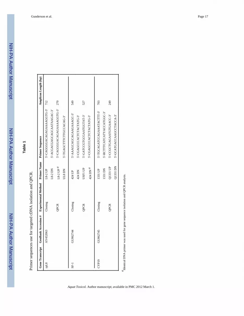
Development of species-specific QPCR primersIsolation of Rana catesbeiana CYP19 and SF-1 cDNA sequences was carried out usingeither non degenerate PCR primers designed against gene-specific cDNA information froma closely related species or degenerate PCR primers developed using ClustalW(http://www.ebi.ac.uk/clustalw/index.html) aligned cDNA sequences available from otheramphibian species deposited in the National Center for Biotechnology Information EntrezNucleotide Database (http://www.ncbi.nlm.nih.gov/sites/entrez?db=Nucleotide) (Table 1).All primers were designed using Primer Premier Version 5.0 software (Premier BiosoftInternational., Palo Alto, CA). Gene-specific information used in SF-1 cloning primerdesign utilized Rana rugosa (GenBank Accession # AB017352) sequence while CYP19degenerate cloning primers were generated from aligned cDNA sequence from Xenopuslaevis (GenBank Accession # AB031278), Rana rugosa (GenBank Accession # AB178482),Pleurodeles waltl (GenBank Accession # AY135485), and Cynops pyrrhogaster (GenBankAccession # AB164064).
All primers were supplied by Integrated DNA Technologies (Coralville, IA, USA). DNAamplification reactions were performed on a MyCycler System (Bio-Rad Laboratories Ltd.,Mississauga, ON) using 2 U PfuTurbo Hotstart DNA polymerase (Agilent Technologies, LaJolla, CA) as per the manufacturer’s recommended protocol. Each 20 μl reaction preparedon ice also contained 2 μl of 20-fold diluted template cDNA and 20 pmol of each primer.The thermocycle program consisted of an initial denaturation step of 95°C (2 min) followedby 46 cycles of 95°C (15 s), 52°C (30 s), and 72°C (45 sec), and a final elongation step of72°C (10 min). Amplified DNA products were evaluated (5 μl) by electrophoresis on a 1.5%agarose gel, visualized with ethidium bromide staining, and DNA amplicons of the correctpredicted size were identified. Each positive PCR reaction (15 μl) was treated with 10 U T4polynucleotide kinase in a 30 μl reaction for one hour at 37°C as per the manufacturer’sprotocol (New England Biolabs Ltd., Pickering, ON). The DNA products were separated byelectrophoresis on a 1.5% agarose gel, visualized with ethidium bromide, and DNAamplicons of the correct predicted size were excised from the gel and placed in an Ultrafree-
Gunderson et al. Page 4
Aquat Toxicol. Author manuscript; available in PMC 2012 March 1.
NIH
-PA Author Manuscript
NIH
-PA Author Manuscript
NIH
-PA Author Manuscript

DA extraction column (Millipore Corp., Bedford, MA). DNA was isolated from the gelmatrix through centrifugation at 13,000 rpm for 10 min in a microcentrifuge at 4°C.
Purified DNA amplicons were ligated into pUC18 plasmid vector pre-cut with SmaIrestriction endonuclease and dephosphorylated with calf intestinal phosphatase using themanufacturer’s recommended protocols (New England Biolabs). Ligation reactions (15 μl)which included 1× ligase buffer, 50 ng of linearized pUC18 plasmid DNA, 4 μl of purifiedDNA amplicon, and 400 U of T4 DNA ligase (New England Biolabs) were incubatedovernight at 16°C prior to transformation with 80 μl chemically competent Escherichia coliDH5α cells. Competent cells mixed with ligated DNA were incubated 1 h on ice followedby 2 min at 42°C and 90 sec on ice. SOC media (250 μl) was added and the cells wereallowed to recover for 1 h at 37°C with shaking. E. coli DH5α cells transformed with ligatedplasmid DNA were selected for by plating 200 μl of each transformation solution onto LBagar plates containing 100 μg/ml ampicillin, 20 mg/ml X-gal, and 0.5 mM isopropyl-beta-d-thiogalactopyranoside (IPTG). Plates were incubated overnight at 37°C.
Individual recombinant colonies were transferred onto a master plate and screened forpositive clones by colony PCR in a 15 μl DNA amplification reaction containing 1 UMaxima Hot Start Taq DNA polymerase (Fermentas) and 1.5 pmol each of M13 forwardand reverse universal sequencing primers. The thermocyle program consisted of an initialdenaturation step of 94°C (5 min) followed by 30 cycles of 94°C (30 sec), 52°C (30 sec),and 72°C (1 min), and a final elongation step of 72°C (10 min). Amplified DNA productswere separated by electrophoresis on a 1.5% agarose gel and visualized with ethidiumbromide. Positive clones were selected and used to inoculate 5 ml of LB broth containing100 μg/ml ampicillin. Cultures were grown at 37°C overnight with shaking at 250 rpm.Plasmid DNA was recovered from the cell cultures using a QiaPrep Spin Miniprep kit(QIAGEN) according to the manufacturer’s instructions. DNA sequencing was performed atthe Centre for Biomedical Research DNA Sequencing Facility (University of Victoria).Rana catesbeiana SF-1 (Genbank Accession # GU062744) and CYP19 (Genbank Accession# GU062745) cDNA was annotated by BLASTn (http://www.ncbi.nlm.nih.gov/BLAST/)DNA alignment analyses and used for further species-specific QPCR primer design (Table1).
QPCR AssayQPCR was performed using a Stratagene MX3005P thermocycler (Stratagene, LaJolla, CA)and 96 well PCR plates and optic flat caps (Ultident Scientific, St. Laurent, PQ) to measurethe steady-state levels of select mRNA transcripts. Each 15 μl reaction contained 5 pmol ofgene-specific primers (Table 1) and additional reagents as described previously (Crump etal., 2002). The thermocycle protocol included 9 min denaturation at 95°C followed by 40cycles of 15 sec at 95°C, 30 sec annealing, and 45 sec elongation at 72°C. For the differentmRNA targets, annealing temperatures included 58°C (SF-1), 62°C (CYP19), and 55°C(rpL8). Samples were run in quadruplicate and controls included a no cDNA and an inter-run cDNA standard for each QPCR run. Inter-run QPCR variation was 3.5 ± 1.6%. QPCRdata was analyzed using the comparative Ct method (ΔΔCt) (Applied Biosystems, 1997)with rpL8 as an invariant input normalizer (Ct= 18.94 ± 0.068). Expression profiles of therpL8 transcript normalizer were used to aid in the identification of poor quality sampleswhich resulted in the removal of 2 out of the total of 90 brain samples from the analysis. Inaddition, the amplified DNA signals for all QPCR reactions were evaluated for specificitybased upon their thermodenaturation profiles. Data that failed to produce a profile indicativeof gene target-specific detection were removed (n=3 and 4 for CYP19 and SF-1,respectively). The remaining normalized data were then expressed as mean fold differencefrom the IPA control.
Gunderson et al. Page 5
Aquat Toxicol. Author manuscript; available in PMC 2012 March 1.
NIH
-PA Author Manuscript
NIH
-PA Author Manuscript
NIH
-PA Author Manuscript

Statistical analysesAll statistical analyses were conducted using PASW18 (SPSS, Chicago, IL, USA). Percentchanges in weights over the 2 day period were analysed using ANOVA followed by theTukey posthoc test. Mean fold difference mRNA abundance data for CYP19 and SF-1violated the assumptions of parametric analysis using the Shapiro-Wilk test for normalityand the Levine’s test for homogeneity of variance. Therefore, Kruskal-Wallis (KW) test wasperformed and pair-wise comparisons were made using a Mann-Whitney U (MWU)statistic. The two exposure experiments are shown and analyzed separately. There was nodifference in CYP19 or SF-1 transcript levels between IPA controls in the two experiments(p=0.210 and 0.634, respectively MWU). Nor was there a difference in these transcriptsbetween the T3-treated groups in the two experiments (p=0.077 and 0.617, respectively,MWU). For the correlation analysis, data from both experiments were combined and thecorrelations between CYP19 and SF-1 transcript levels were determined using Spearman’srho.
ResultsThe isolated partial bullfrog CYP19 cDNA sequence exhibited greater than 90% putativeamino acid sequence similarity across the anuran species included in the alignment (Ranapipiens, Rana rugosa, Xenopus laevis, and Xenopus tropicalis), approximately 89%similarity with two salamanders (caudates) (Pluerodeles waltl and Hynobius retardus), andapproximately 81% similarity with two fish species that were included in the alignment(Oryzias latipes and Rutilus rutilus) (Figure 1). At both DNA sequence and protein encodinglevels, bullfrog CYP19 is highly conserved compared with the species sampled for theanalysis (Table 2).
Brain and gonadal specific isoforms of CYP19 have been identified in fish, salamander, andbirds and it is plausible that multiple isoforms exist in bullfrog (Tchoudakova and Callard,1998; Ramachandran et al., 1999; Kishida and Callard, 2001; Kuntz et al., 2004). Tissue-specific isoforms from two fish species, Orsias latipes (Medaka) and Rutilus rutilus(Roach), were included in the alignment analysis to examine whether the sequence weidentified in bullfrog displayed isoform-specific characteristics (Figure 1). Based on thededuced 221 amino acid bullfrog sequence, a few notable differences exist when comparedto the fish brain and gonadal isoforms of CYP19 (refer to amino acids 5, 66, and 198 inRana catesbeiana in Figure 1 indicated by the asterisks), but there is not enough informationto predict whether we have isolated a brain or gonadal specific isoform or whether multipleisoforms exist in bullfrogs. Brain and gonad specific isoforms have been described in thesalamander Pleurodeles waltl, although the mRNA sequences for the two isoforms differonly in the length of the 5′-untranslated region (Kuntz et al., 2004) for which we have nosequence information in bullfrog for comparison.
The partial bullfrog SF-1 sequence presented in this study exhibited 95.9% amino acidsequence similarity with Rana rugosa (wrinkled frog), ~91% similarity with Pseudacrisregilla (Pacific tree frog) and Xenopus laevis, and 79.4% similarity with the salamanderPleurodeles waltl (Spanish ribbed newt) (Figure 2). At the protein level, bullfrog SF-1 ismore closely related to frog (Rana rugosa), chicken (Gallus gallus), and turtle (Trachemysscripta) (>80%) than to mammalian species Homo sapiens (62.4%) and Mus musculus(63.5%) or to fish (62.2% Danio rerio) (Table 2).
Animals were treated with T3 or EE2 for 48 h in this study so that we could also examinewhether these hormones act as regulators of SF-1 and CYP19 in the brain ofpremetamorphic Rana catesbeiana tadpoles. Treatment with 673 ng/L (1 nM) T3, a quantityknown to affect several transcripts in the brain (Helbing et al., 2007), had no effect on either
Gunderson et al. Page 6
Aquat Toxicol. Author manuscript; available in PMC 2012 March 1.
NIH
-PA Author Manuscript
NIH
-PA Author Manuscript
NIH
-PA Author Manuscript
transcript in both experiments (experiment 1: p=0.191 MWU, CYP19; p=1.000 MWU,SF-1; experiment 2: p=0.566 MWU, CYP19; p=1.000 MWU, SF-1) (Figure 3). EE2treatment (50 ng/L) increased CYP19 expression 2-fold (p = 0.007 MWU) but had no effecton SF-1 expression (p = 0.847 MWU) (Figure 3).
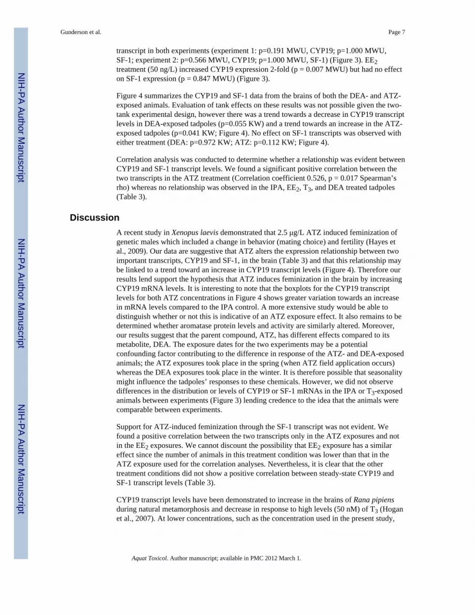
Figure 4 summarizes the CYP19 and SF-1 data from the brains of both the DEA- and ATZ-exposed animals. Evaluation of tank effects on these results was not possible given the two-tank experimental design, however there was a trend towards a decrease in CYP19 transcriptlevels in DEA-exposed tadpoles (p=0.055 KW) and a trend towards an increase in the ATZ-exposed tadpoles (p=0.041 KW; Figure 4). No effect on SF-1 transcripts was observed witheither treatment (DEA: p=0.972 KW; ATZ: p=0.112 KW; Figure 4).
Correlation analysis was conducted to determine whether a relationship was evident betweenCYP19 and SF-1 transcript levels. We found a significant positive correlation between thetwo transcripts in the ATZ treatment (Correlation coefficient 0.526, p = 0.017 Spearman’srho) whereas no relationship was observed in the IPA, EE2, T3, and DEA treated tadpoles(Table 3).
DiscussionA recent study in Xenopus laevis demonstrated that 2.5 μg/L ATZ induced feminization ofgenetic males which included a change in behavior (mating choice) and fertility (Hayes etal., 2009). Our data are suggestive that ATZ alters the expression relationship between twoimportant transcripts, CYP19 and SF-1, in the brain (Table 3) and that this relationship maybe linked to a trend toward an increase in CYP19 transcript levels (Figure 4). Therefore ourresults lend support the hypothesis that ATZ induces feminization in the brain by increasingCYP19 mRNA levels. It is interesting to note that the boxplots for the CYP19 transcriptlevels for both ATZ concentrations in Figure 4 shows greater variation towards an increasein mRNA levels compared to the IPA control. A more extensive study would be able todistinguish whether or not this is indicative of an ATZ exposure effect. It also remains to bedetermined whether aromatase protein levels and activity are similarly altered. Moreover,our results suggest that the parent compound, ATZ, has different effects compared to itsmetabolite, DEA. The exposure dates for the two experiments may be a potentialconfounding factor contributing to the difference in response of the ATZ- and DEA-exposedanimals; the ATZ exposures took place in the spring (when ATZ field application occurs)whereas the DEA exposures took place in the winter. It is therefore possible that seasonalitymight influence the tadpoles’ responses to these chemicals. However, we did not observedifferences in the distribution or levels of CYP19 or SF-1 mRNAs in the IPA or T3-exposedanimals between experiments (Figure 3) lending credence to the idea that the animals werecomparable between experiments.
Support for ATZ-induced feminization through the SF-1 transcript was not evident. Wefound a positive correlation between the two transcripts only in the ATZ exposures and notin the EE2 exposures. We cannot discount the possibility that EE2 exposure has a similareffect since the number of animals in this treatment condition was lower than that in theATZ exposure used for the correlation analyses. Nevertheless, it is clear that the othertreatment conditions did not show a positive correlation between steady-state CYP19 andSF-1 transcript levels (Table 3).
CYP19 transcript levels have been demonstrated to increase in the brains of Rana pipiensduring natural metamorphosis and decrease in response to high levels (50 nM) of T3 (Hoganet al., 2007). At lower concentrations, such as the concentration used in the present study,
Gunderson et al. Page 7
Aquat Toxicol. Author manuscript; available in PMC 2012 March 1.
NIH
-PA Author Manuscript
NIH
-PA Author Manuscript
NIH
-PA Author Manuscript

Hogan et al (2007) did not observe a change in CYP19 transcript abundance which isconsistent with our findings in Rana catesbeiana.
Estrogens regulate CYP19 expression in teleost fish (brain isoform but not gonadal isoform)(Kishida and Callard, 2001; Kazeto and Trant, 2005; Menuet et al., 2005; Pellegrini et al.,2005; Sawyer et al., 2006), mammals (Luo and Wiltbank, 2006), and the salamanderPleurodeles waltl gonad at specific developmental stages (Kuntz et al., 2006), and theinduction of CYP19 is used as a neural marker for estrogenic effects in fish (Sawyer et al.,2006). The estrogen-mediated response of CYP19 varies within different tissues with littlechange in testicular activity (Hecker et al., 2005a) and decreased activity in the ovary ofXenopus laevis exposed to estradiol (Coady et al., 2005). An estrogen-insensitive form ofCYP19 (P450AromA) has also been identified in zebrafish (Kishida and Callard, 2001;Sawyer et al., 2006). Furthermore, estrogen has been demonstrated to regulate SF-1 insalamanders and turtles (Fleming and Crews, 2001; Kuntz et al., 2006). While we found thatEE2 exposure increased CYP19 transcript levels in the Rana catesbeiana tadpole brainwithin 48 h, we found no evidence to suggest that it modulates the expression of the SF-1sequence identified in the present study.
One potential pathway by which ATZ could increase CYP19 (and SF-1 - although notobserved in this study) transcript levels in brain tissues is through alterations in the cyclicAMP mediated pathway. Gonadotropins (8-br-cAMP + FSH), forskolin, and 8-br-cAMPalone increase SF-1 mRNA in vitro in mouse adrenal tumor cells and rat granulosa cells(Shapiro et al., 1996; Lehmann et al., 2005) and cAMP is believed to stimulate thetranscription of human aromatase in the ovary by increasing SF-1 expression and enhancingits binding to the aromatase promoter (Michael et al., 1995). ATZ inhibits phosphodiesteraseactivity (Roberge et al., 2004) in swine heart, brain, and lung but not in the liver or kidney(Roberge et al., 2006) and in rat liver (Messner et al., 1979) with some specificity which canlead to elevated cAMP levels. cAMP and aromatase levels increased in human H295Radrenocortical carcinoma cells exposed to isobutyl methylxanthine (IBMX), forskolin, andATZ with the concentration response curves of IBMX and ATZ being similar, which alsosuggests that ATZ increases cAMP and aromatase through the inhibition ofphosphodiesterases (Mehats et al., 2002; Sanderson et al., 2001, 2002). Whether thismechanism, following ATZ exposure, is important in the frog tadpole brain remains to bedetermined.
ConclusionsThe functional consequence of an increase in CYP19 mRNA transcripts in the brains ofpremetamorphic bullfrog tadpoles is currently unknown. SF-1 and CYP19 expression haveboth been measured in brain tissues, often in the context of sex differentiation, and theypotentially play a role in the extragonadal production of steroid hormones by the brain aswell as the sexual differentiation of CNS and reproductive tissues (Willingham et al., 2000;Crews et al., 2001; Mayer et al., 2002; Kuntz et al., 2003; Kuntz et al., 2006). Little isknown about the brain’s role in frog sex differentiation. It is a plausible hypothesis that ATZpromotes the production of sex steroids at early stages of development prior to gonadaldifferentiation, although there is an ongoing debate about these biological outcomes basedon other field and microcosm studies (Jooste et al., 2005; Murphy et al., 2006b). Sexsteroids play a role in gonadal sex differentiation in amphibians (Hayes, 1998; Miyata et al.,1999; Miyata and Kubo, 2000) and further work is needed to determine the role of CYP19 inthe central nervous system of amphibians and to determine whether ATZ-induced changesin aromatase transcript levels lead to functional effects related to sex differentiation inamphibians.
Gunderson et al. Page 8
Aquat Toxicol. Author manuscript; available in PMC 2012 March 1.
NIH
-PA Author Manuscript
NIH
-PA Author Manuscript
NIH
-PA Author Manuscript

AcknowledgmentsWe would like to thank Brad McPherson at PESC for chemical analysis of the stock solutions used in this study aswell as Dr. Catherine Propper for critical comments. This publication was funded by NSERC to CCH and NIHgrant #R20RR016454 to MG.
ReferencesAltman J. Gonadal hormones humour the brain. Neuroendocrinol. 2004; 79:287–295.Coady KK, Murphy MB, Villeneuve DL, Hecker M, Jones PD, Carr JA, Solomon KR, Smith EE, Van
der Kraak G, Kendall RJ, Giesy JP. Effects of atrazine on metamorphosis, growth, laryngeal andgonadal development, aromatase activity, and sex steroid concentrations in Xenopus laevis.Ecotoxicol Environ Safety. 2005; 62:160–173. [PubMed: 16112017]
Crain DA, Guillette LJ, Rooney AA, Pickford DB. Alterations in steroidogenesis in alligators(Alligator mississippiensis) exposed naturally and experimentally to environmental contaminants.Environ Health Perspect. 1997; 105:528–533. [PubMed: 9222139]
Crews D, Fleming A, Willingham E, Baldwin R, Skipper JK. Role of steroidogenic factor I andaromatase in temperature-dependent sex determination in the red-eared slider turtle. J Exp Zool.2001; 290:597–606. [PubMed: 11748608]
Crump D, Werry K, Veldhoen N, Van Aggelen G, Helbing CC. Exposure to the herbicide acetochloralters thyroid hormone-dependent gene expression and metamorphosis in Xenopus laevis. EnvironHealth Perspect. 2002; 110:1199–205. [PubMed: 12460798]
Davis AM, Seney ML, Stallings NR, Zhao LP, Parker KL, Tobet SA. Loss of steroidogenic factor 1alters cellular topography in the mouse ventromedial nucleus of the hypothalamus. J Neurobiol.2004; 60:424–436. [PubMed: 15307147]
Dellovade TL, Young M, Ross EP, Henderson R, Caron K, Parker K, Tobet SA. Disruption of thegene encoding SF-1 alters the distribution of hypothalamic neuronal phenotypes. J Comp Neurol.2000; 423:579–589. [PubMed: 10880989]
Fan W, Yanase T, Morinaga H, Gondo S, Okabe T, Nomura M, Hayes TB, Takayanagi R, Nawata H.Herbicide atrazine activates SF-1 by direct affinity and concomitant co-activators recruitments toinduce aromatase expression via promoter II. Biochem Biophys Res Comm. 2007a; 355:1012–1018.[PubMed: 17331471]
Fan WQ, Yanase T, Morinaga H, Ondo S, Okabe T, Nomura M, Komatsu T, Morohashi KI, HayesTB, Takayanagi R, Nawata H. Atrazine-induced aromatase expression is SF-1 dependent:Implications for endocrine disruption in wildlife and reproductive cancers in humans. EnvironHealth Perspect. 2007b; 115:720–727. [PubMed: 17520059]
Fleming A, Crews D. Estradiol and incubation temperature modulate regulation of steroidogenic factor1 in the developing gonad of the red-eared slider turtle. Endocrinol. 2001; 142:1403–1411.
Hayes T, Haston K, Tsui M, Hoang A, Haeffele C, Vonk A. Atrazine-induced hermaphroditism at 0.1ppb in American leopard frogs (Rana pipiens): Laboratory and field evidence. Environ HealthPerspect. 2003; 111:568–575. [PubMed: 12676617]
Hayes TB. Sex determination and primary sex differentiation in amphibians: Genetic anddevelopmental mechanisms. J Exp Zool. 1998; 281:373–399. [PubMed: 9662826]
Hayes TB, Collins A, Lee M, Mendoza M, Noriega N, Stuart AA, Vonk A. Hermaphroditic,demasculinized frogs after exposure to the herbicide atrazine at low ecologically relevant doses.Proc Natl Acad Sci USA. 2002; 99:5476–5480. [PubMed: 11960004]
Hayes TB, Khoury V, Narayan A, Nazir M, Park A, Brown T, Adame L, Chan E, Buchholz D, StueveT, Gallipeau S. Atrazine induces complete feminization and chemical castration in male Africanclawed frogs (Xenopus laevis). Proc Natl Acad Sci USA. 2009; 107:4612–7. [PubMed: 20194757]
Hecker M, Kim WJ, Park JW, Murphy MB, Villeneuve D, Coady KK, Jones PD, Solomon KR, VanDer Kraak G, Carr JA, Smith EE, du Preez L, Kendall RJ, Giesy JP. Plasma concentrations ofestradiol and testosterone, gonadal aromatase activity and ultrastructure of the testis in Xenopuslaevis exposed to estradiol or atrazine. Aquat Toxicol. 2005a; 72:383–396. [PubMed: 15848257]
Hecker M, Park JW, Murphy MB, Jones PD, Solomon KR, Van Der Kraak G, Carr JA, Smith EE, duPreez L, Kendall RJ, Giesy JP. Effects of atrazine on CYP19 gene expression and aromatase
Gunderson et al. Page 9
Aquat Toxicol. Author manuscript; available in PMC 2012 March 1.
NIH
-PA Author Manuscript
NIH
-PA Author Manuscript
NIH
-PA Author Manuscript

activity in testes and on plasma sex steroid concentrations of male African clawed frogs (Xenopuslaevis). Tox Sci. 2005b; 86:273–280.
Helbing CC, Maher SK, Han J, Gunderson MP, Borchers C. Peering into molecular mechanisms ofaction with frogSCOPE. Gen Comp Endocrinol. 2010; 168:190–198. [PubMed: 20074577]
Hogan NS, Crump KL, Duarte P, Lean DR, Trudeau VL. Hormone cross-regulation in the tadpolebrain: developmental expression profiles and effect of T3 exposure on thyroid hormone- andestrogen-responsive genes in Rana pipiens. Gen Comp Endocrinol. 2007; 154:5–15. [PubMed:17400220]
Honda S, Morohashi K, Nomura M, Takeya H, Kitajima M, Omura T. Ad4bp regulating steroidogenicP-450 gene is a member of steroid-hormone receptor superfamily. J Biol Chem. 1993; 268:7494–7502. [PubMed: 8463279]
Hutchison JB, Beyer C, Hutchison RE, Wozniak A. Sex differences in the regulation of embryonicbrain aromatase. J Steroid Biochem Mol Biol. 1997; 61:315–322. [PubMed: 9365207]
Ikeda Y, Luo XR, Abbud R, Nilson JH, Parker KL. The nuclear receptor steroidogenic factor-1 isessential for the formation of the ventromedial hypothalamic nucleus. Mol Endocrinol. 1995;9:478–486. [PubMed: 7659091]
Jooste AM, Du Preez LH, Carr JA, Giesy JP, Gross TS, Kendall RJ, Smith EE, Van der Kraak GL,Solomon KR. Gonadal development of larval male Xenopus laevis exposed to atrazine in outdoormicrocosms. Environ Sci Technol. 2005; 39:5255–5261. [PubMed: 16082954]
Kazeto Y, Place AR, Trant JM. Effects of endocrine disrupting chemicals on the expression of CYP19genes in zebrafish (Danio rerio) juveniles. Aquat Toxicol. 2004; 69:25–34. [PubMed: 15210295]
Kazeto Y, Trant JM. Molecular biology of channel catfish brain cytochrome P450 aromatase(CYP19A2): cloning, preovulatory induction of gene expression, hormonal gene regulation andanalysis of promoter region. J Mol Endocrinol. 2005; 35:571–583. [PubMed: 16326841]
Keller JM, McClellan-Green P. Effects of organochlorine compounds on cytochrome P450 aromataseactivity in an immortal sea turtle cell line. Marine Environ Res. 2004; 58:347–351.
Kishida M, Callard GV. Distinct cytochrome P450 aromatase isoforms in zebrafish (Danio rerio) brainand ovary are differentially programmed and estrogen regulated during early development.Endocrinol. 2001; 142:740–750.
Kloas W, Lutz I, Urbatzka R, Springer T, Krueger H, Wolf J, Holden L, Hosmer A. Does atrazineaffect larval development and sexual differentiation of South African clawed frogs? Trends CompEndocrinol Neurobiol. 2009; 1163:437–440.
Kuntz S, Chardard D, Ko CI, Dumond H, Ducatez M, Callier M, Flament S, Chesnel A. Female-enriched and thermosensitive expression of steroidogenic factor-1 during gonadal differentiation inPleurodeles waltl. J Mol Endocrinol. 2006; 36:175–186. [PubMed: 16461937]
Kuntz S, Chesnel A, Duterque-Coquillaud M, Grillier-Vuissoz I, Callier M, Dournon C, Flament S,Chardard D. Differential expression of P450 aromatase during gonadal sex differentiation and sexreversal of the newt Pleurodeles waltl. J Steroid Biochem Mol Biol. 2003; 84:89–100. [PubMed:12648528]
Kuntz S, Chesnel A, Flament S, Chardard D. Cerebral and gonadal aromatase expressions aredifferently affected during sex differentiation of Pleurodeles waltl. J Mol Endocrinol. 2004;33:717–727. [PubMed: 15591030]
Lehmann TP, Biernacka-Lukanty JM, Saraco N, Langlois D, Li JY, Trzeciak WH. Temporal pattern ofthe induction of SF-1 gene expression by the signal transduction pathway involving 3′,5′-cyclicadenosine monophosphate. Acta Biochim Polon. 2005; 52:485–491. [PubMed: 15912208]
Li R, Shen Y. Estrogen and brain: Synthesis, function and diseases. Frontiers Biosci. 2005; 10:257–267.
Luo WX, Wiltbank MC. Distinct regulation by steroids of messenger RNAs for FSHR and CYP19A1in bovine granulosa cells. Biol Reproduction. 2006; 75:217–225.
Lynch JP, Lala DS, Peluso JJ, Luo W, Parker KL, White BA. Steroidogenic Factor-I, an orphannuclear receptor, regulates the expression of the rat aromatase gene in gonadal tissues. MolEndocrinol. 1993; 7:776–786. [PubMed: 8395654]
Gunderson et al. Page 10
Aquat Toxicol. Author manuscript; available in PMC 2012 March 1.
NIH
-PA Author Manuscript
NIH
-PA Author Manuscript
NIH
-PA Author Manuscript

Mayer LP, Overstreet SL, Dyer CA, Propper CR. Sexually dimorphic expression of steroidogenicfactor 1 (SF-1) in developing gonads of the American bullfrog, Rana catesbeiana. Gen CompEndocrinol. 2002; 127:40–47. [PubMed: 12161200]
McClellan KM, Parker KL, Tobet S. Development of the ventromedial nucleus of the hypothalamus.Frontiers Neuroendocrinol. 2006; 27:193–209.
Mehats C, Andersen CB, Filopanti M, Jin SLC, Conti M. Cyclic nucleotide phosphodiesterases andtheir role in endocrine cell signaling. Trends Endocrinol Metabol. 2002; 13:29–35.
Menuet A, Pellegrini E, Brion F, Gueguen MM, Anglade I, Pakdel F, Kah O. Expression and estrogen-dependent regulation of the zebrafish brain aromatase gene. J Comp Neurol. 2005; 485:304–320.[PubMed: 15803511]
Messner B, Berndt J, Still J. Increases in rat liver cyclic-AMP and glycogen-phosphorylase activitycaused by the herbicide atrazine. Biochem Pharmacol. 1979; 28:207–210. [PubMed: 218588]
Michael MD, Kilgore MW, Morohashi K, Simpson ER. Ad4bp/Sf-1 regulates cyclic AMP-inducedtranscription from the proximal promoter (Pii) of the human aromatase P450 (Cyp19) gene in theovary. J Biol Chem. 1995; 270:13561–13566. [PubMed: 7768959]
Miyata S, Koike S, Kubo T. Hormonal reversal and the genetic control of sex differentiation inXenopus. Zool Sci. 1999; 16:335–340.
Miyata S, Kubo T. In vitro effects of estradiol and aromatase inhibitor treatment on sex differentiationin Xenopus laevis gonads. Gen Comp Endocrinol. 2000; 119:105–110. [PubMed: 10882555]
Morohashi K, Honda S, Inomata Y, Handa H, Omura T. A common trans-acting factor, Ad4-BindingProtein, to the promoters of steroidogenic P-450s. J Biol Chem. 1992; 267:17913–17919.[PubMed: 1517227]
Morohashi K, Zanger UM, Honda S, Hara M, Waterman MR, Omura T. Activation of Cyp11a AndCyp11b gene promoters by the steroidogenic cell-specific transcription factor, Ad4bp. MolEndocrinol. 1993; 7:1196–1204. [PubMed: 8247022]
Morohashi KI, Omura T. Cytochromes P450.10. Ad4BP/SF-1, a transcription factor essential for thetranscription of steroidogenic cytochrome P450 genes and for the establishment of thereproductive function. Faseb J. 1996; 10:1569–1577. [PubMed: 9002548]
Murphy MB, Hecker M, Coady KK, Tompsett AR, Higley EB, Jones PD, Du Preez LH, Solomon KR,Carr JA, Smith EE, Kendall RJ, Van Der Kraak G, Giesy JP. Plasma steroid hormoneconcentrations, aromatase activities and GSI in ranid frogs collected from agricultural and non-agricultural sites in Michigan (USA). Aquat Toxicol. 2006a; 77:153–166. [PubMed: 16427146]
Murphy MB, Hecker M, Coady KK, Tompsett AR, Jones PD, Du Preez LH, Everson GJ, SolomonKR, Carr JA, Smith EE, Kendall RJ, Van der Kraak G, Giesy JP. Atrazine concentrations, gonadalgross morphology and histology in ranid frogs collected in Michigan agricultural areas. AquatToxicol. 2006b; 76:230–245. [PubMed: 16300839]
Oka T, Tooi O, Mitsui N, Miyahara M, Ohnishi Y, Takase M, Kashiwagi A, Shinkai T, Santo N,Iguchi T. Effect of atrazine on metamorphosis and sexual differentiation in Xenopus laevis. AquatToxicol. 2008; 87:215–226. [PubMed: 18395276]
Pellegrini E, Menuet A, Lethimonier C, Adrio F, Gueguen MM, Tascon C, Anglade I, Pakdel F, KahO. Relationships between aromatase and estrogen receptors in the brain of teleost fish. Gen CompEndocrinol. 2005; 142:60–66. [PubMed: 15862549]
Piferrer F, Blazquez M. Aromatase distribution and regulation in fish. Fish Physiol Biochem. 2005;31:215–226. [PubMed: 20035461]
Ramachandran B, Schlinger BA, Arnold AP, Campagnoni AT. Zebra finch aromatase gene expressionis regulated in the brain through an alternate promoter. Gene. 1999; 240:209–216. [PubMed:10564828]
Roberge M, Hakk H, Larsen G. Atrazine is a competitive inhibitor of phosphodiesterase but does notaffect the estrogen receptor. Toxicol Lett. 2004; 154:61–68. [PubMed: 15475179]
Roberge MT, Hakk H, Larsen G. Cytosolic and localized inhibition of phosphodiesterase by atrazinein swine tissue homogenates. Food Chem Toxicol. 2006; 44:885–890. [PubMed: 16426721]
Sanderson JT, Boerma J, Lansbergen GW, van den Berg M. Induction and inhibition of aromatase(CYP19) activity by various classes of pesticides in H295R human adrenocortical carcinoma cells.Toxicol Appl Pharmacol. 2002; 182:44–54. [PubMed: 12127262]
Gunderson et al. Page 11
Aquat Toxicol. Author manuscript; available in PMC 2012 March 1.
NIH
-PA Author Manuscript
NIH
-PA Author Manuscript
NIH
-PA Author Manuscript

Sanderson JT, Letcher RJ, Heneweer M, Giesy JP, van den Berg M. Effects of chloro-s-triazineherbicides and metabolites on aromatase activity in various human cell lines and on vitellogeninproduction in male carp hepatocytes. Environ Health Perspect. 2001; 109:1027–1031. [PubMed:11675267]
Sanderson JT, Seinen W, Giesy JP, van den Berg M. 2-chloro-s-triazine herbicides induce aromatase(CYP19) activity in H295R human adrenocortical carcinoma cells: A novel mechanism forestrogenicity? Tox Sci. 2000; 54:121–127.
Sawyer SJ, Gerstner KA, Callard GV. Real-time PCR analysis of cytochrome P450 aromataseexpression in zebrafish: Gene specific tissue distribution, sex differences, developmentalprogramming, and estrogen regulation. Gen Comp Endocrinol. 2006; 147:108–117. [PubMed:16458310]
Shapiro DB, Pappalardo A, White BA, Peluso JJ. Steroidogenic factor-1 as a positive regulator of ratgranulosa cell differentiation and a negative regulator of mitosis. Endocrinol. 1996; 137:1187–1195.
Solomon KR, Baker DB, Richards RP, Dixon DR, Klaine SJ, LaPoint TW, Kendall RJ, Weisskopf CP,Giddings JM, Giesy JP, Hall LW, Williams WM. Ecological risk assessment of atrazine in NorthAmerican surface waters. Environ Toxicol Chem. 1996; 15:31–74.
Stallings NR, Hanley NA, Majdic G, Zhao LP, Bakke M, Parker KL. Development of a transgenicgreen fluorescent protein lineage marker for steroidogenic factor 1. Endocrine Res. 2002; 28:497–504. [PubMed: 12530654]
Stocco DM, Clark BJ, Reinhart AJ, Williams SC, Dyson M, Dassi B, Walsh LP, Manna PR, Wang XJ,Zeleznik AJ, Orly J. Elements involved in the regulation of the StAR gene. Mol Cell Endocrinol.2001; 177:55–59. [PubMed: 11377820]
Sugawara T, Kiriakidou M, McAllister JM, Holt JA, Arakane F, Strauss JF. Regulation of expressionof the steroidogenic acute regulatory protein (StAR) gene: A central role for steroidogenic factor1. Steroids. 1997; 62:5–9. [PubMed: 9029708]
Suzawa M, Ingraham HA. The herbicide atrazine activates endocrine gene networks via non-steroidalnr5a nuclear receptors in fish and mammalian cells. PLoS One. 2008; 3:e2117. Article No.[PubMed: 18461179]
Tanaka M, Fukada S, Matsuyama M, Nagahama Y. Structure and promoter analysis of theCytochrome-P-450 aromatase gene of the teleost fish, Medaka (Oryzias-Latipes). J Biochem.1995; 117:719–725. [PubMed: 7592531]
Taylor AC, Kollros JJ. Stages in the normal development of Rana pipiens larvae. Anat Rec. 1946;94:7–24. [PubMed: 21013391]
Tchoudakova A, Callard GV. Identification of multiple CYP19 genes encoding different cytochromeP450 aromatase isozymes in brain and ovary. Endocrinol. 1998; 139:2179–2189.
Tran PV, Lee MB, Marin O, Xu BJ, Jones KR, Reichardt LF, Rubenstein JR, Ingraham HA.Requirement of the orphan nuclear receptor SF-1 in terminal differentiation of ventromedialhypothalamic neurons. Mol Cell Neurosci. 2003; 22:441–453. [PubMed: 12727442]
Vasudevan N, Ogawa S, Pfaff D. Estrogen and thyroid hormone receptor interactions: physiologicalflexibility by molecular specificity. Physiol Rev. 2002; 82:923–44. [PubMed: 12270948]
Veldhoen N, Skirrow R, Osachoff H, Wigmore H, Clapson D, Gunderson M, van Aggelen G, HelbingC. The bactericidal agent Triclosan modulates thyroid hormone-associated gene expression anddisrupts postembryonic anuran development. Aquat Toxicol. 2006; 80:217–227. [PubMed:17011055]
Willingham E, Baldwin R, Skipper JK, Crews D. Aromatase activity during embryogenesis in thebrain and adrenal-kidney- gonad of the red-eared slider turtle, a species with temperature-dependent sex determination. Gen Comp Endocrinol. 2000; 119:202–207. [PubMed: 10936040]
Gunderson et al. Page 12
Aquat Toxicol. Author manuscript; available in PMC 2012 March 1.
NIH
-PA Author Manuscript
NIH
-PA Author Manuscript
NIH
-PA Author Manuscript

Figure 1.Putative protein sequence alignment of CYP19 from R. catesbeiana brain with otheramphibian species. Clustal alignment were performed using ClustalW2(http://www.ebi.ac.uk/Tools/clustalw2/) with amino acid positions identical to R.catesbeiana shown by a dash. Gap alignment positions are denoted by an asterisk. Aminoacid positions relative to the start of the known putative sequence and sequence similarityscores generated by MatGAT 2.02 (http://bitincka.com/ledion/matgat/) compared to R.catesbeiana CYP19 are shown on the right. Tissue-specific mRNA forms observed in somespecies are denoted by (B) brain and (G) gonad. The asterisk indicates notable differencesbetween the bullfrog sequence and the fish brain and gonadal isoforms.
Gunderson et al. Page 13
Aquat Toxicol. Author manuscript; available in PMC 2012 March 1.
NIH
-PA Author Manuscript
NIH
-PA Author Manuscript
NIH
-PA Author Manuscript

Figure 2.Putative protein sequence alignment of SF-1 from R. catesbeiana brain with otheramphibian species. Clustal alignment were performed using ClustalW2(http://www.ebi.ac.uk/Tools/clustalw2/) with amino acid positions identical to R.catesbeiana shown by a dash. Gap alignment positions are denoted by an asterisk. Aminoacid positions relative to the start of the known putative sequence and sequence similarityscores generated by MatGAT 2.02 (http://bitincka.com/ledion/matgat/) compared to R.catesbeiana SF-1 are shown on the right.
Gunderson et al. Page 14
Aquat Toxicol. Author manuscript; available in PMC 2012 March 1.
NIH
-PA Author Manuscript
NIH
-PA Author Manuscript
NIH
-PA Author Manuscript

Figure 3.Relative fold difference in CYP19 and SF-1 mRNA abundance in whole brain tissue ofpremetamorphic Rana catesbeiana tadpoles exposed for 48 h to 673 ng/L T3, 50 ng/L EE2,or isopropyl alcohol (IPA) vehicle alone. The results from two separate experiments areshown. The medians are shown as solid black lines within the box, and the box indicates thefirst and third quartiles. The whiskers indicate minimum and maximum values. Outlier(cases between 1.5 and 3.0 box lengths from the upper or lower edge of the box) andextreme values (cases >3.0 box lengths from the upper or lower edge of the box) areindicated by an open circle and asterisk, respectively. Significance is indicated by an ‘a’ (p=0.007) relative to the IPA control.
Gunderson et al. Page 15
Aquat Toxicol. Author manuscript; available in PMC 2012 March 1.
NIH
-PA Author Manuscript
NIH
-PA Author Manuscript
NIH
-PA Author Manuscript

Figure 4.Relative fold difference in CYP19 and SF-1 mRNA abundance in whole brain tissue ofpremetamorphic Rana catesbeiana tadpoles exposed for 48 h to isopropyl alcohol (IPA)vehicle alone or 1 μg/L and 100 μg/L of ATZ or DEA. A bevel indicates increasingconcentrations of ATZ or DEA. The medians are shown as solid black lines within the box,and the box indicates the first and third quartiles. The whiskers indicate minimum andmaximum values. Outlier (cases between 1.5 and 3.0 box lengths from the upper or loweredge of the box) and extreme values (cases >3.0 box lengths from the upper or lower edge ofthe box) are indicated by an open circle and asterisk, respectively.
Gunderson et al. Page 16
Aquat Toxicol. Author manuscript; available in PMC 2012 March 1.
NIH
-PA Author Manuscript
NIH
-PA Author Manuscript
NIH
-PA Author Manuscript
NIH
-PA Author Manuscript
NIH
-PA Author Manuscript
NIH
-PA Author Manuscript
Gunderson et al. Page 17
Tabl
e 1
Prim
er se
quen
ces u
se fo
r tar
gete
d cD
NA
isol
atio
n an
d Q
PCR
.
Gen
e T
rans
crip
tG
enB
ank
Acc
essi
on #
Exp
erim
enta
l Met
hod
Prim
er N
ame
Prim
er S
eque
nce
Am
plic
on L
engt
h (b
p)
rpL8
AY
4520
63C
loni
ngL8
-2 U
P5′
-CA
GG
GG
AC
AG
AG
AA
AA
GG
TG-3′
712
L8-2
DN
5′-A
CG
AC
GA
GC
AG
CA
ATA
AG
AC
-3′
QPC
RL8
-2 U
P a
5′-C
AG
GG
GA
CA
GA
GA
AA
AG
GTG
-3′
270
UL8
DN
5′-T
GA
GC
TTTC
TTG
CC
AC
AG
-3′
SF-1
GU
0627
44C
loni
ng42
4 U
P5′
-AA
AG
CA
GC
AG
AA
GA
AA
GC
-3′
549
424
DN
5′-C
GA
GC
CC
AC
TCTA
CTA
TG-3′
QPC
RU
SF1
UP
5′-G
ATC
CG
TGC
GA
ATG
GTA
T-3′
527
424
DN
a5′
-CG
AG
CC
CA
CTC
TAC
TATG
-3′
CY
P19
GU
0627
45C
loni
ng13
31 U
P5′
-TG
CA
GA
TCC
AG
AA
ATA
CTT
T-3′
703
1331
DN
5′-R
CTT
TCA
TCA
TYA
CC
ATG
GC
-3′
QPC
RQ
1331
UP
5′-C
CG
CTG
AG
AA
TGTG
AA
CC
-3′
249
Q13
31 D
N5′
-GC
ATG
AC
CA
AG
CC
TAC
CA
-3′
a Iden
tical
DN
A p
rimer
was
use
d fo
r gen
e se
quen
ce is
olat
ion
and
QPC
R a
naly
sis.
Aquat Toxicol. Author manuscript; available in PMC 2012 March 1.

NIH
-PA Author Manuscript
NIH
-PA Author Manuscript
NIH
-PA Author Manuscript
Gunderson et al. Page 18
Tabl
e 2
Cro
ss-s
peci
es c
ompa
rison
of c
DN
A a
nd p
utat
ive
prot
ein
sequ
ence
s for
SF-
1 an
d C
YP1
9a .
Spec
ies
SF-1
CY
P19
Acc
essi
on #
cDN
APr
otei
nA
cces
sion
#cD
NA
Prot
ein
R. ru
gosa
AB
0173
5289
.295
.9A
B17
8482
94.7
97.7
T. sc
ript
aA
F033
833
70.2
85.3
AF1
7894
973
.389
.6
G. g
allu
sA
B00
2404
67.5
80.0
J040
4772
.286
.9
H. s
apie
nsU
7638
854
.062
.4Y
0750
872
.785
.1
M. m
uscu
lus
S658
7846
.663
.5D
0065
971
.386
.4
D. r
erio
AF0
1492
655
.762
.2A
F004
521
62.0
81.9
a Sequ
ence
alig
nmen
ts w
ere
perf
orm
ed u
sing
Mat
GA
T ve
rsio
n 2.
01. I
dent
ity a
t eac
h al
igne
d cD
NA
bas
e pa
ir po
sitio
n w
as sc
ored
whe
reas
sim
ilarit
y va
lues
wer
e ca
lcul
ated
for t
he c
orre
spon
ding
ded
uced
amin
o ac
id p
ositi
ons.
Aquat Toxicol. Author manuscript; available in PMC 2012 March 1.

NIH
-PA Author Manuscript
NIH
-PA Author Manuscript
NIH
-PA Author Manuscript
Gunderson et al. Page 19
Table 3
Spearman’s rho correlation coefficients for CYP19 and SF-1 mRNA transcripts in the brain. Both 1 μg/L and100 μg/L concentrations were combined for analysis for DEA or ATZ. The asterisk denotes significance.
Treatment Correlation Coefficient p Value n
IPA 0.464 0.071 16
EE2 0.048 0.911 8
T3 0.403 0.097 18
ATZ 0.526 0.017* 20
DEA 0.368 0.121 19
Aquat Toxicol. Author manuscript; available in PMC 2012 March 1.