maternal undernutrition programs offspring adrenal expression of steroidogenic enzymes
TRANSCRIPT

Articles
Maternal Undernutrition ProgramsOffspring Adrenal Expression ofSteroidogenic Enzymes
Naseem M. Khorram, BS1,2,3, Thomas R. Magee, PhD1,2,3,Chen Wang, BS1,2,3, Mina Desai, PhD1,2,3,Michael Ross, MD, MPH1,2,3, and Omid Khorram, MD, PhD1,2,3
AbstractThe aim of this study was to determine the influence of maternal undernutrition (MUN) on maternal and offspring adrenalsteoridogenic enzymes. Pregnant Sprague-Dawley rats were 50% food-restricted from day 10 of gestation until delivery. Controlanimals received ad libitum food. Offspring were killed on day 1 of life (P1) and at 9 months. We determined the messenger RNA(mRNA) expression of steroidogneic enzymes by real-time reverse transcriptase polymerized chain reaction (RT-PCR). Maternalundernutrition inhibited maternal adrenal expression of P450 cholesterol side-chain cleavage enzyme (CYP11A1), 11 beta-hydroxylase (CYP11B1), aldosterone synthase (CYP11B2), and adrenocorticotropic hormone (ACTH) receptor (ACTH-R;MC2 gene) compared with control offspring. There was a marked downregulation in the expression of CYP11B1, CYP11B2, 11b-hydroxysteroid dehydrogenase type 1 and 2 (HSD1 and HSD2), CYP11A1, ACTH receptor, steroidogenic acute regulatory protein(STAR), and mineralocorticoid receptor (MCR; NR3C2 gene) mRNA in P1 MUN offspring (both genders), with no changes in gluco-corticoid receptor (GCR). Quantitative immunohistochemical analysis confirmed the PCR data for GCR and MCR in P1 offspring anddemonstrated lower expression of leptin receptor protein (Ob-Ra/Ob-Rb) and mRNA in P1 MUN offspring. In 9-month adult maleMUN offspring, the expression of HSD1, CYP11A1, CYP11B2, Ob-Ra/Ob-Rb, and GCR mRNA were significantly upregulated with atrend toward an increase in ACTH-R and a decrease in 17 alpha-hydroxylase (CYP17A1) expression. In adult female MUN offspring,similar to males, the expression of CYP11A1, ACTH-R, and Ob-Rb mRNA were increased, whereas GCR and CYP17A1 mRNA weredecreased. These results indicate that the adrenal gland is a target of nutritional programming. In utero undernutrition has a globalsuppressive effect on maternal and P1 offspring adrenal steroidogenic enzymes in association with reduced circulating corticosteronelevels in P1 offspring, which may be secondary to a negative feedback from elevated maternal GC levels and or leptin levels in MUNdams. Gender-specific differences in steroidogenic enzyme expression were found in adult MUN offspring. The common finding ofincreased ACTH receptor expression in MUN adults of both genders suggests an increased sensitivity of these offspring to stress.
Keywordsadrenal, steroidogenesis, STAR protein, leptin receptor, ACTH receptor, maternal undernutrition, glucocorticoids, fetalprogramming
An adverse intrauterine environment induces fetal growth
restriction and is associated with development of metabolic
syndrome in adult life.1 These adverse intrauterine conditions
are associated with maternal stress leading to excess glucocor-
ticoid production which has significant impact on the fetus in
terms of growth and later development of adult diseases.2,3
Several studies have shown hyperactivity of offspring
hypothalamic-pituitary-adrenal axis caused by maternal under-
nutrition.4,5 Most of these studies have focused primarily on
characterization of the adult offspring hypothalamic-pituitary
axis and changes in expression of GC/MC receptors in the cen-
tral nervous system.4,6-8 The effect of maternal undernutrition
on expression of adrenal GC/MC receptors remains elusive.
Glucocorticoids play an essential role in the stress response and
have been proposed to alter signal transduction in the adrenal
cortex thereby regulating adrenocorticotropic hormone
(ACTH) sensitivity.9-11 In addition to negative feedback
1 Department of Obstetrics and Gynecology, Harbor-UCLA Medical Center
and David Geffen School of Medicine at University of California, Los
Angeles, CA, USA2 UCLA School of Public Health, Los Angeles, CA, USA3 Los Angeles Biomedical Research Institute at Harbor-UCLA Medical Center,
Los Angeles, CA, USA
Corresponding Author:
Omid Khorram, Department of Obstetrics and Gynecology, Harbor-UCLA
Medical Center, 1000 W Carson St. Box 489, Torrance, CA 90502, USA
Email: [email protected]
Reproductive Sciences18(10) 931-940ª The Author(s) 2011Reprints and permission:sagepub.com/journalsPermissions.navDOI: 10.1177/1933719111404613http://rs.sagepub.com

regulation at the hypothalamic-pituitary level, glucocorticoid
feedback may occur at the adrenal level,12,13 mediated by adre-
nal glucocorticoid receptor (GCR)/mineralocorticoid receptor
(MCR).9,12 Adrenal ACTH receptor (ACTH-R)14,15 mediates
a long-loop feedback axis, by which the hypothalamus/pitui-
tary regulate the expression of steroidogenic enzymes.
The effect of maternal undernutrition on adrenal steroido-
genic enzyme expression has been examined in sheep where
timing of nutritional stress, that is preconception versus gesta-
tional differentially influences the expression of steroidogenic
enzymes. Periconceptional nutrient restriction in sheep was
reported not to alter the expression of adrenal ACTH-R
(MC2), steroidogenic acute regulatory protein (STAR), side-
chain cleavage enzyme (CYP11A1), 17 alpha-hydroxylase
(CYP17A1), and 3bHSD messenger RNA (mRNA),16 whereas
gestational undernutrition increased the expression of ACTH-R
and STAR mRNA.17 In a rat model of fetal programming in
which dexamethasone was administered to dams from day 13
of gestation to term, adult offspring adrenals showed no
changes in weight or morphology and had a significant increase
ACTH-R expression in both genders with no changes in STAR
or CYP11A1 mRNA.18 The investigators suggested that the
increase in ACTH-R expression in this species may account for
exaggerated GC/MC response of these offspring to stress.18
Maternal undernutrition also alters rat adrenal medullary struc-
ture and function, with defective adrenaline secretion in food-
restricted offspring.19
Since maternal undernutrition is associated with excess
maternal GC production which may play a vital role in pro-
gramming offspring hypertension in the offspring, we sought
to characterize maternal as well as offspring adrenal expression
of major steroidogenic enzymes involved in glucocorticoid/
mineralocorticoid synthesis along with their respective recep-
tors (see Figure 1) and ACTH-Rs. In addition, we determined
the adrenal expression of leptin receptor as leptin has a direct
effect on adrenal steroidogenic enzyme expression20,21 and was
previously reported to show a decline in circulating levels at
birth followed by an increase in adult life in MUN offspring.22
We hypothesized that elevated maternal leptin and corticoster-
one in response to undernutrition programs offspring adrenal
steroidogenic enzymes, GC/MC, and leptin receptor expression
in a way that would increase their vulnerability for develop-
ment of hypertension.
Materials and Methods
Animals/Specimen Handling
The study was approved by the Animal Use and Care Commit-
tee at Los Angeles Biomedical Research Institute at the
Harbor-UCLA Medical Center. We used a well-characterized
animal model of fetal programming developed by our group.22
In this model, first-time-pregnant Sprague Dawley rats
(Charles River Laboratories, Inc, Hollister, California) were
housed in a facility with constant temperature and humidity and
a controlled 12-hour light-12-hour dark cycle. At 10 days of
gestation, rats were provided either an ad libitum diet of stan-
dard laboratory chow (Lab Diet 5001, Brentwood, Missouri:
protein 23%, fat 4.5%, metabolizable energy 3030 kcal/kg)
or 50% food-restricted diet determined by quantification of
Cholesterolmitochondria
Cholesterolcytoplasm
Pregnenolone
STAR
CYP11A1
CYP17A1
DHEA
Progesterone Deoxycoticosterone CorticosteroneGCR
(NR3C1)
3β HSD 21 hydroxylase CYP11B1
Androstenedione
CYP17A1 HSD2
11 Dehydrocorticosterone Corticosterone
Aldosterone
CYP11B2
MCR(NR3C2)
HSD1
Figure 1. Schematic demonstration of the steroidogenic pathway in rat adrenal gland. The expression level of the bolded enzymes weredetermined.
932 Reproductive Sciences 18(10)

normal intake in the ad libitum-fed rats. The respective diets
were given from day 10 of pregnancy to term. Maternal body
weights and the food intake were recorded daily. At day 1 after
birth (P1), all offspring from food-restricted and control rat
dams were cross-fostered to rat dams fed ad libitum, and litter
size was culled to 4 males and 4 females per dam. Offspring
were weaned at 3 weeks of age to ad libitum standardized
laboratory chow. Animals were anesthetized under isofluroane
gas. Adrenal glands were dissected and cleaned of adherent fat,
snap frozen in liquid nitrogen, and stored at �80�C for later
RNA extraction. For immunohistochemical (IHC) analysis,
tissues were fixed for 24 hours in 4% paraformaldehyde and
subsequently stored in 70% ethanol.
Real Time RT-PCR Analysis
RNA was extracted using RNAqueous-4PCR kit (Ambion,
Austin, Texas). The RNA was then treated to remove trace
amounts of DNA using Turbos DNAse (Ambion), and its integ-
rity determined by gel electrophoresis. Synthesis of comple-
mentary DNA (cDNA) was accomplished using Superscript
III 1st Strand Synthesis Supermix for quantitative reverse tran-
scriptase polymerized chain reaction (qRT-PCR kit; Invitro-
gen, Carlsbad, California). Specific rat primers used to
quantify mRNA levels were designed using Probe and Primer
Design software (Table 1). The DNA sequence for each
enzyme was obtained from the National Center for Biotechnol-
ogy Information (NCBI). Real time RT-PCR analysis was con-
ducted using cDNA diluted at a 1:10 concentration. 18S was
used as an internal control. Polymerized chain reaction was
performed with the ABI-Prism 7000 Sequence System
(Applied Biosystems, Foster City, CA) at the following condi-
tions: 10 minutes at 95�C for 1 cycle, 15 seconds at 95oC, and 1
minute at 60oC for 40 cycles. All samples were run in triplicate
and each experiment was replicated at least twice. Control PCR
samples replaced cDNA with water. ABI Sequence Detection
System 1.6 software (Applied Biosystems) was used to select
a threshold level of fluorescence that was in the linear phase
of the PCR product accumulation. Results from the reverse
transcription-PCR assay was determined as the difference
between the CT for a specific mRNA gene and the CT for a ref-
erence mRNA, normalized to 18S threshold expression, and
was expressed as fold change with the formula 2-DDCT.23
Immunohistochemistry
Using specific antibodies to GC receptor alpha, MC receptor,
and leptin receptor (Ob-Ra and Ob-Rb; Santa Cruz) IHC anal-
ysis was performed as previously described.24,25 Four separate
fields were digitally photographed and analyzed in a blinded
fashion at a magnification of �40 by Image Pro Plus (Media-
Cybernetics, Version 4) software. The mean Integrated Optical
Density (IOD) obtained using this software was statistically
analyzed.
Radioimmunoassay (RIA)
Plasma corticosterone (CORT) was determined by RIA using a
commercial kit (Diagnostic Products Corp, Los Angeles, Cali-
fornia). Sensitivity at 90% intercept was 8 ng/mL. Intra and
interassay coefficients of variation were 4% and 6%,
respectively.
Statistical Analysis
All data was initially analyzed by 2-way ANOVA, with gender
and diet as covariates. Since the P1 data showed no gender dif-
ferences with the exception of leptin-receptor, the results from
males and females for all endpoints were combined and ana-
lyzed by Student t-test. In case of adult groups, gender differ-
ences were found with a number of endpoints, and therefore
male and female data were analyzed separately by Student
t-test or Mann-Whitney U test when indicated. The SigmaStat
and Prism software was used for all analysis. Significance was
established at P < .05.
Results
Offspring body weights and adrenal weights are shown in
Table 2. As demonstrated in this table, adult body weights in
MUN offspring were higher in both males and females,
Table 1. Primer Sequences Used for Real Time RT-PCR Analysis
Primer Sequence (50-30) Forward Sequence (50-30) Reverse
CYP11B1 GGCACATACGAGCTGGTGAGT GTCCTCCTGCCTGCATCTCTCYP11B2 TGCTGCTTGGGCAAAGGT CTTTTCGCCCTACCGACTTGACTH-R TATCTCAAGCCTCGTGGCAGTT GCTCCCATGCTCGGAAGATCYP11A1 TCAAGCAGCAAAACTCTGGA CGCTCCCCAAATACAACACTHSD1 CAGTTCTGCGCAAAGATGAG TGGGTAGGGCTCACAGAAATHSD2 TCTTTGGTGCACTTGAGCTG CTGGATGATGCTGACCTTGAGCR ATAAAAGCCTGAGGGGAGGA TCCTCTGCTGCTTGGAATCTMCR ACGCTGTGAGACTGGATTTC AGTTACCCGGAGACACATGASTAR AGGAAAGCCAGCAGGAGAATG GTCCATGGGCTGGTCTAGCACYP17A1 TGAATGGGACCAGCCAGATC CAGCTCCGAAGGGCAAGTAA
Abbreviations: CYP11B1, 11-beta hydroxylase; CYP11B2, aldosterone synthase; ACTH-R, ACTH receptor (MC2); CYP11A1, side chain cleavage enzymes; HSD,hydroxysteroid dehydrogenase; GCR (NR3C1), glucocorticoid receptor; MCR, mineralocorticoid receptor (NR3C2); CYP17A1, 17 alpha-hydroxylase.
Khorram et al 933

confirming our prior studies. Although the absolute adrenal
weights in adult MUN offspring were not different than the
controls, when expressed in terms of body weight, relative
adrenal weights in both genders were significantly lower in the
MUN offspring. Blood pressure of the adult MUN offspring of
both genders were previously reported to be higher.25 Maternal
plasma levels of CORT were higher in MUN dams compared to
controls (Control: 332 + 43 ng/mL; MUN: 623 + 84 ng/mL,
P < .05, N ¼ 6). Plasma levels (pooled from 4 to 6 animals in
case of P1 offspring only, mixed gender) of CORT determined
by RIA was significantly lower in P1 (P < .05), P21 (P < .01),
and adult 9-month-old male (P < .01) MUN offspring com-
pared with respective age-matched controls. There was a trend
(P ¼ 0.1) toward lower CORT levels in 9-month-old adult
MUN females (Figure 2). Leptin levels in the offspring were
previously reported to be lower at birth and higher in the adult
MUN offspring compared with control offspring.22
Real time RT-PCR was performed using the primers listed
in Table 1. The mRNA expression of bolded enzymes in the
steroidogenic pathway shown in Figure 1 were determined by
real time RT-PCR. As shown in Table 3, maternal adrenal
mRNA expression of CYP11B1 and CYP11B2, enzymes
involved in CORT and aldosterone synthesis, respectively,
were significantly reduced in MUN dams, along with an inhi-
bition of ACTH-R (MC2) expression. Similar to maternal
mRNA profiles, adrenal expression of steroidogenic enzymes
in P1 MUN offspring were mostly lower as compared with con-
trols (Table 4). In these offspring, the expression of 11 beta-
hydroxylase (CYP11B1), CYP11B2, HSD1, HSD2, CYP
11A1, ACTH-R (MC2), STAR, and the MCR (NR3C2) mRNA
were significantly downregulated in the P1 MUN offspring,
with no significant changes in GCR (NR3C1) and CYP17A1
mRNA. In adult offspring (Table 5), gender differences were
found. In male MUN offspring, adrenal expression of HSD1,
CYP11B2 (aldosterone synthase), CYP11A1, and GCR were
significantly upregulated with a trend toward an increase in
ACTH-Receptor (P¼ .07) and reduced CYP17A1 expression.
In contrast to males, in female MUN offspring, the expression
of GCR was significantly decreased, with no changes in
HSD1. In similarity to males, the expression of CYP11A1 was
increased and there was a significant decrease in CYP17A1
mRNA expression in female MUN adrenals. When data from
adult males and females were combined for statistical analysis,
the expression of ACTH-R was significantly increased
(1.8-fold, P ¼ .02) and that of CYP17A1 decreased 1.9-fold (P
¼ .04).
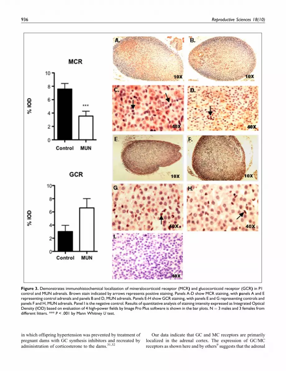
We also determined the expression of GCR (NR3C1) and
MCR (NR3C2) proteins in P1 offspring adrenals by quantita-
tive IHC. As demonstrated in Figure 3, both MC receptor
(panels C and D) and GC receptor (panels G and H) staining was
primarily nuclear. In case of MC receptor, staining pattern was
patchy and involved the 3 layers of adrenal cortex equally, with
0
200
400
600
800
Control MUN
9mos Female
0
200
400
600
800
Control MUN
9mos Male
**
020406080
100
Control MUN
Co
rtic
ost
ero
ne
(ng
/mL
)
Co
rtic
ost
ero
ne
(ng
/mL
)C
ort
ico
ster
on
e(n
g/m
L)
Co
rtic
ost
ero
ne
(ng
/mL
)
P1
*
050
100150200250
Control MUN
P21
**
Figure 2. Plasma corticosterone levels determined by RIA in P1, P21(mixed gender), adult 9-month male and female offspring (fasting) incontrol and MUN offspring. N ¼ 6 per group. * P < .05, ** P < .01.
Table 3. Expression of Adrenal Steroidogenic Enzymes in MaternalAdrenals on Day 21 of Gestationa
Gene Fold Change P Value
CYP11B1 �9.0 + 0.03 .001CYP11B2 �5.9 + 0.07 .001ACTH-R (MC2) �2.0 + 0.11 .05CYP11A1 �2.3 + 0.03 .001HSD1 1.1 + 0.15 NSHSD2 �1.7 + 0.29 NSGCR (NR3C1) �1.2 + 0.13 NSMCR (NR3C2) 1.3 + 0.45 NSSTAR �2.0 + 0.10 NSCYP17A1 �3.2 + 0.13 NS
Abbreviations: CYP11B1, 11-beta hydroxylase; CYP11B2, aldosteronesynthase; ACTH-R, ACTH receptor (MC2); CYP11A1, side chain cleavageenzymes; HSD, hydroxysteroid dehydrogenase; GCR (NR3C1), glucocorticoidreceptor; MCR, mineralocorticoid receptor (NR3C2); CYP17A1, 17 alpha-hydroxylase.a Negative numbers signify lower expression in MUN vs control dams. Valuesare expressed as mean fold change compared to control + SEM. N¼ 4 controland 6 MUN dams.
Table 2. The Effect of Maternal Undernutrition (MUN) on AdultOffspring Body and Adrenal Weightsa
Control MUN P Value
MalesBody weight (g) 647 + 17 742 + 20 <.01Adrenal weight (mg) 71.2 + 1.9 69.6 + 2.2 NSRelative adrenal
weight0.0117 + 0.0001 0.0093 + 0.0003 <.05
FemalesBody weight (g) 350 + 18 420 + 15 <.01Adrenal weight (mg) 100.4 + 6.4 90.8 + 4.3 <.07Relative adrenal
weight0.0295 + 0.0013 0.0233 + 0.0022 <.05
a Values are expressed as mean + SEM relative adrenal weight ¼ adrenalweight (g)/body weight (g) � 100.
934 Reproductive Sciences 18(10)

scant expression in adrenal medullary cells (panels A and B). In
contrast, GC receptor staining was uniformly distributed
throughout the adrenal cortex mostly concentrated in the zona
glomerulosa layer (panels E and F). There was a significant
decrease in percentage integrated optical density (IOD) in MC
receptor expression in MUN offspring, whereas an opposite pat-
tern of expression was obtained for GC receptors although this
change did not achieve statistical significance (P ¼ .06).
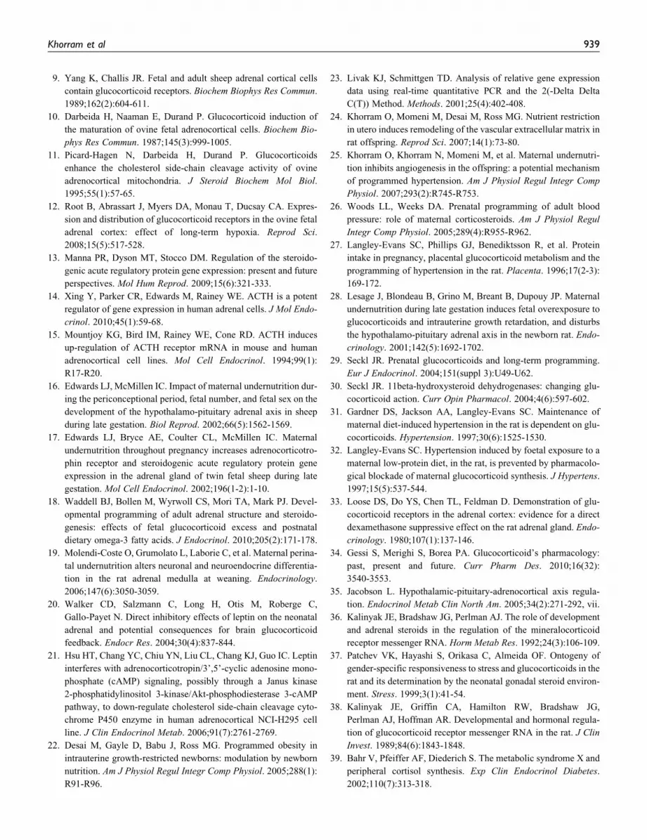
The expression of leptin receptor in MUN adrenals was also
determined by quantitative IHC in P1 adrenals (Figure 4), and
by real time PCR in P1 (Table 4) and adult adrenals (Table 5).
As shown in this figure, the pattern of expression of leptin
receptor was similar to GC receptor, concentrated primarily
in the adrenal cortex with scant staining in the medullary cells.
There was a marked decrease in % IOD in leptin receptor
expression (P < .001) in the P1 MUN adrenals as compared
with controls. Real time RT-PCR showed a marked decrease
in expression of both Ob-Ra and Ob-Rb in P1 male MUN adre-
nals, whereas in females there was a slight increase in Ob-Ra
and no change in Ob-Rb mRNA expression. In adult MUN
male and female offspring, the expression of Ob-Rb mRNA
was significantly increased. The expression of Ob-Ra mRNA
was significantly higher in adult male MUN offspring with a
trend toward an increase in females. The magnitude of changes
in adult Ob-Ra /Ob-Rb mRNA expression were more pro-
nounced for male MUN offspring as compared with females.
Discussion
In this study, the effect of maternal undernutrition on maternal
and offspring adrenal steroidogenic enzyme mRNA expression
in the newborn and adult periods was determined. We hypothe-
sized that elevated maternal leptin and corticosterone in
response to undernutrition programs offspring adrenal steroido-
genic enzymes, GC/MC, and leptin receptors. In support of this
hypothesis, we found a generalized inhibition of steroidogenic
enzymes, GC/MC, and leptin receptors most likely secondary
to a negative feedback from elevated maternal corticosterone
and leptin levels. By 9 months, adrenal profiles of steroido-
genic enzymes in MUN offspring markedly changed in a
gender-specific manner, reflecting not only the effects of early
exposure to elevated maternal corticosterone and leptin but also
gender-specific differences in adaptive responses. In adult
MUN male adrenals, a significant increase in CYP11A1,
CYP11B2, HSD1, and GCR mRNA expression, a trend toward
an increase in ACTH-R, and a decrease in CYP17A1 mRNA
expression was found, whereas in adult female MUN adrenals,
there was a similar mRNA expression profile as males with
respect to CYP 11A1, HSD1, ACTH-R, but an opposite pattern
of expression with respect to GCR and no changes in HSD1
expression. In contrast to the neonatal MUN offspring, adult
adrenal leptin receptor expression was higher in both genders.
There is considerable evidence linking fetal exposure to
excess glucocorticoids and development of metabolic syn-
drome later in life.2,3 Maternal undernutrition stress induces
excess maternal production of corticosterone as demonstrated
by our data, and by others.26 Furthermore, overexpression of
placental 11 HSD-2 in MUN dams, which inactivates active
GC such as corticosterone to inactive 11–keto forms, is inhib-
ited,27,28 resulting in overexposure of the fetus to GC which in
turn would cause growth restriction and other adult chronic dis-
ease.29,30 The importance of excess maternal GC to the patho-
genesis of metabolic syndrome in the offspring was previously
demonstrated in the protein restriction model of programming
Table 4. Expression of Adrenal Steroidogenic Enzymes mRNA in P1Offspringa
Gene Fold Change P Value
CYP11B1 �5.9 + 0.04 .01CYP11B2 �5.9 + 0.03 .04ACTH-R (MC2) �4.2 + 0.06 .05CYP11A1 �4.0 + 0.06 .05HSD1 �2.9 + 0.06 .01HSD2 �5.3 + 0.06 .03STAR �3.8 + 0.05 .01CYP17A1 �3.3 + 0.14 NSGCR (NR3C1) �2.2 + 0.18 NSMCR (NR3C2) �2.7 + 0.08 .02Ob-Ra (female) 1.2 + 0.007 .02Ob-Rb (female) �1.0 + 0.009 NSOb-Ra (male) �3.0 + 0.003 .001Ob-Rb (male) �4.8 + 0.001 .001
Abbreviations: CYP11B1, 11-beta hydroxylase; CYP11B2, aldosteronesynthase; ACTH-R, ACTH receptor (MC2); CYP11A1, side chain cleavageenzymes; HSD, hydroxysteroid dehydrogenase; GCR (NR3C1), glucocorticoidreceptor; MCR, mineralocorticoid receptor (NR3C2); CYP17A1, 17 alpha-hydroxylase.a Values are expressed as mean fold change + SEM compared to control. Neg-ative numbers signify decreased expression in MUN as compared with con-trols. N ¼ 11 per group (5 males/6 females).
Table 5. Adrenal Steroidogenic Enzymes mRNA in 9-Month AdultMale and Female Offspringa
Gene Fold Change P Value Male Fold ChangeP ValueFemale
CYP11B1 �1.1 + 0.07 NS �1.3 + 0.06 NSCYP11B2 1.5 + 0.13 .05 1.1 + 0.10 NSACTH-R 1.9 + 0.57 NS 1.7 + 0.22 .02CYP11A1 1.9 + 0.5 .001 1.3 + 0.28 .03HSD1 2.9 + 0.16 .001 �1.1 + 0.09 NSHSD2 1.1 + 0.03 NS 1.0 + 0.18 NSGCR 2.2 + 0.008 .001 �1.3 + 0.02 .04MCR �1.1 + 0.45 NS 1.1 + 0.09 NSSTAR 1.0 + 0.19 NS �1.1 + 0.06 NSCYP17A1 �1.5 + 0.04 NS �2.3 + 0.03 .04Ob-Ra 2.9 + 0.1 .002 1.2 + 0.03 NSOb-Rb 2.5 + 0.03 .001 1.6 + 0.05 .001
Abbreviations: CYP11B1, 11-beta hydroxylase; CYP11B2, aldosteronesynthase; ACTH-R, ACTH receptor (MC2); CYP11A1, side chain cleavageenzymes; HSD, hydroxysteroid dehydrogenase; GCR (NR3C1), glucocorticoidreceptor; MCR, mineralocorticoid receptor (NR3C2); CYP17A1, 17 alpha-hydroxylase.a Values are expressed as mean fold change + SEM compared to control. N ¼4 males and 4 females.
Khorram et al 935
in which offspring hypertension was prevented by treatment of
pregnant dams with GC synthesis inhibitors and recreated by
administration of corticosterone to the dams.31,32
Our data indicate that GC and MC receptors are primarily
localized in the adrenal cortex. The expression of GC/MC
receptors as shown here and by others9 suggests that the adrenal
Figure 3. Demonstrates immunohistochemical localization of mineralocorticoid receptor (MCR) and glucocorticoid receptor (GCR) in P1control and MUN adrenals. Brown stain indicated by arrows represents positive staining. Panels A-D show MCR staining, with panels A and Erepresenting control adrenals and panels B and D, MUN adrenals. Panels E-H show GCR staining, with panels E and G representing controls andpanels F and H, MUN adrenals. Panel I is the negative control. Results of quantitative analysis of staining intensity expressed as Integrated OpticalDensity (IOD) based on evaluation of 4 high-power fields by Image Pro Plus software is shown in the bar plots. N ¼ 3 males and 3 females fromdifferent litters. *** P < .001 by Mann Whitney U test.
936 Reproductive Sciences 18(10)

gland is a target for feedback regulation by circulating GC and
MC.33 Elevated levels of maternal GC in response to undernu-
trition would be expected to inhibit maternal and fetal adrenal
steroidogenic enzymes as demonstrated by our data. Our data
in the neonatal offspring, showing inhibition of enzymes
involved in corticosterone (CYP11B1) and aldosterone
(CYP11B2), supports this negative feedback regulation of off-
spring adrenal steroidogenic enzymes by maternal GC and is in
agreement with the findings of Lesage et al.28 In addition, in
neonatal MUN offspring, other important components of ster-
oidogenesis such as STAR protein, which drives the movement
of cholesterol from the outer to the inner mitochondrial mem-
brane,13 and CYP11A1, which is the side-chain cleavage
enzyme that is involved in the early steps of steroidogenesis,
were both inhibited. This generalized inhibition of steroido-
genic enzyme mRNA expression in the P1 offspring explains
their lower circulating levels of CORT and could signify
reduced stress reactivity and increased inflammation.34 In the
MUN dams, although both CYP11B1 and CYP11B2 were sup-
pressed, CORT levels were elevated. This discrepancy could
potentially be secondary to a nutritional stress-driven central
CRF drive in these dams, which would stimulate ACTH secre-
tion.35 In contrast to MUN dams, in the P1 offspring the central
expression of MCR/GCR and responsiveness to stress is not
fully developed.36,37 The maturation of negative feedback loop
axis for both GCR and MCR appears to be tissue-dependant in
rats. In case of GCR, GC-induced inhibition of GCR mRNA is
evident between 2 and 7 days of life in liver and between 7 and
14 days in the brain,38 whereas for MCR, negative feedback
was operative in the kidney but not brain in neonates.36 Cur-
rently, there are no studies that have addressed the maturation
of the negative feedback loop between circulating GC/MC and
adrenal steroidogenic enzymes. Our finding of reduced circu-
lating CORT levels in P1 offspring is in agreement with Lesage
et al who also used a 50% food restriction model and reported
an increase in CORT levels at birth in the food-restricted off-
spring, which then decreased 2 hours later,28 and with human
data showing lower cortisol levels in individuals with the meta-
bolic syndrome.39 Similarly, in another model of programming
employing low-sodium diet during pregnancy, fetal levels of
CORT were reduced in the exposed fetuses and this was asso-
ciated with decreased adrenal expression of 11 beta hydroxy-
lase.40 Fetal exposure to long-term hypoxia also inhibited the
expression of key enzymes regulating cortisol biosynthesis in
the ovine fetus,41 whereas in a protein-restriction model of fetal
programming plasma CORT levels were unaffected,42 illustrat-
ing the importance of the nature of stress in fetal programming.
The adrenal response to nutritional programming is highly
species-dependant. Gestational undernutrition resulted in an
increased mRNA expression of ACTH-R and STAR in the
adrenals during late gestation in sheep17 and in ACTH levels
and response to corticotropin-releasing hormone (CRH).16
Coulter et al also found a decreased level of expression of
STAR in the growth-restricted fetus; however, this did not
affect adrenal steroidogenesis.43 Based on these findings, these
investigators suggested that in the ovine growth-restricted
fetus, steroidogenesis may not be ACTH-dependent.43 In
another study using the sheep model, brief undernutrition in
utero did not alter adult adrenal protein expression of
P450C17 and P45011B1.44
The global inhibition of adrenal steroidogenic enzymes in
the P1 MUN adrenals could also be secondary to elevated
maternal leptin levels in MUN dams, which could directly inhi-
bit offspring steroidogenic enzymes, and inhibit offspring lep-
tin receptor expression, as demonstrated by our data. Our group
previously reported that maternal food restriction as other types
of systemic stress45 results in elevated leptin levels most likely
from placental and adipose tissue origin.46 Elevated maternal
leptin could also directly inhibit fetal adrenal steroidogenic
enzymes as leptin was reported to have a direct adrenal sup-
pressive effect on steroidogenic enzymes,20,21,47 and on STAR
protein expression.48 Exposure to high levels of leptin would
also enhance GC negative feedback in the brain through
increased expression of GCR in the hippocampus and paraven-
tricular nucleus as previously reported.49
The profile of adrenal steroidogenic enzymes in the adult
MUN offspring changed during the course of development in
a gender-specific manner, demonstrating different adaptational
strategies in male versus female MUN offspring. Others have
also reported on gender differences in basal cortisol levels or
responsiveness to stress in adult offspring of other species such
as sheep50 and guinea pigs.6 Our data showed that the general-
ized inhibition of steroidogenic enzymes was no longer evident
in the adult MUN offspring. In adult male MUN offspring, the
adrenal expression of HSD1, CYP11A1, CYP11B2, and GCR
were markedly increased, a pattern opposite to that of the neo-
nates. In contrast to males, adult MUN female profile of steroi-
dogenic enzymes showed fewer changes compared with
controls, with only an inhibition of CYP17A1 and an increase
in CYP11A1 expression. In adult male MUN offspring, a trend
toward an inhibition of CYP17A1 was found. This inhibitory
Figure 4. Immunohistochemical localization (�10) of leptin receptorin P1 adrenal of control and MUN offspring (N ¼ 3 males and 3females). Bar plot is a summary of the staining intensity expressed as %IOD in the 2 dietary groups.
Khorram et al 937

trend in CYP17A1 expression was also noted in the P1
offspring. CYP17A1 is a key enzyme in the steroidogenic path-
way, catalyzing the addition of a hydroxyl group to pregneno-
lone and progesterone, thereby controlling the amount of
substrates for downstream steps for synthesis of mineralocorti-
coids, glucocorticoids, androgens, and estrogens. Inhibition of
CYP17A1 in the MUN offspring in the different age group
offspring could account for the lower basal circulating CORT
levels in them and could potentially lead to reduced synthesis
of sex hormone levels in both genders.
In adult MUN offspring of both genders, there was an
increase in adrenal CYP11A1 mRNA (cholesterol side-chain
cleavage enzyme) expression. This enzyme converts choles-
terol into pregneolone, and its expression is principally regu-
lated by angiotensin II, luteinizing hormone (LH), and
ACTH.51 Previous studies have demonstrated an activation of
the renin-angiotensin system in MUN offspring.52 Further-
more, the adrenal expression of angiotensin receptor 1b is
increased in these offspring,53 suggesting increased responsive-
ness of the adrenal to circulating angiotensin II. Elevated
angiotensin II levels and increased responsiveness to its effects
in MUN offspring would be expected to stimulate the expres-
sion of CYP11A1 mRNA as demonstrated by our data. The
more pronounced inhibition of basal CORT levels in adult male
compared with female MUN offspring could also be secondary
to negative feedback inhibition of CORT at the adrenal level
due to (1) increased HSD1 mRNA expression which would
lead to increased active GC levels within the adrenal thereby
inhibiting other steroidogenic enzymes locally and (2)
increased GCR expression in male adrenals would make these
tissues more sensitive to circulating CORT which stimulates
HSD1 expression.54 Gender-related differences in GCR/MCR
imbalance and steroidogenic enzyme expression in adult MUN
offspring could also account for a relative protection of female
MUN offspring from development of programmed hyperten-
sion as compared with males55,56 since in males but not females
the expression of aldosterone synthesizing enzyme, CYP11B2,
was increased. Our data also showed an increase in leptin
receptor expression in MUN adrenals particularly in males.
Although both male and female MUN have hyperleptinemia,
leptin levels in male MUN are higher compared with females,22
yet leptin receptor expression is also higher in males compared
with females, suggesting a probable leptin resistance at the
adrenal level that develops during the course of development
and is more exaggerated in MUN males.
The common finding of increased adrenal ACTH-R expres-
sion in adult males and females would suggest an increased
sensitivity of these animals to the effects of stress. Our data are
in agreement with Waddell et al who reported increased adre-
nal expression of the ACTH-R in 6-month-old offspring and
increased stress-induced levels of plasma corticosterone in a rat
model of programming in which dams received dexamthasone
during gestation.18 In humans, low birth weight is associated
with increased urinary glucocorticoid excretion in children57
and with elevated basal plasma cortisol concentration58 and
greater adrenocortical responsiveness to ACTH in adults.59
This increased responsiveness to stress has been proposed to
contribute to the pathogenesis of chronic diseases.58
In summary, our data demonstrate that the adrenal gland is
an important target for nutritional programming effects. The
changing pattern of GC/MC and ACTH-R in the adrenal would
imply differing sensitivities of this gland to circulating GC/MC
and ACTH during the course of development. The programmed
adrenal expression of steroidogenic enzymes, GC, and leptin
receptors in a gender-specific and developmentally regulated
manner supports an important contribution of the adrenal gland
to the development of metabolic syndrome in MUN offspring and
the gender differences in response to maternal undernutrition.
Acknowledgment
We wish to thank Ms Jeannie Park and Ms Diane Park for their help in
preparation of this manuscript.
Declaration of Conflicting Interests
The authors declared no conflicts of interest with respect to the author-
ship and/or publication of this article.
Funding
The authors disclosed receipt of the following financial support for the
research and/or authorship of this article: NIH RO3 HD054920-01
(O.Khorram) and University of California San Diego Chancellor’s
Research Scholarship (N. Khorram).
Reference
1. Barker DJ, Bull AR, Osmond C, Simmonds SJ. Fetal and placen-
tal size and risk of hypertension in adult life. BMJ.
1990;301(6746):259-262.
2. Harris A, Seckl J. Glucocorticoids, prenatal stress and the pro-
gramming of disease. Horm Behav. 2011;59(3):279-289.
3. Cottrell EC, Seckl JR. Prenatal stress, glucocorticoids and the pro-
gramming of adult disease. Front Behav Neurosci. 2009;3:19.
4. Dutriez-Casteloot I, Breton C, Coupe B, et al. Tissue-specific pro-
gramming expression of glucocorticoid receptors and 11 beta-
HSDs by maternal perinatal undernutrition in the HPA axis of
adult male rats. Horm Metab Res. 2008;40(4):257-261.
5. Vieau D, Sebaai N, Leonhardt M, et al. HPA axis programming by
maternal undernutrition in the male rat offspring. Psychoneuroen-
docrinology. 2007;32(suppl 1):S16-S20.
6. Liu L, Li A, Matthews SG. Maternal glucocorticoid treatment
programs HPA regulation in adult offspring: sex-specific effects.
Am J Physiol Endocrinol Metab. 2001;280(5):E729-E739.
7. Leonhardt M, Lesage J, Dufourny L, Dickes-Coopman A,
Montel V, Dupouy JP. Perinatal maternal food restriction induces
alterations in hypothalamo-pituitary-adrenal axis activity and in
plasma corticosterone-binding globulin capacity of weaning rat
pups. Neuroendocrinol. 2002;75(1):45-54.
8. Lesage J, Dufourny L, Laborie C, et al. Perinatal malnutrition
programs sympathoadrenal and hypothalamic-pituitary-adrenal
axis responsiveness to restraint stress in adult male rats.
J Neuroendocrinol. 2002;14(2):135-143.
938 Reproductive Sciences 18(10)
9. Yang K, Challis JR. Fetal and adult sheep adrenal cortical cells
contain glucocorticoid receptors. Biochem Biophys Res Commun.
1989;162(2):604-611.
10. Darbeida H, Naaman E, Durand P. Glucocorticoid induction of
the maturation of ovine fetal adrenocortical cells. Biochem Bio-
phys Res Commun. 1987;145(3):999-1005.
11. Picard-Hagen N, Darbeida H, Durand P. Glucocorticoids
enhance the cholesterol side-chain cleavage activity of ovine
adrenocortical mitochondria. J Steroid Biochem Mol Biol.
1995;55(1):57-65.
12. Root B, Abrassart J, Myers DA, Monau T, Ducsay CA. Expres-
sion and distribution of glucocorticoid receptors in the ovine fetal
adrenal cortex: effect of long-term hypoxia. Reprod Sci.
2008;15(5):517-528.
13. Manna PR, Dyson MT, Stocco DM. Regulation of the steroido-
genic acute regulatory protein gene expression: present and future
perspectives. Mol Hum Reprod. 2009;15(6):321-333.
14. Xing Y, Parker CR, Edwards M, Rainey WE. ACTH is a potent
regulator of gene expression in human adrenal cells. J Mol Endo-
crinol. 2010;45(1):59-68.
15. Mountjoy KG, Bird IM, Rainey WE, Cone RD. ACTH induces
up-regulation of ACTH receptor mRNA in mouse and human
adrenocortical cell lines. Mol Cell Endocrinol. 1994;99(1):
R17-R20.
16. Edwards LJ, McMillen IC. Impact of maternal undernutrition dur-
ing the periconceptional period, fetal number, and fetal sex on the
development of the hypothalamo-pituitary adrenal axis in sheep
during late gestation. Biol Reprod. 2002;66(5):1562-1569.
17. Edwards LJ, Bryce AE, Coulter CL, McMillen IC. Maternal
undernutrition throughout pregnancy increases adrenocorticotro-
phin receptor and steroidogenic acute regulatory protein gene
expression in the adrenal gland of twin fetal sheep during late
gestation. Mol Cell Endocrinol. 2002;196(1-2):1-10.
18. Waddell BJ, Bollen M, Wyrwoll CS, Mori TA, Mark PJ. Devel-
opmental programming of adult adrenal structure and steroido-
genesis: effects of fetal glucocorticoid excess and postnatal
dietary omega-3 fatty acids. J Endocrinol. 2010;205(2):171-178.
19. Molendi-Coste O, Grumolato L, Laborie C, et al. Maternal perina-
tal undernutrition alters neuronal and neuroendocrine differentia-
tion in the rat adrenal medulla at weaning. Endocrinology.
2006;147(6):3050-3059.
20. Walker CD, Salzmann C, Long H, Otis M, Roberge C,
Gallo-Payet N. Direct inhibitory effects of leptin on the neonatal
adrenal and potential consequences for brain glucocorticoid
feedback. Endocr Res. 2004;30(4):837-844.
21. Hsu HT, Chang YC, Chiu YN, Liu CL, Chang KJ, Guo IC. Leptin
interferes with adrenocorticotropin/3’,5’-cyclic adenosine mono-
phosphate (cAMP) signaling, possibly through a Janus kinase
2-phosphatidylinositol 3-kinase/Akt-phosphodiesterase 3-cAMP
pathway, to down-regulate cholesterol side-chain cleavage cyto-
chrome P450 enzyme in human adrenocortical NCI-H295 cell
line. J Clin Endocrinol Metab. 2006;91(7):2761-2769.
22. Desai M, Gayle D, Babu J, Ross MG. Programmed obesity in
intrauterine growth-restricted newborns: modulation by newborn
nutrition. Am J Physiol Regul Integr Comp Physiol. 2005;288(1):
R91-R96.
23. Livak KJ, Schmittgen TD. Analysis of relative gene expression
data using real-time quantitative PCR and the 2(-Delta Delta
C(T)) Method. Methods. 2001;25(4):402-408.
24. Khorram O, Momeni M, Desai M, Ross MG. Nutrient restriction
in utero induces remodeling of the vascular extracellular matrix in
rat offspring. Reprod Sci. 2007;14(1):73-80.
25. Khorram O, Khorram N, Momeni M, et al. Maternal undernutri-
tion inhibits angiogenesis in the offspring: a potential mechanism
of programmed hypertension. Am J Physiol Regul Integr Comp
Physiol. 2007;293(2):R745-R753.
26. Woods LL, Weeks DA. Prenatal programming of adult blood
pressure: role of maternal corticosteroids. Am J Physiol Regul
Integr Comp Physiol. 2005;289(4):R955-R962.
27. Langley-Evans SC, Phillips GJ, Benediktsson R, et al. Protein
intake in pregnancy, placental glucocorticoid metabolism and the
programming of hypertension in the rat. Placenta. 1996;17(2-3):
169-172.
28. Lesage J, Blondeau B, Grino M, Breant B, Dupouy JP. Maternal
undernutrition during late gestation induces fetal overexposure to
glucocorticoids and intrauterine growth retardation, and disturbs
the hypothalamo-pituitary adrenal axis in the newborn rat. Endo-
crinology. 2001;142(5):1692-1702.
29. Seckl JR. Prenatal glucocorticoids and long-term programming.
Eur J Endocrinol. 2004;151(suppl 3):U49-U62.
30. Seckl JR. 11beta-hydroxysteroid dehydrogenases: changing glu-
cocorticoid action. Curr Opin Pharmacol. 2004;4(6):597-602.
31. Gardner DS, Jackson AA, Langley-Evans SC. Maintenance of
maternal diet-induced hypertension in the rat is dependent on glu-
cocorticoids. Hypertension. 1997;30(6):1525-1530.
32. Langley-Evans SC. Hypertension induced by foetal exposure to a
maternal low-protein diet, in the rat, is prevented by pharmacolo-
gical blockade of maternal glucocorticoid synthesis. J Hypertens.
1997;15(5):537-544.
33. Loose DS, Do YS, Chen TL, Feldman D. Demonstration of glu-
cocorticoid receptors in the adrenal cortex: evidence for a direct
dexamethasone suppressive effect on the rat adrenal gland. Endo-
crinology. 1980;107(1):137-146.
34. Gessi S, Merighi S, Borea PA. Glucocorticoid’s pharmacology:
past, present and future. Curr Pharm Des. 2010;16(32):
3540-3553.
35. Jacobson L. Hypothalamic-pituitary-adrenocortical axis regula-
tion. Endocrinol Metab Clin North Am. 2005;34(2):271-292, vii.
36. Kalinyak JE, Bradshaw JG, Perlman AJ. The role of development
and adrenal steroids in the regulation of the mineralocorticoid
receptor messenger RNA. Horm Metab Res. 1992;24(3):106-109.
37. Patchev VK, Hayashi S, Orikasa C, Almeida OF. Ontogeny of
gender-specific responsiveness to stress and glucocorticoids in the
rat and its determination by the neonatal gonadal steroid environ-
ment. Stress. 1999;3(1):41-54.
38. Kalinyak JE, Griffin CA, Hamilton RW, Bradshaw JG,
Perlman AJ, Hoffman AR. Developmental and hormonal regula-
tion of glucocorticoid receptor messenger RNA in the rat. J Clin
Invest. 1989;84(6):1843-1848.
39. Bahr V, Pfeiffer AF, Diederich S. The metabolic syndrome X and
peripheral cortisol synthesis. Exp Clin Endocrinol Diabetes.
2002;110(7):313-318.
Khorram et al 939

40. Bibeau K, Battista MC, Houde V, Brochu M. Fetal adrenal gland
alterations in a rat model of adverse intrauterine environment. Am
J Physiol Regul Integr Comp Physiol. 2010;298(4):R899-R911.
41. Myers DA, Hyatt K, Mlynarczyk M, Bird IM, Ducsay CA.
Long-term hypoxia represses the expression of key genes
regulating cortisol biosynthesis in the near-term ovine fetus. Am
J Physiol Regul Integr Comp Physiol. 2005;289(6):R1707-R1714.
42. Langley-Evans SC, Gardner DS, Jackson AA. Maternal protein
restriction influences the programming of the rat hypothalamic-
pituitary-adrenal axis. J Nutr. 1996;126(6):1578-1585.
43. Coulter CL, McMillen IC, Bird IM, Salkeld MD. Steroidogenic
acute regulatory protein expression is decreased in the adrenal
gland of the growth-restricted sheep fetus during late gestation.
Biol Reprod. 2002;67(2):584-590.
44. Bloomfield FH, Oliver MH, Giannoulias CD, Gluckman PD,
Harding JE, Challis JR. Brief undernutrition in late-gestation
sheep programs the hypothalamic-pituitary-adrenal axis in adult
offspring. Endocrinology. 2003;144(7):2933-2940.
45. Konishi N, Otaka M, Odashima M, et al. Systemic stress increases
serum leptin level. J Gastroenterol Hepatol. 2006;21(7):
1099-1102.
46. Jelks A, Belkacemi L, Han G, Chong WL, Ross MG, Desai M.
Paradoxical increase in maternal plasma leptin levels in food-
restricted gestation: contribution by placental and adipose tissue.
Reprod Sci. 2009;16(7):665-675.
47. Kruse M, Bornstein SR, Uhlmann K, Paeth G, Scherbaum WA.
Leptin down-regulates the steroid producing system in the adre-
nal. Endocr Res. 1998;24(3-4):587-590.
48. Cherradi N, Capponi AM, Gaillard RC, Pralong FP. Decreased
expression of steroidogenic acute regulatory protein: a novel
mechanism participating in the leptin-induced inhibition of gluco-
corticoid biosynthesis. Endocrinology. 2001;142(8):3302-3308.
49. Proulx K, Clavel S, Nault G, Richard D, Walker CD. High neona-
tal leptin exposure enhances brain GR expression and feedback
efficacy on the adrenocortical axis of developing rats. Endocri-
nology. 2001;142(11):4607-4616.
50. Gardner DS, Van Bon BW, Dandrea J, et al. Effect of periconcep-
tional undernutrition and gender on hypothalamic-pituitary-
adrenal axis function in young adult sheep. J Endocrinol.
2006;190(2):203-212.
51. Lavoie HA, King SR. Transcriptional regulation of steroidogenic
genes: STARD1, CYP11A1 and HSD3B. Exp Biol Med (May-
wood). 2009;234(8):880-907.
52. Riviere G, Michaud A, Breton C, et al. Angiotensin-converting
enzyme 2 (ACE2) and ACE activities display tissue-specific sen-
sitivity to undernutrition-programmed hypertension in the adult
rat. Hypertension. 2005;46(5):1169-1174.
53. Bogdarina I, Haase A, Langley-Evans S, Clark AJ. Glucocorti-
coid effects on the programming of AT1b angiotensin receptor
gene methylation and expression in the rat. PLoS ONE.
2010;5(2):e9237.
54. Shimojo M, Whorwood CB, Stewart PM. 11 beta-Hydroxysteroid
dehydrogenase in the rat adrenal. J Mol Endocrinol. 1996;17(2):
121-130.
55. Ojeda NB, Grigore D, Robertson EB, Alexander BT. Estrogen
protects against increased blood pressure in postpubertal female
growth restricted offspring. Hypertension. 2007;50(4):679-685.
56. Roghair RD, Segar JL, Volk KA, et al. Vascular nitric oxide and
superoxide anion contribute to sex-specific programmed cardio-
vascular physiology in mice. Am J Physiol Regul Integr Comp
Physiol. 2009;296(3):R651-R662.
57. Clark PM, Hindmarsh PC, Shiell AW, Law CM, Honour JW,
Barker DJ. Size at birth and adrenocortical function in childhood.
Clin Endocrinol (Oxf). 1996;45(6):721-726.
58. Phillips DI, Barker DJ, Fall CH, et al. Elevated plasma cortisol
concentrations: a link between low birth weight and the insulin
resistance syndrome? J Clin Endocrinol Metab. 1998;83(3):
757-760.
59. Reynolds RM, Walker BR, Syddall HE, et al. Altered control of
cortisol secretion in adult men with low birth weight and cardio-
vascular risk factors. J Clin Endocrinol Metab. 2001;86(1):
245-250.
940 Reproductive Sciences 18(10)