therapeutic inhibition of pro-inflammatory signaling and toxicity to staphylococcal enterotoxin b by...
TRANSCRIPT

Therapeutic Inhibition of Pro-Inflammatory Signalingand Toxicity to Staphylococcal Enterotoxin B bya Synthetic Dimeric BB-Loop Mimetic of MyD88Teri L. Kissner1, Gordon Ruthel1, Shahabuddin Alam1, Enrique Mann2, Dariush Ajami2, Mitra Rebek2,
Eileen Larkin1, Stefan Fernandez1, Robert G. Ulrich1, Sun Ping3, David S. Waugh3, Julius Rebek, Jr.3,
Kamal U. Saikh1*
1Department of Immunology, United States Army Medical Research Institute of Infectious Diseases, Frederick, Maryland, United States of America, 2Department of
Chemistry, The Skaggs Institute for Chemical Biology, The Scripps Research Institute, La Jolla, California, United States of America, 3Macromolecular Crystallography
Laboratory, National Cancer Institute at Frederick, Frederick, Maryland, United States of America
Abstract
Staphylococcal enterotoxin B (SEB) exposure triggers an exaggerated pro-inflammatory cytokine response that often leadsto toxic shock syndrome (TSS) associated with organ failure and death. MyD88 mediates pro-inflammatory cytokinesignaling induced by SEB exposure and MyD882/2 mice are resistant to SEB intoxication, suggesting that MyD88 may bea potential target for therapeutic intervention. We targeted the BB loop region of the Toll/IL-1 receptor (TIR) domain ofMyD88 to develop small-molecule therapeutics. Here, we report that a synthetic compound (EM-163), mimic to dimericform of BB-loop of MyD88 attenuated tumor necrosis factor (TNF)- a, interferon (IFN)-c, interleukin (IL)-1b, IL-2 and IL-6production in human primary cells, whether administered pre- or post-SEB exposure. Results from a direct binding assay,and from MyD88 co-transfection/co-immunoprecipitation experiments, suggest that EM-163 inhibits TIR-TIR domaininteraction. Additional results indicate that EM-163 prevents MyD88 from mediating downstream signaling. In an NF-kB-driven reporter assay of lipopolysaccharide-stimulated MyD88 signaling, EM-163 demonstrated a dose-dependent inhibitionof reporter activity as well as TNF-a and IL-1b production. Importantly, administration of EM-163 pre- or post exposure toa lethal dose of SEB abrogated pro-inflammatory cytokine responses and protected mice from toxic shock-induced death.Taken together, our results suggest that EM-163 exhibits a potential for therapeutic use against SEB intoxication.
Citation: Kissner TL, Ruthel G, Alam S, Mann E, Ajami D, et al. (2012) Therapeutic Inhibition of Pro-Inflammatory Signaling and Toxicity to StaphylococcalEnterotoxin B by a Synthetic Dimeric BB-Loop Mimetic of MyD88. PLoS ONE 7(7): e40773. doi:10.1371/journal.pone.0040773
Editor: Guillermo H. Giambartolomei, National Council of Sciences (CONICET), Argentina
Received May 2, 2012; Accepted June 13, 2012; Published July 27, 2012
This is an open-access article, free of all copyright, and may be freely reproduced, distributed, transmitted, modified, built upon, or otherwise used by anyone forany lawful purpose. The work is made available under the Creative Commons CC0 public domain dedication.
Funding: This work was supported by the Defense Threat Reduction Agency (DTRA; WWW.jpras.us) grant CBM.THRTOX.03.10.RD.006 to Kamal U. Saikh. Thefunders had no role in study design, data collection and analysis, decision to publish, or preparation of the manuscript.
Competing Interests: The authors have declared that no competing interests exist.
* E-mail: [email protected]
Introduction
The profound clinical consequences of staphylococcal entero-
toxin B (SEB)-induced toxicity are known to stem from an
excessive pro-inflammatory cytokine response that often leads to
toxic shock syndrome (TSS) associated with organ failure and
death. Strong pro-inflammatory responses by antigen-presenting
cells (APCs) and T cells are triggered by the binding of SEB to
MHC class II molecules on APCs and subsequent cross-linking to
T-cell receptors (TCR) [1–5]. SEB is listed by the Centers for
Disease Control and Prevention (CDC) as a Category B select
agent because of its potential use as an aerosolized biological
weapon. There are currently no small-molecule therapeutics
available to treat SEB exposure, making the development of such
medical countermeasures an important goal.
In a mouse model, the biological effects of SEB are potentiated
by lipopolysaccharide (LPS), a bacterial factor that binds to toll-
like receptor 4 (TLR4) on the surface of cells. SEB and LPS
synergistically amplify pro-inflammatory cytokine production,
leading to severe toxicity [6–7]. Consequently, both super-
antigenic exotoxins (SEs) and bacterial LPS (endotoxin) have
been implicated in the pathogenesis of TSS, supported by their
identification in the bloodstream of critically ill patients with
septic shock [8–9]. Results from our laboratory demonstrated
that myeloid differentiation protein 88 (MyD88) gene knockout
(MyD882/2) mice were resistant to lethal SEA or SEB challenge
and showed a concomitant reduction in serum levels of pro-
inflammatory cytokines [10–11]. We also reported that the
binding of SEA or SEB to MHC class II activates MyD88-
mediated pro-inflammatory cytokine signaling in human primary
cells [12]. Consistent with our results, a recent report indicated
that deficiency in MHC class II resulted in impaired TLR-
triggered production of pro-inflammatory cytokines and pro-
tected mice from an otherwise lethal challenge with TLR ligands
and live Gram-negative bacteria [13]. This study also concluded
that both the TLR-and MHC –mediated responses engage
MyD88 [14].
MyD88 is an adaptor protein that functions to recruit signaling
proteins to members of the Toll-like and interleukin-1 receptor
(TLR/IL-1R) [15–16] family as well as the IFN -c receptor [17].
Activation of the MyD88-mediated pro-inflammatory signaling
pathway by this class of receptors is very important for several
PLoS ONE | www.plosone.org 1 July 2012 | Volume 7 | Issue 7 | e40773
aspects of host defense. Because of the critical role of MyD88
signaling, mice deficient in MyD88 have profoundly impaired
innate immune responses and are susceptible to a wide range of
infectious diseases [18]. However, in regards to innate immune-
response regulation, MyD88 signaling performs a delicate balanc-
ing act. Perturbed regulation or excessive stimulation of the innate
immune system can trigger, inflammatory signaling that can spiral
out of control and lead to profound clinical syndrome [18]. For
example, excess of MyD88-mediated signaling can lead to severe
pathological consequences such as toxic shock syndrome (TSS)
and sepsis. However, the crucial role of MyD88 in these disorders
also provides a target for therapeutic intervention.
Earlier results from our laboratory demonstrated that a synthetic
mimetic of the BB-loop in the TIR domain of MyD88 (Compound
1) attenuated SEB-induced pro-inflammatory cytokine production
in human primary cells and increased survivability of mice from
toxic shock-induced death after a lethal SEB challenge [19]. It is
known that the BB-loop region acts as the mediator of the homo-
(adaptor-adaptor) and hetero- (receptor-adaptor) dimerization that
is necessary for the function of TIR domains to induce MyD88-
mediated signaling [15,16]. Seeking to improve the efficacy of
Compound 1 as an inhibitor of MyD88 signaling, we synthesized
a dimeric molecule in which two Compound 1 moieties were
covalently linked together, reasoning that the dimeric compound
would be a more potent inhibitor of protein-protein interactions.
This molecule, EM-163, was tested in primary cultures of human
mononuclear cells (MNCs) for inhibition of cytokine release
associated with exposure to SEB. Our results provide evidence
that, by targeting MyD88, EM-163 inhibited SEB-induced
inflammatory cytokine production in human primary cells.
Importantly, EM-163 abrogated pro-inflammatory cytokine re-
sponse in vivo and completely protected mice from toxic shock-
induced death regardless of whether it was administered pre- or
post-exposure to a lethal SEB challenge.
Materials and Methods
ReagentsStaphylococcal enterotoxin B (SEB) and SEA was purchased
from Porton Down, Inc. (Salisbury, UK) and stored at 250uC.SEB or SEA was endotoxin free and prepared under GMP
conditions. Escherichia coli LPS (055:B5) was purchased from
Sigma-Aldrich (Saint Louis, MO). Pooled human AB sera were
obtained from Pel-Freez (Brown Deer, WI). The BDTM cytometric
bead array (CBA) human Th1/Th2 and inflammatory cytokine kit
was purchased from BD Biosciences (San Diego, CA). The Meso
Scale Discovery (MSD) multi spot array ultra-sensitive cytokine
assay kit was purchased from MSD (Gaithersburg, MD). Magnetic
bead-conjugated anti-CD14 and anti-CD3 mAbs were obtained
from Miltenyi Biotech Inc. (Auburn, CA). Ficoll-Hypaque was
purchased from GE Healthcare Biosciences (Piscataway, NJ).
Primary anti-MyD88 antibody was obtained from AnaSpec, Inc.,
(San Jose, CA) and Alexis Biochemicals (San Diego, CA). Anti-b-actin antibody was purchased from Cell Signaling technology
(Danvers, MA). Plasmids expressing HLA-DRa/b were a kind gift
from Dr. Robert Ulrich (USAMRIID). Plasmid 12287 (pCMV-
HA-MyD88) and plasmid 13093 (MyD88-flag) were purchased
through an MTA agreement with Addgene (Cambridge, MA).
The HEK 293 T cell line was obtained from the ATCC
(Manassas, VA). MyD88 KO HEK293 cell line (HEK293-I3A)
was a kind gift from G. Stark (Dept. of Molecular Genetics, Lerner
Research Institute, Cleveland Clinic, OH). The transfection
reagent lipofectamine was purchased from Invitrogen (Carlsbad,
CA). Vectashield mounting medium containing 4-,6-diamidino-2-
phenylindole (DAPI) was purchased from Vector Laboratories
(Burlingame,CA).
MicePathogen-free, 6–8 weeks old BALB/c and C57BL/6 mice were
obtained from Charles River (NCI-Frederick, Frederick, MD).
Cell Isolation and PurificationPeripheral blood mononuclear cells (MNC) were obtained from
consenting healthy donors in accordance with an Institutional
Review Board-approved research donor protocol. MNCs were
isolated by standard density gradient centrifugation with Ficoll-
Hypaque, harvested from the interface, washed, and re-suspended
in RPMI 1640 medium. Monocytes (CD14+) and T (CD3+) cells
were isolated as previously described [19]. Isolated cell populations
had .98% purity.
Cytokine AnalysisCell cultures were incubated (37uC, 5% CO2) for 16 h.
Cytokines in culture supernatants were measured by a CBA kit
using captured beads coated with antibodies specific for cytokines.
Flow cytometry was performed as described elsewhere [20].
Cytokine measurement was confirmed by dilution of culture
supernatant using Inflammation and Th1/Th2 CBA kits by
acquiring 1800 beads. We also used a Meso Scale Discovery
(MSD) multi spot array ultra sensitive cytokine assay kit to
measure cytokines in culture supernatants (according to the
manufacturer’s protocol) as described [19].
Western Blot AnalysisThe transfected cells were chilled on ice for 5 min before being
pelleted into fresh 1.5 ml centrifuge tubes. Membrane and
cytoplasm separation was done by suspending the pellets in
50 ml of lysis buffer (Active Motif) in the presence of DTT,
protease inhibitors and phosphatase inhibitors and incubated on
ice for 30–60 min. The membrane fraction was collected by
centrifuging the lysates at 140006g for 20 min. The supernatant
contained the cytoplasmic fraction. Samples containing 10 mg of
total cytoplasmic proteins were separated by gel electrophoresis
and transferred to nitrocellulose membranes. The membranes
were blocked overnight in 16Tris-buffered saline (TBS) contain-
ing 0.1% Tween-20 and 3% bovine serum albumin at 4 oC. The
membranes were washed extensively with 16TBS buffer and then
probed with anti-MyD88 polyclonal antibody followed by
horseradish peroxide-conjugated secondary antibody (goat anti-
rabbit). After additional rinsing with 16 TBS buffer, the
membranes were exposed to a chemiluminescent substrate in the
presence of hydrogen peroxide, using Immun-Star WesternC
Chemiluminescent kit (BioRad). A VersaDoc Model 4000
(BioRad) imaging system was used to capture the image.
Intracellular Staining and Confocal MicroscopyFreshly isolated monocytes (CD14+) adhered to sterile culture
slides (Corning Glass, Corning, NY) were incubated with either
SEB (200 ng/ml), SEB+EM-163, or kept untreated in RPMI 1640
medium. Cells were gently washed with PBS (pH 7.4, 4uC) andthen fixed with 1% paraformaldehyde (Tousimis Research,
Rockville, MD) plus 0.1% glutaraldehyde (Sigma-Aldrich) in
PBS (5 min, 20uC). The fixed cells were washed with PBS
containing 0.5% bovine serum albumin (BSA) and incubated
(30 min, 20uC) in a permeabilization solution (Becton Dickinson).
Cells were then washed and incubated (15 min, 20uC) with PBS
containing 5% BSA to block non-specific antibody binding. The
Inhibition of SEB Intoxication by a MyD88 Mimetic
PLoS ONE | www.plosone.org 2 July 2012 | Volume 7 | Issue 7 | e40773
cells were then incubated with primary MyD88 antibody for 1 h,
washed in PBS containing 0.5% BSA, then incubated with Alexa
568-conjugated goat anti-rabbit secondary antibody. The cells
were counterstained with DAPI to detect cell nuclei. Labeled cells
were mounted on glass slides using fluoromount and covered with
glass cover slips. Labeled cells were imaged using a Leica
(Mannheim, Germany) TCS SP5 laser scanning confocal system.
Green and red fluorescence were visualized using 488-nm and
568 nm wavelength laser excitation, respectively, and DAPI
images were collected using two-photon excitation from a Ti/
sapphire laser tuned to 780 nm.
Synthesis of Compound EM-163Initially, Compound 1 was used to synthesize EM-110 [20], as
follows. Briefly, to a stirred solution of 2,5-pyridine dicarboxylic
acid (52 mg; 0.3 mmol) in CH2Cl2 (0.5 ml) and dimethylforma-
mide (10 ml), thionyl chloride (0.4 ml) was added drop-wise at 0uC.The mixture was then heated at 40uC for 1 h, and the solvent was
removed under reduced pressure. The diacid chloride obtained
was added to a solution of amine 2 (200 mg; 0.69 mmol) and
Et3N (97 ml; 0.69 mmol) in dry CH2Cl2 (10 ml). The reaction
mixture was stirred at ambient temperature overnight, the solvent
was evaporated, and the residue was purified by silica gel
chromatography (hexane/AcOEt mixtures) yielding compound 4(36 mg; 17% yield) as a white solid. 1H NMR: (600 MHz, CDCl3)
d 8.39 (s, 1H), 7.69 (d, J =8.1 Hz, 1H), 7.55 (d, J = 8.1 Hz, 1H),
7.20 (m, 4H), 7.11 (m, 2H), 6.94 (m, 2H), 6.86 (m, 2H), 5.21 (m,
2H), 3.70 (m, 4H), 3.59 (m, 2H), 3.43 (m, 2H), 3.31 (m, 4H), 2.51
(m, 2H), 2.40 (m, 2H), 2.29 (m, 2H), 1.98 (m, 4H), 1.90 (m, 4H),
1.68 (m, 2H), 1.54 (m, 2H), 1.04 (m, 12H); 13C NMR: (150 MHz,
CDCl3) d169.6, 169.0, 168.5, 168.4, 155.6, 145.8, 141.4, 140.7,135.1, 133.8, 128.9 (2C), 128.8 (2C), 128.5 (2C), 128.3 (2C), 126.4,
126.2, 123.6, 60.9, 60.4, 47.2, 47.0, 46.5 (2C), 45.2, 44.8, 33.6,
33.4, 31.8, 31.7, 28.3 (2C), 26.6 (2C), 24.6 (2C), 20.5, 20.3, 18.9,
18.8; MS: (ESI-TOF) MH+ calculated:708.4488, found: 708.4461.
EM-163 was synthesized from compound EM-110 [20] as
shown in Figure 1. Compound EM-110 (600 mg, 0.85 mmol) and
methyl iodide (10 ml) were stirred at 70uC for 72 h. The excess
methyl iodide was then eliminated under a stream of nitrogen and
the residue was purified by column chromatography (AcOEt/
MeOH, 95:5) to afford compound EM163 (380 mg, 53%) as
a orange solid. 1H NMR (600 MHz, CD3CN, 50uC, mixture of
conformers) d 9.70 (bs, 1H), 8.58 (bs, 1H), 8.16 (bs, 1H). 7.25–6.90
(m, 10 H), 5.07 (m, 2H), 4.48 (s, 3H), 3.62 (m, 4H), 3.53 (m, 2H),
3.41 (m, 2H), 3.32 (m, 4H), 2.55 (m, 2H), 2.45 (m, 2H), 2.37 (m,
2H), 1.95 (m, 4H), 1.86–1.70(m, 8H), 1.00–0.85 (m, 12H). MS:
(ESI-TOF), M+ calculated: 722.4845, found: 722.4837.
Expression of MyD88 ProteinThe open reading frame encoding the TIR domain of human
MyD88 (residues 157–296) was amplified from pCDNA3-MyD88-
GFP (Addgene plasmid 13026, Addgene, Cambridge, MA, USA)
and inserted into pDONR201 (Invitrogen, Carlsbad, CA,). The
gene was sequence verified and then inserted into the destination
vector pDEST-HisMBP [21] by Gateway recombinatorial cloning
to generate expression vector pPS2218.
The recombinant His-MBP-MyD88(157–296) fusion protein
was expressed in E. coli BL21(DE3) Codon Plus-RIL cells
(Stratagene, La Jolla, CA), which were grown in Luria broth
and induced at mid-log phase with 1 mM IPTG for 4 h at 30uC.The cells were harvested by centrifugation at 4uC and frozen at
280uC until use. The cell pellet was resuspended in 50 mM
sodium phosphate pH 7.1, 150 mM NaCl, 5% glycerol, 25 mM
imidazole, and then the cells were disrupted using an APV Model
G1000 homogenizer (Invensys, Roholmsvej, Denmark). The lysate
was centrifuged at 15,000 rpm at 4uC, filtered, and the fusion
protein was then purified by immobilized metal affinity chroma-
tography (IMAC) as described [22]. Fractions containing the
fusion protein were pooled, cleaved overnight with hexahistidine-
tagged TEV protease, and then subjected to another round of
IMAC as described [22]. The flow-through fractions were pooled,
concentrated to 5 ml and applied to a 320 ml XK26/60
Sephacryl S-100 gel filtration column (GE Healthcare, Piscat-
away, NJ) equilibrated in 25 mM 2-(N-morpholino) ethanesulfonic
acid (MES) pH 6.3, 150 mM NaCl, and 2 mM Tris(2-carbox-
yethyl) phosphine hydrochloride (TCEP). The peak fractions
corresponding to MyD88 (157–296) were pooled and concentrat-
ed to approximately 1 mg/ml.
Microarray-based Binding AssayA microarray-based binding assay was developed and optimized
to measure the binding of compound EM-163 to MyD88.
Nitrocellulose-coated slides (PATH slides, Gentel Biosciences,
Madison, WI) were spotted in quadruplicate with anti-MyD88
antibody or MyD88 monomer using the ArrayJet Marathon
printer (Roslin, Scotland, UK). Slides were blocked overnight
(,16 h) at 4 oC with blocking buffer (50 mM Hepes, 0.08% triton
X-100, 50% glycerol, 140 mM NaCl and 3% BSA). Recombinant
MyD88 was bio-tinylated using the EZ Link Biotin BMCC kit
from Pierce (Cat# PI-21900, Pittsburg PA). Biotinlyated MyD88
(25 mg/ml) was pre-incubated with and without compound
(100 mg/ml and 50 mg/ml) for 1 h on ice and then diluted with
probing buffer (50 mM Hepes, 0.1%Tween, 3% human antibody
serum). MyD88 samples and a probing buffer only control were
added to designated wells on the microarray slide for 2 h at room
temperature. Slides were then washed 6 times for 5 min with
washing buffer (50 mM Hepes, 0.2%Tween, 3% human antibody
serum). Streptavidin labeled with alexa fluor 647 (1:1000,
Invitrogen, Carlsbad CA) was then added for 1 h at room
temperature to detect MyD88 binding interaction. Slides were
again washed as before and then rinsed with distilled water for
10 sec, dried and analyzed using the Genepix 4000B (Molecular
Devices, Sunnyvale, CA).
Cell Culture and TransfectionsHuman embryonic kidney (HEK) 293T cells were cultured in
EMEM, supplemented with 10% fetal bovine serum (FBS)
(Invitrogen, Carlsbad, CA), and grown in a 37uC humidified
atmosphere of 5% CO2. For co-immunoprecipitation of MyD88-
Flag/HA-MyD88, HEK cells were cultured in 6-well plates and
transfected by lipofectamine 2000 (Invitrogen) method with 4–
5 mg of the appropriate plasmids according to the manufacturer’s
instructions. The MyD88 mimetic EM-163 was added to the
medium 6 h after transfection. For SEB-induced cytokine in-
hibition by EM-163, HEK 293 cells were transfected with
plasmids expressing HLA-DR (a/b) and Flag-MyD88. Six h after
transfection, cells were cultured with SEB in the presence or
absence of EM-163. Culture supernatants were collected after
24 h for measuring cytokine production.
Co-immunoprecipitation AssayHEK293T cells (transfected or Mock) were collected 48 h after
transfection, washed with 2 ml of ice-cold PBS, and lysed in 80 mlof buffer [50 mM HEPES, pH7.4]. Cells were pelleted by
centrifugation at 10, 000 x g for 10 min at 4uC, and cytosolic
fractions were collected for immunoprecipitation. Cell extracts
(1 mg total proteins) were incubated with 2 mg of mouse anti –Flag
M2 conjugated with agarose attached to magnetic beads (Sigma-
Inhibition of SEB Intoxication by a MyD88 Mimetic
PLoS ONE | www.plosone.org 3 July 2012 | Volume 7 | Issue 7 | e40773

Aldrich) for 16 h under constant shaking at 4uC. Agarose bead-
bound immunocomplexes were separated by a magnetic separa-
tor, washed three times, and eluted in SDS-PAGE sample buffer
for western blot analysis.
Secreted Alkaline Phosphatase (SEAP) AssayTLR4/MD-2/NF-kB/SEAPorter HEK 293 cells (56105 cells/
ml/well) were cultured with LPS (1 mg/ml) or poly IC (1 mg/ml),
with varying concentrations of EM-163 in a 24-well plate and
incubated at 37uC for 16 h. The culture supernatant was collected
and centrifuged to remove any cell debris. The Great EscAPe
SEAP assay from Clonetech was used to determine the amount of
alkaline phosphatase secreted into the supernatant. A 16dilution
buffer was prepared from a 56 stock solution and 75 ml of the 16dilution buffer was mixed with 25 ml of the supernatant, incubatedfor 30 min at 65 oC to inactivate endogenous alkaline phospha-
tase. The samples were placed on ice for 3 min and then
equilibrated at room temperature. SEAP substrate solution
(100 ml) was added to each sample and read at 10 min intervals
using a chemiluminescence reader.
Statistical AnalysisThe SAS program v9.2 (Cary, NC, USA) was used for statistical
analysis. The planned analyses using T-tests with step-down
Bonferroni adjustment to compare geometric mean NF-kB
activation and cytokine levels between groups were undertaken
for determining statistical significance. Kaplan- Meier survival
analysis (dependent variables: survival status, time to death) and
log-rank test to compare survival curves (with step-down
Bonferroni adjustment for pair wise comparisons) among groups
were performed.
Results
Design and Synthesis of a Dimeric BB-loop Mimetic ofMyD88 (EM-163)Several structural and mutational studies have pointed to the
BB-, DD- and EE-loop regions as mediators of the homo- or
hetero-dimerization function of TIR domains in bacteria and
mammals [23–26]. However, neither the homotypic nor hetero-
typic interactions between TIR domains of receptors and adaptors
are well understood [27]. Our earlier results indicated that
a synthetic mimetic, Compound 1, modeled on a tripeptide
sequence of the BB-loop [(F/Y)-(V/L/I)-(P/G)] of the TIR
domain, showed promise in attenuating SEB as well as TSST1-
induced pro-inflammatory cytokine production in human primary
cell culture [19]. Because of the importance of homotypic and
heterotypic interactions in MyD88 signaling, we hypothesized that
a BB-loop mimetic with a dimeric structure might be more
effective in blocking MyD88-signaling. Therefore, in an attempt to
improve specificity and therapeutic efficacy, a dimeric compound
EM-110 was synthesized based on the original structure of
Compound 1 [20]. Further chemical modification of EM-110 by
treatment with methyl iodide led to the synthesis of a quaternary
ammonium salt: the stable crystalline solid compound EM-163
(Figure 2).
EM-163 Binds to TIR Domain and Inhibits TIR-TIRHomotypic InteractionTIR domain interaction of MyD88 is required for its
recruitment to the receptor complex, a critical step for
MyD88-mediated pro-inflammatory signaling via the ensuing
activation of the downstream kinases IRAK1 and IRAK4. To
determine if EM-163 interferes with TIR-TIR domain in-
teraction, we examined the effect of EM-163 binding to
recombinant TIR domain MyD88 protein in a microarray-
based binding assay. We expressed and purified the recombi-
nant MyD88 TIR domain (amino acids 157–296) as a histidine-
tag GST fusion protein. In SDS-PAGE a molecular mass of
16.4 kDa of the MyD88 TIR domain protein was observed, as
expected. A MyD88 binding array was developed and
optimized to detect inhibition of the TIR–TIR domain in-
teraction. The results shown in Figure 3 indicate interaction of
TIR domain protein (monomer) coated on chips with
biotinylated TIR protein. Pre-incubation of immobilized TIR
domain protein with EM-163 resulted in a dose-dependent
reduction of the signal, suggesting that EM-163 binding to TIR
domain protein inhibited the TIR–TIR domain interaction
(Figure 3).
Figure 1. Synthesize of compound EM-163. EM-163 was synthesized from compound EM-110 as described in Materials and Methods.doi:10.1371/journal.pone.0040773.g001
Inhibition of SEB Intoxication by a MyD88 Mimetic
PLoS ONE | www.plosone.org 4 July 2012 | Volume 7 | Issue 7 | e40773

Increase in Accumulation of MyD88 in the Presence ofEM-163Our earlier results demonstrated that SEB stimulation induces
de novo synthesis of MyD88 in primary cells compared to non-
stimulated cells [12,19]. We examined the effects of EM-163 on
levels of MyD88 protein in SEB-stimulated and unstimulated
primary monocytes. Purified monocytes were treated with SEB in
the presence or absence of EM-163 and levels of MyD88 were
analyzed by confocal microscopy. Consistent with the previously
reported up-regulation of MyD88, monocytes stimulated with SEB
caused an increase in intracellular accumulation of MyD88
(Figure 4). An increase in MyD88 levels was likewise seen with
SEB stimulation in the presence of EM-163 both at 1 hr and 4 hr.
However, an increase in MyD88 levels was observed in some
Figure 2. Structure of monomeric (Compound1) and dimeric (EM-163) mimetics of the BB-loop in the TIR domain of MyD88.doi:10.1371/journal.pone.0040773.g002
Figure 3. EM-163 inhibits TIR-TIR interaction. A microarray-based binding assay was optimized to measure compound EM-163 binding toMyD88. Nitrocellulose coated slides were spotted in quadruplicate with anti-MyD88 antibody and MyD88 TIR domain monomer proteins using theArrayJet Marathon printer. Biotinlyated MyD88 (25 mg/ml) was pre-incubated with compound (100 mg/ml and 50 mg/ml), dimethylsulfoxide ascontrol, or left without pre-incubation. Streptavidin- labeled with alexa fluor 647, was added for 1 h at room temperture to detect MyD88 bindinginteraction. Slides analyzed using the Genepix 4000B. Data represent four similar experiments.doi:10.1371/journal.pone.0040773.g003
Inhibition of SEB Intoxication by a MyD88 Mimetic
PLoS ONE | www.plosone.org 5 July 2012 | Volume 7 | Issue 7 | e40773

unstimulated cells after treatment with EM-163 only at 4 hr.
Similar effects of EM-163 on T cells (CD3+MyD88+) were
observed when examined by dual color flow cytometric analysis
(data not shown). A possible explanation for these results is that
EM-163, having two BB-loop mimetic groups, capable of binding
to the basal MyD88 molecules in the cells and stabilized to allow
accumulation within the cell.
At low concentrations, EM-163 would be expected to be more
likely to bind two MyD88 monomers than at saturating
concentrations, where there would be enough EM-163 molecules
to bind MyD88 monomer at a 1:1 ratio. We therefore examined if
this prediction would be borne out experimentally. In addition,
because SEB stimulation induces up regulation of MyD88, we
wished to test if newly synthesized MyD88 would be targeted by
EM-163. To this end, HEK 293T cells were co-transfected with
plasmids pCMV-HA-MyD88 and MyD88-Flag and 6 h later were
treated with varying concentrations of EM-163. In a co-immuno-
precipitation assay using anti-Flag antibody, followed by SDS-
PAGE and immunoblot analysis with anti-HA antibody, EM-163
treatment inhibited the presence of 62 kDa protein hence,
increased the accumulation of 31 kDa proteins in a dose-de-
pendent manner (Figure 5A). While controls, i.e., no compound
treated pCMV-HA-MyD88 and MyD88-Flag transfected cells
(Figure 5A) or only DMSO treated cells had no inhibitory effect of
62 kDa (data not shown), because more accumulation of 31 kDa
band was observed compared to EM-163 treatment. Using the
same experimental set up as for the results shown in Figure 5A, the
presence of 31 kDa MyD88 was confirmed by reprobing with anti-
MyD88 antibody (data not shown). These results suggest that EM-
163 prevented the association of Flag-MyD88 -HA-MyD88
complex (Figure 5B) in a dose-dependent manner by targeting to
newly synthesized MyD88. To further confirm that newly
expressed MyD88 are targeted by EM-163, we performed an
additional experiment using MyD88 KO HEK293 cell line
(HEK293-I3A). Similar to results shown in Figure 5, co-
immunoprecipitation assay using anti-Flag antibody, followed by
SDS-PAGE and immunoblot analysis demonstrated that EM-163
treatment inhibited in a dose dependent mannar MyD88 dimer,
and thereby, allowed accumulation of 31 kDa MyD88 (Figure S1).
It may likely be that the monomeric form of the MyD88 TIR
domain, with its exposed BB loop, is the molecular target of EM-
163. Thus, EM-163 appeared able to target newly expressed
MyD88 and to prevent MyD88 complexes. However, it was
important to ascertain if the interaction of EM-163 with MyD88
could inhibit downstream signaling.
EM-163 Blocks MyD88-mediated SignalingTo determine if the inhibition of TIR-TIR interaction
demonstrated above had an effect on signaling, we utilized a cell-
based reporter assay. A stable co-transfected (TLR4-MD2-NF-kB/
SEAPorterTM) HEK 293 cell line was used to detect a MyD88-
mediated NF-kB driven SEAP reporter response induced by LPS.
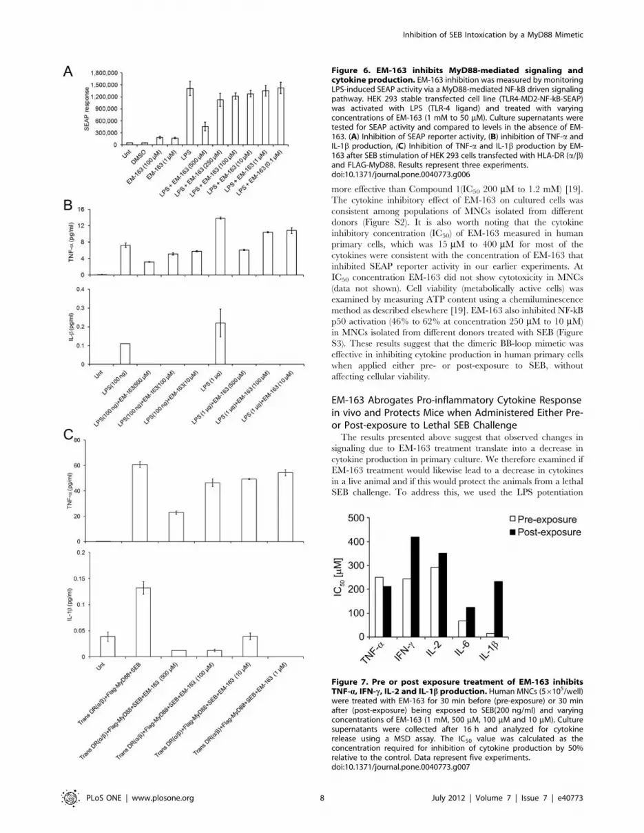
As shown in Figure 6A, EM-163 inhibited the LPS-induced
TLR4- MD2-MyD88-NF-kB-SEAP response in a dose-dependent
manner. In contrast, TLR3 ligand poly IC stimulation, which does
not utilize MyD88, had no effect on SEAP response (data not
shown). In addition to inhibition of the SEAP response to LPS
stimulation, EM-163 also blocked LPS-induced TNF-a and IL-1bproduction by these cells in a dose-dependent manner (Figure 6B).We also confirmed that EM-163 was capable of inhibiting cytokine
production stimulated by SEB in a similar HEK 293 cell model.
HEK 293 cells were co-transfected with HLA-DR (a/b) and Flag-
tagged MyD88 plasmids and stimulated with SEB in the presence
or absence of EM-163. Culture supernatants were then tested for
the presence of TNF-a and IL-1b. Results shown in Figure 6C
demonstrate SEB-stimulated production of TNF-a and IL-1b by
Figure 4. SEB stimulation of human monocytes in the presence of EM-163 leads to intracellular accumulation of MyD88. Increasedaccumulation of MyD88 in CD14+ monocytes treated with SEB in the presence of EM-163 at 1 h and 4 h. Confocal images show expression of nuclei(blue) and intracellular MyD88 (red) proteins in CD14+ monocytes. Monocytes were untreated or treated with SEB, EM-163 or SEB plus EM-163(250 mM). Cell nuclei were labeled with DAPI (blue). Scale bar = 10 mm. Data reperesent three similar experiments with different donors.doi:10.1371/journal.pone.0040773.g004
Inhibition of SEB Intoxication by a MyD88 Mimetic
PLoS ONE | www.plosone.org 6 July 2012 | Volume 7 | Issue 7 | e40773

HEK 293 cells expressing HLA-DR and MyD88, and its
inhibition by EM-163. These results clearly demonstrate that
MHC class II-linked activation of MyD88 signaling [12] was also
inhibited by EM-163.
EM-163 Inhibits SEB-induced TNF-a, IFN-c, IL-1b, IL-2 andIL-6 Production in Human Primary CellsThe results above indicated that EM-163 inhibits downstream
signaling by targeting MyD88 in a transfected HEK 293 cell line,
so next we asked if the change in signaling would likewise lead to
a decrease in cytokine production in cultured human primary
cells. To determine therapeutic potential, it was also important to
examine the efficacy of EM-163 when administered after SEB
exposure. To assess the inhibitory effects of EM-163 on SEB-
induced cytokine production in primary culture, MNCs were
treated with EM-163 for 30 min before (pre-exposure) or 30 min
after (post-exposure) being exposed to SEB. Culture supernatants
were collected after 16 h and analyzed for cytokines. As shown in
Figure 7, pre- or post-exposure treatment with EM-163 inhibited
SEB-induced TNF-a, IFN-c, IL-1b, IL-2 and IL-6 production
(IC50 15 mM to 400 mM), although EM-163 was generally more
effective (had a lower IC50) when applied pre-SEB exposure
compared to post-exposure treatment. As predicted, EM-163 was
Figure 5. EM-163 targets newly expressed MyD88 and stabilizes the MyD88 complex. HEK 293T cells were co-transfected with plasmidsMyD88- Flag or pCMV-HA-MyD88. Seven hours after transfection, cells were incubated for 13 h with or without 2163 (500 mM to 5 mM). (A), Cellextracts were immunoprecipitated (IP) with anti-Flag antibody, and immune-precipitated proteins were analyzed in Western blot with anti-HA. (B)Densitometry analysis of the results shown in (A). The results are representative of three independent experiments.doi:10.1371/journal.pone.0040773.g005
Inhibition of SEB Intoxication by a MyD88 Mimetic
PLoS ONE | www.plosone.org 7 July 2012 | Volume 7 | Issue 7 | e40773
more effective than Compound 1(IC50 200 mM to 1.2 mM) [19].
The cytokine inhibitory effect of EM-163 on cultured cells was
consistent among populations of MNCs isolated from different
donors (Figure S2). It is also worth noting that the cytokine
inhibitory concentration (IC50) of EM-163 measured in human
primary cells, which was 15 mM to 400 mM for most of the
cytokines were consistent with the concentration of EM-163 that
inhibited SEAP reporter activity in our earlier experiments. At
IC50 concentration EM-163 did not show cytotoxicity in MNCs
(data not shown). Cell viability (metabolically active cells) was
examined by measuring ATP content using a chemiluminescence
method as described elsewhere [19]. EM-163 also inhibited NF-kB
p50 activation (46% to 62% at concentration 250 mM to 10 mM)
in MNCs isolated from different donors treated with SEB (Figure
S3). These results suggest that the dimeric BB-loop mimetic was
effective in inhibiting cytokine production in human primary cells
when applied either pre- or post-exposure to SEB, without
affecting cellular viability.
EM-163 Abrogates Pro-inflammatory Cytokine Responsein vivo and Protects Mice when Administered Either Pre-or Post-exposure to Lethal SEB ChallengeThe results presented above suggest that observed changes in
signaling due to EM-163 treatment translate into a decrease in
cytokine production in primary culture. We therefore examined if
EM-163 treatment would likewise lead to a decrease in cytokines
in a live animal and if this would protect the animals from a lethal
SEB challenge. To address this, we used the LPS potentiation
Figure 6. EM-163 inhibits MyD88-mediated signaling andcytokine production. EM-163 inhibition was measured by monitoringLPS-induced SEAP activity via a MyD88-mediated NF-kB driven signalingpathway. HEK 293 stable transfected cell line (TLR4-MD2-NF-kB-SEAP)was activated with LPS (TLR-4 ligand) and treated with varyingconcentrations of EM-163 (1 mM to 50 mM). Culture supernatants weretested for SEAP activity and compared to levels in the absence of EM-163. (A) Inhibition of SEAP reporter activity, (B) inhibition of TNF-a andIL-1b production, (C) Inhibition of TNF-a and IL-1b production by EM-163 after SEB stimulation of HEK 293 cells transfected with HLA-DR (a/b)and FLAG-MyD88. Results represent three experiments.doi:10.1371/journal.pone.0040773.g006
Figure 7. Pre or post exposure treatment of EM-163 inhibitsTNF-a, IFN-c, IL-2 and IL-1b production. Human MNCs (56105/well)were treated with EM-163 for 30 min before (pre-exposure) or 30 minafter (post-exposure) being exposed to SEB(200 ng/ml) and varyingconcentrations of EM-163 (1 mM, 500 mM, 100 mM and 10 mM). Culturesupernatants were collected after 16 h and analyzed for cytokinerelease using a MSD assay. The IC50 value was calculated as theconcentration required for inhibition of cytokine production by 50%relative to the control. Data represent five experiments.doi:10.1371/journal.pone.0040773.g007
Inhibition of SEB Intoxication by a MyD88 Mimetic
PLoS ONE | www.plosone.org 8 July 2012 | Volume 7 | Issue 7 | e40773
model of SEB toxicity in mice [5,10,11]. BALB/c mice (n = 6)
were injected intraperitoneally (i.p.) with varying amounts of EM-
163 (0.21 mg/mouse, 0.42 mg/mouse, and 0.86 mg/mouse) and
30 min later they were treated with SEB (0.5 mg/mouse, 1LD50),
followed by LPS another 2 h later. All animals in the study not
treated with EM-163 succumbed by 48 h. In contrast, treatment
with 0.21 mg/mouse of EM-163 delayed death. Mice that were
treated with EM-163 at concentrations of 0.42 mg/mouse or
0.86 mg/mouse were completely protected (p = 0.05) (Figure 8A ).
When mice were exposed to a higher lethal dose of SEB (5 mg/mouse, 10 LD50), all mice not treated with EM-163 succumbed by
30 h. In contrast, EM-163 pre-treatment with 0.86 mg/mouse
delayed death and pre-treatment with 1.7 mg/mouse protected
completely [Figure 8A]. Using the same experimental paradigm as
for the results shown in Figure 8A, we determined the pro-
inflammatory serum cytokine profile at the 4 h time point. The
results shown in Figure 8B indicated that the cytokine response
was abrogated in mice treated with EM-163, suggesting that
cytokine responses are correlated with the survival of the mice. In
addition to pre-treatment, our results also showed that mice
treated with EM-163 (0.86 mg/mouse) 30 min after SEB exposure
(0.5 mg/mouse, 1LD50) were completely protected. With a higher
challenge dose of SEB (5 mg/mouse, 10LD50) EM-163 still delayed
the death of mice (p = 0.012, log-rank pairwise comparisons tests)
(Figure 8C). As for BALB/c mice challenged with SEB, C57BL/6
mice pretreated with EM-163 and challenged with a lethal dose of
SEA likewise showed inhibition of cytokine responses and were
protected (Figure S4). These results suggest that EM-163
effectively inhibited pro-inflammatory cytokine responses and
protected mice from toxic shock-induced death from lethal SEB or
SEA challenge. Taken together, the results indicate that EM-163
has substantial therapeutic potential against SEB intoxication.
Discussion
Our previous studies showed that MyD88-mediated pro-in-
flammatory cytokine signaling is critical to SEB toxicity [11,12].
This indicated that MyD88 was a valid therapeutic target and
subsequent preliminary work using a synthetic mimetic of the BB-
loop of the Toll/IL-1 receptor (TIR) domain of MyD88, provided
proof of concept for this strategy to treat SEB-induced toxic shock
syndrome [19]. Here, we have shown that synthesis of a BB-loop
mimetic with dual functional groups greatly enhanced the
biological activity and target specificity of the mimetic, a large
improvement over earlier work. The ‘‘dimeric’’ compound EM-
163 attenuated TNF-a, IFN-c, IL-1b, IL-2 and IL-6 production
(IC50 15–400 mM) in human primary cells when applied either
pre- or post-exposure to SEB. Our biochemical and cell-based
reporter assay data suggest that EM-163, by targeting MyD88,
inhibits pro-inflammatory cytokine signaling. Most importantly,
administration of EM-163 abrogated pro-inflammatory cytokine
production and completely protected mice from the toxic shock
induced-death of a lethal SEB challenge.
Recent reports suggest that MyD88-mediated pro-inflammatory
cytokine signaling is not limited to the TLR/IL-1R, but rather is
generally shared by other receptors such as MHC receptors [13].
The role for MHC class II molecules in TLR signaling was first
suggested by studies that showed macrophages with no or low
expression of MHC class II had a defective inflammatory response
to LPS-stimulation [28]. It has also been reported that MHC class
II molecules enhance toll-like receptor mediated innate immune
responses [29]. In line with these observations, our recent results
demonstrated that SEB, which binds to MHC class II receptors,
activates MyD88-mediated pro-inflammatory signaling [12].
Thus, in addition to their classical function in antigen pre-
sentation, MHC class II molecules are now shown to promote
MyD88 signaling. As the MHC class II molecule lacks any known
signaling motifs in its short cytoplasmic tail, it has been proposed
that it might use associated signaling molecules at the membrane.
These signaling molecules include cell receptors (such as CD20,
CD79, CD23) and members of the immunoglobulin family (such
as CD19), the tetraspan family (such as CD81 and CD82), and the
TNF receptor family (such as CD40). All of these receptor families
have been shown to associate with MHC class II molecules, and
thus may likely be involved in initiating intracellular signaling
[13,30]. Whether these signaling proteins are functionally linked,
either directly or indirectly, to MHC class II molecules and
adaptor protein MyD88 is a question that is currently under
investigation in our laboratory. Nevertheless, results from our
laboratory and others have demonstrated that TLR and MHC-
mediated responses both engage the adaptor molecule MyD88
[14,31]. Likewise, SEB engagement of MHC class II molecules has
been shown to up-regulate both TLR4 [32] and MyD88 (Kissner
et al. 2011, unpublished observation) on human monocytes.
Previous work from our laboratory showed that LPS potentiates
SEB-induced lethal shock in mice [5,11,12]. LPS primarily
interacts with CD14 receptors on macrophages, while SEB
triggers MHC class II-positive cells and T-cells to release pro-
inflammatory cytokines. Therefore, it is likely that MHC class II-
SEB binding up regulates MyD88 and TLR4, and subsequent
LPS stimulation may synergistically trigger interaction between
the respective TIR domains of MyD88 and TLR/IL-1R1
receptors. This causes activation of several signaling cascade
pathways that include nuclear factor NF-kB and transcription
factor AP-1, as well as various stress-associated kinases, resulting in
release of pathological levels of pro-inflammatory cytokines.
Although the underlying mechanism by which MHC class II
signals recruit MyD88 is currently under investigation, the adaptor
protein is known to integrate signals through TIR domain-TIR
domain interactions [16,33]. In this study, our results indicated
that the compound EM-163 (designed to be structurally similar to
the BB-loop in the TIR domain of MyD88) inhibited the MyD88-
mediated signaling initiated by exposure to SEB. In addition,
a preliminary result from our laboratory indicated that EM-163
was capable of inhibiting MyD88-mediated pro-inflammatory
cytokine responses with exposure to Francisella tularensis LPS or
irradiated Burkholderia mallei (unpublished observation). As signaling
through MyD88 appears to be a common link in many of the host-
directed inflammatory process, it is likely that EM-163 may have
a potential for broader use against exposure to other bio-threat
agents.
The most potent microbial products implicated in the patho-
genesis of septic shock are gram-positive-derived superantigenic
exotoxins and gram-negative endotoxins. Both types of toxin
induce comparable pro-inflammatory cytokines from human
mononuclear cells in vitro, cause lethal shock in vivo, and have
been identified in the bloodstream of critically ill patients [8,9].
The combination of endotoxins and superantigens has been shown
to have particularly severe consequences in several animal models.
For example, it has been reported that SEB potentiates LPS-
induced hepatic dysfunction and cytokine responses in chronically
catheterized rats [34]. Results from our laboratory also firmly
established that LPS and SEB synergize to produce a dose
dependent toxicity in mice that is several fold higher than the
toxicity of SEB or LPS alone. Our results raise the possibility that
recognition of SEB by MHC class II receptors may exacerbate the
pro-inflammatory response of monocytes to gram-negative in-
fection or endotoxin through activation of a common MyD88-
Inhibition of SEB Intoxication by a MyD88 Mimetic
PLoS ONE | www.plosone.org 9 July 2012 | Volume 7 | Issue 7 | e40773

Figure 8. EM-163 attenuates SEB -induced pro-inflammatory serum cytokines and lethality in mice. BALB/c mice (n = 6) were injected(i.p.) with different amounts of EM-163, 30 min later injected with SEB followed by LPS 2 h later. Mice were observed to determine if mice survived, ortime to death if they did not. Control mice injected only with 60 mg of LPS or 1 mg of SEB survived. Data represent three separate experiments. Kaplan
Inhibition of SEB Intoxication by a MyD88 Mimetic
PLoS ONE | www.plosone.org 10 July 2012 | Volume 7 | Issue 7 | e40773

mediated signaling. In both cases, targeting of MyD88 by EM-163
inhibited pro-inflammatory cytokine signaling and protected mice
from SEB intoxication. EM-163 treatment caused intracellular
accumulation of MyD88 in primary monocytes when stimulated
with SEB. It is likely that EM-163 binds to MyD88, including
MyD88 that is newly synthesized in response to SEB, rendering it
incapable of downstream signal transduction. EM-163 may or
may not be capable of disrupting dimers of the MyD88 TIR
domain, but it is clear that stable, monomeric forms of MyD88
must exist since they engage interchangeably in both homo and
heterotypic interactions. It seems likely that the monomeric form
of the MyD88 TIR domain, with its exposed BB loop, is the
molecular target of EM-163.
In summary, our results provide evidence that a synthetic BB-
loop mimetic, EM-163, that targets the TIR-domain of MyD88
can limit hyper-inflammation and prevent toxic shock. The
effectiveness of this mimetic when administered either pre- or
post-SEB exposure further supports its therapeutic potential. An
ongoing effort is underway to further refine EM-163 by chemical
modification to increase potency, bioavailability and drug-like
properties for potential clinical use against toxic shock.
Supporting Information
Figure S1 EM-163 targets newly expressed MyD88.MyD88 KO HEK293 (HEK293-I3A) cell line were co-transfected
with plasmids MyD88 flag and pCMV-HA-MyD88. Seven hours
after transfection, cells were incubated for 13 h with or without
EM-163 (100 mM to 1 mM). At the end of incubation, cells were
lysed, and cytoplasmic fractions were separated. (A), MyD88
expression was detected by Western blot analysis using anti-
MyD88 antibody. (B), Cell extracts were immunoprecipitated (IP)
with anti-Flag antibody, and immune-precipitated proteins were
analyzed in Western blot with anti-MyD88 antibody. (C)
Immunoblot (B) was stripped and reprobed with anti-HA
antibody. The results are representative of two independent
experiments.
(TIF)
Figure S2 EM-163 inhibits SEB-induced cytokine re-sponse in different donors. MNCs (16106) from two normal
donors (Donor 1 and Donor 2) were cultured with SEB (200 ng/
ml) with or without EM-163 (500 mM to 10 mM) for 20 h. The
culture supernatants were collected and measured for cytokine by
MSD assay. The IC50 value was calculated as the concentration
required for inhibition of cytokine production by 50% relative to
the control. Data are representative of three experiments.
(TIF)
Figure S3 Inhibition of NF-kB p50 activation in thepresence of EM-163 in MNCs stimulated with SEB.Activation of NF-kB in primary mononuclear cells treated with
SEB in the absence or presence of different concentration EM-163
was determined as described elsewhere [12]. Data are presented in
the figure as percentage increase over the control and represent
one of three experiments using separate donors. Significant
differences (p#0.005) are indicated for MNCs treated with SEB
vs MNCs treated SEB in the presence of EM-163 (*).
(TIF)
Figure S4 EM-163 attenuates pro-inflammatory cyto-kines and SEA induced lethality in mice. (A), Administra-
tion of EM-163 protected mice from toxic shock induced death
challenged with lethal dose of SEA. C57BL/6 mice mice (n = 4)
were injected with EM-163 (0.85 mg, or 1.7 mg, 100 ml volume/
mouse), 30 min later injected with SEB (5 mg/mouse) followed by
LPS 2 h later. Mice were observed for survival. Control mice
injected with 150 mg of LPS or 1 mg of SEB survived. Data are
representative of two separate experiments; (B), Administration of
EM-163 in mice inhibited pro-inflammatory cytokine response,mice were bled at 24 h, serum were pooled from each group and
measured serum cytokines.
(TIF)
Acknowledgments
The authors thank Thomas Plummer for his help with animal experiments,
Lorraine Farinick for figure preparation and Diana Fisher for statistical
analysis. We are also grateful to the investigators at Addgene for providing
plasmids originally published in Proc. Natl. Acad. Sci 2006 Jul.18.103(29):
10961–6.
Author Contributions
Conceived and designed the experiments: Performed cytokine measure-
ment and micro-array experiments: TLK. Partly involved in writing the
manuscript: TLK. Performed the confocal image data collection and edited
the manuscript: GR. Performed the transfection, co-immunoprecipitation
and animal experiments: SA. Contributed in the synthesis of compounds:
EM DA. Contributed optimization and data analysis of micro-array based
binding experiments: EL SF RU. Contributed design and synthesis of BB-
loop mimetic: MR. Contributed expression and purification of TIR
domain recombinant protein: SP DSW. Designed and supervised overall
synthetic chemistry: JR. Designed and supervised the research and wrote
the manuscript: KUS. Performed the experiments: TLK GR SA EM DA
EL SF SP KUS. Analyzed the data: TLK GR SA EM DA MR EL SF SP
DSW JR KUS. Contributed reagents/materials/analysis tools: RGU.
Wrote the paper: TLK SP MR KUS.
References
1. Fraser JD (1989) High-affinity binding of staphylococcal enterotoxins A and B to
HLA-DR. Nature 339: 221–223.
2. Dinges MM, Orwin PM, Schlievert PM (2000) Exotoxins of Staphylococcus
aureus. Clin Microbiol Rev 13: 16–34.
3. Scholl P, Diez A, Mourad W, Parsonnet J, Geha RS, et al. (1989) Toxic shock
syndrome toxin 1 binds to major histocompatibility complex class II molecules.
Proc Natl Acad Sci USA 86: 4210–4214.
4. Ulrich RG, Bavari S, Olson MA (1995) Bacterial superantigens in human
disease: structure, function and diversity. Trends Microbiol 3: 463–468.
5. Stiles BG, Bavari S, Krakauer T, Ulrich RG (1993) Toxicity of staphylococcal
enterotoxins potentiated by lipopolysaccharide: major histocompatibility com-
plex class II molecule dependency and cytokine release. Infect Immun 61: 5333–
5338.
6. Blank C, Luz A, Bendigs S, Erdmann A, Wagner H, et al. (1997) Superantigen
and endotoxin synergize in the induction of lethal shock. Eur J Immunol 27:
825–833.
7. Schlievert P (1982) Enhancement of host susceptibility to lethal endotoxin shock
by staphylococcal pyrogenic exotoxin type C. Infect Immun 36: 123–128.
Meier survival analysis and log-rank tests were performed to compare survival curves with stepdown Bonferroni adjustment for pair wise comparisonsbetween groups. (A), Pre-treatment of EM-163, 0.42 mg/mouse, or 0.86 mg/mouse protected mice against lethal SEB (0.5 mg/mouse,1 LD50)
challenge, p = 0.0015; Pre-treatment of EM-163 protected mice challenged with SEB (5 mg/mouse equivalent to10 LD50), p#0.0261; (B). Administrationof EM-163 in mice inhibited pro-inflammatory cytokine response. Similar to experimental settings as in Figure 7A,mice (n = 6) were bled at 4 h, serumwere pooled from each group [LPS, SEB+LPS, EM-163 (1.7 mg/mouse)] and measured serum cytokines. (C). Post-exposure to SEB (0.5 mg I LD50), EM-163 treated mice were protected, p = 0.0102; Post-exposure to SEB challenge, EM-163 delayed death (5 mg= 10 LD50), p#0.0129.doi:10.1371/journal.pone.0040773.g008
Inhibition of SEB Intoxication by a MyD88 Mimetic
PLoS ONE | www.plosone.org 11 July 2012 | Volume 7 | Issue 7 | e40773
8. Danner R L, Elin RJ, Hosseini JM, Wesley RA, Reilly JM, et al. (1991)
Endotoxemia in human septic shock. Chest 99: 169–175.9. Azuma K, Koike K, Kobayashi T, Mochizuki T, Mashiko K, et al. (2004)
Detection of circulating superantigens in an intensive care unit population.
Int J Infect Dis 8: 292–298.10. Kissner TL, Cisney ED, Ulrich RG, Fernandez S, Saikh KU (2010)
Staphylococcal enterotoxin A induction of pro-inflammatory cytokines andlethality in mice is primarily dependent on MyD88. Immunology 130: 516–526.
11. Kissner TL, Ruthel G, Cisney ED, Ulrich RG, Fernandez S, et al. (2011)
MyD88-dependent pro-inflammatory cytokine response contributes to lethaltoxicity of staphylococcal enterotoxin B in mice. Innate Immunity 17: 451–462.
12. Kissner TL, Ruthel G, Alam S, Ulrich RG, Fernandez S, et al. (2011) Activationof MyD88 signaling upon staphylococcal enterotoxin binding to MHC class II
molecules. PLoS ONE 6: e15985.13. Liu X, Zhan Z, Li D, Xu L, Ma F, et al. (2011) Intracellular MHC class II
molecules promote TLR-triggered innate immune responses by maintaining
activation of the kinase Btk. Nat Immunol 12: 416–424.14. Hassan GS, Mourad W (2011) An unexpected role for MHC class II. Nat
Immunol 12: 375–376.15. Akira S, Takeda K (2004) Toll-like receptor signaling. Nat Rev Immunol 4: 499–
511.
16. O’Neill LAJ (2003) The role of MyD88-like adapters in Toll-like receptor signaltransduction. Biochem Soc Trans 31: 643–647.
17. Sun D, Ding A (2006) MyD88-mediated stabilization of interferon-gamma-induced cytokine and chemokine mRNA. Nat Immunol 7: 375–381.
18. Cook DN, Pisetsky DS, Schwartz DA (2004) Toll-like receptors in thepathogenesis of human diseases. Nat Immunol 5: 975–979.
19. Kissner TL, Moisan L, Mann E, Alam S, Ruthel G, et al. (2011) A small
molecule that mimics the BB-loop in the Toll/IL-1 receptor domain of MyD88attenuates staphylococcal enterotoxin B induced pro-inflammatory cytokine
production and toxicity in mice. J Biol Chem 286: 31385–31396.20. Davis CN, Mann E, Behrens MM, Gaidarova S, Rebek M, et al. (2006) MyD88-
dependent and –independent signaling by IL-1 in neurons probed by
bifunctional Toll/IL-1 receptor domain/BB-loop mimetics. Proc Natl AcadSci USA 103: 2953–2958.
21. Nallamsetty S, Austin BP, Penrose KJ, Waugh DS (2005) Gateway vectors forthe production of combinatorially-tagged His6-MBP fusion proteins in the
cytoplasm and periplasm of Escherichia coli. Protein Sci 14: 2964–2971.
22. Tropea JE, Cherry S, Waugh DS (2009) Expression and purification of soluble
His (6)-tagged TEV protease. Methods Mol Biol 498: 297–307.23. Xu Y, Tao X, Shen B, Horng T, Medzhitov R, et al. (2000) Structural basis for
signal transduction by the Toll/interleukin-1 receptor domains. Nature 408:
111–115.24. Nyman T, Stenmark P, Flodin S, Johansson I, Hammarstrom M, et al. (2008)
The crystal structure of the human toll-like recptor 10 cytoplasmic domainreveals a putative signaling dimer. J Biol Chem 283:11861–11865.
25. Chan SI, Low LY, Hsu S, Li S, Liu T, et al. (2009) Molecular mimicry in innate
immunity: crystal structure of a bacterial TIR domain. J Biol Chem 284: 21386–21392.
26. Burns K, Martinon F, Esslinger C, Pahl H, Schneider P, et al. (1998) MyD88, anadapter protein involved in interleukin-1 signaling. J Biol Chem 273: 12203–
12209.27. Basith S, Manavalan B, Govindaraj RG, Choi S (2011) In silico approach to
inhibition of signaling pathways of Toll-like receptors 2 and 4 by ST2L. PLoS
One 6: e23989.28. Piani A, Hossle JP, Birchler T, Siegrist CA, Heumann D, et al. (2000) Expression
of MHC class II molecules contributes to lipopolysaccharide responsiveness.Eur J Immunol 30: 3140–3146.
29. Frei R, Steinle J, Birchler T, Loeliger S, Roduit C, et al. (2010) MHC class II
molecules enhance Toll-like receptor mediated innate immune responses. PLoSOne 5: e8808.
30. Al-Daccak R, Mooney N, Charron D (2004) MHC class II signaling in antigenpresenting cells. Curr Opin Immunol 16: 108–113.
31. Gabhann JN, Jefferies CA (2011) TLR-induced activation of Btk-role forendosomal MHC class II molecules revealed. Cell Res 21: 998–1001.
32. Hopkins PA, Fraser JD, Pridmore AC, Russell HH, Read RC, et al. (2005)
Superantigen recognition by HLA class II on monocytes up-regulates toll-likereceptor 4 and enhances proinflammatory responses to endotoxin. Blood 105:
3655–3662.33. Dunne A, O’Neill LA (2003) The interleukin-1 receptor/Toll-like receptor
superfamily: signal transduction during inflammation and host defense. Sci
STKE 2003: re3.34. Beno DW, Uhing MR, Goto M, Chen Y, Jiyamapa-Serna VA, et al. (2001)
Staphylococcal enterotoxin B potentiates LPS-induced hepatic dysfunction inchronically catherized rats. Am J Physiol Gastrointest Liver Physiol 280: G866–
G872.
Inhibition of SEB Intoxication by a MyD88 Mimetic
PLoS ONE | www.plosone.org 12 July 2012 | Volume 7 | Issue 7 | e40773