pyogenic bacterial infections in humans with myd88 deficiency
TRANSCRIPT

DOI: 10.1126/science.1158298 , 691 (2008); 321Science
et al.Horst von Bernuth,MyD88 DeficiencyPyogenic Bacterial Infections in Humans with
www.sciencemag.org (this information is current as of August 1, 2008 ):The following resources related to this article are available online at
http://www.sciencemag.org/cgi/content/full/321/5889/691version of this article at:
including high-resolution figures, can be found in the onlineUpdated information and services,
http://www.sciencemag.org/cgi/content/full/321/5889/691/DC1 can be found at: Supporting Online Material
http://www.sciencemag.org/cgi/content/full/321/5889/691#otherarticles, 16 of which can be accessed for free: cites 29 articlesThis article
http://www.sciencemag.org/cgi/collection/immunologyImmunology
: subject collectionsThis article appears in the following
http://www.sciencemag.org/about/permissions.dtl in whole or in part can be found at: this article
permission to reproduce of this article or about obtaining reprintsInformation about obtaining
registered trademark of AAAS. is aScience2008 by the American Association for the Advancement of Science; all rights reserved. The title
CopyrightAmerican Association for the Advancement of Science, 1200 New York Avenue NW, Washington, DC 20005. (print ISSN 0036-8075; online ISSN 1095-9203) is published weekly, except the last week in December, by theScience
on
Aug
ust 1
, 200
8 w
ww
.sci
ence
mag
.org
Dow
nloa
ded
from

tivated T cells. HnRNPLL is induced upon T cellstimulation and as such is likely to correspond tothe inducible, cycloheximide-sensitive factor postu-lated to promote the CD45RA > RO transitionduringTcell activation (10).Depletion of hnRNPLLcauses an overall shift in patterns of alternativesplicing, which by and large are in the oppositedirection from those seen in activated T cells inwhich hnRNPLLexpression is increased. Inductionof hnRNPLL during the process of Tcell activationand differentiation may represent a mechanism bywhich the cell can rapidly shift its transcriptometo favor proliferation and inhibit cell death.
There is increasing evidence that splicing andpre-mRNA processing involve multiprotein com-plexes in which individual components influencedistinct aspects of transcript production (20, 21).Several proteins, including hnRNPL, PSF (poly-pyrimidine tract binding protein–associated splic-ing factor), and hnRNPE2, have been shown tobind CD45 transcripts (14, 15). However, none ofthese factors is induced upon T cell activation(14, 15), and only PSF (splicing factor proline/glutamine-rich) was identified in our screen, albeitas a weak hit (table S2). Together with its as-sociated factors, hnRNPL may influence diverseaspects of CD45 pre-mRNA processing, in-cluding basal splicing, mRNA stability, andexport (22, 23). After activation, hnRNPLL in-duction and recruitment to CD45 transcripts
may then lead to the assembly of a high-affinityexon repression complex that mediates efficientCD45RO production as well as global changes inalternative splicing.
References and Notes1. J. M. Johnson et al., Science 302, 2141 (2003).2. B. Modrek, C. Lee, Nat. Genet. 30, 13 (2002).3. D. L. Black, Annu. Rev. Biochem. 72, 291 (2003).4. A. J. Matlin, F. Clark, C. W. Smith, Nat. Rev. Mol.
Cell Biol. 6, 386 (2005).5. M. L. Hermiston, Z. Xu, A. Weiss, Annu. Rev. Immunol.
21, 107 (2003).6. K. W. Lynch, Nat. Rev. Immunol. 4, 931 (2004).7. R. J. Salmond, L. McNeill, N. Holmes, D. R. Alexander,
Int. Immunol. 20, 819 (2008).8. N. Holmes, Immunology 117, 145 (2006).9. L. McNeill et al., Immunity 27, 425 (2007).10. K. W. Lynch, A. Weiss, Mol. Cell. Biol. 20, 70 (2000).11. J. Moffat et al., Cell 124, 1283 (2006).12. Materials and methods are available as supporting
material on Science Online.13. I. S. Trowbridge, M. L. Thomas, Annu. Rev. Immunol. 12,
85 (1994).14. C. R. Rothrock, A. E. House, K. W. Lynch, EMBO J. 24,
2792 (2005).15. A. A. Melton, J. Jackson, J. Wang, K. W. Lynch, Mol. Cell.
Biol. 27, 6972 (2007).16. I. Shur, D. Ben-Avraham, D. Benayahu, Gene 334, 113
(2004).17. S. A. Tenenbaum, P. J. Lager, C. C. Carson, J. D. Keene,
Methods 26, 191 (2002).18. M. J. Moore, P. A. Silver, RNA 14, 197 (2008).19. National Center for Biotechnology Information Database;
www.ncbi.nlm.nih.gov/sites/entrez?db=gene
20. X. M. Ma, S. O. Yoon, C. J. Richardson, K. Julich, J. Blenis,Cell 133, 303 (2008).
21. P. Valencia, A. P. Dias, R. Reed, Proc. Natl. Acad.Sci. U.S.A. 105, 3386 (2008).
22. S. Guang, A. M. Felthauser, J. E. Mertz, Mol. Cell. Biol.25, 6303 (2005).
23. L. H. Hung et al., RNA 14, 284 (2008).24. We thank A. Weiss for the JSL1 cell line, E. Kieff for BJAB
and BL41 cells, B. North for the lentiviral expressionplasmid, and the RNAi Consortium at the Broad Institutefor providing the plasmids to produce the splicing factorlibrary. We thank C. Moita for assistance in assemblingthe library and A. Astier and D. Hafler for aid indeveloping the protocol for infecting and silencing genesin primary human T cells. We thank K. Eger andD. Unutmaz for the naïve and activated cord bloodsamples. We thank J. Burke at Biotique for assistancewith the array analysis. This work was supported by NIHgrants CA42471, AI40127, and AI44432 to A.R., andU19 AI070352, R21 AI071060, and Defense AdvancedResearch Projects Agency grant W81XWH-04-C-0139 toN.H. S.O. is supported by postdoctoral training grantT32 HL066987 from the Joint Program in TransfusionBiology and Medicine, Children’s Hospital, Boston.
Supporting Online Materialwww.sciencemag.org/cgi/content/full/1157610/DC1Materials and MethodsSOM TextFigs. S1 to S4Tables S1 to S4References
11 March 2008; accepted 20 June 2008Published online 10 July 2008;10.1126/science.1157610Include this information when citing this paper.
Pyogenic Bacterial Infectionsin Humans with MyD88 DeficiencyHorst von Bernuth,1,2 Capucine Picard,1,2,3 Zhongbo Jin,4,5 Rungnapa Pankla,4,6Hui Xiao,7 Cheng-Lung Ku,1,2 Maya Chrabieh,1,2 Imen Ben Mustapha,1,2,8 Pegah Ghandil,1,2Yildiz Camcioglu,9 Júlia Vasconcelos,10 Nicolas Sirvent,11 Margarida Guedes,10Artur Bonito Vitor,12 María José Herrero-Mata,13 Juan Ignacio Aróstegui,14 Carlos Rodrigo,15Laia Alsina,16 Estibaliz Ruiz-Ortiz,13 Manel Juan,14 Claudia Fortuny,16 Jordi Yagüe,14Jordi Antón ,16 Mariona Pascal,14 Huey-Hsuan Chang,17 Lucile Janniere,1,2 Yoann Rose,1,2Ben-Zion Garty,18 Helen Chapel,19 Andrew Issekutz,20 László Maródi,21Carlos Rodriguez-Gallego,22 Jacques Banchereau,4 Laurent Abel,1,2 Xiaoxia Li,7Damien Chaussabel,4 Anne Puel,1,2 Jean-Laurent Casanova1,2,23*
MyD88 is a key downstream adapter for most Toll-like receptors (TLRs) and interleukin-1receptors (IL-1Rs). MyD88 deficiency in mice leads to susceptibility to a broad range ofpathogens in experimental settings of infection. We describe a distinct situation in a naturalsetting of human infection. Nine children with autosomal recessive MyD88 deficiency sufferedfrom life-threatening, often recurrent pyogenic bacterial infections, including invasivepneumococcal disease. However, these patients were otherwise healthy, with normal resistanceto other microbes. Their clinical status improved with age, but not due to any cellularleakiness in MyD88 deficiency. The MyD88-dependent TLRs and IL-1Rs are thereforeessential for protective immunity to a small number of pyogenic bacteria, but redundantfor host defense to most natural infections.
The search for human genetic etiologiesof pediatric infectious diseases aims todecipher the molecular mechanism of
disease and to reveal the function of immunegenes in natura (1). The immunological inves-tigation of children with invasive pneumococcal
disease (IPD) led to the discovery of childrenlacking interleukin-1 receptor–associated kinase4 (IRAK-4), which is selectively recruited to Toll-like receptors (TLRs) and interleukin-1 receptors(IL-1Rs) by MyD88 (2). The patients presentwith a life-threatening but narrow and transient
predisposition to infection, apparently restrictedto pyogenic bacterial diseases, particularly IPD,during the first 10 years of life (3). This clinicalphenotype is surprising given the central rolecommonly attributed to both TLRs and IL-1Rs,and the high susceptibility of MyD88-deficientmice to experimental infections with at least 35pathogens—19 bacteria, seven viruses, five para-sites, and four fungi (4) (tables S1 to S3). How-ever, IRAK-4–deficient mice have thus far beenchallenged with only a few pathogens (5). Fibro-
1Human Genetics of Infectious Diseases, INSERM U550, Paris,France. 2Paris Descartes University, France. 3Study Center ofPrimary Immunodeficiencies, Assistance Publique Hôpitaux deParis, Necker Hospital, Paris, France. 4Baylor Institute for Im-munology Research, Dallas, TX 75204, USA. 5Baylor University,Waco, TX 76798, USA. 6Khon Kaen University, Thailand. 7Cleve-land Clinic Foundation, OH 44195, USA. 8Pasteur Institute ofTunis, Tunisia. 9CerrahpasaMedical School, Istanbul University,Turkey. 10General Hospital of Santo António, Porto, Portugal.11University Hospital Archet 2, Nice, France. 12Hospital S.João,Porto, Portugal. 13LIRAD–Banco de Sangre y Tejidos, Institutode Investigación Germans Trias i Pujol, Badalona, Barcelona,Spain. 14Immunology Department, Hospital Clinic, IDIBAPS,Barcelona, Spain. 15Germans Trias i Pujol Hospital, BarcelonaAutonomous University, Spain. 16Sant Joan de Déu Hospital,Barcelona University, Spain. 17Dendritic Cell Immunobiology,Institut Pasteur and INSERM U818, Paris, France. 18SchneiderChildren’s Medical Center, Petah Tiqva, Israel. 19University ofOxford and Oxford Radcliffe Hospital, Oxford, UK. 20DalhousieUniversity, Halifax, Nova Scotia, Canada. 21Debrecen Uni-versity, Hungary. 22Gran Canaria Dr Negrin Hospital, Las Palmasde Gran Canaria, Spain. 23Pediatric Hematology-ImmunologyUnit, Necker Hospital, Paris, France.
*To whom correspondence should be addressed. E-mail:[email protected]
www.sciencemag.org SCIENCE VOL 321 1 AUGUST 2008 691
REPORTS
on
Aug
ust 1
, 200
8 w
ww
.sci
ence
mag
.org
Dow
nloa
ded
from

blasts and individual leukocyte subsets fromIRAK-4–deficient patients fail to respond to theTLR agonists tested, at least for the orthologs ofthe mouse MyD88-dependent target genes tested(3, 6). The resistance of IRAK-4–deficient pa-tients might thus be explained by IRAK-4–independent but MyD88-dependent TLR orIL-1R responses in other cell types and/or forother target genes.
We investigated nine children (P1 to P9) withinvasive pyogenic bacterial diseases and with-out IRAK-4 deficiency, from five unrelated kin-dreds (supplementary note 1). Three childrendied between 1 and 11 months of age and six arenow between 3 and 16 years old. A homozygousin-frame MYD88 deletion was found in P1, P6,P8, and P9 (160del3, designated E52del), com-pound heterozygous missense mutations in P2(278 T→C, L93P; 586 C→T, R196C), and ahomozygous missense mutation in P3 and P4(586 C→T, R196C) (Fig. 1, A and B, and fig.S1). The deletion and missense mutations werenot found in 100 and 1728 healthy individuals,respectively. These mutations are nonconservativeand affect residues conserved across species (Fig.1C). Residues 195 to 197 are crucial for Toll/IL-1receptor (TIR)/TIR interaction (7). The segrega-tion of the MYD88 genotype and of the clinicalphenotype is consistent with an autosomal
recessive trait (supplementary note 1, Fig. 1A,and figs. S2 to S5). TheMYD88 mRNA in fibro-blasts from patients P1 to P4 (representing thethree combinations of alleles) was of normal mo-lecular weight and abundance (Fig. 1D and fig. S6).MyD88 protein was detected in SV40-transformedfibroblasts in trace amounts for P1, small amountsfor P2, and normal amounts for P3 and P4, asshown by Western blotting (Fig. 1E). These datasuggest that our patients have functional MyD88deficiency, with low (P1 and P2, P5 to P9) ornormal (P3 and P4) MyD88 protein levels.
The functional impact of the MyD88 muta-tions was then tested in cell lines derived fromthe patients. As in IRAK-4–deficient cells (2, 3, 6),IRAK-1 was not degraded in the patients’ SV40-transformed fibroblasts up to 6 hours afterstimulation with IL-1b (Fig. 2, A and D). Sim-ilarly, IRAK-1 was not degraded in the patients’Epstein-Barr virus–transformed B (EBV-B) cellsup to 2 hours after stimulation with the TLR7/TLR8 agonist R-848 (fig. S7, A and B). Phos-phorylation of the mitogen-activated protein ki-nases (MAPKs) p38 and c-Jun N-terminal kinase(JNK) and the DNA binding activity of nuclearfactor kB (NF-kB) were impaired in the patients’SV40-transformed fibroblasts after stimulationwith IL-1b (Fig. 2, B and C). Furthermore, thepatients’ EBV-B cells showed a complete lack of
response to stimulation with R-848 for 24 hours,in terms of tumor necrosis factor–a (TNF-a) in-duction (fig. S7C). Similarly, the production ofIL-6, IL-8, interferon-b (IFN-b), and IFN-l wasabolished after 24 hours of incubation of thepatients’ SV40-transformed fibroblasts with IL-1b, although these cells responded normally toTNF-a and poly(I:C), a TLR3-dependent agonistin human fibroblasts (8) (Fig. 2, E toH). Thus, twocell types derived from our patients carrying twomutant MYD88 alleles, with normal (P3 and P4)or impaired (P1 and P2) MyD88 protein pro-duction, showed a selective failure to respond tothe stimulation of two key IRAK-4–dependentsignaling pathways (TLR7/8 and IL-1R). Thisuniform cellular phenotype suggests that eachof the patients presents an autosomal recessive,functionally complete deficiency in MyD88.
Fibroblasts from a healthy control and fromP1 and P2 (representing the three mutant alleles)and from an IRAK-4–deficient child were tran-siently transfected with expression vectors encod-ing wild-type (WT) MyD88 or IRAK-4. Cellsfrom P1 and P2 regained IL-1b responsivenessafter transfection with the WTMYD88 gene only,as shown by the levels of IL-8 production (Fig.2I). IRAK-4–deficient cells, used as a control,were complemented with WT IRAK4 only (Fig.2I). The human embryonic kidney (HEK) 293–
Fig. 1. (A) Kindreds and patients with MYD88 mutations. (B) Positions ofthe MyD88 mutations in the death domain (DD) or the TIR domain of theprotein. (C) Parts of the DD and TIR domain of MyD88 in humans and thecorresponding regions in 24 other species. The residues mutated areindicated in red. Amino acid D197 (gray) is conserved in all species. (D) Full-length MYD88 transcripts in SV40-transformed fibroblasts from a healthycontrol donor (C+), four MyD88-deficient patients (P1 to P4), the MyD88-deficient HEK293 cell line (I3A), and the parental MyD88-positive HEK293cell line. (E) MyD88 protein expression in SV40-transformed fibroblasts froma healthy control (C+), four patients (P1 to P4), the I3A line, and theparental HEK293 cell line. The MyD88-specific antibody recognizes residues279 to 296. Abbreviations for the amino acid residues are as follows: A, Ala;C, Cys; D, Asp; E, Glu; F, Phe; G, Gly; H, His; I, Ile; K, Lys; L, Leu; M, Met; N,Asn; P, Pro; Q, Gln; R, Arg; S, Ser; T, Thr; V, Val; W, Trp; and Y, Tyr.
1 AUGUST 2008 VOL 321 SCIENCE www.sciencemag.org692
REPORTS
on
Aug
ust 1
, 200
8 w
ww
.sci
ence
mag
.org
Dow
nloa
ded
from
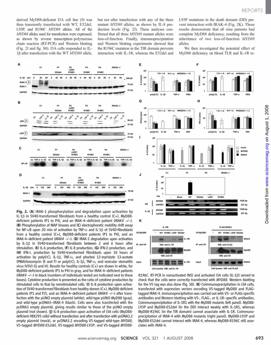
derived MyD88-deficient I3A cell line (9) wasthen transiently transfected with WT, E52del,L93P, and R196C MYD88 alleles. All of theMYD88 alleles used for transfection were expressed,as shown by reverse transcription–polymerasechain reaction (RT-PCR) and Western blotting(Fig. 2J and fig. S8). I3A cells responded to IL-1b after transfection with the WT MYD88 allele,
but not after transfection with any of the threemutant MYD88 alleles, as shown by IL-8 pro-duction levels (Fig. 2J). These analyses con-firmed that all three MYD88 mutant alleles wereloss-of-function. Finally, immunoprecipitationand Western blotting experiments showed thatthe R196C mutation in the TIR domain preventsinteraction with IL-1R, whereas the E52del and
L93P mutations in the death domain (DD) pre-vent interaction with IRAK-4 (Fig. 2K). Theseresults demonstrate that all nine patients hadcomplete MyD88 deficiency, resulting from theinheritance of two loss-of-function MYD88alleles.
We then investigated the potential effect ofMyD88 deficiency on blood TLR and IL-1R re-
Fig. 2. (A) IRAK-1 phosphorylation and degradation upon activation byIL-1b in SV40-transformed fibroblasts from a healthy control (C+), MyD88-deficient patients (P1 to P4), and an IRAK-4–deficient patient (IRAK4 −/−).(B) Phosphorylation of MAP kinases and (C) electrophoretic mobility shift assayfor NF-kB upon 20 min of activation by TNF-a and IL-1b of SV40-fibroblastsfrom a healthy control (C+), MyD88-deficient patients (P1 to P4), and anIRAK-4–deficient patient (IRAK4 −/−). (D) IRAK-1 degradation upon activationby IL-1b in SV40-transformed fibroblasts between 2 and 6 hours afterstimulation. (E) IL-6 production, (F) IL-8 production, (G) IFN-b production, and(H) IFN-l production by SV40-transformed fibroblasts upon 18 hours ofactivation by poly(I:C), IL-1b, TNF-a, and phorbol 12-myristate 13-acetate(PMA)/ionomycin (E and F) or poly(I:C), IL-1b, TNF-a, and vesicular stomatitisvirus (VSV) (G and H). Results for healthy controls (C+) are shown in white, forMyD88-deficient patients (P1 to P4) in gray, and for IRAK-4–deficient patients(IRAK4 −/−) in black (numbers of individuals tested are indicated next to theseboxes). Cytokine production is presented as the ratio of cytokine production bystimulated cells to that by nonstimulated cells. (I) IL-8 production upon activa-tion of SV40-transformed fibroblasts from healthy donors (C+), MyD88-deficientpatients (P1 and P2), and an IRAK-4–deficient patient (IRAK4 −/−) after trans-fection with the pUNO empty plasmid (white), wild-type pUNO-MyD88 (gray),and wild-type pcDNA3–IRAK-4 (black). Cells were also transfected with thepcDNA3 empty plasmid, giving results similar to those of the pUNO emptyplasmid (not shown). (J) IL-8 production upon activation of I3A cells (MyD88-deficient HEK293 cells) without transfection and after transfection with pcDNA3.2empty plasmid (mock), or pcDNA3.2 encoding V5-tagged wild-type MYD88,V5-tagged MYD88-E52del, V5-tagged MYD88-L93P, and V5-tagged MYD88-
R196C. RT-PCR in nonactivated (NS) and activated I3A cells (IL-1b) served tocheck that the cells were correctly transfected with MYD88. Western blottingfor the V5 tag was also done (fig. S8). (K) Coimmunoprecipitation in I3A cells,transfected with expression vectors encoding V5-tagged MyD88 and FLAG-tagged IRAK-4. Immunoprecipitation was carried out with V5- or FLAG-specificantibodies and Western blotting with V5-, FLAG-, or IL-1R–specific antibodies.Coimmunoprecipitation of IL-1R1 with the MyD88 mutants (left panel). MyD88-L93P and MyD88-E52del (in the DD) interact weakly with IL-1R1, whereasMyD88-R196C (in the TIR domain) cannot associate with IL-1R. Coimmuno-precipitation of IRAK-4 with MyD88 mutants (right panel). MyD88-L93P andMyD88-E52del cannot interact with IRAK-4, whereas MyD88-R196C still asso-ciates with IRAK-4.
www.sciencemag.org SCIENCE VOL 321 1 AUGUST 2008 693
REPORTS
on
Aug
ust 1
, 200
8 w
ww
.sci
ence
mag
.org
Dow
nloa
ded
from

1 AUGUST 2008 VOL 321 SCIENCE www.sciencemag.org694
REPORTS
on
Aug
ust 1
, 200
8 w
ww
.sci
ence
mag
.org
Dow
nloa
ded
from

sponses. IL-6 production in peripheral blood mo-nonuclear cells (PBMCs) after stimulation withagonists of TLR4, TLR7 and TLR8 (P1), and inwhole blood after stimulation with agonists of IL-1R and TLR4 (P2, P3, and P4) was abolished(figs. S2 and S3). The shedding of CD62L bygranulocytes normally seen in response to TLR1/2,TLR4, TLR7, and TLR8 stimulation was alsoimpaired (figs. S4 and S5). Whole blood from P2,P3, and P4 was then stimulated with a broad rangeof TLR agonists, and cytokine secretion was mea-sured by means of a multiplex cytometry-basedsystem. Whole blood from MyD88-deficient pa-tients showed no cytokine response to six of theeight TLR agonists tested, for any of the ninecytokines induced in controls by at least oneTLR (Fig. 3A). After activation with poly(I:C),whole-blood cells from the patients displayed theinduction of several cytokines to levels similar tothose in healthy control cells, but no productionof IL-6 and IL-8 was observed. After activationwith lipopolysaccharide (LPS), whole-blood cellsfrom the patients displayed normal induction ofa smaller number of cytokines, including IP10.These data are similar to those previously reportedfor IRAK-4–deficient PBMCs (3, 6). Thus, MyD88deficiency generally abolishes cytokine responsesto the TLR stimulation of blood cells, althoughmost cytokines were produced after poly(I:C)treatment [possibly via receptors other than TLR3(8)] and some were produced in response to LPS.
We investigated whether residual, MyD88-independent signaling occurred by evaluatingthe IL-1R pathway of MyD88-deficient patientsin more detail, through the analysis of genome-wide transcriptional profiles (48,701 probes) infibroblasts. In control fibroblasts, 275 transcriptswere regulated by IL-1b, TNF-a, or poly(I:C)after 2 hours of culture and 1451 transcriptswere regulated after 8 hours (tables S4 to S8).The prototypic signatures in cell lines derivedfrom patients were as follows: NEMO-deficientcells (10) were unresponsive to all three stimuli,UNC-93B–deficient cells (11) did not respondto poly(I:C), and signal transducers and activa-tors of transcription 1 (STAT1)–deficient cells(12) responded only weakly to poly(I:C) after 8hours (Fig. 3, B and C). MyD88- and IRAK-4–
deficient cells had indistinguishable phenotypes,as both were unresponsive to IL-1b at both timepoints. The signatures obtained in response toTNF-a and poly(I:C) [via TLR3 (8)] in these cellswere similar to that of control fibroblasts (Fig. 3,B and C, and supplementary note 2). We thenfocused on the functional pathways regulatedby IL-1b, TNF-a, and poly(I:C) in control fibro-blasts and in fibroblasts derived from patients.Control fibroblasts treated with IL-1b respondedby a rapid increase in production of inflamma-tory cytokines and chemokines (including TNF-a,IL-1b, IL-6, and IL-8) and cell surface receptors (in-cluding ICAM1,OLR1, IFN-gR2, and IFN-aR2)(Fig. 3D). Differences in the activation status ofthe IL-1b, TNF-a, and poly(I:C) functionalnetworks among fibroblasts derived from patientsclearly identified a complete, specific lack ofresponse to IL-1b as a defining characteristic ofIRAK-4 and MyD88 deficiencies (Fig. 3D).
We thus report nine children with inheritedMyD88 deficiency. The immunological pheno-type is similar to that of MyD88-deficient mice(tables S9 and S10) (13–15), but the infectiousphenotype is different. Like MyD88-deficient mice,the patients are vulnerable to Streptococcus pneu-moniae (16, 17), Staphylococcus aureus (18, 19),and Pseudomonas aeruginosa (20–23). How-ever, the nine MyD88-deficient patients werenormally resistant to most common bacteria, vi-ruses, fungi, and parasites (supplementary note1 and tables S10 and S11). By contrast, MyD88-deficient mice are vulnerable to almost all path-ogens tested (at least 35 microbes, tables S1 toS3). Because MyD88- and IRAK-4–deficientpatients have indistinguishable cellular pheno-types, the clinical phenotype of the 32 knownIRAK-4–deficient patients may be taken intoaccount when comparing mice and humans. Thebroad vulnerability of MyD88-deficient mice toexperimental infections suggested that TLRswere the principal pathogen-associated molecu-lar pattern receptors (24) or the principal mi-crobial sensors (25) of innate immunity in vivo.The natural history of 41 patients with MyD88or IRAK-4 deficiency suggests that the MyD88-and IRAK-4–dependent TLRs (and IL-1Rs) playa narrow, nonredundant role in protective im-
munity in natura (1, 26). Although narrow, thisrole is vital early in life, because all affectedchildren would probably have died before theadvent of antibiotics. TIR signaling seems to beless important for survival later in life (supple-mentary note 1) (3). This may be due to the com-pensatory effect of adaptive immunity (27, 28)and/or the maturation of TIR-independent innateimmunity (29, 30).
References and Notes1. J. L. Casanova, L. Abel, Science 317, 617 (2007).2. C. Picard et al., Science 299, 2076 (2003).3. C. L. Ku et al., J. Exp. Med. 204, 2407 (2007).4. Supporting material is available on Science Online.5. N. Suzuki et al., Nature 416, 750 (2002).6. K. Yang et al., Immunity 23, 465 (2005).7. C. Li, J. Zienkiewicz, J. Hawiger, J. Biol. Chem. 280,
26152 (2005).8. S. Y. Zhang et al., Science 317, 1522 (2007).9. Z. Jiang, T. W. Mak, G. Sen, X. Li, Proc. Natl. Acad.
Sci. U.S.A. 101, 3533 (2004).10. A. Smahi et al., Nature 405, 466 (2000).11. A. Casrouge et al., Science 314, 308 (2006).12. A. Chapgier et al., J. Immunol. 176, 5078 (2006).13. O. Adachi et al., Immunity 9, 143 (1998).14. T. Kawai, O. Adachi, T. Ogawa, K. Takeda, S. Akira,
Immunity 11, 115 (1999).15. K. Hoebe et al., Nature 424, 743 (2003).16. B. Albiger et al., Cell. Microbiol. 7, 1603 (2005).17. A. Q. Khan, Q. Chen, Z. Q. Wu, J. C. Paton, C. M. Snapper,
Infect. Immun. 73, 298 (2005).18. L. S. Miller et al., Immunity 24, 79 (2006).19. O. Takeuchi, K. Hoshino, S. Akira, J. Immunol. 165, 5392
(2000).20. M. R. Power, Y. Peng, E. Maydanski, J. S. Marshall,
T. J. Lin, J. Biol. Chem. 279, 49315 (2004).21. M. R. Power, J. S. Marshall, M. Yamamoto, S. Akira,
T. J. Lin, Clin. Exp. Immunol. 146, 323 (2006).22. S. J. Skerrett, H. D. Liggitt, A. M. Hajjar, C. B. Wilson,
J. Immunol. 172, 3377 (2004).23. S. J. Skerrett, C. B. Wilson, H. D. Liggitt, A. M. Hajjar,
Am. J. Physiol. Lung Cell. Mol. Physiol. 292, L312 (2007).24. C. A. Janeway Jr., R. Medzhitov, Annu. Rev. Immunol. 20,
197 (2002).25. B. Beutler et al., Annu. Rev. Immunol. 24, 353 (2006).26. L. Quintana-Murci, A. Alcais, L. Abel, J. L. Casanova,
Nat. Immunol. 8, 1165 (2007).27. A. L. Gavin et al., Science 314, 1936 (2006).28. A. Meyer-Bahlburg, S. Khim, D. J. Rawlings, J. Exp. Med.
204, 3095 (2007).29. M. S. Hirsch, B. Zisman, A. C. Allison, J. Immunol. 104,
1160 (1970).30. L. N. Pham, M. S. Dionne, M. Shirasu-Hiza, D. S. Schneider,
PLoS Pathog. 3, e26 (2007).31. We thank the patients and their families, the members
of the laboratory, and our collaborators A. Alcais,
Fig. 3. (A) Multiple cytokine secretion by whole-blood cells from threehealthy donors and three MyD88-deficient patients (P2, P3, and P4), activatedby incubation with various TLR agonists for 24 hours. Cytokine levels arepresented as ratios of secretion by cells from MyD88-deficient patients tosecretion by cells from the healthy control. (B) Transcriptional profiles offibroblasts from healthy controls and patients stimulated with IL-1b, TNF-a,and poly(I:C) for 2 hours. Transcriptional signature of 275 genes differ-entially regulated upon stimulation with IL-1b, TNF-a, or poly(I:C) in at leasttwo of three control fibroblast lines. Genes were arranged by hierarchicalclustering and, for each donor, changes in expression with respect to thecorresponding untreated conditions are represented on a heat map. Red indi-cates a relative increase in expression levels, blue indicates a relative decrease,and yellow indicates no change in expression level. Samples are grouped bystimulus and ordered by donor: controls (1 to 3), IRAK-4–deficient patient(3), MyD88-deficient patient, UNC-93B–deficient patients (1 and 2) (11),STAT1-deficient patient (12), and NEMO-deficient patient (10). (C) Tran-
scriptional profiles of fibroblasts stimulated with IL-1b, TNF-a, and poly(IC)for 8 hours. Transcriptional signature of 1451 genes differentially regulatedupon stimulation with IL-1b, TNF-a, or poly(IC) in at least two of three controlfibroblast lines. (D) Functional pathways regulated in fibroblasts treated withIL-1b, TNF-a, or poly(IC) for 2 hours. Genes or gene products regulated bythese factors are represented as nodes, and the biological relation betweentwo nodes is represented as an edge (line). Solid and dashed lines indicatedirect and indirect relations, respectively. All edges are supported by at leastone reference from the literature. Nodes are arranged according to thecellular distribution of the corresponding gene products. Expression levels ofindividual genes are represented on a color scale on the main network: whitedenotes <1.5-fold difference from nonstimulated conditions; solid reddenotes >5-fold difference from nonstimulated conditions. The main networkindicates the average expression level for the three control cell lines. Thescaled-down networks indicate the levels of expression obtained for cellsderived from patients.
www.sciencemag.org SCIENCE VOL 321 1 AUGUST 2008 695
REPORTS
on
Aug
ust 1
, 200
8 w
ww
.sci
ence
mag
.org
Dow
nloa
ded
from
C. Bidalled, M. Courat, M. N’Guyen, T. Leclerc,K. von Bernuth, E. von Bernuth, A. von Bernuth,J. von Bernuth, M. Gahr, F. de la Rocque, C. Levy,A. Lecuyer, M. Albert, F. Barrat, and R. Miller. Thiswork was supported by grants from the INSERM, AgenceNationale de la Recherche, Institut des Maladies Rares,Programme Hospitalier de Recherche Clinique, Banquenationale de Paris Paribas Foundation, and the DanaFoundation (to J.-L.C.); grants from the NIH (U19AIO57234-02) and the Dana Foundation (to D.C.);
grants from the Hungarian Research Fund (to L.M.); andgrants FIS/PI060241 (to J.Y.) and FIS/PI070329 (to M.J.)from Spain’s Ministry of Health. H.v.B. was supportedby the Deutsche Forschungsgemeinschaft, Legs Poix, andUniversity San Rafaele Salute. J.-L.C. is an InternationalScholar of the Howard Hughes Medical Institute. The authorsdeclare that they have no financial conflict of interest.The microarray data used in this study have been depositedin NCBI’s Gene Expression Omnibus (GEO) with theaccession number GSE 12124.
Supporting Online Materialwww.sciencemag.org/cgi/content/full/321/5889/691/DC1SOM TextFigs. S1 to S8Tables S1 to S11References
26 March 2008; accepted 3 July 200810.1126/science.1158298
Censoring of Autoreactive B CellDevelopment by the Pre-B Cell ReceptorRebecca A. Keenan,1 Alessandra De Riva,1 Björn Corleis,1,2 Lucy Hepburn,1 Steve Licence,1Thomas H. Winkler,3 Inga-Lill Mårtensson1*
Antibody diversity occurs randomly as B cells recombine their immunoglobulin (Ig) heavy- andlight-chain genes during development. This process inevitably generates reactivity against selfstructures, and several mechanisms prevent the development of autoreactive B cells. We reporthere a role for the pre-B cell receptor, composed of Ig heavy and surrogate light chains,in the negative selection of cells expressing Ig heavy chains with the potential to generateautoantibodies. Surrogate light-chain–deficient (SLC–/–) mice harbored elevated levels ofantinuclear antibodies (ANAs) in their serum and showed evidence of escape of pre-B cellsexpressing prototypic autoantibody heavy chains from negative selection, leading to matureautoantibody secreting CD21–CD23– B cells in the periphery. Thus, the pre-B cell receptorappears to censor the development of certain autoantibody-secreting cells and may represent animportant factor in multifactorial autoimmune diseases.
Blymphocytes express B cell receptors(BCRs), composed of Ig heavy chains(HCs) and light chains (LCs), on their
surface. In order to respond to a vast number ofdifferent antigens, a diverse repertoire of suchreceptors is required and this is established by thesomatic recombination of V, D, and J gene seg-ments [V(D)J] (1). However, a consequence ofthis largely stochastic process is the generation ofBCRs that have the potential to recognize selfstructures (2). Studies in transgenic mice haveshown that several mechanisms eliminate imma-ture B cells expressing autoreactive BCRs (3–5),which, by some estimates, constitute as much ashalf of the immature B cell population (6). Someof these are polyreactive, with others havingspecificity for nuclear antigens, such as DNA.Negative selection of autoreactive B cells is thusimperative for avoiding pathogenesis of auto-immune diseases, such as systemic lupus ery-thematosus (7).
V(D)J recombination takes place in a step-wise fashion, beginning at the HC locus andresulting in progenitor (pro-) and precursor (pre-)B cells expressing a pre-BCR (8). The pre-BCRis composed of HC and surrogate light chain(SLC); the latter is encoded by the invariant
VpreB1/2 and l5 genes (9). A process of qualitycontrol that acts on the pre-BCRs serves to posi-tively select and expand pre-B cells that haveundergone a successful VH to DJH recombination[synthesizing m heavy chains, the first isotype tobe expressed (mHC)] (10–12). In mice lackingSLC (SLC–/–), cells progress past the Wpre-BCRWcheckpoint and develop into immature and matureB cells, albeit in reduced numbers (13). Never-theless, serum antibody levels are normal, andthese mice respond to both T-cell–dependent andindependent antigens.
In the course of analyzing serum from SLC–/–
mice, IgG autoantibodies were detected (Fig. 1A),with levels of antinuclear antibodies (ANAs) (14)significantly higher in sera from SLC–/– than fromcontrol mice (Fig. 1B). These were predomi-nantly of the IgG2b and IgG3 isotypes (fig. S1).In addition, IgG chromatin-specific antibodieswere also detected in the same sera (Fig. 1C) aswere IgG autoantibodies against single-stranded(ss) DNA (Fig. 1D). The levels of IgM antibodiesto ssDNA and double-stranded DNA (dsDNA)and anticardiolipin antibodies were also signifi-cantly higher in SLC–/– mice (Fig. 1D).
Current models of negative selection of Bcells suggest that several mechanisms operate inboth primary and secondary lymphoid organs toensure that autoreactive B cells are not allowedto differentiate into autoantibody-secreting plas-ma cells (15). The presence of autoantibodies insera from SLC–/– mice was thus somewhat un-expected, because it indicates that some B cellshad WescapedW negative selection and that, in
turn, implied a potential role for the pre-BCR incensoring the development of autoreactive B cells.This would be unanticipated in light of the majorrole of the pre-BCR in positively selecting andexpanding pre-B cells after successful VH to DJHrecombination.
To test whether a subpopulation of B cells inSLC–/– mice had indeed escaped negative selec-tion, the composition of splenic B cells in SLC–/–
mice was first analyzed. As expected, because ofthe substantial reduction in immature B cells inthe bone marrow (BM) of SLC–/– mice (13), theabsolute numbers of immature—determined bythe surface expression of CD93—and mature(CD93–) B cells (B220+IgM+) were lower inSLC–/– than in control mice (fig. S2). Among themature B cells, the ratio of marginal zone (MZ) tofollicular (FO) B cells was skewed toward theformer in SLC–/– mice (Fig. 2A). As previouslyobserved in l5-deficient mice (16), SLC–/– an-imals contained a CD93–IgMlowCD21–CD23–
B cell population (hereafter termed CD21–23–),that was measurably more prominent than sucha population in control mice in the periphery(Fig. 2A), BM, and lymph nodes (fig. S2). AsCD21–23– cells were CD43–CD5–Mac-1–, theywere not typical B-1 B-lineage cells (fig. S2).The absolute number of CD21–23– cells increasedwith age in both control and SLC–/–mice, but wasconsistently overrepresented in the latter. By theage of ~25 weeks, CD21–23–B cells had reachedcontrol levels, as hadMZ but not FOB cells (Fig.2A). Thus, the composition of splenic B cells inSLC–/– mice was clearly different from that incontrol animals.
We next investigated the autoreactive poten-tial of the mature B cell populations from SLC–/–
mice. After activation of CD21–23– and MZ Bcells from both SLC–/– and control mice withlipopolysaccharide (LPS), only supernatants fromSLC–/– CD21–23– cells showed strong autoreac-tive staining (Fig. 2B). From this, we conclude thatANAs are secreted after in vitro stimulation of theCD21–23– B cell population from SLC–/– mice.
In the antibody HC, the complementarity-determining regions (CDR1 to 3) contact antigen;CDR1 and 2 are encoded by the VH and CDR3by the VH-D-JH junction (Fig. 2C). The diversityof the CDR3 is further increased by N nucleotideadditions and/or deletions, so that the diversity ofthe HC-CDR3 is sufficient for most antibodyspecificities (17). In contrast to conventionalantibodies, the HC-CDR3 of ANAs frequentlycontains at least one basic amino acid, such asarginine (18), and in anti-DNA and anticardiolipin
1Laboratory of Lymphocyte Signalling and Development, TheBabraham Institute, Cambridge CB22 3AT, UK. 2Department ofMolecular Immunology, Faculty for Biology, University Freiburg,Stübeweg 51, 79108 Freiburg, Germany. 3Hematopoiesis Unit,Nikolaus-Fiebiger-Center, 91054 Erlangen, Germany.
*To whom correspondence should be addressed. E-email:[email protected]
1 AUGUST 2008 VOL 321 SCIENCE www.sciencemag.org696
REPORTS
on
Aug
ust 1
, 200
8 w
ww
.sci
ence
mag
.org
Dow
nloa
ded
from