the use of concanavalin a to study the immunoregulation - ncbi
TRANSCRIPT
Clin. exp. Immunol. (1981) 46, 237-249.
REVIEW
The use of concanavalin A to study the immunoregulationof human T cells
J. M. DWYER & CHALINE JOHNSON Section of Clinical Immunology, Department ofMedicine, Yale University School ofMedicine, New Haven, Connecticut, USA
(Acceptedfor publication 27 February 1981)
INTRODUCTION
Control mechanisms in biology are impressively precise, an essential requisite when dealing withsystems such as the immune response with its capacity for producing potent inflammation. In recentyears the active nature of the control mechanisms which regulate both thymus-derived 'T' cell andbone marrow-derived 'B' cell effector function has been established clearly. Nature's controlmechanisms for immune responses utilize her 'tug-of-war' approach, so successfully applied to theautonomic nervous system and elsewhere. Opposing forces, often simultaneously operative,produce a net immunological effect. With the introduction of antigen to the immune system,effector and suppressor cell mechanisms are activated that together decide the nature and intensityofthe response. Sir Isaac Newton would no doubt have appreciated the efficiency ofsuch a system inwhich for every immunological effector action there seems to be an almost equal but oppositeregulatory response. Many different and complex immunoregulatory systems have already beendescribed for both animal and human immune responses. In fact, the reader of this review may wellwonder how any successful immune response ever occurs with so many suppressor influences inexistence. The study of immunoregulatory mechanisms has been profitable in terms of increasedunderstanding of both the normal physiology of the immune response and our perception ofimmunopathology. The readily proposed theoretical mechanisms by which defects in immunoregu-latory cells might produce disease have now been established as important in numerous clinicalstudies. Thus immunoregulatory action can be inappropriately excessive or restrained and effectorcells may be both unusually sensitive or insensitive to control mechanisms. In this review onemechanism for assaying, in vitro, the integrity of one subset of immunoregulatory cells-thoseactivated by the plant mitogen concanavalin A (Con A)-will be examined. Such cells can regulateboth humoral and cell-mediated immunity but such is the complexity of the subject that theregulation of humoral immune responses (reviewed by Gershon, 1974; Waldman & Broder, 1977)will not be discussed. This review concentrates on studies of the regulation of T cells by ConA-activated T suppressor cells. Studies in the mouse are reviewed before studies in normal humans.A number of the clinical studies on the immunoregulation of T cells in different disease states,thought to have an immune pathogenesis, are tabulated.
REGULATION OF MURINE T CELL RESPONSES BY CON A-ACTIVATED TCELLS
The ability ofthe lectin, Con A, usually extracted from thejackbean to activate suppressor cells, wasfirst recognized in studies with mouse lymphocytes by Dutton in 1972. He reported that in additionto the well-established induction of T cell proliferation that follows the exposure of splenic
Correspondence: John M. Dwyer, MD, Yale University School of Medicine, 333 Cedar Street, New Haven,Connecticut 06510, USA.
0009-9104/81/1100-0237502.00 ©) 1981 Blackwell Scientific Publications
237
J. M. Dwyer & Chaline Johnson
CONCANAVALIN -A
RADIO SENSITIVE FcR I-J
CYTOTOXIC | MITOGEN STIMULATED ACTIVATION OF INDUCTION OF +T CELLS CELL PROLIFERATION | ° SUPPRESSORCELL HELPER T CELLS |PRECURSORS
STEROID SENSITIVE MITOMYCIN C RESISTANT
Ly 2.3 MITOMYCIN C RESISTANT
ACTIVATED SUPPRESSOR CELLS
E CEL MEDIATE HUMORAL
SUPPRESSION OF T CELLRESPONSES
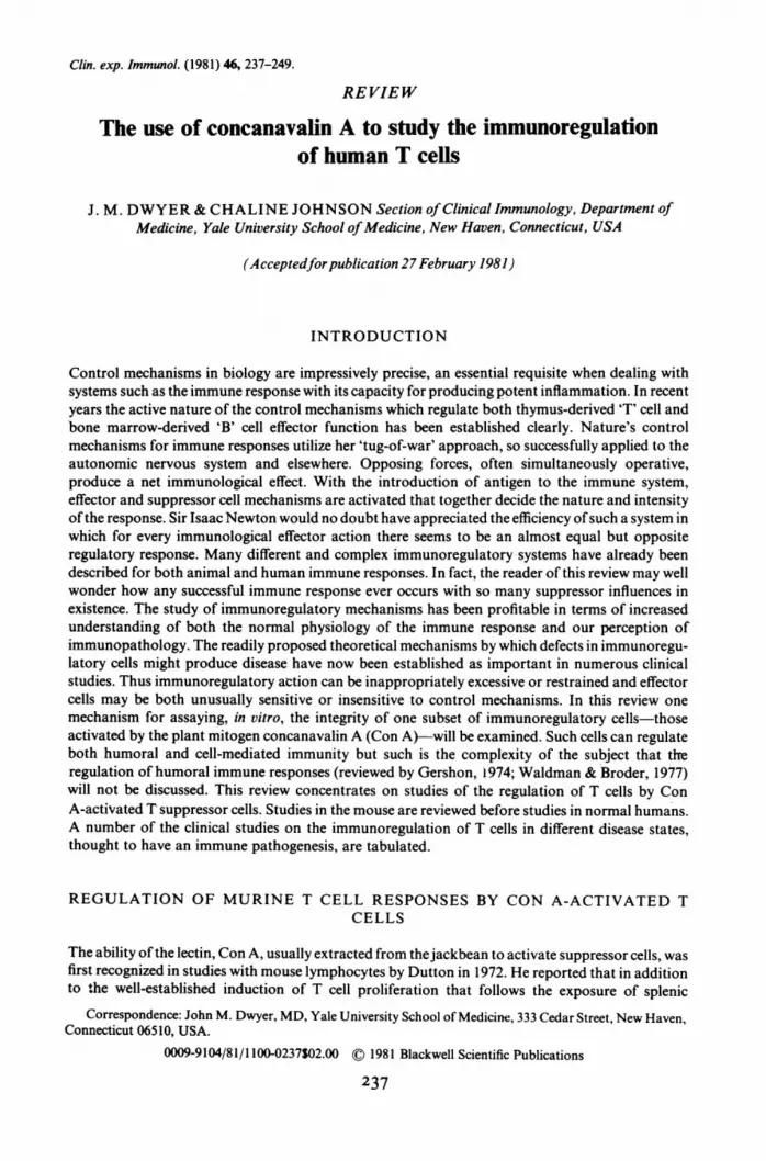
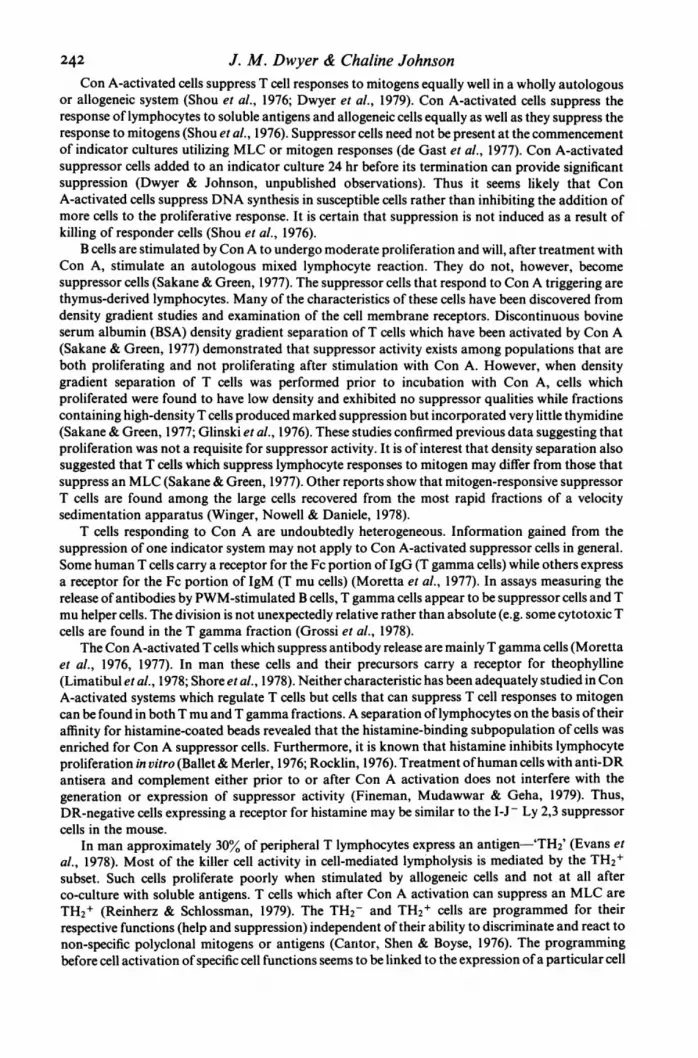
Fig. 1. T cell subsets stimulated by concanavalin A give rise to four functionally distinct T cell populations. Theactivation of suppressor cell precursors or promoters, leads, in turn, to active suppressor cell stimulation thatresults in the regulation ofboth B cell production ofantibody (virulent effect on helper cells) and the suppressionof T cell responses.
lymphocytes to Con A, both helper and suppressor cell activities were triggered. Since then, studiesin the mouse have identified at least four functionally, and in some respects physically, different Tcells that respond to Con A (Fig. 1).
(i) Con A activates cells capable of helping B cells respond to thymus-dependent antigens(Dutton, 1972; Tse & Dutton, 1976; Cantor & Boyse, 1975). Such cells are radiosensitive, smalllymphocytes that express the Ly 1 surface antigen. They sediment slowly through a Ficoll velocitygradient and do not require the continued presence of Con A on the helper cell's membrane afterinitial contact (Tse & Dutton, 1977). These cells proliferate poorly after Con A activation and theirhelper cell function does not require DNA synthesis or cell division as the cells are resistant tomitomycin C (Tse & Dutton, 1977). Con A-susceptible helper cells do not carry receptors for the Fcportion of IgG nor do they express surface antigens coded for in the I-J subregion of the mouse'smajor histocompatibility complex (MHC) but may express I-A subregion antigens (Okumura et al.,1976; Tada, Tanigushi & David, 1976; Tada et al., 1976; Tada, 1977).
(ii) Con A activates cells that proliferate industriously as measured by 3H-thymidineincorporation and cell division. These cells are denser than the helper cells and sediment deeper andmore rapidly through a Ficoll velocity gradient (Tse & Dutton, 1977).
(iii) Con A can activate a subpopulation of T cells to become cytotoxic for a number ofallogeneic normal and malignant cells (Bevan, 1975; MacDonald et al., 1975). Helper T cells areprobably necessary (Pilarski, Bretscher & Baum, 1977). Although these cells have the same densitycharacteristics as cells that proliferate after exposure to Con A, they may be different as they becomecytotoxic independent of DNA synthesis (Tse & Dutton, 1977).
(iv) Con A activates a population of suppressor-cell inducers or promoters (Niederhuber et al.,1976; Frelinger, Niederhuber & Shreffler, 1976). These cells are Ly 1 + and actively synthesize DNAafter Con A activation. In this sense they differ from the Ly 1 + helper cells. Such cells are Ia+ andonce activated they recruit other cells which are la- that subsequently proliferate. The murinesuppressor cell precursors carry surface antigens coded for in the I-J region of the mousehistocompatibility complex. I-J+ cells have been found to be important in immunoregulation in anumber of animal systems (Tada et al., 1976). Promoter cells which are quite radiosensitive (Tse &Dutton, 1977; Feldman et al., 1977) appear to be resistant to cyclophosphamide but are killed by
238
Con A-induced suppressor cells in mananti-Ly 1 antisera and complement (Whisler & Stobo, 1978). After activation they in turn activate asubpopulation of suppressor T cells. For the fullest activation of these latter cells, macrophageinteraction may be essential (Tadakuma & Pierce, 1976). The suppressor cells so activated are I-J,Ly 2,3+ well-differentiated cells which are relatively radioresistant. In such a population exist cells(perhaps distinctive) that can suppress both T and B cell responses in vitro. They can be recoveredfrom the same section of a Ficoll velocity gradient occupied by proliferating cells responding to themitogenic effect of Con A (Tse & Dutton, 1977). However, these Con A-activated suppressor cellsproliferate little and at no stage of either the induction or expression of their regulatory activity isDNA synthesis or cell proliferation mandatory (Tse & Dutton, 1977). Their suppressor effects arenot mediated by cytotoxicity directed towards responding cells.
Con A activates suppressor mechanisms very rapidly. Rich & Pierce (1973) showed that aninhibitory factor can be isolated by culturing spleen cells with Con A for just 12 hr. In the rabbit,Redelman et al. (1976) demonstrated the existence of suppressor activity in Con A-stimulatedpopulations well before DNA synthesis commenced. Roszman (1975) observed that a 2-hr pulse ofrabbit spleen cells with Con A was sufficient to activate suppressor cells without DNA synthesis.
Once in tissue culture, murine suppressor cells capable of responding to Con A may beshort-lived (Dutton, 1972), although this may represent artifacts and vary with the cultureconditions (Sampson, Grotelueschen & Kauffman, 1975).
Although the response to Con A is non-specific, immunoregulatory T cell mechanisms areactivated by this mitogen, thus providing a phenomenon that offers an opportunity for dissection ofsuppressor mechanisms similar to those offered by mitogen-stimulated cell proliferation inanalysing effector responses. After the initial reports of suppressor cell activation by Con A in themouse the system was soon adapted with success to human studies. As in the mouse, ConA-activated cells were found to suppress both B and T cell responses.
REGULATION OF HUMAN T CELL RESPONSES BY CON A-ACTIVATED TCELLS
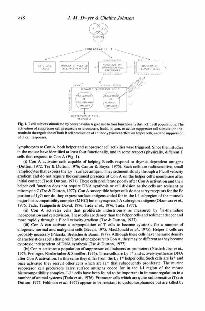
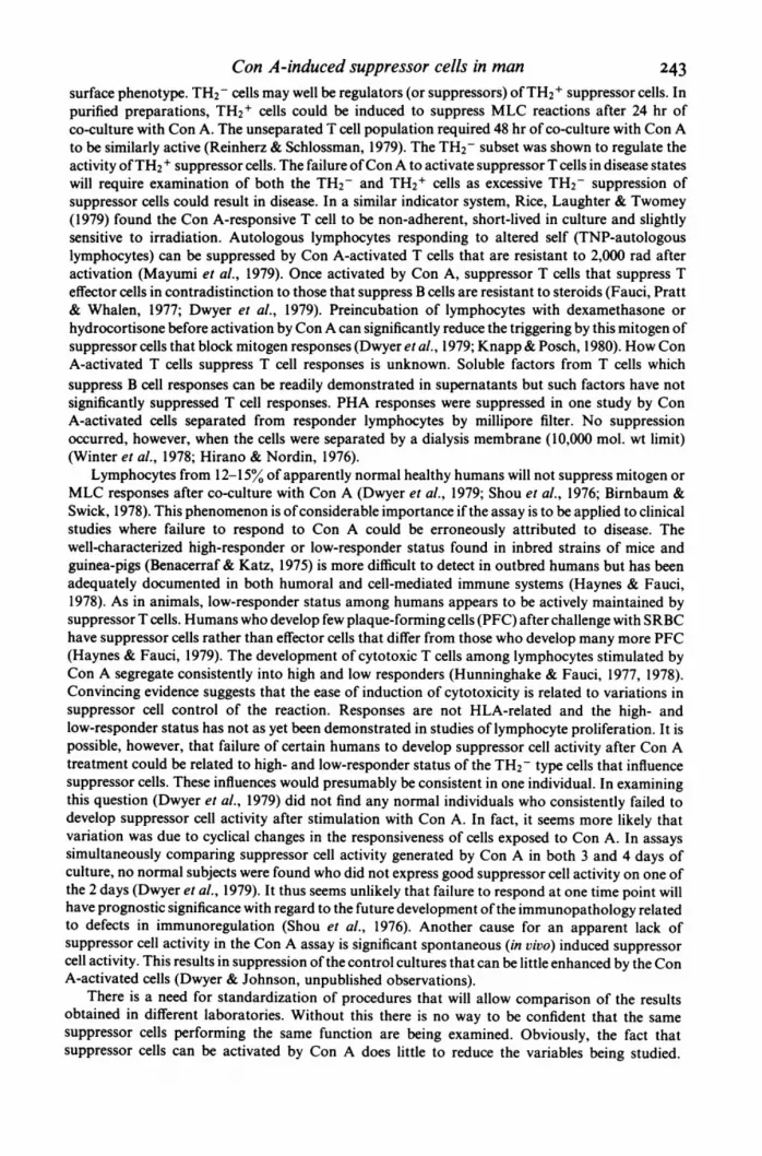
The picture of immunoregulation in humans is certainly no less complex than that of mice. Fig. 2illustrates some of this complexity. Peripheral T lymphocyte subsets (Broder et al., 1978; Stobo,
THYMUSPROTHYMOCYTES
DELAYED THYMYTESfhJWE ISITIVITY' -HICYE6sA~~~~~T1_ l& 0
FFECO CE PERIPHERALT LYMPHOCYTE SUBSETS
RESSO EL NON T SUPPRESSOR CELLS
HELPER SUPPRESSIONICO-(i ij)@jTCELLS OF HELP
I PEDE SI SEC
ENHANCED SEFG
ANTIBODY REGULATION OF CELLULARPRODUCTION AND HUMORAL IMMUNITY
Fig. 2. The peripheral T lymphocyte subsets of man include numerous types of suppressor T cells. These,together with non-T cell suppressors, regulate both cellular and humoral immunity. One subpopulation ofsuppressor T cells is sensitive to concanavalin A.
239

240 J. M. Dwyer & Chaline Johnson
SUPF
(oV11CON-A CON-A CON-A CON-A+ + + +
SPECIFIC SPECIFIC SUPPRESSOR CELL PO SPECIFICT CELLS T CELLS PRECURSORS SUPPRESSOR T CELLS
CELL
HELPER/AMPLIFIER MITOGEN CYTOTOXICOF TCELLS PROLIFERATION ACTIVE AND CELLS
DIFFERENT TYPES
\IN THEIR MLR / \M GNSUPPRE/S/I\N INDUCED
/ pCELLS THAT\ r~~~~~CELLS\\/ \ CAN BLOCK T HA TCAN BLOCK
CELLS THAT ALL MLR CYTOXICITY CAN LOKINHIBIT ROLIFERATIONNDUCED IN T CELLM MLR RESPONSES
-/~~~~~~~~~~~~~~~~~~~~T MIOGN
/CLLS THA\CAN SUPPRESS
T CELLRESPONSES
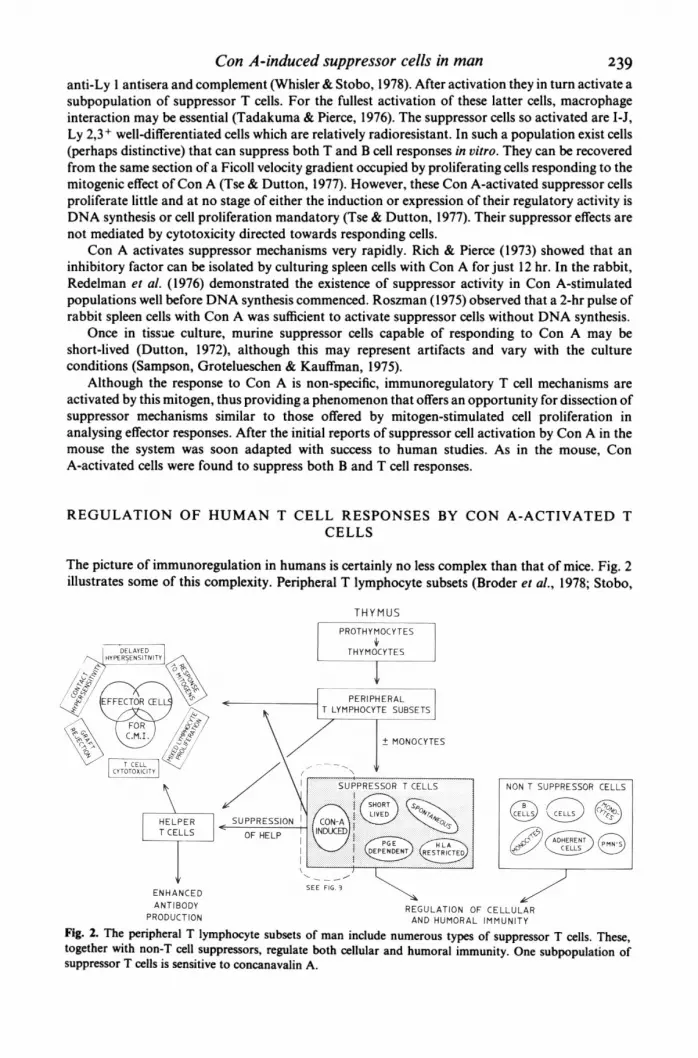
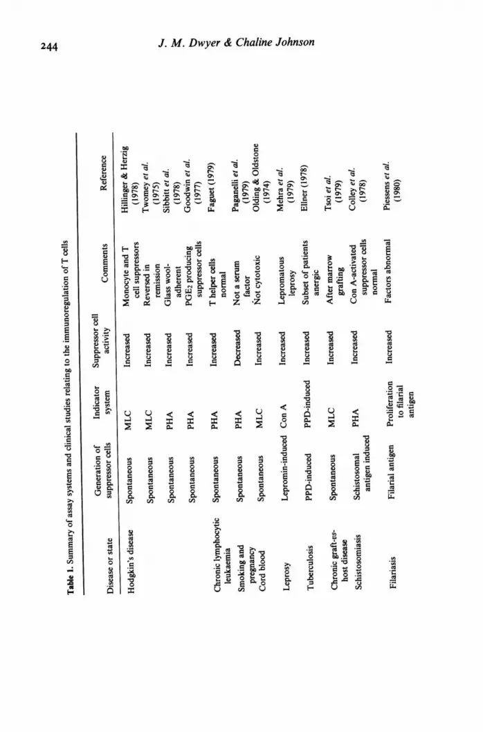
Fig. 3. Suppressor cell precursors that are activated by concanavalin A give rise to active and different types ofsuppressor cells that are distinguished in a functional sense. Such suppressor cells are able to exerciseimmunoregulatory activity over at least seven different T cell functions. While it is certain that there are differenttypes ofsuppressor cells responding to concanavalin A, it is likely that there is a functional overlap on the part ofa number of these cells. Note that apart from suppressor cell precursors giving rise to active suppressor cells,there is another order of immunoregulatory activity represented by the TH2-negative cells that are capable ofsuppressing suppressor cell activation.
1977) include different inflammation-producing cells that can express the full range ofcell-mediatedimmune responses, helper cells (Shore, Dosch & Gelfand, 1978) and suppressor cells.
Depending on the assay system used, apparently distinctive T suppressor cells have beenidentified in man although there is probably some overlap among cells identified in single-functionstudies. Spontaneously active (Bresnihan & Jasin, 1977), short-lived (Shou, Schwartz & Good,1976; Dwyer, Johnson & Desaules, 1979), prostaglandin-producing (Pelus& Strausser, 1977; Webb& Nowowiejski, 1978) and suppressor cells whose function is HLA-restricted (Engleman &McDevitt, 1978; Engleman, 1978) co-exist with suppressor cells that can be activated by Con A. Inaddition, good evidence suggests that B cells (Neta& Salvin, 1976), null cells (Muchmore, Decker &Blaese, 1977) and adherent cells of both the polymorphonuclear (Hsu, Wu & Rivera-Arcilla, 1979)and more especially the monocyte/macrophage series can be immunosuppressive (Ptak & Gershon,1975; Birnbaum & Swick, 1978).
As in the mouse, more than one subset ofhuman peripheral blood lymphocytes is triggered byCon A (Fig. 3). So far, Con A-activated T cells have been associated with suppression of (1) theeffectiveness of the lymphokine macrophage inhibition factor (Fox & Rajaraman, 1978); (2) theproliferation of responder cells in a mixed leucocyte culture (MLC) (Reinherz & Schlossman, 1979;Shou et al., 1976); (3) the proliferation of HLA-D-specific responder cells in a mixed leucocyteculture (MLC) (Engleman & McDevitt, 1978); (4) T cell proliferative responses to soluble antigens(Shou et al., 1976); (5) the cytotoxicity directed against allogeneic cells in the second phase of anMLC; (6) T cells proliferating in response to mitogens (e.g. PHA and Con A itself) (Hubert,Delespesse & Govaerts, 1976; Dwyer et al., 1979); (7) mitogen-induced, T cell-mediated,
Con A-induced suppressor cells in mannon-specific cytotoxicity (Hunninghake & Fauci, 1978); and (8) T cell proliferation in response tochallenge with 'altered self' (e.g. trinitrophenylated autologous lymphocytes) (Mayumi et al., 1979).
Most of the studies in man have used similar methods for inducing suppressor activity andmeasuring this in a responsive indicator system. Putative suppressor cells are stimulated with ConA, exposed to mitomycin C to prevent further DNA synthesis among these cells, washed and thenadded to autologous or allogeneic cells subsequently stimulated with mitogen or cell surfaceantigens. Control cultures feature identical cells not stimulated by Con A. Suppression is usuallyreported as the percentage reduction in the responsiveness of the indicator cells co-cultured with theCon A-stimulated cells when compared to that of indicator cells co-cultured with control cells.Many studies using this system or variations on this theme have revealed interesting aspects ofhuman suppressor cell performance.
A wide range of concentrations of Con A obtained from a number of sources can activatesuppressor activity. Various laboratories have used from 1 to 100 mg of Con A per 106 cellsstimulated. While it is not clear if the same suppressor cell is activated in each case, it is obvious thatdoses used to produce a strong mitogenic response are not necessary for suppressor cell activation.Suppressor cells need only a brief exposure to Con A to be activated although for optimalsuppressor cell activity a culture period of at least 12-24 hr may be necessary (de Gast et al., 1977). Itis of interest that induction of killer activity by Con A requires its continued presence (Asherson,Ferluga & Janossy, 1973). Con A appears to cross-link cell surface receptors in activatingsuppressor cells (Greaves & Janossy, 1972). It is possible, therefore, that Con A will be carried overinto the indicator system. Most examinations of this possibility (Sakane & Green, 1977; Dwyer etal., 1978), but not all (de Gast et al., 1977), have concluded that in practice this does not occur to anysignificant extent. Mitomycin C treatment of activated suppressor cells does not affect their functionindicating, as in the mouse, that DNA synthesis is not needed for active suppression (Shou et al.,1976; de Gast et al., 1977). Mitomycin C treatment of cells before their exposure to Con A blocks thegeneration of suppressor activity that can suppress B cell production of immunoglobulin and thosesuppressor cells specific for mitogen-induced cytotoxicity (Hunninghake & Fauci, 1978; Siegal &Siegal, 1977). These latter suppressor cells are not steroid-sensitive nor dependent on macrophagesand do not die rapidly in culture (Hunninghake & Fauci, 1978). Con A-activated suppressor cellsthat modify responses to mitogens and allogeneic cells, on the other hand, can still be activated byCon A even after treatment with mitomycin C (Lobo & Spencer, 1979; Kurnick, Bell & Grey, 1976;Sakane & Green, 1977). Such observations strongly suggest that humans as well as mice havedistinctive subpopulations of suppressor cells triggered by Con A.
Many studies have reported that cells able to respond to Con A in vitro and subsequentlysuppress mitogen and MLC reactivity, die rapidly in culture (Bresnihan & Jasin, 1977; Birnbaum &Swick, 1978; Feighery et al., 1978). This conclusion is based on complementary evidencedemonstrating that (a) successful generation of suppressor cells with normal cells precultured for 24hr is difficult, while (b) proliferative responses to mitogens and antigens are increased bypreculturing responder cells for 24 hr (Feighery et al., 1978; Dwyer et al., 1979). After 24 hr inculture, however, the situation changes and suppressor cells are more readily activated. Evensuppressor cells precultured for 8 days can be activated by Con A (Dwyer et al., 1979). It seemslikely that a considerable number of suppressor cells die the first 24 hr in culture, perhaps releasingtransiently active suppressor substances into the culture medium that prevent activation by Con Aof other suppressor cells. After this period the putative suppressor cells die slowly (Dwyer,unpublished observations). There is evidence to suggest that some suppressor cells go throughcyclical periods of responsiveness and unresponsiveness to Con A (Dwyer et al., 1979).
Low-density macrophages are probably necessary for optimal activation of suppressor cells byCon A (PHA-stimulated indicator system) (Raff, Cochrum & Stobo, 1978). Indeed, preferentialactivation of helper or suppressor cell mechanisms may depend to some extent on the way antigensare presented by macrophages (Shevach, 1976). Abnormalities of macrophage-T interactions mayalso serve as the basis of immunological hyporeactivity found in some clinical disorders such asHodgkin's disease (Twomey et al., 1975) and multiple myeloma (Broder et al., 1975). However,suboptimal but definite suppressor cell induction follows Con A stimulation of highly purified Tcells (Sakane & Green, 1977).
24I
J. M. Dwyer & Chaline JohnsonCon A-activated cells suppress T cell responses to mitogens equally well in a wholly autologous
or allogeneic system (Shou et al., 1976; Dwyer et al., 1979). Con A-activated cells suppress theresponse of lymphocytes to soluble antigens and allogeneic cells equally as well as they suppress theresponse to mitogens (Shou et al., 1976). Suppressor cells need not be present at the commencementof indicator cultures utilizing MLC or mitogen responses (de Gast et al., 1977). Con A-activatedsuppressor cells added to an indicator culture 24 hr before its termination can provide significantsuppression (Dwyer & Johnson, unpublished observations). Thus it seems likely that ConA-activated cells suppress DNA synthesis in susceptible cells rather than inhibiting the addition ofmore cells to the proliferative response. It is certain that suppression is not induced as a result ofkilling of responder cells (Shou et al., 1976).
B cells are stimulated by Con A to undergo moderate proliferation and will, after treatment withCon A, stimulate an autologous mixed lymphocyte reaction. They do not, however, becomesuppressor cells (Sakane & Green, 1977). The suppressor cells that respond to Con A triggering arethymus-derived lymphocytes. Many of the characteristics of these cells have been discovered fromdensity gradient studies and examination of the cell membrane receptors. Discontinuous bovineserum albumin (BSA) density gradient separation of T cells which have been activated by Con A(Sakane & Green, 1977) demonstrated that suppressor activity exists among populations that areboth proliferating and not proliferating after stimulation with Con A. However, when densitygradient separation of T cells was performed prior to incubation with Con A, cells whichproliferated were found to have low density and exhibited no suppressor qualities while fractionscontaining high-density T cells produced marked suppression but incorporated very little thymidine(Sakane & Green, 1977; Glinski et al., 1976). These studies confirmed previous data suggesting thatproliferation was not a requisite for suppressor activity. It is of interest that density separation alsosuggested that T cells which suppress lymphocyte responses to mitogen may differ from those thatsuppress an MLC (Sakane & Green, 1977). Other reports show that mitogen-responsive suppressorT cells are found among the large cells recovered from the most rapid fractions of a velocitysedimentation apparatus (Winger, Nowell & Daniele, 1978).
T cells responding to Con A are undoubtedly heterogeneous. Information gained from thesuppression of one indicator system may not apply to Con A-activated suppressor cells in general.Some human T cells carry a receptor for the Fc portion of IgG (T gamma cells) while others expressa receptor for the Fc portion of IgM (T mu cells) (Moretta et al., 1977). In assays measuring therelease of antibodies by PWM-stimulated B cells, T gamma cells appear to be suppressor cells and Tmu helper cells. The division is not unexpectedly relative rather than absolute (e.g. some cytotoxic Tcells are found in the T gamma fraction (Grossi et al., 1978).
The Con A-activated T cells which suppress antibody release are mainly T gamma cells (Morettaet al., 1976, 1977). In man these cells and their precursors carry a receptor for theophylline(Limatibul et al., 1978; Shore et al., 1978). Neither characteristic has been adequately studied in ConA-activated systems which regulate T cells but cells that can suppress T cell responses to mitogencan be found in both T mu and T gamma fractions. A separation of lymphocytes on the basis of theiraffinity for histamine-coated beads revealed that the histamine-binding subpopulation of cells wasenriched for Con A suppressor cells. Furthermore, it is known that histamine inhibits lymphocyteproliferation in vitro (Ballet & Merler, 1976; Rocklin, 1976). Treatment ofhuman cells with anti-DRantisera and complement either prior to or after Con A activation does not interfere with thegeneration or expression of suppressor activity (Fineman, Mudawwar & Geha, 1979). Thus,DR-negative cells expressing a receptor for histamine may be similar to the I-J - Ly 2,3 suppressorcells in the mouse.
In man approximately 30% of peripheral T lymphocytes express an antigen-'TH2' (Evans etal., 1978). Most of the killer cell activity in cell-mediated lympholysis is mediated by the TH2'subset. Such cells proliferate poorly when stimulated by allogeneic cells and not at all afterco-culture with soluble antigens. T cells which after Con A activation can suppress an MLC areTH2+ (Reinherz & Schlossman, 1979). The TH2- and TH2+ cells are programmed for theirrespective functions (help and suppression) independent of their ability to discriminate and react tonon-specific polyclonal mitogens or antigens (Cantor, Shen & Boyse, 1976). The programmingbefore cell activation of specific cell functions seems to be linked to the expression of a particular cell
242
Con A-induced suppressor cells in mansurface phenotype. TH2- cells may well be regulators (or suppressors) ofTH2+ suppressor cells. Inpurified preparations, TH2+ cells could be induced to suppress MLC reactions after 24 hr ofco-culture with Con A. The unseparated T cell population required 48 hr of co-culture with Con Ato be similarly active (Reinherz & Schlossman, 1979). The TH2- subset was shown to regulate theactivity ofTH2 + suppressor cells. The failure ofCon A to activate suppressor T cells in disease stateswill require examination of both the TH2- and TH2+ cells as excessive TH2- suppression ofsuppressor cells could result in disease. In a similar indicator system, Rice, Laughter & Twomey(1979) found the Con A-responsive T cell to be non-adherent, short-lived in culture and slightlysensitive to irradiation. Autologous lymphocytes responding to altered self (TNP-autologouslymphocytes) can be suppressed by Con A-activated T cells that are resistant to 2,000 rad afteractivation (Mayumi et al., 1979). Once activated by Con A, suppressor T cells that suppress Teffector cells in contradistinction to those that suppress B cells are resistant to steroids (Fauci, Pratt& Whalen, 1977; Dwyer et al., 1979). Preincubation of lymphocytes with dexamethasone orhydrocortisone before activation by Con A can significantly reduce the triggering by this mitogen ofsuppressor cells that block mitogen responses (Dwyer et al., 1979; Knapp & Posch, 1980). How ConA-activated T cells suppress T cell responses is unknown. Soluble factors from T cells whichsuppress B cell responses can be readily demonstrated in supernatants but such factors have notsignificantly suppressed T cell responses. PHA responses were suppressed in one study by ConA-activated cells separated from responder lymphocytes by millipore filter. No suppressionoccurred, however, when the cells were separated by a dialysis membrane (10,000 mol. wt limit)(Winter et al., 1978; Hirano & Nordin, 1976).
Lymphocytes from 12-15% of apparently normal healthy humans will not suppress mitogen orMLC responses after co-culture with Con A (Dwyer et al., 1979; Shou et al., 1976; Birnbaum &Swick, 1978). This phenomenon is of considerable importance if the assay is to be applied to clinicalstudies where failure to respond to Con A could be erroneously attributed to disease. Thewell-characterized high-responder or low-responder status found in inbred strains of mice andguinea-pigs (Benacerraf & Katz, 1975) is more difficult to detect in outbred humans but has beenadequately documented in both humoral and cell-mediated immune systems (Haynes & Fauci,1978). As in animals, low-responder status among humans appears to be actively maintained bysuppressor T cells. Humans who develop few plaque-forming cells (PFC) after challenge with SRBChave suppressor cells rather than effector cells that differ from those who develop many more PFC(Haynes & Fauci, 1979). The development of cytotoxic T cells among lymphocytes stimulated byCon A segregate consistently into high and low responders (Hunninghake & Fauci, 1977, 1978).Convincing evidence suggests that the ease of induction of cytotoxicity is related to variations insuppressor cell control of the reaction. Responses are not HLA-related and the high- andlow-responder status has not as yet been demonstrated in studies of lymphocyte proliferation. It ispossible, however, that failure of certain humans to develop suppressor cell activity after Con Atreatment could be related to high- and low-responder status of the TH2- type cells that influencesuppressor cells. These influences would presumably be consistent in one individual. In examiningthis question (Dwyer et al., 1979) did not find any normal individuals who consistently failed todevelop suppressor cell activity after stimulation with Con A. In fact, it seems more likely thatvariation was due to cyclical changes in the responsiveness of cells exposed to Con A. In assayssimultaneously comparing suppressor cell activity generated by Con A in both 3 and 4 days ofculture, no normal subjects were found who did not express good suppressor cell activity on one ofthe 2 days (Dwyer et al., 1979). It thus seems unlikely that failure to respond at one time point willhave prognostic significance with regard to the future development ofthe immunopathology relatedto defects in immunoregulation (Shou et al., 1976). Another cause for an apparent lack ofsuppressor cell activity in the Con A assay is significant spontaneous (in vivo) induced suppressorcell activity. This results in suppression of the control cultures that can be little enhanced by the ConA-activated cells (Dwyer & Johnson, unpublished observations).
There is a need for standardization of procedures that will allow comparison of the resultsobtained in different laboratories. Without this there is no way to be confident that the samesuppressor cells performing the same function are being examined. Obviously, the fact thatsuppressor cells can be activated by Con A does little to reduce the variables being studied.
243
J. M. Dwyer & Chaline Johnson
04~~~~~~~~~~~~~~~~~~~~~~~- 00
._~~~~~~~~~~~~~~~a
N~~~~~~~~~~~~~~O o-00~~~~~~
oo t r
a) ce V
Wn]9 X = h fZ h E~~~rA cs W
000 2~~0%~ %
0-.O- 0'- z'
g' Ce CUec)
CU
'0
.. U U <
-. -.
tC). U) U) U)
. a) a) a)a) . CU CU
. 0 0 0U) 0. .
. CUa)
CU *.
a)CU .L)a) .O.. 0
o o =
U)U)
~ ~
< < <u E.
U-
00 0 0~
t ~~ ~ ~ ~a)VC w T
C) ~ ~ ~ )C
o0 0 0Ea)
o 04 0 0,
U Ena ~C
4--
o- 0 r
1.
.U
CU
CU
._
244
C)0
u
C.)a)
.a)
r.
0
F).~
4C)
~CAri 4-
CU.
0
a)
0*O
CU
a
'0C-
C)
cU
0
.
4-
Cd
la0;WCd
.2
E
-
0
CU
0Ya)
0
I
Con A-induced suppressor cells in man
0 Cd0
U Cd00 U~~~~~~~~~CU
(U w
>~ ~
Cd O 0> la
*- 0 E ---_
Q~ ~ ~) Q c) E
= : n : : : a : : : ncn
~ ~ ~~~~~W A (Cd C d Cd Cd C m c Cd
d (U 0 (U Q
.0 0 0 0
co UU U U CU
_ 2 Y . O|2 -
CU CU CU CU CU CU U CU CU CU
>
.0~~~~~
0~~~~
o~~~~~~~~~~~~~0 c
00000000 0~~~~~~~~~~~~~~c 0
O Ud
-
245
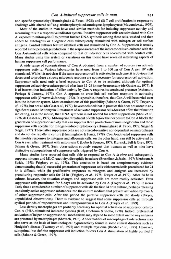
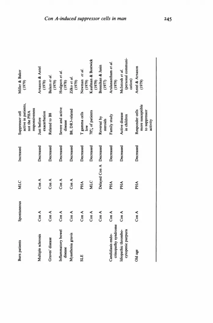
246 J. M. Dwyer & Chaline JohnsonNevertheless, the assay systems described above have been applied in many clinical studies andsome of the more important are tabulated and referenced in Table 1. In addition, some of the otherclinical studies relating to the immunoregulation of T cells are included. In a number of clinicalstudies, assays of suppressor cell performance have been consistently abnormal and related well toimmunological phenomena observed in vivo. Assays using Con A-stimulated suppressor T cells arenot suitable for detecting enhanced suppressor cell activity. However, an expansion of the work onspontaneous or non-Con A-activated suppressor cell activity may soon overcome this problem.There seems no reason to doubt that more information about human immunoregulatory cells willbe supplied by the Con A stimulation assay. Such studies combined with advances in our ability toexamine antigen-specific suppression will predictably lead to enhanced understanding of clinicalsituations and ways of deliberately manipulating suppressor cells, the current 'anti-heroes' ofimmunopathogenesis.
This work was supported in part by NIH Grant All 1785. Dr Dwyer is an investigator for the Howard HughesMedical Institute.
REFERENCES
ANTEL, J. & ARNASON, B.G.W. (1979) Suppressor cellfunction in man: evidence for altered sensitivity ofresponder cells with age. Clin. Immunol. Immuno-pathol. 13, 119.
ARNASON, B.G.W. & ANTEL, J. (1978) Suppressor cellfunctions in multiple sclerosis. Ann. Immunol.(Paris), 129C, 159.
ARULANANTHAM, K., DWYER, J.M. & GENEL, M.(1979) Evidence for defective immunoregulation inthe syndrome of familial candidiasis endocrino-pathy. N. Engl. J. Med. 3W0, 164.
ASHERSON, G.L., FERLUGA, J. & JANOSSY, G. (1973)Nonspecific cytotoxicity by T cells activated byplant mitogens in vitro and the requirement forplant agents during the killing reaction. Clin. exp.Immunol. 15, 573.
BALAZS, Cs., STENSKY, V., KOZMA, L., SZERZE, D. &LEOVEY, A. (1979) Connection between HLA-B8antigen and suppressor T cell activity in Graves'disease. Transplant. Proc. 11, 1314.
BALLET, J.J. & MERLER, E. (1976) The separation andreactivity in vitro of a subpopulation of humanlymphocytes which bind histamine. Correlation ofhistamine reactivity with cellular maturation. Cell.Immunol. 24, 250.
BENACERRAF, B. & KATZ, D.H. (1975) The nature andfunction of histocompatibility linked immune re-sponse genes. In Immunogenetics and Immunodefi-ciencies (ed. by B. Benacerraf), p. 118. UniversityPark Press, Baltimore.
BEVAN, M.J. (1975) Induction of cytotoxic effector Tcells by Con A. In Concanavalin A as a Tool (ed. byH. Bitteger and H. P. Schnebi), p. 523. John Wiley& Sons, New York.
BIRNBAUM, G. & SWICK, L. (1978) Human suppressorlymphocytes. I. Induction and characterization.Cell Immunol. 40, 16.
BRESNIHAN, B. & JASIN, H.E. (1977) Suppressorfunction of peripheral blood mononuclear cells innormal individuals and in patients with systemiclupus erythematosus. J. cdin. Invest. 59, 106.
BRODER, S., HUMPHREY, R., DURM, M., BLACKMAN,
M., MEADE, B., GOLDMAN, C., STROBER, W. &WALDMANN, T. (1975) Impaired synthesis of poly-clonal (non-paraprotein) immunoglobulins by cir-culating lymphocytes from patients with multiplemyeloma. N. Engl. J. Med. 293, 887.
BRODER, S., POPLACK, P., WHANG-PENG, J., DURM, J.,GOLDMAN, C., MJUL, L. & WALDMANN, T.A.(1978) Characterization of a suppressor cell leuke-mia. Evidence for the requirement ofan interactionof two T cells in the development of humansuppressor effector cells. N. Engl. J. Med. 298, 66.
CANTOR, H. & BOYSE, E.A. (1975) Functional sub-classes of T lymphocytes bearing different Lyantigens. I. The generation of functionally distinctT cell subclasses is a differentiative process indepen-dent of antigen. J. exp. Med. 141, 1376.
CANTOR, H., SHEN, F.W. & BOYSE, E.A. (1976)Separation of helper T cells from suppressor T cellsexpressing different Ly components. II. Activationby antigen: after immunization, antigen-specificsuppressor and helper activities are mediated bydistinct T-cell subclasses. J. exp. Med. 143, 1391.
COLLEY, D.G., LEWIS, F.A. & GOODGAME, R.W.(1978) Immune responses during human schistoso-miasis mansoni, J. Immunol. 120, 1225.
DE GAST, G.C., THE, T.H., PONDS, E. & KALLENBERG,C. (1977) Suppression ofDNA synthesis by Con Aactivated human lymphocytes. Stimulation by ConA bound to non T cells unless removed afteractivation. Clin. exp. Immunol. 30, 457.
DUTTON, R.W. (1972) Inhibitory and stimulatoryeffects of concanavalin A on the response ofmousespleen cell suspensions to antigen. I. Characteriza-tion of the inhibitory cell activity. J. exp. Med. 136,1445.
DwYER, J.M., JOHNSON, C. & DESAULES, M. (1979)Behaviour of human immunoregulatory cells inculture. I. Variables requiring consideration forclinical studies. Clin. exp. Immunol. 38, 499.
ELLNER, J.J. (1978) Suppressor cells in human tuber-culosis. J. Immunol. 121, 2573.
ENGLEMAN, E.G. (1978) A simple method for detect-
Con A-induced suppressor cells in man 247ing suppressor cells of the mixed lymphocytereaction in man: application to a healthy popula-tion. Transplant. Proc. 10, 895.
ENGLEMAN, E.G. & McDEvITT, H.O. (1978) A sup-pressor T cell of the mixed lymphocyte reactionspecific for the HLA-D region in man. J. clin.Invest. 161, 828.
EVANS, R.L., LAZARUS, H., PENTA, A.C. & SCHLOSS-MAN, S.F. (1978) Two functionally distinct subpo-pulations of human T cells that collaborate in thegeneration of cytotoxic cells responsible for cellmediated lympholysis. J. Immunol. 120, 1423.
FAGUET, G.B. (1979) Mechanisms of lymphocyteactivation. The role of suppressor cells in theproliferative response of chronic lymphatic leuke-mia lymphocytes. J. clin. Invest. 63, 67.
FAUCI, A.S., PRATT, K.R. & WHALEN, G. (1977)Activation of human B lymphocytes. IV. Regula-tory effects of corticosteroids on the triggeringsignal in the plaque-forming cell response ofhumanperipheral blood B lymphocytes to polyclonalactivation. J. Immunol. 119, 598.
FEIGHERY, C., WHELAN, C.A., WEIR, D.G. &GREALLY, J.F. (1978) In vitro studies of suppressorcell function in human peripheral blood mononuc-lear cells. Clin. exp. Immunol. 32, 459.
FELDMAN, M., BEVERLEY, P.C.L., ERB, P., HOWIE, S.KONTIAINEN, S., MAOZ, A., MATHIES, M., McKEN-ZIE, I. & WOODY, J. (1977) Current concepts of theantibody response: heterogeneity oflymphoid cells,interaction, and factors. Cold Spring Harbor Symp.Quant. Biol. 41, 113.
FINEMAN, S.M., MUDAWWAR, F.B. & GEHA, R.S.(1979) Characteristics and mechanism of action ofthe concanavalin A activated suppressor cell inman. Cell. Immunol. 45, 120.
Fox. R.A. & RAJARAMAN, K. (1978) The role ofsuppressor cells in the production of macrophagemigration inhibition factor. Immunol. Commun. 7,311.
FRELINGER, J.A., NIEDERHUBER, J.E. & SHREFFLER,D.C. (1976) Effects of anti-Ia sera on mitogenicresponses. III. Mapping the genes controlling theexpression of Ia determinants on concanavalinA-reactive cells to the I-J subregion of the H-2 genecomplex. J. exp. Med. 144, 1141.
GERSHON, R.K. (1974) T cell control of antibodyproduction. Contemp. Top. Mol. Immunol. 3, 1.
GLINSKI, W., GERSHWIN, M.E., BUDMAN, D.R. &STEINBERG, A.D. (1976) Study of lymphocyte sub-populations in normal humans and patients withsystemic lupus erythematosus by fractionation ofperipheral blood lymphocytes on a discontinuousFicol gradient. Clin. exp. Immunol. 26, 228.
GOODWIN, J.S., MESSNER, R.P., BANKHURST, A.D.,PEAKE, G.T., SAIKI, J.H. & WILLIAMS, R.C. (1977)Prostaglandin producing suppressor cells in Hodg-kin's disease. N. Engl. J. Med. 297, 963.
GREAVES, M. & JANOSSY, G. (1972) Elicitation ofselective T and B lymphocyte responses by cellsurface binding ligands. Transplant. Rev. 11, 87.
GROSSI, C.E., WEBB, S.R., ZICCA, A., LYDYARD, P.M.,MORETTA, L., MINGARI, M.C. & COOPER, M.D.(1978) Morphological and histochemical analysesof two human T cell subpopulations bearing recep-tors for IgM or IgG. J. exp. Med. 147, 1405.
HAYNES, B.F. & FAUCI, A.S. (1978) Activation ofhumn B lymphocytes. V. Kinetics and mechanismsof suppression of plaque-forming cell responses byconcanavalin A generated suppressor cells. J. Im-munol. 120, 700.
HAYNES, B.F. & FAUCI, A.S. (1979) Activation ofhuman B lymphocytes. XIII. Characterization ofmultiple populations of naturally occurring im-munoregulatory cells of polyclonally induced invitro human B cell function. J. Immunol. 123, 1289.
HILLINGER, S.M. & HERZIG, G.P. (1978) Impairedcell-mediated immunity in Hodgkin's diseasemediated by suppressor lymphocytes and mond-cytes. J. clin. Invest. 61, 1620.
HIRANO, T. & NORDIN, A. (1976) Cell-mediatedimmune response in vitro. II. The mechanism(s)involved in the suppression of the development ofcytotoxic lymphocytes. J. Immunol. 117, 2226.
HODGSON, H.J.F., WANDS, J.R. & ISSELBACHER, K.J.(1978) Decreased suppressor cell activity in inflam-matory bowel disease. Clin. exp. Immunol. 32, 451.
Hsu, C.S.S., Wu, M.Y.B. & RivERA-ARCILLA, J.(1979) Inhibition of lymphocyte reactivity in vitroby autologous polymorphonuclear cells (PMN).Cell. Immunol. 48, 288.
HUBERT, C., DELESPESSE, G. & GOvAERTS, A. (1976)Concanavalin A activated suppressor cells in nor-mal human peripheral blood lymphocytes. Clin.exp. Immunol. 26, 95.
HUNNINGHAKE, G.W. & FAUCI, A.S. (1977) Lympho-cyte mediated cytotoxicity against human allo-geneic and autologous lymphoid target cells afterconcanavalin A activation of cytotoxic effectorcells. J. Immunol. 119, 1122.
HUNNINGHAKE, G.W. & FAUCI, A.S. (1978) Suppres-sion of the generation of human Con A-inducedcytotoxic effector cells by Con A-activated suppres-sor cells. J. Immunol. 120, 1828.
KAUFMAN, D.B. & BoSTwICK, E. (1979) Defectivesuppressor T cell activity in systemic lupus erythe-matosus. Clin. Immunol. Immunopathol. 13, 9.
KNAPP, W. & POSCH, B. (1980) Concanavalin inducedsuppressor cell activity: opposing effects of hydro-cortisone. J. Immunol. 124, 168.
KURNICK, J., BELL, C. & GREY, H.M. (1976) PHAinduced activation of suppressor cells in normalhuman peripheral blood lymphocytes. Scand. J.Immunol. 5, 771.
LIMATIBUL, S., SHORE, A., DOSCH, H.M. & GELFAND,E.W. (1978) Theophylline modulation of E rosetteformation: an indicator of T cell maturation. Clin.exp. Immunol. 33, 503.
LoBo, P.I. & SPENCER, C.E. (1979) Inhibition ofhumoral and cell mediated immune responses inman by distinct suppressor cell systems. J. din.Invest. 63, 1157.
MACDONALD, H.R., SONDAT, B., CEROTrINI, J.C. &BRUNNER, K.T. (1975) Generation of cytotoxic Tlymphocytes in vitro. IV. Functional activation ofmemory cells in the absence of DNA synthesis. J.exp. Med. 142, 622.
MAYUMI, M., YOSHIDA, T., SHINOMIYA, K., NISHIK-AWA, S., HIRATA, T., IZUMI, T. & MIKAWA, H.(1979) Effects of concanavalin A induced cells onthe proliferative response of T cells. ConcanavalinA-induced suppressor and amplifier cells to the
248 J. M. Dwyer & Chaline Johnsonproliferative response of human T cells to trinitro-phenyl-modified autologous lymphocytes. J. Im-munol. 123, 772.
MEHRA, V., MASON, L.H., FIELDS, J.P. & BLOOM, B.R.(1979) Lepromin-induced suppressor cells inpatients with leprosy. J. Immunol. 123, 1813.
MILLER, C.L. & BAKER, C.C. (1979) Changes inlymphocyte activity after thermal injury. The roleof suppressor cells. J. clin. Invest. 63, 202.
MORETTA, L., FERRARINI, M., MINGARI, M.C., MOR-ETTA, A. & WEBB, S.R. (1976) Subpopulations ofhuman T cells identified by receptors for immuno-globulins and mitogen responsiveness. J. Immunol.117,2171.
MORETTA, L., WEBB, S.R., GRossi, C.E., LYDYARD,P.M. & COOPER, M.D. (1977) Functional analysisof two human T cell subpopulations: help andsuppression of B cell responses by T cells bearingreceptors for IgM or IgG. J. exp. Med. 146, 184.
MUCHMORE, A.V., DECKER, J.M. & BLAESE, R.M.(1977) Spontaneous cytotoxicity of human peri-pheral blood mononuclear cells toward red bloodcell targets. II. Time dependent loss of suppressorcell activity. J. Immunol. 119, 1686.
NIEDERHUBER, J.E., FRELINGER, J.A., DINE, M.S.,SHOFFNER, P., DUGAN, E. & SHREFFLER, D.C. (1976)Effects of anti-Ia sera on mitogenic responses. II.Differential expression of the Ia marker on phyto-hemagglutinin and concanavalin A reactive cells tothe I-J subregion of the H-2 gene complex. J. exp.Med. 143, 372.
NETA, R. & SALVIN, S.B. (1976) T and B lymphocytesin the regulation of delayed hypersensitivity. J.Immunol. 117, 2014.
NEWMAN, B., BLANK, S., LOMNITZER, R., PISLER, P. &RABSON, A.R. (1979) Lack of suppressor cell acti-vity in systemic lupus erythematosus. Clin. Im-munol. Immunopathol. 13,187.
OKUMURA, K., HERZENBERG, L.A., MURPHY, D.B.,MCDEVITT, H.O. & HERZENBERG, L.S. (1976) Selec-tive expression of H-2 (I - region) loci controllingdeterminants on helper and suppressor T lympho-cytes. J. exp. Med. 144, 685.
OLDING, L.B. & OLDSTONE, M.B.A. (1974) Lympho-cytes from human newborns abrogate mitosis oftheir mothers' lymphocytes. Nature, 249, 161.
PAGANELLI, R., RAMADAS, D., LAYWARD, L., HARvEY,B.A.M. & SOOTHILL, J.F. (1979) Maternal smokingand cord blood immunity function. Clin. exp.Immunol. 36, 256.
PELUS, L.M. & STRAUSSER, H.R. (1977) Prostaglan-dins and the immune response. Life Sci. 20, 903.
PIESSENS, W.F., RATIWAYANTO, S., TUTI, S., PALMIERI,J.H., PIESSENS, P.W., KOMAN, I. & DENNIS, D.T.(1980) Antigen-specific suppressor cells and sup-pressor factors in human filariasis with Brugiamalaye. N. Engl. J. Med. 302, 833.
PILARSKI, L.M., BRETSCHER, P.A. & BAUM, L.L.(1977) Helper T cells are required for the polyclonalstimulation of cytotoxic T cells by concanavalin A.J. exp. Med. 145, 1237.
PTAK, W. & GERSHON, R.K. (1975) Immunosuppres-sion effected by macrophage surfaces. J. Immunol.115, 1346.
RAFF, H.V., COCHRUM, K.C. & STOBO, J.D. (1978)Macrophage-T cell interactions in the Con A
induction ofhuman suppressor T cells. J. Immunol.121, 2311.
REDELMAN, D., SCOTT, D.B., SHEPERD, H.W. & SELL,S. (1976) In vitro studies of the rabbit immunesystem. V. Suppressor T cells activated by concana-valin A block the proliferation, not the induction ofantierythrocyte plaque forming cells. J. exp. Med.143, 919.
REINHERZ, E.L. & SCHLOSSMAN, S.F. (1979) Con Ainducible suppression ofMLC: evidence for media-tion by the TH2+ T cell subset in man. J. Immunol.122, 1335.
RICE, L., LAUGHTER, A.H. & TWOMEY, J.J. (1979)Three suppressor systems in human blood thatmodulate lymphoproliferation. J. Immunol. 122,991.
RICH, R.R. & PIERCE, C.W. (1973) Biological ex-pression of lymphocyte activation. II. Generationof a population of thymus derived suppressorlymphocytes. J. exp. Med. 137, 649.
ROCKLIN, R.E. (1976) Modulation ofcellular immuneresponses in vivo and in vitro by histamine receptorbearing lymphocytes. J. clin. Invest. 57, 1051.
RoszMAN, T.L. (1975) Suppression of the in vitrosecondary antibody response of rabbit lymphoidcells by concanavalin A. Proc. Soc. exp. Biol. Med.149, 407.
SAKANE, T. & GREEN, I. (1977) Human suppressor Tcells induced by concanavalin A: suppressor T cellsbelong to a distinctive T cell subclass. J. Immunol.119, 1169.
SAMPSON, D., GROTELUESCHEN, C. & KAUFFMAN, H.M.(1975) The human splenic suppressor cell. Trans-plantation, 30, 362.
SHEVACH, E.M. (1976) The function of macrophagesin antigen recognition by guinea pigT lymphocytes.III. Genetic analysis of the antigens mediatingmacrophage lymphocyte interaction. J. Immunol.116, 1482.
SHORE, A., DOSCH, H.M. & GELFAND, E.W. (1978)Induction and separation of antigen-dependent Thelper and T suppressor cells in man. Nature, 274,586.
SHOU, L., SCHWARTZ, S.A. & GOOD, R.H. (1976)Suppressor cell activity after concanavalin A treat-ment of lymphocytes from normal donors. J. exp.Med. 143, 1100.
SIBBITT, W.L., BANKHURST, A.D. & WILLIAMS, R.C.(1978) Studies of cell subpopulations mediatingmitogen hyporesponsiveness in patients with Hodg-kin's disease. J. cdin. Invest. 61, 55.
SIEGAL, F.P. & SIEGAL, M. (1977) Enhancement byirradiated T cells ofhuman plasma cell production:dissection of helper and suppressor functions invitro. J. Immunol. 118, 642.
STOBO, J.D. (1977) Immunosuppression in man: sup-pression by macrophages can be mediated byinteraction with regulatory T cells. J. Immunol. 119,918.
TADA, T. (1977) Regulation of the antibody responseby T cell products determined by different I subre-gions. In Regulation of the Immune System (ed. byL. Herzenberg and E. Sercarz. Academic Press,New York.
TADA, T., TANIGUSHI, M. & DAVID, C.S. (1976)Properties of the antigen specific suppressive T cell
Con A-induced suppressor cells in man 249factor in the regulation of antibody response in themouse. IV. Special subregion assignment of thegene(s) that codes for the suppressive T cell factor inthe H-2 histocompatability complex. J. exp. Med.144, 713.
TADA, T., TANIGUSH, M., TOKUHISA, T. & DAVID,C.S. (1976) Antigen specific suppressive andenhancing T cell factors determined by distinct lociin the I region of H-2 complex. Cell. Immunol. 27,336.
TADAKUMA, T. &PIERCE, C.W. (1976) Site ofaction ofa soluble immune response suppressor (SiRs) pro-duced by concanavalin A activated spleen cells. J.Immunol. 117, 967.
TSE, H. & DUTFON, R.W. (1976) Separation of helperand suppressor T lymphocytes on a Ficoll velocitysedimentation gradient. J. exp. Med. 143, 1199.
TsE, H.Y. & DUTTON, R.W. (1977) Separation ofhelper and suppressorT lymphocytes. II. Lypheno-types and lack of DNA synthesis requirement forthe generation of concanavalin A helper and sup-pressor cells. J. exp. Med. 146, 747.
Tsoi, M.S., STORB, R., DOBBS, S., KOPECKY, K.J.,SANTOS, E., WEIDEN, P.L. & THOMAS, E.D. (1979)Nonspecific suppressor cells in patients withchronic graft-vs-host disease after marrow grafting.J. Immunol. 123, 1970.
TWoMEY, J.J., LAUGHTER, A.H., FARRow, S. &DOUGLAS, C.C. (1975) Hodgkin's disease: an im-
munodepleting and immunosuppressive disorder.J. clin. Invest. 56, 467.
WALDMANN, T.A., BLAME, R.M., BRODER, S. &KRAKAUER, R.S. (1978) Disorders of suppressorimmunoregulatory cells in the pathogenesis ofimmunodeficiency and autoimmunity. Ann. intern.Med. 88, 226.
WALDMANN, T.A. & BRODER, S. (1977) Suppressorcells in the regulation of the immune response. InProgress in Clinical Immunology (Ed. by R.Schwartz), p. 155. Grune & Stratton, New York.
WEBB, D.R. & NowowIEisKI, I. (1978) Mitogen-induced changes in lymphocyte prostaglandinlevels: a signal for the induction of suppressor cellactivity. Cell. Immunol. 41, 72.
WHISLER, R.L. & STOBO, J.D. (1978) Suppression ofhumoral and delayed hypersensitivity response bydistinct T cell subpopulations. J. Immunol. 121, 539.
WINGER, L.P., NOWELL, P.C. & DANIELE, R.P. (1978)Sequential proliferation induced in human peri-pheral blood lymphocytes by mitogen. III. Possiblemechanisms of suppression by PHA activated cells.J. Immunol. 121, 2422.
ZILKO, P.J., DAWKINS, R.L., HOLMES, K. & WITT, C.(1979) Genetic control of suppressor lymphocytefunction in myasthenia gravis: relationship of im-paired suppressor function to HLA-B8/DRW3 andcold reactive lymphocytotoxic antibodies. Clin.Immunol. Immunopathol. 4, 222.