the root determined nodulation1 gene regulates nodule number in roots of medicago truncatula and...
TRANSCRIPT
The ROOT DETERMINED NODULATION1 GeneRegulates Nodule Number in Roots of Medicagotruncatula and Defines a Highly Conserved,Uncharacterized Plant Gene Family1[C][W][OA]
Elise L. Schnabel2, Tessema K. Kassaw2, Lucinda S. Smith, John F. Marsh, Giles E. Oldroyd,Sharon R. Long, and Julia A. Frugoli*
Department of Genetics and Biochemistry, Clemson University, Clemson, South Carolina 29634 (E.L.S., T.K.K.,J.A.F.); Department of Biology, Stanford University, Stanford, California 94305 (L.S.S., S.R.L.); and Departmentof Disease and Stress Biology, John Innes Centre, Norwich NR4 7UH, United Kingdom (J.F.M., G.E.O.)
The formation of nitrogen-fixing nodules in legumes is tightly controlled by a long-distance signaling system in whichnodulating roots signal to shoot tissues to suppress further nodulation. A screen for supernodulating Medicago truncatulamutants defective in this regulatory behavior yielded loss-of-function alleles of a gene designated ROOT DETERMINEDNODULATION1 (RDN1). Grafting experiments demonstrated that RDN1 regulatory function occurs in the roots, not theshoots, and is essential for normal nodule number regulation. The RDN1 gene, Medtr5g089520, was identified by geneticmapping, transcript profiling, and phenotypic rescue by expression of the wild-type gene in rdn1 mutants. A mutation in aputative RDN1 ortholog was also identified in the supernodulating nod3 mutant of pea (Pisum sativum). RDN1 is predicted toencode a 357-amino acid protein of unknown function. The RDN1 promoter drives expression in the vascular cylinder,suggesting RDN1 may be involved in initiating, responding to, or transporting vascular signals. RDN1 is a member of a small,uncharacterized, highly conserved gene family unique to green plants, including algae, that we have named the RDN family.
Legume plants benefit from their symbiosis withrhizobial bacteria because the bacteria are able to fixmolecular nitrogen and share it with the plant, allow-ing legumes to grow under nitrogen-limiting condi-tions. In exchange, the plant provides the rhizobiaresiding in root nodules with fixed carbon from pho-tosynthesis. The interaction is complex and involvesmultiple layers of regulation by both partners. Geneticanalysis of nodulation, initially begun because of thepotential for agricultural improvement offered by un-
derstanding nitrogen-fixing symbioses, has revealedregulators relevant both to nodule formation and tononleguminous plants (Kouchi et al., 2010).
The establishment of the symbiosis follows a similarpattern in most legumes. Legume roots secrete flavo-noid signals into the rhizosphere. Rhizobia respond toflavonoids by producing a lipochitin oligosaccharidetermed Nod factor. Perception of species-specific Nodfactor by the compatible species of legume triggers Ca2+
spiking in root hair cells and induces changes in geneexpression. Perception also results in a physical re-sponse; the plant root hair cell curls to sequester thebacteria. In indeterminate nodulators such as pea(Pisum sativum) and alfalfa (Medicago sativa) the innercortical cells leave the G0 stage of the cell cycle andbegin to divide. At the same time, the plant forms astructure called an infection thread, which allows thetrapped, dividing bacteria to pass through the root hairand epidermal and outer cortical cells to be released insymbiosomes within the dividing inner cortical cells.The resulting structure, called a nodule, establishes thephysical and biochemical environment to support ni-trogen fixation (for review, see Oldroyd and Downie,2008).
Because the maintenance of active nodules has anenergy cost to the plant estimated at 12 to 17 g ofcarbon per gram of nitrogen obtained (Crawford et al.,2000), regulation of nodule number by the plant ispresumed important to balance the need for fixednitrogen with the cost of supporting the rhizobia. In
1 This work was supported by the National Science Foundation(grant nos. IOB–0641848 and 0812404 to J.F.), by a Clemson Univer-sity Public Service and Agriculture Next Generation GraduateFellowship (to T.K.), by the Hoover Circle fund, and prior supportfrom the Howard HughesMedical Institute and the U.S. Departmentof Energy (grant no. DE–FG03–90ER20010 to S.R.L), and by theBiotechnology and Biological Sciences Research Council as a DavidPhilips Fellowship and a grant in aid (to G.O.).
2 These authors contributed equally to the article.* Corresponding author; e-mail [email protected] author responsible for distribution of materials integral to the
findings presented in this article in accordance with the policydescribed in the Instructions for Authors (www.plantphysiol.org) is:Julia A. Frugoli ([email protected]).
[C] Some figures in this article are displayed in color online but inblack and white in the print edition.
[W] The online version of this article contains Web-only data.[OA] Open Access articles can be viewed online without a sub-
scription.www.plantphysiol.org/cgi/doi/10.1104/pp.111.178756
328 Plant Physiology!, September 2011, Vol. 157, pp. 328–340, www.plantphysiol.org " 2011 American Society of Plant Biologists. All Rights Reserved.
addition to regulating nodule initiation based onavailable nitrogen status, the plant regulates spatiallocation of the nodules and the number of nodules thatform in a given symbiotic interaction (for review, seeFerguson et al., 2010). In wild-type plants, early nod-ules suppress the development of later nodules (auto-regulation of nodulation [AON]; Caetano-Anolles andGresshoff, 1991). Grafting experiments demonstratedthat AON involves whole plant signal transduction aswell as local signaling events (Delves et al., 1986).Genetic analysis of AON has identified several mu-tants with an increased number of nodules, often ac-companied by an inability to regulate nodule numberbased on nitrogen status and by abnormalities in rootlength and lateral root formation. The nodules formedon these mutants have normal morphology and areable to fix nitrogen.Genes corresponding to these mutants can be di-
vided into those with disruptions in genes that regu-late nodule number from the shoot (shoot-controlledsupernodulators) and those with a point of action inthe root (root-controlled supernodulators). For some ofthese supernodulators, the corresponding gene hasbeen cloned, while others are presently identified onlyby phenotype. Additional genes are likely to be in-volved in the pathway, evidenced by nodulation phe-notypes that result from gene overexpression, but arenot yet represented by mutations in the genes them-selves.The first AON gene cloned, HAR1 in Lotus japonicus
(ortholog Sym29 in pea), encodes a Leu-rich repeatreceptor-like kinase (LRR-RLK) with homology to theArabidopsis (Arabidopsis thaliana) meristematic regu-lator CLV1. HAR1 functions in the shoots to regulatenodulation (Krusell et al., 2002; Nishimura et al.,2002a). Orthologs in soybean (Glycine max; NARK;Searle et al., 2003) and Medicago truncatula (SUNN;Schnabel et al., 2005) have also been identified. Plantswith mutations in these genes display shortened roots,excessive nodules (5- to 10-fold more than wild-typeplants), nodulation in the presence of nitrate levelsthat prevent nodulation in wild-type plants, and insome cases excessive lateral roots (Carroll et al., 1985;Sagan and Duc, 1996; Wopereis et al., 2000; Schnabelet al., 2005). Identified as an independent genetic le-sion, the lss shoot-controlled supernodulator inM. trun-catula has greatly reduced SUNN expression (Schnabelet al., 2010). Another gene encoding an LRR-RLK kinaseinvolved in shoot regulation of nodulation, KLAVIER(KLV) in L. japonicus, has recently been identified (Miyazawaet al., 2010). The klv mutant, like har1 mutants, super-nodulates and is able to nodulate in the presence ofabundant nitrate. Additionally, the klv mutant hasdwarf shoots and roots, altered vascular and floraldevelopment, and delayed flowering (Oka-Kira et al.,2005). Shoot-controlled supernodulators with similarnodulation phenotypes but for which the molecularidentity is unknown include ntsn in bean (Phaseolusvulgaris; Park and Buttery, 1989) and sym28 in pea(Sagan and Duc, 1996).
A number of root-controlled AON loci have beenidentified by mutational analysis, but only one, the M.truncatula EIN2 ortholog SICKLE, has been cloned(Penmetsa et al., 2008). The supernodulation pheno-type of sickle mutants, which have disrupted ethylenesignaling, demonstrates the role of ethylene in con-trolling nodulation. The mutants rdh1, tml, and plentyof L. japonicus and nod3 of pea-like har1/sym29/nark/sunn, form short roots with excessive nodules andnodulate in the presence of nitrate, but the nodulationphenotype of a grafted plant depends on the genotypeof the root, not the shoot (Postma et al., 1988; Ishikawaet al., 2008; Magori et al., 2009; Yoshida et al., 2010).None of these mutants appear to have a defect inethylene signaling.
The astray mutant of L. japonicus has approximatelytwice the nodules of wild-type plants (Nishimuraet al., 2002c), which is termed enhanced rather thansuper nodulation. Also in contrast, nodulation in thismutant is sensitive to nitrate in the same degree aswild type. ASTRAY encodes a basic Leu zipper proteinwith a RING-finger motif, but whether it acts in theshoot or root has not been reported (Nishimura et al.,2002b).
Overexpression of nodulation-induced CLE pep-tides (Okamoto, et al., 2009; Mortier et al., 2010) hasbeen shown to reduce nodule number. In L. japonicus,overexpression of LjCLE-RS1 or LjCLE-RS2 systemi-cally reduces nodule number in a HAR1-dependentmanner (Okamoto et al., 2009), while in soybean over-expression of the CLE peptides RIC1, RIC2, or NIC1systemically reduce nodulation in a NARK-dependentmanner (Reid et al., 2011). Similar effects of MtCLE12or MtCLE13 overexpression are seen in M. truncatula(Mortier et al., 2010). Additionally overexpression ofMtCLE12 andMtCLE13 in roots impacts shoot growth,allowing speculation that the CLE peptides act as long-distance signaling molecules. However, long-distancetransport of CLE peptides in any system has not beendemonstrated.
Plant hormones have also been shown to be in-volved in nodule number regulation. The sunn-1 mu-tant has a defect in long-distance auxin transport thatmay affect nodule number (van Noorden et al., 2006);cytokinin receptor mutations can suppress the nodulenumber defect of har1 (Murray et al., 2007) and sunn-1(E. Schnabel and J. Frugoli, unpublished data); andinducing abscisic acid insensitivity by expression of adominant negative allele of Arabidopsis ABSCISICACID INSENSITIVE1 results in hypernodulation (Dinget al., 2008). Methyl jasmonate and brassinosteroidhave also been implicated in nodule number regula-tion (Nakagawa and Kawaguchi, 2006; Terakado et al.,2006).
Here we report the cloning of a gene from M. trun-catula and its ortholog in pea with an essential root-localized function in AON. The ROOT DETERMINEDNODULATION1 (MtRDN1) gene and PsNOD3 aremembers of a previously uncharacterized gene fam-ily conserved across the plant kingdom from green
Control of Nodule Number by MtRDN1
Plant Physiol. Vol. 157, 2011 329
algae to higher plants. RDN1 encodes a protein ofunknown function that appears to be expressed at lowlevels in the vasculature of M. truncatula. AlthoughRDN1 is involved in the legume AON signal trans-duction pathway, the high level of conservation ofRDN family genes throughout the green plant lineagesuggests a role for RDN family proteins in basic plantfunction.
RESULTS
Identification and Mapping of a Root-ControlledSupernodulation Locus in M. truncatula
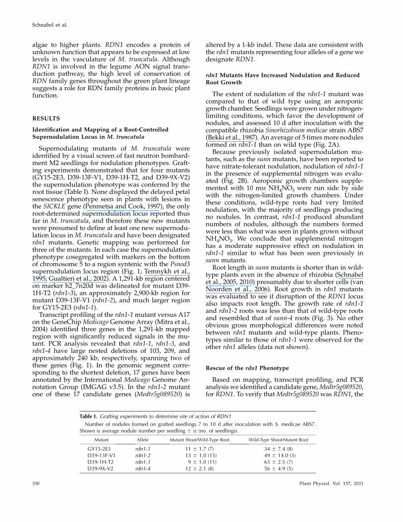
Supernodulating mutants of M. truncatula wereidentified by a visual screen of fast neutron bombard-ment M2 seedlings for nodulation phenotypes. Graft-ing experiments demonstrated that for four mutants(GY15-2E3, D39-13F-V1, D39-1H-T2, and D39-9X-V2)the supernodulation phenotype was conferred by theroot tissue (Table I). None displayed the delayed petalsenescence phenotype seen in plants with lesions inthe SICKLE gene (Penmetsa and Cook, 1997), the onlyroot-determined supernodulation locus reported thusfar in M. truncatula, and therefore these new mutantswere presumed to define at least one new supernodu-lation locus inM. truncatula and have been designatedrdn1 mutants. Genetic mapping was performed forthree of the mutants. In each case the supernodulationphenotype cosegregated with markers on the bottomof chromosome 5 to a region syntenic with the Psnod3supernodulation locus region (Fig. 1; Temnykh et al.,1995; Gualtieri et al., 2002). A 1,291-kb region centeredon marker h2_7n20d was delineated for mutant D39-1H-T2 (rdn1-3), an approximately 2,900-kb region formutant D39-13F-V1 (rdn1-2), and much larger regionfor GY15-2E3 (rdn1-1).
Transcript profiling of the rdn1-1mutant versus A17on the GeneChipMedicago Genome Array (Mitra et al.,2004) identified three genes in the 1,291-kb mappedregion with significantly reduced signals in the mu-tant. PCR analysis revealed that rdn1-1, rdn1-3, andrdn1-4 have large nested deletions of 103, 209, andapproximately 240 kb, respectively, spanning two ofthese genes (Fig. 1). In the genomic segment corre-sponding to the shortest deletion, 17 genes have beenannotated by the International Medicago Genome An-notation Group (IMGAG v3.5). In the rdn1-2 mutantone of these 17 candidate genes (Medtr5g089520) is
altered by a 1-kb indel. These data are consistent withthe rdn1mutants representing four alleles of a gene wedesignate RDN1.
rdn1 Mutants Have Increased Nodulation and ReducedRoot Growth
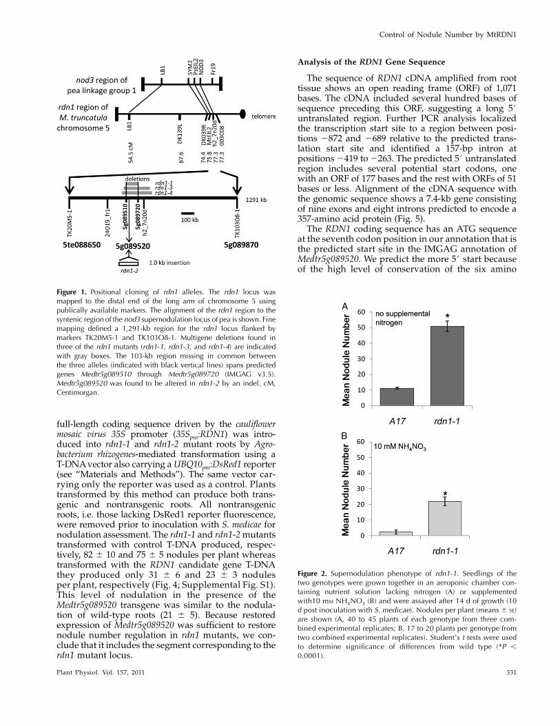
The extent of nodulation of the rdn1-1 mutant wascompared to that of wild type using an aeroponicgrowth chamber. Seedlings were grown under nitrogen-limiting conditions, which favor the development ofnodules, and assessed 10 d after inoculation with thecompatible rhizobia Sinorhizobium medicae strain ABS7(Bekki et al., 1987). An average of 5 times more nodulesformed on rdn1-1 than on wild type (Fig. 2A).
Because previously isolated supernodulation mu-tants, such as the sunn mutants, have been reported tohave nitrate-tolerant nodulation, nodulation of rdn1-1in the presence of supplemental nitrogen was evalu-ated (Fig. 2B). Aeroponic growth chambers supple-mented with 10 mM NH4NO3 were run side by sidewith the nitrogen-limited growth chambers. Underthese conditions, wild-type roots had very limitednodulation, with the majority of seedlings producingno nodules. In contrast, rdn1-1 produced abundantnumbers of nodules, although the numbers formedwere less than what was seen in plants grown withoutNH4NO3. We conclude that supplemental nitrogenhas a moderate suppressive effect on nodulation inrdn1-1 similar to what has been seen previously insunn mutants.
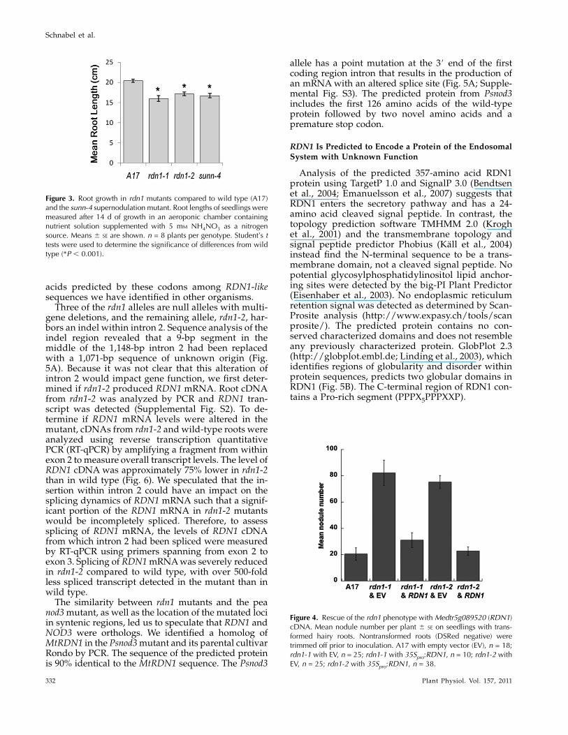
Root length in sunn mutants is shorter than in wild-type plants even in the absence of rhizobia (Schnabelet al., 2005, 2010) presumably due to shorter cells (vanNoorden et al., 2006). Root growth in rdn1 mutantswas evaluated to see if disruption of the RDN1 locusalso impacts root length. The growth rate of rdn1-1and rdn1-2 roots was less than that of wild-type rootsand resembled that of sunn-4 roots (Fig. 3). No otherobvious gross morphological differences were notedbetween rdn1 mutants and wild-type plants. Pheno-types similar to those of rdn1-1 were observed for theother rdn1 alleles (data not shown).
Rescue of the rdn1 Phenotype
Based on mapping, transcript profiling, and PCRanalysis we identified a candidate gene,Medtr5g089520,for RDN1. To verify thatMedtr5g089520was RDN1, the
Table I. Grafting experiments to determine site of action of RDN1
Number of nodules formed on grafted seedlings 7 to 10 d after inoculation with S. medicae ABS7.Shown is average nodule number per seedling 6 SE (no. of seedlings).
Mutant Allele Mutant Shoot/Wild-Type Root Wild-Type Shoot/Mutant Root
GY15-2E3 rdn1-1 11 6 1.7 (7) 34 6 7.4 (8)D39-13F-V1 rdn1-2 13 6 1.0 (15) 49 6 14.0 (3)D39-1H-T2 rdn1-3 9 6 1.0 (11) 63 6 2.5 (7)D39-9X-V2 rdn1-4 12 6 2.1 (8) 56 6 4.9 (5)
Schnabel et al.
330 Plant Physiol. Vol. 157, 2011
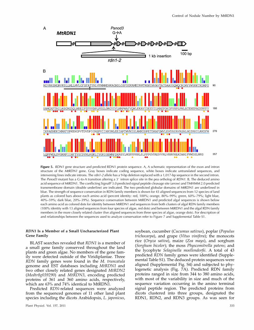
full-length coding sequence driven by the cauliflowermosaic virus 35S promoter (35Spro:RDN1) was intro-duced into rdn1-1 and rdn1-2 mutant roots by Agro-bacterium rhizogenes-mediated transformation using aT-DNAvector also carrying aUBQ10pro:DsRed1 reporter(see “Materials and Methods”). The same vector car-rying only the reporter was used as a control. Plantstransformed by this method can produce both trans-genic and nontransgenic roots. All nontransgenicroots, i.e. those lacking DsRed1 reporter fluorescence,were removed prior to inoculation with S. medicae fornodulation assessment. The rdn1-1 and rdn1-2mutantstransformed with control T-DNA produced, respec-tively, 82 6 10 and 75 6 5 nodules per plant whereastransformed with the RDN1 candidate gene T-DNAthey produced only 31 6 6 and 23 6 3 nodulesper plant, respectively (Fig. 4; Supplemental Fig. S1).This level of nodulation in the presence of theMedtr5g089520 transgene was similar to the nodula-tion of wild-type roots (21 6 5). Because restoredexpression of Medtr5g089520 was sufficient to restorenodule number regulation in rdn1 mutants, we con-clude that it includes the segment corresponding to therdn1 mutant locus.
Analysis of the RDN1 Gene Sequence
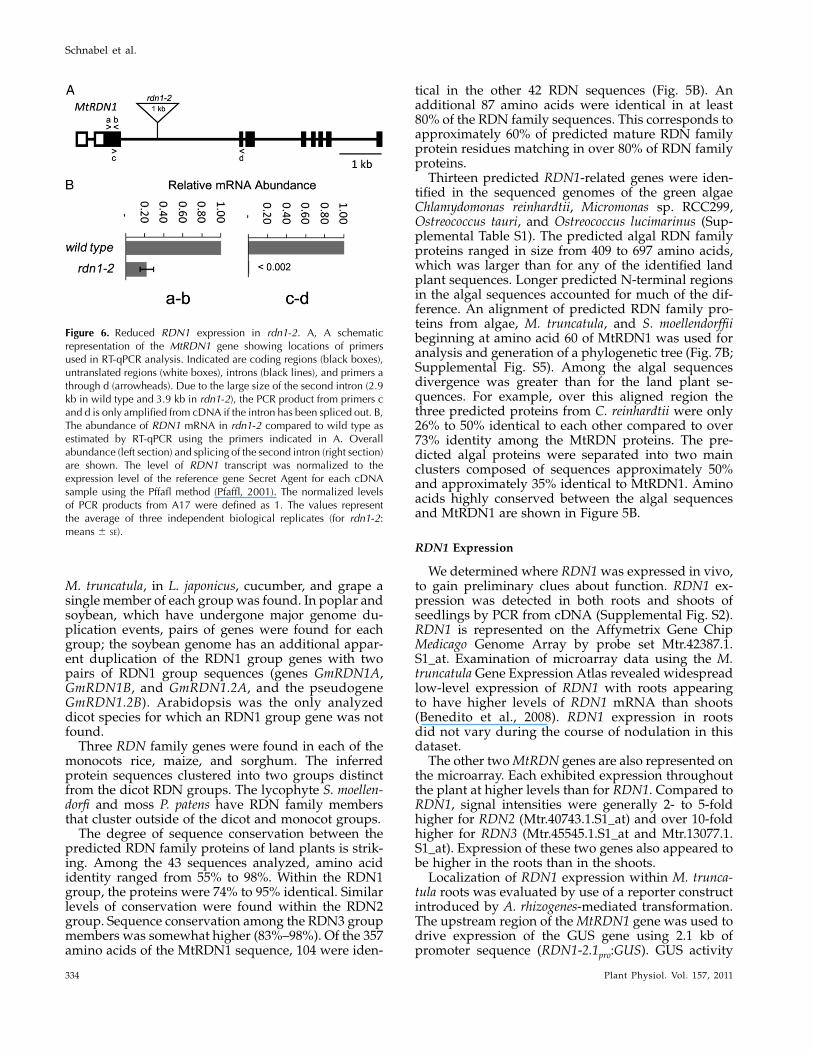
The sequence of RDN1 cDNA amplified from roottissue shows an open reading frame (ORF) of 1,071bases. The cDNA included several hundred bases ofsequence preceding this ORF, suggesting a long 5#untranslated region. Further PCR analysis localizedthe transcription start site to a region between posi-tions 2872 and 2689 relative to the predicted trans-lation start site and identified a 157-bp intron atpositions2419 to2263. The predicted 5# untranslatedregion includes several potential start codons, onewith an ORF of 177 bases and the rest with ORFs of 51bases or less. Alignment of the cDNA sequence withthe genomic sequence shows a 7.4-kb gene consistingof nine exons and eight introns predicted to encode a357-amino acid protein (Fig. 5).
The RDN1 coding sequence has an ATG sequenceat the seventh codon position in our annotation that isthe predicted start site in the IMGAG annotation ofMedtr5g089520. We predict the more 5# start becauseof the high level of conservation of the six amino
Figure 1. Positional cloning of rdn1 alleles. The rdn1 locus wasmapped to the distal end of the long arm of chromosome 5 usingpublically available markers. The alignment of the rdn1 region to thesyntenic region of the nod3 supernodulation locus of pea is shown. Finemapping defined a 1,291-kb region for the rdn1 locus flanked bymarkers TK20M5-1 and TK103O8-1. Multigene deletions found inthree of the rdn1 mutants (rdn1-1, rdn1-3, and rdn1-4) are indicatedwith gray boxes. The 103-kb region missing in common betweenthe three alleles (indicated with black vertical lines) spans predictedgenes Medtr5g089510 through Medtr5g089720 (IMGAG v3.5).Medtr5g089520 was found to be altered in rdn1-2 by an indel. cM,Centimorgan.
Figure 2. Supernodulation phenotype of rdn1-1. Seedlings of thetwo genotypes were grown together in an aeroponic chamber con-taining nutrient solution lacking nitrogen (A) or supplementedwith10 mM NH4NO3 (B) and were assayed after 14 d of growth (10d post inoculation with S. medicae). Nodules per plant (means 6 SE)are shown (A, 40 to 45 plants of each genotype from three com-bined experimental replicates; B, 17 to 20 plants per genotype fromtwo combined experimental replicates). Student’s t tests were usedto determine significance of differences from wild type (*P ,0.0001).
Control of Nodule Number by MtRDN1
Plant Physiol. Vol. 157, 2011 331
acids predicted by these codons among RDN1-likesequences we have identified in other organisms.
Three of the rdn1 alleles are null alleles with multi-gene deletions, and the remaining allele, rdn1-2, har-bors an indel within intron 2. Sequence analysis of theindel region revealed that a 9-bp segment in themiddle of the 1,148-bp intron 2 had been replacedwith a 1,071-bp sequence of unknown origin (Fig.5A). Because it was not clear that this alteration ofintron 2 would impact gene function, we first deter-mined if rdn1-2 produced RDN1 mRNA. Root cDNAfrom rdn1-2 was analyzed by PCR and RDN1 tran-script was detected (Supplemental Fig. S2). To de-termine if RDN1 mRNA levels were altered in themutant, cDNAs from rdn1-2 and wild-type roots wereanalyzed using reverse transcription quantitativePCR (RT-qPCR) by amplifying a fragment from withinexon 2 to measure overall transcript levels. The level ofRDN1 cDNA was approximately 75% lower in rdn1-2than in wild type (Fig. 6). We speculated that the in-sertion within intron 2 could have an impact on thesplicing dynamics of RDN1 mRNA such that a signif-icant portion of the RDN1 mRNA in rdn1-2 mutantswould be incompletely spliced. Therefore, to assesssplicing of RDN1 mRNA, the levels of RDN1 cDNAfrom which intron 2 had been spliced were measuredby RT-qPCR using primers spanning from exon 2 toexon 3. Splicing of RDN1mRNAwas severely reducedin rdn1-2 compared to wild type, with over 500-foldless spliced transcript detected in the mutant than inwild type.
The similarity between rdn1 mutants and the peanod3mutant, as well as the location of the mutated lociin syntenic regions, led us to speculate that RDN1 andNOD3 were orthologs. We identified a homolog ofMtRDN1 in the Psnod3mutant and its parental cultivarRondo by PCR. The sequence of the predicted proteinis 90% identical to the MtRDN1 sequence. The Psnod3
allele has a point mutation at the 3# end of the firstcoding region intron that results in the production ofan mRNAwith an altered splice site (Fig. 5A; Supple-mental Fig. S3). The predicted protein from Psnod3includes the first 126 amino acids of the wild-typeprotein followed by two novel amino acids and apremature stop codon.
RDN1 Is Predicted to Encode a Protein of the EndosomalSystem with Unknown Function
Analysis of the predicted 357-amino acid RDN1protein using TargetP 1.0 and SignalP 3.0 (Bendtsenet al., 2004; Emanuelsson et al., 2007) suggests thatRDN1 enters the secretory pathway and has a 24-amino acid cleaved signal peptide. In contrast, thetopology prediction software TMHMM 2.0 (Kroghet al., 2001) and the transmembrane topology andsignal peptide predictor Phobius (Kall et al., 2004)instead find the N-terminal sequence to be a trans-membrane domain, not a cleaved signal peptide. Nopotential glycosylphosphatidylinositol lipid anchor-ing sites were detected by the big-PI Plant Predictor(Eisenhaber et al., 2003). No endoplasmic reticulumretention signal was detected as determined by Scan-Prosite analysis (http://www.expasy.ch/tools/scanprosite/). The predicted protein contains no con-served characterized domains and does not resembleany previously characterized protein. GlobPlot 2.3(http://globplot.embl.de; Linding et al., 2003), whichidentifies regions of globularity and disorder withinprotein sequences, predicts two globular domains inRDN1 (Fig. 5B). The C-terminal region of RDN1 con-tains a Pro-rich segment (PPPX5PPPXXP).
Figure 3. Root growth in rdn1 mutants compared to wild type (A17)and the sunn-4 supernodulation mutant. Root lengths of seedlings weremeasured after 14 d of growth in an aeroponic chamber containingnutrient solution supplemented with 5 mM NH4NO3 as a nitrogensource. Means 6 SE are shown. n = 8 plants per genotype. Student’s ttests were used to determine the significance of differences from wildtype (*P , 0.001).
Figure 4. Rescue of the rdn1 phenotype with Medtr5g089520 (RDN1)cDNA. Mean nodule number per plant 6 SE on seedlings with trans-formed hairy roots. Nontransformed roots (DSRed negative) weretrimmed off prior to inoculation. A17 with empty vector (EV), n = 18;rdn1-1 with EV, n = 25; rdn1-1 with 35Spro:RDN1, n = 10; rdn1-2 withEV, n = 25; rdn1-2 with 35Spro:RDN1, n = 38.
Schnabel et al.
332 Plant Physiol. Vol. 157, 2011
RDN1 Is a Member of a Small Uncharacterized PlantGene Family
BLAST searches revealed that RDN1 is a member ofa small gene family conserved throughout the landplants and green algae. No members of the gene fam-ily were detected outside of the Viridiplantae. ThreeRDN family genes were found in the M. truncatulagenome and EST databases including MtRDN1 andtwo other closely related genes designated MtRDN2(Medtr8g039290) and MtRDN3, encoding predictedproteins of 361 and 360 amino acids, respectively,which are 63% and 74% identical to MtRDN1.Predicted RDN-related sequences were analyzed
from the sequenced genomes of 11 other land plantspecies including the dicots Arabidopsis, L. japonicus,
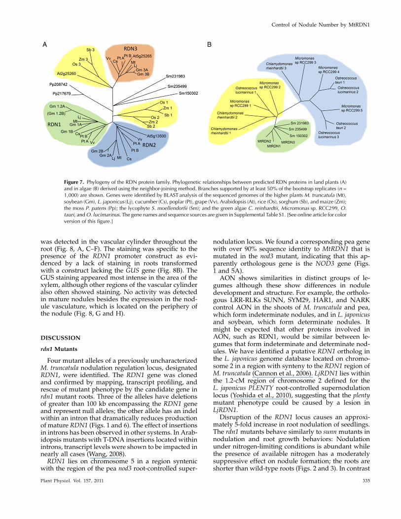
soybean, cucumber (Cucumus sativus), poplar (Populustrichocarpa), and grape (Vitus vinifera); the monocotsrice (Oryza sativa), maize (Zea mays), and sorghum(Sorghum bicolor); the moss Physcomitrella patens; andthe lycophyte Selaginella moellendorfii. A total of 43predicted RDN family genes were identified (Supple-mental Table S1). The deduced protein sequences werealigned (Supplemental Fig. S4) and subjected to phy-logenetic analysis (Fig. 7A). Predicted RDN familyproteins ranged in size from 344 to 380 amino acids,with most of the variability in size and much of thesequence variation occurring in the amino terminalsignal peptide region. The predicted proteins fromdicots clustered into three groups, designated theRDN1, RDN2, and RDN3 groups. As was seen for
Figure 5. RDN1 gene structure and predicted RDN1 protein sequence. A, A schematic representation of the exon and intronstructure of the MtRDN1 gene. Gray boxes indicate coding sequence, white boxes indicate untranslated sequences, andintervening lines indicate introns. The rdn1-2 allele has a 9-bp deletion replaced with a 1,017-bp sequence in the second intron.The Psnod3 mutant has a G-to-A transition altering a 3# intron splice site in the pea ortholog of RDN1. B, The deduced aminoacid sequence of MtRDN1. The conflicting SignalP 3.0 predicted signal peptide cleavage site (arrow) and TMHMM2.0 predictedtransmembrane domain (double underline) are indicated. The two predicted globular domains of MtRDN1 are underlined inblue. The strength of sequence conservation in RDN family members is shown for 43 aligned sequences from 12 species of landplants as colored bars above each amino acid (percent identity: red, 100%; orange, 80%–99%; green, 60%–79%; light blue,40%–59%; dark blue, 20%–39%). Sequence conservation between MtRDN1 and predicted algal sequences is shown beloweach amino acid as colored dots for identity between MtRDN1 and sequences from both clusters of algal RDN family members(100% identity with 13 aligned sequences from four species of algae, red dots) and between MtRDN1 and the algal RDN familymembers in the more closely related cluster (five aligned sequences from three species of algae, orange dots). For description ofand relationships between the sequences used to analyze conservation refer to Figure 7 and Supplemental Table S1.
Control of Nodule Number by MtRDN1
Plant Physiol. Vol. 157, 2011 333
M. truncatula, in L. japonicus, cucumber, and grape asingle member of each groupwas found. In poplar andsoybean, which have undergone major genome du-plication events, pairs of genes were found for eachgroup; the soybean genome has an additional appar-ent duplication of the RDN1 group genes with twopairs of RDN1 group sequences (genes GmRDN1A,GmRDN1B, and GmRDN1.2A, and the pseudogeneGmRDN1.2B). Arabidopsis was the only analyzeddicot species for which an RDN1 group gene was notfound.
Three RDN family genes were found in each of themonocots rice, maize, and sorghum. The inferredprotein sequences clustered into two groups distinctfrom the dicot RDN groups. The lycophyte S. moellen-dorfi and moss P. patens have RDN family membersthat cluster outside of the dicot and monocot groups.
The degree of sequence conservation between thepredicted RDN family proteins of land plants is strik-ing. Among the 43 sequences analyzed, amino acididentity ranged from 55% to 98%. Within the RDN1group, the proteins were 74% to 95% identical. Similarlevels of conservation were found within the RDN2group. Sequence conservation among the RDN3 groupmembers was somewhat higher (83%–98%). Of the 357amino acids of the MtRDN1 sequence, 104 were iden-
tical in the other 42 RDN sequences (Fig. 5B). Anadditional 87 amino acids were identical in at least80% of the RDN family sequences. This corresponds toapproximately 60% of predicted mature RDN familyprotein residues matching in over 80% of RDN familyproteins.
Thirteen predicted RDN1-related genes were iden-tified in the sequenced genomes of the green algaeChlamydomonas reinhardtii, Micromonas sp. RCC299,Ostreococcus tauri, and Ostreococcus lucimarinus (Sup-plemental Table S1). The predicted algal RDN familyproteins ranged in size from 409 to 697 amino acids,which was larger than for any of the identified landplant sequences. Longer predicted N-terminal regionsin the algal sequences accounted for much of the dif-ference. An alignment of predicted RDN family pro-teins from algae, M. truncatula, and S. moellendorffiibeginning at amino acid 60 of MtRDN1 was used foranalysis and generation of a phylogenetic tree (Fig. 7B;Supplemental Fig. S5). Among the algal sequencesdivergence was greater than for the land plant se-quences. For example, over this aligned region thethree predicted proteins from C. reinhardtii were only26% to 50% identical to each other compared to over73% identity among the MtRDN proteins. The pre-dicted algal proteins were separated into two mainclusters composed of sequences approximately 50%and approximately 35% identical to MtRDN1. Aminoacids highly conserved between the algal sequencesand MtRDN1 are shown in Figure 5B.
RDN1 Expression
We determined where RDN1was expressed in vivo,to gain preliminary clues about function. RDN1 ex-pression was detected in both roots and shoots ofseedlings by PCR from cDNA (Supplemental Fig. S2).RDN1 is represented on the Affymetrix Gene ChipMedicago Genome Array by probe set Mtr.42387.1.S1_at. Examination of microarray data using the M.truncatula Gene Expression Atlas revealed widespreadlow-level expression of RDN1 with roots appearingto have higher levels of RDN1 mRNA than shoots(Benedito et al., 2008). RDN1 expression in rootsdid not vary during the course of nodulation in thisdataset.
The other twoMtRDN genes are also represented onthe microarray. Each exhibited expression throughoutthe plant at higher levels than for RDN1. Compared toRDN1, signal intensities were generally 2- to 5-foldhigher for RDN2 (Mtr.40743.1.S1_at) and over 10-foldhigher for RDN3 (Mtr.45545.1.S1_at and Mtr.13077.1.S1_at). Expression of these two genes also appeared tobe higher in the roots than in the shoots.
Localization of RDN1 expression within M. trunca-tula roots was evaluated by use of a reporter constructintroduced by A. rhizogenes-mediated transformation.The upstream region of theMtRDN1 gene was used todrive expression of the GUS gene using 2.1 kb ofpromoter sequence (RDN1-2.1pro:GUS). GUS activity
Figure 6. Reduced RDN1 expression in rdn1-2. A, A schematicrepresentation of the MtRDN1 gene showing locations of primersused in RT-qPCR analysis. Indicated are coding regions (black boxes),untranslated regions (white boxes), introns (black lines), and primers athrough d (arrowheads). Due to the large size of the second intron (2.9kb in wild type and 3.9 kb in rdn1-2), the PCR product from primers cand d is only amplified from cDNA if the intron has been spliced out. B,The abundance of RDN1 mRNA in rdn1-2 compared to wild type asestimated by RT-qPCR using the primers indicated in A. Overallabundance (left section) and splicing of the second intron (right section)are shown. The level of RDN1 transcript was normalized to theexpression level of the reference gene Secret Agent for each cDNAsample using the Pffafl method (Pfaffl, 2001). The normalized levelsof PCR products from A17 were defined as 1. The values representthe average of three independent biological replicates (for rdn1-2:means 6 SE).
Schnabel et al.
334 Plant Physiol. Vol. 157, 2011
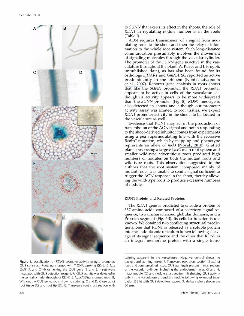
was detected in the vascular cylinder throughout theroot (Fig. 8, A, C–F). The staining was specific to thepresence of the RDN1 promoter construct as evi-denced by a lack of staining in roots transformedwith a construct lacking the GUS gene (Fig. 8B). TheGUS staining appeared most intense in the area of thexylem, although other regions of the vascular cylinderalso often showed staining. No activity was detectedin mature nodules besides the expression in the nod-ule vasculature, which is located on the periphery ofthe nodule (Fig. 8, G and H).
DISCUSSION
rdn1 Mutants
Four mutant alleles of a previously uncharacterizedM. truncatula nodulation regulation locus, designatedRDN1, were identified. The RDN1 gene was clonedand confirmed by mapping, transcript profiling, andrescue of mutant phenotype by the candidate gene inrdn1 mutant roots. Three of the alleles have deletionsof greater than 100 kb encompassing the RDN1 geneand represent null alleles; the other allele has an indelwithin an intron that dramatically reduces productionof mature RDN1 (Figs. 1 and 6). The effect of insertionsin introns has been observed in other systems. In Arab-idopsis mutants with T-DNA insertions located withinintrons, transcript levels were shown to be impacted innearly all cases (Wang, 2008).RDN1 lies on chromosome 5 in a region syntenic
with the region of the pea nod3 root-controlled super-
nodulation locus. We found a corresponding pea genewith over 90% sequence identity to MtRDN1 that ismutated in the nod3 mutant, indicating that this ap-parently orthologous gene is the NOD3 gene (Figs.1 and 5A).
AON shows similarities in distinct groups of le-gumes although these show differences in noduledevelopment and structure. For example, the ortholo-gous LRR-RLKs SUNN, SYM29, HAR1, and NARKcontrol AON in the shoots of M. truncatula and pea,which form indeterminate nodules, and in L. japonicusand soybean, which form determinate nodules. Itmight be expected that other proteins involved inAON, such as RDN1, would be similar between le-gumes that form indeterminate and determinate nod-ules. We have identified a putative RDN1 ortholog inthe L. japonicus genome database located on chromo-some 2 in a region with synteny to the RDN1 region ofM. truncatula (Cannon et al., 2006). LjRDN1 lies withinthe 1.2-cM region of chromosome 2 defined for theL. japonicus PLENTY root-controlled supernodulationlocus (Yoshida et al., 2010), suggesting that the plentymutant phenotype could be caused by a lesion inLjRDN1.
Disruption of the RDN1 locus causes an approxi-mately 5-fold increase in root nodulation of seedlings.The rdn1 mutants behave similarly to sunn mutants innodulation and root growth behaviors: Nodulationunder nitrogen-limiting conditions is abundant whilethe presence of available nitrogen has a moderatelysuppressive effect on nodule formation; the roots areshorter than wild-type roots (Figs. 2 and 3). In contrast
Figure 7. Phylogeny of the RDN protein family. Phylogenetic relationships between predicted RDN proteins in land plants (A)and in algae (B) derived using the neighbor-joining method. Branches supported by at least 50% of the bootstrap replicates (n =1,000) are shown. Genes were identified by BLAST analysis of the sequenced genomes of the higher plants M. truncatula (Mt),soybean (Gm), L. japonicus (Lj), cucumber (Cs), poplar (Pt), grape (Vv), Arabidopsis (At), rice (Os), sorghum (Sb), andmaize (Zm);the moss P. patens (Pp); the lycophyte S. moellendorfii (Sm); and the green algae C. reinhardtii, Micromonas sp. RCC299, O.tauri, andO. lucimarinus. The gene names and sequence sources are given in Supplemental Table S1. [See online article for colorversion of this figure.]
Control of Nodule Number by MtRDN1
Plant Physiol. Vol. 157, 2011 335
to SUNN that exerts its effect in the shoots, the role ofRDN1 in regulating nodule number is in the roots(Table I).
AON requires transmission of a signal from nod-ulating roots to the shoot and then the relay of infor-mation to the whole root system. Such long-distancecommunication presumably involves the movementof signaling molecules through the vascular cylinder.The promoter of the SUNN gene is active in the vas-culature throughout the plant (A. Karve and J. Frugoli,unpublished data), as has also been found for itsorthologs LjHAR1 and GmNARK, reported as activepredominantly in the phloem (Nontachaiyapoomet al., 2007). Reporter gene analysis in roots showsthat like the SUNN promoter, the RDN1 promoterappears to be active in cells of the vasculature al-though its activity appears to be more widespreadthan the SUNN promoter (Fig. 8). RDN1 message isalso detected in shoots and although our promoteractivity assay was limited to root tissues, we expectRDN1 promoter activity in the shoots to be located inthe vasculature as well.
Evidence that RDN1 may act in the production ortransmission of the AON signal and not in respondingto the shoot-derived inhibitor comes from experimentsusing a pea supernodulating line with the recessiveRisfixC mutation, which by mapping and phenotyperepresents an allele of nod3 (Novak, 2010). Graftedplants possessing a large RisfixCmain root system andsmaller wild-type adventitious roots produced highnumbers of nodules on both the mutant roots andwild-type roots. This observation suggested to theauthors that the root system, composed mainly ofmutant roots, was unable to send a signal sufficient totrigger the AON response in the shoot, thereby allow-ing the wild-type roots to produce excessive numbersof nodules.
RDN1 Protein and Related Proteins
The RDN1 gene is predicted to encode a protein of357 amino acids composed of a secretory signal se-quence, two uncharacterized globular domains, and aPro-rich segment (Fig. 5B). Its cellular function is un-known. We obtained two conflicting structural predic-tions: one that RDN1 is released as a soluble proteininto the endoplasmic reticulum lumen following cleav-age of its signal sequence and the other that RDN1 isan integral membrane protein with a single trans-
Figure 8. Localization of RDN1 promoter activity using a promoter:GUS construct. Roots transformed with T-DNA carrying RDN1-2.1pro:GUS (A and C–H) or lacking the GUS gene (B and E, inset) wereincubatedwith GUS detection reagent. A, GUS activity was detected inthe central cylinder throughout RDN1-2.1pro:GUS transformed roots. B,Without the GUS gene, roots show no staining. C and D, Close up ofroot tissue (C) and root tip (D). E, Transverse root cross section with
staining apparent in the vasculature. Negative control shows nobackground staining (inset). F, Transverse root cross section (3 mM) offixed and counterstained tissue. GUS staining is present in most regionsof the vascular cylinder, including the endodermal layer. G and H,Intact nodule (G) and nodule cross section (H) showing GUS activityonly in the vasculature around the nodule following extended incu-bation (36 h) with GUS detection reagent. Scale bars where shown are50 mm.
Schnabel et al.
336 Plant Physiol. Vol. 157, 2011
membrane domain. However, the predicted localiza-tion of RDN proteins to membranes is supported bythe identification of the Arabidopsis RDN3 family pro-tein At5g25265 in proteomics analyses in plasmamem-brane (Marmagne et al., 2007; Mitra et al., 2009) andvacuolar fractions (Carter, et al., 2004; Jaquinod et al.,2007). Determining the subcellular location of RDN1experimentally would provide an important clue as tothe function of the protein, such as whether RDN1 issecreted, located in the plasma membrane, or targetedelsewhere in the cell.RDN1-related sequences were found in the genomes
of all plants examined, including those of green algae,and represent a gene family we have designated theRDN family. All of the putative RDN family proteinsidentified are predicted by TargetP to have signalsequences, although, as was found for MtRDN1, thereare conflicting predictions for whether the signal se-quences are cleaved. Three RDN family genes werefound in most land plant genomes. For example, M.truncatula has two genes similar to MtRDN1, namedMtRDN2 and MtRDN3, which are both predicted toencode proteins approximately 70% identical and over80% similar to MtRDN1. Predicted dicot RDN familyproteins clustered into three groups represented by thethree MtRDN proteins (Fig. 7). Predicted monocotRDN family proteins clustered separately into twogroups. The structure of the RDN family phylogenetictree suggests the existence of an RDN gene familyprior to the divergence of monocots and dicots, fol-lowed by duplications forming the RDN1 and RDN2groups of the dicots and forming one of the monocotclusters.Arabidopsis does not have an RDN1 ortholog but
does have an additional RDN family gene, At2g25260,divergent from other dicot RDN family genes. Analy-sis of database sequences from other plants of theorder Brassicacales revealed no RDN1 orthologs inclose relatives of Arabidopsis in the family Brassica-ceae, but a gene similar to At2g25260 was found, whileCarica papaya, a member of the family Caricaceae,possesses RDN1, RDN2, and RDN3 homologs like theother dicot lineages. This suggests that loss of RDN1and appearance of the At2g25260 type gene occurredin the Brassicaceae lineage.The predicted RDN family proteins from land plants
are highly conserved. Over 60% of the amino acids ofthe predicted mature proteins are highly conservedamong family members with over 80% of the familymembers identical at those positions. Many residues(10%) are also invariant in the algal RDN family se-quences.The conservation of RDN family genes across the
Viridiplantae, including unicellular algae, suggestsa basic cellular function for RDN family proteins.Of the nearly 7,000 protein families identified in theChlamydomonas genome, 172 families appear to beunique to the green plants, with almost two-thirds ofthese proteins predicted to be chloroplast localized(Merchant et al., 2007). The RDN family is one of only
61 identified green plant-specific protein familieswhose members are not predicted to be localized tochloroplasts and one of only 10 whose members arepredicted to have secretory signal sequences.
Expression of RDN Family Genes
A survey of the major organ systems ofM. truncatula(Benedito et al., 2008; He et al., 2009) using the Medi-cago Gene Expression Array revealed that the threeRDN genes are expressed in all organs examined, in-cluding leaves, stems, flowers, vegetative buds, roots,nodules, pods, and seeds (Supplemental Fig. S6). Theabundance of themessage appears to vary between thegenes with RDN1 detected only at low levels, RDN2and RDN3 at higher levels.
Similarly, using the Arabidopsis Electronic Fluores-cent Pictograph browser for whole genome tiling arraydata (Winter et al., 2007; Laubinger et al., 2008), theArabidopsis RDN2 and RDN3 family genes At5g13500and At5g25265 were found to be expressed in most tis-sues examined (roots, leaves, shoot apex, cotyledons,hypocotyls, seeds, flowers, young siliques) with theRDN3 family gene being expressed at higher levels(Supplemental Fig. S3). In contrast, At2g25260, theArabidopsis RDN family gene without an ortholog inthe other dicots, was detected primarily in roots andshoot apex inflorescences at high and moderate levels,respectively, and only weakly in other tissues.
In microarray analyses of fluorescently sorted cellsfrom the roots of a series of GFP-marked lines, theArabidopsis RDN2 family gene was most stronglyexpressed in the root hair cell lineage with strong ex-pression also throughout the elongation zone. Strongelongation zone expression was also observed in an-other study (Brady et al., 2007; Dinneny et al., 2008).Thus, it appears that RDN family proteins are ex-pressed in most plant organs, with perhaps higherlevels of expression in certain cell types such as vas-cular cells for MtRDN1 and root hair cells for theArabidopsis RDN2 family gene.
Consistent with the demonstrated role of MtRDN1in AON and its expression in vascular tissue, we pro-pose that MtRDN1 is involved in initiating, respond-ing to, or transporting vascular signals and that thisvascular signaling function of RDN1-related proteinsmay be present in all dicots. Furthermore, the wideconservation of RDN family genes across the greenplants, including unicellular algae, suggests other con-served molecular functions for the RDN family pro-teins in the plant cell.
MATERIALS AND METHODS
Plant Materials
Preparation ofMedicago truncatula seeds and growth of plants in aeroponicchambers was performed as described in Schnabel et al. (2010). For all otherassays, scarified and imbibed seeds were vernalized in the dark at 4#C for 2 don Harrison Modified Farheus agar (Huo et al., 2006) covered with Whatman
Control of Nodule Number by MtRDN1
Plant Physiol. Vol. 157, 2011 337
filter paper. Following overnight germination at room temperature, seedlingswere transferred to plates half covered with filter paper with the radicals onthe paper and the cotyledons on the uncovered medium, placed vertically in agrowth chamber at 25#C with a 16-h photoperiod for 5 d, and then used forexperiments. For all nodulations experiments the strain Sinorhizobium medicaestrain ABS7 was used (Bekki et al., 1987).
The rdn1 mutants were identified from independent M2 seed poolscollected from fast neutron bombarded M1 seeds of M. truncatula ‘Jemalong’.M3 plants were rescreened in an aeroponic chamber for nodulation phenotypeand in the greenhouse for petal senescence. Lines used for detailed analyseswere backcrossed to A17 wild type three times (rdn1-1) or once (rdn1-2).
A near-isogenic line of the pea (Pisum sativum ‘Rondo’) mutant nod3backcrossed into pea cv Juneau (nod3I, PI 598367) and pea cv Rondo were ob-tained through the National Plant Germplasm System (http://www.ars-grin.gov/).
Mapping of rdn and Sequence Analysis
The F2 self-pollinated progeny from crosses of rdn1mutants GY15-2E, D39-1H-T2, and D39-13F-V1 to M. truncatula ecotype A20 were used for geneticmapping of the rdn1 locus. DNA from F2 individuals with high nodulenumbers was evaluated for the segregation of cleaved amplified polymorphicsequence and other markers as previously described (Schnabel et al., 2010;Supplemental Table S2). For each mutant, 80 to 195 supernodulating F2 plantswere tested. For D39-1H-T2 and D39-13F-V1, supernodulating plants repre-sented approximately 25% of the F2 progeny as expected for a single recessivelocus. In the F2 progeny of the GY15-2E mapping cross, fewer than expectedsupernodulating plants were observed (approximately 10%); recombinationaround the rdn1 locus was suppressed at least 5-fold; and skewed markersegregation was observed, with cosegregation of markers from the long armsof chromosomes 5 and 8. These data suggest a genomic rearrangement withinthe GY15-2E mutant.
The Gene Chip Medicago Genome Array (Affymetrix) was used foroligonucleotide hybridization experiments comparing transcript profiles ofbuffer-inoculated wild-type A17 roots with buffer-inoculated rdn1-1 mutantroots. Methods were as described by Mitra et al. (2004).
The search for RDN family members used BLAST algorithms blastp andtblastn (Altschul et al., 1990). Sequences from selected organisms with se-quenced genomes were used for analysis. No sequences with a probabilityrelationship to MtRDN1 of e , 3 were found outside of the Viridiplantae.
Phylogenetic Analysis
RDN family predicted protein sequences were aligned using the ClustalWalgorithm of MegAlign in Lasergene 7.1.0 (DNAStar). Phylogenetic trees wereconstructed with MEGA version 4 (Tamura et al., 2007) using the neighbor-joining method with 1,000 bootstrap replicates. Branches supported by at least500 of the 1,000 bootstrap replicates are shown in Figure 7.
RNA Isolation and RT-qPCR
RNA was isolated from plant tissues using the Qiagen RNeasy mini kit(http://www.qiagen.com), treated with RQ1 RNase-Free DNase (http://www.promega.com) followed by phenol and chloroform extractions and ethanolprecipitation, and quantified spectrophotometrically. cDNA was synthesizedin 20-mL reactions from 0.5 to 2 mg RNA using random hexanucleotideprimers and Superscript Reverse Transcriptase II (http://www.invitrogen.com) following themanufacturer’s recommendations. The absence of genomicDNA in cDNAs was verified by PCR with primers JF1330 and JF1331, specificfor a noncoding region near Medtr5g089720. The expression of RDN1 and thespecificity of RDN1 primers was evaluated by visualizing PCR products fromgenomic DNA and cDNA on 1% tris-borate-EDTA gels following PCR am-plification (Supplemental Fig. S2).
Expression levels were quantified in an iQ5 thermocycler (http://www.bio-rad.com) using PerfeCTa SYBR green supermix (Quanta Biosciences).Expression levels and splicing of RDN1 transcripts were assessed usingprimers for amplifying within exon 2 (primers qPCR-a and qPCR-b) and fromexon 2 to exon 3 (primers qPCR-c and qPCR-d). Levels of cDNA werenormalized using the Secret Agent gene as a reference (Kuppusamy et al.,2004) and ratios were calculated using the Pfaffl method (Pfaffl, 2001). Threeindependent biological replicates were evaluated. For each cDNA threetechnical replicates were performed and the values averaged. The efficiency
of each primer pair was assessed by use of a dilution series. In all runs, theprimer pairs for exon 2, exon 2 to exon 3, and Secret Agent had measuredefficiencies of 2.0, 2.0, and 1.8, respectively. Across the biological replicates,threshold cycles for all products from the cDNAs fell within the valid range ofthe standard curves with the exception of spliced products from rdn1-2 thathad high Ct values.
Generation of Transgenic Hairy Roots andHistochemical Analysis
For expression of RDN1 in transgenic plants, a fragment of RDN1 cDNAwas PCR amplified from M. truncatula cDNA using primers RDN1cDNA-Aand RDN1cDNA-B and cloned downstream of the cauliflower mosaic virus35S promoter in pC-DsRED2 using KpnI and XhoI. The vector pC-DsRed2was constructed from pCAMBIA0390 by replacing a portion of the poly-linker with the polylinker region of pCAMBIA3201 (EcoRI to PstI), addingthe AscI/HindIII UBQ10pro:DsRed1 fragment of pRedRoot (Limpens et al.,2004), and adding additional restriction sites to the polylinker by ligating anEcoRI/MluI/XhoI adaptor into the EcoRI site. For promoter activity analysis,3.3- and 2.1-kb fragments from upstream of the predicted RDN1 translationstart site were amplified by PCR from M. truncatula genomic DNA usingprimers P2.1-F and P2.1-R and primers P3.3-F and P3.3-R, respectively, andcloned using NcoI and EcoRI into pC2381ES (Huo et al., 2006). The codingand promoter sequences in the binary vector were confirmed by sequencing.
Prepared seedlings were transformed as previously described (Limpenset al., 2004) using Agrobacterium rhizogenes strain ARqua1 (Quandt et al., 1993)containing the appropriate binary vector. Seedlings were maintained on platesuntil sufficient root tissue had grown. For nodulation experiments, non-transgenic roots (those lacking DsRed fluorescence) were trimmed off prior totransfer of plants to pots of perlite mixed with Harrison Modified Farheusmedium without nitrate. After 5 d of nitrogen starvation, plants were floodinoculated with S. medicae (OD600 = 0.1) and nodules were counted 21 d later.
For promoter experiments, transformed tissue was washed twice in 0.1 M
Na2HPO4/NaH2PO4, pH 7.2 for 15 min and GUS activity was localized basedon a protocol by Jefferson and others (Jefferson et al., 1987). Samples wereinfiltrated with substrate under vacuum for 30 min and incubated at 37#C for18 h, unless otherwise indicated. Where indicated, roots were fixed in 5%glutaraldehyde/0.1 M sodium phosphate buffer, pH 7.2 for 2 h under vacuum.Serial ethanol dehydration was then performed by increasing the concentra-tion (10%, 30%, 50%, 70%, 90%, and 100%) at room temperature for 10 mineach. Samples were embedded in Technovit 7100 resin (Heraeus Kulzer) usingthe manufacturer’s instruction. Sections were prepared using a RM2165microtome (Leica Microsystems), dried onto glass slides at 42#C, and counter-stained for 1 min in 1% aqueous saffronin-O solution. Slides were washedbriefly with water, dried, and mounted in permount (Fisher Scientific). Tissuewas photographed using a Zeiss Axiostar plus microscope and a Nikon E600microscope with a Retiga EXi FAST monochrome CCD 12-bit camera.
Accession numbers are as follows: MtRDN1 mRNA (GU580937), PsNOD3gene (GU580938), and PsNOD3 mRNA (GU580939).
Supplemental Data
The following materials are available in the online version of this article.
Supplemental Figure S1. Nodulation on rdn1 roots rescued with theMedtr5g089520 (RDN1) cDNA.
Supplemental Figure S2. PCR analysis of RDN1 expression.
Supplemental Figure S3. The structure of the PsNOD3 coding region inpea cultivar Rondo and the derived mutant nod3.
Supplemental Figure S4. Alignment of the predicted sequences of RDNfamily proteins from 12 land plant species.
Supplemental Figure S5. Alignment of the predicted sequences of RDNfamily proteins from the green algae with RDN protein sequences fromland plants.
Supplemental Figure S6. Expression levels of RDN family genes ofM. truncatula and Arabidopsis in various tissues.
Supplemental Table S1. RDN family genes identified in the genomes of 12land plants and four algae.
Schnabel et al.
338 Plant Physiol. Vol. 157, 2011
Supplemental Table S2. Primers used in RDN1 mapping, T-DNA vectorpreparation, and RT-qPCR.
Note Added in Proof
Recently, mutations in a CLV2-like gene were found in pea sym28 shoot-controlled supernodulation mutants. Similarly, in Lotus japonicus, a mutantwith a lesion in a CLV2-like gene had increased nodule number (Krusell L,Sato N, Fukuhara I, Koch BE, Grossmann C, Okamoto S, Oka-Kira E,Otsubo Y, Aubert G, Nakagawa T, et al [2011] The Clavata2 genes of pea andLotus japonicus affect autoregulation of nodulation. Plant J 65: 861–871). Thisfinding adds to the number of known genes involved in nodule numberregulation.
ACKNOWLEDGMENTS
We would like to thank Leah Howell, Christine Gianniny, Kyle Ames, andSherri-Hughes Murphree for preliminary help in the mapping of rdn1; SteveEllis and Nancy Korn for assistance in use of the Clemson Animal andVeterinary Sciences Histology Core Facility; Harry Kurtz, Jr. for use of hismicroscope; and Clarice Coyne of the U.S. Department of AgricultureWestern Regional Plant Introduction Station for providing seeds of pea lines.This manuscript is Technical Contribution number 5890 of the ClemsonExperiment Station.
Received April 22, 2011; accepted July 7, 2011; published July 8, 2011.
LITERATURE CITED
Altschul SF, Gish W, Miller W, Myers EW, Lipman DJ (1990) Basic localalignment search tool. J Mol Biol 215: 403–410
Bekki A, Trichant JC, Rigaud J (1987) Nitrogen fixation (C2H2 reduction)by Medicago nodules and bacteroids under sodium chloride stress.Physiol Plant 71: 61–67
Bendtsen JD, Nielsen H, von Heijne G, Brunak S (2004) Improvedprediction of signal peptides: SignalP 3.0. J Mol Biol 340: 783–795
Benedito VA, Torres-Jerez I, Murray JD, Andriankaja A, Allen S, KakarK, Wandrey M, Verdier J, Zuber H, Ott T, et al (2008) A gene expressionatlas of the model legume Medicago truncatula. Plant J 55: 504–513
Brady SM, Orlando DA, Lee JY, Wang JY, Koch J, Dinneny JR, Mace D,Ohler U, Benfey PN (2007) A high-resolution root spatiotemporal mapreveals dominant expression patterns. Science 318: 801–806
Caetano-Anolles G, Gresshoff PM (1991) Plant genetic control of nodu-lation. Annu Rev Microbiol 45: 345–382
Cannon SB, Sterck L, Rombauts S, Sato S, Cheung F, Gouzy J, Wang X,Mudge J, Vasdewani J, Schiex T, et al (2006) Legume genome evolutionviewed through the Medicago truncatula and Lotus japonicus genomes.Proc Natl Acad Sci USA 103: 14959–14964
Carroll BJ, McNeil DL, Gresshoff PM (1985) A supernodulation andnitrate-tolerant symbiotic (nts) soybean mutant. Plant Physiol 78: 34–40
Carter C, Pan S, Zouhar J, Avila EL, Girke T, Raikhel NV (2004) Thevegetative vacuole proteome of Arabidopsis thaliana reveals predictedand unexpected proteins. Plant Cell 16: 3285–3303
Crawford N, Kahn M, Leustek T, Long S (2000). Nitrogen and Sulfur.American Association of Plant Biologists, Rockville, MD
Delves AC, Mathews A, Day DA, Carter AS, Carroll BJ, Gresshoff PM(1986) Regulation of the soybean-Rhizobium nodule symbiosis by shootand root factors. Plant Physiol 82: 588–590
Ding Y, Kalo P, Yendrek C, Sun J, Liang Y, Marsh JF, Harris JM, OldroydGE (2008) Abscisic acid coordinates nod factor and cytokinin signalingduring the regulation of nodulation inMedicago truncatula. Plant Cell 20:2681–2695
Dinneny JR, Long TA, Wang JY, Jung JW, Mace D, Pointer S, Barron C,Brady SM, Schiefelbein J, Benfey PN (2008) Cell identity mediates theresponse of Arabidopsis roots to abiotic stress. Science 320: 942–945
Eisenhaber B, Wildpaner M, Schultz CJ, Borner GH, Dupree P, EisenhaberF (2003) Glycosylphosphatidylinositol lipid anchoring of plant proteins:sensitive prediction from sequence- and genome-wide studies for Arabi-dopsis and rice. Plant Physiol 133: 1691–1701
Emanuelsson O, Brunak S, von Heijne G, Nielsen H (2007) Locating
proteins in the cell using TargetP, SignalP and related tools. Nat Protoc 2:953–971
Ferguson BJ, Indrasumunar A, Hayashi S, Lin MH, Lin YH, Reid DE,Gresshoff PM (2010) Molecular analysis of legume nodule developmentand autoregulation. J Integr Plant Biol 52: 61–76
Gualtieri G, Kulikova O, Limpens E, Kim DJ, Cook DR, Bisselin T,
Geurts R (2002) Microsynteny between pea and Medicago truncatula inthe SYM2 region. Plant Mol Biol 50: 225–235
He J, Benedito VA, Wang M, Murray JD, Zhao PX, Tang Y, Udvardi MK(2009) The Medicago truncatula gene expression atlas web server. BMCBioinformatics 10: 441
Huo X, Schnabel E, Hughes K, Frugoli J (2006) RNAi phenotypes and thelocalization of a protein:GUS fusion imply a role for Medicago trunca-tula PIN genes in nodulation. J Plant Growth Regul 25: 156–165
Ishikawa K, Yokota K, Li Y, Wang Y, Liu C, Suzuki S, Aono T, Oyaizu H(2008) Isolation of a novel root-determined hypernodulation mutantrdh1 of Lotus japonicus. Soil Sci Plant Nutr 54: 259–263
Jaquinod M, Villiers F, Kieffer-Jaquinod S, Hugouvieux V, Bruley C,Garin J, Bourguignon J (2007) A proteomics dissection of Arabidopsisthaliana vacuoles isolated from cell culture. Mol Cell Proteomics 6:394–412
Jefferson RA, Kavanagh TA, Bevan MW (1987) GUS fusions: beta-glucu-ronidase as a sensitive and versatile gene fusion marker in higherplants. EMBO J 6: 3901–3907
Kall L, Krogh A, Sonnhammer EL (2004) A combined transmembranetopology and signal peptide prediction method. J Mol Biol 338:
1027–1036Kouchi H, Imaizumi-Anraku H, Hayashi M, Hakoyama T, Nakagawa T,
Umehara Y, Suganuma N, Kawaguchi M (2010) How many peas in apod? Legume genes responsible for mutualistic symbioses under-ground. Plant Cell Physiol 51: 1381–1397
Krogh A, Larsson B, von Heijne G, Sonnhammer EL (2001) Predictingtransmembrane protein topology with a hidden Markov model: appli-cation to complete genomes. J Mol Biol 305: 567–580
Krusell L, Madsen LH, Sato S, Aubert G, Genua A, Szczyglowski K, DucG, Kaneko T, Tabata S, de Bruijn F, et al (2002) Shoot control of rootdevelopment and nodulation is mediated by a receptor-like kinase.Nature 420: 422–426
Kuppusamy KT, Endre G, Prabhu R, Penmetsa RV, Veereshlingam H,Cook DR, Dickstein R, Vandenbosch KA (2004) LIN, a Medicagotruncatula gene required for nodule differentiation and persistence ofrhizobial infections. Plant Physiol 136: 3682–3691
Laubinger S, Zeller G, Henz S, Sachsenberg T, Widmer C, Naouar N,Vuylsteke M, Scholkopf B, Ratsch G, Weigel D (2008) At-TAX: a wholegenome tiling array resource for developmental expression analysis andtranscript identification in Arabidopsis thaliana. Genome Biol 9: R112
Limpens E, Ramos J, Franken C, Raz V, Compaan B, Franssen H, BisselingT, Geurts R (2004) RNA interference in Agrobacterium rhizogenes-transformed roots of Arabidopsis and Medicago truncatula. J Exp Bot55: 983–992
Linding R, Russell RB, Neduva V, Gibson TJ (2003) GlobPlot: exploringprotein sequences for globularity and disorder. Nucleic Acids Res 31:3701–3708
Magori S, Oka-Kira E, Shibata S, Umehara Y, Kouchi H, Hase Y, TanakaA, Sato S, Tabata S, Kawaguchi M (2009) Too much love, a rootregulator associated with the long-distance control of nodulation inLotus japonicus. Mol Plant Microbe Interact 22: 259–268
Marmagne A, Ferro M, Meinnel T, Bruley C, Kuhn L, Garin J, Barbier-Brygoo H, Ephritikhine G (2007) A high content in lipid-modifiedperipheral proteins and integral receptor kinases features in the arabi-dopsis plasma membrane proteome. Mol Cell Proteomics 6: 1980–1996
Merchant SS, Prochnik SE, Vallon O, Harris EH, Karpowicz SJ, WitmanGB, Terry A, Salamov A, Fritz-Laylin LK, Marechal-Drouard L, et al(2007) The Chlamydomonas genome reveals the evolution of key animaland plant functions. Science 318: 245–250
Mitra RM, Gleason CA, Edwards A, Hadfield J, Downie JA, Oldroyd GE,
Long SR (2004) A Ca2+/calmodulin-dependent protein kinase requiredfor symbiotic nodule development: gene identification by transcript-based cloning. Proc Natl Acad Sci USA 101: 4701–4705
Mitra SK, Walters BT, Clouse SD, Goshe MB (2009) An efficient organicsolvent based extraction method for the proteomic analysis of Arabi-dopsis plasma membranes. J Proteome Res 8: 2752–2767
MiyazawaH, Oka-Kira E, Sato N, Takahashi H, WuGJ, Sato S, Hayashi M,
Control of Nodule Number by MtRDN1
Plant Physiol. Vol. 157, 2011 339
Betsuyaku S, Nakazono M, Tabata S, et al (2010) The receptor-likekinase KLAVIER mediates systemic regulation of nodulation and non-symbiotic shoot development in Lotus japonicus. Development 137:4317–4325
Mortier V, Den Herder G, Whitford R, Van de Velde W, Rombauts S,D’Haeseleer K, Holsters M, Goormachtig S (2010) CLE peptidescontrol Medicago truncatula nodulation locally and systemically. PlantPhysiol 153: 222–237
Murray JD, Karas BJ, Sato S, Tabata S, Amyot L, Szczyglowski K (2007) Acytokinin perception mutant colonized by Rhizobium in the absence ofnodule organogenesis. Science 315: 101–104
Nakagawa T, Kawaguchi M (2006) Shoot-applied MeJA suppresses rootnodulation in Lotus japonicus. Plant Cell Physiol 47: 176–180
Nishimura R, Hayashi M, Wu GJ, Kouchi H, Imaizumi-Anraku H,Murakami Y, Kawasaki S, Akao S, Ohmori M, Nagasawa M, et al(2002a) HAR1 mediates systemic regulation of symbiotic organ devel-opment. Nature 420: 426–429
Nishimura R, Ohmori M, Fujita H, Kawaguchi M (2002b) A Lotus basicleucine zipper protein with a RING-finger motif negatively regulates thedevelopmental program of nodulation. Proc Natl Acad Sci USA 99:15206–15210
Nishimura R, Ohmori M, Kawaguchi M (2002c) The novel symbioticphenotype of enhanced-nodulating mutant of Lotus japonicus: astraymutant is an early nodulating mutant with wider nodulation zone. PlantCell Physiol 43: 853–859
Nontachaiyapoom S, Scott PT, Men AE, Kinkema M, Schenk PM,Gresshoff PM (2007) Promoters of orthologous Glycine max and Lo-tus japonicus nodulation autoregulation genes interchangeably drivephloem-specific expression in transgenic plants. Mol Plant MicrobeInteract 20: 769–780
Novak K (2010) Early action of pea symbiotic gene NOD3 is confirmed byadventitious root phenotype. Plant Sci 179: 472–478
Oka-Kira E, Tateno K, Miura K, Haga T, Hayashi M, Harada K, Sato S,Tabata S, Shikazono N, Tanaka A, et al (2005) klavier (klv), a novelhypernodulation mutant of Lotus japonicus affected in vascular tissueorganization and floral induction. Plant J 44: 505–515
Okamoto S, Ohnishi E, Sato S, Takahashi H, Nakazono M, Tabata S,Kawaguchi M (2009) Nod factor/nitrate-induced CLE genes that driveHAR1-mediated systemic regulation of nodulation. Plant Cell Physiol50: 67–77
Oldroyd GE, Downie JA (2008) Coordinating nodule morphogenesis withrhizobial infection in legumes. Annu Rev Plant Biol 59: 519–546
Park S, Buttery B (1989) Inheritance of nlterate-tolerant supernodulation inEMS-induced mutants of common bean (Phaseolus vulgaris L.). J Hered80: 486–488
Penmetsa RV, Cook DR (1997) A legume ethylene-insensitive mutanthyperinfected by its rhizobial symbiont. Science 275: 527–530
Penmetsa RV, Uribe P, Anderson J, Lichtenzveig J, Gish JC, Nam YW,Engstrom E, Xu K, Sckisel G, Pereira M, et al (2008) The Medicago
truncatula ortholog of Arabidopsis EIN2, sickle, is a negative regulator ofsymbiotic and pathogenic microbial associations. Plant J 55: 580–595
Pfaffl MW (2001) A new mathematical model for relative quantification inreal-time RT-PCR. Nucleic Acids Res 29: e45
Postma J, Jacobsen E, Feenstra W (1988) Three pea mutants with an alterednodulation studied by genetic analysis and grafting. J Plant Physiol 132:424–430
Quandt HJ, Puehler A, Broer I (1993) Transgenic root nodules of Viciahirsuta: a fast and efficient system for the study of gene expression inindeterminate-type nodules. Mol Plant Microbe Interact 6: 699–706
Reid DE, Ferguson BJ, Gresshoff PM (2011) Inoculation- and nitrate-induced CLE peptides of soybean control NARK-dependent noduleformation. Mol Plant Microbe Interact 24: 606–618
Sagan M, Duc G (1996) Sym28 and Sym29, two new genes involvedin regulation of nodulation in pea (Pisum sativum L.). Symbiosis 20:229–245
Schnabel E, Journet EP, de Carvalho-Niebel F, Duc G, Frugoli J (2005) TheMedicago truncatula SUNN gene encodes a CLV1-like leucine-richrepeat receptor kinase that regulates nodule number and root length.Plant Mol Biol 58: 809–822
Schnabel E, Mukherjee A, Smith L, Kassaw T, Long S, Frugoli J (2010) Thelss supernodulation mutant ofMedicago truncatula reduces expression ofthe SUNN gene. Plant Physiol 154: 1390–1402
Searle IR, Men AE, Laniya TS, Buzas DM, Iturbe-Ormaetxe I, Carroll BJ,Gresshoff PM (2003) Long-distance signaling in nodulation directed bya CLAVATA1-like receptor kinase. Science 299: 109–112
Tamura K, Dudley J, Nei M, Kumar S (2007) MEGA4: Molecular Evolu-tionary Genetics Analysis (MEGA) software version 4.0. Mol Biol Evol24: 1596–1599
Temnykh S, Kneen B, Weeden N, LaRue T (1995) Localization of nod-3, agene conditioning hypernodulation, and identification of a novel trans-location in Pisum sativum L. cv. Rhondo. J Hered 86: 303–305
Terakado J, Yoneyama T, Fujihara S (2006) Shoot-applied polyaminessuppress nodule formation in soybean (Glycine max). J Plant Physiol163: 497–505
van Noorden GE, Ross JJ, Reid JB, Rolfe BG, Mathesius U (2006)Defective long-distance auxin transport regulation in the Medicagotruncatula super numeric nodules mutant. Plant Physiol 140: 1494–1506
Wang YH (2008) How effective is T-DNA insertional mutagenesis inArabidopsis? J Biochem Tech 1: 11–20
Winter D, Vinegar B, Nahal H, Ammar R, Wilson GV, Provart NJ (2007)An “Electronic Fluorescent Pictograph” browser for exploring andanalyzing large-scale biological data sets. PLoS ONE 2: e718
Wopereis J, Pajuelo E, Dazzo FB, Jiang Q, Gresshoff PM, De Bruijn FJ,Stougaard J, Szczyglowski K (2000) Short root mutant of Lotusjaponicus with a dramatically altered symbiotic phenotype. Plant J 23:97–114
Yoshida C, Funayama-Noguchi S, Kawaguchi M (2010) plenty, a novel hyper-nodulation mutant in Lotus japonicus. Plant Cell Physiol 51: 1425–1435
Schnabel et al.
340 Plant Physiol. Vol. 157, 2011