bioinformatic and comparative localization of rab proteins reveals functional insights into the...
TRANSCRIPT
10.1128/MCB.02405-05.
2006, 26(19):7299. DOI:Mol. Cell. Biol. Whittaker and Ruth N. CollinsAndrew D. Regan, Catherine Z. Chen, Yves Barral, Gary R.Benjamin J. Briggs, Monica Calero, Stephanie Janeczko, Stéphanie Buvelot Frei, Peter B. Rahl, Maria Nussbaum, and Ypt11pinto the Uncharacterized GTPases Ypt10p
Insightsof Rab Proteins Reveals Functional Bioinformatic and Comparative Localization
http://mcb.asm.org/content/26/19/7299Updated information and services can be found at:
These include:
SUPPLEMENTAL MATERIAL Supplemental material
REFERENCEShttp://mcb.asm.org/content/26/19/7299#ref-list-1at:
This article cites 65 articles, 33 of which can be accessed free
CONTENT ALERTS more»articles cite this article),
Receive: RSS Feeds, eTOCs, free email alerts (when new
http://journals.asm.org/site/misc/reprints.xhtmlInformation about commercial reprint orders: http://journals.asm.org/site/subscriptions/To subscribe to to another ASM Journal go to:
on August 28, 2014 by guest
http://mcb.asm
.org/D
ownloaded from
on A
ugust 28, 2014 by guesthttp://m
cb.asm.org/
Dow
nloaded from
MOLECULAR AND CELLULAR BIOLOGY, Oct. 2006, p. 7299–7317 Vol. 26, No. 190270-7306/06/$08.00�0 doi:10.1128/MCB.02405-05Copyright © 2006, American Society for Microbiology. All Rights Reserved.
Bioinformatic and Comparative Localization of Rab Proteins RevealsFunctional Insights into the Uncharacterized GTPases
Ypt10p and Ypt11p†Stephanie Buvelot Frei,1 Peter B. Rahl,2 Maria Nussbaum,3 Benjamin J. Briggs,4 Monica Calero,2
Stephanie Janeczko,5 Andrew D. Regan,6 Catherine Z. Chen,2 Yves Barral,1Gary R. Whittaker,8 and Ruth N. Collins7*
Institut fur Biochemie, Swiss Federal Institute of Technology (ETH), CH-8093 Zurich, Switzerland1; Graduate Program in Pharmacology,Cornell University, Ithaca, New York 148532; Research Apprenticeship in Biological Sciences, Cornell University Summer College,
Ithaca, New York 148533; Honors Program in Undergraduate Biology, Cornell University, Ithaca, New York 148534;Leadership Program for Veterinary Students, College of Veterinary Medicine, Cornell University, Ithaca, New York 148535;
Graduate Program in Microbiology, Cornell University, Ithaca, New York 148536; and Departments ofMolecular Medicine7 and Microbiology and Immunology,8 Cornell University,
Ithaca, New York 14853
Received 18 December 2005/Returned for modification 21 February 2006/Accepted 11 July 2006
A striking characteristic of a Rab protein is its steady-state localization to the cytosolic surface of aparticular subcellular membrane. In this study, we have undertaken a combined bioinformatic and experi-mental approach to examine the evolutionary conservation of Rab protein localization. A comprehensiveprimary sequence classification shows that 10 out of the 11 Rab proteins identified in the yeast (Saccharomycescerevisiae) genome can be grouped within a major subclass, each comprising multiple Rab orthologs fromdiverse species. We compared the locations of individual yeast Rab proteins with their localizations followingectopic expression in mammalian cells. Our results suggest that green fluorescent protein-tagged Rab proteinsmaintain localizations across large evolutionary distances and that the major known player in the Rablocalization pathway, mammalian Rab-GDI, is able to function in yeast. These findings enable us to provideinsight into novel gene functions and classify the uncharacterized Rab proteins Ypt10p (YBR264C) as beinginvolved in endocytic function and Ypt11p (YNL304W) as being localized to the endoplasmic reticulum, wherewe demonstrate it is required for organelle inheritance.
All eukaryotic cells are compartmentalized into distinctmembrane-bound organelles and require tightly regulatedtransport of proteins and lipids between these compartments.Members of the Rab family of small GTPases are major reg-ulators of protein and lipid traffic in the secretory and endo-cytic pathways (46, 65). The action of Rab proteins in mem-brane transport was discovered with the identification of theSEC4 and YPT1 genes in yeast (Saccharomyces cerevisiae) (51,52) and related proteins, termed Rab proteins, from mammals(10). Rab proteins comprise the most numerous subfamily ofthe Ras superfamily, and many are functionally uncharacter-ized. It is not clear whether Rab proteins regulate events thattake place in the donor compartment, the vesicle or transportcarrier, the acceptor compartment, or multiple locations. Alsonot known is whether a function(s) can be described for Rabproteins in general or whether this must be considered on acase-by-case basis. However, it is clear that Rab proteins areessential for eukaryotic cells and absolutely required for thefunction of all organelles connected by SNARE-mediatedmembrane traffic.
With the completion of eukaryotic genome sequencing
projects, there have been efforts to catalog the numbers ofGTPase superfamily proteins. Membrane traffic in higher eu-karyotes connects multiple compartments, and one reflectionof this complexity may be the finding that humans have at least60 different Rab family members (24, 55, 57). Other eukaryoteshave fewer Rab proteins; Caenorhabditis elegans has 29 familymembers, Drosophila melanogaster has 26 members, and theyeast Saccharomyces cerevisiae has 11 members. As a modelsystem, the 11 Rab proteins of the single-celled eukaryoticmicrobe S. cerevisiae can be considered the most minimal“membrome” (24), as other single-celled eukaryotes utilized asmodel systems contain a numerically larger set of Rab-encod-ing genes (35, 49).
A distinctive feature of the Rab protein is its steady-statelocalization to the cytosolic surface of a particular endomem-brane. Each Rab protein has a unique subcellular membranedistribution mediated in part by COOH-terminal hypervari-able sequences that lie just prior to the site of geranylgerany-lation (9). Rab proteins divide their residence between thecytosol and their target membrane(s), and currently there iswidespread support for a model suggesting that a critical stepof Rab protein function is its recruitment from the cytosol to aparticular membrane (1, 45). An alternative view is that thespecific membrane localization of Rab proteins might be areadout of the activity of that organelle. In this scenario, themembrane localization is not a prerequisite for spatially re-stricted functionality but reflects an emergent property of the
* Corresponding author. Mailing address: Department of MolecularMedicine, Cornell University, Ithaca, NY 14853-6401. Phone: (607)253-4123. Fax: (607) 253-3659. E-mail: [email protected].
† Supplemental material for this article may be found at http://mcb.asm.org/.
7299
on August 28, 2014 by guest
http://mcb.asm
.org/D
ownloaded from
set of Rab-interacting proteins. If specific membrane localiza-tion is critical to the functions of Rab proteins, it might be areasonable conjecture to assume that the mechanisms by whichthe specific localizations are achieved are universally sharedamong eukaryotes. This impression has been suggested by sev-eral instances where conserved Rab proteins have been dem-onstrated to localize or function between eukaryotes (see ref-erences 52 and 54 for details), but the notion has not beensystematically examined on a genome-wide basis. Since Rabproteins are well conserved evolutionarily, S. cerevisiae hasbeen extensively used as a model system for the determinationof their specific functions, and much is known about many ofthe yeast Rab proteins.
In this study, we have made use of the existing knowledgeregarding the function and location of characterized yeast Rabproteins to systematically examine (i) the influence of thegreen fluorescent protein (GFP) tag on Rab protein function,(ii) the hypothesis that the process of Rab membrane localiza-tion and recruitment is evolutionary conserved, and (iii)whether Rab localization in animal cells can shed more light onthe identity of the organelle on which a Rab protein residesand the other organelles it directly communicates with viamembrane traffic.
MATERIALS AND METHODS
Expression of yeast Rabs. Yeast Rabs (see Table S1 in the supplementalmaterial) under the control of the endogenous promoter and terminator weretagged with yeast-enhanced GFP (GenBank accession number U73901) at theopen reading frame (ORF) 5� terminus by PCR and cloned into either centro-meric single-copy or integrating plasmids. Constructs were expressed at wild-typelevels either in haploid cells as the only source of the Rab protein or in homozy-gous diploid cells where both copies were disrupted. The exceptions to the use ofendogenous promoters were the Rab proteins Ypt10p and Ypt11p, which couldbarely be detected at endogenous levels, and the genes encoding these proteinswere typically expressed under the control of PYOP1 (8). Diploids were isolatedon selective medium and subsequently sporulated at 23°C. Functionality wastested either by determining the ability of the tagged protein to act as the onlycellular source of an essential gene to rescue a temperature-sensitive allele(Ypt31p/Ypt32p) or by observing the rescue of the mutant phenotype (Ypt7p,Ypt6p, and Ypt51p). YPT10 and GFP-YPT10 constructs under the control ofCu2� were created using the CUP1-1/YHR053C promoter, consisting of 330bases from the start of CUP1, and the endogenous YPT10 3� region. The local-ization was monitored in haploid and diploid yeast cells (Novick laboratory orSC288C strain background); see Tables S2 and S3 in the supplemental materialfor yeast genotypes and plasmid constructs, respectively. Strains containingSec61p-GFP and Spa2-GFP were constructed with the PCR-based integrationsystem (37). Yeasts were grown to log phase in sucrose-dextrose medium sup-plemented with amino acids as required at 25°C. GFP-Sec4p, GFP-Ypt31p, andGFP-Ypt32p were also cotransformed with a red fluorescence protein (RFP)-tagged nuclear protein to monitor the state of the nucleus during budding.Hoechst 33258 stain was used at 0.5 g/ml and incubated with the growing yeastin minimal media for 5 min followed by washing with growth media beforemounting.
Transfection into mammalian cells. HeLa cells (American Type Culture Col-lection, Rockville, MD) were maintained in alpha minimal essential mediumcontaining 10% fetal calf serum, 100 U/ml penicillin, and 10 �g/ml streptomycinand passaged twice weekly. BHK cells (American Type Culture Collection,Rockville, MD) were maintained in Dulbecco’s modified Eagle’s medium con-taining 10% fetal calf serum, 100 U/ml penicillin, and 10 �g/ml streptomycin andpassaged twice weekly.
HeLa and BHK cells were transfected either with an Effectene transfection kit(QIAGEN) or with Lipofectamine 2000 (Invitrogen) according to the manufac-turers’ protocols. For transfection, cells were grown on 12-mm no. 1 coverslips in24-well plates for approximately 24 h and transfected with 0.8 to 1 �g DNA.Transfections were typically allowed to proceed for 5 to 12 h before fixation andanalysis.
Fluorescence microscopy procedures. For live-cell microscopy of GFP-ex-pressing yeast cells, the cells were grown to log phase, 2-�l aliquots were re-moved, and the cells were placed onto microscope slides under a no. 1 coverslipand observed immediately. Images were captured using a Nikon E600 micro-scope with a 60� objective (numerical aperture [NA], 1.4) and 2� Optivar or a100� objective and 1� Optivar and an RT monochrome spot camera (Diagnos-tic Instruments, Inc., Sterling Heights, MI) driven by QED Image (QED Imag-ing, Inc., Pittsburgh, PA) or a Sensicam EM camera (Cooke Instruments, Inc.)driven with IPLab (Scanalytics). A Nikon remote focus accessory was used tocapture stacks (0.2-�m slice size for yeast, 0.25 �m for mammalian cells) fordeconvolution. Three-dimensional blind deconvolution was performed withAutoDeblur, version 9.1 (AutoQuant Imaging, Inc., Watervliet, NY). Stackswere deconvolved with 40 iterations using a medium- or low-noise correctionlevel at the highest quality setting. All two-color images were first deconvolved inmonochrome and then colored after deconvolution. Figures were made in Pho-toshop 7.0 (Adobe Systems, San Jose, CA). For wide-field microscopy of mam-malian cells, cells were viewed on a Nikon Eclipse E600 fluorescence microscopeusing a 60� Plan Apo objective (NA, 1.4). Confocal microscopy was performedusing an Olympus FluoView confocal station. Alexa 488 was excited with the488-nm line of an argon laser, and Alexa 568 was excited with the 568-nm line ofa krypton laser. Cells were viewed with a 60� Plan Apo objective lens (NA, 1.4),and images were captured with FluoView software (Olympus, Melville, NY).Endoplasmic reticulum (ER) inheritance assays were performed with an Olym-pus BX50 fluorescence microscope (100� objective; NA, 1.35) and TILLvisIONsoftware (TILL Photonics, Martinsried, Germany).
Indirect immunofluorescence microscopy. Yeast cells in early log phase wereimmediately fixed in 3.7% formaldehyde for 20 min and fix replaced for 1 h. Cellswere resuspended in spheroplasting buffer (100 mM KPi, pH 7.5, 1.2 M sorbitol),and 40 �g/ml Zymolase 20T was added. Cells were spheroplasted for 40 min at37°C and allowed to settle onto polylysine-coated glass slides. Cells were per-meabilized in 0.1% Triton X-100 and blocked in phosphate-buffered saline(PBS)–0.1% bovine serum albumin. The secondary antibodies used were Alexa568-labeled goat anti-mouse immunoglobulin G (IgG) (Molecular Probes). Theyeast endoplasmic reticulum was identified by using a monoclonal antibodyagainst Pdi1p (EnCor Biotechnology). Nuclei were counterstained with Hoechst33258 (5 �g/ml), and cells were mounted in ProLong Gold (Molecular Probes).
The preparation of HeLa cells for immunofluorescence microscopy was per-formed as described previously (63). Briefly, cells were either fixed with 3%paraformaldehyde in PBS for 15 min, quenched with 50 mM NH4Cl-PBS, andpermeabilized for 5 min with 0.1% Triton X-100–PBS or fixed and permeabilizedin cold methanol for 5 min. After blocking in 10% goat serum, cells wereincubated with primary and secondary antibodies for 30 min each and mountedin Mowiol. The secondary antibodies used were Alexa 568-labeled goat anti-mouse IgG (Molecular Probes).
Antibodies and colocalization studies. Early endosomes were localized using amonoclonal antibody directed against early endosome antigen 1 (EEA1) (Trans-duction Laboratories), late endosomes were localized using a monoclonal anti-body directed against cation-independent mannose 6-phosphate receptor (Af-finity BioReagents), lysosomes were localized using a monoclonal antibodydirected against LAMP-1 (University of Iowa Hybridoma Bank), Golgi mem-branes were localized using a monoclonal antibody directed against the Golgimatrix protein GM130 (Transduction Laboratories), the trans-Golgi network waslocalized using a monoclonal antibody directed against TGN38 (TransductionLaboratories), and the endoplasmic reticulum was localized using monoclonalantibodies directed against protein disulfide isomerase (PDI) (TransductionLaboratories) or using ER-Tracker Blue-White DPX (Molecular Probes) at aconcentration of 500 nM for 30 min at 37°C. The ER-Golgi intermediate com-partment was localized using monoclonal antibodies against the KDEL receptor(Stressgen). To identify recycling endosomes, transferrin uptake assays wereperformed using Alexa 594-labeled human transferrin (kindly provided by ColinParrish, Cornell University). HeLa cells were serum starved for 30 min, incu-bated with 50 �g/ml Alexa 594 transferrin for 20 min at 4°C, washed, andtransferred to 37°C for 15 min before fixation.
Monoclonal anti-Rab 11 antibody was obtained from BD Transduction Labs.Cells were stimulated with the phorbol ester phorbol 12-myristate 13-acetate(PMA) (LC Laboratories) at a concentration of 0.1 �M for 30 min at 37°C. Tolabel the actin cytoskeleton, cells were fixed with 3% paraformaldehyde, perme-abilized with 0.1% Triton X-100, and then incubated with tetramethyl rhodamineisocyanate (TRITC)-phalloidin (Sigma) at a concentration of 10 �g/ml for 10min at room temperature.
For the quinacrine uptake assay, yeast cells in early log phase were harvestedand resuspended in 500 �l yeast extract-peptone-dextrose (YEPD)–PO4, pH 7.6,with 2 mM quinacrine dihydrochloride and incubated for 5 min at room tem-
7300 BUVELOT FREI ET AL. MOL. CELL. BIOL.
on August 28, 2014 by guest
http://mcb.asm
.org/D
ownloaded from
perature in the dark. Cells were washed twice with YEPD-PO4, pH 7.6, andviewed immediately with a fluorescein isothiocyanate filter set. To label vacuolarmembranes, a 500-�l volume of cells grown to early log phase was incubatedwith 12 �g/ml FM4-64 (Molecular Probes) for 15 min. The cells were thenwashed twice with fresh medium, resuspended in 5 ml of YEPD, and incu-bated for 45 min with shaking. To visualize, 1 �l of cells was gently harvestedby centrifugation, mounted for microscopy, and visualized with a rhodaminefilter set.
RESULTS
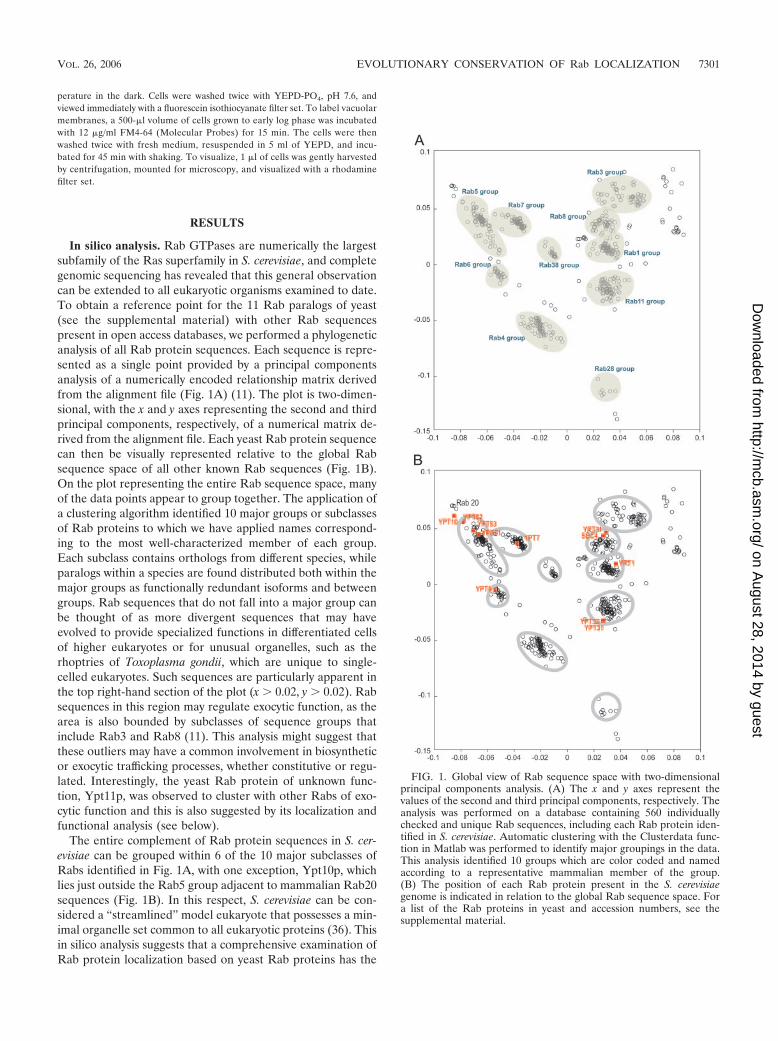
In silico analysis. Rab GTPases are numerically the largestsubfamily of the Ras superfamily in S. cerevisiae, and completegenomic sequencing has revealed that this general observationcan be extended to all eukaryotic organisms examined to date.To obtain a reference point for the 11 Rab paralogs of yeast(see the supplemental material) with other Rab sequencespresent in open access databases, we performed a phylogeneticanalysis of all Rab protein sequences. Each sequence is repre-sented as a single point provided by a principal componentsanalysis of a numerically encoded relationship matrix derivedfrom the alignment file (Fig. 1A) (11). The plot is two-dimen-sional, with the x and y axes representing the second and thirdprincipal components, respectively, of a numerical matrix de-rived from the alignment file. Each yeast Rab protein sequencecan then be visually represented relative to the global Rabsequence space of all other known Rab sequences (Fig. 1B).On the plot representing the entire Rab sequence space, manyof the data points appear to group together. The application ofa clustering algorithm identified 10 major groups or subclassesof Rab proteins to which we have applied names correspond-ing to the most well-characterized member of each group.Each subclass contains orthologs from different species, whileparalogs within a species are found distributed both within themajor groups as functionally redundant isoforms and betweengroups. Rab sequences that do not fall into a major group canbe thought of as more divergent sequences that may haveevolved to provide specialized functions in differentiated cellsof higher eukaryotes or for unusual organelles, such as therhoptries of Toxoplasma gondii, which are unique to single-celled eukaryotes. Such sequences are particularly apparent inthe top right-hand section of the plot (x � 0.02, y � 0.02). Rabsequences in this region may regulate exocytic function, as thearea is also bounded by subclasses of sequence groups thatinclude Rab3 and Rab8 (11). This analysis might suggest thatthese outliers may have a common involvement in biosyntheticor exocytic trafficking processes, whether constitutive or regu-lated. Interestingly, the yeast Rab protein of unknown func-tion, Ypt11p, was observed to cluster with other Rabs of exo-cytic function and this is also suggested by its localization andfunctional analysis (see below).
The entire complement of Rab protein sequences in S. cer-evisiae can be grouped within 6 of the 10 major subclasses ofRabs identified in Fig. 1A, with one exception, Ypt10p, whichlies just outside the Rab5 group adjacent to mammalian Rab20sequences (Fig. 1B). In this respect, S. cerevisiae can be con-sidered a “streamlined” model eukaryote that possesses a min-imal organelle set common to all eukaryotic proteins (36). Thisin silico analysis suggests that a comprehensive examination ofRab protein localization based on yeast Rab proteins has the
FIG. 1. Global view of Rab sequence space with two-dimensionalprincipal components analysis. (A) The x and y axes represent thevalues of the second and third principal components, respectively. Theanalysis was performed on a database containing 560 individuallychecked and unique Rab sequences, including each Rab protein iden-tified in S. cerevisiae. Automatic clustering with the Clusterdata func-tion in Matlab was performed to identify major groupings in the data.This analysis identified 10 groups which are color coded and namedaccording to a representative mammalian member of the group.(B) The position of each Rab protein present in the S. cerevisiaegenome is indicated in relation to the global Rab sequence space. Fora list of the Rab proteins in yeast and accession numbers, see thesupplemental material.
VOL. 26, 2006 EVOLUTIONARY CONSERVATION OF Rab LOCALIZATION 7301
on August 28, 2014 by guest
http://mcb.asm
.org/D
ownloaded from
potential to provide useful insights into the commonalities ofeukaryotic cell biology.
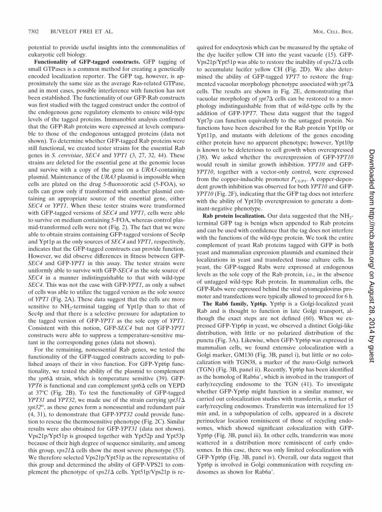
Functionality of GFP-tagged constructs. GFP tagging ofsmall GTPases is a common method for creating a geneticallyencoded localization reporter. The GFP tag, however, is ap-proximately the same size as the average Ras-related GTPase,and in most cases, possible interference with function has notbeen established. The functionality of our GFP-Rab constructswas first studied with the tagged construct under the control ofthe endogenous gene regulatory elements to ensure wild-typelevels of the tagged proteins. Immunoblot analysis confirmedthat the GFP-Rab proteins were expressed at levels compara-ble to those of the endogenous untagged proteins (data notshown). To determine whether GFP-tagged Rab proteins werestill functional, we created tester strains for the essential Rabgenes in S. cerevisiae, SEC4 and YPT1 (3, 27, 32, 44). Thesestrains are deleted for the essential gene at the genomic locusand survive with a copy of the gene on a URA3-containingplasmid. Maintenance of the URA3 plasmid is impossible whencells are plated on the drug 5-fluoroorotic acid (5-FOA), socells can grow only if transformed with another plasmid con-taining an appropriate source of the essential gene, eitherSEC4 or YPT1. When these tester strains were transformedwith GFP-tagged versions of SEC4 and YPT1, cells were ableto survive on medium containing 5-FOA, whereas control plas-mid-transformed cells were not (Fig. 2). The fact that we wereable to obtain strains containing GFP-tagged versions of Sec4pand Ypt1p as the only sources of SEC4 and YPT1, respectively,indicates that the GFP-tagged constructs can provide function.However, we did observe differences in fitness between GFP-SEC4 and GFP-YPT1 in this assay. The tester strains wereuniformly able to survive with GFP-SEC4 as the sole source ofSEC4 in a manner indistinguishable to that with wild-typeSEC4. This was not the case with GFP-YPT1, as only a subsetof cells was able to utilize the tagged version as the sole sourceof YPT1 (Fig. 2A). These data suggest that the cells are moresensitive to NH2-terminal tagging of Ypt1p than to that ofSec4p and that there is a selective pressure for adaptation tothe tagged version of GFP-YPT1 as the sole copy of YPT1.Consistent with this notion, GFP-SEC4 but not GFP-YPT1constructs were able to suppress a temperature-sensitive mu-tant in the corresponding genes (data not shown).
For the remaining, nonessential Rab genes, we tested thefunctionality of the GFP-tagged constructs according to pub-lished assays of their in vivo function. For GFP-Ypt6p func-tionality, we tested the ability of the plasmid to complementthe ypt6� strain, which is temperature sensitive (39). GFP-YPT6 is functional and can complement ypt6� cells on YEPDat 37°C (Fig. 2B). To test the functionality of GFP-taggedYPT31 and YPT32, we made use of the strain carrying ypt31�ypt32ts, as these genes form a nonessential and redundant pair(4, 31), to demonstrate that GFP-YPT32 could provide func-tion to rescue the thermosensitive phenotype (Fig. 2C). Similarresults were also obtained for GFP-YPT31 (data not shown).Vps21p/Ypt51p is grouped together with Ypt52p and Ypt53pbecause of their high degree of sequence similarity, and amongthis group, vps21� cells show the most severe phenotype (53).We therefore selected Vps21p/Ypt51p as the representative ofthis group and determined the ability of GFP-VPS21 to com-plement the phenotype of vps21� cells. Ypt51p/Vps21p is re-
quired for endocytosis which can be measured by the uptake ofthe dye lucifer yellow CH into the yeast vacuole (15). GFP-Vps21p/Ypt51p was able to restore the inability of vps21� cellsto accumulate lucifer yellow CH (Fig. 2D). We also deter-mined the ability of GFP-tagged YPT7 to restore the frag-mented vacuolar morphology phenotype associated with ypt7�cells. The results are shown in Fig. 2E, demonstrating thatvacuolar morphology of ypt7� cells can be restored to a mor-phology indistinguishable from that of wild-type cells by theaddition of GFP-YPT7. These data suggest that the taggedYpt7p can function equivalently to the untagged protein. Nofunctions have been described for the Rab protein Ypt10p orYpt11p, and mutants with deletions of the genes encodingeither protein have no apparent phenotype; however, Ypt10pis known to be deleterious to cell growth when overexpressed(38). We asked whether the overexpression of GFP-YPT10would result in similar growth inhibition. YPT10 and GFP-YPT10, together with a vector-only control, were expressedfrom the copper-inducible promoter PCUP1. A copper-depen-dent growth inhibition was observed for both YPT10 and GFP-YPT10 (Fig. 2F), indicating that the GFP tag does not interferewith the ability of Ypt10p overexpression to generate a dom-inant-negative phenotype.
Rab protein localization. Our data suggested that the NH2-terminal GFP tag is benign when appended to Rab proteinsand can be used with confidence that the tag does not interferewith the functions of the wild-type protein. We took the entirecomplement of yeast Rab proteins tagged with GFP in bothyeast and mammalian expression plasmids and examined theirlocalizations in yeast and transfected tissue culture cells. Inyeast, the GFP-tagged Rabs were expressed at endogenouslevels as the sole copy of the Rab protein, i.e., in the absenceof untagged wild-type Rab protein. In mammalian cells, theGFP-Rabs were expressed behind the viral cytomegalovirus pro-moter and transfections were typically allowed to proceed for 6 h.
The Rab6 family, Ypt6p. Ypt6p is a Golgi-localized yeastRab and is thought to function in late Golgi transport, al-though the exact steps are not defined (60). When we ex-pressed GFP-Ytp6p in yeast, we observed a distinct Golgi-likedistribution, with little or no polarized distribution of thepuncta (Fig. 3A). Likewise, when GFP-Ypt6p was expressed inmammalian cells, we found extensive colocalization with aGolgi marker, GM130 (Fig. 3B, panel i), but little or no colo-calization with TGN38, a marker of the trans-Golgi network(TGN) (Fig. 3B, panel ii). Recently, Ypt6p has been identifiedas the homolog of Rab6a�, which is involved in the transport ofearly/recycling endosome to the TGN (41). To investigatewhether GFP-Ypt6p might function in a similar manner, wecarried out colocalization studies with transferrin, a marker ofearly/recycling endosomes. Transferrin was internalized for 15min and, in a subpopulation of cells, appeared in a discreteperinuclear location reminiscent of those of recycling endo-somes, which showed significant colocalization with GFP-Ypt6p (Fig. 3B, panel iii). In other cells, transferrin was morescattered in a distribution more reminiscent of early endo-somes. In this case, there was only limited colocalization withGFP-Ypt6p (Fig. 3B, panel iv). Overall, our data suggest thatYpt6p is involved in Golgi communication with recycling en-dosomes as shown for Rab6a�.
7302 BUVELOT FREI ET AL. MOL. CELL. BIOL.
on August 28, 2014 by guest
http://mcb.asm
.org/D
ownloaded from
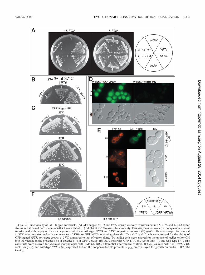
FIG. 2. Functionality of GFP-tagged constructs. (A) GFP-tagged SEC4 and YPT1 constructs were transformed into SEC4� and YPT1� testerstrains and streaked onto medium with (�) or without (�) 5-FOA at 25°C to assess functionality. This assay was performed in comparison to yeasttransformed with empty vector as a negative control and wild-type SEC4 and YPT1 as positive controls. (B) ypt6� cells were assayed for survivalat 37°C when transformed with empty vector-, YPT6-, or GFP-YPT6-containing plasmids. (C) ypt31� ypt31ts cells were assayed for the ability ofGFP-tagged YPT31 to rescue growth at 37°C compared to that of vector alone. (D) vps21� cells were assayed for the uptake of lucifer yellow CHinto the vacuole in the presence (�) or absence (�) of GFP-Vps21p. (E) ypt7� cells with GFP-YPT7 (i), vector only (ii), and wild-type YPT7 (iii)constructs were assayed for vacuolar morphologies with FM4-64. DIC, differential interference contrast. (F) ypt10� cells with GFP-YPT10 (i),vector only (ii), and wild-type YPT10 (iii) expressed behind the copper-inducible promoter PCUP1 were assayed for growth on media � 0.7 mMCuSO4.
VOL. 26, 2006 EVOLUTIONARY CONSERVATION OF Rab LOCALIZATION 7303
on August 28, 2014 by guest
http://mcb.asm
.org/D
ownloaded from
The Rab5 family, Vps21/Ypt51p, Ypt52p, and Ypt52p.Ypt51p (Vps21p) is the yeast counterpart of mammalian Rab5both in localization and in function (28, 53, 54). Ypt51p isgrouped together with Ypt52p and Ypt53p because of a highdegree of sequence similarity. All three of these Ypt proteinsappear to function in endocytosis in yeast cells (53); however,
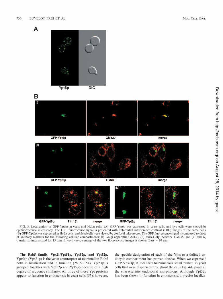
the specific designation of each of the Ypts to a defined en-docytic compartment has proven elusive. When we expressedGFP-Vps21p, it localized to numerous small puncta in yeastcells that were dispersed throughout the cell (Fig. 4A, panel i),the characteristic endosomal morphology. Although Ypt52phas been shown to function in endocytosis, a precise localiza-
FIG. 3. Localization of GFP-Ypt6p in yeast and HeLa cells. (A) GFP-Ypt6p was expressed in yeast cells, and live cells were viewed byepifluorescence microscopy. The GFP fluorescence signal is presented with differential interference contrast (DIC) images of the same cells.(B) GFP-Ypt6p was expressed in HeLa cells, and fixed cells were viewed by confocal microscopy. The GFP fluorescence signal is compared to thoseof antibody markers for the following cellular compartments: (i) Golgi apparatus GM130, (ii) trans-Golgi network TGN38, and (iii and iv)transferrin internalized for 15 min. In each case, a merge of the two fluorescence images is shown. Bars 10 �m.
7304 BUVELOT FREI ET AL. MOL. CELL. BIOL.
on August 28, 2014 by guest
http://mcb.asm
.org/D
ownloaded from
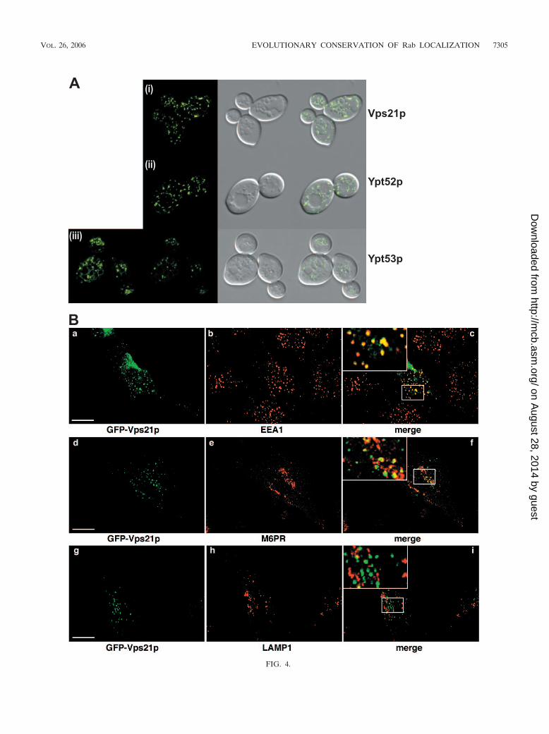
FIG. 4.
VOL. 26, 2006 EVOLUTIONARY CONSERVATION OF Rab LOCALIZATION 7305
on August 28, 2014 by guest
http://mcb.asm
.org/D
ownloaded from
7306 BUVELOT FREI ET AL. MOL. CELL. BIOL.
on August 28, 2014 by guest
http://mcb.asm
.org/D
ownloaded from
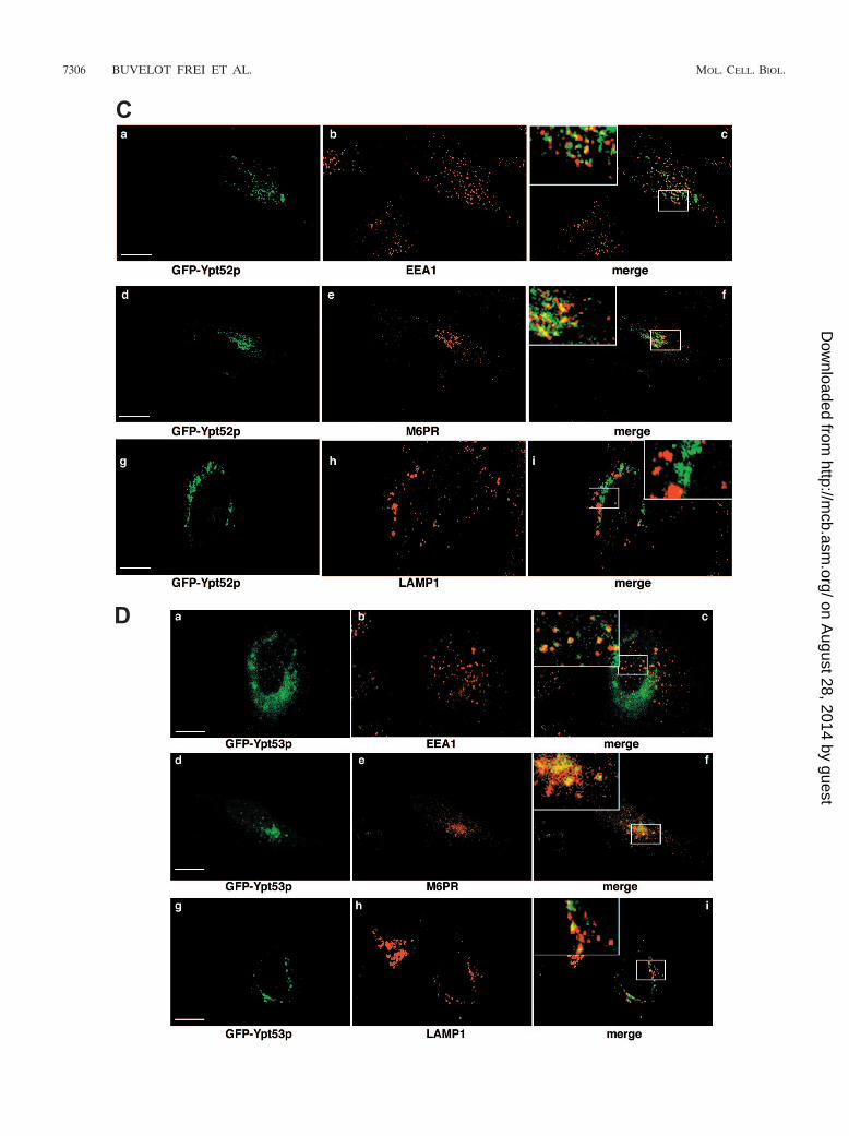
tion within the endocytic pathway has not been determined.We expressed GFP-Ypt52p in yeast cells and observed a char-acteristic pattern that consisted of puncta often larger than theGFP-Vps21p puncta that surrounded (but was excluded from)the vacuolar membrane as well as a few more scattered dots(Fig. 4A, panel ii). This distribution suggested an endosomallocalization that was more advanced in the pathway (i.e., closerto the vacuole rather than the departure point of the plasmamembrane) than that observed for Vps21p. The expression ofGFP-Ypt53p in yeast cells showed a distinct (but relativelyweak) localization to the vacuolar-limiting membrane, in ad-dition to a small number of bright puncta (Fig. 4A, panel iii),many of which were clustered around the vacuole.
In mammalian cells, GFP-Vps21p showed extensive colocal-ization with EEA1, a marker of early endosomes, as well assome colocalization with M6PR, a marker of endosomes thatare in communication with the TGN (typically, but not exclu-sively, late endosomes) (20). GFP-Vps21p showed no signifi-cant colocalization with LAMP1, which is found in late endo-somes and lysosomes (Fig. 4B). Based on these findings, wesuggest that GFP-tagged Vps21p acts, like Rab5, in the for-mation of early endosomes. GFP-Ypt52p showed a pattern oflocalization similar to that of Vps21p in mammalian cells;however, it tended to colocalize more extensively with M6PRthan with EEA1, again suggesting a post-Vps21p function (Fig.4C). As with Vps21p, we saw no colocalization with LAMP1-positive vesicles. GFP-Ypt53p in animal cells showed a diffuseperinuclear distribution, which showed extensive colocalizationwith M6PR, combined with weaker and more-scattered dotsthat colocalized with EEA1 (Fig. 4D). GFP-Ypt53 also showeda distinct but limited degree of overlap with LAMP1. Overall,our results with Ypt53p suggest that it acts in the late endo-some and is the third acting Rab in the functional Vps21p-Ypt52p-Ypt53p chain of endocytosis.
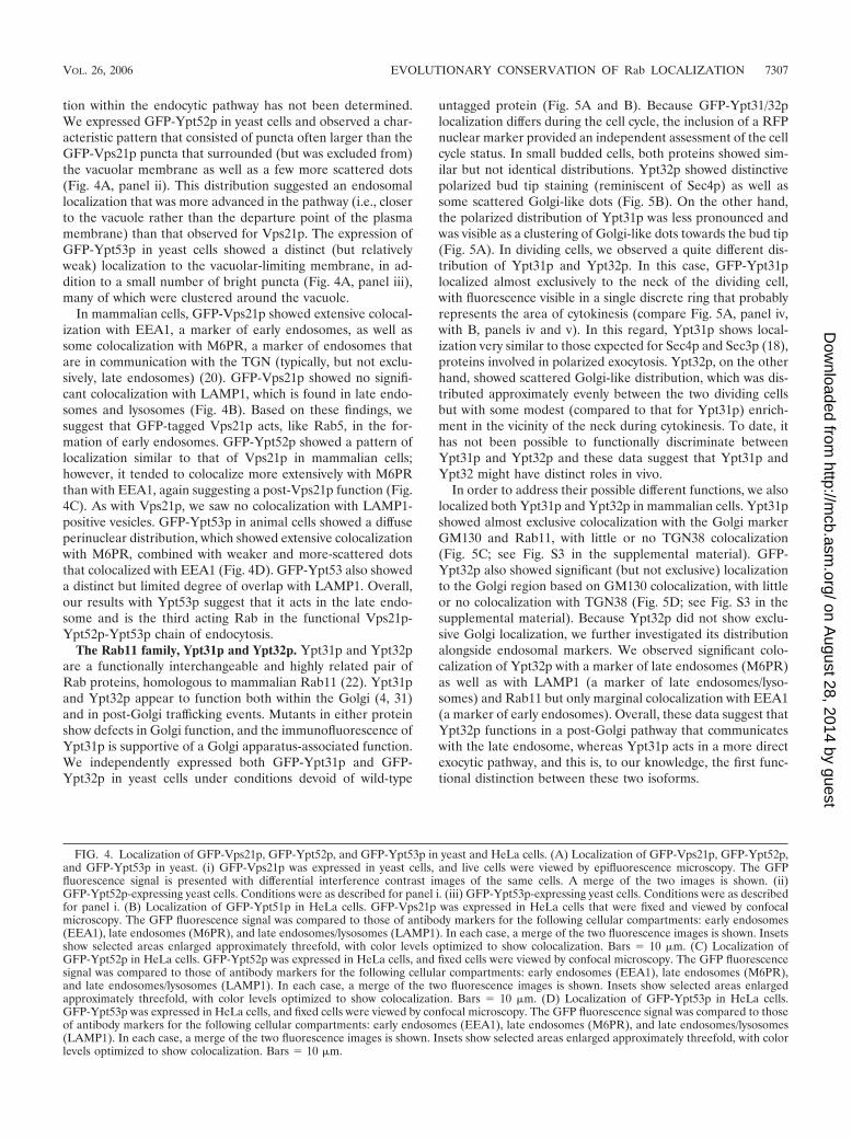
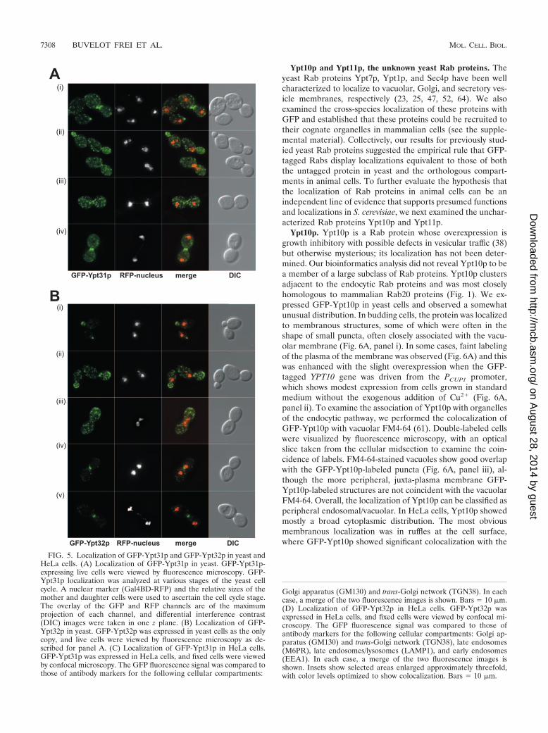
The Rab11 family, Ypt31p and Ypt32p. Ypt31p and Ypt32pare a functionally interchangeable and highly related pair ofRab proteins, homologous to mammalian Rab11 (22). Ypt31pand Ypt32p appear to function both within the Golgi (4, 31)and in post-Golgi trafficking events. Mutants in either proteinshow defects in Golgi function, and the immunofluorescence ofYpt31p is supportive of a Golgi apparatus-associated function.We independently expressed both GFP-Ypt31p and GFP-Ypt32p in yeast cells under conditions devoid of wild-type
untagged protein (Fig. 5A and B). Because GFP-Ypt31/32plocalization differs during the cell cycle, the inclusion of a RFPnuclear marker provided an independent assessment of the cellcycle status. In small budded cells, both proteins showed sim-ilar but not identical distributions. Ypt32p showed distinctivepolarized bud tip staining (reminiscent of Sec4p) as well assome scattered Golgi-like dots (Fig. 5B). On the other hand,the polarized distribution of Ypt31p was less pronounced andwas visible as a clustering of Golgi-like dots towards the bud tip(Fig. 5A). In dividing cells, we observed a quite different dis-tribution of Ypt31p and Ypt32p. In this case, GFP-Ypt31plocalized almost exclusively to the neck of the dividing cell,with fluorescence visible in a single discrete ring that probablyrepresents the area of cytokinesis (compare Fig. 5A, panel iv,with B, panels iv and v). In this regard, Ypt31p shows local-ization very similar to those expected for Sec4p and Sec3p (18),proteins involved in polarized exocytosis. Ypt32p, on the otherhand, showed scattered Golgi-like distribution, which was dis-tributed approximately evenly between the two dividing cellsbut with some modest (compared to that for Ypt31p) enrich-ment in the vicinity of the neck during cytokinesis. To date, ithas not been possible to functionally discriminate betweenYpt31p and Ypt32p and these data suggest that Ypt31p andYpt32 might have distinct roles in vivo.
In order to address their possible different functions, we alsolocalized both Ypt31p and Ypt32p in mammalian cells. Ypt31pshowed almost exclusive colocalization with the Golgi markerGM130 and Rab11, with little or no TGN38 colocalization(Fig. 5C; see Fig. S3 in the supplemental material). GFP-Ypt32p also showed significant (but not exclusive) localizationto the Golgi region based on GM130 colocalization, with littleor no colocalization with TGN38 (Fig. 5D; see Fig. S3 in thesupplemental material). Because Ypt32p did not show exclu-sive Golgi localization, we further investigated its distributionalongside endosomal markers. We observed significant colo-calization of Ypt32p with a marker of late endosomes (M6PR)as well as with LAMP1 (a marker of late endosomes/lyso-somes) and Rab11 but only marginal colocalization with EEA1(a marker of early endosomes). Overall, these data suggest thatYpt32p functions in a post-Golgi pathway that communicateswith the late endosome, whereas Ypt31p acts in a more directexocytic pathway, and this is, to our knowledge, the first func-tional distinction between these two isoforms.
FIG. 4. Localization of GFP-Vps21p, GFP-Ypt52p, and GFP-Ypt53p in yeast and HeLa cells. (A) Localization of GFP-Vps21p, GFP-Ypt52p,and GFP-Ypt53p in yeast. (i) GFP-Vps21p was expressed in yeast cells, and live cells were viewed by epifluorescence microscopy. The GFPfluorescence signal is presented with differential interference contrast images of the same cells. A merge of the two images is shown. (ii)GFP-Ypt52p-expressing yeast cells. Conditions were as described for panel i. (iii) GFP-Ypt53p-expressing yeast cells. Conditions were as describedfor panel i. (B) Localization of GFP-Ypt51p in HeLa cells. GFP-Vps21p was expressed in HeLa cells that were fixed and viewed by confocalmicroscopy. The GFP fluorescence signal was compared to those of antibody markers for the following cellular compartments: early endosomes(EEA1), late endosomes (M6PR), and late endosomes/lysosomes (LAMP1). In each case, a merge of the two fluorescence images is shown. Insetsshow selected areas enlarged approximately threefold, with color levels optimized to show colocalization. Bars 10 �m. (C) Localization ofGFP-Ypt52p in HeLa cells. GFP-Ypt52p was expressed in HeLa cells, and fixed cells were viewed by confocal microscopy. The GFP fluorescencesignal was compared to those of antibody markers for the following cellular compartments: early endosomes (EEA1), late endosomes (M6PR),and late endosomes/lysosomes (LAMP1). In each case, a merge of the two fluorescence images is shown. Insets show selected areas enlargedapproximately threefold, with color levels optimized to show colocalization. Bars 10 �m. (D) Localization of GFP-Ypt53p in HeLa cells.GFP-Ypt53p was expressed in HeLa cells, and fixed cells were viewed by confocal microscopy. The GFP fluorescence signal was compared to thoseof antibody markers for the following cellular compartments: early endosomes (EEA1), late endosomes (M6PR), and late endosomes/lysosomes(LAMP1). In each case, a merge of the two fluorescence images is shown. Insets show selected areas enlarged approximately threefold, with colorlevels optimized to show colocalization. Bars 10 �m.
VOL. 26, 2006 EVOLUTIONARY CONSERVATION OF Rab LOCALIZATION 7307
on August 28, 2014 by guest
http://mcb.asm
.org/D
ownloaded from
Ypt10p and Ypt11p, the unknown yeast Rab proteins. Theyeast Rab proteins Ypt7p, Ypt1p, and Sec4p have been wellcharacterized to localize to vacuolar, Golgi, and secretory ves-icle membranes, respectively (23, 25, 47, 52, 64). We alsoexamined the cross-species localization of these proteins withGFP and established that these proteins could be recruited totheir cognate organelles in mammalian cells (see the supple-mental material). Collectively, our results for previously stud-ied yeast Rab proteins suggested the empirical rule that GFP-tagged Rabs display localizations equivalent to those of boththe untagged protein in yeast and the orthologous compart-ments in animal cells. To further evaluate the hypothesis thatthe localization of Rab proteins in animal cells can be anindependent line of evidence that supports presumed functionsand localizations in S. cerevisiae, we next examined the unchar-acterized Rab proteins Ypt10p and Ypt11p.
Ypt10p. Ypt10p is a Rab protein whose overexpression isgrowth inhibitory with possible defects in vesicular traffic (38)but otherwise mysterious; its localization has not been deter-mined. Our bioinformatics analysis did not reveal Ypt10p to bea member of a large subclass of Rab proteins. Ypt10p clustersadjacent to the endocytic Rab proteins and was most closelyhomologous to mammalian Rab20 proteins (Fig. 1). We ex-pressed GFP-Ypt10p in yeast cells and observed a somewhatunusual distribution. In budding cells, the protein was localizedto membranous structures, some of which were often in theshape of small puncta, often closely associated with the vacu-olar membrane (Fig. 6A, panel i). In some cases, faint labelingof the plasma of the membrane was observed (Fig. 6A) and thiswas enhanced with the slight overexpression when the GFP-tagged YPT10 gene was driven from the PCUP1 promoter,which shows modest expression from cells grown in standardmedium without the exogenous addition of Cu2� (Fig. 6A,panel ii). To examine the association of Ypt10p with organellesof the endocytic pathway, we performed the colocalization ofGFP-Ypt10p with vacuolar FM4-64 (61). Double-labeled cellswere visualized by fluorescence microscopy, with an opticalslice taken from the cellular midsection to examine the coin-cidence of labels. FM4-64-stained vacuoles show good overlapwith the GFP-Ypt10p-labeled puncta (Fig. 6A, panel iii), al-though the more peripheral, juxta-plasma membrane GFP-Ypt10p-labeled structures are not coincident with the vacuolarFM4-64. Overall, the localization of Ypt10p can be classified asperipheral endosomal/vacuolar. In HeLa cells, Ypt10p showedmostly a broad cytoplasmic distribution. The most obviousmembranous localization was in ruffles at the cell surface,where GFP-Ypt10p showed significant colocalization with the
FIG. 5. Localization of GFP-Ypt31p and GFP-Ypt32p in yeast andHeLa cells. (A) Localization of GFP-Ypt31p in yeast. GFP-Ypt31p-expressing live cells were viewed by fluorescence microscopy. GFP-Ypt31p localization was analyzed at various stages of the yeast cellcycle. A nuclear marker (Gal4BD-RFP) and the relative sizes of themother and daughter cells were used to ascertain the cell cycle stage.The overlay of the GFP and RFP channels are of the maximumprojection of each channel, and differential interference contrast(DIC) images were taken in one z plane. (B) Localization of GFP-Ypt32p in yeast. GFP-Ypt32p was expressed in yeast cells as the onlycopy, and live cells were viewed by fluorescence microscopy as de-scribed for panel A. (C) Localization of GFP-Ypt31p in HeLa cells.GFP-Ypt31p was expressed in HeLa cells, and fixed cells were viewedby confocal microscopy. The GFP fluorescence signal was compared tothose of antibody markers for the following cellular compartments:
Golgi apparatus (GM130) and trans-Golgi network (TGN38). In eachcase, a merge of the two fluorescence images is shown. Bars 10 �m.(D) Localization of GFP-Ypt32p in HeLa cells. GFP-Ypt32p wasexpressed in HeLa cells, and fixed cells were viewed by confocal mi-croscopy. The GFP fluorescence signal was compared to those ofantibody markers for the following cellular compartments: Golgi ap-paratus (GM130) and trans-Golgi network (TGN38), late endosomes(M6PR), late endosomes/lysosomes (LAMP1), and early endosomes(EEA1). In each case, a merge of the two fluorescence images isshown. Insets show selected areas enlarged approximately threefold,with color levels optimized to show colocalization. Bars 10 �m.
7308 BUVELOT FREI ET AL. MOL. CELL. BIOL.
on August 28, 2014 by guest
http://mcb.asm
.org/D
ownloaded from
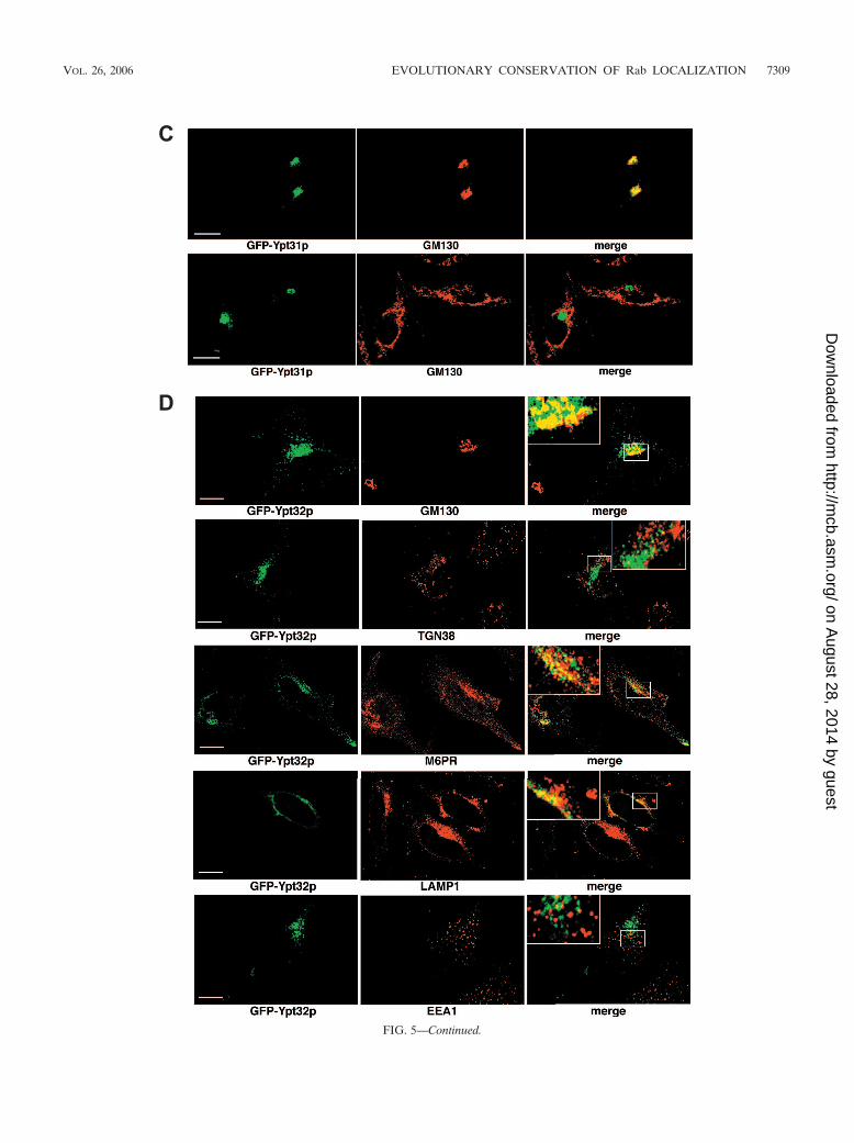
FIG. 5—Continued.
VOL. 26, 2006 EVOLUTIONARY CONSERVATION OF Rab LOCALIZATION 7309
on August 28, 2014 by guest
http://mcb.asm
.org/D
ownloaded from
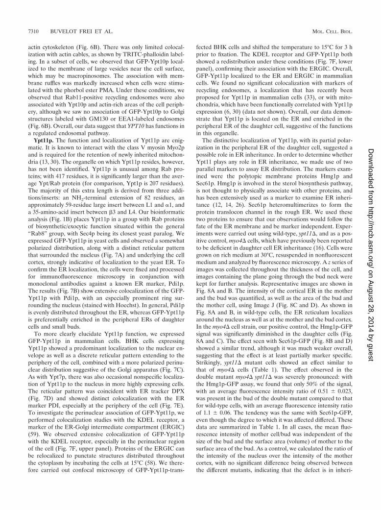
actin cytoskeleton (Fig. 6B). There was only limited colocal-ization with actin cables, as shown by TRITC-phalloidin label-ing. In a subset of cells, we observed that GFP-Ypt10p local-ized to the membrane of large vesicles near the cell surface,which may be macropinosomes. The association with mem-brane ruffles was markedly increased when cells were stimu-lated with the phorbol ester PMA. Under these conditions, weobserved that Rab11-positive recycling endosomes were alsoassociated with Ypt10p and actin-rich areas of the cell periph-ery, although we saw no association of GFP-Ypt10p to Golgistructures labeled with GM130 or EEA1-labeled endosomes(Fig. 6B). Overall, our data suggest that YPT10 has functions ina regulated endosomal pathway.
Ypt11p. The function and localization of Ypt11p are enig-matic. It is known to interact with the class V myosin Myo2pand is required for the retention of newly inherited mitochon-dria (13, 30). The organelle on which Ypt11p resides, however,has not been identified. Ypt11p is unusual among Rab pro-teins; with 417 residues, it is significantly larger than the aver-age Ypt/Rab protein (for comparison, Ypt1p is 207 residues).The majority of this extra length is derived from three addi-tions/inserts: an NH2-terminal extension of 82 residues, anapproximately 59-residue large insert between L1 and 1, anda 35-amino-acid insert between �3 and L4. Our bioinformaticanalysis (Fig. 1B) places Ypt11p in a group with Rab proteinsof biosynthetic/exocytic function situated within the general“Rab8” group, with Sec4p being its closest yeast paralog. Weexpressed GFP-Ypt11p in yeast cells and observed a somewhatpolarized distribution, along with a distinct reticular patternthat surrounded the nucleus (Fig. 7A) and underlying the cellcortex, strongly indicative of localization to the yeast ER. Toconfirm the ER localization, the cells were fixed and processedfor immunofluorescence microscopy in conjunction withmonoclonal antibodies against a known ER marker, Pdi1p.The results (Fig. 7B) show extensive colocalization of the GFP-Ypt11p with Pdi1p, with an especially prominent ring sur-rounding the nucleus (stained with Hoechst). In general, Pdi1pis evenly distributed throughout the ER, whereas GFP-Ypt11pis preferentially enriched in the peripheral ERs of daughtercells and small buds.
To more clearly elucidate Ypt11p function, we expressedGFP-Ypt11p in mammalian cells. BHK cells expressingYpt11p showed a predominant localization to the nuclear en-velope as well as a discrete reticular pattern extending to theperiphery of the cell, combined with a more polarized perinu-clear distribution suggestive of the Golgi apparatus (Fig. 7C).As with Ypt7p, there was also occasional nonspecific localiza-tion of Ypt11p to the nucleus in more highly expressing cells.The reticular pattern was coincident with ER tracker DPX(Fig. 7D) and showed distinct colocalization with the ERmarker PDI, especially at the periphery of the cell (Fig. 7E).To investigate the perinuclear association of GFP-Ypt11p, weperformed colocalization studies with the KDEL receptor, amarker of the ER-Golgi intermediate compartment (ERGIC)(59). We observed extensive colocalization of GFP-Ypt11pwith the KDEL receptor, especially in the perinuclear regionof the cell (Fig. 7F, upper panel). Proteins of the ERGIC canbe relocalized to punctate structures distributed throughoutthe cytoplasm by incubating the cells at 15°C (58). We there-fore carried out confocal microscopy of GFP-Ypt11p-trans-
fected BHK cells and shifted the temperature to 15°C for 3 hprior to fixation. The KDEL receptor and GFP-Ypt11p bothshowed a redistribution under these conditions (Fig. 7F, lowerpanel), confirming their association with the ERGIC. Overall,GFP-Ypt11p localized to the ER and ERGIC in mammaliancells. We found no significant colocalization with markers ofrecycling endosomes, a localization that has recently beenproposed for Ypt11p in mammalian cells (33), or with mito-chondria, which have been functionally correlated with Ypt11pexpression (6, 30) (data not shown). Overall, our data demon-strate that Ypt11p is located on the ER and enriched in theperipheral ER of the daughter cell, suggestive of the functionsin this organelle.
The distinctive localization of Ypt11p, with its partial polar-ization in the peripheral ER of the daughter cell, suggested apossible role in ER inheritance. In order to determine whetherYpt11 plays any role in ER inheritance, we made use of twoparallel markers to assay ER distribution. The markers exam-ined were the polytopic membrane proteins Hmg1p andSec61p. Hmg1p is involved in the sterol biosynthesis pathway,is not thought to physically associate with other proteins, andhas been extensively used as a marker to examine ER inheri-tance (12, 14, 26). Sec61p heteromultimerizes to form theprotein translocon channel in the rough ER. We used thesetwo proteins to ensure that our observations would follow thefate of the ER membrane and be marker independent. Exper-iments were carried out using wild-type, ypt11�, and as a pos-itive control, myo4� cells, which have previously been reportedto be deficient in daughter cell ER inheritance (16). Cells weregrown on rich medium at 30°C, resuspended in nonfluorescentmedium and analyzed by fluorescence microscopy. A z series ofimages was collected throughout the thickness of the cell, andimages containing the plane going through the bud neck werekept for further analysis. Representative images are shown inFig. 8A and B. The intensity of the cortical ER in the motherand the bud was quantified, as well as the area of the bud andthe mother cell, using Image J (Fig. 8C and D). As shown inFig. 8A and B, in wild-type cells, the ER reticulum localizesaround the nucleus as well as at the mother and the bud cortex.In the myo4� cell strain, our positive control, the Hmg1p-GFPsignal was significantly diminished in the daughter cells (Fig.8A and C). The effect seen with Sec61p-GFP (Fig. 8B and D)showed a similar trend, although it was much weaker overall,suggesting that the effect is at least partially marker specific.Strikingly, ypt11� mutant cells showed an effect similar tothat of myo4� cells (Table 1). The effect observed in thedouble mutant myo4� ypt11� was severely pronounced: withthe Hmg1p-GFP assay, we found that only 50% of the signal,with an average fluorescence intensity ratio of 0.51 � 0.023,was present in the bud of the double mutant compared to thatfor wild-type cells, with an average fluorescence intensity ratioof 1.1 � 0.06. The tendency was the same with Sec61p-GFP,even though the degree to which it was affected differed. Thesedata are summarized in Table 1. In all cases, the mean fluo-rescence intensity of mother cell/bud was independent of thesize of the bud and the surface area (volume) of mother to thesurface area of the bud. As a control, we calculated the ratio ofthe intensity of the nucleus over the intensity of the mothercortex, with no significant difference being observed betweenthe different mutants, indicating that the defect is in inheri-
7310 BUVELOT FREI ET AL. MOL. CELL. BIOL.
on August 28, 2014 by guest
http://mcb.asm
.org/D
ownloaded from
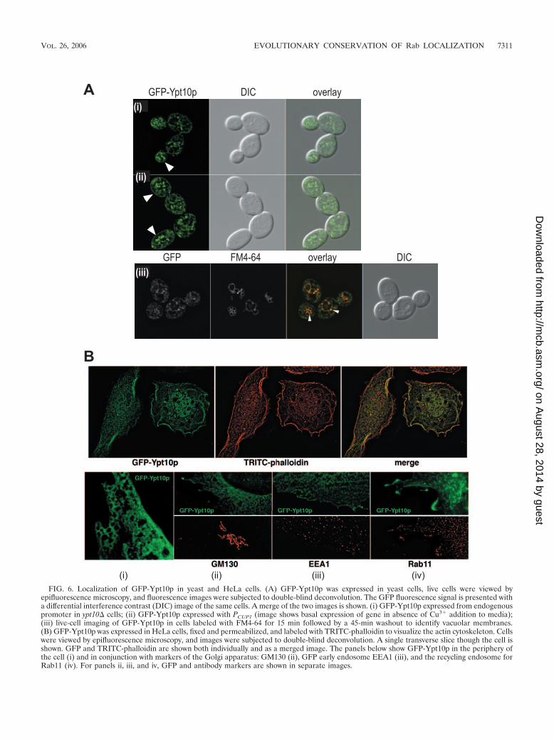
FIG. 6. Localization of GFP-Ypt10p in yeast and HeLa cells. (A) GFP-Ypt10p was expressed in yeast cells, live cells were viewed byepifluorescence microscopy, and fluorescence images were subjected to double-blind deconvolution. The GFP fluorescence signal is presented witha differential interference contrast (DIC) image of the same cells. A merge of the two images is shown. (i) GFP-Ypt10p expressed from endogenouspromoter in ypt10� cells; (ii) GFP-Ypt10p expressed with PCUP1 (image shows basal expression of gene in absence of Cu3� addition to media);(iii) live-cell imaging of GFP-Ypt10p in cells labeled with FM4-64 for 15 min followed by a 45-min washout to identify vacuolar membranes.(B) GFP-Ypt10p was expressed in HeLa cells, fixed and permeabilized, and labeled with TRITC-phalloidin to visualize the actin cytoskeleton. Cellswere viewed by epifluorescence microscopy, and images were subjected to double-blind deconvolution. A single transverse slice though the cell isshown. GFP and TRITC-phalloidin are shown both individually and as a merged image. The panels below show GFP-Ypt10p in the periphery ofthe cell (i) and in conjunction with markers of the Golgi apparatus: GM130 (ii), GFP early endosome EEA1 (iii), and the recycling endosome forRab11 (iv). For panels ii, iii, and iv, GFP and antibody markers are shown in separate images.
VOL. 26, 2006 EVOLUTIONARY CONSERVATION OF Rab LOCALIZATION 7311
on August 28, 2014 by guest
http://mcb.asm
.org/D
ownloaded from
FIG. 7. Localization of GFP-Ypt11p in yeast and HeLa cells. (A) GFP-Ypt11p was expressed in yeast cells, and live cells were stained withHoechst 33258. Cells were viewed by epifluorescence microscopy, and fluorescence images were subjected to double-blind deconvolution. The GFPfluorescence signal is presented with a differential interference contrast (DIC) image of the same cells, and a merge of the three images is shown.(B) GFP-Ypt11p was expressed in yeast cells, fixed, and stained with Hoechst 33258. Cells were analyzed by immunofluorescence microscopy usinganti-Pdi1p antibodies. The GFP and immunofluorescence signal is presented with a differential interference contrast (DIC) image of the same cells.(C) GFP-Ypt11p was expressed in BHK cells and viewed by epifluorescence microscopy, and fluorescence images were subjected to double-blinddeconvolution. Both a maximum projection and a single slice through the cell are shown. (D) GFP-Ypt11p was expressed in BHK cells and live
7312 BUVELOT FREI ET AL. MOL. CELL. BIOL.
on August 28, 2014 by guest
http://mcb.asm
.org/D
ownloaded from
tance of peripheral, cortical ER and not a result of generalcortical organization defects (data not shown). We also exam-ined ER morphology since it has been previously establishedthat some mutations that affect ER morphology at the cortexcan give rise to an ER inheritance phenotype (17). ER mor-phology was checked with a close examination of the networkmorphology at the cell cortex. Neither mutant nor double-mutant cells showed any impairment of the morphologicalstructure of the ER network at the surface (data not shown).Thus, ypt11� affect ER inheritance in a manner similar to thatof myo4�, which is independent of ER morphology.
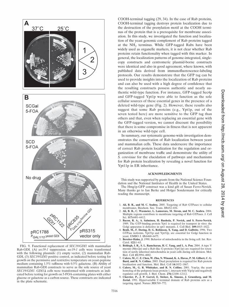
Functional conservation of Rab-GDI. Our experiments dem-onstrate a striking cross-species conservation of Rab proteinlocalization, showing that Rab proteins can be recruited tocognate organelles over large evolutionary distances. In turn,this implies that the machinery responsible for recruiting Rabproteins onto membranes is probably functionally conserved.Very little is known about the molecular nature of the machin-ery that is responsible for the specific subcellular localizationof Rab proteins. Rab protein prenyl moiety is required but notsufficient (7, 21). Rab proteins exist in two pools, a cytoplasmicpool and a membrane-associated pool, with the cytoplasmicpool serving as the reservoir from which organelles recruittheir specific Rabs. The cytoplasmic pool of Rab proteins existsin a complex with Rab-GDI; there is currently no other proteinknown to associate with Rab proteins in the cytoplasm. Rab-GDI therefore plays a critical role in Rab protein localizationbecause the exclusive substrate for membrane recruitment ofRab proteins is a cytosolic heterodimer of a Rab protein withRab-GDI. Moreover, Rab-GDI is a global regulator for Rabproteins, as yeasts contain a single Rab-GDI gene, SEC19,which acts on all Rab proteins. This appears to be a generalrule with between one and three Rab-GDI-encoding genesthat act on multiple Rab proteins found in the genomes ofseveral eukaryotes. To test the functional conservation of Rab-GDI, we expressed the mammalian version of Rab-GDI inyeast behind a regulatable promoter, the galactose promoterwhich expresses at high levels in cells grown in medium withgalactose as a carbon source and is highly repressed in thepresence of glucose. We tested the ability of this construct tosuppress a conditional lethal allele of Rab-GDI, sec19-1, andalso determined whether cells could survive on galactose withmammalian Rab-GDI in place of the SEC19 gene. The resultsof these experiments are shown in Fig. 9; the mammalianRab-GDI construct can suppress the temperature sensitivity ofsec19-1 (Fig. 9A) and also serve as the sole source of Rab-GDI,an essential gene function in S. cerevisiae (Fig. 9B). This effectwas apparent only upon the overexpression of the mammalianRab-GDI construct; the expression level when the mammalianRab-GDI ORF was placed under the control of the endoge-
nous SEC19 5� and 3� regions was not sufficient to provideRab-GDI function (data not shown). These data demonstratethat the essential function of Rab-GDI is conserved from yeastto humans; underlying the functional conservation of Rab pro-tein localization is a conservation of Rab-GDI function.
DISCUSSIONComparative genomic studies in cell biology. In this study,
we have identified 10 major clusters or groups of Rab se-quences that we have termed the Rab5, Rab7, Rab6, Rab4,Rab28, Rab38, Rab11, Rab1, Rab8, and Rab3 groups. Thisbioinformatic analysis reveals that yeast Rab proteins, with oneexception, can be located in global Rab sequence space to-gether with a cluster of homologs from other diverse eu-karyotes. The 11 yeast Rab sequences are not distributedamong major Rab sequence clusters or subclasses; rather,these sequences are derived from six of the major Rab sub-classes identified by our bioinformatic analysis. This may notbe surprising as S. cerevisiae seems to lack many of the or-ganelles possessed by other single-celled eukaryotes (e.g., therhoptries of Toxoplasma) and the specialized organelles lo-cated in the differentiated cells of metazoa. Although it is toosimplistic to assume that the number of Rab proteins corre-lates with the number and complexity of organelles, it is clearthat Rab proteins can be multifunctional so it is possible thatthe Rab proteins of S. cerevisiae cover necessary functions thatare provided by different Rab subclasses in other species. Cer-tainly our localization data support the notion that Rab pro-teins of the same subclass may have both overlapping anddistinct functions.
Our results show that the GFP-tagged Rab proteins main-tain localization between single-celled eukaryotes and mam-malian tissue culture cells. This in turn implies that the local-ization of a protein in animal cells can both confirm organelleresidency and give us more information about the identity ofthe organelle and the other organelles it directly communicateswith via membrane traffic. For Rab proteins of known func-tions, we were able to demonstrate that the yeast Rab proteinlocalizes to an organelle in higher eukaryotes that is the cog-nate of the organelle on which the Rab protein resides in yeast.The advantages of this approach are severalfold. First, it pro-vides an independent cross-check of localization assignment,which is often a technical issue in yeast with its small size andlimited spatial resolution. Second, this analysis demonstratesthat cross-species experiments can be very valuable in ascer-taining the correct locations of novel proteins and it will bevaluable to extend these studies to multicellular model organ-isms. In addition, these studies have practical implications ofincreasing the availability of well-characterized markers in themembrane-trafficking pathway.
cells incubated with ER-Tracker Blue-White DPX. Cells were fixed and viewed by epifluorescence microscopy. The GFP and ER-Tracker DPXsignals are shown individually, and in a merged image with ER-Tracker DPX false-colored red to show colocalization with GFP. (E) GFP-Ypt11pwas expressed in HeLa cells, and fixed cells were viewed by confocal microscopy. The GFP fluorescence signal was compared to that of an antibodymarker for the endoplasmic reticulum, PDI. Images show the periphery of the cell both individually and as a merge of the two fluorescence images.(F) GFP-Ypt11p was expressed in BHK cells, and fixed cells were viewed by confocal microscopy. The GFP fluorescence signal was compared tothat of an antibody marker for the ERGIC, KDEL receptor (KDEL-R). GFP and the KDEL-R are shown both individually and as a merged image.In the lower panels, the cells were incubated for 3 h at 15°C before fixation.
VOL. 26, 2006 EVOLUTIONARY CONSERVATION OF Rab LOCALIZATION 7313
on August 28, 2014 by guest
http://mcb.asm
.org/D
ownloaded from
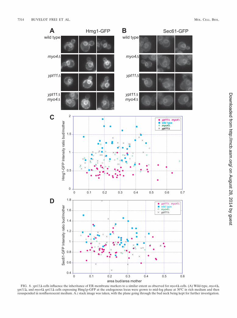
FIG. 8. ypt11� cells influence the inheritance of ER membrane markers to a similar extent as observed for myo4� cells. (A) Wild-type, myo4�,ypt11�, and myo4� ypt11� cells expressing Hmg1p-GFP at the endogenous locus were grown to mid-log phase at 30°C in rich medium and thenresuspended in nonfluorescent medium. A z stack image was taken, with the plane going through the bud neck being kept for further investigation.
7314 BUVELOT FREI ET AL. MOL. CELL. BIOL.
on August 28, 2014 by guest
http://mcb.asm
.org/D
ownloaded from

Role of Ypt10p and Ypt11p. The existence of the two yeastRabs Ypt10p and Ypt11p was revealed only upon the sequenc-ing of the yeast genome, and these ORFs remain functionallyuncharacterized. Little is known about Ypt10p other than theobservation of possible defects in vesicular traffic upon theoverexpression of Ypt10p (38). To date, no function or local-ization has been assigned to Ypt11p. The results of an analysisof the relative localization patterns in yeast and mammaliancells with Rab proteins of known functions suggested that wecould extrapolate from these experiments to help understandthe roles of Ypt10p and Ypt11p.
A sequence analysis of Ypt10p suggested an endocyticfunction for Ypt10p since it clusters with Rab protein se-quences of known endocytic function in global Rab se-quence space. The pattern of Ypt10p localization in HeLacells was very similar to that reported for Rab34 in mouse10T1/2 fibroblasts (56); however, we saw no significant lo-calization to the Golgi apparatus, another reported local-ization of Rab34 (62), or with EEA1. As with Rab34 (56),the association with membrane ruffles was markedly in-creased when cells were stimulated with the phorbol esterPMA. Our phylogenetic analysis shows that Ypt10p is clos-est to the Rab20 sequences from vertebrates. Rab20 hasbeen reported to be localized to apical-dense tubules, en-docytic structures underlying the apical surfaces of polar-ized epithelial cells (40). We propose that the major local-ization of Ypt10p and its orthologs, such as Rab20, is inendocytic structures, where it functions in plasma membrane re-modeling.
Together, the bioinformatics and localization of, andfunctional observations of the effect of, ypt11� suggest a rolefor Ypt11p in the biosynthetic secretory pathway in thecontrol of ER inheritance. Myo4p and Ypt11p are likely towork in parallel pathways to act synergistically in the trans-port of the ER membrane to the bud cortex, although otherexplanations are possible. Interestingly, even in cells withthe most-severe defects, some ER membrane was alwaysseen at the bud cortex, suggesting that ypt11� and myo4�cells are not totally deficient in ER inheritance and that athird pathway exists, perhaps under the control of Sec8p(48). Ypt11p has been reported to cause defects in mito-chondrial inheritance (6, 30); our work demonstrates a lo-calization of Ypt11p on the ER and a role for Ypt11p in ERinheritance. Many cell types show a close apposition of theER with mitochondria (5, 19, 42, 43); one explanation toreconcile these two observations would be that the mito-chondrial inheritance defect of ypt11� cells is a consequenceof their failure to inherit ER.
Mechanism of Rab protein localization. A characteristic fea-ture of a Rab protein is its steady-state localization to the
cytosolic surface of a particular subcellular membrane. Ourresults reveal fundamental similarities between divergent spe-cies, underlying conservation of the basic mechanism of Rabmembrane localization. Each Rab protein has a unique local-ization mediated in part by COOH-terminal hypervariable se-quences that lie just prior to the site of double geranylgerany-lation (2, 9). The machinery that decodes these signals is notwell understood, with one exception, which is the participationof Rab-GDI. Rab-GDI forms a cytosolic heterodimer with Rabproteins, and it is this complex that is the substrate for mem-brane recruitment of Rab proteins, although it is not knownwhether Rab-GDI is an active or a passive player in this pro-cess. We show here that mammalian Rab-GDI can functionallysubstitute for its S. cerevisiae counterpart, demonstrating thatthe essential function of Rab-GDI, a known component of theRab membrane recruitment mechanism, is conserved fromyeast to humans. The functional compensation could be seenonly when the mammalian protein was expressed at high levels.Higher-level expression may be needed to engage in criticalprotein-protein interactions, which have coevolved with theyeast Rab-GDI. These protein interactions may relate to howmammalian Rab-GDI associates with Rab proteins or otherproteins that regulate GDI activity, such as GDI displacementfactors or Rab recycling factors (45, 50), whose molecularidentity in S. cerevisiae remain undefined. Mammalian Rab-GDI, like yeast Rab-GDI, has the capability of interacting witha wide range of Rab proteins; therefore, the first possibility(that the reduced performance of mammalian Rab-GDI inyeast cells is due to compromises in Rab protein interaction)may be less likely. If this is the case, a screen to uncover allelesthat allow mammalian Rab-GDI to functionally substitute foryeast Rab-GDI at regular expression levels may identify regu-lators of Rab-GDI function.
Methodology and implications for large-scale applications.Global localization analyses of the proteome have relied on
Representative pictures are shown for each genotype. (B) Wild-type, myo4�, ypt11�, and myo4� ypt11� cells expressing Sec61p-GFP werephotographed as described for panel A. Images were processed as for panel A, with a representative picture shown for each genotype. (C and D)Pictures shown in panel A (C) or B (D) were analyzed using Image J 1.29 software (http://rsb.info.nih.gov/ij). Four sets of data were extracted fromthe photographs: the intensity signal of the bud cortex, the intensity signal of the mother cortex, the area of the bud, and the area of the mother.Each dot represents one cell and is the ratio of the intensity of the bud cortex/intensity of the mother cortex over the area of the bud/the area ofthe mother. The experiment was repeated three times, and only one of the experiments is represented in the graph; the tendencies were similarin all three experiments. Table 1 shows a summary quantification of the data set.
TABLE 1. Average fluorescence intensity ratio ofSec61p-GFP and Hmg1p-GFP markers
MarkerBud/mother
ratio(mean � SE)
Sec61p-GFPWild type.............................................................................1.11 � 0.035ypt11� ..................................................................................0.90 � 0.038myo4� ..................................................................................0.99 � 0.038ypt11� myo4� .....................................................................0.88 � 0.023
Hmg1p-GFPWild type.............................................................................1.17 � 0.06ypt11� ..................................................................................0.96 � 0.055myo4� ..................................................................................0.81 � 0.03ypt11� myo4� .....................................................................0.51 � 0.023
VOL. 26, 2006 EVOLUTIONARY CONSERVATION OF Rab LOCALIZATION 7315
on August 28, 2014 by guest
http://mcb.asm
.org/D
ownloaded from
COOH-terminal tagging (29, 34). In the case of Rab proteins,COOH-terminal tagging destroys protein localization due tothe destruction of the prenylation motif at the COOH termi-nus of the protein that is a prerequisite for membrane associ-ation. In this study, we investigated the function and localiza-tion of the yeast genomic complement of Rab proteins taggedat the NH2 terminus. While GFP-tagged Rabs have beenwidely used as organelle markers, it is not clear whether Rabproteins retain functionality when tagged with this marker. Ingeneral, the localization patterns of genome-integrated, single-copy constructs and centromeric plasmid-borne constructswere identical and also in good agreement, where known, withpublished data derived from immunofluorescence-labelingprotocols. Our results demonstrate that the GFP tag can beused to provide insights into the localization of Rab proteinsand can also be used with a high degree of confidence thatthe resulting constructs possess authentic and nearly au-thentic wild-type function. For instance, GFP-tagged Sec4pand GFP-tagged Ypt1p were able to function as the solecellular sources of these essential genes in the presence of adeleted wild-type gene (Fig. 2). However, these results alsosuggest that some Rab proteins (e.g., Ypt1p, out of theseven tested here) are more sensitive to the GFP tag thanothers and that, even when replacing an essential gene withthe GFP-tagged version, we cannot discount the possibilitythat there is some compromise in fitness that is not apparentin an otherwise wild-type cell.
In summary, our systematic genome-wide investigation dem-onstrates the conservation of Rab localization between yeastand mammalian cells. These data underscore the importanceof correct Rab protein localization for the regulation and or-ganization of membrane traffic and demonstrate the utility ofS. cerevisiae for the elucidation of pathways and mechanismsfor Rab protein localization by revealing a novel function forYpt11p in ER inheritance.
ACKNOWLEDGMENTS
This study was supported by grants from the National Science Foun-dation and the National Institutes of Health in the United States.
The Hmg1p-GFP construct was a kind gift of Susan Ferro-Novick.Many thanks go to Ian Berke and Holger Sondermann for criticallyreading the manuscript.
REFERENCES
1. Ali, B. R., and M. C. Seabra. 2005. Targeting of Rab GTPases to cellularmembranes. Biochem. Soc. Trans. 33:652–656.
2. Ali, B. R., C. Wasmeier, L. Lamoreux, M. Strom, and M. C. Seabra. 2004.Multiple regions contribute to membrane targeting of Rab GTPases. J. CellSci. 117:6401–6412.
3. Bacon, R. A., A. Salminen, H. Ruohola, P. Novick, and S. Ferro-Novick.1989. The GTP-binding protein Ypt1 is required for transport in vitro: theGolgi apparatus is defective in ypt1 mutants. J. Cell Biol. 109:1015–1022.
4. Benli, M., F. Doring, D. G. Robinson, X. Yang, and D. Gallwitz. 1996. TwoGTPase isoforms, Ypt31p and Ypt32p, are essential for Golgi function inyeast. EMBO J. 15:6460–6475.
5. Bereiter-Hahn, J. 1990. Behavior of mitochondria in the living cell. Int. Rev.Cytol. 122:1–63.
6. Boldogh, I. R., S. L. Ramcharan, H. C. Yang, and L. A. Pon. 2004. A type Vmyosin (Myo2p) and a Rab-like G-protein (Ypt11p) are required for reten-tion of newly inherited mitochondria in yeast cells during cell division. Mol.Biol. Cell 15:3994–4002.
7. Calero, M., C. Z. Chen, W. Zhu, N. Winand, K. A. Havas, P. M. Gilbert, C. G.Burd, and R. N. Collins. 2003. Dual prenylation is required for Rab proteinlocalization and function. Mol. Biol. Cell 14:1852–1867.
8. Calero, M., G. R. Whittaker, and R. N. Collins. 2001. Yop1p, the yeasthomolog of the polyposis locus protein 1, interacts with Yip1p and negativelyregulates cell growth. J. Biol. Chem. 276:12100–12112.
9. Chavrier, P., J. P. Gorvel, E. Stelzer, K. Simons, J. Gruenberg, and M.Zerial. 1991. Hypervariable C-terminal domain of Rab proteins acts as atargeting signal. Nature 353:769–772.
FIG. 9. Functional replacement of SEC19/GDI1 with mammalianRab-GDI. (A) sec19-1 suppression. sec19-1 cells were transformedwith the following plasmids: (1) empty vector, (2) mammalian Rab-GDI, (3) SEC19/GDI1-positive control, as indicated before testing forgrowth on the permissive and restrictive temperature on yeast-peptonemedium containing 1.5% raffinose with 0.5% galactose. (B) Ability ofmammalian Rab-GDI constructs to serve as the sole source of yeastSEC19/GDI1. GDI1� cells were transformed with constructs as indi-cated before testing for growth on 5-FOA-containing plates with eitherglucose or galactose as a carbon source. These constructs are indicatedin the plate schematic.
7316
on August 28, 2014 by guest
http://mcb.asm
.org/D
ownloaded from
10. Chavrier, P., M. Vingron, C. Sander, K. Simons, and M. Zerial. 1990.Molecular cloning of YPT1/SEC4-related cDNAs from an epithelial cell line.Mol. Cell. Biol. 10:6578–6585.
11. Collins, R. N. 2005. Application of phylogenetic algorithms to assess Rabfunctional relationships. Methods Enzymol. 403:19–28.
12. Cronin, S. R., A. Khoury, D. K. Ferry, and R. Y. Hampton. 2000. Regulationof HMG-CoA reductase degradation requires the P-type ATPase Cod1p/Spf1p. J. Cell Biol. 148:915–924.
13. Du, Y., S. Ferro-Novick, and P. Novick. 2004. Dynamics and inheritance ofthe endoplasmic reticulum. J. Cell Sci. 117:2871–2878.
14. Du, Y., M. Pypaert, P. Novick, and S. Ferro-Novick. 2001. Aux1p/Swa2p isrequired for cortical endoplasmic reticulum inheritance in Saccharomycescerevisiae. Mol. Biol. Cell 12:2614–2628.
15. Dulic, V., M. Egerton, I. Elguindi, S. Raths, B. Singer, and H. Riezman.1991. Yeast endocytosis assays. Methods Enzymol. 194:697–710.
16. Estrada, P., J. Kim, J. Coleman, L. Walker, B. Dunn, P. Takizawa, P. Novick,and S. Ferro-Novick. 2003. Myo4p and She3p are required for cortical ERinheritance in Saccharomyces cerevisiae. J. Cell Biol. 163:1255–1266.
17. Estrada de Martin, P., Y. Du, P. Novick, and S. Ferro-Novick. 2005. Ice2p isimportant for the distribution and structure of the cortical ER network inSaccharomyces cerevisiae. J. Cell Sci. 118:65–77.
18. Finger, F. P., T. E. Hughes, and P. Novick. 1998. Sec3p is a spatial landmarkfor polarized secretion in budding yeast. Cell 92:559–571.
19. Franke, W. W., and J. Kartenbeck. 1971. Outer mitochondrial membranecontinuous with endoplasmic reticulum. Protoplasma 73:35–41.
20. Ghosh, P., N. M. Dahms, and S. Kornfeld. 2003. Mannose 6-phosphatereceptors: new twists in the tale. Nat. Rev. Mol. Cell Biol. 4:202–212.
21. Gomes, A. Q., B. R. Ali, J. S. Ramalho, R. F. Godfrey, D. C. Barral, A. N.Hume, and M. C. Seabra. 2003. Membrane targeting of Rab GTPases isinfluenced by the prenylation motif. Mol. Biol. Cell 14:1882–1899.
22. Gotte, M., T. Lazar, J. S. Yoo, D. Scheglmann, and D. Gallwitz. 2000. Thefull complement of yeast Ypt/Rab-GTPases and their involvement in exo-and endocytic trafficking. Subcell. Biochem. 34:133–173.
23. Goud, B., A. Salminen, N. C. Walworth, and P. J. Novick. 1988. A GTP-binding protein required for secretion rapidly associates with secretory ves-icles and the plasma membrane in yeast. Cell 53:753–768.
24. Gurkan, C., H. Lapp, C. Alory, A. I. Su, J. B. Hogenesch, and W. E. Balch.2005. Large-scale profiling of Rab GTPase trafficking networks: the mem-brome. Mol. Biol. Cell 16:3847–3864.
25. Haas, A., D. Scheglmann, T. Lazar, D. Gallwitz, and W. Wickner. 1995. TheGTPase Ypt7p of Saccharomyces cerevisiae is required on both partner vacuolesfor the homotypic fusion step of vacuole inheritance. EMBO J. 14:5258–5270.
26. Hampton, R. Y., A. Koning, R. Wright, and J. Rine. 1996. In vivo examina-tion of membrane protein localization and degradation with green fluores-cent protein. Proc. Natl. Acad. Sci. USA 93:828–833.
27. Haubruck, H., C. Disela, P. Wagner, and D. Gallwitz. 1987. The Ras-relatedypt protein is an ubiquitous eukaryotic protein: isolation and sequence anal-ysis of mouse cDNA clones highly homologous to the yeast YPT1 gene.EMBO J. 6:4049–4053.
28. Horazdovsky, B. F., G. R. Busch, and S. D. Emr. 1994. VPS21 encodes arab5-like GTP binding protein that is required for the sorting of yeastvacuolar proteins. EMBO J. 13:1297–1309.
29. Huh, W. K., J. V. Falvo, L. C. Gerke, A. S. Carroll, R. W. Howson, J. S.Weissman, and E. K. O’Shea. 2003. Global analysis of protein localization inbudding yeast. Nature 425:686–691.
30. Itoh, T., A. Watabe, E. A. Toh, and Y. Matsui. 2002. Complex formation withYpt11p, a Rab-type small GTPase, is essential to facilitate the function ofMyo2p, a class V myosin, in mitochondrial distribution in Saccharomycescerevisiae. Mol. Cell. Biol. 22:7744–7757.
31. Jedd, G., J. Mulholland, and N. Segev. 1997. Two new Ypt GTPases arerequired for exit from the yeast trans-Golgi compartment. J. Cell Biol.137:563–580.
32. Jedd, G., C. Richardson, R. Litt, and N. Segev. 1995. The Ypt1 GTPase isessential for the first two steps of the yeast secretory pathway. J. Cell Biol.131:583–590.
33. Kail, M., M. Hollinshead, M. Kaufmann, J. Boettcher, D. Vaux, and A.Barnekow. 2005. Yeast Ypt11 is targeted to recycling endosomes in mam-malian cells. Biol. Cell 97:651–658.
34. Kumar, A., S. Agarwal, J. A. Heyman, S. Matson, M. Heidtman, S. Piccirillo,L. Umansky, A. Drawid, R. Jansen, Y. Liu, K. H. Cheung, P. Miller, M.Gerstein, G. S. Roeder, and M. Snyder. 2002. Subcellular localization of theyeast proteome. Genes Dev. 16:707–719.
35. Lal, K., M. C. Field, J. M. Carlton, J. Warwicker, and R. P. Hirt. 2005.Identification of a very large Rab GTPase family in the parasitic protozoanTrichomonas vaginalis. Mol. Biochem. Parasitol. 143:226–235.
36. Lazar, T., M. Gotte, and D. Gallwitz. 1997. Vesicular transport: how manyYpt/Rab-GTPases make a eukaryotic cell? Trends Biochem. Sci. 22:468–472.
37. Longtine, M. S., A. McKenzie III, D. J. Demarini, N. G. Shah, A. Wach, A.Brachat, P. Philippsen, and J. R. Pringle. 1998. Additional modules forversatile and economical PCR-based gene deletion and modification in Sac-charomyces cerevisiae. Yeast 14:953–961.
38. Louvet, O., O. Roumanie, C. Barthe, M. F. Peypouquet, J. Schaeffer, F.Doignon, and M. Crouzet. 1999. Characterization of the ORF YBR264c inSaccharomyces cerevisiae, which encodes a new yeast Ypt that is degradedby a proteasome-dependent mechanism. Mol. Gen. Genet. 261:589–600.
39. Luo, Z., and D. Gallwitz. 2003. Biochemical and genetic evidence for theinvolvement of yeast Ypt6-GTPase in protein retrieval to different Golgicompartments. J. Biol. Chem. 278:791–799.
40. Lutcke, A., R. G. Parton, C. Murphy, V. M. Olkkonen, P. Dupree, A. Valencia,K. Simons, and M. Zerial. 1994. Cloning and subcellular localization of novelRab proteins reveals polarized and cell type-specific expression. J. Cell Sci.107:3437–3448.
41. Mallard, F., B. L. Tang, T. Galli, D. Tenza, A. Saint-Pol, X. Yue, C. Antony,W. Hong, B. Goud, and L. Johannes. 2002. Early/recycling endosomes-to-TGN transport involves two SNARE complexes and a Rab6 isoform. J. CellBiol. 156:653–664.
42. Montisano, D. F., J. Cascarano, C. B. Pickett, and T. W. James. 1982.Association between mitochondria and rough endoplasmic reticulum in ratliver. Anat. Rec. 203:441–450.
43. Moore, D. J., W. D. Merritt, and C. A. Lembi. 1971. Connections betweenmitochondria and endoplasmic reticulum in rat liver and onion stem.Protoplasma 73:43–49.
44. Novick, P., and P. Brennwald. 1993. Friends and family: the role of the RabGTPases in vesicular traffic. Cell 75:597–601.
45. Pfeffer, S., and D. Aivazian. 2004. Targeting Rab GTPases to distinct mem-brane compartments. Nat. Rev. Mol. Cell Biol. 5:886–896.
46. Pfeffer, S. R. 2001. Rab GTPases: specifying and deciphering organelleidentity and function. Trends Cell Biol. 11:487–491.
47. Plutner, H., A. D. Cox, S. Pind, R. Khosravi-Far, J. R. Bourne, R.Schwaninger, C. J. Der, and W. E. Balch. 1991. Rab1b regulates vesiculartransport between the endoplasmic reticulum and successive Golgi compart-ments. J. Cell Biol. 115:31–43.
48. Reinke, C. A., P. Kozik, and B. S. Glick. 2004. Golgi inheritance in small budsof Saccharomyces cerevisiae is linked to endoplasmic reticulum inheritance.Proc. Natl. Acad. Sci. USA 101:18018–18023.
49. Saito-Nakano, Y., B. J. Loftus, N. Hall, and T. Nozaki. 2005. The diversity ofRab GTPases in Entamoeba histolytica. Exp. Parasitol. 110:244–252.
50. Sakisaka, T., T. Meerlo, J. Matteson, H. Plutner, and W. E. Balch. 2002.Rab-GDI activity is regulated by a Hsp90 chaperone complex. EMBO J.21:6125–6135.
51. Salminen, A., and P. J. Novick. 1987. A ras-like protein is required for apost-Golgi event in yeast secretion. Cell 49:527–538.
52. Segev, N., J. Mulholland, and D. Botstein. 1988. The yeast GTP-bindingYPT1 protein and a mammalian counterpart are associated with the secre-tion machinery. Cell 52:915–924.
53. Singer-Kruger, B., H. Stenmark, A. Dusterhoft, P. Philippsen, J. S. Yoo, D.Gallwitz, and M. Zerial. 1994. Role of three rab5-like GTPases, Ypt51p,Ypt52p, and Ypt53p, in the endocytic and vacuolar protein sorting pathwaysof yeast. J. Cell Biol. 125:283–298.
54. Singer-Kruger, B., H. Stenmark, and M. Zerial. 1995. Yeast Ypt51p andmammalian Rab5: counterparts with similar function in the early endocyticpathway. J. Cell Sci. 108:3509–3521.
55. Stenmark, H., and V. M. Olkkonen. 2001. The Rab GTPase family. GenomeBiol. 2:REVIEWS3007.
56. Sun, P., H. Yamamoto, S. Suetsugu, H. Miki, T. Takenawa, and T. Endo.2003. Small GTPase Rah/Rab34 is associated with membrane ruffles andmacropinosomes and promotes macropinosome formation. J. Biol. Chem.278:4063–4071.
57. Takai, Y., T. Sasaki, and T. Matozaki. 2001. Small GTP-binding proteins.Physiol. Rev. 81:153–208.
58. Tang, B. L., S. H. Low, H. P. Hauri, and W. Hong. 1995. Segregation ofERGIC53 and the mammalian KDEL receptor upon exit from the 15degrees C compartment. Eur. J. Cell Biol. 68:398–410.
59. Tang, B. L., S. H. Wong, X. L. Qi, S. H. Low, and W. Hong. 1993. Molecularcloning, characterization, subcellular localization and dynamics of p23, themammalian KDEL receptor. J. Cell Biol. 120:325–328.
60. Tsukada, M., and D. Gallwitz. 1996. Isolation and characterization of SYSgenes from yeast, multicopy suppressors of the functional loss of the trans-port GTPase Ypt6p. J. Cell Sci. 109:2471–2481.
61. Vida, T. A., and S. D. Emr. 1995. A new vital stain for visualizing vacuolarmembrane dynamics and endocytosis in yeast. J. Cell Biol. 128:779–792.
62. Wang, T., and W. Hong. 2002. Interorganellar regulation of lysosome posi-tioning by the Golgi apparatus through Rab34 interaction with Rab-inter-acting lysosomal protein. Mol. Biol. Cell 13:4317–4332.
63. Whittaker, G., I. Kemler, and A. Helenius. 1995. Hyperphosphorylation ofmutant influenza virus matrix (M1) protein causes its retention in the nu-cleus. J. Virol. 69:439–445.
64. Wichmann, H., L. Hengst, and D. Gallwitz. 1992. Endocytosis in yeast: evidencefor the involvement of a small GTP-binding protein (Ypt7p). Cell 71:1131–1142.
65. Zerial, M., and H. McBride. 2001. Rab proteins as membrane organizers.Nat. Rev. Mol. Cell Biol. 2:107–117.
VOL. 26, 2006 EVOLUTIONARY CONSERVATION OF Rab LOCALIZATION 7317
on August 28, 2014 by guest
http://mcb.asm
.org/D
ownloaded from