the cpg island methylator phenotype and chromosomal instability are inversely correlated in sporadic...
TRANSCRIPT
B
TI
ADM
*‡
GSMH
BncpslMwssCaMuMsafammMLtiaTCrealia
Gi
GASTROENTEROLOGY 2007;132:127–138
ASIC–ALIMENTARY TRACT
he CpG Island Methylator Phenotype and Chromosomal Instability Arenversely Correlated in Sporadic Colorectal Cancer
JAY GOEL,* TAKESHI NAGASAKA,* CHRISTIAN N. ARNOLD,‡ TORU INOUE,§ CODY HAMILTON,�
ONNA NIEDZWIECKI,¶ CAROLYN COMPTON,# ROBERT J. MAYER,** RICHARD GOLDBERG,‡‡
ONICA M. BERTAGNOLLI,§§ and C. RICHARD BOLAND*
Division of Gastroenterology, Department of Internal Medicine and the Baylor Charles A. Sammons Cancer Center, Baylor University Medical Center, Dallas, Texas;University of Freiburg, Department of Internal Medicine, Division of Gastroenterology, Freiburg, Germany; §Department of Surgical Oncology, Osaka City Universityraduate School of Medicine, Osaka, Japan; �Institute for Health Care Research and Improvement, Baylor University Medical Center, Dallas, Texas; ¶CALGBtatistical Center, Duke University Medical Center, Durham, North Carolina; #Department of Pathology, McGill University, Montreal, Canada; **Department of
‡‡ §§
edicine, Dana Farber Cancer Institute, Boston, Massachusetts; University of North Carolina-Chapel Hill, Chapel Hill, North Carolina; and Brigham and Women’sospital, Boston, Massachusettst(mmtaecet4csp
mssafbcatusayfm
nfhP
BA
SIC–
ALI
MEN
TARY
TRA
CT
ackground & Aims: The CpG island methylator phe-otype (CIMP) is one of the mechanisms involved inolorectal carcinogenesis (CRC). Although CIMP isrobably the cause of high-frequency microsatellite in-tability (MSI-H) sporadic CRCs, its role in microsatel-ite stable (MSS) tumors is debated. The majority of
SS CRCs demonstrate chromosomal instability (CIN)ith frequent loss of heterozygosity (LOH) at key tumor
uppressor genes. We hypothesized that the majority ofporadic CRCs without CIN would be associated withIMP. Methods: We tested 126 sporadic CRCs for MSInd LOH and categorized tumors into MSI, LOH, or
SI�/LOH� subgroups. Methylation status was eval-ated using 6 CIMP-related markers (MINT1, MINT2,INT31, p16INK4�, p14ARF, and hMLH1) and 6 tumor
uppressor genes (PTEN, TIMP3, RUNX3, HIC1, APC,nd RAR�2). BRAF V600E mutation analysis was per-ormed using allele-specific polymerase chain reactionnd DNA sequencing. Results: We observed frequentethylation at all 12 loci in all CRCs. BRAF V600Eutations correlated with the MSI (P < .0001) andSI�/LOH� (P � .03) subgroups. MSI and MSI�/
OH� tumors exhibited more promoter methylationhan CRCs with LOH (P < .0001). We also found annverse correlation between the frequencies of methyl-tion and LOH (� � �0.36; P < .0001). Conclusions:he associations between methylation frequencies atIMP-related markers and MSI or MSI�/LOH� spo-
adic CRCs suggest that the majority of these tumorsvolve through CIMP. These findings suggest that CINnd CIMP represent 2 independent and inversely re-ated mechanisms of genetic and epigenetic instabilityn sporadic CRCs and confirm that MSI cancers arise as
consequence of CIMP.
enomic instability is a key mechanistic componentof cancer progression.1,2 Three mechanisms that
ncrease the diversity of gene expression have been iden-
ified in colorectal cancer (CRC): microsatellite instabilityMSI), chromosomal instability (CIN), and CpG island
ethylator phenotype (CIMP). MSI occurs in approxi-ately 15% of sporadic CRCs and is defined by inactiva-
ion of the DNA mismatch repair (MMR) system throughcquired hypermethylation of the hMLH1 gene promot-r.3 CIN is present in more than 50% of CRCs and isharacterized by aneuploidy and frequent loss of het-rozygosity (LOH), facilitating the sequential inactiva-ion of APC, DCC/SMAD4, and p53.2 As many as 35%–0% of CRC demonstrate CIMP, an epigenetic changeausing transcriptional silencing by methylation of cyto-ine residues at CpG-rich sequences (CpG islands) in theromoter regions of many tumor suppressor genes.4 – 6
Current data indicate that CIMP is an importantechanism of gene inactivation in human carcinogene-
is, and there is growing evidence that a number of tumoruppressor genes, including p16, p14, MGMT,and hMLH1,re silenced by promoter methylation in CRC.7,8 Evidenceor CIMP can be found in colorectal adenomas and maye a characteristic feature of the serrated pathway ofolorectal tumorigenesis.9 However, in contrast to MSInd CIN, which are recognized as distinct biologic sub-ypes of CRC, it is not clear whether CIMP represents anique mechanistic pathway for colorectal carcinogene-is10,11 or whether this characteristic occurs through theccumulation of multiple stochastic and random meth-lation events.12–14 A key factor in this controversy is theact that previous investigations have not used uniform
ethylation detection methods, have utilized different
Abbreviations used in this paper: CIMP, CpG island methylator phe-otype; CIN, chromosomal instability; CRC, colorectal cancer; LOH,requent loss of heterozygosity; MINT, methylated in tumor loci; MSI-H,igh-frequency microsatellite instability; MSP, methylation-specificCR; MSS, microsatellite stable.
© 2007 by the AGA Institute0016-5085/07/$32.00
doi:10.1053/j.gastro.2006.09.018
mdse
mhtwfewoCaama
mmollamfbotagaaeMe
mwkpas
dwtNt
dfitfmi
ttCtaapapamaftwMslo
qssP3
t3aDDilnDuwt
Vou
BA
SIC–
ALIM
ENTA
RY
TRA
CT
128 GOEL ET AL GASTROENTEROLOGY Vol. 132, No. 1
ethylation targets, and have used arbitrary criteria forefining CIMP.10,12,13 In addition, most of the previoustudies have not used sufficiently large sample sizes tostablish convincingly the case for CIMP in CRCs.
Based on current published data, CIMP colorectal tu-ors are characteristically sporadic (nonfamilial) and
ave a distinct clinical profile that includes proximalumor location, female sex, older age, high tumor grade,ild-type TP53, higher BRAF and K-Ras mutations, and
requent MSI.10,15–18 However, even if MSI tumors werexcluded, significant relationships would still be evidentith older age, proximal location, and mucinous histol-gy as well as BRAF V600E and K-Ras mutations.18,19
RCs with MSI generally lack K-Ras and TP53 mutationsnd are associated with a proximal colonic location andbetter prognosis than microsatellite stable (MSS) tu-ors.20 These associations indicate that sporadic MSI
nd CIMP tumors share similar biologic features.6,21
Sporadic MSI tumors arise as a consequence of hMLH1ethylation3 and also show an increased frequency ofethylation at other tumor suppressor genes. We previ-
usly reported that as many as 35% of all sporadic CRCsack characteristics of MSI or CIN,6 and a recent popu-ation-based study found high-frequency CIMP in 25% ofll MSS tumors.10 There is a clear need to address theechanistic basis of CIMP, not only to unify the criteria
or “methylation signatures” in various gene promoters,ut also to study the relationship between CIMP andther forms of genetic alterations. MSI and CIN are, forhe most part, mutually exclusive,6 and CIMP stronglyssociates with sporadic MSI CRCs.3 However, no investi-ations have determined the relationship between CIMPnd CIN. The present study tests the hypothesis that CINnd CIMP are 2 mutually exclusive pathways of genetic andpigenetic instability in sporadic CRCs and that sporadic
SI cancer evolves through the CIMP pathway followingpigenetic inactivation of the hMLH1 gene.
Materials and MethodsTissue SpecimensThe study was performed on a cohort of 126 pri-
ary colon cancers, which were obtained from patientsith sporadic CRC collected through the Cancer and Leu-emia Group B (CALGB)-protocol 9865. Patients signed arotocol-specific informed consent for use of their tissuesnd institutional review board approval was granted for thistudy performed on anonymized samples.6
Microdissection and DNA AmplificationSerial sections from formalin-fixed, paraffin-embed-
ed, matched normal and neoplastic primary tissues (5 �m)ere stained with H&E, and representative normal and
umor regions were identified by microscopic examination.ormal control tissue (nontumor) was obtained from his-
ologically normal mucosa and/or normal lymph nodes. a
Genomic DNA was isolated from the paraffin-embed-ed microdomains removed from the slides by deparaf-nizing them in multiple xylene washes. Subsequently,he tissues were hydrated, digested in Proteinase K, andollowed by DNA extraction using the QIAamp DNA
ini kit (Qiagen, Valencia, CA), per the manufacturer’snstructions with some modifications.
MSI AnalysisMicrosatellite analysis of all matched normal and
umor tissues was performed by polymerase chain reac-ion (PCR) amplification using a panel of 5 Nationalancer Institute (NCI)-workshop recommended markers
hat included 2 mononucleotide (BAT25 and BAT26)nd 3 dinucleotide repeat sequences (D2S123, D5S346,nd D17S250).22 PCR was performed using 32P-labeledrimers and subsequent electrophoresis on 8% polyacryl-mide gels as described previously.6 Changes in the electro-horetic mobility of DNA amplified by PCR were used tossess MSI. Tumors showing a shift in at least 2 of the 5arkers were classified as high-frequency MSI (MSI-H), in
ccordance with the international consensus criteria.22 Low-requency MSI (MSI-L) was defined as a shift in only 1 ofhe 5 markers. Tumors that did not show any allelic shiftsere classified as MSS. In this study, we grouped MSI-L/SS tumors together for comparison purposes and for all
tatistical analyses because both have similar clinical, patho-ogic, and mutational features and do not differ in clinicalutcome.23
LOH AnalysisEight sets of polymorphic microsatellite se-
uences that are tightly linked to known tumor suppres-or genes and DNA MMR genes were used to identifyignificant allelic losses in the colon cancer specimens.CR amplification of genomic DNA was performed using
2P-end-labeled primers at microsatellite loci linked tohe hMSH2 locus on 2p16 (D2S123), the hMLH1 locus onp23-21.3 (D3S1029), the APC locus on 5q21 (D5S346),nd the p53 locus on 17p13 (D17S250, D17S261) and theCC/SMAD2/SMAD4 region on 18q21.3 (D18S64,18S69, and D18S474). Assessment of LOH (or allelic
mbalance) was assigned when a tumor allele showed ateast a 50% reduction in the relative intensity of 1 allele ineoplastic tissue compared with the matched normalNA as described previously.6 Because the LOH markerstilized in this study have been extensively characterized,e categorized a tumor showing 1 or more LOH events in
he 8 markers to have CIN.
BRAF V600E Mutation AnalysisAllele-specific PCR was performed to identify
600E mutations in the BRAF gene as described previ-usly.24 Briefly, 2 sets of different forward primers weretilized to amplify either the wild-type or the mutant
lleles of the BRAF gene. One of the forward primers
flGtA5wACcrgaafa1cBw3F
omamgRmM(hy5mtmh
sudcmfidwfiCPuod
AmmsfDMaa
MBfmam
qwc(thsmsUw
(sap((wapdmTeCwc
ssltlot
BA
SIC–
ALI
MEN
TARY
TRA
CT
January 2007 CIMP IN SPORADIC COLON CANCER 129
anked the exon-15 sequence (F1- 5=-TAGGTGATTTT-GTCTAGCTACAGT-3=) and was used as a positive con-
rol to amplify the wild-type as well as the mutant BRAF.second primer with substitution of 2 bases at the
=-end (F2-5=-GGTGATTTTGGTCTAGCTACAAA-3=)as designed to amplify the mutant BRAF sequence only.common reverse primer (R1-5=-GGCCAAAATTTAAT-
AGTGGA-3=) was used for both reactions. The PCRonditions for both reactions were similar. Hot starteactions were performed using HotStar PCR Mix (Qia-en) with an initial denaturation for 15 minutes at 94°Cnd subsequent denaturation for 30 seconds at 94°C,nnealing for 45 seconds at 52°C, and a final extensionor 45 seconds at 72°C. Thirty-five cycles were used tomplify the PCR product with the expected amplicon of29 base pair. Genomic DNA from HT-29 colon cancerells was used as a positive control for the detection ofRAF mutations. BRAF mutation-positive specimensere subsequently subjected to sequencing on an ABI100-Avant DNA sequencer (Applied Biosystems Inc.,oster City, CA) for confirmation.
Sodium Bisulfite Modification andMethylation-Specific PCR AssaysMethylation-specific PCR (MSP) was performed
n bisulfite-modified DNA templates obtained from hu-an colon cancer tissue materials to study the methyl-
tion status of 12 methylation targets. Among these, 9ethylation markers mapped to promoter regions of
enes including hMLH1, APC, p16INK4�, p14ARF, TIMP3,UNX3, HIC1, PTEN, and RAR�2, and the remaining 3arkers amplified methylated in tumor loci (MINT):INT1, MINT2, and MINT31. Six of these 12 markers
MINT1, MINT2, MINT31, p14ARF, p16INK4�, and hMLH1)ave been proposed for identifying cancer-specific meth-lation, also referred to as CIMP.25,26 For hMLH1, the=-region of the gene promoter was investigated forethylation analysis. The primer sequences, PCR condi-
ions, and product sizes for each of the methylationarkers analyzed and the specificity of the MSP assays
ave been described previously.27–31
Genomic DNA obtained from paraffin-embedded tis-ue sections was bisulfite modified to convert all thenmethylated cytosine residues to uracils for subsequentetection of methylated cytosines using methylation-spe-ific primers. MSP assays were performed on the bisulfite-odified DNA using 2 sets of primers specific for ampli-
cation of methylated and unmethylated alleles asescribed previously.32 Briefly, 0.5–2.0 �g genomic DNAere denatured with NaOH, treated with sodium bisul-te, and subsequently purified using the Wizard DNAlean-up System (Promega, Madison, WI). Step-downCR reactions were performed in a 25-�L reaction vol-me containing 1X PCR buffer (Invitrogen Life Technol-gies, Carlsbad, CA), 2.5 mmol/L MgCl2, 200 �mol/L
NTPs, 0.5 �mol/L of each PCR primer, 0.75 units of ympliTaq polymerase, and approximately 25 ng bisulfite-odified DNA. Reactions were hot started at 95°C for 5inutes. This was followed by 33 cycles at 95°C for 45
econds, 57°C for 30 seconds, and 72°C for 30 seconds,ollowed by a 10-minute extension at 72°C in a PTC 200NA Engine Thermocyler (MJ Research, Inc., Waltham,A). The amplification products were separated on a 3%
garose gel and visualized by ethidium bromide stainingnd ultraviolet (UV) transillumination.
Human placental DNA (Sigma Chemical Co., St. Louis,O) treated in vitro with SssI methylase (New England
iolabs Inc., Beverly, MA) was used as a positive controlor MSP of methylated alleles, whereas DNA from nor-
al lymphocytes was used as a control for unmethylatedlleles. Water was used as a negative PCR control toonitor for contamination.
Statistical AnalysesThe relationships among the methylation fre-
uencies at each locus and LOH, MSI, and MSI�/LOH�ere assessed for potential associations with a number of
linicopathologic parameters including tumor stagestages II or III), age at diagnosis of the disease (years),umor location (proximal, including cecum, right colon,epatic flexure, and transverse colon; distal, includingplenic flexure, left colon, sigmoid colon, and rectosig-
oid), differentiation (poor, moderate, or well), nodaltatus (0, �1 and �3, or �4), and sex (male or female).nivariate associations of baseline prognostic variablesere assessed using the �2 test or Fisher exact test.Differences in the frequency of CIMP-positive tumors
�3 methylated CIMP markers) between each epigeneticubgroup (MSI vs LOH and MSI�/LOH� vs LOH) werelso analyzed with the �2 test. In addition, ratios com-aring the relative odds of a tumor being CIMP positive�3 methylated markers) between epigenetic groupsMSI vs LOH and MSI�/LOH� vs LOH) were calculatedith the corresponding 95% confidence interval (CI). Tonalyze the association between LOH and methylationrofiles, we calculated LOH and methylation ratios byividing the total number of loci showing LOH and/orethylation by the total number of informative cases.he differences between the mean methylation ratios inach subset of CRCs were analyzed by the Wilcoxon test.orrelations between methylation ratios and LOH ratiosere analyzed using Spearman rank correlation coeffi-
ients (�).To ascertain the relative risk of a tumor harboring a
pecific genetic alteration based on the methylationtatus for any given CIMP-related marker, we calcu-ated the odds ratio (OR) for methylation in each ofhe subgroups for each marker. A 95% CI was calcu-ated for each OR. An OR � 1.00 indicates that thedds of a tumor being MSI, LOH, or MSI�/LOH� arehe same whether the given promoter target is meth-
lated or not. However, for a given CRC subgroup (eg,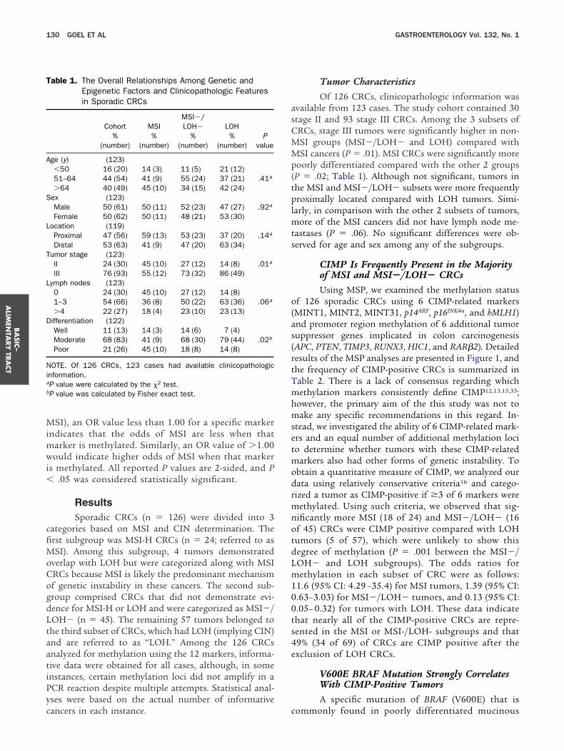
Mimwi�
cfiMoCogdLtaatiPyc
asCMMp(tplmts
o(as(rtTmhmsetmodrmnotdLm100ts4e
T
A
S
L
T
L
D
Nia
b
BA
SIC–
ALIM
ENTA
RY
TRA
CT
130 GOEL ET AL GASTROENTEROLOGY Vol. 132, No. 1
SI), an OR value less than 1.00 for a specific markerndicates that the odds of MSI are less when that
arker is methylated. Similarly, an OR value of �1.00ould indicate higher odds of MSI when that marker
s methylated. All reported P values are 2-sided, and P.05 was considered statistically significant.
ResultsSporadic CRCs (n � 126) were divided into 3
ategories based on MSI and CIN determination. Therst subgroup was MSI-H CRCs (n � 24; referred to asSI). Among this subgroup, 4 tumors demonstrated
verlap with LOH but were categorized along with MSIRCs because MSI is likely the predominant mechanismf genetic instability in these cancers. The second sub-roup comprised CRCs that did not demonstrate evi-ence for MSI-H or LOH and were categorized as MSI�/OH� (n � 45). The remaining 57 tumors belonged tohe third subset of CRCs, which had LOH (implying CIN)nd are referred to as “LOH.” Among the 126 CRCsnalyzed for methylation using the 12 markers, informa-ive data were obtained for all cases, although, in somenstances, certain methylation loci did not amplify in aCR reaction despite multiple attempts. Statistical anal-ses were based on the actual number of informative
able 1. The Overall Relationships Among Genetic andEpigenetic Factors and Clinicopathologic Featuresin Sporadic CRCs
Cohort MSIMSI�/LOH� LOH
Pvalue
%(number)
%(number)
%(number)
%(number)
ge (y) (123)�50 16 (20) 14 (3) 11 (5) 21 (12)51–64 44 (54) 41 (9) 55 (24) 37 (21) .41a
�64 40 (49) 45 (10) 34 (15) 42 (24)ex (123)Male 50 (61) 50 (11) 52 (23) 47 (27) .92a
Female 50 (62) 50 (11) 48 (21) 53 (30)ocation (119)Proximal 47 (56) 59 (13) 53 (23) 37 (20) .14a
Distal 53 (63) 41 (9) 47 (20) 63 (34)umor stage (123)II 24 (30) 45 (10) 27 (12) 14 (8) .01a
III 76 (93) 55 (12) 73 (32) 86 (49)ymph nodes (123)0 24 (30) 45 (10) 27 (12) 14 (8)1–3 54 (66) 36 (8) 50 (22) 63 (36) .06a
�4 22 (27) 18 (4) 23 (10) 23 (13)ifferentiation (122)Well 11 (13) 14 (3) 14 (6) 7 (4)Moderate 68 (83) 41 (9) 68 (30) 79 (44) .02b
Poor 21 (26) 45 (10) 18 (8) 14 (8)
OTE. Of 126 CRCs, 123 cases had available clinicopathologicnformation.P value were calculated by the �2 test.P value was calculated by Fisher exact test.
ancers in each instance. c
Tumor CharacteristicsOf 126 CRCs, clinicopathologic information was
vailable from 123 cases. The study cohort contained 30tage II and 93 stage III CRCs. Among the 3 subsets ofRCs, stage III tumors were significantly higher in non-SI groups (MSI�/LOH� and LOH) compared withSI cancers (P � .01). MSI CRCs were significantly more
oorly differentiated compared with the other 2 groupsP � .02; Table 1). Although not significant, tumors inhe MSI and MSI�/LOH� subsets were more frequentlyroximally located compared with LOH tumors. Simi-
arly, in comparison with the other 2 subsets of tumors,ore of the MSI cancers did not have lymph node me-
astases (P � .06). No significant differences were ob-erved for age and sex among any of the subgroups.
CIMP Is Frequently Present in the Majorityof MSI and MSI�/LOH� CRCsUsing MSP, we examined the methylation status
f 126 sporadic CRCs using 6 CIMP-related markersMINT1, MINT2, MINT31, p14ARF, p16INK4�, and hMLH1)nd promoter region methylation of 6 additional tumoruppressor genes implicated in colon carcinogenesisAPC, PTEN, TIMP3, RUNX3, HIC1, and RAR�2). Detailedesults of the MSP analyses are presented in Figure 1, andhe frequency of CIMP-positive CRCs is summarized inable 2. There is a lack of consensus regarding whichethylation markers consistently define CIMP12,13,15,33;
owever, the primary aim of the this study was not toake any specific recommendations in this regard. In-
tead, we investigated the ability of 6 CIMP-related mark-rs and an equal number of additional methylation locio determine whether tumors with these CIMP-related
arkers also had other forms of genetic instability. Tobtain a quantitative measure of CIMP, we analyzed ourata using relatively conservative criteria16 and catego-ized a tumor as CIMP-positive if �3 of 6 markers were
ethylated. Using such criteria, we observed that sig-ificantly more MSI (18 of 24) and MSI�/LOH� (16f 45) CRCs were CIMP positive compared with LOHumors (5 of 57), which were unlikely to show thisegree of methylation (P � .001 between the MSI�/OH� and LOH subgroups). The odds ratios forethylation in each subset of CRC were as follows:
1.6 (95% CI: 4.29 –35.4) for MSI tumors, 1.39 (95% CI:.63–3.03) for MSI�/LOH� tumors, and 0.13 (95% CI:.05– 0.32) for tumors with LOH. These data indicatehat nearly all of the CIMP-positive CRCs are repre-ented in the MSI or MSI-/LOH- subgroups and that9% (34 of 69) of CRCs are CIMP positive after thexclusion of LOH CRCs.
V600E BRAF Mutation Strongly CorrelatesWith CIMP-Positive TumorsA specific mutation of BRAF (V600E) that is
ommonly found in poorly differentiated mucinous
CmgFBt(ML
iP
mm
FCwmtMsao(iemotm
BA
SIC–
ALI
MEN
TARY
TRA
CT
January 2007 CIMP IN SPORADIC COLON CANCER 131
RCs has been associated with CIMP.10,11 We deter-ined the presence of V600E mutations in the 3 sub-
roups of CRCs. As depicted in Tables 3 and 4 andigure 1, a total of 26 of 126 (21%) CRCs harboredRAF mutations. Among these, a significant correla-ion was observed between BRAF mutation and MSI70.8%; 17 of 24; P � .0001 for MSI vs MSI�) and
SI�/LOH� (15.5%; 7 of 45; P � .03 for MSI�/
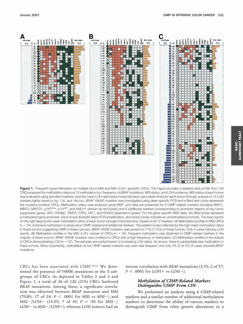
igure 1. Frequent hypermethylation at multiple loci in MSI and MSI-/RCs analyzed for methylation status at 12 methylation loci, frequency oas evaluated using standard markers, and the mean LOH ratios (bluearkers tightly linked to 5q, 17p, and 18q loci. BRAF V600E mutation w
he mutation-positive CRCs. Methylation status was analyzed using MINT2, MINT31, p16INK4�, p14ARF, and hMLH1 (shown as red boxes)
uppressor genes: APC, RAR�2, TIMP3, PTEN, HIC1, and RUNX3 (demethylated gene promoter, black boxes illustrate failed PCR amplifican the right depicts the mean methylation ratios of each tumor (orange h
n � 24). Extensive methylation is observed in CIMP-related and additionn these tumors suggesting CIMP in these cancers. BRAF V600E mutatvents. (B) Methylation profiles in the MSI-/LOH- subset of CRCs (n �ajority of these tumors. BRAF V600E mutation was confined to CRCsf CRCs demonstrating LOH (n � 57). The samples are sorted based ohese tumors. More importantly, methylation at the CIMP-related markutations.
OH� vs MSI�/LOH�), whereas LOH tumors had an d
nverse correlation with BRAF mutations (3.5%; 2 of 57;� .0001 for LOH� vs LOH�).
Methylation of CIMP-Related MarkersDistinguishes CIMP From CINWe performed an analysis using 6 CIMP-related
arkers and a similar number of additional methylationarkers to determine the ability of various markers to
sporadic CRCs. The Figure provides a detailed data profile from 126F mutations, MSI status, and LOH incidence. MSI status of each tumorntal bars) were determined for each tumor through analysis of 10 LOHvestigated using allele-specific PCR and a filled dark circle representsnd data are presented for 6 CIMP-related markers including MINT1,additional markers corresponding to promoter regions of key tumor
d in green). For the gene-specific MSP data, the filled boxes representnd empty boxes indicate an unmethylated promoter. The final columntal bars), based on all 12 markers. (A) Methylation profiles in MSI CRCsrkers. This pattern is also reflected by the high mean methylation ratiosas present in 71% (17/24) of these tumors. Only 4 cases had any LOH. Frequent methylation was observed in CIMP-related markers in thea high frequency of methylation. (C) Methylation profiles in the subset
reasing LOH ratios. As shown, there is substantially less methylation inas even less frequent, and only 3% (2 of 57) of cases showed BRAF
LOH-f BRAhorizo
as inSP, aand 6pictetion, aorizonal maion w
45)with
n incers w
istinguish CIMP from other genetic alterations in a
camlCw(((
nf
mmmnmMeMqstsbaatafpe
est
T
M
ML
a
T
G
C
A
Wa
BA
SIC–
ALIM
ENTA
RY
TRA
CT
132 GOEL ET AL GASTROENTEROLOGY Vol. 132, No. 1
ohort of sporadic CRCs. The frequency of methylationt each marker in the total cohort of all CRCs is sum-arized in Table 3. We observed that methylation of at
east 1 locus was present in 110 of 126 (87.3%) of allRCs. The frequency of promoter methylation by geneas as follows: MINT1 (18%), MINT2 (30%), MINT31
45%), HIC1 (62%), p14ARF (33%), RAR�2 (38%), TIMP326%), APC (25%), hMLH1 (22%), p16INK4� (24%), RUNX316%), and PTEN (6%).
When the tumors were segregated based on the desig-ated patterns of genomic or epigenetic alterations, we
ound that MSI cancers were frequently methylated at
able 2. Overall Frequency of Epigenetic Alterations inVarious Subsets of Sporadic CRCs
% (number) ofCIMP�(�3/6
markers)Odds ratio forCIMP (95% CI)
P valuea
(vsMSI�/LOH�)
SI�/LOH�(n � 45)
36 (16) 1.39 (0.63–3.03) N/A
SI (n � 24) 75 (18) 11.6 (4.29–35.4) .002OH (n � 57) 9 (5) 0.13 (0.05–0.32) .001
P values were calculated by the �2 test.
able 3. The Relationship Between Methylation FrequenciesDemonstrating MSI and CIN
All CRCs%
(number)(n � 126)
MSI s
MSI(n � 24
enetic marker BRAF Mutant 21 (26) 71 (17V600E Wt 79 (100) 29 (7)
IMP-relatedmarkers
MINT1 M 18 (22) 50 (12U 82 (100) 50 (12
MINT2 M 30 (35) 59 (13U 70 (81) 41 (9)
MINT31 M 45 (53) 78 (18U 55 (64) 22 (5)
p16INK4� M 24 (30) 26 (6)U 76 (93) 74 (17
p14ARF M 33 (42) 50 (12U 67 (84) 50 (12
hMLH1 M 22 (25) 60 (12U 78 (89) 40 (8)
dditionalepigeneticmarkers
APC M 25 (31) 29 (7)U 75 (94) 71 (17
RAR�2 M 38 (48) 42 (10U 62 (78) 58 (14
TIMP3 M 26 (33) 75 (18U 74 (92) 25 (6)
PTEN M 6 (7) 23 (5)U 94 (109) 77 (17
HIC1 M 62 (76) 92 (22U 38 (46) 8 (2)
RUNX3 M 16 (19) 39 (9)U 84 (99) 61 (14
t, wild type; M, methylated; U, unmethylated.
P values were based on the �2 test.ajority of the markers, and 75% (9 of 12) of thesearkers demonstrated a significantly higher frequency ofethylation (P � .05 to P � .0001) compared with
on-MSI CRCs (Table 3 and Figure 1A). Among these 9arkers, 5 were CIMP-related markers (MINT1, MINT2,INT31, p14ARF, and hMLH1). As shown in Table 5, the
stimated odds ratios indicate an association betweenSI (OR values range from 1.12 to 17.2) and the fre-
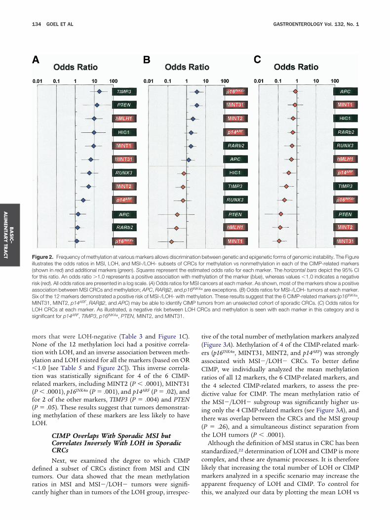
uency of methylation. Figure 2A illustrates the relation-hip between MSI and methylation of each marker withhe OR values plotted in descending order. This Figureuggests no obvious segregation in the odds of MSIetween the 2 sets of markers (CIMP related and thedditional markers) because most markers demonstratedstrong association between MSI and methylation. In-
erestingly, among the CIMP-related markers, methyl-tion of hMLH1 had the strongest association (OR, 9.35),ollowed by MINT1, MINT31, MINT2, p14ARF, and16INK4�, confirming that hMLH1 methylation is a keyvent in the genesis of sporadic MSI CRCs.
Methylation was frequently observed at several mark-rs in the 45 tumors that were MSI�/LOH�. However, atatistically significant association was observed only forhe CIMP-related markers, including MINT2 (P � .001),
igenetic Markers and BRAF Mutations in Sporadic CRCs
% (number)
P valuea
CIN status % (number)
P valueaMSI-L/MSS(n � 102)
LOH (�)(n � 61)
LOH (�)(n � 65)
9 (9) �.0001 7 (4) 34 (22) .000191 (93) 93 (57) 66 (43)10 (10) �.0001 17 (10) 19 (12) .790 (88) 83 (50) 81 (50)23 (22) .001 11 (6) 47 (29) �.000177 (72) 89 (48) 53 (33)37 (35) .0004 21 (12) 68 (41) �.000163 (59) 79 (45) 32 (19)24 (24) .83 12 (7) 37 (23) .00176 (76) 88 (53) 63 (40)29 (30) .05 23 (14) 43 (28) .0271 (72) 77 (47) 57 (37)14 (13) �.0001 16 (9) 27 (16) .1786 (81) 84 (46) 73 (43)24 (24) .58 23 (14) 26 (17) .7276 (77) 77 (46) 74 (48)37 (38) .69 33 (20) 43 (28) .2363 (64) 67 (41) 57 (37)15 (15) �.0001 15 (9) 38 (24) .00485 (86) 85 (52) 62 (40)2 (2) .0003 2 (1) 10 (6) .05
98 (92) 98 (57) 90 (52)55 (54) .0009 57 (35) 67 (41) .2645 (44) 43 (26) 33 (20)11 (10) .0008 12 (7) 20 (12) .2889 (85) 88 (50) 80 (49)
of Ep
tatus
)
)
)))
)
))))
))))
))
)
M.CctMwu
6Mlp94
s
T
G
C
A
Wa
T
C
O
a
4
BA
SIC–
ALI
MEN
TARY
TRA
CT
January 2007 CIMP IN SPORADIC COLON CANCER 133
INT31 (P � .0001), p16INK4� (P � .002), and p14ARF(P �04) (Table 4 and Figure 1B). Of note, these same 4IMP-related markers showed a significant inverse asso-
iation between methylation and LOH. A distinctive dis-ribution of ORs measuring the association between
SI�/LOH� and methylation of the individual markersas observed (Table 5 and Figure 2B). We found that,nlike MSI, where all 12 markers had an OR �1.0, only
able 4. The Relationship Between Epigenetic Alterations anEvidence for CIN
enetic marker BRAF V600E MutantWt
IMP-related markers MINT1 MU
MINT2 MU
MINT31 MU
p16INK4� MU
p14ARF MU
hMLH1 MU
dditional epigenetic markers APC MU
RAR�2 MU
TIMP3 MU
PTEN MU
HIC1 MU
RUNX3 MU
t, wild type; M, methylated; U, unmethylated.P values were based on the �2 test.
able 5. Odds Ratios for Methylation vs Nonmethylation in V
Gene markers MSI ORa
IMP-related markers MINT1 8.80 (3.1MINT2 4.73 (1.7MINT31 6.07 (2.0p16INK4� 1.12 (0.4p14ARF 2.40 (0.9hMLH1 9.35 (3.2
ther epigenetic markers APC 1.32 (0.4RAR�2 1.20 (0.4TIMP3 17.2 (5.8PTEN 13.5 (2.4HIC1 8.96 (2.0RUNX3 5.46 (1.8
Odds ratios were calculated for the odds of MSI (n � 24) vs other (n
5) vs other (n � 81).of the 12 markers demonstrated an OR �1.0 forSI�/LOH�. Among the 6 markers, 4 were CIMP re-
ated, including p16INK4� (OR, 3.20; 95% CI: 1.37–7.48),14ARF (OR, 1.58; 95% CI: 0.74 –3.40), MINT31 (OR, 3.15;5% CI: 1.43– 6.93), and MINT2 (OR, 2.00; 95% CI: 0.89 –.49).
By contrast, the overall incidence of methylation wasignificantly lower in LOH tumors compared with tu-
AF Mutations in MSS Sporadic CRCs Without
Total %(number)(n � 102)
MSS CRCs % (number)
P valueaLOH (�)(n � 45)
LOH (�)(n � 57)
9 (9) 16 (7) 4 (2) .0391 (93) 84 (38) 96 (55)10 (10) 5 (2) 14 (8) .1290 (88) 95 (40) 86 (48)23 (22) 40 (17) 10 (5) �.00177 (72) 60 (26) 90 (46)37 (35) 63 (26) 17 (9) �.000163 (59) 37 (15) 83 (44)24 (24) 39 (17) 13 (7) .00276 (76) 61 (27) 88 (49)29 (30) 40 (18) 21 (12) .0471 (72) 60 (27) 79 (45)14 (13) 10 (4) 17 (9) .3586 (81) 90 (36) 83 (45)24 (24) 27 (12) 21 (12) .5476 (77) 73 (33) 79 (44)38 (37) 44 (20) 32 (18) .1862 (63) 56 (25) 68 (39)15 (15) 20 (9) 11 (6) .1685 (86) 80 (35) 89 (51)2 (2) 3 (1) 2 (1) .83
98 (92) 98 (39) 98 (53)55 (54) 56 (23) 54 (31) .8745 (44) 44 (18) 46 (26)11 (10) 12 (5) 9 (5) .789 (85) 88 (37) 91 (48)
s Subsets of CRCs for Each of the Epigenetic Markers
CI) MSI�/LOH� ORa (95% CI) LOH ORa (95% CI)
.7) 0.15 (0.03–0.68) 0.83 (0.33–2.10)
.5) 2.00 (0.89–4.49) 0.14 (0.05–0.38)
.8) 3.15 (1.43–6.93) 0.12 (0.05–0.29)5) 3.20 (1.37–7.48) 0.23 (0.09–0.59)4) 1.58 (0.74–3.40) 0.39 (0.18–0.85).2) 0.28 (0.09–0.89) 0.53 (0.21–1.31)6) 1.17 (0.51–2.70) 0.86 (0.38–1.94)7) 1.51 (0.72–3.19) 0.64 (0.31–1.33).4) 0.61 (0.25–1.46) 0.29 (0.12–0.69).5) 0.30 (0.03–2.58) 0.15 (0.02–1.31).2) 0.68 (0.31–1.46) 0.66 (0.31–1.37).8) 0.60 (0.20–1.80) 0.57 (0.21–1.57)
2). LOH positive (n � 61) vs other (n � 63), and MSI�/LOH� (n �
d BR
ariou
(95%
3–248–127–170–3.17–5.91–279–3.59–2.97–502–750–409–15
� 10
mNty�tr(f(iL
dtrc
t(eaCrtdtit(t
sclma
Fi(fraSMLs
BA
SIC–
ALIM
ENTA
RY
TRA
CT
134 GOEL ET AL GASTROENTEROLOGY Vol. 132, No. 1
ors that were LOH-negative (Table 3 and Figure 1C).one of the 12 methylation loci had a positive correla-
ion with LOH, and an inverse association between meth-lation and LOH existed for all the markers (based on OR1.0 [see Table 5 and Figure 2C]). This inverse correla-
ion was statistically significant for 4 of the 6 CIMP-elated markers, including MINT2 (P � .0001), MINT31P � .0001), p16INK4� (P � .001), and p14ARF (P � .02), andor 2 of the other markers, TIMP3 (P � .004) and PTENP � .05). These results suggest that tumors demonstrat-ng methylation of these markers are less likely to haveOH.
CIMP Overlaps With Sporadic MSI butCorrelates Inversely With LOH in SporadicCRCsNext, we examined the degree to which CIMP
efined a subset of CRCs distinct from MSI and CINumors. Our data showed that the mean methylationatios in MSI and MSI�/LOH� tumors were signifi-
igure 2. Frequency of methylation at various markers allows discriminallustrates the odds ratios in MSI, LOH, and MSI-/LOH- subsets of CRshown in red) and additional markers (green). Squares represent the eor this ratio. An odds ratio �1.0 represents a positive association withisk (red). All odds ratios are presented in a log scale. (A) Odds ratios forssociation between MSI CRCs and methylation; APC, RAR�2, and p1ix of the 12 markers demonstrated a positive risk of MSI-/LOH- with mINT31, MINT2, p14ARF, RAR�2, and APC) may be able to identify CIM
OH CRCs at each marker. As illustrated, a negative risk between LOignificant for p14ARF, TIMP3, p16INK4�, PTEN, MINT2, and MINT31.
antly higher than in tumors of the LOH group, irrespec- t
ive of the total number of methylation markers analyzedFigure 3A). Methylation of 4 of the CIMP-related mark-rs (p16INK4�, MINT31, MINT2, and p14ARF) was stronglyssociated with MSI�/LOH� CRCs. To better defineIMP, we individually analyzed the mean methylation
atios of all 12 markers, the 6 CIMP-related markers, andhe 4 selected CIMP-related markers, to assess the pre-ictive value for CIMP. The mean methylation ratio ofhe MSI�/LOH� subgroup was significantly higher us-ng only the 4 CIMP-related markers (see Figure 3A), andhere was overlap between the CRCs and the MSI groupP � .26), and a simultaneous distinct separation fromhe LOH tumors (P � .0001).
Although the definition of MSI status in CRC has beentandardized,22 determination of LOH and CIMP is moreomplex, and these are dynamic processes. It is thereforeikely that increasing the total number of LOH or CIMP
arkers analyzed in a specific scenario may increase thepparent frequency of LOH and CIMP. To control for
between genetic and epigenetic forms of genomic instability. The Figurer methylation vs nonmethylation in each of the CIMP-related markersted odds ratio for each marker. The horizontal bars depict the 95% CIylation of the marker (blue), whereas values �1.0 indicates a negativeancers at each marker. As shown, most of the markers show a positiveare exceptions. (B) Odds ratios for MSI-/LOH- tumors at each marker.tion. These results suggest that the 6 CIMP-related markers (p16INK4�,ors from an unselected cohort of sporadic CRCs. (C) Odds ratios for
Cs and methylation is seen with each marker in this category and is
tionCs fostimameth
MSI c6INK4�
ethylaP tumH CR
his, we analyzed our data by plotting the mean LOH vs

tsao3fh�asLa
uP.Pis
r
Fiirwbmac(mcboesui� at the
BA
SIC–
ALI
MEN
TARY
TRA
CT
January 2007 CIMP IN SPORADIC COLON CANCER 135
he methylation ratios. This approach should reveal as-ociations between the methylator phenotype and CINnd compensate for changes in the total number of LOHr methylation events (Figure 3B–E). As shown in FigureB, differences between the mean methylation ratiosrom the 12 markers vs the LOH ratios for all CRCs wereighly significant (Spearman rank correlation coefficient,� �0.36; P � .0001). Additionally, analyses of methyl-
tion and LOH ratios of all MSS CRCs demonstratedignificant inverse correlations between methylation andOH events. As depicted in Figure 3C–E, the inverse
igure 3. Sporadic CRCs with extensive methylator phenotype inversedentifying CIMP and the relationship between CIMP and LOH in sporadic Cn each tumor. (A) Mean methylation ratios were determined based on the nepresent the mean methylation ratios in MSI (blue), MSI-/LOH- (red), and Lith vertical columns) represent the 95% CI of the mean methylation ratioetween the mean methylation ratios in each subset of CRCs; the P valuesethylation ratios using only 4 CIMP-related markers in comparison with
ssociation for MSI and MSI-/LOH- tumors but a clear segregation and invalculated using all 12 methylation markers was compared with LOH ratios
� � �0.3690; P � .0001) between the incidence of methylation and LOarkers or LOH markers would have no effect on the relationship betwee
losely relate to CIMP, underscoring the relationship between CIMP and LOetween the methylation ratios in MSI-negative cases and the LOH burdenbtained using all 12 markers. As indicated, even following exclusion of Mvident (� � �0.2981; P � .002). (D) The relationship between LOH andignificant inverse relationship was observed (� � �0.3079; P � .002), andsing all 12 markers. (E) The relationship between LOH and the methylatio
nverse association between LOH and methylation was more prounounce0.2981 with 12 markers or � � �0.3079 with 6 markers), suggesting th
ssociation was strongest when the data were analyzed o
sing only 4 selected CIMP-related markers (� � �0.41;� .0001), rather than all 12 markers (� � �0.29; P �
002) or the original 6 CIMP-related markers (� � �0.30;� .002). Taken together, these data show that a signif-
cant excess of promoter methylation is present in theporadic MSI and MSI�/LOH� tumors.
DiscussionPreviously, we characterized a large cohort of spo-
adic CRCs by determining whether they exhibited MSI
relate with LOH. This Figure illustrates the utility of selected markers forFor these analyses, we compared the mean LOH vs the methylation ratioser of markers methylated in the 3 subsets of CRCs. The 3 vertical columnshite) CRCs. The error bars denote the SD. The filled circles (color matchedrectangular boxes in the upper panels represent the pairwise correlationcalculated by the Wilcoxon test. As shown in the 3 panels, analysis of the12 or 6 CIMP-related markers clearly demonstrates a significant positiveorrelation for LOH CRCs. (B) The relationship between methylation ratiostotal cohort of 126 CRCs. As shown, an inverse correlation was observed
hese results suggested that increasing either the number of methylationhenotypes, which are mutually exclusive. Because sporadic MSI tumorse excluded all MSI cases and reanalyzed the data to show the relationshipse tumors. (C) The relationship between mean LOH and methylation ratiosses, a significant inverse correlation between methylation and LOH was
ylation incidence using 6 CIMP-related markers in MSI-negative cases. Awas no added gain using only the CIMP-related markers, compared withs of the 4 CIMP-related markers in MSI-negative CRCs. Interestingly, then CIMP data were compared using 4 markers (� � �0.4175; versus � �se selected markers will help identify high-frequency methylated CRCs.
ly corRCs.umb
OH (ws. Thewere
eithererse cin theH. Tn 2 pH, w
in theSI ca
meththere
n ratiod whe
r CIN and found that �35% of tumors lacked charac-
ttCroo
CCCMsrmrisasbfiiMmpCta
mficeiCdeioTiclgsc5Cmwh5crd
peCoa
coc(dtmdCpAsbMsc
aMCsctacatl
smadsMpaismpottwomm
BA
SIC–
ALIM
ENTA
RY
TRA
CT
136 GOEL ET AL GASTROENTEROLOGY Vol. 132, No. 1
eristics of either of these.6 In addition, we demonstratedhat sporadic MSI CRCs were distinct from those withIN, a finding that has been confirmed by others.34 These
esults led us to hypothesize that CIMP is a mechanismf tumor promoter gene silencing in CRCs lacking CINr MSI.
The present study used a panel of markers to defineIMP in a large cohort of CRCs for which MSI andIN status was known. Our results indicate that (1)IMP is frequently present both in MSI tumors, and inSI-L/MSS CRCs, and involves as many as half of all
poradic CRCs; (2) the 6 originally proposed CIMP-elated markers are highly specific for identifying tu-
ors with the methylator phenotype; and (3) CIMPepresents a distinct subtype of sporadic CRCs that isnversely associated with CIN. Collectively, these re-ults suggest that CIN and CIMP constitute 2 majornd mutually exclusive pathways of tumor evolution inporadic CRCs. According to this model, CRCs developy loss of multiple tumor suppressors, and these de-ects occur by allelic loss in tumors with CIN, bynability to repair single nucleotide mismatches andnstability at microsatellite sequences in tumors with
SI, and by methylation-associated silencing in tu-ors with CIMP. Because methylation of the hMLH1
romoter is the cause of MSI in nearly all sporadicRCs,3 tumors that are MSI constitute a subset of
umors with CIMP. CRCs in Lynch syndrome make upminority of all MSI cancers and evolve differently.The overall frequency of DNA methylation in nor-al tissues increases with age. CIMP, however, is de-
ned by a set of methylation-specific markers that areonsistently methylated in tumors to a much greaterxtent than in normal intestinal mucosa from elderlyndividuals. Although many studies support a role forIMP in pathogenesis of sporadic CRC,7,10,11 markedlyifferent frequencies of this characteristic are report-d,11,15,33,35 with a lack of agreement among differentnvestigators, likely because of differences in method-logy and criteria used to define the CIMP phenotype.he work performed here provides a framework for
dentifying CIMP by placing methylation-specifichanges in the context of MSI and CIN in a relativelyarge group of sporadic CRCs. The overall frequency ofenetic and epigenetic alterations examined in thistudy is similar to those reported previously. In thisontext, we observed that, although methylation of the=-region of hMLH1 was strongly associated with MSIRCs, some proportion of non-MSI cancers were alsoethylated. Similar observations were made previouslyhen it was suggested that 3=-region methylation of
MLH1 primarily associates with MSI CRCs, whereas=-region methylation may be present in non-MSI can-ers, and extensive methylation of these regions isequired for transcriptionally silencing.17,18,30 When we
efined CIMP as being present in tumors exhibiting sositivity in �50% of a panel of CIMP-related mark-rs,16 we found that approximately half of sporadicRCs met this standard. This figure is larger than inther studies in which the frequency of CIMP waspproximately 20%–30%.10
Investigators interested in CIMP have identified a spe-ific set of methylation markers that allows identificationf the methylator phenotype.7,10,15,16 In this study, weonfirmed that the 6 conventional CIMP-related markersMINT1, MINT2, MINT31, p16INK4�, p14ARF, and hMLH1)ifferentiated between tumors with MSI and LOH andhat examination of promoter methylation at other tu-
or suppressor loci did not significantly impact thisistinction. Interestingly, only 4 of the conventionalIMP-related markers (MINT2, MINT31, p16INK4�, and
14ARF) were associated with MSI�/LOH� tumor status.dditionally, the methylation status at these 4 markers
howed overlap between MSI�/LOH� and MSI CRCsut clearly distinguished this group of tumors from theSI�/LOH� neoplasms, suggesting that the first 2 sub-
ets of tumors collectively represent CIMP and inverselyorrelate with CIN.
Although many recent studies have utilized MSPssays to detect CIMP,10,13 it has been suggested thatSP may not be an ideal methodology to detect
IMP.7 If the MSP assay conditions are not sufficientlytringent, one may overestimate methylation frequen-ies for a given target gene. However, in this study, weook precautions in designing and performing MSPssays using highly stringent conditions and furtheronfirmed the reproducibility and specificity of MSPssays by ensuring methylation detection only in theumor tissues and not in the DNA from normal co-onic epithelium.
Although the published studies are small, it has beenuggested that there are phenotypic characteristics com-
on to CRCs with CIMP. These include the featuresssociated with MSI, such as proximal location and poorifferentiation, and changes not seen in MSI tumors,uch as a high rate of mutations in BRAF and K-Ras
SI-L/MSS and CIMP� tumors, may have a particularlyoor prognosis.36 Mutations in the BRAF and K-Ras genesre common in sporadic CRC but are mutually exclusiven these neoplasms.10,18 In this study, we observed aignificant association between MSI and V600E BRAF
utations, which has been shown to link tightly withromoter methylation of hMLH1 in MSI CRCs.18 Webserved a similar but somewhat weaker correlation be-ween BRAF mutations and MSI�/LOH� as well, buthese mutations were not associated with LOH. Althoughe did not perform K-Ras mutation analysis in this study,ur data suggest that MSI�/LOH� cancers may harborore K-Ras mutations.16 An explanation for why oneutation occurs as opposed to the other has been elu-
ive.
taa
pcp
ICDDMMRRSSTUUUUUUUUUUUWW
January 2007 CIMP IN SPORADIC COLON CANCER 137
In conclusion, until a clear etiology for CIMP is iden-ified, we cannot know for certain whether CIMP is ancquired defect with a primary etiology or whether this
bnormal pattern of promoter methylation is a random Calter Reed Army Medical Center Washington, D
methylator phenotype. Nat Clin Pract Oncol 2005;2:398–405.
1
1
1
1
1
1
1
rocess that is selected for in tumor cells. The formeroncept is supported by our observation that CIMP isresent not only in MSI tumors but in tumors that lack
IN and MSI characteristics.BA
SIC–
ALI
MEN
TARY
TRA
CT
Appendix
The following institutions participated in the study:
nstitution name Location Principal Investigator Grant No.ALGB Statistical Office Durham, NC Stephen George, PhD Supported by CA33601ana Farber Cancer Institute Boston, MA George P. Canellos, MD Supported by CA32291artmouth Medical School–Norris Cotton Cancer Center Lebanon, NH Marc Ernstoff, MD Supported by CA04326assachusetts General Hospital Boston, MA Michael L. Grossbard, MD Supported by CA12449ount Sinai School of Medicine New York, NY Lewis Silverman, MD Supported by CA04457hode Island Hospital Providence, RI William Sikov, MD Supported by CA08025oswell Park Cancer Institute Buffalo, NY Ellis Levine, MD Supported by CA02599outheast Cancer Control Consortium Inc. CCOP Goldsboro, NC James N. Atkins, MD Supported by CA45808UNY Upstate Medical University Syracuse, NY Stephen L. Graziano, MD Supported by CA21060he Ohio State University Columbus, OH Clara D. Bloomfield, MD Supported by CA77658niversity of California at San Diego San Diego, CA Stephen Seagren, MD Supported by CA11789niversity of California at San Francisco San Francisco, CA Alan Venook, MD Supported by CA60138niversity of Chicago Medical Center Chicago, IL Gini Fleming, MD Supported by CA41287niversity of Illinois at Chicago Chicago, IL David Gustin, MD Supported by CA74811niversity of Iowa Iowa City, IA Gerald Clamon, MD Supported by CA47642niversity of Maryland Cancer Center Baltimore, MD David Van Echo, MD Supported by CA31983niversity of Massachusetts Medical Center Worcester, MA Mary Ellen Taplin, MD Supported by CA37135niversity of Minnesota Minneapolis, MN Bruce A. Peterson, MD Supported by CA16450niversity of Missouri/Ellis Fischel Cancer Center Columbia, MO Michael C. Perry, MD Supported by CA12046niversity of North Carolina at Chapel Hill Chapel Hill, NC Thomas C. Shea, MD Supported by CA47559niversity of Tennessee Memphis Memphis, TN Harvey B. Niell, MD Supported by CA47555ake Forest University School of Medicine Winston-Salem, NC David D. Hurd, MD Supported by CA03927
C John C. Byrd, MD Supported by CA26806
References
1. Loeb LA. Mutator phenotype may be required for multistage car-cinogenesis. Cancer Res 1991;51:3075–3079.
2. Lengauer C, Kinzler KW, Vogelstein B. Genetic instability in colo-rectal cancers. Nature 1997;386:623–627.
3. Kane MF, Loda M, Gaida GM, Lipman J, Mishra R, Goldman H,Jessup JM, Kolodner R. Methylation of the hMLH1 promotercorrelates with lack of expression of hMLH1 in sporadic colontumors and mismatch repair-defective human tumor cell lines.Cancer Res 1997;57:808–811.
4. Baylin SB, Herman JG, Graff JR, Vertino PM, Issa JP. Alterationsin DNA methylation: a fundamental aspect of neoplasia. AdvCancer Res 1998;72:141–196.
5. Jones PA, Laird PW. Cancer epigenetics comes of age. Nat Genet1999;21:163–167.
6. Goel A, Arnold CN, Niedzwiecki D, Chang DK, Ricciardiello L,Carethers JM, Dowell JM, Wasserman L, Compton C, Mayer RJ,Bertagnolli MM, Boland CR. Characterization of sporadic coloncancer by patterns of genomic instability. Cancer Res 2003;63:1608–1614.
7. Rashid A, Issa JP. CpG island methylation in gastroenterologic neo-plasia: a maturing field. Gastroenterology 2004;127:1578–1588.
8. Issa JP. CpG island methylator phenotype in cancer. Nat RevCancer 2004;4:988–993.
9. Jass JR. Serrated adenoma of the colorectum and the DNA-
0. Samowitz WS, Albertsen H, Herrick J, Levin TR, Sweeney C, Mur-taugh MA, Wolff RK, Slattery ML. Evaluation of a large, population-based sample supports a CpG island methylator phenotype in coloncancer. Gastroenterology 2005;129:837–845.
1. Ogino S, Cantor M, Kawasaki T, Brahmandam M, Kirkner G,Weisenberger DJ, Campan M, Laird PW, Loda M, Fuchs CS. CpGisland methylator phenotype (CIMP) of colorectal cancer is bestcharacterized by quantitative DNA methylation analysis and pro-spective cohort studies. Gut 2006;55:1000–1006.
2. Yamashita K, Dai T, Dai Y, Yamamoto F, Perucho M. Geneticssupersedes epigenetics in colon cancer phenotype. Cancer Cell2003;4:121–131.
3. Anacleto C, Leopoldino AM, Rossi B, Soares FA, Lopes A, RochaJC, Caballero O, Camargo AA, Simpson AJ, Pena SD. Colorectalcancer “methylator phenotype”: fact or artifact? Neoplasia 2005;7:331–335.
4. Suzuki K, Suzuki I, Leodolter A, Alonso S, Horiuchi S, YamashitaK, Perucho M. Global DNA demethylation in gastrointestinal can-cer is age dependent and precedes genomic damage. CancerCell 2006;9:199–207.
5. Toyota M, Ahuja N, Ohe-Toyota M, Herman JG, Baylin SB, Issa JP.CpG island methylator phenotype in colorectal cancer. Proc NatlAcad Sci U S A 1999;96:8681–8686.
6. Toyota M, Ohe-Toyota M, Ahuja N, Issa JP. Distinct genetic pro-files in colorectal tumors with or without the CpG island methy-
lator phenotype. Proc Natl Acad Sci U S A 2000;97:710–715.
1
1
1
2
2
2
2
2
2
2
2
2
2
3
3
3
3
3
3
3
MsaMt7
AM9IS
aN
BA
SIC–
ALIM
ENTA
RY
TRA
CT
138 GOEL ET AL GASTROENTEROLOGY Vol. 132, No. 1
7. Hawkins N, Norrie M, Cheong K, Mokany E, Ku SL, Meagher A,O’Connor T, Ward R. CpG island methylation in sporadic colorec-tal cancers and its relationship to microsatellite instability. Gas-troenterology 2002;122:1376–1387.
8. Nagasaka T, Sasamoto H, Notohara K, Cullings HM, Takeda M,Kimura K, Kambara T, MacPhee DG, Young J, Leggett BA, JassJR, Tanaka N, Matsubara N. Colorectal cancer with mutation inBRAF, KRAS, and wild-type with respect to both oncogenes show-ing different patterns of DNA methylation. J Clin Oncol 2004;22:4584–4594.
9. van Rijnsoever M, Grieu F, Elsaleh H, Joseph D, Iacopetta B.Characterisation of colorectal cancers showing hypermethylationat multiple CpG islands. Gut 2002;51:797–802.
0. Samowitz WS, Holden JA, Curtin K, Edwards SL, Walker AR, LinHA, Robertson MA, Nichols MF, Gruenthal KM, Lynch BJ, LeppertMF, Slattery ML. Inverse relationship between microsatellite in-stability and K-ras and p53 gene alterations in colon cancer. Am JPathol 2001;158:1517–1524.
1. Jones PA. DNA methylation errors and cancer. Cancer Res 1996;56:2463–2467.
2. Boland CR, Thibodeau SN, Hamilton SR, Sidransky D, EshlemanJR, Burt RW, Meltzer SJ, Rodriguez-Bigas MA, Fodde R, RanzaniGN, Srivastava S. A National Cancer Institute Workshop on Mic-rosatellite Instability for cancer detection and familial predispo-sition: development of international criteria for the determinationof microsatellite instability in colorectal cancer. Cancer Res1998;58:5248–5257.
3. Young J, Simms LA, Biden KG, Wynter C, Whitehall V, KaramaticR, George J, Goldblatt J, Walpole I, Robin SA, Borten MM, Stitz R,Searle J, McKeone D, Fraser L, Purdie DR, Podger K, Price R,Buttenshaw R, Walsh MD, Barker M, Leggett BA, Jass JR. Fea-tures of colorectal cancers with high-level microsatellite instabil-ity occurring in familial and sporadic settings: parallel pathwaysof tumorigenesis. Am J Pathol 2001;159:2107–2116.
4. Xu X, Quiros RM, Gattuso P, Ain KB, Prinz RA. High prevalence ofBRAF gene mutation in papillary thyroid carcinomas and thyroidtumor cell lines. Cancer Res 2003;63:4561–4567.
5. Toyota M, Issa JP. CpG island methylator phenotypes in agingand cancer. Semin Cancer Biol 1999;9:349–357.
6. Toyota M, Ahuja N, Suzuki H, Itoh F, Ohe-Toyota M, Imai K, BaylinSB, Issa JP. Aberrant methylation in gastric cancer associatedwith the CpG island methylator phenotype. Cancer Res 1999;59:5438–5442.
7. Goel A, Arnold CN, Tassone P, Chang DK, Niedzwiecki D, DowellJM, Wasserman L, Compton C, Mayer RJ, Bertagnolli MM, BolandCR. Epigenetic inactivation of RUNX3 in microsatellite unstablesporadic colon cancers. Int J Cancer 2004;112:754–759.
8. Goel A, Arnold CN, Niedzwiecki D, Carethers JM, Dowell JM,Wasserman L, Compton C, Mayer RJ, Bertagnolli MM, Boland CR.
Frequent inactivation of PTEN by promoter hypermethylation inmicrosatellite instability-high sporadic colorectal cancers. CancerRes 2004;64:3014–3021.
9. Arnold CN, Goel A, Niedzwiecki D, Dowell JM, Wasserman L,Compton C, Mayer RJ, Bertagnolli MM, Boland CR. APC promoterhypermethylation contributes to the loss of APC expression incolorectal cancers with allelic loss on 5q. Cancer Biol Ther 2004;3:960–964.
0. Goel A, Li M-S, Nagasaka T, Shin SK, Fuerst F, Ricciardiello L,Wasserman L, Boland CR. Association of JC Virus T-antigenexpression with the methylator phenotype in sporadic colorectalcancers. Gastroenterology 2006;130:1950–1961.
1. Lee S, Lee HJ, Kim JH, Lee HS, Jang JJ, Kang GH. Aberrant CpGisland hypermethylation along multistep hepatocarcinogenesis.Am J Pathol 2003;163:1371–1378.
2. Herman JG, Graff JR, Myohanen S, Nelkin BD, Baylin SB. Meth-ylation-specific PCR: a novel PCR assay for methylation status ofCpG islands. Proc Natl Acad Sci U S A 1996;93:9821–9826.
3. Chan AO, Issa JP, Morris JS, Hamilton SR, Rashid A. ConcordantCpG island methylation in hyperplastic polyposis. Am J Pathol2002;160:529–536.
4. Matsuzaki K, Deng G, Tanaka H, Kakar S, Miura S, Kim YS. Therelationship between global methylation level, loss of heterozy-gosity, and microsatellite instability in sporadic colorectal cancer.Clin Cancer Res 2005;11:8564–8569.
5. Kim HC, Roh SA, Ga IH, Kim JS, Yu CS, Kim JC. CpG islandmethylation as an early event during adenoma progression incarcinogenesis of sporadic colorectal cancer. J GastroenterolHepatol 2005;20:1920–1926.
6. Ward RL, Cheong K, Ku SL, Meagher A, O’Connor T, Hawkins NJ.Adverse prognostic effect of methylation in colorectal cancer isreversed by microsatellite instability. J Clin Oncol 2003;21:3729–3736.
Received May 25, 2006. Accepted August 31, 2006.Address requests for reprints to: Ajay Goel, PhD, Baylor Universityedical Center, 3500 Gaston Avenue, Gastrointestinal Cancer Re-
earch Laboratory, Suite H-250, Dallas, Texas 75246. e-mail:[email protected]; fax: (214) 818-9292, and C. Richard Boland,D, Baylor University Medical Center, 3500 Gaston Avenue, Gastroin-
estinal Cancer Research Laboratory, Suite H-250, Dallas, Texas5246. e-mail: [email protected]; fax: (214) 818-9292.Supported by a grant from the NIH (RO1-CA 72851; to C.R.B.), an
merican Cancer Society-IRG award (to A.G), and a grant from Dr.ildred-Scheel-Stiftung, Germany (to C.N.A.); and research for CALGB865 was supported, in part, by grants from the National Cancer
nstitute (CA31946) to the Cancer and Leukemia Group B (Richard L.chilsky, Chairman).The contents of this manuscript are solely the responsibility of the
uthors and do not necessarily represent the official views of theational Cancer Institute.
Please see Appendix for participating institutes.