long interspersed nuclear element-1 hypomethylation is a potential biomarker for the prediction of...
TRANSCRIPT
E
Long interspersed nuclear element-1hypomethylation is a potential biomarker for theprediction of response to oral fluoropyrimidines inmicrosatellite stable and CpG island methylatorphenotype-negative colorectal cancerKazuyuki Kawakami,1,3 Aika Matsunoki,1,2 Mami Kaneko,1,2 Kenichiro Saito,1,2 Go Watanabe2 andToshinari Minamoto1
1Division of Translational and Clinical Oncology, Cancer Research Institute, Kanazawa University; 2Department of General and Cardiothoracic Surgery,Kanazawa University Graduate School of Medicine, Kanazawa, Japan
(Received August 16, 2010 ⁄ Revised October 6, 2010 ⁄ Accepted October 8, 2010 ⁄ Accepted manuscript online October 18, 2010 ⁄ Article first published online November 19, 2010)
3To whom correspondence should be addressed.E-mail: [email protected]
We investigated the clinical value of methylation of long inter-spersed nuclear element-1 (LINE-1) for the prognosis of colorectalcancer (CRC) and for the survival benefit from adjuvant chemother-apy with oral fluoropyrimidines. LINE-1 methylation in tumor DNAwas measured by quantitative methylation-specific PCR in 155samples of stage II and stage III CRC. The presence of microsatelliteinstability and CpG island methylator phenotype (CIMP) wereassessed and 131 microsatellite stable ⁄ CIMP) cases were selectedfor survival analysis, of which 77 patients had received postopera-tive adjuvant chemotherapy with oral fluoropyrimidines. The CRCcell lines were used to investigate possible mechanistic linksbetween LINE-1 methylation and effects of 5-fluorouracil (5-FU).High LINE-1 methylation was a marker for better prognosis inpatients treated by surgery alone. Patients with low LINE-1 meth-ylation who were treated with adjuvant chemotherapy survivedlonger than those treated by surgery alone, suggestive of a sur-vival benefit from the use of oral fluoropyrimidines. In contrast, asurvival benefit from chemotherapy was not observed for patientswith high LINE-1 methylation. The CRC cell lines treated with 5-FUshowed increased expression of LINE-1 mRNA. This was associatedwith upregulation of the phospho-histone H2A.X in cells with lowLINE-1 methylation, but not in cells with high LINE-1 methylation.The 5-FU-mediated induction of phospho-histone H2A.X, a markerof DNA damage, was inhibited by knockdown of LINE-1. Theseresults suggest that LINE-1 methylation is a novel predictive mar-ker for survival benefit from adjuvant chemotherapy with oral flu-oropyrimidines in CRC patients. This finding could be importantfor achieving personalized chemotherapy. (Cancer Sci 2011; 102:166–174)
pigenetic alterations including CpG island hypermethylationand global hypomethylation of DNA are commonly
observed in colorectal cancer (CRC).(1) The aberrant hyperme-thylation of CpG islands is associated with distinctive clinicalfeatures of CRC and shows promise as a diagnostic marker.(2,3)
Global hypomethylation, on the other hand, has attracted muchless attention and its clinical significance in cancer has not beenextensively investigated.
Long interspersed nuclear element-1 (LINE-1) is a non-long-terminal-repeat class of retroposon that is the most successfullyintegrated mobile element and accounts for approximately 18%of the human genome.(4–6) The LINE-1 sequence is 6 kb inlength and contains a 5¢ untranslated region (UTR), two openreading frames and a 3¢UTR. The 5¢UTR has internal promoter
Cancer Sci | January 2011 | vol. 102 | no. 1 | 166–174
activity and the open reading frame encodes for nuclease andreverse transcriptase activities that are necessary for transposi-tion. Although the majority of retroposons no longer have theability to transpose due to mutations and deletions in theirsequence, approximately 100 full-length copies of LINE-1 inthe human genome retain this ability.(7,8) Because of the abun-dance and functional ability of LINE-1, the level of its methyla-tion is a surrogate of global gene methylation(9) and itshypomethylation has been suggested to cause chromosomalinstability.
Hypomethylation of LINE-1 is predicted to be a factor forunfavorable prognosis in CRC patients because of the associa-tion between global hypomethylation and chromosomal instabil-ity,(10) a known marker of poor prognosis.(11,12) Consistent withthis, our preliminary analysis of stage II and III CRC patientswho underwent curative surgery found that LINE-1 hypomethy-lation was associated with a worse patient outcome.(13) Anotherrecent study also found that LINE-1 hypomethylation was inde-pendently associated with shorter survival of colon cancerpatients.(14) However, these studies did not take into account theuse of postoperative adjuvant chemotherapy in their patientcohorts and its possible influence on the observed prognosticvalue of LINE-1 hypomethylation.
In this study, we investigated the prognostic value of LINE-1methylation in stage II and III CRC patients who underwentcurative surgery and in whom the postoperative adjuvant che-motherapy status was known. Our preliminary investigationssuggested that LINE-1 hypomethylation could be an indicator ofsurvival benefit from postoperative adjuvant chemotherapy withoral fluoropyrimidines. However, this marker is associated withthe microsatellite instability (MSI) and CpG island methylator(CIMP) phenotypes,(15,16) both of which have been implicatedin the response to 5-fluorouracil (5-FU).(2,17–19) To avoid apossible confounding influence from these phenotypes, in thepresent study we evaluated the prognostic and predictive signifi-cance of LINE-1 methylation in a cohort of CRC patients withmicrosatellite stable (MSS) and CIMP-negative (CIMP))tumors.
Materials and Methods
Patients and specimens. The present study included 155patients with stage II or III CRC who underwent surgery with
doi: 10.1111/j.1349-7006.2010.01776.xªª 2010 Japanese Cancer Association
curative intent at Kanazawa University Hospital. The patientscomprised 90 males and 65 females and ranged in age from 33to 93 years (mean, 65.6 years). The site of tumor was classifiedas proximal or distal to the splenic flexure. A total of 49 patientswere thus defined as having proximal CRC and 106 with distalCRC. Tumor stage was defined according to the InternationalUnion Against Cancer (UICC) TNM system.(20) The postopera-tive course of patients treated with and without adjuvant chemo-therapy was retrospectively assessed by referring to theirmedical records. In all, 94 patients received adjuvant chemother-apy with oral fluoropyrimidines and 61 were treated withsurgery alone. None of the patients received pre-operativeradio-chemotherapy or postoperative radiation therapy. The oralfluoropyrimidines used were 5-FU, UFT (a combination of tega-fur and uracil), doxifluridine and carmofur in 14, 52, 17 and 11patients, respectively. The median period of adjuvant chemo-therapy use was 105 weeks (range, 21–275 weeks). The medianfollow-up time was 58 months (range, 11–128 months) and 49patients died of disease recurrence during the course of followup. Survival data for the 13 patients who died of other causeswas censored at the time of death.
Tissue samples for methylation analysis were obtained fromformalin-fixed and paraffin-embedded tumor blocks selected onthe basis of tumor cell content. The tumor tissue was dissectedmanually from 10 lm paraffin sections. After deparaffinizationusing xylene and ethanol, genomic DNA was extracted from thetissue using QIAamp DNA mini kits (Qiagen, Hilden, Germany)according to the manufacturer’s protocol. DNA samples fromadjacent normal tissues were prepared using the same method.This project was approved by the Kanazawa University MedicalEthics Committee.
Cell lines and culture conditions. The CRC cell lines SW48,SW480, HCT116, HT-29, COLO205, LS411N and Caco-2 wereobtained from the American Type Culture Collection (Manas-sas, VA, USA). Other cell lines were provided by Health Sci-ence Research Resources Bank (Osaka, Japan). The cells weremaintained at 37�C with 5% CO2 in Dulbecco’s modified Eagle(SW48, SW480, SW837, HCT116, HT-29, Caco-2, CaR-1,CCK81, LoVo) or RPMI 1640 (COLO205, CW2, DLD-1,LS411N) medium supplemented with 10% fetal bovine serumand antibiotics (100 units ⁄ mL penicillin G and 100 lg ⁄ mLstreptomycin; Gibco, Grand Island, NY, USA). Cells wereharvested during the exponential growth phase for extraction ofDNA, RNA and protein.
DNA methylation analysis. Genomic DNA extracted from tis-sues and cultured cell lines was treated with bisulfite asdescribed previously.(21) Following the bisulfite treatment,LINE-1 methylation was analyzed using a methylation-specificreal-time PCR assay as previously described.(22)
Methylation levels of CpG islands within promoter regionswere evaluated by MethyLight assay for the CIMP panel ofmarkers comprising CACNA1G, IGF2, NEUROG1, RUNX3and SOCS1, where the percentage methylated reference (PMR)values were derived using the ALU normalization control reac-tion.(23) Simultaneous hypermethylation (PMR ‡ 10) of three ormore of these five markers was used to define a tumor asCIMP+.
Microsatellite analysis for MSI and LOH. The MSI status wasdetermined using a three-marker panel of mononucleotiderepeats (BAT26, NR21 and NR27) reported to be highly sensi-tive for the identification of MSI in CRC.(24) Primer sequencesand PCR conditions were described previously(25) and senseprimers were end-labeled with 6-carboxyfluorescein (BAT26and NR-21) or VIC (NR-27). The PCR amplified fragmentswere analyzed by capillary electrophoresis using the ABI-PRISM 310 Sequence Detection System (Applied Biosystems,Foster, CA, USA). GeneMapper software version 4.0 (AppliedBiosystems) was used to detect the fragment signal and estimate
Kawakami et al.
the allelic size of each marker. Matched DNA samples fromtumor and adjacent normal tissues were analyzed in 139 cases,and cases showing new alleles in the tumor DNA compared withnormal DNA were classified as having MSI. Microsatelliteinstability was determined without reference to matching normalDNA in 16 cases. For these cases, an allelic size difference of>3 bp for BAT26 and >2 bp for the NR markers relative to thequasi-monomorphic variation range was considered to representMSI, as described previously.(24) The quasi-monomorphic varia-tion range of each marker was determined from investigation of139 normal DNA samples. Tumors were classified as MSI ifthey showed instability at one or more markers. The MSI statusof CRC cell lines was also determined using the quasi-mono-morphic variation range and the three-marker panel describedabove.
The LOH status was determined by screening three microsat-ellite loci (D18S58, D18S61, D18S64) on chromosome 18q.DNA samples from matched tumor and normal tissues wereamplified with sets of primers (Table S1) in which the forwardprimer was labeled with 6-carboxyfluorescein. The PCR condi-tions and determination of LOH were described previously.(26)
Tumors that showed LOH at one or more loci were classified asLOH positive.
Northern blot analysis. The RNA probes to detect LINE-1and b-actin mRNA were made by cloning into pGEM-T easyVector (Promega, Madison, WI, USA) of the RT-PCR fragmentof each gene from SW480 using M-MLV reverse transcriptase(Invitrogen, Carlsbad, CA, USA), random hexamers and primerslisted in Table S1. This was followed by confirmation of thesequence using BigDye Terminator Cycle Sequencing kit Ver-sion 1.1 (Applied Biosystems). The RNA probes were synthe-sized by SP6 RNA polymerase using the DIG Northern Starterkit (Roche, Mannheim, Germany).
Total RNA was prepared by the single-step guanidinium iso-thiocyanate method using ISOGEN (Nippon Gene, Toyama,Japan). Polyadenylated (polyA) RNA was then purified usingoligotex-dT30 (TaKaRa Bio, Otsu, Japan). Total RNA (50 lg)was denatured for 5 min at 65�C in a mixture of 200 lL bindingbuffer (10 mM Tris–HCl at pH 8.0, 1 mM EDTA, 0.1% SDS)and 15 lL of oligotex-dT30 followed by the addition of 40 lLof 5 M NaCl and incubation for 10 min at 37�C. The solutionwas centrifuged at 18 000g for 3 min. The pellet of oligotex-dT30-binding polyA RNA was washed in 1 mL buffer (10 mMTris–HCl at pH 8.0, 1 mM EDTA, 0.1% SDS, 0.5 M NaCl) andthen centrifuged at 18 000g for 3 min. The pellet was suspendedin 200 lL TE (10 mM Tris–HCl at pH 8.0, 1 mM EDTA) anddenatured for 5 min at 65�C followed by rapid chilling on iceand centrifugation at 18 000g for 3 min. The supernatants werecollected and polyA RNA was precipitated in 440 lL of ethanolcontaining 20 lL of 3 M sodium acetate and 20 lg of glycogen.After centrifugation at 18 000g for 15 min at 4�C, the precipi-tated RNA was washed with 70% ethanol, air dried and sus-pended in 10 lL of nuclease-free water.
Purified polyA RNA was electrophoresed in formaldehyde-denatured 1% agarose gels and blotted onto Hybond N+ (Amer-sham, Little Chalfont, Buckinghamshire, UK). The blottedmembrane was prehybridized in DIG Easy Hyb buffer (Roche)at 68�C for 30 min. Heat-denatured LINE-1 probe (95�C for5 min) was then added to fresh DIG Easy Hyb buffer and incu-bated with the membrane in a plastic bag at 68�C overnight. Thehybridized probe signal was developed using DIG Wash andBlock Buffer Set (Roche), alkaline phosphatase-labeled anti-DIG antibody (Roche) and CDP-Star (New England BioLabs,Beverly, MA, USA) following the manufacturer’s protocol. Theb-actin probe was used as an internal control.
Western blot analysis. Histone proteins were extracted fromcultured cells using EpiQuik Total Histone Extraction kit(Epigentek, Brooklyn, NY, USA) following the manufacturer’s
Cancer Sci | January 2011 | vol. 102 | no. 1 | 167ªª 2010 Japanese Cancer Association
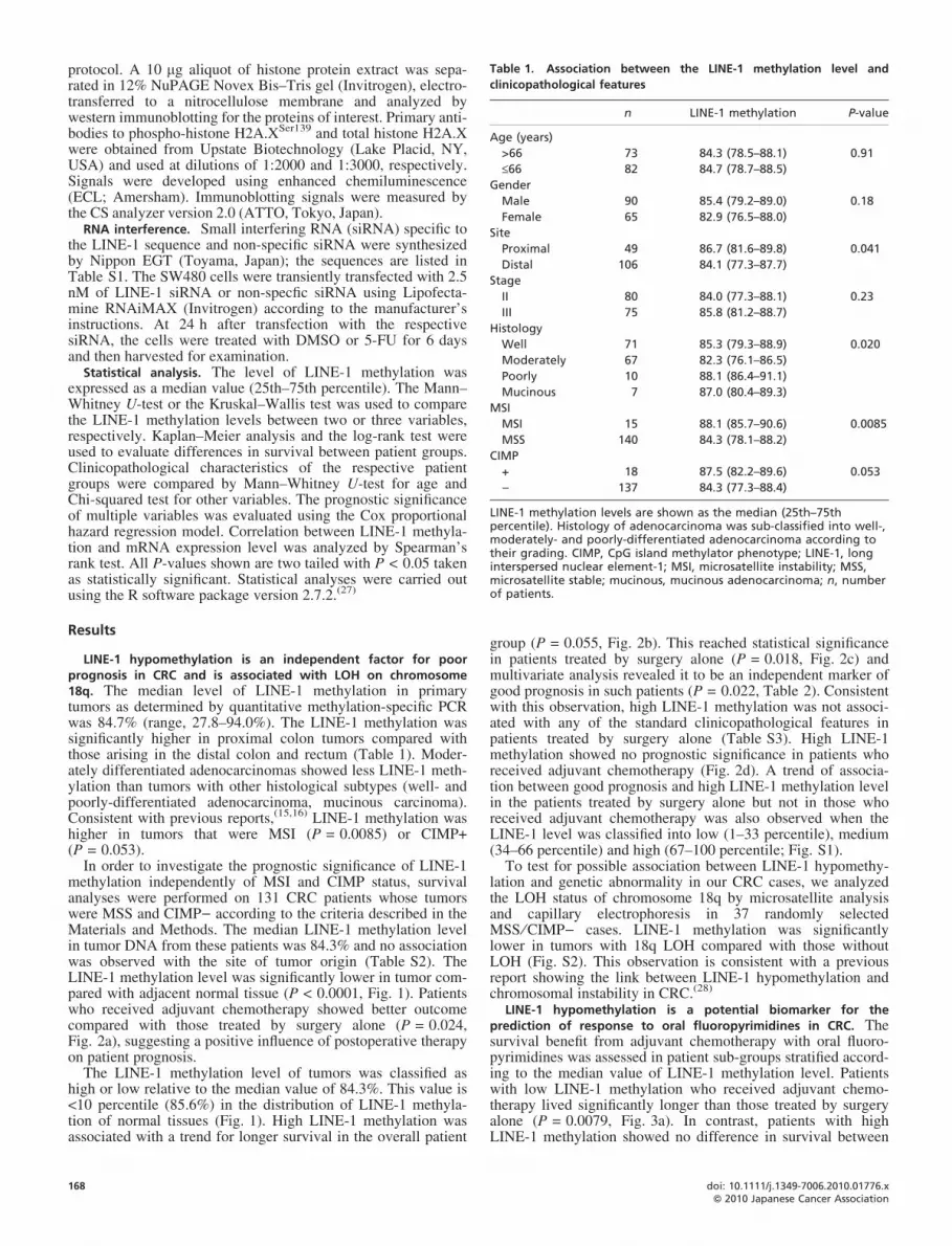
Table 1. Association between the LINE-1 methylation level and
clinicopathological features
n LINE-1 methylation P-value
Age (years)
>66 73 84.3 (78.5–88.1) 0.91
£66 82 84.7 (78.7–88.5)
Gender
Male 90 85.4 (79.2–89.0) 0.18
Female 65 82.9 (76.5–88.0)
Site
Proximal 49 86.7 (81.6–89.8) 0.041
Distal 106 84.1 (77.3–87.7)
Stage
II 80 84.0 (77.3–88.1) 0.23
III 75 85.8 (81.2–88.7)
Histology
Well 71 85.3 (79.3–88.9) 0.020
Moderately 67 82.3 (76.1–86.5)
Poorly 10 88.1 (86.4–91.1)
Mucinous 7 87.0 (80.4–89.3)
MSI
MSI 15 88.1 (85.7–90.6) 0.0085
MSS 140 84.3 (78.1–88.2)
CIMP
+ 18 87.5 (82.2–89.6) 0.053
) 137 84.3 (77.3–88.4)
LINE-1 methylation levels are shown as the median (25th–75thpercentile). Histology of adenocarcinoma was sub-classified into well-,moderately- and poorly-differentiated adenocarcinoma according totheir grading. CIMP, CpG island methylator phenotype; LINE-1, longinterspersed nuclear element-1; MSI, microsatellite instability; MSS,microsatellite stable; mucinous, mucinous adenocarcinoma; n, numberof patients.
protocol. A 10 lg aliquot of histone protein extract was sepa-rated in 12% NuPAGE Novex Bis–Tris gel (Invitrogen), electro-transferred to a nitrocellulose membrane and analyzed bywestern immunoblotting for the proteins of interest. Primary anti-bodies to phospho-histone H2A.XSer139 and total histone H2A.Xwere obtained from Upstate Biotechnology (Lake Placid, NY,USA) and used at dilutions of 1:2000 and 1:3000, respectively.Signals were developed using enhanced chemiluminescence(ECL; Amersham). Immunoblotting signals were measured bythe CS analyzer version 2.0 (ATTO, Tokyo, Japan).
RNA interference. Small interfering RNA (siRNA) specific tothe LINE-1 sequence and non-specific siRNA were synthesizedby Nippon EGT (Toyama, Japan); the sequences are listed inTable S1. The SW480 cells were transiently transfected with 2.5nM of LINE-1 siRNA or non-specfic siRNA using Lipofecta-mine RNAiMAX (Invitrogen) according to the manufacturer’sinstructions. At 24 h after transfection with the respectivesiRNA, the cells were treated with DMSO or 5-FU for 6 daysand then harvested for examination.
Statistical analysis. The level of LINE-1 methylation wasexpressed as a median value (25th–75th percentile). The Mann–Whitney U-test or the Kruskal–Wallis test was used to comparethe LINE-1 methylation levels between two or three variables,respectively. Kaplan–Meier analysis and the log-rank test wereused to evaluate differences in survival between patient groups.Clinicopathological characteristics of the respective patientgroups were compared by Mann–Whitney U-test for age andChi-squared test for other variables. The prognostic significanceof multiple variables was evaluated using the Cox proportionalhazard regression model. Correlation between LINE-1 methyla-tion and mRNA expression level was analyzed by Spearman’srank test. All P-values shown are two tailed with P < 0.05 takenas statistically significant. Statistical analyses were carried outusing the R software package version 2.7.2.(27)
Results
LINE-1 hypomethylation is an independent factor for poorprognosis in CRC and is associated with LOH on chromosome18q. The median level of LINE-1 methylation in primarytumors as determined by quantitative methylation-specific PCRwas 84.7% (range, 27.8–94.0%). The LINE-1 methylation wassignificantly higher in proximal colon tumors compared withthose arising in the distal colon and rectum (Table 1). Moder-ately differentiated adenocarcinomas showed less LINE-1 meth-ylation than tumors with other histological subtypes (well- andpoorly-differentiated adenocarcinoma, mucinous carcinoma).Consistent with previous reports,(15,16) LINE-1 methylation washigher in tumors that were MSI (P = 0.0085) or CIMP+(P = 0.053).
In order to investigate the prognostic significance of LINE-1methylation independently of MSI and CIMP status, survivalanalyses were performed on 131 CRC patients whose tumorswere MSS and CIMP) according to the criteria described in theMaterials and Methods. The median LINE-1 methylation levelin tumor DNA from these patients was 84.3% and no associationwas observed with the site of tumor origin (Table S2). TheLINE-1 methylation level was significantly lower in tumor com-pared with adjacent normal tissue (P < 0.0001, Fig. 1). Patientswho received adjuvant chemotherapy showed better outcomecompared with those treated by surgery alone (P = 0.024,Fig. 2a), suggesting a positive influence of postoperative therapyon patient prognosis.
The LINE-1 methylation level of tumors was classified ashigh or low relative to the median value of 84.3%. This value is<10 percentile (85.6%) in the distribution of LINE-1 methyla-tion of normal tissues (Fig. 1). High LINE-1 methylation wasassociated with a trend for longer survival in the overall patient
168
group (P = 0.055, Fig. 2b). This reached statistical significancein patients treated by surgery alone (P = 0.018, Fig. 2c) andmultivariate analysis revealed it to be an independent marker ofgood prognosis in such patients (P = 0.022, Table 2). Consistentwith this observation, high LINE-1 methylation was not associ-ated with any of the standard clinicopathological features inpatients treated by surgery alone (Table S3). High LINE-1methylation showed no prognostic significance in patients whoreceived adjuvant chemotherapy (Fig. 2d). A trend of associa-tion between good prognosis and high LINE-1 methylation levelin the patients treated by surgery alone but not in those whoreceived adjuvant chemotherapy was also observed when theLINE-1 level was classified into low (1–33 percentile), medium(34–66 percentile) and high (67–100 percentile; Fig. S1).
To test for possible association between LINE-1 hypomethy-lation and genetic abnormality in our CRC cases, we analyzedthe LOH status of chromosome 18q by microsatellite analysisand capillary electrophoresis in 37 randomly selectedMSS ⁄ CIMP) cases. LINE-1 methylation was significantlylower in tumors with 18q LOH compared with those withoutLOH (Fig. S2). This observation is consistent with a previousreport showing the link between LINE-1 hypomethylation andchromosomal instability in CRC.(28)
LINE-1 hypomethylation is a potential biomarker for theprediction of response to oral fluoropyrimidines in CRC. Thesurvival benefit from adjuvant chemotherapy with oral fluoro-pyrimidines was assessed in patient sub-groups stratified accord-ing to the median value of LINE-1 methylation level. Patientswith low LINE-1 methylation who received adjuvant chemo-therapy lived significantly longer than those treated by surgeryalone (P = 0.0079, Fig. 3a). In contrast, patients with highLINE-1 methylation showed no difference in survival between
doi: 10.1111/j.1349-7006.2010.01776.xªª 2010 Japanese Cancer Association

Fig. 1. Distribution of the long interspersed nuclear element-1 (LINE-1) methylation level in normal and tumor tissues. The LINE-1methylation is significantly lower in tumor compared with normaltissue (P < 0.0001).
(a)
(b)
(c)
(d)
those treated with or without adjuvant chemotherapy (P = 0.68,Fig. 3b). When the LINE-1 level was classified into low (1–33percentile), medium (34–66 percentile) and high (67–100 per-centile), the patients who received adjuvant chemotherapyshowed better survival in the low (P = 0.030) and medium(P = 0.068) but not in the high (P = 0.98) LINE-1 methylationgroup (Fig. S3). These results suggest that low levels of LINE-1methylation are predictive of response to oral fluoropyrimidines,but not high levels.
The clinicopathological features of the four patient subgroupsshown in Figure 3 are outlined in Table S4. In both the high andlow methylation groups, patients who received adjuvant chemo-therapy were significantly younger than those who did not.While this could potentially account for the survival differenceobserved between treatment groups, it does not explain whyonly patients with low LINE-1 methylation appeared to benefitfrom adjuvant chemotherapy (Fig. 3). Apart from age, no othersignificant differences in clinicopathological characteristics
Fig. 2. Kaplan–Meier survival analysis of patient groups stratifiedaccording to use of postoperative adjuvant chemotherapy (a) and ⁄ orlong interspersed nuclear element-1 (LINE-1) methylation (b–d). Theprognosis of LINE-1 low group (dashed line) and those of LINE-1 highgroup (solid line) were compared in the overall MSS ⁄ CIMP) cohort (b)and in patients treated by surgery alone (c) or with adjuvantchemotherapy (d). The log-rank test was used for each comparisonand P-values are shown. CIMP, CpG island methylator phenotype;MSS, microsatellite stable. tx, chemotherapy.
Kawakami et al. Cancer Sci | January 2011 | vol. 102 | no. 1 | 169ªª 2010 Japanese Cancer Association
Table 2. Multivariate analysis for the prognostic significance of
clinicopathological factors and LINE-1 methylation in MSS ⁄ CIMP- CRC
treated with surgery alone
Variables n Hazard ratio (95% CI) P-value
Age (years)
>66 40 1.00 0.10
£66 14 0.41 (0.14–1.19)
Gender
Male 29 1.00 0.46
Female 25 0.72 (0.30–1.72)
Site
Proximal 14 1.00 0.56
Distal 40 1.41 (0.44–4.55)
Stage
II 30 1.00 0.77
III 24 0.88 (0.37–2.11)
Histology
Others 28 1.00 0.10
Well 26 0.44 (0.16–1.19)
LINE-1 methylation
High 26 1.00 0.022
Low 28 3.02 (1.17–7.80)
Histology of adenocarcinoma was sub-classified into well-,moderately- and poorly-differentiated adenocarcinoma according totheir grading. Cases of moderately- (n = 26) and poorly-differentiatedadenocarcinoma (n = 1) and mucinous adenocarcinoma (n = 1) werecombined for the analysis. CI, confidence interval; CIMP, CpG islandmethylator phenotype; CRC, colorectal cancer; LINE-1, longinterspersed nuclear element-1; MSS, microsatellite stable; n, numberof patients.
(a)
(b)
Fig. 3. Kaplan–Meier survival analysis of patients stratified accordingto the use of postoperative adjuvant chemotherapy in patients withlow long interspersed nuclear element-1 (LINE-1) methylation (a) andin patients with high LINE-1 methylation (b). Solid lines indicatepatients treated with surgery alone and dashed lines indicate patientstreated with adjuvant chemotherapy. The log-rank test was used foreach comparison and P-values are shown. tx, chemotherapy.
were apparent between patients treated with or without adjuvantchemotherapy. Multivariate analysis revealed that adjuvant che-motherapy was an independent prognostic factor for better sur-vival in patients with low LINE-1 methylation, but not inpatients with high LINE-1 methylation (Table 3). These resultssuggest that LINE-1 hypomethylation is a potential biomarkerfor the response of CRC patients to oral fluoropyrimidines in theadjuvant setting.
5-FU increases LINE-1 mRNA expression and induces DNAdamage in CRC cells with LINE-1 hypomethylation. In vitro stud-ies were performed to investigate the possible mechanism bywhich LINE-1 hypomethylation was associated with a goodclinical response to oral fluoropyrimidines. Of the 13 CRC celllines screened for MSI and CIMP status, three (SW480, Caco-2,CaR-1) were found to be MSS ⁄ CIMP) (Fig. 4a). The relation-ship between LINE-1 methylation and expression of its full-length transcript was examined by northern blot analysis in theseMSS ⁄ CIMP) cell lines and in HT-29, HCT116 and SW48 cells(Fig. 4b). LINE-1 mRNA was detected as a single band ofapproximately 6 kb in size, representing its known full-lengthtranscript (Fig. 4b, upper panel). The LINE-1 methylation levelwas inversely correlated with the expression of its full-lengthmRNA (Fig. 4b, lower panel).
Since LINE-1 mRNA was highly expressed in CRC cellswith LINE-1 hypomethylation, we hypothesized that it mayplay a role in the therapeutic effect of 5-FU. To investigate thishypothesis, we looked for changes in the expression of LINE-1mRNA in CRC cells following treatment with 5-FU. The 5-FUtreatment had the effect of increasing LINE-1 mRNA expres-sion in all three MSS ⁄ CIMP) cell lines, although the baselineexpression level of the LINE-1 mRNA was less in CaR-1 cellsthan SW480 and Caco-2 cells (Fig. 4c, upper panels). TheLINE-1 sequence contains an open reading frame that encodesa protein with nuclease activity,(29) suggesting that increasedLINE-1 expression may be responsible for the DNA double
170
strand breaks (DSB) observed in cells following treatment with5-FU. To test this possibility, the phosphorylation of histoneH2A.X at serine 139 (p-histone H2A.XSer139) was analyzed bywestern immunoblotting. Phospho-histone H2A.XSer139 is awell-established marker of DSB(30) and have been used toassess the effect of 5-FU.(31,32) In SW480 and Caco-2 cells, thelevel of p-histone H2A.XSer139 increased in parallel with theupregulation of LINE-1 mRNA after 5-FU treatment (Fig. 4c,lower panels). In contrast, the level of p-histone H2A.XSer139
showed no change in CaR-1 cells following 5-FU treatment.The CaR-1 cells express less LINE-1 mRNA and have moreLINE-1 methylation compared with SW480 and Caco-2 cells(Fig. 4b,c).
To further investigate whether increased LINE-1 expressionis responsible for DNA damage following 5-FU treatment, RNAinterference was used to deplete LINE-1 prior to treatment ofthe SW480 cells. When the cells were pretreated with non-spe-cific control siRNA, p-histone H2A.XSer139 expression was sig-nificantly induced following 5-FU treatment. However, whencells were pretreated with LINE-1-specific siRNA this inductionof p-histone H2A.XSer139 by 5-FU was suppressed (Fig. 4d).These results suggest that in CRC cells with low LINE-1methylation, augmented high expression of LINE-1 mRNA by5-FU subsequently leads to an increase in DSB and eventuallyto cell death. 5-FU may be ineffective in cells with high LINE-1
doi: 10.1111/j.1349-7006.2010.01776.xªª 2010 Japanese Cancer Association
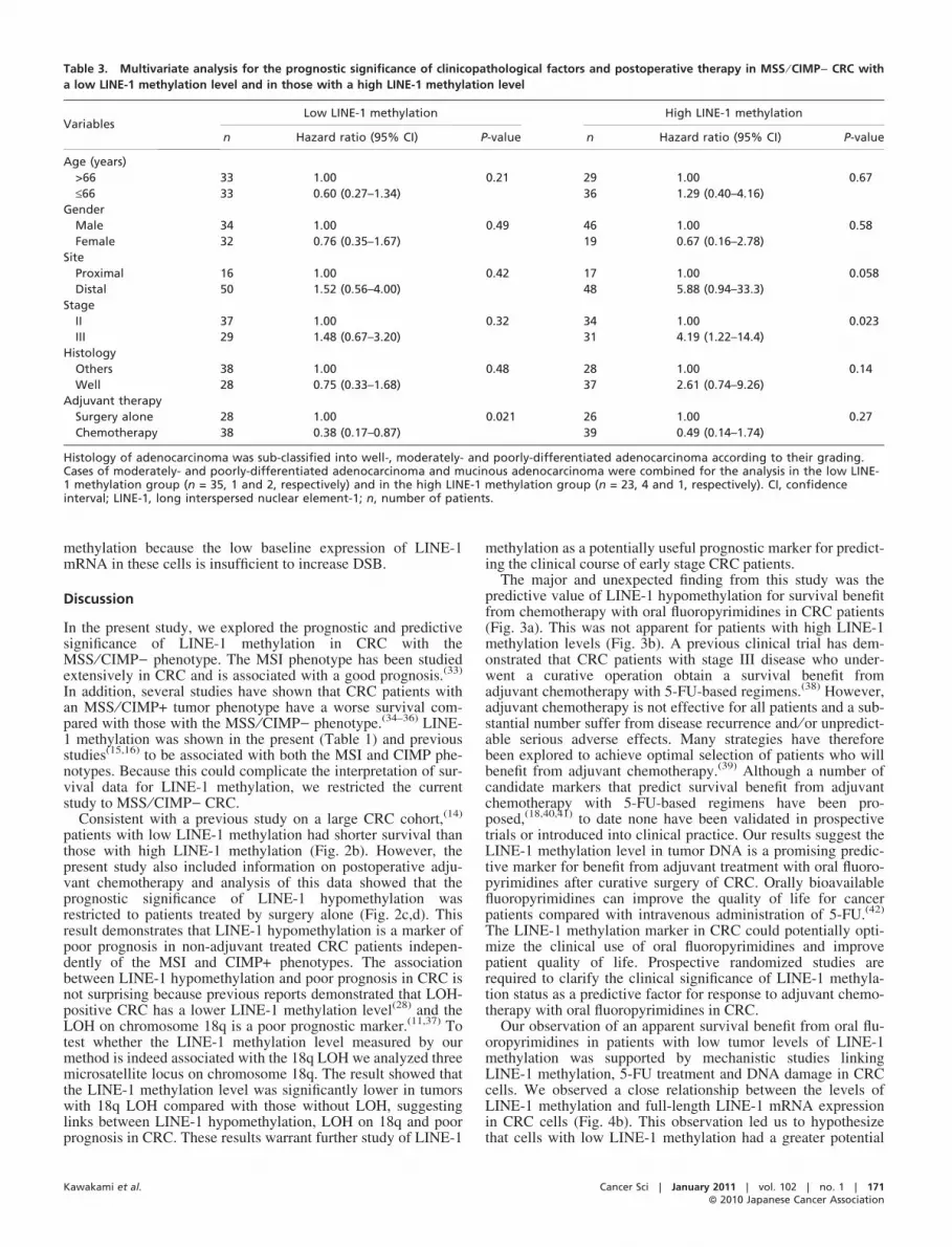
Table 3. Multivariate analysis for the prognostic significance of clinicopathological factors and postoperative therapy in MSS ⁄ CIMP) CRC with
a low LINE-1 methylation level and in those with a high LINE-1 methylation level
VariablesLow LINE-1 methylation High LINE-1 methylation
n Hazard ratio (95% CI) P-value n Hazard ratio (95% CI) P-value
Age (years)
>66 33 1.00 0.21 29 1.00 0.67
£66 33 0.60 (0.27–1.34) 36 1.29 (0.40–4.16)
Gender
Male 34 1.00 0.49 46 1.00 0.58
Female 32 0.76 (0.35–1.67) 19 0.67 (0.16–2.78)
Site
Proximal 16 1.00 0.42 17 1.00 0.058
Distal 50 1.52 (0.56–4.00) 48 5.88 (0.94–33.3)
Stage
II 37 1.00 0.32 34 1.00 0.023
III 29 1.48 (0.67–3.20) 31 4.19 (1.22–14.4)
Histology
Others 38 1.00 0.48 28 1.00 0.14
Well 28 0.75 (0.33–1.68) 37 2.61 (0.74–9.26)
Adjuvant therapy
Surgery alone 28 1.00 0.021 26 1.00 0.27
Chemotherapy 38 0.38 (0.17–0.87) 39 0.49 (0.14–1.74)
Histology of adenocarcinoma was sub-classified into well-, moderately- and poorly-differentiated adenocarcinoma according to their grading.Cases of moderately- and poorly-differentiated adenocarcinoma and mucinous adenocarcinoma were combined for the analysis in the low LINE-1 methylation group (n = 35, 1 and 2, respectively) and in the high LINE-1 methylation group (n = 23, 4 and 1, respectively). CI, confidenceinterval; LINE-1, long interspersed nuclear element-1; n, number of patients.
methylation because the low baseline expression of LINE-1mRNA in these cells is insufficient to increase DSB.
Discussion
In the present study, we explored the prognostic and predictivesignificance of LINE-1 methylation in CRC with theMSS ⁄ CIMP) phenotype. The MSI phenotype has been studiedextensively in CRC and is associated with a good prognosis.(33)
In addition, several studies have shown that CRC patients withan MSS ⁄ CIMP+ tumor phenotype have a worse survival com-pared with those with the MSS ⁄ CIMP) phenotype.(34–36) LINE-1 methylation was shown in the present (Table 1) and previousstudies(15,16) to be associated with both the MSI and CIMP phe-notypes. Because this could complicate the interpretation of sur-vival data for LINE-1 methylation, we restricted the currentstudy to MSS ⁄ CIMP) CRC.
Consistent with a previous study on a large CRC cohort,(14)
patients with low LINE-1 methylation had shorter survival thanthose with high LINE-1 methylation (Fig. 2b). However, thepresent study also included information on postoperative adju-vant chemotherapy and analysis of this data showed that theprognostic significance of LINE-1 hypomethylation wasrestricted to patients treated by surgery alone (Fig. 2c,d). Thisresult demonstrates that LINE-1 hypomethylation is a marker ofpoor prognosis in non-adjuvant treated CRC patients indepen-dently of the MSI and CIMP+ phenotypes. The associationbetween LINE-1 hypomethylation and poor prognosis in CRC isnot surprising because previous reports demonstrated that LOH-positive CRC has a lower LINE-1 methylation level(28) and theLOH on chromosome 18q is a poor prognostic marker.(11,37) Totest whether the LINE-1 methylation level measured by ourmethod is indeed associated with the 18q LOH we analyzed threemicrosatellite locus on chromosome 18q. The result showed thatthe LINE-1 methylation level was significantly lower in tumorswith 18q LOH compared with those without LOH, suggestinglinks between LINE-1 hypomethylation, LOH on 18q and poorprognosis in CRC. These results warrant further study of LINE-1
Kawakami et al.
methylation as a potentially useful prognostic marker for predict-ing the clinical course of early stage CRC patients.
The major and unexpected finding from this study was thepredictive value of LINE-1 hypomethylation for survival benefitfrom chemotherapy with oral fluoropyrimidines in CRC patients(Fig. 3a). This was not apparent for patients with high LINE-1methylation levels (Fig. 3b). A previous clinical trial has dem-onstrated that CRC patients with stage III disease who under-went a curative operation obtain a survival benefit fromadjuvant chemotherapy with 5-FU-based regimens.(38) However,adjuvant chemotherapy is not effective for all patients and a sub-stantial number suffer from disease recurrence and ⁄ or unpredict-able serious adverse effects. Many strategies have thereforebeen explored to achieve optimal selection of patients who willbenefit from adjuvant chemotherapy.(39) Although a number ofcandidate markers that predict survival benefit from adjuvantchemotherapy with 5-FU-based regimens have been pro-posed,(18,40,41) to date none have been validated in prospectivetrials or introduced into clinical practice. Our results suggest theLINE-1 methylation level in tumor DNA is a promising predic-tive marker for benefit from adjuvant treatment with oral fluoro-pyrimidines after curative surgery of CRC. Orally bioavailablefluoropyrimidines can improve the quality of life for cancerpatients compared with intravenous administration of 5-FU.(42)
The LINE-1 methylation marker in CRC could potentially opti-mize the clinical use of oral fluoropyrimidines and improvepatient quality of life. Prospective randomized studies arerequired to clarify the clinical significance of LINE-1 methyla-tion status as a predictive factor for response to adjuvant chemo-therapy with oral fluoropyrimidines in CRC.
Our observation of an apparent survival benefit from oral flu-oropyrimidines in patients with low tumor levels of LINE-1methylation was supported by mechanistic studies linkingLINE-1 methylation, 5-FU treatment and DNA damage in CRCcells. We observed a close relationship between the levels ofLINE-1 methylation and full-length LINE-1 mRNA expressionin CRC cells (Fig. 4b). This observation led us to hypothesizethat cells with low LINE-1 methylation had a greater potential
Cancer Sci | January 2011 | vol. 102 | no. 1 | 171ªª 2010 Japanese Cancer Association

(a)
(c)
(b)
(d)
Fig. 4. In vitro mechanistic studies linking long interspersed nuclear element-1 (LINE-1) methylation, 5-fluorouracil (5-FU) treatment and DNAdamage in colorectal cancer (CRC) cells. (a) Results for 13 CRC cell lines screened for microsatellite instability (MSI), CpG island methylatorphenotype (CIMP) and LINE-1 methylation level. The methylation status of five CIMP-defining CpG island markers was determined by MethyLightassay in which a reading of the percentage of methylated reference (PMR) ‡10 was defined as hypermethylated (black boxes). White boxesindicate PMR <10. (b) The relationship between the LINE-1 methylation level and expression of LINE-1 mRNA. Northern blot analysis of full-length LINE-1 mRNA and b-actin expression is shown in the upper panel. The LINE-1 methylation level correlated inversely with the relativeamount of LINE-1 mRNA quantified by densitometry and normalized to b-actin with statistical significance (Spearman’s q = )0.94, P = 0.017,lower panel). (c) Microsatellite stable (MSS) ⁄ CIMP) cells were treated with the indicated 5-FU concentration for 6 days followed by cellharvesting to isolate RNA and histone protein. LINE-1 and b-actin mRNA were detected by northern blot analysis and are shown in the upperpanel with numbers reflecting the relative amount of LINE-1 mRNA quantified by densitometry and normalized to b-actin. Phospho-histoneH2A.XSer139 (p-H2A.X) and total histone H2A.X (H2A.X) proteins were detected by western blot analysis and are shown in the lower panel withnumbers reflecting the relative amount of p-H2A.X protein normalized to H2A.X. (d) SW480 cells were transfected with either non-specific (NCsiRNA) or LINE-1-specific siRNA (LINE-1 siRNA) followed by treatment with either DMSO or 5-FU (2 lM) for 6 days. The expression of p-H2A.Xand H2A.X was quantified using densitometry and the relative expression of p-H2A.X to H2A.X (p-H2A.X ⁄ H2A.X) is shown in the bar graph atthe bottom of the panel. The SW480 cells treated with non-specific siRNA and DMSO was set as 1. Values shown are the mean and the errorbars represent standard deviation from three separate experiments. The asterisk indicates statistical significance (P < 0.05) for comparison ofDMSO and 5-FU-treated cells in the same experimental conditions for transfected siRNA.
to undergo DSB following 5-FU treatment because the full-length LINE-1 mRNA encodes for a protein with nuclease activ-ity.(29) Our in vitro study demonstrated that 5-FU treatmentincreases the expression of LINE-1 mRNA in MSS ⁄ CIMP)CRC cells (Fig. 4c). This was paralleled by increased DNAdamage (assessed by p-histone H2A.XSer139 expression) inSW480 and Caco-2 cells with low LINE-1 methylation (54%and 57%), but not in CaR-1 cells with high LINE-1 methylation(72%). Therefore, the increase in LINE-1 expression may becausally linked to 5-FU-induced DNA damage in cases wheretumor cells have low LINE-1 methylation and express highbaseline levels of LINE-1 mRNA (in case of SW480 and Caco-2). A causal link between increased LINE-1 mRNA and DNAdamage induced by 5-FU treatment was further suggested byknockdown experiments of LINE-1 expression (Fig. 4d). Theseresults could explain why tumors with LINE-1 hypomethylationappear to be responsive to adjuvant chemotherapy using oral flu-oropyrimidines. On the other hand, we postulate that 5-FU may
172
be ineffective in CRC cells with high LINE-1 methylationbecause their low baseline expression of LINE-1 mRNA isinsufficient to induce DSB following 5-FU treatment (in case ofCaR-1). This mechanism could explain our observation thatCRC patients whose tumors show high LINE-1 methylationrespond poorly to oral fluoropyrimidines (Fig. 3b). Our in vitroresults suggest that further studies on the possible links betweenLINE-1 hypomethylation, LINE-1 expression, nuclease activity,DSB formation and sensitivity to 5-FU may lead to novel strate-gies for improving the cytotoxic effect of 5-FU.
In conclusion, we found that LINE-1 hypomethylation inMSS ⁄ CIMP) CRC had predictive value for benefit from adju-vant chemotherapy with oral fluoropyrimidines. Our in vitrostudies have suggested a possible underlying mechanism for thelink between LINE-1 hypomethylation in CRC cells and theirsusceptibility to fluoropyrimidines. These observations couldhave important implications for the future development of per-sonalized chemotherapy and novel 5-FU-based regimens.
doi: 10.1111/j.1349-7006.2010.01776.xªª 2010 Japanese Cancer Association
Acknowledgments
The authors thank Dr. Barry Iacopetta (School of Surgery, University ofWestern Australia) for critical reading of the manuscript and valuablesuggestions. This work was supported in part by Grants-in-Aid for Scien-tific Research from the Japan Society for the Promotion of Science(20390353).
Kawakami et al.
Disclosure Statement
The authors have no conflict of interest.
References
1 Jones PA, Baylin SB. The fundamental role of epigenetic events in cancer.Nat Rev Genet 2002; 3: 415–28.
2 Shen L, Catalano PJ, Benson AB III, O’Dwyer P, Hamilton SR, Issa JP.Association between DNA methylation and shortened survival in patients withadvanced colorectal cancer treated with 5-fluorouracil based chemotherapy.Clin Cancer Res 2007; 13: 6093–8.
3 Joensuu EI, Abdel-Rahman WM, Ollikainen M, Ruosaari S, Knuutila S,Peltomaki P. Epigenetic signatures of familial cancer are characteristic oftumor type and family category. Cancer Res 2008; 68: 4597–605.
4 Lander ES, Linton LM, Birren B et al. Initial sequencing and analysis of thehuman genome. Nature 2001; 409: 860–921.
5 Deininger PL, Moran JV, Batzer MA, Kazazian HH Jr. Mobile elements andmammalian genome evolution. Curr Opin Genet Dev 2003; 13: 651–8.
6 Kazazian HH Jr. Mobile elements: drivers of genome evolution. Science 2004;303: 1626–32.
7 Sassaman DM, Dombroski BA, Moran JV et al. Many human L1 elements arecapable of retrotransposition. Nat Genet 1997; 16: 37–43.
8 Brouha B, Schustak J, Badge RM et al. Hot L1s account for the bulk ofretrotransposition in the human population. Proc Natl Acad Sci U S A 2003;100: 5280–5.
9 Yang AS, Estecio MR, Doshi K, Kondo Y, Tajara EH, Issa JP. A simplemethod for estimating global DNA methylation using bisulfite PCR ofrepetitive DNA elements. Nucleic Acids Res 2004; 32: e38.
10 Rodriguez J, Frigola J, Vendrell E et al. Chromosomal instability correlateswith genome-wide DNA demethylation in human primary colorectal cancers.Cancer Res 2006; 66: 8462–9468.
11 Watanabe T, Wu TT, Catalano PJ et al. Molecular predictors of survival afteradjuvant chemotherapy for colon cancer. N Engl J Med 2001; 344: 1196–206.
12 Sheffer M, Bacolod MD, Zuk O et al. Association of survival and diseaseprogression with chromosomal instability: a genomic exploration of colorectalcancer. Proc Natl Acad Sci U S A 2009; 106: 7131–6.
13 Kawakami K, Jin M, Saito K et al. Methylation level of LINE-1 repeats as aprognostic factor for the patients with primary colorectal cancer [abstract991]. Proc Am Assoc Cancer Res 2008; 49: 232.
14 Ogino S, Nosho K, Kirkner GJ et al. A cohort study of tumoral LINE-1hypomethylation and prognosis in colon cancer. J Natl Cancer Inst 2008; 100:1734–8.
15 Estecio MR, Gharibyan V, Shen L et al. LINE-1 hypomethylation in cancer ishighly variable and inversely correlated with microsatellite instability. PLoSONE 2007; 2: e399.
16 Ogino S, Kawasaki T, Nosho K et al. LINE-1 hypomethylation is inverselyassociated with microsatellite instability and CpG island methylatorphenotype in colorectal cancer. Int J Cancer 2008; 122: 2767–73.
17 Jover R, Zapater P, Castells A et al. Mismatch repair status in the predictionof benefit from adjuvant fluorouracil chemotherapy in colorectal cancer. Gut2006; 55: 848–55.
18 Van Rijnsoever M, Elsaleh H, Joseph D, McCaul K, Iacopetta B. CpG islandmethylator phenotype is an independent predictor of survival benefit from 5-fluorouracil in stage III colorectal cancer. Clin Cancer Res 2003; 9: 2898–903.
19 Sargent DJ, Marsoni S, Monges G et al. Defective mismatch repair as apredictive marker for lack of efficacy of fluorouracil-based adjuvant therapy incolon cancer. J Clin Oncol 2010; 28: 3219–26.
20 Sobin LH, Wittekind Ch, eds. International Union Against Cancer (UICC):‘‘TNM Classification of Malignant Tumors’’, 6th edn. New York: Wiley, 2002.
21 Kawakami K, Brabender J, Lord RV et al. Hypermethylated APC DNA inplasma and prognosis of patients with esophageal adenocarcinoma. J NatlCancer Inst 2000; 92: 1805–11.
22 Iacopetta B, Grieu F, Phillips M et al. Methylation levels of LINE-1 repeatsand CpG island loci are inversely related in normal colonic mucosa. CancerSci 2007; 98: 1454–60.
23 Weisenberger DJ, Siegmund KD, Campan M et al. CpG island methylatorphenotype underlies sporadic microsatellite instability and is tightly associatedwith BRAF mutation in colorectal cancer. Nat Genet 2006; 38: 787–93.
24 Goel A, Nagasaka T, Hamelin R, Boland CR. An optimized pentaplex PCRfor detecting DNA mismatch repair-deficient colorectal cancers. PLoS ONE2010; 5: e9393.
25 Buhard O, Cattaneo F, Wong YF et al. Multipopulation analysis ofpolymorphisms in five mononucleotide repeats used to determine themicrosatellite instability status of human tumors. J Clin Oncol 2006; 24: 241–51.
26 Saito K, Kawakami K, Matsumoto I, Oda M, Watanabe G, Minamoto T. Longinterspersed nuclear element 1 hypomethylation is a marker of poor prognosisin stage IA non-small cell lung cancer. Clin Cancer Res 2010; 16: 2418–26.
27 R Development Core Team. R: A Language and Environment for StatisticalComputing. Vienna ⁄ Austria: R Foundation for Statistical Computing, 2008,ISBN 3-900051-07-0.
28 Matsuzaki K, Deng G, Tanaka H, Kakar S, Miura S, Kim YS. Therelationship between global methylation level, loss of heterozygosity, andmicrosatellite instability in sporadic colorectal cancer. Clin Cancer Res2005; 11: 8564–9.
29 Feng Q, Moran JV, Kazazian HH Jr, Boeke JD. Human L1 retrotransposonencodes a conserved endonuclease required for retrotransposition. Cell 1996;87: 905–16.
30 Bonner WM, Redon CE, Dickey JS et al. cH2AX and cancer. Nat Rev Cancer2008; 8: 957–67.
31 Xiao Z, Xue J, Sowin TJ, Rosenberg SH, Zhang H. A novel mechanism ofcheckpoint abrogation conferred by Chk1 downregulation. Oncogene 2005;24: 1403–11.
32 Kunz C, Focke F, Saito Y et al. Base excision by thymine DNAglycosylase mediates DNA-directed cytotoxicity of 5-fluorouracil. PLoS Biol2009; 7: e91.
33 Boland CR, Goel A. Microsatellite instability in colorectal cancer.Gastroenterology 2010; 138: 2073–87.
34 Barault L, Charon-Barra C, Jooste V et al. Hypermethylator phenotype insporadic colon cancer: study on a population-based series of 582 cases.Cancer Res 2008; 68: 8541–6.
35 Kim JH, Shin SH, Kwon HJ, Cho NY, Kang GH. Prognostic implications ofCpG island hypermethylator phenotype in colorectal cancers. Virchows Arch2009; 455: 485–94.
36 Dahlin AM, Palmqvist R, Henriksson ML et al. The role of the CpG islandmethylator phenotype in colorectal cancer prognosis depends on microsatelliteinstability screening status. Clin Cancer Res 2010; 16: 1845–55.
37 Ogunbiyi OA, Goodfellow PJ, Herfarth K et al. Confirmation thatchromosome 18q allelic loss in colon cancer is a prognostic indicator. J ClinOncol 1998; 16: 427–33.
38 Moertel CG, Fleming TR, Macdonald JS et al. Fluorouracil plus levamisole aseffective adjuvant therapy after resection of stage III colon carcinoma: a finalreport. Ann Intern Med 1995; 122: 321–6.
39 van’t Veer LJ, Bernards R. Enabling personalized cancer medicine throughanalysis of gene-expression patterns. Nature 2008; 452: 564–70.
40 Salonga D, Danenberg KD, Johnson M et al. Colorectal tumors responding to5-fluorouracil have low gene expression levels of dihydropyrimidinedehydrogenase, thymidylate synthase, and thymidine phosphorylase. ClinCancer Res 2000; 6: 1322–7.
41 Kawakami K, Watanabe G. Identification and functional analysis of singlenucleotide polymorphism in the tandem repeat sequence of thymidylatesynthase gene. Cancer Res 2003; 63: 6004–7.
42 Watanabe T, Sano M, Takashima S et al. Oral uracil and tegafur comparedwith classic cyclophosphamide, methotrexate, fluorouracil as postoperativechemotherapy in patients with node-negative, high-risk breast cancer:National Surgical Adjuvant Study for Breast Cancer 01 Trial. J Clin Oncol2009; 27: 1368–74.
Cancer Sci | January 2011 | vol. 102 | no. 1 | 173ªª 2010 Japanese Cancer Association
Supporting Information
Additional Supporting Information may be found in the online version of this article:
Fig. S1. Kaplan–Meier survival analysis of patient groups stratified according to LINE-1 methylation.
Fig. S2. Association between LINE-1 methylation and LOH on chromosome 18q in CRC.
Fig. S3. Kaplan–Meier survival analysis of the patients stratified according to the use of post-operative adjuvant chemotherapy.
Table S1. Primer and siRNA sequences used in the present study.
Table S2. Association between the LINE-1 methylation level and clinicopathological features in MSS ⁄ CIMP) patients.
Table S3. Patient characteristics according to LINE-1 methylation in the subgroup treated with surgery alone.
Table S4. Patient characteristics in the subgroups analyzed for associations between LINE-1 methylation, prognosis and benefit from adjuvant che-motherapy.
Please note: Wiley-Blackwell are not responsible for the content or functionality of any supporting materials supplied by the authors. Any queries(other than missing material) should be directed to the corresponding author for the article.
174 doi: 10.1111/j.1349-7006.2010.01776.xªª 2010 Japanese Cancer Association