tejaswi chavan ph.d. thesis.pdf
TRANSCRIPT
MODULATION OF PATHOPHYSIOLOGY OF
HEPATOTOXICITY WITH NUTRITIONAL AND HERBAL
INTERVENTIONS IN ANIMAL MODELS
THESIS SUBMITTED TO
M
RS
. TE
JAS
WI C
HA
ND
RA
KA
NT
CH
AV
AN
BHARATI VIDYAPEETH DEEMED UNIVERSITY, PUNE
FOR THE AWARD
OF
DOCTOR OF PHTLOSOPHY (Ph.D.)
IN
BIOTECHNOLOGY
UNDER
FACULTY OF SCIENCE
BY
MRS. TEJASWI CHANDRAKANT CHAVAN
UNDER THE GUIDANCE
OF
DR. ANIKET A. KUVALEKAR
BHARATI VIDYAPEETH DEEMED UNIVERSITY
INTERACTIVE RESEARCH SCHOOL FOR HEALTH AFFAIRS (IRSHA)
PUNE, MAHARASHTRA, INDIA
September 2016
Ph.D. THESIS
September 2016
MODULATION OF PATHOPHYSIOLOGY OF
HEPATOTOXICITY WITH NUTRITIONAL AND HERBAL
INTERVENTIONS IN ANIMAL MODELS
Thesis submitted to
Bharati Vidyapeeth Deemed University, Pune
for award of
DOCTOR OF PHILOSOPHY (Ph.D.)
in Biotechnology
Under
Faculty of Science
By
Mrs. Tejaswi Chandrakant Chavan (M.Sc.)
Under the Guidance of
Dr. Aniket A. Kuvalekar
Bharati Vidyapeeth Deemed University,
Interactive Research School for Health Affairs (Irsha),
Pune, Maharashtra, India
September 2016
Certificate
This is to certify that the work incorporated in the thesis entitled “Modulation of
Pathophysiology of Hepatotoxicity with Nutritional and Herbal Interventions in
Animal Models” for the degree of ‘Doctor of Philosophy’ in the subject of
Biotechnology under the faculty of Science has been carried out by Mrs. Tejaswi
Chandrakant Chavan in the Diabetes Laboratory, Interactive Research School for
Health Affairs at Bharati Vidyapeeth Deemed University, Pune, during the period
from November 2012 to July 2016, under the guidance of Dr. Aniket A. Kuvalekar.
Place: Pune Dr. A.C.Mishra
Date: Director
Certification of Guide
This is to certify that the work incorporated in the thesis entitled “Modulation of
Pathophysiology of Hepatotoxicity with Nutritional and Herbal Interventions in
Animal Models” submitted by Mrs. Tejaswi Chandrakant Chavan for the degree
of ‘Doctor of Philosophy’ in the subject of Biotechnology under the faculty of
Science has been carried out in the Diabetes Laboratory, Interactive Research School
for Health Affairs, Bharati Vidyapeeth Deemed University, Pune, during the period
from November 2012 to July 2016, under my direct supervision/guidance.
Place: Pune Dr. Aniket A. Kuvalekar
Date: Research Guide
Declaration by the Candidate
I hereby declare that the thesis entitled “Modulation of Pathophysiology of
Hepatotoxicity with Nutritional and Herbal Interventions in Animal Models”
submitted by me to the Bharati Vidyapeeth University, Pune for the degree of Doctor
of Philosophy (PhD) in Biotechnology under the faculty of Science is original piece
of work carried out by me under the supervision of Dr. Aniket A. Kuvalekar. I
further declare that it has not been submitted to this or any other university or
institution for the award of any degree or diploma.
I also confirm that all the material which I have borrowed from other sources and
incorporated in this thesis is duly acknowledged. If any material is not duly
acknowledged and found incorporated in this thesis, it is entirely my responsibility. I
am fully aware of the implications of any such act which might have been committed
by me advertently or inadvertently.
Place: Mrs. Tejaswi Chandrakant Chavan
Date: Research Student

Acknowledgements
“Research is to see what everybody has seen but think what nobody has
thought.” Research involves collaborative efforts by team of persons striving to
conquer new horizons in the different fields of sciences. This study would not have
been completed without the encouragement, guidance and co-operation of my
teachers, parents, friends, well-wishers and relatives. Getting such help, I feel, is
comparable to our humanity. Almighty has given us a wonderful body in which
organs and systems work together, synchronously to lead a healthy life. Similarly, my
thesis work was like a human body where many people helped me in some or the
other way for its successful completion. I would like to take this opportunity to
express my deep gratitude towards all of them.
I wish to express my sincere thanks and profound respect towards my guide
Dr. Aniket Kuvalekar (Diabetes Laboratory, Bharati Vidyapeeth University,
IRSHA, Pune) with a deep sense of my gratitude for his guidance, invaluable advice,
constant support, intellectual supervision and professional expertise he has bestowed
upon me for the timely completion of my research work. I would also like to thank
him for giving me freedom of thoughts and guiding me on the right path with the
trust. I am proud to have him as my guide.
I am thankful to Dr. Akhilesh Mishra (Director, BVDU-IRSHA, Pune), Dr.
Prabhakar Ranjekar (Former Director, BVDU-IRSHA, Pune) and Dr. Sahebrao
Mahadik (Professor, Medical college of Georgia, Augusta, U.S.A.) for their support
and invaluable guidance during my entire research work. I would like to thank Dr.
Digambar Mokat (Assistant Professor, Savitribai Phule Pune University) for
providing plant material. I am also thankful to the Medicinal Plants
Conservation Centre, Pune (MPCC) for identification and authentication of plant
material. I would like to thank Dr. Omkar Kulkarni for providing ayurvedic
expertise for preparation of satwa for my research work. I am also thankful to Mr.
Ravi Mulik for his help in executing animal experiments.
My sincere thanks also go to Dr. Vijaya Pandit (Head, Department of
Pharmacology, Bharati Vidyapeeth Medical College, Pune) for reviewing animal
experiment protocols and Dr. Manjiri Karandikar (Associate Professor, Department
of Pathology, Bharati Vidyapeeth Medical College, Pune) for her support in
histopathological study in my research work.
A special note of thanks to my friend Mr. Abhijit Ghadge for his help and
encouragement throughout the tenure of my thesis.
It will be very unfair if I forget my labmates Mr. Suresh Khadke and Mrs.
Shubhangi Harke for giving their helping hands whenever and wherever required
and maintaining lively atmosphere in lab. Their invaluable interactions always
strengthened me emotionally and encouraged me for my work. I am grateful to my
juniors Mrs. Amruta Mandhare, Mr. Suyash Pawar, Miss. Amruta Kakde for
their cordial support throughout my research work.
I am fortunate to have parents like mine. My sincere thanks and appreciation
to my mother Mrs. Kamal Patil, my father Mr. Bhimarao Patil, my mother-in-law
Mrs. Sindhu Chavan and father-in-law Mr. Shivaji Chavan for their unconditional
true love, affection, blessing, sacrifice and support which paved my path in life and
the encouragement I needed to succeed.

I have no words to show my hearty gratitude towards my lovely son Atharva
and husband Mr. Chandrakant who are my inspiration, support and true love.
My apologies as it is not possible to mention everyone’s name here but their
blessings helped for successful completion of my work.
Last but not the least, I wish to express my gratitude towards “God almighty”, who
gave me the strength and courage to fulfill my dream and bestowed his best
blessings upon me always.
Thankful I ever remain.

List of Abbreviations
Abbreviations Full Form
ALP - Alkaline phosphate
ANOVA - Analysis of variance
APAP - Acetaminophen
b.w. - Body weight
BIL - Bilirubin
BSA - Bovine serum albumin
CAT - Catalase
CPCSEA - Committee for the purpose of control and
supervision of experiments on animals
cDNA - Complementary DNA
CHO - Total cholesterol
D.P.X. - Diphenyl phthalate xylene
DEPC - Diethylpyrocarbonate
DMSO - Dimethyl sulfoxide
dNTPs - Deoxynucleotides
DTNB - Dithionitrobenzoic acid
DTT - Dithiothreitol
EDTA - Ethylene diamine tetra acetic acid
FABP - Fatty acid binding protein
GAPDH - Glyceraldehyde 3-phosphate dehydrogenase
GSH - Reduced glutathione
HCl - Hydrochloric acid
HDL - High density lipoprotein
KCl - Potassium chloride
LDL - Low density lipoprotein
MDA - Malondialdehyde
NaOH - Sodium hydroxide
NFkβ - Nuclear factor kappaβ
PBS - Phosphate buffered saline
PPARγ - Peroxisome proliferator-activated receptor
gamma
RNA - Ribonucleic acid
RNaseOUT - Recombinant ribonuclease inhibitar
RT - Reverse transcriptase
SD - Standard deviation
SE - Standard error
SGOT - Serum glutamic oxaloacetic transaminase
SGPT - Serum glutamic pyruvic transaminase
SOD - Superoxide dismutase
SREBP - Sterol regulatory element binding protein
TGL - Triglycerides
TNFα - Tumor necrosis factor alpha
INDEX
Sr.No Particular Page
No
Chapter 1 Introduction
1.1 Liver 1
1.2 Hepatotoxicity 2
1.3 Prevalence of Liver Disease 3
1.3.1 International Scenario 3
1.3.2 National Scenario 4
1.4 Risk Factor for Liver Disease 5
1.4.1 Age and Gender 5
1.4.2 Genetic Factors 6
1.4.3 Obesity 6
1.4.4 Arsenic 6
1.4.5 Aflatoxins 6
1.4.6 Dietary Supplements 6
1.4.7 Industrial Toxins 7
1.4.8 Diabetes 7
1.4.9 Alcoholism 7
1.4.10 Long Term Use of Certain Medicinal Drugs 7
1.5 Biochemical Markers 8
1.5.1 Liver Function Tests 8
1.5.2 Total Protein 9
1.5.3 Lipid Profile 9
1.5.4 Oxidative Stress Marker 9
1.6 Management of Hepatic Diseases 10
1.6.1 Surgical Procedures 10
1.6.2 Hepatoprotective Agents 11
1.6.2.1 Silymarin 11
1.6.3 Herbal Formulations 11
1.6.4 Medicinal Plants 12
1.6.4.1 Tinospora forms 13
(a) Tinospora cordifolia 14
Sr. No Particular Page
No
(b) Tinospora sinensis 15
(c) Neem-giloe 16
1.6.5 Satwa 16
1.6.6 Nutritional Supplements 18
1.6.6.1 Polyunsaturated Fatty Acid (Omega-3 Fatty
Acids) 18
1.7 Animal Models for Hepatotoxicity 18
1.7.1 Carbon Tetrachloride Induced Hepatotoxicity 19
1.7.2 Acetaminophen Induced Hepatotoxicity 19
1.7.3 Alcohol Induced Hepatotoxicity 20
1.8 Genesis of the Thesis 21
Hypothesis 22
Chapter 2 Objectives
Objectives 23
Chapter 3 Review of Literature
3.1 Acetaminophen Induced Hepatotoxicity 24
3.2 Alcohol Induced Hepatotoxicity 28
3.3 Treatments 32
3.4 Alternative Treatments 33
3.4.1 Hepatoprotective Herbal Formulations 33
3.4.2 Hepatoprotective Medicinal Plants 41
3.4.2.1 Tinospora cordifolia 49
3.4.2.2 Tinospora sinensis 50
3.4.2.3 Neem-giloe 51
3.4.3 Guduchi Satwa 51
3.4.4 Nutritional Supplements 53
3.4.4.1 Polyunsaturated Fatty Acid (Omega-3
Fatty Acids) 57
Chapter 4 Materials and Methods
4.1 Collection of Plant Material 60
4.2 Identification and Authentication of Plant Material 61
Sr. No Particular Page
No
4.3 Preparation of Satwa from Three Tinospora forms 61
4.4 Nutritional Analysis of Satwa 64
4.4.1 Protein 64
4.4.2 Total Carbohydrates 65
4.4.3 Starch 67
4.4.4 Total Lipids 68
4.4.5 Crude Fibre 69
4.4.6 Total Ash 70
4.5 Hepatoprotective Activty of Satwa from Three Different
Tinospora forms 72
4.6 Heaptorpotective Activity of Flax Oil and Fish Oil 77
4.7 Hepatoprotective Activity of Combination of Best
Performing Herbal and Nutritional Intervention
81
4.8 Biochemical Parameters 86
4.8.1 Serum Biochemical Parameters 86
4.9 Methods for Estimation of Serum Biochemical Markers 86
4.9.1 Estimation of Serum Glutamic Oxaloacetic
Transaminase (SGOT) 86
4.9.2 Estimation of Serum Glutamic Pyruvic Transaminase
(SGPT) 87
4.9.3 Estimation of Alkaline Phosphatase (ALP) 89
4.9.4 Estimation of Total Bilirubin 90
4.9.5 Estimation of Total Cholesterol 91
4.9.6 Estimation of Triglycerides 91
4.9.7 Estimation of HDL-D Cholesterol 93
4.9.8 Estimation of LDL-D Cholesterol 94
4.10 Liver Biochemical Parameters 95
4.10.1 Estimation of Lipid Peroxidation 95
4.10.2 Estimation of Superoxide Dismutase (SOD) 96
4.10.3 Estimation of Catalase 98
4.10.4 Estimation of Total Protein 100
Sr. No Particular Page
No
4.10.5 Estimation of Reduced Glutathione 100
4.10.6 Estimation of Total Cholesterol 102
4.10.7 Estimation of Triglycerides 102
4.10.8 Estimation of HDL-D Cholesterol 103
4.10.9 Estimation of LDL-D Cholesterol 103
4.11 Liver Histology 104
4.11.1 Fixation 104
4.11.2 Tissue Processing 104
4.11.3 Section Cutting 105
4.11.4 Observation 105
4.12 Gene Expression Study 106
4.12.1 RNA Extraction 106
4.12.2 RNA Quantification and Quality Check 106
4.12.3 cDNA Synthesis 107
4.12.4 Semi-Quantitative Polymerase Chain Reaction 107
4.13 Statistical Analysis 109
Chapter 5 Results
5.1 Organoleptic Characteristics 110
5.2 Nutritional Analysis of Three Tinospora forms 111
5.3 Hepatoprotective Activity of Satwa from Three different
forms of Tinospora
115
5.3.1 Hepatoprotective Activity of Satwa against
Acetaminophen Induced Hepatotoxicity
115
5.3.2 Hepatoprotective Activity of Satwa against Ethanol
Induced Hepatotoxicity
122
5.4 Hepatoprotective Activity of Flax Oil and Fish Oil 129
5.4.1 Hepatoprotective Activity of Flax Oil and Fish Oil
against Acetaminophen Induced Hepatotoxicity
129
5.4.2 Hepatoprotective Activity of Flax Oil and Fish Oil
against Ethanol Induced Hepatotoxicity
136

Sr. No Particular Page
No
5.5 Hepatoprotective Activity of Combination of Best
Performing Herbal and Nutritional Intervention
142
5.5.1 Effects of Protective Treatment of Combination of
Neem-giloe Satwa and Fish Oil against
Acetaminophen Induced Hepatotoxicity
142
5.5.2 Effects of Corrective Treatment of Combination of
Neem-giloe Satwa and Fish Oil against
Acetaminophen Induced Hepatotoxicity
149
5.5.3 Effects of Prophylactic Treatment of combination of
T. sinensis Satwa and Flax Oil against Ethanol
Induced Hepatotoxicity
155
Chapter 6 Discussion
6.1 Guduchi Satwa (T. cordifolia, T. sinensis and Neem-giloe) 162
6.1.1 Organoleptic Characteristics of Three Tinospora
forms 162
6.1.2 Nutritional Analysis of Three Tinospora forms 163
6.2 Drug Induced Liver Injury 164
6.3 Hepatoprotective Activity of Satwa against
Acetaminophen Induced Hepatotoxicity
165
6.3.1 Biochemical Parameters 165
6.3.2 Histological Analysis 167
6.3.3 Gene Expression 168
6.4 Hepatoprotective Activity of Satwa against Ethanol
Induced Hepatotoxicity
176
6.4.1 Biochemical Parameters 176
6.4.2 Histological Analysis 177
6.4.3 Gene Expression 178
6.5 Hepatoprotective Activity of Flax Oil and Fish Oil against
Acetaminophen Induced Hepatotoxicity 182
6.5.1 Biochemical Parameters 182
6.5.2 Histological Analysis 183
6.5.3 Gene Expression 184
Sr. No Particular Page
No
6.6 Hepatoprotective Activity of Flax Oil and Fish Oil against
Ethanol Induced Hepatotoxicity
187
6.6.1 Biochemical Parameters 187
6.6.2 Histological Analysis 188
6.6.3 Gene Expression 188
6.7 Protective and Corrective Effect of Combination of Neem-
giloe Satwa and Fish Oil against Acetaminophen Induced
Hepatotoxicity
191
6.7.1 Biochemical Parameters 191
6.7.2 Histological Analysis 192
6.7.3 Gene Expression 193
6.8 Prophylactic Effect of Combination of Tinospora sinensis
Satwa and Flax Oil against Ethanol Induced
Hepatotoxicity
195
6.8.1 Biochemical Parameters 195
6.8.2 Histological Analysis 195
6.8.3 Gene Expression 195
Summary and Conclusions 198
Future Direction 201
Bibliography 202
Annexure I Publications 273
Annexure II Awards 275
Annexure III Presentations 276
LIST OF TABLES
Table
No
Title Page
No
1 Classification of T. cordifolia 14
2 Classification of T. sinensis 15
3 Differences between T. cordifolia and T. sinensis 17
4 List of Hepatoprotective Herbal Formulations 34
5 List of Hepatoprotective Medicinal Plants 42
6 List of Hepatoprotective Nutritional Supplements 54
7 List of Primers Used for the Study 108
8 Organoleptic Characteristics of Guduchi Satwa from Three different
forms of Tinospora
110
9 Nutritional Analysis of Satwa from Three Tinospora forms 112
10 Effect of Satwa from Three Tinospora forms on Liver Function Markers
and Serum Lipid Profile in Animals with Acetaminophen Induced
Hepatotoxicity
118
11 Effect of Satwa from Three Tinospora forms on Hepatic Oxidative Stress
Markers, Total Protein and Hepatic Lipid Profile in Animals with
Acetaminophen Induced Hepatotoxicity
119
12 Effect of Satwa from Three Tinospora forms on Liver Function Markers
and Serum Lipid Profile in Animals with Ethanol Induced Hepatotoxicity 125
13 Effect of Satwa from Three Tinospora forms on Hepatic Oxidative Stress
Markers, Total Protein and Hepatic Lipid Profile in Animals with
Ethanol Induced Hepatotoxicity
126
14 Effect of Flax Oil/Fish Oil on Liver Function Markers and Serum Lipid
Profile in Animals with Acetaminophen Induced Hepatotoxicity
132
15 Effect of Flax Oil/Fish Oil on Hepatic Oxidative Stress Markers, Total
Protein and Hepatic Lipid Profile in Animals with Acetaminophen
Induced Hepatotoxicity
133
16 Effect of Flax Oil/Fish Oil on Liver Function Markers and Serum Lipid
Profile in Animals with Ethanol Induced Hepatotoxicity
138
17 Effect of Flax Oil/Fish Oil on Hepatic Oxidative Stress Markers, Total
Protein and Hepatic Lipid Profile in Animals with Ethanol Induced
Hepatotoxicity 139
18 Protective Effect of Combination of Neem-giloe Satwa and Fish Oil on
Liver Function Markers and Serum Lipid Profile in Animals with
Hepatotoxicity Induced with A Single High Dose of Acetaminophen
145
19 Protective Effect of Combination of Neem-giloe Satwa and Fish Oil on
Hepatic Oxidative Stress Markers, Total Protein and Hepatic Lipid
Profile in Animals with Hepatotoxicity Induced with A Single High Dose
of Acetaminophen
146
20 Corrective Effect of Combination of Neem-giloe Satwa and Fish oil on
Liver Function Markers and Serum Lipid Profile in Animals with
Hepatotoxicity Induced with A Single High Dose of Acetaminophen
151
21 Corrective Effect of Combination of Neem-giloe Satwa and Fish Oil on
Hepatic Oxidative Stress Markers, Total Protein and Hepatic Lipid
Profile in Animals with Hepatotoxicity Induced with A Single High Dose
of Acetaminophen
152
22 Prophylactic Effect of Combination of T. sinensis Satwa and Flax Oil on
Liver Function Markers and Serum Lipid Profile in Animals with
Ethanol Induced Hepatotoxicity
157
23 Prophylactic Effect of Combination of T. sinensis Satwa and Flax oil on
Hepatic Oxidative Stress Markers, Total Protein and Hepatic Lipid
Profile in Animals with Ethanol Induced Hepatotoxicity
158
LIST OF FIGURES
Figure
No
Title Page
No
1 Structure of Liver 1
2 Risk Factors for Liver Disease 5
3 Three different forms of Tinospora A. T. cordifolia, B T. sinensis,
C. Neem-giloe 16
4 Schematic Representation Depicting Activation of Acetaminophen
and NAPQI-Mediated Acetaminophen Toxicity 26
5 Alcohol Metabolism in Hepatocytes and Acetaminophen of
Reactive Oxygen Species (ROS) Leading to Liver Diseases 29
6 Stem Pieces of Guduchi 62
7 Medium Size Stem Diameter (1.6-2.0 cm diameter) 62
8 Preparation of Guduchi Satwa 63
9 Guduchi Satwa from Three different forms of Tinospora 111
10 Levels of Lipids in T. cordifolia, T. sinensis and Neem-giloe 113
11 Levels of Carbohydrate in T. cordifolia, T. sinensis and Neem-giloe 113
12 Levels of Protein in T. cordifolia, T. sinensis and Neem-giloe 114
13 Levels of Starch in T. cordifolia, T. sinensis and Neem-giloe 114
14 Effect of T. cordifolia, T. sinensis and Neem-giloe Satwa on Liver
on Liver Histology in Animals with Acetaminophen Induced
Hepatotoxicity
120
15 Gene Expression from Liver Tissues of Experimental Animals 121
16 Effect of T. cordifolia, T. sinensis and Neem-giloe Satwa on Liver
on Liver Histology in Animals with Ethanol Induced
Hepatotoxicity
127
17 Gene Expression from Liver Tissues of Experimental Animals 128
18 Effect of Polyunsaturated Fatty Acids (Flax oil and fish oil) on
Liver Histology in Animals with Acetaminophen Induced
Hepatotoxicity
134
19 Gene Expression from Liver Tissues of Experimental Animals 135
20 Effect of Polyunsaturated Fatty Acids (Flax oil and fish oil) on
Liver Histology in Animals with Ethanol Induced Hepatotoxicity 140
21 Gene Expression from Liver Tissues of Experimental Animals 141
22 Protective Effect of Combination of Neem-giloe and Fish oil on
Liver in Rats Treated with A Single High Dose of Acetaminophen
147
23 Gene Expression from Liver Tissues of Experimental Animals 148
24 Corrective Effect of Combination of Neem-giloe and Fish oil on
Liver in Rats Treated with A Single High Dose of Acetaminophen
153
25 Gene Expression from Liver Tissues of Experimental Animals 154
26 Prophylactic Effect of Combination of T. sinensis Satwa and Flax
Oil on Liver Histology in Animals with Ethanol Induced
Hepatotoxicity
159
27 Gene Expression from Liver Tissues of Experimental Animals. 160
28 A Model for Probable Molecular Mechanism of Action of Satwa
from Three different forms of Tinospora
175
29 A Model for Probable Molecular Mechanism of Action of Satwa
from Three different forms of Tinospora
181
30 A Model for Probable Molecular Mechanism of Action of Flax Oil
and Fish Oil Interventions.
186
31 A Model for Probable Molecular Mechanism of Action of Flax Oil
and Fish Oil Interventions
190
32 A Model for Probable Molecular Mechanism of Action of Neem-
giloe and Fish Oil
194
33 A Model for Probable Mechanism of Action of T. sinensis Satwa
and Flax Oil
197

CHAPTER 1
INTRODUCTION
Awards
Second prize in oral
Presentation Award
Best Poster Presentation
Award

1
1.1 Liver
Liver is the largest gland in the body weighing about 1500g in an adult and
accounts for approximately 2.5% of total body weight (Singh et al., 2012; Juza and
Pauli, 2014). Liver is called as the metabolic “engine-room of the body” (Vishal,
2013). The liver is divided into two lobes, right and left, a large right lobe and a
smaller left lobe are separated by falciform ligament. Liver has 50,000-100,000
lobules (Caruthers, 1997). Each lobule consists of a central vein surrounded by tiny
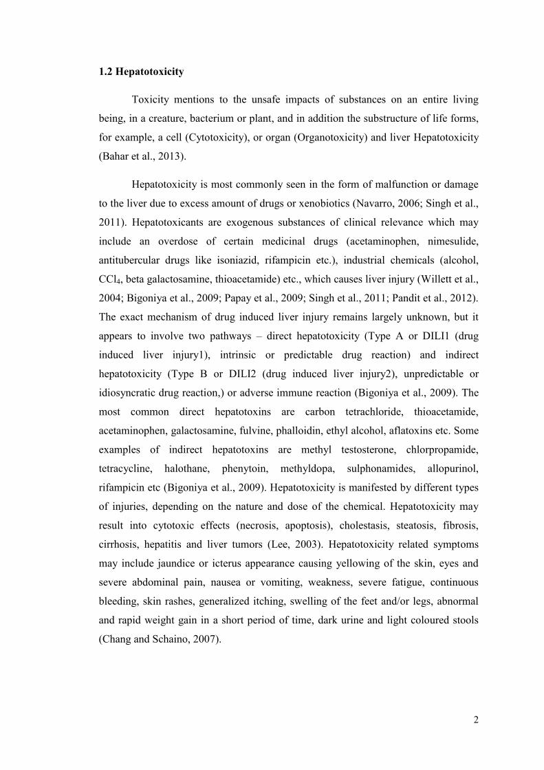
cells (Fig. 1) like hepatocytes, endothelial cells, kupffer cells and stellate cells
grouped in sheets or bundles (Jacobs et al., 2010; Ho et al., 2013). The liver
metabolizes xenobiotics and drugs into toxic intermediates (Bhattacharjee and Sil,
2006).
Liver performs vital role in wide range of functions such as metabolism of
nutrients like amino acids, carbohydrates, lipids, minerals, vitamins; it also helps in
blood clotting through synthesis and secretion of plasma proteins; eliminates dead red
blood cells from blood circulation; eliminates bacteria; detoxifies chemicals, drugs,
xenobiotics, helps in digestion and fat metabolism by excretion of bile salts; and
excretion of end products of metabolism through urine (Singh et al., 2012). Liver
plays role in both metabolism as well as biochemical transformation (Sahu, 2007).
Fig. 1. Structure of Liver
(From Ho et al., 2013)
2
1.2 Hepatotoxicity
Toxicity mentions to the unsafe impacts of substances on an entire living
being, in a creature, bacterium or plant, and in addition the substructure of life forms,
for example, a cell (Cytotoxicity), or organ (Organotoxicity) and liver Hepatotoxicity
(Bahar et al., 2013).
Hepatotoxicity is most commonly seen in the form of malfunction or damage
to the liver due to excess amount of drugs or xenobiotics (Navarro, 2006; Singh et al.,
2011). Hepatotoxicants are exogenous substances of clinical relevance which may
include an overdose of certain medicinal drugs (acetaminophen, nimesulide,
antitubercular drugs like isoniazid, rifampicin etc.), industrial chemicals (alcohol,
CCl4, beta galactosamine, thioacetamide) etc., which causes liver injury (Willett et al.,
2004; Bigoniya et al., 2009; Papay et al., 2009; Singh et al., 2011; Pandit et al., 2012).
The exact mechanism of drug induced liver injury remains largely unknown, but it
appears to involve two pathways – direct hepatotoxicity (Type A or DILI1 (drug
induced liver injury1), intrinsic or predictable drug reaction) and indirect
hepatotoxicity (Type B or DILI2 (drug induced liver injury2), unpredictable or
idiosyncratic drug reaction,) or adverse immune reaction (Bigoniya et al., 2009). The
most common direct hepatotoxins are carbon tetrachloride, thioacetamide,
acetaminophen, galactosamine, fulvine, phalloidin, ethyl alcohol, aflatoxins etc. Some
examples of indirect hepatotoxins are methyl testosterone, chlorpropamide,
tetracycline, halothane, phenytoin, methyldopa, sulphonamides, allopurinol,
rifampicin etc (Bigoniya et al., 2009). Hepatotoxicity is manifested by different types
of injuries, depending on the nature and dose of the chemical. Hepatotoxicity may
result into cytotoxic effects (necrosis, apoptosis), cholestasis, steatosis, fibrosis,
cirrhosis, hepatitis and liver tumors (Lee, 2003). Hepatotoxicity related symptoms
may include jaundice or icterus appearance causing yellowing of the skin, eyes and
severe abdominal pain, nausea or vomiting, weakness, severe fatigue, continuous
bleeding, skin rashes, generalized itching, swelling of the feet and/or legs, abnormal
and rapid weight gain in a short period of time, dark urine and light coloured stools
(Chang and Schaino, 2007).

3
1.3 Prevalence of Liver Disease
Liver diseases are fatal and leading cause of illness and deaths worldwide
(Wang et al., 2014a). As per study, liver disorders cause about 18000 to 20000 deaths
every year globally (Fatma and Uphadhyay, 2015; Akila and Prasanna, 2014). In
United States, about 2-5 % of hospital admissions are due to liver injury out of which
10% results in acute liver failures (Pandit et al., 2012). In United States, rate of liver
transplantation is more than 75% for Type B drug reactions (Ostapowicz et al., 2002).
The crude incidence of liver disorder is 14 per 100000 per annum globally, whereas
the standard incidence is 8.1 per 100000 per annum (Bedi et al., 2016). Acute liver
failure rate is up to 13% of the cases in developed nations like USA whereas it is less
(5%) in tropical countries like India (MeMahon, 2005).
1.3.1 International Scenario
Acetaminophen or paracetamol is easily available as over the counter drug and
has become major reason of self-poisoning in recent years (Ghaffar and Tadvi, 2014).
About 50% of self-poisoning cases are due to paracetamol causing 100-200 deaths per
annum in UK (Kumar et al., 2005). In USA, 61.8% of paracetamol overdose cases
were found unintentional and 30.5% were related with suicidal attempts (Serper et al.,
2016). It has been reported as the most common drug overdose either accidentally or
unintentionally resulting into acute liver failures (ALF) in the United Kingdom (UK,
60-75% of ALF aetiology), Europe (2% of ALF aetiology in France), Canada, United
States (US, approximately 20% of ALF aetiology), and Australia (Ostapowicz and
Lee, 2000; Robinson et al., 2000; Ostapowicz et al., 2002; Ayonrinde et al., 2005;
Marzilawati et al., 2012). However, the incidence of acetaminophen-induced acute
liver failure cases in US has increased exponentially (Larson, 2005). In year 2009,
401 deaths were reported due to acetaminophen overdose by American Association of
Poison Control Centers (Sreejith et al., 2015). According to a recent report, about
42% of acute liver failure cases out of more than 80000 emergency visits and 30000
hospitalizations are reported in US (Lancaster et al., 2015).
Alcohol is one of the main causes of end stage liver disease and leading cause
of morbidity and mortality worldwide (WHO, 2011; Wang et al., 2014a). Deaths due
to alcohol liver diseases are increased since last decade (Mandayam et al., 2004) and
4
have become common reason for cirrhosis in western countries (WHO, 2011). In
USA, second leading cause for liver transplantation is alcoholic cirrhosis (Varma et
al., 2010). The mortality rate for alcoholic liver disease (ALD) was 7.9 per 100000 in
the United States (Roizen et al., 1999). In 2006, 22,073 deaths in the United States
(excluding accidents/homicides) were related to alcohol, with approximately 13,000
deaths specifically due to ALD (Beier et al., 2011). The Global Status Report on
Alcohol and Health by WHO reported highest alcohol consumption in developed
world including Western and Eastern Europe (WHO, 2011). European countries
reported 1 in 7 male deaths and 1 in 13 female deaths due to ALD in 2004 (WHO,
2012). Nearly 88,000 people (approximately 62,000 men and 26,000 women) die
from alcohol-related causes annually, making it the fourth leading preventable cause
of death in the United States (CDC, 2013; Stahre et al., 2014). A WHO study in 2012
reported about 3.3 million deaths worldwide, of which 5.9% were caused by alcohol
consumption (WHO, 2014). About 3.8% of global mortality is accounted for alcohol
consumption (Li et al., 2015).
1.3.2 National Scenario
In India, 33.2% patients were reported with acetaminophen overdose in the
study on 1024 patients (Median age 23 years, 82.0% female) from January 2005 to
December 2009 (Marzilawati et al., 2012). The data on acetaminophen self-poisoning
in India is highly insufficient as compared to that of western countries (Nambiar,
2012).
In India, the prevalence rate of liver injury due to alcohol is higher than that of
acetaminophen which is largely attributable to utilization of illegal alcohol (Malik et
al., 2015). In India, 5% of all deaths are because of liver diseases for which the most
critical culprit is alcohol (Vasudevan, 2011). Alcoholism is the most common cause
of fatty liver and cirrhosis in India (Vasudevan, 2011). The prevalence of alcohol
consumption ranges from 7% in Gujarat, to 75% in Arunachal Pradesh (Murthy et al.,
2010; Punia, 2014). The per capita consumption is 2 litres per adult per year which
accounts for 50% of chronic liver diseases (Punia, 2014). In India, mortality rates due
to ALD for males (11/100000) are reported to be higher than that of females
(6/100000) (Nagaraju, 2014). Significantly higher alcohol consumption has been
5
recorded among tribal, country and lower socio-economic urban sections (Benegal,
2005; Punia, 2014).
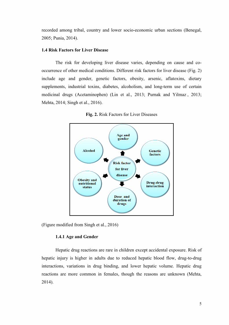
1.4 Risk Factors for Liver Disease
The risk for developing liver disease varies, depending on cause and co-
occurrence of other medical conditions. Different risk factors for liver disease (Fig. 2)
include age and gender, genetic factors, obesity, arsenic, aflatoxins, dietary
supplements, industrial toxins, diabetes, alcoholism, and long-term use of certain
medicinal drugs (Acetaminophen) (Lin et al., 2013; Purnak and Yilmaz , 2013;
Mehta, 2014; Singh et al., 2016).
Fig. 2. Risk Factors for Liver Diseases
(Figure modified from Singh et al., 2016)
1.4.1 Age and Gender
Hepatic drug reactions are rare in children except accidental exposure. Risk of
hepatic injury is higher in adults due to reduced hepatic blood flow, drug-to-drug
interactions, variations in drug binding, and lower hepatic volume. Hepatic drug
reactions are more common in females, though the reasons are unknown (Mehta,
2014).

6
1.4.2 Genetic Factors
P-450 protein is in charge of the metabolism of most of the medications.
Hereditary varieties in the P-450 compounds can lead to abnormal reactions to drug
including peculiar adverse reactions. Variations in the P-450 can be recognized by
amplification in polymerase chain reaction of mutant genes. This has prompted the
likelihood of future identification of persons who can have anomalous responses to a
medication (Mehta, 2014).
1.4.3 Obesity
Risk of liver disease increases with weight and obese individuals are more
likely to develop liver complications than non-obese individuals. The fat cells which
aggregate in liver of obese persons cause liver damage and scarring (sclerosis)
(Lieber, 2003). With increasing weight, the likelihood of liver disease advancement,
cirrhosis, and NASH (Non Alcoholic Steato-Hepatitis) goes on increasing
(Tolman and Dalpiaz, 2007). Fat accumulation in liver consequently leads to NAFLD
(Non Alcoholic Fatty Liver Disease) (Kneeman et al., 2012).
1.4.4 Arsenic
An increased risk in development of some form of liver cancers has been
reported due to chronic exposure to naturally occurring arsenic through drinking
water (Contaminations in some wells) (Lin et al., 2013).
1.4.5 Aflatoxins
Aflatoxin is a substance made by fungus that contaminates mouldy wheat,
corn, soybeans, rice, and some types of nuts which causes cancer. Storage of the food
stuff in a moist, warm environment causes this kind of contamination and is more
common in warmer and tropical countries (Egner et al., 2001).
1.4.6 Dietary Supplements
Poor nutrition and fasting involves risk of liver disease (Malik et al., 2015).
Liver plays key role in regulating the nutritional state and the energy balance in the
body. Development of malnutrition is common in patients with hepatic disorders
7
(Purnak and Yilmaz, 2013). Deficiency of vitamin A and E may aggravate effects of
alcohol induced liver damage by preventing regeneration of hepatocytes (Addagudi et
al., 2013). This is a particular concern as alcoholics are usually malnourished due to
poor diet, anorexia and encephalopathy (Narayanan Menon et al., 2001).
1.4.7 Industrial Toxins
Many chemicals and organic solvents used in different industrial processes
may be associated with hepatotoxicity. Industrial toxins include dimethylformamide,
trichloroethylene, tetrachloroethylene, xylene, toluene, carbon tetrachloride, and vinyl
chloride (Malaguarnera et al., 2012).
1.4.8 Diabetes
Risk of developing chronic liver disease and hepatocellular carcinoma is
higher in diabetes patients than in normoglycemic individuals (El-Serag et al., 2004).
1.4.9 Alcoholism
Alcohol consumption is common reason for liver cirrhosis which increases
risk of liver cancer (Tome and Lucey, 2004; Galicia-Moreno and Gutierrez-Reyes,
2014). The quantity and duration of alcohol intake increases risk of liver disease
(Bruha et al., 2012). One in five heavy drinkers develops alcoholic hepatitis, and one
in four develops cirrhosis (Bruha et al., 2012). It is estimated that, individuals
consuming more than 200mL alcohol per day for more than 14 years may develop
liver disease (Malik et al., 2015). Alcohol also causes increased hepatotoxicity of
several xenobiotics (Radhika et al., 2011). Alcohol induced hepatic injury is due to
accumulation of reactive oxygen species and consequent lipid peroxidation of cellular
layers, proteins and DNA oxidation (Zhou et al., 2002; Galicia-Moreno and Gutierrez-
Reyes, 2014).
1.4.10 Long-Term Use of Certain Medicinal Drugs
Long term use of analgesics and antipyretics cause hepatic injury and on
prolonged conditions it leads to centrilobular hepatic necrosis (Pandit et al., 2012;
Bebnista and. Nowak, 2014). Consumption of acetaminophen like drugs may lead to
hepatocellular carcinoma (Singh et al., 2012). Oxidative stress plays a central role in
8
the hepatic damage caused by acetaminophen and antioxidants have been tested as
alternative treatment against acetaminophen toxicity (Li et al., 2015). Paracetamol
(Acetaminophen) overdose is the most frequent reason for medication induced liver
failure in the United States and in Great Britain (Jaeschke and Bajt, 2006). Long-
acting medications might bring about more harm than short-acting medications
(Mehta, 2014).
1.5 Biochemical Markers
1.5.1 Liver Function Tests
The hepatotoxin causes certain histological changes with typical clinical signs
which indicate liver injury (Singh et al., 2011). Clinical assessment of liver damage
and injury include assessment of serum biochemical markers like serum glutamic
oxaloacetic transaminase (SGOT), serum glutamate-pyruvate transaminase (SGPT),
alkaline phosphatase (ALP) and bilirubin, which are known as liver function test
markers.
Based on the mechanism of injury, hepatotoxicity can be broadly classified
into hepatocellular and cholestatic injury (Lee, 2003; Singh et al., 2011). Increase in
SGOT, SGPT, and ALP levels is an indication of cellular leakage. It further causes
decreased functional integrity of hepatic cell membranes resulting into hepatocellular
damage (Basu et al., 2012). Total bilirubin indicates functional status of the hepatic
cells (Basu et al., 2012). The obvious signs of hepatocellular injury primarily involve
increase in SGOT, SGPT preceding increase in total bilirubin level and small increase
in ALP level (Singh et al., 2011). In cholestatic injury, elevation in ALP level is more
prominent as compared to that in SGOT and SGPT. Generally mixed type of injuries,
involving both hepatocellular and cholestasis injuries, are observed clinically
(Teschke, 2009). The ratio of SGOT: SGPT plays an important role in deciding the
type of liver damage by hepatotoxins (Mishra, 2012). SGOT: SGPT ratio in
hepatocellular damage is greater than or equal to five while it is less than or equal to
two during cholestatic liver damage (Singh et al., 2011). The ratio ranges between two
and five for mixed type of liver damage (Singh et al., 2011).

9
1.5.2 Total Protein
The liver is the major source of most of the serum proteins and amount of
serum total proteins indicate the functional status of the hepatic cells (Gupta et al.,
2007; Thapa and Walia, 2007; Basu et al., 2012). Liver cells synthesize albumin,
fibrinogen, prothrombin, alpha-1-antitrypsin, hepatoglobin, ceruloplasmin, transferrin,
alpha foetoproteins etc., while damaged liver shows decreased levels of these plasma
proteins (Thapa and Walia, 2007; Mishra, 2012).
1.5.3 Lipid Profile
The liver is a major organ regulating lipid metabolism and it synthesizes and
metabolizes cholesterol, bile acids and phospholipids (Werner et al., 2000). Liver
synthesizes nearly 80% of the cholesterol produced in the body from Acetyl-CoA via
a pathway that connects metabolism of carbohydrates with that of lipid. Liver can
synthesize, store and export triglycerides (Dean et al., 2009; Rui, 2014).
Acetaminophen intoxicated rats show elevated levels of cholesterol and
triglycerides, indicating hepatic damage and consequent impairment in fat metabolism
(Haldar et al., 2011). The liver controls cholesterol and triglyceride levels in the body
by assembling, secreting, and taking up various lipoprotein particles (Cox and Garcia-
Palmieri, 1990). During these functions, loss of lipid and protein components causes
change in structure of VLDL particles (Marais, 2004). The resulting LDL particles are
then returned to the liver through LDL receptors on hepatocytes (Marais, 2004).
Greater increase of LDL and VLDL may also cause a greater decrease of HDL,
disturbing lipid metabolism in liver (Al-Assaf, 2013).
1.5.4 Oxidative Stress Markers
Oxidative stress is one of the important reasons for pathogenesis of hepatic
dysfunction in humans (Rahman et al., 2012) and animals (Abd Ellah et al., 2009;
Abd Ellah, 2010). Toxicity of a xenobiotic is also affected by the generation of
reactive oxygen species (ROS) by a few different mechanisms including
mitochondrial damage, activation of cytochrome P450 2E1 (CYP2E1), and infiltration
of Kupffer cells and granulocytes (Arteel, 2003; Smathers, 2006; Albano, 2008).
Oxidative stress within the cells by partially reduced free oxygen species such as, O2-,
H2O2 and OH. Causes damage to hepatic parenchymal cells leading to hepatic injury
10
(Jothy et al., 2012; Novo and Parola, 2012). Liver releases free radicals during
detoxification of chemicals, drugs and other toxic materials (Abd Ellah, 2011). The
elevation in free radicals and decreased scavenging potential of the cells is observed
in hepatic injury (Jothy et al., 2012). Liver cells have endogenous antioxidant system
comprising of superoxide dismutase (SOD), catalase (CAT), glutathione peroxidase
(GPx), reduced glutathione (GSH) and malondialdehyde (MDA) which offers
protection against oxidative stress (Haldar et al., 2011). Elevated levels of antioxidant
enzymes and decrease in MDA helps in hepatoprotection while decrease in
antioxidant enzyme activities and increased MDA results in hepatocellular damage
which leads to degradation of cellular macromolecules in liver by chemical induced
toxicity (Janero, 1990; Durairaj et al., 2007; Palanivel et al., 2008; Haldar et al.,
2011).
1.6 Management of Hepatic Diseases
In today’s world, liver diseases are a major serious health problem. Despite
considerable progress in modern medicine, the drugs or agents which can stimulate
liver function or help regeneration of hepatic cells or offer protection to the liver
damage are still wanted (Vishal, 2013). In addition, the synthetic drugs used in
modern medicine have been reported to have many undesirable side effects. Hence,
there is a recent renewal of interest in search for the natural resources like medicinal
plants which have promising potential to offer several herbal medicines with less side
effects (Sudaroli, 2013). Thousands of medicinal plants are used worldwide to prevent
as well as cure many diseases, but the mode of their protective and curative actions
still remains unclear (Pan et al., 2014).
The therapies available to treat liver diseases include surgical procedures,
hepatoprotective agents, herbal formulations, medicinal plants, nutritional
supplements etc.
1.6.1 Surgical Procedures
Liver transplantation has become an acceptable means with excellent long-
term outcomes to treat end-stage liver diseases, but it is very costly. Liver failure due
to viral hepatitis (especially hepatitis B and C) is a common indication for liver
transplantation (Terrault et al., 2005).
11
1.6.2 Hepatoprotective Agents
Hepatoprotective agents have recently been given attention due to their roles
in the additional treatment of liver disease (Flatland, 2003; Sartor and Trepanier,
2003; Twedt, 2004). These products include both prescription drugs and
nutraceuticals. In order for a compound to be used as a drug, it must be harmless and
effective for its intended use. The drug can be released in the market only after
undergoing an extensive Food and Drugs Administration’s (FDA) drug approval
process which is lengthy and costly. Apart from modern drugs, there are several
hepatoprotective agents like L-carnitine (Yapar et al., 2007), Vitamin C (Adikwu and
Deo, 2013), N-acetylcysteine (Maheswari et al., 2014), and Milk Thistle (Silymarin)
(Vargas-Mendoza et al., 2014). Silymarin is a unique flavonoid complex derived from
milk thistle and is commonly used for regeneration of liver cells, decongestion of
liver, complementary treatment to the patients of liver cirrhosis and viral hepatitis etc.
It is also used for hepatoprotection against industrial chemicals and pharmaceuticals
(Das et al., 2011).
1.6.2.1 Silymarin
Silymarin is a standardized extract from the seeds of a plant called milk thistle
(Silybum marianum L.; Family: Asteraceae). In rural areas, it has been used as a
natural remedy to treat liver diseases (Saller et al., 2001). Silymarin helps to protect
and enhance the regeneration of liver cells in most of the liver diseases like cirrhosis,
hepatitis and jaundice (Flora et al., 1998). Silymarin possess membrane stabilizing,
anti-oxidative, anti-lipid peroxidative (Pascual et al., 1993), anti-fibrotic (Jia et al.,
2001), and immune-modulatory properties and helps in liver regeneration (Pradhan
and Girish, 2006). Studies on human beings demonstrated that about 20-40%
silymarin is excreted as sulphates and glucuronide conjugates in bile (Saller et al.,
2001). There are a few reports of low level of silymarin toxicity causing allergic skin
rashes and gastrointestinal disturbances (Saller et al., 2001).
1.6.3 Herbal Formulations
Numerous medicinal plants and their formulations are used to treat liver
disorders in ethno medicine practice as well as traditional system of medicine in India.
There are about 600 commercial herbal formulations available in market all over the
12
world, which are claimed to have hepatoprotective activity (Bedi et al., 2016). In
India, about 40 anti-hepatotoxic, patented, polyherbal formulations representing a
variety of combination of 93 medicinal plants from 44 families are available (Sharma
et al., 1991). More than 700 mono and poly-herbal hepatoprotective preparations from
more than 100 plants are in clinical use in the form of decoction, tincture, tablets and
capsules. Recent global increase in the utilization of herbal drugs has also been
reported in the literature (Girish et al., 2009).
1.6.4 Medicinal Plants
Eastern countries have been using herbal drugs to treat liver diseases since
ancient time (Rajaratnam et al., 2014). The ancient Chinese and Egyptian written
records are available which describe medicinal uses of plants (Rajaratnam et al.,
2014). In ancient India (Vedic period) and China (Xia dynasty), records on use of
herbal medicines track back to 2100 BC. The first written reports date back to 600
B.C. with the Charka Samhita of India and the early notes of the Eastern Zhou
dynasty of China around 400 B.C (Onyije and Avwioro, 2012).
Ayurveda, an indigenous system of medicine in India, has a long tradition of
treating liver disorders with plant drugs. Minimizing side effects and increasing
therapeutic efficacy of medicines is the basic need of today. Alternative system of
medicine like Ayurveda, Unani etc. has been proved to be effective with minimum
side effects. With rich diversity of plants, over 45,000 diverse plant species are found
in India out of which about 15,000-20,000 plants have medicinal and therapeutic
properties. Of these, only about 7,000-7,500 are being used by traditional practitioners
(Bedi et al., 2016). As per WHO report, around three quarters of the world’s
population uses herbs and other traditional medicines to cure various diseases,
including liver disorders (Chaudhury and Refei, 2001). The medicinal plant such as
Guduchi (Sharma and Pandey, 2010), Elephantopus scaber (Ho et al., 2012),
Aquilegia vulgaris (Adamska et al., 2003), Strychnos potatorum (Sanmugapriya &
Venkataraman, 2006), Tridax procumbens (Ravikumar et al., 2006), Picrorhiza
kurroa (Mohd et al., 2012), Silybum marianum (Hermenean et al., 2015),
Andrographis paniculata (Nasir et al., 2013), Azadirachta indica (Johnson et al.,
2015) and Glycyrrhiza glabra (Sharma and Agrawal, 2014) has proven
hepatoprotective properties and are used to treat liver disorders. Guduchi (Tinospora
13
sp.) is one of the most versatile rejuvenating shrubs, also known as ’Giloya’ in Indian
vernacular, and is reported to have many therapeutic applications (Pandey et al.,
2012). Guduchi, as it is most commonly called, has been described as “one which
protects the body” (Gawhare, 2013).
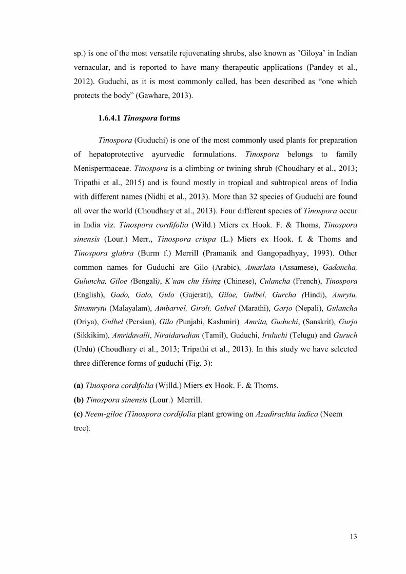
1.6.4.1 Tinospora forms
Tinospora (Guduchi) is one of the most commonly used plants for preparation
of hepatoprotective ayurvedic formulations. Tinospora belongs to family
Menispermaceae. Tinospora is a climbing or twining shrub (Choudhary et al., 2013;
Tripathi et al., 2015) and is found mostly in tropical and subtropical areas of India
with different names (Nidhi et al., 2013). More than 32 species of Guduchi are found
all over the world (Choudhary et al., 2013). Four different species of Tinospora occur
in India viz. Tinospora cordifolia (Wild.) Miers ex Hook. F. & Thoms, Tinospora
sinensis (Lour.) Merr., Tinospora crispa (L.) Miers ex Hook. f. & Thoms and
Tinospora glabra (Burm f.) Merrill (Pramanik and Gangopadhyay, 1993). Other
common names for Guduchi are Gilo (Arabic), Amarlata (Assamese), Gadancha,
Guluncha, Giloe (Bengali), K’uan chu Hsing (Chinese), Culancha (French), Tinospora
(English), Gado, Galo, Gulo (Gujerati), Giloe, Gulbel, Gurcha (Hindi), Amrytu,
Sittamrytu (Malayalam), Ambarvel, Giroli, Gulvel (Marathi), Garjo (Nepali), Gulancha
(Oriya), Gulbel (Persian), Gilo (Punjabi, Kashmiri), Amrita, Guduchi, (Sanskrit), Gurjo
(Sikkikim), Amridavalli, Niraidarudian (Tamil), Guduchi, Iruluchi (Telugu) and Guruch
(Urdu) (Choudhary et al., 2013; Tripathi et al., 2013). In this study we have selected
three difference forms of guduchi (Fig. 3):
(a) Tinospora cordifolia (Willd.) Miers ex Hook. F. & Thoms.
(b) Tinospora sinensis (Lour.) Merrill.
(c) Neem-giloe (Tinospora cordifolia plant growing on Azadirachta indica (Neem
tree).
14
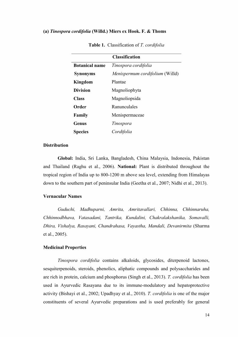
(a) Tinospora cordifolia (Willd.) Miers ex Hook. F. & Thoms
Table 1. Classification of T. cordifolia
Distribution
Global: India, Sri Lanka, Bangladesh, China Malaysia, Indonesia, Pakistan
and Thailand (Raghu et al., 2006). National: Plant is distributed throughout the
tropical region of India up to 800-1200 m above sea level, extending from Himalayas
down to the southern part of peninsular India (Geetha et al., 2007; Nidhi et al., 2013).
Vernacular Names
Guduchi, Madhuparni, Amrita, Amritavallari, Chhinna, Chhinnaruha,
Chhinnodbhava, Vatasadani, Tantrika, Kundalini, Chakralakshanika, Somavalli,
Dhira, Vishalya, Rasayani, Chandrahasa, Vayastha, Mandali, Devanirmita (Sharma
et al., 2005).
Medicinal Properties
Tinospora cordifolia contains alkaloids, glycosides, diterpenoid lactones,
sesquiterpenoids, steroids, phenolics, aliphatic compounds and polysaccharides and
are rich in protein, calcium and phosphorus (Singh et al., 2013). T. cordifolia has been
used in Ayurvedic Rasayana due to its immune-modulatory and hepatoprotective
activity (Bishayi et al., 2002; Upadhyay et al., 2010). T. cordifolia is one of the major
constituents of several Ayurvedic preparations and is used preferably for general
Classification
Botanical name Tinospora cordifolia
Synonyms Menispermum cordifolium (Willd)
Kingdom Plantae
Division Magnoliophyta
Class Magnoliopsida
Order Ranunculales
Family Menispermaceae
Genus Tinospora
Species Cordifolia
15
debility, dyspepsia, fever, urinary diseases, jaundice, skin diseases, diabetes, anaemia,
cancer, liver disorder, heart disease, Parkinson’s disease, emaciations, and hepatitis B
and E (Sinha et al., 2004; Ganguly and Prasad, 2011).
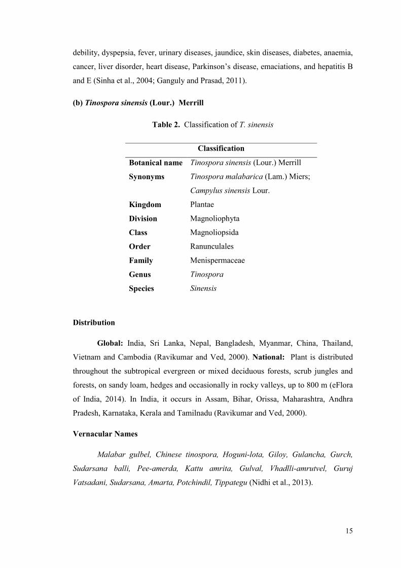
(b) Tinospora sinensis (Lour.) Merrill
Table 2. Classification of T. sinensis
Distribution
Global: India, Sri Lanka, Nepal, Bangladesh, Myanmar, China, Thailand,
Vietnam and Cambodia (Ravikumar and Ved, 2000). National: Plant is distributed
throughout the subtropical evergreen or mixed deciduous forests, scrub jungles and
forests, on sandy loam, hedges and occasionally in rocky valleys, up to 800 m (eFlora
of India, 2014). In India, it occurs in Assam, Bihar, Orissa, Maharashtra, Andhra
Pradesh, Karnataka, Kerala and Tamilnadu (Ravikumar and Ved, 2000).
Vernacular Names
Malabar gulbel, Chinese tinospora, Hoguni-lota, Giloy, Gulancha, Gurch,
Sudarsana balli, Pee-amerda, Kattu amrita, Gulval, Vhadlli-amrutvel, Guruj
Vatsadani, Sudarsana, Amarta, Potchindil, Tippategu (Nidhi et al., 2013).
Classification
Botanical name Tinospora sinensis (Lour.) Merrill
Synonyms Tinospora malabarica (Lam.) Miers;
Campylus sinensis Lour.
Kingdom Plantae
Division Magnoliophyta
Class Magnoliopsida
Order Ranunculales
Family Menispermaceae
Genus Tinospora
Species Sinensis
16
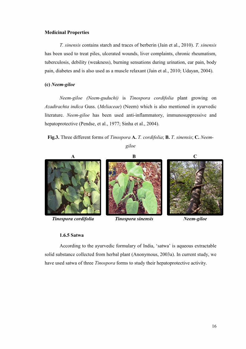
Medicinal Properties
T. sinensis contains starch and traces of berberin (Jain et al., 2010). T. sinensis
has been used to treat piles, ulcerated wounds, liver complaints, chronic rheumatism,
tuberculosis, debility (weakness), burning sensations during urination, ear pain, body
pain, diabetes and is also used as a muscle relaxant (Jain et al., 2010; Udayan, 2004).
(c) Neem-giloe
Neem-giloe (Neem-guduchi) is Tinospora cordifolia plant growing on
Azadirachta indica Guss. (Meliaceae) (Neem) which is also mentioned in ayurvedic
literature. Neem-giloe has been used anti-inflammatory, immunosuppressive and
hepatoprotective (Pendse, et al., 1977; Sinha et al., 2004).
Fig.3. Three different forms of Tinospora A. T. cordifolia; B. T. sinensis; C. Neem-
giloe
1.6.5 Satwa
According to the ayurvedic formulary of India, ‘satwa’ is aqueous extractable
solid substance collected from herbal plant (Anonymous, 2003a). In current study, we
have used satwa of three Tinospora forms to study their hepatoprotective activity.
A B C
Tinospora cordifolia Tinospora sinensis Neem-giloe
17
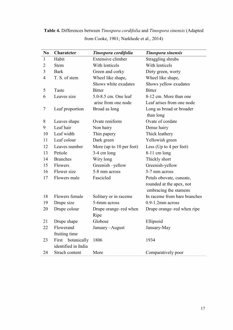
Table 4. Differences between Tinospora cordifolia and Tinospora sinensis (Adapted
from Cooke, 1901; Narkhede et al., 2014)
No Charatcter Tinospora cordifolia Tinospora sinensis
1 Habit Extensive climber Straggling shrubs
2 Stem With lenticels With lenticels
3 Bark Green and corky Dirty green, worty
4 T. S. of stem Wheel like shape,
Shows white exudates
Wheel like shape,
Shows yellow exudates
5 Taste Bitter Bitter
6 Leaves size 5.0-8.5 cm. One leaf
arise from one node
8-12 cm. More than one
Leaf arises from one node
7 Leaf proportion Broad as long Long as broad or broader
than long
8 Leaves shape Ovate reniform Ovate of cordate
9 Leaf hair Non hairy Dense hairy
10 Leaf width Thin papery Thick leathery
11 Leaf colour Dark green Yellowish green
12 Leaves number More (up to 10 per feet) Less (Up to 4 per feet)
13 Petiole 3-4 cm long 8-11 cm long
14 Branches Wiry long Thickly short
15 Flowers Greenish –yellow Greenish-yellow
16 Flower size 5-8 mm across 5-7 mm across
17 Flowers male Fascicled Petals obovate, cuneate,
rounded at the apex, not
embracing the stamens
18 Flowers female Solitary or in raceme In raceme from bare branches
19 Drupe size 5-6mm across 0.9-1.2mm across
20 Drupe colour Drupe orange–red when
Ripe
Drupe orange–red when ripe
21 Drupe shape Globose Ellipsoid
22 Flowerand
fruiting time
January –August January-May
23 First botanically
identified in India
1806 1934
24 Strach content More Comparatively poor
18
1.6.6 Nutritional Supplements
It is very important for patients with liver disease, to have balanced diet with
suitable calories, carbohydrates, fats and proteins (Worman, 1999). Balanced diet with
good nutritive value helps in regeneration of liver cells. Dietary supplements contain
herbal products, vitamins, minerals, and any product that is not a drug (medication)
(American Cancer Society, 2015). Several Asian nations use numerous food and
nutrition supplements, in routine diet that possess hepatoprotective activity. Several
phytochemicals present in nutritional supplements possess potential ability to prevent
or reverse different kinds of liver injuries (Shukla and Kumar, 2013). Omega-3 fatty
acids are also known to offer significant benefits as nutritional supplement or dietary
supplement for hepatoprotection.
1.6.6.1 Polyunsaturated Fatty Acid (Omega-3 Fatty Acids)
Ancient people consumed food containing a lot more omega-3 fatty acids than
we do today. Omega-3 fatty acids are characterized by double bond (C=C) between
third and fourth carbon atom from methyl end of the carbon chain (Scorletti and
Byrne, 2013). α-linolenic acid (ALA), found in plant oils and eicosapentaenoic acid
(EPA), docosahexaenoic acid (DHA), both commonly found in marine oils, are three
physiologically important omega-3 fatty acids (Rodriguez-Leyva et al., 2010). Plant
oils mainly contain ALA and it is obtained from walnut, edible seeds, clary sage seed
oil, algal oil, flaxseed oil, Sacha Inchi oil, Echium oil, and hemp oil (Simopoulos,
2008). EPA and DHA are chief components of fish oils, egg oil, squid oils, and krill
oil (Demark-Wahnefried et al., 2001). Omega-3: Omega-6 fatty acids ratio in wild
animals is 1:1 which is ideal for normal biological processes (Simopoulos, 2008).
1.7 Animal Models for Hepatotoxicity
In in vivo experimental models, animals are administered with repeated dose
of hepatotoxin to induce liver damage. To evaluate hepatoprotective ability of drugs,
various hepatotoxins (chemical agents) such as carbon tetrachloride, acetaminophen,
alcohol, are used in experimental models.
19
1.7.1 Carbon Tetrachloride Induced Hepatotoxicity
Formerly, carbon tetrachloride (CCl4) was used in fire-extinguishers, as a
grain fumigant and also as a refrigerant (El-Sayed et al., 2015). Carbon tetrachloride
(CCl4) is one of the commonly used model drug (hepatotoxin) to induce
hepatotoxicity in experimental models to study acute and chronic liver failures (Singh
et al., 2012; Olatosin et al., 2014). In endoplasmic reticulum and mitochondria,
cytochrome P-450 metabolizes CCl4 and forms a reactive oxidative free radical called
CCl3O which induces lipid peroxidation (Weber et al., 2003). Within 3 hours of single
dose of CCl4 administration, poison reaches its maximum concentration, and within
24 hours centrilobular necrosis and fatty changes are observed in experimental rats
(Singh et al., 2012). Different methods and concentrations of doses used in
experimental models are: repetitive dose of 0.1 to 3ml CCl4/kg bw, I.P. for 7 days; 1
ml CCl4/kg bw, I.P in liquid paraffin (30% v/v); 1ml CCl4/kg bw, I.P. single dose to
induce acute hepatotoxicity (Adewale et al., 2014; Sarkar et al., 2014).
1.7.2 Acetaminophen Induced Hepatotoxicity
Acetaminophen (international name used in USA and Japan) and Paracetamol
(international name used in Europe) are two official names of the same chemical
compound derived from its chemical name: N-acetyl-para-aminophenol (Benista and
Nowak, 2014). Acetaminophen is over-the-counter analgesic and anti-pyretic
medicine. Acetaminophen is a dynamic metabolite of phenacetin used to get relief
from fever, migraine, muscle hurts, joint inflammation, spinal pain, toothache and
frosty (Vidhya Malar and Bai, 2012). Overdose of acetaminophen leads to
‘Acetaminophen hepatotoxicity,’ causes liver injury and is one of the most common
causes of poisoning all over world (Vidhya Malar and Bai, 2012). Therapeutic dose of
acetaminophen is safe but, its overdose can cause hepatotoxicity and acute liver
failure (Michaut et al., 2014). Higher dose of acetaminophen causes hepatotoxicity in
human and animal models (Jaeschke et al., 2014). Events of acetaminophen
hepatotoxicity lead to liver cirrhosis, hepatitis etc. (Fontana, 2008).
It is primarily metabolized by sulfation and glucuronidation to unreactive
metabolites, and then activated by cytochrome P450 system. Bioactivation of
acetaminophen produces a toxic electrophile, N-acetyl p- benzoquinone imine
20
(NAPQI). NAPQI binds covalently to tissue macromolecules and also induces lipid
oxidation. Apart from oxidizing lipids, NAPQI also oxidizes sulfhydryl groups in
protein thiols. NAPQI is also known to alter the homeostasis of calcium (Lin et al.,
1997). Different reactive metabolites are produced during acetaminophen metabolism,
which covalently modify proteins (Bernareggi, 1998), impose oxidative stress (Ritter
and Giganti, 1998) and results in mitochondrial injury (Mingatto et al., 2000). Several
studies in animals and human have demonstrated that paracetamol overdose causes
liver damage primarily due to enhanced production and/or decreased glutathione
conjugation of NAPQI that eventually results in increased covalent binding of NAPQI
to cell proteins (Leung et al., 2012; Sharoud, 2015). Several experimental studies
reveal that daily treatment of acetaminophen to Wistar albino rats [at a dose of
600mg/kg for 14 days (Ita et al., 2009) or a single dose of 500 mg/kg to 3 gm/kg
(Murugesh et al., 2005; Juraj et al., 2004) or a single dose of 2g/kg (Meganathan et
al., 2011; Prabu et al., 2011) or a dose of 400 mg/kg for seven days (Kanchan and
Sadiq, 2011)] leads to liver injury and consequent increase in the levels of liver
marker enzymes.
1.7.3 Alcohol Induced Hepatotoxicity
Liver is among the organs most susceptible to the toxic effects of ethanol.
Alcoholic liver disease (ALD) is considered a major health and economic problem
worldwide (Bruha et al., 2012; Cui et al., 2013). Alcohols are hydroxy derivatives of
aliphatic hydrocarbons and commonly consumed alcohol is ethyl alcohol or ethanol
(Tripathi, 2013). Alcohol is a psychoactive and addictive substance which is quickly
absorbed by the body and detoxified by liver (Hadzic et al., 2013). Alcohol (Ethanol)
is one of the most important and commonly used hepatotoxic agents in the
experimental study of liver related disorders. The alcohol over dose leads to liver
damages (Arulkumaran et al., 2009) caused by complex mechanisms involving
metabolites of ethanol with ability to form protein adducts with several proteins of
hepatocytes (Zimmerman, 1999). Further, it has direct cytotoxicity which leads to
increase in reduced form of nicotinamide adenine dinucleotide (NADH) causing fat
accumulation (Zimmerman, 1999). Many pathways are reported to be involved in
ALD, including oxidative stress and mitochondrial damage (Stewart et al., 2001; Wu
and Cederbaum, 2003; Gramenzi et al., 2006). Ethanol is metabolized in the body by
enzyme catalyzed oxidative processes into acetaldehyde. The acetaldehyde is further
21
oxidized to acetate which is then converted to carbon dioxide via the citric acid cycle
(Samundeeswari et al., 2013). Alcohol metabolites also cause induction of free
radicals leading to peroxidation and inflammatory response (Jarvelainen, 2000).
Various different concentrations of alcohol at different doses have been reported to be
hepatotoxic in animal studies. Alcohol (15% to 40%) at a dose of 2ml-25 ml/kg bw or
1.5gm-24gm/kg bw (Mahendran and Devi, 2001; Ghosh et al., 2007; Arulkumaran et
al., 2009; Patel et al., 2010; Nigam and Paarakh, 2011; Radhika et al., 2011; Singh
and Gupta, 2011; Vetriselvan et al., 2011; Sudhir et al., 2012) is reported to induce
hepatic damage in Wistar rats.
In recent years, herbal medicine and nutritional supplementation have been
used as alternative medicine to treat liver disorder (Lee et al., 2007; Abo El-Magd et
al., 2015). Tinospora is known to be used in many ailments in alternative medicine.
Omega 3 fatty acids are also known to have several health benefits (Wu et al., 2012;
Li et al., 2014). In current study, protective, corrective and prophylactic effects of
Guduchi satwa and omega-3 fatty acids against acetaminophen and alcohol induced
hepatotoxicity were analyzed.
1.8 Genesis of Thesis
Herbal medicines and nutrition are known to play an important role in
management of various health ailments. Acetaminophen and alcohol induced
hepatotoxicity may be modulated through interventions with herbal and nutritional
supplements. The efficacy of the interventions can be enhanced by their simultaneous
delivery during progression of hepatotoxicity and may serve as curative as well as
preventive therapies against liver toxicity.
In this study three different forms of Tinospora i.e. T. cordifolia, T.
sinensis and Neem-giloe were analyzed for the hepatoprotective efficacy of their
satwa. T. cordifolia is easily available in the fields and hence it is used frequently. But
it is observed that the description of Guduchi in Ayurvedic literature matches with T.
sinensis. Hence, to ascertain the most potent variant of Guduchi, hepatoprotective
activity of all three satwa (from T. cordifolia, T. sinensis and Neem-giloe) was
assessed against acetaminophen and alcohol induced hepatotoxicity in rats.
22
In this study, flax oil and fish oil were used as nutritional supplements (rich
source of omega-3 fatty acids) to assess their hepatoprotective activity against
acetaminophen and alcohol induced liver toxicity. The protective and corrective
effects of nutritional and herbal interventions against acetaminophen and alcohol
induced hepatotoxicity in rats were also studied.
Hypothesis
Pathophysiology of acetaminophen and alcohol induced hepatotoxicity may be
modulated through interventions with herbal and nutritional supplements. The
efficacy of the interventions can be enhanced by their simultaneous delivery during
progression of hepatotoxicity and may have corrective/preventive roles in liver
toxicity.

LITERATU
CHAPTER 2
OBJECTIVES
Awards
Second prize in oral
Presentation Award
Best Poster Presentation
Award
23
Objectives
1. To study the hepatoprotective efficacy of residual marc of aqueous extract of
Tinospora cordifolia, Tinospora sinensis and Neem-giloe (T. cordifolia
growing on Azadirachta indica A. Juss.) on acetaminophen and alcohol
induced hepatotoxicity in rats at biochemical, histological and molecular level.
2. To study the effect of polyunsaturated fatty acid supplementation against
acetaminophen and alcohol induced hepatotoxicity in rats at biochemical,
histological and molecular level.
3. To study the combinatorial protective and corrective effect of nutritional and
herbal interventions against acetaminophen and alcohol induced
hepatotoxicity in rats at biochemical, histological and molecular level.
LITERATU
CHAPTER 3
REVIEW OF LITERATURE
24
Hepatotoxicity implies chemical driven or drug induced liver damage. The
agents responsible for such liver damage are called hepatotoxicants. Such
hepatotoxicants are employed as inducers of hepatotoxicity in several experimental
animal models and such induction of hepatotoxicity is indispensable for testing of
novel hepatoprotective agents and their safety. Several herbal formulations, medicinal
plants and nutritional supplements are recommended for treatment of liver diseases. In
the present study, satwa from three different forms of Tinospora (Guduchi satwa) and
omega-3 fatty acids were analyzed for their hepatoprotective activities. This chapter
reviews literature about drug (acetaminophen and alcohol) induced hepatotoxicity and
hepatoprotective effects of different interventions.
3.1 Acetaminophen Induced Hepatotoxicity
Acetaminophen is the most popular over-the-counter, analgesic and antipyretic
drug commonly used to treat mild to moderate pain. It was discovered in 1893 and
first introduced in Canada in 1950’s (Meredith and Goulding., 1980; Jackson et al.,
1984; Anker and Smiikstein, 1994; Bessems and Vermeulen, 2001; James et al.,
2003). The therapeutic dose (650-1000 mg every 4 to 6 hours) of acetaminophen is
considered to be safe (Jackson et al., 1984). First case of acetaminophen
hepatotoxicity was reported in UK in 1966 (Davidson and Eastham, 1966) followed
by in North America in 1971 (Boyer and Rouff, 1971; Hinson et al., 2010).
Acetaminophen-induced hepatotoxicity has been studied in experimental animals
(Mitchell et al., 1973; Lim et al., 1995; Hinson et al., 2010) as well as in clinical cases
(Boyd and Bereczky, 1966; McJunkin et al., 1976; Golden et al., 1981; Whitcomb and
Block, 1994). Higher dose of acetaminophen produces centrilobular hepatic necrosis,
fulminant hepatic failure and renal tubular necrosis in humans and laboratory animals
(Boyd and Bereczky, 1966; Boyer and Rouff, 1971; Jollow et al., 1973).
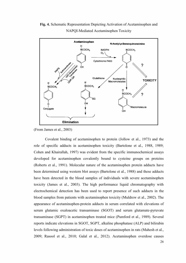
Mechanism of Acetaminophen Induced Hepatotoxicity
Several studies reported mechanism of acetaminophen induced hepatotoxicity
(Jollow et al., 1973; Potter et al., 1973; Cohen and Khairallah, 1997; Bessems and
Vermeulen, 2001; Irwin et al., 2004). Acetaminophen is metabolized in liver via
glucuronidation, sulfation and the hepatic cytochrome P450 enzyme system. The
cytochrome P450 enzyme system metabolically activates acetaminophen and forms a
reactive metabolite N-Acetyl-P-benzo-quinoneimine (NAPQI) (Prescott, 1980;
Awards
Second prize in oral
Presentation Award
Best Poster Presentation
Award
25
Vidhya Malar and Bai, 2012). NAPQI covalently binds to proteins and forms
acetaminophen-protein adducts (James et al., 2003) NAPQI is formed by direct two-
electron oxidation of acetaminophen by cytochrome P450 (Dahlin et al., 1984).
Several reports demonstrated that acetaminophen is oxidized into the reactive
metabolite by the cytochromes 2E1, 1A2, 3A4, and 2A6 (Patten et al., 1993;
Thummel et al., 1993; Chen et al., 1998). Toxic dose of acetaminophen leads to
almost 90% depletion of total hepatic Glutathione (GSH) (Mitchell et al., 1973). The
mechanism is shown in Fig. 4. Acetaminophen mainly undergoes glucuronidation
(47%-62%) and sulfation (25%-36%) in the liver when taken in therapeutic dose
(Prescott, 1980; Pacifici and Allegaert, 2015). The hepatic cytochrome P450 enzyme
system (specifically CYP2E1) metabolizes about 5 to 10% of therapeutic
acetaminophen resulting into production of toxic product (NAPQI) which is
detoxified by combining with the sulfhydryl groups of glutathione and then excreted
through urine (Pacifici and Allegaert, 2015). Acetaminophen overdose causes
increased formation of toxic product NAPQI and utilization of substantial amounts of
GSH from liver. This leads to inability of the liver to detoxify NAPQI which
accumulates in liver to toxic concentrations (Dahlin et al., 1984; Raucy et al., 1989;
Pacifici and Allegaert, 2015). The formation of NAPQI is dependent on cytochrome
P450 such as CYP1A1, CYP1A2, and CYP2E1 (Patten et al., 1993; Laine et al., 2009;
Cederbaum, 2015) in rats; CYP1A2 and CYP3A4 in mice (Hazai et al., 2002); and
CYP2E1, CYP1A2 and CYP3A4 in humans (Raucy et al., 1989; Thummel et al.,
1993; Prescott, 2000). Acetaminophen also obstructs intracellular processes in
nucleus (Ray et al., 1996), plasma membrane (Moore et al., 1985), and cytoplasm
(Pumford et al., 1989). Acetaminophen toxicity has been attributed to the interference
of many metabolic processes including depletion of glutathione, impairment of
mitochondrial respiration, and interference with Ca2+
homeostasis (Boulares and Ren,
2004). In addition to formation of NAPQI, high levels of reactive oxygen species
(ROS) also contribute to cell injury process (Shon and Nam, 2002).
26
Fig. 4. Schematic Representation Depicting Activation of Acetaminophen and
NAPQI-Mediated Acetaminophen Toxicity
(From James et al., 2003)
Covalent binding of acetaminophen to protein (Jollow et al., 1973) and the
role of specific adducts in acetaminophen toxicity (Bartolone et al., 1988, 1989;
Cohen and Khairallah, 1997) was evident from the specific immunochemical assays
developed for acetaminophen covalently bound to cysteine groups on proteins
(Roberts et al., 1991). Molecular nature of the acetaminophen protein adducts have
been determined using western blot assays (Bartolone et al., 1988) and these adducts
have been detected in the blood samples of individuals with severe acetaminophen
toxicity (James et al., 2003). The high performance liquid chromatography with
electrochemical detection has been used to report presence of such adducts in the
blood samples from patients with acetaminophen toxicity (Muldrew et al., 2002). The
appearance of acetaminophen-protein adducts in serum correlated with elevations of
serum glutamic oxaloacetic transaminase (SGOT) and serum glutamate-pyruvate
transaminase (SGPT) in acetaminophen treated mice (Pumford et al., 1989). Several
reports indicate elevations in SGOT, SGPT, alkaline phosphatase (ALP) and bilirubin
levels following administration of toxic doses of acetaminophen in rats (Mahesh et al.,
2009; Rasool et al., 2010; Galal et al., 2012). Acetaminophen overdose causes
27
decrease in antioxidant enzyme activities such as superoxide dismutase, catalase and
glutathione peroxidase and increased levels of lipid peroxidation in animal models
(Wendel, 1979; Nakae et al., 1990; Arnaiz et al., 1995; Chen and Lin, 1997).
Hepatoprotective activity of different medicinal plants or herbal medicines has been
extensively studied against acetaminophen induced hepatotoxicity in animals
(Manokaran et al., 2007; Rajkapoor et al., 2008; Kumar et al., 2011; Kanchana and
Sadiq, 2011; Manivannan et al., 2011; Mahmood et al., 2014). Histological analyses
revealed necrosis and apoptosis of hepatocytes in acetaminophen treated mice (Ray et
al., 1996).
Several studies reported gene expression analysis of acetaminophen induced
hepatotoxicity in rodents (Reilly et al., 2001; Ruepp et al., 2002; Irwin et al., 2004;
Ishida et al., 2004; Kim et al., 2007; Germoush and Mahmoud, 2014; Gong et al.,
2014; Mahmoud et al., 2014; Mohar et al., 2014). Critical role of kupffer cells in
development of acetaminophen hepatotoxicity was demonstrated by Laskin et al.
(1995) through pretreating rats with compounds suppressing kupffer cell function
which leads to secretion of multiple cytokines in acetaminophen toxicity (Blazka et
al., 1995; Bourdi et al., 2002). Elevation in serum levels of tumor necrosis factor
alpha (TNF-α), and Interleukin-1 alpha (IL-1α) was reported by Blazka et al. (1995)
and Ishida et al. (2004) in mice treated with acetaminophen. Some studies reported
that treatment of acetaminophen intoxicated mice with either anti-TNF-α or anti-IL-
1α antibody partially prevented hepatotoxicity (Blazka et al., 1996). Other genes
involved in the production of inflammatory cytokines, chemokines, cell adhesion
molecules, and growth factors, involved in acetaminophen induced hepatotoxicity, are
in turn regulated by nuclear factor κβ (Richmond, 2002). It has also been reported that
NFκβ protects numerous cell types from TNF-α induced cell death (Beg and
Baltimore, 1996; Baichwal and Baeuerle, 1997; Boulares et al., 2000). NFκβ is known
to activate expression of different apoptotic as well as cell proliferation proteins
(Richmond, 2002). Overdose of acetaminophen is known to increase the DNA
binding activity of NFκβ while protective effect of NFκβ against acetaminophen
induced hepatotoxicity is associated with reduced NFκβ (Meraz et al., 1996; Kim et
al., 2013). Gong et al. (2014) also showed the protective role of fatty acid binding
protein 1 (FABP1) in in vitro model of acetaminophen induced oxidative stress.
28
3.2 Alcohol Induced Hepatotoxicity
Alcoholic liver disorders are frequently observed all through the world,
including India (Bruha et al., 2012), where alcohol is mostly consumed in the form of
country made liquor (CML) (Kumar et al., 2007). Alcohol is one of the direct
hepatotoxic agents, and has intoxicating effects which produces oxidative stress and
impairs tissues when consumed in high doses (Askay et al., 2009; Beier and McClain,
2010; Pompili et al., 2010). Carson and Pruett (1996) proposed animal model for
acute ethanol-induced hepatotoxicity which was later modified by Song et al. (2003).
DeCarli and Liber (1976) reported one of the first successful rodent model for
alcoholic liver disease (ALD). Studies on variety of systems, cells and species
including humans have demonstrated that acute and chronic ethanol treatment
increases production of reactive oxygen species like superoxide, hydroxyl radical and
hydrogen peroxide in the hepatic cells that oxidize the glutathione (Arteel, 2003; Lu
and Cederbaum, 2008), increase oxidative stress (Sharma et al., 2011), enhance
peroxidation of lipids, oxidation of protein and DNA (Albano, 2006; Conde de la
Rosa et al., 2008) resulting in hepatic damage. Metabolism of alcohol primarily
occurs in liver (Shanmugam et al., 2010) through oxidation reactions through a
pathway known as ADH (alcohol dehydrogenase pathway) (Crabb et al., 2004).
Mechanism of Alcohol Induced Hepatotoxicity
Absorbed alcohol is detoxified by the liver through oxidation and expelled
from the blood, preventing alcohol accumulation in cells and organs (NIAAA, 1997).
Around 80-90% of ingested alcohol is metabolized in liver in a two stage oxidative
procedure, first to acetaldehyde by alcohol dehydrogenase and to acetate by
acetaldehyde dehydrogenase (Horton and Mills, 1979). The hepatocytes contain three
fundamental pathways for ethanol digestion system, each situated in an alternate sub
cell compartment: the alcohol dehydrogenase (ADH) pathway of the cytosol, the
microsomal ethanol oxidizing system (MEOS) located in the endoplasmic reticulum
and catalase located in the peroxisomes (Lieber, 1991; Lieber, 1997). Each of these
pathways produces a particular metabolic and toxic unsettling influence and generates
acetaldehyde, a highly toxic metabolite (Lieber, 1997). In the second oxidation step,
acetaldehyde is quickly metabolized to acetate by mitochondrial acetaldehyde

29
dehydrogenase (ALDH) (Harada, 2001; Caballeria, 2003). All these biochemical
pathways produce acetaldehyde as a toxic product (Fig. 5).
Fig. 5. Alcohol Metabolism in Hepatocytes and Accumulation of Reactive Oxygen
Species (ROS) Leading to Liver Disease
(From Li et al., 2015)
Alcohol Dehydrogenase (ADH) System
The principle pathway for ethanol oxidation in the liver is by means of
alcohol dehydrogenases (ADH) to acetaldehyde, which is associated with reduction of
nicotinamide adenine dinucleotide (NAD) to NADH. NADH increases xanthine
oxidase activity, which elevates generation of superoxide (Lieber, 1997; Zima et al.,
2001). The excess of reduced equivalents, mainly NADH, produces a change in the
redox system of the cytosol, which is demonstrated, by a change in the
lactate/pyruvate ratio (Caballeria, 2003). This redox imbalance is responsible for a
series of metabolic alterations, which favor liver damage (Caballeria, 2003).
Acetaldehyde causes nucleic acid oxidation and lipid peroxidation by formation of
hybrid-adducts with reactive residues (e.g. malondialdehyde adduct) which acts on
proteins or small molecules (e.g. cysteines) (Gramenzi et al., 2006). ADH and
oxidation of ethanol to acetaldehyde and subsequent formation of acetate alters
30
NADH to NAD+ ratio which leads to fatty liver via inhibition of gluconeogenesis and
fatty acid synthesis in alcoholic persons (Lieber, 1994; Sahekia et al., 2005; Gramenzi
et al., 2006).
Microsomal Ethanol Oxidizing System (MEOS) and Catalase
The microsomal ethanol oxidizing framework (MEOS), generally situated in
the endoplasmic reticulum, constitutes a second system that can oxidize alcohol
(Caballeria, 2003; Siegmund and Brenner, 2005). An increase in the MEOS action is a
consequence of chronic alcohol consumption which influences CYP2E1, an ethanol
inducible part of cytochrome P450, capable of activating different hepatotoxins
(Lieber, 1999). At high concentration of alcohol and in chronic alcoholism, the
MEOS is included, unlike non-inducible ADH (Caballeria, 2003).
Peroxisomal catalase oxidizes alcohol in vitro in presence of hydrogen
peroxide. Under physiological conditions peroxisomal catalase has a little role in
alcohol metabolism (Lieber, 1997). Catalase might add to the oxidation of fatty acids
compensating, in part, the lower oxidation because of mitochondrial damage
(Caballeria, 2003). This leads to impaired carbohydrate and lipid metabolism, finally
causing impairment of gluconeogenesis and diversion of metabolism to ketogenesis
and fatty acid synthesis (Gramenzi et al., 2006). Several studies suggest that the
activity of alcohol dehydrogenases is a key determinant of ethanol oxidation
(Lumeng et al., 1980; Braggins and Crow, 1981; Vonlanthen et al., 2000). The rate of
alcohol disposition is known to be dependent upon the ratio of reduced to oxidized
nicotinamide adenince dinucleotide (NAD) as well as the acetaldehyde concentration
in the cells (Cheema-Dhadli et al., 1987; Cronholm et al., 1988; Zorzano and Herrera,
1990; Vonlanthen et al., 2000).
Acetaldehyde Dehydrogenase
Acetaldehyde dehydrogenase (ALDH) is located in the soluble and insoluble
fractions of hepatocytes and it rapidly metabolizes acetaldehyde to acetate (Harada,
2001).
The possible mechanism of hepatotoxicity of ethanol is CYP2E1-dependent
ethanol metabolism which produces oxidative stress through generation of reactive
31
oxygen species (ROS) (Bondy, 1992). Cytochrome P4502E1 induction by ethanol is a
central pathway by which ethanol generates oxidative stress. Induction of CYP2E1
causes significant alcohol liver injury in the intragastric model of ethanol feeding in
rats (Nanji et al., 1999). Elevation in the non-heme iron content of the liver was
observed in rodents treated with ethanol (Tsukamoto et al., 1995; Lu and Cederbaum,
2008). It has been reported that long-term consumption of ethanol produces liver
apoptosis in mice or rats (Cohen et al., 2009).
Elevated levels of SGOT, SGPT, ALP and bilirubin were observed in rats
treated with alcohol whereas treatment with medicinal plants helps to reduce these
levels which are indicative of hepatoprotective activity of the medicinal plants (Flora
et al., 1998; Kumar et al., 2007; Singanan et al., 2007; Parmar et al., 2008;
Arulkumaran et al., 2009; Arun and Balasubramanian, 2011; Nigam and Paarakh,
2011; Singh and Gupta, 2011; Sharma et al., 2012; Sudhir et al., 2012; Sharma, 2013).
There are many reports on hepatic gene expression analysis in chronic ethanol
treatment models which use administration of alcohol-containing liquid diets or
intragastric alcohol infusion (Tadic et al., 2002; Deaciuc et al., 2004; Yin et al., 2007).
Modulation of pro-inflammatory cytokines such as TNF-α and IL-1, produced by
Kupffer cells due to endotoxin stimulation, has been widely studied in ethanol
induced hepatotoxicity (Hoek and Pastorino, 2002; Wheeler, 2003; Shaw et al., 2010).
Tilg and Diehl (2000) have reviewed several studies depicting correlation between
endotoxin levels and alcohol-induced liver damage. Enhanced formation of cytokines,
especially TNF-α by hepatic Kupffer cells, has a significant role in liver injury
(Thurman et al., 1998; Zhou et al., 2003). McClain and Cohen (1989) reported that
the increased serum TNF-α has been observed in patients with alcoholic hepatitis.
Studies have shown that ethanol increases NF-κβ activation via both ROS-dependent
(Hill et al., 1999) and ROS-independent pathways (Roman et al., 1999). NF-κβ is
transcription factor of the genes related to proinflammatory response and its
expression is one of the important events linking proinflammatory response to
alcoholic liver disease (Mandrekar et al., 2009). Recently, You et al. (2002) have
reported that the metabolism of ethanol increased hepatic lipogenesis by activating
SREBP-1, and that this effect of ethanol may contribute to the development of an
alcoholic fatty liver. Several reports showed that the expression levels of SREBP-1
32
were upregulated in ethanol induced hepatotoxicity in mice (Yin et al., 2007; Cui et
al., 2013). Pelsers et al. (2002) have recommended FABP as a promising biomarker
for early detection of liver damage. Number of reports showed that expression levels
of FABP1 were downregulated in animal models (Rat and mice) of ethanol induced
hepatotoxicity (Nanji et al., 2004; Kim et al., 2007; Smathers, 2011).
3.3 Treatments
Treatment options available for common liver diseases, such as cirrhosis, fatty
liver and chronic hepatitis are inadequate in modern medicine (Padmanabhan and
Jangle, 2014). A liver disease treatment is dependent upon the causative agent, the
degree of liver dysfunction, and the age and general health condition of the individual
(Mehta, 2014). There is no effective treatment other than preventing the causative
medication or removal from the exposure to the causative agent and providing general
supportive care (Singh et al., 2011). The best mode is to discontinue the use of any
medicinal drug that may put excess stress on the liver and use an alternate medication
that helps to manage the side effects of hepatotoxicity (Singh et al., 2011). N-
acetylcysteine, vitamins (A, C, E), S-adenosylmethionine/ursodiol/ursodeoxycholic
acid and zinc are used as hepatoprotective agents (Prescott, 1979; Smilkstein et al.,
1988; Keays et al., 1991; Perry and Shannon, 1998; Flatland, 2003; Sartor and
Trepanier, 2003; Center, 2004; Twedt, 2004; Polson and Lee, 2005).
Hepatoprotective agents have been recently given attention due to their role in
the treatment of liver disease (Flatland, 2003; Sartor and Trepanier, 2003; Center,
2004; Twedt, 2004). Several treatment regimens for acetaminophen hepatotoxicity
have been proposed, but N-acetylcysteine (NAC) is a most frequent treatment for
severe acetaminophen toxicity (Prescott, 1979; Smilkstein et al., 1988; Keays et al.,
1991; Perry and Shannon, 1998; Center, 2004; Polson and Lee, 2005). Studies
demonstrated the ability of NAC to prevent hepatotoxicity in animals (Piperno et al.,
1978). Intravenous carnitine in valproate-induced hepatotoxicity has been reported for
the treatment of acute liver injury (Lheureux and Hantson, 2009). Vitamin C has been
reported to have protective activity towards hepatic damage against some drugs and
chemicals including ethanol (Shalan et al., 2007; Awodele et al., 2010; Bashandy and
Alwasel, 2011; Adikwu and Deo, 2013; Hassanin et al., 2013). S-adenosylmethionine
(SAMe) has been shown to have protective effect on the erythrocytes in cats with
33
experimental acetaminophen toxicity (Webb et al., 2003). Ursodiol or
ursodeoxycholic acid and Zinc have also been used as hepatoprotective agents
(Flatland, 2003; Sartor and Trepanier, 2003).
3.4 Alternative Treatments
Alternative treatments in the form of herbal medicines for the treatment of
liver diseases are now frequently sought instead of currently used drugs of doubtful
efficacy and safety (Vetriselvan et al., 2010; Padmanabhan and Jangle, 2014). Herbal
formulations, medicinal plants and nutritional supplements against liver diseases have
long term benefits without major side effects. Now-a-days, herbal remedies are
gaining popularity in commercial and developing economies of the world (Pan et al.,
2014). Number of medicinal plants have been reported to have promising effects,
either experimentally in animal models or in cell cultures (in vitro models) (Saad et
al., 2006; Mukazayire et al., 2010), or even in clinical trials (Teschke and Eickhoff,
2015).
3.4.1 Hepatoprotective Herbal Formulations
Dahanukar et al. (2000) have reviewed the experimental and clinical research
related to hepatoprotective effects of various formulations available in the Indian
market. The usage of alternative medicine, including herbal preparations, has
increased due to limited therapeutic options and disappointing success of modern
medicine in treating liver ailments (Stickel and Schuppan, 2007). Most frequently
used plants in liver disorders are, Adhatoda vasica Nees., Aloe barbadensis Mill.,
Andrographis paniculata Nees, Lawsonia alba Lam., Tinospora cordifolia,
Picrorhiza kurroa Royle ex Benth., Silybum marianum Linn. These plants have been
scientifically validated in experimental animal models and are also used by ayurvedic
industries as constituents of herbal formulations for liver disorders. There are many
scientific reports about favorable results of some hepatoprotective herbal
formulations, (mostly in the form of extracts) such as G-LIV-DS syrup, Livex, HD-
03, Hepatomed, Live 100, Hepatoguard, Liv-52, Hepta-B, Livotone, Liv up, Stimuliv,
Tefroli etc. These formulations have also been found to be effective against chemical
induced hepatotoxicity. The details of the frequently used formulations and their
constituent plants are shown in Table 4.
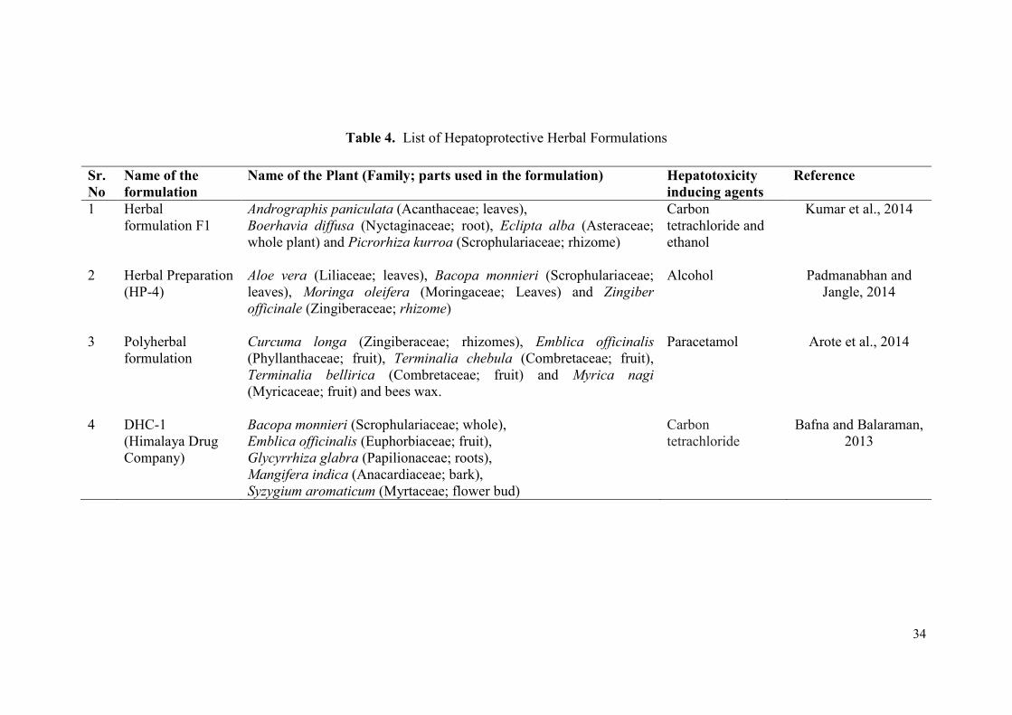
34
Table 4. List of Hepatoprotective Herbal Formulations
Sr.
No
Name of the
formulation
Name of the Plant (Family; parts used in the formulation) Hepatotoxicity
inducing agents
Reference
1 Herbal
formulation F1
Andrographis paniculata (Acanthaceae; leaves),
Boerhavia diffusa (Nyctaginaceae; root), Eclipta alba (Asteraceae;
whole plant) and Picrorhiza kurroa (Scrophulariaceae; rhizome)
Carbon
tetrachloride and
ethanol
Kumar et al., 2014
2 Herbal Preparation
(HP-4)
Aloe vera (Liliaceae; leaves), Bacopa monnieri (Scrophulariaceae;
leaves), Moringa oleifera (Moringaceae; Leaves) and Zingiber
officinale (Zingiberaceae; rhizome)
Alcohol Padmanabhan and
Jangle, 2014
3 Polyherbal
formulation
Curcuma longa (Zingiberaceae; rhizomes), Emblica officinalis
(Phyllanthaceae; fruit), Terminalia chebula (Combretaceae; fruit),
Terminalia bellirica (Combretaceae; fruit) and Myrica nagi
(Myricaceae; fruit) and bees wax.
Paracetamol Arote et al., 2014
4 DHC-1
(Himalaya Drug
Company)
Bacopa monnieri (Scrophulariaceae; whole),
Emblica officinalis (Euphorbiaceae; fruit),
Glycyrrhiza glabra (Papilionaceae; roots),
Mangifera indica (Anacardiaceae; bark),
Syzygium aromaticum (Myrtaceae; flower bud)
Carbon
tetrachloride
Bafna and Balaraman,
2013
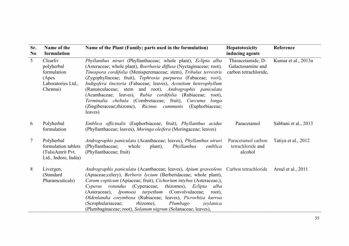
35
Sr.
No
Name of the
formulation
Name of the Plant (Family; parts used in the formulation) Hepatotoxicity
inducing agents
Reference
5 Clearliv
polyherbal
formulation
(Apex
Laboratories Ltd.,
Chennai)
Phyllanthus niruri (Phyllanthaceae; whole plant), Eclipta alba
(Asteraceae; whole plant), Boerhavia diffusa (Nyctaginaceae; root),
Tinospora cordifolia (Menisperemaceae; stem), Tribulus terrestris
(Zygophyllaceae; fruit), Tephrosia purpurea (Fabaceae; root),
Indigofera tinctoria (Fabaceae; leaves), Aconitum heterophyllum
(Ranunculaceae; stem and root), Andrographis paniculata
(Acanthaceae; leaves), Rubia cordifolia (Rubiaceae; root),
Terminalia chebula (Combretaceae; fruit), Curcuma longa
(Zingiberaceae;rhizome), Ricinus cummunis (Euphorbiaceae;
leaves)
Thioacetamide, D-
Galactosamine and
carbon tetrachloride,
Kumar et al., 2013a
6 Polyherbal
formulation
Emblica officinalis (Euphorbiaceae; fruit), Phyllanthus acidus
(Phyllanthaceae; leaves), Moringa oleifera (Moringaceae; leaves)
Paracetamol Sabbani et al., 2013
7 Polyherbal
formulation tablets
(TulsiAmrit Pvt.
Ltd., Indore, India)
Andrographis paniculata (Acanthaceae; leaves), Phyllanthus niruri
(Phyllanthaceae; whole plant), Phyllanthus emblica
(Phyllanthaceae; fruit)
Paracetamol carbon
tetrachloride and
alcohol
Tatiya et al., 2012
8 Livergen,
(Standard
Pharamcuticals)
Andrographis paniculata (Acanthaceae; leaves), Apium graveolens
(Apiaceae;celery), Berberis lycium (Berberidaceae; whole plant),
Carum copticum (Apiaceae; fruit), Cichorium intybus (Asteraceae;),
Cyperus rotundus (Cyperaceae; rhizomes), Eclipta alba
(Asteraceae), Ipomoea turpethum (Convolvulaceae; root),
Oldenlandia corymbosa (Rubiaceae; leaves), Picrorhiza kurroa
(Scrophulariaceae; rhizome), Plumbago zeylanica
(Plumbaginaceae; root), Solanum nigrum (Solanaceae; leaves),
Carbon tetrachloride Arsul et al., 2011
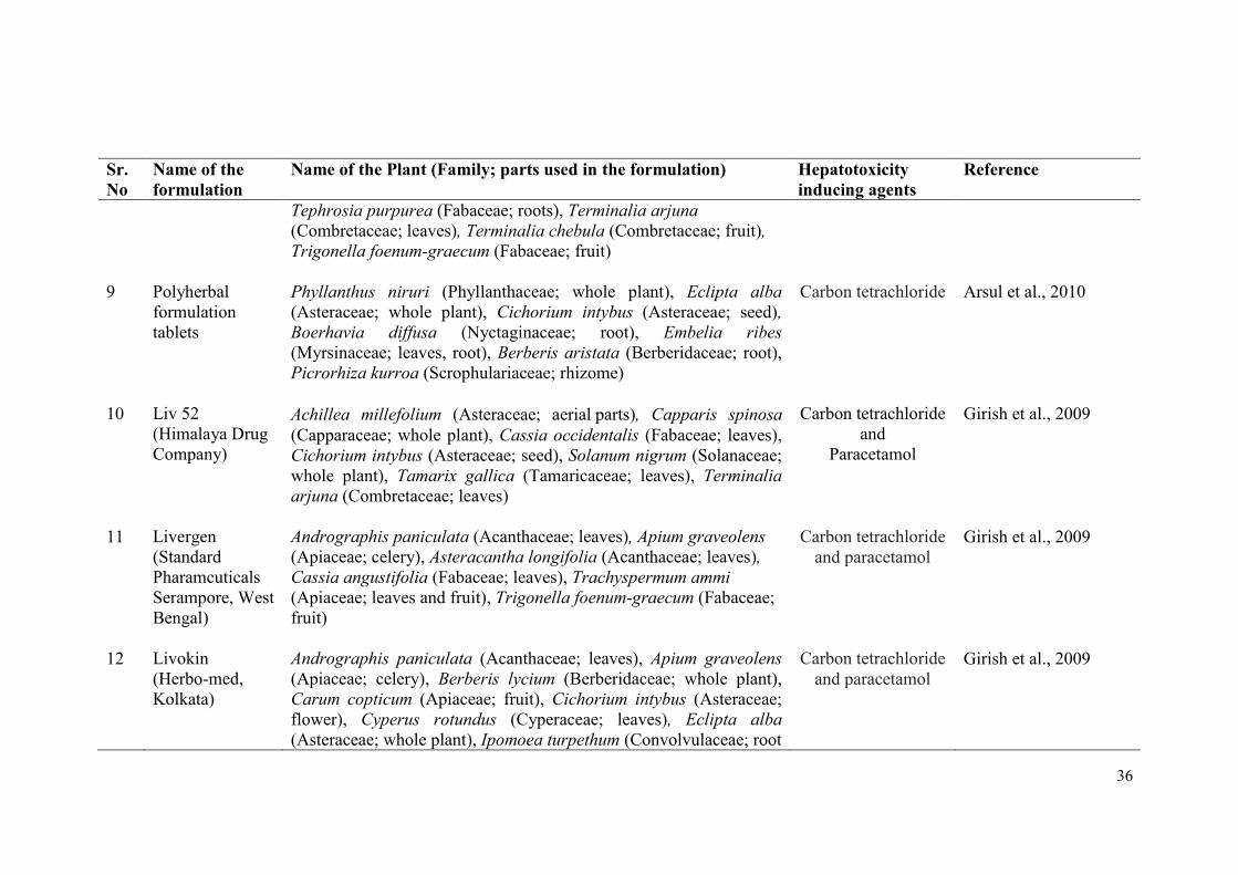
36
Sr.
No
Name of the
formulation
Name of the Plant (Family; parts used in the formulation) Hepatotoxicity
inducing agents
Reference
9
Polyherbal
formulation
tablets
Tephrosia purpurea (Fabaceae; roots), Terminalia arjuna
(Combretaceae; leaves), Terminalia chebula (Combretaceae; fruit),
Trigonella foenum-graecum (Fabaceae; fruit)
Phyllanthus niruri (Phyllanthaceae; whole plant), Eclipta alba
(Asteraceae; whole plant), Cichorium intybus (Asteraceae; seed),
Boerhavia diffusa (Nyctaginaceae; root), Embelia ribes
(Myrsinaceae; leaves, root), Berberis aristata (Berberidaceae; root),
Picrorhiza kurroa (Scrophulariaceae; rhizome)
Carbon tetrachloride
Arsul et al., 2010
10 Liv 52
(Himalaya Drug
Company)
Achillea millefolium (Asteraceae; aerial parts), Capparis spinosa
(Capparaceae; whole plant), Cassia occidentalis (Fabaceae; leaves),
Cichorium intybus (Asteraceae; seed), Solanum nigrum (Solanaceae;
whole plant), Tamarix gallica (Tamaricaceae; leaves), Terminalia
arjuna (Combretaceae; leaves)
Carbon tetrachloride
and
Paracetamol
Girish et al., 2009
11 Livergen
(Standard
Pharamcuticals
Serampore, West
Bengal)
Andrographis paniculata (Acanthaceae; leaves), Apium graveolens
(Apiaceae; celery), Asteracantha longifolia (Acanthaceae; leaves),
Cassia angustifolia (Fabaceae; leaves), Trachyspermum ammi
(Apiaceae; leaves and fruit), Trigonella foenum-graecum (Fabaceae;
fruit)
Carbon tetrachloride
and paracetamol
Girish et al., 2009
12 Livokin
(Herbo-med,
Kolkata)
Andrographis paniculata (Acanthaceae; leaves), Apium graveolens
(Apiaceae; celery), Berberis lycium (Berberidaceae; whole plant),
Carum copticum (Apiaceae; fruit), Cichorium intybus (Asteraceae;
flower), Cyperus rotundus (Cyperaceae; leaves), Eclipta alba
(Asteraceae; whole plant), Ipomoea turpethum (Convolvulaceae; root
Carbon tetrachloride
and paracetamol
Girish et al., 2009
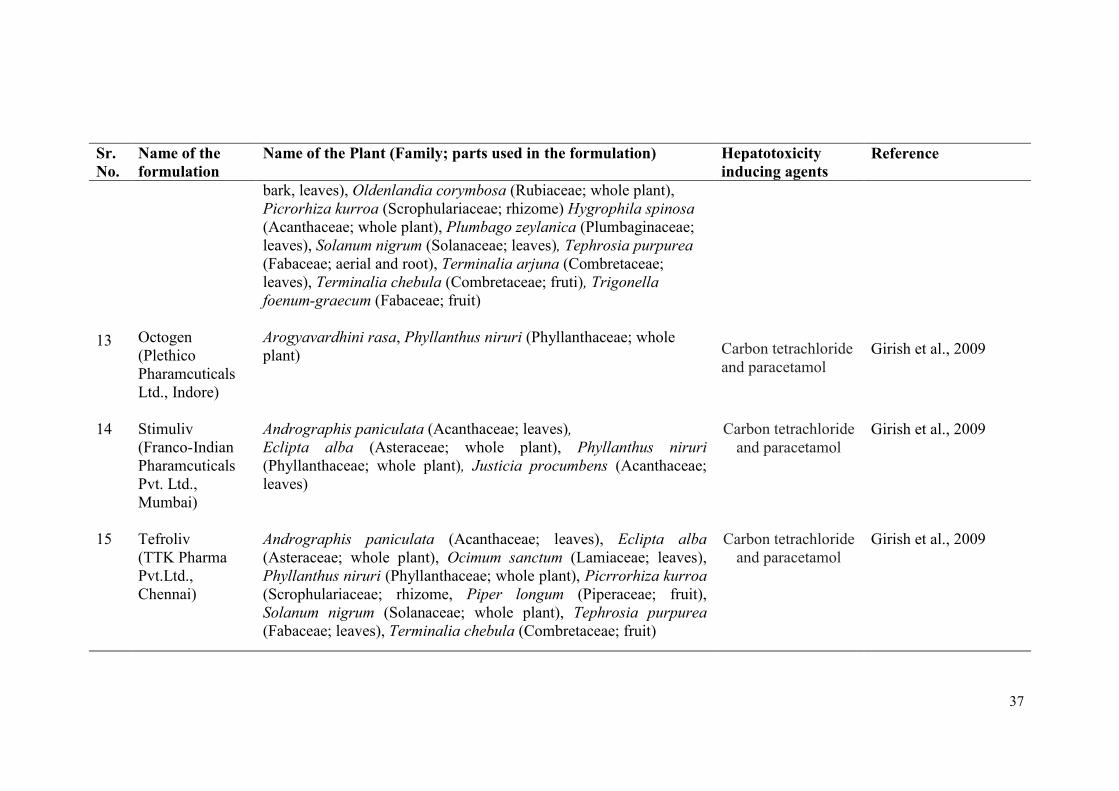
37
Sr.
No.
Name of the
formulation
Name of the Plant (Family; parts used in the formulation) Hepatotoxicity
inducing agents
Reference
13
Octogen
(Plethico
Pharamcuticals
Ltd., Indore)
bark, leaves), Oldenlandia corymbosa (Rubiaceae; whole plant),
Picrorhiza kurroa (Scrophulariaceae; rhizome) Hygrophila spinosa
(Acanthaceae; whole plant), Plumbago zeylanica (Plumbaginaceae;
leaves), Solanum nigrum (Solanaceae; leaves), Tephrosia purpurea
(Fabaceae; aerial and root), Terminalia arjuna (Combretaceae;
leaves), Terminalia chebula (Combretaceae; fruti), Trigonella
foenum-graecum (Fabaceae; fruit)
Arogyavardhini rasa, Phyllanthus niruri (Phyllanthaceae; whole
plant)
Carbon tetrachloride
and paracetamol
Girish et al., 2009
14 Stimuliv
(Franco-Indian
Pharamcuticals
Pvt. Ltd.,
Mumbai)
Andrographis paniculata (Acanthaceae; leaves),
Eclipta alba (Asteraceae; whole plant), Phyllanthus niruri
(Phyllanthaceae; whole plant), Justicia procumbens (Acanthaceae;
leaves)
Carbon tetrachloride
and paracetamol
Girish et al., 2009
15 Tefroliv
(TTK Pharma
Pvt.Ltd.,
Chennai)
Andrographis paniculata (Acanthaceae; leaves), Eclipta alba
(Asteraceae; whole plant), Ocimum sanctum (Lamiaceae; leaves),
Phyllanthus niruri (Phyllanthaceae; whole plant), Picrrorhiza kurroa
(Scrophulariaceae; rhizome, Piper longum (Piperaceae; fruit),
Solanum nigrum (Solanaceae; whole plant), Tephrosia purpurea
(Fabaceae; leaves), Terminalia chebula (Combretaceae; fruit)
Carbon tetrachloride
and paracetamol
Girish et al., 2009
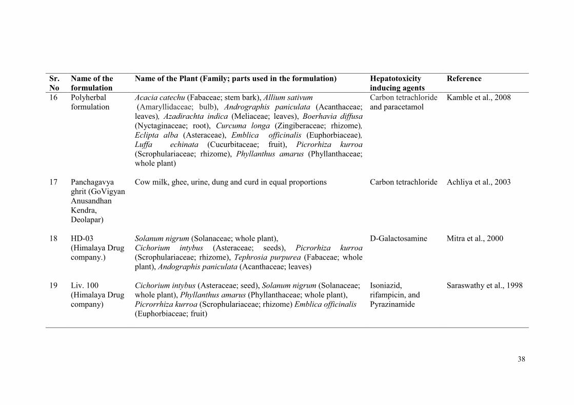
38
Sr.
No
Name of the
formulation
Name of the Plant (Family; parts used in the formulation) Hepatotoxicity
inducing agents
Reference
16 Polyherbal
formulation
Acacia catechu (Fabaceae; stem bark), Allium sativum
(Amaryllidaceae; bulb), Andrographis paniculata (Acanthaceae;
leaves), Azadirachta indica (Meliaceae; leaves), Boerhavia diffusa
(Nyctaginaceae; root), Curcuma longa (Zingiberaceae; rhizome),
Eclipta alba (Asteraceae), Emblica officinalis (Euphorbiaceae),
Luffa echinata (Cucurbitaceae; fruit), Picrorhiza kurroa
(Scrophulariaceae; rhizome), Phyllanthus amarus (Phyllanthaceae;
whole plant)
Carbon tetrachloride
and paracetamol
Kamble et al., 2008
17 Panchagavya
ghrit (GoVigyan
Anusandhan
Kendra,
Deolapar)
Cow milk, ghee, urine, dung and curd in equal proportions
Carbon tetrachloride Achliya et al., 2003
18 HD-03
(Himalaya Drug
company.)
Solanum nigrum (Solanaceae; whole plant),
Cichorium intybus (Asteraceae; seeds), Picrorhiza kurroa
(Scrophulariaceae; rhizome), Tephrosia purpurea (Fabaceae; whole
plant), Andographis paniculata (Acanthaceae; leaves)
D-Galactosamine Mitra et al., 2000
19 Liv. 100
(Himalaya Drug
company)
Cichorium intybus (Asteraceae; seed), Solanum nigrum (Solanaceae;
whole plant), Phyllanthus amarus (Phyllanthaceae; whole plant),
Picrorrhiza kurroa (Scrophulariaceae; rhizome) Emblica officinalis
(Euphorbiaceae; fruit)
Isoniazid,
rifampicin, and
Pyrazinamide
Saraswathy et al., 1998
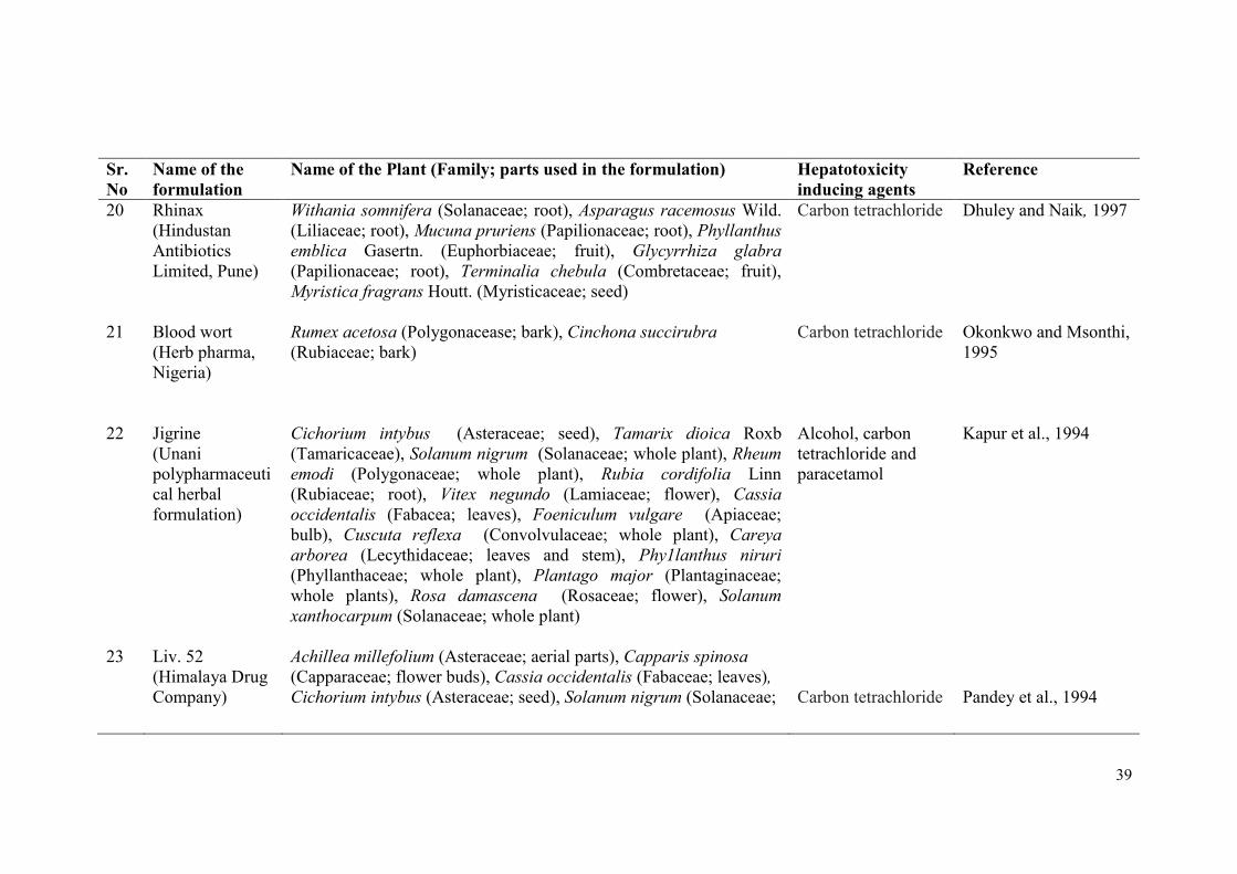
39
Sr.
No
Name of the
formulation
Name of the Plant (Family; parts used in the formulation) Hepatotoxicity
inducing agents
Reference
20 Rhinax
(Hindustan
Antibiotics
Limited, Pune)
Withania somnifera (Solanaceae; root), Asparagus racemosus Wild.
(Liliaceae; root), Mucuna pruriens (Papilionaceae; root), Phyllanthus
emblica Gasertn. (Euphorbiaceae; fruit), Glycyrrhiza glabra
(Papilionaceae; root), Terminalia chebula (Combretaceae; fruit),
Myristica fragrans Houtt. (Myristicaceae; seed)
Carbon tetrachloride Dhuley and Naik, 1997
21 Blood wort
(Herb pharma,
Nigeria)
Rumex acetosa (Polygonacease; bark), Cinchona succirubra
(Rubiaceae; bark)
Carbon tetrachloride Okonkwo and Msonthi,
1995
22 Jigrine
(Unani
polypharmaceuti
cal herbal
formulation)
Cichorium intybus (Asteraceae; seed), Tamarix dioica Roxb
(Tamaricaceae), Solanum nigrum (Solanaceae; whole plant), Rheum
emodi (Polygonaceae; whole plant), Rubia cordifolia Linn
(Rubiaceae; root), Vitex negundo (Lamiaceae; flower), Cassia
occidentalis (Fabacea; leaves), Foeniculum vulgare (Apiaceae;
bulb), Cuscuta reflexa (Convolvulaceae; whole plant), Careya
arborea (Lecythidaceae; leaves and stem), Phy1lanthus niruri
(Phyllanthaceae; whole plant), Plantago major (Plantaginaceae;
whole plants), Rosa damascena (Rosaceae; flower), Solanum
xanthocarpum (Solanaceae; whole plant)
Alcohol, carbon
tetrachloride and
paracetamol
Kapur et al., 1994
23 Liv. 52
(Himalaya Drug
Company)
Achillea millefolium (Asteraceae; aerial parts), Capparis spinosa
(Capparaceae; flower buds), Cassia occidentalis (Fabaceae; leaves),
Cichorium intybus (Asteraceae; seed), Solanum nigrum (Solanaceae;
Carbon tetrachloride
Pandey et al., 1994
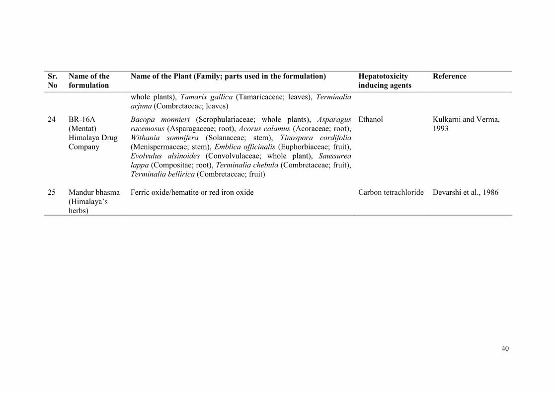
40
Sr.
No
Name of the
formulation
Name of the Plant (Family; parts used in the formulation) Hepatotoxicity
inducing agents
Reference
whole plants), Tamarix gallica (Tamaricaceae; leaves), Terminalia
arjuna (Combretaceae; leaves)
24 BR-16A
(Mentat)
Himalaya Drug
Company
Bacopa monnieri (Scrophulariaceae; whole plants), Asparagus
racemosus (Asparagaceae; root), Acorus calamus (Acoraceae; root),
Withania somnifera (Solanaceae; stem), Tinospora cordifolia
(Menispermaceae; stem), Emblica officinalis (Euphorbiaceae; fruit),
Evolvulus alsinoides (Convolvulaceae; whole plant), Saussurea
lappa (Compositae; root), Terminalia chebula (Combretaceae; fruit),
Terminalia bellirica (Combretaceae; fruit)
Ethanol Kulkarni and Verma,
1993
25 Mandur bhasma
(Himalaya’s
herbs)
Ferric oxide/hematite or red iron oxide Carbon tetrachloride Devarshi et al., 1986

41
The synchronous activity of ingredients from polyherbal formulation is believed to be
responsible for hepatoprotective activities of these formulations (Range et al., 2003;
Arsul et al., 2011). Individual or polyherbal drugs are used for treatment of various
liver disorders caused by toxic chemicals, viruses and excess alcohol intake (Kashaw
et al., 2011). Herbs and herbal preparations are generally viewed as safe. But along
with the beneficial components, they may contain many bioactive compounds which
could impose potentially deleterious effects (Lal et al., 2007).
3.4.2 Hepatoprotective Medicinal Plants
Medicinal plants play an important role in human health care. Management of
liver diseases is still a challenge for modern medicine (Deepthi, 2015). Even the
developed countries are now looking for time-tested traditional and alternative
medicines as a remedy for liver diseases (Sheth, 2005). As per WHO report, more
than 80% of global population uses medicines derived from plants or plant products
(Sheth, 2005; WHO, 2011). The traditional medicine system specifies a broad range
of natural health care practices including folk/tribal practices as well as Ayurveda,
Siddha and Unani (WHO, 2011). To a large extent these medical practices, originated
from time immemorial, have developed gradually based on practical experiences
(Shaik et al., 2012; Deepthi, 2015). Many medicinal plants exhibit significant
hepatoprotective activity in animal models. These plants and their parts used for
treatment are arranged systemically in Table 5.
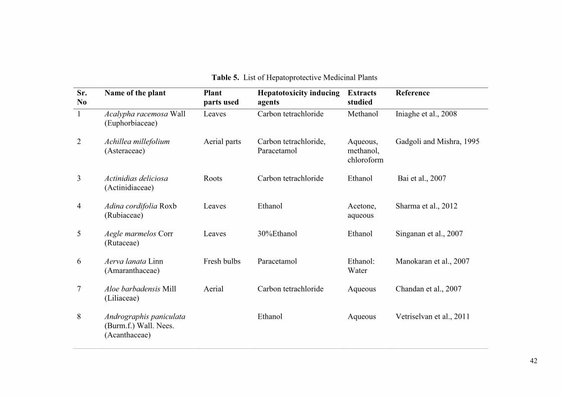
42
Table 5. List of Hepatoprotective Medicinal Plants
Sr.
No
Name of the plant Plant
parts used
Hepatotoxicity inducing
agents
Extracts
studied
Reference
1 Acalypha racemosa Wall
(Euphorbiaceae)
Leaves Carbon tetrachloride
Methanol Iniaghe et al., 2008
2 Achillea millefolium
(Asteraceae)
Aerial parts
Carbon tetrachloride,
Paracetamol
Aqueous,
methanol,
chloroform
Gadgoli and Mishra, 1995
3 Actinidias deliciosa
(Actinidiaceae)
Roots Carbon tetrachloride Ethanol Bai et al., 2007
4 Adina cordifolia Roxb
(Rubiaceae)
Leaves Ethanol Acetone,
aqueous
Sharma et al., 2012
5 Aegle marmelos Corr
(Rutaceae)
Leaves 30%Ethanol Ethanol Singanan et al., 2007
6 Aerva lanata Linn
(Amaranthaceae)
Fresh bulbs Paracetamol Ethanol:
Water
Manokaran et al., 2007
7 Aloe barbadensis Mill
(Liliaceae)
Aerial Carbon tetrachloride Aqueous Chandan et al., 2007
8 Andrographis paniculata
(Burm.f.) Wall. Nees.
(Acanthaceae)
Ethanol Aqueous Vetriselvan et al., 2011
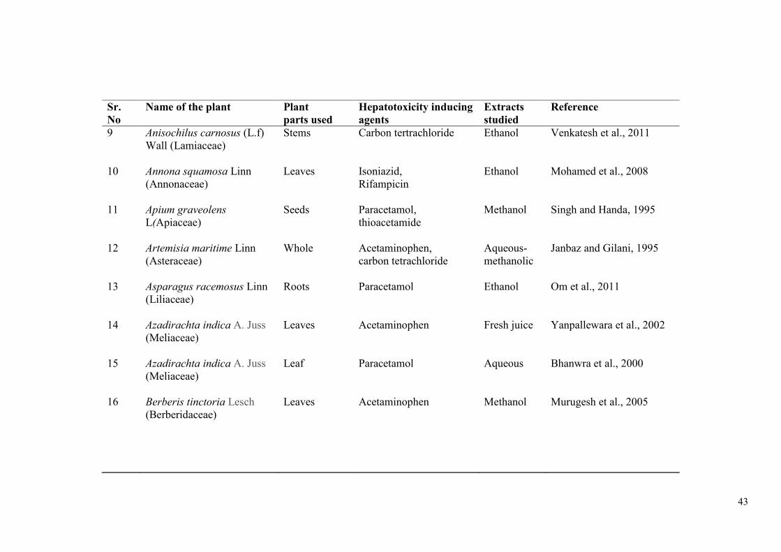
43
Sr.
No
Name of the plant Plant
parts used
Hepatotoxicity inducing
agents
Extracts
studied
Reference
9 Anisochilus carnosus (L.f)
Wall (Lamiaceae)
Stems Carbon tertrachloride Ethanol Venkatesh et al., 2011
10 Annona squamosa Linn
(Annonaceae)
Leaves Isoniazid,
Rifampicin
Ethanol Mohamed et al., 2008
11 Apium graveolens
L(Apiaceae)
Seeds Paracetamol,
thioacetamide
Methanol Singh and Handa, 1995
12 Artemisia maritime Linn
(Asteraceae)
Whole Acetaminophen,
carbon tetrachloride
Aqueous-
methanolic
Janbaz and Gilani, 1995
13 Asparagus racemosus Linn
(Liliaceae)
Roots Paracetamol Ethanol Om et al., 2011
14 Azadirachta indica A. Juss
(Meliaceae)
Leaves Acetaminophen Fresh juice Yanpallewara et al., 2002
15 Azadirachta indica A. Juss
(Meliaceae)
Leaf Paracetamol Aqueous Bhanwra et al., 2000
16 Berberis tinctoria Lesch
(Berberidaceae)
Leaves Acetaminophen Methanol Murugesh et al., 2005
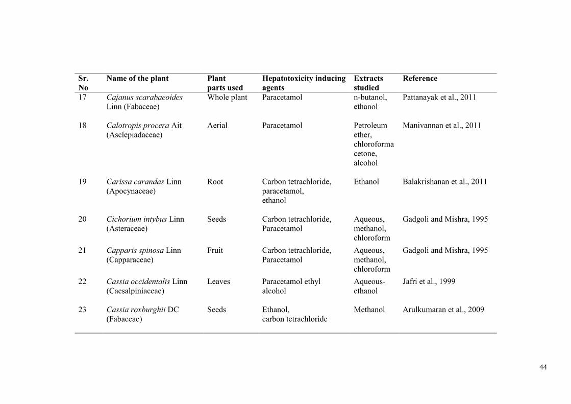
44
Sr.
No
Name of the plant Plant
parts used
Hepatotoxicity inducing
agents
Extracts
studied
Reference
17 Cajanus scarabaeoides
Linn (Fabaceae)
Whole plant
Paracetamol n-butanol,
ethanol
Pattanayak et al., 2011
18 Calotropis procera Ait
(Asclepiadaceae)
Aerial Paracetamol Petroleum
ether,
chloroforma
cetone,
alcohol
Manivannan et al., 2011
19 Carissa carandas Linn
(Apocynaceae)
Root
Carbon tetrachloride,
paracetamol,
ethanol
Ethanol Balakrishanan et al., 2011
20 Cichorium intybus Linn (Asteraceae)
Seeds
Carbon tetrachloride,
Paracetamol
Aqueous,
methanol,
chloroform
Gadgoli and Mishra, 1995
21 Capparis spinosa Linn
(Capparaceae)
Fruit Carbon tetrachloride,
Paracetamol
Aqueous,
methanol,
chloroform
Gadgoli and Mishra, 1995
22 Cassia occidentalis Linn
(Caesalpiniaceae)
Leaves Paracetamol ethyl
alcohol
Aqueous-
ethanol
Jafri et al., 1999
23 Cassia roxburghii DC
(Fabaceae)
Seeds Ethanol,
carbon tetrachloride
Methanol Arulkumaran et al., 2009
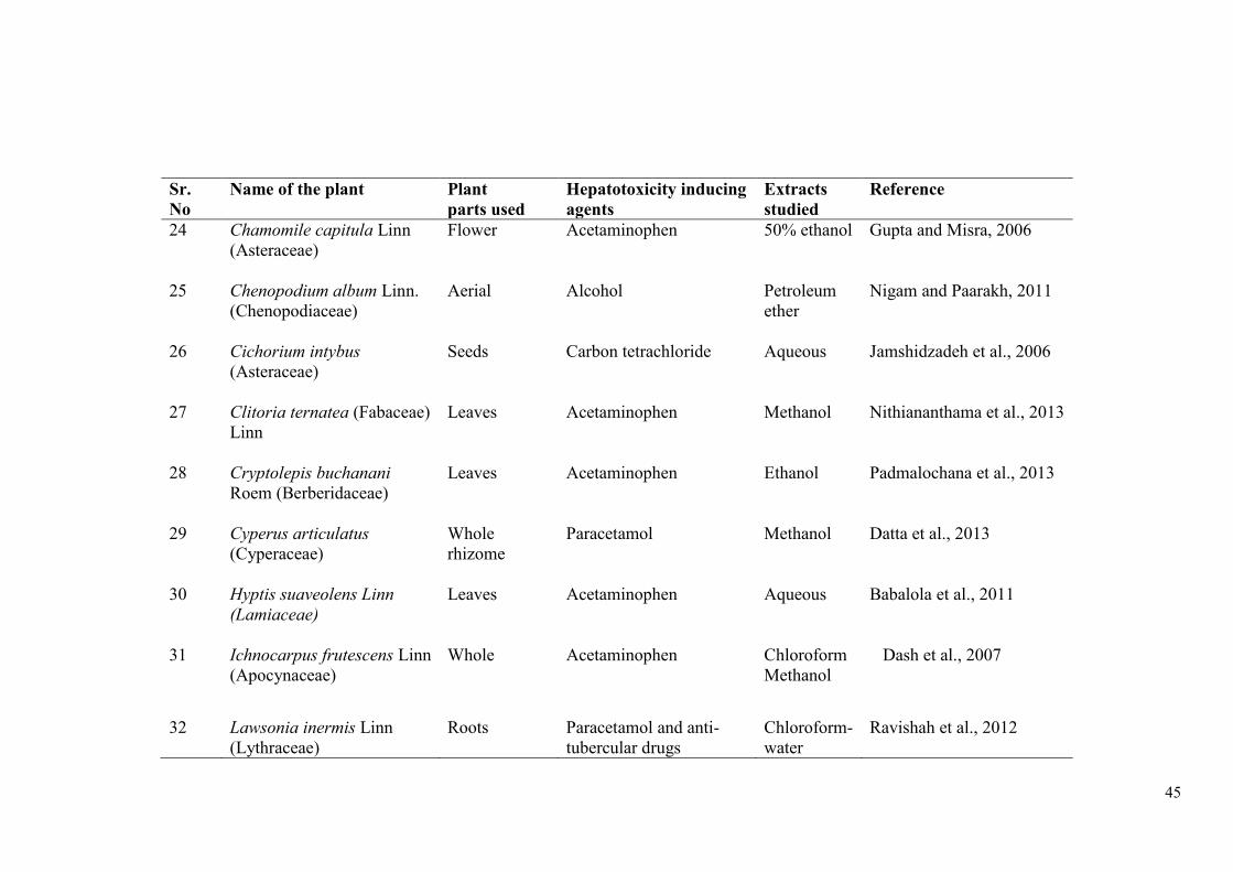
45
Sr.
No
Name of the plant Plant
parts used
Hepatotoxicity inducing
agents
Extracts
studied
Reference
24 Chamomile capitula Linn
(Asteraceae)
Flower Acetaminophen
50% ethanol Gupta and Misra, 2006
25 Chenopodium album Linn.
(Chenopodiaceae)
Aerial Alcohol Petroleum
ether
Nigam and Paarakh, 2011
26 Cichorium intybus
(Asteraceae)
Seeds Carbon tetrachloride Aqueous Jamshidzadeh et al., 2006
27 Clitoria ternatea (Fabaceae)
Linn
Leaves Acetaminophen Methanol Nithiananthama et al., 2013
28 Cryptolepis buchanani
Roem (Berberidaceae)
Leaves Acetaminophen Ethanol Padmalochana et al., 2013
29 Cyperus articulatus
(Cyperaceae)
Whole
rhizome
Paracetamol Methanol Datta et al., 2013
30 Hyptis suaveolens Linn
(Lamiaceae)
Leaves Acetaminophen Aqueous Babalola et al., 2011
31 Ichnocarpus frutescens Linn
(Apocynaceae)
Whole Acetaminophen
Chloroform
Methanol
Dash et al., 2007
32 Lawsonia inermis Linn
(Lythraceae)
Roots Paracetamol and anti-
tubercular drugs
Chloroform-
water
Ravishah et al., 2012
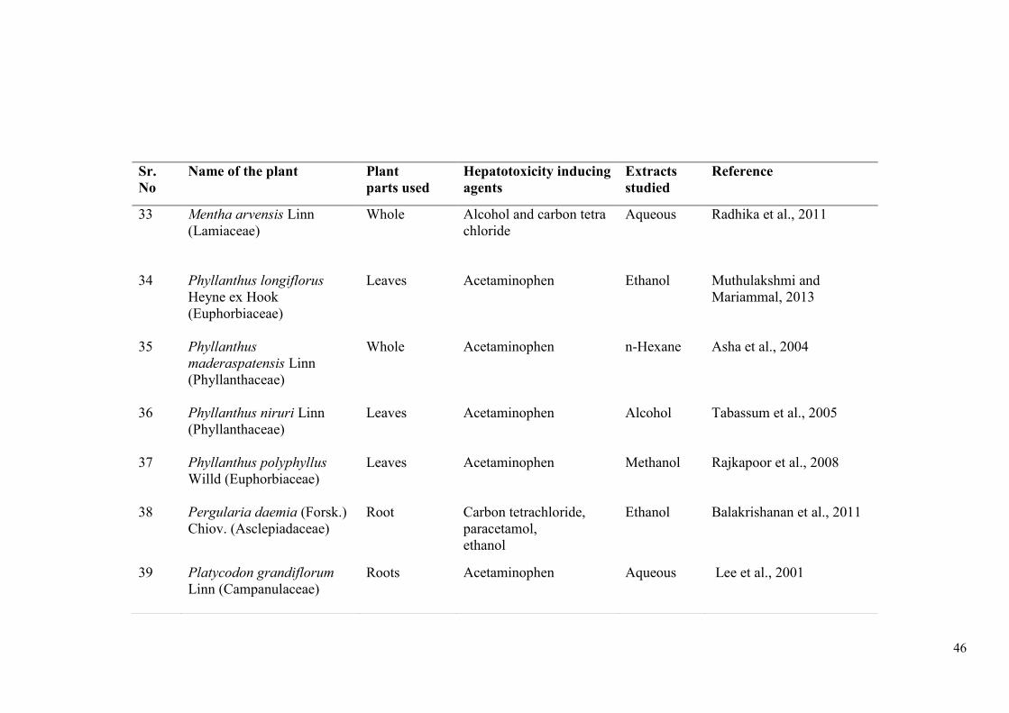
46
Sr.
No
Name of the plant Plant
parts used
Hepatotoxicity inducing
agents
Extracts
studied
Reference
33 Mentha arvensis Linn
(Lamiaceae)
Whole Alcohol and carbon tetra
chloride
Aqueous Radhika et al., 2011
34 Phyllanthus longiflorus
Heyne ex Hook
(Euphorbiaceae)
Leaves Acetaminophen Ethanol Muthulakshmi and
Mariammal, 2013
35 Phyllanthus
maderaspatensis Linn
(Phyllanthaceae)
Whole Acetaminophen
n-Hexane Asha et al., 2004
36 Phyllanthus niruri Linn
(Phyllanthaceae)
Leaves Acetaminophen
Alcohol Tabassum et al., 2005
37 Phyllanthus polyphyllus
Willd (Euphorbiaceae)
Leaves Acetaminophen
Methanol Rajkapoor et al., 2008
38 Pergularia daemia (Forsk.)
Chiov. (Asclepiadaceae)
Root
Carbon tetrachloride,
paracetamol,
ethanol
Ethanol Balakrishanan et al., 2011
39 Platycodon grandiflorum
Linn (Campanulaceae)
Roots Acetaminophen Aqueous Lee et al., 2001
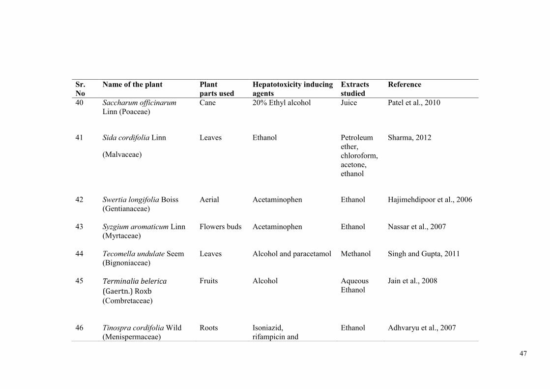
47
Sr.
No
Name of the plant Plant
parts used
Hepatotoxicity inducing
agents
Extracts
studied
Reference
40 Saccharum officinarum
Linn (Poaceae)
Cane 20% Ethyl alcohol Juice Patel et al., 2010
41 Sida cordifolia Linn
(Malvaceae)
Leaves Ethanol Petroleum
ether,
chloroform,
acetone,
ethanol
Sharma, 2012
42 Swertia longifolia Boiss
(Gentianaceae)
Aerial Acetaminophen
Ethanol Hajimehdipoor et al., 2006
43 Syzgium aromaticum Linn
(Myrtaceae)
Flowers buds Acetaminophen
Ethanol Nassar et al., 2007
44 Tecomella undulate Seem
(Bignoniaceae)
Leaves Alcohol and paracetamol Methanol Singh and Gupta, 2011
45 Terminalia belerica
(Gaertn.) Roxb (Combretaceae)
Fruits Alcohol Aqueous
Ethanol
Jain et al., 2008
46 Tinospra cordifolia Wild
(Menispermaceae)
Roots Isoniazid,
rifampicin and
Ethanol Adhvaryu et al., 2007
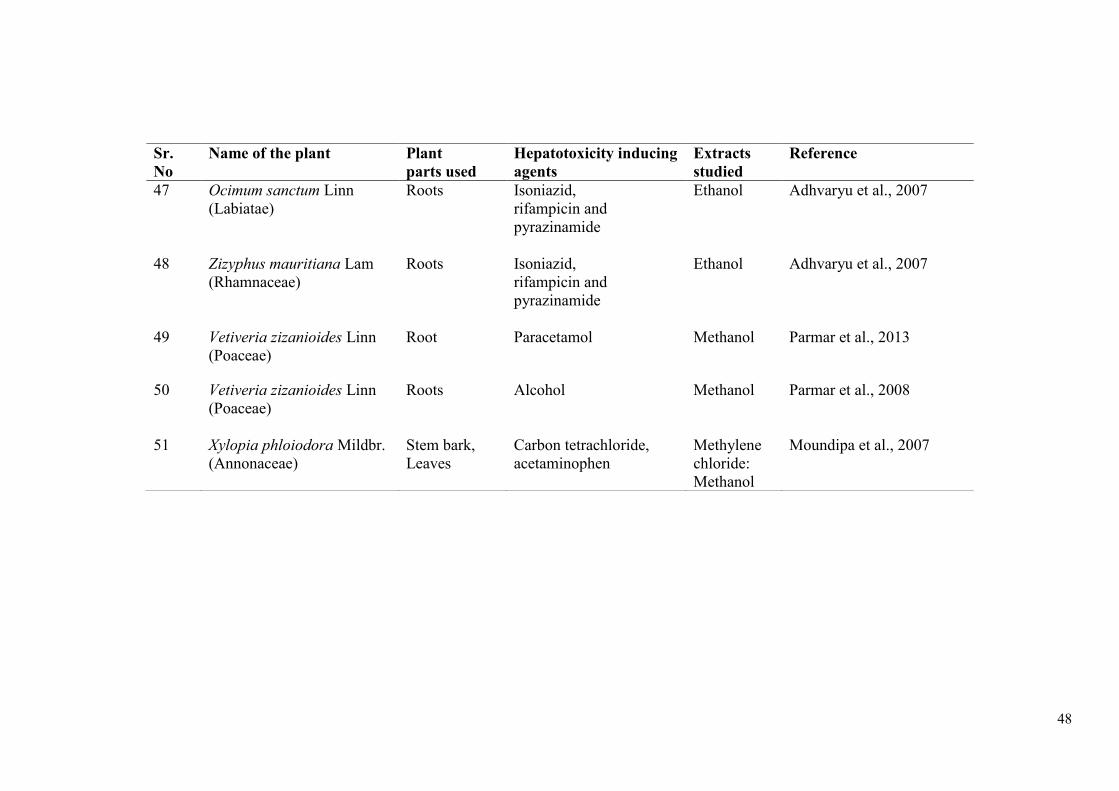
48
Sr.
No
Name of the plant Plant
parts used
Hepatotoxicity inducing
agents
Extracts
studied
Reference
47 Ocimum sanctum Linn
(Labiatae)
Roots Isoniazid,
rifampicin and
pyrazinamide
Ethanol Adhvaryu et al., 2007
48 Zizyphus mauritiana Lam
(Rhamnaceae)
Roots Isoniazid,
rifampicin and
pyrazinamide
Ethanol Adhvaryu et al., 2007
49 Vetiveria zizanioides Linn
(Poaceae)
Root Paracetamol Methanol Parmar et al., 2013
50 Vetiveria zizanioides Linn
(Poaceae)
Roots Alcohol Methanol Parmar et al., 2008
51 Xylopia phloiodora Mildbr.
(Annonaceae)
Stem bark,
Leaves
Carbon tetrachloride,
acetaminophen
Methylene
chloride:
Methanol
Moundipa et al., 2007
49
Various herbs and plant extracts have significant hepatoprotective activity as
indicated from studies in animal models (Chaudhary et al., 2010). The
hepatoprotective activity is probably due to the presence of active principles which
are mostly in the form of secondary metabolites in medicinal plants (Singh et al.,
2011). The literature till date indicates that extracts of leaves, stems, fruits, roots or
even whole plants have significant potential towards treatment of hepatic diseases
(Ramalingum and Mahomoodally, 2014).
3.4.2.1 Tinospora cordifolia (Willd.) Miers ex Hook. F. & Thoms.
T. cordifolia has been reported to possess strong hepatoprotective and
antioxidant activities. Root and stem of T. cordifolia exhibit anti-inflammatory
(Patgiri et al., 2014), antipyretic (Ashok et al., 2010), immunomodulatory (Upadhyaya
et al., 2011) and anti-cancer activities (Ahmad et al., 2015). The immunomodulatory
activity of T. cordifolia is attributed to G1-4A, an arabinogalactan, found in fresh
stem of T. Cordifolia (Desai et al., 2007).
Bishayi et al. (2002) and Kavitha et al. (2011b) reported hepatoprotective and
immunostimulatory effects of T. cordifolia in mature rats intoxicated with carbon
tetrachloride (CCl4). Administration of T. cordifolia extract (100 mg/kg body weight
for 15 days) was found to protect the liver in CCl4 intoxicated rats, as indicated by
significant reduction in serum levels of SGOT, SGPT, ALP, and bilirubin. Extracts of
aerial roots of T. cordifolia reported hepatoprotective effects and immunosuppression
activity against isoniazid, rifampicin and pyrazinamide induced liver injury in guinea
pigs (Adhvaryu et al., 2007). A patented polyherbal formulation with T. cordifolia
extract also exhibited in vitro inactivation of Hepatitis B and E surface antigens
within 48-72 hrs of treatment (Mehrotra et al., 2000). The aqueous extract of roots of
T. cordifolia showed anti-oxidant action in alloxan induced diabetic rats. The
treatment of root extract of T. cordifolia (25, 50 mg/kg body weight) for 6 weeks in
alloxan induced diabetic rats showed significant reduction in serum and tissue
cholesterol, phospholipids and free fatty acids (Stanely et al., 1999). Treatment of
alloxan induced diabetic rats with alcoholic extracts of root (at a dose of 100 mg/kg
orally for six weeks) is reported to be highly effective in normalizing antioxidant
status of heart, brain, liver and kidney (Prince et al., 2004). Being major determinant
in outcome of liver injury, Kupffer cells activity has been studied in chronic liver

50
disease model. Carbon clearance test after T. cordifolia treatment showed significant
improvement in Kupffer cell function and normalization of SGOT, SGPT, ALP and
bilirubin levels (Nagarkatti et al., 1994). Sharma and Pandey (2010) reported
hepatoprotective activity of aqueous extracts of stem and leaves (400 mg/kg body
weight, orally) against lead nitrate induced toxicity in Swiss albino male mice.
Increase in SOD and CAT activity while decrease in the levels of SGOT, SGPT and
ALP enzymes were reported in mice treated with aqueous stem and leaves extract
along with lead nitrate, depicting hepatoprotective activity of T. cordifolia. Kumar et
al. (2013b) also reported hepatoprotective activity of aqueous extract of aerial parts of
T. cordifolia against CCl4 induced hepatotoxicity in wistar albino rats by reducing
levels of serum total bilirubin, SGPT, SGOT, and ALP. Treatment of T. cordifolia
aqueous extract also showed radio-protective activity against a sub-lethal dose of
gamma radiation in mice (Kapur et al., 2010; Mittal et al., 2014). A clinical study has
reported that T. cordifolia plays an important role in normalization of altered liver
functions (ALT, AST) (Karkal and Bairy, 2007).
3.4.2.2 Tinospora sinensis (Lour.) Merrill
The alcoholic and aqueous extract of T. sinensis has been reported to possess
anti-inflammatory (Li et al., 2003), anti-diabetic (Yonemitsu et al., 1993),
hepatoprotective (Chavan et al., 2013a), immunomodulatory (Manjrekar et al., 2000)
adaptogenic (Gupta et al., 2012) and other biological potentials (Yonemitsu et al.,
1993) with different animal models. It has also been shown to have anti-cancer
activity (Punitha et al., 2012) in human malignant cancer cell lines.
Yonemitsu et al. (1993) isolated a new phenolic glycoside, tinosinen from the
fresh stems of T. sinensis Merr. The structure was established on the basis of acid
hydrolysis and spectral data (IR, NMR, mass) as (E)-l-(3-hydroxy-1-propenyl)-3, 5-
dimethoxyphenyl 4-0-13-napiofuranosyl-(l-->3)-J3-D-glucopyranoside. Maurya et al.
(2009) reported isolation of two new compounds 4-methyl-heptadec-6-enoic acid
ethyl ester and 3-hydroxy-2, 9, 11-trimethoxy-5, 6-dihydro isoquino [3, 2-a]
isoquinolinylium from ethanolic extract of the stems of T. sinensis, along with other
six known compounds. Detailed spectroscopic studies were performed to establish
structures of these new compounds. Activity guided purification of ethanol extract of
T. sinensis yielded two compounds with significant in vitro inhibitory activity against
51
protein tyrosine phosphatase 1B (PTP1B) (Gupta et al., 2012). Naik et al. (2013)
reported hepatoprotective activity of ethanolic root extract of T. sinensis against
carbon tetrachloride (CCl4) induced liver damage in rats at a dose of 150mg/kg body
weight and 300mg/kg body weight orally in rats. The animals exhibited reduction in
levels of SGOT, SGPT and ALP indicating hepatoprotective activity of ethanolic
extract of T. sinensis roots. Treatment of ethanolic extract of T. sinensis leaf at a dose
of 300 mg/kg body weight showed anti-arthritic activity against adjuvant (Freud’s)
induced arthritis in rats (Sumathy et al., 2014). Analgesic activity of ethanolic extract
of T. sinensis leaves at a dose of 250 mg/kg and 500 mg/kg has also been reported in
rats (Sandhyarani and Kumar, 2014).
3.4.2.3 Neem-giloe
Neem-giloe (Neem-guduchi) is a traditionally used medicine from Ayurveda. It
is a Guduchi plant growing on Neem tree (Azadirachta indica). It is bitter to taste and
is believed to contain the medicinal virtues of Neem also and hence supposed to be
more efficacious (Sinha et al., 2004). Chemical investigations of water extract of the
stem of Neem-guduchi revealed that it contains glycoside giloin and non-glycosides,
bitter gilenin and glosteral (Bhide et al., 1941; Pendse et al., 1977). Oral and
intraperitoneal treatment of the water extract of the stem of Neem-guduchi at dose of
60 mg/100g displayed anti-inflammatory anti-arthritic effects (Pendse et al., 1977). It
also significantly reversed formation of antibody by typhoid "H" antigen (Pendse et
al., 1977). These reports indicate anti-inflammatory, immunosuppressive and
analgesic properties of the Satwa (Pendse, et al., 1977; Upadhyay, 2010). Nagarkar et
al. (2013) reported that Neem-guduchi showed hepatoprotective activity against a
single high-dose induced acetaminophen toxicity in rats. As per Bhalerao et al.
(2012), immunostimulatory effect produced by Neem-guduchi may be due to cell
mediated and humoral activation of T and B cells. Administration of Neem-guduchi
extract at a dose of 200mg/kg body weight showed radical scavenging and
immunomodulatory activity in rats (Bhalerao et al., 2012).
3.4.3 Guduchi Satwa
Satwa is an aqueous extractable solid substance collected from herbal drug
(Patil and Chaudhary, 2013). Guduchi satwa is the most common example of
52
herbal satwa and is very commonly prescribed in Ayurveda (Sharma et al., 2013b).
Rasendra Mangalam mentions Guduchi satwa for the first time in the literature
(Nagarjuna, 2008; Sharma et al., 2012). Guduchi satwa preparation has been
mentioned in Yoga Ratnakara (Yogaratnakar, 2002) and then Siddha Yoga Sangraha
(Tzrikamaji, 1954), Rasa Yoga Sagar (Sharma, 2004) etc. All these texts have
mentioned different methods for satwa preparation. Due to its usefulness in diseases
like fever and high nutritive and digestive values (Sinha et al., 2004), it is known as
Indian Quinine (Sharma et al., 2012) and is used as a general tonic (Thakur et al.,
1989).
Earlier studies reported the pharmaceutical aspect of guduchi satwa with
quantitative variations in the final product (Sharma et al., 2012). Guduchi Satwa (a
whitish starch like material extracted from T. cordifolia) is commonly recommended
in conditions like Jwara (fever), Daha (burning sensation) and Pitta predominant
disorders (Mishra and Vaishya, 2002; Sharma et al., 2013b). As per the report of
Pandey (2012), Guduchi Satwa is highly valued for many ailments like fever, chronic
diarrhea, chronic dysentery, burning sensation, secondary syphilis, chronic gonorrhea,
leucorrhoea, jaundice, rheumatism, urinary disorders etc. Prasad et al. (2014) have
reported scientific validation and standardization of the pharmaceutical procedure of
Guduchi Satwa for treating Kshaya (Phthisis), Raktapitta (Bleeding Disorder) and
Pada daha (Burning sensation of feet).
The standardization of Guduchi satwa includes parameters like organoleptic
characters, physicochemical parameters, phyto-analysis and development of TLC as
recommended by WHO and pharmacopoeia committee (Anonymous, 1987; Prasad et
al., 2014). Guduchi satwa has been reported to contain arabinogalactan with
immunological activity (Chintalwar et al., 1999). Ayurvedic system has also
recommended the use of Guduchi satwa in different stages of prameha (Shastri, 2002;
Panashikar et al., 2011; Sharma et al., 2013a). Efforts have been made to evaluate the
acceptability of Guduchi incorporated food products and thus the utilization of its
proven ethno-medicinal properties in improving nutritional status of vulnerable
sections of the society (Geeta and Kumari, 2013).

53
3.4.4 Nutritional Supplements
Nutrition or dietary supplements help the regeneration of liver cells forming a
basis for treatment of some liver disorders (Shukla and Kumar, 2013). Nutritional
supplements contain herbal products, vitamins, minerals, and any product that is not a
food or drug (medication) (American Cancer Society, 2015). There are very few
reports available on hepatoprotective activity of nutritional supplements (Shukla and
Kumar, 2013).
The list of nutritional supplements used in liver disorders is given in Table 6.
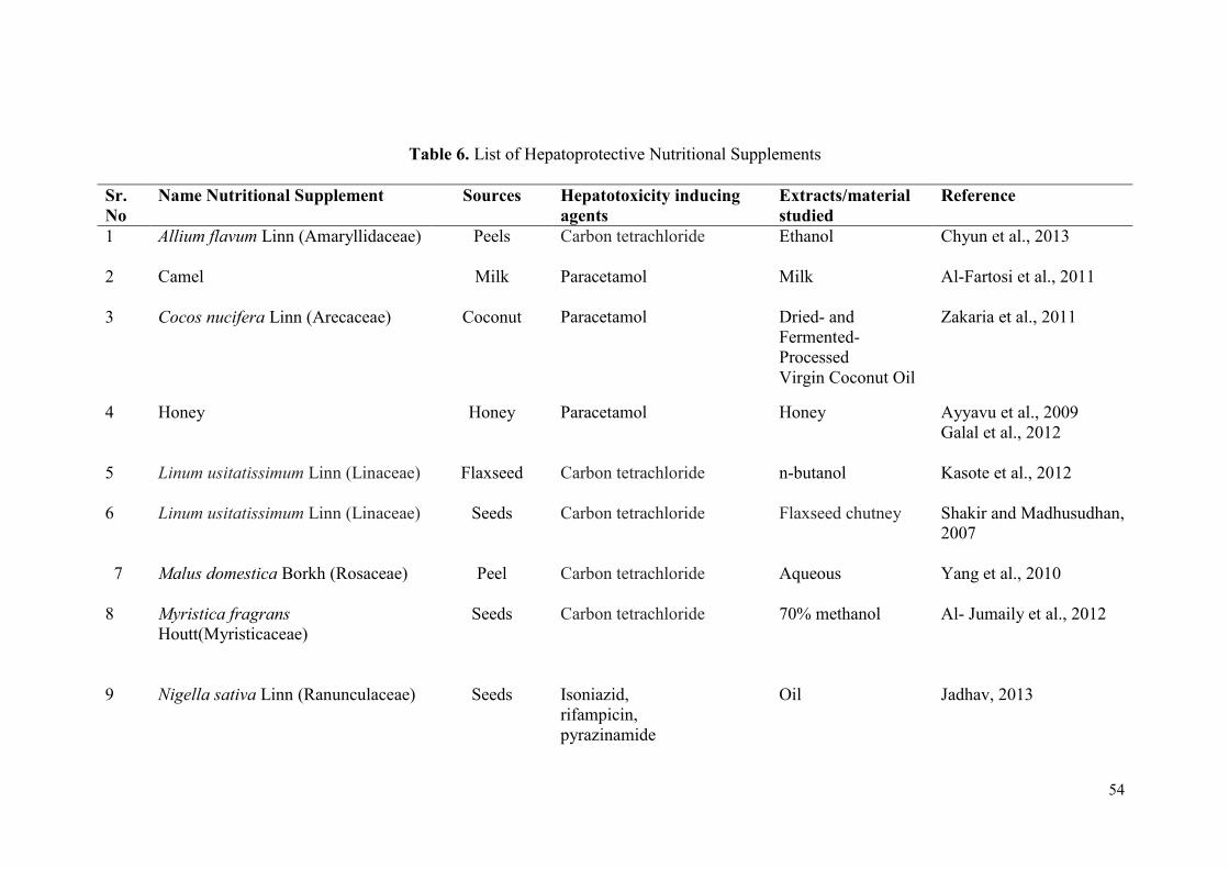
54
Table 6. List of Hepatoprotective Nutritional Supplements
Sr.
No
Name Nutritional Supplement Sources Hepatotoxicity inducing
agents
Extracts/material
studied
Reference
1 Allium flavum Linn (Amaryllidaceae) Peels Carbon tetrachloride Ethanol Chyun et al., 2013
2 Camel Milk Paracetamol Milk Al-Fartosi et al., 2011
3 Cocos nucifera Linn (Arecaceae) Coconut Paracetamol Dried- and
Fermented-
Processed
Virgin Coconut Oil
Zakaria et al., 2011
4 Honey Honey Paracetamol Honey Ayyavu et al., 2009
Galal et al., 2012
5 Linum usitatissimum Linn (Linaceae) Flaxseed Carbon tetrachloride n-butanol Kasote et al., 2012
6 Linum usitatissimum Linn (Linaceae) Seeds Carbon tetrachloride Flaxseed chutney Shakir and Madhusudhan,
2007
7 Malus domestica Borkh (Rosaceae) Peel
Carbon tetrachloride Aqueous Yang et al., 2010
8 Myristica fragrans
Houtt(Myristicaceae)
Seeds Carbon tetrachloride 70% methanol Al- Jumaily et al., 2012
9 Nigella sativa Linn (Ranunculaceae) Seeds Isoniazid,
rifampicin,
pyrazinamide
Oil Jadhav, 2013
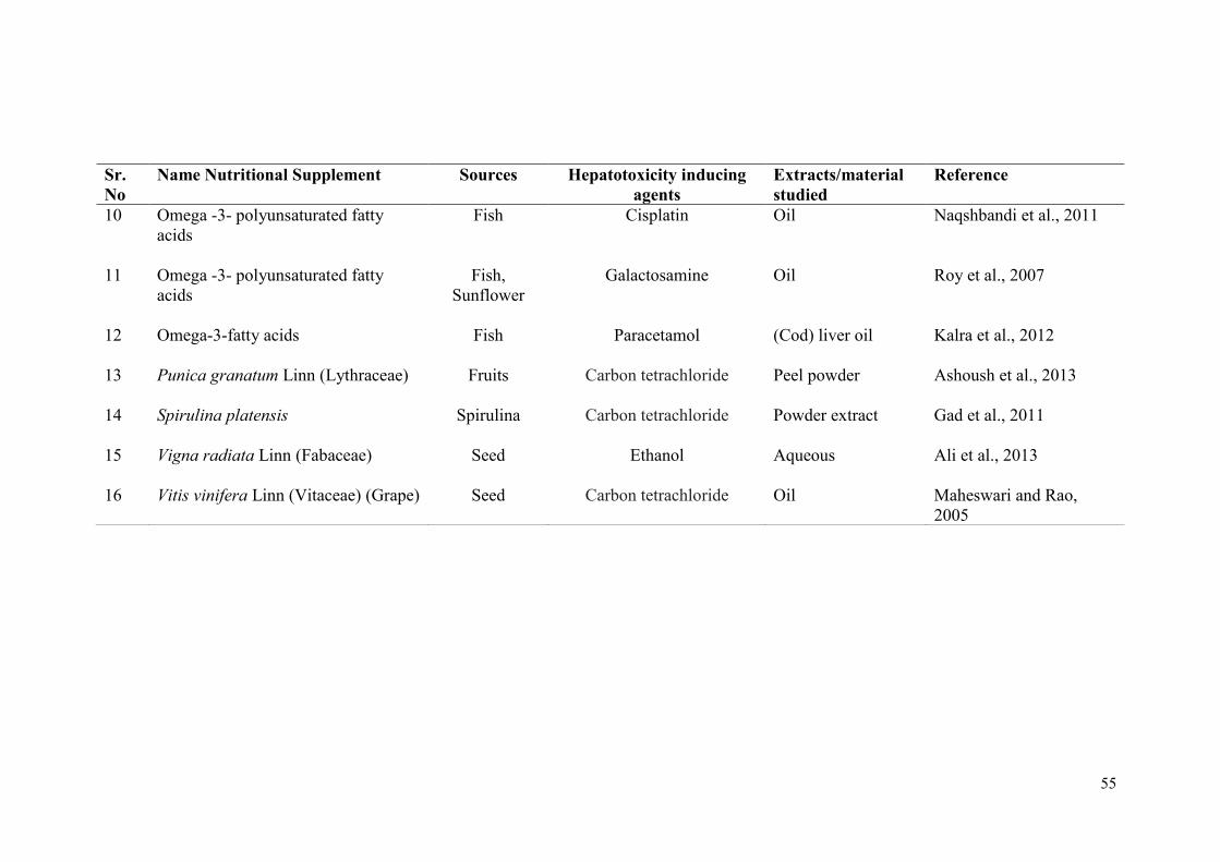
55
Sr.
No
Name Nutritional Supplement Sources Hepatotoxicity inducing
agents
Extracts/material
studied
Reference
10 Omega -3- polyunsaturated fatty
acids
Fish Cisplatin Oil Naqshbandi et al., 2011
11 Omega -3- polyunsaturated fatty
acids
Fish,
Sunflower
Galactosamine Oil Roy et al., 2007
12 Omega-3-fatty acids Fish Paracetamol (Cod) liver oil Kalra et al., 2012
13 Punica granatum Linn (Lythraceae) Fruits Carbon tetrachloride Peel powder Ashoush et al., 2013
14 Spirulina platensis Spirulina
Carbon tetrachloride Powder extract Gad et al., 2011
15 Vigna radiata Linn (Fabaceae) Seed Ethanol Aqueous Ali et al., 2013
16 Vitis vinifera Linn (Vitaceae) (Grape) Seed Carbon tetrachloride Oil Maheswari and Rao,
2005
57
3.4.4.1 Polyunsaturated Fatty Acid (Omega-3 Fatty Acids)
In recent years, there has been increased research interest in the use natural
sources to develop hepatoprotective, anticancer, cardio-protective and several other
pharmacological therapies (Varkey and Vahab, 2015). Several studies using different
animal models have been reported on beneficial effects of omega-3 long-chain
polyunsaturated fatty acids (PUFA) supplementation on liver injury (Lee et al., 2007;
Wu et al., 2012; Li et al., 2014). Bang and Dyerberg (1972) pioneered
epidemiological studies in early 1970s and hypothesized that consumption of fish oil
and other marine animals containing long chain, highly unsaturated omega-3 fatty
acids, produced beneficial effects in Eskimos. Diet supplemented with omega-3 fatty
acids has profound beneficial health effects against various pathologies (Simopoulos,
1991). Fish oil showed hepatoprotective activity against different hepatotoxins (Ruiz-
Gutierrez et al., 1999; Aguilera et al., 2003; Asaad and Aziz, 2012; Abdel-Dayem et
al., 2014). Flax seed oil is one of the rich sources of PUFA containing about 51% to
55% alpha-linolenic acid (ALA) (Prasad, 2000; Vijaimohan et al., 2006). ALA
content of flaxseed oil may be helpful in preventing clinical conditions such as
cardiovascular disease, blood pressure, cancer, skin diseases and immune disorders
like renal failure, rheumatoid arthritis, multiple sclerosis etc. (Kelley et al., 1991;
Thompson et al., 1996; Prasad et al., 1998; Cohen et al., 2005; Morris et al., 2005;
Rajesha et al., 2006; Vijaimohan et al., 2006; Sekine et al., 2008; Zanwar et al.,
2011).
Alwayn et al. (2005) reported the use of both enteral and parenteral ω3-PUFA
to treat nonalcoholic fatty liver disease (NAFLD) in a mouse model. There have been
numerous studies reporting the use of fish oil in humans and animals to treat
parenteral nutrition-associated liver disease (PNALD) (Gura et al., 2006; Gura et al.,
2008). Treatment of rodents with nonalcoholic fatty liver disease with EPA and DHA
showed improvements in liver health by reducing steatosis (Capanni et al., 2006).
Omega-3 polyunsaturated fatty acid (Fish oil) treatment in albino mice showed
hepatoprotective activity against galactosamine induced toxicity (Roy et al., 2007).
Flaxseed chutney demonstrated hepatoprotective activity against CCl4 induced
hepatotoxicity in rats (Shakir and Madhusudhan, 2007). Flaxseed contains ether
insoluble phenolic components of n-butanol fraction which plays important role in
58
mitigating hepatotoxicity due to carbon tetrachloride (CCL4) intoxication (Kasote et
al., 2012). Masterton et al. (2010) showed promising effect of omega-3 fatty acids in
non-alcoholic fatty liver disease in the form of reduced hepatic steatosis, improved
insulin sensitivity and reduced inflammatory markers. Tillman and Helms (2011)
showed some success in treatment of parenteral nutrition associated liver
disease (PNALD) using parenteral fish oil–based intravenous fat emulsion (IVFE)
(Omegaven, Fresenius Kabi AG, Bad Homburg VDH, Germany). Meganathan et al.
(2011) reported hepatoprotective activity of fish oil in paracetamol induced
hepatotoxicity at dose levels of 300 mg/Kg/day. Significant increase in the levels of
superoxide dismutase (SOD), reduced glutathione (GSH), and catalase (CAT)
indicated protective effect of fish oil. Fish oil (4ml/kg, i.p for 1 week) administration
to the single high dose acetaminophen intoxicated albino rats resulted in decreased
level of malondialdehyde (MDA) indicating hepatoprotective effect (Kalra et al.,
2012). Treatment of pure lignan (25 mg/ml) and partial lignan (25 mg/ml) of flaxseed
lignin against paracetamol intoxicated (200 mg/kg, oral) male rabbit showed decrease
in serum levels of liver markers like SGOT, SGPT, ALP and bilirubin, confirming
hepatoprotective activity of omega-3 fatty acids (Al-Jumaily and AL-Azawi, 2013).
Abdel-Dayem et al. (2014) reported hepatoprotective effect of DHA (120 and 200
mg/kg body weight) treatment against valproate (VPA) (500 mg/kg body weight for
14 days) induced liver damage. Varkey and Vahab (2015) studied the
hepatoprotective activity of Stolephorus commersonnii fish extracts against isoniazid
induced hepatotoxicity in albino rats. Treatment of fish extract at a dose of 300 mg/kg
body weight showed improvements in liver histology and liver function markers. The
beneficial effect of omega-3 fatty acids treatment on methotrexate induced
hepatotoxicity in children with acute lymphoblastic leukemia (Elbarbary et al., 2016:
Clinical Trial Registry Number: NCT02373579) has also been
reported. Administration of omega-3 fatty acids (100 mg/kg bw.p.o) in rats with
cyclosporine-A induced liver apoptosis protected liver injury by reducing apoptotic
cell count (Erol et al., 2011).
Several reports demonstrated that omega-3 fatty acids are involved in
regulation of expression of several genes such as adhesion molecules, lipid
metabolism, inflammatory cytokines etc. involved in endothelial activation in
response to inflammatory and pro-atherogenic stimuli and also nuclear transcription
59
factors, peroxisome proliferator-activated receptor, sterol regulatory element binding
protein-1, cyclooxygenase-2, etc. (Price et al., 2000; De Caterina and Massaro, 2005;
Deckelbaum et al., 2006). Omega-3 fatty acids affect the expression of several key
proteins related to inflammation, lipid metabolism and they help to reduce
inflammation and lipid oxidation (Deckelbaum et al., 2006). Omega-3 fatty acids have
also been shown to act through modulation of cytokine expression (Lee et al., 2007).
Omega-3 fatty acids have been shown to downregulate TNFα which is supposed to be
a direct role of omega-3 fatty acids in hepatoprotection (Babcock et al., 2000).
Omega-3 fatty acids also show anti-inflammatory properties (Asaad and Aziz, 2012),
they act as natural ligands for activation of PPARγ (Trombetta et al., 2007; Yamazaki
et al., 2007; Selvaraj et al., 2010), they down regulate SREBP-1c (Kim et al., 1999;
Yahagi et al., 1999; Xu et al., 1999; Botolin et al., 2006; Wada et al., 2008; Kim et al.,
2014) and up regulate FABP-1 (Dutta-Roy, 2000; Gaca et al., 2012).
Based on the review of literature, it was evident that the comparative efficacy
of the satwa from three Tinospora forms has not yet been studied. In addition, the
mechanism of action of the satwa on hepatotoxicity is not yet clear. Hence it was
decided to undertake studies on hepatoprotective activities of satwa of three
Tinospora forms (T. cordifolia, T. sinensis, Neem-giloe) and nutritional supplements
(Flax oil/Fish oil) and their comparative study against acetaminophen and alcohol
induced hepatotoxicity in rats.
CHAPTER
1TRODUCTION
CHAPTER 4
MATERIALS AND METHODS
Awards
Second prize in oral
Presentation Award
Best Poster Presentation
Award
Awards
Second prize in oral
Presentation Award
Best Poster Presentation
Award

60
The present study was designed to assess the effects of an ayurvedic
formulation, Guduchi satwa and omega-3 fatty acids (Flax oil and fish oil) on
acetaminophen and alcohol induced hepatotoxicity in rats. The elaborate process for
preparation of Guduchi satwa is described in Ayurvedic literature, which requires
collection of fresh plant material. The omega-3 fatty acids (Alpha linoleic acid
(ALA), Eicosapentaenoic acid (EPA) and Docosahexaenoic acid (DHA)) are available
commercially. The detail procedures for preparation of satwa, its nutritional analysis
and design and methodology of animal experiments with satwa and omega-3 fatty
acid interventions, is explained here. 4.1 Collection of Plant Material
As Guduchi is advocated to be collected in fresh condition, stems of
Tinospora cordifolia (Willd.) Miers ex Hook. F. & Thoms., Tinospora sinensis
(Lour.) Merr. and Neem-giloe (Tinospora cordifolia (Willd.) Miers ex Hook. F. &
Thoms., growing on neem (Azadirachta indica A. Juss.) tree) were collected during
February to April 2012 from Research Farm of Dr. Balasaheb Sawant Konkan Krishi
Vidyapeeth, Dapoli, Maharashtra, India (Lat Long 17.45°N, 73.10°E) (Map 1 and 2).
The matured stem was separated from other parts like roots,
leaves, flowers, fruits and washed thoroughly with water for three times.
Map. 1. Google Earth image of Maharashtra showing the Site of Collection of
Plant Material
Awards
Second prize in oral
Presentation Award
Best Poster Presentation
Award
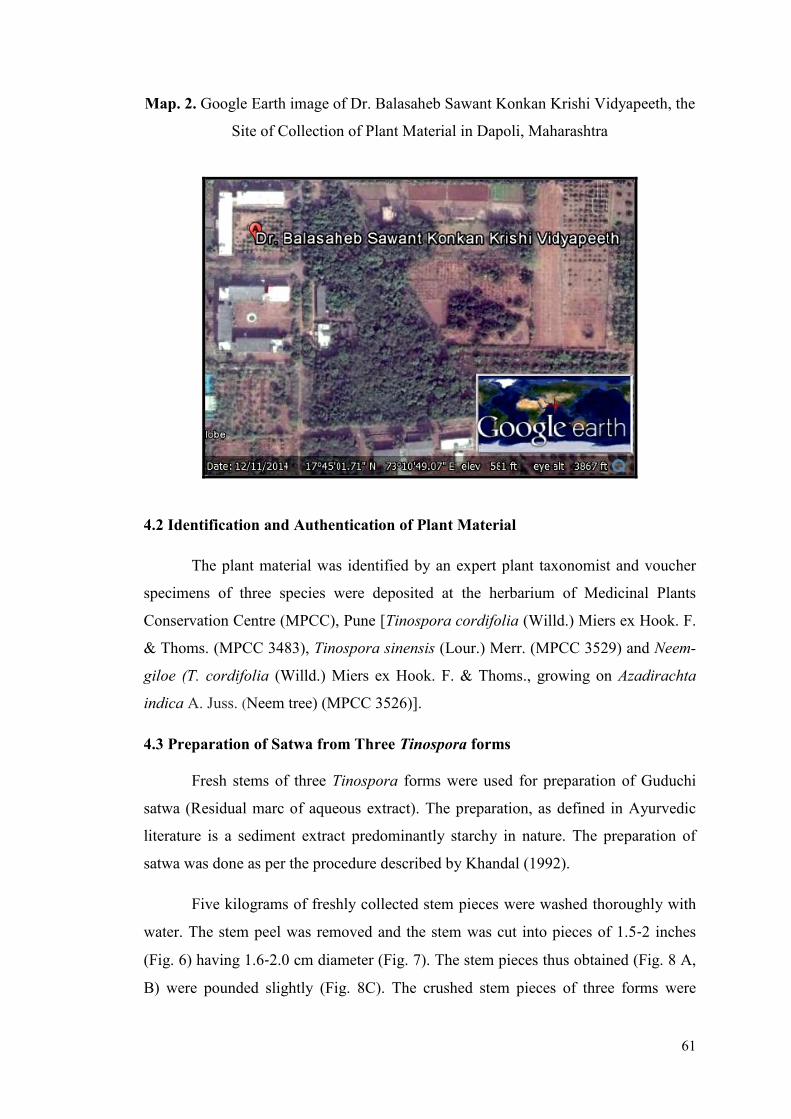
61
Map. 2. Google Earth image of Dr. Balasaheb Sawant Konkan Krishi Vidyapeeth, the
Site of Collection of Plant Material in Dapoli, Maharashtra
4.2 Identification and Authentication of Plant Material
The plant material was identified by an expert plant taxonomist and voucher
specimens of three species were deposited at the herbarium of Medicinal Plants
Conservation Centre (MPCC), Pune [Tinospora cordifolia (Willd.) Miers ex Hook. F.
& Thoms. (MPCC 3483), Tinospora sinensis (Lour.) Merr. (MPCC 3529) and Neem-
giloe (T. cordifolia (Willd.) Miers ex Hook. F. & Thoms., growing on Azadirachta
indica A. Juss. (Neem tree) (MPCC 3526)].
4.3 Preparation of Satwa from Three Tinospora forms
Fresh stems of three Tinospora forms were used for preparation of Guduchi
satwa (Residual marc of aqueous extract). The preparation, as defined in Ayurvedic
literature is a sediment extract predominantly starchy in nature. The preparation of
satwa was done as per the procedure described by Khandal (1992).
Five kilograms of freshly collected stem pieces were washed thoroughly with
water. The stem peel was removed and the stem was cut into pieces of 1.5-2 inches
(Fig. 6) having 1.6-2.0 cm diameter (Fig. 7). The stem pieces thus obtained (Fig. 8 A,
B) were pounded slightly (Fig. 8C). The crushed stem pieces of three forms were
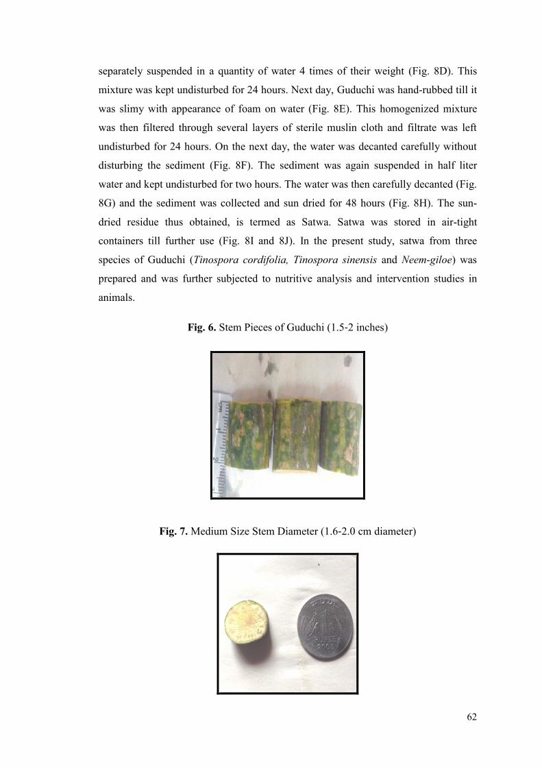
62
separately suspended in a quantity of water 4 times of their weight (Fig. 8D). This
mixture was kept undisturbed for 24 hours. Next day, Guduchi was hand-rubbed till it
was slimy with appearance of foam on water (Fig. 8E). This homogenized mixture
was then filtered through several layers of sterile muslin cloth and filtrate was left
undisturbed for 24 hours. On the next day, the water was decanted carefully without
disturbing the sediment (Fig. 8F). The sediment was again suspended in half liter
water and kept undisturbed for two hours. The water was then carefully decanted (Fig.
8G) and the sediment was collected and sun dried for 48 hours (Fig. 8H). The sun-
dried residue thus obtained, is termed as Satwa. Satwa was stored in air-tight
containers till further use (Fig. 8I and 8J). In the present study, satwa from three
species of Guduchi (Tinospora cordifolia, Tinospora sinensis and Neem-giloe) was
prepared and was further subjected to nutritive analysis and intervention studies in
animals.
Fig. 6. Stem Pieces of Guduchi (1.5-2 inches)
Fig. 7. Medium Size Stem Diameter (1.6-2.0 cm diameter)
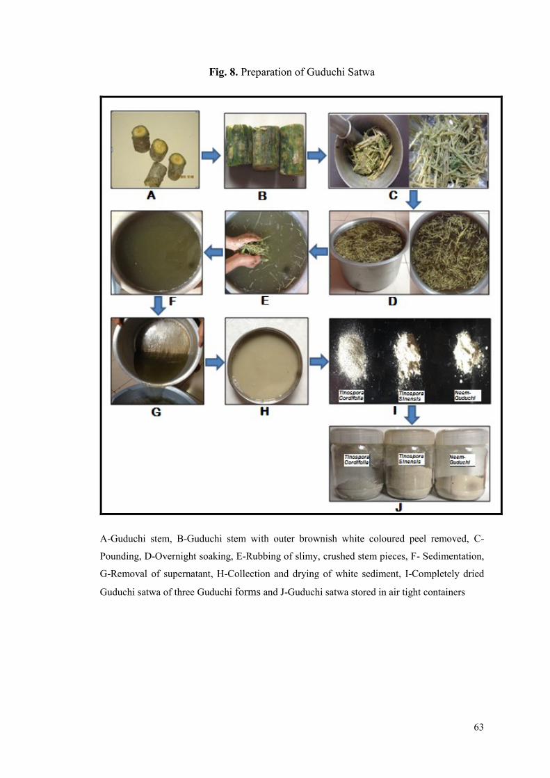
63
Fig. 8. Preparation of Guduchi Satwa
A-Guduchi stem, B-Guduchi stem with outer brownish white coloured peel removed, C-
Pounding, D-Overnight soaking, E-Rubbing of slimy, crushed stem pieces, F- Sedimentation,
G-Removal of supernatant, H-Collection and drying of white sediment, I-Completely dried
Guduchi satwa of three Guduchi forms and J-Guduchi satwa stored in air tight containers
64
4.4 Nutritional Analysis of Satwa
Estimation of proteins, carbohydrates, starch, total lipids, crude fibres and total
ash content of satwa from three Tinospora forms was done by following standard
techniques.
Chemicals/Reagents
Folin-Ciocalteu’s phenol reagent (Sigma-Aldrich Chemie GmbH),
Chloroform, Methanol, Anthrone reagent (Sigma-Aldrich Chemical, Bangalore,
India), Sodium carbonate, Sulphuric acid, Phenol (Qualigens Fine Chemicals,
GlaxoSmithKline Pharmaceuticals Limited, Mumbai, India), Perchloric acid, Sodium
hydroxide, Hydrochloric acid (Merck Specialities Private Limited, Mumbai, India),
Alkaline copper sulphate, Glucose (Hi-Media Laboratory Private Limited, Mumbai,
India), Bovine serum albumin (Sisco Research Laboratory Private Limited, Mumbai,
India), Ethanol (Changshu Yangyuan Chemical, China), Distilled water.
4.4.1 Protein
There are different methods for protein and total nitrogen content estimation.
Hydrolysing the protein and estimating the amino acid alone will give the exact
quantification (Sadasivam and Manickam, 1991a). The method developed by Lowry
et al. (1951) is largely followed as it is sensitive enough to give moderately constant
value and was used for estimation of proteins from satwa.
Principle
The blue colour developed by the reduction of the phosphomolybdic-
phosphotungstic components in the Folin-Ciocalteu reagents by the amino acid
tyrosine and tryptophan present in the protein and the colour developed by Biuret
reaction of the protein with alkaline cupric tartrate are measured in the Lowry’s
method (Sadasivam and Manickam, 1991a).
Materials
1. Reagent A: 2% (w/v) Sodium Carbonate in 0.1 N Sodium Hydroxide
2. Reagent B: 0.5% (w/v) Copper Sulphate (CuSO4.5H2O) in 1% (w/v)
Potassium Sodium Tartrate
65
3. Reagent C: Alkaline Copper solution: Reagent A (50mL) and Reagent B
(1mL) was mixed prior to use
4. Reagent D: Folin-Ciocalteu’s Phenol Reagent commercially available reagent
was diluted 1:1 in distilled water and used
5. Protein Solution (Stock Standard)
Accurately weighed 50mg bovine serum albumin (Fraction V) was dissolved in
distilled water and volume of the solution was made up to 50mL.
Extraction of Protein from Satwa
The satwa from three Guduchi forms were used for extraction of proteins. Pre-
weighed (500mg) satwa from each Tinospora forms was homogenized with 5mL
10mM phosphate buffer (pH 7.4), separately. The homogenates were centrifuged at
2000 rpm for 10 min at room temperature. The supernatants were collected and used
for protein estimation.
Procedure
Known volumes of standard protein solution (0.2, 0.4, 0.6, 0.8 and 1.0mL)
were pipetted in a test tube and the volume was made up to 1mL with sterile distilled
water in all tubes. The reaction with 1mL distilled water (without standard protein)
served as a blank. The protein extract from satwa (0.1mL) was taken in a separate test
tube and the volume was made up to 1mL with sterile distilled water. The contents of
the tubes were mixed well, 5.0mL alkaline copper sulphate (Reagent C) was added to
the tubes and the contents were mixed thoroughly. The tubes were incubated for 10
minutes at room temperature and then 0.5mL Folin-Ciocalteu’s Phenol (Reagent D)
was added to the tubes. The contents were mixed well and the reactions were
incubated at room temperature, in dark for 30 minutes. The blue colour developed
was read in a microplate reader at 660 nm (Bio-Rad Laboratories Inc., Berkeley,
California, U.S.A.) against blank. Intensity of the colour developed is directly
proportional to protein content in the sample.
4.4.2 Total Carbohydrates
Carbohydrates are the main components of storage and structural materials in
the plants and they occur as free sugars and polysaccharides. Monosaccharaides are
the basic units of carbohydrates which cannot be split by hydrolysis into simpler

66
sugars. The carbohydrate content from the samples can be measured by acid
hydrolysis of polysaccharides to monosaccharides followed by estimation of
monosaccharaides (Sadasivam and Manickam, 1991b)
Principle
Glucose undergoes dehydration reaction in hot acidic medium resulting in the
formation of hydroxymethyl furfural which forms a green coloured product with
phenol with absorption maximum at 490 nm. The intensity of colour developed is
directly proportional to the amount of carbohydrates in the sample (Sadasivam and
Manickam, 1991b).
Materials
1. 2.5 N HCl
2. Sodium Carbonate
3. Phenol 5%: Redistilled (Reagent grade) phenol (5gm) dissolved in water and
diluted to 100mL.
4. Sulphuric acid 96% reagent grade.
5. Standard Glucose Stock- 1mg/mL glucose solution
6. Working standard: 10mL stock diluted to 100mL with distilled water
(100µg/mL working stock concentration)
Extraction of Carbohydrates from Satwa (Sadasivam and Manickam, 1991b)
The satwa (100mg) from each forms were suspended in 5mL 2.5N
Hydrochloric acid and were hydrolyzed in a boiling water bath for 3 hours. After
cooling to room temperature, it was neutralized with solid sodium carbonate until
there was no effervescence and the volume was made to 20mL with distilled water. It
was then centrifuged at 2000 rpm for 10 minutes. Volume of the supernatant was
measured and adjusted to 20mL and used for carbohydrate estimation.
Procedure
Total carbohydrates were estimated by following phenol sulphuric acid
method (Dubois et al., 1956). The known volume of working glucose standard (0.2,
0.4, 0.6, 0.8 and 1.0 mL) was taken in separate test tubes and the volume was made up
to 1mL with sterile distilled water. Blank was prepared by taking 1mL distilled water
67
instead of standard glucose solution or extract. Satwa extract (0.1mL) was taken in a
test tube and volume was made up to 1mL with sterile distilled water. Phenol solution
(1mL) and 96% sulphuric acid (5mL) was added to all the tubes and mixed. The tubes
were incubated at room temperature for 10 minutes with intermittent mixing of the
contents. The tubes were then placed in water bath for 20 minutes at 25-30°C. The
green colour developed was read at 490 nm in a microplate reader.
4.4.3 Starch
Starch is an important polysaccharide and is the storage form of carbohydrates
in plants and is richly found in roots, tubers, stems, fruits and cereals. Starch is a
mixture of two types of components namely amylose and amylopectin which are
composed of several glucose molecules. Dilute acids are used to hydrolyse starch into
simple sugars and the quantity of simple sugars is measured colorimetrically
(Sadasivam and Manickam, 1991c).
Principle
The sample is treated with 80% alcohol to remove sugars and the starch is
extracted with perchloric acid. In hot acidic medium, starch is hydrolysed to glucose
and dehydrated to hydroxymethyl furfural. This compound forms a green coloured
product with anthrone which can be measured at 630 nm (Sadasivam and Manickam,
1991c).
Material
1. Anthrone: Dissolve 200mg Anthrone in 100mL of ice-cold 95% Sulphuric
acid
2. 80% (v/v) Ethanol
3. 52% (v/v) Perchloric Acid
4. Standard Glucose: 1mg/mL Glucose Solution.
5. Working Standard: 10mL of stock diluted to 100mL with water (giving
100µg/mL working stock).
Extraction of Starch from Satwa (Sadasivam and Manickam, 1991c)
Each satwa was separately processed for extraction of starch. Known amount
of satwa (500mg) was homogenized in hot 80% ethanol to remove sugars. The residue
was retained after centrifugation at 2000 rpm for 10 minutes. The residue was washed
68
with hot 80% ethanol till the washings did not give colour with anthrone reagent. The
residue was dried well and 5mL water and 6.5mL 52% Perchloric acid was added to
the residue. Starch was extracted at 0°C for 20 minutes. The residue was retained after
centrifugation at 2000 rpm at 4°C for 10 minutes and the supernatant was collected in
separate test tube. The extraction of the residue was repeated with fresh 52%
Perchloric acid. The supernatants were pooled after centrifugation and the volume
was made up to 100mL with sterile distilled water.
Procedure for Estimation of Starch
Anthrone method (Hodge and Hofreiter, 1962) was used for starch estimation.
Known volume of standard glucose solution (0.2, 0.4, 0.6, 0.8 and 1.0mL) was
pipetted in separate test tubes and the volume was made up to 1mL with sterile
distilled water. Satwa extract (0.1mL) was pipetted in a separate test tube and volume
was made up to 1mL with sterile distilled water. Blank was prepared by taking 1mL
sterile distilled water instead of standard glucose solution or extract. Anthrone reagent
(4mL) was added to all the tubes. The reaction mixture was incubated in boiling water
bath for 8 minutes and cooled rapidly. The green colour developed was read at 630
nm in a microplate reader. Content of starch was estimated from standard graph.
4.4.4 Total Lipids
Lipids are structural components of cell membranes and play a critical role in
gene transcription, signaling and metabolism. Several lipid species exist in biological
systems, including phospholipids, cholesterol, triglycerides and fatty acids (Fahy et
al., 2005)
Principle
Various solvents or solvent combinations are suggested for extraction of lipid
fractions from the material depending upon the polarity of the lipids. But chloroform:
methanol extraction of lipids as suggested by Folch (Folch et al., 1957) extracts
maximum lipids from the material. The partitioning of the extract with water removes
the non-lipid contaminants from the extract. The organic phase can be used for lipid
estimation after phase separation.

69
Material
1. Chloroform
2. Methanol
3. Chloroform: methanol 2:1 (v/v)
Procedure
Total lipids were estimated by a method modified from Folch (Folch et al.,
1957) as described by Alam (Alam et al., 2008). Satwa (1gm) of three Tinospora
forms were suspended separately in 10mL chloroform: methanol (2:1 v/v) mixture,
mixed thoroughly and were left undisturbed at room temperature for 3 days. The
solutions were filtrated through a muslin cloth and centrifuged at 3000 rpm for 5
minutes. The upper layer of methanol was removed and the interface was washed
three times with chloroform: methanol: water (3:48:47) without disturbing the lower
phase. This has the effect of removing any 'fluff' at the interface. The suspensions
were then centrifuged at 3000 rpm for 5 minutes. The remaining was the crude lipid.
The lower layer (“lipid extract”) was transferred to pre-weighed petri plate. The
extract in a petri plate was dried in incubator and the petri plate with dried lipids was
weighed again. Difference between the weights was recorded as the weight of total
lipids.
4.4.5 Crude Fibre
Crude fibre contains 60% to 80% of the cellulose and 4% to 6% of the lignin
and some mineral matter. Content of crude fibre is a measure of nutritive value of
poultry and livestock feed. It is also used in the analysis of various foods and food
products to detect adulteration, quality and quantity (Sadasivam and Manickam,
1991d).
Principle
During the acid and subsequent alkali treatment, oxidative hydrolytic
degradation of the native cellulose and considerable degradation of lignin occurs. The
residue obtained after final filtration is weighed, incinerated, cooled and weighed
again. The loss in weight indicates the crude fibre content (Sadasivam and Manickam,
1991d).
70
Materials
1. Sulphuric acid solution (0.255 ± 0.005N): 1.25gm concentrated sulphuric acid
diluted to 100mL distilled water.
2. Sodium hydroxide solution (0.313 ± 0.005N): 1.25gm sodium hydroxide in
100mL distilled water.
3. Alcohol
4. Crucible
Procedure
Crude fiber contents in the satwa of T. cordifolia, T. sinensis and Neem-giloe
were estimated according to the method described by Maynard (1970). Known
amounts (1gm) of each satwa were taken separately in a beaker and 100mL of (0.255
± 0.005N) sulphuric acid was added. The mixture was boiled for 30 minutes keeping
the volume constant by the addition of water at frequent intervals. The mixture was
filtered through a muslin cloth and the residue washed with hot water till free from
acid. The material was then transferred to the same beaker and 100mL of boiling
(0.255 ± 0.005N) sodium hydroxide solution was added. After boiling for 30 minutes
(keeping the volume constant as before) the mixture was filtered through a muslin
cloth and residue washed with hot water till free from alkali, followed by single
350mL of water and 25mL alcohol wash. The residue was then removed and
transferred to a crucible (Preweighed dish W1) and dried for 2 hours at 130±2ºC. The
crucible was cooled in a desiccator and weighed again (W2). The crucible was heated
in a muffle furnace at 600±150ºC for 5 hours, cooled and weighed again (W3). Crude
fibre was determined using fat-free samples.
Crude fibre content was determined using the following Equation:
% crude fibre in ground sample
Loss in weight on ignition (W2-W1) - (W3-W1)/Weight of sample) * 100
4.4.6 Total Ash
The ash content is a measure of the total amount of minerals present within a
food, whereas the mineral content is a measure of the amount of specific inorganic
components present within a food, such as Ca, Na, K and Cl (Zhang and Dotson,
1994).
71
Principle
Ash is the inorganic or mineral component of the sample left after complete
ignition of the sample at 450ºC in muffle furnace (The Ayurvedic Pharmacopoeia of
India, Vol II, 2008).
Material
1. Crucible
Procedure
Total ash content was determined as described in The Ayurvedic
Pharmacopoeia of India (2008). One gram of each satwa from three Tinospora forms
was weighed separately into different pre-weighed crucibles. The crucibles were
placed on a clay pipe triangle and heated first over a low flame till all the material was
completely charred, followed by heating in a muffle furnace for 4 hours at 450ºC. The
crucibles were then cooled in desiccators and reweighed. The procedure was repeated
till two consecutive weights were same and the ash was almost white or grayish
white.
The ash content was determined by the equation as follows:
Ash Content (g/100gm sample) = weight of ash x 100 / weight of sample taken
72
4.5 Hepatoprotective Activity of Satwa from Three different forms of Tinospora
Liver diseases are a major health problem. The allopathic drugs prescribed for
liver disorders are known to have major side effects. Hence, time tested and reliable
herbal medicines are attractive candidates for management of various liver disorders
(Dash et al., 2007; Suryawanshi et al., 2011 and Kavitha et al., 2011a). Number of
medicinal plants and their formulations are common for the treatment of liver diseases
(Kavitha et al., 2011a). Modern medicine has little to suggest for improvement of
hepatic diseases and it is essentially the plant based preparations which are employed
for the treatment of liver disorders (Chatterjee, 2000). The present study was
undertaken to assess the hepatoprotective activity of satwa from three different forms
of Tinospora against two experimentally induced hepatotoxicity models, namely,
acetaminophen and ethanol induced hepatotoxicity in rats.
Drugs / Chemicals
Acetaminophen (Paramol; Ranbaxy Laboratories Ltd.) was purchased from
the local pharmacy and dissolved in sterile water to make the stock solution
convenient for animal administration as inducing agent for hepatotoxicity. Alcohol
(Ethanol) was obtained from Changshu Yangyuan Chemical, China. Ethanol
(Alcohol) (30%) was used as hepatotoxicant for animal administration. Silymarin
(Silybon-140; Micro Labs) was purchased from local pharmacy and dissolved in
sterile water to make the stock solution convenient for animal administration.
Formalin, ethylenediamine tetra-acetic acid (EDTA) and sodium dihydrogen
phosphate were purchased from Sigma-Aldrich (St. Louis, MO, U.S.A.).
Animals
Three months old Male Albino Wistar rats weighing between 150-200 gm
were procured for the study from institutional animal house.
Housing of Animals
The animals were acclimatized for seven days and were maintained under
standard husbandry conditions (Temperature 25±2°C, 12-h light: 12-h dark cycle)
throughout the experimentation. The animals were fed with standard pellet diet
(Nutrivet life science, Pune, M.S., India) and water was supplied ad-libitum. The

73
studies were carried out as per the CPCSEA guidelines and after approval of the
Institutional Animal Ethical Committee (Ref. No. BVDUMC/443/2012-2013).
Selection and Preparation of Dose
The dose of satwa was finalized on the basis of previous studies carried out in
the Laboratory. The quantity of satwa for administration to each animal was
calculated based on the weight of the animal. The required quantity of satwa was
weighed and suspended in water for administration to animals.
Standard Drug, Hepatotoxicants and Interventions
Silymarin (100mg/kg b.w./day, p.o.) was used as standard.
Acetaminophen (1000mg/kg b.w./day, p.o.) and 30% Ethanol (Alcohol) (1ml/100g
b.w./day, p.o.) was used to induce hepatotoxicity.
The satwa of three Tinospora forms (200mg/kg b.w./day, p.o.) was administered to
rats to study their hepatoprotective activity.
Assessment of Hepatoprotective Activity
Male Albino Wistar rats were administered acetaminophen or 30% ethanol
daily for 15 days to induce hepatotoxicity. Satwa from three forms of Guduchi (T.
cordifolia, T. sinensis and Neem-giloe) was orally administered to the rats, daily for
15 days. The satwa was administered 30 minutes before the administration of the
hepatotoxicant. The animals were sacrificed after completion of the experiment and
blood and liver were collected. Blood samples were collected by cardiac puncture and
were subjected to liver function tests (like SGOT, SGPT, Bilirubin, Alkaline
phosphatase etc.) and lipid profile (Total cholesterol, HDL-D cholesterol, LDL-D
cholesterol, and Triglycerides). Part of the liver tissues were stored in 10% neutral-
buffered formalin for histopathology while the remaining part was snap frozen in
liquid nitrogen for assessment of liver biochemical parameters (Lipid peroxidation,
Superoxide dismutase activity, Catalase activity, Reduced glutathione content, Total
protein and Lipid profile) and for molecular analysis.
74
Evaluation of Hepatoprotective Activity of Satwa against Acetaminophen
Induced Hepatotoxicity
The animals were divided into six groups by random assignment of six
animals per group. The variation in the average weight of the animals in and between
the groups was less than 20%.
The treatment protocol to assess the hepatoprotective potential of satwa of
three different forms of Tinospora (T. cordifolia, T. sinensis and Neem-giloe) against
acetaminophen induced liver injury is outlined below:
1. Group I: Healthy Control (n=6); received feed and water normally for 15
days
2. Group II: Negative Control (n=6); rats were administered acetaminophen
(1000mg/kg b.w./day, p.o.), daily for 15 days
3. Group III: Positive Control (n=6); the rats in this group were treated daily
with acetaminophen (1000mg/kg b.w./day, p.o.), 30 minutes after
administration of silymarin (100mg/kg b.w./day, p.o.), for 15 days
4. Group IV: Treatment group 1 (n=6); the rats in this group were treated daily
with acetaminophen (1000mg/kg b.w./day, p.o.), 30 minutes after
administration of Tinospora cordifolia satwa (200mg/kg b.w./day, p.o.), for 15
days
5. Group V: Treatment group 2 (n=6); the rats in this group were treated daily
with acetaminophen (1000mg/kg b.w./day, p.o.), 30 minutes after
administration of Tinospora sinensis satwa (200mg/kg b.w./day, p.o.), for 15
days
6. Group VI: Treatment group 3 (n=6); the rats in this group were treated daily
with acetaminophen (1000mg/kg b.w./day, p.o.), 30 minutes after
administration of Neem-giloe satwa (200mg/kg b.w./day, p.o.), for 15 days
During the period of experiment, animals were observed daily for signs of
infection and/or discomfort. After completion of the experiment (15 days), all animals
were fasted overnight and were humanely sacrificed under light ether anaesthesia.
Blood samples were collected by cardiac puncture for evaluating the serum
biochemical parameters. Blood samples were allowed to clot at room temperature for
75
30 minutes and serum was collected by centrifugation at 2000 rpm for 15 minutes.
Aliquots of sera were stored in eppendorfs at -20°C till further use. Liver was excised
from the dissected animals immediately, blotted off blood, washed with saline,
weighed and the left medial lobe was stored in 10% neutral-buffered formalin for
histopathological evaluation. The remaining liver tissues were collected and snap
frozen in liquid nitrogen. Frozen tissues were stored at -80°C till further use.
Evaluation of Hepatoprotective Activity of Satwa against Ethanol Induced
Hepatotoxicity
The animals were divided into six groups by random assignment of six
animals per group. The variation in the average weight of the animals in and between
the groups was less than 20%.
The treatment protocol to assess the hepatoprotective potential of satwa of
three different forms of Tinospora (T. cordifolia, T. sinensis and Neem-giloe) against
ethanol induced liver injury is outlined below:
1. Group I: Healthy Control (n=6); received feed and water normally for 15
days
2. Group II: Negative Control (n=6); administrated 30% ethanol (1ml/100g
b.w./day, p.o.), for 15 days
3. Group III: Positive Control (n=6); the rats in this group were treated daily
with 30% ethanol (1ml/100g b.w./day, p.o.), 30 minutes after administration of
silymarin (100mg/kg b.w./day, p.o.), for 15 days
4. Group IV: Treatment group 1 (n=6); the rats in this group were treated daily
with 30% ethanol (1ml/100g b.w./day, p.o.), 30 minutes after administration of
Tinospora cordifolia satwa (200mg/kg b.w./day, p.o.), for 15 days
5. Group V: Treatment group 2 (n=6); the rats in this group were treated daily
with 30% ethanol (1ml/100g b.w./day, p.o.), 30 minutes after administration of
Tinospora sinensis satwa (200mg/kg b.w./day, p.o.), for 15 days
6. Group VI: Treatment group 3 (n=6); the rats in this group were treated daily
with 30% ethanol (1ml/100g b.w./day, p.o.), 30 minutes after administration of
Neem-giloe satwa (200mg/kg b.w./day, p.o.), for 15 days
76
During the period of experiment, animals were observed daily for any signs of
infection and/or discomfort. After completion of the experiment (15 days), all animals
were fasted overnight and were humanely sacrificed under light ether anaesthesia.
Blood samples were collected by cardiac puncture for evaluating the serum
biochemical parameters. Blood samples were allowed to clot at room temperature for
30 minutes and serum was collected by centrifugation at 2000 rpm for 15 minutes.
Aliquots of sera were stored in eppendorfs at -20°C till further use. Liver was excised
from the dissected animals immediately, blotted off blood, washed with saline,
weighed and the left medial lobe was stored in 10% neutral-buffered formalin for
histopathological evaluation. The remaining liver tissues were collected and snap
frozen in liquid nitrogen. Frozen tissues were stored at -80°C till further use.
77
4.6 Hepatoprotective Activity of Flax Oil and Fish Oil
In previous experiment, the hepatoprotective activity of satwa from three
Tinospora forms was studied. Apart from herbal medicines; nutrition and various
dietary supplements are also known to play an important role in management of
various health ailments. Omega-3 fatty acids or polyunsaturated fatty acids (PUFA)
are one of such dietary supplements (Ruxton et al., 2004). Omega-3 fatty acids have
been shown to have beneficial effects against number of different pathologies
(Simopoulos et al., 1991). Fish oil and Flax oil are the richest sources of omega-3
fatty acids and are potent antioxidants. Hence, in the present study, we analyzed the
hepatoprotective activity of polyunsaturated fatty acids or omega-3 fatty acids (Fish
oil and Flax oil) against acetaminophen and ethanol induced hepatotoxicity.
Chemicals/reagents
Flax oil (Alpha Lite) was purchased from EnSigns Diet Care Pvt. Ltd. (Pune,
MS, India) and contained 50% alpha-linolenic acid (ALA), 20% Oleic acid (OA) and
12% Linoleic acid (LA). Fish oil (Maxepa) was purchased from Merck Limited (Goa,
India) and contained 60% EPA and 40% DHA.
Animals
Three months old Male Albino Wistar rats weighing between 150-200 gm
were procured for the study from institutional animal house.
Housing of Animals
The animals were acclimatized for seven days and were maintained under
standard husbandry conditions (Temperature 25±2°C, 12-h light: 12-h dark cycle)
throughout the experimentation. The animals were fed with standard pellet diet
(Nutrivet life science, Pune, M.S., India) and water was supplied ad-libitum. The
studies were carried out as per the CPCSEA guidelines and after approval of the
Institutional Animal Ethical Committee (Ref. No. BVDUMC/443/2012-2013).
Selection and Preparation of Dose
The dose of flax oil and fish oil was finalized on the basis of previous studies
carried out on the references (Meganathan et al., 2011; Kasote et al., 2012).
78
Commercially available flax oil and fish oil were used directly for intervention
studies.
Standard Drug, Hepatotoxicants and Interventions
Silymarin (100mg/kg b.w./day, p.o.) was used as standard.
Acetaminophen (1000mg/kg b.w. /day, p.o.) or 30% Ethanol (Alcohol) (1ml/100g
b.w./day, p.o.) was used to induce hepatotoxicity.
The flax oil (500mg/kg b.w./day, p.o.) or fish oil (500mg/kg b.w./day, p.o.) was
administered to study their hepatoprotective activity.
Assessment of Hepatoprotective Activity
Male Albino Wistar rats were administered acetaminophen or 30% ethanol
daily for 15 days to induce hepatotoxicity. Flax oil and fish oil were administered to
rats daily, 30 minutes before the administration of the hepatotoxicant, for 15 days.
The animals were sacrificed after the treatment and blood and liver were collected.
Blood samples were collected by cardiac puncture and were subjected to liver
function tests (like SGOT, SGPT, Bilirubin, Alkaline phosphatase etc.) and lipid
profile (Total cholesterol, HDL-D cholesterol, LDL-D cholesterol, and Triglycerides).
Part of the liver tissues were stored in 10% neutral-buffered formalin for
histopathology while the remaining part was snap frozen in liquid nitrogen for
assessment of liver biochemical parameters (Lipid peroxidation, Superoxide
dismutase activity, Catalase activity, Reduced glutathione content, Total protein and
lipid profile) and for molecular analysis.
Evaluation of Hepatoprotective Activity of Flax Oil and Fish Oil against
Acetaminophen Induced Hepatotoxicity
The animals were divided into five groups by random assignment of six
animals per group. The variation in the average weight of the animals in and between
the groups was less than 20%.
The treatment protocol to assess the hepatoprotective potential of
polyunsaturated fatty acids against acetaminophen induced liver injury is outlined
below:
79
1. Group I: Healthy Control (n=6); received feed and water normally for 15
days.
2. Group II: Negative Control (n=6); rats were administered acetaminophen
(1000mg/kg b.w./day, p.o.), for 15 days.
3. Group III: Positive Control (n=6); the rats in this group were treated daily
with acetaminophen (1000mg/kg b.w./day, p.o.), 30 minutes after
administration of silymarin (100mg/kg b.w./day, p.o.), for 15 days.
4. Group IV: Treatment group 1 (n=6); the rats in this group were treated daily
with acetaminophen (1000mg/kg b.w./day, p.o.), 30 minutes after
administration of flax oil (500mg/kg b.w./day, p.o.), for 15 days.
5. Group V: Treatment group 2 (n=6); the rats in this group were treated daily
with acetaminophen (1000mg/kg b.w./day, p.o.), 30 minutes after
administration of fish oil (500mg/kg b.w./day, p.o.), for 15 days.
During the period of experiment, animals were observed daily for signs of
infection and/or discomfort. After completion of the experiment (15 days), all animals
were fasted overnight and were humanely sacrificed under light ether anaesthesia.
Blood samples were collected by cardiac puncture for evaluating the serum
biochemical parameters. Blood samples were allowed to clot at room temperature for
30 minutes and serum was collected by centrifugation at 2000 rpm for 15 minutes.
Aliquots of sera were stored in eppendorfs at -20°C till further use. Liver was excised
from the dissected animals immediately, blotted off blood, washed with saline,
weighed and the left medial lobe was stored in 10% neutral-buffered formalin for
histopathological evaluation. The remaining liver tissues were collected and snap
frozen in liquid nitrogen. Frozen tissues were stored at -80°C till further use.
Evaluation of Hepatoprotective Activity of Flax Oil and Fish Oil against Ethanol
Induced Hepatotoxicity
The animals were divided into five groups by random assignment of six
animals per group. The variation in the average weight of the animals in and between
the groups was less than 20%.
The treatment protocol to assess the hepatoprotective potential of
polyunsaturated fatty acids against ethanol induced liver injury is outlined below:
80
1. Group I: Healthy Control (n=6); fed on a normal diet and water for 15 days.
2. Group II: Negative Control (n=6); rats were administered 30% ethanol
(1ml/100g b.w./day, p.o.), for 15 days.
3. Group III: Positive Control (n=6); the rats in this group were treated daily
with 30% ethanol (1ml/100g b.w./day, p.o.), 30 minutes after administration of
silymarin (100mg/kg b.w./day, p.o.), for 15 days.
4. Group IV: Treatment group 1 (n=6); the rats in this group were treated daily
with 30% ethanol (1ml/100g b.w./day, p.o.), 30 minutes after administration of
flax oil (500mg/kg b.w./day, p.o.), for 15 days.
5. Group V: Treatment group 2 (n=6); the rats in this group were treated daily
with 30% ethanol (1ml/100g b.w./day, p.o.), 30 minutes after administration of
fish oil (500mg/kg b.w./day, p.o.), for 15 days.
During the period of experiment, animals were observed daily for signs of
infection and/or discomfort. After completion of the experiment (15 days), all animals
were fasted overnight and were humanely sacrificed under light ether anaesthesia.
Blood samples were collected by cardiac puncture for evaluating the serum
biochemical parameters. Blood samples were allowed to clot at room temperature for
30 minutes and serum was collected by centrifugation at 2000 rpm for 15 minutes.
Aliquots of sera were stored in eppendorfs at -20°C till further use. Liver was excised
from the dissected animals immediately, blotted off blood, washed with saline,
weighed and the left medial lobe was stored in 10% neutral-buffered formalin for
histopathological evaluation. The remaining liver tissues were collected and snap
frozen in liquid nitrogen. Frozen tissues were stored at -80°C till further use.
The effect of satwa and omega 3 fatty acids on acetaminophen induced
hepatotoxicity was studied in the same experiment (with common healthy, positive
and negative control groups) while the effect of satwa and omega 3 fatty acids on
alcohol induced hepatotoxicity was studied in another experiment (with common
healthy, positive and negative control groups).

81
4.7 Hepatoprotective Activity of Combination of Best Performing Herbal and
Nutritional Intervention
In previous experiments, the hepatoprotective activity of satwa of three
Tinospora forms and polyunsaturated fatty acids was studied, separately, against
acetaminophen and alcohol induced hepatotoxicity in rats. Based on the results of
these experiments, the combination of Neem-giloe and fish oil was used for
intervention in acetaminophen induced hepatotoxicity while combination of
Tinospora sinensis and flax oil was used for intervention in ethanol induced
hepatotoxicity in rats.
Animals
Three months old Male Albino Wistar rats weighing between 150-200 gm
were procured for the study from institutional animal house.
Housing of Animals
The animals were acclimatized for seven days and were maintained under
standard husbandry conditions (Temperature 25±2°C, 12-h light: 12-h dark cycle)
throughout the experimentation. The animals were fed with standard pellet diet
(Nutrivet life science, Pune, M.S., India) and water was supplied ad-libitum. The
studies were carried out as per the CPCSEA guidelines and after approval of the
Institutional Animal Ethical Committee (Ref. No.: BVDUMC/2679/2012-2013).
Selection and Preparation of Dose
The dose of best performing satwa and omega-3 fatty acids were based on the
results of previous experiments on acetaminophen and alcohol induced liver injury.
Standard Drug, Hepatotoxicants and Interventions
Silymarin (100mg/kg b.w./day, p.o.) was used as standard.
Acetaminophen (1000mg/kg b.w. /day, p.o.) and 30% Ethanol (Alcohol) (1ml/100g
b.w./day, p.o.) was used to induce hepatotoxicity.
Combination of Neem-giloe (200mg/kg; b.w/day, p.o.) and fish oil (500mg/kg;
b.w/day, p.o.) was used for screening its hepatoprotective activity against
acetaminophen induced hepatotoxicity.
82
Combination of Tinospora sinensis satwa (200mg/kg b.w./day, p.o.) and flax oil
(500mg/kg b.w/day, p.o.) was used for screening its hepatoprotective activity against
ethanol induced hepatotoxicity.
Assessment of Hepatoprotective Activity
Male Albino Wistar rats were administered single dose of acetaminophen on
8th
day or 1st day or 30% ethanol daily for 15 days to induce hepatotoxicity. The best
performing Guduchi and omega-3 fatty acids at the selected dosage (As per the results
from experiment 4.5 and 4.6, respectively) were administered simultaneously, every
24 hours, to rats treated with hepatotoxicants. The combination of interventions was
administered to rats for assessing their protective or corrective effects in
acetaminophen induced liver injury. In case of alcohol induced liver damage, the
hepatotoxicant and combination of interventions were administered simultaneously to
assess the prophylactic effect of the intervention. The animals were sacrificed after
the treatment and blood and liver were collected. Blood samples were collected by
cardiac puncture and were subjected to liver function tests (like SGOT, SGPT,
Bilirubin, Alkaline phosphatase etc.) and lipid profile (Total cholesterol, HDL-D
cholesterol, LDL-D cholesterol, and Triglycerides). Part of the liver tissues were
stored in 10% neutral-buffered formalin for histopathology while the remaining part
were snap frozen in liquid nitrogen for liver biochemical parameters (Lipid
Peroxidation, Superoxide dismutase, Catalase, Reduced glutathione, Total protein
and Lipid profile) and for molecular analysis.
Effects of Protective and Corrective Treatment of Combination of Neem-giloe
Satwa and Fish oil against Acetaminophen Induced Hepatotoxicity
The animals were divided into seven groups by random assignment of six
animals per group. The variation in the average weight of the animals in and between
the groups was less than 20%.
The treatment protocol to assess the combinatorial protective and corrective
effects of Neem-giloe satwa and fish oil interventions against acetaminophen induced
liver injury is outlined below:
83
1. Group I: Healthy Control (n=6); received feed and water normally for 8 days
2. Group II (Negative Control for Protective Treatment): Negative Control
(n=6); rats were administered a single dose of acetaminophen (1000mg/kg;
b.w./day, p.o.), on 8th
day
3. Group III (Negative Control for Corrective Treatment): Negative Control
(n=6); rats were administered a single dose of acetaminophen (1000mg/kg;
b.w./day, p.o.), on 1st day
4. Group IV (Positive Control for Protective Treatment): (n=6); the rats in
this group were administered with standard drug silymarin (100mg/kg;
b.w./day, p.o.), for 7 days and administered a single dose of acetaminophen
(1000mg/kg; b.w/day, p.o.), on 8th
day
5. Group V (Positive Control for Corrective Treatment): (n=6); the rats in
this group were treated on 1st day with a single dose of acetaminophen
(1000mg/kg; b.w/day, p.o.) and administered standard drug silymarin
(100mg/kg; b.w./day, p.o.), for next 7 days
6. Group VI (Experimental Group for Protective Treatment): (n=6); the rats
in this group were administered with the combined intervention of Neem-giloe
satwa (200mg/kg; b.w/day, p.o.) and fish oil (500mg/kg; b.w/day, p.o.) for 7
days and a single dose of acetaminophen (1000mg/kg; b.w/day, p.o.), on 8th
day
7. Group VII (Experimental Group for Corrective Treatment): (n=6); the
rats in this group were treated on 1st day with single dose of acetaminophen
(1000mg/kg; b.w/day, p.o.) and administered combined interventions (satwa
of Neem-giloe (200mg/kg; b.w/day, p.o.) and fish oil (500mg/kg; b.w/day,
p.o.), for next 7 days.
During the period of the experiment, animals were observed daily for signs of
infection and/or discomfort. After completion of the experiment, all animals were
fasted overnight and were humanely sacrificed under light ether anaesthesia. Blood
samples were collected by cardiac puncture for evaluating the serum biochemical
parameters. Blood samples were allowed to clot at room temperature for 30 minutes
and serum was collected by centrifugation at 2000 rpm for 15 minutes. Aliquots of
sera were stored in eppendorfs at -20°C till further use. Liver was excised from the
dissected animals immediately, blotted off blood, washed with saline, weighed and
84
the left medial lobe was stored in 10% neutral-buffered formalin for histopathological
evaluation. The remaining liver tissues were collected and snap frozen in liquid
nitrogen. Frozen tissues were stored at -80°C till further use.
Prophylactic Effect of Combination of Tinospora sinensis Satwa and Flax Oil
against Alcohol Induced Hepatotoxicity
The animals were divided into four groups by random assignment of six
animals per group. The variation in the average weight of the animals, in and between
the groups, was less than 20%.
The treatment protocol to assess the prophylactic effect of combination of
Tinospora sinensis satwa and flax oil against ethanol induced liver injury is outlined
below:
1. Group I: Healthy Control (n=6); received feed and water normally, for 15
days
2. Group II: Negative Control (n=6); rats were administrated 30% ethanol
(1ml/100g b.w./day, p.o.), for 15 days
3. Group III: Positive Control (n=6); the rats in this group were treated daily
with 30% ethanol (1ml/100g b.w./day, p.o.), 30 minutes after administration of
silymarin (100mg/kg b.w./day, p.o.), for 15 days
4. Group IV: Treatment group (n=6); the rats in this group were treated daily
with 30% ethanol (1ml/100g b.w./day, p.o.), 30 minutes after administration of
Tinospora sinensis satwa (200mg/kg b.w./day, p.o.) and flax oil (500mg/kg
b.w/day, p.o.), for 15 days
During the period of the experiment, animals were observed daily for signs of
infection and/or discomfort. After 15 days of the protocol, all animals were fasted
overnight and were humanely sacrificed under light ether anaesthesia. Blood samples
were collected by cardiac puncture for evaluating the serum biochemical parameters.
Blood samples were allowed to clot at room temperature for 30 minutes and serum
was collected by centrifugation at 2000 rpm for 15 minutes. Aliquots of sera were
stored in eppendorfs at -20°C till further use. Liver was excised from the dissected
animals immediately, blotted off blood, washed with saline, weighed and the left
medial lobe was stored in 10% neutral-buffered formalin for histopathological

85
evaluation. The remaining liver tissues were collected and snap frozen in liquid
nitrogen. Frozen tissues were stored at -80°C till further use.
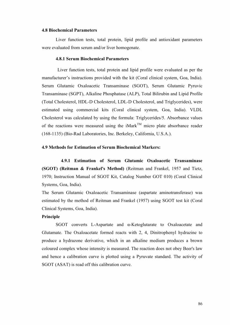
86
4.8 Biochemical Parameters
Liver function tests, total protein, lipid profile and antioxidant parameters
were evaluated from serum and/or liver homogenate.
4.8.1 Serum Biochemical Parameters
Liver function tests, total protein and lipid profile were evaluated as per the
manufacturer’s instructions provided with the kit (Coral clinical system, Goa, India).
Serum Glutamic Oxaloacetic Transaminase (SGOT), Serum Glutamic Pyruvic
Transaminase (SGPT), Alkaline Phosphatase (ALP), Total Bilirubin and Lipid Profile
(Total Cholesterol, HDL-D Cholesterol, LDL-D Cholesterol, and Triglycerides), were
estimated using commercial kits (Coral clinical system, Goa, India). VLDL
Cholesterol was calculated by using the formula: Triglycerides/5. Absorbance values
of the reactions were measured using the iMarkTM
micro plate absorbance reader
(168-1135) (Bio-Rad Laboratories, Inc. Berkeley, California, U.S.A.).
4.9 Methods for Estimation of Serum Biochemical Markers:
4.9.1 Estimation of Serum Glutamic Oxaloacetic Transaminase
(SGOT) (Reitman & Frankel's Method) (Reitman and Frankel, 1957 and Tietz,
1970; Instruction Manual of SGOT Kit, Catalog Number GOT 010) (Coral Clinical
Systems, Goa, India).
The Serum Glutamic Oxaloacetic Transaminase (aspartate aminotransferase) was
estimated by the method of Reitman and Frankel (1957) using SGOT test kit (Coral
Clinical Systems, Goa, India).
Principle
SGOT converts L-Aspartate and α-Ketoglutarate to Oxaloacetate and
Glutamate. The Oxaloacetate formed reacts with 2, 4, Dinitrophenyl hydrazine to
produce a hydrazone derivative, which in an alkaline medium produces a brown
coloured complex whose intensity is measured. The reaction does not obey Beer's law
and hence a calibration curve is plotted using a Pyruvate standard. The activity of
SGOT (ASAT) is read off this calibration curve.
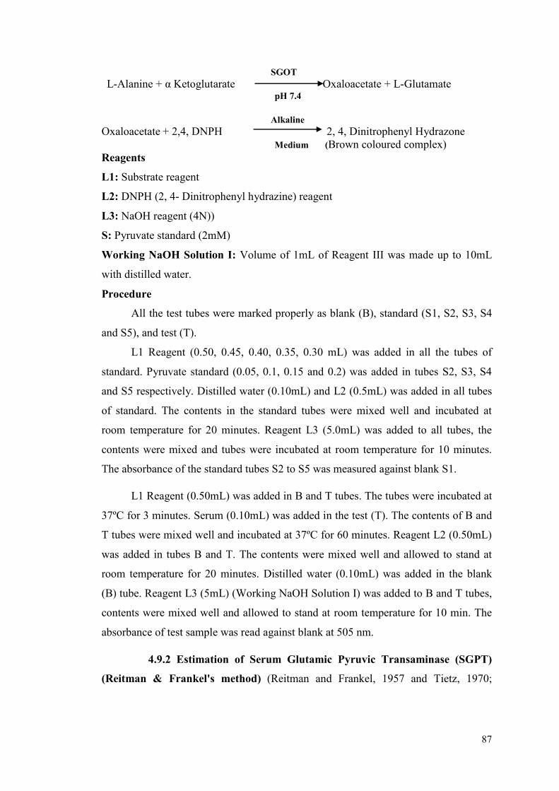
87
SGOT
L-Alanine + α Ketoglutarate Oxaloacetate + L-Glutamate pH 7.4
Alkaline
Oxaloacetate + 2,4, DNPH 2, 4, Dinitrophenyl Hydrazone Medium (Brown coloured complex)
Reagents
L1: Substrate reagent
L2: DNPH (2, 4- Dinitrophenyl hydrazine) reagent
L3: NaOH reagent (4N))
S: Pyruvate standard (2mM)
Working NaOH Solution I: Volume of 1mL of Reagent III was made up to 10mL
with distilled water.
Procedure
All the test tubes were marked properly as blank (B), standard (S1, S2, S3, S4
and S5), and test (T).
L1 Reagent (0.50, 0.45, 0.40, 0.35, 0.30 mL) was added in all the tubes of
standard. Pyruvate standard (0.05, 0.1, 0.15 and 0.2) was added in tubes S2, S3, S4
and S5 respectively. Distilled water (0.10mL) and L2 (0.5mL) was added in all tubes
of standard. The contents in the standard tubes were mixed well and incubated at
room temperature for 20 minutes. Reagent L3 (5.0mL) was added to all tubes, the
contents were mixed and tubes were incubated at room temperature for 10 minutes.
The absorbance of the standard tubes S2 to S5 was measured against blank S1.
L1 Reagent (0.50mL) was added in B and T tubes. The tubes were incubated at
37ºC for 3 minutes. Serum (0.10mL) was added in the test (T). The contents of B and
T tubes were mixed well and incubated at 37ºC for 60 minutes. Reagent L2 (0.50mL)
was added in tubes B and T. The contents were mixed well and allowed to stand at
room temperature for 20 minutes. Distilled water (0.10mL) was added in the blank
(B) tube. Reagent L3 (5mL) (Working NaOH Solution I) was added to B and T tubes,
contents were mixed well and allowed to stand at room temperature for 10 min. The
absorbance of test sample was read against blank at 505 nm.
4.9.2 Estimation of Serum Glutamic Pyruvic Transaminase (SGPT)
(Reitman & Frankel's method) (Reitman and Frankel, 1957 and Tietz, 1970;
88
Instruction Manual of SGPT Kit, Catalog Number GPT 010) (Coral Clinical Systems,
Goa, India).
The Serum Glutamic Pyruvic Transaminase (alanine aminotransferase) was estimated
by the method of Reitman and Frankel (1957) using SGPT test kit (Coral Clinical
System, Goa, India).
Principle
SGPT converts L-Alanine and α-Ketoglutarate to Pyruvate and Glutamate.
The Pyruvate formed reacts with 2, 4, Dinitrophenyl hydrazine to produce a
hydrazone derivative, which in an alkaline medium produces a brown coloured
complex whose intensity is measured. The reaction does not obey Beer's law and
hence a calibration curve is plotted using a pyruvate standard. The activity of SGPT
(ALAT) is read off this calibration curve.
SGPT
L-Alanine + α Ketoglutarate Pyruvate + L-Glutamate pH 7.4 Alkaline
Pyruvate + 2,4, DNPH 2, 4, Dinitrophenyl Hydrazone Medium (Brown coloured complex)
Reagents
L1: Substrate reagent
L2: DNPH (2, 4- Dinitrophenyl hydrazine) reagent
L3: NaOH reagent (Sodium hydroxide, (4N))
S: Pyruvate standard (2mM)
Working NaOH Solution I: Volume of 1mL of Reagent III was made up to 10mL
with distilled water.
Procedure
All the test tubes were marked properly as blank (B), standard (S1, S2, S3, S4
and S5), and test (T).
L1 Reagent (0.50, 0.45, 0.40, 0.35, 0.30 mL) was added in all the tubes of
standard. Pyruvate standard (0.05, 0.1, 0.15 and 0.2) was added in tubes S2, S3, S4
and S5 respectively. Distilled water (0.10mL) and L2 (0.5mL) was added in all tubes
of standard. The contents in the standard tubes were mixed well and incubated at
room temperature for 20 minutes. Reagent L3 (5.0mL) was added to all tubes, the
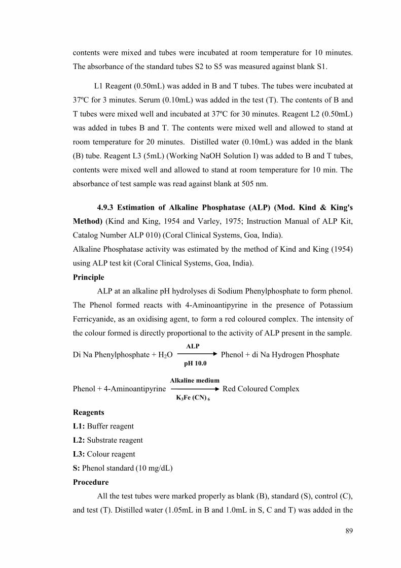
89
contents were mixed and tubes were incubated at room temperature for 10 minutes.
The absorbance of the standard tubes S2 to S5 was measured against blank S1.
L1 Reagent (0.50mL) was added in B and T tubes. The tubes were incubated at
37ºC for 3 minutes. Serum (0.10mL) was added in the test (T). The contents of B and
T tubes were mixed well and incubated at 37ºC for 30 minutes. Reagent L2 (0.50mL)
was added in tubes B and T. The contents were mixed well and allowed to stand at
room temperature for 20 minutes. Distilled water (0.10mL) was added in the blank
(B) tube. Reagent L3 (5mL) (Working NaOH Solution I) was added to B and T tubes,
contents were mixed well and allowed to stand at room temperature for 10 min. The
absorbance of test sample was read against blank at 505 nm.
4.9.3 Estimation of Alkaline Phosphatase (ALP) (Mod. Kind & King's
Method) (Kind and King, 1954 and Varley, 1975; Instruction Manual of ALP Kit,
Catalog Number ALP 010) (Coral Clinical Systems, Goa, India).
Alkaline Phosphatase activity was estimated by the method of Kind and King (1954)
using ALP test kit (Coral Clinical Systems, Goa, India).
Principle
ALP at an alkaline pH hydrolyses di Sodium Phenylphosphate to form phenol.
The Phenol formed reacts with 4-Aminoantipyrine in the presence of Potassium
Ferricyanide, as an oxidising agent, to form a red coloured complex. The intensity of
the colour formed is directly proportional to the activity of ALP present in the sample.
ALP
Di Na Phenylphosphate + H2O Phenol + di Na Hydrogen Phosphate pH 10.0 Alkaline medium Phenol + 4-Aminoantipyrine Red Coloured Complex K3Fe (CN) 6
Reagents
L1: Buffer reagent
L2: Substrate reagent
L3: Colour reagent
S: Phenol standard (10 mg/dL)
Procedure
All the test tubes were marked properly as blank (B), standard (S), control (C),
and test (T). Distilled water (1.05mL in B and 1.0mL in S, C and T) was added in the

90
tubes. L1 Reagent (1mL) and L2 Reagent (0.10mL) were added in all the tubes. The
contents were mixed well and incubated at 37°C for 3 min. Serum (0.05mL) was
added in test (T) and Phenol standard (0.05mL) was added in standard (S). Contents
of the tubes were mixed well and incubated at 37°C for 15 min. L3 Reagent (1mL)
was added in all the tubes. Serum (0.05mL) was added in control (C). Contents of the
tubes were mixed well and absorbance was read at 510 nm against distilled water as
blank. Serum alkaline phosphatase activity is expressed as KA units.
Abs.T-Abs.C
Total ALP activity in K.A. Units = X 10
Abs.S-Abs.B
4.9.4 Estimation of Total Bilirubin (Mod. Jendrassik and Grof’s Method)
(Jendrassik and Grof’s, 1938; Sherlock, 1951; Instruction Manual of BIL Kit, Catalog
Number BIL 010) (Coral Clinical Systems, Goa, India).
Total Bilirubin was estimated by the method of Jendrassik and Grof’s (1938) using
BIL test kit (Coral Clinical System, Goa, India).
Principle
Bilirubin reacts with diazotized sulphanilic acid to form a coloured
azobilirubin compound. The unconjugated bilirubin couples with the sulphanilic acid
in presence of a caffein-benzoate accelerator. The intensity of the colour formed is
directly proportional to the amount of bilirubin present in the sample.
Bilirubin + Diazotized Sulphanilic acid Azobilirubin Compound
Reagents
L1: Total bilirubin reagent
L2: Total nitrite reagent
Procedure
All the test tubes were marked properly as blank (B) and test (T). L1 Reagent
(1mL) was added in blank (B) and test (T) tubes. L2 Reagent (0.05mL) was added in
test (T). Serum (0.1mL) was added in test (T) and blank (B) tubes. Contents of the
tubes were mixed well and incubated at room temperature for 10 minutes and
absorbance of test sample T was read against blank B at 546 nm.
Total bilirubin in mg/dL = Abs. T × 13
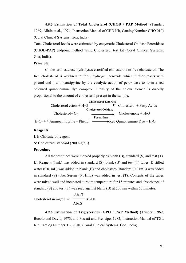
91
4.9.5 Estimation of Total Cholesterol (CHOD / PAP Method) (Trinder,
1969; Allain et al., 1974; Instruction Manual of CHO Kit, Catalog Number CHO 010)
(Coral Clinical Systems, Goa, India).
Total Cholesterol levels were estimated by enzymatic Cholesterol Oxidase Peroxidase
(CHOD-PAP) endpoint method using Cholesterol test kit (Coral Clinical Systems,
Goa, India).
Principle
Cholesterol esterase hydrolyses esterified cholesterols to free cholesterol. The
free cholesterol is oxidised to form hydrogen peroxide which further reacts with
phenol and 4-aminoantipyrine by the catalytic action of peroxidase to form a red
coloured quinoneimine dye complex. Intensity of the colour formed is directly
proportional to the amount of cholesterol present in the sample.
Cholesterol Esterase
Cholesterol esters + H2O Cholesterol + Fatty Acids Cholesterol Oxidase
Cholesterol+ O2 Cholestenone + H2O Peroxidase
H2O2 + 4 Aminoantipyrine + Phenol Red Quinoneimine Dye + H2O
Reagents
L1: Cholesterol reagent
S: Cholesterol standard (200 mg/dL)
Procedure
All the test tubes were marked properly as blank (B), standard (S) and test (T).
L1 Reagent (1mL) was added in standard (S), blank (B) and test (T) tubes. Distilled
water (0.01mL) was added in blank (B) and cholesterol standard (0.01mL) was added
in standard (S) tube. Serum (0.01mL) was added in test (T). Contents of the tubes
were mixed well and incubated at room temperature for 15 minutes and absorbance of
standard (S) and test (T) was read against blank (B) at 505 nm within 60 minutes.
Abs.T
Cholesterol in mg/dL = X 200
Abs.S
4.9.6 Estimation of Triglycerides (GPO / PAP Method) (Trinder, 1969;
Bucolo and David, 1973, and Fossati and Prencipe, 1982; Instruction Manual of TGL
Kit, Catalog Number TGL 010) (Coral Clinical Systems, Goa, India).
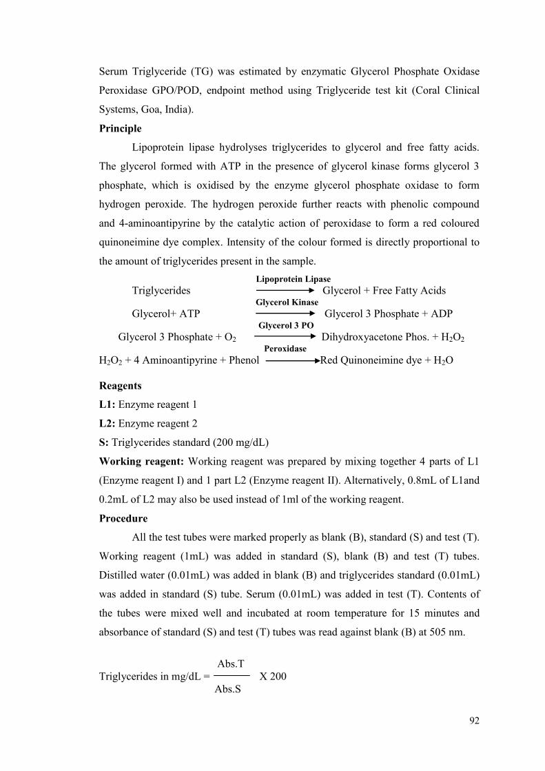
92
Serum Triglyceride (TG) was estimated by enzymatic Glycerol Phosphate Oxidase
Peroxidase GPO/POD, endpoint method using Triglyceride test kit (Coral Clinical
Systems, Goa, India).
Principle
Lipoprotein lipase hydrolyses triglycerides to glycerol and free fatty acids.
The glycerol formed with ATP in the presence of glycerol kinase forms glycerol 3
phosphate, which is oxidised by the enzyme glycerol phosphate oxidase to form
hydrogen peroxide. The hydrogen peroxide further reacts with phenolic compound
and 4-aminoantipyrine by the catalytic action of peroxidase to form a red coloured
quinoneimine dye complex. Intensity of the colour formed is directly proportional to
the amount of triglycerides present in the sample.
Lipoprotein Lipase
Triglycerides Glycerol + Free Fatty Acids Glycerol Kinase
Glycerol+ ATP Glycerol 3 Phosphate + ADP Glycerol 3 PO
Glycerol 3 Phosphate + O2 Dihydroxyacetone Phos. + H2O2
Peroxidase
H2O2 + 4 Aminoantipyrine + Phenol Red Quinoneimine dye + H2O
Reagents
L1: Enzyme reagent 1
L2: Enzyme reagent 2
S: Triglycerides standard (200 mg/dL)
Working reagent: Working reagent was prepared by mixing together 4 parts of L1
(Enzyme reagent I) and 1 part L2 (Enzyme reagent II). Alternatively, 0.8mL of L1and
0.2mL of L2 may also be used instead of 1ml of the working reagent.
Procedure
All the test tubes were marked properly as blank (B), standard (S) and test (T).
Working reagent (1mL) was added in standard (S), blank (B) and test (T) tubes.
Distilled water (0.01mL) was added in blank (B) and triglycerides standard (0.01mL)
was added in standard (S) tube. Serum (0.01mL) was added in test (T). Contents of
the tubes were mixed well and incubated at room temperature for 15 minutes and
absorbance of standard (S) and test (T) tubes was read against blank (B) at 505 nm.
Abs.T
Triglycerides in mg/dL = X 200
Abs.S
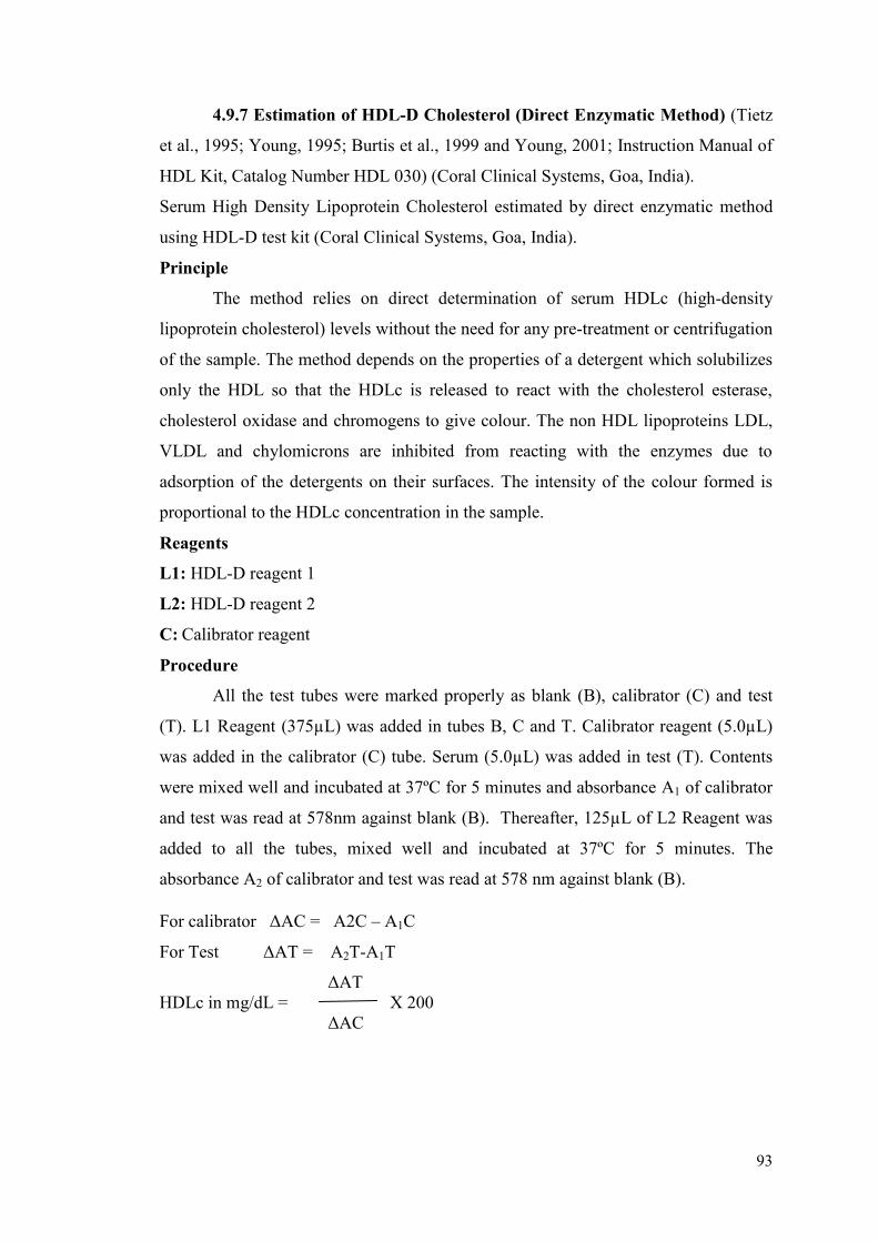
93
4.9.7 Estimation of HDL-D Cholesterol (Direct Enzymatic Method) (Tietz
et al., 1995; Young, 1995; Burtis et al., 1999 and Young, 2001; Instruction Manual of
HDL Kit, Catalog Number HDL 030) (Coral Clinical Systems, Goa, India).
Serum High Density Lipoprotein Cholesterol estimated by direct enzymatic method
using HDL-D test kit (Coral Clinical Systems, Goa, India).
Principle
The method relies on direct determination of serum HDLc (high-density
lipoprotein cholesterol) levels without the need for any pre-treatment or centrifugation
of the sample. The method depends on the properties of a detergent which solubilizes
only the HDL so that the HDLc is released to react with the cholesterol esterase,
cholesterol oxidase and chromogens to give colour. The non HDL lipoproteins LDL,
VLDL and chylomicrons are inhibited from reacting with the enzymes due to
adsorption of the detergents on their surfaces. The intensity of the colour formed is
proportional to the HDLc concentration in the sample.
Reagents
L1: HDL-D reagent 1
L2: HDL-D reagent 2
C: Calibrator reagent
Procedure
All the test tubes were marked properly as blank (B), calibrator (C) and test
(T). L1 Reagent (375µL) was added in tubes B, C and T. Calibrator reagent (5.0µL)
was added in the calibrator (C) tube. Serum (5.0µL) was added in test (T). Contents
were mixed well and incubated at 37ºC for 5 minutes and absorbance A1 of calibrator
and test was read at 578nm against blank (B). Thereafter, 125µL of L2 Reagent was
added to all the tubes, mixed well and incubated at 37ºC for 5 minutes. The
absorbance A2 of calibrator and test was read at 578 nm against blank (B).
For calibrator ΔAC = A2C – A1C
For Test ΔAT = A2T-A1T
ΔAT
HDLc in mg/dL = X 200
ΔAC
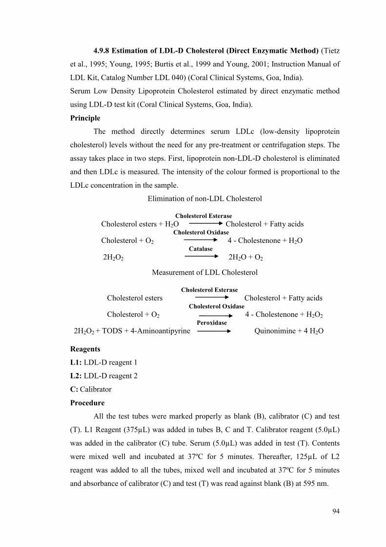
94
4.9.8 Estimation of LDL-D Cholesterol (Direct Enzymatic Method) (Tietz
et al., 1995; Young, 1995; Burtis et al., 1999 and Young, 2001; Instruction Manual of
LDL Kit, Catalog Number LDL 040) (Coral Clinical Systems, Goa, India).
Serum Low Density Lipoprotein Cholesterol estimated by direct enzymatic method
using LDL-D test kit (Coral Clinical Systems, Goa, India).
Principle
The method directly determines serum LDLc (low-density lipoprotein
cholesterol) levels without the need for any pre-treatment or centrifugation steps. The
assay takes place in two steps. First, lipoprotein non-LDL-D cholesterol is eliminated
and then LDLc is measured. The intensity of the colour formed is proportional to the
LDLc concentration in the sample.
Elimination of non-LDL Cholesterol
Cholesterol Esterase
Cholesterol esters + H2O Cholesterol + Fatty acids Cholesterol Oxidase
Cholesterol + O2 4 - Cholestenone + H2O Catalase
2H2O2 2H2O + O2
Measurement of LDL Cholesterol
Cholesterol Esterase
Cholesterol esters Cholesterol + Fatty acids Cholesterol Oxidase
Cholesterol + O2 4 - Cholestenone + H2O2 Peroxidase
2H2O2 + TODS + 4-Aminoantipyrine Quinonimine + 4 H2O
Reagents
L1: LDL-D reagent 1
L2: LDL-D reagent 2
C: Calibrator
Procedure
All the test tubes were marked properly as blank (B), calibrator (C) and test
(T). L1 Reagent (375µL) was added in tubes B, C and T. Calibrator reagent (5.0µL)
was added in the calibrator (C) tube. Serum (5.0µL) was added in test (T). Contents
were mixed well and incubated at 37ºC for 5 minutes. Thereafter, 125µL of L2
reagent was added to all the tubes, mixed well and incubated at 37ºC for 5 minutes
and absorbance of calibrator (C) and test (T) was read against blank (B) at 595 nm.
95
Abs.T
LDLc in mg/dL = X Conc of calibrator
Abs.C
4.10 Liver Biochemical Parameters
Cell free supernatant from liver homogenate was used to estimate Lipid
Peroxidation (OxisResearchTM
, USA), Catalase (CAT) (Sigma-Aldrich, USA),
Superoxide Dismutase (SOD) (Sigma-Aldrich, USA), Reduced Glutathione (Cayman
Chemical Compan, USA), Total Protein and Lipid Profile (Total Cholesterol, HDL-D
Cholesterol, LDL-D Cholesterol, Triglycerides) using commercial kits (Coral clinical
systems, Goa, India). VLDL Cholesterol was estimated by using the formula:
Triglycerides/5. Absorbance of the reactions was read using iMarkTM
micro plate
absorbance reader (168-1135) (Bio-Rad Laboratories, Inc. Berkeley, California,
USA).
Preparation of Liver Homogenate
Liver tissues from rats of all groups were excised, blotted and homogenized
with phosphate buffered saline (1/10 weight/volume), (pH 7.0) and the homogenate
was centrifuged at 30,000 rpm for 15 minutes at 4ºC. Cell free supernatant was
collected and stored in a freezer (-80ºC) until further use.
4.10.1 Estimation of Lipid Peroxidation (Colorimetric Method) (Bull and
Marnett, 1985; Esterbauer et al., 1991; Botsoglou, 1994; Instruction Manual of MDA
Kit, Catalog Number 21044).
Measurement of MDA was used as an indicator of Lipid Peroxidation. Bioxytech®
MDS-586 Lipid Peroxidation Kit (OxisResearchTM
, USA) was used to estimate MDA
from liver homogenate.
Principle
The MDA-586 method is based on the reaction of a chromogenic reagent N-
methyl-2-phenylindole (NMPI), with MDA at 45°C. One molecule of MDA reacts
with 2 molecules of NMPI to yield a stable carbocyanine dye.
Reagents
Reagent R1: N-methyl-2-phenylindole in acetonitrile
Reagent R2: Concentrated hydrochloric acid
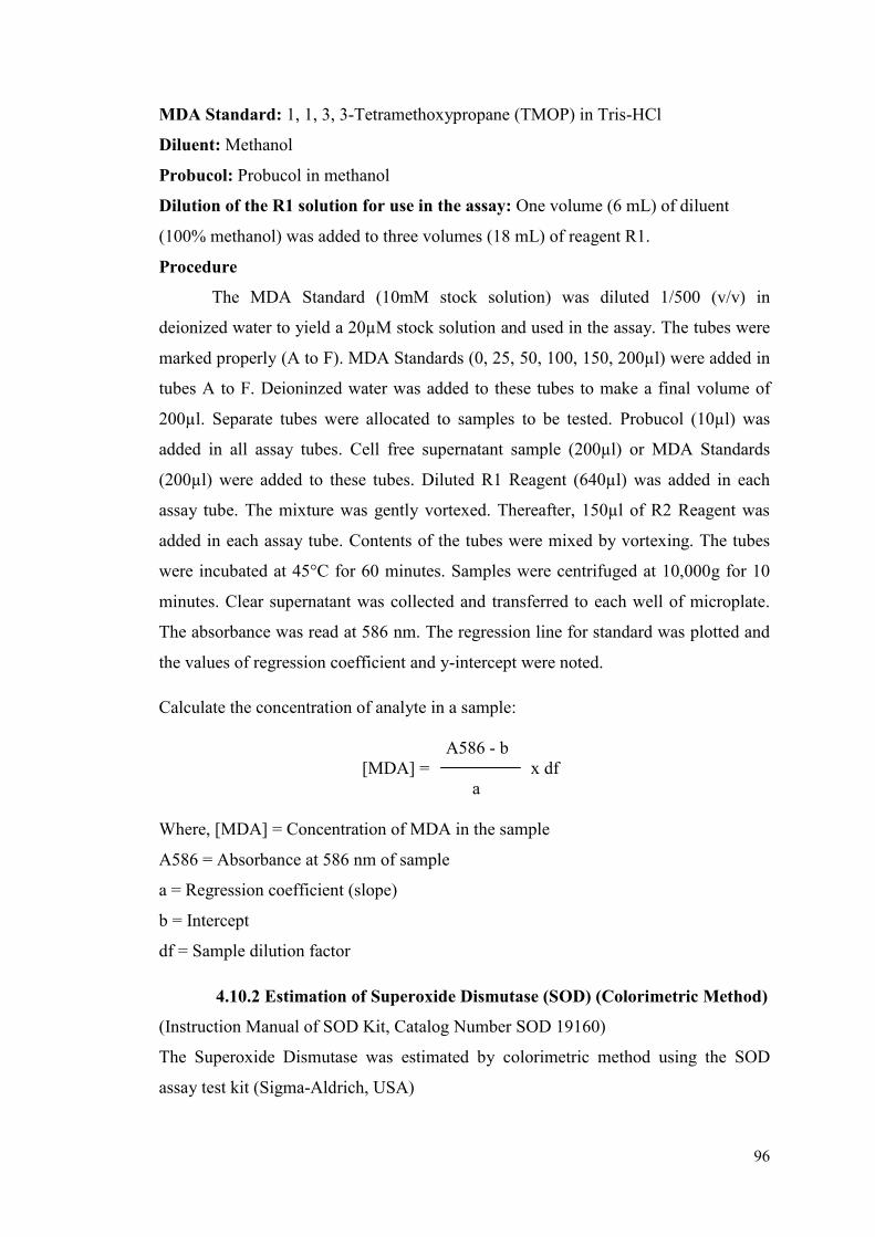
96
MDA Standard: 1, 1, 3, 3-Tetramethoxypropane (TMOP) in Tris-HCl
Diluent: Methanol
Probucol: Probucol in methanol
Dilution of the R1 solution for use in the assay: One volume (6 mL) of diluent
(100% methanol) was added to three volumes (18 mL) of reagent R1.
Procedure
The MDA Standard (10mM stock solution) was diluted 1/500 (v/v) in
deionized water to yield a 20µM stock solution and used in the assay. The tubes were
marked properly (A to F). MDA Standards (0, 25, 50, 100, 150, 200µl) were added in
tubes A to F. Deioninzed water was added to these tubes to make a final volume of
200µl. Separate tubes were allocated to samples to be tested. Probucol (10µl) was
added in all assay tubes. Cell free supernatant sample (200µl) or MDA Standards
(200µl) were added to these tubes. Diluted R1 Reagent (640µl) was added in each
assay tube. The mixture was gently vortexed. Thereafter, 150µl of R2 Reagent was
added in each assay tube. Contents of the tubes were mixed by vortexing. The tubes
were incubated at 45°C for 60 minutes. Samples were centrifuged at 10,000g for 10
minutes. Clear supernatant was collected and transferred to each well of microplate.
The absorbance was read at 586 nm. The regression line for standard was plotted and
the values of regression coefficient and y-intercept were noted.
Calculate the concentration of analyte in a sample:
A586 - b
[MDA] = x df
a
Where, [MDA] = Concentration of MDA in the sample
A586 = Absorbance at 586 nm of sample
a = Regression coefficient (slope)
b = Intercept
df = Sample dilution factor
4.10.2 Estimation of Superoxide Dismutase (SOD) (Colorimetric Method)
(Instruction Manual of SOD Kit, Catalog Number SOD 19160)
The Superoxide Dismutase was estimated by colorimetric method using the SOD
assay test kit (Sigma-Aldrich, USA)

97
Principle
Superoxide dismutase (SOD) is one of the most important antioxidative
enzymes. It catalyses the dismutation of the superoxide anion (O2.-) into hydrogen
peroxide and molecular oxygen. An indirect method using nitroblue tetrazolium
(NBT) is commonly used due to its convenience and ease of use. The procedure from
this kit allows convenient SOD assaying by utilizing Dojindo’s highly water-soluble
tetrazolium salt, WST-1(2-(4-Iodophenyl)-3-(4-nitrophenyl)-5-(2, 4-disulfophenyl)-
2H-tetrazolium, monosodium salt) producing a water-soluble formazan dye after
reduction with a superoxide anion. The rate of the reduction with O2 are linearly
related to the xanthine oxidase (XO) activity, and is inhibited by SOD. Therefore, the
IC50 (50% inhibition activity of SOD or SOD-like materials) can be determined by a
colorimetric method.
Reagents
Reagent I: WST solution
Reagent II: Enzyme solution
Reagent III: Buffer solution
Reagent IV: Dilution buffer
WST working solution: Dilute 1mL of WST Solution (Reagent I) with 19mL of
Buffer Solution (Reagent II)
Enzyme working solution: Dilute 15µl of Enzyme Solution (Reagent III) with
2.5mL of Dilution Buffer (Reagent IV)
Procedure
The microplate wells were marked as test (T), blank 1, blank 2 and blank 3.
Sample (20µl) was added in test (T) and blank 2 wells, and double distilled water
(20µl) was added to blank 1 and blank 3 wells. WST (200µl) working solution was
added in each well and mixed properly. Dilution buffer (20µl) was added to blank 2
and blank 3 wells. Enzyme working solution (20µl) was added to test and blank 1
well. The contents were mixed well and the plate was incubated at 37°C for 20
minutes. The absorbance was read at 450 nm using a micro plate reader.
SOD activity (inhibition rate %) was calculated using the following equation:
SOD activity (inhibition rate %) =
{[(Ablank 1 - Ablank 3) - Asample - Ablank 2)]/ (Ablank 1 - Ablank 3)} x 100
98
4.10.3 Estimation of Catalase (Colorimetric Method) (Deisseroth and
Dounce, 1970; Zamocky and Koller, 1999 and Zhou and Kang, 2000; Instruction
Manual of CAT kit, Catalog Number CAT100).
The Catalase was estimated by colorimetric method using the CAT assay test kit
(Sigma-Aldrich, USA).
Principle
Catalase is an antioxidant enzyme universally present in mammalian and non-
mammalian aerobic cells containing a cytochrome system. Catalase is able to
decompose hydrogen peroxide by two different reaction pathways. In the first, known
as the “catalatic” pathway, 2 molecules of hydrogen peroxide are converted to water
and oxygen (catalatic activity)
Protein-Fe3+
+ H2O2 → Protein-Fe3+
-OOH (Primary Complex) + H2O
Protein-Fe3+
-OOH + H2O2 → Protein-Fe3+
-OH + H2O + O2
The overall reaction gives: 2 H2O2 → 2 H2O + O2
The primary complex can also decompose by another pathway (peroxidatic
decomposition): Protein-Fe3+
-OOH + AH2 → Protein-Fe3+
-OH + H2O + A
Where AH2 is an internal or external donor of hydrogen. Low molecular weight
alcohols can serve as electron donors. The catalatic pathway is predominant when the
hydrogen peroxide concentration is greater than 0.1mM and the peroxidatic pathway
is dominant when the hydrogen peroxide concentration is less than 0.1mM or the
substrate is alkyl peroxide.
Reagents
Reagent I: Assay buffer 10× (500mM potassium phosphate buffer, pH 7.0)
Reagent II: Chromogen reagent
Reagent III: Stop solution (15mM sodium azide in water)
Reagent IV: 3% (w/w) Hydrogen peroxide solution
Reagent V: Enzyme dilution buffer (50mM potassium phosphate buffer, pH 7.0, and
containing 0.1% TritonX-100)
1X Assay Buffer: Dilute 2mL of 10X assay buffer (reagent I) 10-folds to 20mL with
distilled water
Colorimetric Assay Substrate Solution (200mM H2O2): Dilute 200µl of 3% H2O2
to 1mL with 1X assay buffer. Dilute 50µl of the above solution to 1mL (20-fold) with
1X assay buffer
Catalase Colorimetric Enzymatic Reaction Scheme
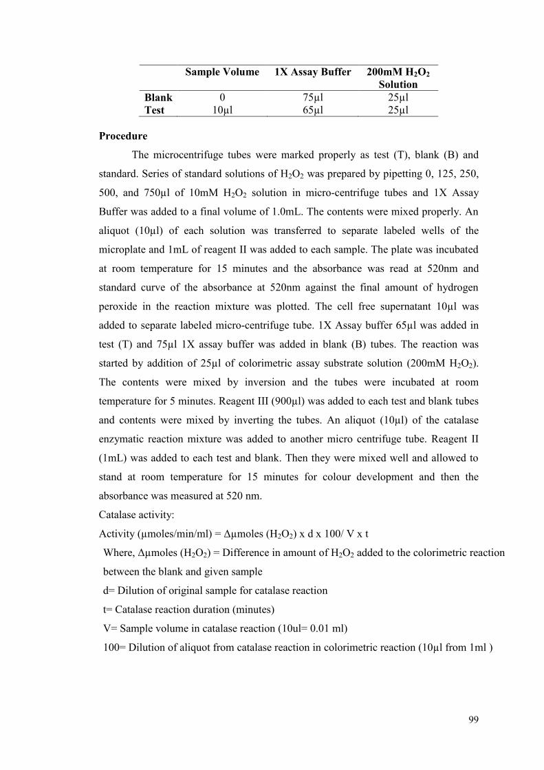
99
Sample Volume 1X Assay Buffer 200mM H2O2
Solution
Blank 0 75µl 25µl
Test 10µl 65µl 25µl
Procedure
The microcentrifuge tubes were marked properly as test (T), blank (B) and
standard. Series of standard solutions of H2O2 was prepared by pipetting 0, 125, 250,
500, and 750µl of 10mM H2O2 solution in micro-centrifuge tubes and 1X Assay
Buffer was added to a final volume of 1.0mL. The contents were mixed properly. An
aliquot (10µl) of each solution was transferred to separate labeled wells of the
microplate and 1mL of reagent II was added to each sample. The plate was incubated
at room temperature for 15 minutes and the absorbance was read at 520nm and
standard curve of the absorbance at 520nm against the final amount of hydrogen
peroxide in the reaction mixture was plotted. The cell free supernatant 10µl was
added to separate labeled micro-centrifuge tube. 1X Assay buffer 65µl was added in
test (T) and 75µl 1X assay buffer was added in blank (B) tubes. The reaction was
started by addition of 25µl of colorimetric assay substrate solution (200mM H2O2).
The contents were mixed by inversion and the tubes were incubated at room
temperature for 5 minutes. Reagent III (900µl) was added to each test and blank tubes
and contents were mixed by inverting the tubes. An aliquot (10µl) of the catalase
enzymatic reaction mixture was added to another micro centrifuge tube. Reagent II
(1mL) was added to each test and blank. Then they were mixed well and allowed to
stand at room temperature for 15 minutes for colour development and then the
absorbance was measured at 520 nm.
Catalase activity:
Activity (µmoles/min/ml) = Δµmoles (H2O2) x d x 100/ V x t
Where, Δµmoles (H2O2) = Difference in amount of H2O2 added to the colorimetric reaction
between the blank and given sample
d= Dilution of original sample for catalase reaction
t= Catalase reaction duration (minutes)
V= Sample volume in catalase reaction (10ul= 0.01 ml)
100= Dilution of aliquot from catalase reaction in colorimetric reaction (10µl from 1ml )
100
4.10.4 Estimation of Total Protein (Biuret Method) (Gornall et al., 1949
and Doumas, 1975; Instruction Manual of Total Protein Kit, Catalog Number TPR
010) (Coral Clinical Systems, Goa, India).
The Total Protein was estimated by modified Biuret method using the total protein
test kit (Coral Clinical System, Goa, India).
Principle
Proteins, in an alkaline medium, bind with the cupric ions present in the biuret
reagent to form a blue-violet coloured complex. The intensity of the colour formed is
directly proportional to the amount of Proteins present in the sample.
Proteins + Cu+ +
Blue Violet Coloured Complex
Reagents
L1: Biuret reagent
S: Protein standard (8g/dL)
Procedure
All the test tubes were marked properly as blank (B), standard (S) and test (T).
L1 Reagent (1.0mL) was added to all the test tubes. Thereafter, Distilled water
(0.02mL) was added in blank (B) and protein standard (0.02mL) was added in the
standard test tube (S). Cell free supernatant sample (0.02mL) was added in the tube
for test sample (T). Contents of the tubes were mixed well and incubated at 37ºC for
10 minutes and absorbance of standard (S) and test (T) was read against blank (B) at
550 nm within 60 minutes.
Abs. T
Total Proteins in g/dl = x 8
Abs. S
4.10.5 Estimation of Reduced Glutathione (End Point Method) (Griffith,
1980; Lash et al., 1985 and Baillie and Slatter, 1991 and Instruction Manual of
Reduced Glutathione (GSH) kit, Catalog Number GSH 703002).
The assay was estimated by end point method using the GSH assay test kit (Cayman
Chemical Company).
Estimation of reduced glutathione was done from deproteinized samples.
101
Procedure for Deproteination of Samples
Part of the homogenate was deproteinated with metaphosphoric acid (MPA)
reagent and 4M triethanolamine (TEAM) reagent and used for estimation of reduced
glutathione (GSH) content in the tissue by using commercially available kits (Cayman
Chemical Company, USA).
MPA Reagent: Dissolve 5g of metaphosphoric acid (Sigma-Aldrich) in 50mL water.
The MPA solution was stable for 4 hours at 25°C. Equal volumes of MPA reagent
was added to the sample and mixed by vortexing. The mixture was allowed to stand at
room temperature for 5 minutes and centrifuged at 2000 rpm for 2 minutes, and the
supernatant was collected.
TEAM Reagent: A 4M solution of triethanolamine (Sigma-Aldrich) in water was
prepared by mixing 531µl of the triethanolamine with 469µl of water. The TEAM
solution was stable for 4 hours at 25°C. 50µl of TEAM reagent was added per 1mL of
the supernatant and vortexed immediately. The TEAM reagent increased the pH of
the sample. The sample was ready for assay of the total GSH.
Principle
Glutathione (GSH) is a tripeptide (γ-glutamylcysteinylglycine) widely
distributed in both plants and animals. GSH serves as a nucleophilic co-substrate to
glutathione transferases in the detoxification of xenobiotics and is an essential
electron donor to glutathione peroxidase in the reduction of hydroperoxides. GSH is
also involved in amino acid transport and maintenance of protein sulfhydryl reduction
status. The sulfhydryl group of GSH reacts with DTNB (5, 5’- dithio-bis-2-
(nitrobenzoic acid), Ellman’s reagent) and produces a yellow coloured 5-thio-2-
nitrobenzoic acid (TNB). The mixed disulfide GSTNB (between GSH and TNB) that
is concomitantly produced, is reduced by glutathione reductase to recycle the GSH
and produce more TNB. The rate of TNB production is directly proportional to this
recycling reaction, which is in turn directly proportional to the concentration of GSH
in the sample.
Reagents
Reagent I: GSH MES buffer (2X)
The buffer consists of 0.4M 2-(N-morpholino) ethanesulphonic acid, 0.1M phosphate,
and 2mM EDTA. PH 6.0
102
This GSH MES buffer was added to equal volume of HPLC-grade water (60mL of the
buffer with 60mL of HPLC-grade water) to make a 1X buffer for assay.
Reagent II: GSSG standard
Ready to use, 2mL of 25µM GSSG in MES buffer was supplied with the kit.
Reagent III: GSH Co-factor mixture
The vial contained a lyophilized powder of NADP+ and glucose-6-phosphate. The
contents of the vial were reconstituted with 0.5mL water and mixed well.
Reagent IV: GSH enzyme mixture
The vials contained glutathione reductase and glucose-6-phosphate dehydrogenase in
0.2mL buffer. 1X MES buffer (2mL) was added to the vial before use, the cap was
replaced, and mixed well.
Reagent V: GSH DTNB vial contained a lyophilized powder of DTNB (5, 5’-dithio-
bis-(2-nitrobenzoic acid). The contents of the vial were reconstituted with 0.5mL of
water and mixed well.
Assay cocktail: MES buffer (11.25mL), reconstituted cofactor mixture (0.45mL),
reconstituted enzyme mixture (2.1mL), water (2.3mL) and reconstituted DTNB
(0.45mL).
Procedure
The tubes were marked properly (A to H). Reagent II (0, 5, 10, 20, 40, 80,
120, 160 µl) was added in tubes A to H. Reagent I was added to these tubes to make a
final volume of 500µl. The contents of the tubes were mixed well. The microplate
wells were accordingly labelled and 50µl of standard was added in labelled wells.
Deproteinated samples (50µl) were added to separate labelled wells of the micro
plate. Freshly prepared assay cocktail (150µl) was mixed with the standard wells (A-
H) and each well of samples. The plate was covered and incubated in dark, on an
orbital shaker, for 25 minutes. The absorbance was measured at 415 nm.
GSSG or Total GSH (µM):
[Total GSH] or [GSSG] =
[(Absorbance at 415nm) - (Y-intercept)/Slope] × 2 × Sample Dilution
4.10.6 Estimation of Total cholesterol (CHOD / PAP Method)
As described in chapter 4, section 4.9.5
4.10.7 Estimation of Triglycerides (GPO / PAP Method)
As described in chapter 4, section 4.9.6
103
4.10.8 Estimation of HDL-D Cholesterol (Direct Enzymatic Method)
As described in chapter 4, section 4.9.7
4.10.9 Estimation of LDL-D Cholesterol (Direct Enzymatic Method)
As described in chapter 4, section 4.9.8
104
4.11 Liver Histology
Portion of the liver tissues (left medial lobe) from rats of all groups were used
for the histopathological analysis. The tissues were transferred in 10% neutral-
buffered formalin solution for fixation and later on processed for histopathological
studies following the standard procedure. The sections were cut on microtome
(Microm, HM 315), processed, and stained with Mayer’s haematoxylin and eosin
(H&E staining) (Mayer, 1891) for examination. The stained tissues were observed
under a light microscope at 20X magnifying power and photographed using Image
Pro Plus (v5.1.2.59).
4.11.1 Fixation
Fixation is the process of preserving, hardening and preventing changes in the
tissues. The tissues were excised out immediately after sacrificing, washed with
saline, blot dried and cut into pieces of such thickness that the fixative readily
penetrated throughout the tissue to be fixed. Tissue was transferred to the 10%
formalin solution till further processing.
4.11.2 Tissue Processing
Tissue processing involves dehydration, clearing and infiltration of the tissue
with paraffin. The usual dehydrating agent is ethyl alcohol; acetone and isopropyl
alcohol can also be used. Following dehydration, the tissue was transferred to a
paraffin solvent, which is miscible with the dehydrating agent as well. These are
known as clearing agents such as chloroform and xylene. Tissues were thoroughly
washed by placing them under running tap water and then conveyed through a series
of following solvents as per schedule for dehydration, clearing and paraffin
infiltration:
Solvent Grades Time
Alcohol 70% 20 minutes
Alcohol 80% 20 minutes
Alcohol 90% 20 minutes
Alcohol 95% 20 minutes
Isopropyl alcohol 20 minutes
Acetone 20 minutes
Chloroform 20 minutes
Melted paraffin wax
(60°C)
20 minutes
105
The tissues were then embedded in paraffin wax to prepare tissue blocks,
which were oriented so that sections could be cut in desired plane of the tissue.
Tissues were then fixed in cassette after trimming them to suitable size.
4.11.3 Section Cutting
A smear of 5% Mayer’s egg albumin was prepared and smeared onto the slide
and dried. The tissue sections of 4µM thickness were cut with the help of spencer type
rotating microtome. The tissue sections were put on slide and then sections were
floated in water on slide at 55-60°C, water was drained off and slides were dried on
hot plate at 50°C for 30 minutes. The sections were thus ready for staining.
Staining Procedure
Reagents
1) Mayer’s hematoxylin stain
2) Eosin stain, 2% w/v in alcohol
After fixing the sections on slides, they were stained by serially passing them through
following reagents:
Reagents Time
Xylol 3 minutes
Acetone 3 minutes
Alcohol 95% 3 minutes
Haematoxylin stain 20 minutes
Running water 20 minutes
Eosin stain 5 minutes
Alcohol 95% (3 changes) 3 minutes each
Acetone (2 changes) 3 minutes each
Xylol (2 changes) 3 minutes each
After passing through all the above reagents and stains, the slides were
mounted with D.P.X. (Diphenyl Phthalate Xylene) and cover slip was placed. Care
was taken to avoid trapping air bubbles while mounting the slide.
4.11.4 Observation
Changes in histopathological characteristics were observed for all the slides
and photographs were taken.
106
4.12 Gene Expression Study
4.12.1 RNA Extraction
RNA extraction was performed by TRIzol method (Chomczynski and Sacchi,
1987). Total RNA extraction was performed from liver tissues from rats of all groups
after completion of the experiment (8th
or 15th
day liver tissue). Total RNA was
extracted using TRIzol reagent (Sigma-Aldrich). Each frozen liver sample (~100mg
of tissue) was crushed in liquid nitrogen with mortar and pestle and made into a fine
powder. The powdered tissue was added in 1mL of TRIzol reagent before thawing
and vortexed vigorously for 15 seconds. Chloroform (200µl) was added in these tubes
and the contents were gently mixed by inverting the tubes. The tubes were incubated
for 2 to 3 minutes at room temperature. The mixture was centrifuged at 12000 rpm for
15 minutes at 4°C. The aqueous phase was transferred carefully to a new tube,
without disturbing the interphase. Chilled isopropyl alcohol was added to the aqueous
phase in a new tube and incubated overnight at -20°C. Next day, the mixture was kept
for 10 minutes at room temperature. The mixture was centrifuged at 12000 rpm for 15
minutes at 4°C. The pellet was washed with 500µl 75% chilled ethanol (Freshly
prepared) and centrifuged at 7500 rpm for 5 minutes at 4°C. The supernatant was
discarded and pellet was suspended in 30µl diethylpyrocarbonate-treated water
(DEPC) water. RNA samples (2µl) were loaded on 0.8% agarose gel.
4.12.2 RNA Quantification and Quality Check
Quantification of RNA was performed with UV spectrophotometer
(NanoDrop; Eppendorf). RNA quantification and purity was checked at 260 nm, 230
nm and 280 nm using a spectrophotometer. RNA concentration was determined by
the measurement of absorbances at 260 nm, while the ratios 260/280 and 260/230
were recorded to detect contamination by phenols, proteins and other organic
compounds – ideally in pure RNA preparations both of these ratios would give a
value of 2. The isolated RNA with 260nm/280nm ratio between 1.5 to 2.0 is a
dimensionless parameter for RNA purity. Quality of RNA was checked on agarose
gel by ethidium bromide staining.

107
4.12.3 cDNA Synthesis
Total RNA was reverse transcribed using the Super Script First-Strand cDNA
synthesis kit (Invitrogen, USA) according to the manufacturer’s instructions. First
strand synthesis of complementary DNA (cDNA) was done by reverse transcription.
Briefly, 4µg RNA was mixed with 3µl of random hexamer (50ng/µl) and 1µl of dNTP
(10mM) in a total volume of 12µl. The mixture was incubated at 65°C for 5 minutes.
After the incubation, the reaction was cooled rapidly on ice for 1 minute, followed by
addition of 4µl 5x first strand buffer (Promega), [250 mM Tris-HCl (pH 8.3), 375mM
KCl, 15mM MgCl2], 2µl 0.1M DTT (Invitrogen, USA) and 1µl RNaseOUT™
Recombinant ribonuclease inhibitor (40 units/µl, Invitrogen, USA). The tubes were
incubated at 37°C for 2 minutes followed by addition of 0.5μl M-MLV RT (200 units,
Promega). The contents of the reaction were mixed gently by pipetting up and down.
Reverse transcription was carried out using random priming and Moloney murine
leukaemia virus (MMLV) reverse transcriptase (RT) (Promega, USA), in a reaction
volume of 20μl. This RT also has an endogenous RNase H function which ensures
degradation of the RNA. Reverse transcription included the following three phases:
The reaction was incubated at 25°C for 10 minutes for RT enzyme activation
followed by 50 minutes at 37°C for reverse transcription and the reaction was
inactivated by heating at 70°C for 15 minutes. Products of the reaction were
appropriately diluted and subjected to end point PCR using house-keeping gene
GAPDH. The synthesized cDNA was stored at -80°C.
4.12.4 Semi-Quantitative Polymerase Chain Reaction
The cDNA was diluted with 1:40 Tris buffer (T10E1 buffer) (10 mM, Tris (pH
8.0), 1mM EDTA (pH 8.0) and used for semi-quantitative polymerase chain reaction
(PCR). The diluted cDNA was used for normalization using gene specific primers for
amplification of house-keeping gene GAPDH. For semi-quantitative PCR, normalized
concentration of 1:40 diluted cDNA was used for amplification in a 25µl reaction
consisting of 2.5µl 10X PCR buffer (Sigma- Aldrich, USA), 2μl 2.5mM dNTPs
(GeNei, India), 0.3μl Taq DNA polymerase (5U/μl, Sigma-Aldrich, USA), 0.5μl each
of forward and reverse KiCqStart® primers (10pM/μl stock) (Sigma-Aldrich, USA).
The temperature profile for semi-quantitative PCR was as below:
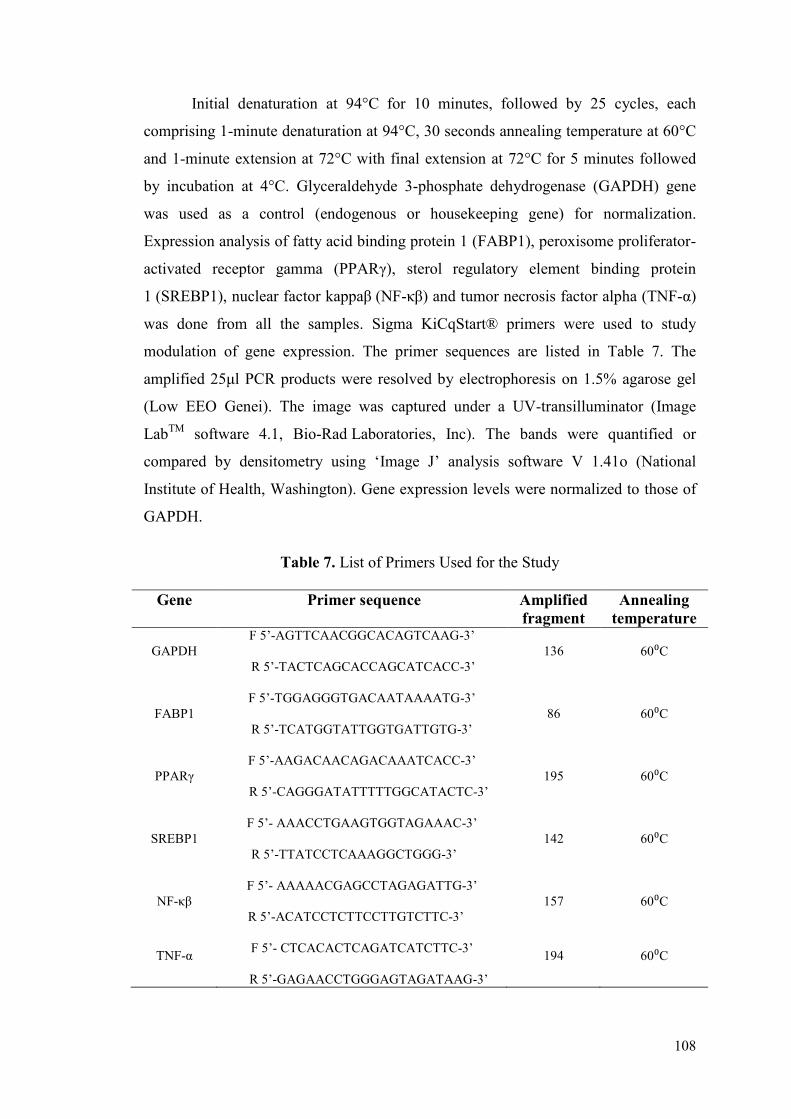
108
Initial denaturation at 94°C for 10 minutes, followed by 25 cycles, each
comprising 1-minute denaturation at 94°C, 30 seconds annealing temperature at 60°C
and 1-minute extension at 72°C with final extension at 72°C for 5 minutes followed
by incubation at 4°C. Glyceraldehyde 3-phosphate dehydrogenase (GAPDH) gene
was used as a control (endogenous or housekeeping gene) for normalization.
Expression analysis of fatty acid binding protein 1 (FABP1), peroxisome proliferator-
activated receptor gamma (PPARγ), sterol regulatory element binding protein
1 (SREBP1), nuclear factor kappaβ (NF-κβ) and tumor necrosis factor alpha (TNF-α)
was done from all the samples. Sigma KiCqStart® primers were used to study
modulation of gene expression. The primer sequences are listed in Table 7. The
amplified 25μl PCR products were resolved by electrophoresis on 1.5% agarose gel
(Low EEO Genei). The image was captured under a UV-transilluminator (Image
LabTM
software 4.1, Bio-Rad Laboratories, Inc). The bands were quantified or
compared by densitometry using ‘Image J’ analysis software V 1.41o (National
Institute of Health, Washington). Gene expression levels were normalized to those of
GAPDH.
Table 7. List of Primers Used for the Study
Gene Primer sequence Amplified
fragment
Annealing
temperature
GAPDH
F 5’-AGTTCAACGGCACAGTCAAG-3’
R 5’-TACTCAGCACCAGCATCACC-3’
136 60⁰C
FABP1
F 5’-TGGAGGGTGACAATAAAATG-3’
R 5’-TCATGGTATTGGTGATTGTG-3’
86 60⁰C
PPARγ
F 5’-AAGACAACAGACAAATCACC-3’
R 5’-CAGGGATATTTTTGGCATACTC-3’
195 60⁰C
SREBP1
F 5’- AAACCTGAAGTGGTAGAAAC-3’
R 5’-TTATCCTCAAAGGCTGGG-3’
142 60⁰C
NF-κβ
F 5’- AAAAACGAGCCTAGAGATTG-3’
R 5’-ACATCCTCTTCCTTGTCTTC-3’
157 60⁰C
TNF-α F 5’- CTCACACTCAGATCATCTTC-3’
R 5’-GAGAACCTGGGAGTAGATAAG-3’
194 60⁰C
109
4.13 Statistical Analysis
The data were presented as Mean ± Standard Error (SE). The Dunnett
Multiple Comparison Test and One Way Analysis of Variance (ANOVA) was done to
estimate the statistical significance between groups. GraphPad Instat (Trial Version
3.06, GraphPad Software, San Diego, CA, USA) was used for statistical analysis
while graphs were plotted using GraphPad Prism (Trial Version 5.0, GraphPad
Software, San Diego, CA, USA).

CHAPTER 5
RESULTS
Awards
Second prize in oral
Presentation Award
Best Poster Presentation
Award
Awards
Second prize in oral
Presentation Award
Best Poster Presentation
Award Awards
Second prize in oral
Presentation Award
Best Poster Presentation
Award
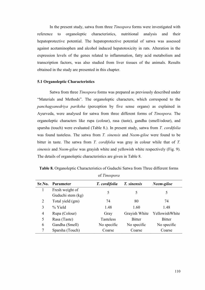
110
In the present study, satwa from three Tinospora forms were investigated with
reference to organoleptic characteristics, nutritional analysis and their
hepatoprotective potential. The hepatoprotective potential of satwa was assessed
against acetaminophen and alcohol induced hepatotoxicity in rats. Alteration in the
expression levels of the genes related to inflammation, fatty acid metabolism and
transcription factors, was also studied from liver tissues of the animals. Results
obtained in the study are presented in this chapter.
5.1 Organoleptic Characteristics
Satwa from three Tinospora forms was prepared as previously described under
“Materials and Methods”. The organoleptic characters, which correspond to the
panchagyanedriya pariksha (perception by five sense organs) as explained in
Ayurveda, were analysed for satwa from three different forms of Tinospora. The
organoleptic characters like rupa (colour), rasa (taste), gandha (smell/odour), and
sparsha (touch) were evaluated (Table 8.). In present study, satwa from T. cordifolia
was found tasteless. The satwa from T. sinensis and Neem-giloe were found to be
bitter in taste. The satwa from T. cordifolia was gray in colour while that of T.
sinensis and Neem-giloe was grayish white and yellowish white respectively (Fig. 9).
The details of organoleptic characteristics are given in Table 8.
Table 8. Organoleptic Characteristics of Guduchi Satwa from Three different forms
of Tinospora
Sr.No. Parameter T. cordifolia T. sinensis Neem-giloe
1 Fresh weight of
Guduchi stem (kg) 5 5 5
2 Total yield (gm) 74 80 74
3 % Yield 1.48 1.60 1.48
4 Rupa (Colour) Gray Grayish White YellowishWhite
5 Rasa (Taste) Tasteless Bitter Bitter
6 Gandha (Smell) No specific No specific No specific
7 Sparsha (Touch) Coarse Coarse Coarse
Awards
Second prize in oral
Presentation Award
Best Poster Presentation
Award
Awards
Second prize in oral
Presentation Award
Best Poster Presentation
Award
Awards
Second prize in oral
Presentation Award
Best Poster Presentation
Award
Awards
Second prize in oral
Presentation Award
Best Poster Presentation
Award
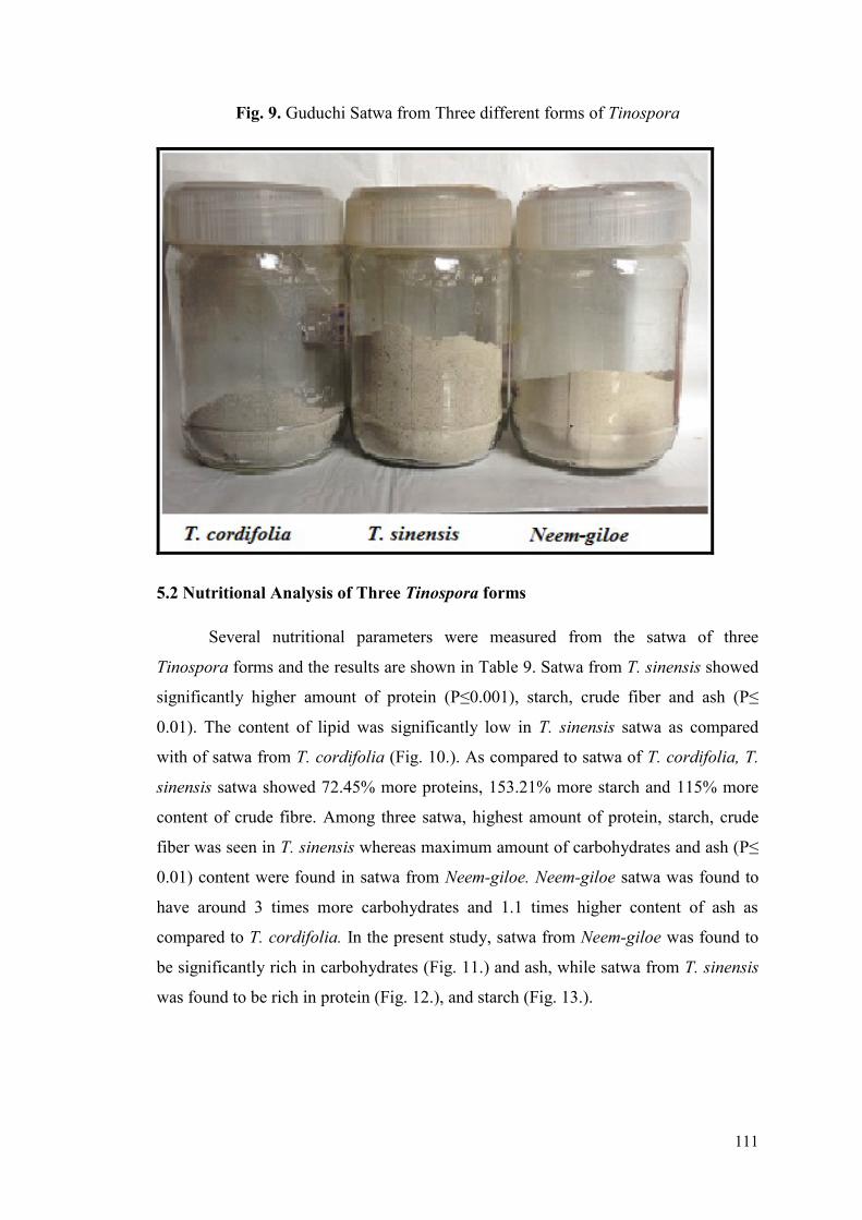
111
Fig. 9. Guduchi Satwa from Three different forms of Tinospora
5.2 Nutritional Analysis of Three Tinospora forms
Several nutritional parameters were measured from the satwa of three
Tinospora forms and the results are shown in Table 9. Satwa from T. sinensis showed
significantly higher amount of protein (P≤0.001), starch, crude fiber and ash (P≤
0.01). The content of lipid was significantly low in T. sinensis satwa as compared
with of satwa from T. cordifolia (Fig. 10.). As compared to satwa of T. cordifolia, T.
sinensis satwa showed 72.45% more proteins, 153.21% more starch and 115% more
content of crude fibre. Among three satwa, highest amount of protein, starch, crude
fiber was seen in T. sinensis whereas maximum amount of carbohydrates and ash (P≤
0.01) content were found in satwa from Neem-giloe. Neem-giloe satwa was found to
have around 3 times more carbohydrates and 1.1 times higher content of ash as
compared to T. cordifolia. In the present study, satwa from Neem-giloe was found to
be significantly rich in carbohydrates (Fig. 11.) and ash, while satwa from T. sinensis
was found to be rich in protein (Fig. 12.), and starch (Fig. 13.).
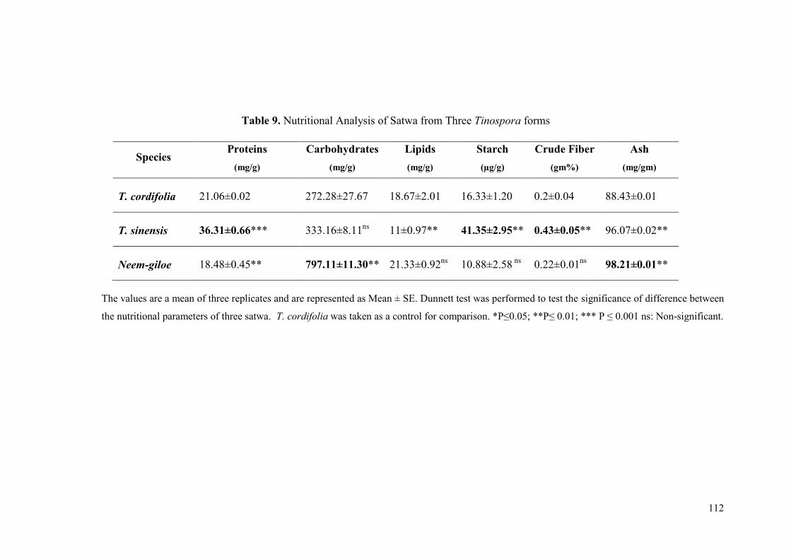
112
Table 9. Nutritional Analysis of Satwa from Three Tinospora forms
The values are a mean of three replicates and are represented as Mean ± SE. Dunnett test was performed to test the significance of difference between
the nutritional parameters of three satwa. T. cordifolia was taken as a control for comparison. *P≤0.05; **P≤ 0.01; *** P ≤ 0.001 ns: Non-significant.
Species Proteins
(mg/g)
Carbohydrates
(mg/g)
Lipids
(mg/g)
Starch
(µg/g)
Crude Fiber
(gm%)
Ash
(mg/gm)
T. cordifolia 21.06±0.02 272.28±27.67 18.67±2.01 16.33±1.20 0.2±0.04 88.43±0.01
T. sinensis 36.31±0.66*** 333.16±8.11ns
11±0.97** 41.35±2.95** 0.43±0.05** 96.07±0.02**
Neem-giloe 18.48±0.45** 797.11±11.30** 21.33±0.92ns
10.88±2.58 ns
0.22±0.01ns
98.21±0.01**
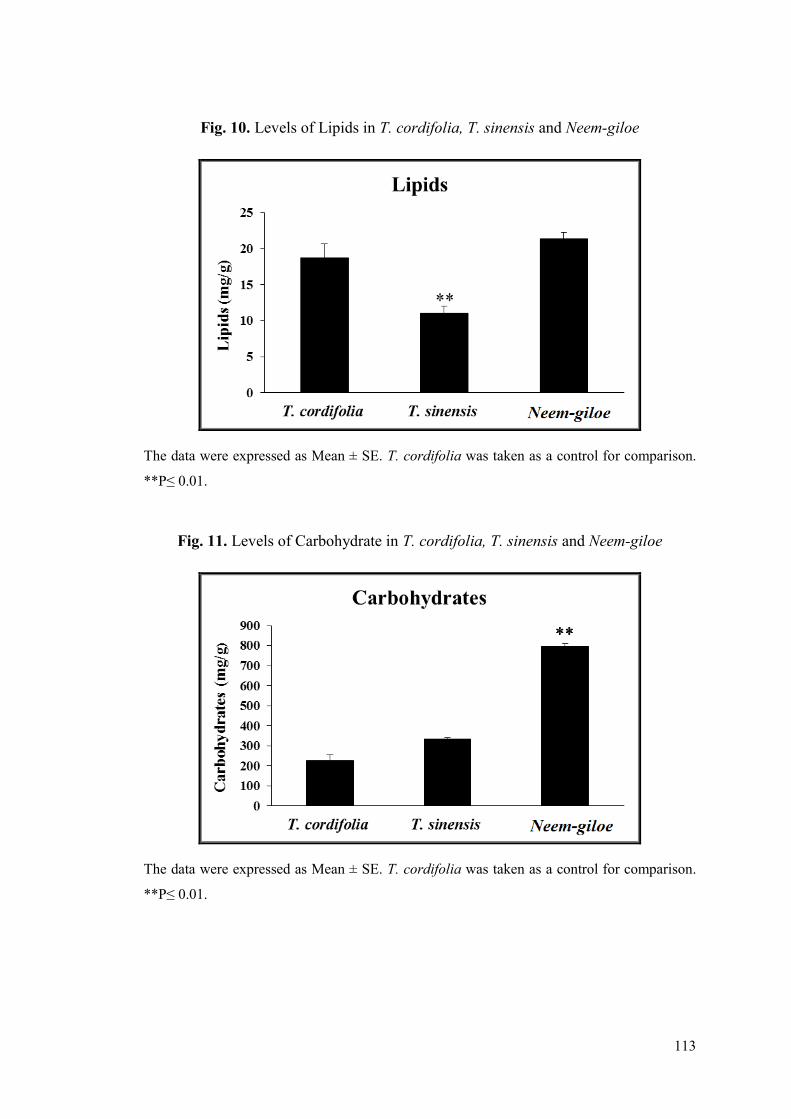
113
Fig. 10. Levels of Lipids in T. cordifolia, T. sinensis and Neem-giloe
The data were expressed as Mean ± SE. T. cordifolia was taken as a control for comparison.
**P≤ 0.01.
Fig. 11. Levels of Carbohydrate in T. cordifolia, T. sinensis and Neem-giloe
The data were expressed as Mean ± SE. T. cordifolia was taken as a control for comparison.
**P≤ 0.01.

114
Fig. 12. Levels of Protein in T. cordifolia, T. sinensis and Neem-giloe
The data were expressed as Mean ± SE. T. cordifolia was taken as a control for comparison.
**P≤ 0.01; *** P≤ 0.001.
Fig. 13. Levels of Starch in T. cordifolia, T. sinensis and Neem-giloe
The data were expressed as Mean ± SE. T. cordifolia was taken as a control for comparison.
**P≤ 0.01.
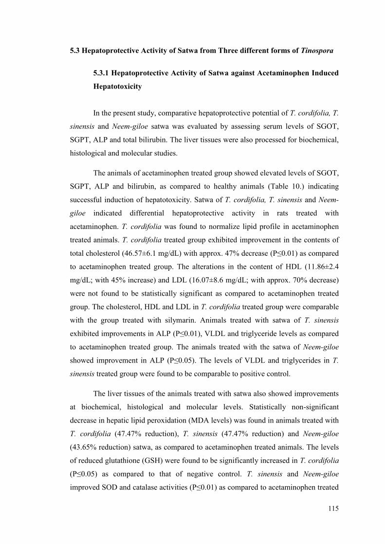
115
5.3 Hepatoprotective Activity of Satwa from Three different forms of Tinospora
5.3.1 Hepatoprotective Activity of Satwa against Acetaminophen Induced
Hepatotoxicity
In the present study, comparative hepatoprotective potential of T. cordifolia, T.
sinensis and Neem-giloe satwa was evaluated by assessing serum levels of SGOT,
SGPT, ALP and total bilirubin. The liver tissues were also processed for biochemical,
histological and molecular studies.
The animals of acetaminophen treated group showed elevated levels of SGOT,
SGPT, ALP and bilirubin, as compared to healthy animals (Table 10.) indicating
successful induction of hepatotoxicity. Satwa of T. cordifolia, T. sinensis and Neem-
giloe indicated differential hepatoprotective activity in rats treated with
acetaminophen. T. cordifolia was found to normalize lipid profile in acetaminophen
treated animals. T. cordifolia treated group exhibited improvement in the contents of
total cholesterol (46.57±6.1 mg/dL) with approx. 47% decrease (P≤0.01) as compared
to acetaminophen treated group. The alterations in the content of HDL (11.86±2.4
mg/dL; with 45% increase) and LDL (16.07±8.6 mg/dL; with approx. 70% decrease)
were not found to be statistically significant as compared to acetaminophen treated
group. The cholesterol, HDL and LDL in T. cordifolia treated group were comparable
with the group treated with silymarin. Animals treated with satwa of T. sinensis
exhibited improvements in ALP (P≤0.01), VLDL and triglyceride levels as compared
to acetaminophen treated group. The animals treated with the satwa of Neem-giloe
showed improvement in ALP (P≤0.05). The levels of VLDL and triglycerides in T.
sinensis treated group were found to be comparable to positive control.
The liver tissues of the animals treated with satwa also showed improvements
at biochemical, histological and molecular levels. Statistically non-significant
decrease in hepatic lipid peroxidation (MDA levels) was found in animals treated with
T. cordifolia (47.47% reduction), T. sinensis (47.47% reduction) and Neem-giloe
(43.65% reduction) satwa, as compared to acetaminophen treated animals. The levels
of reduced glutathione (GSH) were found to be significantly increased in T. cordifolia
(P≤0.05) as compared to that of negative control. T. sinensis and Neem-giloe
improved SOD and catalase activities (P≤0.01) as compared to acetaminophen treated

116
animals (Table 11).The total protein and lipid content of liver tissues showed
significant improvement (P≤0.01) in Neem-giloe treated group as compared to
acetaminophen treated animals (Table 11). T. cordifolia, T. sinensis and Neem-giloe
satwa treated animals showed 15.2%, 19.2% and 42.4% decrease in LDL (P≤0.01)
and 56.84%, 58.93% and 64.73% decrease in VLDL (P≤0.01) respectively as
compared to animals from negative control group. T. cordifolia, T. sinensis and Neem-
giloe treated group showed 56.83%, 58.99% and 64.94% decrease in triglycerides
respectively as compared to the negative control group (P≤0.01) (Table 11).
Liver from healthy (Fig. 14A) group showed normal architecture. Sections of
liver from acetaminophen treated group (Fig. 14B) showed mild conjunction of
central vein and apoptotic death of hepatocytes. Intra and extracellular hyaline
globules were seen around central vein. The hepatocytes surrounding the central vein
showed ballooning and degeneration along with mild nucleomegaly. Liver from
silymarin treated group (Fig. 14C) showed near normal liver architecture with a few
swollen hepatocytes. The differential hepatoprotective effects of Guduchi satwa
prepared from three Tinospora forms were also evident from liver histology (Fig.
14D-F). The liver histology of the animals treated with T. cordifolia satwa exhibits
improvements over acetaminophen treated group (Fig. 14D) but intermittently
swollen centrilobular hepatocytes were visible in liver sections which are known to be
more prone to ischemic injury. The periportal hepatocytes were normal in animals
treated with satwa of T. cordifolia. The liver histology of the group treated with T.
sinensis exhibited near normal histology (Fig. 14E) with prominent hepato-
regeneration as evident from distribution of normal hepatocytes among degenerating
swollen hepatocytes. This group also showed normal periportal hepatocytes. The liver
histology of Neem-giloe satwa treated group (Fig. 14F) was strikingly normal without
any histologically detectable anomalies.
Fig. 15A-E depicts the modulation of expression levels of the genes from liver
tissues of animals treated with the satwa of three different forms of Tinospora.
Expression levels of FABP1 (Fig. 15A) and PPARγ (Fig. 15B) were found to be
decreased in acetaminophen induced hepatotoxicity. Treatment with the satwa of
Neem-giloe significantly improved (P≤0.05) the expression of FABP1. The increase
in the expression level of PPARγ observed in satwa treated groups was statistically
not significant. NF-κβ, SREBP1 and TNF-α were up-regulated in acetaminophen
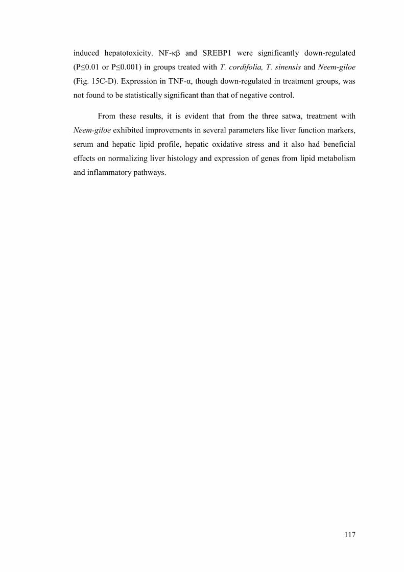
117
induced hepatotoxicity. NF-κβ and SREBP1 were significantly down-regulated
(P≤0.01 or P≤0.001) in groups treated with T. cordifolia, T. sinensis and Neem-giloe
(Fig. 15C-D). Expression in TNF-α, though down-regulated in treatment groups, was
not found to be statistically significant than that of negative control.
From these results, it is evident that from the three satwa, treatment with
Neem-giloe exhibited improvements in several parameters like liver function markers,
serum and hepatic lipid profile, hepatic oxidative stress and it also had beneficial
effects on normalizing liver histology and expression of genes from lipid metabolism
and inflammatory pathways.

118
Table 10. Effect of Satwa from Three Tinospora forms Species on Liver Function Markers and Serum Lipid Profile in Animals with
Acetaminophen Induced Hepatotoxicity
Group I is healthy control while Group II and III served as negative and positive control respectively. Group IV, V and VI were treated with 200mg/kg b.w. T.
cordifolia, T. sinensis and Neem-giloe satwa (p.o.) respectively. Results are a mean of three replicates and are represented as mean ± standard error (SE). The
data were subjected to Dunnett Multiple Comparison Test. *P≤0.05; **P≤0.01; Degrees of freedom for one variable: Treatments 5; Residuals 24; Total 29;
SGOT: serum glutamic oxaloacetic transaminase; SGPT: serum glutamic pyruvic transaminase; ALP: Alkaline phosphatase; BIL: Total bilirubin; HDL: High
density lipoprotein; LDL: Low density lipoprotein; VLDL: Very low density lipoprotein; TG: Triglycerides.
Group
SGOT
(U/mL)
SGPT
(U/mL)
ALP
(U/mL)
BIL
(mg/dL)
Lipid Profile
Total
Cholesterol
(mg/dL)
HDL
(mg/dL)
LDL
(mg/dL)
VLDL
(mg/dL)
TG
(mg/dL)
I 129.38±11.8* 151.64±6.3 29.94±5.7
** 0.84±0.2
* 59.00±4.6
** 22.75±5.2 20.38±7.7 15.87±2.1 79.36±10.5
II 186.10±9.7 183.40±3.9 53.96±4.4 1.56±0.1 87.45±7.9 6.49±0.7 53.33±4.9 27.62±3.6 138.12±18.1
III 128.14±20.4* 175.12 ±3.7 26.31±4.1
** 1.12±0.3 42.65±5.4
** 9.16±0.9 18.11±3.2 15.38±4.1 76.89±20.6
IV 148.98±11.3 178.10±4.6 36.77±2.9 1.39±0.1 46.57±6.1**
11.86±2.4 16.07±8.6 18.64±3.3 93.22±16.3
V 150.71±8.5 176.60±4.4 22.13±6.5**
1.55±0.2 56.40±2.6**
11.57±1.2 28.39±4.8 16.43±2.6 82.15±13
VI 147.43±18.9 182.10±1.1 33.56±2.9* 1.05±0.1 54.37±4.5
** 9.29±0.5 25.39±6.7 19.68±2.5 98.43±12.3

119
Table 11. Effect of Satwa from Three Tinospora forms on Hepatic Oxidative Stress Markers, Total Protein and Hepatic Lipid Profile in Animals with
Acetaminophen Induced Hepatotoxicity
Group I is healthy control while Group II and III served as negative and positive control respectively. Group IV, V and VI were treated with 200mg/kg b.w. T. cordifolia, T.
sinensis and Neem-giloe satwa (p.o.) respectively. Results are a mean of three replicates and are represented as mean ± standard error (SE). The data were subjected to
Dunnett multiple comparison test. *P≤ 0.05; **P ≤0.01; Degrees of freedom for one variable: Treatments 5; Residuals 24; Total 29; SOD: Superoxide dismutase; CAT:
Catalase; MDA: Malondialdehyde; GSH: Reduced glutathione; TC: Total cholesterol; HDL: High density lipoprotein; LDL: Low density lipoprotein; VLDL: Very low
density lipoprotein; TG: Triglycerides.
Group SOD
(U/mg)
CAT
(mU/g Tissue)
MDA
(μM)
GSH
(μM)
Total
Protein
(mg/g Tissue)
Lipid Profile
Total
Cholesterol
(mg/dL)
HDL
(mg/dL)
LDL
(mg/dL)
VLDL
(mg/dL)
TG
(mg/dL)
I 68.24±0.8**
0.66±0.5**
5.5±1.0* 0.26±0.03
** 5.36±0.2 24.61±2.2
* 15.04±0.8
** 6.4±0.1
** 1.34±0.1
** 6.7±0.6
**
II 49.60±1.1 0.57±1.1 12.3±1.6 0.09±0.00 2.97±0.31 34.35±0.4 8.54±1.1 12.50±0.2 6.21±0.2 31.09±1.1
III 72.36±1.4**
0.68±0.3**
8.36±1.1 0.05±0.04 2.93±0.08 20.91±3.8**
6.49±0.34 6.5±0.2**
2.10±0.1**
10.5±0.8*
IV 73.25±1.4**
0.65±0.2**
6.46±1.4 0.2±0.02*
4.51±1.1 23.31±1.5**
7.00±0.7 10.6±0.3**
2.68±0.1**
13.42±0.6**
V 73.08±1.17**
0.64±0.7**
6.46±2.8 0.12±0.01 4.72±0.6 31.59±2.1 11.28±1.2 10.1±0.1**
2.55±0.1**
12.75±0.9**
VI 74.05±2.1**
0.66±0.6**
6.93±1.3 0.11±0.04 9.41±0.9**
26.36±0.5 12.3±1.7**
7.2±0.4**
2.19±0.1**
10.9±0.9**
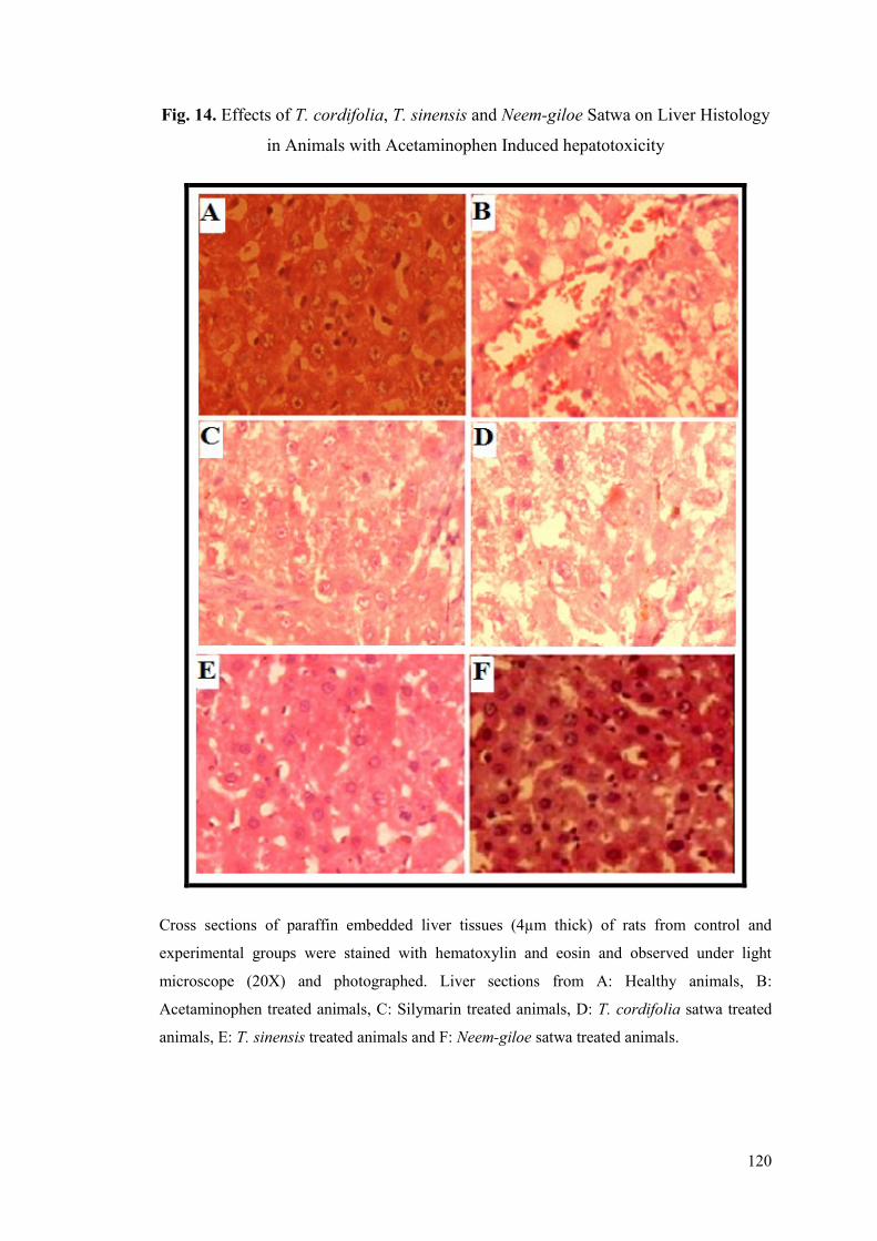
120
Fig. 14. Effects of T. cordifolia, T. sinensis and Neem-giloe Satwa on Liver Histology
in Animals with Acetaminophen Induced hepatotoxicity
Cross sections of paraffin embedded liver tissues (4µm thick) of rats from control and
experimental groups were stained with hematoxylin and eosin and observed under light
microscope (20X) and photographed. Liver sections from A: Healthy animals, B:
Acetaminophen treated animals, C: Silymarin treated animals, D: T. cordifolia satwa treated
animals, E: T. sinensis treated animals and F: Neem-giloe satwa treated animals.
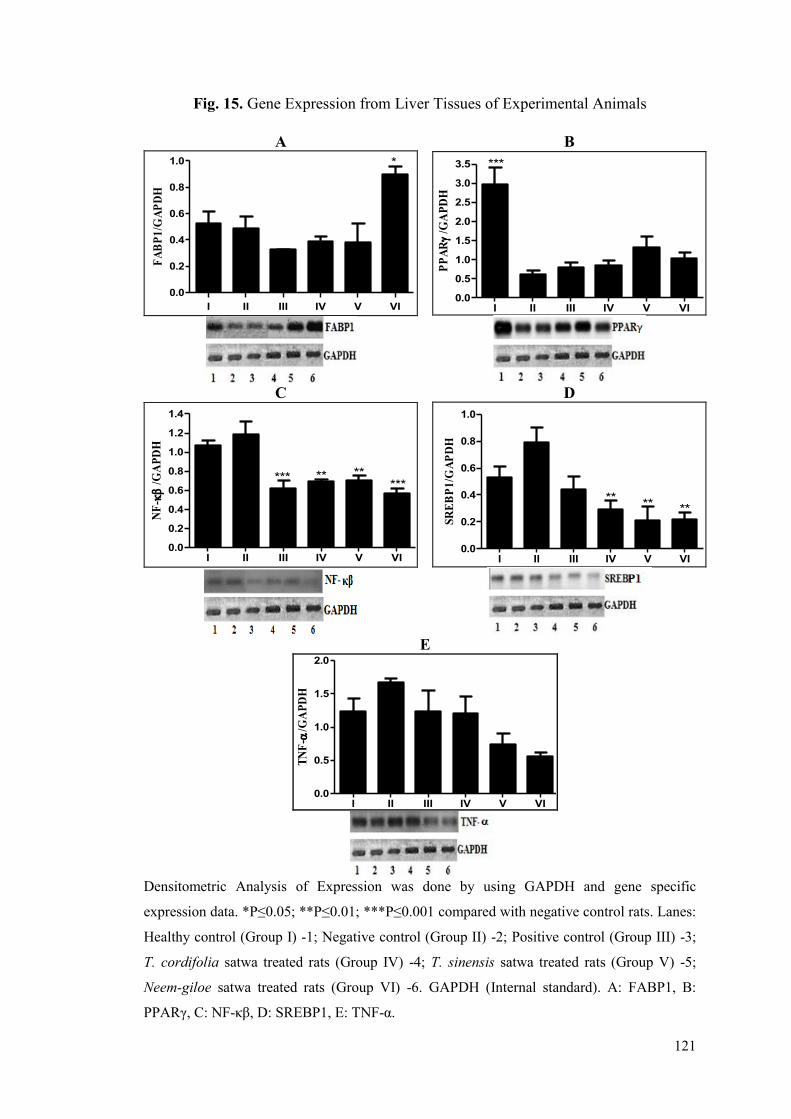
121
Fig. 15. Gene Expression from Liver Tissues of Experimental Animals
Densitometric Analysis of Expression was done by using GAPDH and gene specific
expression data. *P≤0.05; **P≤0.01; ***P≤0.001 compared with negative control rats. Lanes:
Healthy control (Group I) -1; Negative control (Group II) -2; Positive control (Group III) -3;
T. cordifolia satwa treated rats (Group IV) -4; T. sinensis satwa treated rats (Group V) -5;
Neem-giloe satwa treated rats (Group VI) -6. GAPDH (Internal standard). A: FABP1, B:
PPARγ, C: NF-κβ, D: SREBP1, E: TNF-α.
A B
I II III IV V VI
0.0
0.2
0.4
0.6
0.8
1.0 *F
AB
P1
/GA
PD
H
I II III IV V VI0.0
0.5
1.0
1.5
2.0
2.5
3.0
3.5 ***
PP
AR
/GA
PD
H
C D
I II III IV V VI0.0
0.2
0.4
0.6
0.8
1.0
1.2
1.4
******
** **
NF
-
/G
AP
DH
I II III IV V VI0.0
0.2
0.4
0.6
0.8
1.0
** ** **
SR
EB
P1
/GA
PD
H
E Data 5
I II III IV V VI0.0
0.5
1.0
1.5
2.0
TN
F-
/GA
PD
H

122
5.3.2 Hepatoprotective Activity of Satwa against Ethanol Induced
Hepatotoxicity
In the present study, comparative hepatoprotective potential of T. cordifolia, T.
sinensis and Neem-giloe satwa were estimated by evaluating serum SGOT, SGPT,
ALP and total bilirubin. The animals from negative control (ethanol treated) group
showed significant increase in the levels of SGOT, SGPT, ALP, bilirubin and lipid
profiles as compared to healthy control group indicating successful induction of liver
injury (Table 12). The satwa of T. cordifolia, T. sinensis and Neem-giloe exhibited
hepatoprotective activity against ethanol induced hepatotoxicity. Significant
improvements in lipid profile were observed in the animals treated with the satwa of
Neem-giloe. The experimental group treated with Neem-giloe showed significant
decrease in total cholesterol, LDL, VLDL and triglycerides (P≤0.01) and increase in
HDL (P≤0.01) over 30% ethanol treated group. The lipid profile from Neem-giloe
treated animals was comparable with the animals treated with standard drug
silymarin. Animals treated with the satwa of T. sinensis showed marked
improvements in liver function markers (P≤0.01) as compared to the animals with
30% ethanol treated group. The lipid profile in the animals treated with T. sinensis
satwa also exhibited improvements (P≤0.05 or P≤0.01) as compared to the animals
treated with 30% ethanol. The animals treated with the satwa of T. cordifolia
exhibited improvements in lipid profile and liver function test markers as compared to
the animals treated with 30% ethanol.
Liver tissues of animals treated with three different satwa exhibited alterations
at biochemical, histological and molecular levels. The level of lipid peroxidation
(MDA levels) was significantly decreased in animals treated with T. sinensis satwa
(84.56% reduction) as compared to 30% ethanol treated animals (P≤0.01). The levels
of reduced glutathione (GSH) were also significantly increased in Neem-giloe
(P≤0.01; increase by 11.11%) and T. sinensis (P≤0.05; increase by 8.33%) than
ethanol treated animals (Table 13). T. cordifolia, T. sinensis and Neem-giloe improved
SOD and catalase activities (P≤0.01) as compared to ethanol treated animals (Table
13). The total protein content of liver tissue significantly improved (P≤0.01) in T.
cordifolia, T. sinensis and Neem-giloe satwa treated groups as compared to negative
control. Hepatic lipid profile (Table 13) displayed significant improvements in
123
animals treated with three different satwa. Total cholesterol, LDL, VLDL and TG
contents improved significantly (P≤0.01) by the treatments as compared to negative
control. Significant increase in HDL was seen only in animals treated with T. sinensis
satwa.
The differential hepatoprotective effects of satwa prepared from these three
Tinospora forms were also evident from liver histology (Fig. 16A-F). Liver from
healthy (Fig. 16A) group exhibited normal architecture. Sections of liver from alcohol
treated group (Fig. 16B) had swollen hepatocytes with granular cytoplasm.
Collections of few polymorphs were seen in hepatic parenchyma, suggesting small
foci of necrosis. Liver from silymarin treated group (Fig. 16C) also displayed near
normal liver architecture. The liver tissues of animals treated with T. cordifolia satwa
(Fig. 16D) showed collection of lymphocytes within the hepatic parenchyma. Kupffer
cells were seen prominent in this group and portal triad showed lymphocyte
infiltration. The liver histology of the group treated with T. sinensis satwa was found
to be absolutely normal (Fig. 16E). The liver histology of Neem-giloe satwa treated
group (Fig. 16F) had some portal triads and sinusoids showing collection of
lymphocytes.
Fig 17A-E shows the effect of three different forms of Tinospora on FABP1,
PPARγ, NF-κβ, SREBP1 and TNF-α expression in ethanol induced hepatotoxicity in
rats. The expression of FABP1 (Fig. 17A) and PPARγ (Fig. 17B), were down-
regulated while expression levels of NF-κβ (Fig. 17C), SREBP1 (Fig. 17D) and TNF-
α (Fig. 17E) were increased in ethanol induced hepatotoxicity as compared to healthy
control group. FABP1 expression was significantly up-regulated (P≤0.001) by
treatment with T. sinensis and Neem-giloe (Fig. 17A). Treatment with Neem-giloe
leads to significant down-regulation while treatment with T. sinensis showed
significant up-regulation of PPARγ expression (Fig. 17B). Expression of NF-κβ was
not altered in the positive control group while all treatment groups exhibited
significantly reduced (P≤0.001 or P≤0.001) expression of NF-κβ. Similarly,
expression of TNF-α was marginally reduced in positive control group while the three
treatment groups showed significant decrease (P≤0.001) in the expression levels of
TNF-α (Fig. 17E). Treatment with Neem-giloe lead to significant down-regulation
(P≤0.05) of SREBP1 (Fig. 17D).
124
From the results, it is evident that from the three satwa, treatment with T.
sinensis exhibited improvements in several parameters like liver function markers,
serum and hepatic lipid profile, hepatic oxidative stress and it also had beneficial
effects on normalizing liver histology and expression of genes from lipid metabolism
and inflammatory pathways.

125
Table 12. Effect of Satwa from Three Tinospora forms on Liver Function Markers and Serum Lipid Profile in Animals with Ethanol Induced
Hepatotoxicity
Group I is healthy control while Group II and III served as negative and positive control respectively. Group IV, V and VI were treated with 200mg/kg b.w. T.
cordifolia, T. sinensis and Neem-giloe satwa (p.o.) respectively. Results are a mean of three replicates and are represented as Mean ± Standard Error (SE). The
data were subjected to Dunnett Multiple Comparison Test. *P≤0.05; **P≤0.01; Degrees of freedom for one variable: Treatments 5; Residuals 30; Total 35;
SGOT: serum glutamic oxaloacetic transaminase; SGPT: serum glutamic pyruvic transaminase; ALP: Alkaline phosphatase; BIL: Total bilirubin; HDL: High
density lipoprotein; LDL: Low density lipoprotein; VLDL: Very low density lipoprotein; TG: Triglycerides.
Group
SGOT
(U/mL)
SGPT
(U/mL)
ALP
(U/mL)
BIL
(mg/dL)
Lipid profile
Total
Cholesterol
(mg/dL)
HDL
(mg/dL)
LDL
(mg/dL)
VLDL
(mg/dL)
TG
(mg/dL)
I 134.49±3.0**
154.67±2.4**
30.40±1.3**
1.13±0.1**
85.89±1.6**
19.31±0.5**
17.1±1.3**
27.71±0.5**
138.54±2.5**
II 162.31±0.8 182.50±2.5 55.89±2.7 2.11±0.2 203.36±7.2 13.51±1.0 40.2±1.0 74.37±0.8 371.87±3.9
III 137.56±1.5**
174.77±1.0 33.33±0.7**
1.14±0.1**
110.09±2.6**
14.02±0.5 20.1±0.6**
39.9±0.7**
199.48±3.3**
IV 138.85±1.1**
144.08±2.0**
36.52±1.4**
0.91±0.1**
128.84±2.7**
13.84±0.5 18.6±0.8**
48.28±0.8**
241.40±3.9**
V 131.92±2.7**
131.58±2.9**
32.90±0.9**
0.71±0.02**
132.05±3.3**
16.41±0.7* 19.2±0.9
** 46.09±1.0
** 230.47±5.0
**
VI 139.49±2.9**
150.83±2.4**
36.62±0.5**
0.93±0.04**
110.89±2.1**
16.58±0.4**
17.4±1.0**
39.27±0.7**
196.36±3.3**
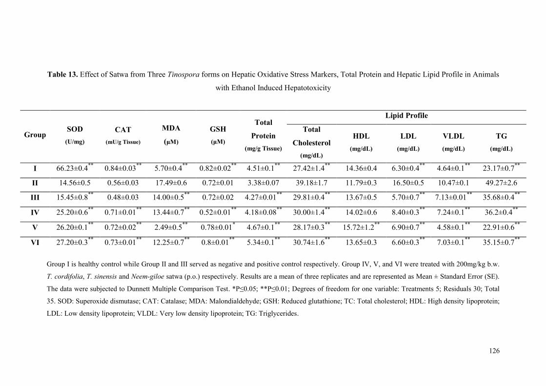
126
Table 13. Effect of Satwa from Three Tinospora forms on Hepatic Oxidative Stress Markers, Total Protein and Hepatic Lipid Profile in Animals
with Ethanol Induced Hepatotoxicity
Group I is healthy control while Group II and III served as negative and positive control respectively. Group IV, V, and VI were treated with 200mg/kg b.w.
T. cordifolia, T. sinensis and Neem-giloe satwa (p.o.) respectively. Results are a mean of three replicates and are represented as Mean ± Standard Error (SE).
The data were subjected to Dunnett Multiple Comparison Test. *P≤0.05; **P≤0.01; Degrees of freedom for one variable: Treatments 5; Residuals 30; Total
35. SOD: Superoxide dismutase; CAT: Catalase; MDA: Malondialdehyde; GSH: Reduced glutathione; TC: Total cholesterol; HDL: High density lipoprotein;
LDL: Low density lipoprotein; VLDL: Very low density lipoprotein; TG: Triglycerides.
Group SOD
(U/mg)
CAT
(mU/g Tissue)
MDA
(μM)
GSH
(μM)
Total
Protein
(mg/g Tissue)
Lipid Profile
Total
Cholesterol
(mg/dL)
HDL
(mg/dL)
LDL
(mg/dL)
VLDL
(mg/dL)
TG
(mg/dL)
I 66.23±0.4**
0.84±0.03**
5.70±0.4**
0.82±0.02**
4.51±0.1**
27.42±1.4**
14.36±0.4 6.30±0.4**
4.64±0.1**
23.17±0.7**
II 14.56±0.5 0.56±0.03 17.49±0.6 0.72±0.01 3.38±0.07 39.18±1.7 11.79±0.3 16.50±0.5 10.47±0.1 49.27±2.6
III 15.45±0.8**
0.48±0.03 14.00±0.5**
0.72±0.02 4.27±0.01**
29.81±0.4**
13.67±0.5 5.70±0.7**
7.13±0.01**
35.68±0.4**
IV 25.20±0.6**
0.71±0.01**
13.44±0.7**
0.52±0.01**
4.18±0.08**
30.00±1.4**
14.02±0.6 8.40±0.3**
7.24±0.1**
36.2±0.4**
V 26.20±0.1**
0.72±0.02**
2.49±0.5**
0.78±0.01* 4.67±0.1
** 28.17±0.3
** 15.72±1.2
** 6.90±0.7
** 4.58±0.1
** 22.91±0.6
**
VI 27.20±0.3**
0.73±0.01**
12.25±0.7**
0.8±0.01**
5.34±0.1**
30.74±1.6**
13.65±0.3 6.60±0.3**
7.03±0.1**
35.15±0.7**

127
Fig. 16. Effects of T. cordifolia, T. sinensis and Neem-giloe Satwa on Liver Histology
in Animals with Ethanol Induced Hepatotoxicity
Cross sections of paraffin embedded liver tissues (4µm thick) of rats from control and
experimental groups were stained with hematoxylin and eosin and observed under light
microscope (20X) and photographed. Liver sections from A: Healthy animals, B: Ethanol
treated animals, C: Silymarin treated animals, D: T. cordifolia satwa treated animals, E: T.
sinensis satwa treated animals and F: Neem-giloe satwa treated animals.
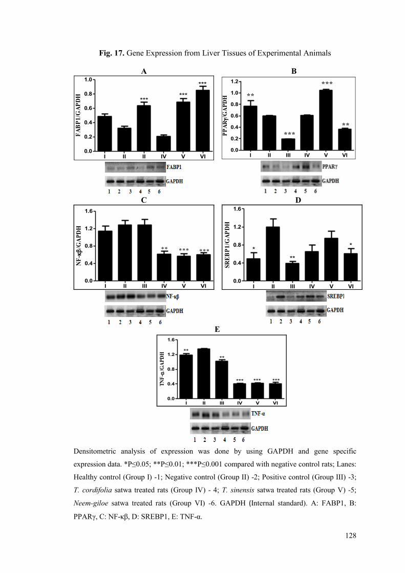
128
Fig. 17. Gene Expression from Liver Tissues of Experimental Animals
Densitometric analysis of expression was done by using GAPDH and gene specific
expression data. *P≤0.05; **P≤0.01; ***P≤0.001 compared with negative control rats; Lanes:
Healthy control (Group I) -1; Negative control (Group II) -2; Positive control (Group III) -3;
T. cordifolia satwa treated rats (Group IV) - 4; T. sinensis satwa treated rats (Group V) -5;
Neem-giloe satwa treated rats (Group VI) -6. GAPDH (Internal standard). A: FABP1, B:
PPARγ, C: NF-κβ, D: SREBP1, E: TNF-α.
A B
I II II IV V VI0.0
0.2
0.4
0.6
0.8
1.0***
******
FA
BP
1/G
AP
DH
I II III IV V VI0.0
0.2
0.4
0.6
0.8
1.0
1.2
PP
AR/
GA
PD
H
***
**
***
**
C D
I II III IV V VI0.0
0.4
0.8
1.2
1.6
***
** *** ***
NF
-
/GA
PD
H
I II III IV V VI0.0
0.4
0.8
1.2
1.6
* *
**
SR
EB
P1
/GA
PD
H
E
I II III IV V VI0.0
0.4
0.8
1.2
1.6
*** *** ***
**
**
TN
F-
/GA
PD
H
129
5.4 Hepatoprotective Activity of Flax Oil and Fish Oil (Polyunsaturated fatty
acids)
In the present study, nutritional interventions (Polyunsaturated fatty acids)
were assessed for their hepatoprotective activity. The hepatoprotective potential of
flax oil and fish oil against acetaminophen and alcohol induced hepatotoxicity in rats
was studied. Alteration in the expression levels of the genes related to inflammation,
fatty acid metabolism, and transcription factors, were also studied from liver tissues of
the animals. Results obtained in the study are presented below.
5.4.1 Hepatoprotective Activity of Flax Oil and Fish Oil against
Acetaminophen Induced Hepatotoxicity
In the present study, serum SGOT, SGPT, ALP, bilirubin and lipid profile
from negative control group showed a sharp and significant shift as compared to a
healthy control group, indicating successful induction of hepatotoxicity by repeated
dosing of acetaminophen (Table 14). Administration of flax oil and fish oil at a dose
of 500mg/kg b.w./day had a beneficial effect on the levels of these biochemical
markers in the treatment groups. SGOT, SGPT and ALP activities were found to be
elevated in the negative control group as compared to healthy control. Animals treated
with the fish oil showed significant improvement in SGOT (P≤0.01) and SGPT
(P≤0.05) and flax oil showed significant decrees in SGOT (P≤0.05) as compared with
negative control group. Treatment with silymarin helped to lower the activities of
these enzymes. As compared to negative control group, flax oil and fish oil displayed
remarkable benefits in relation to improvements of lipid profiles (Table 14). Flax oil
and fish oil treated groups showed decrease in total cholesterol (P≤0.01), LDL
(P≤0.01) as compared to the negative control group. Significant (P≤0.05) increase in
HDL was seen only in the animals treated with fish oil (Table 14). Silymarin treated
groups showed decreased in total cholesterol (P≤0.01), LDL (P≤0.01), VLDL
(P≤0.01), triglycerides (P≤0.01).
Flax and fish oil supplementation was found to be significantly effective in
ameliorating the oxidative damage through decrease in the extent of lipid peroxidation
and increase in the contents of reduced glutathione which acts as a principal
antioxidant compound in liver (Table 15). The extent of lipid peroxidation (MDA
130
levels) was significantly decreased in flax oil and fish oil (P≤0.001) treated animals as
compared to acetaminophen treated animals while the levels of reduced glutathione
(GSH) were significantly increased in flax oil and fish oil supplemented groups
(P≤0.05) as compared to acetaminophen treated animals. Flax oil and fish oil
supplementation improved SOD and catalase activities (P≤0.01 or P≤0.001) as
compared to acetaminophen treated animals (Table 15). The total protein content of
liver tissues significantly improved in flax oil and fish oil treated groups as compared
to acetaminophen treated group (P≤0.01). The lipid profile of liver also displayed
significant improvements in flax oil and fish oil treated groups when compared with
negative control group. Flax oil and fish oil treated groups displayed decrease in total
cholesterol (P≤0.01), LDL (P≤0.001), and VLDL (P≤0.01). The treatment of animals
with flax oil and fish oil also decreased triglycerides (P≤0.01) as compared to the
negative control group. Significant (P≤0.01) increase in HDL was seen only in the
animals treated with flax oil (Table 15).
The hepatoprotective effects of flax oil and fish oil were also evident from the
prominent variations in liver histology (Fig. 18A-E). The healthy control group (Fig.
18A) displayed normal liver histology while the acetaminophen treated group (Fig.
18B) showed swollen or occasionally apoptotic hepatocytes with coarse granular
cytoplasm and compressed sinusoids. The group also displayed a few nucleated cells
indicating spontaneous regeneration of hepatocytes. The group treated with the
standard drug silymarin (Fig. 18C) had near normal liver histology with mild
congestion of central vein and mildly swollen hepatocytes. Both flax oil and fish oil
treated groups (Fig. 18D-E) exhibited strikingly normal liver histology without any
anatomically detectable anomalies.
Fig. 19A-E show the effect of flax and fish oil on the expression of FABP1,
PPARγ, NF-κβ, SREBP1 and TNF-α. The expression of FABP1 (Fig. 19A) and
PPARγ (Fig. 19B) was decreased in acetaminophen treated animals than in healthy
animals. Though treatment with flax oil and fish oil up-regulated the expression of
FABP1 (Fig. 19A), it was found to be statistically insignificant. PPARγ (Fig. 19B)
was significantly up-regulated (P≤0.05) in animals treated with fish oil. NF-κβ (Fig.
19C), SREBP1 (Fig. 19D) and TNF-α (Fig.19E) were up-regulated in acetaminophen
treated animals. Treatment with flax oil (Fig. 19C) normalized the expression of NF-
κβ (P≤0.05) (Fig. 19C) while the SREBP1 (Fig. 19D) and TNF-α (Fig. 19E)
131
expressions were significantly down regulated by both flax and fish oil (P≤0.05 or
P≤0.01).
From the results, it is evident that treatment with fish oil exhibited
improvements in several parameters like liver function markers, serum and hepatic
lipid profile, hepatic oxidative stress and it also had beneficial effects on normalizing
liver histology and expression of genes from lipid metabolism fatty acid metabolism
and inflammatory pathways.
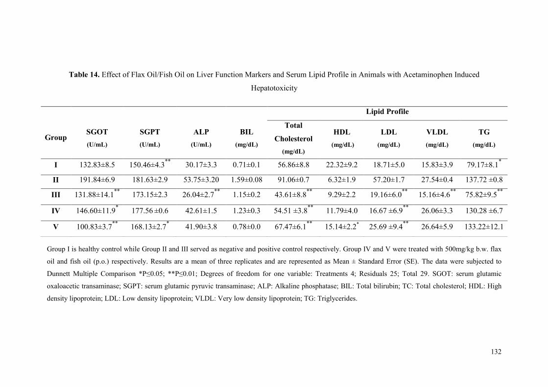
132
Table 14. Effect of Flax Oil/Fish Oil on Liver Function Markers and Serum Lipid Profile in Animals with Acetaminophen Induced
Hepatotoxicity
Group I is healthy control while Group II and III served as negative and positive control respectively. Group IV and V were treated with 500mg/kg b.w. flax
oil and fish oil (p.o.) respectively. Results are a mean of three replicates and are represented as Mean ± Standard Error (SE). The data were subjected to
Dunnett Multiple Comparison *P≤0.05; **P≤0.01; Degrees of freedom for one variable: Treatments 4; Residuals 25; Total 29. SGOT: serum glutamic
oxaloacetic transaminase; SGPT: serum glutamic pyruvic transaminase; ALP: Alkaline phosphatase; BIL: Total bilirubin; TC: Total cholesterol; HDL: High
density lipoprotein; LDL: Low density lipoprotein; VLDL: Very low density lipoprotein; TG: Triglycerides.
Group
SGOT
(U/mL)
SGPT
(U/mL)
ALP
(U/mL)
BIL
(mg/dL)
Lipid Profile
Total
Cholesterol
(mg/dL)
HDL
(mg/dL)
LDL
(mg/dL)
VLDL
(mg/dL)
TG
(mg/dL)
I 132.83±8.5 150.46±4.3**
30.17±3.3 0.71±0.1 56.86±8.8 22.32±9.2 18.71±5.0 15.83±3.9 79.17±8.1*
II 191.84±6.9 181.63±2.9 53.75±3.20 1.59±0.08 91.06±0.7 6.32±1.9 57.20±1.7 27.54±0.4 137.72 ±0.8
III 131.88±14.1**
173.15±2.3 26.04±2.7**
1.15±0.2 43.61±8.8**
9.29±2.2 19.16±6.0**
15.16±4.6**
75.82±9.5**
IV 146.60±11.9* 177.56
±0.6 42.61±1.5 1.23±0.3 54.51 ±3.8
** 11.79±4.0 16.67 ±6.9
** 26.06±3.3 130.28 ±6.7
V 100.83±3.7**
168.13±2.7* 41.90±3.8 0.78±0.0 67.47±6.1
** 15.14±2.2
* 25.69 ±9.4
** 26.64±5.9 133.22±12.1
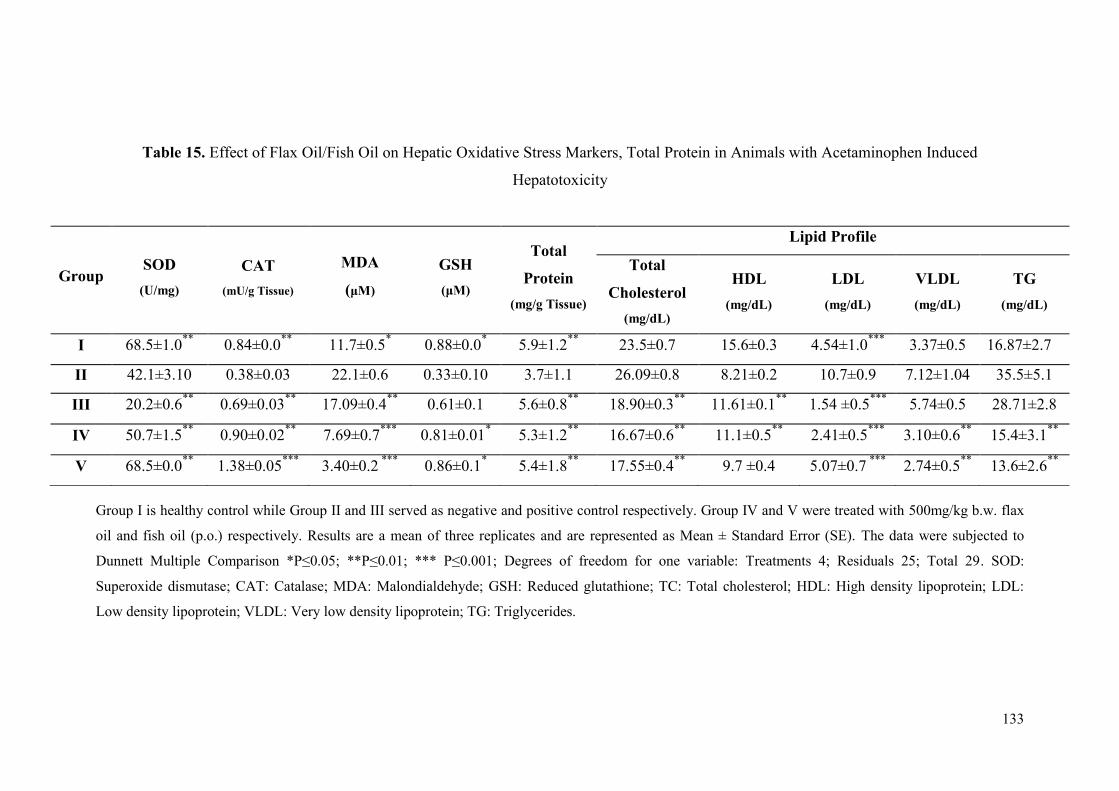
133
Table 15. Effect of Flax Oil/Fish Oil on Hepatic Oxidative Stress Markers, Total Protein in Animals with Acetaminophen Induced
Hepatotoxicity
Group I is healthy control while Group II and III served as negative and positive control respectively. Group IV and V were treated with 500mg/kg b.w. flax
oil and fish oil (p.o.) respectively. Results are a mean of three replicates and are represented as Mean ± Standard Error (SE). The data were subjected to
Dunnett Multiple Comparison *P≤0.05; **P≤0.01; *** P≤0.001; Degrees of freedom for one variable: Treatments 4; Residuals 25; Total 29. SOD:
Superoxide dismutase; CAT: Catalase; MDA: Malondialdehyde; GSH: Reduced glutathione; TC: Total cholesterol; HDL: High density lipoprotein; LDL:
Low density lipoprotein; VLDL: Very low density lipoprotein; TG: Triglycerides.
Group SOD
(U/mg)
CAT
(mU/g Tissue)
MDA
(μM)
GSH
(μM)
Total
Protein
(mg/g Tissue)
Lipid Profile
Total
Cholesterol
(mg/dL)
HDL
(mg/dL)
LDL
(mg/dL)
VLDL
(mg/dL)
TG
(mg/dL)
I 68.5±1.0**
0.84±0.0**
11.7±0.5* 0.88±0.0
* 5.9±1.2
** 23.5±0.7 15.6±0.3 4.54±1.0
*** 3.37±0.5 16.87±2.7
II 42.1±3.10 0.38±0.03 22.1±0.6 0.33±0.10 3.7±1.1 26.09±0.8 8.21±0.2 10.7±0.9 7.12±1.04 35.5±5.1
III 20.2±0.6**
0.69±0.03**
17.09±0.4**
0.61±0.1 5.6±0.8**
18.90±0.3**
11.61±0.1**
1.54 ±0.5***
5.74±0.5 28.71±2.8
IV 50.7±1.5**
0.90±0.02**
7.69±0.7***
0.81±0.01* 5.3±1.2
** 16.67±0.6
** 11.1±0.5
** 2.41±0.5
*** 3.10±0.6
** 15.4±3.1
**
V 68.5±0.0**
1.38±0.05***
3.40±0.2 ***
0.86±0.1* 5.4±1.8
** 17.55±0.4
** 9.7 ±0.4 5.07±0.7
*** 2.74±0.5
** 13.6±2.6
**
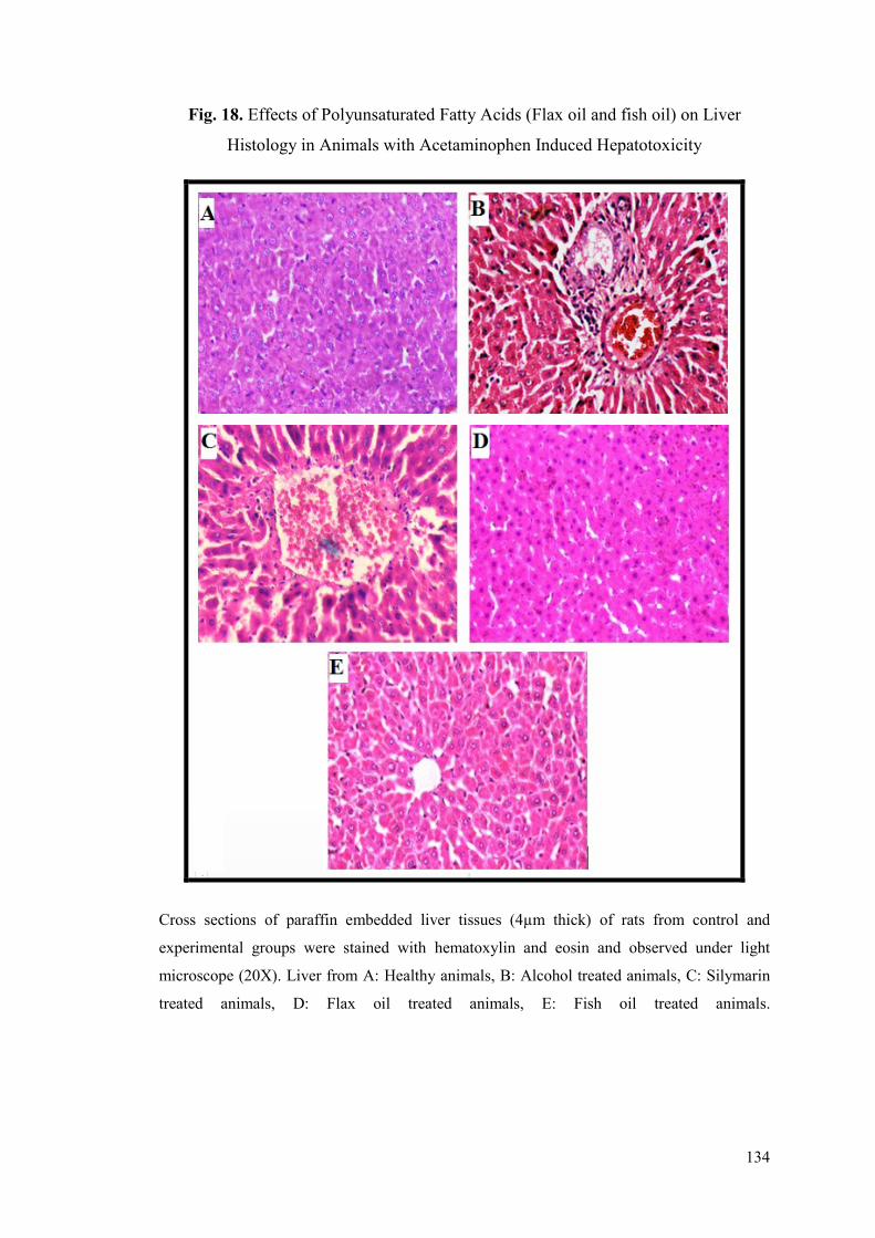
134
Fig. 18. Effects of Polyunsaturated Fatty Acids (Flax oil and fish oil) on Liver
Histology in Animals with Acetaminophen Induced Hepatotoxicity
Cross sections of paraffin embedded liver tissues (4µm thick) of rats from control and
experimental groups were stained with hematoxylin and eosin and observed under light
microscope (20X). Liver from A: Healthy animals, B: Alcohol treated animals, C: Silymarin
treated animals, D: Flax oil treated animals, E: Fish oil treated animals.
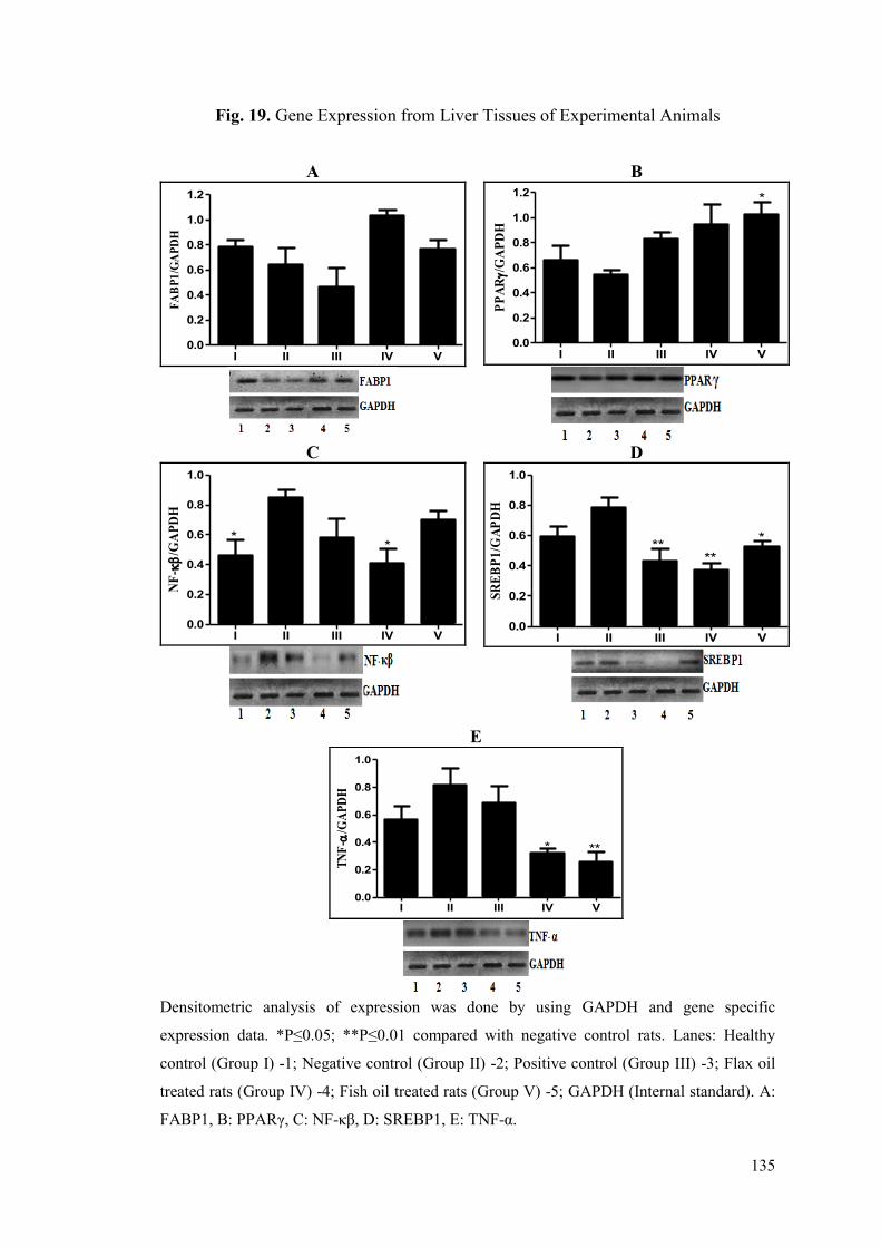
135
Fig. 19. Gene Expression from Liver Tissues of Experimental Animals
Densitometric analysis of expression was done by using GAPDH and gene specific
expression data. *P≤0.05; **P≤0.01 compared with negative control rats. Lanes: Healthy
control (Group I) -1; Negative control (Group II) -2; Positive control (Group III) -3; Flax oil
treated rats (Group IV) -4; Fish oil treated rats (Group V) -5; GAPDH (Internal standard). A:
FABP1, B: PPARγ, C: NF-κβ, D: SREBP1, E: TNF-α.
A B
I II III IV V0.0
0.2
0.4
0.6
0.8
1.0
1.2
FA
BP
1/G
AP
DH
I II III IV V0.0
0.2
0.4
0.6
0.8
1.0
1.2*
PP
AR/
GA
PD
H
C D
I II III IV V0.0
0.2
0.4
0.6
0.8
1.0
*
*
NF
-
/GA
PD
H
I II III IV V0.0
0.2
0.4
0.6
0.8
1.0
****
*
SR
EB
P1
/GA
PD
H
E
I II III IV V0.0
0.2
0.4
0.6
0.8
1.0
** *
TN
F-
/GA
PD
H
136
5.4.2 Hepatoprotective Activity of Flax Oil and Fish Oil against Ethanol Induced
Hepatotoxicity
In the present study, activities of serum SGOT, SGPT, ALP, bilirubin and
lipid profile from negative control group showed a sharp and significant increase as
compared to a healthy control group indicating successful induction of hepatic
damage by repeated dosing of 30% alcohol (Table 16). Administration of flax oil and
fish oil at a dose of 500mg/kg b.w./day had a favorable effect on the levels of these
biochemical markers in the treatment groups.
Animals treated with flax oil and fish oil exhibited improvements in SGOT,
SGPT, ALP and bilirubin (P≤0.01) levels but the animals treated with the fish oil had
statistically insignificant improvement in SGOT as compared to ethanol treated group.
Treatment with silymarin helped to decrease the activities of these enzymes (Table
16). As compared to negative control group, flax oil and fish oil treatment exhibited
improvement in lipid profiles in the animals with ethanol induced liver damage. Flax
oil and fish oil treated groups displayed decrease in total cholesterol (P≤0.01), LDL
(P≤0.01), VLDL (P≤0.01), and triglycerides (P≤0.01) as compared to the negative
control group. Significant (P≤0.01) increase in HDL was seen in the animals treated
with flax oil and fish oil (Table 16).
The lipid peroxidation (MDA levels) was significantly reduced in flax oil and
fish oil (P≤0.05 or P≤0.01) treated animals. Flax oil and fish oil treatment did not
show any effect in improving the levels of reduced glutathione (GSH) as compared to
alcohol treated animals (Table 17). SOD and catalase activities were found to be
significantly increased in flax oil and fish oil treated animals (P≤0.01) as compared to
negative control group (Table 17). The total protein content of liver tissues was
improved only in flax oil (P≤0.01) treated group as compared to ethanol group. The
lipid profile of liver also displayed improvements in flax and fish oil treated groups
when compared with negative control. Flax and fish oil treated groups displayed
decrease in total cholesterol, LDL, VLDL and triglycerides (P≤0.01 for all lipid
profile parameters) as compared to negative control. Significant increase in HDL
(P≤0.01) was seen in the animals treated with flax oil and fish oil (Table 17).
137
The effects of treatment with flax oil and fish oil were also visible at
histological levels in animals. Liver from healthy (Fig. 20A) group showed normal
architecture. Sections of liver from ethanol treated group (Fig. 20B) exhibited
coarsely condensed cytoplasm in some cells with prominent coarse granularity in
other cells, the sinusoid appeared compressed and hence the trabecular pattern of
arrangement appeared disrupted. The hepatocytes were found to be swollen.
Cytoplasmic borders were distinct. Liver from silymarin treated group (Fig. 20C) had
near normal liver architecture. The liver histology of the animals treated with flax oil
(Fig. 20D) exhibited absolutely normal histology. The liver histology of the group
treated with fish oil (Fig. 20E) exhibited improvements in the architecture except a
few portal triad areas with collection of lymphocytes.
Fig. 21A-E, shows effect of flax oil and fish oil on the expression of FABP1,
PPARγ, NF-κβ, SREBP1 and TNF-α. Expression of FABP1 (Fig. 21A) and PPARγ
(Fig. 21B) decreased in ethanol treated animals than in healthy animals. Treatment
with flax oil showed marginal improvement in the expression of FABP1 (Fig. 21A)
but it was found to be statistically insignificant. PPARγ (Fig. 21B) was significantly
up-regulated (P≤0.001, 2.16 fold) in animals treated with fish oil and in animals
treated with flax oil (P≤0.05). NF-κβ (Fig. 21C), SREBP1 (Fig. 21D) and TNF-α (Fig.
21E) were up-regulated in ethanol treated animals than in healthy animals. Treatment
with flax oil normalized the expression of NF-κβ (P≤0.05) (Fig. 21C) while
expression of SREBP1 (Fig. 21D) was significantly down regulated in both flax oil
and fish oil treated groups (P≤0.05). Treatment with fish oil exhibited 2.18-fold
decrease in SREBP1 (Fig. 21D). TNF-α (Fig. 21E) expressions were significantly
down regulated by both flax oil and fish oil (P≤0.001 and P≤0.01, respectively).
Treatment with flax oil showed 2.56-fold decrease in TNF-α (Fig. 21E).
From the results, it is evident that treatment with flax oil exhibited
improvements in several parameters like liver function markers, serum and hepatic
lipid profile, hepatic oxidative stress and it also had beneficial effects on normalizing
liver histology and expression of genes from lipid metabolism and inflammatory
pathways.

138
Table 16. Effect of Flax Oil/Fish Oil on Liver Function Markers and Serum Lipid Profile in Animals with Ethanol Induced Hepatotoxicity
Group I is healthy control while Group II and III served as negative and positive control respectively. Group IV and V were treated with 500mg/kg b.w. flax
oil and fish oil (p.o.) respectively. Results are a mean of measurements from six animals and are represented as Mean±Standard Error. The data were
subjected to Dunnett Multiple Comparison Test. *P≤0.05; **P≤0.01. Degree of freedom for one variable: Treatments 4; Residuals 25; Total 29. SGOT:
Serum glutamic oxaloacetic transaminase; SGPT: Serum glutamic pyruvic transaminase; ALP: Alkaline phosphatase; BIL: Total bilirubin; HDL: High
density lipoprotein; LDL: Low density lipoprotein; VLDL: Very low density lipoprotein; TG: Triglycerides.
Group
SGOT
(U/mL)
SGPT
(U/mL)
ALP
(U/mL)
BIL
(mg/dL)
Lipid profile
Total
Cholesterol
(mg/dL)
HDL
(mg/dL)
LDL
(mg/dL)
VLDL
(mg/dL)
TG
(mg/dL)
I 134.49±3.0**
154.67±2.4**
30.40±1.3**
1.13±0.1**
85.89±1.6**
19.31±0.5**
17.1±1.3**
27.71±0.5**
138.54±2.5**
II 162.31±0.8 182.50±2.5 55.89±2.7 2.11±0.2 203.36±7.2 13.51±1.0 40.2±1.0 74.37±0.8 371.87±3.9
III 137.56±1.5**
174.77±1.0* 33.33±0.7
** 1.14±0.1
** 110.09±2.6
** 14.02±0.5 20.1±0.6
** 39.9±0.7
** 199.48±3.3
**
IV 143.84±1.2**
143.67±1.1**
32.82±1.0**
0.95±0.1**
89.10±1.3**
19.65±0.3**
19.5±1.1**
49.42±0.7**
247.13±3.4**
V 156.15±3.7 140.67±2.2**
32.87±0.7**
0.99±0.2**
113.62±2.9**
18.80±0.3**
20.1±0.6**
63.80±0.3**
319.01±1.7**
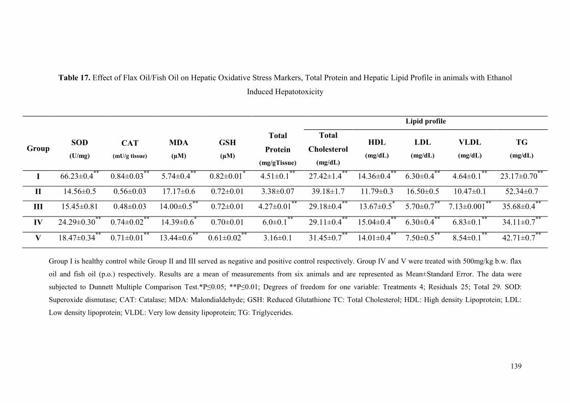
139
Table 17. Effect of Flax Oil/Fish Oil on Hepatic Oxidative Stress Markers, Total Protein and Hepatic Lipid Profile in animals with Ethanol
Induced Hepatotoxicity
Group I is healthy control while Group II and III served as negative and positive control respectively. Group IV and V were treated with 500mg/kg b.w. flax
oil and fish oil (p.o.) respectively. Results are a mean of measurements from six animals and are represented as Mean±Standard Error. The data were
subjected to Dunnett Multiple Comparison Test.*P≤0.05; **P≤0.01; Degrees of freedom for one variable: Treatments 4; Residuals 25; Total 29. SOD:
Superoxide dismutase; CAT: Catalase; MDA: Malondialdehyde; GSH: Reduced Glutathione TC: Total Cholesterol; HDL: High density Lipoprotein; LDL:
Low density lipoprotein; VLDL: Very low density lipoprotein; TG: Triglycerides.
Group
SOD
(U/mg)
CAT
(mU/g tissue)
MDA
(μM)
GSH
(μM)
Total
Protein
(mg/gTissue)
Lipid profile
Total
Cholesterol
(mg/dL)
HDL
(mg/dL)
LDL
(mg/dL)
VLDL
(mg/dL)
TG
(mg/dL)
I 66.23±0.4**
0.84±0.03**
5.74±0.4**
0.82±0.01* 4.51±0.1
** 27.42±1.4
** 14.36±0.4
** 6.30±0.4
** 4.64±0.1
** 23.17±0.70
**
II 14.56±0.5 0.56±0.03 17.17±0.6 0.72±0.01 3.38±0.07 39.18±1.7 11.79±0.3 16.50±0.5 10.47±0.1 52.34±0.7
III 15.45±0.81 0.48±0.03 14.00±0.5**
0.72±0.01 4.27±0.01**
29.18±0.4**
13.67±0.5* 5.70±0.7
** 7.13±0.001
** 35.68±0.4
**
IV 24.29±0.30**
0.74±0.02**
14.39±0.6* 0.70±0.01 6.0±0.1
** 29.11±0.4
** 15.04±0.4
** 6.30±0.4
** 6.83±0.1
** 34.11±0.7
**
V 18.47±0.34**
0.71±0.01**
13.44±0.6**
0.61±0.02**
3.16±0.1 31.45±0.7**
14.01±0.4**
7.50±0.5**
8.54±0.1**
42.71±0.7**
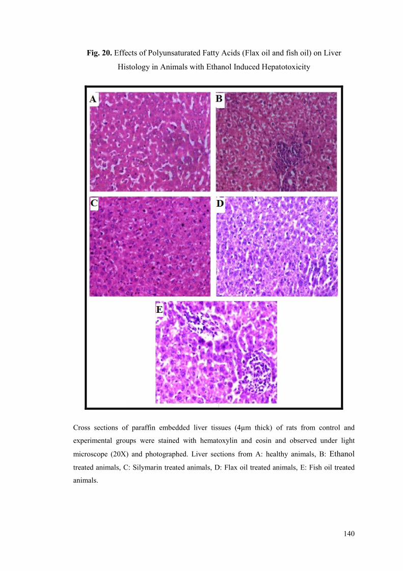
140
Fig. 20. Effects of Polyunsaturated Fatty Acids (Flax oil and fish oil) on Liver
Histology in Animals with Ethanol Induced Hepatotoxicity
Cross sections of paraffin embedded liver tissues (4µm thick) of rats from control and
experimental groups were stained with hematoxylin and eosin and observed under light
microscope (20X) and photographed. Liver sections from A: healthy animals, B: Ethanol
treated animals, C: Silymarin treated animals, D: Flax oil treated animals, E: Fish oil treated
animals.
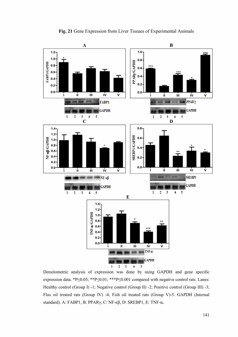
141
Fig. 21 Gene Expression from Liver Tissues of Experimental Animals
Densitometric analysis of expression was done by using GAPDH and gene specific
expression data. *P≤0.05; **P≤0.01; ***P≤0.001 compared with negative control rats. Lanes:
Healthy control (Group I) -1; Negative control (Group II) -2; Positive control (Group III) -3;
Flax oil treated rats (Group IV) -4; Fish oil treated rats (Group V)-5. GAPDH (Internal
standard). A: FABP1, B: PPARγ, C: NF-κβ, D: SREBP1, E: TNF-α.
A B
I II III IV V0.0
0.2
0.4
0.6
0.8
1.0
1.2 *
FA
BP
1/G
AP
DH
I II III IV V0.0
0.2
0.4
0.6
0.8
1.0
***
***
***
*
PP
AR/
GA
PD
H
C D
I II III IV V0.0
0.2
0.4
0.6
0.8
1.0
1.2
1.4
*
NF
-
/GA
PD
H
I II III IV V0.0
0.2
0.4
0.6
0.8
**
* *
SR
EB
P1
/GA
PD
H
E
I II III IV V0.0
0.2
0.4
0.6
0.8
1.0
1.2
1.4
***
**
*
TN
F-
/GA
PD
H
142
5.5 Hepatoprotective Activity of Combination of Best Performing Herbal and
Nutritional Intervention
The previous studies reported in this thesis, revealed that treatment with
Neem-giloe satwa and T. sinensis satwa was more effective in acetaminophen and
alcohol induced hepatotoxicity, respectively. Nutritional intervention in the form of
fish oil and flax oil was effective in acetaminophen and alcohol induced
hepatotoxicity, respectively. In the present study, the corrective and protective effects
of combination of Neem-giloe satwa and fish oil were assessed against hepatotoxicity
induced with a single high-dose of acetaminophen. The effect of prophylactic
treatment of combination of T. sinensis satwa and flax oil was studied in ethanol
induced hepatotoxicity in rats.
5.5.1 Effects of Protective Treatment of Combination of Neem-giloe Satwa
and Fish oil against Acetaminophen Induced Hepatotoxicity
In the present study, the animals treated with single dose of acetaminophen on
8th
day showed elevated levels of SGOT, SGPT, ALP and bilirubin as compared to
healthy control group indicating the induction of liver damage (Table 18). Animals
treated with combination of Neem-giloe satwa and fish oil showed protective effect by
maintaining the levels of SGOT, SGPT, ALP and bilirubin, and also a significant
protection towards imbalance in lipids, especially in comparison with the negative
control group (Table 18).
Combination of Neem-giloe satwa and fish oil treated animals had protective
effect on SGOT, SGPT, ALP and bilirubin (P≤0.05 or P≤0.01) levels as compared to
acetaminophen treated group. Combination of Neem-giloe satwa and fish oil treated
animals also protected against imbalance in lipid profile. The animals exhibited
comparable total cholesterol levels after acetaminophen dosing. The level of HDL
cholesterol was found to be decreased by 73.28% after acetaminophen dosing but the
animals treated with the combination showed 28.90% decrease, thereby maintaining
the HDL cholesterol levels. The LDL and VLDL cholesterol levels showed 73.13%
and 130.20% increase after acetaminophen dosing in negative control animals. The
animals pre-treated with the combination showed only 1.49% increase in LDL
cholesterol while the levels of VLDL cholesterol were found to be lower than the
143
healthy animals. TG levels showed 130% increase after treatment with
acetaminophen, but the animals receiving the interventions exhibited the TG content
lower than the healthy animals. The protective effect of the combination on total
cholesterol, HDL, LDL and VLDL was found to be statistically significant (P≤0.05 or
P≤0.01) as compared with negative control (Table 18).
The pre-treatment of animals with a combination of Neem-giloe satwa and fish
oil was found to be protective towards oxidative damage through decrease in the
extent of lipid peroxidation and increase in the contents of reduced glutathione (Table
19). The extent of lipid peroxidation (MDA levels) was significantly less in
combination of Neem-giloe satwa and fish oil (P≤0.01) treated animals while the
levels of reduced glutathione (GSH) were higher in the animals receiving the
intervention of Neem-giloe satwa and fish oil as compared to acetaminophen treated
animals (Table 19). Catalase activity was significantly higher in combination of
Neem-giloe satwa and fish oil treated animals (P≤0.01) as compared to negative
control group (Table 19). The total protein content of liver tissues was also higher in
combination of Neem-giloe satwa and fish oil treated animals as compared to that of
negative control. The intervention also protected against variations in the hepatic lipid
profile of the animals. The treatment group displayed lower levels of LDL (P≤0.01),
VLDL and triglycerides (P≤0.01) as compared to negative control group.
Significantly high HDL (P≤0.01) was seen in the animals treated with combination of
Neem-giloe satwa and fish oil (Table 19).
Hematoxylin and eosin stained cross sections of paraffin embedded liver
tissues of rats from control and experimental groups showed variations in the liver
architecture. Healthy group (Fig. 22A) showed normal hepatic architecture. Sections
of liver from the animals treated with single dose of acetaminophen on 8th
day (Fig.
22B) indicated swelling and degenerative changes in hepatocytes. Animals treated
with silymarin (Fig. 22C) showed hepatocytes in periportal area with degenerative
changes. Animals treated with combination of Neem-giloe satwa and fish oil (Fig.
22D) exhibited periportal hepatocytes with degenerative changes. Some foci of
hepatocytes also showed degenerative changes.
144
Fig. 23A-E, shows the protective effect of combination of Neem-giloe satwa
and fish oil on the expression of FABP1, PPARγ, NF-κβ, SREBP1 and TNF-α. The
expressions of FABP1 (Fig. 23A), PPARγ (Fig. 23B) were decreased in
acetaminophen treated animals than in healthy animals. Treatment with combination
of Neem-giloe satwa and fish oil significantly up-regulated (P≤0.001 or P≤0.01) the
expression of FABP1 and PPARγ by 2.8 fold and 1.7 fold respectively. The treatment
with combination of Neem-giloe satwa and fish oil prevented increase in the
expression of NF-κβ (Fig. 23C) which was 3.02 fold less than negative control
(P≤0.01). The expressions of SREBP1 (Fig. 23D) (P≤0.01; 2.07 fold less than
negative control) and TNF-α (Fig. 23E) (P≤0.001; 4.1 fold less than negative control)
genes were also less in the animals treated with the intervention.

145
Table 18. Protective Effect of Combination of Neem-giloe Satwa and Fish Oil on Liver Function Markers and Serum Lipid Profile in Animals
with Hepatotoxicity Induced with a Single High Dose of Acetaminophen
Group I is healthy control while Group II is negative control (single dose of acetaminophen on 8th day), Group III is positive control (silymarin (100mg/kg
b.w./day; p.o.) for 7 days and a single high dose of acetaminophen on 8th day) and Group IV is treatment group (Combination of Neem-giloe satwa
(200mg/kg; p.o.; daily) and fish Oil (500mg/kg; p.o.; daily) for 7 days and a single high dose of acetaminophen on 8th day). Results are a mean of
measurements from six animals and are represented as Mean±Standard Error (SE). The data were subjected to Dunnett Multiple Comparison Test. *P≤0.05;
**P≤0.01. Degrees of freedom for one variable: Treatments 3, Residuals 20, Total 23. SGOT: Serum glutamic oxaloacetic transaminase; SGPT: Serum
glutamic pyruvic transaminase; ALP: Alkaline phosphatase; BIL: Total bilirubin; HDL: High density lipoprotein; LDL: Low density lipoprotein; VLDL:
Very low density lipoprotein; TG: Triglycerides.
Group
SGOT
(U/mL)
SGPT
(U/mL)
ALP
(U/mL)
BIL
(mg/dL)
Lipid profile
Total
Cholesterol
(mg/dL)
HDL
(mg/dL)
LDL
(mg/dL)
VLDL
(mg/dL)
TG
(mg/dL)
I 135.00±5.3 123.5±8.2**
28.17±0.3**
0.72±0.1 99.47±6.1* 41.80±3.7
** 20.1±0.6
** 16.19±0.9
** 80.99±4.8
**
II 188.33±25.5 158.5±8.8 38.45±1.2 1.129±0.1 146±20.5 11.17±0.7 34.8±0.3 37.27±3.6 186.3±18.1
III 126.17±8.3* 134.5±7.3 26.16±2.5
** 0.564±0.1
* 108.03±5.5 30.08±2.3
** 27.9±0.4
** 20.04±0.4
** 100.22±2.0
**
IV 123.5±14.1* 108.29±4.3
** 20.09±1.3
** 0.494±0.1
* 97.75±6.4
* 29.72±1.4
** 20.4±1.6
** 13.9±0.5
** 69.80±3.0
**

146
Table 19. Protective Effect of Combination of Neem-giloe Satwa and Fish Oil on Hepatic Oxidative Stress Markers, Total Protein and Hepatic
Lipid Profile in Animals with Hepatotoxicity Induced with a Single High Dose of Acetaminophen
Group I is healthy control while Group II is negative control (single dose of acetaminophen on 8th day), Group III is positive control (silymarin (100mg/kg
b.w./day; p.o.) for 7 days and a single dose of acetaminophen on 8th day) and Group IV is treatment group (Combination of Neem-guduchi satwa (200mg/kg;
p.o.; daily) and fish Oil (500mg/kg; p.o.;) for 7 days and single dose of acetaminophen on 8th day). Results are a mean of measurements from six animals and
are represented as Mean±Standard Error. The data were subjected to Dunnett Multiple Comparison Test. *P≤0.05; **P≤0.01. Degrees of freedom for one
variable; Treatments 3, Residuals 20, Total 23. SOD: Superoxide dismutase; CAT: Catalase; MDA: Malondialdehyde; GSH: Reduced glutathione; TC: Total
cholesterol; HDL: High density lipoprotein; LDL: Low density lipoprotein; VLDL: Very low density lipoprotein; TG: Triglycerides.
Group SOD
(U/mg)
CAT
(mU/g Tissue)
MDA
(μM)
GSH
(μM)
Total
Protein
(mg/g Tissue)
Lipid profile
Total
Cholesterol
(mg/dL)
HDL
(mg/dL)
LDL
(mg/dL)
VLDL
(mg/dL)
TG
(mg/dL)
I 57.11±2.8**
0.66±0.5**
27.93±1.70**
0.78±0.048**
6.64±0.4 27.79±0.5**
16.58±0.7**
5.45±0.5**
5.14±0.7**
23.71±1.6**
II 20.31±0.8 0.59±1.1 277.14±4.7 0.54±0.055 4.9±0.9 32.93±0.9 8.83±0.6 10.18±0.5 12.79±0.3 63.98±1.32
III 17.83±1.6 0.62±0.5 39.04±3.9**
0.63±0.041 8.33±0.6 26.08±0.5**
11.71±0.6**
7.64±0.7* 5.77±0.3
** 28.86±1.6
**
IV 19.83±0.5 0.65±0.5**
34.28±2.30**
0.65±0.024 8.48±2.03 29.11±0.8 16.22±0.4**
7.28±0.8**
5.37±0.2**
26.84±1.2**
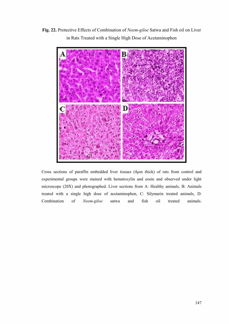
147
Fig. 22. Protective Effects of Combination of Neem-giloe Satwa and Fish oil on Liver
in Rats Treated with a Single High Dose of Acetaminophen
Cross sections of paraffin embedded liver tissues (4µm thick) of rats from control and
experimental groups were stained with hematoxylin and eosin and observed under light
microscope (20X) and photographed. Liver sections from A: Healthy animals, B: Animals
treated with a single high dose of acetaminophen, C: Silymarin treated animals, D:
Combination of Neem-giloe satwa and fish oil treated animals.
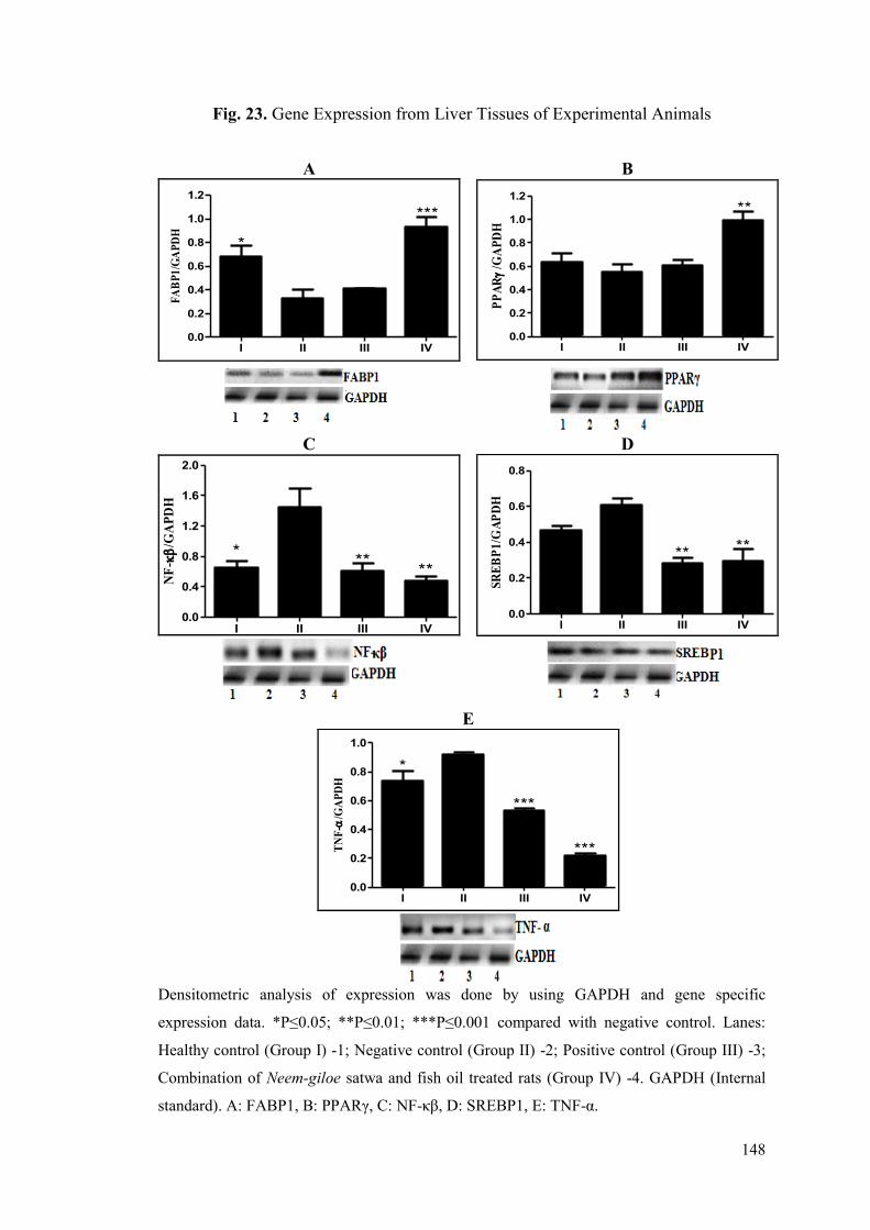
148
Fig. 23. Gene Expression from Liver Tissues of Experimental Animals
Densitometric analysis of expression was done by using GAPDH and gene specific
expression data. *P≤0.05; **P≤0.01; ***P≤0.001 compared with negative control. Lanes:
Healthy control (Group I) -1; Negative control (Group II) -2; Positive control (Group III) -3;
Combination of Neem-giloe satwa and fish oil treated rats (Group IV) -4. GAPDH (Internal
standard). A: FABP1, B: PPARγ, C: NF-κβ, D: SREBP1, E: TNF-α.
A B
I II III IV0.0
0.2
0.4
0.6
0.8
1.0
1.2
*
***
FA
BP
1/G
AP
DH
I II III IV
0.0
0.2
0.4
0.6
0.8
1.0
1.2
**
**
PP
AR
/GA
PD
H
C D
I II III IV0.0
0.4
0.8
1.2
1.6
2.0
*
****
NF
-
/GA
PD
H
I II III IV0.0
0.2
0.4
0.6
0.8
** **
SR
EB
P1
/GA
PD
H
E
I II III IV0.0
0.2
0.4
0.6
0.8
1.0
*
***
***TN
F-
/GA
PD
H
149
5.5.2. Effects of Corrective Treatment of Combination of Neem-giloe
Satwa and Fish Oil against Acetaminophen Induced Hepatotoxicity
The animals from negative control showed significant increase in the levels of
SGOT, SGPT, ALP and bilirubin after administration of a single high dose of
acetaminophen on 1st day. Levels of SGOT, SGPT, ALP and bilirubin increased
significantly after acetaminophen treatment in all animals as compared to healthy
control indicating successful induction of liver injury.
Animals receiving the intervention of Neem-giloe satwa and fish oil
combination showed significant decrease (P≤0.01) in SGOT, SGPT, ALP and
bilirubin levels as compared to acetaminophen treated group (Table 20). These
animals also exhibited significant improvement in lipid profile parameters as
compared to negative control group. Animals treated with combination of Neem-giloe
satwa and fish oil were found to have improvements in the contents of total
cholesterol with approx. 38.25% decrease (P≤0.01), HDL with 191.43% increase
(P≤0.01), LDL with approx. 36.19% decrease (P≤0.01) and with about 48% increase
(P≤0.01) in VLDL and triglyceride contents, as compared to negative control group.
All these variations in the biochemical parameters were found to be statistically
significant when compared to negative control group (Table 20).
Corrective treatment of animals with combination of Neem-giloe satwa and
fish oil, lead to decrease in oxidative stress, through decrease (P≤0.01) in lipid
peroxidation (by 62.80%) and increase in reduced glutathione (by about 21.56%)
content as compared to negative control group (Table 21). Significant increase
(P≤0.01) in SOD activity (by 106.86%) and catalase activity (by 16.59%) was
observed in the intervention group as against the animals from negative control group
(Table 21). The total protein content of liver tissues was also found to be significantly
improved (P≤0.01) in the treatment group. The hepatic lipid profile displayed
significant improvements in treatment group with decrease in total cholesterol
(P≤0.01), LDL (P≤0.05) and VLDL (P≤0.01) and triglycerides (P≤0.01) as compared
to negative control group. Significant (P≤0.01) increase in HDL was seen in the
animals treated with combination of Neem-giloe satwa and fish oil (Table 21).
150
The effects of treatment with combination of Neem-giloe satwa and fish oil
were also visible at histological levels in animals. Healthy group (Fig. 24A) showed
normal hepatic architecture. Sections of liver from the animals treated with
acetaminophen indicated congestion of central vein, swollen hepatocytes, compressed
sinusoids and ballooning degeneration of hepatocytes (Fig. 24B). Animals treated
with silymarin showed normal liver histology (Fig. 24C). Animals treated with
combination of Neem-giloe satwa and fish oil also exhibited normal liver architecture
(Fig. 24D).
Fig. 25A-E, shows the corrective effect of combination of Neem-giloe satwa
and fish oil on the expression of FABP1, PPARγ, NF-κβ, SREBP1 and TNF-α. The
expressions of FABP1 (Fig. 25A), PPARγ (Fig. 25B) were down-regulated in
acetaminophen treated animals. Treatment with combination of Neem-giloe satwa and
fish oil significantly up-regulated (P≤0.05) the expression of FABP1 (Fig. 25A) and
PPARγ (Fig. 25B) by 2.3 fold and 1.5 fold respectively. The expression levels of NF-
κβ (Fig. 25C), SREBP1 (Fig. 25D) and TNF-α (Fig. 25E) which were up-regulated in
acetaminophen treated animals were significantly improved (P≤0.01) in treatment
group.
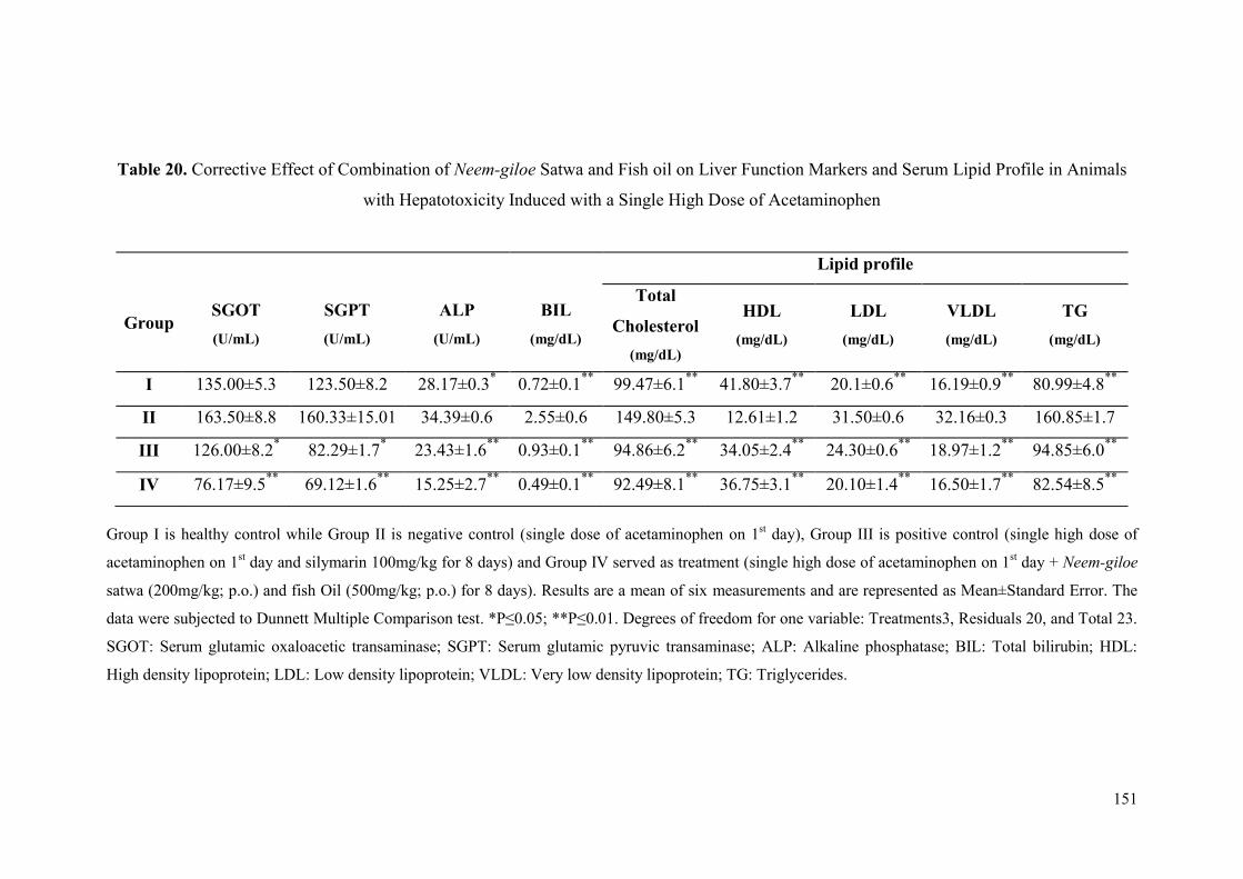
151
Table 20. Corrective Effect of Combination of Neem-giloe Satwa and Fish oil on Liver Function Markers and Serum Lipid Profile in Animals
with Hepatotoxicity Induced with a Single High Dose of Acetaminophen
Group I is healthy control while Group II is negative control (single dose of acetaminophen on 1st day), Group III is positive control (single high dose of
acetaminophen on 1st day and silymarin 100mg/kg for 8 days) and Group IV served as treatment (single high dose of acetaminophen on 1
st day + Neem-giloe
satwa (200mg/kg; p.o.) and fish Oil (500mg/kg; p.o.) for 8 days). Results are a mean of six measurements and are represented as Mean±Standard Error. The
data were subjected to Dunnett Multiple Comparison test. *P≤0.05; **P≤0.01. Degrees of freedom for one variable: Treatments3, Residuals 20, and Total 23.
SGOT: Serum glutamic oxaloacetic transaminase; SGPT: Serum glutamic pyruvic transaminase; ALP: Alkaline phosphatase; BIL: Total bilirubin; HDL:
High density lipoprotein; LDL: Low density lipoprotein; VLDL: Very low density lipoprotein; TG: Triglycerides.
Group
SGOT
(U/mL)
SGPT
(U/mL)
ALP
(U/mL)
BIL
(mg/dL)
Lipid profile
Total
Cholesterol
(mg/dL)
HDL
(mg/dL)
LDL
(mg/dL)
VLDL
(mg/dL)
TG
(mg/dL)
I 135.00±5.3 123.50±8.2 28.17±0.3* 0.72±0.1
** 99.47±6.1
** 41.80±3.7
** 20.1±0.6
** 16.19±0.9
** 80.99±4.8
**
II 163.50±8.8 160.33±15.01 34.39±0.6 2.55±0.6 149.80±5.3 12.61±1.2 31.50±0.6 32.16±0.3 160.85±1.7
III 126.00±8.2* 82.29±1.7
* 23.43±1.6
** 0.93±0.1
** 94.86±6.2
** 34.05±2.4
** 24.30±0.6
** 18.97±1.2
** 94.85±6.0
**
IV 76.17±9.5**
69.12±1.6**
15.25±2.7**
0.49±0.1**
92.49±8.1**
36.75±3.1**
20.10±1.4**
16.50±1.7**
82.54±8.5**
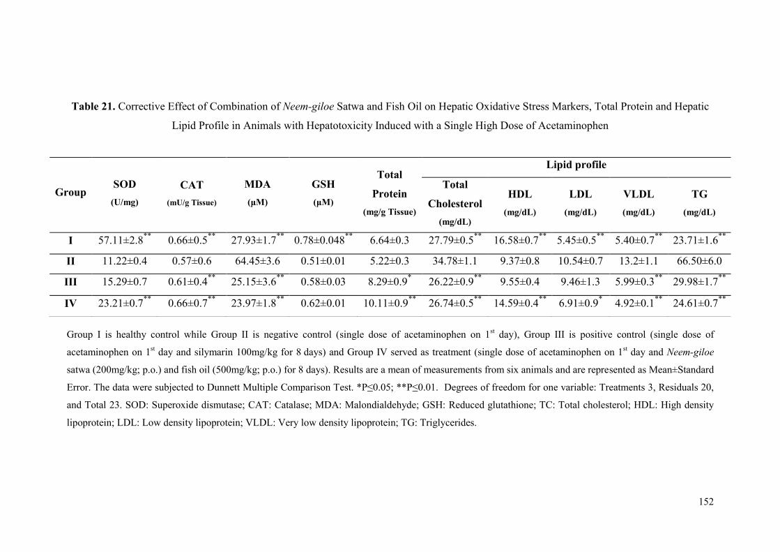
152
Table 21. Corrective Effect of Combination of Neem-giloe Satwa and Fish Oil on Hepatic Oxidative Stress Markers, Total Protein and Hepatic
Lipid Profile in Animals with Hepatotoxicity Induced with a Single High Dose of Acetaminophen
Group I is healthy control while Group II is negative control (single dose of acetaminophen on 1st day), Group III is positive control (single dose of
acetaminophen on 1st day and silymarin 100mg/kg for 8
days) and Group IV served as treatment (single dose of acetaminophen on 1
st day and Neem-giloe
satwa (200mg/kg; p.o.) and fish oil (500mg/kg; p.o.) for 8 days). Results are a mean of measurements from six animals and are represented as Mean±Standard
Error. The data were subjected to Dunnett Multiple Comparison Test. *P≤0.05; **P≤0.01. Degrees of freedom for one variable: Treatments 3, Residuals 20,
and Total 23. SOD: Superoxide dismutase; CAT: Catalase; MDA: Malondialdehyde; GSH: Reduced glutathione; TC: Total cholesterol; HDL: High density
lipoprotein; LDL: Low density lipoprotein; VLDL: Very low density lipoprotein; TG: Triglycerides.
Group SOD
(U/mg)
CAT
(mU/g Tissue)
MDA
(μM)
GSH
(μM)
Total
Protein
(mg/g Tissue)
Lipid profile
Total
Cholesterol
(mg/dL)
HDL
(mg/dL)
LDL
(mg/dL)
VLDL
(mg/dL)
TG
(mg/dL)
I 57.11±2.8**
0.66±0.5**
27.93±1.7**
0.78±0.048**
6.64±0.3 27.79±0.5**
16.58±0.7**
5.45±0.5**
5.40±0.7**
23.71±1.6**
II 11.22±0.4 0.57±0.6 64.45±3.6 0.51±0.01 5.22±0.3 34.78±1.1 9.37±0.8 10.54±0.7 13.2±1.1 66.50±6.0
III 15.29±0.7 0.61±0.4**
25.15±3.6**
0.58±0.03 8.29±0.9* 26.22±0.9
** 9.55±0.4 9.46±1.3 5.99±0.3
** 29.98±1.7
**
IV 23.21±0.7**
0.66±0.7**
23.97±1.8**
0.62±0.01 10.11±0.9**
26.74±0.5**
14.59±0.4**
6.91±0.9* 4.92±0.1
** 24.61±0.7
**
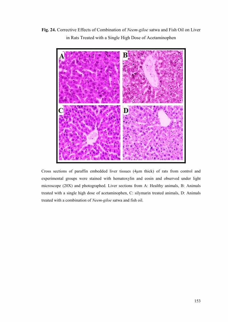
153
Fig. 24. Corrective Effects of Combination of Neem-giloe satwa and Fish Oil on Liver
in Rats Treated with a Single High Dose of Acetaminophen
Cross sections of paraffin embedded liver tissues (4µm thick) of rats from control and
experimental groups were stained with hematoxylin and eosin and observed under light
microscope (20X) and photographed. Liver sections from A: Healthy animals, B: Animals
treated with a single high dose of acetaminophen, C: silymarin treated animals, D: Animals
treated with a combination of Neem-giloe satwa and fish oil.
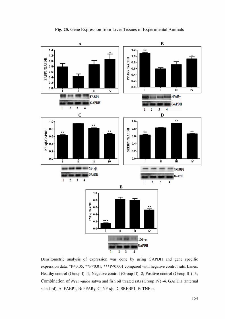
154
Fig. 25. Gene Expression from Liver Tissues of Experimental Animals
Densitometric analysis of expression was done by using GAPDH and gene specific
expression data. *P≤0.05; **P≤0.01; ***P≤0.001 compared with negative control rats. Lanes:
Healthy control (Group I) -1; Negative control (Group II) -2; Positive control (Group III) -3;
Combination of Neem-giloe satwa and fish oil treated rats (Group IV) -4. GAPDH (Internal
standard). A: FABP1, B: PPARγ, C: NF-κβ, D: SREBP1, E: TNF-α.
A B
I II III IV0.0
0.2
0.4
0.6
0.8
1.0
1.2
1.4
*
FA
BP
1/G
AP
DH
I II III IV
0.0
0.2
0.4
0.6
0.8
1.0
1.2 **
*
PP
AR
/GA
PD
H
C D
I II III IV0.0
0.2
0.4
0.6
0.8
1.0
**
**
**
NF
-
/GA
PD
H
I II III IV
0.0
0.2
0.4
0.6
0.8
1.0
**
**
**S
RE
BP
1/G
AP
DH
E
I II III IV0.0
0.2
0.4
0.6
0.8
1.0
***
**
TN
F-
/GA
PD
H
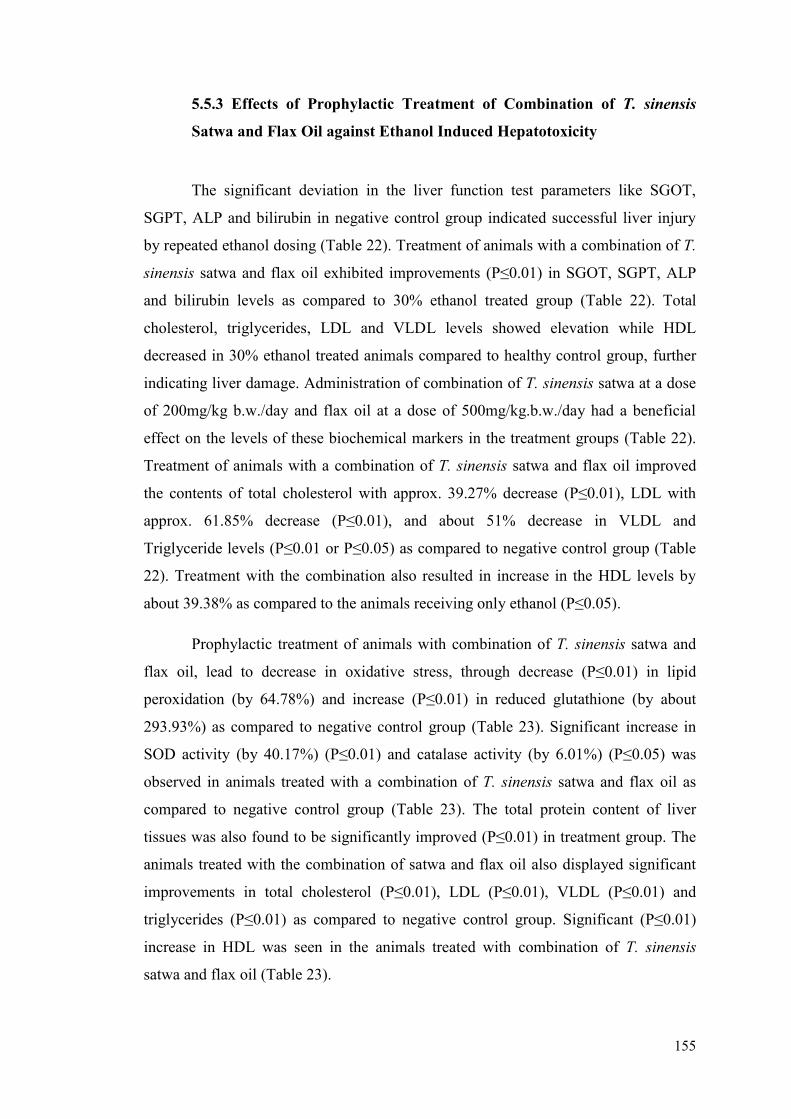
155
5.5.3 Effects of Prophylactic Treatment of Combination of T. sinensis
Satwa and Flax Oil against Ethanol Induced Hepatotoxicity
The significant deviation in the liver function test parameters like SGOT,
SGPT, ALP and bilirubin in negative control group indicated successful liver injury
by repeated ethanol dosing (Table 22). Treatment of animals with a combination of T.
sinensis satwa and flax oil exhibited improvements (P≤0.01) in SGOT, SGPT, ALP
and bilirubin levels as compared to 30% ethanol treated group (Table 22). Total
cholesterol, triglycerides, LDL and VLDL levels showed elevation while HDL
decreased in 30% ethanol treated animals compared to healthy control group, further
indicating liver damage. Administration of combination of T. sinensis satwa at a dose
of 200mg/kg b.w./day and flax oil at a dose of 500mg/kg.b.w./day had a beneficial
effect on the levels of these biochemical markers in the treatment groups (Table 22).
Treatment of animals with a combination of T. sinensis satwa and flax oil improved
the contents of total cholesterol with approx. 39.27% decrease (P≤0.01), LDL with
approx. 61.85% decrease (P≤0.01), and about 51% decrease in VLDL and
Triglyceride levels (P≤0.01 or P≤0.05) as compared to negative control group (Table
22). Treatment with the combination also resulted in increase in the HDL levels by
about 39.38% as compared to the animals receiving only ethanol (P≤0.05).
Prophylactic treatment of animals with combination of T. sinensis satwa and
flax oil, lead to decrease in oxidative stress, through decrease (P≤0.01) in lipid
peroxidation (by 64.78%) and increase (P≤0.01) in reduced glutathione (by about
293.93%) as compared to negative control group (Table 23). Significant increase in
SOD activity (by 40.17%) (P≤0.01) and catalase activity (by 6.01%) (P≤0.05) was
observed in animals treated with a combination of T. sinensis satwa and flax oil as
compared to negative control group (Table 23). The total protein content of liver
tissues was also found to be significantly improved (P≤0.01) in treatment group. The
animals treated with the combination of satwa and flax oil also displayed significant
improvements in total cholesterol (P≤0.01), LDL (P≤0.01), VLDL (P≤0.01) and
triglycerides (P≤0.01) as compared to negative control group. Significant (P≤0.01)
increase in HDL was seen in the animals treated with combination of T. sinensis
satwa and flax oil (Table 23).

156
The hepatoprotective effects of combination of T. sinensis satwa and flax oil
were also evident from the noticeable variations in liver histology (Fig. 26A-D). The
healthy control group (Fig. 26A) exhibited normal liver histology while the 30%
ethanol treated group (Fig. 26B) showed mild swollen hepatocytes with coarse
granularity, normal periveinular hepatocytes and ballooning degeneration in some
hepatocytes. The group treated with the standard drug silymarin (Fig. 26C) had near
normal histology. Animals treated with combination of T. sinensis satwa and flax oil
(Fig. 26D) exhibited strikingly normal liver histology without any anatomically
detectable anomalies.
Fig. 27(A-E) shows the prophylactic effect of combination of T. sinensis satwa
and flax oil on the expression of FABP1, PPARγ, NF-κβ, SREBP1 and TNF-α. The
FABP1 (Fig. 27A) and PPARγ (Fig. 27B) genes were down-regulated in 30% ethanol
treated animals. Treatment with combination of T. sinensis satwa and flax oil showed
improvement in the expression of FABP1 but it was found to be statistically
insignificant. Treatment with combination of T. sinensis satwa and flax oil showed
significant improvement (P≤0.001) in the expression of PPARγ (3.0 fold) as
compared to negative control group. NF-κβ (Fig. 27C), SREBP1 (Fig. 27D) and TNF-
α (Fig. 27E) were up-regulated in 30% ethanol treated animals. The treatment with
combination of T. sinensis satwa and flax oil down-regulated (P≤0.001) in the levels
of NF-κβ by about 2.3 fold. Though not statistically significant, there was
improvement in the expression of SREBP1 and TNF-α.
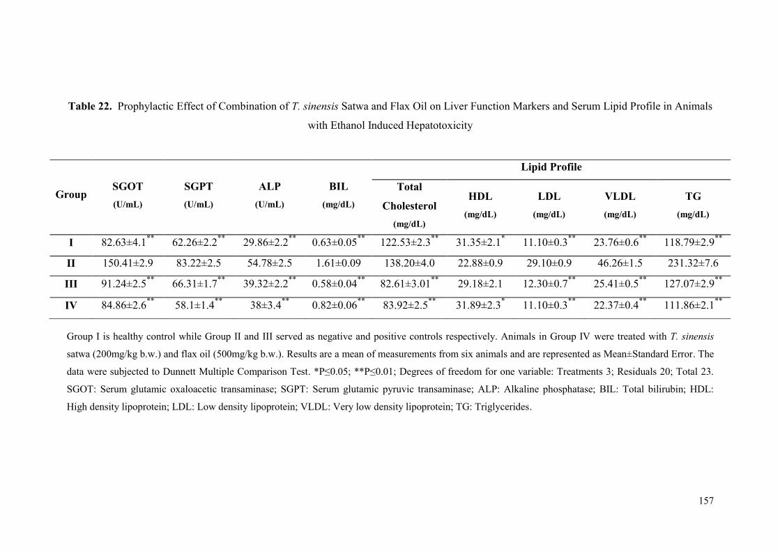
157
Table 22. Prophylactic Effect of Combination of T. sinensis Satwa and Flax Oil on Liver Function Markers and Serum Lipid Profile in Animals
with Ethanol Induced Hepatotoxicity
Group I is healthy control while Group II and III served as negative and positive controls respectively. Animals in Group IV were treated with T. sinensis
satwa (200mg/kg b.w.) and flax oil (500mg/kg b.w.). Results are a mean of measurements from six animals and are represented as Mean±Standard Error. The
data were subjected to Dunnett Multiple Comparison Test. *P≤0.05; **P≤0.01; Degrees of freedom for one variable: Treatments 3; Residuals 20; Total 23.
SGOT: Serum glutamic oxaloacetic transaminase; SGPT: Serum glutamic pyruvic transaminase; ALP: Alkaline phosphatase; BIL: Total bilirubin; HDL:
High density lipoprotein; LDL: Low density lipoprotein; VLDL: Very low density lipoprotein; TG: Triglycerides.
Group SGOT
(U/mL)
SGPT
(U/mL)
ALP
(U/mL)
BIL
(mg/dL)
Lipid Profile
Total
Cholesterol
(mg/dL)
HDL
(mg/dL)
LDL
(mg/dL)
VLDL
(mg/dL)
TG
(mg/dL)
I 82.63±4.1**
62.26±2.2**
29.86±2.2**
0.63±0.05**
122.53±2.3**
31.35±2.1* 11.10±0.3
** 23.76±0.6
** 118.79±2.9
**
II 150.41±2.9 83.22±2.5 54.78±2.5 1.61±0.09 138.20±4.0 22.88±0.9 29.10±0.9 46.26±1.5 231.32±7.6
III 91.24±2.5**
66.31±1.7**
39.32±2.2**
0.58±0.04**
82.61±3.01**
29.18±2.1 12.30±0.7**
25.41±0.5**
127.07±2.9**
IV 84.86±2.6**
58.1±1.4**
38±3.4**
0.82±0.06**
83.92±2.5**
31.89±2.3* 11.10±0.3
** 22.37±0.4
** 111.86±2.1
**

158
Table 23. Prophylactic Effect of Combination of T. sinensis Satwa and Flax Oil on Hepatic Oxidative Stress Markers, Total Protein and Hepatic
Lipid Profile in Animals with Ethanol Induced Hepatotoxicity
Group I is healthy control while Group II and III served as negative and positive controls respectively. Animals in Group IV were treated with T. sinensis
satwa (200mg/kg b.w.) and flax oil (500mg/kg b.w.). Results are a mean of measurements from six animals and are represented as Mean±Standard Error. The
data were subjected to Dunnett Multiple Comparison test. *P≤0.05; **P≤0.01; Degrees of freedom for one variable: Treatments 3; Residuals 20; Total 23.
SOD: Superoxide dismutase; CAT: Catalase; MDA: Malondialdehyde; GSH: Reduced glutathione TC: Total cholesterol; HDL: High density Lipoprotein;
LDL: Low density lipoprotein; VLDL: Very low density lipoprotein; TG: Triglycerides.
Group SOD
(U/mg)
CAT
(mU/g Tissue)
MDA
(μM)
GSH
(μM)
Total
Protein
(mg/g Tissue)
Lipid Profile
Total
Cholesterol
(mg/dL)
HDL
(mg/dL)
LDL
(mg/dL)
VLDL
(mg/dL)
TG
(mg/dL)
I 54.88±1.7**
0.70±1.0 4.71±0.4**
0.125±0.01**
7.67±0.2* 25.23±1.6
** 16.06±1.2
** 5.1±0.3
** 2.18±0.1
** 10.961±0.8
**
II 38.38±0.4 0.66±1.5 16.78±0.9 0.033±0.01 6.84±0.2 33.56±1.1 10.77±0.4 16.2±1.3 5.85±0.3 29.30±1.6
III 47.44±1.6**
0.66±0.2 15.83±1.0 0.135±0.001**
9.28±0.2**
29.86±1.1 15.89±0.51**
9±0.01**
4.7±0.1**
23.49±0.7**
IV 53.80±1.1**
0.70±1.1* 5.91±0.7
** 0.130±0.01
** 9.3±0.1
** 26.39±1.2
** 14.87±0.9
** 7.5±0.3
** 3.78±0.2
** 18.79±1.2
**
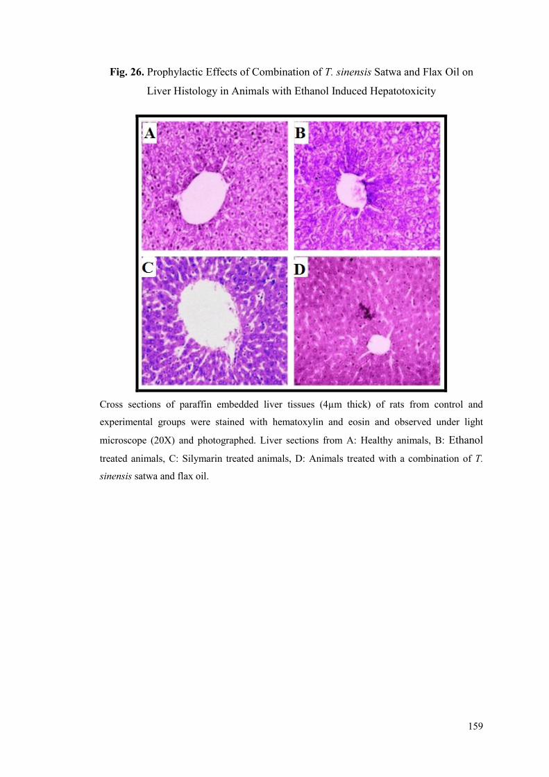
159
Fig. 26. Prophylactic Effects of Combination of T. sinensis Satwa and Flax Oil on
Liver Histology in Animals with Ethanol Induced Hepatotoxicity
Cross sections of paraffin embedded liver tissues (4µm thick) of rats from control and
experimental groups were stained with hematoxylin and eosin and observed under light
microscope (20X) and photographed. Liver sections from A: Healthy animals, B: Ethanol
treated animals, C: Silymarin treated animals, D: Animals treated with a combination of T.
sinensis satwa and flax oil.
160
Fig. 27. Gene Expression from Liver Tissues of Experimental Animals
Densitometric analysis was done by using GAPDH and gene specific expression data.
*P≤0.05; **P≤0.01; ***P≤0.001 compared with negative control rats. Lanes: Healthy control
(Group I) -1; Negative control (Group II) -2; Positive control (Group III) -3; Treated with
combination of T. sinensis satwa and flax oil (Group IV) -4. GAPDH (Internal standard). A:
FABP1, B: PPARγ, C: NF-κβ, D: SREBP1, E: TNF-α.
A B
I II III IV0.0
0.4
0.8
1.2
1.6
2.0
2.4
FA
BP
1/G
AP
DH
I II III IV0.0
0.4
0.8
1.2
1.6
2.0
2.4 ***
*
PP
AR
/GA
PD
H
C D
I II III IV0.0
0.4
0.8
1.2
1.6
*** ***
*
NF
-
/GA
PD
H
I II III IV0.0
0.4
0.8
1.2
1.6
2.0
2.4
2.8
*
SR
EB
P1
/GA
PD
H
E
I II III IV0.0
0.4
0.8
1.2
1.6
2.0
2.4
**
TN
F-
/GA
PD
H

CHAPTER 6
Discussion Awards
Second prize in oral
Presentation Award
Best Poster Presentation
Award
161
Liver is the largest organ in the human body and is crucial in maintenance of
good health. It is involved in almost all biochemical pathways that allow growth,
protection from diseases, synthesis and supply of nutrients etc. The liver performs
more than 500 vital functions of metabolic importance (Naruse et al., 2007). Liver is
constantly exposed to several environmental and chemical insults to which it gives a
specific response. Chronic exposure to such insults leads to inflammation and
compromise in liver function (Sahu, 2007). Several drugs and/or chemicals are known
to cause hepatotoxicity (Bigoniya et al., 2009). Several adverse effects of liver
protective drugs in allopathic medical practices have prompted the untiring search for
herbs and nutritional supplements for management of various liver disorders. Use of
number of medicinal plants and their formulations are common for the treatment of
liver diseases in ethno-medical practices and in traditional medicine system (Dange,
2010; Kumar et al., 2011; Shaik et al., 2012). Modern medicine has little to suggest
for improvement of hepatic diseases and it is essentially the plant based preparations
which are employed for the treatment of liver disorders (Chatterjee, 2000).
Approximately 80% of the world population depends on the use of traditional
medicine system which is predominantly based on plant materials (WHO, 2002). It is
estimated that local health traditions, mostly in rural and tribal villages of India, use
about 7,500 plants. Out of these, the real medicinal value of over 4,000 plants is either
little known or yet to be known to the mainstream population. The classical systems
of medicine such as Ayurveda, Siddha, Unani, Tibetan, and Amchi use about 1,200
plants (Pandey, 2011). Various herbs and plant extracts have significant
hepatoprotective activity as indicated from studies in animal models (Malhotra and
Singh, 2006; Chaudhary et al., 2010). Nutritional supplements with omega-3 fatty
acids and diet rich in carbohydrates, fats and proteins with adequate calories and helps
in regeneration of liver cell for patients with liver diseases (Drevon, 2009; Shukla and
Kumar, 2013)
Studies in the present thesis have indicated hepatoprotective activities of
herbal and nutritional supplement(s), and the combinatorial hepatoprotective effects
of best performing herbal and nutritional supplements against acetaminophen and
alcohol induced liver damage.
Awards
Second prize in oral
Presentation Award
Best Poster Presentation
Award

162
6.1 Guduchi Satwa (T. cordifolia, T. sinensis and Neem-giloe)
6.1.1 Organoleptic Characteristics of Three Tinospora forms
There are very few reports available for organoleptic characteristics of T.
cordifolia and Neem-giloe satwa (Sharma et al., 2012; Patil and Chaudhary, 2013;
Sharma et al., 2013a; Chavan et al., 2014; Sharma et al., 2015). As per classics, the
taste of Guduchi satwa from T. cordifolia is swadu (palatable/pleasant) (Navare,
2011) but some recent reports describe Guduchi satwa from T. cordifolia as slight
bitter (Sharma et al., 2013a; Sharma et al., 2015) and Guduchi satwa from Neem-
guduchi as tasteless (Sharma et al., 2012; Sharma et al., 2015). However, in present
study, satwa from T. cordifolia and T. sinensis was found to be tasteless while that
from Neem-giloe was found to be bitter in taste. In Rasa Yoga Sagara (Sharma,
2004), the colour of satwa is mentioned as ‘Shubhrakhandnibha’ (clear white like
sugar cubes) (Sharma, 2004) and Yoga Ratnakara reveals it as ‘Shankhanibha’ (clear
white like conch shell) (Shastri, 2002) but some texts mention the colour of satwa as
greenish white (Reddy, 2005) or greyish white (Hiremath, 2005) and the colour of
satwa from Neem-guduchi as pale (Sharma et al., 2012) to clear white (Sharma et al.,
2015). In the present work, colour of the satwa prepared from T. cordifolia, T.
sinensis and Neem-giloe were found to be grey, greyish white and yellowish white
respectively, which resembles the recent reports. The texture and taste of satwa also
depends on the age of the plant material used for satwa preparation. A recent study by
Patil and Chaudhary (2013) have examined stem pieces of different thickness and
maturity of T. cordifolia to find best size of the stem to achieve maximum yield of
satwa. A yield of 0.48% -0.1% satwa from T. cordifolia was reported from fresh stem
(Mehra and Puri, 1969; Rao and Rao, 1981; Salunke and Pimpalgaonkar, 1997) and
1.20% (Rao and Rao, 1981) with that of dried stem. Sharma et al. (2012) have
reported that the thickness of T. cordifolia stem also has effect(s) on the yield of satwa
with 1.6-2.0 cm thick stem giving maximum yield of satwa. These variations may be
due to different ecotypes, size of the stem, collection time and levels of maturity of
the plant. In the present study stem pieces of 1.6-2.0 cm were used for preparation of
satwa and the yield of satwa ranged from 1.48%-1.60% in three different forms of
Tinospora. Sharma et al. (2012) have reported that the yield of satwa from Neem-
guduchi is highly variable and it depends upon multiple factors like size, environment,

163
nature of cellular activities etc. They have also reported that the medium sized stem
(1.5-2.0cm) of Guduchi yields maximum satwa since there is maximum accumulation
of starch at this stage during development. Sharma et al. (2015) have reported that the
yield of Neem-guduchi satwa from male and female stem was 2.25 % and 3.18 %
respectively since relatively higher starch and mucilage contents are found in female
plants, suggesting that more yield of satwa can be obtained from them.
6.1.2 Nutritional Analysis of Three Tinospora forms
The nutritional composition of medicinal plants depends heavily on
environmental and physiological conditions as well as the time of harvesting of the
plant (Kutbay and Ok, 2003; Sharma et al., 2013b). Guduchyadi varga from
Bhavprakash nighantu (1999) mentioned the time of collection of Guduchi as end of
May. Accumulation of active components of a drug is also reported to be maximum
during specific seasons in many medicinal plants (Kutbay and Ok, 2003; Nasreen et
al., 2010; Patil and Gaikwad, 2011). The variability in the active components of an
Ayurvedic drug due to these factors can be studied with the help of principles to
determine different nutritional/pharmaceutical contents in the drug (Nasreen et al.,
2010; Patil and Gaikwad, 2011; Geeta and Kumari, 2013; Mahima et al., 2013). In
case of T. cordifolia, crude protein and fiber are reported to be the principal
components of the powder from dried stem pieces (Hussain et al., 2009). There are
reports about the proximate and elemental analysis of powder prepared from fresh or
dried stem of T. cordifolia (Nile and Khobragade, 2009; Mahima et al., 2014).
Powder prepared from dried stem of T. cordifolia was found to be rich in
phytochemicals like alkaloids, glycosides, sterols and carbohydrates (Nasreen et al.,
2010; Tanwar et al., 2012). The presence of wide range of phytochemicals is also
reported in different solvent extracts (methanol, petroleum ether, water, chloroform
and ethyl acetate) of T. cordifolia stem (Sivakumar and Rajan, 2011; Pradhan et al.,
2013). Nutritive value of the medicinal plants can be determined with the help of
proximate and elemental analysis (Mahima et al., 2013). The crude protein content of
T. cordifolia is of great importance to its nutritive value (Ajibade and Fagbohun,
2010). Satwa is a potential source of nutrition and has shown beneficial effects for
boosting the immune system-response and body building (Ajibade and Fagbohun,
2010; Geeta and Kumari, 2013; Mahima et al., 2014).
164
The reports on the nutritional analysis of Guduchi satwa are available only for
T. cordifolia satwa. There are no reports of nutritional and/or comparative analysis of
T. sinensis and Neem guduchi. In view of this, the present analysis was undertaken to
identify the nutritional richness of these Tinospora forms. The present study has lead
to the first report of the nutritional analysis of satwa prepared from T. cordifolia, T.
sinensis and Neem-guduchi (Chavan et al., 2014). In present study, the lipid, ash, and
carbohydrate contents of Neem-giloe were greater than that of T. cordifolia and T.
sinensis. T. sinensis showed higher amount of protein, starch, and crude fiber than
Neem-giloe and T. cordifolia.
6.2 Drug Induced Liver Injury
The ability to prevent damage to the liver is called as hepatoprotection (Mishra
et al., 2014). Hepatic damage may be caused due to viral hepatitis, bile duct
obstruction, cholesterol overload, etc. and also due to some chemical factors such as
overdose of several drugs, alcohol intake etc. With increase in the incidence of
hepatotoxicity, there is a need to find effective ways for prevention or management of
liver damage (Wang et al., 2009). The hepatoprotective efficacy of a drug is measured
in terms of its ability to restore normal hepatic functions or to reduce damage to the
liver (Yadav and Dixit, 2003). The drugs used for treating hepatic disorders in modern
medicine have several undesirable side effects (Singh, 2013; Ananthi and Anuradha,
2015).
The two hepatotoxicants selected in this study are Acetaminophen
(Paracetamol) and Alcohol (Ethanol) which are routinely used for medicinal or other
purposes. The incidence of acetaminophen induced liver damage has been reported to
be increased from 28% in 1998 to 52% in 2003, in USA (Larson et al., 2005). A
recent Indian report has estimated that about 33% of the people consuming
acetaminophen are subjected to liver damage (Marzilawati et al., 2012). Easy and
over-the-counter availability of acetaminophen and self-medication (Kaufman et al.,
2002; Larson et al., 2005; Abay and Amelo, 2010) is believed to be a reason behind
intentional or unintentional overdose (Lee, 2004; Larson et al., 2005). Alcohol
increases the hepatotoxicity of various xenobiotics and the interaction between
alcohol and hepatotoxins is well recognized (Zimmerman, 1999). Alcoholic liver
disease (ALD) is a common consequence of prolonged and heavy alcohol abuse. In
165
India, the incidence of alcohol induced hepatotoxicity is reported to be 31% (WHO,
2011).
In the present study, animals were dosed repeatedly with hepatotoxicant
(acetaminophen and alcohol separately) and simultaneously treated with satwa from
one of the three different forms of Tinospora or Omega-3 fatty acids (Flax oil and
Fish oil) and combination of best Tinospora forms and best omega-3 fatty acids.
Biochemical, histological and molecular analyses were performed to determine the
beneficial effects of satwa from three Tinospora forms and omega-3 fatty acids and
their combination.
6.3 Hepatoprotective Activity of Satwa against Acetaminophen Induced
Hepatotoxicity
6.3.1 Biochemical Parameters
Elevated levels of serum glutamic oxaloacetic transaminase (SGOT) indicate
liver damage, which may be due to viral hepatitis, cardiac infarction and muscle
injury while serum glutamic pyruvic transaminase (SGPT) is more specific to liver,
and is thus a better parameter for detecting liver injury (Reitman and Frankel, 1957;
Thapa and Walia, 2007). Increase in serum levels of alkaline phosphate (ALP) is due
to increased synthesis of the enzyme in presence of increasing biliary pressure (Kind
and King, 1954; Thapa and Walia, 2007). Serum bilirubin level is related to function
of hepatic cells (Jendrassik and Grof, 1938; Thapa and Walia, 2007). Increased
SGOT, SGPT and ALP are indicative of cellular leakage and reduced functional
integrity of the liver cell membranes indicating hepatocellular damages (Gutierrez and
Solis et al., 2009; Basu et al., 2012). In the current study, administration of
acetaminophen caused a significant elevation of SGOT, SGPT, ALP, and total
bilirubin when compared to healthy control, which is indication of hepatic damage.
Several reports indicate elevated levels of liver function markers like SGOT, SGPT,
ALP and bilirubin in rats subjected to acetaminophen induced liver injury (Duairaj et
al., 2007; Ramachandran et al., 2010; Sundari et al., 2011; Basu et al., 2012; Galal et
al., 2012). Our study reveals comparative hepatoprotective effect of T. cordifolia, T.
sinensis and Neem-giloe satwa. The previous studies have reported the
hepatoprotective effect of T. cordifolia alone against different hepatotoxicants
166
(Bishayi et al., 2002; Sharma and Pandey, 2010; Kavitha et al., 2011b;
Venkatalakshmi and Ragadevi, 2012; Kumar et al., 2013b). In the present study,
treatment of rats with Neem-giloe (200 mg/kg) decreased levels of SGOT and
bilirubin while T. sinensis showed effects on improvements in serum SGPT and ALP
against acetaminophen induced hepatotoxicity. Apart from liver protection, studies on
T. sinensis indicate anti-inflammatory (Li et al., 2003) and anti-diabetic activities
(Yonemitsu et al., 1993). Some reports also show the immunomodulatory activity of
T. cordifolia (growing on neem tree) (Bhalerao et al., 2012; Narkhede et al., 2014).
Elevated levels of total cholesterol, phospholipids, triglycerides and free fatty
acids in the plasma have been reported in acetaminophen treated rats which is an
indication of reduced or impaired fat metabolism secondary to liver damage
(Ramachandran et al., 2010; Haldar et al., 2011; Malarvizhi et al., 2012). Several
reports indicate elevated levels of serum lipids (Total cholesterol, triglyceride, and
LDL) in rats subjected to acetaminophen overdose (Duairaj et al., 2007; Sundari et al.,
2011; Basu et al., 2012; Singh, 2013; Singh et al., 2015). In present study, animals
treated with acetaminophen showed increase in the levels of cholesterol, triglyceride,
very-low-density lipoprotein cholesterol (VLDL), low-density lipoprotein cholesterol
(LDL), and decrease in high-density lipoprotein cholesterol (HDL) levels in serum
and liver homogenates as compared to healthy animals. The earlier studies revealed
that treatment with T. cordifolia root extract resulted in significant reduction in levels
of serum and tissue cholesterol, phospholipids and free fatty acids in alloxan induced
diabetic rats (Stanely et al., 1999). In present study, T. cordifolia satwa exhibited
improvements in the serum levels of total cholesterol, HDL and LDL, T. sinensis
satwa showed improvement in VLDL and triglycerides levels while Neem-giloe satwa
showed significant improvements in total protein and lipid profile (HDL, LDL,
VLDL, Triglyceride) in liver tissues. The alterations in the lipid profile due to
chemically induced liver damage also leads to generation of several other chemical
entities which may have direct or indirect effect on the liver functions (Basu et al.,
2012).
Free radicals are generated during liver injury which attack many sub-cellular
organelles and systems. Acetaminophen hepatotoxicity is mediated through oxidative
stress due to activation of acetaminophen by cytochrome P450 (Zoubair et al., 2013).
Excessive peroxidation causes increased reduced glutathione (GSH) consumption
167
(Nandy et al., 2012). GSH is a scavenger of toxic metabolites, including NAPQI,
which is a metabolite of acetaminophen (Hsu et al., 2008). Depletion in the levels of
reduced glutathione leads to compromised antioxidant capacity of the tissue.
Acetaminophen overdose causes decrease in antioxidant enzyme activities such as
superoxide dismutase (SOD), catalase (CAT), glutathione peroxidase (Lores et al.,
1995; Duairaj et al., 2007; Basu et al., 2012) and increases lipid peroxidation levels in
liver (Duairaj et al., 2007; Basu et al., 2012). In the present study, the activity of SOD
and catalase as well as GSH contents in liver significantly decreased, indicating
oxidative stress while lipid peroxidation (MDA) levels increased indicating increased
lipid peroxidation. Interventions with Neem-giloe satwa showed significant increase
in the levels of SOD, CAT, and GSH. Though statistically insignificant, animals
treated with the satwa from T. sinensis showed decreased MDA levels in liver
homogenates than that in acetaminophen treated group. The administration of T.
cordifolia (50, 100, 200 gm/kg orally daily for 25 days) has been reported to exhibit
protective effect by reducing the contents of thiobarbituric acid reactive substances
(TBARS) and increasing GSH, ascorbic acid, protein, and the activities of antioxidant
enzymes viz., SOD, CAT, glutathione peroxidase, glutathione S-transferase, (GST),
and glutathione reducates (GR) in liver and kidney against aflatoxin B1 (AFB1)
toxicity in mice (Gupta et al., 2011). Prince and Menon (2001) reported that
administration of aqueous extract of the roots of T. cordifolia (5.0 g/kg for 6 weeks)
resulted in significant reduction in TBARS and an increase in GSH, CAT and SOD in
alloxan induced diabetic rats.
6.3.2 Histological Analysis
Histological study of liver sections of rats treated with acetaminophen
displayed degenerative changes in hepatocytes and cells that line the blood sinusoids.
The anatomical changes in liver due to injury or toxicity are known to be positively
correlated with increase in transaminase activities (Galal et al., 2012). Acetaminophen
induced liver injury frequently shows intense centrilobular necrosis and
vascuolisation (Manivannan et al., 2011), severe intense congestion, hydropic
degeneration, pyknosis and occasional necrosis (Kanchana and Sadiq, 2011; Prabu et
al., 2011), sinusoidal haemorrhages and dilatations with chronic inflammatory cell
infiltrate in portal tracts (Sundari et al., 2011). In present study, sections of liver from
168
acetaminophen treated group showed mild congestion of central vein, hepatocytes
exhibited apoptotic death with few intra and extracellular hyaline globules and
ballooning and degeneration around central vein. Mild nucleomegaly was also visible
under microscope. Significant recovery of hepatocellular lesions was observed in the
animals treated with the satwa of Neem-giloe which restored the hepatic histology to
near normal architecture. Such normal hepatic lobule architecture was found in the
animals treated with 2 ml/100g of T. cordifolia for 30 days against CCL4 induced
hepatotoxicity (Kumar et al., 2013b). Normal liver histology, without any detectable
necrosis and vacuolisation, was also observed in rats treated with T. cordifolia extract
(200 mg/kg) once daily for 3 days against CCl4 induced hepatotoxicity (Kavitha et al.,
2011b).
6.3.3 Gene Expression
Expression of Genes Involved in Lipid Metabolism
PPARγ (peroxisome proliferator-activated receptor-gamma) and SREBP1
(Sterol regulatory element-binding protein 1) are transcription factors and regulators
of lipid homeostasis in hepatocytes and a target for fatty acids and hypolipidemic
drugs (Eberle et al., 2004; Shan et al., 2008; Gong et al., 2014).
The proteins encoded by different PPAR genes have the ability to induce
hepatic peroxisome proliferation in response to xenobiotic stimuli (Sahu, 2007). The
three PPAR isoforms (PPARα, PPARδ and PPARγ) are believed to play a central role
in regulation of carbohydrate and lipid metabolism, fatty acid metabolism and are also
assumed to possess anti-inflammatory activity (Wang et al., 2014b). These PPAR
isoforms inhibit the induction of pro-inflammatory cytokines and stimulate the
production of anti-inflammatory molecules (Kostadinova et al., 2005). Dysregulation
of some PPAR isoforms contribute to development of a wide range of liver diseases
(Peyrou et al., 2012). The pathways for action of PPARs have been well studied.
PPARs combine with PPRE (peroxisome proliferator response element) in the
promoter region of their target genes involved in fatty acid transport and lipid
catabolism (Sears et al., 2007). Differential effects could be explained by promoter
and cell context and also by the availability of co-factors but at the same time there
are site specific conformational changes in the receptors which are induced by PPARγ
169
ligands that ultimately lead to chromatin modeling of target genes and differential
promoter activation (Olefsky, 2000). Majority of studies deal with PPARγ in diabetic
and obese mouse livers (Memon et al., 2000; Gavrilova et al., 2003; Bedoucha et al.,
2001) but the mechanistic relationship of increase of PPARγ expression in
hepatotoxicity, remains unclear till date.
Liver is a primary site of biotransformation and is critical in modulating
metabolically and chemically induced toxicity and there are reports which suggest
that PPARs modulate hepatotoxicity. Pioglitazone, a PPARγ agonist, inhibits CCl4
(carbon tetrachloride) induced hepatic fibrosis through inhibition of inflammation and
hepatic stellate cell proliferation indicating protective role of PPARγ in hepatotoxicity
(Yuan et al., 2004). An isoform of PPAR, PPARβ, enhances chemically induced liver
toxicity (Hellemans et al., 2003). Expression of PPARβ messenger RNA was
increased in hepatic stellate cells, as they undergo spontaneous activation (Hellemans
et al., 2003).
PPARγ has direct involvement in regulation of the functional expression of
drug transporters, such as the ABCG2, ABCA1 etc. ABCG2, an ATP-binding cassette
transporter is known to perform clearance of endogenous and exogenous toxic agents.
Activation of PPARγ and consecutively increased amounts of the ABCG2 transporter
protein were shown to significantly increase efflux of xenobiotics in human dendritic
cells (Szatmari et al., 2006). Peroxisomal proliferators like clofibrate, aspirin,
valproate, ethylhexanol, ciprofibrate and perfluorooctanoate might cause local toxicity
due to inhibition of mitochondrial oxygen uptake (Keller et al., 1992). In chronic liver
injury induced by carbon tetrachloride, for instance, a downregulation of PPARγ
expression was observed in hepatocytes, while increased levels of these transcription
factors were found in Kupffer cells associated with inverse correlation to levels of
activated NFκβ (Orfila et al., 2005). PPARγ also plays an antitoxic role by inducing
liver cells to deposit harmless lipids thereby preventing the accumulation of toxic
lipids (Medina-Gomez et al., 2007). Miyahara et al. (2000), and Zhang et al. (2012)
reported PPARγ deficiency in hepatic stellate cells associated with excessive
formation of fibrotic tissue in the liver. The role of PPARγ in manifestation of
inflammation is gaining momentum (Szeles, 2007) and expression in hepatoma cell
line indicates its potential role in liver function (Koga et al., 2007). Down regulation
of PPARγ mRNA expression has been reported in isoniazid induced hepatotoxicity

170
(Mahmoud et al., 2014). In accordance with the above reports, the present study also
reported down regulation of PPARγ expression in acetaminophen induced
hepatotoxicity as compared with healthy control. Several medicinal plant extracts are
reported to activate PPARγ (Local Food-Nutraceuticals Consortium, 2005;
Christensen et al., 2009; Andaloussi et al., 2010; Vogl et al., 2013; Yang et al., 2013).
Curcuminoids have also been reported to inhibit pro-inflammatory induction by
enhancing PPARγ activation (Jacob et al., 2007). The protective effects of berberine
against isoniazid-induced hepatotoxicity may be attributed to its ability to upregulate
PPARγ and subsequently suppress NF-κβ, iNOS and release of proinflammatory
cytokines (Mahmoud et al., 2014). The mechanism of action of hepatoprotection by
several secondary metabolites from plants has been shown to be through reduction in
oxidative stress which is achieved via activating PPARγ (Duval et al., 2014). Though
statistically insignificant, the present study reported marginal improvement in PPARγ
expression in the livers of rats treated with Neem-giloe satwa and T. sinensis satwa
exhibiting near normal liver architecture.
The liver plays a central role in lipid metabolism through de novo lipid
synthesis and fatty acid oxidation. SREBPs (Sterol regulatory element-binding
proteins) are the important transcription factors which activate the expression of genes
involved in the biosynthesis of TGs (Triglycerides), fatty acids and cholesterol. The
transcription factors in SREBP family are master regulators of lipid metabolism,
which control the expression of genes required for fatty acid and cholesterol
biosynthesis. In mammals, there are three isoforms of SREBPs, SREBP- 1a, 1c and 2
(Horton et al., 2002). SREBP-1c is the predominant isoform of SREBP-1 in liver and
SREBP-1c is mainly responsible for the biosynthesis of TGs. Studies have shown that
transgenic mice overexpress SREBP-1c or SREBP-1a and produce massive fatty liver
due to accumulation of TGs and cholesterylesters (Shimano et al., 1996; Shimomura
et al., 1999). SREBP1 is involved in the activation of genes associated with fatty acid
metabolism, and involved directly in cholesterol homeostasis (Horton et al., 1998; Pai
et al., 1998). SREBP1 specifically activates several of the key genes involved in
lipogenesis (Horton et al., 2002; Horton et al., 2003) like fatty acid synthase (FAS),
and Acetyl-CoA carboxylase alpha (ACACA) (Ronnebaum et al., 2008). SREBPs are
bound as precursors to the endoplasmic reticulum and nuclear envelope. When
activated, they are released from the membrane and escorted to the Golgi complex by
171
SREBP cleavage-activating protein (SCAP) and then cleaved by specific proteases
and translocated to the nucleus, where they bind to sterol regulatory element-1
(SRE1), which is a decameric sequence flanking the low density lipoprotein receptor
gene and some genes involved in sterol biosynthesis (Sozio and Crabb, 2008). Sterols
inhibit the cleavage of the precursor, and the mature nuclear form is rapidly
catabolized, thereby reducing transcription. The genes which are activated by SREBP
are in turn regulated by AMPK (AMP-Activated protein kinase) since AMPK directly
phosphorylates and binds to SREBP-2 and SREBP-1c (Li et al., 2011) inhibiting the
expression of FAS and ACC which are key lipogenic enzymes (Viollet et al., 2009).
SREBP-1 gene knockout mice show a very low basal expression of FAS
hardly possess the ability to upregulate de novo lipogenesis (Juvet et al., 2003).
SREBP1 gene expression was observed to be down-regulated in animals treated with
single high dose of acetaminophen, carbon tetrachloride, tetracycline amiodarone
(Fukushima et al., 2006). The present study however, involved daily dosing of
acetaminophen for 15 days in rats. Thorough literature search indicated that the
reports for effects of such treatment on SREBP expression are not yet available. In the
present study, SREBP1 expression was reported to be higher in the animals which
were repeatedly treated with high dose of acetaminophen for 15 days as compared to
the healthy control.
Scanty references are available on the effect of herbal interventions on SREBP
expression in animal models for hepatotoxicity. A study involving C57BL/6-Lep
ob/ob mice reported the prevention of fatty liver by carbenoxolone intervention, an
active component of Glycyrrhiza glabra. The hepatoprotective ability of the
intervention was attributed to SREBP-1c inhibitory activity and anti-apoptotic action
of the intervention (Rhee et al., 2012). The diosgenin fraction from fenugreek inhibits
triglyceride accumulation in livers of diabetic obese KK-Ay mice as well as in HepG
2 cells with simultaneous decrease in the expressions of SREBP-1c, ACC and FAS
(Uemura et al., 2011). In the present study, expression of SREBP-1 was significantly
decreased in animals treated with the satwa of T. cordifolia, T. sinensis and Neem-
giloe as compared to negative control.
Mammalian intracellular fatty acid-binding proteins (FABPs) comprise a
superfamily of lipid-binding proteins which are involved in the fatty acid uptake,
172
intracellular transport and in regulating lipid metabolism, cellular signaling
pathways and other lipid ligands (Wang et al., 2007; Storch and Thumser, 2010).
FABP is highly expressed in adipocytes, liver, muscle, heart, brain and macrophages
and the expression and activation of FABP1 has been reported to contribute to the
pathogenesis of obesity, metabolic syndrome and associated inflammation (Makowski
and Hotamisligil, 2004). Fatty acids transport, in and out of the cells, is a complicated
process and is important for function and utilization of lipids (Kim et al., 2007). The
effect of acetaminophen induced hepatotoxicity on FABP1 expression has been
studied with relation to oxidative stress. A dose-dependent increase in oxidative stress
induced by acetaminophen was associated with significantly low FABP1 expression
(Gong et al., 2014). FABP1 also plays an early protective role in acetaminophen
induced mitochondrial impairment through scavenging free radicals within the
mitochondria itself as well as in the cytosol (Gong et al., 2014). The role of L-FABP
in liver disease was further assessed by Wang et al. (2007). FABP1 has been reported
to possess strong antioxidant properties (Yan et al., 2009). In accordance with this
role of FABP, the present study observed decreased expression levels of FABP1 in
acetaminophen treated group and significantly higher expression in animals treated
with Neem-giloe satwa.
Expression of Genes Involved in Inflammation
NF-κβ (Nuclear factor-κβ) is one of the most important transcription factors
and it is activated by inflammatory cytokines like TNF-α (Tumor necrosis factor-
alpha) (Richard, 2001). Both are related to injury and inflammation, hepatitis,
immunity, including various etiologies of muscle catabolism and osteoclastogenesis
(Zwart et al., 2009). The NF-κβ pathway is complex and is activated by
phosphorylation, ubiquitination, and proteolysis of the inhibitory protein IκB (I kappa
B), which nominally binds NF-κβ in the cytosol in the inactive form (Zwart et al.,
2009). Blazka et al. (1995) reported the up-regulation of TNF-α in liver of
acetaminophen treated mice. Dambach et al. (2006) and Song et al. (2014) recently
reported significantly up-regulated expression of TNF-α and NF-κβ in
acetaminophen-induced hepatotoxicity in mice. Various inflammatory cytokines
produced during drug induced liver injury have been reported to be involved in tissue
damage (Ishida et al., 2002). Ishida et al. (2004) reported that, liver injury in mice
173
deficient in the 55KDa TNF receptor (TNF-Rp55), was attenuated after APAP
challenge. In addition, APAP-induced mortality was reduced in TNF-Rp55 KO mice.
Studies in mice deficient in CCR2, the primary receptor for the chemokine MCP-1
(Monocyte chemoattractant protein-1), showed that these mice had increased toxicity
of TNF-α and IFN-α (Hogaboam et al., 2000). TNF-α is reported to promote tissue
damage during acetaminophen toxicity (Boess et al., 1998). mRNA and protein
expressions of TNF-α and NF-κβ were significantly upregulated in D-galactosamine–
induced hepatotoxicity (Aristatile et al., 2013). Tu et al. (2012) observed significant
increase in TNF-α in carbon tetrachloride intoxicated rats. The serum levels of pro-
inflammatory cytokines, such as TNF-α and NF-κβ were significantly elevated in
isoniazid induced hepatotoxicity in albino rats (Mahmoud et al., 2014). Roles of NF-
κβ in suppression (Van Antwerp et al., 1996) as well as induction (Grilli et al., 1996)
of apoptosis have been reported and NF-κβ is also thought to play a major role in liver
regeneration (Ulloa et al., 2002; Tsung et al., 2005; Luedde and Schwabe, 2011). In
the present study, NF-κβ and TNF-α gene expression were higher in acetaminophen
induced hepatotoxicity as compared with healthy control group.
The diabetic rats, treated for 24 weeks with Tinospora cordifolia extract
(250 mg/kg) exhibited significantly reduced amount of inflammatory markers such as
TNF-α and IL-1β (Agrawal et al., 2012). Several chemical constituents like alkaloids,
diterpenoid lactones, steroids, glycosidesetc from different parts of T. cordifolia are
known to inhibit the activity of NF-κβ and TNF-α (Mittal et al., 2014). G1-4A, an
arabinogalatan, found in T. cordifolia, increases levels of TNF-α and IL-10 against
endotoxic shock through modulation of cytokines and nitric oxide in mice (Desai et
al., 2007). Treatment of albino rats with Berberine, an isoquinoline alkaloid, leads to
decrease in the serum levels of TNF-α and NF-κβ in isoniazid and cyclophosphamide-
induced hepatotoxicity (Germoush and Mahmoud, 2014; Mahmoud et al., 2014).
Treatment of male Sprague-dawley rats with curcumin significantly reduced the levels
of proinflammatory mediators (TNF- α, IL-6 and MCP-1) mRNA in carbon
tetrachloride induced liver toxicity (Tu et al., 2012).
In present study, NF-κβ gene expression was found to be significantly
decreased in the rats treated with the satwa of T. cordifolia, T. sinensis and Neem-
giloe while there was statistically insignificant decrease in TNF-α gene expression in
Neem-giloe satwa treated rats. Silymarin, a standard drug used in the present study,

174
has been reported to suppress NF-κβ gene expression in the hepatoma cell line
HEPG2 (Saliou et al., 1998). Apart from the intervention groups in the present study,
NF-κβ gene expression was also found to be significantly decreased in the rats treated
with Silymarin (positive control).
Based on the results obtained from biochemical, histological and gene
modulation studies in animals receiving interventions of satwa of T. cordifolia, T.
sinensis and Neem-giloe, hepatoprotective activity of Neem-giloe was found to be
better than that of T. cordifolia and T. sinensis, in acetaminophen induced
hepatotoxicity.
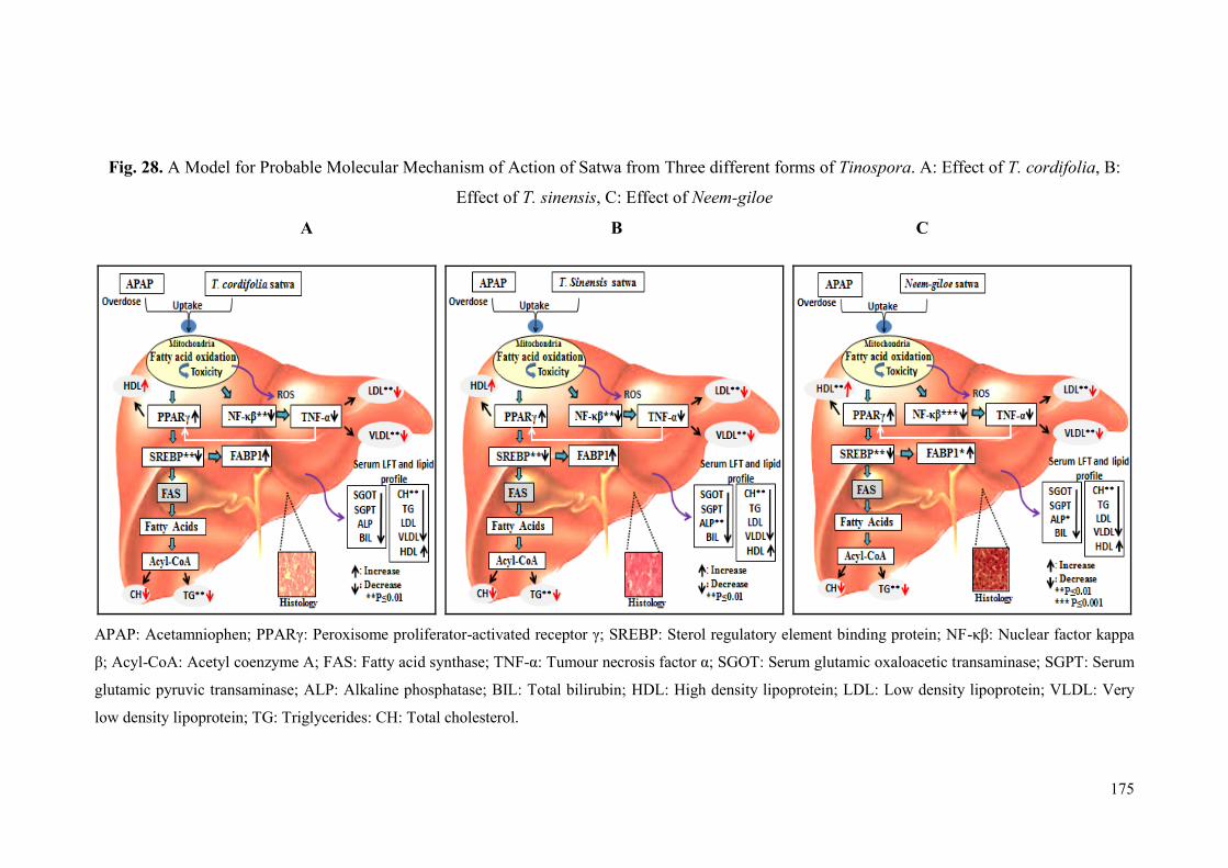
175
Fig. 28. A Model for Probable Molecular Mechanism of Action of Satwa from Three different forms of Tinospora. A: Effect of T. cordifolia, B:
Effect of T. sinensis, C: Effect of Neem-giloe
A B C
APAP: Acetamniophen; PPARγ: Peroxisome proliferator-activated receptor γ; SREBP: Sterol regulatory element binding protein; NF-κβ: Nuclear factor kappa
β; Acyl-CoA: Acetyl coenzyme A; FAS: Fatty acid synthase; TNF-α: Tumour necrosis factor α; SGOT: Serum glutamic oxaloacetic transaminase; SGPT: Serum
glutamic pyruvic transaminase; ALP: Alkaline phosphatase; BIL: Total bilirubin; HDL: High density lipoprotein; LDL: Low density lipoprotein; VLDL: Very
low density lipoprotein; TG: Triglycerides: CH: Total cholesterol.
176
6.4 Hepatoprotective Activity of Satwa against Ethanol Induced Hepatotoxicity
6.4.1 Biochemical Parameters
In the present study, administration of alcohol caused a significant elevation of
SGOT, SGPT, ALP, and total bilirubin when compared to healthy control, which is
indication of hepatic damage. Several reports indicate elevated levels of liver function
markers like SGOT, SGPT, ALP and bilirubin in ethanol-induced hepatotoxicity in
rats (Yue et al., 2006; Arulkumaran et al., 2009; Vidhya and Indira, 2009; Nigam and
Paarakh, 2011; Singh and Gupta, 2011; Cui et al., 2013; Padmanabhan and Jangle
2014; Seif, 2014). In the present study, serum levels of SGOT, SGPT, ALP and
bilirubin were significantly decreased after treatment with T. sinensis (200 mg/kg) as
compared to ethanol treated group. The hepatoprotective activity of ethanolic extract
of T. sinensis roots has previously been demonstrated in carbon tetrachloride induced
hepatotoxicity in rats (Naik et al., 2013). Treatment of albino rats with acetone and
aqueous extracts of Adina cordifolia showed significant decrease in the levels of
serum markers (SGOT, SGPT, ALP and total bilirubin), indicating the protection of
hepatic cells against ethanol induced hepatocellular injury (Sharma et al., 2012).
Treatment with aqueous extract of Andrographis paniculata (50mg/kg, 100mg/kg,
200mg/kg of body weight) was found to protect the rats from hepatotoxic action of
ethanol as evidenced by significant reduction in the elevated levels of SGOT, SGPT,
ALP and bilirubin (Vetriselvan et al., 2011).
Alcohol has significant effect on lipid and lipoproteins metabolism. Alcohol
induced liver injury is characterized by lipid accumulation in affected hepatocytes
(Ontko, 1973). In the present study, animals treated with ethanol showed increase in
the levels of serum and hepatic cholesterol, triglyceride, VLDL, LDL, along with
decrease in HDL levels as compared to healthy control. Such abnormal level of lipids
is an indication of disturbed lipid metabolism. Earlier studies also have reported
increased levels of serum and tissue lipids (Triglyceride, total cholesterol, and
phospholipids) in alcohol treated rats (Tomita et al., 2004; Kim et al., 2007;
Arulkumaran et al., 2009; Cui et al., 2013; Samundeeswari et al., 2013). In the present
study, Neem-giloe satwa exhibited normalization of serum lipid profile while T.
sinensis satwa normalized the lipid profile in liver. Treatment of animals with Neem-
177
giloe satwa also revealed significant increase in total protein content. An earlier study
on polyherbal formulation, Vimliv, which contains T. cordifolia, showed significant
improvement of lipid profile (Triglyceride, total cholesterol, phospholipid) in serum
and liver tissue in ethanol-induced hepatic damage in female albino wistar rats
(Samundeeswari et al., 2013). A recent report by Sharma and Dabur (2015) also
indicated improvements in serum lipid profile after treatment of alcoholic volunteers
with water extract of T. cordifolia. They also showed that the water extract helped in
enhancing intestinal vitamin absorption and preventing multivitamin deficiency in
liver due to alcohol intake.
Oxidative stress plays an important role in the development of ALD
(Alcoholic liver disease) (Yurt and Celik, 2011). Recent literature of alcohol induced
hepatotoxicity indicates decrease in the levels of SOD, GSH and catalase and increase
in malondialdehyde, hydroperoxides in the liver homogenate of alcohol treated rats
(Das and Vasudevan, 2006; Mallikarjuna et al., 2009; Shanmugam et al., 2010; Arun
and Balasubramanian, 2011; Singh and Gupta, 2011; Rejitha et al., 2012; Seif, 2014).
Administration of aqueous extract of stem and leaves (400 mg/kg body weight, orally)
of T. cordifolia increased the activities of SOD and CAT and decreased the levels of
SGOT, SGPT and ALP enzymes in lead nitrate induced hepatotoxicity in mice
(Sharma and Pandey, 2010). In the present study, contents of hepatic SOD, GSH and
catalase significantly decreased while hepatic MDA increased in the ethanol treated
group. Treatment with T. sinensis satwa resulted in significant decrease in the levels
of MDA and significant increase in the levels of SOD, CAT and GSH in the liver
homogenate as compared with ethanol treated group.
6.4.2 Histological Analysis
The hepatic architecture in rats treated with alcohol frequently exhibits
hepatocytic necrosis, inflammation in centrilobular region with portal triads (Arun
and Balasubramanian, 2011), congestion, macrovesicular and microvesicular steatosis
(Nigam and Paarakh, 2011), fatty changes in the hepatocytes with intense
centrilobular necrosis and vacuolization (Singh and Gupta, 2011; Seif, 2014), tubular
epithelial cell degeneration with mononuclear cell infiltration, oedema, necrosis
(Samundeeswari et al., 2013), an enlargement of the hepatocytes, higher steatosis (fat
accumulation) and inflammatory injury (Kupffer cell activation) (Cui et al., 2013). T.
178
cordifolia has been reported to protect liver damage against carbon tetrachloride
induced hepatotoxicity through preventing fibrosis, stimulating hepatic regeneration
and through diminishing congestion, inflammation, vacuolation, fatty changes etc.
(Bishayi et al., 2002; Sharma and Pandey, 2010). In the present study, sections of
liver from ethanol treated group showed swollen hepatocytes with granular
cytoplasm. Collections of few polymorphs were visible in hepatic parenchyma,
suggesting foci of necrosis. Treatment with T. sinensis satwa demonstrated near
normal liver histology as compared to ethanol treated group.
6.4.3 Gene Expression
Expression of Genes Involved in Lipid Metabolism
Excessive ethanol consumption leads to alcoholic liver disease (ALD) through
multifactorial and complex mechanism. Several animal experiments have shown the
effect of ethanol through regulation of hepatic expression of PPARγ and PPARγ
agonists have been shown to prevent alcohol-induced liver injury (Enomoto et al.,
2003; Ohata et al., 2004; Tomita et al., 2004). A recent study has shown that PPAR-α
and PPARγ agonist treatments reduced severity of chronic alcohol induced liver
injury including hepatic architectural disorder and steatosis (De la Monte et al., 2011).
PPAR-α and PPARγ expressions at protein levels and mRNA concentrations were
upregulated in the livers of Fischer rats with alcohol feeding (Luvizotto et al., 2010).
Chronic alcohol administration significantly reduced the hepatic expression of
PPARα, which is involved in lipid metabolism (Park et al., 2014). Alcohol intoxicated
mice supplemented with Aloe vera polysaccharides exhibit marked increase in mRNA
levels of PPAR-α which otherwise is down-regulated after alcohol treatment leading
to liver damage (Cui et al., 2013). Treatment of albino rats with 8β-Glycyrrhetinic
acid has been shown to exert hepatoprotective effects against cyclophosphamide-
induced hepatotoxicity through up-regulation of PPARγ (Mahmoud and Al Dera,
2015). In present study also, PPARγ gene expression was seen to be significantly
higher in T. sinensis treated group while the ethanol treated group showed lower
PPARγ gene expression.
Accumulation of ethanol also affects expression of SREBP and SREBP target
genes, thus further increasing lipid synthesis (Yin et al., 2007). You et al. (2002) have
179
reported that chronic ethanol feeding induces fatty acid synthesis pathway by
activating SREBP-1, and this effect of ethanol may contribute to the development of
alcoholic fatty liver. Acute ethanol (A single oral dose of 0.5 or 5g/kg of body weight)
affects the expression levels of SREBP-1 and many other SREBP-1 target genes,
thereby increasing fatty acid synthesis in male ICR mice (Yin et al., 2007) and male
C57BL/6 mice (Ji and Kaplowitz, 2003). Cui et al. (2013) showed that alcohol
consumption decreases AMPK-α2 expression and elevates SREBP-1c levels in mice.
Huang et al. (2010) investigated the effects of Antrodia camphorata fruiting bodies
against chronic alcohol consumption in rats and found that the expression level of
SREBP-1c, Acetyl-CoA carboxylase, 3-hydroxy-3-methylglutaryl-CoA reductase,
fatty acid synthase and malic enzyme was down-regulated. The studies on traditional
Chinese medicines like Schisandra chinensis (Park et al., 2014) and Gentiana
manshurica (Lu et al., 2012) have demonstrated prevention of alcohol induced liver
damage through decreased expression of SREBP and decrease in SREBP-1 regulated
fatty acid synthesis. The present study also reports higher expression of SREBP-1 in
ethanol treated rats while its expression was significantly reduced in animals treated
with Neem-giloe satwa.
FABP1 has been reported in many metabolic disease processes, such as
cholestatic liver disease, cancer, diabetes, obesity, and atherosclerosis (Furuhashi and
Hotamisligil, 2008). FABP1 prevents free fatty acid induced lipotoxicity and is
known to be down regulated in the pathogenesis of non-alcoholic fatty liver disease
(NAFLD) in animal models as well as in NAFLD patients (Guzman et al., 2013).
Administration of Radix Platycodi (PR), the roots of Platycodon grandiflorum
(Traditional Oriental Medicine) significantly prevented alcohol-induced elevation of
serum and liver lipids by normalizing the FABP expression in alcohol-treated rats
(Kim et al., 2007). Nanji et al. (2004) also found reduced expression of FABP in
alcohol-fed rats. Protein as well as mRNA expression of L-FABP showed significant
decrease following ethanol consumption in mice (Smathers, 2011). In present study
also, expression of FABP1 decreased in the ethanol treated group while significant
increase in FABP1 expression was found in animals treated with T. sinensis and
Neem-giloe satwa.
180
Expression of Genes Involved in Inflammation
Acute ethanol administration causes prominent hepatic microvesicular
steatosis with mild necrosis and increased levels of SGPT and TNF-α in mice (Song
et al., 2006). Alcoholic hepatitis is characterized by hepatic inflammation with higher
levels of TNF-α, IL-1, IL-8 in animal models as well as patients (McClain et al.,
1999). Treatment of alcohol treated rats with Sida cordifolia is reported to decrease
the hepatic expression of inflammatory markers like NF-κβ and TNF-α (Rejitha et al.,
2012). Treatment of alcohol intoxicated mice with polysaccharides from Aloe vera is
reported to decrease the expression of TNF-α (Cui et al., 2013). Roy et al. (1994)
observed high TNF-α level in carbon tetrachloride treated rats while treatment of
animals with Liv.52 (Polyherbal hepatoprotective formulation) decreased levels of the
inflammatory marker. α-D glucan, a polysaccharide found in T. cordifolia, is reported
to enhance generation of TNF-α and other cytokines from human peripheral blood
mononuclear cells (Nair et al., 2004). In the present study also, expression of NF-κβ
and TNF-α was increased in ethanol treated animals while significant decrease in the
expression levels of the markers was found in the livers of the animals treated with T.
cordifolia satwa, T. sinensis satwa and Neem-giloe satwa.
Based on the results obtained from biochemical, histological and gene
modulation studies in animals receiving interventions of satwa of T. cordifolia, T.
sinensis and Neem-giloe, hepatoprotective activity of T. sinensis was found to be
better than that of T. cordifolia and T. sinensis, in ethanol induced hepatotoxicity.

181
Fig. 29. A Model for Probable Molecular Mechanism of Action of Satwa from Three different forms of Tinospora. A: Effect of T. cordifolia, B:
Effect of T. sinensis, C: Effect of Neem-giloe
A B C
PPARγ: Peroxisome proliferator-activated receptor γ; SREBP: Sterol regulatory element binding protein; NF-κβ: Nuclear factor kappa β; Acyl-CoA: Acetyl
coenzyme A; FAS: Fatty acid synthase; TNF-α: Tumour necrosis factor α; SGOT: Serum glutamic oxaloacetic transaminase; SGPT: Serum glutamic pyruvic
transaminase; ALP: Alkaline phosphatase; BIL: Total bilirubin; HDL: High density lipoprotein; LDL: Low density lipoprotein; VLDL: Very low density
lipoprotein; TG: Triglycerides: CH: Total cholesterol.
182
6.5 Hepatoprotective Activity of Flax Oil and Fish Oil against Acetaminophen
Induced Hepatotoxicity
6.5.1 Biochemical Parameters
Omega-3 fatty acid supplementation is known to improve liver function
markers in chronic diseases (Heller et al., 2004; Lee et al., 2007; Wu et al., 2012; Li et
al., 2014). Omega-3 fatty acids are essential dietary nutrients for normal growth and
development (Innis, 2004). Fish oil has been clinically and experimentally evaluated
in earlier studies for its beneficial effects in cardiovascular diseases, cancer,
rheumatoid arthritis, bone disease, psychiatric and immune disorders (Simopoulos,
1991; De Caterina et al., 1994; Liu et al., 2001). Khanchandani et al. (2014) revealed
that the administration of omega-3 fatty acid (Fish oil) at a dose of 600 mg/kg b.w. to
albino rabbits reduced the elevated serum liver enzymes like SGOT, SGPT, ALP,
bilirubin when compared with CCl4 administered group which indicates
heptoprotective nature of omega-3 fatty (Fish oil) acids. Treatment of albino rats with
fish oil showed significant decrease in serum SGOT, SGPT and creatinine levels in
ifosfamide induced toxicity (Asaad and Aziz, 2012). Fish oil at a dose of 300 mg/kg
revealed significant lowering of enzymes like SGOT, SGPT but had a less marked
effect on the levels of ALP and total bilirubin (Meganathan et al., 2011). Roy et al.
(2007) reported that dietary supplementation of fish oil exhibited strong
hepatoprotective activity against galactosamine induced liver damage in mice. In the
present study also, treatment of rats with fish oil (500 mg/kg bw) showed significant
reduction in the serum levels of SGOT, SGPT, ALP and total bilirubin against
acetaminophen induced hepatotoxicity.
Fish oil predominantly contains EPA (Eicosapentaenoic acid) and DHA
(Docosachexaenoic acid) (Egert et al., 2009; Skulas-Ray et al., 2011). Several studies
have reported the triglyceride lowering effect of dietary fish oil (Rivellese et al., 1996;
Montori et al., 2000; De Caterina et al., 2007; Devarshi et al., 2013; Lorente-Cebrian
et al., 2013; Bremer et al., 2014). A clinical study has shown a significant increase in
HDL and a decrease in cholesterol, triglyceride levels in hyperlipidemic individuals
after receiving 1.3g fish oil in bread daily for 2 to 4 weeks (Liu et al., 2000). Flax oil
and fish oil treatment reduced serum triglycerides and VLDL levels in STZ and STZ-
NIC induced diabetic rats (Kaithwas and Majumdar, 2012; Devarshi et al., 2013).

183
Treatment with fish oil and flax oil showed reduction in the plasma levels of
triglycerides and cholesterol in mice fed on high fat diet (Riediger et al., 2008). In the
present study, flax oil treatment exhibited significant reduction in the serum levels of
total cholesterol, LDL, triglycerides and VLDL as compared to acetaminophen treated
group while fish oil treatment lead to significant increase in the HDL level. The
hepatic lipid profile in the present study was also found to be normalized by flax and
fish oil treatment.
Naqshbandi et al. (2011) reported antioxidant properties of fish oil through
increase in the levels of SOD, CAT, GSH and decrease in the levels of MDA in liver
homogenates of cisplatin-induced hepatotoxic rats. The antioxidant property of fish
oil has also been demonstrated by decrease in the hepatic MDA level in
acetaminophen induced hepatotoxicity by pre-treatment of rats with fish oil (Kalra et
al., 2012). The animals fed on fish oil showed significant higher activities of catalase,
glutathione peroxidase and superoxide dismutase in rat liver (Ruiz-Guiterrez et al.,
1999). Besides improvement in antioxidant status, fish oil also decreases the hepatic
hydroperoxide contents in atherosclerotic rabbits (Aguilera et al., 2003). Fish oil and
flax oil are the richest sources of omega-3 fatty acids and the present study indicated
that they are also potent antioxidants which significantly improve the antioxidant
markers in acetaminophen treated rats (Chavan et al., 2013b). In the present study,
fish oil exhibited higher antioxidant properties than flax oil, as indicated by
significant increase in the levels of SOD, CAT and GSH and significant decrease in
MDA levels in liver homogenates in acetaminophen induced hepatotoxicity.
6.5.2 Histological Analysis
Damage to the liver due to acetaminophen induced hepatotoxicity at
histological level is discussed earlier in the thesis. In the present study, acetaminophen
treated group showed swollen or occasionally apoptotic hepatocytes with coarse
granular cytoplasm and compressed sinusoids. Treatment with both fish and flax oil
exhibited strikingly normal liver histology without any anatomically detectable
anomalies. The hepatoprotective activity of omega-3 fatty acids has been
demonstrated by pre-treatment of albino mice with fish oil which reduces liver
damage caused by galactosamine (Roy et al., 2007). Meganathan et al. (2011)
observed healthy liver histology in rats treated with fish oil in acetaminophen induced
184
liver injury. Omega-3 fatty acid (Fish oil) treated groups exhibited protection of
hepatic lobules with mild fatty changes and localized necrosis when compared with
carbon tetrachloride treated group (Khanchandani et al., 2014). As described above,
the present study also reports near normal liver histology in the animals treated with
flax oil and fish oil.
6.5.3 Gene Expression
Expression of Genes Involved in Lipid Metabolism
Dietary omega-3 fatty acids play various physiological roles and serve as
biological regulators. Polyunsaturated fatty acids are a fundamental part of cell
membrane and they also act as signaling molecules triggering cascade of intra-cellular
signaling (Hwang and Rhee, 1999; Merendino et al., 2013). In recent years, direct
involvement of omega-3 fatty acids in regulation of gene expression has been
established (Price et al., 2000; De Caterina and Massaro, 2005; Deckelbaum et al.,
2006). Omega-3 fatty acids have been shown to decrease the transcriptional activation
of many genes like adhesion molecules, chemoattractants, and inflammatory
cytokines involved in endothelial activation in response to inflammatory and pro-
atherogenic stimuli (De Caterina and Massaro, 2005). EPA and DHA, the
predominant omega 3 fatty acids present in fish oil, have been reported to act as
natural ligands for activation of PPARγ (Trombetta et al., 2007). Increase in the
dietary polyunsaturated fatty acids upregulates PPARα and PPARγ expression in the
spleen, liver and bursa of chickens (Selvaraj et al., 2010). In the present study, hepatic
expression of PPARγ was significantly increased in animals treated with fish oil as
compared to acetaminophen treated group which exhibited decreased levels of PPARγ
expression.
Treatment of rats with flax oil and fish oil (Omega-3 fatty acid) showed down
regulation of hepatic SREBP-1 expression along with decreased serum triglyceride
levels (Davidson, 2006; Devarshi et al., 2013). There are numerous reports on omega-
3 supplementation in animals indicating down regulation of SREBP-1c (Kim et al.,
1999; Xu et al., 1999; Yahagi et al., 1999; Botolin et al., 2006; Kim et al., 2014). Fish
oil supplementation up regulates FABP-1 mRNA, involved in DHA uptake and
decreases the expression of SREBP-1 (Dutta-Roy, 2000; Gaca et al., 2012). In present
study also, significant decrease in the expression of SREBP-1 was observed in rats
185
treated with flax and fish oil and FABP1 was found to be up-regulated in flax oil as
compared with acetaminophen treated animals.
Expression of Genes Involved in Inflammation
Omega-3 fatty acids such as EPA and DHA are bioactive dietary compounds.
Omega-3 fatty acids intervention in rats prevented inflammation and severe hepatic
steatosis when fed on methionine/choline deficient diets (Marsman et al., 2013).
Omega-3 fatty acids decrease the production of inflammatory proteins which may be
mediated by altered activation of key transcription factors regulating NF-κβ and TNF-
α (Babcock et al., 2000; Novak et al., 2003; Kang and Weylandt, 2008; Scorletti and
Byrne, 2013). TNF-α has been linked to hepatotoxicity, sepsis, and the inflammatory
response (Wielockx et al., 2001; Lee, 2007). In a mouse model, TNF-α induction was
observed in apoptosis and necrosis of hepatocytes leading to liver failure (Wielockx et
al., 2001). In the present study, expression of TNF-α and NF-κβ was higher in
acetaminophen treated group, indicating inflammatory response in the hepatocytes.
The TNF-α expression was significantly reduced in animals treated with flax and fish
oil while reduction in NF-κβ was observed in rats treated with flax oil.
Based on the results obtained from biochemical, histological and gene
modulation studies in animals receiving interventions of polyunsaturated fatty acids
(flax oil and fish oil), hepatoprotective activity of fish oil was found to be better than
that of flax oil, in acetaminophen induced hepatotoxicity.
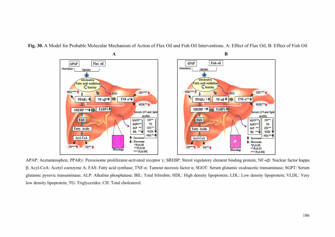
186
Fig. 30. A Model for Probable Molecular Mechanism of Action of Flax Oil and Fish Oil Interventions. A: Effect of Flax Oil, B: Effect of Fish Oil
A B
APAP: Acetamniophen; PPARγ: Peroxisome proliferator-activated receptor γ; SREBP: Sterol regulatory element binding protein; NF-κβ: Nuclear factor kappa
β; Acyl-CoA: Acetyl coenzyme A; FAS: Fatty acid synthase; TNF-α: Tumour necrosis factor α; SGOT: Serum glutamic oxaloacetic transaminase; SGPT: Serum
glutamic pyruvic transaminase; ALP: Alkaline phosphatase; BIL: Total bilirubin; HDL: High density lipoprotein; LDL: Low density lipoprotein; VLDL: Very
low density lipoprotein; TG: Triglycerides: CH: Total cholesterol.
187
6.6 Hepatoprotective Activity of Flax Oil and Fish Oil against Ethanol Induced
Hepatotoxicity
6.6.1 Biochemical Parameters
In the present study, flax oil intervention in ethanol treated rats produced
significant normalization of liver function. Phenolic components in flaxseed have
been shown to be effective in restoration of increased activities of liver function
enzymes (Kasote et al., 2012). Ismail et al. (2009) have reported normalization of
serum SGPT and SGOT levels after flax oil treatment in carbon tetrachloride
intoxicated rats. Shakir and Madhusudhan (2007) reported that rats treated with
flaxseed chutney showed decrease in the levels of liver markers such as SGOT,
SGPT, ALP and bilirubin in serum and liver homogenate against carbon tetrachloride
induced hepatotoxicity. In present study also, treatment of rats with flax oil (500
mg/kg) significantly reduced levels of SGOT, SGPT, ALP and bilirubin as compared
to ethanol treated group.
Raw flaxseeds as well as dietary flaxseeds as baked products, are known to
have hypolipidemic, hypoglycemic and hypocholesterolemic effects (Cunnane et al.,
1995; Shakir and Madhusudhan, 2007). Several animal and human intervention
studies indicate that flaxseed oil has beneficial effects on serum lipid profile
(Mahmud et al., 2004; Vijaimohan et al., 2006; Riediger et al., 2008; Bassett et al.,
2009; Newairy and Abdou, 2009; Kaithwas and Mujumdar, 2012). Administration of
15% flaxseed chutney or flaxseed oil to rats showed improvements of the lipid profile
(Increased level of HDL, decreased level of total cholesterol and LDL) in serum and
liver homogenates (Shakir and Madhusudhan, 2007). Ismail et al. (2009)
demonstrated that supplementation of flaxseed oil resulted in significant reduction in
serum total cholesterol, LDL and VLDL in rats treated with carbon tetrachloride.
High cholesterol diet-induced hypercholesterolemic rats exhibited lower serum total
cholesterol, triacylglycerols, LDL, VLDL, phospholipids, and increase in HDL after
flaxseed oil intervention (Hussein et al., 2014). In the present study also, flax oil
showed normolipidemic effects with decreased levels of total cholesterol, LDL,
triglycerides, VLDL and increased HDL in serum as well as liver homogenates.

188
Hepatoprotective and anti-oxidant activity of flaxseed has been demonstrated
through restoration of antioxidant enzymes in the liver of flaxseed pre-treated animals
challenged with carbon tetrachloride (Rajesha et al., 2006). Treatment of the hull
fraction of flaxseed also resulted in a significant increase in hepatic anti-oxidant
enzymes such as SOD, CAT, peroxidase as compared to carbon tetrachloride treated
group which is attributed to secoisolariciresinol diglucoside (SDG) content of the
flaxseed hull (Rajesha et al., 2010). Treatment of rats with flax lignans showed
decrease in TBARS and increase in the activities of glutathione S-transferase, SOD,
glutathione reductase and CAT as compared to lead acetate treated rats (Newairy and
Abdou, 2009). Several studies reported reduced levels of lipid peroxides (MDA) in
liver tissue in flaxseed oil treated animals (Lee and Prasad, 2003; Abdel-Moneim et
al., 2011). The present study also reports significant increase in activity of antioxidant
enzymes (SOD, CAT), increase in reduced glutathione and reduced levels of lipid
peroxides in flax oil treated animals as compared to ethanol treated group.
6.6.2 Histological Analysis
Several studies on liver damage due to ethanol induced hepatotoxicity have
already been discussed. In the present study, animals treated with flax oil showed near
normal hepatic architecture. There are several reports which indicate almost normal
liver architecture with minor histological anomalies after treatment with omega-3
fatty acids like flax oil or fish oil (Ismail et al., 2009; Meganathan et al., 2011; Kasote
et al., 2012; Khanchandani et al., 2014).
6.6.3 Gene Expression
Expression of Genes Involved in Lipid Metabolism
In the present study, PPARγ level was significantly increased in fish oil
treated animals and SREBP-1 was significantly decreased in flax oil and fish oil as
compared to ethanol treated animals. Omega-3 fatty acids are known to have their
effects on modulation of gene expression in cultured hepatocytes as well as in the
animal livers. The mRNA and protein expression of SREBP-1c was found to be
suppressed after treatment of the cells or animals with PUFAs (Sekiya et al., 2000;
Hannah et al., 2001; Yoshikawa et al., 2002; Nakatani et al., 2003). Fish oil has been
reported to increase the expression of PPARγ mRNA in mice liver (Yamazaki et al.,
189
2007). Fish oil also has effects on reduction of SREBP-1c and increase in the PPARα
expression in ethanol induced fatty liver (Wada et al., 2008).
Nanji et al. (2004) have reported that the expression of FABP decreases with
increase in the extent of fatty liver in ethanol intoxicated rats. They detected lowest
expression of L-FABP in the animals which developed severe fatty liver. Intestinal
expression of L-FABP was found to be upregulated in mice treated with sunflower oil
which was shown to be due to linoleic acid in sunflower oil (Poirier et al., 1997). In
the present study also, near normal liver function and histology was associated with
flax oil treatment which showed increase in the level of FABP1 expression as
compared to ethanol treated animals.
Expression of Genes Involved in Inflammation
Fish oil supplementation in healthy human volunteers lead to decreased
production of inflammatory cytokines (like TNF-α, IL-1 and IL-6) by mononuclear
cells (Caughey et al., 1996; Baumann et al., 1999; Trebble et al., 2003). Baumann et
al. (1999) have exclusively demonstrated that only omega-3 and not omega-6 and
omega-9 fatty acids, can significantly alter the expression levels of the genes involved
in inflammation and atherosclerosis. Treatment of mice with omega-3 fatty acids
reduced serum alanine aminotransferase (SGPT) levels and decreased inflammatory
response with decreased plasma TNF-α levels and this treatment also led to reduced
hepatic expression of TNF-α, IL-1β, IFN-γ and IL-6 as compared to
lipopolysaccharide/D-galactosamine-induced hepatitis model (Schmocker et al.,
2007). In the present study, expression of NF-κβ and TNF-α was significantly
decreased in flax oil treated animals as compared to alcohol treated animals.
Based on the results obtained from biochemical, histological and gene
modulation studies in animals receiving interventions of polyunsatured fatty acids
(flax oil and fish oil), hepatoprotective activity of flax oil was found to be better than
that of fish oil, in ethanol induced hepatotoxicity.
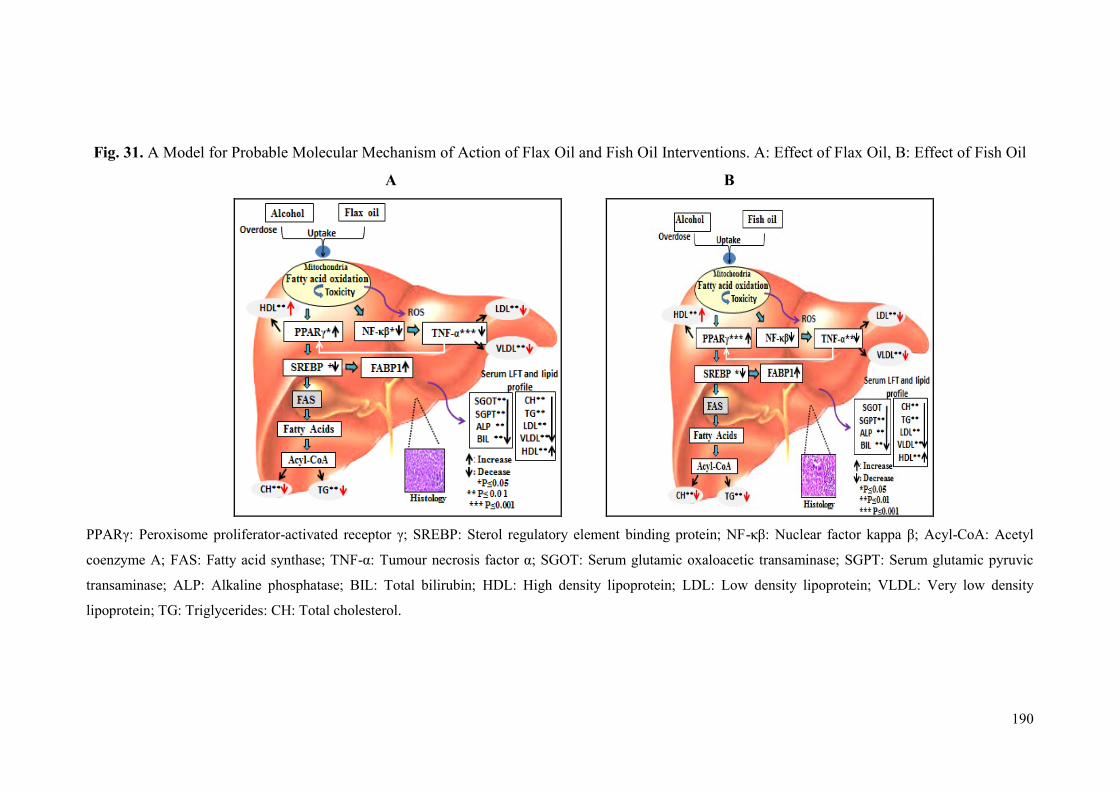
190
Fig. 31. A Model for Probable Molecular Mechanism of Action of Flax Oil and Fish Oil Interventions. A: Effect of Flax Oil, B: Effect of Fish Oil
A B
PPARγ: Peroxisome proliferator-activated receptor γ; SREBP: Sterol regulatory element binding protein; NF-κβ: Nuclear factor kappa β; Acyl-CoA: Acetyl
coenzyme A; FAS: Fatty acid synthase; TNF-α: Tumour necrosis factor α; SGOT: Serum glutamic oxaloacetic transaminase; SGPT: Serum glutamic pyruvic
transaminase; ALP: Alkaline phosphatase; BIL: Total bilirubin; HDL: High density lipoprotein; LDL: Low density lipoprotein; VLDL: Very low density
lipoprotein; TG: Triglycerides: CH: Total cholesterol.
191
6.7 Protective and Corrective Effects of Combination of Neem-giloe Satwa and
Fish Oil against Acetaminophen Induced Hepatotoxicity
6.7.1 Biochemical Parameters
Hepatoprotective effects of Neem-giloe satwa and fish oil were separately
evaluated against acetaminophen induced hepatotoxicity. The studies revealed that the
comparative hepatoprotective activity of Neem-giloe satwa and fish oil was higher
than other two satwa (T. cordifolia and T. sinensis) and flax oil, respectively. The
present study was carried out to evaluate protective and corrective effects of
combination of Neem-giloe satwa and fish oil against acetaminophen induced
hepatotoxicity.
Herbal medicines and nutraceutical supplements have been shown to have
preventive role in several diseases. Several medicinal herbs with hepatoprotective
properties have been reported in literature, e.g. Allium sativum (Garlic) (Ajayi et al.,
2009), Silybum marianum (Milk Thistle) (Freitag et al., 2015), Glycyrhizza glabra
(Licorice Root) (Sharma and Agrawal, 2014), Curcuma longa (Turmeric root) (Bae et
al., 2006) and Neem-guduchi (Chavan et al., 2013a; Nagarkar et al., 2013), Tinospora
cordifolia (Bishayi et al., 2002). In Asian countries, several studies revealed that
many dietary food items and supplements possess hepatoprotective activity (Shukla
and Kumar, 2013). There are a number of different food items and supplements e.g.
hepatoprotective spices like Turmeric (Luper, 1999), Coriander (Samojlik et al.,
2010), Garlic (Ajai et al., 2009), Red chili (Kim et al., 2005); hepatoprotective fruits
like Grapes (Carbo et al., 1999), Custard apple (Chavan et al., 2011), Apple (Miura et
al., 2007), Pomegranate (Toklu et al., 2007); hepatoprotective vegetables and grains
like Carrot (Bishayee et al., 1995), Sweet corn (Guo et al., 2009), Soy (Hu et al.,
2004), hepatoprotective drinks like Green tea (Luper, 1999), Coffee (Wang et al.,
2009); hepatoprotective omega-3 fatty acids like flax oil (Shakir and Madhusudhan,
2007; Ismail et al., 2009; Kasote et al., 2012) and fish oil (Meganathan et al., 2011;
Asaad and Aziz, 2012; Khanchandani et al., 2014). Food ingredients contain several
phytochemicals beneficial in liver injuries and possess potential to prevent or reverse
different kinds of liver injuries. There are several studies reported on either herbal or
nutritional supplements (Omega-3 fatty acid) against different hepatotoxicants
192
(Bishayi et al., 2002; Khanchandani et al., 2014; Sharma and Agrawal, 2014; Freitag
et al., 2015). A recent report shows that treatment of rats with a combination of
Glycyrrhizin and omega-3 fatty acid (fish oil) leads to significant decrease in SGOT
and SGPT activities as compared to thioacetamide treated rats (Abo El-Magd et al.,
2015). In the current study, protective and corrective treatment of rats with a
combination of Neem-giloe satwa and fish oil showed significant decrease in the
levels of SGOT, SGPT, ALP and bilirubin as compared to acetaminophen treated
groups. In a single-blind, placebo-controlled, crossover study of combinatorial
treatment of fish oil and garlic showed decrease in cholesterol, triglyceride, and LDL
levels, as well as increase in HDL as compared to placebo treated group (Morcos,
1997). In present study also, protective and corrective treatment of combination of
Neem-giloe satwa and fish oil showed improvement in the lipid profile (decrease in
cholesterol, LDL, VLDL and triglyceride and increase in HDL) in serum and liver
homogenates. Total protein of liver homogenate was significantly improved in
corrective effect of the combination as compared to acetaminophen treated group.
A combination of Glycyrrhizin and fish oil to thioacetamide intoxicated rats
exhibited strong antioxidant activity with significant increase in hepatic MDA levels
(Abo El-Magd et al., 2015). In the present study, combination of Neem-giloe satwa
and fish oil was also found to have antioxidant effects and it decreased the extent of
lipid peroxidation and improved the levels of SOD, reduced glutathione and CAT as
compared to acetaminophen treated group.
6.7.2 Histological Analysis
In the present study, sections of liver from the animals treated with a single
dose of acetaminophen on 8th
day showed swelling and degenerative changes in
hepatocytes with compressed sinusoids. The protective treatment of combination of
Neem-giloe satwa and fish oil showed improvement in hepatic architecture which still
showed some hepatocytes with degenerative changes. The corrective treatment of
combination of Neem-giloe satwa and fish oil showed near normal liver histology as
compared to that in the animals treated with acetaminophen which exhibited
congestion of central vein and swollen hepatocytes with ballooning degeneration.
193
6.7.3 Gene Expression
Expression of Genes Involved in Lipid Metabolism
Thorough literature search indicated lack of references on expression studies
of genes from lipid metabolism, in acetaminophen induced hepatotoxicity and
combination of herbal and nutraceutical interventions.
In the present study, the animals with the intervention of combination of satwa
and fish oil, exhibited significant up-regulation of PPARγ and FABP1 while
expression of SREBP-1 was down-regulated.
Expression of Genes Involved in Inflammation
A recent report by Abo El-Magd et al. (2015) has reported increase in the
expression of NF-κβ in hepatic tissues of thioacetamide treated rats which was
reduced after treatment with a combination of glycyrrhizin and fish oil (Abo El-Magd
et al., 2015). In the present study also, protective and corrective treatment of
combination of interventions showed significant down-regulation of NF-κβ and TNF-
α.

194
Fig. 32. A Model for Probable Molecular Mechanism of Action of Neem-giloe and Fish oil. A: Protective Effect of Combination of Neem-giloe
Satwa, B: Corrective Effect of Combination of Neem-giloe Satwa
A B
APAP: Acetamniophen; PPARγ: Peroxisome proliferator-activated receptor γ; SREBP: Sterol regulatory element binding protein; NF-κβ: Nuclear factor kappa
β; Acyl-CoA: Acetyl coenzyme A; FAS: Fatty acid synthase; TNFα: Tumour necrosis factor α; SGOT: Serum glutamic oxaloacetic transaminase; SGPT: Serum
glutamic pyruvic transaminase; ALP: Alkaline phosphatase; BIL: Total bilirubin; HDL: High density lipoprotein; LDL: Low density lipoprotein; VLDL: Very
low density lipoprotein; TG: Triglycerides: CH: Total cholesterol.

195
6.8 Prophylactic Effect of Combination of Tinospora sinensis Satwa and Flax Oil
against Ethanol Induced Hepatotoxicity
6.8.1 Biochemical Parameters
Hepatoprotective effects of T. sinensis satwa and flax oil were separately
evaluated against ethanol induced hepatotoxicity. The studies revealed that the
comparative hepatoprotective activity of T. sinensis satwa and flax oil was higher than
other two satwa (T. cordifolia and Neem-giloe) and fish oil, respectively. The present
study was carried out to study the prophylactic effects of combination of T. sinensis
satwa and flax oil against ethanol induced hepatotoxicity. In the present study,
combination of T. sinensis satwa and flax oil showed significant reduction of SGOT,
SGPT, ALP and bilirubin as compared to ethanol treated group. The treatment also
normalized serum and hepatic lipid profile and improved hepatic antioxidant status. A
report by Wahba and Ibrahim (2013) showed improvement in the serum levels of liver
function markers, lipid profile as well as increase in tissue antioxidant enzymes after
treatment with flax oil in combination with vitamin E in potassium bromate treated
rats.
6.8.2 Histological Analysis
Effects of ethanol treatment and interventions on the hepatic architecture are
described in the results section. There are no previous reports describing hepatic
architecture in ethanol intoxicated rats treated with combination of herbal and
nutraceutical interventions.
6.8.3 Gene Expression
Expression of Genes Involved in Lipid Metabolism
PPARγ level was significantly up regulated and SREBP-1 level was found
normalized in treatment with combination of T. sinensis satwa and flax oil. In
prophylactic effect, FABP1 was down regulated in ethanol treated group while it was
up regulated in treatment with combination of T. sinensis satwa and flax oil.
There are no previous reports on the gene expression studies of combination
of interventions in ethanol induced hepatotoxicity. In the present studies, PPARγ was

196
significantly up-regulated while NF-κβ was shown to be significantly down-regulated
in intervention group.
Expression of Genes Involved in Inflammation
In the present study, NF-κβ and TNF-α expressions were up regulated in
ethanol treated animals. NF-κβ gene expression was found significantly decreased in
the animals treated with a combination of T. sinensis satwa and flax oil. TNF-α
expression was significantly decreased in healthy control and no significant change
was observed due to treatment of combination of T. sinensis satwa and flax oil.
Treatment of animals with combinations of Neem-giloe and fish oil and T.
sinensis and flax oil in acetaminophen and ethanol induced hepatotoxicity
respectively, showed comparatively better results than intervention of any one of
them. So combination of herbal and nutritional intervention has shown beneficial
effects on liver disease.

197
Fig. 33. A Model for Probable Mechanism of Action of T. sinensis Satwa and Flax oil
A
PPARγ: Peroxisome proliferator-activated receptor γ; SREBP: Sterol regulatory element
binding protein; NF-κβ: Nuclear factor kappa β; Acyl-CoA: Acetyl coenzyme A; FAS: Fatty
acid synthase; TNF-α: Tumour necrosis factor α; SGOT: Serum glutamic oxaloacetic
transaminase; SGPT: Serum glutamic pyruvic transaminase; ALP: Alkaline phosphatase;
BIL: Total bilirubin; HDL: High density lipoprotein; LDL: Low density lipoprotein; VLDL:
Very low density lipoprotein; TG: Triglycerides: CH: Total cholesterol.

SUMMARY AND Conclusions
Awards
Prophylactic effect of combination of herbal and nutritional interventions against
alcohol induced hepatotoxicity in rats
Hepatoprotective Effect Of Polyunsaturated Fatty Acids And Satwa From Three
Tinospora Species Exhibits Differential Hepatoprotective Activity Against Repeated
Alcohol Dosing In Rats
Hepatoprotective Effect Of Polyunsaturated Fatty Acids Against Repeated Subacute
Alcohol Dosing In Rats
Comparative Hepatoprotective Potential Of T.cordifolia, T.sinensis and Neem-Giloe
(Tinospora Cordifolia Growing On Azadirachta Indica (Neem)) Against
Paracetamol Intoxication In Rats
Protein synthesis
Second prize in oral
Presentation Award
Best Poster Presentation
Award

198
Liver diseases have serious adverse effects on health. Allopathic medicines
prescribed for liver diseases have several side effects and hence modern medicine is
still short of reliable and effective drugs for protection and repair of liver. There are
many herbs being used to treat various liver disorders. In India, traditional medicine
system and ethno-medical practices rely on several medicinal plants and their
formulations to treat liver disorders.
The objective of the present study was to assess hepatoprotective activity of
ayurvedic formulation and nutritional supplements that can potentially be used as
hepatoprotective agent having less/no side effects. Based on the literature review,
medicinal values and availability, three different forms of Tinospora (satwa of stem)
and omega-3 fatty acids (Flax oil and fish oil) were selected to evaluate their
hepatoprotective potential.
The current section summarizes organoleptic characteristics and nutritional
analysis of satwa forms three Tinospora forms and also hepatoprotective activity of
satwa from three forms of Tinospora as well as that of flax oil and fish oil against
acetaminophen and alcohol induced hepatotoxicity.
Methodology
The different organoleptic characteristics and nutritional parameters like
protein, carbohydrates, lipid, starch, crude fiber, and ash were analyzed from satwa,
from three different forms of Tinospora. The hepatoprotective activity of satwa from
three different forms of Tinospora and omega-3 fatty acids (Flax oil and fish oil) was
studied by employing acetaminophen and alcohol induced hepatotoxicity in animal
models. The biochemical parameters like serum SGPT, SGOT, ALP, bilirubin, total
cholesterol, triglycerides, HDL cholesterol and LDL cholesterol, Hepatic antioxidant
parameters like catalase, SOD, MDA, GSH, total protein and functional parameters
like histopathology of liver, were studied. Further study on modulation of expression
of the genes involved in lipid metabolism and inflammation was also carried out. As
per the current study, maximum yield of satwa was obtained in T. sinensis and Neem-
giloe. T. sinensis and Neem-giloe are rich sources of nutritional contents. The contents
of lipid, ash, and carbohydrates are found to be greater in Neem-giloe than
199
T. cordifolia and T. sinensis. Protein, starch and crude fiber content is found higher in
T. sinensis as compared with T. cordifolia and Neem-giloe.
Results of this study reported significant increase in serum levels of liver
function tests (SGOT, SGPT, ALP, and bilirubin) and lipid profile (Total cholesterol,
LDL cholesterol, VLDL, Triglycerides and decrease in HDL cholesterol levels) in the
groups treated with acetaminophen and alcohol indicating induction of hepatotoxicity.
It further indicated abnormal integrity of hepatocytes, resulting into liver damage.
This resulted into significant elevation of lipid peroxidation with simultaneous decline
in GSH, CAT, SOD and total protein levels in liver tissue.
Among the three satwa, Neem-giloe satwa and T. sinensis satwa exhibited
higher hepatoprotective/hepatoregenerative activity in acetaminophen and alcohol
induced liver toxicity. The intervention of fish oil and flax oil proved beneficial in
acetaminophen and alcohol induced liver damage. The combination of Neem-giloe
satwa and fish oil in acetaminophen induced liver toxicity and combination of T.
sinensis satwa and flax oil for alcohol induced liver damage indicated improvement
over their individual activities. These treatments showed significant decrease in serum
levels of SGOT, SGPT, ALP, bilirubin and lipid profile (Total cholesterol, LDL
cholesterol, VLDL, Triglycerides and increase in HDL cholesterol levels) and also
exhibited reduction in lipid peroxidation and significant increase in levels of GSH,
CAT, SOD and total protein as compared with acetaminophen and alcohol treated
groups respectively. Histopathological observations showed that the above treatments
also improved hepatic architecture when compared with acetaminophen and alcohol
treated groups.
Expressions of FABP1 and PPARγ were down regulated while NF-κβ, TNF-α
and SREBP1 were upregulated in acetaminophen and alcohol treated groups.
Treatment with individual satwa and omega-3 fatty acids as well as their
combinations (as indicated above) lead to downregulation of the exression of NF-κβ,
TNF-α and SREBP1 and upregulation of FABP1 and PPARγ.
Herbal and nutraceutical intervations showed beneficial effects at biochemical,
histological and molecular levels in chemical induced hepatotoxicity. The
hepatoprotective effect of T. sinensis satwa, Neem-giloe satwa, fish oil and flax oil
200
may be due individual phytochemicals and nutritional constituents or their synergistic
action.
Conclusions
The present study clearly establishes the hepatoprotective/hepatoregenerative
effects of Neem-giloe and T. sinensis (Herbal interventions) and flax and fish oil
(Nutraceutical interventions) on hepatic disorders caused by repeated acetaminophen
and alcohol dosing in rats.
Neem-giloe satwa showed hepatoprotective effect against acetaminophen and
Tinospora sinensis satwa showed hepatoprotective effect against alcohol
induced hepatotoxicity.
Fish oil and flax oil showed hepatoprotective effect against acetaminophen
and alcohol induced hepatotoxicity respectively.
Combination of Neem-giloe and fish oil showed hepatoprotective effect
against acetaminophen and combination of T. sinensis and flax oil showed
hepatoprotective effect against alcohol induced hepatotoxicity.
The herbal medicines and omega-3 fatty acid supplements and their combination
counter the liver toxicity at the biochemical, histological and molecular levels by
normalizing oxidative stress and inflammation. Their action at cellular level could be
seen in the form of repair and regeneration of hepatocytes. The combination reported
in the present study is worth further investigation, especially in clinical cases of liver
damage due to long term alcohol abuse.

FUTURE Direction
Awards
Prophylactic effect of combination of herbal and nutritional interventions against
alcohol induced hepatotoxicity in rats
Hepatoprotective Effect Of Polyunsaturated Fatty Acids And Satwa From Three
Tinospora Species Exhibits Differential Hepatoprotective Activity Against Repeated
Alcohol Dosing In Rats
Hepatoprotective Effect Of Polyunsaturated Fatty Acids Against Repeated Subacute
Alcohol Dosing In Rats
Comparative Hepatoprotective Potential Of T.cordifolia, T.sinensis and Neem-Giloe
(Tinospora Cordifolia Growing On Azadirachta Indica (Neem)) Against
Paracetamol Intoxication In Rats
Protein synthesis
Second prize in oral
Presentation Award
Best Poster Presentation
Award
201
Future Direction
The hepatoprotective/hepatoregenerative effects of Neem-giloe satwa, T.
sinensis satwa and omega-3 fatty acids (Flax oil and fish oil) are worth further
investigations in humans through well-designed clinical trials to ascertain their
efficacy in clinical liver damage cases of varying severity.
CHAPTER 8
REFERE

BIBLIOGRAPHY
TER 8
Awards
Prophylactic effect of combination of herbal and nutritional interventions against
alcohol induced hepatotoxicity in rats
Hepatoprotective Effect Of Polyunsaturated Fatty Acids And Satwa From Three
Tinospora Species Exhibits Differential Hepatoprotective Activity Against Repeated
Alcohol Dosing In Rats
Hepatoprotective Effect Of Polyunsaturated Fatty Acids Against Repeated Subacute
Alcohol Dosing In Rats
Comparative Hepatoprotective Potential Of
Second prize in oral
Presentation Award
Best Poster Presentation
Award
Awards
Second prize in oral
Presentation Award
Best Poster Presentation
Award
Awards
Prophylactic effect of combination of herbal and nutritional interventions against
alcohol induced hepatotoxicity in rats
Hepatoprotective Effect Of Polyunsaturated Fatty Acids And Satwa From Three
Tinospora Species Exhibits Differential Hepatoprotective Activity Against Repeated
Alcohol Dosing In Rats
Hepatoprotective Effect Of Polyunsaturated Fatty Acids Against Repeated Subacute
Alcohol Dosing In Rats
Comparative Hepatoprotective Potential Of T.cordifolia, T.sinensis and Neem-Giloe
(Tinospora Cordifolia Growing On Azadirachta Indica (Neem)) Against
Paracetamol Intoxication In Rats
Protein synthesis
Second prize in oral
Presentation Award
Best Poster Presentation
Award
202
Abay S.M. and Amelo W. (2010) “Assessment of Self-Medication Practices Among
Medical, Pharmacy, and Health Science Students in Gondar University,
Ethiopia” Journal of Young Pharmacists 2(3): 306-310.
Abd Ellah M.E. (2011) “The Role of Liver Biopsy in Detection of Hepatic Oxidative
Stress” Veterinary Medicine International 613602: 7.
Abd Ellah M.R., Okada K., Goryo M., Oishi A., Yasuda J. (2009) “Superoxide
Dismutase Activity as a Measure of Hepatic Oxidative Stress in Cattle
Following Ethionine Administration” Veterinary Journal 182(2): 336-341.
Abdel-Dayem M.A., Elmarakby A.A., Abdel-Aziz A.A., Pye C., Said S.A., El-
Mowafy A.M. (2014) “Valproate-Induced Liver Injury: Modulation by the
Omega-3 Fatty Acid DHA Proposes a Novel Anticonvulsant Regimen” Drugs
in R & D 14: 85-94.
Abd Ellah M.R. (2010) “Involvement of Free Radicals in Animal Diseases”
Comparative Clinical Pathology 19(6): 615-619.
Abdel-Moneim A.E., Dkhil M.A., Al-Quraishy S. (2011) “The Potential Role of
Flaxseed Oil on Lead Acetateinduced Kidney Injure in Adult Male Albino
Rats” African Journal of Biotechnology 10(8): 1436-1451.
Abo El-Magd N.F.A., El-Karef A., El-Shishtawy M.M., El-Magd L.A.E. (2015)
“Hepatoprotective Effects of Glycyrrhizin and Omega-3 Fatty Acids on
Nuclear Factor-Kappaβ Pathway in Thioacetamide-Induced Fibrosis in Rats”
Egyptian Journal of Basic and Applied Sciences 2(2): 265-274.
Achliya G.S., Kot N.R., Agale S.G., Wadodkar A.K. (2003) “Hepatoprotective
Activity of Panchagavya Ghrita against Carbontetrachloride Induced
Hepatotoxicity in Rats” Indian Journal of Pharmacology 35(5): 308-311.
Adamska T., Młynarczyk W., Jodynis-Liebert J., Bylka W. Matławska I. (2003)
“Hepatoprotective Effect of the Extract and Isocytisoside from Aquilegia
vulgaris Phytotherapy Research 17(6): 691-696.
Addagudi S., Subash V., Manohar K. (2013) “Alcoholic Liver Disease a Minireveiw”
International Journal of Experimentla Pharmacology 3(2): 61-69.
Adewale O.B., Adekeye A.O., Akintayo C.O., Onikann A., Saheed S. (2014).
“Carbon Tetrachloride (CCL4) Induced Heaptic Damage in Experimental
Sprague Dawley Rats: Antioxidant Potential of Xylopia aethiopica” Journal of
Phytopharmacology 3(2): 118-113.
Adhvaryu M.R., Reddy N., Parabia M.H. (2007) “Effects of Four Indian Medicinal
Awards
Prophylactic effect of combination of herbal and nutritional interventions against
alcohol induced hepatotoxicity in rats
Hepatoprotective Effect Of Polyunsaturated Fatty Acids And Satwa From Three
Tinospora Species Exhibits Differential Hepatoprotective Activity Against Repeated
Alcohol Dosing In Rats
Hepatoprotective Effect Of Polyunsaturated Fatty Acids Against Repeated Subacute
Alcohol Dosing In Rats
Comparative Hepatoprotective Potential Of T.cordifolia, T.sinensis and Neem-Giloe
(Tinospora Cordifolia Growing On Azadirachta Indica (Neem)) Against
Paracetamol Intoxication In Rats
Protein synthesis
Second prize in oral
Presentation Award
Best Poster Presentation
Award
204
Herbs on Isoniazid, Rifampicin and Pyrazinamide-Induced Hepatic Injury and
Immunosuppression in Guinea Pigs” World Journal of Gastroenterology
13(23): 3199-3205.
Adikwu E. and Deo O. (2013) “Hepatoprotective Effect of Vitamin C (Ascorbic
Acid)” Journal of Pharmacology & Pharmacy 4(1): 84-92.
Agrawal S.S., Naqvi S., Gupta S.K., Srivastava S. (2012) “Prevention and
Management of Diabetic Retinopathy in STZ Diabetic Rats by Tinospora
cordifolia and its Molecular Mechanisms” Toxicology 50(9): 3126-3132.
Aguilera C.M., Mesa M.D., Ramirez-Tortosa M.C., Quiles J.L., Gil A. (2003) “Virgin
Olive and Fish Oils Enhance the Hepatic Antioxidant Defence System in
Atherosclerotic Rabbits” Clinical Nutrition 22(4): 379-384.
Ahmad R., Srivastava A. N., Khan M.A. (2015) “Evaluation of in Vitro Anticancer
Activity of Stem of Tinospora Cordifolia against Human Breast Cancer and
Vero Cell Lines” Journal of Medicinal Plants Studies 3(4): 33-37.
Ajayi G.O., Adeniyi T.T., Babayemi D.O. (2009) “Hepatoprotective and Some
Haematological Aspects of Allium Sativum and Vitamin C in Lead-Exposed
Wistar Rats” International Journal of Medicine and Medical Sciences 1(3): 64-
67.
Ajibade V.A. and Fagbohun E.D. (2010) “Phytochemical, Proximate Analysis and
Antimicrobial Actiivties of Methanolic Crude Extract of Tyliphora Glauca
(Bullock)” Journal of Pharmaceutical and Biomedical Sciences 1(1): 7-12.
Akila M. and Prasanna G. (2014) “Hepatoprotective Effect of Indigofera Linnael Ali.
On Carbon Tetrachloride Induced Wistar Albino Rats” International Research
Journal of Pharmacy 5(5): 392-395.
Alam N., Amin R., Khan A., Ara I., Shim M.J., Lee M.W., Lee T.S., (2008)
“Nutritional Analysis of Cultivated Mushrooms in Bangladesh-Pleurotus
ostreatus, Pleurotus sajor-caju, Pleurotus florida and Calocybe indica”
Journal of Mycobiology 36(4): 228-232.
Al-Assaf A.H. (2013) “Preventive Effect of Corosolic Acid on Lipid Profile agains
Carbon Tetrachloride-Induced Hepatotoxic Rats” Pakistan Journal of Nutrition
12 (8): 748-752.
Albano E. (2006) “Alcohol, Oxidative Stress and Free Radical Damage” Proceedings
of the Nutrition Society 65(3): 278-290.
Al-Fartosi K.G., Khuon O.S., Al-Tae H. I. (2011) “Protective Role of Camel's Milk
205
against Paracetamol Induced Hepatotoxicity in Male Rats” International
Journal of Research in Pharmaceutical and Biomedical Sciences 2(4): 1795-
1799.
Ali N.M., Yusof H.M., Long K., Yeap S.K., Ho W.Y., Beh B.K., Koh S.P., Abdullah
P.M., and Alitheen N.B. (2013) “Antioxidant and Hepatoprotective Effect of
Aqueous Extract of Germinated and Fermented Mung Bean on Ethanol-
Mediated Liver Damage” BioMed Research International 693613: 9.
Al-Jumaily E.F. and AL-Azawi A.H. (2013) “Hepatoprotective Activity of Lignan
Compound from Flaxseed (Linumusitatissimum L.) Against Acetaminophen
induced Hepatotoxicity in Rabbits” World Journal of Pharmacy and
Pharmaceutical Sciences 3(1): 56-72.
Al-Jumaily E.F., Al-Amiry M.H.A., Assad J.A. (2012) “Hepatotoxic Activity of
Essential Oil from Nutmeg (Myristica fragrans) against Tetrachloride-Induced
Hepatic Damage in Mice” Journal of Pharmacy and Biological Sciences 2(6):
1-8.
Allain C.C., Poon L.S., Chan C.S., Richmond W., Fu P.C. (1974) “Enzymatic
Determination of Total Serum Cholesterol” Clinical Chemistry 20(4): 470-
475.
Alwayn I.P., Gura K., Nose V., Zausche B., Javid P., Garza J., Verbesey J., Voss S.,
Ollero M., Andersson C., Bistrian B., Folkman J., Puder M. (2005) “Omega-3
Fatty Acid Supplementation Prevents Hepatic Steatosis in A Murine Model of
Nonalcoholic Fatty Liver Disease” Journal of Pediatric Research 57(3): 445-
452.
American Cancer Society. (2015) “Dietary Supplements: What Is Safe?” American
Cancer Society.
Ananthi T. and R. Anuradha R. (2015) “Hepatoprotective Activity of Michelia
champaca L. against Carbon Tetrachloride Induced Hepatic Injury in Rats”
Journal of Chemical and Pharmaceutical Research 7(9): 270-274.
Andaloussi A B., Martineau L.C., Vallerand D., Haddad Y., Afshar A., Settaf A.,
Haddad P.S. (2010) “Multiple Molecular Targets Underlie the Antidiabetic
Effect of Nigella sativa Seed Extract in Skeletal Muscle, Adipocyte and Liver
Cells Journal of Diabetes, Obesity and Metabolism 12(2): 148-157.
Anker A.L. and Smilkstein M.J. (1994) “Acetaminophen Concepts and
Controversies” Journal of Emergency Medicine Clinics of North America
206
12(2): 335-349.
Anonymous. (2003b) “Quality Standards of Indian Medicinal Plants” Indian Council
of Medical Research, New Delhi, Volume. 1: 212-218.
Anonymous. (1987) “Pharmacopeial Standards for Ayurvedic Formulations, Central
Council of Research in Ayurveda and Siddha” Government of India, Ministry
of Health & Family Welfare, The Controller of Publications, New Delhi, pp
189.
Anonymous. (2003a) “The Ayurvedic Formulary of India” Ministry of Health and
Family Welfare, Government of India, New Delhi, Second Edition, Part-1 pp
560.
Aristatile B., Al-Assaf A.H., Pugalendi K.V. (2013) “Carvacrol Suppresses the
Expression of Inflammatory Marker Genes in D-Galactosamine-Hepatotoxic
Rats” Asian Pacific Journal of Tropical Medicine 6(3): 205-211.
Arnaiz S.L., Llesuy S., Cutrín J.C Boveris A. (1995) “Oxidative Stress by Acute
Acetaminophen Administration in Mouse Liver” Journal of Free Radical
Biology & Medicine 19(3): 303-310.
Arote S., Gupta S.K., Lodha P., Yadav P., Lodha M. (2014) “Hepatoprotective
Activity of Herbal Formulation against Paracetamol-Induced Hepatotoxicity in
Rats” Indo American Journal of Pharmaceutical Research 4(1): 451-456.
Arsul V.A., Ganjiwale R.O., Yeole P.G. (2010) “Phytochemical and Pharmacological
Standardization of Polyherbal Tablets for Hepatoprotective Activity against
Carbon Tetrachloride Induced Hepatotoxicity” International Journal of
Pharmaceutical Sciences 2(4): 265-268.
Arsul V.A., Wagh S.R., Rahul V. (2011) “Hepatoprotective Activity of Livergen, A
Polyherbal Formulation against Carbon Tetrachloride Induced Hepatotoxicity
in Rats” International Journal of Pharmacy and Pharmaceutical 3(3): 228-231.
Arteel G.E. (2003) "Oxidants and Antioxidants in Alcohol-Induced Liver Disease"
Journal of Gastroenterology 124(3): 778-790.
Arulkumaran K.S., Rajasekaran A., Ramasamy A., Jegadee-san M., Kavimani S.,
Somasundaram A. (2009) “Cassia roxburghii Seeds Protect Liver against
Toxic Effects of Ethanol and Carbontetrachloride in Rats” International
Journal of PharmTech Research 1(2): 273-246.
Arun K. and Balasubramanian U. (2011) “Comapartive Study on Hepatoprotective
Activity of Phyllanthus amarus and Eclipta prostrata against Alcohol Induced
207
In Albino Rats” International Journal of Environmental Sciences 2(1): 361-
379.
Asaad H.R. and Aziz F.M. (2012) “Protective Role of Omega-3 Fish Oil against the
Toxicity of Ifosfamide in Male Rats” Jordan Journal of Biological Sciences
5(1): 37-46.
Asha V.V., Akhilab S., Willsa P.J., Subramoniamc A. (2004) “Further studies on the
Antihepatotoxic Activity of Phyllanthus maderaspatensis Linn” 92(1): 67-70.
Ashok B. K., Ravishankar B., Prajapati P.K., Bhat S.D. (2010) “Antipyretic activity
of Guduchi ghrita Formulations in Albino Rats” Journal of Pharmacological
Research 31(3): 367-370.
Ashoush I.S., El-Batawy O.I., El-Shourbagy G.A. (2013) “Antioxidant Activity and
Hepatoprotective Effect of Pomegranate Peel and Whey Powders in Rats”
Journal of Annals of Agricultural Science 58(1): 27-32.
Askay S.W., Bombardier C.H., Patterson D.R. (2009) “Effect of Acute and Chronic
Alcohol Abuse on Pain Management in a Trauma Center” "Expert Review of
Neurotherapeutics 9(2): 271-277
Awodele O., Olayemi S.O., Alimba C.G., Egbejiogu C., Akintonwa A. (2010)
“Protective Effect of Vitamin C and or Vitamin E on Micronuclei Induction by
Rifampicin in Mice” Tanzania Journal of Health Research 12(2): 1-7.
Ayonrinde O.T., Phelps G.J., Hurley J.C., Ayonrinde O.A. (2005) “Paracetamol
Overdose and Hepatotoxicity at A Regional Australian Hospital: A 4-Year
Experience” Internal Medicine Journal 35(11): 655-660.
Ayurvedic Pharmacopoeia of India. (2008) “Ministry of Health and Family Welfare”
Department of Ayurveda, Yoga & Naturopathy, Unani, Siddha and
Homoeopathy, Government of India, Delhi, First Edition, Volume II, Part II,
pp 153-272.
Ayyavu M., Jabbith S., Thangadurai D., Muralidhara Rao D. (2009) “Protective
Effect of Indian Honey on Acetaminophen Induced Oxidative Stress and Liver
Toxicity in Rat” Journal of Biology 64(6): 1225-1231.
Babalola O.O., Ojo O.E., Oloyede F.A. (2011) “Hepatoprotective Activity of
Aqueous Extract of the Leaves of Hyptis suaveolens (l.) Poit on
Acetaminophen Induced Hepatotoxicity in Rabbits” Research Journal of
Chemical Sciences 1(7): 85-88.
Babcock T., Helton W.S., Espat N.J. (2000) “Eicosapentaenoic Acid (EPA): An Anti-
208
Inflammatory Omega-3 Fat with Potential Clinical Applications” Journal of
Nutrition 16(11-12): 1116-1118.
Bae M.K., Kim S.H., Jeong J.W., Lee Y.M., Kim H.S., Kim S.R., Yun I., Bae
S.K., Kim K.W. (2006) “Curcumin Inhibits Hypoxia Induced Angiogenesis by
Bafna P.A. and Balaraman R. (2013) ‘Protective Effect of DHC-1, A Polyherbal
Formulation, against CCl4 Induced Liver Damage” Hygeia: Journal for Drugs
and Medicines 5(1): 10-18.
Bahar E., Ara J., Hossain M., Nath B., Runi N. (2013) “Cytotoxic (In-Vitro) Effect of
Methanol and Petroleum Ether Extracts of the Aerva lanata” Journal of
Pharmacognosy and Phytochemistry 2(1): 92-100.
Bhanwra, S.J., Singh J., Khosla P.(2000) “Effect Of Azadirachta Indica (Neem) Leaf
Aqueous Extract on Paracetamol-Induced Liver Damage in Rats” Indian
Journal of Physiology Pharmacology 44 (1): 64-68
Downregulation of HIF-1” Oncology Report 15(6): 1557-1562.
Bai X., Qiu A., Guan J., Shi Z. (2007) “Antioxidant and Protective Effect of an
Oleanolic Acidenriched Extract of A. Deliciosa Root on Carbon Tetrachloride
Induced Rat Liver Injury” Asian Pacific Journal of Clinical Nutrition 16(1):
169-173.
Baichwal V.R. and Baeuerle P.A. (1997) “Apoptosis: Activate NF-κβ or Die?”
Journal of Current Biology 7(2): 94-96.
Baillie T.A. and Slatter J.G. (1991) “Glutathione: A Vehicle for the Transport of
Chemically Reactive Metabolites in Vitro” American Chemical Society
Publications 24(9): 264-270.
Balakrishanan N., Balasubaramaniam A., Sangameswaran B., Bhaskar V. H. (2011)
“Hepatoprotective Activity of Two Indian Medicinal Plants from Western
Ghats, Tamil Nadu” Journal of Natural Pharmaceuticals 2(2): 92-98.
Bang H.O. and Dyerberg J. (1972) “Plasma Lipids and Lipoproteins in Greenlandic
West Coast Eskimos” Journal of Acta Medica Scandinavica 192(1-2): 85-94.
Bartolone J.B., Birge R.B., Sparks K., Cohen S.D., Khairallah E.A. (1988)
“Immunochemical Analysis of Acetaminophen Covalent Binding to Proteins.
Partial Characterization of the Major Acetaminophen-Binding Liver Proteins”
Journal of Biochemical Pharmacology 37(24): 4763-4774.
Bartolone J.B., Cohen S.D., Khairallah E.A. (1989) “Immunohistochemical
Localization of Acetaminophen-Bound Liver Proteins” Fundamental and
209
Applied Toxicology 13(4): 859-862.
Bashandy S.A. and Alwasel S.H. (2011) “Carbon Tetrachloride-Induced
Hepatotoxicity and Nephrotoxicity in Rats: Protective Role of Vitamin C”
Journal of Pharmacology and Toxicology 6(3): 283-292.
Bassett C.M.C., Rodriguez-Leyva D., Pierce G.N. (2009) “Experimental and Clinical
Research Findings on the Cardiovascular Benefits of Consuming Flaxseed”
Journal of Applied Physiology, Nutrition, and Metabolism 34(5): 965-974.
Basu S., Haldar N., Bhattachary S., Biswas S. and Biswas M. (2012) “Hepato-
protective activity of Litchi Chinensis Laeves against Paracetamol Induced
Liver Damage in Rats” American–Eurasisn Journal of Scientific research 7(2):
77-81.
Baumann K.H., Hessel F., Larass I., Muller T., Angerer P., Kiefl R., Schacky C.V.
(1999) “Dietary α-3, α-6, and α-9 Unsaturated Fatty Acids and Growth Factor
and Cytokine Gene Expression in Unstimulated and Stimulated Monocytes a
Randomized Volunteer Study” Journal of Arteriosclerosis Thrombosis and
Vascular Biology 19(1): 59-66.
Bebnista M.J. And. Nowak J.Z. (2014) “Paracetamol: Mechanism of Action,
Applications and Safety Concern” Journal of Acta Poloniae Pharmaceutica -
Drug Research 71(1): 11-23.
Bedi O., Bijjem K.R.V., Kumar P., Gauttam V. (2016) “Herbal Induced
Hepatoprotection and Hepatotoxicity: A Critical Review” Indian Journal of
Physiological Pharmacology 60(1): 6-21.
Bedoucha M., Atzpodien E., Boelsterli U.A. (2001) “Diabetic KKAy Mice Exhibit
Increased Hepatic PPARGamma Gene Expression and Develop Hepatic
Steatosis upon Chronic Treatment with Antidiabetic Thiazolidinediones”
Journal of Hepatology 35(1): 17-23.
Beg A.A. and Baltimore D. (1996)” An Essential Role for NF-κβ in Preventing TNF-
Alpha Induced Cell Death” Journal of Science 274(5288): 782-784.
Beier J.I. and McClain C.J. (2010) “Mechanisms and Cell Signaling in Alcoholic
Liver Disease” The Journal of Biological Chemistry 391(11): 1249-1264.
Beier J.I., Arteel G.E., McClain C.J. (2011) “Advances in Alcoholic Liver Disease”
Current Gastroenterology Reports 13(1): 56-64.
Benegal V. (2005) “India: Alcohol and Public Health Addiction 100(8): 1051-1056.
Bernareggi A. (1998) “Clinical Pharmacokinetics of Nimesulide” Journal of Clinical
210
Pharmacokinetics 35(4): 247-274.
Bessems J.G. and Vermeulen N.P. (2001) “Paracetamol (Acetaminophen)-Induced
Toxicity: Molecular and Biochemical Mechanisms, Analogues and Protective
Approaches” Journal of Critical Reviews in Toxicology 31(1): 55-138.
Bhalerao B.M. Kasote D.M, Nagarkar B.E., Jagtap S.D., Vishwakarma K.S. Pawar
P.K. Maheshwari V.L. (2012) “Comparative Analysis of Radical Scavenging
and Immunomodulatory Activities of Tinospora cordifolia Growing with
Different Supporting Trees” Journal of Acta Biologica Szegediensis 56(1): 65-
71.
Bhattacharjee R. and Sil P.C. (2006) “Protein Isolate from the Herb, Phyllanthus
Niruri, Protects Liver from Acetaminophen Induced Toxicity” Biomedical
Research 17(1): 75-79.
Bhide B. V., Phalnikar N. L., Parajpe, K. (1941) “Chemical Investigation of
Tinospora cordifolia (Miers)” Journal of University Bombay 10(3): 89-92.
Bigoniya P., Singh C. S., Shukla A., (2009) “A Comprehensive Review of Different
Liver Toxicants Used in Experimental Pharmacology” International Journal of
Pharmaceutical Sciences and Drug Research 1(3): 124-135.
Bishayee A., Sarkar A., Chartterjee, M. (1995) “The Hepatoprotective Activity of
Carrot (Daucus carota L.) against Carbon Tetrachloride in Mouse Liver”
Journal of Ethnopharmacology 47(2): 69-74.
Bishayi B., Roychowdhury S., Ghosh S., Sengupta M. (2002) “Hepatoprotective and
Immunomodulatory Properties of Tinospora cordifolia in CCL4 Intoxicated
Mature Albino Rats” Journal of Toxicology 27(3): 139-146.
Blazka M.E., Elwell M.R., Holladay S.D., Wilson R.E., Luster M.I. (1996)
“Histopathology of Acetaminophen-Induced Liver Changes: Role of
Interleukin 1 Alpha and Tumor Necrosis Factor Alpha” Journal of Toxicologic
Pathology 24(2): 181-189.
Blazka M.E., Wilmer J.L., Holladay S.D., Wilson R.E., Luster M.I. (1995) “Role of
Inflammatory Cytokines in Acetaminophen Hepatotoxicity” Toxicology and
Applied Pharmacology 133(1): 43-52.
Boess F., Bopst M., Althaus R., Polsky S., Cohen S.D., Eugster H.P and Boelsterli
U.A. (1998) “Acetaminophen Hepatotoxicity in Tumor Necrosis Factor/
Lymphotoxinalpha Gene Knockout Mice” Journal of Hepatology 27(4): 1021-
1029.
211
Bondy S.C (1992) “Ethanol Toxicity and Oxidative Stress” Toxicology Letters 63(3):
231-241.
Botolin D., Wang Y., Christian B., Jump D.B. (2006) “Docosahexaneoic Acid (22:6,
N-3) Regulates Rat Hepatocyte SREBP-1 Nuclear Abundance by Erk- and
26S Proteasome-Dependent Pathways” Journal of Lipid Research 47(1): 181-
192.
Botsoglou N.A, Fletouris D.J, Papageorgiou G.E, Vassilopoulos V.N, Mantis A.J,
Trakatellis A.G. (1994) “Rapid, Sensitive, and Specific Thiobarbituric Acid
Method for Measuring Lipid Peroxidation in Animal Tissue, Food and
Feedstuff Samples” Journal of Agricultural and Food Chemistry 42(9): 1931-
1937.
Boulares A.H., Giardina C., Inan M.S., Khairallah E.A., Cohen S.D., (2000)
“Acetaminophen Inhibits NF-Kappab Activation by Interfering with The
Oxidant Signal in Murine Hepa 1-6 Cells” The Journal of Toxicological
Sciences 55(2): 370-375.
Boulares A.H. and Ren T. (2004) “Mechanism of Acetaminophen-Induced Apoptosis
in Cultured Cells: Roles of Caspase-3, DNA Fragmentation Factor, and the
Ca2+ and Mg2+ Endonuclease DNAS1L3” Basic & Clinical Pharmacology &
Toxicology 94(1): 19-29.
Bourdi M., Masubuchi Y., Reilly T.P., Amouzadeh H.R., Martin J.L., George J.W.,
Shah A.G., Pohl L.R. (2002) “Protection against Acetaminophen-Induced
Liver Injury and Lethality by Interleukin 10: Role of Inducible Nitric Oxide
Synthase” Journal of Hepatology 35(2): 289-298.
Boyd E.M. and Bereczky G.M. (1966) “Liver Necrosis from Paracetamol” British
Journal of Pharmacology 26(3): 606-614.
Boyer T.D. and Rouff S.L. (1971) “Acetaminophen-Induced Hepatic Necrosis and
Renal Failure” Journal of the American Medical Association 218(3): 440-441.
Braggins T.J., Crow K.E. (1981) “The Effects of High Ethanol Doses on Rates of
Ethanol Oxidation in Rats. A Reassessment of Factors Controlling Rates of
Ethanol Oxidation in Vivo” European Journal of Biochemistry 119(3): 633-
640.
Bremer A.A., Stanhope K.L., Graham J.L., Cummings B.P., Ampah S.B., Saville
B.R., Havel P.J. (2014) “Fish Oil Supplementation Ameliorates Fructose-
212
Induced Hypertriglyceridemia and Insulin Resistance in Adult Male Rhesus
Macaques” The Journal of Nutrition 144(1): 5-11.
Bruha R., Dvorak K., Petrtyl J (2012) “Alcoholic Liver Disease” World Journal of
Hepatology 4(3): 81-90.
Bucolo G. and David H. (1973) “Quantitative Determination of Serum Triglycerides
by the Use of Enzymes” Clinical Chemistry 19(5): 476-482.
Bull A.W. and Marnett, L.J. (1985) “Determination of Malondialdehyde by Ion
Pairing High-Performance Liquid Chromatography” Analytical Biochemistry
149(1): 284-290.
Burtis C.A. and Ashwood, E.R. (1999) “Tietz Textbook of Clinical Chemistry” W. B.
Saunders, Philadelphia, Thired Edition, 1136.
Chang C.Y and Schiano T.D. (2007) “Review Article: Drug Hepatotoxicity” Journal
of Alimentary Pharmacology & Therapeutics 25(10): 1135-1151.
Caballeria J. (2003) “Current Concepts in Alcohol Metabolism” Journal of Annals of
Hepatology 2(2): 60-68.
Capanni M., Calella F., Biagini M.R., Genise S., Raimondi L., Bedogni G., Svegliati-
Baroni G., Sofi F., Milani S., Abbate R., Surrenti C., Casini A. (2006)
“Prolonged N-3 Polyunsaturated Fatty Acid Supplementation Ameliorates
Hepatic Steatosis in Patients with Non-Alcoholic Fatty Liver Disease: A Pilot
Study” Journal of Alimentary Pharmacology & Therapeutics 23(8): 1143-
1151.
Carbo N., Costelli P., Baccino F.M. (1999). “Resveratrol, a Natural Product Present in
Wine, Decreases Tumor Growth in a Rat Tumor Model” Biochemical and
Biophysical Research Communications 254(3): 739-743.
Carson E.J. and Pruett S.B. (1966) “Development and Characterization of A Binge
Drinking Model in Mice for Evaluation of the Immunological Effects of
Ethanol” Alcoholism Clinical and Experimental Research Journal 20(1): 132-
138.
Caruthers B. (1997) “The Liver: An Overview” The surgical Technologist 31-34.
Caughey G.E., Mantzioris E., Gibson R.A., Cleland L.G., James M.J. (1996) “The
Effect on Human Tumor Necrosis Factor Alpha and Interleukin 1 Beta
Production of Diets Enriched in N-3 Fatty Acids from Vegetable Oil or Fish
Oil” American Journal of Clinical Nutrition 63(1): 116-122.
213
Cederbaum I. (2015) “Molecular Mechanisms of the Microsomal Mixed Function
Oxidases and Biological and Pathological Implications” Redox Biology 4: 60-
73.
Center S.A. (2004) “Metabolic, Antioxidant, Nutraceutical, Probiotic, and Herbal
Therapies Relating to the Management of Hepatobiliary Disorders” Veterinary
Clinics of North America: Small Animal Practice 34(1): 67-172.
Centers for Disease Control and Prevention (CDC) (2013) “Alcohol Related Disease
Impact (ARDI) Application” Available at www.cdc.gov/ARDI.
Chandan B.K., Saxenaa A.K., Shuklac S., Sharmaa N., Guptab D.K., Surib K.A.,
Surid J., Bhadauriac M., Singha B. (2007) “Hepatoprotective Potential of Aloe
barbadensis Mill. against Carbon Tetrachloride Induced Hepatotoxicity”
Journal of Ethnopharmacology 111(3): 560-566.
Chang C.Y. and Schiano T.D. (2007) “Review Article: Drug Hepatotoxicity”
Alimentary Pharmacology and Therapeutics 25(10): 1135-1151.
Chatterjee T.K. (2000) “Medicinal Plant with Hepatoprotective Properties Herbal
Options” Book and Applied Allied (P) Ltd., Calcutta, 143-149.
Chaudhury R.R. and Refei U.M. (2001) “Traditional Medicine in Asia” New Delhi,
WHO Regional Office for South-East Asia, World Health Organization
Regional Publication No.39, ISBN 92 9022 2247.
Chavan M.J., Wakte P.S., Shinde D.B. (2011) “Analgesic and Anti-inflammatory
Activities of 18-Acetoxyent-Kaur-16-ene from Annona squamosa L. Bark”
Inflammopharmacology 19(2): 111-115.
Chavan T., Khadke S., Harke S., Ghadge A., Karandikar M., Pandit V., Ranjekar P.,
Omkar Kulkarni O., Kuvalekar A. (2013a) “Satwa from three tinospora
species exhibits differential hepatoprotective activity against repeated
acetaminophen dosing in rats” Journal of Pharmacy Research 6(1): 123-128.
Chavan T., Khadke S., Harke S., Ghadge A., Karandikar M., Pandit V., Ranjekar P.,
Omkar Kulkarni O., Kuvalekar A. (2013b) “Hepatoprotective Effect of
Polyunsaturated Fatty Acids against Repeated Subacute Acetaminophen
Dosing in Rats” International Journal of Pharm Bio Sciences 4(2): 286-295.
Chavan T., Mandhare A., Kulkarani O., Kuvalekar A. (2014) “Nutritional Evaluation
of Satwa, An Ayurvedic Formulation of Three Tinospora Species from India”
Vedic Research International Phytomedicine 2(2): 53-58.
Cheema-Dhadli S., Halperin F.A., Sonnenberg K., MacMillan V., Halperin M.L
214
(1987) “Regulation of Ethanol Metabolism in the Rat” Journal of
Biochemistry and Cell Biology 65(5): 458-466.
Chen L.H. and Lin S. M. (1997) “Modulation of Acetaminophen-Induced Alterations
of Antioxidant Defense Enzymes by Antioxidants or Glutathione Precursors in
Cultured Hepatocytes” Biochemical Archive 13(2): 113-125.
Chen W., Koenigs L.L., Thompson S.J., Peter R.M., Rettie A.E., Trager W.F., Nelson
S.D. (1998) “Oxidation of Acetaminophen to Its Toxic Quinone Imine and
Nontoxic Catechol Metabolites by Baculovirus-Expressed and Purified
Human Cytochromes P450 2E1 and 2A6” Chemical Research in Toxicology
11(4): 295-301.
Chintalwar G., Jaina A., Sipahimalania A., Banerjia A., Sumariwallab P.,
Ramakrishnanb R., Krishna Sainisb K. (1999) “An Immunologically Active
Arabinogalactan from Tinospora cordifolia” Journal of Phytochemistry 52(6):
1089-1093.
Chomczynski P. and Sacchi N. (1987) “Single-Step Method of RNA Isolation by
Acid Guanidinium Thiocyanate-Phenol-Chloroform Extraction” Analytical
Biochemistry 162(1): 156-159.
Choudhary N., Siddiqui M.B., Azmat S., Khatoon S. (2013) “Tinospora cordifolia:
Ethnobotany, Phytopharmacology and Phytochemistry” Internatioanl Journal
of Pharmactical Sciences and Reaserch 4(3): 891-899.
Chaudhary G.D., Kamboj P., Singh I., Kalia A.N. (2010) “Herbs as Liver Savers-A
Review” Indian Journal of Natural Products and Resources 1(4): 397-408.
Christensen K.B., Minet A., Svenstrup H., Grevsen K., Zhang H., Schrader E.,
Rimbach G., Wein S., Wolffram S. Kristiansen K., Christensen L.P. (2009)
“Identification of Plant Extracts with Potential Antidiabetic Properties: Effect
on Human Peroxisome Proliferator-activated Receptor (PPAR), Adipocyte
Differentiation and Insulin Stimulated Glucose Uptake” Phytotherapy
Research 23(9): 1326-1325.
Chyun J.H., Park H.J., Yim J.E. (2013) “Onion Peel Extracts Have Hepatoprotective
Effects and Decrease Blood Levels of LDL Cholesterol in Liver Injured Rats”
The Federation of American Societies for Experimental Biology Journal 27
(1): 861.
Cohen J.I., Roychowdhury S., DiBello P.M. Donald W. Jacobsen D.W., Laura E.
Nagy L.E. (2009) “Exogenous Thioredoxin Prevents Ethanol-Induced
215
Oxidative Damage and Apoptosis in Mouse Liver” Journal of Hepatology
49(5): 1709-1717.
Cohen S.D. and Khairallah E.A. (1997) “Selective Protein Arylation and
Acetaminophen-Induced Hepatotoxicity” Drug Metabolism Reviews 29(1-2):
59-77.
Cohen S.L., Moore A.M., Ward W.E. (2005) “Flaxseed Oil and Inflammation-
Associated Bone Abnormalities in Interleukin-10 Knockout Mice” Journal of
Nutritional Biochemistry 16(6): 368-374.
Conde de la Rosa L., Moshage H., Nieto N. (2008) “Hepatocyte Oxidant Stress and
Alcoholic Liver Disease” Revista Española De Enfermedades Digestivas
100(3): 156-163.
Cook T. (1901) “The Flora of Presidency of Bombay” Volume. I published by
Botanical Survey of India 18-19.
Cox R.A. and Garcia-Palmieri M.R. (1990) “Cholesterol, Triglycerides, and
Associated Lipoproteins” Chapter 31 Clinical Methods: The History, Physical,
and Laboratory Examinations, Thired Edition, ISBN-10: 0-409-90077-X.
Crabb D.W., Matsumoto M., Chang D., You M. (2004) “Overview of the Role of
Alcohol Dehydrogenase and Aldehyde Dehydrogenase and their Variants in
the Genesis of Alcohol-Related Pathology” Proceedings of the Nutrition
Society 63(1): 49-63.
Cronholm T., Jones A.W., Skagerberg S. (1988) “Mechanism and Regulation of
Ethanol Elimination in Humans: Intermolecular Hydrogen Transfer and
Oxidoreduction in Vivo” Alcoholism Clinical and Experimental Research
Journal 12(5): 683-686.
Cui Y., Ye Q., Wang H, Li Y., Yao W., Qian H. (2013) “Hepatoprotective Potential
of Aloe vera Polysaccharides against Chronic Alcohol-Induced Hepatotoxicity
in Mice” Journal of the Science of Food and Agriculture 94(9): 1764-1771.
Cunnane S.C., Hamadeh M.J., Liede A.C., Thompson L.U., Wolever T.M., Jenkins D.
J. (1995) “Nutritional Attributes of Traditional Flaxseed in Healthy Young
Adults” The American Journal of Clinical Nutrition 61(1): 62-68.
Dahanukar S.A., R.A. Kulkarni R.A., Rege N. N. (2000) “Pharmacology of Medicinal
Plants and Natural Products” Indian Journal of Pharmacology 32(4): 81-118.
Dahlin D.C., Miwa G.T., Lu A.Y., Nelson S.D. (1984) N-Acetyl-p-benzoquinone
imine: A Cytochrome P-450-Mediated Oxidation Product of Acetaminophen”
216
Proceedings of the National Academy of Sciences 81(5): 1327-1331.
Dambach D.M., Durham
S.K., Laskin J. D., Laskin D.L. (2006) “Distinct Roles of
NF-Κb P50 in the Regulation of Acetaminophen-Induced Inflammatory
Mediator Production and Hepatotoxicity” Journal of Toxicology and Applied
Pharmacology 211(2): 157-165.
Dange S.V. (2010) “Liv.52 in the Prevention of Hepatotoxicity in Patients Receiving
Antitubercular Drugs: A Meta-Analysis” Indian Journal of Clinical Practice
21(2): 81-86.
Das A., Biswas P., Chakrabarty P. (2011) “Hepatotoxicity and Hepatoprotective
Herbs: Herbal Remidies” International Journal of Research in Ayurveda and
Pharmacy 2(4): 1073-1078.
Das S.K. and Vasudevan D.M. (2006) “Protective Effects of Silymarin, a Milk Thistle
(Silybium Marianum) Derivative on Ethanol-Induced Oxidative Stress in
Liver” Indian Journal of Biochemistry & Biophysics 43(5): 306-311.
Dash D.K., Yeligar V.C., Nayak S.S., Ghosh T., Rajalingam D., Sengupta P., Maiti
B.C., Maity T.K. (2007) “Evaluation of Hepatoprotective and Antioxidant
Activity of Ichnocarpus frutescens (Linn.) R.Br. on Paracetamol Induced
Hepatotoxicity in Rats” Tropical Journal of Pharmaceutical Research 6(3):
755-765.
Datta S., Dhar S., Nayak S.S., Dinda S.C. (2013) “Hepatoprotective activity of
Cyperus articulatus Linn. Against Paracetamol Induced Hepatotoxicity in
Rats” Journal of Chemical and Pharmaceutical Research 5(1): 314-319.
Davidson D.G. and Eastham W.N. (1966) “Acute Liver Necrosis Following Overdose
of Paracetamol” British Medical Journal 55(12): 497-499.
Davidson M.H. (2006) “Mechanisms for the Hypotriglyceridemic Effect of Marine
Omega-3 Fatty Acids” American Journal of Cardiology 98(4): 27-33.
De Caterina R. and Massaro M. (2005) “Omega-3 Fatty Acids and the Regulation of
Expression of Endothelial Pro-Arterogenic and Pro-Inflammatory Genes”
Journal of Membrane Biology 206(2): 103-116.
De Caterina R., Endres S., Kristensen S.D., Schmidt E.B. (1994) “N-3 Fatty Acids
and Renal Diseases” American Journal of Kidney Diseases 24(3): 397-415.
De Caterina R., Husted S., Wallentin L., Agnelli G., Bachmann F., Baigent
C., Jespersen J., Kristensen S.D., Montalescot G., Siegbahn A., Verheugt
217
F.W., Weitz J. (2007) “Anticoagulants in Heart Disease: Current Status and
Perspectives” European Heart Journal 28(7): 880-913.
De la Monte S.M., Pang M., Chaudhry R., Duan K., Longato L., Carter J., Ouh
J., Wands J.R. (2011) “Peroxisome Proliferator-Activated Receptor Agonist
Treatment of Alcohol-Induced Hepatic Insulin Resistance” Hepatology
Research 41(4): 386-398.
Deaciuc I.V., Peng X., D'Souza N.B., Shedlofsky S.I., Burikhanov R., Voskresensky
I.V., De Villiers W.J. (2004) “Microarray Gene Analysis of the Liver in a Rat
Model of Chronic, Voluntary Alcohol Intake” Alcohol 32(2): 113-127.
Dean J.T., Tran L., Beaven S., Tontonoz P., Reue K., Dipple K.M., Liao J.C.
(2009) “Resistance to Diet-Induced Obesity in Mice with Synthetic
Glyoxylate Shunt” Cell Metabolism 9(6): 525-536.
DeCarli LM, Lieber CS. (1976).” Fatty Liver in the Rat after Prolonged Intake of
Ethanol with a Nutritionally Adequate New Liquid Diet” Journal of Nutrition
91(3): 331-336.
Deckelbaum R.J., Worgall T.S., Seo T. (2006) “n-3 Fatty Acids and Gene
Expression1–4
” The American Journal of Clinical Nutrition 83(6): 1520-1525
Deepthi P. (2015) “Evaluation of Hepatoprotective and Antioxidant Activity of
Phyllanthus virgatus against Experimental Induced Hepatotoxicity in Male
Wistar Rats” International Journal of Current Pharmaceutical and Clinical
Research 5(2): 78-85.
Deisseroth A. and Dounce A.L. (1970) “Catalase: Physical and Chemical Properties,
Mechanism of Catalysis, and Physiological Role” Physiological Reviews
50(3): 319-375.
Demark-Wahnefried W., Peterson B.L, Winer E.P., Marks L., Aziz N., Marcom
P.K., Blackwell K., Rimer B.K. (2011) “Changes in Weight, Body
Composition, and Factors Influencing Energy Balance among Premenopausal
Breast Cancer Patients Receiving Adjuvant Chemotherapy” Journal of
Clinical Oncology 19(9): 2381-2390.
Devarshi P., Kanase A., Kanase R., Mane S., Patil S., Varute A.T. (1986) “Effect of
Mandura Bhasma on Lipolytic Activities of Liver, Kidney and Adipose Tissue
of Albino Rat during CCl4 Induced Hepatic Injury” Journal of Biosciences
10(2): 227-234.
Devarshi P.P., Jangale N.M., Ghule A.E. Bodhankar S.L. Harsulkar A.M. (2013)
218
“Beneficial Effects of Flaxseed Oil and Fish Oil Diet are through Modulation
of Different Hepatic Genes Involved in Lipid Metabolism in Streptozotocin–
Nicotinamide Induced Diabetic Rats” Genes Nutrition 8: 329-342.
Desai V.R., Ramkrishnan R., Chintalwar G.J., Sainis K.B (2007) “G1-4A, An
Immunomodulatory Polysaccharide from Tinospora cordifolia, Modulates
Macrophages Response and Protects Mice against Lipopolysaccharide
Induced Endotoxic Shock” International Immunopharmcology 7(2007): 1375-
1386.
Dhuley J.N. and Naik S.R. (1997) “Protective Effect of Rhinax, a Herbal
Formulation, Against CCL4Induced Liver Injury and Survival in Rats” Journal
Ethnopharmacology 56(2): 159-164.
Doumas B.T. (1975) “Standards for Total Serum Protein Assays-A Collaborative
Study” Clinical Chemistry 21(8): 1159-1166.
Drevon C.A. (2009) “Omega-3 Fatty Acids-Metabolism and Mechanism of Action of
Essential Fatty Acids” Omega-3 Fatty acids Mollers 14(14): 1-34.
Dubois M., Gilles K.A., Hamilton J.K., Rebers P.A., Smith F. (1956) “Colorimetric
Method for Determination of Sugars and Related Substances” Journal of
Analytical Chemistry 28(3): 350-356.
Durairaj A.K., Vaiyapuri T.S., Mazumder U. K., Gupta M. (2007) “Hepatoprotective
and Inhibition of Oxidative Stress in Liver Tissue of Oxystelma esculentum on
Paracetamol Induced Hepatic Damage in Rats” Pharmacologyonline 3: 52-72.
Dutta-Roy A.K. (2000) “Transport Mechanisms for Long-Chain Polyunsaturated
Fatty Acids in the Human Placenta1–3
” American Journal of Clinical Nutrition
71(1): 315-322.
Duval F., Moreno-Cuevas J.E., González-Garza M. T., Rodriguez-Montalvo C., Cruz-
Vega D.E. (2014) “Protective Mechanisms of Medicinal Plants Targeting
Hepatic Stellate Cell Activation and Extracellular Matrix Deposition in Liver
Fibrosis” Chinese Medicine 9(1): 1-11.
Eberle D., Hegarty B., Bossard P., Ferre P. and Foufelle F. (2004) “SREBP
Transcription Factors: Master Regulation of Lipid Homeostasis” France
Biochimie 86(11): 839-848.
eFlora of India. (2014) “Government of India Ministry of Environment and Forest &
Climate Change Botanical Survey of India” Government of India.
219
Egert S., Kannenberg F., Somoza V., Erbersdobler H.F., Wahrburg U. (2009)
“Dietary a-Linolenic Acid, EPA, and DHA Have Differential Effects on LDL
Fatty Acid Composition but Similar Effects on Serum Lipid Profiles in
Normolipidemic Humans1,2”
Journal of Nutrition 139(5): 861-868.
Egner P.A., Wang J.B., Zhu Y.R., Zhang B.C., Wu Y., Zhang Q.N., Qian G.N.,
Kuang S.Y., Gange S.J., Jacobson L.P., Helzlsouer K.J., Bailey G.S.,
Groopman J.D., Kensler T.W. (2001) “Chlorophyll in Intervention Reduces
Aflatoxin–DNA Adducts in Individuals at High Risk for Liver Cancer”
Proceedings of the National Academy of Sciences 98(25): 14601-14606.
Elbarbary N.S., Ismail E.A., Farahat R.K., El-Hamamsy M. (2016) “Ω-3 Fatty Acids
as an Adjuvant Therapy Ameliorates Methotrexate-Induced Hepatotoxicity in
Children and Adolescents with Acute Lymphoblastic Leukemia: A
Randomized Placebo-Controlled Study. Nutrition 32(1): 41-47.
El-Sayed Y.S., Lebda M.A., Hassinin M., Neoman S.A. (2015) “Protective Effect of
Lycopene against Carbon Tetrachloride-Induced Hepatic Damage in Rats”
International Journal of Pharma Sciences 5(1): 875-881.
El-Serag H.B., Tran T., James E. Everhart J.E. (2004) “Diabetes Increases the Risk of
Chronic Liver Disease and Hepatocellular Carcinoma” Gastroenterology
126(2): 460-468.
Enomoto N., Takei Y., Hirose M., Konno A., Shibuya T., Matsuy- ama S., Suzuki S.,
Kitamura K.I., Sato N. (2003) “Prevention of Ethanol-Induced Liver Injury in
Rats by an agonist of Peroxisome Proliferator-Activated Receptor-Gamma,
Pioglitazone” Journal of Pharmacology and Experimental Therapeutics
306(3): 846-854.
Erol A.Y.G., Bulbul A., Avcı G., Ozdemir M., Akkaya O. (2011) “The Protective
Effects of Omega-3 Fatty Acids and Sesame Oil on Cyclosporine-A Induced
Liver Apoptosis” Journal of Academic Research in Medicine 1: 8-11.
Esterbauer H., Schaur R.J., Zollner H. (1991) “Chemistry and Biochemistry of 4-
Hydroxynonenal, Malonaldehyde and Related Aldehydes” Free Radical
Biology & Medicine 11(1): 81-128.
Fahy E., Subramaniam S., Alex Brown H., Glass C.K., Merrill Jr. A.H., Murphy R.C.,
Raetz C.R.H., Russell D.W., Seyama Y., Shaw W., Shimizu T., Spener F.

220
Meer G.V, Michael S. Van Nieuwenhze M.S. White S.H., Joseph L., Witztum
J.L., Dennis E. A. (2005) “Comprehensive Classification System for Lipids”
Journal of Lipid Research 46(5): 839-862.
Fatma N. and Uphadhyay R.P. (2015) “Euphorbia Nivulia Buch. Ham.: A Boon for
Jaundice (A Case Study)” Annals of Plant Sciences 4(6): 1137-1139.
Flatland B. (2003) “Botanicals, Vitamins, and Minerals and the Liver: Therapeutic
Applications and Potential Toxicities” Compendium on Continuing Education
for the Practising Veterinarian Practitioners 25(7): 514-524.
Flora K., Hahn M., Rosen H., Benner K. (1998) “Milk Thistle (Silybum marianum)
for the Therapy of Liver Disease” The American Journal of Gastroenterology
93(2): 139-143.
Folch J., Lees M. and Sloane-Stanely G.H. (1957) “A Simple Method for the Isolation
and Purification of Total Lipids from Animal Tissues” Journal of Biological
Chemistry 226(1): 497-509.
Fontana R.J. (2008) “Acute Liver Failure including Acetaminophen Overdose”
Medical Clinics of North America 92(4): 761-794.
Fossati P. and Prencipe L. (1982) “Serum Triglycerides Determined Calorimetrically
with an Enzyme that Produces Hydrogen Peroxide” Clinical Chemistry
28(10): 2077-2080.
Freitag A. F., Cardia G.F.E., da Rocha B.A. Aguiar R.P., de Souza Silva-Comar F.M.,
Spironello R.A., Grespan R., Caparroz-Assef S.M., Bersani-Amado C.A.,
Cuman R.K.N. (2015) “Hepatoprotective Effect of Silymarin (Silybum
marianum) on Hepatotoxicity Induced by Acetamniophen in Spontaneously
Hypertensive Rats” Evidence-Based Complementary and Alternative
Medicine 538317: 8.
Fukushima T., Kikkawa R., Hamada Y. Horii I. (2006) “Genomic Cluster and
Network Analysis for Predictive Screening for Hepatotoxicity” The Journal of
Toxicological Sciences 31 (5): 419-432.
Furuhashi M., Hotamisligil G.S. (2008) “Fatty Acid-Binding Proteins: Role in
Metabolic Diseases and Potential as Drug Targets” NatureReviews Drug
Discovery 7(6): 489-503.
Gaca K., Maciej Firląg M., Balasinska B. (2012) “The Effect of Fish Oil
Supplementation of Pigs Maternal Diet During Pregnancy and Lactation, and

221
the Effect of Fish Oil Addition to Formula Milk on Gene Expression and Fttty
Acids Composition in Small Intestine, Liver and Muscle of Offspring” Journal
of Pre-Clinical and Clinical Research 6(2): 98-104.
Gad A.S., Khadrawy Y.A., El-Nekeety A.A., Mehamed S.R., Hassan N.S., Abdel-
wahhab M.A. (2011) “Antioxidant and Hepatoprotective Effects of Whey
Protein and Spirulina in Rats” Nutrition 27(5): 582-589.
Gadgoli C. and Mishra S.H. (1995) “Preliminary Screening of Achillea Millefolium,
Cichorium intybus and Capparis spinosa for Antihepatotoxic Activity”
Fitoterapia 66(4): 319-323.
Galal R.M., Zaki H.F., El-Nasr S.M.M., Agha A.M. (2012) “Potential Protective
Effect of Honey against Paracetamol Induced Hepatotoxicity” Archives of
Iranian Medicine 15(11): 674-680.
Galicia-Moreno M. and Gutierrez-Reyes G. (2014) “The Role of Oxidative Stress in
the Development of Alcoholic Liver Disease” Revista De Gastroenterologia
De Mexico 79(2): 135-144.
Ganguly S. and Prasad A. (2011) “Role of Plant Extracts and Cow Urine Distillate as
Immunomodulators: A Review” Journal of Medicinal Plants Research 5(4):
649-651.
Gavrilova O., Haluzik M., Matsusue K., Cutson J.J., Johnson L., Dietz K.R., Nicol
C.J., Vinson C., Gonzalez F.J., Reitman M.L. (2003) “Liver Peroxisome
Proliferator-activated Receptor -Contributes to Hepatic Steatosis, Triglyceride
Clearance, and Regulation of Body Fat Mass” The Journal of Biological
Chemistry 278(36): 34268-34276.
Gawhare V.S. (2013) “A Review on Guduchi through Ayurvedic Texts” International
Ayurvedic Medical Journal 1(3): 1-7.
Geeta K. and Kumari S. (2013) “Nutritional Evaluation of Giloe (Tinospora
cordifolia) Extract Incorporated Energy Dense Food Products” Indian Journal
of Research 2(9): 41-43.
Geetha K.A. and Josphin M., Maiti S. (2007) “Gender Instability in Tinospora
cordifolia-An Immunomodulatory” Current Science 92(5): 591-592.
Germoush M.O.and Mahmoud A.M. (2014) ‘Berberine Mitigates Cyclophosphamide-
Induced Hepatotoxicity by Modulating Antioxidant Status and Inflammatory
Cytokines” Cancer Reserch Clinical Oncology 140(7): 1103-1109.
Ghaffar U.B. and Tadvi N.A. (2014) “Paracetamol Toxicity: A Review” Journal of

222
Contemporary Medical 2(3): 12-15.
Ghosh T., Maity T.K., Dash D.K. and Bose A. (2007) “Effect of Various Fractions of
Bacopa monnieri Linn. Aerial Parts on Ethanolinduced Hepatotoxicity in
Rats” Oriental Pharmacy and Experimental Medicine 7(3): 297-303.
Gilani A.H. and Janbaz K.H. (1995) “Preventive and Curative Effects of Artemisia
absinthium on Acetaminophen and CCl4-Induced Hepatotoxicity” General
Pharmacology 26(2): 309-315.
Girish C., Koner B.C., Jayanthi S., Rao K.R., Rajesh B., Pradhan S.C. (2009)
“Hepatoprotective Activity of Six Polyherbal Formulations in Paracetamol
Induced Liver Toxicity in Mice” Indian Journal of Medical Research 129(5):
569-578.
Golden D.P., Mosby E.L., Smith D.J., Mackercher P. (1981) “Acetominophen
Toxicity Report of Two Cases” Oral Surg Oral Med Oral Pathol 51(4): 385-
389.
Gong Y., Wang G., Gong Y., Yan J., Chen Y., Burczynski F.J. (2014)
“Hepatoprotective Role of Liver Fatty Acid Binding Protein in
Acetaminophen Induced Toxicity” Gastroenterology 14: 44.
Gornall A.G., Bardawill C. J., David M.M. (1949) “Determination of Serum Proteins
by Means of the Biuret Reaction” The Journal of Biological Chemistry 177(2):
751-766.
Gramenzi A., Caputo F., Biselli M., Kuria F., Loggi E., Andreone P., Bernardi M.
(2006) “Review Article: Alcoholic Liver Disease -Pathophysiological Aspects
and Risk Factors” Alimentary Pharmacology & Therapeutics 24(8): 1151-
1161.
Griffith O.W. (1980) “Determination of Glutathione and Glutathione Disulfide using
Glutathione Reductase and 2-vinylpyridine” Analytical Biochemistry 106(1):
207-212.
Grilli M., Pizzi M., Memo M., Spano P. (1996) “Neuroprotection by Aspirin and
Sodium Salicylate through Blockade of NF-kappaβ Activation” Science
274(5291): 1383-1385.
Guo H., Sun J., He H., Yu G., Du J. (2009) “Antihepatotoxic Effect of Corn Peptides
against Bacillus Calmette-Guerin/Lipopolysaccharide-Induced Liver Injury in
Mice” Food and Chemical Technology 47(10): 2431-2435.
Gupta A.K. and Misra N. (2006) “Hepatoprotective Activity of Aqueous Ethanolic
223
Extract of Chamomile Capitula in Paracetamol Intoxicated Albino Rats”
American Journal of Pharmacology and Toxicology 1(1): 17-20.
Gupta M., Mazumder U.K., Thamilselvan V., Manikandan L., Senthilkumar G.P.,
Suresh R., Kakotti B.K. (2007) “Potential Hepatoprotective Effect and
Antioxidant Role of Methanol Extract of Oldenlandia umbellata in Carbon
Tetrachloride Induced Hepatotoxicity in Wistar Rats” Iranian Journal of
Pharmacology & Therapeutics 6(1): 5-9.
Gupta R., Sharma V, Sharma S. (2011) “Chemopreventive Potential of Tinospora
cordifolia Root Extract against Aflatoxin B1 Induced Toxicity in Swiss Albino
Mice” International Journal of Biological and Medical Research 2(4): 1115-
1121.
Gura K.M., Duggan C.P., Collier S.B., Jennings R.W., Folkman J., Bistrian B.R.,
Puder M. (2006) “Reversal of Parenteral Nutrition-Associated Liver Disease
in Two Infants with Short Bowel Syndrome Using Parenteral Fish Oil:
Implications for Future Management” Pediatrics 118(1): 197-201.
Gura K.M., Lee S., Valim C., Zhou J., Kim S., Modi B.P., Arsenault D.A., Strijbosch
R.A., Lopes S., Uggan C., Puder M. (2008) “Safety and Efficacy of a Fish-
Oil-Based Fat Emulsion in the Treatment of Parenteral Nutrition-Associated
Liver Disease” Pediatrics 121(3): 678-686.
Gutierrez R.M.P. and Solis R.V. (2009) “Hepatoprotective and Inhibition of
Oxidative Stress in Liver of Prostechea michuacana” Records of Natural
Products 3(1): 46-51.
Guzman C., Benet M., Pisonero-Vaquero S., Moya M., Garcia-Mediavilla M.V.,
Martinez-Chantar M.L., Gonzalez-Gallego J., Castell J.V., Sanchez-Campos
S., Jover R. (2013) “The Human Liver Fatty Acid Binding Protein (FABP1)
Gene is Activated by FOXA1 and Pparα; and Repressed by C/Ebpα:
Biochimica Biophysica Acta 1831(4): 803-818.
Hadzic S., Dedic A., Gojkov-Vukelic M., Mehic-Basara N., Hukic M., Babic M.,
Beslagic E. (2013) “The Effect of Psychoactive Substances (Drugs) on the
Presence and Frequency of Oral Candida Species and Candida Dubliniensis”
Mater Sociomed 25(4): 223-225.
Hajimehdipoor H., Sadeghi Z., Elmi A., Khansari M.G., Amanzadeh Y., Sadat-
Ebrahimi S.E. (2006) “Protective Effects of Swertia longifolia Boiss and its
224
Active Compound Swerchin on Paracetamol Induced Hepatotoxicity in Mice”
Journal of Pharmacy and Pharmacology 58(2): 277-280.
Haldar P.K., Biswas M., Bhattacharya S. Karan T.K. and Ghosh A.K. (2011)
“Hepatoprotective Activity of Dregea Volubilis Fruit against Paracetamol-
Induced Liver Damage in Rats” Indian Journal of Pharmaceutical Education
and Research 46(1): 17-22.
Hannah V.C., Ou J., Luong A., Goldstein J.L., Brown M.S. (2001) “Unsaturated Fatty
Acids Down-Regulate SREBP Isoforms 1a and 1c by Two Mechanisms in
HEK-293 Cells” The Journal of Biological Chemistry 276(6): 4365-4372.
Harada S. (2001) “Classification of Alcohol Metabolizing Enzymes and
Polymorphisms-Specificity in Japanese” Nihon Arukoru Yakubutsu Igakkai
Zasshi 36(2): 85-106.
Hassanin K.M.A., Hashem K.S., Abdel-Kawi S.H. (2013) “Hepatoprotective of
Vitamin C and Micronized Vitamin C against Paracetamol Induced
Hepatotoxicity in Rats: A Comparative Study” International Journal of
Biochemistry and Biotechnology 2(7): 474-483.
Hazai E., Vereczkey L.S., Monostory K. (2002) “Reduction of Toxic Metabolite
Formation of Acetaminophen” Biochemical and Biophysical Research
Communications 291(4): 1089-1094.
Hedge J.E. and Hofreiter B.T. (1962) “Estimation of Carbohydrate. In: Whistler R. L.
and Bemiller J. N. (eds.)” Carbohydrate Chemistry, pp 17-22, Academic Press,
New York.
Hellemans K., Michalik L., Dittie A., Knorr A., Rombouts K., de Jong J., Heirman C.,
Quartier E., Schuit F., Wahli W., Geerts A. (2003) “Peroxisome Proliferator-
Activated Receptor-Β Signaling Contributes to Enhanced Proliferation of
Hepatic Stellate Cells” Journal of Gastroenterology 124(1): 184-201.
Heller A.R., Rossel T., Gottschlich B., Tiebel O., Menschikowski M., Litz R.J.,
Zimmermann T., Koch T. (2004) “Omega-3 Fatty Acids Improve Liver and
Pancreas Function in Postoperative Cancer Patients” International Journal of
Cancer 111(4): 611-616.
Hermenean A., Stan M., Ardelean A., Pilat L., Mihali C.V., Popescu C., Nagy L.,
Deak G., Zsuga M., Keki S., Bacskay I., Fenyvesi F., Costache M.,
Dinischiotu A., Miklos V. (2015) “Antioxidant and Hepatoprotective Activity

225
of Milk thistle (Silybum marianum L. Gaertn.) Seed Oil” Life Sciences 10(1):
225-236.
Hill D.B., Devalaraja R., Joshi-Barve S., Barve S., McClain C.J. (1999) “Antioxidants
Attenuate Nuclear Factor-Kappa β Activation and Tumor Necrosis Factor-
Alpha Production in Alcoholic Hepatitis Patient Monocytes and Rat Kupffer
Cells, in Vitro” Clinical Biochemistry 32(7): 563-570.
Hinson J. A., Roberts D. W. and James L.P. (2010) “Mechanisms of Acetaminophen-
Induced Liver Necrosis” Handb Experimental Pharmacology 196: 369-405.
Hiremath S.G. (2005) “Satva Kalpana” in A Text Book Bhaishajya Kalpana,
Chaukhamba Orientalia, Varanasi Chapter.19, Second Edition, Part 1, pp 220.
Ho W. Y., Yeap S.K., Ho C.L., Rahim R.A., Alitheen N.B. (2012) “Hepatoprotective
Activity of Elephantopus scaber on Alcohol-Induced Liver Damage in Mice”
Evidence-Based Complementary and Alternative Medicine 417953: 8.
Ho C.T., Lin R.Z., Chen R.J., Chin C.K., Gong S.E., Chang H.Y., Peng H.L., Hsu L.,
Yew T.R., Chang S.F., Liu C.H. (2013) “Liver-Cell Patterning Lab Chip:
Mimicking the Morphology of Liver Lobule Tissue” Lab Chip 13: 3578-3587.
Hoek J.B. and Pastorino J.G. (2002) “Ethanol, Oxidative Stress, and Cytokine-
Induced Liver Cell Injury” Journal of Alcohol 27(1): 63-68.
Hogaboam C.M., Bone-Larson C.L., Steinhauser M.L., Matsukawa A., Gosling
J., Boring L., Charo I.F., Simpson K.J., Lukacs N.W., Kunkel S.L. (2000)
“Exaggerated Hepatic Injury Due to Acetaminophen Challenge in Mice
Lacking C-C Chemokine Receptor 2” American Journal of Pathology 156(4):
1245-1252.
Horton AA, Mills DJ. (1979).” Developmental Patterns of Alcohol Dehydrogenase
and Aldehyde Dehydrogenases in Homogenates and Subcellular Fractions of
Rat Liver” Mechanisms of Ageing and Development 11(5-6): 363-370.
Horton J. D., Goldstein J. L., Brown M. S. (2002) “Srebps: Activators of the
Complete Program of Cholesterol and Fatty Acid Synthesis in the Liver”
Journal of Clinical Investigation 109(9): 1125-1131.
Horton J.D., Shah N.A. Warrington J.A., Anderson N.N., Park S.W., Brown M.S.,
Goldstein J.L. (2003)” Combined Analysis of Oligonucleotide Microarray
Data from Transgenic and Knockout Mice Identifies Direct SREBP Target
Genes” Proceedings of the National Academy of Sciences 100 (21): 12027-
12032.

226
Horton J.D., Shimomura l. Brown M.S., Hammer R.E., Goldstein J.L., Shimano H.
(1998) “Activation of Cholesterol Synthesis in Preference to Fatty Acid
Synthesis in Liver and Adipose Tissue of Transgenic Mice Overproducing
Sterol Regulatory Element-binding Protein-2” Journal of Clinical
Investigation 101(11): 2331-2339.
Hsu C. Lin K., Wang Z., Lin W. and Yin M. (2008) “Preventive Effect of Ganoderma
Amboinense on Acetaminophen-Induced Acute Liver Injury” Phytomedicine
15(11): 946-950.
Hu C.C., Hsiao C.H., Huang, S.Y., Fu S.H., Lai C.C., Hong T.M., Chen H.H., Lu F.J.
(2004) “Antioxidant Activity of Fermented Soybean Extract” Journal of
Agricultural and Food Chemistry 52(18): 5735-5739.
Huang C. H., Chang Y.Y., Liu C.W., Kang W. Y., LinY. L., Chang H.C., Chen Y.C.
(2010) “Fruiting Body of Niuchangchih (Antrodia camphorate) Protects Livers
agsinst Chronic Alcohol Consumption Damage” Journal of Agriculture Food
Chemistry 30(20): 40.
Hussain J., Khan A.L., Rehman N., Hamayun M, Shinwari Z. K., Ullah W. and Lee
I.J (2009) “Assessment of Herbal Products and Their Composite Medicinal
Plants Through Proximate and Micronutrients Analysis” Journal of Medicinal
Plant Research 3(12): 1072-1077.
Hussein S.A., El-Senosi V.A., Ragab M.R., Hammad M.M.F. (2014) “Beneficial
Effect of Flaxseed Oil on Lipid Metabolism in High Cholesterol Diet Fed
Rats” Benha Veterinary Medical Journal 27(2): 290‐301.
Hwang D. and Rhee S.H. (1999) “Receptor-Mediated Signaling Pathways: Potential
Targets of Modulation by Dietary Fatty Acids” American Journal of clinical
Nutriation 70(4): 545-556.
Iniaghe O. M., Malomo S. O., Adebayo J. O., Arise R. O. (2008) “Evaluation of the
Antioxidant and Hepatoprotective Properties of the Methanolic Extract of
Acalypha racemosa Leaf in Carbon Tetrachloride-Treated Rats” African
Journal of Biotechnology 7(11): 1716-1720.
Innis S.M. (2004) “Polyunsaturated Fatty Acids in Human Milk: An Essential Role in
Infant Development” Advances in Experimental Medicine and Biology 554:
27-43.
Irwin R.D., Boorman G.A., Cunningham M.L., Heinloth A.N., Malarkey D. E. and S.
Paules R.S. (2004) “Application of Toxicogenomics to Toxicology: Basic

227
Concepts in the Analysis of Microarray Data” Toxicologic Pathology 32(1):
72-83.
Ishida Y., Kondo T., Tsuneyama K., Lu P., Takayasu T., Mukaida N. (2004) “The
Pathogenic Roles of Tumor Necrosis Factor Receptor P55 in Acetaminophen-
Induced Liver Injury in Mice” Journal of Leukocyte Biology 75: 59-67.
Ismail R.S.A., El-Megeid A.A.A., Abdel-Moemin A.R. (2009) “Carbon Tetrachloride
Induced Liver Disease in Rats: The Potential Effect of Supplement Oils with
Vitamins E and C on the Nutritional Status Language” German Medical
Science 7: 1-10.
Ita S.O., Akpanyung E.O., Umoh B.I., Ben E.F., Ukafia S.O. (2009) “Acetaminophen
Induced Hepatic Toxicity: Protective Role of Ageratum conyzoides” Pakistan
Journal of Nutrition 8(7): 928-932.
Jackson C. H., Nancy C. M., Cornett J.W.D. (1984). “Acetaminophen: A Practical
Pharmacologic Overview” Canadian Medical Association Journal 131(1): 25-
32.
Jacob A., Wu R., Zhou M., Wang P. (2007) “Mechanism of the Anti-inflammatory
Effect of Curcumin: PPAR-γ Activation” Hindawi Publishing Corporation
PPAR Research 89369: 5.
Jacobs F., Wisse E., Geest B.D. (2010) “The Role of Liver Sinusoidal Cells in
Hepatocyte-Directed Gene Transfer” The American Journal of Pathology 176
(1): 14-20.
Jadhav R M. (2013) “Mateenuddin Effect of Nigella Sativa oil on Hepatotoxicity
Induced by Antitubercular Drugs in Albino Rats” Indian Medical Gazette
147(4): 147-151.
Jaeschke H. and Bajt M.L. (2006) “Review Intracellular Signaling Mechanisms of
Acetaminophen-Induced Liver Cell Death” Toxicological Sciences 89(1): 31-
41.
Jaeschke H., Xie Y., McGill M.R. (2014) “Acetaminophen-induced Liver Injury:
from Animal Models to Humans” Journal of Clinical Translational
Hepatology 2 (3): 153-161.
Jafri M.A., Jalis Subhani M., Javed K., Singh S. (1999) “Hepatoprotective Activity of
Leaves of Cassia Occidentalis against Paracetamol and Ethyl Alcohol
Intoxication in Rats” Journal of Ethnopharmacology 66(3): 355-361.
Jain R., Nandakumar K., Srivastava V., Kumar S., Patet S., Kumar P. (2008)
228
“Hepatoprotective Actvity of Ethanolic and Aqueous Extract of Terminalia
belerica in Rats” Pharmacologyonline 2: 411-427.
Jain S., Sherlekar B., Barik R. (2010) “Evaluation of Antioxidant Potential of
Tinospora cordifolia and Tinospora sinensis” International Journal of
Pharmaceutical Sciences and Research 1(11): 122-128.
James L.P., Mayeux P.P., Hinson J.A. (2003) “Acetaminophen-Induced
Hepatotoxicity” Drug Metabolism and Disposition 31(12): 1499-1506.
Jamshidzadeh A., Khoshnood M.J., Dehghani Z., Niknahad H. (2006)
“Hepatoprotective Activity of Cichorium intybus L. Leaves Extract against
Carbon Tetrachloride Induced Toxicity” Iranian Journal of Pharm Research
5(1): 41-46.
Janbaz K.H. and Gilani A.H. (1995) “Evaluation of the Protective Potential of
Artemisia maritima Extract on Acetaminophen and CCl4-Induced Liver
Damage” Journal of Ethnopharmacology 47(1): 43-47.
Janero D.R. (1990) “Malondialdehyde and Thiobarbituric Acid-Reactivity as
Diagnostic Indices of Lipid Peroxidation and Peroxidative Tissue Injury” Free
Radical Biology and Medicine 9(6): 515-540.
Jarvelainen H. (2000) “Inflammatory Responses in Alcoholic Liver Disease” National
Public Health Institute, Helsinkin, pp 1-75.
Jendrassik L. and Grof P. (1938) “Colorimetric Method for Estimation of Serum
Bilirubin” Biochemistry 2(297): 81.
Ji C. and Kaplowitz N. (2003) “Betaine Decreases Hyperhomocysteinemia,
Endoplasmic Reticulum Stress, and Liver Injury in Alcohol-Fed Mice”
Gastroenterology 124(5): 1488-1499.
Jia J.D., Bauer M., Cho J.J., Ruehl M., Milani S. Boigk G., Riecken E.O.,
Schuppan D. (2001) “Antifibrotic Effect of Silymarin in Rat Secondary
Biliary Fibrosis is Mediated by Downregulation of Procollagenα1 (I) and
TIMP-1” Journal of Hepatology 35(3): 392-398.
Johnson M., Olufunmilayo L.A., Anthony D.O., Olusoji E.O. (2015)
“Hepatoprotective Effect of Ethanolic Leaf Extract of Vernonia amygdalina
and Azadirachta indica against Acetaminophen-Induced Hepatotoxicity in
Sprague-Dawley Male Albino Rats” American Journal of Pharmacological
Sciences 3(3): 79-86.
Jollow D.J., Mitchell J.R., Potter W.Z., Davis D.C., Gillette J.R., Brodie B.B. (1973)

229
“Acetaminophen-Induced Hepatic Necrosis. II. Role of Covalent Binding in
Vivo” Journal of Pharmacology and Experimental Therapeutics 187(1): 195-
202.
Jothy S.L., Aziz A., Chen Y., Sreenivasan Sasidharan S. (2012) “Antioxidant Activity
and Hepatoprotective Potential of Polyalthia longifolia and Cassia spectabilis
Leaves against Paracetamol-Induced Liver Injury” Evidence-Based
Complementary and Alternative Medicine 561284: 10.
Juraj G., Zuzana C., Maria M., Zenobia C., Vladimira T., Gabriela M., Jaroslav K.
(2004) “Effect of Quercetin on Paracetamol Induced Rat Liver Mitochondria
Dysfunction” Biologia, Bratislava 59(3): 399-403.
Juvet L.K., Andresen S.M., Schuster G.U., Dalen K.T., Tobin K.A., Hollung K.,
Haugen F., Jacinto S., Ulven S.M., Bamberg K., Gustafsson J., Nebb H.I.
(2003) “On the Role of Liver X Receptors in Lipid Accumulation in
Adipocytes” Molecular Endocrinology 17(2): 172-182.
Juza R.M. and Pauli E.M. (2014) “Clinical and Surgical Anatomy of the Liver: A
Review for Clinicians” Clinical Anatomy 27(5): 764-769.
Kaithwas G. and Majumdar D.K. (2012) “In Vitro Antioxidant and In Vivo
Antidiabetic, Antihyperlipidemic Activity of Linseed Oil against
Streptozotocin-Induced Toxicity in Albino Rats” European Journal of Lipid
Science and Technology 114(11): 1237-1245.
Kalra J., Ali B., Kalra S. Pant K.K. (2012) “Fish Oil and its Role in Acetaminophen
Induced Hepatic Injury” Asian Journal of Experimental Biological
Sciences 3(4): 826-829.
Kamble M.B., Dumbre R.K., Rangari V.D. (2008) “Hepatoprotective Activity Studies
of Herbal Formulations” International Journal of Green Pharmacy 2(3): 147-
151.
Kanchana N. and Sadiq M.A. (2011) “Hepatoprotective Effect of Plumbago zeylanica
on Paracetamol Induced Liver Toxicity in Rats” International Journal of
Pharmacy and Pharmaceutical Sciences 3(1): 151-154.
Kang J.X. and Weylandt K.H. (2008) “Modulation of Inflammatory Cytokines by
Omega-3 Fatty Acids” Subcell Biochem 49: 133-143.
Kapur P., Wuttke W., Jarry H., Seidlova-Wuttke D. (2010) “Beneficial Effects of
Beta-Ecdysone on the Joint, Epiphyseal Cartilage Tissue and Trabecular Bone
in Ovariectomized Rats” Phytomedicine 17(5): 350-355.
230
Kapur V., Pillai K.K., Hussian S.Z., Balani D.K. (1994) ‘Hepatoprotective Activity of
“Jigrine” on Liver Damage Caused by Alcohol, Carbontetrachloride and
Paracetamol in Rats” Indian Journal of Pharamacology 26(1): 35-40.
Karkal Y.R. and Bairy L.K. (2007) “Safety of Aqueous Extract of Tinospora
cordifolia (Tc) in Healthy Volunteers: A Double Blind Randomised Placebo
Controlled Study” Iranian Journal of Pharmacology & Therapeutics 6(1): 59-
61.
Kashaw V., Nema A.K., Agarwal A. (2011) “Hepatoprotective Prospective of Herbal
Drugs and Their Vesicular Carriers-A Review” International Journal of
Research in Pharmaceutical and Biomedical Sciences 2(2): 360-374.
Kasote D.M., Badhe Y.S., Zanwar A.A., Hegde M.V., Deshmukh K.K. (2012)
“Hepatoprotective Potential of Ether Insoluble Phenolic Components of n-
Butanol Fraction (EPC-BF) of Flaxseed against CCl4-Induced Liver Damage
in Rats” Journal of Pharmacy and Bioallied Sciences 4(3): 231-235.
Kaufman D.W., Kelly J.P., Rosenburg L., Anderson T.E., Mitchell A.A. (2002)
“Recent Patterns of Medication Use in the Ambulatory Adult Population of
the United States: The Slone Survey” Journal of Academic Research in
Medicine 287(3): 337-344.
Kavitha B.T., Shruthi S.D., Padmalatha Rai S.P., Ramachandra Y.L. (2011b)
“Phytochemical Analysis and Hepatoprotective Properties of Tinospora
Cordifolia against Carbon Tetrachloride-Induced Hepatic Damage in Rats”
Journal of Basic and Clinical Pharmacy 2(3): 139-142.
Kavitha M., Karimulla S.K., Deepak K., Vinoth K.S., Sathish K.R. and Gurucharan
M. (2011a) “Hepatoprotective Activity of a Poly Herbal Extract in
Carbontetra-chloride Intoxicated Hepatotoxicity in Male Albino Rats”
International Journal of Pharma and Bio Sciences 2(3): 307-312.
Keays R., Harrison P.M., Wendon J.A., Forbes A., Gove C., Alexander G. J.M.,
Williams R. (1991) “Intravenous Acetylcysteine in Paracetamol Induced
Fulminant Hepatic Failure: A Prospective Controlled Trial” British Medical
Journal 303(6809): 1026-1029.
Keller B.J., Yamanaka H., Thurman R.G. (1992) “Inhibition of Mitochondrial
Respiration and Oxygen-Dependent Hepatotoxicity by Six Structurally
Dissimilar Peroxisomal Proliferating agents” Toxicology 71(1-2): 49-61.
Kelley D.S., Branch L.B., Love J.E., Taylor P.C., Rivera Y.M., Lacono J.M. (1991)
231
“Dietary Alinolenic Acid and Immunocompetence in Humans” The American
Journal of Clinical Nutrition 53: 40-46.
Khanchandani R., Pratap Singh S.P., Asha Agarwal A. (2014) “Role of Omega-3
Fatty Acid in Hepatoprotection against Carbon Tetrachloride Induced Liver
Injury in Albino Rabbits” Journal of Biomedical and Pharmaceutical Research
3(6): 131-135.
Khandal Santosh Kumar Sharma. (1992) “Rasa Bhaishajya Kalpana Vignana”
Publication Scheme, New Delhi, First Edition, Volume 2.
Kim H.J., Takahashi M., Ezaki O. (1999) “Fish Oil Feeding Decreases Mature Sterol
Regulatory Element-Binding Protein 1 (SREBP-1) by Down-Regulation of
SREBP-1c mRNA in Mouse Liver. A Possible Mechanism for Down-
Regulation of Lipogenic Enzyme Mrnas” The Journal of Biological Chemistry
274(36): 25892-25898.
Kim H.K., Kim D., Cho H. (2007) “Protective Effect of Platycodi radix on Alcohol
Induced Fatty Liver” Bioscience, Biotechnology, Biochem, 71(6): 1550-1552.
Kim J.A., Kang Y.S., Lee Y.S. (2005) “A Phospholipase-C Dependent Intracellular
Ca2þ Release Pathway Mediates the Capsaicin-Induced Apoptosis in Hepg2
Human Hepatoma Cells” Archives of Pharmacal Research 28(1): 73–80.
Kim J.Y., Lee K.S., Lee D.K., Lee S.Y., Chang H.Y., Choi J., Lee J.I. (2014)
“Omega-3 Polyunsaturated Fatty Acid and Ursodeoxycholic Acid Have an
Additive Effect in Attenuating Diet-Induced Nonalcoholic Steatohepatitis in
Mice” Journal of Experimental & Molecular Medicine 46(12): 1-7.
Kim Y.R., Lee N.J., Ban J.O., Yoo H.Y., Lee Y.M., Yoon Y.P., Eum S.Y., Jeong
H.S., Yoon D., Han S.B., Hong J.T. (2013) “Curative Effects of
Thiacremonone against Acetaminophen-Induced Acute Hepatic Failure via
Inhibition of Proinflammatory Cytokines Production and Infiltration of
Cytotoxic Immune Cells and Kupffer Cells” Evidence-Based Complementary
and Alternative Medicine 974794: 13.
Kind P.R. and King E.J. (1954) “Estimation of Plasma Phosphatase by Determination
of Hydrolysed Phenol with Amino-antipyrine” Journal of Clinical Pathology
7(4): 322-326.
Kneeman J.M., Misdraji J., Corey K.E. (2012) “Secondary Causes of Nonalcoholic
Fatty Liver Disease” Therapeutic Advances in Gastroenterology 5(3): 199-
207.
232
Koga H, Sakisaka S, Harada M, Takagi T, Hanada S, Taniguchi E, Kawaguchi T,
Sasatomi K, Kimura R, Hashimoto O, Ueno T, Yano H, Kojiro M, Sata M
(2007) “Involvement of p21(WAF1/Cip1), p27(Kip1), and p18(INK4c) in
Troglitazone-Induced Cell-Cycle Arrest in Human Hepatoma Cell Lines”
Hepatology 33:1087-1097.
Kostadinova R., Wahli W; Michalik L. (2005) “PPARs in Diseases: Control
Mechanisms of Inflammation” Current Medicinal Chemistry 12(25): 2995-
3009.
Kulkarni S.K. and Verma A. (1993) “Protective Effect of BR-16A (Mentat), an
Herbal Preparation on Alcohol Abstinence-Induced Anxiety and Convulsions”
Indian Journal of Experimental Biology 31(5): 435-439.
Kumar C.H., Ramesh A., Kumar J.N.S., Mohammed I.B. (2011) “A Review on
Hepatoprotective Activity of Medicinal Plants” International Journal of
Pharmaceutical Sciences and Research 2(3): 501-515.
Kumar E.P., Rajan V.R., Kumar A.D., Parasuraman S., Emerson S.F.(2013a)
“Hepatoprotective Activity of Clearliv A Polyherbal Formulation in Wistar
Rats” Archives Medicines and Health Sciences 1(2): 120-125.
Kumar G., Banu G.S., Pandian M.R. (2007) “Biochemical Activity of Selenium and
Glutathione on Country Made Liquor (Cml) Induced Hepatic Damage in Rats”
Indian Journal of Clinical Biochemistry 22(1): 105-108.
Kumar M, Mukhtar HM, Goyal R, Goyal R, Singh V N. (2014) “Enhancement of
Hepatoprotective Effect of Herbal Formulation (f1) on Carbon Tetrachloride
and Ethanol Induced Liver Toxicity in Albino Rats” Journal of Drug Design,
Development and Therapy 2(19): 30-39.
Kumar R., Singh B., Singh N., Singh J. (2005) “Review of Management of Common
Poisoning in India” Indmedica. 5(2): 4-6.
Kumar V., Modi P., Saxna K. (2013b) “Exploration of Hepatoprotective Activity of
Aqueous Extract of Tinospora cordifolia - An Experimental Study” Asian
Journal of Pharmaceutical and Clinical Research 6(1): 87-91.
Kutbay H.G. and Ok T. (2003) “Foliar N and P Resorption and Nutrient Levels along
an Elevation Gradien in Juniperus oxycedrus L. Subsp. Macrocarpa (Sibth. &
Sm.) Ball” Annals of Forest Science 60(5): 449-454.
Laine J.E., Auriola S., Pasanen M., Juvonen R.O. (2009) “Acetaminophen
Bioactivation by Human Cytochrome P450 Enzymes and Animal Microsomes

233
” Xenobiotica 39(1): 11-21.
Lal A.A., Murthy B., Pillai K.S. (2007) “Hepatoprotective Activity of Annona
squamosa Linn. On Experimental Animal Model” International Journal of
Applied Research in Natural Products 1(3): 1-7.
Lancaster E.M., Hiatt J.R., Zarrinpar A. (2015) “Acetaminophen Hepatotoxicity: An
Updated Review” Arch Toxicol 89(2): 193-199.
Larson A.M., Hiatt J.R., Zarrinpar A. (2005) “Acetaminophen Induced Acute Liver
Failure: Results of a United States Multicenter, Prospective study” Hepatology
Hepatology 42(2): 1364-1372.
Lash L.H. and Jones D. P. (1985) “Distribution of Oxidized and Reduced forms of
Glutathinone and Cysteine in Rat Plasma” Archives of Biochemistry and
Biophysics 240(2): 583-592.
Laskin D.L., Gardner C.R., Price V.F., Jollow D.J. (1995) “Modulation of
Macrophage Functioning Abrogates the Acute Hepatotoxicity of
Acetaminophen” Hepatology 21(4): 1045-1050.
Lee K.J., You J.H, Park S.J., Kim Y.S., Chung C.Y., Jeong T.C., Jeong H.G. (2001)
“Hepatoprotective Effects of Platycodon Grandiflorum on Acetaminophen-
Induced Liver Damage in Mice” Cancer Letters 174(1): 73-81.
Lee P. and
Prasad K. (2003) “Effects of Flaxseed Oil on Serum Lipids and
Atherosclerosis in Hypercholesterolemic Rabbits” Journal of Cardiovasc
Pharmacol Ther 8(3): 227-235.
Lee S., Guru K.M., Puder M. (2007) “Omega-3 Fatty Acids and Liver Disease”
Hepatology 45(4): 855-864.
Lee W.M. (2003) “Drug-Induced Hepatotoxicity” The New England Journal of
Medicine 349(5): 474-485.
Lee W.M. (2004) “Acetaminophen and the U.S. Acute Liver Failure Study Group:
Lowering the Risks of Hepatic Failure” Hepatology 40(1): 6-9.
Leung S.C., Lam K., Kurth M.J., Coppel R.L., Gershwin M.E. (2012) “Xenobiotics
and Autoimmunity: Does Acetaminophen Cause Primary Biliary Cirrhosis?”
Trends in Molecular Medicine 18(10): 577-582.
Lheureux P.E. and Hantson P. (2009) “Carnitine in the Treatment of Valproic Acid-
Induced Toxicity” Journal of Clinical Toxicology 47(2): 101-111.
Li C.C., Yang H.T., Hou Y.C., Chiu Y.S., Chiu W.C. (2014) “Dietary Fish Oil
Reduces Systemic Inflammation and Ameliorates Sepsis-Induced Liver Injury

234
By Up-Regulating the Peroxisome Proliferator-Activated Receptor Gamma-
Mediated Pathway in Septic Mice” Journal of Nutrition Biochemistry 25(1):
19-25.
Li R.W., David Lin G., Myers S.P., Leach D.N. (2003) “Anti-Inflammatory Activity
of Chinese Medicinal Vine Plants” Journal of Ethnopharmacol 85(1): 61-70.
Li S., Tan H-Y., Wang N., Zhang Z-J., Lao L., Wong C-W., Feng Y. (2015) “The
Role of Oxidative Stress and Antioxidants in Liver Diseases” International
Journal of Molecular Sciences 16(11): 26087-26124.
Li Y., Xu S., Mihaylova M., Zheng B., Hou X., Jiang B., Park O., Luo Z., Lefai E.,
Shyy J., Gao B., Wierzbicki M., Verbeuren T.J., Shaw R.J., Cohen R.A., Zang
M. (2011) “AMPK Phosphorylates and Inhibits SREBP Activity to Attenuate
Hepatic Steatosis and Atherosclerosis in Diet-induced Insulin Resistant Mice”
Cell Metabolism 13(4): 376-388.
Lieber C.S. (1991) “Hepatic, Metabolic and Toxic Effects of Ethanol: 1991 Update”
Alcoholism: Clinical and Experimental Research 15(4): 573-592.
Lieber C.S. (1994) “Alcohol and the Liver: 1994 Update” Gastroenterology 106(4):
1085-105.
Lieber C.S. (1997).” Ethanol Metabolism, Cirrhosis and Alcoholism” Journal of
Clinica Chimica Acta 257(1-3): 59-84.
Lieber C.S. (1999) “Prevention and Treatment of Liver Fibrosis Based on
Pathogenesis” Alcohol Clinical Experiemntal Reaserch 23(5): 944-949.
Lieber C.S. (2003) “Relationship between Nutriation, Alcohol Use, and Liver
Disease” Alcohol Research & Health 27(3): 221-231.
Lim S.P., Andrews F.J, O'Brien P.E. (1995) “Acetaminophen-Induced Microvascular
Injury in the Rat Liver: Protection with Misoprostol” Hepatology 22(6): 1776-
1781.
Lin C.C., Shieh D.E., Yen M.H. (1997) “Hepatoprotective Effect of the Fractions of
Ban-Zhi-Lian on Experimental Liver Injuries in Rats” Journal of
Ethnopharmacology 56(3): 193-200.
Lin H.J., Sung T.I., Chen C.Y., Guo H.R. (2013) “Arsenic Levels in Drinking Water
and Mortality of Liver Cancer in Taiwan” Journal of Hazardous Materials
262: 1132-1138.
Liu M., Wallin R., Saldeen T.(2001) “Effect of Bread Containing Stable Fish Oil on
Plasma Phospholipid Fatty Acids, Triglycerides, HDL-Cholesterol, and
235
Malondialdehyde in Subjects with Hyperlipidemia” Nutrition Research
21(11): 1403-1410.
Local Food-Nutraceuticals Consortium (2005) “Understanding Local Mediterranean
Diets: A Multidisciplinary Pharmacological and Ethnobotanical Approach”
Pharmacological Research 52(4): 353-66.
Lorente-Cebrian S., Costa A.G., Navas-Carretero S., Zabala M., Martínez
J.A., Moreno-Aliaga M.J. (2013) “Role of Omega-3 Fatty Acids in Obesity,
Metabolic Syndrome, and Cardiovascular Diseases: A Review of the
Evidence” Journal of Physiology and Biochemistry 69(3): 633-51.
Lores A.S., Llesuy S., Cutrín J.C., Boveris A. (1995) “Oxidative Stress by Acute
Acetaminophen Administration in Mouse Liver” Free Radical Biology &
Medicine 19(3): 303-310.
Lowry O.H., Rosebrough N.J., Farr A.L., Randall R.J. (1951) “Protein Measurement
with the Folin Phenol Reagent” Journal of Biological Chemistry 193(1): 265-
275.
Lu K.H., Liu C.T., Raghu R., Sheen L.Y. (2012) “Therapeutic Potential of Chinese
Herbal Medicines in Alcoholic Liver Disease” Journal of Traditional
Complement Medicine 2(2): 115-122.
Lu Y. and. Cederbaum A.I. (2008) “CYP2E1 and Oxidative Liver Injury by Alcohol”
Journal of Free Radical Biology & Medicine 44(5): 723-738.
Luedde T and Schwabe R.F. (2011) “NF-κB in The Liver-Linking Injury Fibrosis and
Hepatocellular Carcinoma” Nature Reviews Gastroenterology & Hepatology
8(2): 108-118.
Lumeng L., Bosron W.F., Li T.K. (1980) “Rate-Determining Factors for Ethanol
Metabolism in Vivo during Fasting” Advances in Experimental Medicine and
Biology 132: 489-96.
Luper S. (1999) “A Review of Plants Used in the Treatment of Liver Disease: Part
Two” Alternative Medicine Review 4(3): 178-88.
Luvizotto R. A.M., Nascimento A.F., Veeramachaneni S., Liu C., Wang X.-D. (2010)
“Chronic Alcohol Intake Upregulates Hepatic Expressions of Carotenoid
Cleavage Enzymes and PPAR in Rats” The Journal of Nutrition 140(10):
1808-1814.
Mahendran P. and Devi S.C.S. (2001) “The Modulating Effect of Garcinia cambogia
Extract on Ethanol Induced Peroxidative Damage in Rats” Indian Journal of

236
Pharmacology 33(2): 87-91.
Mahesh A., Shaheetha J., Thangadurai D., Rao M.D. (2009) “Protective Effect of
Indian Honey on Acetaminophen Induced Oxidative Stress and Liver Toxicity
in Rat” Biologia 64 (6): 1225-1231.
Maheswari E., Saraswathy G.R., Thakur Santhranii T. (2014) “Hepatoprotective and
Antioxidant Activity of N-acetyl Cysteine in Carbamazepine-Administered
Rats” Indian Journal of Pharmacology 46(2): 211-215.
Maheswari M.U. and Rao P.G.M (2005) “Indian, Antihepatotoxic Effect of Grape
Seed Oil in Rats” Indian Journal of Pharamcology 37(3): 179-182.
Mahima A.K., Rahal A., Prakash A., Verma A.K., Kumar V. and Roy D. (2014)
“Proximate and Elemental Analysis of Tinospora cordifolia Stem” Pakistan
Journal of Biological sciences 17(5): 744-747.
Mahima A.K., Verma R., Tiwari K., Karthik S., Chakraborty R., Deb R. and Dhama
K. (2013) “Nutracuticals from Fruits and Vegetables at a Glance: A Review”
Journal of Biological Sciences 13(2): 38-47.
Mahmoud A.M. and Al Dera H.S. (2015) “18b-Glycyrrhetinic Acid Exerts Protective
Effects against Cyclophosphamide-Induced Hepatotoxicity: Potential Role of
PPARγ and Nrf2 Upregulation” Genes & Nutrition 10(6): 41
Mahmoud A.M., Germoush M.O., Soliman A.S. (2014). Berberine Attenuates
Isonizid Hepatotoxiicty by Modulating Peroxisome Profliferator–Activated
Receptor Gamma, Oxidative Stress and Inflammation” International Journal of
Pharmacology 10(8): 451-460.
Mahmud I., Hossain A., Hossain S., Hannan A., Ali L Hashimoto M. (2004) “Effects
of Hilsa Ilisa Fish Oil on the Atherogenic Lipid Profile and Glycaemic Status
of Streptozotocin-Treated Type 1 Diabetic Rats” Clinical and Experimental
Pharmacology and Physiology 31(1-2): 76-81.
Makowski L. and Hotamisligil G.S. (2004) “Fatty Acid Binding Proteins-The
Evolutionary Crossroads of Inflammatory and Metabolic Responses1”
Nutrition and Gene Regulation 134(9): 2464-2468.
Malaguarnera G., Cataudella E., Giordano M., Nunnari G., Chisari G., Malaguarnera
M. (2012) “Toxic Hepatitis in Occupational Exposure to Solvents” World
Journal of Gastroenterol 18(22): 2756-2766.
Malarvizhi P., Selvaraj B., Ulaganathan I. and Shanmugapandiyan P (2012)
“Membrane Stabilizing Potential of Ecbolium Viride on Acetaminophen
237
Provoked Hepatotoxicity” International Journal of Biological &
Pharmaceutical Research 3(7): 883-889.
Malhotra S. and Singh A.P. (2006) “Hepatoprotective Natural Product” Available
from: http://www. Siv.edu/edl/leaflets/hepato.htm (Cited on 2006 Nov 11).
Malik K., Chugh K., Gupta G., Dahyia K., Gulia D., Tiwari R. (2015) “Significance
of Protein Bound Sialic Acid in Alcoholic Liver Disease” International Journal
of Interdisciplinary and Multidisciplinary Studies 2(6): 8-12.
Mallikarjuna K., Nishanth K., Hou C-W., Kuo
C-H., K. Sathyavelu Reddy K.S.
(2009) “Effect of Exercise Training on Ethanol-Induced Oxidative Damage in
Aged Rats Alcohol 43(1): 59-64.
Mandal S.C., Jana G.K., Das S., Sahu R., Venkidesh R. Dewanjee S. (2008)
“Hepatoprotective and Antioxidant Activities of Smilax chinensis L. Root”
Pharmacologyonline 2: 529-535.
Mandayam S., Jamal M.M., Morgan T.R. (2004) “Epidemiology of Alcoholic Liver
Disease” Seminars in Liver Disease 24(3): 217-232.
Mandrekar P., Bala S., Catalano D., Kodys K., Szabo G. (2009) “The Opposite
Effects of Acute and Chronic Alcohol on Lipopolysaccharide-Induced
Inflammation Are Linked to IRAK-M in Human Monocytes1”
Journal of
Immunology 83(2): 1-22.
Manivannan E., Rajaram S., Kothai R., Arul B. and Jayakar B. (2011) “Effect of
Calotropis Procera Linn. against Paracetamol Induced Hepatotoxicity in
Rats” International Journal of Research in Pharmaceutical and Biomedical
Sciences 2(2): 701-703.
Manjrekar P.N., Jolly C.I., Narayanan S. (2000) “Comparative Studies of the
Immunomodulatory Activity of Tinospora cordifolia and Tinospora sinensis”
Fitoterapia 71(3): 254-7.
Manokaran S., Jaswanth A., Sengottuvelu S., Nandhakumar J., Duraisamy R.,
Karthikeyan D., Mallegaswari R. (2007) “Hepatoprotective Activity of Aerva
lanata Linn. against Paracetamol Induced Hepatotoxicity in Rats” Research
Journal of Pharmacy and Technology 1(4): 398-400.
Marais A.D. (2004) “Familial Hypercholesterolaemia” Clinical Biochemistry Review
25(1): 49-68.
Moore M., Thor H., Moore G., Nelsons S., Moldeus P., Orrenius S. (1985) “The
Toxicity of Acetaminophen and N-Acetyl-p-benzoquinone Imine in Isolated

238
Hepatocytes is Associated with Thiol Depletion and Increased” The Journal of
Biological. Chemistry 260(24): 13035-13040.
Marsman H.A., de Graaf W., Heger M., Van Golen R.F., Ten Kate F.J., Bennink R.,
Van Gulik T.M. (2013) “Hepatic Regeneration and Functional Recovery
Following Partial Liver Resection in an Experimental Model of Hepatic
Steatosis Treated with Omega-3 Fatty Acids” British Journal of Surger 100(5):
674-683.
Marzilawati A-R., Ngau Y-Y., Mahadeva S. (2012) “Low Rates of Hepatotoxicity
Among Asian Patients with Paracetamol Overdose: A Review of 1024 Cases”
BMC Pharmacology and Toxicology 13: 8.
Masterton G.S., Plevris J.N., Hayes P.C. (2010) “Review Article: Omega-3 Fatty
Acids - A Promising Novel Therapy for Non-Alcoholic Fatty Liver Disease”
Alimentary Pharmacology & Therapeutics 31(7): 679-692.
Maurya R., Gupta P., Chand K., Kumar M., Dixit P., Singh N., Dube A. (2009)
“Constituents of Tinospora sinensis and their Antileishmanial Activity against
Leishmania Donovan” Natural Product Research: Formerly Natural Product
Letters 23(12): 1134-1143.
Mayer P. (1981) “Ueber das Farben mit Haematoxyline” Mitt Zool Star Neapel 10:
170-186.
Maynard A.J. (1970) “Methods in Food Analysis” Academic Press, New York,
London, pp 176.
McClain C.J. and Cohen D.A. (1989) “Increased Tumor Necrosis Factor Production
by Monocytes in Alcoholic Hepatitis” Hepatology 9(3): 349-351.
McClain C.J., Barve S., Deaciuc I., Kugelmas M., Hill D. (1999) “Cytokines in
Alcoholic Liver Disease” Seminer in Liver Diseases 19(2): 205-219
McJunkin B., Barwick K.W., Little W.C., Winfield J.B. (1976) “Fatal Massive
Hepatic Necrosis Following Acetaminophen Overdose” Journal of American
Medical Association 236(16): 1874-1845.
Medina-Gomez G., Gray S.L., Yetukuri L., Shimomura K., Virtue S., Campbell M.,
Curtis R.K., Jimenez-Linan M., Blount M., Yeo G.S.H., Lopez M., Laakso
T.S., Ashcroft F.M., Oresic M., Vidal-Puig A. (2007) “PPAR Gamma 2
Prevents Lipotoxicity by Controlling Adipose Tissue Expandability and
Peripheral Lipid Metabolism” PLoS Genetics 3(4):634-647.
239
Meganathan M. K., Madhana G. K., Sasikala P., Mohan J., Gowdhaman N.,
Balamurugan K., Nirmala P., Santhakumari S., Samuel V. (2011) “Evaluation
of Hepatoprotective Effect of Omega 3-Fatty Acid against Paracetamol
Induced Liver Injury in Albino Rats” Global Journal of Pharmacology 5(1):
50-53.
Mehra P.N. and Puri H.S. (1969) “Studies on Gaduchi satwa” Indian Journal of
Pharmacology 31(6): 180-182.
Mehrotra R., Katiyar C.K., Gupta A. P. (2000) “Hepatoprotective Compositions and
Composition for Treatment of Conditions Related to Hepatitis B and E
Infection” US Patent 749296.
Mehta N. (2014) “Drug-Induced Hepatotoxicity: Overview, Metabolism of Drugs,
Clinical and Pathological Manifestations of Drug-Induced Liver Disease”
http://emedicine.medscape.com/article/169814-overview.
MeMahon B.J. (2005) “Epidemiology and Natural History of Hepatitis B” Seminars
in Liver Disease 25(1): 3-8.
Memon R.A., Tecott L.H., Nonogaki K., Beigneux A., Moser A.H., Grunfeld C.,
Feingold K.R. (2000) “Upregulation of Peroxisome Proliferatoractivated
Receptors (PPAR-Alpha) and PPAR-Gamma Messenger Ribonucleic Acid
Expression in the Liver in Murine Obesity: Troglitazone Induces Expression
of PPARgamma- Responsive Adipose Tissue-Specific Genes in The Liver of
Obese Diabetic Mice” Endocrinology 141(11): 4021-4031.
Meredith T.J. and Goulding R. (1980) “Paracetamol” Postgraduate Medical Journal
56(657): 459-473.
Merendino N., Costantini L., Manzi L., Molinari R., D’Eliseo D., Velotti F. (2013)
“Dietary -3 Polyunsaturated Fatty Acid DHA: A Potential Adjuvant in the
Treatment of Cancer” BioMed Research International 310186: 11
Meraz M.A., White J.M., Sheehan K.C.F., Bach E.A., Rodig S.J., Anand S. Dighe
A.S., Kaplan FD.H., Riley J.K., Greenlund A.C., Campbell D., Moore C.K.,
DuBois R.N., Clark R. Aguet M., Schreiber R.D. (1996) “Targeted Bisruption
of the Stat1 Gene in Mice Reveals Unexpected Physiologic Specificity in the
JAK-STAT Signaling Pathway” Cell 84(3): 431-442
Michaut A., Moreau C., Robin M-A., Fromenty B. (2014) “Acetaminophen-Induced
Liver Injury in Obesity and Nonalcoholic Fatty Liver Disease” Liver
International 34(7): 171-179.

240
Mingatto E. F., Santos C.D.A., Rodrigues T., Pigoso A.A., Uyemura A.S. and Curti
C. (2000) “Effects of Nimesulide and its Reduced Metabolite on
Mitochondria” British Journal of Pharmacology 131(6): 1154-1160.
Mishra B, Vaisya R. (2002) “Bhavaprakasha of Shri Bhavamishra, Purva khanda,
Karpuradi Varga/42” Vidhyodini Hindi Commentary, Chaukhambha Sanskita
Bhavana Varanasi, Volume 1, Nine Edition.
Mishra S. (2012) “Serum and Hepatocyte Enzyme” Journal of Scientific & Innovative
Research 1(3): 11-14.
Mishra S., Aeri V., Katare D.P. (2014) “Hepatoprotective Medication for Liver
Injury” World Journal of Pharmacy and Pharmaceutical Sciences 3(5): 891-
932.
Mitchell J.R., Jollow D.J., Potter W.Z., Davis D.C., Gillette J.R., Brodie B.B. (1973)
“Acetaminophen-Induced Hepatic Necrosis.I. Role of Drug Metabolism”
Journal of Pharmacology and Experimental Therapeutics 187(1): 185-194.
Mitra S.K., Seshadri S.J., Venkataranganna M.V., Gopumadhavan S., Venkatesha U.,
Sarma D.N.K. (2000) “Effect of HD-03 - An Herbal Formulation in
Galactosamine Induced Hepatotoxicity in Rats” Indian Journal of Physiology
and Pharmacology 44(1): 82-96.
Mittal J., Sharma M.M., Amla Batra A. (2014) “Tinospora cordifolia: Multipurpose
Medicinal Plant- A Review” Journal of Medicinal Plants Studies 2(2): 32-47.
Miura D., Miura Y., Yagasaki K. (2007) “Effect of Apple Polyphenol Extract on
Hepatoma Proliferation and Invasion in Culture and on Tumor Growth,
Metastasis, and Abnormal Lipoprotein Profiles in Hepatomabearing Rats”
Bioscience, Biotechnology, and Biochemistry 71(11): 2743-2750.
Miyahara T., Schrum L., Rippe R., Xiong S., Yee H.F., Motomura K., Anania F.A.,
Willson T.M., Tsukamoto H. (2000) “Peroxisome Proliferator-Activated
Receptors and Hepatic Stellate Cell Activation” Journal of Biological
Chemistry 275(46): 35715-35722.
Mohamed T.S., Christina A.J.M., Chidambaranathan N., Ravi V., Gauthaman K.
(2008) “Hepatoprotective Activity of Annona squamosa Linn. on
Experimental Animal Model” International Journal of Applied Research in
Natural Products 1(3): 1-7.
Mohar I., Stamper B.D., Rademacher P.M., White C.C., Nelson S.D., KavanaghT.J.
(2014) “Acetaminophen Induced Liver Damag Einmiceis Associated with
241
Gender-Specific Adduction of Peroxiredoxin-6 Redox” Biology 2(2014): 377-
387.
Mohd J., Akhtar A.J. Abuzer A., Tajuddin T.E., Sayeed S. (2012) “Hepatoprotective
Evidence of Higher Altitude Medicinal Plant Picrorhiza kurroa Royle Ex
Benth: Threatened with Extinction” Journal of Herbal Medicine and
Toxicology 6 (2): 1-5.
Montori V.M., Farmer A., Wollan P.C., Dinneen S.F. (2000) “Fish Oil
Supplementation in Type 2 Diabetes” Diabetes Care 23(9): 1407-1415.
Morcos N.C. (1997) “Modulation of Lipid Profile by Fish Oil and Garlic
Combination” Journal of the National Medical Association 89(10): 673-680.
Morris M.C., Evans D.A., Tangney C.C., Bienias J.L., Wilson R.S. (2005) “Fish
Consumption and Cognitive Decline with Age in a Large Community Study”
Archives of Neurology 62(12):1849-1853.
Moundipa P.F., Ngouela S., Tchamba G.A. (2007) “Antihepatotoxicity Activity of
Xylopia phloiodara Extracts on Some Experimental Models of Liver Injury of
Rats” International Journal of Pharmaceutics 3(1): 74-79.
Mukazayire M.J., Allaeys V., Buc Calderon P., Stevigny C., Bigendako M.J., Duez P.
(2010) “Evaluation of The Hepatotoxic and Hepatoprotective Effect of
Rwandese Herbal Drugs on in Vivo (Guinea Pigs Barbiturate-Induced
Sleeping Time) and in Vitro (Rat Precision-Cut Liver Slices, PCLS) Models”
Experimental and Toxicologic Pathology 62(3): 289-299.
Muldrew K.L., James L.P., Coop L., McCullough S.S., Hendrickson H.P., Hinson
J.A., Mayeux P.R. (2002) “Determination of Acetaminophen-Protein Adducts
in Mouse Liver and Serum and Human Serum after Hepatotoxic Doses of
Acetaminophen Using High-Performance Liquid Chromatography with
Electrochemical Detection” Drug Metabolism and Disposition 30(4): 446-451.
Murthy P., Manjunatha N, Subodh B.N., Chand P.K., Benegal V. (2010) “Substance
Use and Addiction Research in India” Indian Journal of Psychiatry 52(1): 189-
199.
Murugesh S.K., Yeligar C. V., Maiti C. B. and Maity K.T (2005) “Hepato Protective
and Antioxidant Role of Berberis tinctoria Lesch Leaves on Paracetamol
Induced Hepatic Damage in Rats” Iranian Journal of Pharmacology &
Therapeutics 4(1): 64-69.
242
Muthulakshmi K. and Mariammal S. (2013) “Hepatoprotective Activity of
Phyllanthus longiflorus Heyne Ex. Hook. F. against Acetaminophen Induced
Hepatotocxicity in Rats” International Journal of PharmTech Research 5(1):
63-69.
Nagaraju B. (2014) “Alcohol Induced Liver Disease” Alcoholism.
Nagarjuna (2008) “Rasendra Mangalam of Nagarjuna. Edited by Sharma H.S. verse
3/112; Varanasi: Chaukhamba orientalia, P 84.
Nagarkar A.N., Jagtap S.D., Kasote D.M., Kulkarni O.P. Harsulkar A.M. (2014)
“Comparative Immunomodulation Potential of Tinospora cordifolia (Willd.)
Miers ex Hook.F., Tinospora sinensis (Lour.) Merrill and Tinospora cordifolia
growing on Azadirachta indica A. Juss” Indian Journal of Experimental
Biology 52(8): 808-813.
Nagarkar B., Kulkarni R., Bhondave P., Deepak Kasote D., Kulkarni O., Harsulkar
A., Jagtap S. (2013) “Comparative Hepatoprotective Potential of Tinospora
cordifolia, Tinospora sinensis and Neem-guduchi” British Journal of
Pharmaceutical Research 3(4): 906-916.
Nagarkatti D. S., Rege N. N., Desai N. K., Dahanukar S. A. (1994) “Modulation of
Kupffer Cell Activity by Tinospora cordifolia in Liver Damage” Journal of
Postgraduate Medicine 40(2): 65-70.
Naik N.D., Salma Fathima S.S., Durga K., Ashwani K., Elumalai A., Malothu R.
(2013) “Evaluation of Hepato Protective Activity of Ethanolic Root Extract of
Tinospora sinensis” International Journal of Biological & Pharmaceutical
Research 4(12): 1065-1069.
Nair P.K., Rodriguez S., Ramachandran R., Alamo A., Melnick S.J., Escalon E.
(2004) “Immune Stimulating Properties of a Novel Polysaccharide from
Medical Plant Tinospora cordifolia” International Immunopharmacology
4:1645-1659.
Nakae D., Yoshiji H., Yamamoto K., Maruyama H., Kinugasa T., Takashima Y.,
Denda A., Konishi Y. (1990) “Influence of Timing of Administration of
Liposome-Encapsulated Superoxide Dismutase on its Prevention of
Acetaminophen-Induced Liver Cell Necrosis in Rats” Acta Pathologica
Japonica 40(8): 568-573.
Nakatani T., Kim H.J., Kaburagi Y., Yasuda K., Ezaki O. (2003) “A Low Fish Oil
Inhibits SREBP-1 Proteolytic Cascade, While A High-Fish-Oil Feeding

243
Decreases SREBP-1 Mrna in Mice Liver: Relationship to Anti-Obesity”
Journal of Lipid Research 44(2): 369-379.
Nambiar N.J. (2012) “Management of Paracetamol Poisoning the Old and the New”
Journal of Clinical and Diagnostic Research 6(6): 1101-1104.
Nandy S., Paul S.H., Barman R.N., Chakraborty B. (2012) “In vitro evaluation of
antioxidant activity of Leucas plukenetii (Roth) Spreng” Asian Journal of
Plant Science and Research (3): 254-262.
Nanji A. A., Zhao S., Sadrzadeh S. M. H., Dannenberg A. J., Tahan S. R., Waxman,
D. J. (1994). “Markedly Enhanced Cytochrome P450 2E1 Induction and Lipid
Peroxidation is Associated with Severe Liver Injury in Fish Oil-Ethanol-Fed
Rats” Alcoholism Clinical and Experimental Research Journal 18(5): 1280–
1285.
Nanji A.A., Dannenberg A.J., Jokelainen K., Bass N.M. (2004) “Alcoholic Liver
Injury in the Rat is Associated with Reduced Expression of Peroxisome
Proliferator-(Ppar)-Regulated Genes and Is Ameliorated by Ppar-Activation”
Journal of Pharmacology and Experimental Therapeutics 310(1): 417-424.
Naqshbandi A., Khan M.W., Rizwan S., Yusufi A.N.K. Khan F. (2011) “Studies on
the Protective Effect of Fish Oil against Cisplatin Induced Hepatotoxicity”
Biology and Medicine 3(2): 86-97.
Narayanan Menon K.V., Gores G.J., Shah V.H. (2001) “Pathogenesis, Diagnosis, and
Treatment of Alcoholic Liver Disease” Mayo Clinic Proceedings 76(10):
1021-1029.
Narkhede A.N, Jagtap S.D., Kasote D.M., Kulkarni O.P., Harsulkar A.M. (2014)
“Comparative Immunomodulation Potential of Tinospora cordifolia (Willd.)
Miers ex Hook. F., Tinospora sinensis (Lour.) Merrill and Tinospora
cordifolia Growing on Azadirachta indica A. Juss.” Indian Journal of
Experimental Biology 52(8):808-813.
Naruse K., Tang W., Makuuchi M. (2007) “Artificial and Bioartificial Liver Support:
A Review of Perfusion Treatment for Hepatic Failure Patients” World Journal
of Gastroenterol 13(10): 1516-1521.
Nasir A., Abubakar M.G., Shehu R.A., Aliyu U., Toge B.K. (2013) “Hepatoprotective
Effect of the Aqueous Leaf Extract of Andrographis paniculata Nees against
Carbon Tetrachloride Induced Hepatotoxicity in Rats” Nigerian Journal of
Basic and Applied Science 21(1): 45-54.

244
Nasreen S., Radha R., Jayashree N., Selvaraj B. and Rajendran A. (2010)
“Assessment of Quality of Tinospora cordifolia (Willd) Miers.
(menispermaceae): Pharmacognostical and Mhyto-Physicochemical Profil”
International Journal of Comprehensive Pharmacy 5(3): 1-4.
Nassar M.I., Gaara A.H., El-Ghorab A.H., Farrag A-R. H., Shen H. Huq E., Mabry
T.J. (2007) “Chemical Constituents of Clove (Syzygium Aromaticum, Fam.
Myrtaceae) And Their Antioxidant Activity” Revista Latinoamericana De
Quimica 35(3) 47-56.
National Institute on Alcohol Abuse and Alcoholism (NIAAA). (1997) No. 35; PH
371.
Navare K. (2011) “Nighantu Ratnakar of Panshikar VL and Soman KV”
Choukhambha Sanskrit Sansthan, Delhi, First Edition, Part 1, pp 75.
Navarro V.J. and Senior J.R. (2006) “Drug-Related Hepatotoxicity” The New
England Journal of Medicine 354(7): 731-739.
Newairy A. A. and Abdou H.M. (2009) “Protective Role of Flax Lignans against
Lead Acetate Induced Oxidative Damage and Hyperlipidemia in Rats” Food
and Chemical Toxicology 47: 813-818.
Nidhi P., Patel Swati P., Krishnamurthy R. (2013) “Indian Tinospora Species: Natural
Immunomodulators and Therapeutic Agents” International Journal of
Pharmaceutical Biological and Chemical Sciences 2(2): 1-9.
Nigam V. and Paarakh P.M. (2011) “Hepatoprotective Activity of Chenopodium
Album Linn. against Alcohol Induced Liver Damage” International Journal of
Phytomedicine 3(4): 511-523.
Nighantu B. (1999) “Guduchyadi varga” by Ganga Sahay Pandey ad Krishnachandra
Chunekar. Publisher Chaukhamba Bharati Academy, pp 270.
Nile S.H. and Khobragade C.N. (2009) “Determination of Nutritive Value and
Mineral Elements of some Important Medicinal Plants from Western Part of
India” Journal of Medicinal Plants 8(5): 79-88.
Nithiananthama K., Pingb K.Y., Lathab l.Y., Jothyb S.L., Darahc I., Chend Y.,
Chewb A., Sasidharanb S. (2013) “Evaluation of Hepatoprotective Effect of
Methanolic Extract of Clitoria ternatea (Linn.) Flower against Acetaminophen
Induced Liver Damage” Asian Pacific Journal of Tropical Disease 3(4): 314-
319.
Novak T.E., Babcock T.A., Jho D.H., Helton W.S., Espat N.J. (2003) “NF-Kappa B
245
Inhibition by Omega -3 Fatty Acids Modulates LPS-Stimulated Macrophage
TNF-Alpha Transcription” American Journal of Physiology Lung Cellular and
Molecular 284(1): 84-90.
Novo E. and Parola M. (2012) “The Role of Redox Mechanisms in Hepatic Chronic
Wound Healing and Fibrogenesis” Fibrogenesis & Tissue Repair 5(1): 1-4.
Ohata M., Suzuki H., Sakamoto K., Hashimoto K., Nakajima H., Yamauchi M.,
Hokkyo K., Yamada H., Toda G. (2004) ‘Pioglitazone Prevents Acute Liver
Injury Induced by Ethanol and Lipopolysac-Charide Through the Suppression
of Tumor Necrosis Factor-Alpha” Alcoholism Clinical and Experimental
Research Journal 28(8): 139-144.
Okonkwo J O. and Msonthi J.D. (1995) “Preliminary Study on the Effect of Nigerian
Blood Wort on Experimentally Induced Liver Damage” Fitoterapia 66(5):
387-389.
Olatosin T.M., Akinduko D.S., Uche C.Z. (2014) “Antioxidant Capacity of Moringa
oleifera Seed Oil Against CCl4-Induced Hepatocellular Lipid Peroxidation in
Wistar Albino Rats” European Journal of Experimental Biology 4(1): 514-
518.
Olefsky J.M. (2000) “Treatment of Insulin Resistance with Peroxisome Proliferator-
Activated Receptor γAgonists” The Journal of Clinical Investigation 106(4):
467-472.
Om R.F., Kumar M. R., Mani T. T., Niyas K M., Kumar B S., Phaneendra P.,
Surendra B. (2011) “Hepatoprotective Activity of “Asparagus Racemosus
Root” on Liver Damage Caused by Paracetamol in Rats” Indian Journal of
Novel Drug delivery 3(2): 112-117.
Ontko J.A. (1973) “Effects of Ethanol on the Metabolism of Free Fatty Acids in
Isolated Liver Cells” Journal of Lipid Research 14(1): 78-86.
Onyije F. M. And Avwioro O. G. (2012) “Effect of Ethanolic Extract of Bauhinia
Monandra Leaf on the Liver of Alloxan Induced Diabetic Rats” Journal of
Physiology and Pharmacology Advances 2(1): 59-63.
Orfila C., Sorensen S.O., Harholt J., Geshi N., Crombie H., Truong H.N., Reid
J.S., Knox J.P., Scheller H.V. (2005) “QUASIMODO1 is Expressed in
Vascular Tissue of Arabidopsis Thaliana Inflorescence Stems, and Affects
Homogalacturonan and Xylan Biosynthesis” Planta 222(4): 613-622.
Ostapowicz G. and Lee W.M. (2000) “Acute Hepatic Failure: A Western
246
Perspective” Journal of Gastroenterology and Hepatology 15(5): 480-488.
Ostapowicz G., Fontana R.J., Schiodt F.V., Larson A., Davern T.J., Han S.H.,
McCashland T.M., Shakil A.O., Hay J.E., Hynan L. (2002) “Results of a
Prospective Study of Acute Liver Failure at 17 Tertiary Care Centers in the
United States” Annals of Internal Medicine 137(12): 947-954.
Pacifici G.M. and Allegaert K (2015) “Clinical Pharmacology of Paracetamol in
Neonates: A Review” Current Therapeutic Research 77(2015): 24-30.
Padmalochana K., Rajan D., Lalitha R., Sivasankari H. (2013) “Evaluation of The
Antioxidant and Hepatoprotective Activity of Cryptolepis buchanani” Journal
of Applied Pharmaceutical Science 3 (2): 99-104.
Padmanabhan P. and Jangle S.N. (2014) “Hepatoprotective Activity of Herbal
Preparation (Hp-4) against D-Galactosamine Induced Hepatotoxicity in Mice”
International Journal of Pharmaceutical Science and Drug 6(1): 31-37.
Pai J., Guryev O., Brown M.S. and Goldstein J.L. (1998) “Differential Stimulation of
Cholesterol and Unsaturated Fatty Acid Biosynthesis in Cells Expressing
Individual Nuclear Sterol Regulatory Element-binding Proteins” Journal of
Biological Chemistry 273 (40): 26138-26148.
Palanivel M.G., Rajkapoor B., Kumar R.S., Einstein J.W., Kumar E.P., Kumar M.R.,
Kavitha K., Kumar M.P., Jayakar B. (2008) “Hepatoprotective and
Antioxidant Effect of Pisonia Aculeata L. against CCl4-Induced Hepatic
Damage in Rats” Scientia Pharmaceutica 76: 203-215
Pan S. Y., Zhou S.F., Gao S.H., Zhi-Ling Yu Z.L., Zhang S.F., Tang M.K., Sun J.N.,
Ma D.L., Han Y.F., Fong W.F., Ko K.M. (2013) “New Perspectives on How
to Discover Drugs from Herbal Medicines: CAM’s Outstanding Contribution
to Modern Therapeutics” Evidence-Based Complementary and Alternative
Medicine 627375: 25.
Pan S.Y., Litscher G., Gao S.H., Zhou S.F., Yu Z.L., Chen H.Q., Zhang S.F., Min-Ke
Tang M.K., Sun J. N., Ko
K.M. (2014) “Historical Perspective of Traditional
Indigenous Medical Practices: The Current Renaissance and Conservation of
Herbal Resources” Evidence-Based Complementary and Alternative Medicine
525340: 20.
Panashikar V.L., Somana K.V., Navare K. (2011) “Ratnakar Nighantu”
Choukhambha Sanskrit Sansthan, Delhi, First Edition, Part 2, pp 579.
Pandey M., Chikara S.K., Manoj K. Vyas M.K., Sharma R., Thakur G.S., Bisen P.S.
247
(2012) “Tinospora cordifolia: A Climbing Shrub in Health Care
Management” International Journal of Pharma and Bio Sciences 3(4): 612-
628.
Pandey S., Gujrati V.R., Shanker K., Singh N., Dhawan K.N. (1994)
“Hepatoprotective Effect of Liv.52 Against CVl4-Induced Lipid Peroxidation
in Liver of Rats” Indian Journal of Experimental Biology 32(9): 674-675.
Pandey G. (2011) “Medicinal Plants against Liver Diseases” International Reserch
Jounral of Pharmacy 2(5): 115-121.
Pandit A., Sachdeva T., Bafna P. (2012) “Drug-Induced Hepatotoxicity: A Review”
Journal of Applied Pharmaceutical Science 2 (5): 233-243.
Papay J.I., Clines D., Rafi R., Yuen N., Britt S.D., Walsh J.S., Hunt C.M. (2009)
Drug-Induced Liver Injury Following Positive Drug Rechallenge” Regulatory
Toxicology and Pharmacology 54(1): 84-90.
Park H.J., Lee S.J., Song Y., Jang S.H., Ko Y.G., Kang S.N., Chung B.Y., Kim H.D.,
Kim G.S., Cho J.H. (2014) “Schisandra chinensis Prevents Alcohol-Induced
Fatty Liver Disease in Rats” Journal of Medicinal Food 17(1): 103-110.
Parmar M., Shah P.V., Thakkar V., Al-Rejaie S., Tejal G. (2013) “Hepatoprotective
Potential of Methanolic Extract of Vetiveria zizanioides Roots against Carbon
Tetrachlorideinduced Acute Liver Damage in Rats” Digest Journal of of
Nanomaterials and Biostructures 8(2): 835-844.
Parmar M., Shah P.V., Thakkar V., Tejal G. (2008) “Hepatoprotective Activity of
Vetiveria zizanioides Linn. against Ethanol-Induced Liver Damage in Rats”
Pharmacognosy Magazine 4(16): 216-221.
Pascual C., Gonz R., Armesto J., Muriel P. (1993) “Effect of Silymarin and Silybinin
on Oxygen Radicals” Drug Development Research 29(1): 73-77.
Patel B.A., Patel J.D., Raval B.P., Gandhi T.R., Patel K., Patel P.U. (2010)
“Hepatoprotective Activity of Saccharum officianarum against Ethyl Alcohol
Induced Hepatotoxicity in Rats” Der Pharmacia Lettre 2(1): 94-101.
Patgiri B., Umretia B.L., Vaishnav P.U., Prajapati P.K., Shukla V.J. Ravishankar B.
(2014) “Anti-Inflammatory Activity of Guduchi Ghana (Aqueous Extract of
Tinospora cordifolia Miers.)” Interational Journal of Research in Ayurved
35(1):108-110.
Patil S.
and Chaudhary A.K. (2013) “Pharmaceutical Standardization of Guduchi
Satva” International Journal of Pharmaceutical & Biological Archives 4(1):
248
109-113
Patil U. H. and Gaikwad D. K. (2011) “Seasonal Dynamics in the Nutritional and
Antinutriational Status of Stem Bark of Anogeissus Latifolia” International
Journal of Applied Biology and Pharacutical Technology 2(1): 370-378.
Pattanayak S., Nayak S.S., Panda D.P., Dinda S.C., Shende V., Jadav A. (2011)
“Hepatoprotective Activity of Crude Flavonoids Extract of Cajanus
scarabaeoides (L) In Paracetamol Intoxicated Albino Rats” Asian Journal of
Pharmaceutical and Biological Research1(1): 22-27.
Patten C.J., Thomas P.E., Guy R.L., Lee M., Gonzalez F.J., Guengerich F.P., Yang
C.S. (1993) “Cytochrome P450 Enzymes Involved in Acetaminophen
Activation by Rat and Human Liver Microsomes and their Kinetics” Chemical
Research in Toxicology 6(4): 511-518.
Pelsers M.M., Morovat A., Alexander G.J., Hermens W.T., Trull A.K., Glatz J.F.
(2002) “Liver Fatty Acid-Binding Protein as A Sensitive Serum Marker of
Acute Hepatocellular Damage in Liver Transplant Recipients” Clinical
Chemistry 48(11): 2055-2057.
Pendse V.K., Dadhich A.P., Mathur P.N., Bal M.S., Madam B.R. (1977) “Anti-
Inflammatory, Immunosuppressive and Some Related Pharmacological
Actions of the Water Extract of Neem Giloe (Tinospora cordrfolla): A
Preliminary Report Indian Journal of Pharmacology 9(3): 221-224.
Perry H.E. and Shannon M.W. (1998) “Efficacy of Oral Versus Intravenous N-
Acetylcysteine in Acetaminophen Overdose: Results of an Open-Label,
Clinical Trial” Journal of Pediatrics 132(1): 149-152.
Peyrou M., Ramadori P., Bourgoin L., Foti M. (2012) “PPARs in Liver Diseases and
Cancer: Epigenetic Regulation by MicroRNAs” Hindawi Publishing
Corporation 757803: 16.
Piperno E., Mosher A.H., Berssenbruegge D.A., Winkler J.D., Smith R.B. (1978)
“Pathophysiology of Acetaminophen Overdosage Toxicity: Implications for
Management” The Journal of Pediatrics 62(5-2): 880-889.
Poirier H., Niot I., Degrace P., Monnot M.C., Bernard A., Besnard P. (1997) “Fatty
Acid Regulation of Fatty Acid-Binding Protein Expression in the Small
Intestine” American Journal of Physiology - Gastrointestinal and Liver
Physiology 273(2): 289-295.
Polson J. and Lee W.E. (2005) “AASLD Position Paper: The Management of Acute
249
Liver Failure” Journal of Hepatology 41(5): 1179-1197.
Pompili M., Serafini G., Innamorati M., Dominici G., Ferracuti S., Kotzalidis G.D.,
Serra G., Girardi P., Janiri L. Tatarelli R., Sher L., Lester D. (2010) “Suicidal
Behavior and Alcohol Abuse” International Journal of Environmental
Research and Public Health 7(4): 1392-1431.
Potter W.Z., Davis D.C., Mitchell J.R., Jollow D.J., Gillette J.R., Brodie B.B. (1973)
“Acetaminophen-Induced Hepatic Necrosis. 3. Cytochrome P-450-Mediated
Covalent Binding in Vitro” Journal of Pharmacology and Experimental
Therapeutics 187(1): 203-210.
Prabu K., Kanchana N. and Mohamed Sadiq A. (2011) “Hepatoprotective Effect of
Eclipta alba n Paracetamol Induced Liver Toxicity in Rats” Journal of
Microbiology and Biotechnology Research 1(3): 75-79.
Pradhan D., Ojha V. and Pandey A.K. (2013) “Phytochemical Analysis of Tinospora
cordifolia (willd.) Miers Ex Hook. F. & Thoms Stem of Varied Thickness
International Journal of Pharmacutical Sciences and Research 4(8): 3051-
3056.
Pradhan S.C. and Girish C. (2006) “Hepatoprotective Herbal Drug, Silymarin from
Experimentalpharmacology to Clinical Medicines” Indian Journal of Medical
Research 124(5): 491-504.
Pramanik A. and Gangopadhyay M. (1993) “Menispermaceae. In: Flora of India”
Sharma, B.D. Balakrishnan, N.P. Rao, R.R. and Hajra, P.K.(eds) Calcutta;
Botanical Survey of India.
Prasad A.B., Agrawal S., Rothe S., Shankar K.R. (2014) “Standardization of Guduchi
sattva” International Journal of Ayurvedic Medical 2(4): 547-554.
Prasad K., Mantha S.V., Muir A.D., Westcott N.D. (1998) “Reduction of
Hypercholesterolemic Atherosclerosis by CDC-Flaxseed with Very Low
Alpha-Linolenic Acid” Journal of Atherosclerosis 136(2): 367-375.
Prasad, K. (2000). Flaxseed: A Source of Hypocholesterolemic and Antiatherogenic
Agents” Drug News & Perspectives 13(2): 99-104.
Prescott L.E. (1980) “Kinetics and Metabolism of Paracetamoland Phenacetin”
British Journal of Clinical Pharmacology 10(2): 291-298.
Prescott L.E., Illingworth R.N., Critchley J., Stewart M.J., Adam R.D., Proudfoot
A.T. (1979) “Intravenous N-Acetylcysteine: The Treatment of Choice for
Paracetamol Poisoning” British Medical Journal 1(6121): 1215-1216.
250
Prescott L.F. (2000) “Paracetamol, Alcohol and the Liver” British Journalof Clinical
Pharmacology 49(4): 291-301.
Price P.T., Nelson C.M., Clarke S.D. (2000) “Omega-3 Polyunsaturated Fatty Acid
Regulation of Gene Expression” Current Opinion in Lipidology 11 (1): 3-7.
Prince P.S., Padmanabhan M., Menon V.P. (2004) “Restoration of Antioxidant
Defence by Ethanolic Tinospora cordifolia Root Extract in Alloxan-Induced
Diabetic Liver and Kidney” Phytotherapy Research 18(9): 785-7.
Prince P.S.M. and Menon V.P. (2001)” Antioxidant Action of Tinospora cordifolia
Root Extract in Alloxan Diabetic Rats” Phytotherapy Research 15(3): 213-
218.
Pumford N.R., Hinson J.A., Potter D.W., Rowland K.L., Benson R.W., Roberts D.W.
(1989) “Immunochemical Quantitation of 3-(Cystein-S-Yl) Acetaminophen
Adducts in Serum and Liver Proteins of Acetamniophen-Treated Mice”
Journal of Pharmacology and Experimental Therapeutics 248(1): 190-206.
Punia R.K. (2014) “Study of Association of Trauma and Alcohol Consumption in
Outpatient” Journal of Indian Academy of Forensic Medicine 36(1): 28-30.
Punitha D., Udhayasankar M.R., Danya U., Arumugasamy K., Shalimol A. (2013)
“Anti-Inflammatory Activity of Characterized Compound Diosgenin Isolated
from Tinospora Malabarica Miers in Ann. (Menispermaceae) in Animals
Model” International Journal of Herbal Medicine 1 (3): 76-78.
Purnak T. and Yilmaz Y. (2013) “Liver Disease and Malnutrition” Best Practice &
Research Clinical Gastroenterology 27(4): 619-629.
Pushpangadan P. (1995) “Role of Traditional Medicine in Primary Health Care. In:
Iyengar P.K., Damodaran V.K., Pushpangadan P. Editors, Science for Health.
Published by State Committee on Science, Technology and Environment,
Government of Kerala.
Radhika. J., Sharmila.L., Akilavalli.N., Nivethetha.M., Jothi.G., Brindha.P. (2011)
“Hepatoprotective Activity of Mentha arvensis Linn. against Alcohol-CCl4
Induced Toxicity in Albino Rats” Pharmacologyonline 1: 289-303.
Raghu A.V., Geetha S.P., Martin G., Balachandran I., Ravindran P.N. (2006) “In
Vitro Clonal Propagation Through Mature Nodes of Tinospora cordifolia
(Willd.) Hook. F. & Thoms.: An Important Ayurvedic Medicinal Plant” In
Vitro Cellular & Developmental Biology-Plant 42(6): 584-588.
251
Rahman T., Hosen I., Islam M.M., Shekhar H.U. (2012) “Oxidative Stress and
Human Health” Advances in Bioscience and Biotechnology 3(74): 997-1019.
Rajaratnam M., Prystupa A., Lachowska-Kotowska P., Załuska W., Filip R. (2014)
“Herbal Medicine for Treatment and Prevention of Liver Diseases” Journal of
Pre-Clinical and Clinical Research 8(2): 55-60.
Rajesha J., Chidambara Murthy K.N., Kumar M.K., Madhusudhan B., Gokare A.
Ravishankar G.A. (2006) “Antioxidant Potentials of Flaxseed by in Vivo
Model” Journal of Agricultural and Food Chemistry 54 (11): 3794-3799.
Rajesha J., Rao A.R., Kumar K.M., Ravishankar G.A. (2010) “Hepato-protective
Potential of Hull Fraction from Indian Flaxseed Cultivar” Asian Journal of
Medical Sciences 1(2): 20-25.
Rajkapoor B., Venugopal Y., Anbu J., Harikrishnan N., Gobinath M., Ravichandran
V. (2008) “Protective Effect of Phyllanthus Polyphyllus on Acetaminophen
Induced Heapatotoxicity in Rats” Pakistan Journal of Pharmaceutical Sciences
21(1): 57-62.
Ramachandra V., Deepa S.J., Raja B. (2010) “Combined Effects of Vanillic and
Syringic Acid on Hepatic Markers, Lipid Peroxides and Antioxidants in
Acetaminophen Induced Hepatotoxicity in Wistar Rats: Biochemical and
Histopathological Evidences” Pharmacologyonline 2: 475-486.
Ramalingum N.and Mahomoodally F.M. (2014) “The Therapeutic Potential of
Medicinal Foods” Hindawi Publishing Corporation Advances
Pharmacological Sciences 354264: 18.
Range H.P., Dala M.M., Ritter J.M., Moore P.K. (2003) “Harmful Effects of Drugs”
Elsevier Science, London, Fifth Edition.
Rao E.V. and Rao M.V. (1981) “Studies on the Polysaccharide Preparation (Guduchi
Satwa) Derived from Tinospora cordifolia” Indian Journal of Pharmaceutical
Sciences 43: 103-106.
Rasool M.K., Sabina E.P., Ramya S.R., Preety P., Patel A., Mandal N., Mishra P.,
Samuel J. (2010) “Hepatoprotective and Antioxidant Effects of Gallic Acid in
Paracetamol-Induced Liver Damage in Mice” Journal of Pharmacy and
Pharmacology 62(5): 638-643.
Raucy J.L., Lasker J.M., Lieber C.S., Black M. (1989) “Acetaminophen Activation by
Human Liver Cytochromes P450IIE1 and P450IA2” Archives of Biochemistry
and Biophysics 271(2): 270-283.
252
Ravikumar K.and Ved D.K. (2000) “100 Red Listed Medicinal Plants of Conservation
Concern in Southern India” Foundation for Revitalisation of Local Health
Traditions 372-374.
Ravikumar V., Shivashangari K.S., Devaki T. (2006) “Effect of Tridax procumbens
on Liver Antioxidant Defense System during Lipopolsaccharid- Induced in
Dgalactosomine Sensitized Rats” Molecular and Cellular Biochemistry 269(1-
2): 131-136.
Ravishah S., Manjula S.N., Mruthunjaya K., Krishnanand P., Pramod Chakravarthy
K.N., Raghav M.M., Sweety J., Basirian M. (2012) “Hepatoprotective Activity
of Roots of Lawsonia inermis against Paracetamol and Anti-Tubercular Drugs
Induced Hepatotoxicity in Rats” International Journal of Pharmaceutics 2(2):
306-316.
Ray S.D., Mumaw V.R., Raje R.R., Fariss M.W. (1996) “Protection of
Acetamniophen-Induced Hepato-Cellular Apoptosis and Necrosis by
Cholesteryl Hemisuccinate Pretreatment” Journal of Pharmacology and
Experimental Therapeutics 279(3): 1470-1483.
Reddy K.R. (2005) “Bhaishajya Kalpana Vijnanam” Chaukhamba Samskrita Bhawan,
Varanasi, Second Edition, Chapter 4, pp 234.
Reilly T.P., Brady J.N., Marchick M.R., Bourdi M., George J.W., Radonovich
M.F., Pise-Masison C.A. and Pohl L.R. (2001) “A Protective Role for
Cyclooxygenase-2 in Drug-Induced Liver Injury in Mice” Chemical Research
in Toxicology 14(12): 1620-1628.
Reitman S. and Frankel S. (1957) “A Colorimetric Method for the Determination of
Serum Glutamic Oxalacetic and Glutamic Pyruvic Transaminases” American
Journal of Clinical Pathology 28(1): 56-63.
Rejitha S., Prathibha P., Indira M. (2012) “Amelioration of Alcohol-Induced
Hepatotoxicity by the Administration of Ethanolic Extract of Sida cordifolia
Linn.” British Journal of Nutrition 108(7): 1256-1263.
Rhee S.D., Kim C.H., Park
J.S., Jung
W.H., Park
S.B., Kim
H.Y.,Bae
G.H., Kim
T.J., Kim K.Y. (2012) “Carbenoxolone Prevents The Development of Fatty
Liver in C57BL/6-Lep ob/ob Mice Via the Inhibition of Sterol Regulatory
Element Binding Protein-1c Activity and Apoptosis” European Journal of
Pharmacology 691(1-3): 9-18.

253
Richmond A. (2002) “NF-κB, Chemokine Gene Transcription and Tumour Growth”
Journal of Nature Reviews Immunology 2(9): 664-674.
Riediger N.D., Othman R., Fitz E., Pierce G.N., Suh M., Moghadasian M.H. (2008)
Low N-6: N-3 Fatty Acid Ratio, With Fish- or Flaxseed Oil, in A High Fat
Diet Improves Plasma Lipids and Beneficially” European Journal of Nutrition
47(3): 153-60.
Ritter C. L. and Giganti M.D. (1998) “Nitroreduction of Nitrated and C-9 Oxidized
Fluorenes in Vitro” Chemical Research in Toxicology 11(11): 1361-1367.
Richard D. Y. (2001) “Regulation of nuclear factor kB activation by G-
proteincoupled Receptors” Journal of Leukocyte Biology 70: 839-848.
Rivellese A.A., Maffettone A., Iovine C., Di Marino L., Annuzzi G., Mancini M.,
Riccardi G. (1996) “Long-Term Effects of Fish Oil on Insulin Resistance and
Plasma Lipoproteins in NIDDM Patients” Diabetes Care19(11): 1207-1213.
Roberts D.W., Bucci T.J., Benson R.W., Warbritton A.R., McRae T.A., Pumford
N.R., Hinson J.A. (1991) “Immunohistochemical Localization
andQuantification of the 3-(Cystein-S-Yl)-Acetaminophen Protein Adduct in
Acetaminophen Hepatotoxicity” American Journal of Pathology 138(2): 359-
71.
Robinson D., Smith A.M., Johnston G.D. (2000) “Severity of Overdose after
Restriction of Paracetamol Availability: Retrospective Study” British Medical
Journal 321(7266): 926-927.
Rodriguez-Leyva D., Bassett C.M.C.., McCullough R., Pierce G.N. (2010) “The
Cardiovascular Effects of Flaxseed and its Omega-3 Fatty Acid, Alpha-
Linolenic Acid” Canadian Journal of Cardiology 26(9): 489-496.
Roizen R., Kerr W.C., Fillmore K.M. (1999) "Cirrhosis Mortality and Per Capita
Consumption of Distilled Spirits United States, 1949-94: Trend
analysis" British Medical Journal 319(7211): 666-670.
Roman J., Colell A., Blasco C., Caballeria J., Pares A., Rodes J, Fernandez-Checa
J.C. (1999) “Differential Role of Ethanol and Acetaldehyde in the Induction of
Oxidative Stress in HEP G2 Cells: Effect on Transcription Factors AP-1 and
NF-Kappab” Hepatology 30(6): 1473-1480.

254
Ronnebaum S.M., Joseph J.W., Ilkayeva O., Burgess S.C., Lu D., Becker T.C., Sherry
A.D., Newgard C.B. (2008) “Chronic Suppression of Acetyl-Coa Carboxylase
1 in Beta-Cells Impairs Insulin Secretion Via Inhibition of Glucose Rather
Than Lipid Metabolism” The Journal of Biological Chemistry 283(21): 14248
-14256.
Roy A., Soni G.R., Kolhapure R.M., Karnik U.R., Patki P.S. (1994) “Down
Regulation of Tumour Necrosis Factor Activity in Experimental Hepatitis by
A Herbal Formulation, Liv. 52” Indian Journal of Experimental Biology
32(10): 694-697.
Roy R., Chandrasekhar D., Pujari P. (2007) “Dietary Fish oil as Hepatoprotective
Agent in Mus musculus” Indian Journal of Experimental Biology 45(4): 367-
370.
Ruepp S.U., Tonge R.P., Shaw J., Wallis N., Pognan F. (2002) “Genomics and
Proteomics Analysis of Acetaminophen Toxicity in Mouse Liver”
Toxicological Sciences 65(1): 135-150.
Rui L. (2014) “Energy Metabolism in the Liver” Comprehensive Physiology 4(1):
177-197.
Ruiz-Gutierrez V., Perez-Espinosa A., Vazquez C.M., Santa-Maria C. (1999) “Effects
of Dietary Fats (Fish, Olive and High-Oleic-Acid Sunflower Oils) on Lipid
Composition and Antioxidant Enzymes in Rat Liver” British Journal of
Nutrition 82(3): 233-241.
Ruxton C.H.S., Reed S.C., Simpson M.J., Millington K.J. (2004) “The Health
Benefits of Omega‐3 Polyunsaturated Fatty Acids: A Review of the Evidence”
Journal of Human Nutrition and Dietetics 17(5): 449-459.
Saad B., Dakwar S., Said O., Abu-Hijleh G., Al Battah F., Kmeel A., Aziazeh H.
(2006) “Evaluation of Medicinal Plant Hepatotoxicity in Co-cultures of
Hepatocytes and Monocytes” Evidence-Based Complementary and
Alternative Medicine 3(1): 93-98.
Sabbani V., Alluri R., Mohan B., Pasha Y., Rajkumar M. (2013) “Evaluation of
Hepatoprotective Activity of Poly- Herbal Formulation against Paracetamol
Induced Toxicity” Indo American Journal of Pharmaceutical 3(8): 6001-6008.
Sadasivam S. and Manickam A. (1991a) “Biochemical methods” New Age
International (P) Limited Publishers, New Delhi, pp 56-57.
255
Sadasivam S. and Manickam A. (1991b) “Biochemical methods” New Age
International (P) Limited Publishers, New Delhi, pp 8-11.
Sadasivam S. and Manickam A. (1991c) “Biochemical methods” New Age
International (P) Limited Publishers, New Delhi, pp 11-12.
Sadasivam S. and Manickam A. (1991d) “Biochemical methods” New Age
International (P) Limited Publishers, New Delhi, pp 20-21.
Sahekia T., Kobayashia K., Iijimaa M., Moriyamab M., Yazakic M., Takeic Y.,
Ikedac S. (2005) “Metabolic Derangements in Deficiency of Citrin, A Liver-
Type Mitochondrial Aspartate–Glutamate Carrier” Hepatology Research
33(2): 181-184.
Sahu, (2007) “Hepatotoxicity: from Genomics to in vitro and vivo Models US Food
and Drug Administration” Laurel, MD, USA.
Saliou C., Rihn B., Cillard J., Okamoto T., Packer L. (1998) “Selective Inhibition of
NF-Kappab Activation by the Flavonoid Hepatoprotector Silymarin in Hepg2.
Evidence for Different Activating Pathways” FEBS Lett 440(1-2): 8-12.
Saller R., Meier R., Brignoli R. (2001) “The Use of Silymarin in the Treatment of
Liver Diseases” Drugs 61(14): 2035-2063.
Salunke P. and Pimpalgaonkar P.B. (1997) “A Comparative Study of Guduchi Satva
and Ghana. MD Dessertation R.A Podar Ayurveda college, Mumbai.
Samojlik I., Lakic N., Mimica-Dukic N., Dakovic-savicer K., Bozin B. (2010).
Antioxidant and Hepatoprotective Potential of Essential Oils of Coriander
(Coriandrum sativum L.) and Caraway (Carum carvi L.) (Apiaceae)” Journal
of Agricultural and Food Chemistry 58(15): 8848-8853.
Samundeeswari N., Rajadurai M., Ganapathy P., Shairibha S R. (2013) “Effect of
Vimliv on Lipid Profile and Histopathology in Ethanol-Induced
Hepatotoxicity in Albino Wistar Rats” International Journal of Nutrition,
Pharmacology, Neurological Diseases 3(2): 114-120.
Sandhyarani G. and Kumar P.K. (2014) “Evaluation of Analgesic Activity of
Ethanolic Extract of Tinospora sinensis Leaves in Rats” International Journal
of Preclinical & Pharmaceutical Research 5(1): 34-37.
Sanmugapriya E. and Venkataraman S. (2006) “Studies on Hepatoprotective and
Antioxidant Actions Ofstrychnos potatorum Linn. Seeds on CCl4-Induced
Acute Hepatic Injury in Experimental Rats” 105(1-2): 154-160.
256
Saraswathy S.D., Suja V., Gurumurthy P., Devi C.S. (1998) “Effect of Liv.100
against antitubercular Drugs (Isoniazid, Rifampicin and Pyrazinamide)
Induced Hepatotoxicity in Rats” Indian Jornal of Pharmacology 30(4): 233-
238.
Sarkar C., Bose S., Banerjee S. (2014) “Evaluation of Hepatoprotective activity of
vasicinone in Mice” Indian Journal of Experimental Biology 54(4): 705-711.
Sartor L.L. and Trepanier L.A. (2003) “Rational Pharmacologic Therapy of
Hepatobiliary” Compendium on Continuing Education for the
Practising Veterinarian 25(6): 432-445.
Schmocker C., Weylandt K.H., Kahlke L., Wang J., Lobeck H., Tiegs G., Berg
T., Kang J.X. (2007) “Omega-3 Fatty Acids Alleviate Chemically Induced
Acute Hepatitis by Suppression of Cytokines” Hepatology 45(4): 864-869.
Scorletti E. and Byrne C.D. (2013) “Omega-3 Fatty Acids, Hepatic Lipid Metabolism,
and Nonalcoholic Fatty Liver Disease” Annual Review of Nutrition 33: 231-
248.
Sears D.D., Hsiao G. Hsiao A., Yu G.J., Courtney C.H., Ofrecio J.M., Chapman J.,
Subramaniam S. (2007) “Mechanisms of Human Insulin Resistance and
Thiazolidinedione-Mediated Insulin Sensitization” Proceedings of the
National Academy of Sciences 106(44): 18745-18750.
Seif H. S.A. (2014) “Ameliorative Effect of Pumpkin oil (Cucurbita pepo L.) against
Alcohol-Induced Hepatotoxicity and Oxidative Stress in Albino Rats” Ben i-
suef University Journal of Basic and Applied Sciences 3(2014): 178-185.
Sekine S., Sasanuki S., Murano Y., Aoyama T., Takeuchi H. (2008) “Alpha-Linolenic
Acid-Rich Flaxseed Oil Ingestion Increases Plasma Adiponectin Level in
Rats” International Journal for Vitamin and Nutrition Research 78(4-5): 223-
229.
Sekiya M., Yahagi N., Matsuzaka T., Najima Y., Nakakuki M., Nagai R., Ishibashi S.,
Osuga J., Yamada N., Shimano H. (2003) “Polyunsaturated Fatty Acids
Ameliorate Hepatic Steatosis in Obese Mice by SREBP-1 Suppression”
Hepatology 38(6): 1529-1539.
Selvaraj R.K.
Shanmugasundaram R., Klasing K.C. (2010) “Effects of Dietary
Lutein and PUFA on PPAR and RXR Isomer Expression in Chickens during
an Inflammatory Response” Comparative Biochemistry and Physiology Part
A: Molecular & Integrative Physiology 157(3): 198-203.

257
Serper M., Wolf M.S., Parikh N.A., Tillman H., Lee W.M., Ganger D.R. (2016) “Risk
Factors, Clinical Presentation, and Outcomes in Overdose with
Acetaminophen Alone or with Combination Products: Results from the Acute
Liver Failure Study Group” Journal of Clinical Gastroenterology 50(1): 85-91.
Shaik A., Elumalai A., Eswaraiah M.C., Usha (2012) “An Updated Review on
Hepatoprotective Medicinal Plants” Journal of Drug Delivery & Therapeutics
2(2): 1-3.
Shakir K.A.F and Madhusudhan B (2007) “Hypocholesterolemic and
Hepatoprotective Effects of Flaxseed Chutney: Evidence from Animal
Studies” Indian Journal of Clinical Biochemistry 22(1): 117-121.
Shalan M.G., Abd Ali W. Dh., Shalan A.G. (2007) “The Protective Efficacy of
Vitamins (C and E), Selenium and Silymarin Supplements against Alcohol
Toxicity” World Rabbit Science 15(2): 103-110.
Shan W., Nicol C.J., Ito S., Bility M. T., Kennett M.J., Ward J.M., Gonzalez F. J.,
Peters J.M. (2008) “Peroxisome Proliferator-Activated Receptor-β/δ Protects
against Chemically Induced Liver Toxicity in Mice” Hepatology 47(1): 225-
235.
Shanmugam K.R. Ramakrishna C.H., Mallikarjuna K., Reddy K.S. (2010) “Protective
Effect of Ginger against Alcohol-Induced Renal Damage and Antioxidant
Enzymes in Male Albino Rats” Indian Journal of Experimental Biology 48(2):
143-149.
Sharma A., Sangameswaran B., Jain V., Saluja M.S. (2012) “Hepatoprotective
Activity of Adina cordifolia Against Ethanol Induces Hepatotoxicity in Rats”
International Current Pharmaceutical Journal 1(9): 279-284.
Sharma A., Singh R.T., Sehgal V., Handa S.S. (1991) “Antihepatotoxicity Activity of
Some Plants Used in Herbal Formulations” Fitoterapia 62: 131-138.
Sharma B. and Dabur R. (2015) “Protective Effects of Tinospora cordifolia on
Hepatic and Gastrointestinal Toxicity Induced by Chronic and Moderate
Alcoholism” Alcohol Alcohol 50(3): 271-281.
Sharma H. (2004) “Rasayogasagar” Chaukhambha Surbharti Prakashan, Varanasi,
volume 1, verse 477, pp 378,
Sharma P.C., Yelne M.B., Dennis T.J. (2005) “Database on Medicinal Plants Used in
Ayurveda & Siddha” New Delhi: CCRAS, Department of AYUSH, Ministry
of Health and Family Welfare, New Delhi, Goverment of India 1: 469-495.
258
Sharma R., Amin H. and Prajapati P.K. (2015) “Physicochemical Evaluation of Satva
extracted from Male and Female Plants of Guduchi (Tinospora cordifolia
(Willd.) Miers)” Journal of Ayurvedic and Herbal Medicine 1(1): 13-16.
Sharma R., Amin H., Galib R., Prajapati P.K. (2013a) “Seasonal Variations in
Physicochemical Profiles of Guduchi Satva (Starchy Substance from
Tinospora cordifolia [Willd.] Miers)” Journal of Ayurvedic and Integrative
Medicine 4(4): 193-197.
Sharma R., Amin H., Shukla V.J 2, Kartar D., Galib R., Prajapati P.K. (2013b)
“Quality Control Evaluation of Guduchi Satva (solid aqueous extract of
Tinospora cordifolia (Willd.) Miers): An Herbal Formulation” International
Journal of Green Pharmacy 7(3): 258-263.
Sharma R., Harisah C.R., Galib R., Patgiri B.J., Prajapati P.K. (2012) “Quantitative
Estimation of Satva Extracted from Different Stem Sizes of Guduchi
(Tinospora cordifolia (Willd.) (Miers)” Journal of Pharmaceutical and
Scientific Innovation 1(1): 38-40.
Sharma V, Gupta R, Sharma S. (2011) “Effect of Oral Administration of Ethanolic
Root Extract of Tinospora cordifolia on Aflotoxin B1-Induced Toxicity in
Swiss Albino Mice” Journal of National Pharmaceutical 2(3):125-132.
Sharma V. and Pandy D. (2010) “Protective Role Tinospora cordifolia against Lead-
Induced Heptotoxicity” Toxicology 17(1): 12-17.
Sharma V. and Agrawal R.C. (2014) “In Vivo Antioxidant and Hepatoprotective
Potential of Glycyrrhiza Glabra Extract on Carbon Tetrachloride (CCl4)
Induced Oxidative-Stress Mediated Hepatotoxicity” International Journal of
Research in Medical Sciences 2(1): 314-320.
Sharma V. and Pandey D. (2010) “Protective Role of Tinospora cordifolia against
Lead-Induced Hepatotoxicity” International Journal of Toxicology 17(1): 12-
17.
Sharoud N.M.M. (2015) “Protective Effect of Spirulina against Paracetamol-Induced
Hepatic Injury in Rats” Journal of Experimental Biology and Agricultural
Sciences 3(10): 44-53.
Shastri B. (2002) “Commentary Vidhyotani of Shastri L on Yogaratnakara of
Anonymous” Choukhamba Sanskrit Sansthan, Varanasi, Seventh Edition, pp
118.
259
Shaw P.J., Ganey P.E., Roth R. A (2010) “Idiosyncratic Drug-Induced Liver Injury
and the Role of Inflammatory Stress with an Emphasis on an Animal Model of
Trovafloxacin Hepatotoxicity” Toxicological Sciences 118(1): 7-18.
Sherlock S. (1951) “In Liver Disease” Churchill, London, pp 204.
Sheth P. P. (2005) “Global Opportunities and Challenges for Medicinal Uses of
Ayurveda, Herbal Products, Neutraceuticals and Alternatives Health
Administrator XIX (1): 74-75.
Shimano H. Horton J.D., Hammer R.E., Shimomura I., Brown M.S., Goldstein J.L.
(1996) “Overproduction of Cholesterol and Fatty Acids Causes Massive Liver
Enlargement in Transgenic Mice Expressing Truncated SREBP-1a” Journal of
Clinical Investigation 98(7): 1575-1584.
Shimomura I., Bashmakov Y., Ikemoto S., Horton J.D., Brown M.S., Goldstein J.L.
(1999) “Insulin Selectively Increases SREBP-1c Mrna in the Livers of Rats
with Streptozotocin-Induced Diabetes” Proceedings of the National Academy
of Sciences USA 96(24): 13656-13661.
Shon Y.H., Nam K.S. (2002) “Protective Effect of Moutan Cortex Extract on
Acetaminophen-Induced Cytotoxicity in Human Chang Liver Cells”
Biological and Pharmaceutical Bulletin 25(11): 1427-1431.
Shukla S.K. and Kumar V. (2013) “Bioactive Foods and Supplements for Protection
against Liver Diseases” Bioactive Food as Dietary Interventions for liver and
Gastrointestinal Disease Chapter 36: 557-567.
Siegmund S.V. and Brenner D.A. (2005) “Molecular Pathogenesis of Alcohol-
Induced Hepatic Fibrosis” Alcoholism: Clinical and Experimental Research
29(11): 102-109.
Simopoulos A.P. (1991) “Omega-3 Fatty Acids in Health and Disease and in Growth
and Development” The American Journal of Clinical Nutrition 54(3): 438-
463.
Simopoulos A.P. (2008) “The Importance of the Omega-6/Omega-3 Fatty Acid Ratio
in Cardiovascular Disease and Other Chronic Diseases” Experimental Biology
Medicine (Maywood) 233(6): 674-688.
Singanan V., Singanan M., Begum H. (2007) “The Hepatoprotective Effect of Ael
Leaves (Aegle Marmelos) in Alcohol Induced Liver Injury in Albino Rats”
International Journal of Environmental Science and Technology 2(2): 83-92.
260
Singh A. and Handa S.S. (1995) “Hepatoprotective Activity of Apium graveolens and
Hygrophila auriculata against Paracetamol and Thioacetamide Intoxication in
Rats” Journal of Ethnopharmacology 49(3): 119-126.
Singh A., Bhat T.K., Sharma O.P. (2011) “Clinical Biochemistry of Hepatotoxicity”
Journal of Clinical Toxicology 4(1): 1-19.
Singh D. and Gupta R.S. (2011) “Hepatoprotective Activity of Methanol Extract of
Tecomella Undulata against Alcohol and Paracetamol Induced Hepatotoxicity
in Rats” Life Sciences and Medicine Research LSMR-26: 1-8.
Singh D., Cho W.C. and Upadhyay G. (2016) “Drug-Induced Liver Toxicity and
Prevention by Herbal Antioxidants: An Overview” Frontiers in Physiology 6:
363-381.
Singh R., Kumar S., Rana A.C., Sharma N. (2012) “Different Models of
Hepatotoxicity and Related Liver Disesase: A Review” International research
Journal of Pharmacy 3(7): 86-95
Singh S.K. (2013) “Aegle marmelos Protects Hepatocytes from Paracetamol Induced
Hepatotoxicity” Indian Journal of Sciences Research 4(1): 53-56.
Singh S.S., Pandey S.C., Srivastava S., Gupta V.S., Patro B., Ghosh A.C. (2013)
“Chemistry and Medicinal Properties of Tinospora cordifolia (Guduchi)”
Indian Journal of Pharmacology 35(2): 83-91
Sinha K., Mishr N.P., Singh J., Khanuja S.P.S. (2004). “Tinospora cordifolia, A
Reservoir Plant for Therapeutic Application” Indian Journal of Traitional
Knowledge” 3(3): 257-270.
Sivakumar V. and Dhana Rajan M.S. (2011) “Standardization & Characterization of
Tinospora cordifolia (Willd.) Miers ex Hook. F. & Thoms. Plant Stem Extract
in Different Solvent Fractions” Asian Journal of Biochemical and
Pharmaceutical Research 4 (1): 105-112.
Skulas-Ray A.C., Kris-Etherton P.M., Harris W.S., Heuvel J.P.V., Wagner P.R., West
S.G. (2011) “Dose-Response Effects of Omega-3 Fatty Acids on triglycerides,
Inflammation and Endothelial Function in Healthy Persons with Moderate
Hypertriglyceridemia1–3”
The American Journal of Clinical Nutrition 93(2):
243-252.
261
Smathers R.L. (2006) “Liver Fatty Acid-Binding Protein: Analyses of 4-Hne
Adduction, Lipid Regulation, and the Oxidative Stress Response in A Model
of Early-Stage Alcoholic Liver Disease” Ph.D. thesis, Norther state
University.
Smathers R.L., Galligan J.J., Stewart B.J., Peter D.R. (2011) “Overview of Lipid
Peroxidation Products and Hepatic Protein Modification in Alcoholic Liver
Disease” Chemico-Biological Interactions 192(1-2): 107-112.
Smilkstein M.J., Knapp G.L., Kulig K.W., Rumack B.H. (1988) “Efficacy of Oral N-
Acetylcysteine in the Treatment of Acetaminophen Overdose. Analysis of the
National Multicenter Study (1976 to 1985)” The New England Journal of
Medicine 319(24): 1557-1562.
Song Z., Deaciuc I., Song M., Lee D.Y., Liu Y., Ji X., McClain C. (2006) “Silymarin
Protects Against Acute Ethanol-Induced Hepatotoxicity in Mice” Alcohol
Clinical Experimetnal Reserch 30(3): 407-413.
Song E., Fu J., Xia X., Su C., Song Y. (2014) “Bazhen Decoction Protects against
Acetaminophen Induced Acute Liver Injury by Inhibiting Oxidative Stress,
Inflammation and Apoptosis in Mice” Plos One 9(9): 1-9.
Song J., Koller D.L., Foroud T., Carr K., Zhao J., Rice J., Nurnberger Jr J.I., Begleiter
H., Porjesz B., Smith T.L., Schuckit M.A., Edenberg H.J. (2003) “Association
of GABAA Receptors and Alcohol Dependence and the Effects of Genetic
Imprinting” American Journal of Medical Genetics Part B (Neuropsychiatric
Genetics) 117B: 39-45.
Sozio M. and Crabb D.W. (2008) “Alcohol and Lipid Metabolism” American Journal
of Physiology Endocrinolog Metabolism 295(1): 10-16.
Sreejith G.S., Ittoop J.A., Anitha M.G. Ravishankar B. (2015) “Hepatoprotective
Activity of Dushivishari Agada in Paracetamol Induced Hepatotoxicity of
Wistar Rats” International Journal of Reasrch of Ayurveda Pharma 6(5): 602-
608.
Stahre M., Roeber J., Kanny D., Robert D.B., Xingyou Z. (2014) “Contribution of
Excessive Alcohol Consumption to Deaths and Years of Potential Life Lost in
the United States” Preventing Chronic Disease 11: 1-12.
Stanely M.P., Menon V.P. Gunasekaran G. (1999) “Hypolipidaemic Action of
Tinospora cordifolia Roots in Alloxan Diabetic Rats” Journal of
Ethnopharmacology 64(1): 53-57.
262
Stewart S., Jones D., Day C.P. (2001) “Alcoholic Liver Disease: New Insights into
Mechanisms and Preventative Strategies” Trends in Molecular Medicine 7(9):
408-413.
Stickel F. and Schuppan D. (2007) “Herbal Medicine in the Treatment of Liver
Diseases” Digestive and Liver Disease 39 (2007): 293-304.
Storch J. and Thumser A.E. (2010) “Tissue-Specific Functions in the Fatty Acid-
Binding Protein Family” The Journal of Biological Chemistry (285): 43
32679-32683.
Sudhir N S. Patil P.A. Patil S. A. (2012) “Hepatoprotective Activity of Mussaenda
Frondosa Linn Extract in Ethanol Treated Rats” International Journal of Drug
Research and Technology 2(6): 446-453.
Sumathy P., Sucharitha P., Sandhaya P. (2014) “Evaluation of Anti-Arthritic Activity
of Ethanolic Extract of Tinospora sinensis” Journal of Pharmaceutical
Biology, 4(3):119-121.
Sundari K., Govindaraju G. and Bharathi B. (2011) “Hepatoprotective effect of
Ethanolic Extracts of Sphaeranthus indicus (Linn) on Paracetamol-Induced
Liver Toxicity in Rats” International Journal of Applied Biology and
Pharmaceutical Technology 2(2): 315-321.
Suryawanshi K., Khakre S., Chourasia A., Chaurasiya P.K., Pawar R.S., Jhade D.
(2011) “Hepatoprotective Activity of Stem Bark Extracts of Ficus religiosa
Linn in Rats” International Journal of Biomedical Research 2(8): 466-475.
Szatmari T., Lumniczky K., Desaknai S., Trajcevski S., Hidvegi E.J., Hamada
H., Safrany G. (2006) “Detailed Characterization of the Mouse Glioma 261
Tumor Model for Experimental Glioblastoma Therapy” Cancer Sciences
97(6): 546-553.
Szeles L, Torocsik D, Nagy L (2007) “PPAR Gamma in Immunity and Inflammation:
Cell Types and Diseases” Biochim Biophys Acta 1771: 1014-1030.
Tabassum N. and Agrawal S.S. (2004) “Hepatoprotective Activity of Eclipta Alba
Hassk. against Paracetamol Induced Hepatocellular Damage in Mice”
Experimental Medicine JK-Practitioner 11(4): 278-280.
Tabassum N., Chatturvedi S., Aggrawal S.S., Ahmed N. (2005) “Hepatoprotective
Studies on Phyllanthus Niruri on Paracetamol Induced Liver Cell Damage in
Albino Mice” Experimental Medicine JK-Practitioner 12(4): 211-212.
263
Tadic S.D., Elm M.S., Li H.S., Van Londen G.J., Subbotin V.M., Whitcomb D.C.,
Eagon P.K. (2002) “Sex Differences in Hepatic Gene Expression in A Rat
Model of Ethanol-Induced Liver Injury” Journal of Applied Physiology 93(3):
1057-1068.
Tanwar S., Jain J., Verma S. and Solanki D. (2012) “Standardization and
Phytochemical Evaluation of Tinospora cordifolia (willd.) Miers.
(Menispermaceae)” International Journal of Pharmacy and Pharmaceutical
Sciences 1(4): 219-223.
Tatiya A.U., Surana S.J., Sutar M.P., Gamit N.H. (2012) “Hepatoprotective Effect of
Poly Herbal Formulation against Various Hepatotoxic Agents in Rats” Journal
of Pharmacognosy Research 4(1): 50-58.
Terrault N., Roche B., Samuel D. (2005) “Management of the Hepatitis B Virus in the
Liver Transplantation Setting: A European and an American Perspective”
Liver Transplanation 11(7): 716-732.
Teschke R. (2009) “Hepatotoxicity by Drugs and Dietary Supplements: Safety
Perspectives on Clinical and Regulatory Issues” Annals of Hepatology 8(3):
184-195.
Teschke R. And Eickhoff A. (2015) “Herbal Hepatotoxicity Intraditional and Modern
Medicine: Actual Key Issues and New Encouraging Steps” Frontiers in
Pharmacology 6(72): 1-40.
Thakur R.S., Puri H. S., Hussain A. (1989) “Major Medicinal Plants of India” (Central
Institute of Medicinal and Aromatic Plants, Lucknow), 499.
Thapa B.R. and Walia A. (2007) “Liver Function Tests and their Interpretation”
Indian Journal of Pediatrics 74(7): 663-671.
Thompson L.U., Rickard S.E., Orcheson L.J. Seidl M.M. (1996) “Flaxseed and Its
Lignan and Oil Components Reduce Mammary Tumor Growth at A Late
Stage of Carcinogenesis” Carcinogenesis 17(6): 1373-1376.
Thummel K.E., Lee C.A., Kunze K.L., Nelson S.D., Slattery J.T. (1993) “Oxidation
of Acetaminophen to N-Acetyl-P-Aminobenzoquinone Imine by Human
CYP3A4” Biochemical Pharmacology 45(8): 1563-1569.
Tietz N. W. (1995) “Clinical Guide to Laboratory Tests, W. B. Saunders, Saunders,
Philadelphia.
Tietz, N.W. (1970) “Fundamentals of Clinical Chemistry” W.B. Saunders
Philadelphia.
264
Tilg H. and Diehl A.M. (2000) “Cytokines in Alcoholic and Nonalcoholic
Steatohepatitis” The New England Journal of Medicine 343: 1467-1476.
Tillman E.M. and Helms R.A. (2011) “Omega-3 Long Chain Polyunsaturated Fatty
Acids for Treatment of Parenteral Nutrition–Associated Liver Disease: A
Review of the Literature” The Journal of Pediatric Pharmacology and
Therapeutics 16(1): 31-38.
Toklu H.Z., Dumlu M.U., Sehirili O., Ercan F., Gedik N., Gokmen V. (2007).
“Pomegranate Peel Extract Prevents Liver Fibrosis in Biliary Obstructed Rats”
The Journal of Pharmacy and Pharmacology 59(9): 1287-1295.
Tolman K.G. and Dalpiaz A.S. (2007) “Treatment of Non-Alcoholic Fatty Liver
Disease” Therapeutics and Clinical Risk Management 3(6): 1153-1163.
Tome S. and Lucey M. R. (2004) “Review Article: Current Management of Alcoholic
Liver Disease” Alimentary Pharmacology & Therapeutics 19(7): 707-714.
Tomita K., Azuma T., Kitamura N., Nishida J., Tamiya G., Oka A., Inokuchi S.,
Nishimura T., Suematsu M., Ishii H. (2004) “Pioglit-Azone Prevents Alcohol-
Induced Fatty Liver in Rats through Up-Regulation of C-Met”
Gastroenterology 126(3): 873–885.
Trebble T., Arden N.K., Stroud M.A., Wootton S.A., Burdge G.C., Miles
E.A., Ballinger A.B., Thompson R.L., Calder P.C. (2003) “Inhibition of
Tumour Necrosis Factor-Alpha And Interleukin 6 Production By Mononuclear
Cells Following Dietary Fish-Oil Supplementation in Healthy Men and
Response to Antioxidant Co-Supplementation” British Journal of Nutrition
90(2): 405-412.
Trinder P. (1969) “Determination of Glucose in Blood Using Glucose Oxidase with
an Alternative Oxygen Receptor” Annals of Clinical Biochemistry 6(1): 24-
27.
Tripathi B.K., Srivastava S., Rastogi R., Deepak Raina D., Ram V. Srivastava A.K.
(2003) “Hepatoprotection by 3-Bromo-6-(4-Chlorophenyl)-4- -Methylthio-2h-
Pyran-2-One Against Experimentally Induced Liver Injury in Rats” Acta
Pharmaceutica 53(2003): 91-100.
Tripathi B.M., Singh D.C., Chaubey S., Kour G., Arya R. (2015) “Critical Review On
Guduchi (Tinospora Cordifolia (Willd.) Miers) and Its Medicinal Properties”
International Journal of Ayurveda and Pharma Research 3(5): 1-12.
265
Tripathi K.D. (2013) “Ethyl and Methayl Alcohol” Essentials of Medical
Pharmacology.
Trombetta A., Maggiora M., Martinasso G., Cotogni P., Canuto R.A., Muzio G.
(2007) “Arachidonic and Docosahexaenoic Acids Reduce the Growth of A549
Human Lung-Tumor Cells Increasing Lipid Peroxidation and
PPARS” Chemico-Biological Interactions 165(3): 239-250.
Tsukamoto H., Horne W., Kamimura S., Niemela O., Parkkila S., Yla-Herttuala S.,
Brittenham G.M. (1995) “Experimental Liver Cirrhosis Induced by Alcohol
and Iron” Journal of Clinical Investigation 96(1): 620-630.
Tsung A., Sahai R., Tanaka H., Nakao A., Fink M.P., Lotze M.T., Yang H., Li
J., Tracey K.J., Geller D.A., Billiar T.R. (2005) “The Nuclear Factor HMGB1
Mediates Hepatic Injury after Murine Liver Ischemi are Perfusion” The
Journal of Experimental Medicine 201(7): 1135-1143.
Tu C., Yao Q., Xu B., Wang J. Zhou C, Zhang S. (2012) “Protective Effects of
Curcumin against Hepatic Fibrosis Induced by Carbon Tetrachloride:
Modulation of High-Mobility Group Box 1, Toll-Like Receptor 4 and 2
Expression” Food and Chemical Toxicology 50(9): 3343-3351.
Twedt D.C. (2004) “The Use of Nutraceutical in Liver Disease” Proceedings of the
28th Annual Royal Canin/OSU Symposium. Columbus, Oct 16-17: 63-66.
Tzrikamaji A.Y. (1954) “SiddhaYoga Sangraha” Shri Baidhnath Ayurved Bhavan
Ltd; First Edition Chapter 14: 84.
Udayan P.S. (2004) “Tinospora sinensis (Lour.) Merr. From sickupara, kollohills
forest, Namakkal District Tamilnadu. Zoo’s Print Journal 19(9): 1622-1623.
Uemura T., Goto T., Kang M., Mizoguchi N., Hirai S., Lee J., Nakano Y., Shono J.,
Hoshino S., Taketani K., Tsuge N., Narukami T., Makishima M, Takahashi
N., Kawada T. (2011) “Diosgenin, The Main Aglycon of Fenugreek, Inhibits
LXRa Activity in HepG2 Cells and Decreases Plasma and Hepatic
Triglycerides in Obese Diabetic Mice1–3” Journal of Nutrition Biochemical,
Molecular, and Genetic Mechanisms 141(1): 17-23.
Ulloa L., Ochani M., Yang H., Tanovic M., Halperin D., Yang R., Czura C.J., Fink
M.P., Tracey K.J. (2002) “Ethyl Pyruvate Prevents Lethality in Mice with
Established Lethal Sepsis and Systemic Inflammation” Proceedings of the
National Academy of Sciences 99(19): 12351-12356.
266
Upadhyay A.K., Kumar K., Kumar A., and Mishra H.S. (2010) “Tinospora
cordifolia (Willd.) Hook. f. and Thoms. (Guduchi)-Validation of the
Ayurvedic Pharmacology through Experimental and Clinical Studies.
International Journal of Ayurveda Research 1(2): 112-121.
Upadhyaya R., Pandey R.P., Sharma V., Verma A. K. (2011) “Assessment of the
Multifaceted Immunomodulatory Potential of the Aqueous Extract of
Tinospora cordifolia” Research Journal of Chemical Sciences 1(6): 71-79.
Van Antwerp D.J., Martin S.J., Kafri T., Green D.R., Verma I.M. (1996)
“Supperssion of TNF-α Induced Apoptosis by NF-kB” Sciences 274(1): 787.
Vargas-Mendoza N., Madrigal-Santillan E, Morales-González A, Esquivel-Soto H,
Esquivel-Chirino C, González-Rubio G.M., Gayosso-de-Lucio J.A. (2014)
“Hepatoprotective effect of silymarin” World Journal of Hepatology 6(3):
144-149.
Varkey J.A. and Vahab A.A. (2015) “Evaluation of Hepatoprotective Activity of the
Commerson’s Anchovy (Stolephorus commersonnii) (Stolephorus
commersonnii)” Indian Journal of Pharmacology 48(1): 70-73.
Varley H. (1975) “Practical Clinical Biochemistry” Arnold Heinman (India) Limited,
New Delhi, Fourth Edition, pp 465-465.
Varma V., Webb K., Mirza D.F. (2010) “Liver Transplantation for Alcoholic Liver
Disease” World Journal of Gastroenterol 16(35): 4377-4393
Vasudevan D.M., Sreekumari S., Vaidyanathan K. (2011) “Text Book of
Biochemistry for Medicial Students” Sixth Edition, ISBN 978-93-5025-016-7.
Venkatalakshmi P. and Ragadevi M. (2012) “Protective Effect of Tinospora
Cordifolialinn on Paracetamnol and Alcohol Induced Hepatotoxicity in Albino
Rats” Reascrh Journal of Pharmacy and Technology 5(2): 281-284.
Venkatesh P., Dinakar A., Senthilkumar N. (2011) “Hepatoprotective Activity of
Ethanolic Extract of the Stems of Anisochilus Carnosus against Carbon
Tetrachlorideinduced Hepatotoxicity in Rats” International Journal of Health
Research 3(3): 179-183.
Vetriselvan U.S., Victor Rajamanickam C., Thirumurugu S. (2011) “Hepatoprotective
Activity of Andrographis Paniculata in Ethanol Induced Hepatotoxicity in
Albino Wistar Rats” Pharmacie Globale International Journal of
Comprehensive Pharmacy 2(2): 1-4.
Vidhya A. and M. Indira M. (2009) “Protective Effect of Quercetin in the Regression
267
of Ethanol-Induced Hepatotoxicity” Indian Journal of Pharmaceutical
Sciences 71(5): 527-532.
Vidhya Malar H.L. and Bai M.M. (2012) “Beware of Paracetamol Toxicity” Clinical
Toxicology 2(6): 2-7.
Vijaimohan K., Jainu M., Sabitha K.E., Subramaniyam S., Anandhan C., Shyamala
Devi C.S. (2006) “Beneficial Effects of Alpha Linolenic Acid Rich Flaxseed
Oil on Growth Performance and Hepatic Cholesterol Metabolism in High Fat
Diet Fed Rats” Life Sciences 79(5): 448-454.
Viollet B., Athea Y., Mounier R., Guigas B., Zarrinpashneh E., Horman S., Lantier
L., Hebrard S., Devin-Leclerc J., Beauloye C., Foretz M., Andreelli
F., Ventura-Clapier R., Bertrand L.(2009) “AMPK: Lessons from Transgenic
and Knockout Animals” Frontiers in Bioscience 14: 19-44.
Vishal R. (2013) “Protective Role of Indian Medicinal Plants against Liver Damage”
The Journal of Phytopharmacology 2(3): 1-3.
Vogl S., Picker P., Mihaly-Bison J., Fakhrudin N., Atanasov A.G., Heiss E.H.,
Wawrosch C., Reznicek G., Dirsch V.M., Saukel J., Kopp B. (2013)
“Ethnopharmacologicali in Vitro Studies on Austria’s Folk Medicine-An
Unexplored Lore in Vitro Anti-Inflammatory Activities of 71 Austrian
Traditional Herbal Drugs” Journal of Ethnopharmacology 149(3): 750-771.
Vonlanthen R., Beer J.H., Lauterburg B.H. (2000) “Effect of Methylene Blue on the
Disposition of Ethanol” Alcohol and Alcoholism 35(5): 424-426.
Wada S., Yamazaki T., Kawano Y., Miura S., Ezaki O. (2008) “Fish Oil Fed Prior to
Ethanol Administration Prevents Acute Ethanol-Induced Fatty Liver in Mice”
Journal of Hepatology 49(3): 441-450.
Wahba H.M.A. and Ibrahim A.A.T. (2013) “Protective Effect of Flaxseed Oil and
Vitamin E on Potassium Bromate-Induced Oxidative Stress in Male
Rats” International Journal of Current Microbiology and Applie 2(9): 299-309.
Wang F.S., Fan J.G., Zhang Z., Gao B., Wang H.Y. (2014a) “The Global Burden of
Liver Disease: The Major Impact of China” Hepatology 60(6): 2099-2108.
Wang G., Shen H., Rajaraman G., Roberts M.S., Gong Y., Jiang P., Burczynski F.
(2007) “Expression and Antioxidant Function of Liver Fatty Acid Binding
Protein in Normal and Bile-duct Ligated Rats” European Journal of
Pharmacology 560(1): 61-68.
268
Wang L., Cheng D., Wang H. Di., L Zhou X., Xu T., Yang X. and Liu Y. (2009) “The
Hepatoprotective and Antifibrotic Effects of Saururus Chinensis Carbon
Tetrachloride Induced Hepatic Fibrosis in Rats” Journal of
Ethnopharmacology 126(3): 487-491.
Wang L., Waltenberger B., Pferschy-Wenzig E., Blunder M., Liu X., Malainer C.,
Blazevic T., Schwaiger S., Rollinger J.M., Heiss E.H., Schuster D., Kopp B.,
Bauer R., Stuppner H., Dirsch V.M., Atanasov A.G. (2014b) “Natural Product
Agonists of Peroxisome Proliferator-Activated Receptor Gamma
(Pparg): A Review” Biochemical Pharmacology 92(1): 73-89.
Webb C.B., Twedt D.C., Fettman M.J., Mason G. (2003) “S-Adenosylmethionine
(Same) in A Feline Acetaminophen Model of Oxidative Injury”
Journal of Feline Medicine & Surgery 5(2): 69-75.
Weber W.D., Boll M., Stampfl A. (2003) “Hepatotoxicity and Mechanism of Action
of Haloalkanes: Carbon Tetrachloride as A Toxicological Model” Critical
ReviewsIn Toxicology 33(2): 105-136.
Wendel A., Feuerstein S., Konz K.H. (1979) “Acute Paracetamol Intoxication of
Starved Mice Leads to Lipid Peroxidation in Vivo” Biochemical
Pharmacology 28(13): 2051-2055.
Werner A., Kuipers F., Verkade H.J. (2000) “Fat Absorption and Lipid Metabolism in
Cholestasis” Madame Curie Bioscience Database [Internet] Landes Bioscience
2002-2003.
Wheeler M.D. (2001) “Endotoxin and Kupffer Cell Activation in Alcoholic Liver
Disease” Alcohol Research & Health 27(4): 301-306.
Whitcomb D.C. and Block G.D. (1994) “Association of Acetaminophen
Hepatotoxicity with Fasting and Ethanol Use” Journal of the American
Medical Association 272(23): 1845-1850.
Wielockx B., Lannoy K., Shapiro S.D., Itoh T., Itohara S., Vandekerckhove J., Libert
C. (2001) “Inhibition of Matrix Metalloproteinases Blocks Lethal Hepatitis
And Apoptosis Induced by Tumor Necrosis Factor and Allows Safe Antitumor
Therapy” Nature Medicine 7(11): 1202-1208.
Willett K.L., Roth R.A., Larry Walker L. (2004) “Workshop Overview:
Hepatotoxicity Assessment for Botanical Dietary Supplements” Toxicological
Sciences 79 (1): 4-9.
World Health Organization (2002) “WHO Traditional Medicine Strategy 2002-2005”.
269
World Health Organization (2011) “Global Status Report on Alcohol”.
World Health Organization (2012) “Alcohol in the European Union”.
World Health Organization (2014) “Global status report on alcohol and health 2014”.
Worman H.J. (1999) “The Liver Disorders Sourcebook”, New York: McGraw-Hill.
ISBN 0737300906.
Wu D. and Cederbaum A.I. (2003) “Alcohol, Oxidative Stress, and Free Radical
Damage” Alcohol Reserch Health 27(4): 277-284.
Wu J.H., Micha R., Imamura F., Pan A., Biggs M.L. Ajaz O., Djousse L., Hu F.
Mozaffarian D. (2012) “Omega-3 Fatty Acids and incident Type 2 Diabetes: A
Systematic Review and Meta-Analysis” British Journal of Nutrition 107(2):
214-227.
Xu J., Nakamura M.T., Cho H.P., Clarke S.D. (1999) “Sterol Regulatory Element
Binding Protein-1 Expression Is Suppressed by Dietary Polyunsaturated Fatty
Acids. A Mechanism for the Coordinate Suppression of Lipogenic Genes by
Polyunsaturated Fats” Journal of Biological Chemistry 274(33): 23577-23583.
Yadav N.P. and Dixit V.K. (2015) “Hepatoprotective activity of Leaves of Kalanchoe
Pinnata Pers” Journal of Ethanopharmacology 86(2-3): 197-202.
Yahagi N., Shimano H., Hasty A.H., Amemiya-Kudo M., Okazaki H., Tamura
Y., Iizuka Y., Shionoiri F., Ohashi K., Osuga J., Harada K., Gotoda T., Nagai
R., Ishibashi S., Yamada N. (1999) “A Crucial Role of Sterol Regulatory
Element-Binding Protein-1 in the Regulation of Lipogenic Gene Expression
by Polyunsaturated Fatty Acids” The Journal of Biological Chemistry
274(50): 35840-35844.
Yamazaki T., Nakamori A., Sasaki E., Wada S., Ezaki O. (2007) “Fish Oil Prevents
Sucrose-Induced Fatty Liver but Exacerbates High-Safflower Oil-Induced
Fatty Liver in Mice” Hepatology 46(6): 1779-1790.
Yan J, Gong Y, She YM, Wang G, Roberts MS, Burczynski FJ. (2009) “Molecular
Mechanism of Recombinant Liver Fatty Acid Binding Protein’s Antioxidant
Activity” Journal of Lipid Research 50(12): 2445-2454.
Yang J., Li Y., Wang F., Wu C. (2010) “Hepatoprotective Effects of Apple
Polyphenols on Ccl4-Induced Acute Liver Damage in Mice” Journal of
Agriculture Food Chemistry 58(10): 6525-6531.
Yang M.H., Avula B., Smillie T., Khan I.A., Khan S.I. (2013) “Screening of
Medicinal Plants for Pparalpha and Ppargamma Activation and Evaluation of
270
their Effects on Glucose Uptake and 3T3-L1 Adipogenesis” Planta Medica
79(12): 1084-95.
Yanpallewara S.U., Sena S., Tapasb S., Kumarb M., Rajuc S.S., Acharyaa S.B. (2002)
“Effect of Azadirachta Indica on Paracetamol-Induced Hepatic Damage in
Albino Rats” Phytomedicine 10(5): 391-396.
Yapar K., Kart A., Karapehlivan M., Atakisi O., Tunca R., Erginsoy S., Citil M.
(2007) “Hepatoprotective Effect of L-Carnitine against Acute Acetaminophen
Toxicity in Mice” Experimental and Toxicologic Pathology 59(2): 121-128.
Yin M., Wheeler
M.D., Kono H., Bradford B.U., Gallucci R.M., Luster M.I.,
Thurman R.G. (2007) “Essential Role of Tumor Necrosis Factor Α in Alcohol-
Induced Liver Injury in Mice” Gastroenterology 117(4): 942-952.
Yogaratnakar. (2002) Vidhyotani Hindi Commentary by Shri Laksmipati Shastry.
Varanasi: Chaukhamba Sanskrti Sansthan, Seventh Edition, pp 118.
Yonemitsu M., Fukuda N., Kimura T. (1993) “Studies on The Constituents of
Tinospora sinensis Separation and Structure of the New Phenolic Glycoside
Tinosinen” Planta Medica 59(6): 552-553.
Yongke L.u. and Arthur I. Cederbaum (2008) “CYP2E1 and Oxidative Liver Injury
by Alcohol” Free Radical Biology Medicine 44(5): 723-738.
Yoshikawa T., Shimano H., Yahagi N., Ide T., Amemiya- Kudo M., Matsuzaka T.,
Nakakuki M., Tomita S., Okazaki H., Tamura Y. (2002) “Polyunsaturated
Fatty Acids Suppress Sterol Regulatory Element-Binding Protein1c Promoter
Activity by Inhibition of Liver X Receptor (LXR) Binding to LXR Response
Elements” The Journal of Biological Chemistry 277(3): 1705-1711.
You M., Fischer M., Deeg M.A., Crabb D.W. (2002) “Ethanol Induces Fatty Acid
Synthesis Pathways by Activation of Sterol Regulatory Element-binding
Protein (SREBP)” Journal of Biological Chemistry 277(32): 29342-29347.
Young D.S. (2001) “Effects of Disease on Clinical Laboratory Tests” D.S. Young and
R.B. Friedman, Washington, DC: AACC Press, Fourth Edition, Vol. 1 & 2.
Young D.S. (1995) “Effects of Drugs on Clinical Laboratory Tests” Fourth Edition,
AACC Press, Washington, D.C.
Yuan G., Zhang M., Gong Z. (2004) “Effects of PPARg agonist Pioglitazone on Rat
Hepatic Fibrosis” World Journal of Gastroenterology 10(7): 1047-1051.
Yue M., Ni Q., Yu C., Ren K., Chen W., Li Y. (2006) “Transient Elevation of Hepatic
271
Enzymes in Volunteers After Intake of Alcohol” Hepatobiliary and Pancreatic
Diseases International 5(1): 52-55.
Yurt B. and Celik I. (2011) “Hepatoprotective Effect and Antioxidant Role of Sun,
Sulphited-Dried Apricot (Prunus armeniaca L.) and Its Kernel Against
Ethanol-Induced Oxidative Stress in Rats” Food and Chemical Toxicology
49(2): 508-513.
Zakaria Z.A., Rofiee M.S., Somchit M.N., Zuraini A., Sulaiman M.R., Teh L.K.,
Salleh M.Z., Long K. (2011) “Hepatoprotective Activity of Dried- and
Fermented-ProcessedVirgin Coconut Oil” Evidence-Based Complementary
and Alternative Medicine 142739: 8.
Zamocky M. and Koller F. (1999) “Understanding the Structure and Function of
Catalases: Clues from Molecular Evolution and in Vitro Mutagenesis”
Progress in Biophysics & Molecular Biology 72(1): 19-66.Zanwar A.A.,
Hegde M.V., Subhash L. Bodhankar S.I. (2011) “Cardioprotective Activity
of Flax Lignin Concentrate Extracted from Seeds of Linum usitatissimum in
Isoprenalin Induced Myocardial Necrosis in Rats” Interdisciplinary
Toxicology 4(2): 90-97.
Zhang F., Lu Y., Zheng S. (2012) “Peroxisome Proliferator Activated Receptor-γ
Cross-Regulation of Signalin Events Implicated in Liver Fibrogenesis”
Cellular Signalling 2(493): 596-605.
Zhang H. and Dotson P. (1994) “Use of Microwave Muffle Furnace for Dry Ashing
Plant Tissue Samples” Communications in Soil Science and Plant Analysis
25(9-10): 1321-1327.
Zhou A., Scoggin S., Gaynor R.B., Williams N.S. (2003) “Identification of NF-Kappa
B-Regulated Genes Induced by Tnfalpha Utilizing Expression Profiling and
RNA Interference” Oncogene 22(13): 2054-2064.
Zhou Z. and Kang Y.J. (2000) “Cellular and Subcellular Localization of Catalase in
the Heart of Transgenic Mice” Journal of Histochemistry & Cytochemistry
48(5): 585-594
Zhou Z., Sun X., James K. Y. (2002) “Metallothionein Protection against Alcoholic
Liver Injury through Inhibition of Oxidative Stress” Experimental Biology and
Medicine 227(3): 214-222.
272
Zima T., Fialova L., Mestek O., Janebova M., Crkovska J., Malbohan I., Stipek S.,
Mikulikova L., Popov P. (2001) “Oxidative Stress, Metabolism of Ethanol and
Alcohol-Related Diseases” Journal of Biomed Science 8(1): 59-70.
Zimmerman, H. J (1999) Hepatotoxicty: The Adverse Effects of Drug and other
Chemicals on the Liver. Lippincott Williams and Wilkins, Philadelphia 147-
175.
Zorzano A. and Herrera E. (1990) “In Vivo Ethanol Elimination in Man, Monkey and
Rat: A Lack of Relationship between the Ethanol Metabolism and the Hepatic
Activities of Alcohol and Aldehyde Dehydrogenases” Life Science 46(3):
223-230.
Zwart S. R., Pierson D., Mehta S., Gonda S., Smith S.M. (2009) “Capacity of Omega-
3 Fatty Acids or Eicosapentaenoic Acid to Counteract Weightlessness Induced
Bone Loss by Inhibiting NF-kβ Activation: From Cells to Bed Rest to
Astronauts” Journal of Bone and Mineral Research 5(5): 1049-1057.
Zoubair B., Azzahra F.A., Fouzia H., Mohammed L., Brahim B., Noureddine B.
(2013) “Evaluation of Acetaminophen Effect on Oxidative Stressed Mice by
Peroxide Hydrogen” American Journal of Biomedical Research 1(4): 75-79.
PUBLICATIONs
ECES
Awards
Prophylactic effect of combination of herbal and nutritional interventions against
alcohol induced hepatotoxicity in rats
Second prize in oral
Presentation
Awards
Second prize in oral
Presentation

273
Publications (Total no: 5)
Total Impact Factor (8.99)
[1] Chavan T, Khadke S, Harke S, Ghadge A, Karandikar M, Pandit V, Ranjekar
P, Kulkarni O, Kuvalekar A. Satwa from three Tinospora species exhibits
differential hepatoprotective activity against repeated acetaminophen dosing in
rats. Journal of Pharmacy Research. 2013; 123-128. (Impact Factor : 2.63)
[2] Chavan T, Khadke S, Harke S, Ghadge A, Karandikar M, Pandit V, Ranjekar
P, Kulkarni O, Kuvalekar A. Hepatoprotective effect of polyunsaturated fatty
acids against repeated subacute acetaminophen dosing in rats. International
Journal of Pharma and Bio Science. 2013; 4(2): 286-295. (Impact Factor:
5.12)
[3] Chavan T, Mandhare A, Kulkarni O, Kuvalekar A. Nutritional evaluation of
satwa, an ayurvedic formulation of three Tinospora species from India
International Journal of Vedic Research Phytomedicine. 2014; 2(2): 53-58.
[4] Chavan T, Ghadge A, Karandikar M, Pandit V, Ranjekar P, Kulkarni O,
Kuvalekar A. Prophylactic effect of combination of herbal and nutritional
interventions against alcohol induced hepatotoxicity in rats. UGC Sponsored
Nation Conference on Innovative Ideas and Research in Life Science for
Sustainable Development: January 2015; ISBN: 978-81-925586-6-0,
Organized by: Nowrosjee Wadia College of Art and science, Pune. 80-89.
[5] Chavan T, Ghadge A, Karandikar M, Pandit V, Ranjekar P, Kulkarni O,
Kuvalekar A. Mantri N. Hepatoprotective activity of stawa, an ayurvedic
formulation, from three forms of Tinospora against alcohol induced liver
injury in rats. Alternative Therapies in Health and Medicine. 2016
(Impact Factor: 1.24)
274
Publication (Communicated)
[1] Chavan T, Kuvalekar A. A review on drug induced hepatotoxicity and
alternative therapies. Asian Pacific Journal of Tropical Biomedicine
Under Preparation
[1] Chavan T, Ghadge A, Kuvalekar A. Mantri N. Modulation of expressions of
genes from lipid metabolism and inflammatory pathways in response to
intervention of satwa of three different Tinospora forms in acetaminophen and
alcohol induced hepatotoxicity in rats.
[2] Chavan T, Ghadge A, Karandikar M, Pandit V, Kuvalekar A.
Hepatoprotective effects and modulation of genes from lipid metabolism and
inflammatory pathway in response to intervention of polyunsaturated fatty
acids (flax oil and fish oil) in alcohol induced hepatotoxicity in rats

AWARDS
Awards
Prophylactic effect of combination of herbal and nutritional interventions against
alcohol induced hepatotoxicity in rats
Hepatoprotective Effect Of Polyunsaturated Fatty Acids And Satwa From Three
Tinospora Species Exhibits Differential Hepatoprotective Activity Against Repeated
Alcohol Dosing In Rats
Hepatoprotective Effect Of Polyunsaturated Fatty Acids Against Repeated Subacute
Alcohol Dosing In Rats
Comparative Hepatoprotective Potential Of T.cordifolia, T.sinensis and Neem-Giloe
(Tinospora Cordifolia Growing On Azadirachta Indica (Neem)) Against
Paracetamol Intoxication In Rats
Protein synthesis
Second prize in oral
Presentation Award
Best Poster Presentation
Award

275
Awards
[1] SECOND PRIZE IN ORAL PRESENTATION Titled “Prophylactic
Effect of Combination of Herbal and Nutritional Interventions against
Alcohol Induced Hepatotoxicity in Rats” in National Conference on
“Innovative Ideas and Research in Life Sciences for Sustainable
Development” organized by Department of Zoology, Nowrosjee Wadia
College of Arts and Science, Pune on 16th
and 17th
January 2015.
[2] BEST POSTER PRESENTATION AWARD for paper titled
“Hepatoprotective Effect of Polyunsaturated Fatty Acids against
Repeated Subacute Alcohol Dosing in Rats” in National Conference on
“Recent Trends in Cell Biology and Biotechnology” organized by Department
of Zoology, Balwant College, Vita on 6th
and 7th
September, 2013.

1
INT
PRESENTATIONs
Awards
Prophylactic effect of combination of herbal and nutritional interventions against
alcohol induced hepatotoxicity in rats
Hepatoprotective Effect Of Polyunsaturated Fatty Acids And Satwa From Three
Tinospora Species Exhibits Differential Hepatoprotective Activity Against Repeated
Alcohol Dosing In Rats
Hepatoprotective Effect Of Polyunsaturated Fatty Acids Against Repeated Subacute
Alcohol Dosing In Rats
Comparative Hepatoprotective Potential Of T.cordifolia, T.sinensis and Neem-Giloe
(Tinospora Cordifolia Growing On Azadirachta Indica (Neem)) Against
Paracetamol Intoxication In Rats
Protein synthesis
Second prize in oral
Presentation Award
Best Poster Presentation
Award
276
International Conference
Chavan T, Ghadge A, Karandikar M, Pandit V, Ranjekar P, Kulkarni O,
Kuvalekar A. “Hepatoprotective effect of polyunsaturated fatty acids and
satwa from three Tinospora species exhibits differential hepatoprotective
activity against repeated alcohol dosing in rats” at the “International
Conference on Biotechnology and Bioinformatics (ICBB-2014)”, organized
from 1-2 February, 2014 at Yashada Auditorium, Pune, India (Poster
Presentation)
National Conferences
[1] Chavan T, Ghadge A, Karandikar M, Pandit V, Ranjekar P, Kulkarni O,
Kuvalekar A. “Prophylactic effect of combination of herbal and nutritional
interventions against alcohol induced hepatotoxicity in rats” at the National
Conference on “Innovative ideas and research in life sciences for
sustainable development” organized by Department of Zoology, Nowrosjee
Wadia College of Arts and Science, Pune on 16th
and 17th
January 2015 (Oral
Presentation)
[2] Chavan T, Khadke S, Harke S, Ghadge A, Karandikar M, Pandit V, Ranjekar
P, Kulkarni O, Kuvalekar A. “Hepatoprotective effect of polyunsaturated
fatty acids against repeated subacute alcohol dosing in rats” at the National
Conference on “Recent Trends in Cell Biology and Biotechnology”
organized by Department of Zoology, Balwant College, Vita on 6th
and 7th
September 2013 (Poster Presentation)
[3] Chavan T, Kulkarni R, Kasote D, Kulkarni O, Harsulkar A, Jagtap S,
“Comparative hepatoprotective potential of Tinospora cordifolia, Tinospora
sinensis and Neem-guduchi (Tinospora cordifolia growing on Azadirachta
indica (Neem)) against paracetamol intoxication in rats” at the “National
conference on Plant Biodiversity for sustainable development”, organized
by Department of Botany, University of Pune on 10th
to 12th
March 2011
(Oral Presentation).