systematic characterization of the murine mitochondrial proteome using functionally validated...
TRANSCRIPT

Systematic Characterization of the Murine MitochondrialProteome Using Functionally Validated Cardiac Mitochondria
Jun Zhang1,2, Xiaohai Li1,2, Michael Mueller4, Yueju Wang1,2, Chenggong Zong1,2, NingDeng1,2, Thomas M. Vondriska3,2, David A. Liem1,2, Jeong-In Yang1,2, Paavo Korge1, HenryHonda2,1, James N. Weiss2,1, Rolf Apweiler4, and Peipei Ping1,21Department of Physiology, David Geffen School of Medicine at UCLA, Los Angeles, CA2Department of Medicine/Division of Cardiology, David Geffen School of Medicine at UCLA, LosAngeles, CA3Department of Anesthesiology, David Geffen School of Medicine at UCLA, Los Angeles, CA4EMBL/EBI, Hinxton, UK.
AbstractMitochondria play essential roles in cardiac pathophysiology and the murine model has beenextensively used to investigate cardiovascular diseases. In the present study, we characterized murinecardiac mitochondria using an LC/MS/MS approach. We extracted and purified cardiacmitochondria; validated their functionality to ensure the final preparation contains necessarycomponents to sustain their normal function; and subjected these validated organelles to LC/MS/MS-based protein identification. A total of 940 distinct proteins were identified from murine cardiacmitochondria, among which, 480 proteins were not previously identified by major proteomicprofiling studies. The 940 proteins consist of functional clusters known to support oxidativephosphorylation, metabolism and biogenesis. In addition, there are several other clusters--includingproteolysis, protein folding, and reduction/oxidation signaling-which ostensibly represent previouslyunder-appreciated tasks of cardiac mitochondria. Moreover, many identified proteins were found tooccupy other subcellular locations, including cytoplasm, ER, and golgi, in addition to their presencein the mitochondria. These results provide a comprehensive picture of the murine cardiacmitochondrial proteome and underscore tissue- and species-specification. Moreover, the use offunctionally intact mitochondria insures that the proteomic observations in this organelle are relevantto its normal biology and facilitates decoding the interplay between mitochondria and otherorganelles.
Keywordscardiac mitochondria; mass spectrometry; proteome; sample preparation; target validation
IntroductionProteins populating an individual organelle contribute to, and often dictate, its biologicalfunctions. Compared to global cell proteomic analyses, studies targeting specific organellesoffer the advantage of reduced sample complexity along with information about the spatial and
Correspondence: Peipei Ping, PhD Cardiovascular Research Laboratories Departments of Physiology and Medicine Division ofCardiology David Geffen School of Medicine at UCLA Room 1619 MRL Building Los Angeles, CA 90095 Tel: 310.267.5624 Fax:310.267.5623 [email protected].
NIH Public AccessAuthor ManuscriptProteomics. Author manuscript; available in PMC 2009 December 29.
Published in final edited form as:Proteomics. 2008 April ; 8(8): 1564–1575. doi:10.1002/pmic.200700851.
NIH
-PA Author Manuscript
NIH
-PA Author Manuscript
NIH
-PA Author Manuscript

functional relevance of the identified proteins [1−3]. Mitochondria have recently receivedextensive attention due to their importance in cellular function and known causative role indiseases [4−6]. Mammalian mitochondria are double-membrane organelles, serving as themetabolic powerhouses of eukaryotic cells. In addition to oxidative phosphorylation machineryand ATP synthesis, mitochondria coordinate functions including ionic homeostasis, apoptoticsignaling, fatty acid metabolism and biogenesis.
Computational studies of the human genome have predicted that ∼2,000 distinct gene productsmay constitute the mitochondrial proteome [1,2,4]. The first experimental mitochondrialproteomic survey was carried out in 1998 from human placenta using 2DE separation followedby MALDI analyses and led to the identification of 46 proteins [7]. In the intervening years,major advances in technology enabled subsequent large-scale proteomic investigations, whichachieved the identification of a significant number of proteins from yeast [8,9], human [10,11], and rodent [12−15] mitochondria. These studies substantially contributed to theexperimental dataset of mitochondrial proteins. Recently, using an integrative genomicapproach, a seminal study by Mootha and colleagues [16] experimentally and computationallyexpanded the current list of mammalian mitochondrial proteins, creating a mitochondrialprotein repertoire across species and cell types. Thus far, three public mitochondrial databasesare available: the MITOP.2 (http://www.mitop.de/) [17], the HMPDb(http://bioinfo.nist.gov/hmpd/index.html) and Mito-Proteome(http://www.mitoproteome.org). All three collect data from computational predictions andproteomic mapping of mitochondrial proteins from all organs. However, they lack informationrepresenting organ and species origin, as well as the distinct roles that mitochondria (and theassociated proteins) play in diseased phenotypes [2,6,16]. In this regard, organ-specificmitochondrial proteomic data obtained by experimental approaches would be moreinformative.
Cardiac mitochondrial dysfunction has been causally linked to myocardial ischemic injury andcell death [6,18,19]. Because the mouse is an animal model used extensively to study multiplecardiovascular diseases, comprehensive murine cardiac mitochondrial proteomic profiling isa necessary step to understand the function of these organelles during various diseases.Accordingly, in the present study, we focused on the comprehensive characterization of murinecardiac mitochondria by an LC/MS/MS-based approach.
We functionally validated the physiological state of murine cardiac mitochondria prior tosystematically characterizing the proteins localized to this organelle. This approach entailscareful sample isolation and purification prior to measuring oxygen consumption and ATPgeneration; only after this validation step are the organelles characterized by LC/MS/MS,followed by annotation of the proteins as well as target validation. With this approach we haveidentified 940 distinct proteins that grouped into 11 different functional clusters. This studydemonstrates that murine cardiac mitochondria host proteins with multiple biological functionsand underscores tissue- and species-specification.
Materials and MethodsAll procedures were performed in accordance with the Animal Research Committee guidelinesat UCLA and the Guide for the Care and Use of Laboratory Animals, published by the NationalInstitutes of Health.
1. MaterialsPolyclonal anti-ANT1, LAMP1, GRP75, and GRP78 antibodies were purchased from SantaCruz Biotechnology (Santa Cruz, CA); Polyclonal anti-DHPRα1 antibodies, AffinityBioreagents (Golden, CO); Anti-NuMA, HSP60, cathepsin B, metaxin, flotillin-1, Tim50 and
Zhang et al. Page 2
Proteomics. Author manuscript; available in PMC 2009 December 29.
NIH
-PA Author Manuscript
NIH
-PA Author Manuscript
NIH
-PA Author Manuscript

Tim44, BD Pharmingen (San Diego, CA); tetratricopeptide repeat protein 11 (TTC11) anddecorin antibodies, Abcam (Cambridge, MA); anti-Bcl-2-like 13 protein polyclonal antibodies,GenWay (San Diego, CA). Anti-VDAC1 protein monoclonal antibodies, EMD chemicals, Inc(San Diego, CA); glutathione peroxidase 1 antibodies, Biogenesis (Kingston, NH); ANT2antibodies, GenWay Biotech, Incp; n-dodecyl β-D-maltoside (DDM), Avanti Polar Lipid, Inc(Alabaster, AL); HEPES, percoll, cytochrome c oxidase assay kit, lactate dehydrogenase(LDH) activity assay kit and all other chemicals, from Sigma-Aldrich (St Louis, MO); andECL, GE Healthcare (Piscataway, NJ).
2. Isolation of cardiac mitochondriaMitochondria were isolated from mouse hearts by differential centrifugation [20,21]. For eachset of experiments, 45 hearts (8−10 week old, ICR strain) were collected and immediatelypooled, minced, and homogenized in isolation buffer (250mM sucrose, 1mM EGTA, 20mMHEPES, pH7.5). The homogenates were centrifuged to remove the nuclear fraction andunbroken cells. The supernatant was then subjected to centrifugation at 4,000g for 20min andthe pellet was taken as the crude mitochondrial fraction. Subsequently, the crude mitochondrialpellet was resuspended in 19% percoll in isolation buffer, and slowly layered on two layers of30% and 60% percoll (v/v) [22]. After centrifugation at 10,000g for 15min, mitochondria werecollected and washed (3x) with isolation buffer. The final purified mitochondria were dividedinto three parallel groups: Group 1 was used for validation including immunoblotting withsubcellular location-specific protein markers, LDH activity assay (kit from Sigma),cytochrome c oxidase activity assay (kit from Sigma) (20), functional assays (O2 consumptionand mitochondrial swelling assay), and electron microscopic analyses; Group 2 was used forthe LC/MS/MS analyses; and Group 3 for validation of identified proteins by immunoblotting.This entire process was repeated three independent times (each consisting of 45 hearts). Allprocedures were performed at 4°C.
3. Assessment of mitochondrial functionMitochondrial O2 consumption and membrane potential were measured as described [21,24].Briefly, PO2 in the buffer was continuously recorded via a fiber-optic oxygen sensor insertedthrough a hole in the cuvette cover. The tip of the oxygen sensor fiber was positioned in thecenter of the cuvette where it reacted to changes in PO2. O2 consumption was monitored duringthe addition of different concentrations of ADP. Mitochondrial membrane potential wasrecorded by including tetramethylrhodamine methyl ester (TMRM; 200nM) in the cuvettesolution. ΔΨ was estimated from TMRM fluorescence at 580nM and expressed as a percentageof the TMRM fluorescence in the presence of coupled mitochondria and substrates (100%)relative to that after the addition of alamethicin to fully depolarize mitochondria (0%). TMRMfluorescence emission was recorded simultaneously with PO2.
Mitochondrial permeability transition (MPT) was determined by Ca2+-induced swelling ofisolated cardiac mitochondria, which is measured spectrophotometrically as a reduction inabsorbance at 520 nm (A520) [20]. Isolated cardiac mitochondria (250μg) were resuspendedin swelling buffer (120mM KCl, 10mM Tris-HCl (pH 7.4), 20mM MOPS, and 5mM). MPTpore opening was induced by treatment of CaCl2 to final concentration of 100μM and measuredat A520nm. In parallel, the effect of cyclosporin A (CsA) on calcium challenge was measuredby preincubating cyclosporin A (final concentration 30nM) with mitochondria 5 min at roomtemperature followed by calcium treatment.
Mitochondrial suspensions were fixed in 2% glutaraldehyde and 2% folmaldehyde in isolationbuffer at room temperature for 2 hours. After washing and post-fixation in 1% OsO4 in 0.1Mphosphate buffer for 1 hour, the suspensions were dehydrated by graded addition of ethanol,followed by treatment with propylene oxide and embeding in Epon. Sections approximately
Zhang et al. Page 3
Proteomics. Author manuscript; available in PMC 2009 December 29.
NIH
-PA Author Manuscript
NIH
-PA Author Manuscript
NIH
-PA Author Manuscript

65−75 nm thick were cut on a Reichert-Jung Ultracult E Ultramicrotome and picked up onformvar coated copper grids. The sections were stained with saturated uranyl acetate followedby Reynolds lead citrate and examined on a JEOL 100CX electron microscope at 80kV [25].
4. Proteomic survey of murine cardiac mitochondria200μg of percoll-purified mitochondria were resuspended in isolation buffer with 0.5% DDM.After incubation for 30min on ice, the sample was centrifuged at 13,000g for 30min.Supernatant was collected, mixed with Laemmli buffer for 30min at room temperature withoutboiling, followed by separation with standard SDS-PAGE, and visualization by CommassieBrilliant Blue G250 staining.
Commassie-stained SDS-PAGE gels were sequentially cut into ∼3mm strips and gel plugswere subjected to in-gel trypsin digestion following reduction/alkylation as described [26]. LC/MS/MS experiments were performed on an LTQ linear ion trap instrument (ThermoFisher,Waltham, MA) with a Surveyor LC pump system. The peptides were separated on a reversedphase column (75μm i.d. 10cm, BioBasic C18 5μm particle size, New Objectives, Woburn,MA, USA). Mobile phase A was 0.1% formic acid, 2% ACN in water, and mobile phase Bwas 0.1% formic acid, 20% water in ACN. The flow rate was 250nl/min and the following LCgradient was used: 5% B to 40% B in 70min, 40% to 100% B in 20min, and isocratic 100% Bfor 10min. The mass spectrometer was operated in data-dependent mode to switch betweenMS and MS/MS spectral acquisition. The normalized collision energy of linear ion trap wasset up at 35% for ion fragmentation, the temperature of the ion transfer capillary was held at190°C and the spray voltage was 1.8kV.
All MS/MS spectra were searched against the IPI mouse database (version 3.26) using theSEQUEST algorithm [27].The database search was performed using the following parameters:partial tryptic digest allowing 2 missed cleavages; differential modification of cysteine withcarbamidomethylation (+57Da) and methionine with oxidation (+16Da), the peptide andfragment mass tolerances were set up at 1.5Da and 1.0 Da, respectively. Peptides matching thefollowing criteria were used for protein identification: DeltaCN ≥0.1; Rsp=1; Xcorr ≥ 4.3, 4.7for partially tryptic peptides with charge state +2 and +3, respectively and Xcorr ≥ 1.6, 2.4, 3.2for fully tryptic peptides with charge state +1, +2 and +3, respectively [28]. All proteins wereidentified by more than two unique peptides and those identified with only two peptides weremanually verified to minimize false-positive identification. Furthermore, a comprehensiveBLAST analysis was performed to remove redundantly identified proteins. In addition, thereversed database search was performed as described [29].
5. Functional annotationFunctional annotation, including sequence features (MW, pI, GRAVY, transmembranedomain, transit sequences), gene distribution, gene ontology and InterPro families, werepreformed as described [30−34].
6. Statistical AnalysisFor swelling assays, data are reported as mean±SEM. Differences among the experimentalgroups were analyzed using one-way ANOVA. If the ANOVA showed an overall significance,post hoc contrasts were performed with Student t test [35].
ResultsPart I. Isolation, purification and validation of murine cardiac mitochondria
Crude mitochondrial fractions were isolated from murine hearts by differential centrifugationand further enriched by percoll centrifugation. Subsequently, the purity, integrity and
Zhang et al. Page 4
Proteomics. Author manuscript; available in PMC 2009 December 29.
NIH
-PA Author Manuscript
NIH
-PA Author Manuscript
NIH
-PA Author Manuscript

functionality of mitochondria were determined, which ensured quality and accuracy in thesubsequent proteomic studies (Supplemental Materials Figure S1).
First, subcellular marker proteins and LDH activity were used to determine the purity ofmitochondria (Figure 1A). Note that the protein markers for non-mitochondrial fractions wereundetectable in the post-percoll samples, indicating that this additional step effectivelyeliminated potential contaminations from lysosome, sarcrolemmal membrane, cytosol, andnucleus. Figure 1A also illustrate that the percoll step had no detectable effect on mitochondrialmarker proteins, including HSP60 and GRP 75 (matrix), VDAC (outer membrane) and ANT(inner membrane). The ER protein marker GRP78 remained visible in the post-percoll gradientfraction, suggesting that the purification procedure removed some, but not all, ER proteins.
Next, mitochondrial integrity was examined by measuring cytochrome c oxidase activity(Figure 1B). The mitochondrial outer membrane acts as a barrier for the entrance of substrateinto the organelle; therefore, where mitochondria are intact, no activity will be observed. DDM(0.1%) was added to break outer membranes of mitochondria, allowing the exogenous substrateferrocytochrome c to be in contact with cytochrome c oxidase enzyme located in the innermitochondrial membrane (thereby catalyzing the light-sensitive assay). Measurementsfollowing treatment with DDM were used to estimate the total enzyme activity. Our resultsshowed that the percoll gradient significantly reduced the presence of broken mitochondria inthe preparation (from 15.0±2.0% pre-percoll to 4.0±0.9% post-percoll), rendering a preparationwith mostly intact outer membranes.
Subsequently, mitochondrial membrane potential and O2 consumption in response to ADPaddition were determined (Figure 1C). Purified mitochondria (0.4 mg/ml) were added to KClbuffer (140mM KCl and 10mM HEPES, pH 7.4), and 5mM Pi and complex I substrates(pyruvate (Pyr), malate (Mal), and glutamate (Glu), each 1.5mM) were added to maintain themitochondrial function, followed by addition of ADP at the different concentrations listed tostimulate O2 consumption. As recorded on the top trace, mitochondrial membrane potential(ΔΨ) dissipated transiently after each ADP addition but fully recovered after a time delayproportional to the amount of ADP added. Simultaneously, mitochondrial O2 consumption(i.e., buffer PO2 decrease) was continuously recorded via an oxygen sensor fiber electrode.The O2 consumption rate, which is represented by the slope of the bottom tracing in Figure1C, also accelerated transiently during ADP addition, indicating that the mitochondria wereindeed functional insofar as they were able to use substrates and consume oxygen to generateATP from ADP (which ruptured, damaged, or non-mitochondrial organelles are incapable of).
The permeability transition is an organellar death response of mitochondria which causes thematrix to swell as long as the inner membrane is intact. Figure 1D showed that calciumchallenge led to mitochondrial swelling, which was prevented by pretreatment with themitochondrial permeability transition pore inhibitor, cyclosporin A (CsA) [20, 36]. This dataindicates the mitochondrial preparation had intact inner membranes, consistent with theirability to generation membrane potential.
Finally, EM was used to verify the morphology of the isolated mitochondria (Figure 1E). Ascompared to the pre-percoll fractions, mitochondria purified by percoll gradient represented amore homogeneous population of organelles with intact membranes and orthodox cristaestructure. Taken together, the studies in Part I demonstrate that the isolated mitochondria usedin this study were pure, with intact membranes, and functionally complete metabolic and cell-death responses.
Zhang et al. Page 5
Proteomics. Author manuscript; available in PMC 2009 December 29.
NIH
-PA Author Manuscript
NIH
-PA Author Manuscript
NIH
-PA Author Manuscript

Part II. Characterization of murine cardiac mitochondrial proteomeII.1. Protein identification—Mitochondria were treated with 0.5%DDM to extractmembrane proteins, separated by SDS-PAGE followed by Coomassie Brilliant G250 staining.Bands were sequentially cut from the continuum of the gel lane, proteins digested with trypsin,and peptides analyzed by LC/MS/MS (Supplemental Materials Figure S1 and S2). A total of6732 unique peptides were identified in the present study, corresponding to 940 distinctproteins in murine cardiac mitochondria. The SEQUEST criteria used for peptide identification(see methods) have been shown to afford >95% confidence based on the reversed databasesearch. A detailed list of total proteins identified in this study, together with their molecularweight, pI, GRAVY and sequence coverage are provided in the Supplemental Materials TableS1.
II.2. Gene/protein sequence analyses and biochemical features of identifiedproteinsa. Chromosomal distribution of genes for identified proteins: Among the identifiedproteins, genome distribution of genes encoding identified proteins was analyzed. Notsurprisingly, the vast majority of proteins identified in our study were encoded by the nucleargenome, since only a small number of proteins are encoded by mitochondrial genome.However, we found 11 out of the 13 known mitochondrial-encoded proteins (Figure 2A), ofwhich NADH-ubiquinone oxidoreductase subunit 6 and cytochrome c oxidase subunit 3escaped our LC/MS/MS analyses. NADH-ubiquinone oxidoreductase subunit 6, one of themajor proteins in the respiratory chain complex I, is obviously abundant but was not detected.Primary sequence analysis revealed that there are no tryptic cleavage sites on NADH-ubiquinone (molecular weight of 18.6 kDa), explaining why it escaped our analyses. Digestionwith other enzymes (such as endoproteinase Glu-C) would likely make this protein detectablevia LC/MS/MS in future studies.
We next analyzed the representation of identified proteins across 6 different databases basedon the protein having at least one reference in the source databases in the order UniProtKB/Swiss-Prot, UniProtKB/TrEMBL, Vega, Havana, ENSEMBL, RefSeq (Figure 2B). Of allidentified IPI entries, 85.88% had a corresponding entry in UniProtKB/Swiss-Prot, a very well-annotated protein database. Of those sequences not represented in UniProtKB/Swiss-Prot7.18% had an entry in ENSEMBL. Only a very small percentage (0.32%) of identified proteinsequences were based only on translations of transcript predictions (RefSeq_model/RefSeq_predicted).
b. Primary sequence features of identified proteins potentially related to function: Of theidentified proteins, 469 are predicted to harbor an N-terminal transit sequence regulatingmitochondrial import. Since mitochondria are unique double membrane organelles, many oftheir proteins presumably reside on or within these membranes, necessitating good recoveryand identification ability for transmembrane proteins. The occurrence of transmembranedomains in identified proteins was predicted using TMHMM 2.0 [29,31]. Among 940identified proteins, 195 proteins had at least one transmembrane domain, corresponding to20.7% of the total identified proteins (Figure 3A), indicating our sample extraction methodwas reasonably effective in facilitating identification of membrane proteins.
c. Biochemical properties of mitochondrial proteins: We analyzed the biochemicalproprieties of identified proteins including: molecular weight (MW, in kDa), isoelectricfocusing point (pI) and average hydrophobicity (GRAVY) (Figures 3 B-D), as calculated usingthe online ProtParam tool available through ExPASY (www.expasy.org). As shown in Figure3B, similar to the result of Taylor et al [11], we found that 50% of identified proteins hadpredicted molecular weight ≤40kDa, indicating that mitochondria may predominately host
Zhang et al. Page 6
Proteomics. Author manuscript; available in PMC 2009 December 29.
NIH
-PA Author Manuscript
NIH
-PA Author Manuscript
NIH
-PA Author Manuscript

lower molecular weight proteins, potentially because of the limitations of the import and exportprocesses. Figure 3C displays the pI distribution of identified proteins, highlighting that morethan half of the identified mitochondrial proteins had alkaline pI (≥8.0), again consistent withthe work of Taylor and colleagues in human cardiac mitochondria. The hydrophobic indexGRAVY is also an important parameter for protein characterization. In Figure 3D, thesymmetric distribution of GRAVY values indicates a range of hydrophobic character inmitochondrial proteins.
II. 3. Functional annotation of identified proteins—We next sought to understand theirfunction using functional annotations obtained from the Gene Ontology Annotation database(GOA) [37], the InterPro database [38] and Online Mendelian Inheritance in Man (OMIM)[39].
a. Functional and spatial distributions: The 940 mitochondrial proteins were assigned to 11functional clusters including apoptosis (26 proteins), DNA/RNA/protein synthesis (141),metabolism (257), oxidative phosphorylation (96), protein binding/folding (72), proteolysis(40), redox (32), signal transduction (107), structure (41), transport (107) and cell adhesion(8). We were unable to assign functional group for 59 out of 940 proteins which were thereforeclassified as “unknown” (Figure 4).
Since a major function of mitochondria is ATP production, it is not surprising that about 9.8%of identified proteins were related to the electron transport chain. Moreover, a significantproportion of proteins were involved in transporting metabolites or proteins (11%), and 26%of proteins were engaged in metabolism of amino acids, nucleotides, lipids, and carbohydrates,supporting the well-established metabolic role of this organelle in many metabolic tasks (Figure4). We also found a significant umber of proteins involved in the biological processes ofapoptosis (Supplemental Materials Table S2A), reduction/oxidation (Supplemental MaterialsTable S2B) and protein folding. Interestingly, 40 proteins identified from cardiac mitochondriahad proteolytic and peptidolytic activities (Supplemental Materials Table S2C), which mayplay important roles in mitochondrial protein maturation and turnover.
Based on their GO association, 276 proteins were previously annotated to have sub-mitochondrial locations (Supplemental Materials Figure S4). Among them, 33 proteins wereidentified from the outer membrane of mitochondria (OMM) with multiple functions includingapoptosis/signal transduction, metabolism and transport. From the intermembrane space ofmitochondria, 12 proteins were found which are involved in apoptosis/signal transduction,electron transport, redox and metabolism. 145 proteins were found in the inner membrane ofmitochondria (IMM), and they were preferentially involved in the electron transport chain andtransportation; whereas 86 proteins were found in mitochondrial matrix, many of which belongto the function clusters of metabolism and DNA/RNA/protein synthesis.
The mitochondrial electron transport chain consists of a group of protein complexes located atthe inner membrane of mitochondria comprising oxidative phosphorylation machinery.Among 90 different subunits within 5 different protein complexes, we identified about 90%of these proteins, including all subunits of complex II (4) and complex V (16), 42 out of 46subunits of complex I, 9 out of 11 subunits of complex III, and 8 out of 13 subunits of complexIV. In addition, we identified all of the proteins involved in the tricarboxcylic acid cycle.
b. Protein families: The identified proteins were categorized into families based oninformation from the InterPro database. Protein families were ranked by the relative frequencyof identified proteins associated with a particular InterPro family (Table 1) which was thencompared to the overall ranking of protein families in the murine proteome represented by allIPI entries. The highest ranking protein family amongst proteins identified in the present study
Zhang et al. Page 7
Proteomics. Author manuscript; available in PMC 2009 December 29.
NIH
-PA Author Manuscript
NIH
-PA Author Manuscript
NIH
-PA Author Manuscript

was Ras GTPase (IPR001806), which was also ranked at #1 in the total murine proteome. Thesmall GTPases have a low molecular weight and generally serve as molecular transducers fora variety of cellular signaling events. The mitochondrial substrate carrier protein family(IPR001993), mitochondrial carrier protein family (IPR002067), adenine nucleotidetranslocator 1 (ANT1) (IPR002113), mitochondrial import inner membrane translocasesubunit Tim (IPR003397, IPR004217) and porin (IPR001925) were found to be the #3, #6, #7,#8, and #9 categories among identified proteins, participating in transporting proteins andmetabolites across the mitochondrial double membranes. The other top ten protein categorieswere metabolism-related proteins, heat shock chaperone proteins, proteolytic proteins andtransporters.
c. Disease association: We examined the potential relevance of the identified murine proteinsfor the study of human diseases using association of their human orthologs with entries inOMIM, a catalogue of human genes and genetic disorders developed by NCBI. Overall, 260identified IPI entries were described by the known disease gene sequences. In addition, 125IPI entries were related to disease phenotypes, of which 91 cases had known causative genesequences and 34 cases had known molecular mechanisms. The specific disease phenotypesindicated for these proteins include Leigh syndrome, Naxos disease, Pheochromocytoma,trifunctional protein deficiency, myopathy due to carnitne palmitoyltransferase II deficiencyand others.
II.4. Validation of identified proteins—To further confirm mitochondrial localization ofprotein identified by mass spectrometry, we carried out target validation by immunoblottingand immunocytochemistry using two independent samples. The top panels of Figure 5confirmed the expression of five proteins in the mitochondria previously reported by others[11,12,15] including Hexokinase II, Bcl-2 like 13 proteins, tetratricopeptide repeat protein 11and ANT2. The bottom panels displayed mitochondrial localization of five proteins identifiedin the present study but were not previously reported [11,12,15] including metaxin, TIM50,decorin, TIM44, and flotillin-1. We also validated the mitochondrial localization of proteinsby confocal microscopy (data not shown).
DiscussionThe present study presents a proteomic data repertoire of experimentally identified proteins ofmurine cardiac mitochondria. We find that among the 940 identified proteins; more than 400were not previously reported to be associated with mitochondria by large scale proteomicscreens [11,12,15]. The 940 proteins include a collection of protein functional clusters,implicating multiple biological functions of this organelle beyond their well-known role inbiogenesis and metabolism. These investigations provide the basis for future studies in cardiacmitochondrial function and biomarker development for mitochondrial and cardiovasculardiseases.
Global organellar proteomics and data discrepanciesMitochondria are multifunctional organelles in eukaryotic cells, playing a critical role in energymetabolism and have been the focus of recent proteomic studies. Pflieger et al first adoptedSDS-PAGE and LC/MS/MS to identify 179 proteins from yeast mitochondria in 2002 [8].Subsequently, Taylor et al characterized human mitochondrial proteome, identifying a total of615 distinct proteins [11]. More recently, Mootha et al. identified 591 mitochondrial proteins,186 of which were identified from mouse hearts [12]. In another study by the same group[13], 689 proteins were identified from mitochondria of multiple organs including skeletalmuscle, heart and liver. A further analysis showed that approximately 50% of these proteinswere shared among different organs, suggesting a large number of mitochondrial proteins--the
Zhang et al. Page 8
Proteomics. Author manuscript; available in PMC 2009 December 29.
NIH
-PA Author Manuscript
NIH
-PA Author Manuscript
NIH
-PA Author Manuscript
other 50% or so—may be tissue specific. Recently, Kislinger and colleagues [15] used LC/MS/MS to characterize four organellar compartments including cytosol, membranes,mitochondria and nuclei in six organs (brain, heart, kidney, liver, lung, and placenta) of mice.Among 4768 proteins identified, 662 were from murine cardiac mitochondria. Additionalstudies characterizing mitochondrial subproteomes of either the inner or outer membranes [40−43] shed light on protein compartmentalization of this organelle. However, a comparison ofcardiac mitochondrial proteins illustrated in the large scale proteomic studies by Kislinger etal., Mootha et al., and Taylor et al, indicated considerable variations among the reporteddatasets.
Using functionality and morphological validated mitochondria, we identified a total of 940proteins. We compared these proteins with those published by other groups [11,12,15]; wetreated two different proteins as non-distinct if they share more than 95% amino acid homologyand had BLAST e-values less than 10−4. If proteins from different species share 75%homologies, they were also considered to be of the same protein species. Accordingly, 302 outof 940 proteins in our studies were shared in the murine dataset reported by Kislinger et al;147 proteins by the studies from Mootha et al. (also in mouse cardiac mitochondria); and 320proteins by the human mitochondrial proteome reported by Taylor et al (SupplementalMaterials Figure S5). The present investigation identified 480 murine cardiac mitochondrialproteins not previously reported by any of these three publications [11,12,15]. A comparisonof all technology platforms utilized by the different studies was discussed in the SupplementalMaterials and the Table S5.
Importance of sample preparation and quality controlOne of the challenges for organelle proteomic analysis is to distinguish contaminants (i.e.organelles other than the target) during sample preparations. Sample preparation remains themost critical step allowing for the subsequent functional validations to be conducted withconfidence.
For mitochondria, the contaminants obviously arise from the biological milieu in which theyreside. The mechanical procedure to release mitochondria from their host cells should be asgentle as possible to preserve the intactness of the mitochondria [44]. We assessed the level ofsample contamination by immunblotting with various subcellular compartment-specificmarker proteins. Morphological examination by electron microscopy, a traditional andstraightforward method, was also applied. Mitochondrial functional assays such asmitochondrial membrane potential measurements and oxygen consumption also provided keyaspects for functional validation of the isolated mitochondrial samples.
Although we were effective in removing the majority of contaminants during isolation ofmurine cardiac mitochondria, protein markers of ER remained prominent in the samples(Figure 1A). Further stripping the mitochondrial outer membrane proteins led to disruptionsof mitochondrial function. In fact, there are about 59 proteins identified in the present studywere originally annotated to the ER (Supplemental Materials Table S3). Montisano et al.[45] observed by electron microscopy that approximately 81% of isolated mitochondria werein contact with rough ER, demonstrating areas of close contacts between these two organelles.These data suggest these contacts may have cellular functions such as communication.Hajoncrky et al. [46] also found that the subdomains of the sarco-endoplasmic reticulum arein tight association with mitochondria, suggesting they may mediate calcium signaling to themitochondria. These studies and our results support the concept that mitochondria may bephysically associated with ER and that their interactions maybe critical to facilitate biologicalfunctions of these organelles.
Zhang et al. Page 9
Proteomics. Author manuscript; available in PMC 2009 December 29.
NIH
-PA Author Manuscript
NIH
-PA Author Manuscript
NIH
-PA Author Manuscript
Significance of functional clusters in the murine cardiac mitochondrial proteomeOf the 1,158 predicted murine mitochondrial proteins in the Mootha et al [16] (correspondingto 960 proteins from IPI v3.26 database), 444 proteins were experimentally confirmed by ourstudy. Interestingly, these experimentally confirmed proteins concentrated on severalfunctional clusters, such as the oxidative phosphorylation system, cell metabolism, and DNA/RNA/proteins biosynthesis (Supplemental Materials Table S4), representing the fundamentalphysiological functions of this preserved machinery in all cell/organ types. Therefore, it ispossible that these differences in dataset of the predicted proteins and experimentally-identifiedproteins may be related to organ specificity (experimentally measured in the heart in this studyversus predicted based upon genomic information in Mootha et al.). Among 940experimentally identified proteins, 496 proteins were not predicted by the studies from Moothaet al. The functional annotation of these proteins revealed that these proteins primarily residein clusters associated with signal transduction, proteolysis, apoptosis and protein-foldingprocesses. These functional roles have been implicated in a diverse range of cardiac diseases,but not specifically related to mitochondrial dysfunction. (Supplemental Materials Table S4,ref.16).
Because of the necessity for interactions between the mitochondrial proteome and itssurrounding cellular milieu, proteins/metabolites trafficking across the mitochondrialmembranes are inevitable. This process may represent another factor that contributes to whydifferent proteins are predicted based upon genomic information versus experimentallyidentified by proteomics. Moreover, due to the variety of roles that this organelle plays in manyphysiological and pathological processes, the mitochondrial proteome maybe highly regulatedin cell- and organ-specific manners. Therefore, mitochondrial proteomes identified byexperimental approaches are likely to be dynamic and heterogeneous. Expanding ourunderstanding of the functional clusters of proteins in mitochondria (and populating them withnew members) may shed light on the biological function of this critical organelle.
Supplementary MaterialRefer to Web version on PubMed Central for supplementary material.
AcknowledgmentsThis study was supported by the NIH grants HL-76526 (to PP), HL-63901 (to PP), HL-65431 (to PP), and HL-80111(to PP), HL-78109 (to JZ), RR-022371−01 (to TV) and the Laubisch Endowment at UCLA (to PP).
Abbreviations
DDM N-dodecyl β-D-maltoside
COX cytochrome c oxidase
CsA cyclosporin A
TMRM tetramethylrhodamine methyl ester
OMM outer mitochondrial membrane
IMS inter-membrane space
IMM inner mitochondrial membrane
pI isoelectric point
GRAVY grand average hydrophobicity
Zhang et al. Page 10
Proteomics. Author manuscript; available in PMC 2009 December 29.
NIH
-PA Author Manuscript
NIH
-PA Author Manuscript
NIH
-PA Author Manuscript

ER endoplasmic reticulum
References1. Taylor SW, Fahy E, Ghosh SS. Global organellar proteomics. Trends Biotechnol 2003;21:82–88.
[PubMed: 12573857]2. Mayr M, Zhang J, Greene AS, Gutterman DD, Perloff JK, Ping P. Proteomic based development of
biomarkers in cardiovascular disease: Mechanistic, clinical, and therapeutic insights. Mol CellProteomics 2006;5:1853–1864. [PubMed: 16733263]
3. Yates JR 3rd, Gilchrist A, Howell KE, Bergeron JJ. Proteomics of organelles and large cellularstructures. Nat Rev Mol Cell Biol 2005;6:702–714. [PubMed: 16231421]
4. McDonald TG, Van Eyk JE. Mitochondrial proteomics. Undercover in the lipid bilayer. Basic ResCardiol 2003;98:219–227. [PubMed: 12835951]
5. Weiss JN, Korge P, Honda HM, Ping P. Role of the mitochondrial permeability transition in myocardialdisease. Circ Res 2003;93:292–301. [PubMed: 12933700]
6. Honda HM, Korge P, Weiss JN. Mitochondria and ischemia/reperfusion injury. Ann N Y Acad Sci2005;1047:248–258. [PubMed: 16093501]
7. Rabilloud T, Kieffer S, Procaccio V, Louwagie M, Courchesne PL, et al. Two-dimensionalelectrophoresis of human placental mitochondria and protein identification by mass spectrometry:toward a human mitochondrial proteome. Electrophoresis 1998;19:1006–1014. [PubMed: 9638947]
8. Pflieger D, Le Caer JP, Lemaire C, Bernard BA, Dujardin G, et al. Systematic identification ofmitochondrial proteins by LC-MS/MS. Anal Chem 2002;74:2400–2406. [PubMed: 12038767]
9. Sickmann A, Reinders J, Wagner Y, Joppich C, Zahedi R, et al. The proteome of Saccharomycescerevisiae mitochondria. Proc Natl Acad Sci 2003;100:13207–13212. [PubMed: 14576278]
10. Reinders J, Zahedi RP, Pfanner N, Meisinger C, Sickmann A. Toward the complete yeastmitochondrial proteome: multidimensional separation techniques for mitochondrial proteomics. JProteome Res 2006;5:1543–154. [PubMed: 16823961]
11. Taylor SW, Fahy E, Zhang B, Glenn GM, Warnock DE, et al. Characterization of the human heartmitochondrial proteome. Nat Biotechnol 2003;21:281–286. [PubMed: 12592411]
12. Mootha VK, Bunkenborg J, Olsen JV, Hjerrild M, et al. Integrated analysis of protein composition,tissue diversity, and gene regulation in mouse mitochondria. Cell 2003;115:629–640. [PubMed:14651853]
13. Forner F, Foster LJ, Campanaro S, Valle G, Mann M. Quantitative proteomic comparison of ratmitochondria from muscle, heart, and liver. Mol Cell Proteomics 2006;5:608–619. [PubMed:16415296]
14. Foster LJ, de Hoog CL, Zhang Y, Zhang Y, Xie X, et al. Mammalian organelle map by proteincorrelation profiling. Cell 2006;25:187–199. [PubMed: 16615899]
15. Kislinger T, Cox B, Kannan A, Chung C, Hu P, et al. Global survey of organ and organelle proteinexpression in mouse: combined proteomic and transcriptomic profiling. Cell 2006;123:173–186.[PubMed: 16615898]
16. Calvo S, Jain M, Xie X, Sheth SA, Chang B, et al. Systematic identification of human mitochondrialdisease genes through integrative genomics. Nat Genet 2006;38:576–582. [PubMed: 16582907]
17. Prokisch H, Andreoli C, Ahting U, Heiss K, Ruepp A, et al. MitoP2: the mitochondrial proteomedatabase--now including mouse data. Nucleic Acids Res 2006;34:D705–711. [PubMed: 16381964]
18. Lesnefsky E,J, Moghaddas S, Tandler B, Kerner J, Hoppel C,L. Mitochondrial dysfunction in cardiacdisease ischemia-reperfusion, aging and heart failure. J Mol Cell Cardiol 2001;33:1065–1089.[PubMed: 11444914]
19. Varadarajan S,G, An J, Novalija E, Smart SC, Stowe DF. Changes in [Na(+)](i), compartmental [Ca(2+)], and NADH with dysfunction after global ischemia in intact hearts. Am J Physiol2001;280:H280–293.
Zhang et al. Page 11
Proteomics. Author manuscript; available in PMC 2009 December 29.
NIH
-PA Author Manuscript
NIH
-PA Author Manuscript
NIH
-PA Author Manuscript

20. Wang G, Liem DA, Vondriska TM, Honda HM, Korge P, et al. Nitric oxide donors protect murinemyocardium against infarction via modulation of mitochondrial permeability transition. Am J Physiol2005;288:H1290–1295.
21. Korge P, Honda HM, Weiss JN. K+-dependent regulation of matrix volume improves mitochondrialfunction under conditions mimicking ischemia-reperfusion. Am J Physiol 2005;289:H66–77.
22. Halestrap AP. The regulation of the oxidation of fatty acids and other substrates in rat heartmitochondria by changes in the matrix volume induced by osmotic strength, valinomycin and Ca2+. Biochem J 1987;244:159–164. [PubMed: 3663110]
23. Musatov A, Ortega-Lopez J, Robinson NC, Musatov A, Ortega-Lopez J, et al. Detergent-solubilizedbovine cytochrome c oxidase: dimerization depends on the amphiphilic environment. Biochemistry2000;39:12996–3004. [PubMed: 11041865]
24. Korge P, Honda HM, Weiss JN. Regulation of the mitochondrial permeability transition by matrixCa(2+) and voltage during anoxia/reoxygenation. Am J Physiol 2001;280:C517–526.
25. Morton DJ, Hoppel C, Cooper C. The action of digitonin on rat liver mitochondria. Electronmicroscopy. Biochem J 1968;107:377–380. [PubMed: 5650364]
26. Edmondson RD, Vondriska TM, Biederman KJ, Zhang J, Jones RC, et al. Protein kinase C epsilonsignaling complexes include metabolism- and transcription/translation-related proteins:complimentary separation techniques with LC/MS/MS. Mol Cell Proteomics 2002;1:421–433.[PubMed: 12169683]
27. Eng J, McCormack JR, Yates JR 3rd. An approach to correlate tandem mass spectral data of peptideswith amino acid sequences in a protein database. J. Am. Soc. Mass Spectrom 1994;5:976–989.
28. Metz TO, Jacobs JM, Gritsenko MA, Fonte's G, Qian WJ, et al. Characterization of the humanpancreatic islet proteome by two-dimensional LC/MS/MS. J. Proteome Res 2006;5:3345–3354.[PubMed: 17137336]
29. Qian WJ, Liu T, Monroe ME, Strittmatter EF, Jacobs JM, et al. Probability-based evaluation of peptideand protein identifications from tandem mass spectrometry and SEQUEST analysis: the humanproteome. J. Proteome Res 2005;4:53–62. [PubMed: 15707357]
30. Mueller M, Martens L, Reidegeld KA, Hamacher M, Stephan C, et al. Functional annotation ofproteins identified in human brain during the HUPO brain proteome project pilot study. Proteomics2006;6:5059–5075. [PubMed: 16912974]
31. Sonnhammer EL, Eddy SR, Birney E, Bateman A, Durbin R. Pfam: multiple sequence alignmentsand HMM-profiles of protein domains. Nucleic Acids Res 1998;26:320–322. [PubMed: 9399864]
32. Emanuelsson O, Nielsen H, Brunak S, von Heijne G. Predicting subcellular localization of proteinsbased on their N-terminal amino acid sequence. J Mol Biol 2000;300:1005–1016. [PubMed:10891285]
33. Hubbard T, Andrews D, Caccamo M, Cameron G, et al. Ensembl 2005. Nucleic Acids Res2005;33:D447–453. [PubMed: 15608235]
34. Wu CH, Apweiler R, Bairoch A, Natale DA, Barker WC, et al. The Universal Protein Resource(UniProt): an expanding universe of protein information. Nucleic Acids Res 2006;34:D187–191.[PubMed: 16381842]
35. Wallenstein S, Zucker CL, Fleiss JL. Some statistical methods useful in circulation research. CircRes 1998;47:1–9. [PubMed: 7379260]
36. Baines CP, Song CX, Zheng YT, Wang GW, Zhang J, et al. Protein kinase Cepsilon interacts withand inhibits the permeability transition pore in cardiac mitochondria. Circ Res 2003;92:873–880.[PubMed: 12663490]
37. Camon E, Magrane M, Barrell D, Lee V, Dimmer E, et al. The Gene Ontology Annotation (GOA)Database: sharing knowledge in Uniprot with Gene Ontology. Nucleic Acids Res 2004;32:D262–266. [PubMed: 14681408]
38. Mulder NJ, Apweiler R, Attwood TK, et al. InterPro, progress and status in 2005. Nucleic Acids Res2005;33:D201–205. [PubMed: 15608177]
39. Hamosh A, Scott AF, Amberger JS, Bocchini CA, McKusick VA. Online Mendelian Inheritance inMan (OMIM), a knowledgebase of human genes and genetic disorders. Nucleic Acids Res2005;33:D514–517. [PubMed: 15608251]
Zhang et al. Page 12
Proteomics. Author manuscript; available in PMC 2009 December 29.
NIH
-PA Author Manuscript
NIH
-PA Author Manuscript
NIH
-PA Author Manuscript
40. Da Cruz S, Xenarious I, Langridge J, Vilbois F, Parone PA, et al. Proteomic analusis of the mouseliver mitochondrial inner membrane. J Biol Chem 2003;278:41566–41571. [PubMed: 12865426]
41. Hopper RK, Carroll S, Aponte AM, Johnson DT, French S, et al. Mitochondrial matrixphosphoproteome: effect of extra mitochondrial calcium. Biochemistry 2006;45:2524–2536.[PubMed: 16489745]
42. McDonald T, Sheng S, Stanley B, Chen D, Ko Y, et al. Expanding the subproteome of the innermitochondria using protein separation technologies: one- and two-dimensional liquidchromatography and two-dimensional gel electrophoresis. Mol Cell Proteomics 2006;5:2392–2411.[PubMed: 17000643]
43. Distler AM, Kerner J, Peterman SM, Hoppel CL. A targeted proteomic approach for the analysis ofrat liver mitochondrial outer membrane proteins with extensive sequence coverage. Anal Biochem2006;356:18–29. [PubMed: 16876102]
44. Klingenberg M. Enzyme profiles in mitochondria. Methods in enzymology 1967;X:3–7.45. Montisano DF, Cascarano J, Pickett CB, James TW. Association between mitochondria and rough
endoplasmic reticulum in rat liver. Anat Rec 1982;203:441–450. [PubMed: 7137598]46. Hajnoczky G, Csordas G, Madesh M, Pacher P. The machinery of local Ca2+ signalling between
sarco-endoplasmic reticulum and mitochondria. J Physiol 2000;529(Pt 1):69–81. [PubMed:11080252]
Zhang et al. Page 13
Proteomics. Author manuscript; available in PMC 2009 December 29.
NIH
-PA Author Manuscript
NIH
-PA Author Manuscript
NIH
-PA Author Manuscript

Figure 1. Purity, integrity, functional and morphological validation of purified mitochondriaA. Percoll gradient improved sample purity. Equal amounts (50μg) of mitochondria either withor without further enrichment by percoll purifications were separated by SDS-PAGE followedby immunoblotting with subcellular marker proteins including, LAMP-1 (lysosome),DHPRα1 (sacrolemma [SM]), GRP78 (endoplasmic reticulum [ER]), NuMA (nucleus), as wellas LDH activities (left middle panel). Note that percoll effectively eliminated thecontaminations from lysosome, sarcrolemmal membrane, cytosol, and nucleus. Percoll stephad no detectable effect on mitochondrial proteins (right panel), including HSP60 and GRP 75(matrix), VDAC (outer membrane) and ANT (inner membrane). Although percoll removedsome ER proteins, the ER marker GRP78 remained visible in the post-percoll gradient fraction(left bottom panel). Ponceau S-stained nitrocellulose membranes document equal loading(right bottom panel). B. Percoll-gradient removed mitochondria with broken outer membranes.The mitochondrial integrity was assessed by cytochrome c oxidase activity in the presence andabsence of 0.1% DDM, which broke outer membranes and established maximal (100%)cytochrome c oxidase activity. The percoll gradient effectively reduced broken mitochondriafrom 15% to 4% of the total, rendering a preparation with improved integrity. C. Functionalvalidation of purified mitochondria. Mitochondrial function was documented by respiratoryratio (>5) and intact mitochondrial membrane potential. Purified mitochondria (0.4 mg/ml)were added to KCl buffer (140 mM KCl and 10 mM HEPES, pH 7.4 with Tris). At the arrows,5 mM Pi and complex I substrates [pyruvate (Pyr), malate (Mal), and glutamate (Glu), each
Zhang et al. Page 14
Proteomics. Author manuscript; available in PMC 2009 December 29.
NIH
-PA Author Manuscript
NIH
-PA Author Manuscript
NIH
-PA Author Manuscript

1.5 mM] were added, followed by addition of ADP at the indicated concentrations. The toptracing shows that membrane potential dissipated transiently after each ADP addition but fullyrecovered after a time delay proportional to the amount of added ADP. The bottom tracingshows that O2 consumption (i.e., buffer PO2 decrease) also accelerated transiently during ADPphosphorylation. At the end, alamethicin, a non-specific membrane permeabilizing agent (Ala;5 μg/ml), was added to induce complete dissipation and maximum swelling for calibrationpurposes. D. Assessment of mitochondrial functionality by swelling assay. Calcium challengeleads to mitochondrial swelling, which could be prevented by pretreatment with cyclosphorinA (CsA). E. The morphology of the isolated mitochondria by electron microscope. Arrows inleft panel indicate that ruptured mitochondria and non-mitochondrial membranes werevirtually absent following percoll (magnification 19,000x).
Zhang et al. Page 15
Proteomics. Author manuscript; available in PMC 2009 December 29.
NIH
-PA Author Manuscript
NIH
-PA Author Manuscript
NIH
-PA Author Manuscript
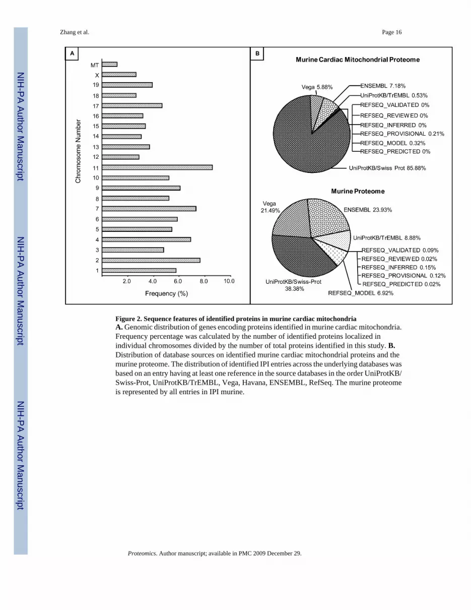
Figure 2. Sequence features of identified proteins in murine cardiac mitochondriaA. Genomic distribution of genes encoding proteins identified in murine cardiac mitochondria.Frequency percentage was calculated by the number of identified proteins localized inindividual chromosomes divided by the number of total proteins identified in this study. B.Distribution of database sources on identified murine cardiac mitochondrial proteins and themurine proteome. The distribution of identified IPI entries across the underlying databases wasbased on an entry having at least one reference in the source databases in the order UniProtKB/Swiss-Prot, UniProtKB/TrEMBL, Vega, Havana, ENSEMBL, RefSeq. The murine proteomeis represented by all entries in IPI murine.
Zhang et al. Page 16
Proteomics. Author manuscript; available in PMC 2009 December 29.
NIH
-PA Author Manuscript
NIH
-PA Author Manuscript
NIH
-PA Author Manuscript

Figure 3. Chemical and physical properties of identified proteinsA. Transmembrane domain and transit peptides were predicted using TMHMM 2.0 andTargetP1.1. Among 940 identified proteins, 195 proteins (20.7%) had one or moretransmembrane domains; in addition, 469 out of 940 proteins had mitochondrial transit peptidesequences (inlet). B-D Physiochemical property classifications of identified proteins. B.Molecular weight (MW, in kDa) analyses; C. Isoelectric focusing point (pI) distributions; D.Grand average hydrophobicity (GRAVY) distributions. Molecular weight, isoelectric point,and grand average hydropathicity value were calculated using the online ProtParam toolavailable through ExPASY.
Zhang et al. Page 17
Proteomics. Author manuscript; available in PMC 2009 December 29.
NIH
-PA Author Manuscript
NIH
-PA Author Manuscript
NIH
-PA Author Manuscript

Figure 4. Function annotation and spatial distribution of identified proteinsIndividual protein was assigned to their respective function cluster(s) and spatial location(s)according to the Gene Ontology Annotations database (GOA), the InterPro database, OMIM,and author initiated Pubmed search. The y axis represents the subcellular compartment(s)where a protein resides, whereas the x axis indicates the function cluster it belongs to. Ourproteomic approach identified a total of 940 unique proteins residing in the cardiacmitochondria. In addition to mitochondria, many proteins reside in one or more other cellularlocations. Among 940 proteins, 169 were also found in cytoplasm, 50 in ER, 20 in Golgi, 144in sacrolemma or other membrane fractions, 16 in lysosome, 24 in perixosome, 57 in nucleus,and 137 in extra-cellular space. Furthermore, 47 out of 940 proteins were assigned to morethan one function cluster(s). Arrows point to three examples, including the eukaryotic peptidechain release factor that is experimentally identified in the cardiac mitochondria; this proteinwas also previously found in the cytoplasm; and this protein belongs to the function cluster ofDNA/RNA/Protein synthesis.
Zhang et al. Page 18
Proteomics. Author manuscript; available in PMC 2009 December 29.
NIH
-PA Author Manuscript
NIH
-PA Author Manuscript
NIH
-PA Author Manuscript
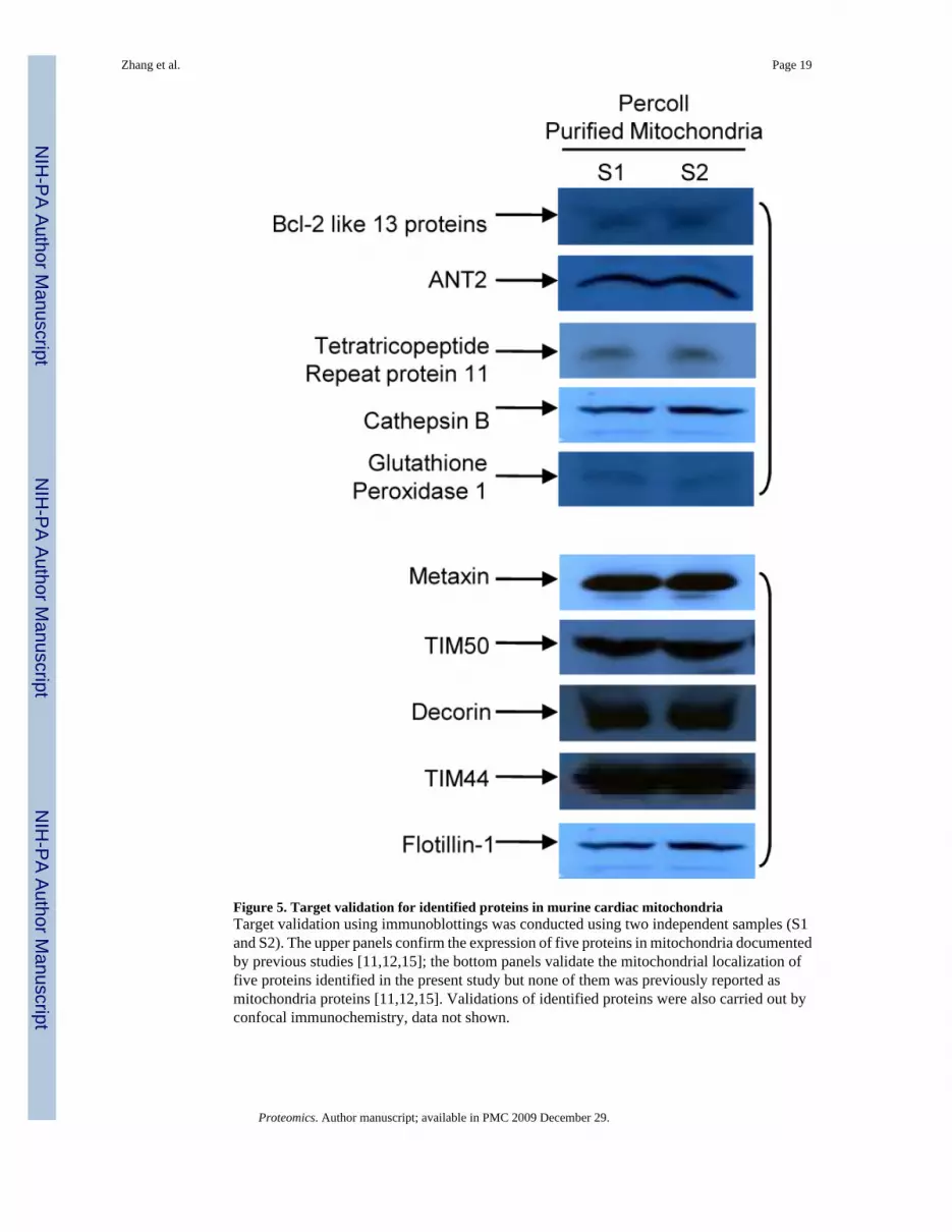
Figure 5. Target validation for identified proteins in murine cardiac mitochondriaTarget validation using immunoblottings was conducted using two independent samples (S1and S2). The upper panels confirm the expression of five proteins in mitochondria documentedby previous studies [11,12,15]; the bottom panels validate the mitochondrial localization offive proteins identified in the present study but none of them was previously reported asmitochondria proteins [11,12,15]. Validations of identified proteins were also carried out byconfocal immunochemistry, data not shown.
Zhang et al. Page 19
Proteomics. Author manuscript; available in PMC 2009 December 29.
NIH
-PA Author Manuscript
NIH
-PA Author Manuscript
NIH
-PA Author Manuscript