shaping the mitochondrial proteome
TRANSCRIPT
http://www.elsevier.com/locate/bba
Biochimica et Biophysica Ac
Review
Shaping the mitochondrial proteome
Toni Gabaldon*, Martijn A. Huynen
NCMLS, Nijmegen Center for Molecular Life Sciences, P/O: CMBI, Center for Molecular and Biomolecular Informatics, University of Nijmegen,
Toernooiveld 1, 6525 ED Nijmegen, The Netherlands
Received 4 June 2004; received in revised form 15 July 2004; accepted 28 July 2004
Available online 11 September 2004
Abstract
Mitochondria are eukaryotic organelles that originated from a single bacterial endosymbiosis some 2 billion years ago. The transition from
the ancestral endosymbiont to the modern mitochondrion has been accompanied by major changes in its protein content, the so-called
proteome. These changes included complete loss of some bacterial pathways, amelioration of others and gain of completely new complexes
of eukaryotic origin such as the ATP/ADP translocase and most of the mitochondrial protein import machinery. This renewal of proteins has
been so extensive that only 14–16% of modern mitochondrial proteome has an origin that can be traced back to the bacterial endosymbiont.
The rest consists of proteins of diverse origin that were eventually recruited to function in the organelle. This shaping of the proteome content
reflects the transformation of mitochondria into a highly specialized organelle that, besides ATP production, comprises a variety of functions
within the eukaryotic metabolism. Here we review recent advances in the fields of comparative genomics and proteomics that are throwing
light on the origin and evolution of the mitochondrial proteome.
D 2004 Elsevier B.V. All rights reserved.
Keywords: Mitochondria; Mitochondrial evolution; Alpha-proteobacteria; Proteome; Endosymbiosis
1. Introduction
According to the widely accepted endosymbiotic theory,
mitochondria are eukaryotic organelles of bacterial descent.
Phylogenetic data supporting this theory point to an ancient
alpha-proteobacterium, most likely an ancestor of the
Rickettsiales, establishing a symbiotic relationship inside a
primitive eukaryotic cell circa 2 billion years ago [1–3].
Since then the mitochondrion has undergone major changes
that transformed it into a highly specialized organelle that
plays a key role within the metabolism of most eukaryotic
cells. This process involved not only a dramatic reduction at
the level of its genome but also an extensive renewal at the
level of its proteome that affected more than 80% of the
protein content of modern mitochondria [4].
Nowadays, mitochondria are present in a multitude of
eukaryotic organisms adapted to a variety of niches [5],
0005-2728/$ - see front matter D 2004 Elsevier B.V. All rights reserved.
doi:10.1016/j.bbabio.2004.07.011
* Corresponding author. Tel.: + 31 24 3653374; fax: +31 24 3652977.
E-mail address: [email protected] (T. Gabaldon).
with modern mitochondrial proteomes reflecting a large
diversity in organellar functions. Moreover, there is
increasing evidence that certain organisms that lack
mitochondria bsensu-strictoQ actually harbor relicts of
these organelles. These, together with the hydrogeno-
somes, most of which appear of mitochondrial descent
[6], reflect extreme forms of mitochondrial adaptation to
microaerophilic or anaerobic environments. The evolution
of the mitochondrial genome in both its structure and
gene content has been the focus of several reviews [7–9],
but the scarcity of data has long prevented similar surveys
on the evolution of the mitochondrial proteome. Recent
advances in the fields of proteomics and comparative
genomics give us a picture of the processes that shaped
the mitochondrial proteome from the early stages of
endosymbiosis to modern adaptations of mitochondria to
diverse environments and cellular functions. Recent
reviews have focused on the evolution of a specific
mitochondrial system, such as the mitochondrial import
machinery [10], or have analyzed more generally the
processes that transformed ancestors of both mitochondria
ta 1659 (2004) 212–220
T. Gabaldon, M.A. Huynen / Biochimica et Biophysica Acta 1659 (2004) 212–220 213
and plastids into modern organelles [11]. Here we review
recent data that define the proteome of the mitochondrial
ancestor and the modern mitochondria. We also discuss
the most relevant events that affected the mitochondrial
protein content and hence its metabolic capacities.
2. The starting point: the proto-mitochondrial proteome
To unravel the evolution of the mitochondrial proteome,
it is essential to get an idea of what it looked like during the
first endosymbiotic stages, the common point from which
all modern mitochondrial proteomes evolved. Defining the
proteome of the mitochondrial ancestor would also help in
understanding how the initial symbiosis was established and
perpetuated, an issue that is hotly debated and for which
several hypotheses have been proposed [12–16]. First
approaches to infer the nature of the mitochondrial ancestor
were based on the similarity, in terms of metabolic
capabilities, of mitochondria with some bacterial groups.
In this way the gamma-proteobacterium Bdellovibrio and
the alpha-proteobacterium Paracoccus were the first pro-
posed models for the proto-mitochondrion [17]. Later on,
phylogenetic analyses of small subunit ribosomal RNAs and
proteins from the respiratory complexes confirmed a
monophyletic origin of mitochondria from an alpha-proteo-
bacterial ancestor [1,3]. Moreover, the sequencing of the
genome of the bacterium Rickettsia prowazekii [18] and
subsequent phylogenetic analyses [18,19] identified mem-
bers of the Rickettsia genus as the closest relatives of
modern mitochondria. Some of the Rickettsiales are obligate
intracellular parasites, a feature that suggested a similar life-
style for the mitochondrial ancestor [18,20]. However, any
attempt to establish parallels between the proto-mitochon-
drial proteome and those of modern alpha-proteobacteria
must be cautious, bearing in mind the roughly estimated 2
billion years of evolution that separate them. Indeed, the
adaptation to an intracellular lifestyle of the modern
Rickettsiales is the result of a different event than that of
the endosymbiosis of mitochondria [18]. Therefore, the
similarities in their respective adaptations are probably the
result of convergent evolution [18,21]. The identification of
its phylogenetic affiliation with the alpha-proteobacteria
narrows the scope of the speculations on the proto-
mitochondrion’s lifestyle. However, the great diversity of
size and composition of modern alpha-proteobacterial
genomes, ranging from 834 to more than 8000 protein-
coding genes, provides enough room for many alternative
models.
The problem can be reformulated as to what extent
does the proto-mitochondrial proteome resemble that of
modern alpha-proteobacteria. To answer this question, it
is necessary to distinguish truly common features from
those resulting from secondary adaptations. A valid
approach is to trace the origin of the modern proteomes
to determine which proteins are directly derived from the
endosymbiotic event. Assuming no genetic transfer to the
mitochondrial genome, the proteins encoded there con-
stitute a dbona-fideT subset of proteins derived from the
proto-mitochondrion. But mitochondrial genomes
sequenced so far encode only 3–67 proteins that are
involved in few processes, mainly respiration and protein
synthesis and, occasionally, transcription, RNA maturation
and protein import [7]. The set has been extended by a
phylogenetic analysis of the yeast mitochondrial proteome
[22] that identified an additional number (~20) of
nuclear-encoded proteins whose phylogenies indicated
an alpha-proteobacterial origin. When combined, both
sets form a reduced core of the proto-mitochondrial
proteome whose deduced metabolic capabilities reflect
that of a cell harboring few metabolic pathways, but able
to couple electron transport to the production of ATP as
well as with the capacity to synthesize the required
proteins.
This picture changed considerably after a large-scale
phylogenetic comparison of alpha-proteobacterial and
eukaryotic genomes [4] identified 630 eukaryotic proteins
that were likely derived from the alpha-proteobacterial
ancestor of mitochondria. Mapping the metabolic func-
tions of these orthologous groups, a minimal metabolism
for the proto-mitochondrion ancestor could be recon-
structed. Besides the abovementioned processes, other
pathways such as the oxidation and synthesis of fatty
acids, biotin and heme synthesis, iron-sulfur cluster
assembly, and fructose and sucrose metabolism pathways
emerged. Also notable was the presence of many
metabolite transporters.
Altogether the accumulation of data on the proto-
mitochondrial proteome point towards a (facultatively)
aerobic organism living on several compounds provided
by the host. Whether the proto-mitochondrion was a
parasite, something that is compatible with the data
available, depends on the existence of potential benefits
for the host. In the case of a mutual benefit, the lack of
an ATP transporter suggests that ATP was not the main
currency used by the proto-mitochondrion to pay back
host’s services. Alternative benefits for the host have been
proposed in hypotheses that consider a hydrogen-produc-
ing [12] or an oxygen-detoxifying [13] endosymbiont.
The presence of the Fe–S cluster assembly pathway and
the ancestor of the ABC transporter that is likely involved
in the export of Fe–S clusters from mitochondria (ATM1)
[23] indicate an alternative benefit to the host that could
have been a key in the initial symbiotic relationship. This
would be in agreement with the finding that proteins of
this pathway are among the few conserved by mitochon-
drial remnants in the microsporidian Enzephalitozoon
cuniculi [24], the protozoan Giardia intestinalis [25]
and the apicomplexan Cryptosporidium parvum [26];
although we cannot discard a secondary loss in these
organisms of another pathway that provided the original
selective pressure for the symbiosis.
T. Gabaldon, M.A. Huynen / Biochimica et Biophysica Acta 1659 (2004) 212–220214
3. A crucial step: the origin of the mitochondrial import
machinery
The proto-mitochondrial proteome soon underwent a
series of transformations that shaped it. Most important to
this transformation was the acquisition of a mechanism
that facilitated the import of proteins from the cytosol into
the mitochondrion. The abovementioned proto-mitochon-
drial reconstruction points to an ancient endosymbiont
that was autonomous in protein synthesis, with no
sophisticated system for the import of proteins synthe-
sized in the cytosol. This contrasts with the modern
situation in which most mitochondrial proteins are
encoded by nuclear genes, synthesized in the cytoplasm
and subsequently imported into the organelle. The latter
step is carried out by a complex machinery consisting of
dozens of proteins located in the inner and outer
membranes of the mitochondria [27], as well as many
soluble chaperones that assist in the process. This
machinery recognizes specific N-terminal signals that are
sufficient and necessary to direct the import of proteins
into mitochondria. Such a system is a prerequisite not
only for the escape from mitochondria to the nucleus of
genes whose products should be targeted back but also
for the recruitment of proteins of different origin to the
organelle. Considering that both processes have been
rampant [4,8,22], there is little doubt that the emergence
of the protein import system was a crucial step in the
evolution of mitochondria. Indeed, it might well be the
event that marked the beginning of the transition from
endosymbiont to organelle. Once genes encoding essential
mitochondrial proteins were transferred to the nucleus, the
host took command of the mitochondrion.
The phylogenetic analysis of the mitochondrial import
machinery [10] reveals that although most of its components
are of eukaryotic origin, some of the components in the
inner membrane and most of the soluble chaperones that
assist the translocation have bacterial homologs. This mixed
origin argues in favor of the hypothesis [10] that a
rudimentary system of chaperones and porins already
existed in the ancestor. This system rapidly evolved into a
more sophisticated machinery able to import proteins with
high efficiency and specificity. Furthermore, in its initial
stages the protein import system could have been tightly
coupled with translation as suggested by the preferential
synthesis of ancient mitochondrial proteins by polysomes
attached to mitochondria [28]. The sophistication of the
translocation system should have run parallel to the
evolution of the addressing sequences that direct the
targeting of the mitochondrial proteins. It has been
suggested that these sequences have evolved from proteins
with an inherent propensity to be targeted to the mitochon-
dria [29]. Furthermore, such targeting sequences are not
uncommon in prokaryotic proteins [29] and can easily be
matched by random sequences [30]. Once present in a few
genes, it is easy to conceive that these pre-sequences were
passed to other genes by means of duplication and
recombination events [31], allowing evolution to potentially
test the mitochondrial localization of any nuclear-encoded
protein. Such a transport system would presumably pave the
way for the recruitment of proteins for service in the
mitochondrion. Therefore, the protein transport system
contributed a new dimension to the evolution of the
mitochondrial proteome by facilitating expansion of the
proteome as well as reduction of the organellar genome.
4. Turning mitochondria into cell’s energy factory
Perhaps the function gain that most radically affected the
role of the mitochondrion within the eukaryotic cell was the
acquisition of an ATP/ADP translocase and therefore the
ability to exchange ATP between the mitochondria and the
cell’s cytoplasm. Although the ATP-production system is
derived from the bacterial ancestor, the ATP/ADP translo-
case has a eukaryotic origin [32]. The origin of the ATP/
ADP exchanger provided mitochondria with a new function
for the cell: that of an energy-converting organelle. This
new role might have also favored subsequent mitochondrial
transformations such us the elaborate folding pattern of the
inner membrane (cristae) and the increase in complexity of
the ATP-synthase [33] as well as the electron transport chain
(ETC) complexes [34].
This increase in complexity has affected all mitochon-
drial ETC complexes with the exception of succinate
dehydrogenase, notably the only ETC complex that does
not pump protons across the inner membrane. For the other
complexes, as much as a threefold increase in terms of
protein content may be observed when the bacterial
components are compared to their mammal counterparts: 3
to 11 subunits in cytochrome bc1 complex, 4 to 13 in
cytochrome c oxidase and 14 to 46 in NADH:ubiquinone
oxidoreductase (complex I). In the latter, the addition of
extra subunits has not altered the characteristic L-shaped
structure of the complex [35]. Notably, not all new subunits
recruited to the ETC complexes are of eukaryotic origin.
The acyl carrier component (ACP) of complex I is actually
present in alpha-proteobacteria, but has never been identi-
fied as part of complex I in prokaryotes [36]. It is difficult to
assess the function of the so-called subsidiary subunits.
Their participation in the biogenesis or stabilization of the
complexes is the most common speculative explanation of
their roles [35,37]. A processing peptidase activity has been
confirmed for some subsidiary subunits such as the 8-kDa
subunit of cytochrome bc1 complex in plants [38]. How-
ever, different roles have been proposed for other subunits
such as the participation in fatty acid synthesis of the ACP
component of Neurospora Complex I [39].
The presence of the mitochondrial-type ATP/ADP trans-
locase in both aerobic and anaerobic mitochondria studied
so far [40] as well as in hydrogenosomes [6,41] suggests
that this gain of function preceded the diversification of
T. Gabaldon, M.A. Huynen / Biochimica et Biophysica Acta 1659 (2004) 212–220 215
mitochondria in terms of the terminal acceptor of electrons
used to fuel the synthesis of ATP. It also raises doubts on
whether oxygen was the original sink for the electron
transport chain [5]. Besides oxygen, modern mitochondria
are able to use alternative electron acceptors such as nitrate,
nitrite or fumarate. Although some anaerobic mitochondria,
such as the ones present in parasitic helminths and fresh-
water snails, seem to be the result of secondary adaptations
to anerobiosis [42], current data on other anaerobic
mitochondria and hydrogenosomes [5] can also be
explained by a scenario in which main anaerobic traits are
vertically derived from a facultative anaerobic ancestor. The
recent finding of an evolutionarily related but distinct ATP/
ADP translocase in the flagellate Trichomonas gallinae
[43,44] may indicate multiple origins for ATP/ADP
exchangers.
5. Modern mitochondrial proteomes
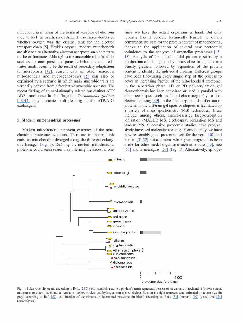
Modern mitochondria represent extremes of the mito-
chondrial proteome evolution. There are in fact multiple
ends, as mitochondria diverged along the different eukary-
otic lineages (Fig. 1). Defining the modern mitochondrial
proteome could seem easier than inferring the ancestral one,
Fig. 1. Eukaryotic phylogeny according to Refs. [2,87] (left); symbols next to a ph
mitosomes or other mitochondrial remnants (yellow circles) and hydrogenosomes
grey) according to Ref. [59], and fraction of experimentally determined prote
(Arabidopsis).
since we have the extant organisms at hand. But only
recently has it become technically feasible to obtain
comprehensive data for the protein content of mitochondria,
thanks to the application of several new proteomic
techniques to the analyses of organellar proteomes [45–
48]. Analysis of the mitochondrial proteome starts by a
purification of the organelle by means of centrifugation on a
density gradient followed by separation of the protein
content to identify the individual proteins. Different groups
have been fine-tuning every single step of the process to
cover an increasing fraction of the mitochondrial proteome.
In the separation phase, 1D or 2D polyacrylamide gel
electrophoresis has been combined or used in parallel with
other techniques such us liquid-chromatography or iso-
electric focusing [49]. In the final step, the identification of
proteins in the different gel-spots or aliquots is facilitated by
a variety of mass spectrometry (MS) techniques. These
include, among others, matrix-assisted laser-desorption
ionization (MALDI) MS, electrospray ionization MS and
tandem MS. Successive proteomic studies have progres-
sively increased molecular coverage. Consequently, we have
now reasonably good proteomic sets for the yeast [50] and
human [51,52] mitochondria, while great progress has been
made for other model organisms such as mouse [49], rice
[53] and Arabidopsis [54] (Fig. 1). Alternatively, epitope-
ylum’s name represents possession of canonic mitochondria (brown ovals),
(red circles). Bars on the right represent total estimated proteome size (in
ome (in black) according to Refs. [51] (human), [50] (yeast) and [54]
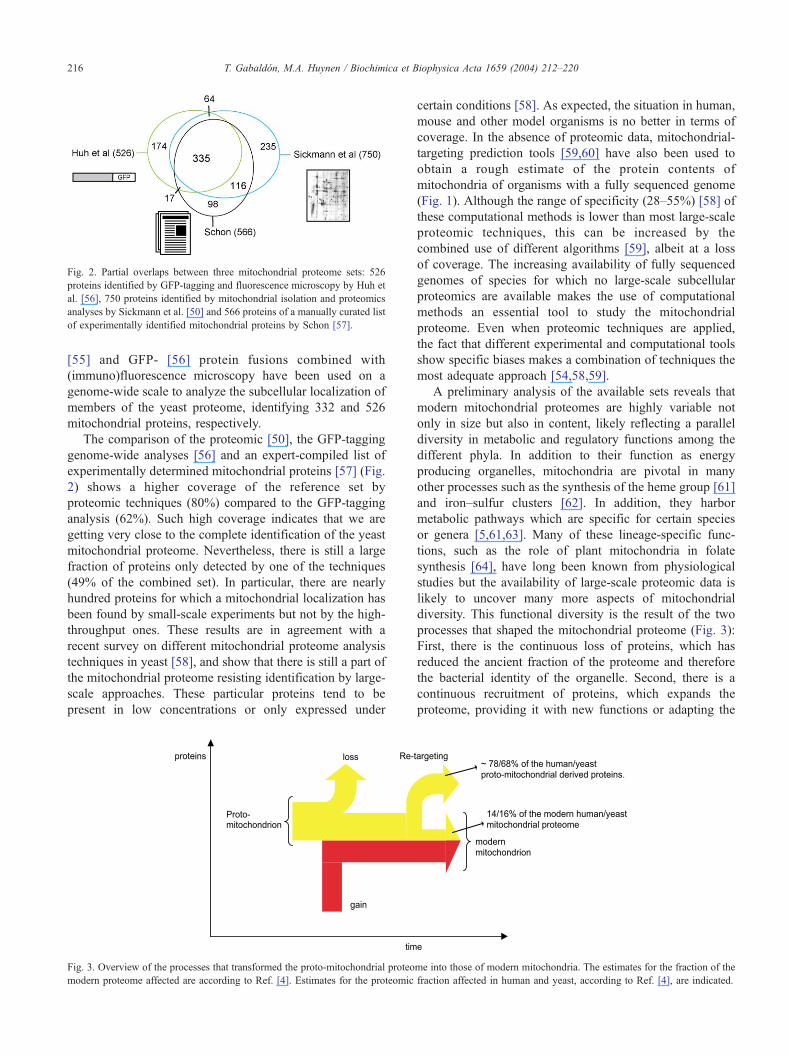
Fig. 2. Partial overlaps between three mitochondrial proteome sets: 526
proteins identified by GFP-tagging and fluorescence microscopy by Huh et
al. [56], 750 proteins identified by mitochondrial isolation and proteomics
analyses by Sickmann et al. [50] and 566 proteins of a manually curated list
of experimentally identified mitochondrial proteins by Schon [57].
T. Gabaldon, M.A. Huynen / Biochimica et Biophysica Acta 1659 (2004) 212–220216
[55] and GFP- [56] protein fusions combined with
(immuno)fluorescence microscopy have been used on a
genome-wide scale to analyze the subcellular localization of
members of the yeast proteome, identifying 332 and 526
mitochondrial proteins, respectively.
The comparison of the proteomic [50], the GFP-tagging
genome-wide analyses [56] and an expert-compiled list of
experimentally determined mitochondrial proteins [57] (Fig.
2) shows a higher coverage of the reference set by
proteomic techniques (80%) compared to the GFP-tagging
analysis (62%). Such high coverage indicates that we are
getting very close to the complete identification of the yeast
mitochondrial proteome. Nevertheless, there is still a large
fraction of proteins only detected by one of the techniques
(49% of the combined set). In particular, there are nearly
hundred proteins for which a mitochondrial localization has
been found by small-scale experiments but not by the high-
throughput ones. These results are in agreement with a
recent survey on different mitochondrial proteome analysis
techniques in yeast [58], and show that there is still a part of
the mitochondrial proteome resisting identification by large-
scale approaches. These particular proteins tend to be
present in low concentrations or only expressed under
Fig. 3. Overview of the processes that transformed the proto-mitochondrial proteo
modern proteome affected are according to Ref. [4]. Estimates for the proteomic
certain conditions [58]. As expected, the situation in human,
mouse and other model organisms is no better in terms of
coverage. In the absence of proteomic data, mitochondrial-
targeting prediction tools [59,60] have also been used to
obtain a rough estimate of the protein contents of
mitochondria of organisms with a fully sequenced genome
(Fig. 1). Although the range of specificity (28–55%) [58] of
these computational methods is lower than most large-scale
proteomic techniques, this can be increased by the
combined use of different algorithms [59], albeit at a loss
of coverage. The increasing availability of fully sequenced
genomes of species for which no large-scale subcellular
proteomics are available makes the use of computational
methods an essential tool to study the mitochondrial
proteome. Even when proteomic techniques are applied,
the fact that different experimental and computational tools
show specific biases makes a combination of techniques the
most adequate approach [54,58,59].
A preliminary analysis of the available sets reveals that
modern mitochondrial proteomes are highly variable not
only in size but also in content, likely reflecting a parallel
diversity in metabolic and regulatory functions among the
different phyla. In addition to their function as energy
producing organelles, mitochondria are pivotal in many
other processes such as the synthesis of the heme group [61]
and iron–sulfur clusters [62]. In addition, they harbor
metabolic pathways which are specific for certain species
or genera [5,61,63]. Many of these lineage-specific func-
tions, such as the role of plant mitochondria in folate
synthesis [64], have long been known from physiological
studies but the availability of large-scale proteomic data is
likely to uncover many more aspects of mitochondrial
diversity. This functional diversity is the result of the two
processes that shaped the mitochondrial proteome (Fig. 3):
First, there is the continuous loss of proteins, which has
reduced the ancient fraction of the proteome and therefore
the bacterial identity of the organelle. Second, there is a
continuous recruitment of proteins, which expands the
proteome, providing it with new functions or adapting the
me into those of modern mitochondria. The estimates for the fraction of the
fraction affected in human and yeast, according to Ref. [4], are indicated.

able 1
ist of E. cuniculi proteins with a likely proto-mitochondrial origin [4] that
ave a detectable homolog (E value b10�15, with BlastP search) in the
enome of C. parvum
roteins within the same box belong to the same orthologous group and
roteins involved in Fe–S cluster assembly are marked with an asterisk.
olumn on the right denote the mitochondrial localization of a yeast
itochondrial ortholog according to [50,57], whenever an orthologous
east protein exists; otherwise, absence of a yeast ortholog is indicated by
n.pQ (not present).
T. Gabaldon, M.A. Huynen / Biochimica et Biophysica Acta 1659 (2004) 212–220 217
old ones to the new circumstances. We emphasize that the
loss from the mitochondrial proteome does not necessary
imply loss from the total cell’s proteome. Indeed, ancient
mitochondrial proteins can be retargeted to other locations
in the cell [4] as exemplified by the peroxisomal location of
part of the beta-oxidation pathway in yeast.
Gain and loss processes have acted along the various
lineages in a different manner in terms of quality and
intensity. Mitochondria have selectively lost proteins whose
functions were no longer needed in a certain lineage, such as
the specific complex I loss from yeast, while keeping
required functions. In the yeast mitochondrial proteome,
most of what remains from the alpha-proteobacterial
ancestor (59 out of 97 proteins) is related to respiration
according to a genome-wide analysis [65]. This suggests
that these mitochondria are more specialized towards
respiration than their ancestor.
Major expansions in the size of the proteome are
observed in vascular plants and metazoans and these may
reflect the adaptation to multicellularity and tissue differ-
entiation. Indeed, many tissue-specific mitochondrial prop-
erties and proteins have been described in plants [66] and
mammals [49]. In plants, comparisons between photo-
synthetic and non-photosynthetic tissues have demonstrated
that the composition of mitochondrial proteomes varies in
accordance to their different metabolic needs. For example,
there is a greater demand for the mitochondrial oxidation of
glycine associated with photo-respiration [67]. In mammals,
mitochondria of certain tissues have been shown to
participate in tissue-specific processes such as the regulation
of insulin secretion in pancreatic beta-cells [68] or steroido-
genesis in adrenal cortex [69].
Besides these lineage-specific expansions, the mitochon-
drial proteome likely gained a common set of proteins
before the divergence of most eukaryotic species. These
common gains include the abovementioned ATP/ADP
translocase and part of the mitochondrial protein import
machinery. The total overlap between yeast and human
mitochondrial proteomes, 371 proteins in the E. Schon set
[57], greatly exceeds the alpha-proteobacterial fraction, 79
proteins (~20%) [4], indicating that the mitochondrial
proteome experienced a major expansion before the
divergence of fungi and metazoa. Consistently the ETC
complexes that are present in both organisms share most of
the subsidiary subunits of eukaryotic origin.
It is worth recalling that the recruitment of proteins to the
mitochondrial proteome was not limited to proteins of
eukaryotic origin. In principle, any nuclear-encoded protein
could potentially be targeted to mitochondria once the
necessary targeting-sequence is attached to the gene.
Therefore, it is not surprising to find mitochondrial proteins
of diverse origin such as the components of the oxidative
branch of the Krebs cycle, which are phylogenetically close
to the Cyptophaga–Flavobacterium–Bacteroides (CFB)
bacterial group [70], although a scenario in which CFB
bacteria gained these enzymes from eukaryotes cannot be
ruled out. Similarly the viral-like mitochondrial RNA-
polymerase seems to be a clear case of non-orthologous
gene displacement, because the proto-mitochondrial ances-
tor likely harbored the eubacterial-type RNA-polymerase
that is encoded in the mitochondrion of the protist
Reclinomonas americana [71]. The RNA-polymerase case
is not the only example of a viral-like protein working in
mitochondria. The so-called twinkle protein is a mitochon-
drial DNA-helicase with homology to phage-T7 primase
helicase [72]. Phylogenetic analyses of mitochondrial
proteomes have so far not identified other proteins of viral
origin (results not shown).
6. Mitochondrial proteome remnants in
bamitochondriateQ eukaryotes
The monophyletic origin of mitochondria and their
nearly ubiquitous distribution among eukaryotes suggest
that the acquisition of these organelles occurred very early
in the evolution of eukaryotes. How early is still a matter of
discussion. The initial placing of most amitochondriate
eukaryotes at the base of the reconstructed phylogenies
suggested that these diversified prior to the mitochondrial
endosymbiosis [73]. Recent phylogenetic reconstructions
T
L
h
g
P
p
C
m
y
b
T. Gabaldon, M.A. Huynen / Biochimica et Biophysica Acta 1659 (2004) 212–220218
[74,75], however, support the relocation of some of the
amitochondriates, such as microsporidia, certain amoebozoa
and parabasalia, higher in the tree. Their lack of mitochon-
dria is now considered the result of secondary adaptations to
anaerobic environments. Recently subcellular structures that
appear to be mitochondrial remnants have been described in
microsporidia [76], cryptosporidia [77], amoebozoa [78]
and diplomonads [25]. In addition, there are hydrogeno-
somes in parabasalids, ciliates and anaerobic fungi [79] that
might represent forms of highly specialized—rather than
degraded—mitochondria. The existence of these subcellular
structures supports the hypothesis that mitochondrial endo-
symbiosis predated the diversification of the analyzed
eukaryotic species. This view is also supported by the
presence of proteins of a likely mitochondrial origin in all
fully sequenced eukaryotes so far.
A search for proto-mitochondrial derived proteins in nine
fully sequenced eukaryotic genomes [4] revealed an
ubiquitous but variable distribution of ancient mitochondrial
proteins, including 29 proteins in the microsporidian
Encephalitozoon cuniculi. A large fraction (22) of the
proto-mitochondrial derived proteins in E. cuniculi have
yeast orthologs targeted to mitochondria, which suggests
they might be part of the mitosome. These results are similar
to those of a large-scale prediction of mitochondrial
targeting signals in eukaryotic genomes [59] that predicted
a mitochondrial localization for 156 proteins in E. cuniculi,
26 of which have alpha-proteobacterial homologs. More-
over, 18 of the proto-mitochondrial derived proteins,
including those involved in Fe–S cluster assembly, can also
be found in the recently sequenced genome of C. parvum
[80], another parasite with a mitochondrial remnant (Table
1). In addition, other Fe–S cluster assembly genes and
mitochondrial chaperonin-like genes have been found in
amoebozoans, diplomonads, amitochondriate apicomplex-
ans and parabasalids [81–84]. Some of these are targeted to
the mitochondrial remnants.
The apparently ubiquitous presence of proto-mitochon-
drial derived proteins in virtually all eukaryotes supports the
view of an early endosymbiosis at the root of the eukaryotic
tree. Alternatively it might be argued that these proteins
could have been gained via subsequent horizontal transfers.
In the end, the greater plasticity of the mitochondrial
proteome, when compared to that of the mitochondrial
genome, makes it less useful in the search for the origin of
highly reduced remnants. The discovery of a potential
residual genome in the hydrogenosomes of the ciliate
Nyctoterus ovalis [85] provides a way out of this dilemma.
The initial analyses of this genome strongly suggest a
mitochondrial ancestry for the hydrogenosomes in this
species [86].
The emerging picture is that there is no extant eukaryote
whose amitochondriate state can be categorically considered
ancestral. If this is proven to be true, especially when a
greater diversity of early-diverging eukaryotes are
sequenced, it would have tremendous implications for the
role that mitochondrial endosymbiosis played in the process
of eukaryogenesis.
Acknowledgements
We are grateful to Charles Kurland for critically
reviewing the manuscript. This work was supported in part
by a grant from the Netherlands organization for Scientific
Research (NWO).
References
[1] M.W. Gray, G. Burger, B.F. Lang, Mitochondrial evolution, Science
283 (1999) 1476–1481.
[2] S.B. Hedges, J.E. Blair, M.L. Venturi, J.L. Shoe, A molecular time
scale of eukaryote evolution and the rise of complex multicellular life
BMC, Evol. Biol. 4 (2004) 2.
[3] D. Yang, Y. Oyaizu, H. Oyaizu, G.J. Olsen, C.R. Woese, Mitochon-
drial origins, Proc. Natl. Acad. Sci. U. S. A. 82 (1985) 4443–4447.
[4] T. Gabaldon, M.A. Huynen, Reconstruction of the proto-mitochon-
drial metabolism, Science 301 (2003) 609.
[5] A.G. Tielens, C. Rotte, J.J. van Hellemond, W. Martin, Mitochon-
dria as we don’t know them, Trends Biochem. Sci. 27 (2002)
564–572.
[6] F. Voncken, B. Boxma, J. Tjaden, A. Akhmanova, M. Huynen, F.
Verbeek, A.G. Tielens, I. Haferkamp, H.E. Neuhaus, G. Vogels, M.
Veenhuis, J.H. Hackstein, Multiple origins of hydrogenosomes:
functional and phylogenetic evidence from the ADP/ATP carrier of
the anaerobic chytrid Neocallimastix sp, Mol. Microbiol. 44 (2002)
1441–1454.
[7] G. Burger, M.W. Gray, B.F. Lang, Mitochondrial genomes: anything
goes, Trends Genet. 19 (2003) 709–716.
[8] J.N. Timmis, M.A. Ayliffe, C.Y. Huang, W. Martin, Endosymbiotic
gene transfer: organelle genomes forge eukaryotic chromosomes, Nat.
Rev., Genet. 5 (2004) 123–135.
[9] J. Nosek, L. Tomaska, Mitochondrial genome diversity: evolution of
the molecular architecture and replication strategy, Curr. Genet. 44
(2003) 73–84.
[10] J.M. Herrmann, Converting bacteria to organelles: evolution of
mitochondrial protein sorting, Trends Microbiol. 11 (2003) 74–79.
[11] S.D. Dyall, M.T. Brown, P.J. Johnson, Ancient invasions: from
endosymbionts to organelles, Science 304 (2004) 253–257.
[12] W. Martin, M. Mqller, The hydrogen hypothesis for the first
eukaryote, Nature 392 (1998) 37–41.
[13] C.G. Kurl, S.G. Andersson, Origin and evolution of the mitochondrial
proteome, Microbiol. Mol. Biol. Rev. 64 (2000) 786–820.
[14] D. Moreira, P. Lopez-Garcıa, Symbiosis between methanogenic
archaea and delta-proteobacteria as the origin of eukaryotes: the
syntrophic hypothesis, J. Mol. Evol. 47 (1998) 517–530.
[15] W. Martin, M. Hoffmeister, C. Rotte, K. Henze, An overview of
endosymbiotic models for the origins of eukaryotes, their ATP-
producing organelles (mitochondria and hydrogenosomes), and their
heterotrophic lifestyle, Biol. Chem. 382 (2001) 1521–1539.
[16] P. Lopez-Garcia, D. Moreira, Metabolic symbiosis at the origin of
eukaryotes, Trends Biochem. Sci. 24 (1999) 88–93.
[17] L. Margulis, Symbioses in Cell Evolution, W.H. Freeman, San
Francisco, 1981.
[18] S.G. Andersson, A. Zomorodipour, J.O. Andersson, T. Sicheritz-
Ponten, U.C. Alsmark, R.M. Podowski, A.K. Naslund, A.S.
Eriksson, H.H. Winkler, C.G. Kurland, The genome sequence of
Rickettsia prowazekii and the origin of mitochondria, Nature 396
(1998) 133–140.
T. Gabaldon, M.A. Huynen / Biochimica et Biophysica Acta 1659 (2004) 212–220 219
[19] V.V. Emelyanov, Evolutionary relationship of Rickettsiae and mito-
chondria, FEBS Lett. 501 (2001) 11–18.
[20] V.V. Emelyanov, Rickettsiaceae, rickettsia-like endosymbionts, and
the origin of mitochondria, Biosci. Rep. 21 (2001) 1–17.
[21] M. Muller, W. Martin, The genome of Rickettsia prowazekii and some
thoughts on the origin of mitochondria and hydrogenosomes,
Bioessays 21 (1999) 377–381.
[22] O. Karlberg, B. Canback, C.G. Kurl, S.G. Andersson, The dual origin
of the yeast mitochondrial proteome, Yeast 17 (2000) 170–187.
[23] G. Kispal, P. Csere, C. Prohl, R. Lill, The mitochondrial proteins
Atm1p and Nfs1p are essential for biogenesis of cytosolic Fe/S
proteins, EMBO J. 18 (1999) 3981–3989.
[24] C.P. Vivares, M. Gouy, F. Thomarat, G. Metenier, Functional and
evolutionary analysis of a eukaryotic parasitic genome, Curr. Opin.
Microbiol. 5 (2002) 499–505.
[25] J. Tovar, G. Leon-Avila, L.B. Sanchez, R. Sutak, J. Tachezy, M. van
der Giezen, M. Hernandez, M. Mqller, J.M. Lucocq, Mitochondrial
remnant organelles of Giardia function in iron–sulfur protein
maturation, Nature 426 (2003) 172–176.
[26] M.J. LaGier, J. Tachezy, F. Stejskal, K. Kutisova, J.S. Keithly,
Mitochondrial-type iron–sulfur cluster biosynthesis genes (IscS and
IscU) in the apicomplexan Cryptosporidium parvum, Microbiology
149 (2003) 3519–3530.
[27] N. Wiedemann, A.E. Frazier, N. Pfanner, The protein import
machinery of mitochondria, J. Biol. Chem. 279 (2004) 14473–14476.
[28] P. Marc, A. Margeot, F. Devaux, C. Blugeon, M. Corral-Debrinski, C.
Jacq, Genome-wide analysis of mRNAs targeted to yeast mitochon-
dria, EMBO Rep. 3 (2002) 159–164.
[29] R. Lucattini, V.A. Likic, T. Lithgow, Bacterial proteins predisposed
for targeting to mitochondria, Mol. Biol. Evol. 21 (2004) 652–658.
[30] B.D. Lemire, C. Fankhauser, A. Baker, G. Schatz, The mitochondrial
targeting function of randomly generated peptide sequences correlates
with predicted helical amphiphilicity, J. Biol. Chem. 264 (1989)
20206–20215.
[31] K. Kadowaki, N. Kubo, K. Ozawa, A. Hirai, Targeting presequence
acquisition after mitochondrial gene transfer to the nucleus occurs
by duplication of existing targeting signals, EMBO J. 15 (1996)
6652–6661.
[32] H. Amiri, O. Karlberg, S.G. Andersson, Deep origin of plastid/parasite
ATP/ADP translocases, J. Mol. Evol. 56 (2003) 137–150.
[33] P.D. Boyer, The ATP synthase—a splendid molecular machine, Annu.
Rev. Biochem. 66 (1997) 717–749.
[34] S. Berry, Endosymbiosis and the design of eukaryotic electron
transport, Biochim. Biophys. Acta 1606 (2003) 57–72.
[35] V. Guenebaut, A. Schlitt, H. Weiss, K. Leonard, T. Friedrich,
Consistent structure between bacterial and mitochondrial NADH:u-
biquinone oxidoreductase (complex I), J. Mol. Biol. 276 (1998)
105–112.
[36] U. Sackmann, R. Zensen, D. Rohlen, U. Jahnke, H. Weiss, The acyl-
carrier protein in Neurospora crassa mitochondria is a subunit of
NADH:ubiquinone reductase (complex I), Eur. J. Biochem. 200
(1991) 463–469.
[37] J. Carroll, I.M. Fearnley, R.J. Shannon, J. Hirst, J.E. Walker, Analysis
of the subunit composition of complex I from bovine heart
mitochondria, Mol. Cell Proteomics 2 (2003) 117–126.
[38] S. Brumme, V. Kruft, U.K. Schmitz, H.P. Braun, New insights into the
co-evolution of cytochrome c reductase and the mitochondrial
processing peptidase, J. Biol. Chem. 273 (1998) 13143–13149.
[39] R. Zensen, H. Husmann, R. Schneider, T. Peine, H. Weiss, De novo
synthesis and desaturation of fatty acids at the mitochondrial acyl-
carrier protein, a subunit of NADH:ubiquinone oxidoreductase in
Neurospora crassa, FEBS Lett. 310 (1992) 179–181.
[40] F. Palmieri, Mitochondrial carrier proteins, FEBS Lett. 346 (1994)
48–54.
[41] M. van der Giezen, D.J. Slotboom, D.S. Horner, P.L. Dyal, M.
Harding, G.P. Xue, T.M. Embley, E.R. Kunji, Conserved properties of
hydrogenosomal and mitochondrial ADP/ATP carriers: a common
origin for both organelles, EMBO J. 21 (2002) 572–579.
[42] J.J. van Hellemond, A. van der Klei, S.W. van Weelden, A.G. Tielens,
Biochemical and evolutionary aspects of anaerobically functioning
mitochondria, Philos. Trans. R. Soc. Lond., B Biol. Sci. 358 (2003)
205–213 (discussion 213 – 5).
[43] J. Tjaden, I. Haferkamp, B. Boxma, A.G. Tielens, M. Huynen, J.H.
Hackstein, A divergent ADP/ATP carrier in the hydrogenosomes of
Trichomonas gallinae argues for an independent origin of these
organelles, Mol. Microbiol. 51 (2004) 1439–1446.
[44] S.D. Dyall, C.M. Koehler, M.G. Delgadillo-Correa, P.J. Bradley, E.
Plumper, D. Leuenberger, C.W. Turck, P.J. Johnson, Presence of a
member of the mitochondrial carrier family in hydrogenosomes:
conservation of membrane-targeting pathways between hydrogeno-
somes and mitochondria, Mol. Cell. Biol. 20 (2000) 2488–2497.
[45] F.M. Canovas, E. Dumas-Gaudot, G. Recorbet, J. Jorrin, H.P.
Mock, M. Rossignol, Plant proteome analysis, Proteomics 4 (2004)
285–298.
[46] D.E. Warnock, E. Fahy, S.W. Taylor, Identification of protein
associations in organelles, using mass spectrometry-based proteomics,
Mass Spectrom. Rev. 23 (2004) 259–280.
[47] S.W. Taylor, E. Fahy, S.S. Ghosh, Global organellar proteomics,
Trends Biotechnol. 21 (2003) 82–88.
[48] M. Dreger, Proteome analysis at the level of subcellular structures,
Eur. J. Biochem. 270 (2003) 589–599.
[49] V.K. Mootha, J. Bunkenborg, J.V. Olsen, M. Hjerrild, J.R. Wisniew-
ski, E. Stahl, M.S. Bolouri, H.N. Ray, S. Sihag, M. Kamal, N.
Patterson, E.S. Lander, M. Mann, Integrated analysis of protein
composition, tissue diversity, and gene regulation in mouse mitochon-
dria, Cell 115 (2003) 629–640.
[50] A. Sickmann, J. Reinders, Y. Wagner, C. Joppich, R. Zahedi, H.E.
Meyer, B. Schonfisch, I. Perschil, A. Chacinska, B. Guiard, P.
Rehling, N. Pfanner, C. Meisinger, The proteome of Saccharomyces
cerevisiae mitochondria, Proc. Natl. Acad. Sci. U. S. A. 100 (2003)
13207–13212.
[51] D. Cotter, P. Guda, E. Fahy, S. Subramaniam, MitoProteome:
mitochondrial protein sequence database and annotation system,
Nucleic Acids Res. 32 (2004) D463–D467 (Database issue).
[52] S.W. Taylor, E. Fahy, B. Zhang, G.M. Glenn, D.E. Warnock, S. Wiley,
A.N. Murphy, S.P. Gaucher, R.A. Capaldi, B.W. Gibson, S.S. Ghosh,
Characterization of the human heart mitochondrial proteome, Nat.
Biotechnol. 21 (2003) 281–286.
[53] N. Tanaka, M. Fujita, H. Handa, S. Murayama, M. Uemura, Y.
Kawamura, T. Mitsui, S. Mikami, Y. Tozawa, T. Yoshinaga, S.
Komatsu, Proteomics of the rice cell: systematic identification of the
protein populations in subcellular compartments, Mol. Genet.
Genomics (2004) 566–576.
[54] J.L. Heazlewood, J.S. Tonti-Filippini, A.M. Gout, D.A. Day, J.
Whelan, A.H. Millar, Experimental analysis of the Arabidopsis
mitochondrial proteome highlights signaling and regulatory compo-
nents, provides assessment of targeting prediction programs, and
indicates plant-specific mitochondrial proteins, Plant Cell 16 (2004)
241–256.
[55] A. Kumar, S. Agarwal, J.A. Heyman, S. Matson, M. Heidtman, S.
Piccirillo, L. Umansky, A. Drawid, R. Jansen, Y. Liu, K.H. Cheung, P.
Miller, M. Gerstein, G.S. Roeder, M. Snyder, Subcellular localization
of the yeast proteome, Genes Dev. 16 (2002) 707–719.
[56] W.K. Huh, J.V. Falvo, L.C. Gerke, A.S. Carroll, R.W. Howson, J.S.
Weissman, E.K. O’Shea, Global analysis of protein localization in
budding yeast, Nature 425 (2003) 686–691.
[57] E. Schon, in: P.M. Leslie Wilson (Ed.), Methods Cell Biol., Academic
Press, New York, 2001, pp. 463–482.
[58] H. Prokisch, C. Scharfe, D.G. Camp 2nd, W. Xiao, L. David, C.
Andreoli, M.E. Monroe, R.J. Moore, M.A. Gritsenko, C. Kozany,
K.K. Hixson, H.M. Mottaz, H. Zischka, M. Ueffing, Z.S. Herman,
R.W. Davis, T. Meitinger, P.J. Oefner, R.D. Smith, L.M. Steinmetz,
T. Gabaldon, M.A. Huynen / Biochimica et Biophysica Acta 1659 (2004) 212–220220
Integrative analysis of the mitochondrial proteome in yeast PLoS,
Biology 2 (2004) E160.
[59] E. Richly, P.F. Chinnery, D. Leister, Evolutionary diversification of
mitochondrial proteomes: implications for human disease, Trends
Genet. 19 (2003) 356–362.
[60] C. Guda, E. Fahy, S. Subramaniam, MITOPRED: a genome-scale
method for prediction of nucleus-encoded mitochondrial proteins,
Bioinformatics (2004) W372–W374.
[61] I.E. Scheffler, Mitochondria make a come back, Adv. Drug Deliv.
Rev. 49 (2001) 3–26.
[62] R. Lill, G. Kispal, Maturation of cellular Fe–S proteins: an essential
function of mitochondria, Trends Biochem. Sci. 25 (2000) 352–356.
[63] S. Ohta, A multi-functional organelle mitochondrion is involved in
cell death, proliferation and disease, Curr. Med. Chem. 10 (2003)
2485–2494.
[64] M. Neuburger, F. Rebeille, A. Jourdain, S. Nakamura, R. Douce,
Mitochondria are a major site for folate and thymidylate synthesis in
plants, J. Biol. Chem. 271 (1996) 9466–9472.
[65] L.M. Steinmetz, C. Scharfe, A.M. Deutschbauer, D. Mokranjac, Z.S.
Herman, T. Jones, A.M. Chu, G. Giaever, H. Prokisch, P.J. Oefner,
R.W. Davis, Systematic screen for human disease genes in yeast, Nat.
Genet. 31 (2002) 400–404.
[66] C.G. Bowsher, A.K. Tobin, Compartmentation of metabolism within
mitochondria and plastids, J. Exp. Bot. 52 (2001) 513–527.
[67] R. Srinivasan, D.J. Oliver, Light-dependent and tissue-specific
expression of the H-protein of the glycine decarboxylase complex,
Plant Physiol. 109 (1995) 161–168.
[68] P. Maechler, Mitochondria as the conductor of metabolic signals for
insulin exocytosis in pancreatic beta-cells, Cell. Mol. Life Sci. 59
(2002) 1803–1818.
[69] T.J. Rosol, J.T. Yarrington, J. Latendresse, C.C. Capen, Adrenal gland:
structure, function, and mechanisms of toxicity, Toxicol. Pathol. 29
(2001) 41–48.
[70] A.D. Baughn, M.H. Malamy, A mitochondrial-like aconitase in the
bacterium Bacteroides fragilis: implications for the evolution of the
mitochondrial Krebs cycle, Proc. Natl. Acad. Sci. U. S. A. 99 (2002)
4662–4667.
[71] B.F. Lang, G. Burger, C.J. O’Kelly, R. Cedergren, G.B. Golding, C.
Lemieux, D. Sankoff, M. Turmel, M.W. Gray, An ancestral
mitochondrial DNA resembling a eubacterial genome in miniature,
Nature 387 (1997) 493–497.
[72] J.A. Korhonen, M. Gaspari, M. Falkenberg, TWINKLE Has 5VY3VDNA helicase activity and is specifically stimulated by mitochondrial
single-stranded DNA-binding protein, J. Biol. Chem. 278 (2003)
48627–48632.
[73] T. Cavalier-Smith, E.E. Chao, Molecular phylogeny of the free-living
archezoan Trepomonas agilis and the nature of the first eukaryote,
J. Mol. Evol. 43 (1996) 551–562.
[74] T. Cavalier-Smith, The phagotrophic origin of eukaryotes and
phylogenetic classification of Protozoa, Int. J. Syst. Evol. Microbiol.
52 (2002) 297–354.
[75] A. Stechmann, T. Cavalier-Smith, Rooting the eukaryote tree by using
a derived gene fusion, Science 297 (2002) 89–91.
[76] B.A. Williams, R.P. Hirt, J.M. Lucocq, T.M. Embley, A mitochondrial
remnant in the microsporidian Trachipleistophora hominis, Nature
418 (2002) 865–869.
[77] C.E. Riordan, J.G. Ault, S.G. Langreth, J.S. Keithly, Cryptospori-
dium parvum Cpn60 targets a relict organelle, Curr. Genet. 44
(2003) 138–147.
[78] J. Tovar, A. Fischer, C.G. Clark, The mitosome, a novel organelle
related to mitochondria in the amitochondrial parasite Entamoeba
histolytica, Mol. Microbiol. 32 (1999) 1013–1021.
[79] T.M. Embley, M. van der Giezen, D.S. Horner, P.L. Dyal, P. Foster,
Mitochondria and hydrogenosomes are two forms of the same
fundamental organelle, Philos. Trans. R. Soc. Lond., B Biol. Sci.
358 (2003) 191–201 (discussion 201– 2).
[80] M.S. Abrahamsen, T.J. Templeton, S. Enomoto, J.E. Abrahante, G.
Zhu, C.A. Lancto, M. Deng, C. Liu, G. Widmer, S. Tzipori, G.A.
Buck, P. Xu, A.T. Bankier, P.H. Dear, B.A. Konfortov, H.F. Spriggs,
L. Iyer, V. Anantharaman, L. Aravind, V. Kapur, Complete genome
sequence of the apicomplexan, Cryptosporidium parvum, Science 304
(2004) 441–445.
[81] N. Arisue, L.B. Sanchez, L.M. Weiss, M. Mqller, T. Hashimoto,
Mitochondrial-type hsp70 genes of the amitochondriate protists,
Giardia intestinalis, Entamoeba histolytica and two microsporidians,
Parasitol. Int. 51 (2002) 9–16.
[82] A. Germot, H. Philippe, H. Le Guyader, Presence of a mitochondrial-
type 70-kDa heat shock protein in Trichomonas vaginalis suggests a
very early mitochondrial endosymbiosis in eukaryotes, Proc. Natl.
Acad. Sci. U. S. A. 93 (1996) 14614–14617.
[83] A. Germot, H. Philippe, Critical analysis of eukaryotic phylogeny: a
case study based on the HSP70 family, J. Eukaryot. Microbiol. 46
(1999) 116–124.
[84] J. Slapeta, J.S. Keithly, Cryptosporidium parvum mitochondrial-type
HSP70 targets homologous and heterologous mitochondria, Eukary-
otic Cell 3 (2004) 483–494.
[85] A. Akhmanova, F. Voncken, T. van Alen, A. van Hoek, B. Boxma, G.
Vogels, M. Veenhuis, J.H. Hackstein, A hydrogenosome with a
genome, Nature 396 (1998) 527–528.
[86] A.H. van Hoek, A.S. Akhmanova, M.A. Huynen, J.H. Hackstein, A
mitochondrial ancestry of the hydrogenosomes of Nyctotherus ovalis,
Mol. Biol. Evol. 17 (2000) 202–206.
[87] S.L. Baldauf, A.J. Roger, I. Wenk-Siefert, W.F. Doolittle, A kingdom-
level phylogeny of eukaryotes based on combined protein data,
Science 290 (2000) 972–977.