superposition model predicts eeg occipital activity during free viewing of natural scenes
TRANSCRIPT
Behavioral/Systems/Cognitive
Superposition Model Predicts EEG Occipital Activity duringFree Viewing of Natural Scenes
Jose P. Ossandon,1,3 Andrea V. Helo,1,2 Rodrigo Montefusco-Siegmund,1 and Pedro E. Maldonado1
1Programa de Fisiología y Biofísica, Instituto de Ciencias Biomédicas, and 2Escuela de Fonoaudiología, Facultad de Medicina, Universidad de Chile,Santiago, Chile, and 3Institute of Cognitive Science, University of Osnabruck, 49069 Osnabruck, Germany
Visual event-related potentials (ERPs) produced by a stimulus are thought to reflect either an increase of synchronized activity or a phaserealignment of ongoing oscillatory activity, with both mechanisms sharing the assumption that ERPs are independent of the current stateof the brain at the time of stimulation. In natural viewing, however, visual inputs occur one after another at specific subject-pacedintervals through unconstrained eye movements. We conjecture that during natural viewing, ERPs generated after each fixation arebetter explained by a superposition of ongoing oscillatory activity related to the processing of previous fixations, with new activity elicitedby the visual input at the current fixation. We examined the electroencephalography (EEG) signals that occur in humans at the onset ofeach visual fixation, both while subjects freely viewed natural scenes and while they viewed a black or gray background. We found that thefixation ERPs show visual components that are absent when subjects move their eyes on a homogeneous gray or black screen. Single-trialEEG signals that comprise the ERP are predicted more accurately by a model of superposition than by either phase resetting or theaddition of evoked responses and stimulus-independent noise. The superposition of ongoing oscillatory activity and the visually evokedresponse results in a modification of the ongoing oscillation phase. The results presented suggest that the observed EEG signals reflectchanges occurring in a common neuronal substrate rather than a simple summation at the scalp of signals from independent sources.
IntroductionDifferent neuronal mechanisms are studied through the exami-nation of event-related potentials (ERPs), which are typicallyevoked by controlled stimuli. ERPs have generally been consid-ered to express changes in neuronal activity that occur time-locked to stimulation. One hypothesis, known as the additivemodel, explains the generation of ERPs as the summation ofsynchronized neuronal activity directly evoked by the stimulus,with any ongoing activity preceding the ERP, being a result ofindependent neuronal processes or noise (Shah et al., 2004;Makinen et al., 2005; Mazaheri and Jensen, 2006). A second hy-pothesis states that ERPs are the consequence of a mechanismthat resets undefined ongoing oscillatory processes to a fixedphase (Sayers et al., 1974; Makeig et al., 2002; Hanslmayr et al.,2007). Therefore, although these hypotheses offer a different in-terpretation of the ongoing activity, they both share the assump-tion that ERPs are generated in a manner independent of thecurrent state of the brain.
In contrast to these views, studies on local field potentials andsingle-unit activity have demonstrated that there is a clear depen-dence between the response to stimulation and the dynamicsof ongoing activity (Mehta et al., 2002; Lakatos et al., 2007;
Womelsdorf et al., 2007; Kayser et al., 2008; Rajkai et al., 2008). Inthis regard, the activity elicited after a given stimulus could not beadequately characterized by either of the above two models, be-cause ongoing activity appears to modulate and interact withincoming sensory activity. Accordingly, a complete characteriza-tion of ERP responses depends on the dynamics of ongoingactivity at the moment of stimulation. However, in typical ex-perimental paradigms, sensory stimuli are presented in isolationand at intervals defined by the experimenters; therefore, previousongoing activity is unrelated to perceptual processing and cannotcarry successful predictive or preparatory signals. Accordingly,we conjecture that a relationship between ongoing and sensory-evoked activity should be readily apparent during naturalviewing. In this case, visual stimulation occurs in a fast streamof events, because visual exploration occurs through ocularmovement over the visual field (Yarbus, 1967). Here, visual in-puts arrive during fixation periods when gaze remains stationary.As these fixations follow each other with very short latency, it islikely that the ongoing activity specific for visual processing willdepend on the response to previous stimuli. To evaluate the pres-ence and relationship between ongoing and evoked activity dur-ing natural conditions, we recorded electroencephalography(EEG) signals and eye movements during free viewing of naturalscenes and examined the ERP signals that occurred around eachvisual fixation. We found that neither of the two aforementionedmodels by itself was sufficient to accurately describe our results.We concluded that the EEG signals observed during free viewingare best explained by a combination of both models. Single-trialEEG signals are predicted most accurately by the summation ofan evoked visual response and an ongoing oscillatory activity.
Received Nov. 5, 2009; revised Jan. 27, 2010; accepted Feb. 24, 2010.This study was supported in part by the Iniciativa Cientifica Milenio P04-068F, The Puelma Foundation, and
Conicyt (R.M.-S.). We thank Francisco Flores, Michael Plochl, and Robert Muil for comments on an earlier version ofthis manuscript.
Correspondence should be addressed to Dr. Pedro E. Maldonado, Programa de Fisiología y Biofísica, Facultad deMedicina, Universidad de Chile, Casilla 70005, Santiago 7, Chile. E-mail: [email protected].
DOI:10.1523/JNEUROSCI.5769-09.2010Copyright © 2010 the authors 0270-6474/10/304787-09$15.00/0
The Journal of Neuroscience, March 31, 2010 • 30(13):4787– 4795 • 4787
Furthermore, phase undergoes a slight modulation even when itis measured outside the period of evoked activation, suggestingan interaction at the source level rather a simple addition of sig-nals at the scalp.
Materials and MethodsParticipants. Eleven students from the Faculty of Medicine of the Univer-sidad de Chile with normal or corrected-to-normal vision participated inthe study. All participants were right-handed and gave their informedconsent in writing, which was approved by the institution’s Ethics Com-mittee for Human Research.
Stimuli and procedure. A set of photographic images was presented ingrayscale on a 19“ CRT monitor (Samsung SyncMaster 1100P Plus) at arefresh rate of 85 Hz and resolution of 800 � 600. The image set wascomposed of two different categories of natural scenes (NS): landscapes(n � 10) and construction sites (n � 10). To control for the motorcontribution of the eye movements to the EEG signal, we also includedfull-screen black (BK) and gray (GR) images. Each natural scene waspresented three times, and the black and gray images 60 times each. Eachimage was presented for 5 s in random order. A central fixation pointappeared between images. The subjects pressed a button to trigger thenext image presentation. The distance between subjects and monitor was57 cm, subtending a visual field of 40° horizontally and 30° vertically with20 pixels per visual degree. Subjects were instructed to freely explore eachimage, even when no natural image was presented (black and graystimuli). To properly determine eye position and movements, an eyetracker drift correction procedure was performed before each imagepresentation.
Eye tracking and EEG recording. Ocular movements were recordedwith a head-mounted video oculographic eye-tracking system using pu-pil tracking at 500 Hz (Eyelink II; SR Research). Before each block ofimage presentation, a nine-point calibration was performed until averageerror fell below 0.5 degrees. Saccades were defined based on a velocitythreshold of 25 deg/s and a minimum duration of 10 milliseconds. Weincluded in our analysis fixations that lasted longer then 50 ms and beforewhich no blinks had occurred for at least 100 ms.
EEG signals were recorded from 21 scalp sites using the standard10 –20 system montage (EasyCap). Electrodes were referenced to linkedear lobes, with all impedances kept below 5 k�. The EEG signal from eachelectrode site was recorded using a custom-made EEG amplifier builtwith 120 dB CMRR differential amplifiers (Burr Brown), digitized at 1kHz with a bandpass of 0.5–250 Hz (�3 dB low- and high-frequencycutoff, respectively), and was stored for off-line processing.
Signal analysis. To analyze EEG data, the signal was segmented inepochs of 2 s centered on fixation onsets (see Fig. 1 B) and then groupedinto three different condition sets: NS, BK, and GR. Rejection of majornonocular artifacts was performed through visual inspection. All datawere digitally low-pass filtered below 50 Hz, applying forward and re-verse filtering with an elliptic infinite impulse response filter. BecauseERP and phase measurement can be altered by artifacts produced by eyemovements, we performed an independent component analysis (ICA) toidentify and extract artifact components from the data. ICA is a blindsource decomposition algorithm that enables the separation of statisti-cally independent sources from multichannel data, thus separating ocu-lar movement and blink artifacts from EEG data (Jung et al., 2000a,b;Iriarte et al., 2003; Hoffmann and Falkenstein, 2008). We applied ICAInfomax algorithm (Bell and Sejnowski, 1995) as implemented in theEEGLAB toolbox for EEG analysis (Delorme and Makeig, 2004) to thecomplete dataset of each subject. Once blinks and eye movement com-ponents were identified through visual analysis of the components’ to-pography and time series, they were removed from the original data. Afixation-onset ERP (fERP) was generated by averaging all epochs of eachcondition. Time–frequency analysis of the EEG epochs was performedusing a family of Morlet wavelets (Tallon-Baudry et al., 1996) centered atinteger frequency steps between 2 and 50 Hz. Each wavelet is defined byw(t, f ) � A � exp(�t 2/2�t
2) � exp(2i�ft), with �t � 1/(2��f), A �(�t��) �1/2, and a ratio of f/�f � 5. To find the frequency most likelyinvolved in the generation of visual fERPs, we looked for the frequency
with the actual highest power in the upper theta or alpha band. This is thefrequency range more frequently associated with the first components ofERPs (Makeig et al., 2002; Gruber et al., 2005; Mazaheri and Jensen, 2006;Hanslmayr et al., 2007; Klimesch et al., 2007a; Min et al., 2007; Freun-berger et al., 2008). For this purpose, the power spectrum in the periodbetween 0 and 200 ms was calculated. This was done by taking the meanpower of each frequency in the 200 ms time interval for each trial, thenaveraging for each condition for each subject, and finally averaging acrosssubjects. Although this power estimate was obtained through averagingeach frequency wavelet output over a window of only 200 ms, the fre-quency and time resolution is not determined by this window but by the�t and �f parameters of the wavelets. Therefore, the power estimate in the200 ms interval includes some power from the periods immediately be-fore and after the time interval, in a way depending on the �t parameterof the wavelet, which increases at lower frequencies. Once the frequencyof interest for each condition was identified, instantaneous phase valuesin that frequency were calculated at each epoch fixation onset. All subse-quent analyses that depended on the phase at fixation onset were con-ducted at the frequency selected for that respective condition. Trialscould then be sorted into bins according to this calculated phase, and therelationship between this phase and the phase of the subsequent fixationwas examined.
Correlation between image features and visual responses. Visual ERPsare typically studied under experimental paradigms that examine signalchanges after flashed visual stimuli. In our paradigm, subjects were ableto view the images freely. Thus, although the image presented remainedstatic with constant luminance, the image patch presented to the foveachanged with each saccade. To ensure that the EEG signals correspondedto a visual response, we examined the correlation of the amplitude of thesignals after each fixation with a simple visual feature. It has been shownthat visual responses after ocular movements depend on luminance(Green, 1957; Gaarder et al., 1964). Thus, we grouped EEG epochs in 10groups according to the logarithm of the absolute difference in lumi-nance between two square, 1° image patches; the first centered at thecurrent gaze location and the second at the gaze location of the immedi-ately preceding fixation. Then, we correlated the mean amplitude of thefERP’s first positive component (P1, 40 –110 ms) with the mean lumi-nance difference of these groups.
Models of EEG signal generation. To examine different mechanisms ofgeneration of the activity observed in the EEG signals, we compared theobserved ERPs to the predictions of three different models. This wasdone for all epochs between fixation onset (time 0) and the following 200ms (Fig. 1 H). In all models, predicted data were generated according tothe following expression: s(t) � u(t) � a1 � cos(2�ft � �1) �v(t) � a2 � cos(2�ft � �2), where f is the frequency of interest of the corre-sponding experimental condition. The first term represents the ongoingoscillatory component and the second term represents an additive com-ponent. The ongoing component always started at the onset of fixation(time 0) and its instantaneous phase, �1, and amplitude, a1, were calcu-lated from the data. The initial time and phase value of the additivecomponent were arranged such that the additive cosine componentstarted at 0 �V (phase �2 � ��/2) and peaked at the same instant as theactual ERP positive peak. Because a fourth of the period of a cosine at theidentified frequency of interest of the NS condition (8 Hz) (see Fig. 3A)was �31 ms, whereas the ERP peak was at 88 ms (see Fig. 2 A), theadditive component was always initialized with an offset of 57 ms. In thefirst model, named ongoing activity, windowing functions u(t) and v(t)were set to 1 and 0, respectively. In this case, the ERP signal is modeled asan invariant ongoing oscillation. For the second model (phase resetting),u(t) and v(t) are step functions, with u(t) � 1 and v(t) � 0, from fixationonset to 57 ms and then set onwards to u(t) � 0 and v(t) � 1. In the thirdmodel (superposition), u(t) � 1 for the entire epoch and v(t) � 0 fromfixation onset to 57 ms and then set to v(t) � 1.
For all models, we predicted the phase of each fixation onset based onthe phase at the onset of the previous fixation and the time intervalbetween consecutive fixations by the following expression (see Fig. 1 I):�adv � �prev � (�t � 2� � f/fs), with f being the frequency of interest and fsthe sampling frequency in Hz. In the ongoing-activity model, phase atfixation onset is simply the corresponding phase for the elapsed time
4788 • J. Neurosci., March 31, 2010 • 30(13):4787– 4795 Ossandon et al. • EEG Activity during Natural Scenes
from the previous fixation. In this case, �prev � �1prev, meaning the phasemeasured at the onset of the previous fixation, and �t is the time betweenthe onsets of the consecutive fixations. In the phase-resetting model, thephase is reset after the onset of the preceding fixation. Here, �prev � ��/2and �t is the time between the reset moment of the previous fixation (57ms after the onset) and the onset of the current fixation. Finally, the phasein the superposition model is a linear combination of the ongoing andadditive oscillatory components. In this case, the following expres-sion was used: �prev � tan �1(�1sin(�1prev � 57 � 2� � f/fs) � �2sin(��/2))/(�1cos(�1prev � 57 � 2� � f/fs) � �2cos(��/2)) and �t is the same asin the phase-resetting model.
Statistics. Comparisons of the mean amplitude of P1 (40 –110) and N1(115–185 ms) components of fERPs were performed using a one-wayANOVA for different stimulus conditions, different fixation durationsubsets, and different phase groups. To test the performance of the dif-ferent models, Pearson’s linear correlations were calculated between theactual data, the observed ERP, and the data predicted by each of the threemodels. Nonetheless, because the data predicted by the three models arenot completely independent (for example, the first 60 ms of predicteddata of every epoch in the superposition and ongoing-activity model arethe same), we further checked the performance of the superpositionmodel with a partial correlation with the actual data, controlled by thepredictions of the other two models. Partial correlation removes thecommon variance associated with the control variables (ongoing-activityand phase-resetting models) from both the dependent variable (the ac-tual data) and the independent variable (superposition model). Every rvalue was tested against the null hypothesis of no correlation by calculat-ing its respective t value. For comparison of r values, confidence intervals(CIs) were computed through Fisher’s Z transformation. To avoid errorsdue to multiple comparisons, a significance level of 0.001 was used.
All phase calculations were obtained with circular statistics (Batschelet,1981). Rayleigh’s uniformity test was used to assess the uniformity of thedistribution of fixation-onset phases for each condition. To compare thephase predictions, distributions of the differences between predicted andactual phase were constructed. Once nonuniformity and the presence ofonly one mode were confirmed, the estimate of the distribution meanand its confidence intervals was used for model evaluation.
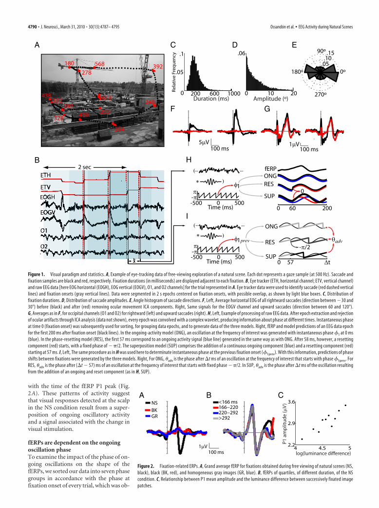
ResultsEEG signals during visual fixationsWe acquired EEG and eye movement data from 11 subjects whilethey explored natural scenes and control (gray and black) images.This unconstrained way of visual exploration produces eyemovement sequences that are highly variable between subjectsfor the same image and within subject for different images. Figure1A shows an example of an eye movement sequence for a naturalscene. The fixation durations are narrowly distributed (me-dian � 200 ms) whereas saccade amplitudes were distributed in amonotonically decreasing fashion (Fig. 1C,D). Eye movementsoccur preferentially along the horizontal axis (Fig. 1E).
To clean the EEG data of ocular artifacts, an ICA decomposi-tion was performed for every subject, and ocular movement com-ponents were identified according to their time series andtopography. These components were removed from the data.The result of this procedure can be observed in the grand averagesfor horizontal and vertical movements in the correspondingelectro-oculogram (EOG) channels (Fig. 1F). The EEG data weresegmented into 2 s epochs (Fig. 1B), aligned to each fixationonset, and averaged over all fixations. We analyzed the EEG datafor a total of 8200 fixations in natural images and 5600 and 6000fixations in gray and black images, respectively. The followingresults correspond only to occipital channels O1 and O2 of the10/20 system, because these showed the highest visually relatedresponses. Furthermore, these channels are less prone to artifactsproduced by eye movements and thus require minimal ICA cor-rections for eye movement artifacts (Fig. 1G).
The grand average fERP for natural scenes— using fixationonset as the reference time event—shows clearly distinguishableP1 and N1 components (Fig. 2A). Because fixation durationsvaried from 50 ms through 300 ms (Fig. 1C), it is possible thatfERPs differ across fixations as a function of their duration, ren-dering further analysis applicable only to subsets of fixations withsimilar duration. However, when we segregated our data accord-ing to fixation– duration quartiles, we did not find significantdifferences in either the P1 (F(1,10) � 0.52, p � 0.67) or the N1(F(1,10) � 0.06, p � 0.97) components (Fig. 2B). Our results showthat these fERP signals constitute a visually evoked response andnot an ocular movement artifact. This is supported by the factthat ICA subtractions had little effect on the occipital signals.Also, the two most prominent peak components occur far fromthe end of the preceding saccade (peak P1 � 88 ms, peak N1 �134 ms) and from the initiation of the next saccade (distance toP1: median � 130 ms, SD � 141 ms; distance to N1: median � 86ms, SD � 141 ms). In addition, the average fERP signal for ho-mogeneous gray or black images shows a significantly diminishedamplitude for P1 (F(1,10) � 64.5, p 0.001) and N1 (F(1,10) �76.5, p 0.0001), despite the occurrence of a saccade in everyevent (Fig. 2A). Finally, when epochs are grouped according todeciles of the absolute difference in luminance between subse-quently fixated image patches, there is a strong correlationbetween these luminance differences and P1 mean amplitude(r � 0.90, p 0.001) (Fig. 2C).
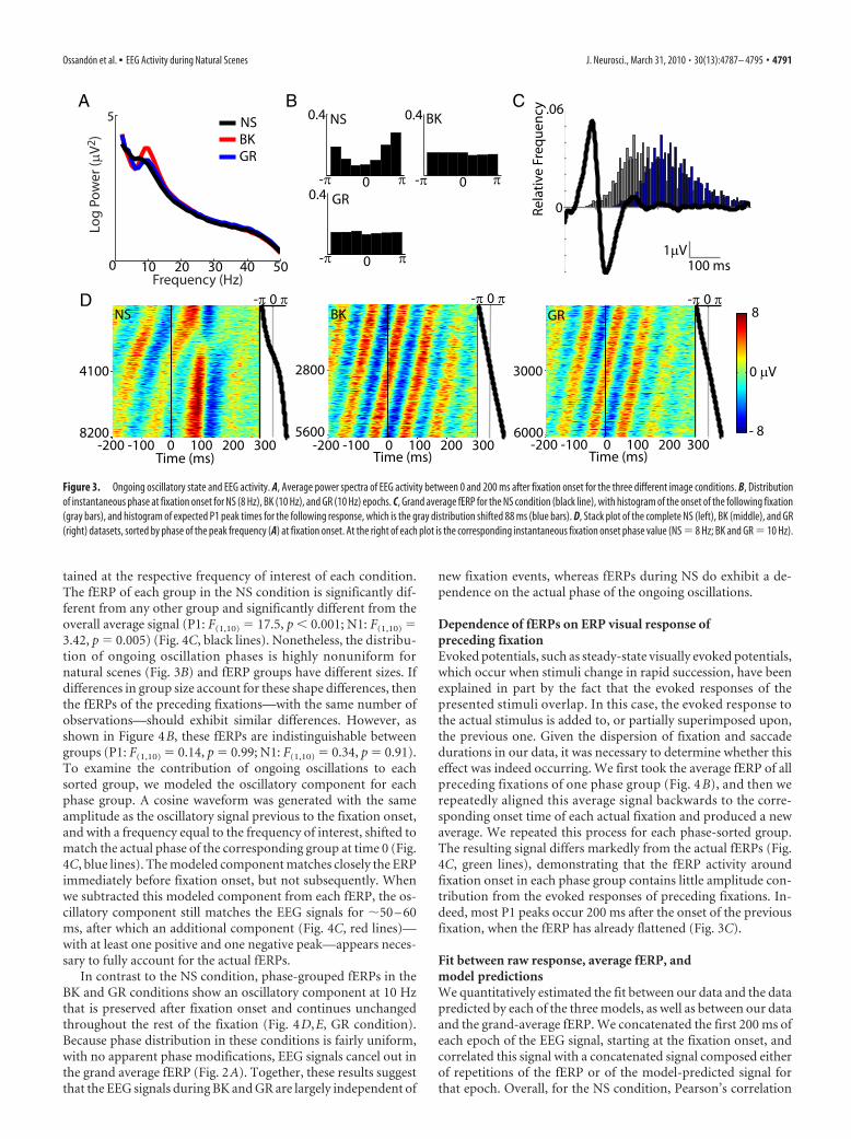
Dependence of visual activity on ongoing activity: effects ofphase on EEG visual activityDuring natural viewing, ocular movements are sequential.Therefore, we can use this sequence to examine whether fERPsare dependent upon some feature of the EEG signal that occurredin the preceding fixation. Here, we looked at the instantaneousfixation onset phase of the frequency in the upper-theta or alphaband (6 –14 Hz) that contributes the most to the variance of thesignal. The average power of the EEG signals for the period be-tween 0 and 200 ms after fixation onset shows a local peak at 8 Hzfor natural scenes and at 10 Hz for both black and gray images(Fig. 3A). For all subsequent analysis where phase informationwas required, we used the phase values at 8 Hz for NS and 10 Hzfor GR and BK. Instantaneous phase values at fixation onset forthe three conditions are distributed differently (Fig. 3B). For NS,the distribution of phases at 8 Hz is highly nonuniform (meanvector length, r � 0.36; Rayleigh’s, z � 1095; p 0.001). Incomparison, the distribution of phases at 10 Hz is only slightlynonuniform for BK epochs (r � 0.03, z � 5.4, p � 0.004) anduniform for GR epochs (r � 0.01, z � 2.34, p � 0.09). We usedthis phase measure to sort and group the epochs of each condi-tion. Stack plots generated with phase-sorted epochs (Fig. 3D)allowed us to examine modifications of the ongoing oscillationthat resulted from the new visual fixation. In the GR and BKconditions, the phase progression of the ongoing oscillatory ac-tivity appears to be unmodified by the visual fixation (Fig. 3D,middle and right). Unlike the previous conditions, during NSstimuli the epochs show a phase realignment of activity after eachfixation onset (Fig. 3D, left). This realignment could not be ex-plained entirely by fixation-evoked resetting of the ongoing os-cillation, because the latencies and widths of peaks and troughs ofactivity in the fixation period depend strongly on the phase atfixation onset. This dependence is particularly clear in the epochswith phases between ��/2 and 0, where the change in the signalfollowing the fixation onset is not followed by another oscillationcycle, as is the case for other phases. Remarkably, this coincides
Ossandon et al. • EEG Activity during Natural Scenes J. Neurosci., March 31, 2010 • 30(13):4787– 4795 • 4789
with the time of the fERP P1 peak (Fig.2A). These patterns of activity suggestthat visual responses detected at the scalpin the NS condition result from a super-position of ongoing oscillatory activityand a signal associated with the change invisual stimulation.
fERPs are dependent on the ongoingoscillation phaseTo examine the impact of the phase of on-going oscillations on the shape of thefERPs, we sorted our data into seven phasegroups in accordance with the phase atfixation onset of every trial, which was ob-
A B C
Figure 2. Fixation-related ERPs. A, Grand average fERP for fixations obtained during free viewing of natural scenes (NS,black), black (BK, red), and homogeneous gray images (GR, blue). B, fERPs of quartiles, of different duration, of the NScondition. C, Relationship between P1 mean amplitude and the luminance difference between successively fixated imagepatches.
A C
F
H
I
G
D E
B
Figure 1. Visual paradigm and statistics. A, Example of eye-tracking data of free-viewing exploration of a natural scene. Each dot represents a gaze sample (at 500 Hz). Saccade andfixation samples are black and red, respectively. Fixation durations (in milliseconds) are displayed adjacent to each fixation. B, Eye tracker (ETH, horizontal channel; ETV, vertical channel)and raw EEG data [here EOG horizontal (EOGH), EOG vertical (EOGV), O1, and O2 channels] for the trial represented in A. Eye tracker data were used to identify saccade (red dashed verticallines) and fixation onsets (gray vertical lines). Data were segmented in 2 s epochs centered on fixation onsets, with possible overlap, as shown by light blue boxes. C, Distribution offixation durations. D, Distribution of saccade amplitudes. E, Angle histogram of saccade directions. F, Left, Average horizontal EOG of all rightward saccades (direction between �30 and30°) before (black) and after (red) removing ocular movement ICA components. Right, Same signals for the EOGV channel and upward saccades (direction between 60 and 120°).G, Averages as in F, for occipital channels (O1 and O2) for rightward (left) and upward saccades (right). H, Left, Example of processing of raw EEG data. After epoch extraction and rejectionof ocular artifacts through ICA analysis (data not shown), every epoch was convolved with a complex wavelet, producing information about phase at different times. Instantaneous phaseat time 0 (fixation onset) was subsequently used for sorting, for grouping data epochs, and to generate data of the three models. Right, fERP and model predictions of an EEG data epochfor the first 200 ms after fixation onset (black lines). In the ongoing-activity model (ONG), an oscillation at the frequency of interest was generated with instantaneous phase �1 at 0 ms(blue). In the phase-resetting model (RES), the first 57 ms correspond to an ongoing activity signal (blue line) generated in the same way as with ONG. After 58 ms, however, a resettingcomponent (red) starts, with a fixed phase of ��/2. The superposition model (SUP) comprises the addition of a continuous ongoing component (blue) and a resetting component (red)starting at 57 ms. I, Left, The same procedure as in H was used here to determinate instantaneous phase at the previous fixation onset (�1prev). With this information, predictions of phaseshifts between fixations were generated by the three models. Right, For ONG, �adv is the phase after �t ms of an oscillation at the frequency of interest that starts with phase �1prev. ForRES, �adv is the phase after (�t � 57) ms of an oscillation at the frequency of interest that starts with fixed phase ��/2. In SUP, �adv is the phase after �t ms of the oscillation resultingfrom the addition of an ongoing and reset component (as in H, SUP).
4790 • J. Neurosci., March 31, 2010 • 30(13):4787– 4795 Ossandon et al. • EEG Activity during Natural Scenes
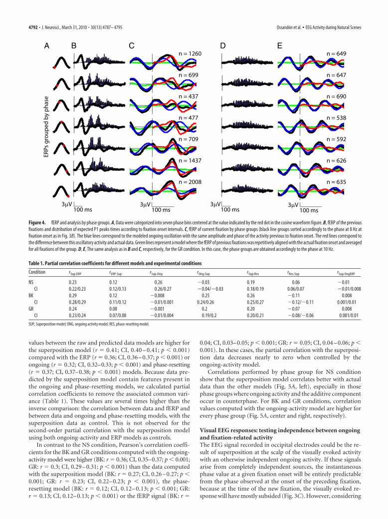
tained at the respective frequency of interest of each condition.The fERP of each group in the NS condition is significantly dif-ferent from any other group and significantly different from theoverall average signal (P1: F(1,10) � 17.5, p 0.001; N1: F(1,10) �3.42, p � 0.005) (Fig. 4C, black lines). Nonetheless, the distribu-tion of ongoing oscillation phases is highly nonuniform fornatural scenes (Fig. 3B) and fERP groups have different sizes. Ifdifferences in group size account for these shape differences, thenthe fERPs of the preceding fixations—with the same number ofobservations—should exhibit similar differences. However, asshown in Figure 4B, these fERPs are indistinguishable betweengroups (P1: F(1,10) � 0.14, p � 0.99; N1: F(1,10) � 0.34, p � 0.91).To examine the contribution of ongoing oscillations to eachsorted group, we modeled the oscillatory component for eachphase group. A cosine waveform was generated with the sameamplitude as the oscillatory signal previous to the fixation onset,and with a frequency equal to the frequency of interest, shifted tomatch the actual phase of the corresponding group at time 0 (Fig.4C, blue lines). The modeled component matches closely the ERPimmediately before fixation onset, but not subsequently. Whenwe subtracted this modeled component from each fERP, the os-cillatory component still matches the EEG signals for �50 – 60ms, after which an additional component (Fig. 4C, red lines)—with at least one positive and one negative peak—appears neces-sary to fully account for the actual fERPs.
In contrast to the NS condition, phase-grouped fERPs in theBK and GR conditions show an oscillatory component at 10 Hzthat is preserved after fixation onset and continues unchangedthroughout the rest of the fixation (Fig. 4D,E, GR condition).Because phase distribution in these conditions is fairly uniform,with no apparent phase modifications, EEG signals cancel out inthe grand average fERP (Fig. 2A). Together, these results suggestthat the EEG signals during BK and GR are largely independent of
new fixation events, whereas fERPs during NS do exhibit a de-pendence on the actual phase of the ongoing oscillations.
Dependence of fERPs on ERP visual response ofpreceding fixationEvoked potentials, such as steady-state visually evoked potentials,which occur when stimuli change in rapid succession, have beenexplained in part by the fact that the evoked responses of thepresented stimuli overlap. In this case, the evoked response tothe actual stimulus is added to, or partially superimposed upon,the previous one. Given the dispersion of fixation and saccadedurations in our data, it was necessary to determine whether thiseffect was indeed occurring. We first took the average fERP of allpreceding fixations of one phase group (Fig. 4 B), and then werepeatedly aligned this average signal backwards to the corre-sponding onset time of each actual fixation and produced a newaverage. We repeated this process for each phase-sorted group.The resulting signal differs markedly from the actual fERPs (Fig.4C, green lines), demonstrating that the fERP activity aroundfixation onset in each phase group contains little amplitude con-tribution from the evoked responses of preceding fixations. In-deed, most P1 peaks occur 200 ms after the onset of the previousfixation, when the fERP has already flattened (Fig. 3C).
Fit between raw response, average fERP, andmodel predictionsWe quantitatively estimated the fit between our data and the datapredicted by each of the three models, as well as between our dataand the grand-average fERP. We concatenated the first 200 ms ofeach epoch of the EEG signal, starting at the fixation onset, andcorrelated this signal with a concatenated signal composed eitherof repetitions of the fERP or of the model-predicted signal forthat epoch. Overall, for the NS condition, Pearson’s correlation
A
D
B C
Figure 3. Ongoing oscillatory state and EEG activity. A, Average power spectra of EEG activity between 0 and 200 ms after fixation onset for the three different image conditions. B, Distributionof instantaneous phase at fixation onset for NS (8 Hz), BK (10 Hz), and GR (10 Hz) epochs. C, Grand average fERP for the NS condition (black line), with histogram of the onset of the following fixation(gray bars), and histogram of expected P1 peak times for the following response, which is the gray distribution shifted 88 ms (blue bars). D, Stack plot of the complete NS (left), BK (middle), and GR(right) datasets, sorted by phase of the peak frequency (A) at fixation onset. At the right of each plot is the corresponding instantaneous fixation onset phase value (NS � 8 Hz; BK and GR � 10 Hz).
Ossandon et al. • EEG Activity during Natural Scenes J. Neurosci., March 31, 2010 • 30(13):4787– 4795 • 4791
values between the raw and predicted data models are higher forthe superposition model (r � 0.41; CI, 0.40 – 0.41; p 0.001)compared with the ERP (r � 0.36; CI, 0.36 – 0.37; p 0.001) orongoing (r � 0.32; CI, 0.32–0.33; p 0.001) and phase-resetting(r � 0.37; CI, 0.37–0.38; p 0.001) models. Because data pre-dicted by the superposition model contain features present inthe ongoing and phase-resetting models, we calculated partialcorrelation coefficients to remove the associated common vari-ance (Table 1). These values are several times higher than theinverse comparison: the correlation between data and fERP andbetween data and ongoing and phase-resetting models, with thesuperposition data as control. This is not observed for thesecond-order partial correlation with the superposition modelusing both ongoing-activity and ERP models as controls.
In contrast to the NS condition, Pearson’s correlation coeffi-cients for the BK and GR conditions computed with the ongoing-activity model were higher (BK: r � 0.36; CI, 0.35–0.37; p 0.001;GR: r � 0.3; CI, 0.29 – 0.31; p 0.001) than the data computedwith the superposition model (BK: r � 0.27; CI, 0.26 – 0.27; p 0.001; GR: r � 0.23; CI, 0.22– 0.23; p 0.001), the phase-resetting model (BK: r � 0.12; CI, 0.12– 0.13; p 0.001; GR:r � 0.13; CI, 0.12– 0.13; p 0.001) or the fERP signal (BK: r �
0.04; CI, 0.03– 0.05; p 0.001; GR: r � 0.05; CI, 0.04 – 0.06; p 0.001). In these cases, the partial correlation with the superposi-tion data decreases nearly to zero when controlled by theongoing-activity model.
Correlations performed by phase group for NS conditionshow that the superposition model correlates better with actualdata than the other models (Fig. 5A, left), especially in thosephase groups where ongoing activity and the additive componentoccur in counterphase. For BK and GR conditions, correlationvalues computed with the ongoing-activity model are higher forevery phase group (Fig. 5A, center and right, respectively).
Visual EEG responses: testing independence between ongoingand fixation-related activityThe EEG signal recorded in occipital electrodes could be the re-sult of superposition at the scalp of the visually evoked activitywith an otherwise independent ongoing activity. If these signalsarise from completely independent sources, the instantaneousphase value at a given fixation onset will be entirely predictablefrom the phase observed at the onset of the preceding fixation,because at the time of the new fixation, the visually evoked re-sponse will have mostly subsided (Fig. 3C). However, considering
A B C D E
Figure 4. fERP and analysis by phase groups. A, Data were categorized into seven phase bins centered at the value indicated by the red dot in the cosine waveform figure. B, fERP of the previousfixations and distribution of expected P1 peaks times according to fixation onset intervals. C, fERP of current fixation by phase groups (black line groups sorted accordingly to the phase at 8 Hz atfixation onset as in Fig. 3B). The blue lines correspond to the modeled ongoing oscillation with the same amplitude and phase of the activity previous to fixation onset. The red lines correspond tothe difference between this oscillatory activity and actual data. Green lines represent a model where the fERP of previous fixations was repetitively aligned with the actual fixation onset and averagedfor all fixations of the group. D, E, The same analysis as in B and C, respectively, for the GR condition. In this case, the phase groups are obtained accordingly to the phase at 10 Hz.
Table 1. Partial correlation coefficients for different models and experimental conditions
Condition rSup.ERP rERP.Sup rSup.Ong rOng.Sup rSup.Res rRes.Sup rSup.OngERP
NS 0.23 0.12 0.26 �0.03 0.19 0.06 �0.01CI 0.22/0.23 0.12/0.13 0.26/0.27 �0.04/�0.03 0.18/0.19 0.06/0.07 �0.01/0.008
BK 0.29 0.12 �0.008 0.25 0.26 �0.11 0.008CI 0.28/0.29 0.11/0.12 �0.01/0.001 0.24/0.26 0.25/0.27 �0.12/�0.11 0.001/0.01
GR 0.24 0.08 �0.001 0.2 0.20 �0.07 0.008CI 0.23/0.24 0.07/0.08 �0.01/0.004 0.19/0.2 0.20/0.21 �0.08/�0.06 0.001/0.01
SUP, Superposition model; ONG, ongoing activity model; RES, phase-resetting model.
4792 • J. Neurosci., March 31, 2010 • 30(13):4787– 4795 Ossandon et al. • EEG Activity during Natural Scenes
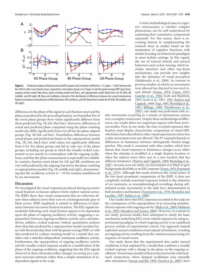
differences in the phase of the signal at each fixation onset and thephase as predicted by the preceding fixation, we found that five ofthe seven phase groups show values significantly different fromthose predicted (Fig. 5B, left, blue line). Moreover, differences ofactual and predicted phase computed using the phase-resettingmodel also differ significantly from 0 in all but the phase-alignedgroups (Fig. 5B, left, red line). Nonetheless, differences betweenactual phases and predictions based on the superposition model(Fig. 5B, left, black line) yield values not significantly differentfrom 0 for five phase groups and fail in only two of the phasegroups, including one group in which the visually evoked com-ponent occurs exactly in counterphase with the ongoing oscilla-tions, and thus the phase measurement is expectedly less reliable.In contrast, fixation-onset phase for GR and BK conditions arevery well predicted by the ongoing, but not by the phase-resettingor superposition models (Fig. 5B, middle and right), demonstrat-ing that the oscillatory activity at �10 Hz remains uninfluencedby eye movements.
DiscussionWe investigated the visual responses produced during successivevisual fixations as human subjects freely explore natural scenes.The fERPs show clear visual components that are otherwise ab-sent when subjects move their eyes on a homogeneously gray orblack screen. fERP amplitude is related to differences in lumi-nance between successive fixation locations. The EEG signals im-mediately following each visual fixation appear to be dependentupon the phase of ongoing oscillatory activity, suggesting a su-perposition between ongoing oscillatory activity and a stimulus-driven, additive, evoked response. Indeed, quantitative analysesshow that data predicted by a superposition model correlate bet-ter with the actual data than with the grand-average fERP, or withdata predicted by a phase-resetting model or a model that con-siders only a stimulus-independent continuous oscillatory activity.Furthermore, the superposition of ongoing oscillatory activityand the visually evoked response results in a modification of thephase of the ongoing oscillation, suggesting that the EEG signalsobserved in these electrodes reflect changes occurring in a com-mon neuronal substrate rather than a simple summation of in-dependent signals at the scalp.
A main methodological issue in cogni-tive neuroscience is whether complexphenomena can be well understood byexplaining their constitutive componentsseparately. For this reason, there is in-creasing interest in complementing theresearch done in studies based on theitemization of cognitive functions withstudies focusing on behavioral paradigmsin more holistic settings. In this regard,the use of natural stimuli and naturalbehaviors such as free viewing, which in-cludes attention and other top-downmechanisms, can provide new insightsinto the dynamics of visual perception(Maldonado et al., 2008). In contrast toprevious studies, in which eye movementswere allowed but directed to low-level vi-sual stimuli (Evans, 1953; Green, 1957;Gaarder et al., 1964; Scott and Bickford,1967; Scott et al., 1967, 1981; Barlow andCiganek, 1969; Yagi, 1981; Riemslag et al.,1987; Billings, 1989; Thickbroom et al.,1991), our study was performed with oc-
ular movements occurring in a stream of autonomous actionsover a complex visual scene. Despite these methodological differ-ences, our results share two important aspects with these previ-ous studies. First, in our study, the clear occipital responses afterfixation onset display characteristic components of visual ERP,which have been described in other visual experiments where freeocular movements were not allowed. The P1 responds linearly todifferences in luminance between subsequently fixated imagepatches. This result is consistent with other studies, which haveshown that visual responses to luminance changes occur eitherwhen the stimulus is modified at a single fixation location orwhen the subjects move their eyes to a new location that hasdifferent luminance (Barlow and Ciganek, 1969; Riemslag et al.,1987). Second, as in our study, eye movements over black or graybackgrounds elicit little or no EEG response (Evans, 1953; Fourmentet al., 1976). Although this result reinforces the visual nature ofthe two most prominent components of the fERP, it does notcompletely exclude nonvisual responses locked to the initiationof eye moments, as neurophysiological recordings during self-initiated ocular movements in the dark have demonstrated inboth monkeys and humans (Fourment et al., 1976; Skrandies andLaschke, 1997; Rajkai et al., 2008).
Our results show that EEG responses recorded at the scalp arethe consequence of the superposition of an incoming stimulus-driven response with ongoing activity (Shah et al., 2004; Makinenet al., 2005; Mazaheri and Jensen, 2006). However, in contrast toour study, previous studies have attempted to clarify the basicmechanism underlying EEG event-related responses by using ex-perimental paradigms in which ongoing activity is an undefinedprocess outside of experimental control. Our approach insteadexploited natural conditions of perceptual stimulation, revealingan ongoing activity resulting from the continuous processing of asequence of visual events.
Our study shows that the experimental data under naturalconditions is best explained by a model that combines a visuallyevoked response with a change in the phase on the ongoing os-cillation. Neural mass models of hierarchical networks with back-ward connections, where damped oscillations arise naturallyafter stimulation (Jansen and Rit, 1995; David et al., 2005), have
A
B
Figure 5. Predictions of phase at fixation onset and EEG response. A, Correlation coefficients (�CI, alpha�0.001) between thefirst 200 ms after every fixation onset, separated in seven phase groups (as in Figure 4) and the grand average fERP (gray lines),ongoing-activity model (blue lines), phase-resetting model (red lines), and superposition model (black lines) for NS (left), BK(middle), and GR (right). B, Mean and confidence intervals of the distribution of differences between the actual instantaneousfixation onset phase and predictions of ONG (blue lines), RES (red lines), and SUP (black lines) models for NS (left), BK (middle), andGR (right).
Ossandon et al. • EEG Activity during Natural Scenes J. Neurosci., March 31, 2010 • 30(13):4787– 4795 • 4793

produced theoretical predictions that agree with the resultsshown here. These kind of models have been successful in pre-dicting late components of real ERPs (Garrido et al., 2007). Twotypes of competing and mutually exclusive theories have soughtto explain the nature of the observed visually evoked EEG signals.The additive model assumes that ERPs are a summation of syn-chronized neuronal activity directly evoked by the stimulus, withunrelated ongoing activity produced by independent neuronalsources or noise whereas the other model assumes a phase-resetting mechanism of ongoing oscillatory activity. This lattermodel has also been generalized to a formulation that includesnot only phase reorganization but also evoked and induced oscil-lations as underlying sources of ERP component (Klimesch et al.,2007b). Our results show that both neuronal activity evoked bythe stimulus and oscillatory phase changes occur during naturalviewing. Phase changes would, however, not occur instantly andindependently of the visually evoked response, but would insteadappear as a result of changes in the dynamics of the neuronalactivity in a common neuronal substrate. Nevertheless, it is im-portant to point out the limitation of instantaneous phase esti-mation in narrow frequency bands of time series that aregenerated by stochastic and nonstationary processes. Waveletanalysis as used here can result in phase biases produced by anevoked response after or before the onset of a fixation (Sauseng etal., 2007). In our data, phase measurements at fixation onset arein good agreement with the previous oscillatory phase rather thanthe evoked component, suggesting this bias has a small impact.
Ongoing oscillatory activity is not a mixture of evoked poten-tials resulting from previous fixations (Fig. 4), suggesting that thepower peaks in the frequency analysis are due, at least in part, tounderlying oscillatory processes. Oscillatory activity in low- tomedium-frequency ranges is usually associated with a mecha-nism for long-range coordination between different brain areasengaged in recurrent loops of activity (Engel et al., 2001; Fries,2005; Schroeder and Lakatos, 2009). Also, a specific role for os-cillations in the alpha range in the generation of visual ERP com-ponents has been suggested, with both inhibitory and long-rangecoordination roles (Klimesch et al., 2007a). In this sense, ongoingactivity during continuous and autonomous selection of stimulicould be both quantitatively and qualitatively different to ongo-ing activity in the case of nonstimulation and when an unpredict-able new stimulus is expected. The latter kind of experimentalcondition might be comparable to the BK and GR condition usedin our experiment. In this case, a visual stimulus after each move-ment is absent or homogeneous and oscillatory power is centeredat a frequency in the middle of the alpha range, typical of pre-stimulation periods in static paradigms. This activity could pos-sibly reflect an inhibitory effect in posterior sensory areas relatedto the lack of change in visual stimulation. In contrast, in the NScondition a peak in a slightly lower frequency is revealed. Takinginto account the limitations of frequency resolution in neighbor-ing frequencies, these results are in good agreement with previousreports of change in the alpha range activity after a visual stimulusevent, in which activity at the middle alpha range (�10 Hz) ispartially suppressed and activity at upper-theta/low-alpha rangeis increased (Gruber et al., 2005; Mazaheri and Jensen, 2006;Freunberger et al., 2008). However, in contrast to reports thatexplain this upper-theta/low-alpha activity as an additive com-ponent (Mazaheri and Jensen, 2006), we show that the activity atthe low-alpha band is a main component of the ongoing activitywhen stimulation is continuous. Although we do not necessarilycommit to an interpretation of the additive component we foundas an evoked oscillation, in the situation of continuous stimula-
tion, it is expected that the neuronal network underlying visualoscillatory phenomena will be uninterruptedly active (David etal., 2005) and therefore the lower-alpha activity seen after single-stimulus presentation in other studies might keep going in thecase of free viewing. In this framework, the arrival of a new input,expressed as an additive component, perturbs the activity of thenetwork, resulting in superposition patterns, maintenance ofthe oscillatory activity, and a change in the phase of oscillation ofthe network. In the NS condition, oscillatory activity is apparentduring continuous visual stimulation, and its phase is changedwith every new fixation, depending on both oscillatory and in-coming components.
Further studies of visual responses in natural viewing experi-ments should consider activity statistics that result from the con-tinuous interdependence of events. In contrast to responses tostatic and transient visual stimulation, neuronal activity duringnatural viewing may contain features intrinsic to the natural flowof goal-oriented behavior and thus would include patterns thatare instrumental for building models of visual perception.
ReferencesBarlow JS, Ciganek L (1969) Lambda responses in relation to visual evoked
responses in man. Electroencephalogr Clin Neurophysiol 26:183–192.Batschelet E (1981) Circular statistics on biology. New York: Academic.Bell AJ, Sejnowski TJ (1995) An information-maximization approach to
blind separation and blind deconvolution. Neural Comput 7:1129 –1159.Billings RJ (1989) The origin of the occipital lambda wave in man. Electro-
encephalogr Clin Neurophysiol 72:95–113.David O, Harrison L, Friston KJ (2005) Modelling event-related responses
in the brain. Neuroimage 25:756 –770.Delorme A, Makeig S (2004) EEGLAB: an open source toolbox for analysis
of single-trial EEG dynamics including independent component analysis.J Neurosci Methods 134:9 –21.
Engel AK, Fries P, Singer W (2001) Dynamic predictions: oscillations andsynchrony in top-down processing. Nat Rev Neurosci 2:704 –716.
Evans CC (1953) Spontaneous excitation of the visual cortex and asso-ciation areas: lambda waves. Electroencephalogr Clin Neurophysiol5:69 –74.
Fourment A, Calvet AF, Bancaud J (1976) Electrocorticography of wavesassociated with eye movements in man during wakefulness. Electroen-cephalogr Clin Neurophysiol 40:457– 469.
Freunberger R, Holler Y, Griesmayr B, Gruber W, Sauseng P, Klimesch W(2008) Functional similarities between the P1 component and alpha os-cillations. Eur J Neurosci 27:2330 –2340.
Fries P (2005) A mechanism for cognitive dynamics: neuronal communica-tion through neuronal coherence. Trends Cogn Sci 9:474 – 480.
Gaarder K, Krauskopf J, Graf V, Kropfl W, Armington JC (1964) Averagedbrain activity following saccadic eye movement. Science 146:1481–1483.
Garrido MI, Kilner JM, Kiebel SJ, Friston KJ (2007) Evoked brain responsesare generated by feedback loops. Proc Natl Acad Sci U S A104:20961–20966.
Green J (1957) Some observations on lambda waves and peripheral stimu-lation. Electroencephalogr Clin Neurophysiol 9:691–704.
Gruber WR, Klimesch W, Sauseng P, Doppelmayr M (2005) Alpha phasesynchronization predicts P1 and N1 latency and amplitude size. CerebCortex 15:371–377.
Hanslmayr S, Klimesch W, Sauseng P, Gruber W, Doppelmayr M, FreunbergerR, Pecherstorfer T, Birbaumer N (2007) Alpha phase reset contributes tothe generation of ERPs. Cereb Cortex 17:1–8.
Hoffmann S, Falkenstein M (2008) The correction of eye blink artefacts inthe EEG: a comparison of two prominent methods. PLoS One 3:e3004.
Iriarte J, Urrestarazu E, Valencia M, Alegre M, Malanda A, Viteri C, Artieda J(2003) Independent component analysis as a tool to eliminate artifacts inEEG: a quantitative study. J Clin Neurophysiol 20:249 –257.
Jansen BH, Rit VG (1995) Electroencephalogram and visual evoked poten-tial generation in a mathematical model of coupled cortical columns. BiolCybern 73:357–366.
Jung TP, Makeig S, Westerfield M, Townsend J, Courchesne E, Sejnowski TJ(2000a) Removal of eye activity artifacts from visual event-related poten-tials in normal and clinical subjects. Clin Neurophysiol 111:1745–1758.
4794 • J. Neurosci., March 31, 2010 • 30(13):4787– 4795 Ossandon et al. • EEG Activity during Natural Scenes

Jung TP, Makeig S, Humphries C, Lee TW, McKeown MJ, Iragui V, SejnowskiTJ (2000b) Removing electroencephalographic artifacts by blind sourceseparation. Psychophysiology 37:163–178.
Kayser C, Petkov CI, Logothetis NK (2008) Visual modulation of neurons inauditory cortex. Cereb Cortex 18:1560 –1574.
Klimesch W, Sauseng P, Hanslmayr S (2007a) EEG alpha oscillations: theinhibition-timing hypothesis. Brain Res Rev 53:63– 88.
Klimesch W, Sauseng P, Hanslmayr S, Gruber W, Freunberger R (2007b)Event-related phase reorganization may explain evoked neural dynamics.Neurosci Biobehav Rev 31:1003–1016.
Lakatos P, Chen CM, O’Connell MN, Mills A, Schroeder CE (2007) Neuro-nal oscillations and multisensory interaction in primary auditory cortex.Neuron 53:279 –292.
Makeig S, Westerfield M, Jung TP, Enghoff S, Townsend J, Courchesne E,Sejnowski TJ (2002) Dynamic brain sources of visual evoked responses.Science 295:690 – 694.
Makinen V, Tiitinen H, May P (2005) Auditory event-related responsesare generated independently of ongoing brain activity. Neuroimage24:961–968.
Maldonado PE, Ossandon JP, Flores FJ (2008) Attention and neurodynamicalcorrelates of natural vision. In: From attention to goal-directed behavior:neurodynamical and methodological and clinical trends (Aboitiz F, Cos-melli D, eds), pp 67– 82. Berlin: Springer.
Mazaheri A, Jensen O (2006) Posterior alpha activity is not phase-reset byvisual stimuli. Proc Natl Acad Sci U S A 103:2948 –2952.
Mehta MR, Lee AK, Wilson MA (2002) Role of experience and oscillationsin transforming a rate code into a temporal code. Nature 417:741–746.
Min BK, Busch NA, Debener S, Kranczioch C, Hanslmayr S, Engel AK,Herrmann CS (2007) The best of both worlds: phase-reset of humanEEG alpha activity and additive power contribute to ERP generation. IntJ Psychophysiol 65:58 – 68.
Rajkai C, Lakatos P, Chen CM, Pincze Z, Karmos G, Schroeder CE (2008)Transient cortical excitation at the onset of visual fixation. Cereb Cortex18:200 –209.
Riemslag FC, van der Heijde GL, van Dongen MM (1987) Are eye move-
ment evoked potentials different from pattern reversal evoked potentials?Doc Ophthalmol 66:279 –289.
Sauseng P, Klimesch W, Gruber WR, Hanslmayr S, Freunberger R, DoppelmayrM (2007) Are event-related potential components generated by phaseresetting of brain oscillations?: A critical discussion. Neuroscience146:1435–1444.
Sayers BM, Beagley HA, Henshall WR (1974) The mechanism of auditoryevoked EEG responses. Nature 247:481– 483.
Schroeder CE, Lakatos P (2009) Low-frequency neuronal oscillations as in-struments of sensory selection. Trends Neurosci 32:9 –18.
Scott DF, Bickford RG (1967) Electrophysiologic studies during scanningand passive eye movements in humans. Science 155:101–102.
Scott DF, Groethuysen UC, Bickford RG (1967) Lambda responses in thehuman electroencephalogram. Neurology 17:770 –778.
Scott DF, Moffett A, Bickford RG (1981) Comparison of two types of visualevoked potentials: pattern reversal and eye movement (lambda). Electro-encephalogr Clin Neurophysiol 52:102–104.
Shah AS, Bressler SL, Knuth KH, Ding M, Mehta AD, Ulbert I, Schroeder CE(2004) Neural dynamics and the fundamental mechanisms of event-related brain potentials. Cereb Cortex 14:476 – 483.
Skrandies W, Laschke K (1997) Topography of visually evoked brain activ-ity during eye movements: lambda waves, saccadic suppression, and dis-crimination performance. Int J Psychophysiol 27:15–27.
Tallon-Baudry C, Bertrand O, Delpuech C, Pernier J (1996) Stimulus spec-ificity of phase-locked and non-phase-locked 40 Hz visual responses inhuman. J Neurosci 16:4240 – 4249.
Thickbroom GW, Knezevic W, Carroll WM, Mastaglia FL (1991) Saccadeonset and offset lambda waves: relation to pattern movement visuallyevoked potentials. Brain Res 551:150 –156.
Womelsdorf T, Schoffelen JM, Oostenveld R, Singer W, Desimone R, EngelAK, Fries P (2007) Modulation of neuronal interactions through neuro-nal synchronization. Science 316:1609 –1612.
Yagi A (1981) Visual signal detection and lambda responses. Electroen-cephalogr Clin Neurophysiol 52:604 – 610.
Yarbus AL (1967) Eye movements and vision. New York: Plenum.
Ossandon et al. • EEG Activity during Natural Scenes J. Neurosci., March 31, 2010 • 30(13):4787– 4795 • 4795