sternal precautions - ace medical co
TRANSCRIPT
Cardiopulmonary Physical Therapy JournalVol 22 No 1 March 2011 5
Sternal Precautions: Is It Time for Change?Precautions versus Restrictions – A Review of Literature
and Recommendations for RevisionLawrence P. Cahalin, PT, PhD, CCS, FAACVPR;1 Tanya Kinney LaPier, PT, PhD, CCS;2
Donald K. Shaw, PT, PhD, D.Min., FAACVPR3
1Northeastern University, Boston, MA2Eastern Washington University, Spokane, WA
3Midwestern University, Glendale, AZ
Address correspondence to: Lawrence P. Cahalin, PT, PhD, CCS, FAACVPR, Northeastern University, Physi-cal Therapy Program, 6 Robinson Hall, 360 Hunting-ton Avenue, Boston, MA 02115 Ph: 617-373-3783, Fax: 617-373-3161 ([email protected]).
ABSTRACTThe processes that occur with normal sternal healing and potential complications related to median sternotomy are of particular interest to physical therapists. The premise of patients following sternal precautions (SP) or specific activ-ity restrictions is the belief that avoiding certain movements will reduce risk of sternal complications. However, current research has identified that many patients remain function-ally impaired long after cardiothoracic surgery. It is possi-ble that some SP may contribute to such functional impair-ments. Currently, SP have several limitations including that they: (1) have no universally accepted definition, (2) are of-ten based on anecdotal/expert opinion or at best supported by indirect evidence, (3) are mostly applied uniformly for all patients without regard to individual differences, and (4) may be overly restrictive and therefore impede ideal recov-ery. The purpose of this article is to present an overview of current research and commentary on median sternotomy procedures and activity restrictions. We propose that the optimal degree and duration of SP should be based on an individual patient’s characteristics (eg, risk factors, comor-bidities, previous activity level) that would enable physical activity to be targeted to particular limitations rather than restricting specific functional tasks and physical activity. Such patient-specific SP focusing on function may be more likely to facilitate recovery after median sternotomy and less likely to impede it.
Key Words: median sternotomy, sternal precautions, physi-cal therapy, exercise protocols
INTRODUCTIONSternal precautions (SP) are almost universally given
to patients following median sternotomy surgeries. How-ever, in clinical practice, SP most commonly represent a wide variety of functional restrictions. In fact, the word ‘precautions’ should probably be replaced by the word ‘re-
strictions’ since this is what many physical therapists have encountered in clinical practice over the years. Restric-tions in shoulder range of motion, lifting, reaching, dress-ing, exercise, driving, and a variety of other tasks have been reported. However, the exact origin of such restrictions is difficult to find. Furthermore, there appears to be no con-sistency in the type or duration of restriction.
The purpose of this paper is to review the available research related to the median sternotomy procedure and physical activity. We begin with a historical perspective of physical activity after myocardial infarction (MI) and a brief overview of coronary artery bypass graft (CABG) surgery. We review the literature regarding (1) complications after cardiac surgery and median sternotomy, (2) symptoms and functional status after cardiac surgery, and (3) the changes in pulmonary function and thoracic motion after cardiac surgery. Finally, we propose an algorithm highlighting the role that appropriately prescribed exercise and functional training, based on specific patient characteristics and limi-tations, may have in improving outcomes after a median sternotomy. Such patient specific precautions, rather than restrictions, which focus on function, may be more likely to facilitate recovery after median sternotomy and less likely to impede it.
HISTORICAL PERSPECTIVEThe attitude regarding physical activity following the
onset of symptomatic coronary artery disease has under-gone substantial change in the past two centuries. Prior to 1876, many practitioners favored moderate activity for patients experiencing angina pectoris and with what was later recognized as MI.1 This position was expressed by Aus-tin Flint in 1886: “Patients exchanging habits of activity for complete rest are likely to become rapidly worse.”2 Near the turn of the 19th century, however, physicians became more conservative following observations linking early ac-tivity to sudden death in several patients with rheumatic fever.3 Early studies dealing with MI and with ventricular aneurysm development during acute MI recovery seemed to favor a more cautious approach to physical rehabilita-tion.4,5
A study supporting the prescription of physical activity after MI appeared in 1944. Levine6 found prolonged im-mobility contributed to 11 negative sequelae ranging from
Cardiopulmonary Physical Therapy Journal Vol 22 No 1 March 20116
bone demineralization to venous thrombosis. He theorized that the sitting position was beneficial because it promoted peripheral venous pooling and decreased venous return thereby reducing the work of the heart. Patients were per-mitted to sit in a chair for one to 2 hours beginning the first post-MI day. This procedure became known as the “arm-chair treatment of coronary thrombosis.”7 Subsequent early studies supported this rehabilitation approach.8,9 Other re-search followed demonstrating both the physiological and psychological benefits of early exercise participation.10-13
Coronary Artery Bypass Graft SurgeryDuring the 1960s, CABG surgery was introduced as a
surgical adjunct to the medical treatment of coronary heart disease.14 Acceptance of this procedure was almost imme-diate; in 1968 René Favaloro and his Cleveland Clinic col-leagues performed 171 CABG operations.15 By 1979, over 100,000 CABG surgeries were documented in America at an average cost to patients of $5,000.16 Not surprisingly, both frequency and expense mushroomed by 2006. A to-tal of 448,000 CABG surgeries were performed that year with procedures ranging from $50,000 to $100,000 per patient.17 Despite the cost involved, it appears CABG sur-gery will continue as a practical option for coronary heart disease treatment until a viable, nonoperative substitute is found.
As the name implies, CABG surgical technique involves locating and ‘bypassing’ occluded coronary arteries. Often a section from the saphenous vein is selected as the conduit of choice; one end of the vessel is affixed to the aorta while the other end is anastomosed to the blocked coronary ar-tery distal to the occlusion. Arterial conduits are often used as well. A partial list of these vessels includes the inter-nal thoracic (aka internal mammary), radial, subscapular, inferior epigastric, and right gastroepiglotic arteries.18 His-torically, all CABG surgeries were performed via a median sternotomy.
With the advent of CABG surgery, a unique group of pa-tients was added to those traditionally involved in cardiac rehabilitation. First, since CABG surgery improves coro-nary blood flow reducing anginal symptoms, these patients become excellent candidates for more aggressive therapy than their post-MI contemporaries.19 Second, since surgi-cal exposure of the heart is often accomplished via median sternotomy, considerable strain is placed on the anatomy of the chest, back, shoulders, and neck as sternal halves are retracted. Thus, it is not uncommon for patients following CABG surgery to manifest with a variety of musculoskeletal and neurological complaints from the procedure.20 Lastly, surgical site infection involving soft and bony tissues al-ways exists as a possible threat. Indeed, early CABG sur-geries were plagued with high infection rates – sometimes resulting in sternectomy or death. Sternal infections and dehiscence at that time were reported in 0.5% to 8.4% of cases with mortality running between 14% and 50% when infection was present.21
STERNAL PRECAUTIONS—CURRENT STATUSThe exact genesis of SP is unknown. However, it is like-
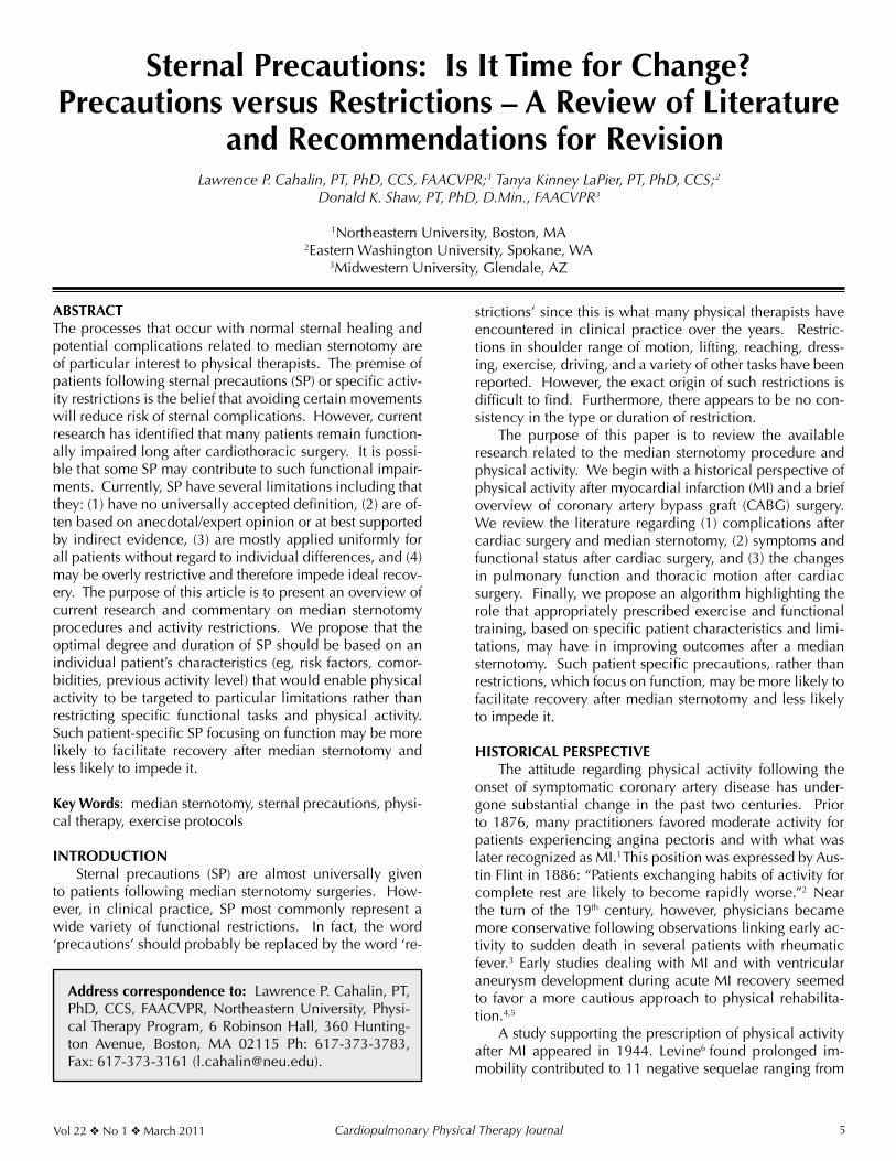
ly that initial concerns regarding sternal infection height-ened topic awareness. Add to this the idea that sternum healing might be compromised by certain upper extrem-ity movements and the ‘precaution’ (or really ‘restriction’) stage was set. Although impaired, sternal healing had never been proven empirically, nurses and therapists be-gan presenting patients with a list of proscribed postsurgi-cal movements and activities (Figure 1). Such lists often warned against arm movements above shoulder level (90°
of flexion/abduction) and scapular adduction. Other pre-cautions included not lifting more than 5 to 10 pounds, avoiding weight-bearing through the upper extremity (ie, using arm rests to stand), and avoiding unilateral reaching posteriorly (ie, providing support while sitting).
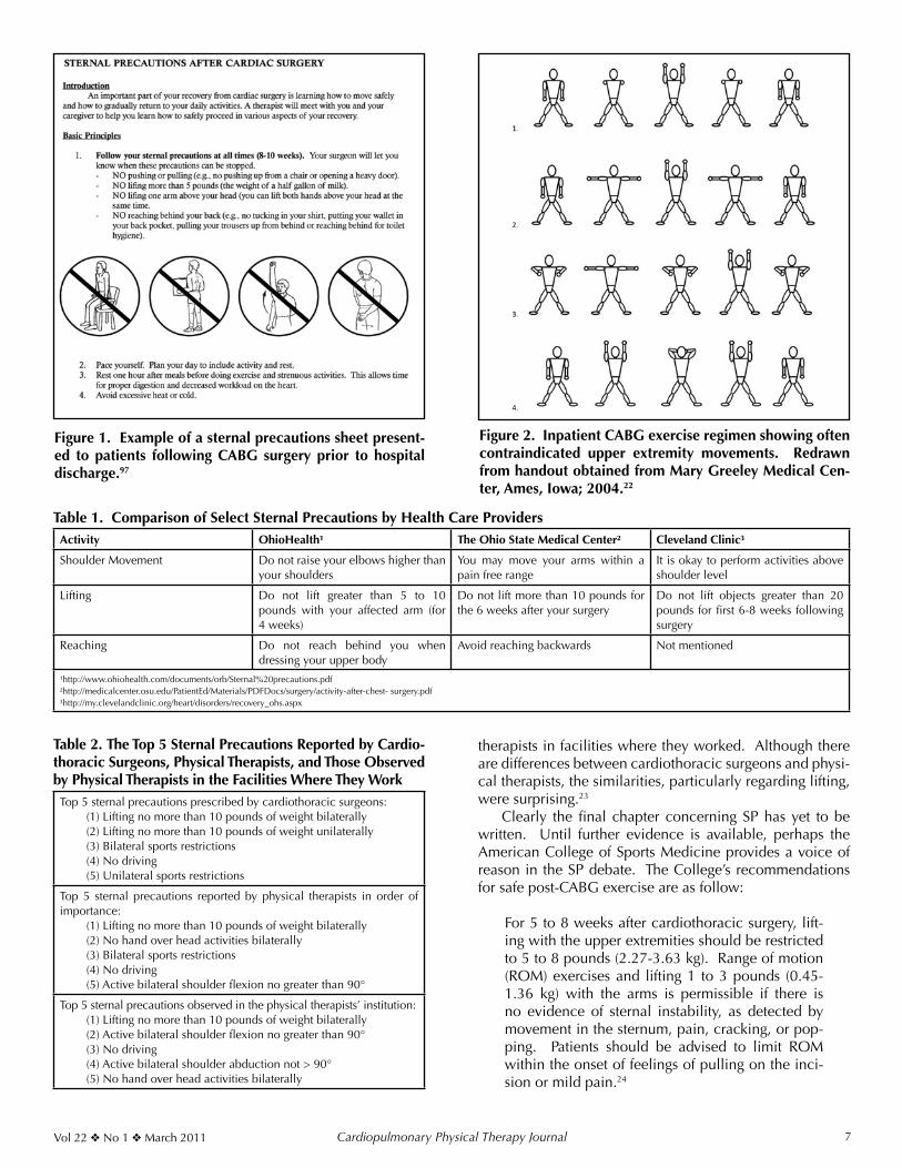
Over time, SP became synonymous with responsible pa-tient care. A plethora of protocols subsequently emerged, often with conflicting advice. Table 1 is illustrative of this point. Note the absence of agreement between 3 health care agencies (all residing in the same state) on shoulder movement, lifting, and reaching. OhioHealth limits shoul-der movement to 90° while the Cleveland Clinic approves movement above shoulder level. The Ohio State Medi-cal Center limits lifting to 10 pounds while the Cleveland Clinic doubles this amount. Also confusing is OhioHealth’s warning about lifting with “your affected arm” since CABG surgery is performed on the chest. Moreover, both Ohio-Health and The Ohio State Medical Center prohibit reach-ing backward while the Cleveland Clinic is without com-ment on the matter. Such lack of consensus pertaining to SP not infrequently leads to contentious interactions be-tween surgeons, nurses, and rehabilitation team members.
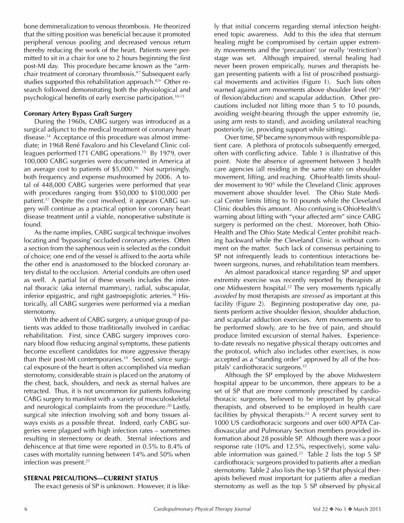
An almost paradoxical stance regarding SP and upper extremity exercise was recently reported by therapists at one Midwestern hospital.22 The very movements typically avoided by most therapists are stressed as important at this facility (Figure 2). Beginning postoperative day one, pa-tients perform active shoulder flexion, shoulder abduction, and scapular adduction exercises. Arm movements are to be performed slowly, are to be free of pain, and should produce limited excursion of sternal halves. Experience-to-date reveals no negative physical therapy outcomes and the protocol, which also includes other exercises, is now accepted as a “standing order” approved by all of the hos-pitals’ cardiothoracic surgeons.22
Although the SP employed by the above Midwestern hospital appear to be uncommon, there appears to be a set of SP that are more commonly prescribed by cardio-thoracic surgeons, believed to be important by physical therapists, and observed to be employed in health care facilities by physical therapists.23 A recent survey sent to 1000 US cardiothoracic surgeons and over 600 APTA Car-diovascular and Pulmonary Section members provided in-formation about 28 possible SP. Although there was a poor response rate (10% and 12.5%, respectively), some valu-able information was gained.23 Table 2 lists the top 5 SP cardiothoracic surgeons provided to patients after a median sternotomy. Table 2 also lists the top 5 SP that physical ther-apists believed most important for patients after a median sternotomy as well as the top 5 SP observed by physical
Cardiopulmonary Physical Therapy JournalVol 22 No 1 March 2011 7
Figure 1. Example of a sternal precautions sheet present-ed to patients following CABG surgery prior to hospital discharge.97
Figure 2. Inpatient CABG exercise regimen showing often contraindicated upper extremity movements. Redrawn from handout obtained from Mary Greeley Medical Cen-ter, Ames, Iowa; 2004.22
Table 1. Comparison of Select Sternal Precautions by Health Care ProvidersActivity OhioHealth¹ The Ohio State Medical Center² Cleveland Clinic³
Shoulder Movement Do not raise your elbows higher than your shoulders
You may move your arms within a pain free range
It is okay to perform activities above shoulder level
Lifting Do not lift greater than 5 to 10 pounds with your affected arm (for 4 weeks)
Do not lift more than 10 pounds for the 6 weeks after your surgery
Do not lift objects greater than 20 pounds for first 6-8 weeks following surgery
Reaching Do not reach behind you when dressing your upper body
Avoid reaching backwards Not mentioned
¹http://www.ohiohealth.com/documents/orb/Sternal%20precautions.pdf ²http://medicalcenter.osu.edu/PatientEd/Materials/PDFDocs/surgery/activity-after-chest- surgery.pdf³http://my.clevelandclinic.org/heart/disorders/recovery_ohs.aspx
Table 2. The Top 5 Sternal Precautions Reported by Cardio-thoracic Surgeons, Physical Therapists, and Those Observed by Physical Therapists in the Facilities Where They Work
Top 5 sternal precautions prescribed by cardiothoracic surgeons:(1) Lifting no more than 10 pounds of weight bilaterally(2) Lifting no more than 10 pounds of weight unilaterally(3) Bilateral sports restrictions(4) No driving(5) Unilateral sports restrictions
Top 5 sternal precautions reported by physical therapists in order of importance:
(1) Lifting no more than 10 pounds of weight bilaterally(2) No hand over head activities bilaterally(3) Bilateral sports restrictions(4) No driving(5) Active bilateral shoulder flexion no greater than 90°
Top 5 sternal precautions observed in the physical therapists’ institution:(1) Lifting no more than 10 pounds of weight bilaterally(2) Active bilateral shoulder flexion no greater than 90°(3) No driving(4) Active bilateral shoulder abduction not > 90°(5) No hand over head activities bilaterally
therapists in facilities where they worked. Although there are differences between cardiothoracic surgeons and physi-cal therapists, the similarities, particularly regarding lifting, were surprising.23
Clearly the final chapter concerning SP has yet to be written. Until further evidence is available, perhaps the American College of Sports Medicine provides a voice of reason in the SP debate. The College’s recommendations for safe post-CABG exercise are as follow:
For 5 to 8 weeks after cardiothoracic surgery, lift-ing with the upper extremities should be restricted to 5 to 8 pounds (2.27-3.63 kg). Range of motion (ROM) exercises and lifting 1 to 3 pounds (0.45-1.36 kg) with the arms is permissible if there is no evidence of sternal instability, as detected by movement in the sternum, pain, cracking, or pop-ping. Patients should be advised to limit ROM within the onset of feelings of pulling on the inci-sion or mild pain.24
Cardiopulmonary Physical Therapy Journal Vol 22 No 1 March 20118
REVIEW OF LITERATUREMedian Sternotomy Complications
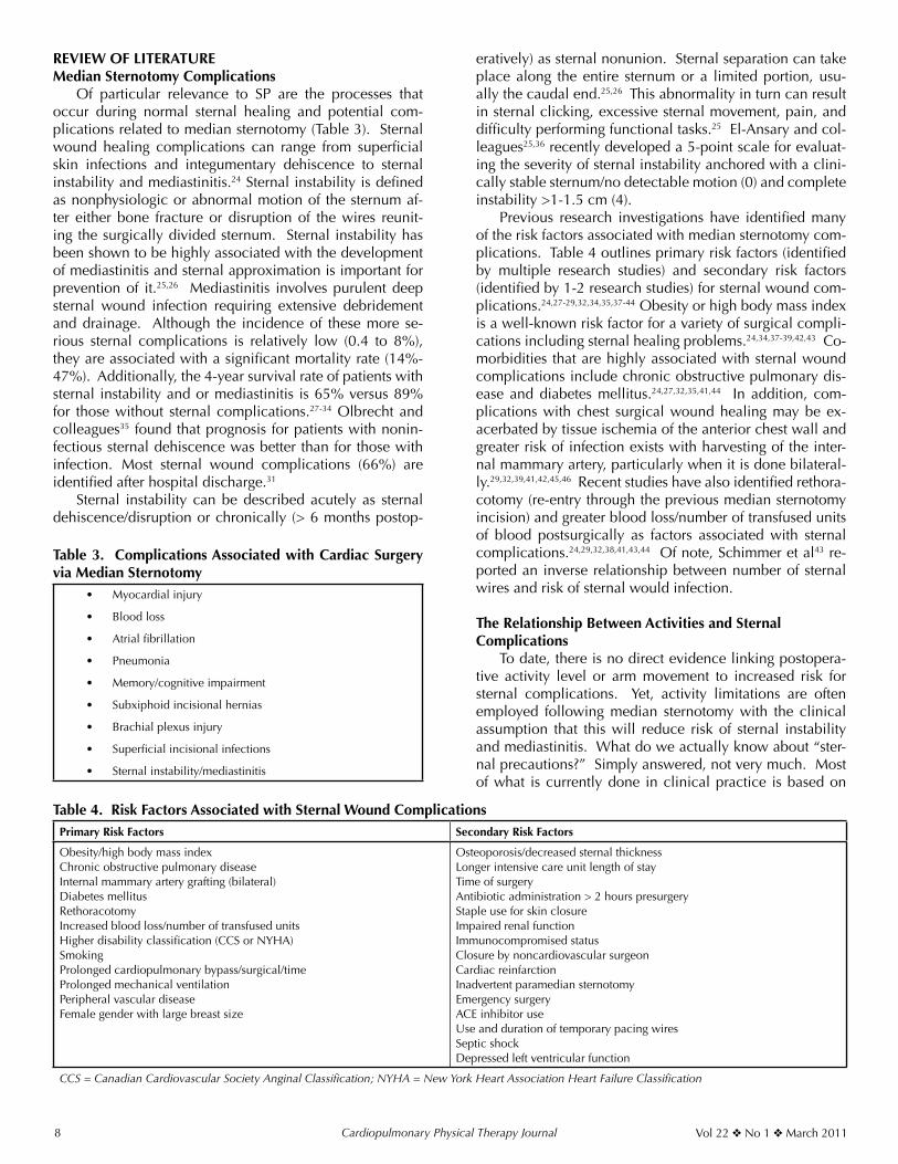
Of particular relevance to SP are the processes that occur during normal sternal healing and potential com-plications related to median sternotomy (Table 3). Sternal wound healing complications can range from superficial skin infections and integumentary dehiscence to sternal instability and mediastinitis.24 Sternal instability is defined as nonphysiologic or abnormal motion of the sternum af-ter either bone fracture or disruption of the wires reunit-ing the surgically divided sternum. Sternal instability has been shown to be highly associated with the development of mediastinitis and sternal approximation is important for prevention of it.25,26 Mediastinitis involves purulent deep sternal wound infection requiring extensive debridement and drainage. Although the incidence of these more se-rious sternal complications is relatively low (0.4 to 8%), they are associated with a significant mortality rate (14%-47%). Additionally, the 4-year survival rate of patients with sternal instability and or mediastinitis is 65% versus 89% for those without sternal complications.27-34 Olbrecht and colleagues35 found that prognosis for patients with nonin-fectious sternal dehiscence was better than for those with infection. Most sternal wound complications (66%) are identified after hospital discharge.31
Sternal instability can be described acutely as sternal dehiscence/disruption or chronically (> 6 months postop-
eratively) as sternal nonunion. Sternal separation can take place along the entire sternum or a limited portion, usu-ally the caudal end.25,26 This abnormality in turn can result in sternal clicking, excessive sternal movement, pain, and difficulty performing functional tasks.25 El-Ansary and col-leagues25,36 recently developed a 5-point scale for evaluat-ing the severity of sternal instability anchored with a clini-cally stable sternum/no detectable motion (0) and complete instability >1-1.5 cm (4).
Previous research investigations have identified many of the risk factors associated with median sternotomy com-plications. Table 4 outlines primary risk factors (identified by multiple research studies) and secondary risk factors (identified by 1-2 research studies) for sternal wound com-plications.24,27-29,32,34,35,37-44 Obesity or high body mass index is a well-known risk factor for a variety of surgical compli-cations including sternal healing problems.24,34,37-39,42,43 Co-morbidities that are highly associated with sternal wound complications include chronic obstructive pulmonary dis-ease and diabetes mellitus.24,27,32,35,41,44 In addition, com-plications with chest surgical wound healing may be ex-acerbated by tissue ischemia of the anterior chest wall and greater risk of infection exists with harvesting of the inter-nal mammary artery, particularly when it is done bilateral-ly.29,32,39,41,42,45,46 Recent studies have also identified rethora-cotomy (re-entry through the previous median sternotomy incision) and greater blood loss/number of transfused units of blood postsurgically as factors associated with sternal complications.24,29,32,38,41,43,44 Of note, Schimmer et al43 re-ported an inverse relationship between number of sternal wires and risk of sternal would infection.
The Relationship Between Activities and Sternal Complications
To date, there is no direct evidence linking postopera-tive activity level or arm movement to increased risk for sternal complications. Yet, activity limitations are often employed following median sternotomy with the clinical assumption that this will reduce risk of sternal instability and mediastinitis. What do we actually know about “ster-nal precautions?” Simply answered, not very much. Most of what is currently done in clinical practice is based on
Table 3. Complications Associated with Cardiac Surgery via Median Sternotomy
Table 4. Risk Factors Associated with Sternal Wound ComplicationsPrimary Risk Factors Secondary Risk Factors
Obesity/high body mass indexChronic obstructive pulmonary diseaseInternal mammary artery grafting (bilateral)Diabetes mellitusRethoracotomyIncreased blood loss/number of transfused unitsHigher disability classification (CCS or NYHA)SmokingProlonged cardiopulmonary bypass/surgical/timeProlonged mechanical ventilationPeripheral vascular diseaseFemale gender with large breast size
Osteoporosis/decreased sternal thicknessLonger intensive care unit length of stayTime of surgeryAntibiotic administration > 2 hours presurgeryStaple use for skin closureImpaired renal function Immunocompromised statusClosure by noncardiovascular surgeonCardiac reinfarctionInadvertent paramedian sternotomyEmergency surgeryACE inhibitor useUse and duration of temporary pacing wiresSeptic shockDepressed left ventricular function
CCS = Canadian Cardiovascular Society Anginal Classification; NYHA = New York Heart Association Heart Failure Classification
Cardiopulmonary Physical Therapy JournalVol 22 No 1 March 2011 9
anecdotal evidence and expert opinion. Some indirect evidence is provided by cadaver studies using material en-gineering approaches. Cohen and Griffin47 evaluated the biomechanical properties of 3 different sternotomy closure techniques and found that sternal separation occurred as a result of wires cutting through bone. Also, sternal distrac-tion (2.0 mm) occurred with the least force in the lateral direction and the greatest force in the rostral-caudal direc-tion with anterior-posterior force intermediate.47 In another investigation, greater separation occurred at the lower end of the sternum than the upper.48
Some studies have provided indirect evidence of the stresses imposed on the sternum during different activi-ties and exercises. Recently, El-Ansary and colleagues25,49 examined a variety of upper body activities in patients with chronic sternal instability. In this patient popula-tion, pushing up from a chair during sit-to-stand transfers created the greatest sternal separation and elevating both arms simultaneously overhead produced the least amount of sternal separation. They also found that patients with chronic sternal instability experienced the greatest amount of pain during transitions from supine to short sitting and sudden loss of balance but the least amount of pain when reaching above shoulder height.25,49 In addition, Irion et al50,51 measured supra-sternal skin movement during a va-riety of daily activities and found the greatest skin move-ment during sit-to-stand and supine-to-long sitting trans-fers using upper extremities and the least movement when lifting containers up to 1 gallon of water (approximately 8 lbs). It has also been suggested that upper extremity move-ments against resistance and/or above shoulder height (> 90o of flexion and abduction), especially those that are unilateral and weighted, place undue stress on the heal-ing sternum. In fact, it has been reported that patients with chronic sternal instability experience pain more of-ten with upper extremity activities that are unilateral and loaded (78%) as compared to unilateral without a load (25%), bilateral and loaded (9%), and bilateral without a load (12.5%).25 In a pilot study, Adams and colleagues52 measured the force required to complete 32 activities of daily living and found that a majority of them elicited forces of greater than the 10 lbs that is typically used to instruct patients post-sternotomy of ‘lifting’ restrictions. Interestingly, some of the greatest forces were necessary to open and close doors.52
Strategies to Reduce Sternal ComplicationsSeveral prevention strategies have been investigated for
patients undergoing median sternotomy aimed at reduc-ing the incidence of sternal instability, dehiscence, and/or mediastinitis. Alternate techniques for wiring the sternal halves may provide better stability and therefore reduce complication rates.47,53 When evaluating the biomechani-cal properties of 3 different sternotomy closure techniques (stainless-steel figure-of-eight wires, figure-of-eight cables, and dynamic fixation plates), Cohen et al47 found that the plate and cable systems were superior to the wire system especially during transverse and longitudinal forces. In ad-dition, the plate system substantially reduced cutting into
the sternal model as compared to the wire and cable sys-tems during distraction and longitudinal forces. The use of metal plates to approximate the sternal borders following median sternotomy is a promising intervention, particularly for patients at risk for sternal complications.47,54-57 Snyder et al56 found that primary sternal plating in high risk patients (obesity, manual laborer, osteoporosis, intraoperative trans-verse sternal fracture) resulted in no early sternal complica-tions (vs. 12% in the control group) and a decreased length of hospital stay. Recently, Gorlitzer and colleagues58,59 have investigated the effects of a sternal harness (Posthorax® Vest) used following median sternotomy and reported decreased hospital length of stay and reoperative rates as compared to a control group. Certainly, clinical use of external thoracic support (‘splinting’) during coughing and other activities that place stress on the sternum is almost universally em-ployed with the rationale that it protects the incision and thereby reduces risk of sternal complications.60,61 In fact, the premise of patients following SP or specific activity re-striction is the belief that avoiding certain movements will reduce risk of sternal complications.
Treatment of sternal instability, dehiscence, and or mediastinitis usually requires invasive procedures, but recently nonsurgical approaches to patient management have emerged. Traditionally sternal complications re-quired surgical debridement, lavage, and reclosure.28 Use of metal plates to stabilize the sternal halves in cases of nonunion has shown promising results in several stud-ies.62,63 When sternectomy is necessary, a flap repair is performed using skeletal muscle (typically rectus abdo-minus or latissimis dorsi) or the omentum as the donor tissue.64,65 Vacuum assisted closure (VAC) therapy has also been used successfully in patients with sternal wound complications.32 Gill and colleagues66 used pulsed ultra-sound therapy (40 minutes per day for 3 months) over the entire sternal surface for a patient with chronic nonunion and reported complete bony union and pain resolution. El-Ansary et al67 recently investigated the effects of sup-portive devices in patients with chronic sternal instabil-ity and found that use of an adjustable fastening brace improved pain and lessened sternal separation. Also, in patients with chronic sternal instability, a series of trunk stabilization exercises performed for 10 minutes, twice daily, over a 6-week period resulted in less sternal separa-tion (decreased by 6.2 mm) and less pain (decreased 14 mm on a 10 cm visual analog scale) during activity.68
Functional Consequences and Symptom Impact of Me-dian Sternotomy
Following cardiac surgery many surgery specific fac-tors produce adverse symptoms and interfere with patient function.69 Common symptoms and functional limitations after cardiac surgery include incisional sternotomy pain and drainage, respiratory problems, feelings of weakness, sleeping difficulties due to chest wall pain with side lying; problems with wound healing; thoracic pain; dissatisfac-tion with postoperative supportive care; problems with eating; pain in the shoulders, back, and neck; and ineffec-tive coping.70-73 Hunt et al74 found that surgery-associated
Cardiopulmonary Physical Therapy Journal Vol 22 No 1 March 201110
pain persisted in patients 12 months following cardiac surgery. Sternal wound pain was present in 61% of the patients with 18% describing the pain as severe and that pain was associated with a poor quality of life. Moore72 found that chest incisional pain was reported by 25% of women and 60% of men 3 weeks following cardiac sur-gery. Zimmerman et al75 examined symptoms in patients 2, 4, and 6 weeks after cardiac surgery and found that shortness of breath, fatigue, and pain were common and related to function. In a separate study, they also found that an intensive (daily for 6 weeks following hospital dis-charge) education intervention focusing on self-efficacy to enhance beliefs and capabilities to manage prospective situations using telehealth technology reduced symptom influence with physical activity in patients recovering from CABG surgery.76 DiMattio et al77 found a significant relationship between pain and functional status during the first 6 weeks of recovery in patients following cardiac sur-gery. In addition, the sternotomy scar is often perceived as disfiguring, that in turn sometimes negatively influences self-esteem and self-confidence, especially in women who have undergone cardiac surgery.78,79 Persistent chest wall pain following median sternotomy is common and has been termed Post-Coronary Artery Bypass Pain Syndrome. Carle et al80 found that the incidence of this syndrome reported by patients was high (46%) despite that a sur-prisingly low incidence was estimated by cardiothoracic surgeons. Biyak and colleagues81 postulated that post-ster-notomy chest pain and paresthesia may be due to neuro-pathic pain and recently reported that pharmacological intervention (gabapentin and diclofenac) targeted towards this type of pain improved patient symptoms. Also, early postoperative pain has been shown to be lower when the pleural integrity is preserved during median sternotomy.82
Lastly, King et al83 reported that 47% of women recovering from sternotomy still reported having incision or breast pain 12 months after cardiac surgery. They also found that increasing chest circumference and harvesting of bilateral internal mammary arteries were associated with ongoing incisional pain.
Previous studies examining the effects of CABG sur-gery and median sternotomy provide information on the potential impact of these procedures on patient functional status.84 In a cross-sectional study that involved patients entering an outpatient cardiac rehabilitation program, re-sults demonstrated that function was more limited in pa-tients surgically treated than those medically treated.84 An-other investigation demonstrated that quality of life scores for physical functioning, role limitation due to physical health, and pain were decreased from presurgery as com-pared to 2 weeks postcardiac surgery and they returned to baseline values at 2 months post-CABG surgery.85 Signifi-cant differences over time were found in patients’ ability to perform daily tasks that involved vigorous and moder-ate activities, lifting or carrying groceries, walking more than a mile, and bathing or dressing. Depressed physical function immediately following cardiac surgery may be related to surgeon dictated SP, fear of activity, and/or pain exacerbated by movement.86 Results also showed that 2
months after CABG surgery many patients reported dif-ficulty and/or pain with mobility, personal care, and hand activity tasks. Two months following CABG surgery many patients reported deficits in performing home chores needing assistance (36%), having difficulty (56%), and/or experiencing pain (44%).85,86 Another study found that patients who had undergone CABG surgery in the past 6 months frequently reported chest incision tenderness/irritation (69%), chest incision numbness/tingling (50%), and waking multiple times at night (75%).87,88 Using a pain diagram, 20% of these study participants indicated having pain over the sternum.87,88 At the time of hospital discharge following cardiac surgery, another study found that 24% to 40% of patients had difficulty and 16% to 36% of patients had pain with personal care and hand activities.89 Although these findings cannot be directly at-tributed to only the consequences of median sternotomy, they are most likely strongly influenced by this iatrogenic effect. Interestingly, one year after CABG surgery 36% of patients subjectively reported their functional status was ‘unsatisfactory.’90 Overly restrictive SP may contribute the functional limitations by directly causing decreased muscle strength and connective tissue mobility and or in-directly by reducing habitual physical activity level.
One potential reason for such prolonged unsatisfactory functional status in many patients after CABG surgery may be the substantial change that occurs in pulmonary func-tion and thoracic motion after a median sternotomy.91-93 The percent change in maximal inspiratory and expiratory pres-sure (MIP & MEP, respectively) as well as respiratory rates after median sternotomy were compared to preoperative measures.91 The MEP was most adversely affected after me-dian sternotomy with almost a 50% reduction at one week postsurgery and was still reduced by 25% at 12 weeks postsurgery. The MIP was also adversely affected and was 17% lower than before surgery at one week postsurgery and worsened to 20% at 12 weeks postsurgery. The respira-tory rate increased more than 30% at one week postsurgery and decreased, but was still approximately 5% higher than before surgery at 12 weeks postsurgery.91
At one week postsurgery, pulmonary function was be-tween 30% to 40% lower than before surgery and while improved by 12 weeks postsurgery, still remained 10% to 15% lower than before surgery. One week after median sternotomy, the total lung capacity and functional residual capacity were reduced by 22% and 17%, respectively, and returned to near preoperative levels by 12 weeks after sur-gery. However, despite the residual volume being reduced only 2% at week 1 postsurgery, it increased to approximately 6% at week 12 postsurgery.91 Such a finding is concerning and suggestive of lung hyperinflation or incomplete empty-ing of the lungs due to sternal pain that occurred weeks after median sternotomy.
Chest wall excursion was also affected by median sternotomy.91 Upper chest motion was most adversely af-fected (almost 90% less than before surgery) and while improved by 12 weeks after sternotomy, was still reduced by more than 40% from preoperative levels. Thoracic mo-tion in other areas were reduced from 20% to more than
Cardiopulmonary Physical Therapy JournalVol 22 No 1 March 2011 11
40% at 1 week postsurgery and returned to near preop-erative levels, but remained less than before surgery at 12 weeks postsurgery.91
WHY IS CHANGE NEEDED?Currently the use of SP has created a number of quan-
daries for clinicians and patients. There is no universally accepted definition causing application of SP to be largely arbitrary. The information on which SP are based is often anecdotal or based on expert opinion and at best sup-ported by indirect evidence. In most cases SP are applied uniformly for all patients over a given timeframe without regard to individual differences, risk factors for compli-cations, and clinical status of the recovery processes. By overly restricting physical activity, optimal sternal healing may be hindered due to insufficient stress on the connec-tive tissue structures of the chest wall. Furthermore, re-stricting functional tasks and exercise is likely to hinder optimal physiologic recovery. Such restriction, therefore, has the potential to promote physiological disuse atrophy and the numerous consequences associated with it, such as pain and impaired pulmonary and chest wall function.94
Currently, many clinicians and researchers are question-ing whether SP are too restrictive. Parker et al95 demonstrated that the force across the median sternotomy during a cough was greater than during lifting activities including lifting 40 lb weights. They concluded that the “strength of the repair is significantly greater than is implied by the recommendation to ‘not lift more than 5 lbs.’” Others have described sternal precautions as, “…vague and/or overly restrictive, limiting the ability of cardiac rehabilitation programs to help pa-tients achieve their desired levels of daily activity in a timely manner…”52 Recently, Brocki and colleagues61 published an extensive literature review of factors leading to sternal complications from which they developed activity recom-mendations. Ironically, although their purpose was to create less restrictive evidence-based guidelines for activity follow-ing sternotomy, many of their recommendations were vague (“keep upper arms to the body” and “loaded movements”), relatively conservative (in place for 6-8 weeks), and not ame-nable to adaptation or progression. For example, “Loaded movements of the arms should be done at a pain-free level, keeping the upper arms to the body during the initial 6 to 8 weeks following sternotomy.”61
The benefits of physical activity and exercise on health and recovery from illness are copious and well-known. Healing and remodeling of connective tissue, including bone, requires appropriate loading to facilitate develop-ment of ideal structural architecture for tensile strength and extensibility. In addition, restricted movement and activity will lead to shortening of connective tissue structures and weakening of skeletal muscle.94 There are also beneficial ef-fects of upper body exercise on arm and chest wall circula-tion that in turn promotes healing.96 Therefore, the optimal degree and duration of activity restriction should be based on the patient’s characteristics (risk factors, comorbidities, previous activity level, etc.) and should have a progression of activity in stages.
Figure 3 presents a proposed algorithm that allows less
restrictive and more individual, dynamic application of SP. The first part of the model proposes placing patients in a risk category for sternal complications based on known risk factors, clinical evaluation of the wound characteris-tics, and other patient factors. Then, based on patient risk, the type and degree of activity precautions could be de-termined more specifically for each situation. Lastly, this model allows progression of activity based on patient re-covery characteristics rather than a sudden lifting of all pre-cautions at an arbitrary timepoint. Example cases using this type of model:1) A 21-year-old male college athlete who had undergone
a single valve replacement could be considered at Low Risk for sternal complications and instructed to use the Moderate Activity Guidelines for 2 weeks. If after 2 weeks he has normal healing he could move on to the Progressive Activity Guidelines and then by 4 weeks postcardiac surgery resume normal activity.
2) An 85-year-old woman who has multiple risk fac-tors for sternal complications (diabetes, osteoporosis, COPD, large breast size) who had CABG surgery could be considered High Risk for complications and in-structed to use the Conservative Activity Guidelines for 2 weeks. If after 2 weeks she has incomplete cutane-ous healing and sternal pain, she could be instructed to follow the same precautions for 2 more weeks. If after 4 weeks she has normal healing, she could move on to the Moderate and Progressive Activity Guidelines for 2 weeks each and by 8 weeks postcardiac surgery resume normal activity.
CONCLUSIONTraditional SP that are currently provided to patients
after a median sternotomy are more restrictive than precau-tionary. A precautionary approach rather than restrictive approach is likely to better facilitate optimal sternal heal-ing and functional recovery after a median sternotomy. Lit-erature strongly suggests that progressive rehabilitation for patients after CABG surgery is needed to improve thoracic motion, pulmonary function, symptoms, and functional status after a median sternotomy. In view of these find-ings, SP are in need of change. Only through more active rehabilitation performed with patient-specific precautions will the above impairments improve. In fact, the current restrictive SP may be related to the poorer outcomes that have been observed in patients after median sternotomy. Therefore, patient-specific SP that focus on function and patient characteristics may be more likely to facilitate re-covery after median sternotomy and less likely to impede it.
REFERENCES1. Certo MC, DeTurk WE, Cahalin LP. History of cardiac
rehabilitation. In: DeTurk WE, Cahalin LP, eds. Cardio-vascular and Pulmonary Physical Therapy: An Evidence-Based Approach. New York, NY: McGraw Hill; 2004:3.
2. Pratt JH. Rest and exercise in the treatment of heart dis-ease. South Med J. 1920;13:481-485.
3. Fothergill JM. Handbook of Treatment. Philadelphia, PA: HC Lea; 1877:35.

Cardiopulmonary Physical Therapy Journal Vol 22 No 1 March 201112
Figure 3. Theoretical algorithm for determining type and duration of activity restrictions for patients following median ster-notomy.
*The Sternal Instability Scale assigns a 0-4 grade of sternal motion after performing a series of special tests which include bilateral upper limb forward flexion, abduc-tion; trunk rotation, lateral flexion, coughing, and opposing movements of the upper limb. The grades are defined as: no detectable motion (0), slight increase in mo-tion without bony separation (1), moderate increase in motion with minimal (< 1 finger space) bony separation (2), marked increase in motion with bony separation between 1.0 - 1.5 finger space (3), and complete instability with bony separation > 1.5 finger spaces (4).25
Cardiopulmonary Physical Therapy JournalVol 22 No 1 March 2011 13
4. Mallory GK, White PD, Salcedo-Salgar J. The speed of healing of myocardial infarction: A study of the patholog-ic anatomy in 72 cases. Am Heart J. 1939;18:647-671.
5. Jetter WW, White PD. Rupture of the heart in patients in metal institutions. Ann Intern Med. 1944;21:783-802.
6. Levine SA. Some harmful effects of recumbency in the treatment of heart disease. JAMA. 1944;126:80-84.
7. Levine SA, Lown B. Armchair treatment of acute coro-nary thrombosis. JAMA. 1952;148:1365.
8. Beckwith JR, Kenodle DT, Sehew AE, Wood JE. Man-agement of myocardial infarction with particular reference to armchair treatment. Ann Intern Med. 1954;41:1189-1195.
9. Wilson JL, Ward JH Jr. Acute myocardial infarction treat-ed by the chair rest regimen. JAMA. 1954;155:226-230.
10. McPherson BD, Pairo A, Yuhasz M, Rechnitzer PA, Pick-ard HA, Lefcoe N. Psychological effects of an exercise program on post infarct and normal men. J Sports Med. 1967;7:95-100.
11. Bruce EN, Frederick R, Bruce RA, Fisher L. Comparison of active participants and dropouts in CAPRI cardiopul-monary programs. Am J Cardiol. 1976;37:553-560.
12. Kentala E. Physical fitness and feasibility of physical re-habilitation after myocardial infarction in men of work-ing age. Ann Clin Res. 1972;9(suppl):1-15.
13. Sanne H. Exercise tolerance and physical training of non-selected patients after myocardial infarction. Acta Med Scand. 1973;551:1-6.
14. Frye RL, Frommer PL. Concensus development confer-ence on coronary artery bypass surgery: medical and scientific aspects. Circulation. 1982;65(suppl 2):1-15.
15. Konstantinov IE. The first coronary artery bypass op-eration and forgotten pioneers. Ann Thorac Surg. 1997;64:1522-1523.
16 Robinson G, Froelicher VF, Utley JR. Continuing med-ical education: rehabilitation of the coronary artery bypass graft patient. J Cardiopulm Rehabil. 1984;4:74-86.
17. American Heart Association. Heart Disease and Stroke Statistics – 2009 Update: A Report from the American Heart Association Statistics Committee and Stroke Sta-tistics Subcommittee. Circulation. 2009;119:e21-181.
18. Canver CC. Conduit options in coronary artery bypass surgery. Chest. 1995;108:1150-1155.
19. American College of Sports Medicine. ACSM’s Guide-lines for Exercise Testing and Prescription. 8th ed. Phila-delphia, PA: Lippincott Williams & Wilkins; 2009.
20. Dafoe WA, Koshal A. Noncardiologic complications of coronary artery bypass surgery and common patient concerns. In: Pashkow FJ, Defoe WA, eds. Clinical Car-diac Rehabilitation: A Cardiologist’s Guide. 2nd ed. Baltimore, MD: Williams & Wilkins; 1999:235-252.
21. Grevious, MA, Henry GI, Ramaswamy R, Grubb KJ. Chest reconstruction, sternal dehiscence. Web site. http://emedicine.medscape.com/article /1278627-overview. Accessed June 4, 2010.
22. Personal communication with Rehabilitation Director, Mary Greeley Medical Center, Ames, Iowa, 2004.
23. Cahalin LP, Saponaro CM, Zuckerman JL, Krumpelbeck M, Kelliher C. A cardiothoracic surgeons perspective on sternal precautions: Implications for rehabilitation professionals. Chest 2009 http://meeting.chestpubs.org/cgi/content/abstract/136/4/98S.
24. Crabtree TD, Codd JE, Fraser VJ, Bailey MS, Olsen MA, Damiano RJ. Multivariate analysis of risk factors for deep and superficial sternal infection after coronary artery bypass grafting at a tertiary care medical center. Sem Thorac Cardiovasc Surg. 2004;16:53-61.
25. El-Ansary D, Waddington G, Adams R. Relationship between pain and upper limb movement in patients with chronic sternal instability following cardiac sur-gery. Physiother Theory Prac. 2007;23(5):273-280.
26. Robicsek F, Fokin AA, Cook J, Bhatia B. Sternal instabil-ity after midline sternotomy. J Thorac Cardiovasc Surg. 2000;4:1-8.
27. Baskett RJF, MacDougall CE, Ross DB. Is mediastinitis a Preventable Complication? A 10-year review. Ann Tho-rac Surg. 1999;67:462-465.
28. Losanoff JE, Richman BW, Jones JW. Disruption and infection of median sternotomy: a comprehensive re-view. Euro J Cardio-thorac Surg. 2002;21:831-839.
29. Mekontso D, Vivier E, Girou E, Brun-Buisson C, Kirsch M. Effect of time to onset on clinical feature and prog-nosis of post sternotomy mediastinitis. Clin Microbiol Infect. 2010; Epub ahead of print.
30. Ramzisham AR, Raflis AR, Khairulasri MG, Fikri AM, Zamrin MD. Figure-of-eight vs. interrupted sternal wire closure of median sternotomy. Asian Cardiovasc Tho-rac Ann. 2009;17(6):587-591.
31. Ridderstolpe L, Gill H, Granfeldt H, Ahlfeldt H, Rutberg H. Superficial and deep sternal wound complications: incidence, risk factors and mortality. Eur J Cardio-thor Surg. 2001;20:1168-1175.
32. Diez C, Koch D, Kuss O, Silber RE, Friedrich I, Boerger-mann J. Risk factors for mediastinitis after cardiac sur-gery – a retrospective analysis of 1700 patients. J Car-diothorac Surg. 2007;2:23-30.
33. Howlader M, Smith J, Madden B. An approach to im-prove early detection of sternal wound infection. Ban-gladesh Med Res Counc Bull. 2009;35(1):11-14.
34. Zeitani J, Penta de Peppo A, Moscarelli M, et al. In-fluence of sternal size and inadvertent paramedian sternotomy on stability of the closure site: a clini-cal and mechanical study. J Thorac Cardiovasc Surg. 2006;132(1):38-42.
35. Olbercht VA, Barreiro CJ, Bonde PN, et al. Clinical out-comes of noninfectious sternal dehiscence after me-dian sternotomy. Ann Thorac Surg. 2006;82:902-908.
36. Goldsborough MA, Miller MH, Gibson J, et al. Preva-lence of leg wound complications after coronary artery bypass grafting: determination of risk factors. Am J Crit Care 1999;8(3):149-153.
37. El-Ansary D, Adams R, Toms L, Elkins M. Sternal insta-bility following coronary artery bypass grafting. Physio-ther Theory Prac. 2000;16(1):27-33.
38. Strecker T, Rosch J, Horch RE, Weyand M, Knesser U.
Cardiopulmonary Physical Therapy Journal Vol 22 No 1 March 201114
Sternal wound infections following cardiac surgery: risk factor analysis and interdisciplinary treatment. Heart Surg Forum. 2007;10:E366-E371.
39. Lu JCY, Grayson AD, Jha P, Srinivasan AK, Fabri BM. Risk factors for sternal wound infection and mid-term survival following coronary artery bypass surgery. Euro J Cardio-thorac Surg. 2003;23:943-949.
40. Bitkover CY, Gardlund B. Mediastinitis after cardiovas-cular operations: a case-control study of risk factors. Ann Thorac Surg. 1998;65:36-40.
41. Kohli M, Yuan L, Escobar M, et al. A risk index for ster-nal surgical wound infection after cardiovascular sur-gery. Infect Control Hosp Epidemiol. 2003;24:17-25.
42. Savage EB, Grab JD, O’Brien SM. Use of both internal thoracic arteries in diabetic patients increases deep ster-nal would infection. Ann Thorac Surg. 2007;83:1002-1007.
43. Schimmer C, Reents W, Berneder S, et al. Prevention of sternal dehiscence and infection in high-risk patients: a prospective reandomized multicenter trial. Ann Thorac Surg. 2009;88(2):707-708.
44. Trick WE, Scheckler WE, Tokars JL, et al. Modifiable risk factors associated with deep sternal site infections after coronary artery bypass grafting. J Thorac Cardio-vasc Surg. 2000;119:108-114.
45. Arom KV, Emery RW, Flavin TF, Petersen RJ. Cost-effec-tiveness of minimally invasive coronary artery bypass surgery. Ann Thorac Surg. 1999;68:1562-1566.
46. Pagni S, Salloum EJ, Tobin GR, VanHimbergen DJ, Spence PA. Serious wound infections after minimal-ly invasive coronary bypass procedures. Ann Thorac Surg. 1998;66:92-94.
47. Cohen DJ, Griffin LV. A biomechanical comparison of three sternotomy closure techniques. Ann Thorac Surg. 2002;73:563-568.
48. McGregor WE, Trumble DR, Magovern JA. Mechanical analysis of midline sternotomy wound closure. Thorac Cardiovasc Surg. 1999;117:1144-1150.
49. El-Ansary D, Waddington G, Adams R. Measurement of non-physiological movement in sternal instability by ultrasound. Ann Thorac Surg. 2007;83:1513-1517.
50. Irion G. Effect of upper extremity movement on sternal skin stress. Acute Care Perspectives. 2006;15:3-6.
51. Irion G, Boyte B, Ingram J, Kirchem C, Weathers J. Ster-nal skin stress produced by functional upper extremity movements. Acute Care Perspectives. 2007;16:1-5.
52. Adams J, Cline MJ, Hubbard M, McCullough T, Hart-man J. A new paradigm for post-cardiac event resis-tance exercise guidelines. Am J Cardiol. 2006;97:281-286.
53. Sharma R, Puri D, Panigrahi BP, Virdi IS. A modified parasternal wire technique for prevention and treatment of sternal dehiscence. Ann Thorac Surg. 2004;77:210-213.
54. Plass A, Grunenfelder J, Reuthebuch O, et al. New transverse plate fixation system for complicated sternal wound infection after median sternotomy. Ann Thorac Surg. 2007;83:1210-1212.
55. Raman J, Song DH, Bolotin G, Jeevanandam V. Sternal closure with titanium plate fixation - a paradigm shift in preventing mediastinitis. Interactive Cardiovasc Thor Surg. 2006;5:336-339.
56. Snyder CW, Graham LA, Byers RE, Holman WL. Pri-mary sternal plating to prevent sternal wound com-plications after cardiac surgery: early experience and patterns of failure. Interactive Cardiovasc Thorac Surg. 2009;9:763-766.
57. Song DH, Lohman RF, Renucci JD, Jeevanandam V, Raman J. Primary sternal plating in high-risk pa-tients prevents mediastinitis. Eur J Cardio-thorac Surg. 2004;26:367-372.
58. Gorlitzer M, Folkmann S, Meinhart J, et al. A newly designed thorax support vest prevents sternum instabil-ity after median sternotomy. Eur J Cardio-Thorac Surg. 2009;36:335-339.
59. Gorlitzer M, Wagner F, Pfeiffer S, et al. A prospcetive randomized multicenter trial shows improvement of sternum related complications in cardiac surgery with the Posthorax® support vest. Interactive Cardiovasc Thor Surg. 2009;36:335-339.
60. Sobush DC. Is the application of external thoracic sup-port following median sternotomy a placebo or a pru-dent intervention strategy? Resp Care. 2008;53(8):1010.
61. Brocki BC, Thorup CB, Andreasen JJ. Precautions relat-ed to midline sternotomy in cardiac surgery: A review or mechanical stress factors leading to sternal compli-cations. Eur J Cardiovasc Nurs. 2010;9:77-84.
62. Voss B, Bauemschmitt R, Will A, et al. Sternal recon-struction with titanium plates in complicated sternal de-hiscence. Eur J Cardiothorac Surg. 2008;34(1):139-145.
63. Huh J, Bakaeen F, Chu D, Wall MJ. Transverse sternal plating in secondary sternal reconstruction. J Thorac Cardiovasc Surg. 2008;136(6):1476-1480.
64. Cabbabe EB, Cabbabe SW. Surgical management of the symptomatic unstable sternum with pectoralis major muscle flaps. Plast Reconstr Surg. 2009;123:1495-1498.
65. Hameed A, Akhtar S, Naqvi A, Pervaiz Z. Reconstruc-tion of complex chest wall defects by using polypro-pylene mesh and a pediciled latissimus dorsi flap: a 6-year experience. J Plast Reconstr Aesthet Surg. 2008;61(6):628-635.
66. Gill IPS, Montgomery RJ, Wallis J. Successful treatment of sternal non-union by ultrasound. Interactive Cardio-Vasc Thorac Surg. 2009;9:389-390.
67. El-Ansary D, Waddington G, Adams R. Control of sep-aration in sternal instability by supportive devices: a comparison of an adjustable fastening brace, compres-sion garment, and sports tape. Arch Phys Med Rehabil. 2008;89:1775-1785.
68. El-Ansary D, Waddington G, Adams R. Trunk stabili-sation exercises reduce sternal separation in chronic sternal instability after cardiac surgery: a randomized cross-over trial. Austral. J Physiother. 2007;53:255-260.
69. LaPier TK, Schenk R. Thoracic musculoskeletal con-siderations following open heart surgery. Cardiopulm
Cardiopulmonary Physical Therapy JournalVol 22 No 1 March 2011 15
Cardiopulm Rehabil. 2003;23:203-207.85. LaPier TL, Wintz G, Holmes W, et al. Analysis of activi-
ties of daily living performance in patients recovering from coronary artery bypass surgery. J Phys Occupa-tional Ther Geriatrics. 2008;27(1):16-35.
86. LaPier TL. Functional status of patients during sub-acute recovery from coronary artery bypass: cross-sectional analysis of multiple domains. Heart Lung. 2007;36(2):114-124.
87. LaPier TL, Wilson B. Prevalence and severity of symp-toms in patients recovering from coronary artery by-pass surgery. Acute Care Perspectives. 2007;16(3):10-15.
88. LaPier TL. Psychometric evaluation of the Heart Sur-gery Symptom Inventory in patients recovering from coronary artery bypass surgery. J Cardiopulm Rehabil. 2006;26:101-106.
89. LaPier TL, Wilson B. Functional deficits at the time of hospital discharge in patients following coronary artery bypass surgery. Cardiopulm Phys Ther J. 2006;17:144.
90. Falcoz PE, Chocron S, Stoica L, et al. Open heart sur-gery: one-year self-assessment of quality of life and functional outcome. Ann Thorac Surg. 2003;76:1598-1604.
91. Locke TJ, Griffiths TL, Mould H, Gibson GJ. Rib cage mechanics after median sternotomy. Thorax. 1990;45:465-468.
92. Ragnarsdottir M, Kristjansdottir A, Ingvarsdottir I, et al. Short-term changes in pulmonary function and respira-tory movements after cardiac surgery via median ster-notomy. Scand Cardiovasc J. 2004;38:46-52.
93. Kristjansdottir A, Ragnarsdottir M, Hannesson P, et al. Respiratory movements are altered three months and one year following cardiac surgery. Scand Cardiovasc J. 2004;38(2):98-103.
94. Lieber R. Skeletal Muscle Structure, Function, and Plas-ticity. Philadelphia, PA: Wolters Kluwer/Lippincott Williams & Wilkins; 2010.
95. Parker R, Adams JL, Ogalo G, et al. Current activ-ity guidelines for CABG patients are too restrictive: a comparison of the forces exerted on the median ster-notomy during a cough vs. lifting activities combimed with valsalva maneuver. Thorac Cardiovasc Surg. 2008;56(4):190-194.
96. Harms CA. Effect of skeletal muscle demand on car-diovascular function (Review). Med Sci Sports Exerc. 2000;32(1):94-99.
97. The Heart Institute at Providence Everett Medical Cen-ter, Everett, WA. A Patient’s Guide to Cardiac Surgery, Treatment and Rehabilitation and Diet for a Healthy Heart. Available at: http://www.ectsa.net/31563all.pdf. Accessed on November 11, 2010.
Phys Ther J. 2002;13(4):16-20.70. Edell-Gustafsson UM, Hetta JE, Aren GB, Hamrin EK.
Measurement of sleep and quality of life before and after coronary artery bypass grafting: a pilot study. Int J Nurs Pract. 1997;3(4):239-246.
71. Anderson G, Feleke E, Perski A. Patient-perceived qual-ity of life after coronary bypass surgery Experienced problems and reactions to supportive care one year af-ter the operation. Scand J Caring Sci. 1999;13:11-17.
72. Moore SM. A comparison of women’s and men’s symp-toms during home recovery after coronary artery by-pass surgery. Heart Lung. 1995;24:495-501.
73. Tack BB, Gilliss CL. Nurse-monitored cardiac recov-ery: a description of the first 8 weeks. Heart Lung. 1990;19:491-499.
74. Hunt JO, Hendrata MV, Myles PS. Quality of life 12 months after coronary artery bypass graft surgery. Heart Lung. 2000;29:401-411.
75. Zimmerman L, Barnason S, Brey BA, Catlin SS, Nieveen J. Comparison of recovery patterns for patients undergo-ing coronary artery bypass grafting and minimally inva-sive direct coronary artery bypass in the early discharge period. Prog Cardiovasc Nurs. 2002;17:132-141.
76. Zimmerman L, Barnason S, Nieveen J, Schmaderer M. Symptom management intervention in elderly coro-nary artery bypass graft patients. Outcomes Manag. 2004;8:5-12.
77. DiMattio MJ, Tulman L. A longitudinal study of func-tional status and correlates following coronary artery bypass graft surgery in women. Nurs Res. 2003;52:98-107.
78. Kantoch MJ, Eustace J, Collins-Nakai RL, Taylor DA, Boisvert JA, Lysak PS. The significance of cardiac sur-gery scars in adult patients with congenital heart dis-ease. Kardiol Pol. 2006;64:51-56.
79. King K, McFetridge-Durdle J, LeBlanc P, Anzarut A, Tsuyuki R. A descriptive examination of the impact of sternal scar formation in women. Eur J Cardiovasc Nurs. 2009;8(2):112-118.
80. Carle C, Ashwörth A, Roscoe A. A survey of post-ster-notomy chronic pain following cardiac surgery. Anaes-thesia. 2009;64(12):1387.
81. Biyik I, Gülcüler M, Karabiga M, Ergene O, Tayyar N. Efficacy of gabapentin versus diclofenac in the treat-ment of chest pain and paresthesia in pain with ster-notomy. Anàdolu Kardiyol Derg. 2009;9(5):390-396.
82. Gullu AU, Ekinci A, Sensoz Y, et al. Preserved pleural integrity provides better respiratory function and pain score after coronary surgery. J Card Surg. 2009;24:374-378.
83. King K, Parry M, Southern D, Faris P, Tsuyuki R. Wom-en’s recovery from sternotomy-extension (WREST-E) study: examining long-term pain and discomfort following sternotomy and their predictors. Heart. 2008;94(4):493-497.
84. LaPier TL. Functional status during acute recovery fol-lowing hospitalization for coronary artery disease. J