skeletal muscle nadph oxidase is increased and triggers stretch-induced damage in the mdx mouse
TRANSCRIPT
Skeletal Muscle NADPH Oxidase Is Increased andTriggers Stretch-Induced Damage in the mdx MouseNicholas P. Whitehead1,2*, Ella W. Yeung3, Stanley C. Froehner2, David G. Allen1
1 Discipline of Physiology, Bosch Institute, The University of Sydney, Sydney, New South Wales, Australia, 2 Department of Physiology & Biophysics, University of
Washington, Seattle, Washington, United States of America, 3 Department of Rehabilitation Sciences, Hong Kong Polytechnic University, Hung Hom, Kowloon, Hong
Kong, Special Administrative Region, People’s Republic of China
Abstract
Recent studies have shown that oxidative stress contributes to the pathogenesis of muscle damage in dystrophic (mdx)mice. In this study we have investigated the role of NADPH oxidase as a source of the oxidative stress in these mice. TheNADPH oxidase subunits gp91phox, p67phox and rac 1 were increased 2–3 fold in tibilais anterior muscles from mdx micecompared to wild type. Importantly, this increase occurred in 19 day old mice, before the onset of muscle necrosis andinflammation, suggesting that NADPH oxidase is an important source of oxidative stress in mdx muscle. In muscles from 9week old mdx mice, gp91phox and p67phox were increased 3–4 fold and NADPH oxidase superoxide production was 2 timesgreater than wild type. In single fibers from mdx muscle NADPH oxidase subunits were all located on or near thesarcolemma, except for p67phox,which was expressed in the cytosol. Pharmacological inhibition of NADPH oxidasesignificantly reduced the intracellular Ca2+ rise following stretched contractions in mdx single fibers, and also attenuated theloss of muscle force. These results suggest that NADPH oxidase is a major source of reactive oxygen species in dystrophicmuscle and its enhanced activity has a stimulatory effect on stretch-induced Ca2+ entry, a key mechanism for muscledamage and functional impairment.
Citation: Whitehead NP, Yeung EW, Froehner SC, Allen DG (2010) Skeletal Muscle NADPH Oxidase Is Increased and Triggers Stretch-Induced Damage in the mdxMouse. PLoS ONE 5(12): e15354. doi:10.1371/journal.pone.0015354
Editor: Mark A. Tarnopolsky, McMaster University, Canada
Received September 3, 2010; Accepted November 11, 2010; Published December 20, 2010
Copyright: � 2010 Whitehead et al. This is an open-access article distributed under the terms of the Creative Commons Attribution License, which permitsunrestricted use, distribution, and reproduction in any medium, provided the original author and source are credited.
Funding: This work was funded by grants from the National Health and Medical Research Council, Australia, and National Institutes of Health (NIH). NPW is therecipient of a Fellowship from Parent Project Muscular Dystrophy (PPMD). The funders had no role in study design, data collection and analysis, decision topublish, or preparation of the manuscript.
Competing Interests: The authors have declared that no competing interests exist.
* E-mail: [email protected]
Introduction
Duchenne muscular dystrophy (DMD) is an X-linked, degen-
erative muscle disease, which affects approximately 1 in 3500
males globally. DMD is caused by the absence of dystrophin, a
large (427 kDa) protein connecting the cytoskeleton to a complex
of sarcolemmal proteins, which bind to the extracellular matrix.
Dystrophin was originally thought to have an important structural
and stabilizing role during muscle contractions, which protected
muscles from contraction-induced, mechanical damage to the
membrane [1]. However, recent evidence from dystrophin-
deficient muscles has revealed a more complicated picture, with
the abnormal regulation of many ion channels and cell signaling
pathways contributing to the disease pathophysiology [2]. In
young DMD patients, muscle damage is followed by regeneration
but as the disease manifests, regeneration is impeded and muscle
fibers are progressively replaced with connective tissue and fatty
deposits. Profound muscle weakness results in loss of mobility by
about the age of 10–12, and eventually death around age 20–30,
due to respiratory and/or cardiac failure.
Reactive oxygen species (ROS) have been implicated in a wide-
range of human diseases. Over two decades ago, ROS were
postulated to contribute to the pathogenesis of DMD, which led to
a number of clinical trials using antioxidants [3]. Overall, these
trials were disappointing in terms of clinical benefits. Retrospec-
tively though, these clinical studies were often carried out on
patients with advanced muscle degeneration and the antioxidants
used were not membrane permeable. This point is highlighted by
recent studies on the mdx mouse, an animal model of DMD, in
which membrane permeable antioxidants administered to young
mice ameliorate the progression of damage in both skeletal [4,5,6]
and cardiac [7] muscle. Therefore, the timing and targeting of the
antioxidant are both key factors to consider in designing these
drugs as therapeutic strategies for DMD. In recent studies, we
have also shown that ROS play an important role in mdx muscle
damage produced by stretched (eccentric) contractions [6,8].
Dystrophic muscles are extremely vulnerable to damage from
stretched contractions, which are performed during everyday
activities, such as walking downhill, when the quadriceps muscle
acts as a brake to control the degree of knee flexion against the
force of gravity.
An important follow up question to previous studies on
oxidative stress is to elucidate the source(s) of excessive ROS
production in dystrophic muscles. Given the inflammatory nature
of dystrophy, an obvious potential source are ROS-producing cells
such as macrophages and neutrophils. However, there is evidence
that mdx muscles are oxidatively stressed before the onset of
observable histological muscle damage and inflammation, which
begins at around 3 to 4 weeks of age. For instance, increased
expression of endogenous antioxidants, SOD and catalase, as well
as lipid peroxidation in pre-necrotic mdx mice (up to 20 days of
age) have been observed [9]. Similarly, increased levels of oxidized
PLoS ONE | www.plosone.org 1 December 2010 | Volume 5 | Issue 12 | e15354
GSH, a measure of cellular ROS reactivity, in muscles of mdx mice
of the same age, has been shown [10]. These findings imply that
the loss of dystrophin initially triggers increased ROS production
by a skeletal muscle-specific source, rather than by invading
inflammatory cells, which are likely to contribute to oxidative
stress during the damage/regeneration phase of the disease.
NADPH oxidase is a multi-protein, enzyme complex, which
uses NADPH as a substrate to convert molecular oxygen to
reactive oxygen species (ROS), usually superoxide or hydrogen
peroxide (H2O2). It is highly expressed in inflammatory cells, such
as neutrophils and macrophages, and is activated during
phagocytosis to kill invading pathogens. The phagocyte NADPH
oxidase consists of the membrane-bound proteins gp91phox, also
called NOX2, and p22phox. In addition, there are several cytosolic
subunits; the organizer subunit, p47phox, the activator subunit
p67phox, the transport subunit p40phox, and the small GTP-ases
rac1 or rac2, which are required for full activation of the enzyme
[11]. This phagocytic NADPH oxidase complex is also present in a
many other cell types including all muscle types; smooth, cardiac
and skeletal [12]. The only phagocytic NADPH oxidase subunit
not detected in skeletal muscle is p40phox [13]. Gene array data has
shown significantly increased mRNA for the NADPH oxidase
subunits gp91phox [14] and p67phox [15] in mdx hindlimb muscles.
Moreover, NADPH oxidase activity is increased in mdx cardiac
muscle [7,16] and in mdx skeletal muscle fibers at rest and
subjected due to hypo-osmotic swelling [17]. These results suggest
that NADPH oxidase is an important source of ROS in mdx
muscle, after the initial onset of muscle damage and regeneration.
However, to test if NADPH oxidase is a major source of ROS in
mdx muscle, studies are also needed on pre-necrotic mdx mice,
younger than 3 weeks of age, to avoid the confounding effects of
ROS stimulated by muscle degradation pathways and infiltrating
inflammatory cells.
In the current study, we tested the hypothesis that NADPH
oxidase is a major source of ROS production in mdx muscle, by
measuring NADPH oxidase protein levels in pre-necrotic mdx
mice. We also measured NADPH oxidase enzyme activity and
protein expression in young adult mdx muscles. Finally, we
investigated if NADPH oxidase contributed to stretch-induced
muscle damage in mdx single fibers. In previous papers we have
shown that both the stretch-induced force loss and the rise in
intracellular Ca2+ are significantly attenuated when stretch-
activated channel (SAC) blockers are used [18]. We have also
shown that ROS stimulate Ca2+ entry through stretch-activated
channels in mdx fibers, both at rest and following stretched
contractions [8]. The data from this previous study also suggested
that stretch-activated channels from mdx but not wild type muscle
are hypersensitive to oxidative modification, either directly or via
other ROS-activated signaling proteins such as src kinase.
Therefore, in the present study, we investigated whether NADPH
oxidase is a source of the stretch-induced ROS, which triggers
increased intracellular Ca2+ in mdx muscle fibers.
Methods
Ethics StatementWild type (WT) and dystrophin-deficient mdx mice, both bred
on the C57BL/10ScSnJ background, were purchased from
Animal Resources Centre, Perth, Australia and from The Jackson
Laboratory, USA. All experimental procedures were approved by
the Animal Ethics Committee of the University of Sydney
(protocol number: K22/6-2007/2/4625) and the Institutional
Animal Care and Use Committee at the University of Washington
(protocol number: 3298-02). Mice were euthanized either by I.P
injection of 163 mg/kg pentobarbitone sodium (University of
Sydney) or by CO2 inhalation followed by cervical dislocation
(University of Washington).
Western BlottingTibialis anterior (TA) muscles of 18–19 day and 9 week old mice
were homogenized (Polytron PT 1200, Kinematica, Littau/
Lucerne, Switzerland) in a lysis buffer containing; 50 mM Tris
(pH 7.5), 150 mM NaCl, 25 mM EGTA, 25 mM EDTA, 1%
Triton X-100, protease inhibitor cocktail, calpain 1 inhibitor and
phosphatase inhibitor (Sigma, USA). After 30 min incubation on
ice, samples were centrifuged at 15,800 g at 4uC and the
supernatant was collected. Total protein was measured by the
Bradford assay technique (Bio-Rad, Hercules, CA, USA).
TA muscle lysates were immunoblotted as described previously
[6]. Membranes were probed using mouse antibodies against rac1
(1:500) (Cytoskeleton, CO), gp91phox (1:1000) and p67phox (1:500)
(BD Biosciences, San Jose, CA), and rabbit antibodies against
p22phox (1:500) and p47phox (1:500) (Santa Cruz, CA). The protein
bands were detected with an ECL Plus kit (Amersham Pharmacia
Biotech, UK) and visualized with an Alpha Innotech FluoChem
SP Imaging System (San Leandro, CA, USA). Densitometry was
performed using ImageJ software. Following imaging, membranes
were incubated in Ponceau S solution (Sigma, USA), to conform
equal loading of protein in each well.
NADPH-dependent superoxide assayNADPH oxidase superoxide production was measured with the
lucigenin chemiluminescent assay, using similar methods to those
previously described [7]. Briefly, TA muscles from WT and mdx
mice were homogenized in a detergent-free lysis buffer (pH 7.4)
containing (mM); 150 NaCl, 50 Tris, 25 EGTA, 25 EDTA, as well
as a protease inhibitor cocktail and phosphatase inhibitors. Protein
concentrations of each sample were determined by Bradford assay.
A total of 2 mg/ml of the protein homogenate, diluted in the same
Tris buffer, was used for each experiment. Immediately before the
experiment, NADPH (200 mM) and lucigenin (10 mM) were added
from stock solutions, so that the final volume was 600 ml. Each
sample was incubated at 37uC and the lucigenin-dependent light
emission was detected by a photomultiplier-based luminometer.
Various drugs were added to block potential sources of ROS; the
non-specific NADPH oxidase inhibitor diphenyleneiodonium
(DPI, 10 mM); the NOS inhibitor, N-nitro-L-arginine methyl ester
hydrochloride (L-NAME, 100 mM); the mitochondrial site I
electron transport inhibitor, rotenone (20 mM); and the xanthine
oxidase inhibitor, oxypurinol (100 mM).
NADPH oxidase immunostaining in single fibersFlexor digitorum brevis (FDB) single fibers from 3 to 8 week old
WT and mdx mice were isolated based on previous methods [19].
Briefly, FDB muscles were dissected in a solution containing;
138 mM NaCl, 2.7 mM KCl, 1.8 mM CaCl2, 1.06 mM MgCl2,
12.5 mM HEPES and 5.6 mM glucose. Muscles were then placed
in the same solution containing 0.2% collagenase type I and 10%
FCS and incubated with 5% CO2 in air at 37uC for 90 min.
Muscles were placed in culture medium (DMEM and 10% FCS)
for 30 min and then single fibers were dissociated by gently
triturating with a plastic Pasteur pipette. Isolated fibers were plated
in 24 well culture dishes on laminin-coated coverlsips and
incubated at 37uC for at least 4 hours to allow for attachment.
Single fibers were rinsed with PBS and fixed for 10 min (2%
paraformaldehyde). Fibers were the permeabilized and blocked in
PBS containing 0.25% saponin, 10% FCS and 1% BSA for
30 min. Primary antibodies, diluted in PBS and 0.1% saponin,
NADPH Oxidase Activity in the mdx Mouse
PLoS ONE | www.plosone.org 2 December 2010 | Volume 5 | Issue 12 | e15354
were incubated overnight at 4uC. Secondary antibodies (Alexa-
Fluor 488 or 555; Molecular Probes, Invitrogen), diluted 1:1000
were incubated with 0.1% saponin and DAPI in PBS for 2 hrs at
room temperature. Fluorescent images were taken by scanning
confocal microscopy (Zeiss LSM 510 Meta), with 40 or 100 x oil
immersion objectives, and viewed with Image J software.
Stretched contractions on mdx single fibersSingle muscle fibers from mdx mice were loaded with a Ca2+
indicator and subjected to stretched contractions, as described
previously [18]. Briefly, single FDB muscle fibres from mdx mice
(8–10 weeks of age) were dissected and attached, via aluminium
tendon clips, to a force transducer and length controller. Fibers
were perfused with a solution containing (mM); NaCl 121, KCl 5,
CaCl2 1.8, MgCl2 0.5, NaH2P04 0.4, NaHCO3 24 and glucose
5.5. The solution was maintained at room temperature and
constantly bubbled with 95% O2–5% CO2 (pH 7.4). Fibers were
loaded with 10 mM Fluo4-AM (Molecular Probes) to measure the
resting intracellular Ca2+ concentration ([Ca2+]i). Experiments
were carried out on non-treated fibers (control) and fibers treated
for 15 min with the NADPH oxidase inhibitor DPI (1 mM).
Confocal microscopy was used to measure changes in [Ca2+]i
before and after 10 stretched contractions, using our previous
protocol [18]. Isometric force was also measured before and
10 min after the stretched contractions.
StatisticsAll results are presented as the mean 6 SEM. Statistical
differences between groups (WT or mdx, control or treated) were
determined using the Student’s unpaired t-test. The level of
statistical significance was set at P,0.05.
Results
NADPH oxidase proteins are increased in 9 week old mdxmuscle
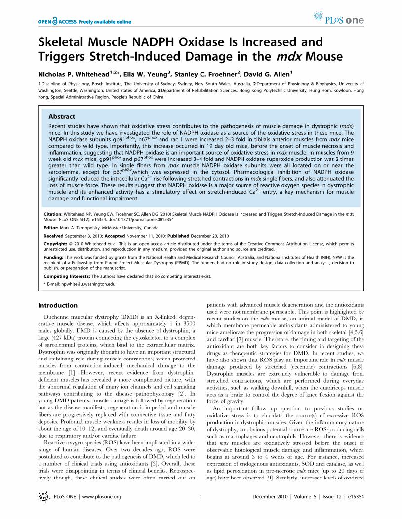
The NADPH oxidase catalytic subunit gp91phox is increased in
4–8 week old mdx skeletal muscles [17]. In the current study, we
found a similar magnitude increase (3–4 fold) in TA muscle
homogenates from 9 week mdx mice, compared to WT (Figure 1).
In addition, at this age, we also measured the expression levels of
the key activator subunit, p67phox. Interestingly, we found that this
protein also increased by 3–4 fold in mdx muscle (see Figure 1).
Both of these proteins were significantly greater for mdx compared
to WT (gp91phox, P,0.05; p67phox, P,0.01).
NADPH oxidase-dependent superoxide production isincreased in mdx muscle
Given the increased protein expression of NADPH oxidase in mdx
muscle, we then investigated whether this would result in enhanced
NADPH oxidase activity. Previously, our lab showed that NADPH
Figure 1. NADPH oxidase subunits are increased in 9 week old mdx mice. A. Representative western blots of gp91phox and p67phox from 9week old WT and mdx TA muscles. B. Pooled results of densitometry analysis for gp91phox and p67phox from WT (n = 4) and mdx (n = 8) muscles.doi:10.1371/journal.pone.0015354.g001
NADPH Oxidase Activity in the mdx Mouse
PLoS ONE | www.plosone.org 3 December 2010 | Volume 5 | Issue 12 | e15354
oxidase activity was increased in heart homogenates from mdx mice
[7]. Therefore, we used the same procedure to investigate whether
NADPH oxidase ROS production was increased in mdx skeletal
muscle compared to WT. NADPH-dependent ROS (superoxide)
production was measured in TA muscle homogenates from 9 week
old WT and mdx mice. We used a well-established technique, the
lucigenin chemiluminescent superoxide assay, using NADPH as the
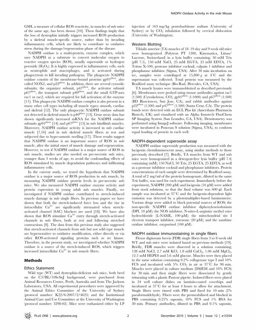
substrate (see Methods for details). Superoxide production was
significantly increased (P,0.05) by 2 fold in mdx muscle compared
to WT (Figure 2A). To establish that NADPH oxidase was the
primary source of the increased superoxide production in mdx
muscles, we used inhibitors of NADPH oxidase and the other main
cellular ROS sources (Figure 2B). For both WT and mdx muscles, L-
NAME and oxypurinol had no effect, while rotenone reducedsuperoxide by about 20% (P,0.05). However, the NADPH oxidaseinhibitor DPI prevented more than 90% of the superoxideproduction (P,0.05), indicating that NADPH oxidase was themain source of the ROS in this assay. Like DPI, the antioxidantNAC, used as a positive control, almost completely preventedsuperoxide production (P,0.01).
NADPH oxidase subunits are differentially increased in18–19 day old (pre-necrotic) mdx mice
Between the ages of about 4 and 8 weeks, mdx muscles undergoextensive cycles of necrosis, inflammation and regeneration. Sinceinflammatory cells such as macrophages and neutrophils express
Figure 2. NADPH oxidase superoxide production is increased in mdx muscle. A. Lucigenin chemiluminescent assay was used to measureNADPH-dependent superoxide production from muscle homogenates of WT and mdx mice (see Methods for details). Values from each experimentwere pooled for WT and mdx muscles (n = 5 in both groups). B. Inhibitors of various sources of ROS were added to the assay and superoxideproduction was compared to the control level (no inhibitor). Pooled values for WT and mdx mice.doi:10.1371/journal.pone.0015354.g002
NADPH Oxidase Activity in the mdx Mouse
PLoS ONE | www.plosone.org 4 December 2010 | Volume 5 | Issue 12 | e15354
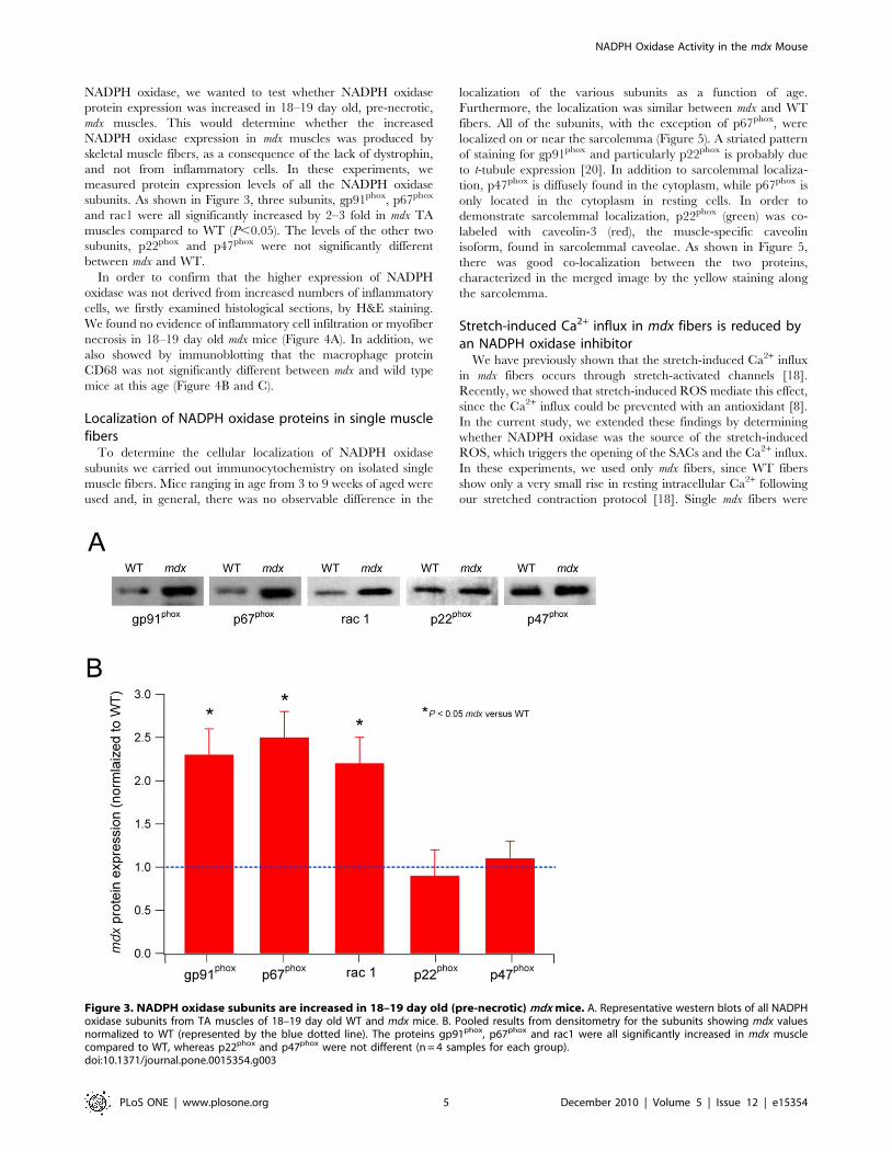
NADPH oxidase, we wanted to test whether NADPH oxidase
protein expression was increased in 18–19 day old, pre-necrotic,
mdx muscles. This would determine whether the increased
NADPH oxidase expression in mdx muscles was produced by
skeletal muscle fibers, as a consequence of the lack of dystrophin,
and not from inflammatory cells. In these experiments, we
measured protein expression levels of all the NADPH oxidase
subunits. As shown in Figure 3, three subunits, gp91phox, p67phox
and rac1 were all significantly increased by 2–3 fold in mdx TA
muscles compared to WT (P,0.05). The levels of the other two
subunits, p22phox and p47phox were not significantly different
between mdx and WT.
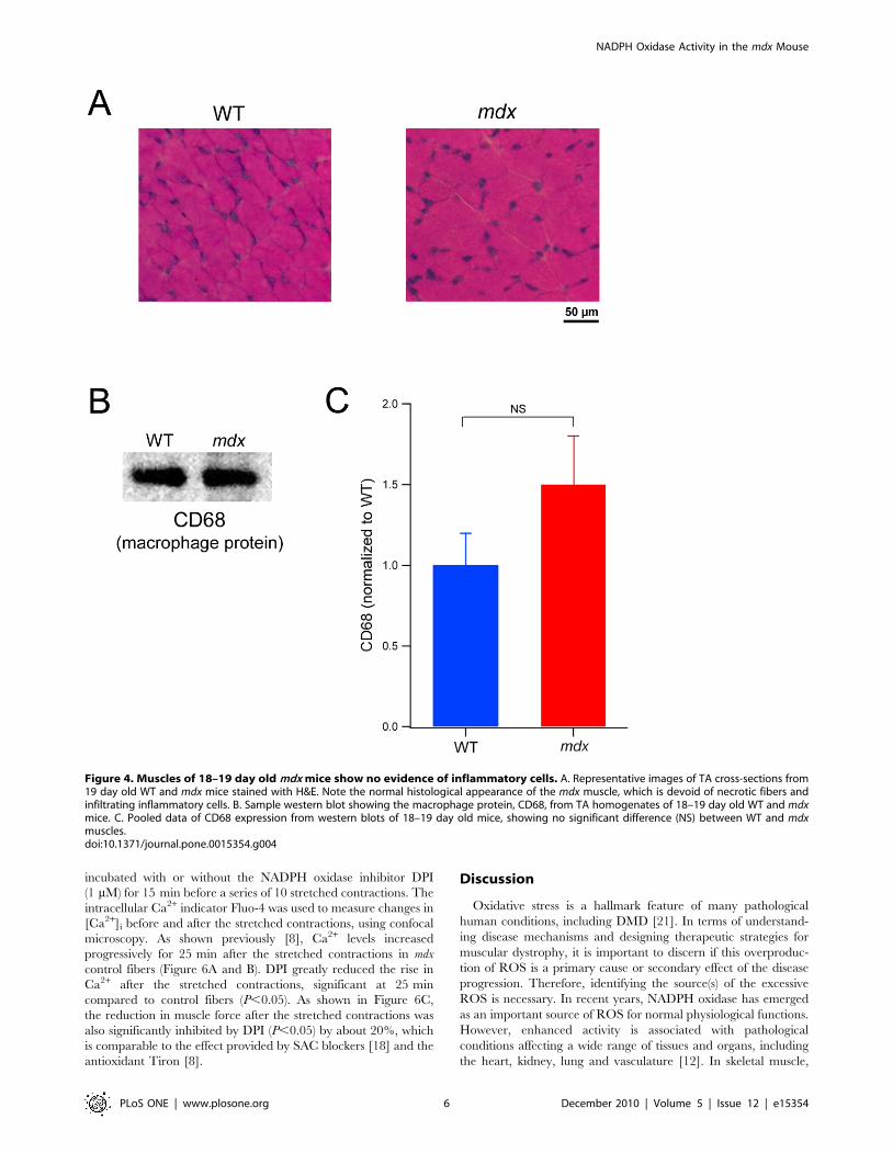
In order to confirm that the higher expression of NADPH
oxidase was not derived from increased numbers of inflammatory
cells, we firstly examined histological sections, by H&E staining.
We found no evidence of inflammatory cell infiltration or myofiber
necrosis in 18–19 day old mdx mice (Figure 4A). In addition, we
also showed by immunoblotting that the macrophage protein
CD68 was not significantly different between mdx and wild type
mice at this age (Figure 4B and C).
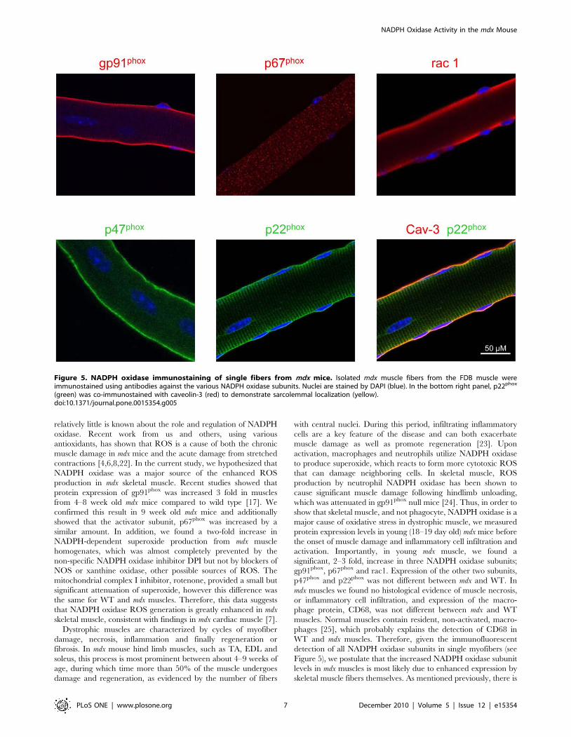
Localization of NADPH oxidase proteins in single musclefibers
To determine the cellular localization of NADPH oxidase
subunits we carried out immunocytochemistry on isolated single
muscle fibers. Mice ranging in age from 3 to 9 weeks of aged were
used and, in general, there was no observable difference in the
localization of the various subunits as a function of age.
Furthermore, the localization was similar between mdx and WT
fibers. All of the subunits, with the exception of p67phox, were
localized on or near the sarcolemma (Figure 5). A striated pattern
of staining for gp91phox and particularly p22phox is probably due
to t-tubule expression [20]. In addition to sarcolemmal localiza-
tion, p47phox is diffusely found in the cytoplasm, while p67phox is
only located in the cytoplasm in resting cells. In order to
demonstrate sarcolemmal localization, p22phox (green) was co-
labeled with caveolin-3 (red), the muscle-specific caveolin
isoform, found in sarcolemmal caveolae. As shown in Figure 5,
there was good co-localization between the two proteins,
characterized in the merged image by the yellow staining along
the sarcolemma.
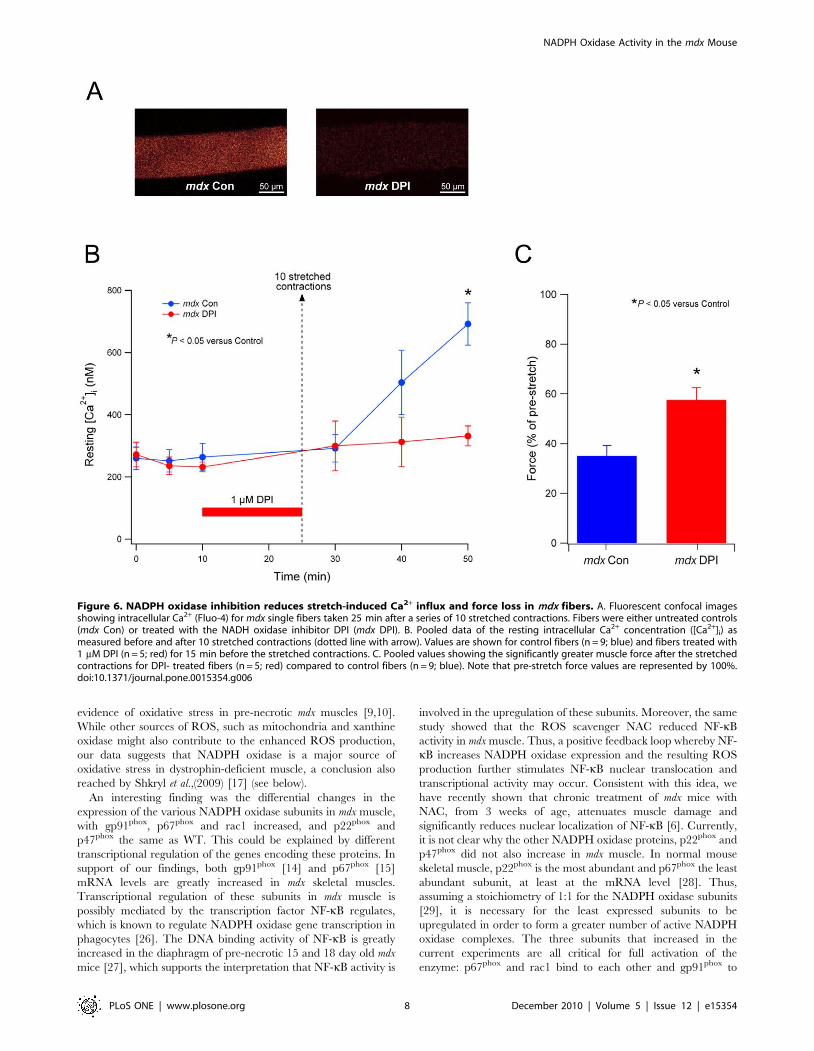
Stretch-induced Ca2+ influx in mdx fibers is reduced byan NADPH oxidase inhibitor
We have previously shown that the stretch-induced Ca2+ influx
in mdx fibers occurs through stretch-activated channels [18].
Recently, we showed that stretch-induced ROS mediate this effect,
since the Ca2+ influx could be prevented with an antioxidant [8].
In the current study, we extended these findings by determining
whether NADPH oxidase was the source of the stretch-induced
ROS, which triggers the opening of the SACs and the Ca2+ influx.
In these experiments, we used only mdx fibers, since WT fibers
show only a very small rise in resting intracellular Ca2+ following
our stretched contraction protocol [18]. Single mdx fibers were
Figure 3. NADPH oxidase subunits are increased in 18–19 day old (pre-necrotic) mdx mice. A. Representative western blots of all NADPHoxidase subunits from TA muscles of 18–19 day old WT and mdx mice. B. Pooled results from densitometry for the subunits showing mdx valuesnormalized to WT (represented by the blue dotted line). The proteins gp91phox, p67phox and rac1 were all significantly increased in mdx musclecompared to WT, whereas p22phox and p47phox were not different (n = 4 samples for each group).doi:10.1371/journal.pone.0015354.g003
NADPH Oxidase Activity in the mdx Mouse
PLoS ONE | www.plosone.org 5 December 2010 | Volume 5 | Issue 12 | e15354
incubated with or without the NADPH oxidase inhibitor DPI
(1 mM) for 15 min before a series of 10 stretched contractions. The
intracellular Ca2+ indicator Fluo-4 was used to measure changes in
[Ca2+]i before and after the stretched contractions, using confocal
microscopy. As shown previously [8], Ca2+ levels increased
progressively for 25 min after the stretched contractions in mdx
control fibers (Figure 6A and B). DPI greatly reduced the rise in
Ca2+ after the stretched contractions, significant at 25 min
compared to control fibers (P,0.05). As shown in Figure 6C,
the reduction in muscle force after the stretched contractions was
also significantly inhibited by DPI (P,0.05) by about 20%, which
is comparable to the effect provided by SAC blockers [18] and the
antioxidant Tiron [8].
Discussion
Oxidative stress is a hallmark feature of many pathological
human conditions, including DMD [21]. In terms of understand-
ing disease mechanisms and designing therapeutic strategies for
muscular dystrophy, it is important to discern if this overproduc-
tion of ROS is a primary cause or secondary effect of the disease
progression. Therefore, identifying the source(s) of the excessive
ROS is necessary. In recent years, NADPH oxidase has emerged
as an important source of ROS for normal physiological functions.
However, enhanced activity is associated with pathological
conditions affecting a wide range of tissues and organs, including
the heart, kidney, lung and vasculature [12]. In skeletal muscle,
Figure 4. Muscles of 18–19 day old mdx mice show no evidence of inflammatory cells. A. Representative images of TA cross-sections from19 day old WT and mdx mice stained with H&E. Note the normal histological appearance of the mdx muscle, which is devoid of necrotic fibers andinfiltrating inflammatory cells. B. Sample western blot showing the macrophage protein, CD68, from TA homogenates of 18–19 day old WT and mdxmice. C. Pooled data of CD68 expression from western blots of 18–19 day old mice, showing no significant difference (NS) between WT and mdxmuscles.doi:10.1371/journal.pone.0015354.g004
NADPH Oxidase Activity in the mdx Mouse
PLoS ONE | www.plosone.org 6 December 2010 | Volume 5 | Issue 12 | e15354
relatively little is known about the role and regulation of NADPH
oxidase. Recent work from us and others, using various
antioxidants, has shown that ROS is a cause of both the chronic
muscle damage in mdx mice and the acute damage from stretched
contractions [4,6,8,22]. In the current study, we hypothesized that
NADPH oxidase was a major source of the enhanced ROS
production in mdx skeletal muscle. Recent studies showed that
protein expression of gp91phox was increased 3 fold in muscles
from 4–8 week old mdx mice compared to wild type [17]. We
confirmed this result in 9 week old mdx mice and additionally
showed that the activator subunit, p67phox was increased by a
similar amount. In addition, we found a two-fold increase in
NADPH-dependent superoxide production from mdx muscle
homogenates, which was almost completely prevented by the
non-specific NADPH oxidase inhibitor DPI but not by blockers of
NOS or xanthine oxidase, other possible sources of ROS. The
mitochondrial complex I inhibitor, rotenone, provided a small but
significant attenuation of superoxide, however this difference was
the same for WT and mdx muscles. Therefore, this data suggests
that NADPH oxidase ROS generation is greatly enhanced in mdx
skeletal muscle, consistent with findings in mdx cardiac muscle [7].
Dystrophic muscles are characterized by cycles of myofiber
damage, necrosis, inflammation and finally regeneration or
fibrosis. In mdx mouse hind limb muscles, such as TA, EDL and
soleus, this process is most prominent between about 4–9 weeks of
age, during which time more than 50% of the muscle undergoes
damage and regeneration, as evidenced by the number of fibers
with central nuclei. During this period, infiltrating inflammatory
cells are a key feature of the disease and can both exacerbate
muscle damage as well as promote regeneration [23]. Upon
activation, macrophages and neutrophils utilize NADPH oxidase
to produce superoxide, which reacts to form more cytotoxic ROS
that can damage neighboring cells. In skeletal muscle, ROS
production by neutrophil NADPH oxidase has been shown to
cause significant muscle damage following hindlimb unloading,
which was attenuated in gp91phox null mice [24]. Thus, in order to
show that skeletal muscle, and not phagocyte, NADPH oxidase is a
major cause of oxidative stress in dystrophic muscle, we measured
protein expression levels in young (18–19 day old) mdx mice before
the onset of muscle damage and inflammatory cell infiltration and
activation. Importantly, in young mdx muscle, we found a
significant, 2–3 fold, increase in three NADPH oxidase subunits;
gp91phox, p67phox and rac1. Expression of the other two subunits,
p47phox and p22phox was not different between mdx and WT. In
mdx muscles we found no histological evidence of muscle necrosis,
or inflammatory cell infiltration, and expression of the macro-
phage protein, CD68, was not different between mdx and WT
muscles. Normal muscles contain resident, non-activated, macro-
phages [25], which probably explains the detection of CD68 in
WT and mdx muscles. Therefore, given the immunofluorescent
detection of all NADPH oxidase subunits in single myofibers (see
Figure 5), we postulate that the increased NADPH oxidase subunit
levels in mdx muscles is most likely due to enhanced expression by
skeletal muscle fibers themselves. As mentioned previously, there is
Figure 5. NADPH oxidase immunostaining of single fibers from mdx mice. Isolated mdx muscle fibers from the FDB muscle wereimmunostained using antibodies against the various NADPH oxidase subunits. Nuclei are stained by DAPI (blue). In the bottom right panel, p22phox
(green) was co-immunostained with caveolin-3 (red) to demonstrate sarcolemmal localization (yellow).doi:10.1371/journal.pone.0015354.g005
NADPH Oxidase Activity in the mdx Mouse
PLoS ONE | www.plosone.org 7 December 2010 | Volume 5 | Issue 12 | e15354
evidence of oxidative stress in pre-necrotic mdx muscles [9,10].
While other sources of ROS, such as mitochondria and xanthine
oxidase might also contribute to the enhanced ROS production,
our data suggests that NADPH oxidase is a major source of
oxidative stress in dystrophin-deficient muscle, a conclusion also
reached by Shkryl et al.,(2009) [17] (see below).
An interesting finding was the differential changes in the
expression of the various NADPH oxidase subunits in mdx muscle,
with gp91phox, p67phox and rac1 increased, and p22phox and
p47phox the same as WT. This could be explained by different
transcriptional regulation of the genes encoding these proteins. In
support of our findings, both gp91phox [14] and p67phox [15]
mRNA levels are greatly increased in mdx skeletal muscles.
Transcriptional regulation of these subunits in mdx muscle is
possibly mediated by the transcription factor NF-kB regulates,
which is known to regulate NADPH oxidase gene transcription in
phagocytes [26]. The DNA binding activity of NF-kB is greatly
increased in the diaphragm of pre-necrotic 15 and 18 day old mdx
mice [27], which supports the interpretation that NF-kB activity is
involved in the upregulation of these subunits. Moreover, the same
study showed that the ROS scavenger NAC reduced NF-kB
activity in mdx muscle. Thus, a positive feedback loop whereby NF-
kB increases NADPH oxidase expression and the resulting ROS
production further stimulates NF-kB nuclear translocation and
transcriptional activity may occur. Consistent with this idea, we
have recently shown that chronic treatment of mdx mice with
NAC, from 3 weeks of age, attenuates muscle damage and
significantly reduces nuclear localization of NF-kB [6]. Currently,
it is not clear why the other NADPH oxidase proteins, p22phox and
p47phox did not also increase in mdx muscle. In normal mouse
skeletal muscle, p22phox is the most abundant and p67phox the least
abundant subunit, at least at the mRNA level [28]. Thus,
assuming a stoichiometry of 1:1 for the NADPH oxidase subunits
[29], it is necessary for the least expressed subunits to be
upregulated in order to form a greater number of active NADPH
oxidase complexes. The three subunits that increased in the
current experiments are all critical for full activation of the
enzyme: p67phox and rac1 bind to each other and gp91phox to
Figure 6. NADPH oxidase inhibition reduces stretch-induced Ca2+ influx and force loss in mdx fibers. A. Fluorescent confocal imagesshowing intracellular Ca2+ (Fluo-4) for mdx single fibers taken 25 min after a series of 10 stretched contractions. Fibers were either untreated controls(mdx Con) or treated with the NADH oxidase inhibitor DPI (mdx DPI). B. Pooled data of the resting intracellular Ca2+ concentration ([Ca2+]i) asmeasured before and after 10 stretched contractions (dotted line with arrow). Values are shown for control fibers (n = 9; blue) and fibers treated with1 mM DPI (n = 5; red) for 15 min before the stretched contractions. C. Pooled values showing the significantly greater muscle force after the stretchedcontractions for DPI- treated fibers (n = 5; red) compared to control fibers (n = 9; blue). Note that pre-stretch force values are represented by 100%.doi:10.1371/journal.pone.0015354.g006
NADPH Oxidase Activity in the mdx Mouse
PLoS ONE | www.plosone.org 8 December 2010 | Volume 5 | Issue 12 | e15354
stimulate catalytic activity of the enzyme [12]. On the other hand,
while p47phox is considered essential for activation in phagocytes
and other cell types following agonist stimulation, it is not required
for basal activity in smooth muscle cells [30]. Future experiments
are required to determine the regulation and role of each NADPH
oxidase subunit in both normal and dystrophic skeletal muscle.
We have previously shown in mdx fibers that the stretch-induced
rise in cytosolic Ca2+ is mediated by SAC [18]. Moreover,
blocking SAC in mdx muscles also significantly attenuated the force
drop following stretched contractions and reduced membrane
permeability [18,31]. More recently, we found that these effects of
SAC blockers could be replicated by antioxidants [6,8], and in the
current study, by the NADPH oxidase inhibitor DPI (see Figure 6).
Taken together, these results imply that stretch of mdx muscle
produces ROS, by activation of NADPH oxidase, which
subsequently triggers Ca2+ entry through SAC leading to muscle
damage and contractile dysfunction. Our interpretation is
consistent with recent findings [17]. Using a different model of
stretch, hypo-osmotic swelling, Shkryl et al., (2009) reported that
NADPH oxidase triggered Ca2+ sparks in mdx fibers. They
postulated that the Ca2+, triggered by NADPH oxidase-derived
ROS, was later sequestered by mitochondria, which then
generated additional ROS. Interestingly, basal ROS production
from NADPH oxidase, but not mitochondria, was greater in mdx
fibers compared to WT. Again, these findings suggest that stretch-
induced NADPH oxidase ROS production precedes, and
subsequently stimulates mitochondrial ROS production via Ca2+
uptake. Consistent with this idea, there is recent evidence that in 5
week old mdx mice, during the extensive muscle damage phase,
mitochondria also contribute to ROS production [32].
Currently, the mechanism by which NADPH oxidase-depen-
dent ROS activates SAC remains unclear. One possibility is that
ROS activates SAC directly, via redox-sensitive sulfhydryl groups,
or indirectly, by ROS-mediated signaling pathways. We have
recently shown that TRPC1, a candidate for the overactive SAC
in mdx muscle [33], was activated via ROS sensitive src kinase [8].
Furthermore, TRPC1 and src kinase were increased in mdx
muscle, and src kinase inhibition significantly improved muscle
force following stretched contractions in mdx EDL muscle [8]. Src
kinase phosphorylates p47phox to initiate its translocation and
organization of the other NADPH oxidase cytosolic subunits to the
membrane-bound complex [34]. Thus, increased expression and
activation of src kinase might be involved in the enhanced stretch-
induced ROS production of NADPH oxidase and the associated
Ca2+ influx. TRPV2 is another potential candidate for the SAC in
mdx muscle [35]. A final point of consideration is that both TRPC1
[8] and src kinase [36] bind to Caveolin-3, which is upregulated in
mdx muscles [37]. Here, we have shown that p22phox co-localized
with caveolin-3 in single fibers (see Figure 5). We also have
preliminary evidence that NADPH oxidase subunits are found in
both caveolae and non-caveolae fractions in mdx and WT muscles
(unpublished observations). Depending on the cell type and
species, caveolae or lipid rafts can regulate activation [38] or
inhibition [39] of NADPH oxidase. Therefore, in future
experiments it will be important to elucidate whether caveolin-3
and caveolae play a role in the regulation of NADPH oxidase
activity in mdx muscles.
In summary, this study has identified NADPH oxidase as a
major source of ROS production in mdx muscle, which is
upregulated during the period of oxidative stress that precedes
muscle damage and inflammation. The other key finding is that
NADPH oxidase is involved in the activation of a ROS-sensitive
SAC in dystrophic muscle, triggered by stretched contractions.
Inhibition of NADPH oxidase prevented the influx of Ca2+ into
the muscle and provided protection against the force deficit.
Therefore, investigating the regulation of NADPH oxidase
expression and activity will be important to gain new insight into
the mechanisms of damage in dystrophic muscle and to potentially
develop new therapeutic approaches for DMD.
Author Contributions
Conceived and designed the experiments: NPW DGA. Performed the
experiments: NPW EWY. Analyzed the data: NPW EWY. Contributed
reagents/materials/analysis tools: DGA SCF. Wrote the paper: NPW
EWY SCF DGA.
References
1. Petrof BJ, Shrager JB, Stedman HH, Kelly AM, Sweeney HL (1993) Dystrophin
protects the sarcolemma from stresses developed during muscle contraction.Proc Natl Acad Sci U S A 90: 3710–3714.
2. Evans NP, Misyak SA, Robertson JL, Bassaganya-Riera J, Grange RW (2009)Dysregulated intracellular signaling and inflammatory gene expression during
initial disease onset in Duchenne muscular dystrophy. Am J Phys Med Rehabil88: 502–522.
3. Rando TA (2002) Oxidative stress and the pathogenesis of muscular dystrophies.Am J Phys Med Rehabil 81: S175–186.
4. Dorchies OM, Wagner S, Vuadens O, Waldhauser K, Buetler TM, et al. (2006)Green tea extract and its major polyphenol (-)-epigallocatechin gallate improve
muscle function in a mouse model for Duchenne muscular dystrophy.Am J Physiol Cell Physiol 290: C616–625.
5. Hnia K, Hugon G, Rivier F, Masmoudi A, Mercier J, et al. (2007) Modulation ofp38 mitogen-activated protein kinase cascade and metalloproteinase activity in
diaphragm muscle in response to free radical scavenger administration indystrophin-deficient Mdx mice. Am J Pathol 170: 633–643.
6. Whitehead NP, Pham C, Gervasio OL, Allen DG (2008) N-Acetylcysteineameliorates skeletal muscle pathophysiology in mdx mice. J Physiol 586:
2003–2014.
7. Williams IA, Allen DG (2007) The role of reactive oxygen species in the hearts of
dystrophin-deficient mdx mice. Am J Physiol Heart Circ Physiol 293:
H1969–1977.
8. Gervasio OL, Whitehead NP, Yeung EW, Phillips WD, Allen DG (2008)TRPC1 binds to caveolin-3 and is regulated by Src kinase - role in Duchenne
muscular dystrophy. J Cell Sci 121: 2246–2255.
9. Disatnik MH, Dhawan J, Yu Y, Beal MF, Whirl MM, et al. (1998) Evidence of
oxidative stress in mdx mouse muscle: studies of the pre-necrotic state. J Neurol
Sci 161: 77–84.
10. Dudley RW, Khairallah M, Mohammed S, Lands L, Des Rosiers C, et al. (2006)
Dynamic responses of the glutathione system to acute oxidative stress in
dystrophic mouse (mdx) muscles. Am J Physiol Regul Integr Comp Physiol 291:R704–710.
11. Cross AR, Segal AW (2004) The NADPH oxidase of professional phagocytes–prototype of the NOX electron transport chain systems. Biochim Biophys Acta
1657: 1–22.
12. Bedard K, Krause KH (2007) The NOX family of ROS-generating NADPHoxidases: physiology and pathophysiology. Physiol Rev 87: 245–313.
13. Javesghani D, Magder SA, Barreiro E, Quinn MT, Hussain SN (2002)Molecular characterization of a superoxide-generating NAD(P)H oxidase in the
ventilatory muscles. Am J Respir Crit Care Med 165: 412–418.
14. Spurney CF, Knoblach S, Pistilli EE, Nagaraju K, Martin GR, et al. (2008)Dystrophin-deficient cardiomyopathy in mouse: expression of Nox4 and Lox are
associated with fibrosis and altered functional parameters in the heart.
Neuromuscul Disord 18: 371–381.
15. Tseng BS, Zhao P, Pattison JS, Gordon SE, Granchelli JA, et al. (2002)Regenerated mdx mouse skeletal muscle shows differential mRNA expression.
J Appl Physiol 93: 537–545.
16. Jung C, Martins AS, Niggli E, Shirokova N (2008) Dystrophic cardiomyopathy:amplification of cellular damage by Ca2+ signalling and reactive oxygen species-
generating pathways. Cardiovasc Res 77: 766–773.
17. Shkryl VM, Martins AS, Ullrich ND, Nowycky MC, Niggli E, et al. (2009)
Reciprocal amplification of ROS and Ca(2+) signals in stressed mdx dystrophicskeletal muscle fibers. Pflugers Arch 458: 915–928.
18. Yeung EW, Whitehead NP, Suchyna TM, Gottlieb PA, Sachs F, et al. (2005)
Effects of stretch-activated channel blockers on [Ca2+]i and muscle damage in
the mdx mouse. J Physiol 562: 367–380.
19. Ravenscroft G, Nowak KJ, Jackaman C, Clement S, Lyons MA, et al. (2007)Dissociated flexor digitorum brevis myofiber culture system–a more mature
muscle culture system. Cell Motil Cytoskeleton 64: 727–738.
20. Hidalgo C, Sanchez G, Barrientos G, Aracena-Parks P (2006) A transverse
tubule NADPH oxidase activity stimulates calcium release from isolated triads
NADPH Oxidase Activity in the mdx Mouse
PLoS ONE | www.plosone.org 9 December 2010 | Volume 5 | Issue 12 | e15354
via ryanodine receptor type 1 S -glutathionylation. J Biol Chem 281:
26473–26482.21. Tidball JG, Wehling-Henricks M (2007) The role of free radicals in the
pathophysiology of muscular dystrophy. J Appl Physiol 102: 1677–1686.
22. Messina S, Altavilla D, Aguennouz M, Seminara P, Minutoli L, et al. (2006)Lipid peroxidation inhibition blunts nuclear factor-kappaB activation, reduces
skeletal muscle degeneration, and enhances muscle function in mdx mice.Am J Pathol 168: 918–926.
23. Tidball JG, Villalta SA (2010) Regulatory interactions between muscle and the
immune system during muscle regeneration. Am J Physiol Regul Integr CompPhysiol 298: R1173–R1187.
24. Nguyen HX, Tidball JG (2003) Null mutation of gp91phox reduces musclemembrane lysis during muscle inflammation in mice. J Physiol 553: 833–841.
25. Pimorady-Esfahani A, Grounds MD, McMenamin PG (1997) Macrophages anddendritic cells in normal and regenerating murine skeletal muscle. Muscle Nerve
20: 158–166.
26. Gauss KA, Nelson-Overton LK, Siemsen DW, Gao Y, DeLeo FR, et al. (2007)Role of NF-kappaB in transcriptional regulation of the phagocyte NADPH
oxidase by tumor necrosis factor-alpha. J Leukoc Biol 82: 729–741.27. Kumar A, Boriek AM (2003) Mechanical stress activates the nuclear factor-
kappaB pathway in skeletal muscle fibers: a possible role in Duchenne muscular
dystrophy. FASEB J 17: 386–396.28. Mofarrahi M, Brandes RP, Gorlach A, Hanze J, Terada LS, et al. (2008)
Regulation of proliferation of skeletal muscle precursor cells by NADPH oxidase.Antioxid Redox Signal 10: 559–574.
29. Huang J, Hitt ND, Kleinberg ME (1995) Stoichiometry of p22-phox and gp91-phox in phagocyte cytochrome b558. Biochemistry 34: 16753–16757.
30. Li JM, Wheatcroft S, Fan LM, Kearney MT, Shah AM (2004) Opposing roles of
p47phox in basal versus angiotensin II-stimulated alterations in vascular O2-
production, vascular tone, and mitogen-activated protein kinase activation.
Circulation 109: 1307–1313.31. Whitehead NP, Streamer M, Lusambili LI, Sachs F, Allen DG (2006)
Streptomycin reduces stretch-induced membrane permeability in muscles from
mdx mice. Neuromuscul Disord 16: 845–854.32. Menazza S, Blaauw B, Tiepolo T, Toniolo L, Braghetta P, et al. (2010)
Oxidative stress by monoamine oxidases is causally involved in myofiber damagein muscular dystrophy. Hum Mol Genet 19: 4207–4215.
33. Vandebrouck C, Martin D, Colson-Van Schoor M, Debaix H, Gailly P (2002)
Involvement of TRPC in the abnormal calcium influx observed in dystrophic(mdx) mouse skeletal muscle fibers. J Cell Biol 158: 1089–1096.
34. Touyz RM, Yao G, Schiffrin EL (2003) c-Src induces phosphorylation andtranslocation of p47phox: role in superoxide generation by angiotensin II in
human vascular smooth muscle cells. Arterioscler Thromb Vasc Biol 23:981–987.
35. Zanou N, Iwata Y, Schakman O, Lebacq J, Wakabayashi S, et al. (2009)
Essential role of TRPV2 ion channel in the sensitivity of dystrophic muscle toeccentric contractions. FEBS Lett 583: 3600–3604.
36. Venema VJ, Ju H, Zou R, Venema RC (1997) Interaction of neuronal nitric-oxide synthase with caveolin-3 in skeletal muscle. Identification of a novel
caveolin scaffolding/inhibitory domain. J Biol Chem 272: 28187–28190.
37. Vaghy PL, Fang J, Wu W, Vaghy LP (1998) Increased caveolin-3 levels in mdxmouse muscles. FEBS Lett 431: 125–127.
38. Vilhardt F, van Deurs B (2004) The phagocyte NADPH oxidase depends oncholesterol-enriched membrane microdomains for assembly. EMBO J 23:
739–748.39. Han W, Li H, Villar VA, Pascua AM, Dajani MI, et al. (2008) Lipid rafts keep
NADPH oxidase in the inactive state in human renal proximal tubule cells.
Hypertension 51: 481–487.
NADPH Oxidase Activity in the mdx Mouse
PLoS ONE | www.plosone.org 10 December 2010 | Volume 5 | Issue 12 | e15354