simultaneous multislice multiband parallel radiofrequency excitation with independent slice-specific...
TRANSCRIPT

IMAGINGMETHODOLOGY -
RapidCommunications
Simultaneous Multislice Multiband ParallelRadiofrequency Excitation with IndependentSlice-Specific Transmit B1 Homogenization
Xiaoping Wu,* Sebastian Schmitter, Edward J. Auerbach, Steen Moeller,
Kamil U�gurbil, and Pierre-Francois Van de Moortele
Purpose: To develop a new parallel transmit (pTx) pulse
design for simultaneous multiband (MB) excitation in order totackle simultaneously the problems of transmit B1 (B1þ) inho-
mogeneity and total radiofrequency (RF) power, so as to allowfor optimal RF excitation when using MB pulses for sliceacceleration for high and ultrahigh field MRI.
Methods: With the proposed approach, each of the bandsthat are simultaneously excited is subject to a band-specific
set of B1 complex shim weights. The method was validated inthe human brain at 7T using a 16-channel pTx system andwas compared to conventional MB pulses operating in the cir-
cularly polarized (CP) mode. Further numerical simulationsbased on measured B1 maps were conducted.Results: The new method improved B1þ homogeneity by
60% when keeping the total RF power constant and reducedtotal RF power by 72% when keeping the excitation fidelity
constant, as compared to the conventional CP mode.Conclusion: A new pTx pulse design formalism is introducedtargeting slice-specific B1þ homogenization in MB excitation
while constraining total RF power. These pulses lead to signifi-cantly improved slice-wise B1þ uniformity and/or largely
reduced total RF power, as compared to the conventionallyemployed MB pulses applied in the CP mode. Magn ResonMed 70:630–638, 2013. VC 2013 Wiley Periodicals, Inc.
Key words: parallel excitation; simultaneous multislice; trans-
mit B1 homogenization; high field MRI; RF pulse design
Recent availability of 7 Tesla (7T) magnets has enabledfunctional brain imaging (fMRI) studies of the humanbrain with increasingly higher spatial resolution andfidelity (1–6). Obtaining such fMRI data over the entirehuman brain, however, encounters the undesirably longvolume repetition times (TR) even when single-shotmethods, such as slice selective Echo-Planar Imaging(EPI), are employed. A solution to this impediment is
simultaneous multiband (MB) radiofrequency (RF) exci-tation and acquisition of multiple slices with subsequentunaliasing using parallel imaging (7), thus reducing thevolume TR by the number of simultaneously excited slices(MB factor). Especially in conjunction with partial shiftingof simultaneously acquired slices along the phase encodedimension using gradient blips (blipped-CAIPI) in EPI(12). The approach has been employed with significantsuccess in task (8,9) and in resting state fMRI (10,11), lead-ing to improved detection of resting state networks (RSNs)(10) and new analysis strategies that reveal RSN temporaldynamics (11). The approach has also allowed significantreductions (10,12) in the otherwise long acquisition timesassociated with high angular diffusion-weighted imaging(HARDI) (13) and diffusion spectrum imaging (DSI) (14,15)thereby making the use of these techniques practical andindispensable for efforts like the Human Connectome Pro-ject (HCP) (16).
Despite these gains, however, the optimal use of the MB
approach at high (3 and 4T) and ultrahigh (�7T) fields is
precluded by transmit B1 (B1þ) inhomogeneities and
power deposition. Signal-to-noise ratio (SNR) and image
contrast becomes spatially nonuniform and suboptimal
because of B1þ heterogeneity resulting from destructive
interferences (17) caused by the traveling wave RF behav-
ior (18). These B1þ inhomogeneities are sufficiently strong
even at 3T to induce spatial variations in SNR, especially
with spin-echo (SE) sequences. Similarly, maximal MB
factors can be limited by power deposition especially at
ultrahigh fields and/or with SE sequences. When the num-
ber of slices and the volume TR are kept the same, the MB
approach deposits the same power as the conventional
single slice excitation. However, accelerating by the MB
factor leads to MB-fold increase in power deposition,
imposing a limit on performance.In this study, we introduce a novel parallel transmit
(pTx) MB pulse design that tackles the aforedescribedproblem of B1þ inhomogeneity with total RF power reg-ularization, and demonstrate it experimentally in thehuman brain at 7T using a 16-channel pTx system driv-ing a 16-channel RF transceiver array; the performanceof these new pulses was compared to conventional MBpulses for the same RF coil operating in the circularlypolarized (CP) mode using both experiments and numer-ical simulations. The results demonstrate that the newpTx MB pulses provide significantly improved B1þhomogenization in multiple slices simultaneously and/or
University of Minnesota, Center for Magnetic Resonance Research, Minne-apolis, Minnesota, USA.
Grant sponsors: WM KECK Foundation and NIH; Grant numbers: P41EB015894, S10 RR026783, R21 EB009133, R01 EB006835 and R01EB007327.
*Correspondence to: Xiaoping Wu, Center for Magnetic ResonanceResearch, University of Minnesota, 2021 6th Street SE, Minneapolis, MN55455. E-mail: [email protected]
Received 30 November 2012; revised 12 April 2013; accepted 8 May 2013
DOI 10.1002/mrm.24828Published online 25 June 2013 in Wiley Online Library (wileyonlinelibrary.com).
Magnetic Resonance in Medicine 70:630–638 (2013)
VC 2013 Wiley-Liss, Inc. 630

significantly reduced RF power relative to a single-channel CP mode application.
THEORY
Two strategies are employed in multichannel MB excita-tion to find RF magnitude and phase modulations (i.e.,RF shim values) of the base RF pulses so as to mitigateB1þ inhomogeneity. The primary strategy, defined hereas Full pTx MB, relies on fully independent multipletransmit channels and calculates, for each transmit chan-nel, a different set of shim values for each of the Mbands. This approach, introduced in the form of a con-ference abstract (19), is fully described here.
In the absence of a full pTx hardware, it is possible toutilize a simpler and less costly hardware that splits thesingle RF channel of the scanner into multiple channelsat the low power stage and impose on the resulting sepa-rate channels a channel-specific phase and amplitudechange (20–23). With this limited but more readilyimplementable hardware, a solution that is possible,defined here as MB B1 shim, is a set of channel-specificRF shim values that are applied to all M bands simulta-neously, rather than channel- and band-specific shimvalues that is possible with full pTx.
In Full pTx MB targeting uniform excitation (i.e., uni-form jB1þj), the RF shim values for M-band Q-channelexcitation can be obtained by solving the following mag-nitude least square problem:
minwfull
jjjAfullwfull�� djj22 þ ljjwfulljj22� �
[1]
with Afull ¼
A1 � � � 0
� . ..
�
0 � � � AM
26664
37775 and wfull ¼
w1
w2
�
wM
26666664
37777775:
Here, Afull is a block diagonal matrix with the diagonalelement matrices Am (m ¼ 1; 2; . . . ;M ) being the systemmatrix involving B1þ spatial sensitivity profiles of indi-vidual channels and the DB0 map for the mth band,wfull
is a concatenated vector with wm (m ¼ 1;2; . . . ;M ) being avector of RF shim values of individual channels for themth band, d is a scalar representing the desired transversemagnetization, and l is the regularization parameter.
Each of the M base RF pulses that constitute the finalsummed MB pulse is played at a different slice-specificfrequency. Consequently, by Parseval’s theorem, total RFpower is simply determined by the number of slicesacquired irrespective of whether the slices are acquiredusing MB approach or the conventional single slice exci-tation procedure. As a result, the use of jjwfulljj22 is anoptimum constraint for total RF power in Full pTx MB.
In the MB B1 shim, one can solve:
minwfjjjAwj � djj22 þ ljjwjj22g [2]
with A ¼
A1
A2
�
AM
26666664
37777775
and w ¼
w1
w2
�
wQ
26666664
37777775
.
Here, A is a combined matrix composed of Am and wis a complex valued vector with wq (q ¼ 1;2; . . . ;Q) beingthe RF shim value for the qth channel.
It should also be emphasized that the minimal hardwareconfiguration relying on a single RF channel describedabove for the MB B1shim pulse would be incapable of gen-erating the Full pTx MB pulses, since a different B1-shimsolution is applied for each individual band (see Fig. 1).
METHODS
All experiments were carried out at 7T driven by a 16-channel prototype pTx system (Siemens, Erlangen,
FIG. 1. Schematic illustration of Full pTx MB vs MB B1 shim in the context of single-spoke pulse design for two-band excitation. Notethat due to different phase evolutions required to target band 1 (red) and band 2 (blue), the two base RF pulses, represented by trian-
gles, are of different pulse shapes. Importantly, although an RF shimming is performed for each band in Full pTx MB, the application offinal RF pulses requires full pTx hardware and cannot be realized simply with a magnitude and phase controller. This is because the final
RF pulse per channel is the sum of two different base pulses each multiplied by a different weight and thus cannot be represented sim-ply by a channel-specific weight, w0q, multiplied by a common pulse. [Color figure can be viewed in the online issue, which is availableat wileyonlinelibrary.com.]
Simultaneous Multislice Parallel RF Excitation 631
Germany) equipped with 1kW RF amplifier per channel.A transceiver array with 16 azimuthally distributed ele-ments (24) with two short elements to leave an openingfor the eyes was used for both RF transmission and MRsignal reception. Human brain images were collected inhealthy volunteers who signed a consent form approvedby local Institutional Review Board. All calculationswere conducted in MATLAB (The Mathworks Inc.,Natick, MA).
Sixteen-channel complex B1þ maps within nine axialslices encompassing the brain region were obtained witha fast hybrid multichannel B1þ mapping technique(25,26) by combining a high flip angle (FA) 3D actualflip angle imaging (AFI) (27) with a series of small FAmultislice gradient echo (GRE) images. The series of mul-tislice GRE images were obtained, with one channeltransmitting at a time while receiving signals on allchannels, with nominal FA ¼ 6
�, FOV ¼ 256(RO) �
176(PE) mm2, matrix size ¼ 128 � 88, slice thickness ¼6 mm, TR/TE ¼ 73/3.4 ms, BW ¼ 260 Hz/pixel, andacquisition time ¼ 3 min 53 s for two averages. Based onthese complex images, a B1þ phase shim solution aim-ing at a CP-mode B1þ distribution over the entire brainwas calculated to avoid in subsequent field mappingacquisitions areas of very weak signal (17,28). The 3DAFI dataset was then acquired with a nonselective exci-tation, nominal FA ¼ 60
�, orientation ¼ Transversal,
FOV ¼ 256(RO) � 176(PE) � 168(SS) mm3, matrix size ¼128 � 88 � 48, slice oversampling ¼ 50%, generalizedautocalibrating partially parallel acquisitions (GRAPPA)Factor ¼ 2, partial Fourier ¼ 6/8 (PE and SS), TR1/TR2/TE ¼ 30/150/2.1 ms, and BW ¼ 390 Hz/pixel, acquisitiontime ¼ 6 min 49 s. Additionally, DB0 maps were derivedfor the same nine slices from two multislice GRE imageswith different TEs (TE1/TE2 ¼ 4.32/5.34 ms) and wereincorporated into RF pulse design to minimize off-resonance effects.
All MB B1 shim and Full pTx MB RF pulses weredesigned with a single spoke (i.e., without gradientencoding in the transverse plane) to simultaneouslyexcite the desired bands in the brain with jB1þj homoge-nization. The unity target was defined as homogeneousjB1þj in the desired slices by manually creating a spatialmask only covering the brain tissues in the bands. RFmagnitude and phase modulations were calculated withthe variable exchange algorithm (29); voxels outside theregion of interest (ROI) were not considered. The sub-RFpulse used was a Hanning filtered SINC pulse with abandwidth time product (BWTP) of 10. The final RFpulses were 1 ms in length, defined with a dwell time of2 ms.
RF pulses were designed for two-band (MB2) andeight-band (MB8) excitation using the two strategies. Theinter-slice distance was 56 mm for MB2 and 14 mm forMB8 design. For both MB2 and MB8 cases, L curvesquantifying the tradeoff between total RF power andexcitation errors were generated by varying the regulari-zation parameter, l, in the pulse design (see Eqs. [1] and[2]). Here, the total RF power was calculated via P /PQ
q¼1
PNn¼1 b2
qðtnÞ with bqðtÞ being the final summed RFpulse shape of the qth channel and N being the numberof time points in the pulse definition. The excitation
error, measured by the root mean squared error (RMSE),was given by jjAwj � djj2=
ffiffiffiffiffiffiffiffiffiffiffiNROI
pfor MB B1 shim and
jjjAfullwfullj � djj2=ffiffiffiffiffiffiffiffiffiffiffiNROI
pfor Full pTx MB with NROI
being the number of voxels in the ROI. For comparison,MB2 and MB8 RF pulses were also assembled for the CPmode (referred to as MB CP mode hereafter), mimicking asingle-channel transmit condition, where the RF magnitudeswere adjusted such that the resulting mean FA averagedover the whole brain would be the same as the nominal FAused in MB B1 shim and Full pTx MB pulse design.
3D FA maps of MB2 excitation were estimated to com-pare the performance of MB CP mode, MB B1 shim, andFull pTx MB RF pulses. However, there was no sequenceon the system capable of running fast, large FA B1þmaps with Full pTx MB pulses at the time of this study.Thus, MB RF pulse FA maps were derived by relativecomparison with standard RF pulse reference, as will bedescribed in the following, exploiting the inherent pro-portionality between local jB1þj and signal intensity insmall FA GRE images. First, a “reference” 3D GRE image,Iref, was acquired in the small FA regime (nominal FA ¼2�) using a nonselective hard RF pulse in CP mode, and
a 3D AFI was acquired also in CP mode with a nonselec-tive hard RF pulse (nominal FA ¼ 60
�) with similar geo-
metrical parameters. The FA map of the reference image,FAref, was directly derived from the 3D AFI map know-ing the two corresponding nominal FAs. Second, a 3DGRE image, IMB, was acquired in the small FA regime(nominal FA ¼ 6
�) using the same TR as for Iref, with a
modified 3D GRE sequence where the RF excitationmodule was replaced by the MB pulse. Finally, the FAmap of the MB RF pulse, FAMB, was estimated by FAMB
¼ (IMB/Iref) FAref. Relevant imaging parameters of the 3DGRE acquisition included the following: orientation ¼Sagittal, FOV ¼ 256(RO) � 232(PE) � 176(SS) mm3,matrix size ¼ 128 � 58 � 44, GRAPPA Factor (PE) ¼ 2,partial Fourier ¼ 6/8(PE and SS), TR/TE ¼ 30/3 ms, BW¼ 390 Hz/pixel, and total acquisition time ¼ 32 s. The3D CP mode AFI were obtained with the same imagingparameters except for TR1/TR2/TE ¼ 30/150/2.1 ms andtotal acquisition time ¼ 3 min 11 s.
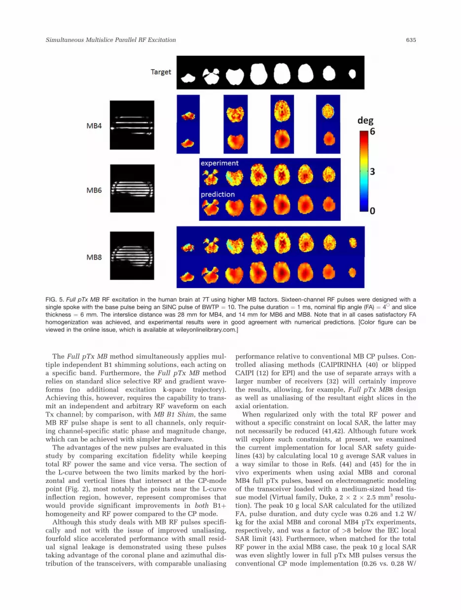
To demonstrate the feasibility of Full pTx MB whensimultaneously exciting a larger number of slices, full pTxRF pulses were designed for four- (MB4), six- (MB6), andeight- (MB8) slice excitation in the brain, and the respec-tive FA maps were estimated using the same imaging pro-tocols as described above for the MB2 excitation. Theinterslice distance was 28 mm in MB4, and 14 mm in MB6and MB8 pulse design, and the nominal FA was set to 4
�for
all the three cases.FA map homogeneity was also compared between Full
pTx MB and MB CP mode RF pulses when simultane-ously exciting four equidistant coronal slices, using simi-lar B1þ and DB0 mapping parameters. The same fourslices were also simultaneously imaged with an MB GREsequence using the two types of pulses, followed byunaliasing of each slice (7). Relevant imaging parametersincluded the following: FOV ¼ 256(RO) � 176(PE) mm2,matrix ¼ 192 � 132, TR/TE ¼ 100/20 ms, BW ¼ 390 Hz/pixel, acquisition time ¼ 13 s. Reference images for theGRAPPA kernel were collected using the same imagingparameters, while exciting a single band at a time.
632 Wu et al.
RESULTS
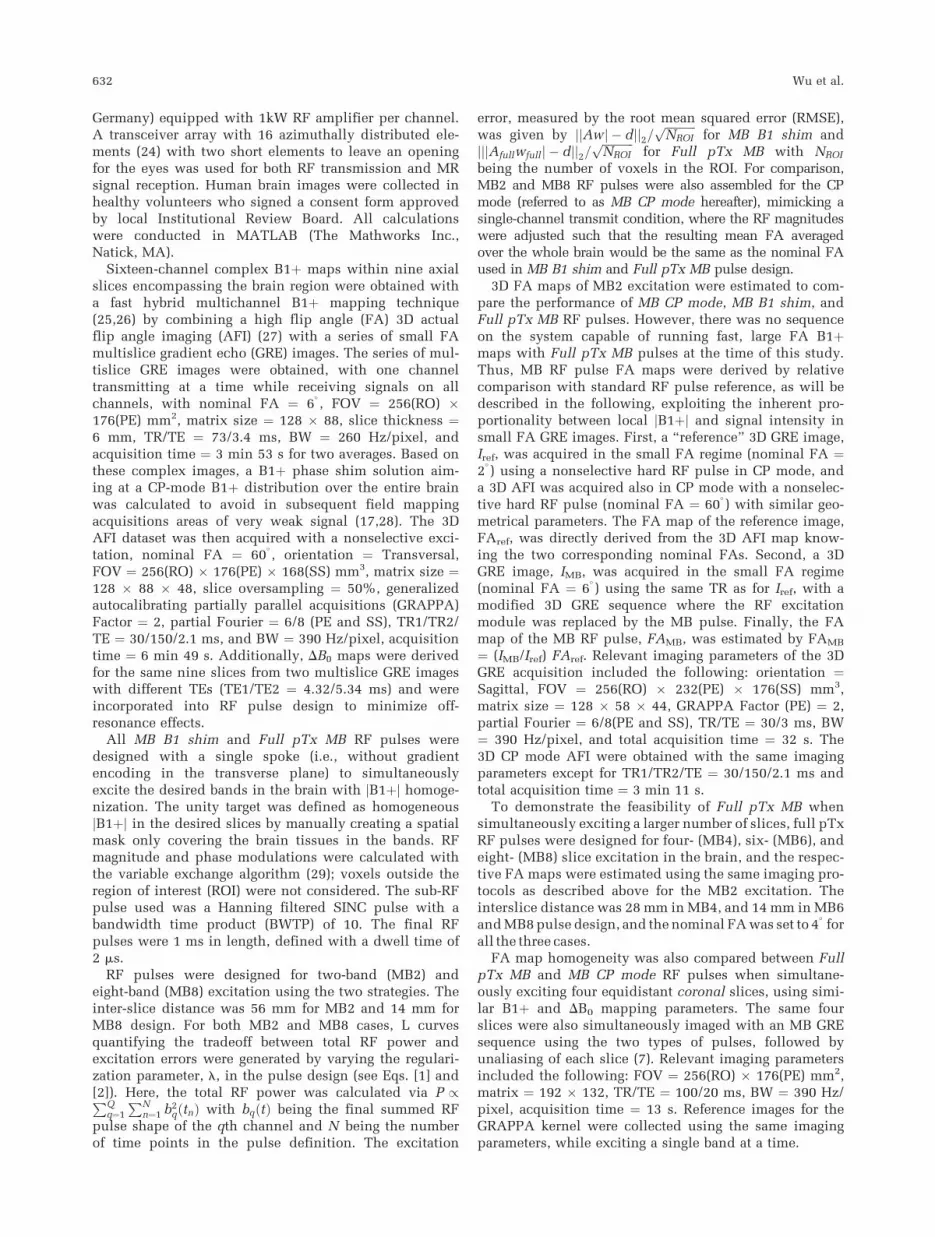
For both single spoke MB2 and MB8 pulse design, FullpTx MB yielded better RF performance than MB B1
shim, and both strategies significantly outperformed theMB CP mode (Fig. 2). More quantitative analyses basedon Bloch simulations (Fig. 3) revealed that when using
FIG. 2. L curves quantifying tradeoffs between total RF power and excitation errors (i.e., root mean square error (RMSE)) in the humanbrain at 7T for Full pTx MB (�) and MB B1 shim (�), along with MB CP mode (�). For both MB2 and MB8 pulse design with a single
spoke, Full pTx MB gave rise to the best excitation fidelity (lowest RMSE) with the same resulting total RF power (as indicated by thehorizontal dashed line), or led to the least RF power requirement when achieving the same excitation fidelity (as indicated by the verticalsolid line). Note that the single value for MB CP mode (�) is at the crossing between the horizontal and vertical lines. [Color figure can
be viewed in the online issue, which is available at wileyonlinelibrary.com.]
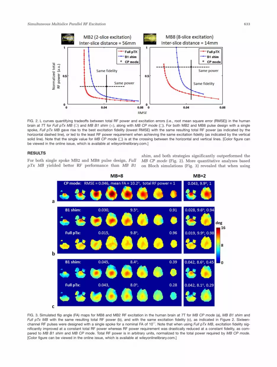
FIG. 3. Simulated flip angle (FA) maps for MB8 and MB2 RF excitation in the human brain at 7T for MB CP mode (a), MB B1 shim andFull pTx MB with the same resulting total RF power (b), and with the same excitation fidelity (c), as indicated in Figure 2. Sixteen-
channel RF pulses were designed with a single spoke for a nominal FA of 10�. Note that when using Full pTx MB, excitation fidelity sig-nificantly improved at a constant total RF power whereas RF power requirement was drastically reduced at a constant fidelity, as com-pared to MB B1 shim and MB CP mode. Total RF power is in arbitrary units, normalized to the total power required by MB CP mode.
[Color figure can be viewed in the online issue, which is available at wileyonlinelibrary.com.]
Simultaneous Multislice Parallel RF Excitation 633

the same total RF power as in the CP mode, the B1þinhomogeneity, measured by SD/mean of B1þ maps,improved from �25% for MB CP mode, to �17% for MBB1 shim and to �10% for Full pTx MB design. Whenachieving the same excitation fidelity (i.e., same RMSE),the total RF power requirement, which in this case isproportional to total power deposited into the head (i.e.,global specific absorption rate (SAR)), decreased by�58% for MB B1 shim and �72% for Full pTx MBdesign, as compared to the MB CP mode. Part of thisdecrease was due to a decreased mean FA induced bythe relatively large regularization imposed on total RFpower (Fig. 3c). However, even accounting for thisresulted in �56% less RF power in the Full pTx MBdesign compared to the MB CP mode. Experimental RFpower values reported by the SAR monitoring system ofthe instrument were consistent with the calculateddifferences.
Figure 4 illustrates experimental results of MB2 RFexcitation in the human brain using MB CP mode, MBB1 shim, and Full pTx MB pulses designed for the sametotal power. Consistent with Bloch Equation simulations(Fig. 3), Full pTx MB strategy yielded the best B1þhomogenization (i.e., the least SD/mean value) in thetwo bands versus the MB B1 shim and MB CP mode,especially for the lower band in the cerebellum. Further-more, good agreement was seen between experimentalresults and the numerical predictions.
Figure 5 displays the in vivo FA estimations for FullpTx MB4, MB6, and MB8 RF excitation. For all thesethree MB factors, improved FA uniformity was achievedand the experimental results were in good agreementwith the numerical predictions.
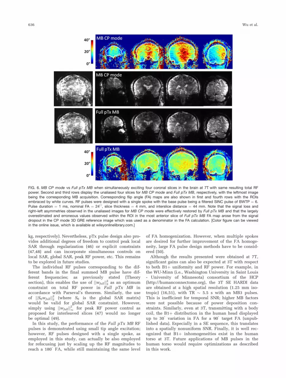
Figure 6 illustrates the comparison of MB CP mode toFull pTx MB RF pulse design when simultaneouslyexciting four coronal slices for the same total RF power.Similar to axial excitations (Fig. 4), Full pTx MB yieldedsignificantly improved B1þ homogenization in the fourslices with image uniformity effectively restored in thechallenging areas (such as the frontal and lower temporallobes) and right–left asymmetries, as compared to MB CPmode.
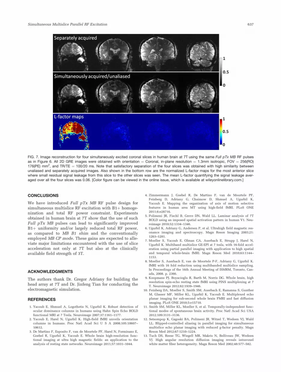
Figure 7 shows MB image reconstruction of four coro-nal slices for Full pTx MB4 RF excitation in human brainusing either the individual components of the Full pTxMB4 pulse one at a time exciting single slices, or as aFull pTx MB pulse exciting and acquiring simultane-ously. In this case, differences are not expected betweenthe two applications. Satisfactory unaliasing of the fourslices was obtained with high similarity between theunaliased and separately acquired images. L-factor maps(30) analyzing signal leakage between simultaneouslyexcited and acquired slices were calculated and exhib-ited small residual aliasing.
DISCUSSION
In standard slice selective or 3D applications, pTx meth-ods have been effective in ameliorating B1þ inhomoge-neities with a variety of techniques, such as B1-shimming (i.e., single spoke) (20–23,31,32), transmitSENSE (33–36), multispoke pulses (3,37,38), kT-pointpulses (39), etc. Here, we demonstrate that the pTx tech-nology permits significant improvements in B1þ homo-geneity and/or RF power in MB pulses, which areincreasingly used in fMRI and dMRI.
FIG. 4. MB2 RF excitation in the human brain at 7T for MB CP mode, MB B1 shim and Full pTx MB with the same resulting total RFpower. Sixteen-channel RF pulses were designed with a single spoke with the base pulse being a filtered SINC pulse of BWTP ¼ 10.
Pulse duration ¼ 1 ms, nominal flip angle ¼ 6�, slice thickness ¼ 6 mm and inter-slice distance ¼ 56 mm. Note that FA inhomogeneity,measured by SD/mean of the FA map, that already improved from 22% for MB CP mode to 18% for MB B1 shim, was significantlyreduced to 7% for Full pTx MB. In all cases, experimental results were in good agreement with numerical predictions.
634 Wu et al.
The Full pTx MB method simultaneously applies mul-tiple independent B1 shimming solutions, each acting ona specific band. Furthermore, the Full pTx MB methodrelies on standard slice selective RF and gradient wave-forms (no additional excitation k-space trajectory).Achieving this, however, requires the capability to trans-mit an independent and arbitrary RF waveform on eachTx channel; by comparison, with MB B1 Shim, the sameMB RF pulse shape is sent to all channels, only requir-ing channel-specific static phase and magnitude change,which can be achieved with simpler hardware.
The advantages of the new pulses are evaluated in thisstudy by comparing excitation fidelity while keepingtotal RF power the same and vice versa. The section ofthe L-curve between the two limits marked by the hori-zontal and vertical lines that intersect at the CP-modepoint (Fig. 2), most notably the points near the L-curveinflection region, however, represent compromises thatwould provide significant improvements in both B1þhomogeneity and RF power compared to the CP mode.
Although this study deals with MB RF pulses specifi-cally and not with the issue of improved unaliasing,fourfold slice accelerated performance with small resid-ual signal leakage is demonstrated using these pulsestaking advantage of the coronal plane and azimuthal dis-tribution of the transceivers, with comparable unaliasing
performance relative to conventional MB CP pulses. Con-trolled aliasing methods (CAIPIRINHA (40) or blippedCAIPI (12) for EPI) and the use of separate arrays with alarger number of receivers (32) will certainly improvethe results, allowing, for example, Full pTx MB8 designas well as unaliasing of the resultant eight slices in theaxial orientation.
When regularized only with the total RF power andwithout a specific constraint on local SAR, the latter maynot necessarily be reduced (41,42). Although future workwill explore such constraints, at present, we examinedthe current implementation for local SAR safety guide-lines (43) by calculating local 10 g average SAR values ina way similar to those in Refs. (44) and (45) for the invivo experiments when using axial MB8 and coronalMB4 full pTx pulses, based on electromagnetic modelingof the transceiver loaded with a medium-sized head tis-sue model (Virtual family, Duke, 2 � 2 � 2.5 mm3 resolu-tion). The peak 10 g local SAR calculated for the utilizedFA, pulse duration, and duty cycle was 0.26 and 1.2 W/kg for the axial MB8 and coronal MB4 pTx experiments,respectively, and was a factor of >8 below the IEC localSAR limit (43). Furthermore, when matched for the totalRF power in the axial MB8 case, the peak 10 g local SARwas even slightly lower in full pTx MB pulses versus theconventional CP mode implementation (0.26 vs. 0.28 W/
FIG. 5. Full pTx MB RF excitation in the human brain at 7T using higher MB factors. Sixteen-channel RF pulses were designed with a
single spoke with the base pulse being an SINC pulse of BWTP ¼ 10. The pulse duration ¼ 1 ms, nominal flip angle (FA) ¼ 4� and slicethickness ¼ 6 mm. The interslice distance was 28 mm for MB4, and 14 mm for MB6 and MB8. Note that in all cases satisfactory FA
homogenization was achieved, and experimental results were in good agreement with numerical predictions. [Color figure can beviewed in the online issue, which is available at wileyonlinelibrary.com.]
Simultaneous Multislice Parallel RF Excitation 635
kg, respectively). Nevertheless, pTx pulse design also pro-vides additional degrees of freedom to control peak localSAR through regularization (46) or explicit constraints(47,48) and can incorporate simultaneous controls onlocal SAR, global SAR, peak RF power, etc. This remainsto be explored in future studies.
The individual RF pulses corresponding to the dif-ferent bands in the final summed MB pulse have dif-ferent frequencies; as previously stated (Theorysection), this enables the use of jjwfulljj22 as an optimumconstraint on total RF power in Full pTx MB inaccordance with Parseval’s theorem. Similarly, the useof jjS0wfulljj22 (where S0 is the global SAR matrix)would be valid for global SAR constraint. However,simply using jjwfulljj21 for peak RF power control asproposed for interleaved slices (47) would no longerbe optimal (49).
In this study, the performance of the Full pTx MB RFpulses is demonstrated using small tip angle excitation;however, RF pulses designed with a single spoke, asemployed in this study, can actually be also employedfor refocusing just by scaling up the RF magnitudes toreach a 180
�FA, while still maintaining the same level
of FA homogenization. However, when multiple spokesare desired for further improvement of the FA homoge-neity, large FA pulse design methods have to be consid-ered (50).
Although the results presented were obtained at 7T,significant gains can also be expected at 3T with respectto both B1þ uniformity and RF power. For example, inthe WU-Minn (i.e., Washington University in Saint Louis- University of Minnesota) consortium of the HCP(http://humanconnectome.org), the 3T SE HARDI dataare obtained at a high spatial resolution (1.25 mm iso-tropic) (16,51), with TR � 5.5 s with an MB3 pulses.This is inefficient for temporal SNR; higher MB factorswere not possible because of power deposition con-straints. Similarly, even at 3T, transmitting with a bodycoil, the B1þ distribution in the human head displayedup to 30
�variation in FA for a 90
�target FA (unpub-
lished data). Especially in a SE sequence, this translatesinto a spatially nonuniform SNR. Finally, it is well rec-ognized that B1þ inhomogeneities exist in the humantorso at 3T. Future applications of MB pulses in thehuman torso would require optimizations as describedin this work.
FIG. 6. MB CP mode vs Full pTx MB when simultaneously exciting four coronal slices in the brain at 7T with same resulting total RF
power. Second and third rows display the unaliased four slices for MB CP mode and Full pTx MB, respectively, with the leftmost imagebeing the corresponding MB acquisition. Corresponding flip angle (FA) maps are also shown in first and fourth rows with the ROIsembraced by white curves. RF pulses were designed with a single spoke with the base pulse being a filtered SINC pulse of BWTP ¼ 6.
Pulse duration ¼ 1 ms, nominal FA ¼ 24�, slice thickness ¼ 4 mm, and interslice distance ¼ 44 mm. Note that the signal loss andright–left asymmetries observed in the unaliased images for MB CP mode were effectively restored by Full pTx MB and that the largely
overestimated and erroneous values observed within the ROI in the most anterior slice of Full pTx MB FA map arose from the signaldropout in the CP mode 3D GRE reference image which was used as a denominator in the FA calculation. [Color figure can be viewedin the online issue, which is available at wileyonlinelibrary.com.]
636 Wu et al.
CONCLUSIONS
We have introduced Full pTx MB RF pulse design forsimultaneous multislice RF excitation with B1þ homoge-nization and total RF power constraint. Experimentsobtained in human brain at 7T show that the use of suchFull pTx MB pulses can lead to significantly improvedB1þ uniformity and/or largely reduced total RF power,as compared to MB B1 shim and the conventionallyemployed MB CP mode. These gains are expected to alle-viate major limitations encountered with the use of sliceacceleration not only at 7T but also at the clinicallyavailable field strength of 3T.
ACKNOWLEDGMENTS
The authors thank Dr. Gregor Adriany for building thehead array at 7T and Dr. Jinfeng Tian for conducting theelectromagnetic simulation.
REFERENCES
1. Yacoub E, Shmuel A, Logothetis N, Ugurbil K. Robust detection of
ocular dominance columns in humans using Hahn Spin Echo BOLD
functional MRI at 7 Tesla. Neuroimage 2007;37:1161–1177.
2. Yacoub E, Harel N, Ugurbil K. High-field fMRI unveils orientation
columns in humans. Proc Natl Acad Sci U S A 2008;105:10607–
10612.
3. De Martino F, Esposito F, van de Moortele PF, Harel N, Formisano E,
Goebel R, Ugurbil K, Yacoub E. Whole brain high-resolution func-
tional imaging at ultra high magnetic fields: an application to the
analysis of resting state networks. Neuroimage 2011;57:1031–1044.
4. Zimmermann J, Goebel R, De Martino F, van de Moortele PF,
Feinberg D, Adriany G, Chaimow D, Shmuel A, Ugurbil K,
Yacoub E. Mapping the organization of axis of motion selective
features in human area MT using high-field fMRI. PLoS ONE
2011;6:e28716.
5. Polimeni JR, Fischl B, Greve DN, Wald LL. Laminar analysis of 7T
BOLD using an imposed spatial activation pattern in human V1. Neu-
roimage 2010;52:1334–1346.
6. Ugurbil K, Adriany G, Andersen P, et al. Ultrahigh field magnetic res-
onance imaging and spectroscopy. Magn Reson Imaging 2003;21:
1263–1281.
7. Moeller S, Yacoub E, Olman CA, Auerbach E, Strupp J, Harel N,
Ugurbil K. Multiband multislice GE-EPI at 7 tesla, with 16-fold accel-
eration using partial parallel imaging with application to high spatial
and temporal whole-brain fMRI. Magn Reson Med 2010;63:1144–
1153.
8. Moeller S, Auerbach E, van de Moortele P-F, Adriany G, Ugurbil K.
fMRI with 16 fold reduction using multibanded multislice sampling.
In Proceedings of the 16th Annual Meeting of ISMRM, Toronto, Can-
ada, 2008. p. 2366.
9. Koopmans PJ, Boyacioglu R, Barth M, Norris DG. Whole brain, high
resolution spin-echo resting state fMRI using PINS multiplexing at 7
T. Neuroimage 2012;62:1939–1946.
10. Feinberg DA, Moeller S, Smith SM, Auerbach E, Ramanna S, Gunther
M, Glasser MF, Miller KL, Ugurbil K, Yacoub E. Multiplexed echo
planar imaging for sub-second whole brain FMRI and fast diffusion
imaging. PLoS ONE 2010;5:e15710.
11. Smith SM, Miller KL, Moeller S, et al. Temporally-independent func-
tional modes of spontaneous brain activity. Proc Natl Acad Sci USA
2012;109:3131–3136.
12. Setsompop K, Gagoski BA, Polimeni JR, Witzel T, Wedeen VJ, Wald
LL. Blipped-controlled aliasing in parallel imaging for simultaneous
multislice echo planar imaging with reduced g-factor penalty. Magn
Reson Med 2012;67:1210–1224.
13. Tuch DS, Reese TG, Wiegell MR, Makris N, Belliveau JW, Wedeen
VJ. High angular resolution diffusion imaging reveals intravoxel
white matter fiber heterogeneity. Magn Reson Med 2002;48:577–582.
FIG. 7. Image reconstruction for four simultaneously excited coronal slices in human brain at 7T using the same Full pTx MB RF pulsesas in Figure 6. All 2D GRE images were obtained with orientation ¼ Coronal, in-plane resolution ¼ 1.3mm isotropic, FOV ¼ 256(RO)
176(PE) mm2, and TR/TE ¼ 100/20 ms. Note that satisfactory separation of the four slices was obtained with high similarity betweenunaliased and separately acquired images. Also shown in the bottom row are the normalized L-factor maps for the most anterior slicewhere small residual signal leakage from this slice to the other slices was seen. The mean L-factor quantifying the signal leakage aver-
aged over all the four slices was 0.06. [Color figure can be viewed in the online issue, which is available at wileyonlinelibrary.com.]
Simultaneous Multislice Parallel RF Excitation 637
14. Wedeen VJ, Hagmann P, Tseng WY, Reese TG, Weisskoff RM. Map-
ping complex tissue architecture with diffusion spectrum magnetic
resonance imaging. Magn Reson Med 2005;54:1377–1386.
15. Callaghan PT, Eccles CD, Xia Y. Rapid Communication: NMR micros-
copy of dynamic displacements: k-space and q-space imaging. J Phys
E Scientific Instrum 1988;21:820–822.
16. Van Essen DC, Ugurbil K. The future of the human connectome. Neu-
roimage 2012;62:1299–1310.
17. Van de Moortele PF, Akgun C, Adriany G, Moeller S, Ritter J, Collins
CM, Smith MB, Vaughan JT, Ugurbil K. B(1) destructive interferences
and spatial phase patterns at 7 T with a head transceiver array coil.
Magn Reson Med 2005;54:1503–1518.
18. Yang QX, Wang J, Zhang X, Collins CM, Smith MB, Liu H, Zhu XH,
Vaughan JT, Ugurbil K, Chen W. Analysis of wave behavior in lossy
dielectric samples at high field. Magn Reson Med 2002;47:982–989.
19. Wu X, Schmitter S, Auerbach E, Moeller S, Ugurbil K, van De Moor-
tele PF. Simultaneous multi-slice parallel RF excitation with in-plane
B1þ homogenization. In Proceedings of the 21st Annual Meeting of
ISMRM, Salt Lake City, Utah, USA, 2013. p. 74.
20. Vaughan T, DelaBarre L, Snyder C, et al. 9.4T human MRI: prelimi-
nary results. Magn Reson Med 2006;56:1274–1282.
21. Adriany G, Van de Moortele PF, Wiesinger F, et al. Transmit and
receive transmission line arrays for 7 Tesla parallel imaging. Magn
Reson Med 2005;53:434–445.
22. Metzger GJ, Snyder C, Akgun C, Vaughan T, Ugurbil K, Van de Moor-
tele PF. Local B1þ shimming for prostate imaging with transceiver
arrays at 7T based on subject-dependent transmit phase measure-
ments. Magn Reson Med 2008;59:396–409.
23. Metzger GJ, Auerbach EJ, Akgun C, Simonson J, Bi X, Ugurbil K, van
de Moortele PF. Dynamically applied B(1) (þ) shimming solutions for
non-contrast enhanced renal angiography at 7.0 tesla. Magn Reson
Med 2013;69:114–126.
24. Adriany G, Van de Moortele PF, Ritter J, Moeller S, Auerbach EJ,
Akgun C, Snyder CJ, Vaughan T, Ugurbil K. A geometrically adjusta-
ble 16-channel transmit/receive transmission line array for improved
RF efficiency and parallel imaging performance at 7 Tesla. Magn
Reson Med 2008;59:590–597.
25. Van de Moortele PF, Snyder C, DelaBarre L, Adriany G, Vaughan JT,
Ugurbil K. Calibration Tools for RF Shim at Very High Field with
Multiple Element RF Coils: from Ultra Fast Local Relative Phase to
Absolute Magnitude B1þ Mapping. In Proceedings of the 20th
Annual Meeting of ISMRM, Berlin, Germany, 2007. p. 1676.
26. Wu X, Vaughan JT, Ugurbil K, Van de Moortele PF. Parallel excita-
tion in the human brain at 9.4 T counteracting k-space errors with RF
pulse design. Magn Reson Med 2010;63:524–529.
27. Yarnykh VL. Actual flip-angle imaging in the pulsed steady state: a
method for rapid three-dimensional mapping of the transmitted
radiofrequency field. Magn Reson Med 2007;57:192–200.
28. Schmitter S, Adriany G, Auerbach E, Ugurbil K, van de Moortele PF.
Neither Flat Profile Nor Black Spots: A Simple Method to Achieve
Acceptable CP-like Mode Transmit B1 Pattern for Whole Brain Imag-
ing with Transmit Arrays at 7 Tesla. In Proceedings of the 20th
Annual Meeting of ISMRM, Melbourne, Australia, 2012. p. 3472.
29. Setsompop K, Wald LL, Alagappan V, Gagoski BA, Adalsteinsson E.
Magnitude least squares optimization for parallel radio frequency
excitation design demonstrated at 7 Tesla with eight channels. Magn
Reson Med 2008;59:908–915.
30. Moeller S, Xu J, Auerbach EJ, Yacoub E, Ugurbil K. Signal Leakage(L-
factor) as a measure for parallel imaging performance among simulta-
neously multi-Slice (SMS) excited and acquired signals. In Proceed-
ings of the 20th Annual Meeting of ISMRM, Melbourne, Australia,
2012. p. 519.
31. Snyder CJ, DelaBarre L, Metzger GJ, van de Moortele PF, Akgun C,
Ugurbil K, Vaughan JT. Initial results of cardiac imaging at 7 Tesla.
Magn Reson Med 2009;61:517–524.
32. Suttie JJ, Delabarre L, Pitcher A, et al. Tesla (T) human cardiovascu-
lar magnetic resonance imaging using FLASH and SSFP to assess car-
diac function: validation against 1.5 T and 3 T. NMR in biomedicine
2012;25:27–34.
33. Katscher U, Bornert P, Leussler C, van den Brink JS. Transmit
SENSE. Magn Reson Med 2003;49:144–150.
34. Zhu Y. Parallel excitation with an array of transmit coils. Magnetic
Resonance in Medicine 2004;51:775–784.
35. Grissom W, Yip CY, Zhang Z, Stenger VA, Fessler JA, Noll DC. Spa-
tial domain method for the design of RF pulses in multicoil parallel
excitation. Magn Reson Med 2006;56:620–629.
36. Ullmann P, Junge S, Wick M, Seifert F, Ruhm W, Hennig J. Experi-
mental analysis of parallel excitation using dedicated coil setups and
simultaneous RF transmission on multiple channels. Magn Reson
Med 2005;54:994–1001.
37. Zhang Z, Yip CY, Grissom W, Noll DC, Boada FE, Stenger VA.
Reduction of transmitter B1 inhomogeneity with transmit SENSE
slice-select pulses. Magn Reson Med 2007;57:842–847.
38. Zelinski AC, Wald LL, Setsompop K, Alagappan V, Gagoski BA,
Goyal VK, Adalsteinsson E. Fast slice-selective radio-frequency exci-
tation pulses for mitigating Bþ1 inhomogeneity in the human brain
at 7 Tesla. Magn Reson Med 2008;59:1355–1364.
39. Cloos MA, Boulant N, Luong M, Ferrand G, Giacomini E, Le Bihan D,
Amadon A. kT -points: short three-dimensional tailored RF pulses
for flip-angle homogenization over an extended volume. Magn Reson
Med 2012;67:72–80.
40. Breuer FA, Blaimer M, Heidemann RM, Mueller MF, Griswold MA,
Jakob PM. Controlled aliasing in parallel imaging results in higher
acceleration (CAIPIRINHA) for multi-slice imaging. Magn Reson Med
2005;53:684–691.
41. Seifert F, Wubbeler G, Junge S, Ittermann B, Rinneberg H. Patient
safety concept for multichannel transmit coils. J Magn Reson Imag
2007;26:1315–1321.
42. van den Bergen B, van den Berg CA, Klomp DW, Lagendijk JJ. SAR
and power implications of different RF shimming strategies in the
pelvis for 7T MRI. J Magn Reson Imaging 2009;30:194–202.
43. International Electrotechnical Commission. International standard,
Medical equipment—IEC 60601-2-33: particular requirements for the
safety of magnetic resonance equipment. 3rd ed., 2010.
44. Zelinski AC, Angelone LM, Goyal VK, Bonmassar G, Adalsteinsson
E, Wald LL. Specific absorption rate studies of the parallel transmis-
sion of inner-volume excitations at 7T. J Magn Reson Imag 2008;28:
1005–1018.
45. Wu X, Schmitter S, Tian J, vaughan JT, Ugurbil K, van De Moortele
PF. SAR analysis of parallel transmission in cardiac imaging at 7T. In
Proceedings of the 19th Annual Meeting of ISMRM, Montreal, Can-
ada, 2011. p. 492.
46. Lee J, Gebhardt M, Wald LL, Adalsteinsson E. Local SAR in parallel
transmission pulse design. Magn Reson Med 2012;67:1566–1578.
47. Guerin B, Adalsteinsson E, Wald LL. Local SAR reduction in multi-
slice pTx via “SAR hopping” between excitations. In Proceedings of
the 20th Annual Meeting of ISMRM, Melbourne, Australia, 2012. p.
642.
48. Eichfelder G, Gebhardt M. Local specific absorption rate control for
parallel transmission by virtual observation points. Magn Reson Med
2011;66:1468–1476.
49. Wu X, Ugurbil K, van De Moortele PF. Peak RF power constrained pulse
design for multi-band parallel excitation. In Proceedings of the 21st
Annual Meeting of ISMRM, Salt Lake City, Utah, USA, 2013. p. 4253.
50. Xu D, King KF, Zhu Y, McKinnon GC, Liang ZP. Designing multi-
channel, multidimensional, arbitrary flip angle RF pulses using an
optimal control approach. Magn Reson Med 2008;59:547–560.
51. Van Essen DC, Ugurbil K, Auerbach E, et al. The Human Connectome
Project: a data acquisition perspective. Neuroimage 2012;62:2222–
2231.
638 Wu et al.