salivary microrna in pancreatic cancer patients
TRANSCRIPT
RESEARCH ARTICLE
Salivary MicroRNA in Pancreatic CancerPatientsMarine Humeau1,2,4, Alix Vignolle-Vidoni1,2,3, Flavie Sicard1,2, Frédéric Martins2,5,Barbara Bournet1,2,3, Louis Buscail1,2,3, Jérôme Torrisani1,2, Pierre Cordelier1,2*
1 Inserm, UMR1037 CRCT, F-31000 Toulouse, France, 2 Université Toulouse III-Paul Sabatier, UMR1037CRCT, F-31000 Toulouse, France, 3 Department of Gastroenterology, CHU Toulouse- Rangueil, Toulouse,France, 4 Department of Surgery, CHU Toulouse- Rangueil, Toulouse, France, 5 INSERMU1048, F-31000Toulouse, France
Abstract
Background
Pancreatic cancer is the fourth leading cause of cancer death in Western countries, with the
lowest 1-year survival rate among commonly diagnosed cancers. Reliable biomarkers for
pancreatic cancer diagnosis are lacking and are urgently needed to allow for curative sur-
gery. As microRNA (miRNA) recently emerged as candidate biomarkers for this disease,
we explored in the present pilot study the differences in salivary microRNA profiles between
patients with pancreatic tumors that are not eligible for surgery, precancerous lesions,
inflammatory disease or cancer-free patients as a potential early diagnostic tool.
Methods
Whole saliva samples from patients with pancreatic cancer (n = 7), pancreatitis (n = 4),
IPMN (n = 2), or healthy controls (n = 4) were obtained during endoscopic examination.
After total RNA isolation, expression of 94 candidate miRNAs was screened by q(RT)PCR
using Biomark Fluidgm. Human-derived pancreatic cancer cells were xenografted in athy-
mic mice as an experimental model of pancreatic cancer.
Results
We identified hsa-miR-21, hsa-miR-23a, hsa-miR-23b and miR-29c as being significantly
upregulated in saliva of pancreatic cancer patients compared to control, showing sensitivi-
ties of 71.4%, 85.7%, 85,7% and 57%, respectively and excellent specificity (100%). Inter-
estingly, hsa-miR-23a and hsa-miR23b are overexpressed in the saliva of patients with
pancreatic cancer precursor lesions. We found that hsa-miR-210 and let-7c are overex-
pressed in the saliva of patients with pancreatitis as compared to the control group, with
sensitivity of 100% and 75%, and specificity of 100% and 80%, respectively. Last hsa-miR-
216 was upregulated in cancer patients as compared to patients diagnosed with pancreati-
tis, with sensitivity of 50% and specificity of 100%. In experimental models of PDAC,
salivary microRNA detection precedes systemic detection of cancer cells markers.
PLOS ONE | DOI:10.1371/journal.pone.0130996 June 29, 2015 1 / 13
OPEN ACCESS
Citation: Humeau M, Vignolle-Vidoni A, Sicard F,Martins F, Bournet B, Buscail L, et al. (2015) SalivaryMicroRNA in Pancreatic Cancer Patients. PLoS ONE10(6): e0130996. doi:10.1371/journal.pone.0130996
Editor: Kandiah Jeyaseelan, National University ofSingapore, SINGAPORE
Received: December 10, 2014
Accepted: May 27, 2015
Published: June 29, 2015
Copyright: © 2015 Humeau et al. This is an openaccess article distributed under the terms of theCreative Commons Attribution License, which permitsunrestricted use, distribution, and reproduction in anymedium, provided the original author and source arecredited.
Data Availability Statement: All relevant data arewithin the paper and its Supporting Information files.
Funding: These authors have no support or fundingto report.
Competing Interests: The authors have filed apatent on the “use of salivary microRNA for thediagnosis of pancreatic cancer”. A draft has beenwritten in collaboration with our IP department(INSERM Transfert), and deposited under the #EP15305020.8 but is not yet available online.Nonetheless, this does not alter the authors'adherence to all PLOS ONE policies on sharing dataand materials. The authors don’t have other relevant

Conclusions
Our novel findings indicate that salivary miRNA are discriminatory in pancreatic cancer
patients that are not eligible for surgery. In addition, we demonstrate in experimental models
that salivary miRNA detection precedes systemic detection of cancer cells markers. This
study stems for the use of salivary miRNA as biomarker for the early diagnosis of patients
with unresectable pancreatic cancer.
IntroductionPancreatic ductal adenocarcinoma (pancreatic cancer, PDAC) is the fourth leading cause ofcancer death in Western countries, with the lowest five-years relative [1] and 1-year survival[2] rates among commonly diagnosed cancers. pancreatic cancer is anticipated to move to thesecond leading cause of cancer death worldwide by 2020 in the absence of improvements intreatment [3]. There are currently no means for the reliable diagnosis of early stages of pancre-atic cancer. Consequently, the vast majority of patients (85%) display an advanced disease thatresults in a low resection rate (15% of patients) leading to a dismal overall median survival of4 to 6 months. Thus, discovering biomarkers for early pancreatic cancer diagnosis may favorearly patients’management and prognosis.
MicroRNAs (miRNAs) have recently emerged as a new class of robust biomarkers for can-cer diagnosis, including PDAC [4]. These potent regulators of gene expression can be thor-oughly quantified in diverse tissues and fluids, due to their inherent high stability as comparedto proteins and messenger RNAs. Of importance, miRNAs can be quantified in very lowamounts of material, including micro-biopsies, and in highly degraded samples. Recent reportsextensively demonstrated that miRNA profiles can successfully discriminate normal fromcancerous pancreatic tissue, and may also predict cancer prognosis or response to treatment[4]. The stability of miRNAs has been once again underscored as miRNA profiling in plasmawas recently demonstrated to differentiate PDAC patients from healthy controls [4]. Such find-ings pave the way for the use of circulating miRNAs as minimally-invasive PDAC biomarkers.
Several other body fluids such as urine, semen and saliva have been recently considered asrepositories for cancer diagnosis [5,6]. Saliva has the superior advantage as sample collection issimple, non-invasive, causes little anxiety on the part of patients and can be repeated. Salivahas been demonstrated to contain proteins/peptides, nucleic acids, electrolytes, and hormonesthat originate from both local and systemic sources and recent studies have prompted interestin using saliva as a source of biomarkers. Accordingly, the use of saliva for detection of oral dis-eases has been extensively demonstrated [7], and saliva recently emerged as a wealthy source ofmiRNAs, such as has-miR-31, for oral cancer diagnosis [8–11]. On the other hand, saliva usefor systemic disease is largely unclear. In recent years, metabolic [12], transcriptomic [13] andmicrobiota [14] salivary profiles were demonstrated to possess discriminatory power for thedetection of PDAC, with high specificity and sensitivity.
To our knowledge, the use of salivary miRNAs for the diagnosis of non resectable pancreaticcancer has not been reported to date. Consequently, the goal of this study was to explore thescientific evidence and provide a rationale for the use of saliva for unresectable PDAC detectionthat represents the vast majority of patients diagnosed with this cancer. In this pilot study, wefound that four salivary miRNAs (hsa-miR-21, hsa-miR-23a, hsa-miR-23b and hsa-miR-29c)successfully segregated PDAC patients from cancer-free donors, while hsa-miR-210 and let-7cindicate pancreatitis and hsa-miR-216 discriminates pancreatitis from cancer. In addition, we
Salivary MicroRNAs for Pancreatic Cancer Diagnosis
PLOS ONE | DOI:10.1371/journal.pone.0130996 June 29, 2015 2 / 13
declarations relating to employment, consultancy,patents, products in development or modifiedproducts.

demonstrate herein in experimental models of PDAC that salivary miRNA detection precedesdetection of systemic cancer cells markers. Taken together, we present preliminary data thatshows significant differences in miRNA profiles between saliva from patients with PDAC andsaliva from patients that are tumor-free. The discovered salivary biomarkers possess inherentdiscriminatory potential for a noninvasive diagnostic tool for PDAC, in patients that are noteligible for surgery.
Materials and Methods
PatientsThis protocol was approved by the Ethical Committee (Comité de Protection des PersonnesSud-Ouest et Outre Mer N°1, number 1-10-21). To avoid blood contamination, patients wereasked not to brush their teeth within 45 minutes prior to sample collection. Saliva was collectedusing sterile tips and micropipettes during endoscopic examination under general anesthesiawith propofol. Saliva was immediately placed in pre-chilled 1.5-ml microcentrifuge tubes con-taining and equal volume of Saliva protect reagent (Qiagen) and stored at -80°C until ready foruse. In this pilot study, we included patients aged>18 years who had given their writteninformed consent. Other criteria for inclusion were no contraindications for general anesthesiaor for endoscopic ultrasound. Fine needle aspiration material was used for histological, cytolog-ical and molecular (KRAS activating mutation analysis[15]) diagnosis of pancreatitis or pan-creatic cancer. Twenty-one patients were included in this study; 7 were diagnosed with locallyadvanced, unresectable pancreatic cancer, 4 were diagnosed with pancreatitis (either acute orchronic) and 4 had unrelated digestive diseases (control group) (Table 1). Patients diagnosedwith intraductal papillary mucinous neoplasia (IPMN) (n = 2) were also included. Patientswere not treated before saliva collection.
Experimental protocolAll animals experiments were conducted according to the national ethical guidelines for experi-mental research and protocol were approved by the regional ethical committee of AnexploUMS 006 for animal experimentation and were performed in accordance with the Guide forthe Care and Use of Laboratory Animals (US National Institutes of Health). Human pancreaticcancer-derived Mia PACA-2 cells expressing secreted Lucia luciferase [16,17] are grown inRPMI medium supplemented with 10% fetal calf serum, L-glutamine, an antibiotic, an antimy-cotic cocktail (Life Technologies), and Plasmocin (InvivoGen) in a humidified incubator at37°C in 5% CO2. Six two-week-old female nu/nu mice were anesthetized by intraperitonealinjection of pentobarbital (80mg/kg) diluted in 0.9% NaCl, supplemented with oral anaesthesiausing oxygen/isofluorane (2.5 mixture) and Mia PACA-2 Lucia cells were implanted in the tailof pancreas as previously described [16,17]. Saliva secretion was not stimulated by pilocarpine.Saliva was obtained from the oral cavity by micropipette and immediately placed in pre-chilled1.5-ml microcentrifuge tubes containing and equal volume of Saliva protect reagent (Qiagen).Collection was completed in 20 minutes and samples were stored at −80°C until analyzed. Fornon-invasive tracking of tumor growth, blood was sampled by retro-orbital collection and cen-trifuged at 1000 ×g for 10 min in microcentrifuge tubes treated with EDTA. Lucia productionwas measured in 5μl of plasma using coelenterazine (50μM) as a substrate. For miRNA quanti-fication studies, tumors were frozen in liquid nitrogen and stored at -80°C until use. At the endof the experiments, mice were killed by injection of a lethal dose of pentobarbital.
Salivary MicroRNAs for Pancreatic Cancer Diagnosis
PLOS ONE | DOI:10.1371/journal.pone.0130996 June 29, 2015 3 / 13

RNA extractionBefore saliva samples were used, they were defrosted on ice and centrifuged for 15 minutes at2600 xg at 4°C. The cell free supernatant was collected from the pellet and used immediately inthe next step. Total RNA was isolated from 250 μL saliva supernatant and from tumors usingTrizol LS reagent (Life technologies) and miRNAeasy extraction kit (Qiagen), respectively.DNase I treatment (DNase I, Qiagen) was used to remove contaminating DNA during RNAextraction. The concentration of total RNA was measured using Nanodrop N-100.
miRNA quantificationTotal salivary, cellular or tumor RNA (20ng) was reverse transcribed and pre-amplified usingthe Universal cDNA synthesis kit (Exiqon), followed by Specific Target Amplification (STA)using TaqMan PreAmp Master Mix (Life technologies) and pooled 94 microRNA LNA PCRprimer sets (Exiqon, listed in S1 Table). Following 15 pre-amplifaction cycles, STA reactionswere diluted 1:10 in nuclease free water. qPCR Assay Mix consisted of TaqMan Gene Expres-sion Master Mix (Life technologies), DNA Binding Dye Sample Loading Reagent (Fluidigm),
Table 1. Patients’ characteristics.
Group: Control
Patient # Age Diagnostic
13 64 colon polyps
14 81 Gallstones
15 70 colon polyps
16 66 Irritable bowel syndrome
mean 70
(64–81)
Group: Pancreatitis
Patient # Age Diagnostic
4 54 Chronic pancreatitis
17 39 Acute pancreatits
18 51 Acute pancreatits
19 54 Chronic pancreatitis
mean 50
(39–54)
Group: Cancer
Patient # Age Diagnostic KRAS status
3 59 Pancreatic adenocarcinoma positive
6 66 Pancreatic adenocarcinoma positive
7 66 Pancreatic adenocarcinoma negative
8 68 Pancreatic adenocarcinoma positive
9 68 Pancreatic adenocarcinoma positive
10 67 Pancreatic adenocarcinoma positive
12 74 Pancreatic adenocarcinoma positive
mean 67
(59–74)
Group: Benign pancreatic masses
Patient # Age Diagnostic
21 52 IPMN (secondary branch ducts)
22 83 IPMN (mixed)
doi:10.1371/journal.pone.0130996.t001
Salivary MicroRNAs for Pancreatic Cancer Diagnosis
PLOS ONE | DOI:10.1371/journal.pone.0130996 June 29, 2015 4 / 13

EvaGreen (Biorad), Forward and Reverse primer mix (Exiqon) and Assay Loading Reagent,and prepared as per the manufacturer’s recommendations. Samples and sample mix wasloaded on a Fluidigm chip (Fluidgm) and quantitative real time PCR reaction was run at 95°Cfor 10 minutes, followed by 30 cycles at 95°C for 10 seconds and 60°C for 1 minute on the Flui-digm platform (Fluidgm). The quantification cycle (Cq) value is defined as the cycle number inthe fluorescence emission, which exceeds that of a fixed threshold. A Cq of 15 to 30 was consid-ered high expression and a Cq of 35 is considered low expression. A Cq value more than 40was considered as undetectable miRNA. Data normalization was conducted using RQ manager1.2.1 and Data Assist v3.0 from Applied Biosystems.
Statistical analysisThe qPCR-based gene expression values between the different groups were compared using thenonparametric Wilcoxon rank-sum test. Candidate biomarker miRNAs were then selectedbased on P< 0.05.
Results
Identification of pancreatic cancer-specific salivary miRNAsFor this pilot study, 94 miRNAs were selected from the literature as follow: previously reportedbiomarkers for cancer, previously reported biomarkers for pancreatic cancer, detected in bloodof patients with cancer or detected in saliva of patients with cancer (S1 Table). Expression ofcandidate miRNAs was screened by q(RT)PCR using Biomark Fluidgm in patients with pan-creatic cancer (n = 7), pancreatitis (n = 4), intraductal papillary mucinous neoplasia (IPMN,n = 2) or without cancer (n = 4) (Table 1).
Of the 94 miRNAs, 23 miRNAs were undetectable in all samples tested (S2 Table). Wefound that 4 miRNAs (hsa-miR-21, hsa-miR23a, hsa-miR-23b and hsa-miR-29c) were signifi-cantly expressed in saliva from patients with pancreatic cancer (n = 7), while undetectable inthe saliva of control patients (n = 4; Wilcoxon test, 0.001< p< 0.03) (Fig 1 and Table 2). Theexpression of the candidate miRNAs was strictly specific of pancreatic cancer (100%) withexcellent sensitivity (ranging from 57% to 86%, Table 2). The candidate miRNAs were alsodetected within saliva of patient diagnosed with other cancers (n = 2, Table 2), while hsa-miR23a and hsa-miR-23b were detected in the saliva of patients diagnosed with IPMN, a well-characterized precursor lesion of PDAC. Of note, hsa-miR-21, hsa-miR23a, hsa-miR-23b andhsa-miR-29c could be detected in the saliva of patients with pancreatitis (Fig 1).
Pancreatitis is a common inflammation of the pancreas. Despite modern imaging tech-niques, difficulties persist to differentiate PDAC from benign diseases such as chronic pancrea-titis especially in its pseudotumoral form [15]. Such consideration is critical to avoidunnecessary resection of benign lesions (such as focal lesions of chronic pancreatitis or autoim-mune pancreatitis) or to delay the treatment of PDAC in a subset of patients. We previouslydemonstrated that RNA signatures [18] or KRASmutation analysis [15,19] may be helpful fordiagnostic. In the present work, we explored whether salivary miRNA may represent a non-invasive screening method for pancreatitis detection. We found that salivary hsa-miR-216 mayhelp discriminate pancreatitis from PDAC, with excellent specificity (100%), but poor sensitiv-ity (50%) (Table 3). On the other hand, hsa-miR-210 and let-7c are overexpressed in the salivaof patients diagnosed with pancreatitis, but could not be detected in the saliva of controlpatients (Table 4). In addition, hsa-miR-210 presents remarkable specificity and sensitivity forpancreatitis, either chronic or acute (100%, Table 3). On the other hand, hsa-miR-210 wasdetected in the saliva of patients with PDAC. Taken together, our pilot study strongly suggests
Salivary MicroRNAs for Pancreatic Cancer Diagnosis
PLOS ONE | DOI:10.1371/journal.pone.0130996 June 29, 2015 5 / 13
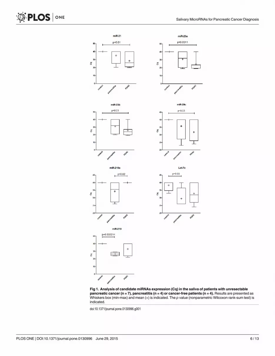
Fig 1. Analysis of candidate miRNAs expression (Cq) in the saliva of patients with unresectablepancreatic cancer (n = 7), pancreatitis (n = 4) or cancer-free patients (n = 4). Results are presented asWhiskers box (min-max) and mean (+) is indicated. The p value (nonparametric Wilcoxon rank-sum test) isindicated.
doi:10.1371/journal.pone.0130996.g001
Salivary MicroRNAs for Pancreatic Cancer Diagnosis
PLOS ONE | DOI:10.1371/journal.pone.0130996 June 29, 2015 6 / 13

that salivary miRNAs could be useful for the diagnosis of pancreatitis and non resectablePDAC.
Salivary miRNAs precede protein-based, systemic detection of PDAC inexperimental modelsWe next investigated the kinetic of salivary miRNA detection in experimental model of pancre-atic cancer. Mia PACA-2 human-derived pancreatic cancer cells were implanted in the pan-creas of athymic mice (n = 6). We found that these cells and resulting xenografts express highlevels of hsa-miR-21, hsa-miR-23a, hsa-miR-23b and hsa-miR-29c (S3 and S4 Tables). Thesecells were engineered to express high-levels of secreted luciferase for protein, systemic-based,non-invasive tumor monitoring [16,17]. Experimental pancreatic cancer tumors were detected25 days following tumor cell engraftment using systemic dosage of secreted luciferase andbefore they became palpable (Fig 2).
Interestingly, hsa-miR-21 was readily detected at high levels in saliva from tumor-bearingmice, as soon as 14 days following tumor induction (mean Cq = 24.41 ±1.29 Fig 2 and S5Table), while undetectable in the saliva of tumor-free animals (data not shown). In addition,salivary hsa-miR-21 expression remained elevated during the course of the experiment (Fig 2).On the other hand, salivary hsa-miR-23a, hsa-miR-23b and hsa-miR-29c were detected at lowlevels in the saliva of PDAC-bearing mice (Fig 2 and S5 Table). Thus, we validate hsa-miR-21as a salivary biomarker in this experimental model of PDAC; in addition our results stronglysuggest that salivary miRNA are more sensitive than systemic protein markers for the diagnosisof pancreatic tumors.
DiscussionAmajor issue in pancreatic cancer research is the need of biomarkers for early diagnosis, notonly for the early detection of the disease in cohort of patients, but also to accelerate decision
Table 2. Average Cq values, sensitivity and specificity of the candidate microRNAs.
Cancer Control
mean Cq SD mean Cq SD p spécificity sensitivity
hsa-miR-21 28,00 3,10 40,00 0,00 0,012 100% 71%
hsa-miR-23a 24,90 2,63 40,00 0,00 0,001 100% 86%
hsa-miR-23b 25,97 2,55 36,75 3,25 0,014 100% 86%
hsa-miR-29c 31,76 2,92 40,00 0,00 0,03 100% 57%
The expression of miRNAs in whole saliva from patients with PDAC (n = 7) were compared to the expression of miRNAs in whole saliva from patients
without cancer (n = 4). The p value (nonparametric Wilcoxon rank-sum test) is indicated.
doi:10.1371/journal.pone.0130996.t002
Table 3. Average Cq values, sensitivity and specificity of the candidate microRNA.
Cancer Pancreatitis
mean Cq SD mean Cq SD p spécificity sensitivity
hsa-miR-216 40,00 0,00 34,25 3,47 0,024 100% 50%
The expression of hsa-miR-216 in whole saliva from patients with PDAC (n = 7) were compared to the expression of miRNAs in whole saliva from patients
with pancreatitis (n = 4). The p value (nonparametric Wilcoxon rank-sum test) is indicated.
doi:10.1371/journal.pone.0130996.t003
Salivary MicroRNAs for Pancreatic Cancer Diagnosis
PLOS ONE | DOI:10.1371/journal.pone.0130996 June 29, 2015 7 / 13

making in difficult-to-diagnose pancreatic masses. This is extremely important consideringthat patients’ survival and prognosis depend on the stage of the tumor at the time of diagnosis.Theoretically, early diagnosis can allow for tumor resection and is usually associated with thebest prognosis. However, the difficulty of early diagnosis and the high prevalence of metastasisassociated with pancreatic cancer contribute to its dismal prognosis [1]. Thus, the past fewyears have witnessed intensive study in searching for more sensitive, specific and cost-effectivebiomarkers. To date, many molecular-based, multi-omics strategies are utilized to achieve thisgoal. Tissue miRNAs were recently demonstrated as novel biomarkers for the diagnosis, prog-nosis and prediction to treatment response for pancreatic cancer patients [4]. Remarkably,these small noncoding RNAs can also be detected in many if not all body fluids [20]. Accord-ingly, miRNA profiling in blood was recently demonstrated to differentiate cancer patientsfrom healthy controls [4], and circulating miRNA analysis have been increasingly suggested asa novel biomarker for pancreatic cancer diagnosis.
In the past few years, miRNAs in human saliva have been demonstrated to be potential bio-markers for diagnosis purposes. Because collection is non-invasive, atraumatic and easilyaccessible, using saliva for early disease detection is ideal. Historically, hsa-miR-31 was one ofthe first discriminatory miRNA salivary biomarkers identified for oral cancer [21]. Recently,over-expression of has-miR-17 and has-miR-20a have been reported to be significantly associ-ated with poor outcome of salivary adenoid cystic carcinoma [22]. In addition, 13 miRNAswere found significantly deregulated in saliva of oral squamous cell carcinoma patients as com-pared to healthy controls [23]. Last, salivary miRNA profiles differ in saliva from patients withmalignant from saliva from patients with a benign parotid gland tumor, and thus represent anew non-invasive diagnostic tool for diagnosing tumors in the salivary glands [9]. During theredaction of this manuscript, Xie et al described that salivary miR-3679-5p and miR-940, twonewly characterized miRNAs that were not studied in the present work, may be specific ofpatients with resectable PDAC, with reasonable specificity and sensitivity [24]. On the otherhand, saliva use for miRNA detection has not been evaluated to date in unresectable PDACpatients that represent the vast majority (85%) of patients diagnosed with this cancer.
In the present proof-of-concept study, we collected saliva from patients with unresectablepancreatic cancer (n = 7), pancreatitis (n = 4), IPMN (n = 2), and cancer-free patients (n = 4)undergoing endoscopic examination. Of more than 90 miRNAs tested, 4 were identified asbeing significantly deregulated in saliva of pancreatic cancer patients compared to control(hsa-miR-21, hsa-miR-23a, hsa-miR-23b and hsa-miR-29c). In addition, hsa-miR-21, hsa-miR-23a and hsa-miR-23b were strictly specific to cancer patients, with excellent sensitivity(71.4% and 85.7%, respectively). On the other hand, Let-7c and hsa-miR-210 were absent inthe saliva of control patients but readily detectable in the saliva of patients with pancreatitis,with exquisite specificity and selectivity (hsa-miR-210).
Table 4. Average Cq values, sensitivity and specificity of the candidate microRNAs.
Conrol Pancreatitis
mean Cq SD mean Cq SD p spécificity sensitivity
hsa-miR-210 40,00 0,00 26,50 1,19 0,000014 100% 100%
Let-7c 38,25 1,75 29,50 3,50 0,033 75% 80%
The expression of hsa-miR-216 in whole saliva from patients with pancreatitis (n = 4) were compared to the expression of miRNAs in whole saliva from
control patients (n = 4). The p value (nonparametric Wilcoxon rank-sum test) is indicated.
doi:10.1371/journal.pone.0130996.t004
Salivary MicroRNAs for Pancreatic Cancer Diagnosis
PLOS ONE | DOI:10.1371/journal.pone.0130996 June 29, 2015 8 / 13

However, at this stage of this project, salivary testing failed to differentiate between pancrea-titis and PDAC, as hsa-miR-216 is detected only in pancreatitis and not in cancer, but withpoor sensitivity. Taken together, we demonstrate for the first time that salivary miRNA areindicative of pancreatic disease and can be used to diagnose unresectable PDAC (hsa-miR-21,
Fig 2. Analysis of salivary hsa-miR-21, hsa-miR-23a, hsa-miR-23b and hsa-miR-29c levels and Lucia blood levels in mice xenografted with MiaPACA-2 Lucia cells at the time indicated following tumor induction. Results are mean ± S.D. of 6 biological replicates done in experimental triplicates.miRNA levels are expressed in Cq, Lucia levels are expressed in relative light units (r.l.u.). The grey zone corresponds to tumor detection using secretedLucia as a systemic, protein-based tumor marker.
doi:10.1371/journal.pone.0130996.g002
Salivary MicroRNAs for Pancreatic Cancer Diagnosis
PLOS ONE | DOI:10.1371/journal.pone.0130996 June 29, 2015 9 / 13
hsa-miR-23a, hsa-miR-23b) or pancreatitis (hsa-miR-210). Hsa-miR-21, hsa-miR-23a andhsa-miR-23b were found significantly deregulated in the saliva of resectable PDAC patients ascompared to healthy control during the discovery phase, but were not further investigated asthey didn’t exhibit at least a 4-fold change in expression between the two groups [24].
In this work, we have started exploring whether salivary miRNAs may help for the diagnosisof population at risk of developing pancreatic cancer, and thus could be used as marker to pre-vent tumor incidence. Intraductal papillary mucinous neoplasms (IPMNs) are non-invasiveprecursor lesions of pancreatic cancer. Recently, miRNAs in cyst fluid have been demonstratedto identify high grade IPMN that requires resection and to exclude non-mucinous cysts imply-ing conservative management with high sensitivity and specificity [25]. We have obtained pre-liminary results suggesting that hsa-miR-23a and hsa-miR-23b are also be present in salivafrom patients diagnosed with IPMN, and could be used for decision making in IPMNmanagement.
However, our study tends to indicate that hsa-miR-21, hsa-miR-23a and hsa-miR-23b arepresent in the saliva of patients with pancreatitis, while hsa-miR-210 is detected in the saliva ofa fraction of patients with PDAC. In addition, hsa-miR-23a and hsa-miR-23b are present inthe saliva of patients with IPMN. This could be easily explained as pancreatitis and IPMN aretwo-well characterized PDAC precursor lesions, indicating that PDAC positive for hsa-miR-210, or hsa-miR-23a and hsa-miR-23b, may have derived from pancreatitis or IPMN, respec-tively. On the contrary, patients diagnosed with pancreatitis and elevated salivary hsa-miR-21,hsa-miR-23a and hsa-miR-23b, or patients diagnosed with IPMN and elevated salivary hsa-miR-23a and hsa-miR-23b may be at-risk of developing PDAC and may require careful clinicalfollow-up. We are aware that the present study suffers from small sample sizing and requiresan external validation population. Consequently, we have recently constituted the first clini-cally annotated cohort of pancreatic cancer patients’ samples from different institutes (theBACAP initiative, http://www.chu-toulouse.fr/-projet-bacap-). Such cohort will be immenselyinformative for further validation and future clinical application of our method, because itrepresents a unique source of PDAC samples, but also because it’s recapitulate the “natural his-tory” of this disease. Such cohort may help to establish salivary miRNAs, together with addi-tional clinical variables, as novel biomarkers for pancreatic cancer patients’management. Inaddition, we have yet to perform comparative studies between different cancer patients to jus-tify that the biomarkers we identified herein are specific for pancreatic cancer.
In this article, we have identified hsa-miR-21, hsa-miR-23a and hsa-miR-23b that were dif-ferently expressed between saliva samples of patients with a malignant tumor and cancer-freepatients, with excellent specificity and sensitivity. While hsa-miR-21 is also associated withmany physiological conditions including but not restricted to cardiovascular and pulmonarydiseases, including cardiac and pulmonary fibrosis as well as myocardial infarction, but alsowith immunological and developmental processes [26], hsa-miR-21 is one of the most citedmiRNA in oncology [27], including pancreatic cancer [4]. We previously demonstratedthat hsa-miR-21 is early expressed during pancreatic carcinogenesis [28], and that targetinghsa-miR-21 provokes tumor regression in experimental models of pancreatic cancer [17].Strikingly, hsa-miR-21 appears to be constantly up regulated in pancreatic cancer, and to beindicative of poor survival, response to treatment and/or metastatic disease [4]. In addition, arecent meta analysis recently demonstrated circulating hsa-miR-21 prognostic rather thandiagnostic value in different cancers [29]. In the present study, we speculate that salivary hsa-miR-21 may also be of interest for pancreatic cancer diagnosis, and complete the previouscharacterization of salivary hsa-miR-21 for the detection of esophageal cancer [10]. To ourknowledge, we provide herein the first demonstration that hsa-miR-23a and hsa-miR-23bcould be detected in the saliva of patients diagnosed with cancer; however, the specificity of
Salivary MicroRNAs for Pancreatic Cancer Diagnosis
PLOS ONE | DOI:10.1371/journal.pone.0130996 June 29, 2015 10 / 13

both candidate miRNAs for PDAC is still to be demonstrated. Hsa-miR-23a has recently beenassociated with KRAS [30] and C-MYC [31] mediated signaling pathway, and described as acandidate driving miRNA in pancreatic cancer [30]. Hsa-miR-23a has also been linked toimpaired NK cell cytotoxicity [32], EMT [33] and resistance to treatment [34–36]. Interest-ingly, hsa-miR-23b was recently demonstrated to regulate autophagy associated with radiore-sistance of pancreatic cancer cells [37].
We next investigated the kinetic of detection of the salivary miRNAs in an experimentalmodel of pancreatic cancer. While hsa-miR-23a and hsa-miR-23b were highly expressed inhuman pancreatic cancer cells-derived xenografts, they were barely detectable in saliva in thismodel of tumor-bearing mice. On the other hand, hsa-miR-21 was readily detected in tumorsand in saliva of mice xenografted with human pancreatic cancer-derived cells, while undetect-able in control animals. This latter finding strongly suggest that salivary hsa-miR-21 originatesfrom experimental tumors, probably via tumor-derived exosomes, as recently described [38].In addition, we demonstrate herein that salivary hsa-miR-21 detection precedes detection ofcancer-cell specific tumor marker in this experimental model of PDAC. This strongly suggeststhat salivary miRNA, including hsa-miR-21, are more sensitive than systemic-based proteinmarkers for the diagnosis of PDAC.
ConclusionTaken together, we demonstrate herein for the first time that salivary miRNA could be valuablebiomarkers for distinguishing patients with unresectable PDAC from healthy controls, andthat salivary miR-210 may help detect pancreatitis. While multicenter studies with larger sam-ple sizes are needed, this work stems for the use of salivary miRNA as novel biomarkers for thediagnosis of unresectable PDAC.
Supporting InformationS1 Table. miRNA quantified in this study.(XLSX)
S2 Table. miRNAs Cq values in whole saliva from patients without cancer (n = 4), begningpancreatitis (n = 4), pancreatic adenocarcinoma (n = 7) or IPMN (n = 2).(XLSX)
S3 Table. Candidate miRNAs Cq values fromMia PACA-2 Lucia cells (n = 3).(XLSX)
S4 Table. Candidate miRNAs Cq values from n = 6 experimental pancreatic tumours (ET).(XLSX)
S5 Table. Secreted Luciferase and salivary candidate miRNAs Cq values from n = 6 micewith experimental pancreatic tumours.(XLSX)
AcknowledgmentsThe authors would like to thanks Drs Geneviève Tavernier and Jean-José Maoret for their help-ful advices.
Salivary MicroRNAs for Pancreatic Cancer Diagnosis
PLOS ONE | DOI:10.1371/journal.pone.0130996 June 29, 2015 11 / 13

Author ContributionsConceived and designed the experiments: PC MH FS JT. Performed the experiments: AV-V FSMH FM. Analyzed the data: PC JT. Contributed reagents/materials/analysis tools: BB LB.Wrote the paper: PC. Discussed the results and commented on the manuscripts: PC MH FS JTAV-V FM BB LB.
References1. Cancer Facts & Figures 2013 [Internet]. [cited 10 Feb 2014]. Available: http://www.cancer.org/
research/cancerfactsfigures/cancerfactsfigures/cancer-facts-figures-2013
2. Burris HA 3rd, Moore MJ, Andersen J, Green MR, Rothenberg ML, Modiano MR, et al. Improvementsin survival and clinical benefit with gemcitabine as first-line therapy for patients with advanced pancreascancer: a randomized trial. J Clin Oncol Off J Am Soc Clin Oncol. 1997; 15: 2403–2413.
3. Pancreatic Cancer Action Network [Internet]. Available: http://www.pancan.org/
4. Humeau M, Torrisani J, Cordelier P. miRNA in clinical practice: Pancreatic cancer. Clin Biochem. 2013;doi: 10.1016/j.clinbiochem.2013.03.019
5. Xiao Y-F, Yong X, Fan Y-H, Lü M-H, Yang S-M, Hu C-J. microRNA detection in feces, sputum, pleuraleffusion and urine: novel tools for cancer screening (Review). Oncol Rep. 2013; 30: 535–544. doi: 10.3892/or.2013.2525 PMID: 23754129
6. Allegra A, Alonci A, Campo S, Penna G, Petrungaro A, Gerace D, et al. Circulating microRNAs: newbiomarkers in diagnosis, prognosis and treatment of cancer (review). Int J Oncol. 2012; 41: 1897–1912.doi: 10.3892/ijo.2012.1647 PMID: 23026890
7. Cheng Y-SL, Rees T, Wright J. A review of research on salivary biomarkers for oral cancer detection.Clin Transl Med. 2014; 3: 3. doi: 10.1186/2001-1326-3-3 PMID: 24564868
8. Yoshizawa JM, Wong DTW. Salivary microRNAs and oral cancer detection. Methods Mol Biol CliftonNJ. 2013; 936: 313–324. doi: 10.1007/978-1-62703-083-0_24
9. Matse JH, Yoshizawa J, Wang X, Elashoff D, Bolscher JGM, Veerman ECI, et al. Discovery and preva-lidation of salivary extracellular microRNA biomarkers panel for the noninvasive detection of benignand malignant parotid gland tumors. Clin Cancer Res Off J Am Assoc Cancer Res. 2013; 19: 3032–3038. doi: 10.1158/1078-0432.CCR-12-3505
10. Xie Z, Chen G, Zhang X, Li D, Huang J, Yang C, et al. Salivary microRNAs as promising biomarkers fordetection of esophageal cancer. PloS One. 2013; 8: e57502. doi: 10.1371/journal.pone.0057502PMID: 23560033
11. Brinkmann O, Wong DTW. Salivary transcriptome biomarkers in oral squamous cell cancer detection.Adv Clin Chem. 2011; 55: 21–34. PMID: 22126022
12. Sugimoto M, Wong DT, Hirayama A, Soga T, Tomita M. Capillary electrophoresis mass spectrometry-based saliva metabolomics identified oral, breast and pancreatic cancer-specific profiles. MetabolomicsOff J Metabolomic Soc. 2010; 6: 78–95. doi: 10.1007/s11306-009-0178-y
13. Zhang L, Farrell JJ, Zhou H, Elashoff D, Akin D, Park N-H, et al. Salivary transcriptomic biomarkers fordetection of resectable pancreatic cancer. Gastroenterology. 2010; 138: 949–957.e1–7. doi: 10.1053/j.gastro.2009.11.010 PMID: 19931263
14. Farrell JJ, Zhang L, Zhou H, Chia D, Elashoff D, Akin D, et al. Variations of oral microbiota are associ-ated with pancreatic diseases including pancreatic cancer. Gut. 2012; 61: 582–588. doi: 10.1136/gutjnl-2011-300784 PMID: 21994333
15. Bournet B, Selves J, Grand D, Danjoux M, Hanoun N, Cordelier P, et al. Endoscopic Ultrasound-guidedFine-Needle Aspiration Biopsy CoupledWith a KRASMutation Assay Using Allelic DiscriminationImproves the Diagnosis of Pancreatic Cancer. J Clin Gastroenterol. 2014; doi: 10.1097/MCG.0000000000000053
16. Delpu Y, Lulka H, Sicard F, Saint-Laurent N, Lopez F, Hanoun N, et al. The Rescue of miR-148aExpression in Pancreatic Cancer: An Inappropriate Therapeutic Tool. Schneider G, editor. PLoS ONE.2013; 8: e55513. doi: 10.1371/journal.pone.0055513 PMID: 23383211
17. Sicard F, Gayral M, Lulka H, Buscail L, Cordelier P. Targeting miR-21 for the Therapy of PancreaticCancer. Mol Ther. 2013; doi: 10.1038/mt.2013.35
18. Bournet B, Pointreau A, Souque A, Oumouhou N, Muscari F, Lepage B, et al. Gene expression signa-ture of advanced pancreatic ductal adenocarcinoma using low density array on endoscopic ultrasound-guided fine needle aspiration samples. Pancreatol Off J Int Assoc Pancreatol IAP Al. 2012; 12: 27–34.doi: 10.1016/j.pan.2011.12.003
Salivary MicroRNAs for Pancreatic Cancer Diagnosis
PLOS ONE | DOI:10.1371/journal.pone.0130996 June 29, 2015 12 / 13

19. Bournet B, Souque A, Senesse P, Assenat E, Barthet M, Lesavre N, et al. Endoscopic ultrasound-guided fine-needle aspiration biopsy coupled with KRASmutation assay to distinguish pancreatic can-cer from pseudotumoral chronic pancreatitis. Endoscopy. 2009; 41: 552–557. doi: 10.1055/s-0029-1214717 PMID: 19533561
20. Weber JA, Baxter DH, Zhang S, Huang DY, Huang KH, Lee MJ, et al. The microRNA spectrum in 12body fluids. Clin Chem. 2010; 56: 1733–1741. doi: 10.1373/clinchem.2010.147405 PMID: 20847327
21. Liu C-J, Lin S-C, Yang C-C, Cheng H-W, Chang K-W. Exploiting salivary miR-31 as a clinical biomarkerof oral squamous cell carcinoma. Head Neck. 2012; 34: 219–224. doi: 10.1002/hed.21713 PMID:22083872
22. Mitani Y, Roberts DB, Fatani H, Weber RS, Kies MS, Lippman SM, et al. MicroRNA profiling of salivaryadenoid cystic carcinoma: association of miR-17-92 upregulation with poor outcome. PloS One. 2013;8: e66778. doi: 10.1371/journal.pone.0066778 PMID: 23825564
23. Momen-Heravi F, Trachtenberg AJ, KuoWP, Cheng YS. Genomewide Study of Salivary MicroRNAsfor Detection of Oral Cancer. J Dent Res. 2014; doi: 10.1177/0022034514531018
24. Xie Z, Yin X, Gong B, Nie W,Wu B, Zhang X, et al. Salivary microRNAs show potential as a noninvasivebiomarker for detecting resectable pancreatic cancer. Cancer Prev Res Phila Pa. 2015; 8: 165–173.doi: 10.1158/1940-6207.CAPR-14-0192
25. Matthaei H, Wylie D, Lloyd MB, Dal Molin M, Kemppainen J, Mayo SC, et al. miRNA biomarkers in cystfluid augment the diagnosis and management of pancreatic cysts. Clin Cancer Res Off J Am AssocCancer Res. 2012; 18: 4713–4724. doi: 10.1158/1078-0432.CCR-12-0035
26. Kumarswamy R, Volkmann I, Thum T. Regulation and function of miRNA-21 in health and disease.RNA Biol. 2011; 8: 706–713. doi: 10.4161/rna.8.5.16154 PMID: 21712654
27. ZhuW, Xu B. MicroRNA-21 identified as predictor of cancer outcome: a meta-analysis. PloS One.2014; 9: e103373. doi: 10.1371/journal.pone.0103373 PMID: 25098165
28. Du Rieu MC, Torrisani J, Selves J, Al Saati T, Souque A, Dufresne M, et al. MicroRNA-21 is inducedearly in pancreatic ductal adenocarcinoma precursor lesions. Clin Chem. 2010; 56: 603–612. doi: 10.1373/clinchem.2009.137364 PMID: 20093556
29. Wang Y, Gao X, Wei F, Zhang X, Yu J, Zhao H, et al. Diagnostic and prognostic value of circulatingmiR-21 for cancer: a systematic review and meta-analysis. Gene. 2014; 533: 389–397. doi: 10.1016/j.gene.2013.09.038 PMID: 24076132
30. Piepoli A, Tavano F, Copetti M, Mazza T, Palumbo O, Panza A, et al. Mirna expression profiles identifydrivers in colorectal and pancreatic cancers. PloS One. 2012; 7: e33663. doi: 10.1371/journal.pone.0033663 PMID: 22479426
31. Li X, Liu X, XuW, Zhou P, Gao P, Jiang S, et al. c-MYC-regulated miR-23a/24-2/27a cluster promotesmammary carcinoma cell invasion and hepatic metastasis by targeting Sprouty2. J Biol Chem. 2013;288: 18121–18133. doi: 10.1074/jbc.M113.478560 PMID: 23649631
32. Sanchez-Martínez D, Krzywinska E, Rathore MG, Saumet A, Cornillon A, Lopez-Royuela N, et al. All-trans retinoic acid (ATRA) induces miR-23a expression, decreases CTSC expression and granzyme Bactivity leading to impaired NK cell cytotoxicity. Int J Biochem Cell Biol. 2014; 49: 42–52. doi: 10.1016/j.biocel.2014.01.003 PMID: 24440757
33. Zheng H, Li W, Wang Y, Xie T, Cai Y, Wang Z, et al. miR-23a inhibits E-cadherin expression and is reg-ulated by AP-1 and NFAT4 complex during Fas-induced EMT in gastrointestinal cancer. Carcinogene-sis. 2014; 35: 173–183. doi: 10.1093/carcin/bgt274 PMID: 23929433
34. Lian S, Shi R, Bai T, Liu Y, MiaoW, Wang H, et al. Anti-miRNA-23a oligonucleotide suppresses gliomacells growth by targeting apoptotic protease activating factor-1. Curr Pharm Des. 2013; 19: 6382–6389.PMID: 23865473
35. Liu X, Ru J, Zhang J, Zhu L, Liu M, Li X, et al. miR-23a targets interferon regulatory factor 1 and modu-lates cellular proliferation and paclitaxel-induced apoptosis in gastric adenocarcinoma cells. PloS One.2013; 8: e64707. doi: 10.1371/journal.pone.0064707 PMID: 23785404
36. Shang J, Yang F, Wang Y, Wang Y, Xue G, Mei Q, et al. MicroRNA-23a antisense enhances 5-fluoro-uracil chemosensitivity through APAF-1/caspase-9 apoptotic pathway in colorectal cancer cells. J CellBiochem. 2014; 115: 772–784. doi: 10.1002/jcb.24721 PMID: 24249161
37. Wang P, Zhang J, Zhang L, Zhu Z, Fan J, Chen L, et al. MicroRNA 23b Regulates Autophagy Associ-atedWith Radioresistance of Pancreatic Cancer Cells. Gastroenterology. 2013; 145: 1133–1143.e12.doi: 10.1053/j.gastro.2013.07.048 PMID: 23916944
38. Lau C, Kim Y, Chia D, Spielmann N, Eibl G, Elashoff D, et al. Role of pancreatic cancer-derived exo-somes in salivary biomarker development. J Biol Chem. 2013; 288: 26888–26897. doi: 10.1074/jbc.M113.452458 PMID: 23880764
Salivary MicroRNAs for Pancreatic Cancer Diagnosis
PLOS ONE | DOI:10.1371/journal.pone.0130996 June 29, 2015 13 / 13