microrna-21 regulates stemness in pancreatic ductal
TRANSCRIPT
�����������������
Citation: Mortoglou, M.; Miralles, F.;
Arisan, E.D.; Dart, A.; Jurcevic, S.;
Lange, S.; Uysal-Onganer, P.
microRNA-21 Regulates Stemness in
Pancreatic Ductal Adenocarcinoma
Cells. Int. J. Mol. Sci. 2022, 23, 1275.
https://doi.org/10.3390/
ijms23031275
Academic Editors: Claudio Luchin,
Aamir Ahmad and Nicoletta Potenza
Received: 17 December 2021
Accepted: 21 January 2022
Published: 24 January 2022
Publisher’s Note: MDPI stays neutral
with regard to jurisdictional claims in
published maps and institutional affil-
iations.
Copyright: © 2022 by the authors.
Licensee MDPI, Basel, Switzerland.
This article is an open access article
distributed under the terms and
conditions of the Creative Commons
Attribution (CC BY) license (https://
creativecommons.org/licenses/by/
4.0/).
International Journal of
Molecular Sciences
Article
microRNA-21 Regulates Stemness in Pancreatic DuctalAdenocarcinoma CellsMaria Mortoglou 1 , Francesc Miralles 2,3 , Elif Damla Arisan 4 , Alwyn Dart 3 , Stipo Jurcevic 5,Sigrun Lange 6 and Pinar Uysal-Onganer 1,*
1 Cancer Research Group, School of Life Sciences, University of Westminster, London W1W 6UW, UK;[email protected]
2 Molecular and Clinical Sciences, Cell Biology Research Centre, St. George’s University of London,Cranmer Terrace, SW17ORE London, UK; [email protected]
3 Institute of Medical and Biomedical Education, St. George’s University of London, Cranmer Terrace,London SW17 ORE, UK; [email protected]
4 Institution of Biotechnology, Gebze Technical University, Gebze 41400, Turkey; [email protected] Department of Biomedical Sciences, School of Life Sciences, University of Westminster,
London W1W 6UW, UK; [email protected] Tissue Architecture and Regeneration Research Group, School of Life Sciences, University of Westminster,
London W1W 6UW, UK; [email protected]* Correspondence: [email protected]
Abstract: Pancreatic ductal adenocarcinoma (PDAC) is the most common and aggressive typeof pancreatic cancer (PCa) with a low survival rate. microRNAs (miRs) are endogenous, non-coding RNAs that moderate numerous biological processes. miRs have been associated with thechemoresistance and metastasis of PDAC and the presence of a subpopulation of highly plastic“stem”-like cells within the tumor, known as cancer stem cells (CSCs). In this study, we investigatedthe role of miR-21, which is highly expressed in Panc-1 and MiaPaCa-2 PDAC cells in associationwith CSCs. Following miR-21 knockouts (KO) from both MiaPaCa-2 and Panc-1 cell lines, reversedexpressions of epithelial–mesenchymal transition (EMT) and CSCs markers were observed. Theexpression patterns of key CSC markers, including CD44, CD133, CX-C chemokine receptor type4 (CXCR4), and aldehyde dehydrogenase-1 (ALDH1), were changed depending on miR-21 status.miR-21 (KO) suppressed cellular invasion of Panc-1 and MiaPaCa-2 cells, as well as the cellularproliferation of MiaPaCa-2 cells. Our data suggest that miR-21 is involved in the stemness ofPDAC cells, may play roles in mesenchymal transition, and that miR-21 poses as a novel, functionalbiomarker for PDAC aggressiveness.
Keywords: pancreatic ductal adenocarcinoma; microRNAs; non-coding RNAs; cancer stem cells;metastasis; epithelial–mesenchymal transition
1. Introduction
Pancreatic ductal adenocarcinoma (PDAC) is the eighth primary source of cancer-related deaths globally with a 5-year survival rate of 3–6% [1–4]. Approximately, 10–20%of PDAC patients are compatible for surgery at the time of diagnosis, and 9.7% of PDACcases are at a local stage when initially diagnosed [5]. microRNAs (miRs) are 18 to 24nucleotides-long, endogenous, non-coding, evolutionarily conserved, single-stranded RNAmolecules. miRs can moderate gene expression at the posttranscriptional level throughthe binding to the complementary sequences of their target mRNAs at the 3′ untranslatedregions (UTRs), which allow them to control the expression levels of several genes andregulate various signaling pathways [6–9]. Preliminary studies have suggested a correlationbetween aberrant expression levels of numerous miRs with PDAC [10–13]. miRs can actas oncogenic miRs (oncomiRs) or tumor suppressor miRs. Especially in PDAC, miR-21,miR-155, and miR-221 have been found to act as oncomiRs, while miR-126 and miR-375
Int. J. Mol. Sci. 2022, 23, 1275. https://doi.org/10.3390/ijms23031275 https://www.mdpi.com/journal/ijms
Int. J. Mol. Sci. 2022, 23, 1275 2 of 23
were shown to act as tumor suppressors miRs [14,15]. miR-21, miR-221, and miR-155 candistinguish cases of PDAC from healthy individuals with a sensitivity of approximately64% and a specificity of 89% [16,17]. Importantly, miRs present a higher sensitivity as adiagnostic marker than the current diagnostic marker carbohydrate antigen (CA 19-9),especially for the early diagnosis of PDAC [18–21].
Cancer stem cells (CSCs) are involved in chemoresistance and play critical roles inthe metastasis of several cancers, including PDAC [22–29]. CSCs contribute to elevatedexpression levels of anti-apoptotic proteins, ABC transporters, and multidrug resistancegenes, high autophagic flux that leads to microenvironment stresses [30–34]. PancreaticCSCs (PCSCs) are less than 1% of all pancreatic cancer cells and are critical mediatorsof PDAC tumor growth, maintenance, metastasis, and chemoresistance [35]. EMT ischaracterized as a critical mechanism of the metastatic cascade, which includes the lossof cell adhesion, elevated cell motility, the repression of E-cadherin, and the upregulationof mesenchymal markers, such as Vimentin, N-cadherin, Snail, and Zeb1 [36]. E-cadherindownregulation is associated with poor prognosis, differentiation, and chemoresistance inPDAC [37–40]. Transcription marker Zeb1 suppresses E-cadherin through the repression ofboth miR-203 (an inhibitor of stemness) and miR-200 family members, which control theexpression levels of stem cell factors [41]. Zeb1 overexpression is linked to advanced PDACstages and poor malignancy outcome, migration, and invasion in response to nuclear factorkappa-light-chain-enhancer of activated B cells (NF-κB) signaling [42–44]. Non-canonicalWnt-11 overexpression is associated with poor prognosis and tumor-node-metastasis (TNM)staging in PDAC [45–49].
Higher expression levels of Snail, a potent EMT-inducing transcription factor, arerelated to 80% of PDAC cases, E-cadherin downregulation, lymph node invasion, highertumor grade, and poorly differentiated PDAC cells [50]. EMT is regulated via molecu-lar pathways linked to oncogenic and tumor suppressor non-coding RNAs, chromatinremodeling, epigenetic and posttranslational modifications, alternative splicing events,and protein stability [51,52]. Vimentin is an essential marker of EMT and associated withNotch and miR-200 expression levels and it can affect treatment response in vitro, includingelevated gemcitabine-resistance in PDAC [53,54]. Overexpression of Vimentin is linkedto metastasis and poor overall survival in PDAC [55,56]. Furthermore, the associationbetween EMT and CSCs has been extensively evaluated, for instance, PDAC cells, whichhave undergone EMT, and express epithelial markers, such as E-cadherin and mesenchymalmarkers, such as Zinc Finger E-Box Binding Homeobox 1 (Zeb1), and Snail exhibit stemcell properties [57,58]. The main markers of PCSCs are CD133, CD24, CD44, ESA/EpCAM(epithelial-specific antigen), c-Met, ALDH1, DclK1, CXCR4, and Lgr5 [30,59,60]. Impor-tantly, EMT and autophagy processes are also closely linked with CSCs markers duringPDAC development [59,60]. Recent reports stated that CD24, CD44, CXCR4, ESA, andnestin are upregulated in advanced pancreatic intraepithelial neoplasia (PanIN) grades [61],while others have shown that the expression of cMet+ CD133+ CD34+ CD45− Ter119−,Pdx1, CD9, CD24, CD44, CD13, and CD133, are linked to poor prognosis of PDAC [61–67].
miR-21 is one of the most oncogenic miRs related to PDAC prognosis; overexpressionof miR-21 was detected in PDAC patients and correlated with poor prognosis and overallsurvival according to the TCGA dataset (Figure S1). Therefore, the role of miR-21 in PDACstemness was examined in this study in-depth, using CRISPR-mediated KO approachesin vitro.
2. Results
In summary, using three different PDAC cell lines, we found that miR-21, miR-221,miR-155, and miR-126 expressions were significantly altered in MiaPaca-2, Panc-1, andBxPC3 PDAC cell lines, compared with normal pancreatic ductal epithelial cell lines(HPDE). Following the knockout of miR-21 in Panc-1 and MiaPaca-2 cells using CRISPR-Cas9, reversed expressions of E-cadherin, Vimentin, Snail, Wnt-11, and Zeb1 were detected,suggesting that these markers are targets of miR-21. Expression levels of the CSC markers,
Int. J. Mol. Sci. 2022, 23, 1275 3 of 23
such as CD133, CD44, CD24, CXCR4, and ALDH1, were significantly downregulateddepending on miR-21 status. KO of miR-21 led to a significant reduction in cellular inva-siveness of Panc-1 and MiaPaCa-2 cells and a significant decrease in cellular proliferation ofMiaPaCa-2 cells. Overall, our data suggest that miR-21 is involved in the pathophysiologyof PDAC.
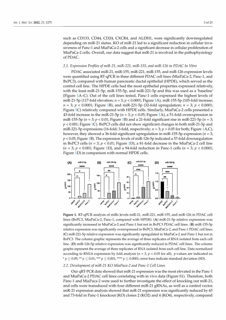
2.1. Expression Profiles of miR-21, miR-221, miR-155, and miR-126 in PDAC In Vitro
PDAC associated miR-21, miR-155, miR-221, miR-155, and miR-126 expression levelswere quantified using RT-qPCR in three different PDAC cell lines (MiaPaCa-2, Panc-1, andBxPC3), compared with human pancreatic ductal epithelial (HPDE), which served as thecontrol cell line. The HPDE cells had the most epithelial properties expressed relatively,with the least miR-21-5p, miR-155-5p, and miR-221-5p and this was used as a ‘baseline’(Figure 1A–C). Out of the cell lines tested, Panc-1 cells expressed the highest levels ofmiR-21-5p (117-fold elevation; n = 3; p < 0.0001; Figure 1A), miR-155-5p (105-fold increase;n = 3; p < 0.0001; Figure 1B), and miR-221-5p (32-fold upregulation; n = 3; p < 0.0001;Figure 1C) relatively compared with HPDE cells. Similarly, MiaPaCa-2 cells presented a43-fold increase in the miR-21-5p (n = 3; p < 0.05; Figure 1A), a 51-fold overexpression inmiR-155-5p (n = 3; p < 0.01; Figure 1B) and a 21-fold significant rise in miR-221-5p (n = 3;p < 0.001; Figure 1C). BxPC3 cells did not show significant changes in both miR-21-5p andmiR-221-5p expressions (16-fold; 3-fold, respectively; n = 3; p > 0.05 for both; Figure 1A,C),however, they showed a 36-fold significant upregulation in miR-155-5p expression (n = 3;p < 0.05; Figure 1B). The expression levels of miR-126-5p indicated a 57-fold downregulationin BxPC3 cells (n = 3; p < 0.01; Figure 1D), a 81-fold decrease in the MiaPaCa-2 cell line(n = 3; p < 0.001; Figure 1D), and a 94-fold reduction in Panc-1 cells (n = 3; p < 0.0001;Figure 1D) in comparison with normal HPDE cells.
Int. J. Mol. Sci. 2022, 23, x FOR PEER REVIEW 3 of 24
markers, such as CD133, CD44, CD24, CXCR4, and ALDH1, were significantly downreg-ulated depending on miR-21 status. KO of miR-21 led to a significant reduction in cellular invasiveness of Panc-1 and MiaPaCa-2 cells and a significant decrease in cellular prolifer-ation of MiaPaCa-2 cells. Overall, our data suggest that miR-21 is involved in the patho-physiology of PDAC.
2.1. Expression Profiles of miR-21, miR-221, miR-155, and miR-126 in PDAC In Vitro PDAC associated miR-21, miR-155, miR-221, miR-155, and miR-126 expression levels
were quantified using RT-qPCR in three different PDAC cell lines (MiaPaCa-2, Panc-1, and BxPC3), compared with human pancreatic ductal epithelial (HPDE), which served as the control cell line. The HPDE cells had the most epithelial properties expressed rela-tively, with the least miR-21-5p, miR-155-5p, and miR-221-5p and this was used as a ‘base-line’ (Figure 1A–C). Out of the cell lines tested, Panc-1 cells expressed the highest levels of miR-21-5p (117-fold elevation; n = 3; p < 0.0001; Figure 1A), miR-155-5p (105-fold in-crease; n = 3; p < 0.0001; Figure 1B), and miR-221-5p (32-fold upregulation; n = 3; p < 0.0001; Figure 1C) relatively compared with HPDE cells. Similarly, MiaPaCa-2 cells presented a 43-fold increase in the miR-21-5p (n = 3; p < 0.05; Figure 1A), a 51-fold overexpression in miR-155-5p (n = 3; p < 0.01; Figure 1B) and a 21-fold significant rise in miR-221-5p (n = 3; p < 0.001; Figure 1C). BxPC3 cells did not show significant changes in both miR-21-5p and miR-221-5p expressions (16-fold; 3-fold, respectively; n = 3; p > 0.05 for both; Figure 1A,C), however, they showed a 36-fold significant upregulation in miR-155-5p expression (n = 3; p < 0.05; Figure 1B). The expression levels of miR-126-5p indicated a 57-fold downregula-tion in BxPC3 cells (n = 3; p < 0.01; Figure 1D), a 81-fold decrease in the MiaPaCa-2 cell line (n = 3; p < 0.001; Figure 1D), and a 94-fold reduction in Panc-1 cells (n = 3; p < 0.0001; Figure 1D) in comparison with normal HPDE cells.
Figure 1. RT-qPCR analysis of miRs levels miR-21, miR-221, miR-155, and miR-126 in PDAC cell lines (BxPC3, MiaPaCa-2, Panc-1, compared with HPDE). (A) miR-21-5p relative expression was significantly increased in MiaPaCa-2 and Panc-1 but not in BxPC3 PDAC cell lines. (B) miR-155-5p relative expression was significantly overexpressed in BxPC3, MiaPaCa-2, and Panc-1 PDAC cell lines. (C) miR-221-5p relative expression was significantly upregulated in MiaPaCa-2 and Panc-1 but not in BxPC3. The column graphic represents the average of three replicates of RNA isolated from each cell line. (D) miR-126-5p relative expression was significantly reduced in PDAC cell lines. The column graphs represent the average of three replicates of RNA isolated from each cell line. Data normalized according to RNU6 expression by fold analysis (n =3, p < 0.05 for all). p-values are indicated as: * p ≤ 0.05; ** p ≤ 0.01; *** p ≤ 0.001; **** p ≤ 0.0001; error bars indicate standard deviation (SD).
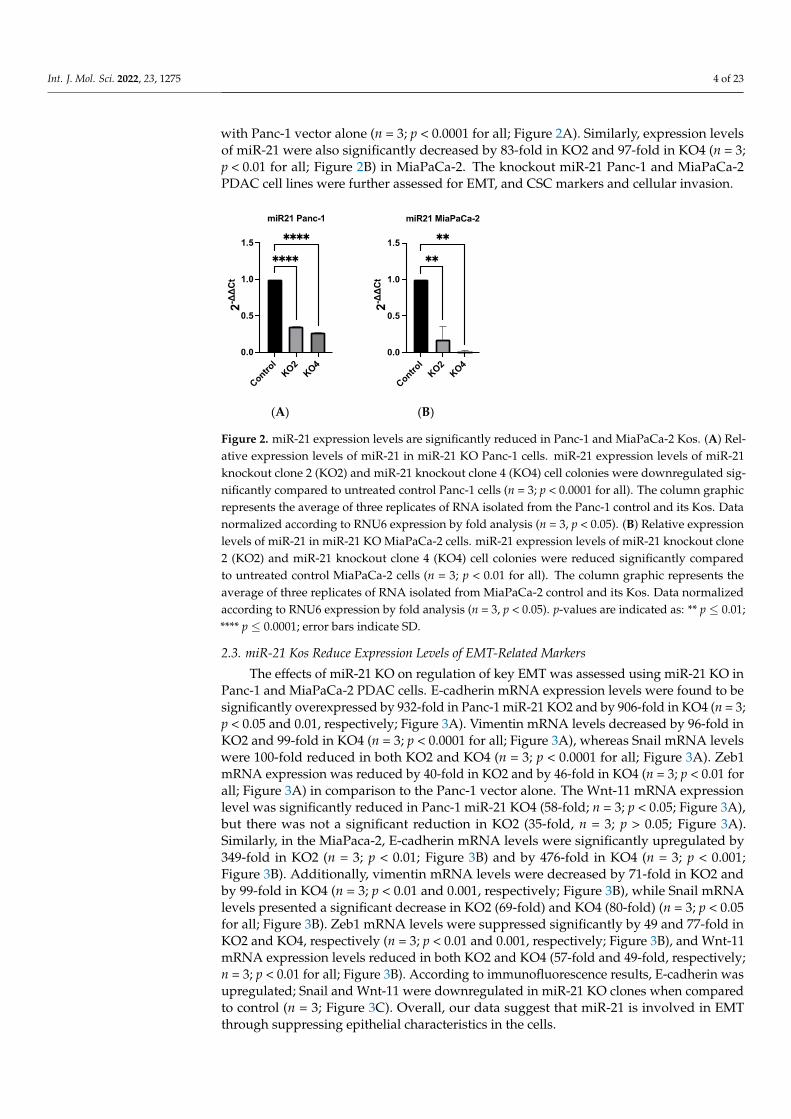
2.2. Development of miR-21 KO MiaPaca-2 and Panc-1 Cell Lines Our qRT-PCR data showed that miR-21 expression was the most elevated in the
Panc-1 and MiaPaCa-2 PDAC cell lines correlating with in vivo data (Figure S1).
Figure 1. RT-qPCR analysis of miRs levels miR-21, miR-221, miR-155, and miR-126 in PDAC celllines (BxPC3, MiaPaCa-2, Panc-1, compared with HPDE). (A) miR-21-5p relative expression wassignificantly increased in MiaPaCa-2 and Panc-1 but not in BxPC3 PDAC cell lines. (B) miR-155-5prelative expression was significantly overexpressed in BxPC3, MiaPaCa-2, and Panc-1 PDAC cell lines.(C) miR-221-5p relative expression was significantly upregulated in MiaPaCa-2 and Panc-1 but not inBxPC3. The column graphic represents the average of three replicates of RNA isolated from each cellline. (D) miR-126-5p relative expression was significantly reduced in PDAC cell lines. The columngraphs represent the average of three replicates of RNA isolated from each cell line. Data normalizedaccording to RNU6 expression by fold analysis (n = 3, p < 0.05 for all). p-values are indicated as:* p ≤ 0.05; ** p ≤ 0.01; *** p ≤ 0.001; **** p ≤ 0.0001; error bars indicate standard deviation (SD).
2.2. Development of miR-21 KO MiaPaca-2 and Panc-1 Cell Lines
Our qRT-PCR data showed that miR-21 expression was the most elevated in the Panc-1and MiaPaCa-2 PDAC cell lines correlating with in vivo data (Figure S1). Therefore, bothPanc-1 and MiaPaca-2 were used to further investigate the effect of knocking out miR-21,and cells were transduced with four different miR-21 gRNAs, as well as a control vector.miR-21 expression analysis showed that miR-21 expression was significantly reduced by 65and 73-fold in Panc-1 knockout (KO) clones 2 (KO2) and 4 (KO4), respectively, compared
Int. J. Mol. Sci. 2022, 23, 1275 4 of 23
with Panc-1 vector alone (n = 3; p < 0.0001 for all; Figure 2A). Similarly, expression levelsof miR-21 were also significantly decreased by 83-fold in KO2 and 97-fold in KO4 (n = 3;p < 0.01 for all; Figure 2B) in MiaPaCa-2. The knockout miR-21 Panc-1 and MiaPaCa-2PDAC cell lines were further assessed for EMT, and CSC markers and cellular invasion.
Int. J. Mol. Sci. 2022, 23, x FOR PEER REVIEW 4 of 24
Therefore, both Panc-1 and MiaPaca-2 were used to further investigate the effect of knock-ing out miR-21, and cells were transduced with four different miR-21 gRNAs, as well as a control vector. miR-21 expression analysis showed that miR-21 expression was signifi-cantly reduced by 65 and 73-fold in Panc-1 knockout (KO) clones 2 (KO2) and 4 (KO4), respectively, compared with Panc-1 vector alone (n = 3; p < 0.0001 for all; Figure 2A). Sim-ilarly, expression levels of miR-21 were also significantly decreased by 83-fold in KO2 and 97-fold in KO4 (n = 3; p < 0.01 for all; Figure 2B) in MiaPaCa-2. The knockout miR-21 Panc-1 and MiaPaCa-2 PDAC cell lines were further assessed for EMT, and CSC markers and cellular invasion.
(A) (B)
Figure 2. miR-21 expression levels are significantly reduced in Panc-1 and MiaPaCa-2 Kos. (A) Rel-ative expression levels of miR-21 in miR-21 KO Panc-1 cells. miR-21 expression levels of miR-21 knockout clone 2 (KO2) and miR-21 knockout clone 4 (KO4) cell colonies were downregulated sig-nificantly compared to untreated control Panc-1 cells (n = 3; p < 0.0001 for all). The column graphic represents the average of three replicates of RNA isolated from the Panc-1 control and its Kos. Data normalized according to RNU6 expression by fold analysis (n = 3, p < 0.05). (B) Relative expression levels of miR-21 in miR-21 KO MiaPaCa-2 cells. miR-21 expression levels of miR-21 knockout clone 2 (KO2) and miR-21 knockout clone 4 (KO4) cell colonies were reduced significantly compared to untreated control MiaPaCa-2 cells (n = 3; p < 0.01 for all). The column graphic represents the average of three replicates of RNA isolated from MiaPaCa-2 control and its Kos. Data normalized according to RNU6 expression by fold analysis (n = 3, p < 0.05). p-values are indicated as: ** p ≤ 0.01; **** p ≤ 0.0001; error bars indicate SD.
2.3. miR-21 Kos Reduce Expression Levels of EMT-Related Markers The effects of miR-21 KO on regulation of key EMT was assessed using miR-21 KO
in Panc-1 and MiaPaCa-2 PDAC cells. E-cadherin mRNA expression levels were found to be significantly overexpressed by 932-fold in Panc-1 miR-21 KO2 and by 906-fold in KO4 (n = 3; p < 0.05 and 0.01, respectively; Figure 3A). Vimentin mRNA levels decreased by 96-fold in KO2 and 99-fold in KO4 (n = 3; p < 0.0001 for all; Figure 3A), whereas Snail mRNA levels were 100-fold reduced in both KO2 and KO4 (n = 3; p < 0.0001 for all; Figure 3A). Zeb1 mRNA expression was reduced by 40-fold in KO2 and by 46-fold in KO4 (n = 3; p < 0.01 for all; Figure 3A) in comparison to the Panc-1 vector alone. The Wnt-11 mRNA ex-pression level was significantly reduced in Panc-1 miR-21 KO4 (58-fold; n = 3; p < 0.05; Figure 3A), but there was not a significant reduction in KO2 (35-fold, n = 3; p > 0.05; Figure 3A). Similarly, in the MiaPaca-2, E-cadherin mRNA levels were significantly upregulated by 349-fold in KO2 (n = 3; p < 0.01; Figure 3B) and by 476-fold in KO4 (n = 3; p < 0.001; Figure 3B). Additionally, vimentin mRNA levels were decreased by 71-fold in KO2 and by 99-fold in KO4 (n = 3; p < 0.01 and 0.001, respectively; Figure 3B), while Snail mRNA levels presented a significant decrease in KO2 (69-fold) and KO4 (80-fold) (n = 3; p < 0.05 for all; Figure 3B). Zeb1 mRNA levels were suppressed significantly by 49 and 77-fold in KO2 and KO4, respectively (n = 3; p < 0.01 and 0.001, respectively; Figure 3B), and Wnt-11
Figure 2. miR-21 expression levels are significantly reduced in Panc-1 and MiaPaCa-2 Kos. (A) Rel-ative expression levels of miR-21 in miR-21 KO Panc-1 cells. miR-21 expression levels of miR-21knockout clone 2 (KO2) and miR-21 knockout clone 4 (KO4) cell colonies were downregulated sig-nificantly compared to untreated control Panc-1 cells (n = 3; p < 0.0001 for all). The column graphicrepresents the average of three replicates of RNA isolated from the Panc-1 control and its Kos. Datanormalized according to RNU6 expression by fold analysis (n = 3, p < 0.05). (B) Relative expressionlevels of miR-21 in miR-21 KO MiaPaCa-2 cells. miR-21 expression levels of miR-21 knockout clone2 (KO2) and miR-21 knockout clone 4 (KO4) cell colonies were reduced significantly comparedto untreated control MiaPaCa-2 cells (n = 3; p < 0.01 for all). The column graphic represents theaverage of three replicates of RNA isolated from MiaPaCa-2 control and its Kos. Data normalizedaccording to RNU6 expression by fold analysis (n = 3, p < 0.05). p-values are indicated as: ** p ≤ 0.01;**** p ≤ 0.0001; error bars indicate SD.
2.3. miR-21 Kos Reduce Expression Levels of EMT-Related Markers
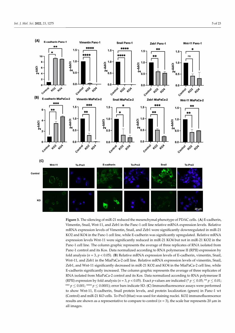
The effects of miR-21 KO on regulation of key EMT was assessed using miR-21 KO inPanc-1 and MiaPaCa-2 PDAC cells. E-cadherin mRNA expression levels were found to besignificantly overexpressed by 932-fold in Panc-1 miR-21 KO2 and by 906-fold in KO4 (n = 3;p < 0.05 and 0.01, respectively; Figure 3A). Vimentin mRNA levels decreased by 96-fold inKO2 and 99-fold in KO4 (n = 3; p < 0.0001 for all; Figure 3A), whereas Snail mRNA levelswere 100-fold reduced in both KO2 and KO4 (n = 3; p < 0.0001 for all; Figure 3A). Zeb1mRNA expression was reduced by 40-fold in KO2 and by 46-fold in KO4 (n = 3; p < 0.01 forall; Figure 3A) in comparison to the Panc-1 vector alone. The Wnt-11 mRNA expressionlevel was significantly reduced in Panc-1 miR-21 KO4 (58-fold; n = 3; p < 0.05; Figure 3A),but there was not a significant reduction in KO2 (35-fold, n = 3; p > 0.05; Figure 3A).Similarly, in the MiaPaca-2, E-cadherin mRNA levels were significantly upregulated by349-fold in KO2 (n = 3; p < 0.01; Figure 3B) and by 476-fold in KO4 (n = 3; p < 0.001;Figure 3B). Additionally, vimentin mRNA levels were decreased by 71-fold in KO2 andby 99-fold in KO4 (n = 3; p < 0.01 and 0.001, respectively; Figure 3B), while Snail mRNAlevels presented a significant decrease in KO2 (69-fold) and KO4 (80-fold) (n = 3; p < 0.05for all; Figure 3B). Zeb1 mRNA levels were suppressed significantly by 49 and 77-fold inKO2 and KO4, respectively (n = 3; p < 0.01 and 0.001, respectively; Figure 3B), and Wnt-11mRNA expression levels reduced in both KO2 and KO4 (57-fold and 49-fold, respectively;n = 3; p < 0.01 for all; Figure 3B). According to immunofluorescence results, E-cadherin wasupregulated; Snail and Wnt-11 were downregulated in miR-21 KO clones when comparedto control (n = 3; Figure 3C). Overall, our data suggest that miR-21 is involved in EMTthrough suppressing epithelial characteristics in the cells.
Int. J. Mol. Sci. 2022, 23, 1275 5 of 23
Int. J. Mol. Sci. 2022, 23, x FOR PEER REVIEW 5 of 24
mRNA expression levels reduced in both KO2 and KO4 (57-fold and 49-fold, respectively; n = 3; p < 0.01 for all; Figure 3B). According to immunofluorescence results, E-cadherin was upregulated; Snail and Wnt-11 were downregulated in miR-21 KO clones when com-pared to control (n = 3; Figure 3C). Overall, our data suggest that miR-21 is involved in EMT through suppressing epithelial characteristics in the cells.
.
Figure 3. The silencing of miR-21 reduced the mesenchymal phenotype of PDAC cells. (A) E-cad-herin, Vimentin, Snail, Wnt-11, and Zeb1 in the Panc-1 cell line relative mRNA expression levels. Relative mRNA expression levels of Vimentin, Snail, and Zeb1 were significantly downregulated in miR-21 KO2 and KO4 in the Panc-1 cell line, while E-cadherin was significantly upregulated. Rela-tive mRNA expression levels Wnt-11 were significantly reduced in miR-21 KO4 but not in miR-21 KO2 in the Panc-1 cell line. The column graphic represents the average of three replicates of RNA isolated from Panc-1 control and its Kos. Data normalized according to RNA polymerase II (RPII) expression by fold analysis (n =3, p < 0.05). (B) Relative mRNA expression levels of E-cadherin,
Figure 3. The silencing of miR-21 reduced the mesenchymal phenotype of PDAC cells. (A) E-cadherin,Vimentin, Snail, Wnt-11, and Zeb1 in the Panc-1 cell line relative mRNA expression levels. RelativemRNA expression levels of Vimentin, Snail, and Zeb1 were significantly downregulated in miR-21KO2 and KO4 in the Panc-1 cell line, while E-cadherin was significantly upregulated. Relative mRNAexpression levels Wnt-11 were significantly reduced in miR-21 KO4 but not in miR-21 KO2 in thePanc-1 cell line. The column graphic represents the average of three replicates of RNA isolated fromPanc-1 control and its Kos. Data normalized according to RNA polymerase II (RPII) expression byfold analysis (n = 3, p < 0.05). (B) Relative mRNA expression levels of E-cadherin, vimentin, Snail,Wnt-11, and Zeb1 in the MiaPaCa-2 cell line. Relative mRNA expression levels of vimentin, Snail,Zeb1, and Wnt-11 significantly decreased in miR-21 KO2 and KO4 in the MiaPaCa-2 cell line, whileE-cadherin significantly increased. The column graphic represents the average of three replicates ofRNA isolated from MiaPaCa-2 control and its Kos. Data normalized according to RNA polymerase II(RPII) expression by fold analysis (n = 3, p < 0.05). Exact p-values are indicated (* p ≤ 0.05; ** p ≤ 0.01;*** p ≤ 0.001; **** p ≤ 0.0001); error bars indicate SD. (C) Immunofluorescence assays were performedto show Wnt-11, E-cadherin, Snail protein levels, and protein localization (green) in Panc-1 wt(Control) and miR-21 KO cells. To-Pro3 (blue) was used for staining nuclei. KO2 immunofluorescenceresults are shown as a representative to compare to control (n = 3), the scale bar represents 20 µm inall images.
Int. J. Mol. Sci. 2022, 23, 1275 6 of 23
2.4. miR-21 Kos Diminish Expressions of CSC Markers in PDAC
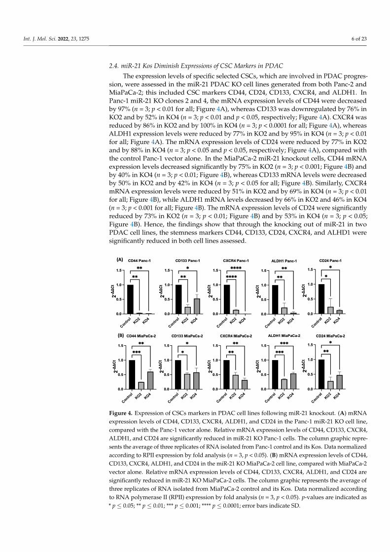
The expression levels of specific selected CSCs, which are involved in PDAC progres-sion, were assessed in the miR-21 PDAC KO cell lines generated from both Panc-2 andMiaPaCa-2; this included CSC markers CD44, CD24, CD133, CXCR4, and ALDH1. InPanc-1 miR-21 KO clones 2 and 4, the mRNA expression levels of CD44 were decreasedby 97% (n = 3; p < 0.01 for all; Figure 4A), whereas CD133 was downregulated by 76% inKO2 and by 52% in KO4 (n = 3; p < 0.01 and p < 0.05, respectively; Figure 4A). CXCR4 wasreduced by 86% in KO2 and by 100% in KO4 (n = 3; p < 0.0001 for all; Figure 4A), whereasALDH1 expression levels were reduced by 77% in KO2 and by 95% in KO4 (n = 3; p < 0.01for all; Figure 4A). The mRNA expression levels of CD24 were reduced by 77% in KO2and by 88% in KO4 (n = 3; p < 0.05 and p < 0.05, respectively; Figure 4A), compared withthe control Panc-1 vector alone. In the MiaPaCa-2 miR-21 knockout cells, CD44 mRNAexpression levels decreased significantly by 75% in KO2 (n = 3; p < 0.001; Figure 4B) andby 40% in KO4 (n = 3; p < 0.01; Figure 4B), whereas CD133 mRNA levels were decreasedby 50% in KO2 and by 42% in KO4 (n = 3; p < 0.05 for all; Figure 4B). Similarly, CXCR4mRNA expression levels were reduced by 51% in KO2 and by 69% in KO4 (n = 3; p < 0.01for all; Figure 4B), while ALDH1 mRNA levels decreased by 66% in KO2 and 46% in KO4(n = 3; p < 0.001 for all; Figure 4B). The mRNA expression levels of CD24 were significantlyreduced by 73% in KO2 (n = 3; p < 0.01; Figure 4B) and by 53% in KO4 (n = 3; p < 0.05;Figure 4B). Hence, the findings show that through the knocking out of miR-21 in twoPDAC cell lines, the stemness markers CD44, CD133, CD24, CXCR4, and ALHD1 weresignificantly reduced in both cell lines assessed.Int. J. Mol. Sci. 2022, 23, x FOR PEER REVIEW 7 of 24
Figure 4. Expression of CSCs markers in PDAC cell lines following miR-21 knockout. (A) mRNA expression levels of CD44, CD133, CXCR4, ALDH1, and CD24 in the Panc-1 miR-21 KO cell line, compared with the Panc-1 vector alone. Relative mRNA expression levels of CD44, CD133, CXCR4, ALDH1, and CD24 are significantly reduced in miR-21 KO Panc-1 cells. The column graphic repre-sents the average of three replicates of RNA isolated from Panc-1 control and its Kos. Data normal-ized according to RPII expression by fold analysis (n =3, p < 0.05). (B) mRNA expression levels of CD44, CD133, CXCR4, ALDH1, and CD24 in the miR-21 KO MiaPaCa-2 cell line, compared with MiaPaCa-2 vector alone. Relative mRNA expression levels of CD44, CD133, CXCR4, ALDH1, and CD24 are significantly reduced in miR-21 KO MiaPaCa-2 cells. The column graphic represents the average of three replicates of RNA isolated from MiaPaCa-2 control and its Kos. Data normalized according to RNA polymerase II (RPII) expression by fold analysis (n =3, p < 0.05). p-values are in-dicated as * p ≤ 0.05; ** p ≤ 0.01; *** p ≤ 0.001; **** p ≤ 0.0001; error bars indicate SD.
2.5. Flow Cytometry Analysis of PCSCs Expression in PDAC miR-21 KO Cells Several key CSCs associated with PDAC, including CD24, CD133, and CD13 were
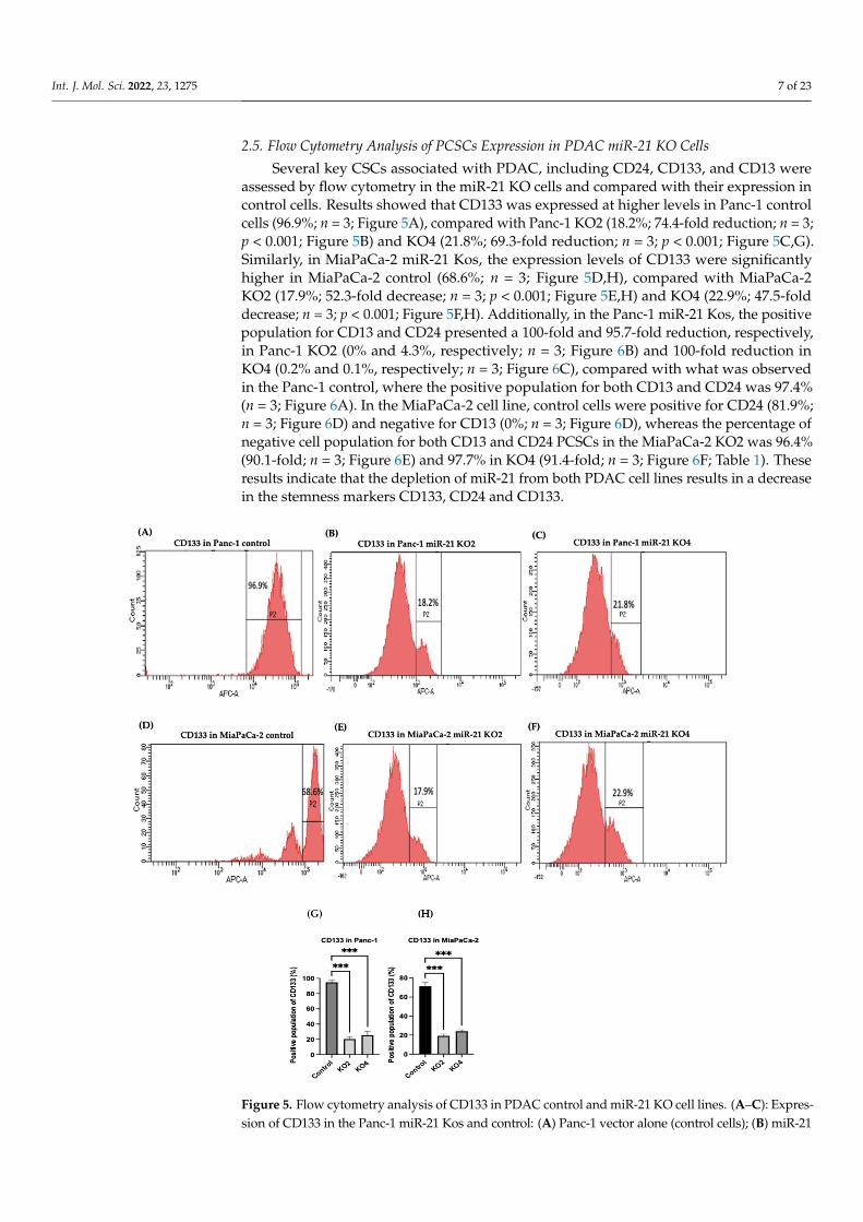
assessed by flow cytometry in the miR-21 KO cells and compared with their expression in control cells. Results showed that CD133 was expressed at higher levels in Panc-1 control cells (96.9%; n = 3; Figure 5A), compared with Panc-1 KO2 (18.2%; 74.4-fold reduction; n = 3; p < 0.001; Figure 5B) and KO4 (21.8%; 69.3-fold reduction; n = 3; p < 0.001; Figure 5C,G). Similarly, in MiaPaCa-2 miR-21 Kos, the expression levels of CD133 were significantly higher in MiaPaCa-2 control (68.6%; n = 3; Figure 5D,H), compared with MiaPaCa-2 KO2 (17.9%; 52.3-fold decrease; n = 3; p < 0.001; Figure 5E,H) and KO4 (22.9%; 47.5-fold de-crease; n = 3; p < 0.001; Figure 5F,H). Additionally, in the Panc-1 miR-21 Kos, the positive population for CD13 and CD24 presented a 100-fold and 95.7-fold reduction, respectively, in Panc-1 KO2 (0% and 4.3%, respectively; n = 3; Figure 6B) and 100-fold reduction in KO4 (0.2% and 0.1%, respectively; n = 3; Figure 6C), compared with what was observed in the Panc-1 control, where the positive population for both CD13 and CD24 was 97.4% (n = 3; Figure 6A). In the MiaPaCa-2 cell line, control cells were positive for CD24 (81.9%; n = 3; Figure 6D) and negative for CD13 (0%; n = 3; Figure 6D), whereas the percentage of
Figure 4. Expression of CSCs markers in PDAC cell lines following miR-21 knockout. (A) mRNAexpression levels of CD44, CD133, CXCR4, ALDH1, and CD24 in the Panc-1 miR-21 KO cell line,compared with the Panc-1 vector alone. Relative mRNA expression levels of CD44, CD133, CXCR4,ALDH1, and CD24 are significantly reduced in miR-21 KO Panc-1 cells. The column graphic repre-sents the average of three replicates of RNA isolated from Panc-1 control and its Kos. Data normalizedaccording to RPII expression by fold analysis (n = 3, p < 0.05). (B) mRNA expression levels of CD44,CD133, CXCR4, ALDH1, and CD24 in the miR-21 KO MiaPaCa-2 cell line, compared with MiaPaCa-2vector alone. Relative mRNA expression levels of CD44, CD133, CXCR4, ALDH1, and CD24 aresignificantly reduced in miR-21 KO MiaPaCa-2 cells. The column graphic represents the average ofthree replicates of RNA isolated from MiaPaCa-2 control and its Kos. Data normalized accordingto RNA polymerase II (RPII) expression by fold analysis (n = 3, p < 0.05). p-values are indicated as* p ≤ 0.05; ** p ≤ 0.01; *** p ≤ 0.001; **** p ≤ 0.0001; error bars indicate SD.
Int. J. Mol. Sci. 2022, 23, 1275 7 of 23
2.5. Flow Cytometry Analysis of PCSCs Expression in PDAC miR-21 KO Cells
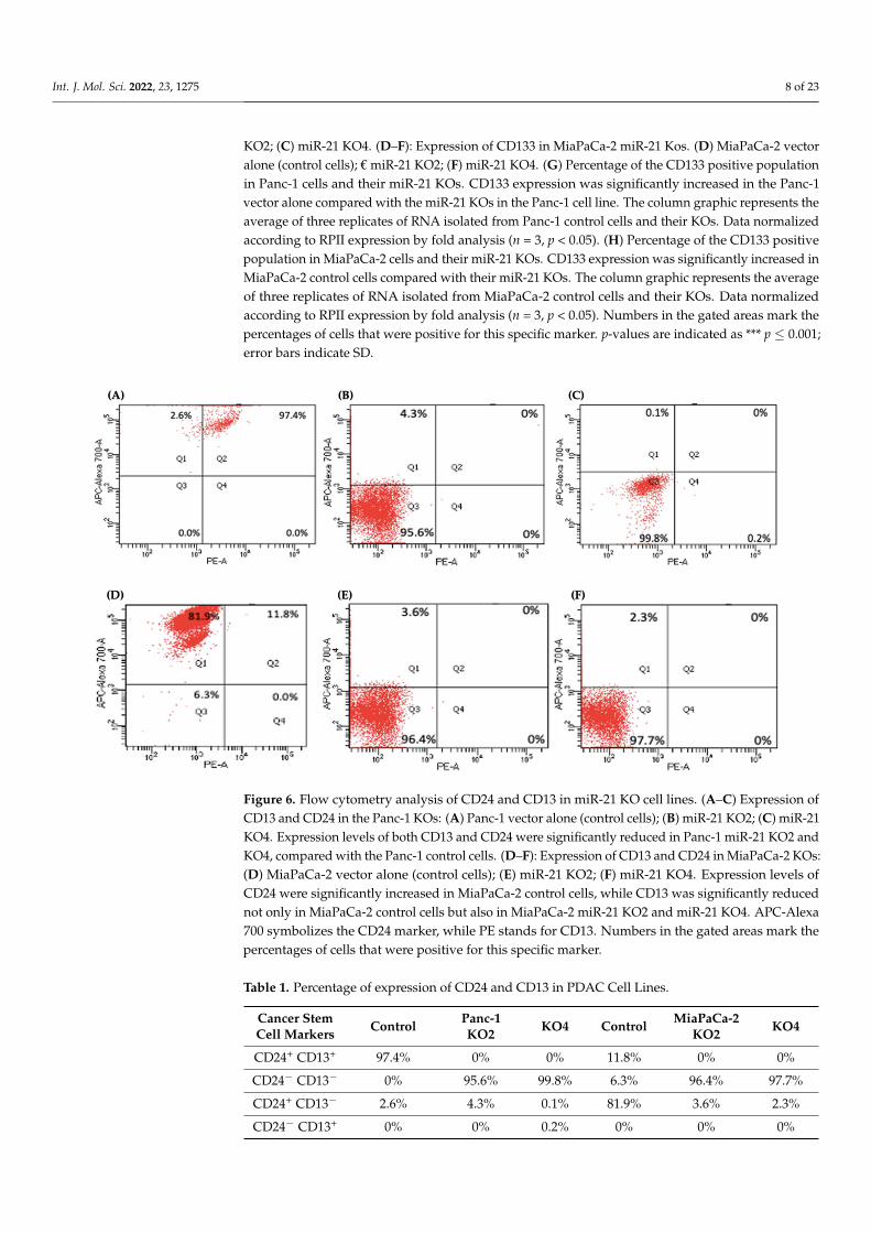
Several key CSCs associated with PDAC, including CD24, CD133, and CD13 wereassessed by flow cytometry in the miR-21 KO cells and compared with their expression incontrol cells. Results showed that CD133 was expressed at higher levels in Panc-1 controlcells (96.9%; n = 3; Figure 5A), compared with Panc-1 KO2 (18.2%; 74.4-fold reduction; n = 3;p < 0.001; Figure 5B) and KO4 (21.8%; 69.3-fold reduction; n = 3; p < 0.001; Figure 5C,G).Similarly, in MiaPaCa-2 miR-21 Kos, the expression levels of CD133 were significantlyhigher in MiaPaCa-2 control (68.6%; n = 3; Figure 5D,H), compared with MiaPaCa-2KO2 (17.9%; 52.3-fold decrease; n = 3; p < 0.001; Figure 5E,H) and KO4 (22.9%; 47.5-folddecrease; n = 3; p < 0.001; Figure 5F,H). Additionally, in the Panc-1 miR-21 Kos, the positivepopulation for CD13 and CD24 presented a 100-fold and 95.7-fold reduction, respectively,in Panc-1 KO2 (0% and 4.3%, respectively; n = 3; Figure 6B) and 100-fold reduction inKO4 (0.2% and 0.1%, respectively; n = 3; Figure 6C), compared with what was observedin the Panc-1 control, where the positive population for both CD13 and CD24 was 97.4%(n = 3; Figure 6A). In the MiaPaCa-2 cell line, control cells were positive for CD24 (81.9%;n = 3; Figure 6D) and negative for CD13 (0%; n = 3; Figure 6D), whereas the percentage ofnegative cell population for both CD13 and CD24 PCSCs in the MiaPaCa-2 KO2 was 96.4%(90.1-fold; n = 3; Figure 6E) and 97.7% in KO4 (91.4-fold; n = 3; Figure 6F; Table 1). Theseresults indicate that the depletion of miR-21 from both PDAC cell lines results in a decreasein the stemness markers CD133, CD24 and CD133.
Int. J. Mol. Sci. 2022, 23, x FOR PEER REVIEW 8 of 24
negative cell population for both CD13 and CD24 PCSCs in the MiaPaCa-2 KO2 was 96.4% (90.1-fold; n = 3; Figure 6E) and 97.7% in KO4 (91.4-fold; n = 3; Figure 6F; Table 1). These results indicate that the depletion of miR-21 from both PDAC cell lines results in a de-crease in the stemness markers CD133, CD24 and CD133.
Figure 5. Flow cytometry analysis of CD133 in PDAC control and miR-21 KO cell lines. (A–C): Ex-pression of CD133 in the Panc-1 miR-21 Kos and control: (A) Panc-1 vector alone (control cells); (B) miR-21 KO2; (C) miR-21 KO4. (D–F): Expression of CD133 in MiaPaCa-2 miR-21 Kos. (D) MiaPaCa-2 vector alone (control cells); € miR-21 KO2; (F) miR-21 KO4. (G) Percentage of the CD133 positive population in Panc-1 cells and their miR-21 KOs. CD133 expression was significantly increased in the Panc-1 vector alone compared with the miR-21 KOs in the Panc-1 cell line. The column graphic represents the average of three replicates of RNA isolated from Panc-1 control cells and their KOs. Data normalized according to RPII expression by fold analysis (n = 3, p < 0.05). (H) Percentage of the CD133 positive population in MiaPaCa-2 cells and their miR-21 KOs. CD133 expression was signif-icantly increased in MiaPaCa-2 control cells compared with their miR-21 KOs. The column graphic represents the average of three replicates of RNA isolated from MiaPaCa-2 control cells and their KOs. Data normalized according to RPII expression by fold analysis (n = 3, p < 0.05). Numbers in the
Figure 5. Flow cytometry analysis of CD133 in PDAC control and miR-21 KO cell lines. (A–C): Expres-sion of CD133 in the Panc-1 miR-21 Kos and control: (A) Panc-1 vector alone (control cells); (B) miR-21
Int. J. Mol. Sci. 2022, 23, 1275 8 of 23
KO2; (C) miR-21 KO4. (D–F): Expression of CD133 in MiaPaCa-2 miR-21 Kos. (D) MiaPaCa-2 vectoralone (control cells); € miR-21 KO2; (F) miR-21 KO4. (G) Percentage of the CD133 positive populationin Panc-1 cells and their miR-21 KOs. CD133 expression was significantly increased in the Panc-1vector alone compared with the miR-21 KOs in the Panc-1 cell line. The column graphic represents theaverage of three replicates of RNA isolated from Panc-1 control cells and their KOs. Data normalizedaccording to RPII expression by fold analysis (n = 3, p < 0.05). (H) Percentage of the CD133 positivepopulation in MiaPaCa-2 cells and their miR-21 KOs. CD133 expression was significantly increased inMiaPaCa-2 control cells compared with their miR-21 KOs. The column graphic represents the averageof three replicates of RNA isolated from MiaPaCa-2 control cells and their KOs. Data normalizedaccording to RPII expression by fold analysis (n = 3, p < 0.05). Numbers in the gated areas mark thepercentages of cells that were positive for this specific marker. p-values are indicated as *** p ≤ 0.001;error bars indicate SD.
Int. J. Mol. Sci. 2022, 23, x FOR PEER REVIEW 9 of 24
gated areas mark the percentages of cells that were positive for this specific marker. p-values are indicated as *** p ≤ 0.001; error bars indicate SD.
Figure 6. Flow cytometry analysis of CD24 and CD13 in miR-21 KO cell lines. (A–C) Expression of CD13 and CD24 in the Panc-1 KOs: (A) Panc-1 vector alone (control cells); (B) miR-21 KO2; (C) miR-21 KO4. Expression levels of both CD13 and CD24 were significantly reduced in Panc-1 miR-21 KO2 and KO4, compared with the Panc-1 control cells. (D–F): Expression of CD13 and CD24 in MiaPaCa-2 KOs: (D) MiaPaCa-2 vector alone (control cells); (E) miR-21 KO2; (F) miR-21 KO4. Expression lev-els of CD24 were significantly increased in MiaPaCa-2 control cells, while CD13 was significantly reduced not only in MiaPaCa-2 control cells but also in MiaPaCa-2 miR-21 KO2 and miR-21 KO4. APC-Alexa 700 symbolizes the CD24 marker, while PE stands for CD13. Numbers in the gated areas mark the percentages of cells that were positive for this specific marker.
Table 1. Percentage of expression of CD24 and CD13 in PDAC Cell Lines.
Cancer Stem Cell Mark-ers
Control Panc-1 KO2
KO4 Control MiaPaCa-2
KO2 KO4
CD24+ CD13+ 97.4% 0% 0% 11.8% 0% 0%
CD24− CD13− 0% 95.6% 99.8% 6.3% 96.4% 97.7%
CD24+ CD13− 2.6% 4.3% 0.1% 81.9% 3.6% 2.3%
CD24− CD13+ 0% 0% 0.2% 0% 0% 0%
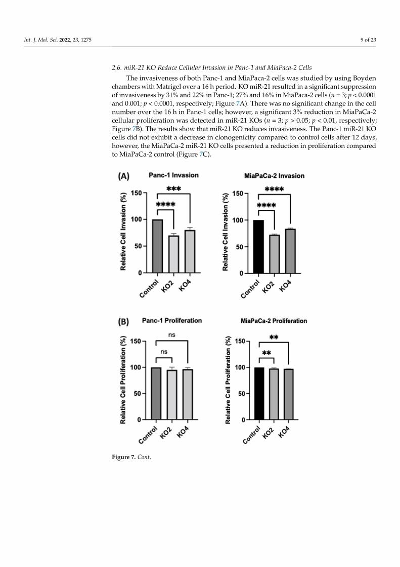
2.6. miR-21 KO Reduce Cellular Invasion in Panc-1 and MiaPaca-2 Cells The invasiveness of both Panc-1 and MiaPaca-2 cells was studied by using Boyden
chambers with Matrigel over a 16 h period. KO miR-21 resulted in a significant suppres-sion of invasiveness by 31% and 22% in Panc-1; 27% and 16% in MiaPaca-2 cells (n = 3; p < 0.0001 and 0.001; p < 0.0001, respectively; Figure 7A). There was no significant change in
Figure 6. Flow cytometry analysis of CD24 and CD13 in miR-21 KO cell lines. (A–C) Expression ofCD13 and CD24 in the Panc-1 KOs: (A) Panc-1 vector alone (control cells); (B) miR-21 KO2; (C) miR-21KO4. Expression levels of both CD13 and CD24 were significantly reduced in Panc-1 miR-21 KO2 andKO4, compared with the Panc-1 control cells. (D–F): Expression of CD13 and CD24 in MiaPaCa-2 KOs:(D) MiaPaCa-2 vector alone (control cells); (E) miR-21 KO2; (F) miR-21 KO4. Expression levels ofCD24 were significantly increased in MiaPaCa-2 control cells, while CD13 was significantly reducednot only in MiaPaCa-2 control cells but also in MiaPaCa-2 miR-21 KO2 and miR-21 KO4. APC-Alexa700 symbolizes the CD24 marker, while PE stands for CD13. Numbers in the gated areas mark thepercentages of cells that were positive for this specific marker.
Table 1. Percentage of expression of CD24 and CD13 in PDAC Cell Lines.
Cancer StemCell Markers Control Panc-1
KO2 KO4 Control MiaPaCa-2KO2 KO4
CD24+ CD13+ 97.4% 0% 0% 11.8% 0% 0%
CD24− CD13− 0% 95.6% 99.8% 6.3% 96.4% 97.7%
CD24+ CD13− 2.6% 4.3% 0.1% 81.9% 3.6% 2.3%
CD24− CD13+ 0% 0% 0.2% 0% 0% 0%
Int. J. Mol. Sci. 2022, 23, 1275 9 of 23
2.6. miR-21 KO Reduce Cellular Invasion in Panc-1 and MiaPaca-2 Cells
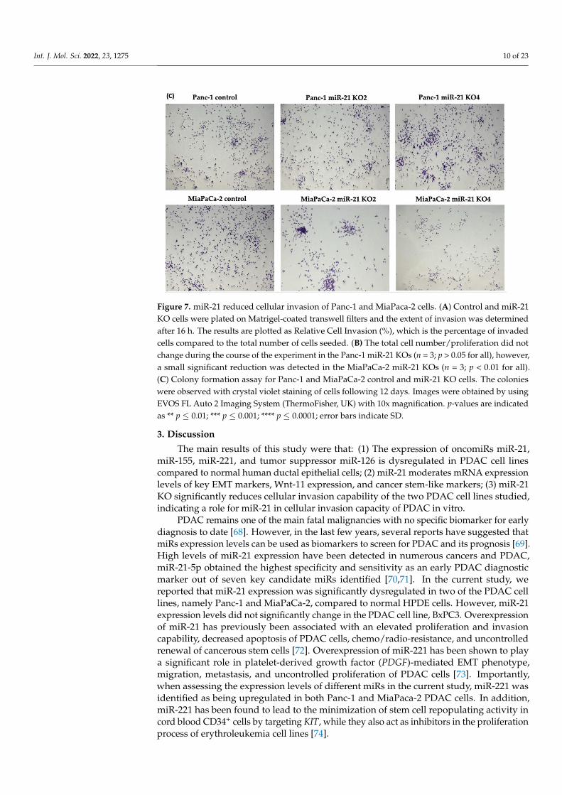
The invasiveness of both Panc-1 and MiaPaca-2 cells was studied by using Boydenchambers with Matrigel over a 16 h period. KO miR-21 resulted in a significant suppressionof invasiveness by 31% and 22% in Panc-1; 27% and 16% in MiaPaca-2 cells (n = 3; p < 0.0001and 0.001; p < 0.0001, respectively; Figure 7A). There was no significant change in the cellnumber over the 16 h in Panc-1 cells; however, a significant 3% reduction in MiaPaCa-2cellular proliferation was detected in miR-21 KOs (n = 3; p > 0.05; p < 0.01, respectively;Figure 7B). The results show that miR-21 KO reduces invasiveness. The Panc-1 miR-21 KOcells did not exhibit a decrease in clonogenicity compared to control cells after 12 days,however, the MiaPaCa-2 miR-21 KO cells presented a reduction in proliferation comparedto MiaPaCa-2 control (Figure 7C).
Int. J. Mol. Sci. 2022, 23, x FOR PEER REVIEW 10 of 24
the cell number over the 16 h in Panc-1 cells; however, a significant 3% reduction in Mi-aPaCa-2 cellular proliferation was detected in miR-21 KOs (n = 3; p > 0.05; p < 0.01, respec-tively; Figure 7B). The results show that miR-21 KO reduces invasiveness. The Panc-1 miR-21 KO cells did not exhibit a decrease in clonogenicity compared to control cells after 12 days, however, the MiaPaCa-2 miR-21 KO cells presented a reduction in proliferation compared to MiaPaCa-2 control (Figure 7C).
Figure 7. Cont.
Int. J. Mol. Sci. 2022, 23, 1275 10 of 23Int. J. Mol. Sci. 2022, 23, x FOR PEER REVIEW 11 of 24
Figure 7. miR-21 reduced cellular invasion of Panc-1 and MiaPaca-2 cells. (A) Control and miR-21 KO cells were plated on Matrigel-coated transwell filters and the extent of invasion was determined after 16 h. The results are plotted as Relative Cell Invasion (%), which is the percentage of invaded cells compared to the total number of cells seeded. (B) The total cell number/proliferation did not change during the course of the experiment in the Panc-1 miR-21 KOs (n = 3; p > 0.05 for all), how-ever, a small significant reduction was detected in the MiaPaCa-2 miR-21 KOs (n=3; p < 0.01 for all). (C) Colony formation assay for Panc-1 and MiaPaCa-2 control and miR-21 KO cells. The colonies were observed with crystal violet staining of cells following 12 days. Images were obtained by using EVOS FL Auto 2 Imaging System (ThermoFisher, UK) with 10x magnification. p-values are indi-cated as ** p ≤ 0.01; *** p ≤ 0.001; **** p ≤ 0.0001; error bars indicate SD.
3. Discussion The main results of this study were that: (1) The expression of oncomiRs miR-21, miR-
155, miR-221, and tumor suppressor miR-126 is dysregulated in PDAC cell lines compared to normal human ductal epithelial cells; (2) miR-21 moderates mRNA expression levels of key EMT markers, Wnt-11 expression, and cancer stem-like markers; (3) miR-21 KO sig-nificantly reduces cellular invasion capability of the two PDAC cell lines studied, indicat-ing a role for miR-21 in cellular invasion capacity of PDAC in vitro.
PDAC remains one of the main fatal malignancies with no specific biomarker for early diagnosis to date [68]. However, in the last few years, several reports have suggested that miRs expression levels can be used as biomarkers to screen for PDAC and its prog-nosis [69]. High levels of miR-21 expression have been detected in numerous cancers and PDAC, miR-21-5p obtained the highest specificity and sensitivity as an early PDAC diag-nostic marker out of seven key candidate miRs identified [70,71]. In the current study, we reported that miR-21 expression was significantly dysregulated in two of the PDAC cell lines, namely Panc-1 and MiaPaCa-2, compared to normal HPDE cells. However, miR-21 expression levels did not significantly change in the PDAC cell line, BxPC3. Overexpres-sion of miR-21 has previously been associated with an elevated proliferation and invasion capability, decreased apoptosis of PDAC cells, chemo/radio-resistance, and uncontrolled renewal of cancerous stem cells [72]. Overexpression of miR-221 has been shown to play a significant role in platelet-derived growth factor (PDGF)-mediated EMT phenotype, mi-gration, metastasis, and uncontrolled proliferation of PDAC cells [73]. Importantly, when assessing the expression levels of different miRs in the current study, miR-221 was iden-tified as being upregulated in both Panc-1 and MiaPaca-2 PDAC cells. In addition, miR-221 has been found to lead to the minimization of stem cell repopulating activity in cord
Figure 7. miR-21 reduced cellular invasion of Panc-1 and MiaPaca-2 cells. (A) Control and miR-21KO cells were plated on Matrigel-coated transwell filters and the extent of invasion was determinedafter 16 h. The results are plotted as Relative Cell Invasion (%), which is the percentage of invadedcells compared to the total number of cells seeded. (B) The total cell number/proliferation did notchange during the course of the experiment in the Panc-1 miR-21 KOs (n = 3; p > 0.05 for all), however,a small significant reduction was detected in the MiaPaCa-2 miR-21 KOs (n = 3; p < 0.01 for all).(C) Colony formation assay for Panc-1 and MiaPaCa-2 control and miR-21 KO cells. The colonieswere observed with crystal violet staining of cells following 12 days. Images were obtained by usingEVOS FL Auto 2 Imaging System (ThermoFisher, UK) with 10x magnification. p-values are indicatedas ** p ≤ 0.01; *** p ≤ 0.001; **** p ≤ 0.0001; error bars indicate SD.
3. Discussion
The main results of this study were that: (1) The expression of oncomiRs miR-21,miR-155, miR-221, and tumor suppressor miR-126 is dysregulated in PDAC cell linescompared to normal human ductal epithelial cells; (2) miR-21 moderates mRNA expressionlevels of key EMT markers, Wnt-11 expression, and cancer stem-like markers; (3) miR-21KO significantly reduces cellular invasion capability of the two PDAC cell lines studied,indicating a role for miR-21 in cellular invasion capacity of PDAC in vitro.
PDAC remains one of the main fatal malignancies with no specific biomarker for earlydiagnosis to date [68]. However, in the last few years, several reports have suggested thatmiRs expression levels can be used as biomarkers to screen for PDAC and its prognosis [69].High levels of miR-21 expression have been detected in numerous cancers and PDAC,miR-21-5p obtained the highest specificity and sensitivity as an early PDAC diagnosticmarker out of seven key candidate miRs identified [70,71]. In the current study, wereported that miR-21 expression was significantly dysregulated in two of the PDAC celllines, namely Panc-1 and MiaPaCa-2, compared to normal HPDE cells. However, miR-21expression levels did not significantly change in the PDAC cell line, BxPC3. Overexpressionof miR-21 has previously been associated with an elevated proliferation and invasioncapability, decreased apoptosis of PDAC cells, chemo/radio-resistance, and uncontrolledrenewal of cancerous stem cells [72]. Overexpression of miR-221 has been shown to playa significant role in platelet-derived growth factor (PDGF)-mediated EMT phenotype,migration, metastasis, and uncontrolled proliferation of PDAC cells [73]. Importantly,when assessing the expression levels of different miRs in the current study, miR-221 wasidentified as being upregulated in both Panc-1 and MiaPaca-2 PDAC cells. In addition,miR-221 has been found to lead to the minimization of stem cell repopulating activity incord blood CD34+ cells by targeting KIT, while they also act as inhibitors in the proliferationprocess of erythroleukemia cell lines [74].
Int. J. Mol. Sci. 2022, 23, 1275 11 of 23
Increased levels of miR-155 have previously been reported in PDAC patients comparedwith normal pancreatic tissues and have been shown to suppress the pro-apoptotic gene p53(TP53INP1), which plays a crucial role in p53 function, in inducing growth inhibition andautophagic cell death, in the repression of tumor cell migration, in cell growth arrest andapoptosis [75]. Furthermore, previous reports have also shown that overexpression of miR-155 is linked to the clinical stage (especially PanIN-2 and PanIN-3), lymph node metastasis,and prognosis in PDAC patients [76–79]. In our current study, elevated expression ofmiR-155 was found in all three PDAC cell lines assessed, and this aligns with previousfindings in PDAC patients highlighted above. The downregulation of miR-126 in PDAChas been reported in previous studies, and this correlates with the findings of our currentstudy, which noted reduced miR-126 expression in the PDAC cell lines assessed [77]. Inparticular, we found that miR-126-5p was strongly decreased in BxPC3, MiaPaCa-2, andPanc-1 cells by 57%, 81%, and 94%, respectively, indicating a strong correlation betweenmiR-126 and PDAC development. miR-126 is known to inhibit CXCR4, which suppressescell proliferation, migration, invasion, cell apoptosis, and arrests the cell cycle at theG0/G1 transition of PDAC cells [80,81]. CXCR4 is a putative mediator between miR-126and the RhoA/ROCK signaling pathway [80,82], promoting mitogen-activated proteinkinase (MAPK) p42/44 phosphorylation and activation of the phosphoinositide-3-kinase(PI3K)/AKT pathway [83–85], which are further linked to lymph node metastasis and theunfavorable overall survival of PDAC patients [80].
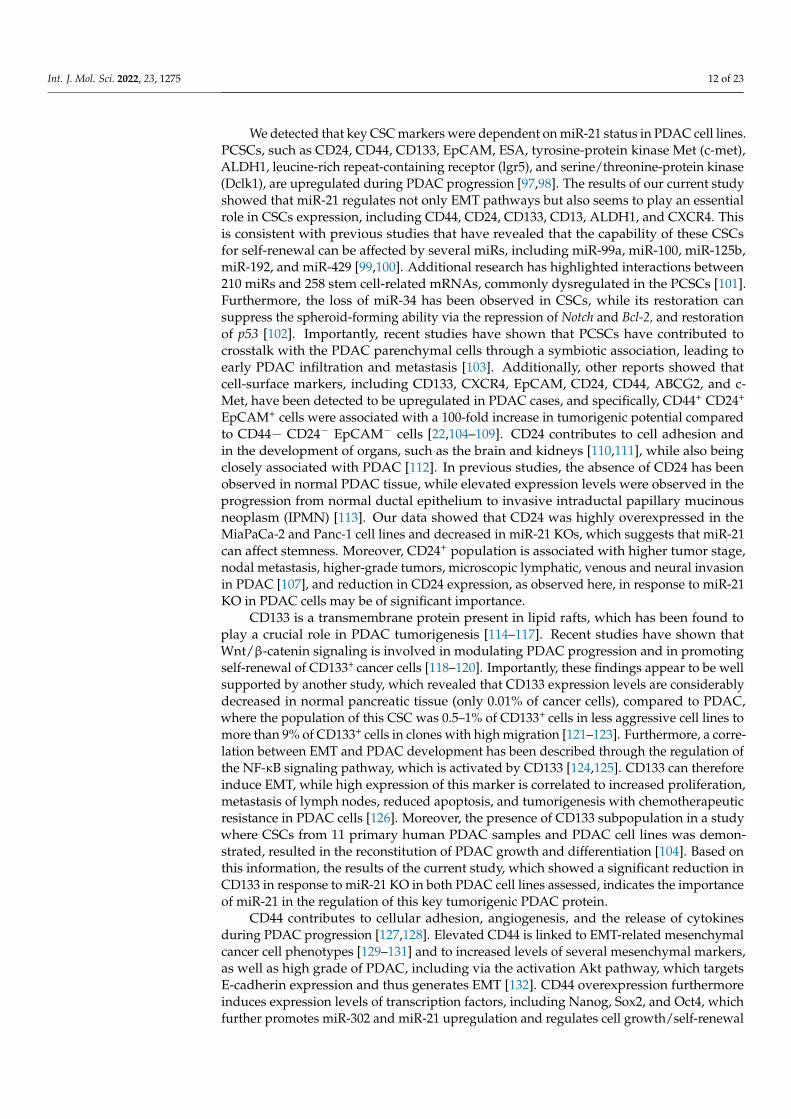
Moreover, in the current study, the EMT-associated markers E-cadherin, Vimentin,Snail, Zeb1, and Wnt-11 were found to be controlled by miR-21 and their mRNA expressionlevels were significantly affected by miR-21 knockouts in the PDAC cells (Figure 8). Further-more, immunofluorescence results indicated that E-cadherin expression was gained, whileSnail and Wnt-11 expressions were reduced in Panc-1 miR-21 KO2 clones when comparedto wt/control (Figure 3C). Several studies have disseminated that a number of miRs canpromote EMT and cancer stemness in PDAC [86–96], whereas our current study providessome new pilot insights into the role of miR-21 depletion in cancer stemness, EMT, and theWnt-11 pathway.
Int. J. Mol. Sci. 2022, 23, x FOR PEER REVIEW 14 of 24
in more vigorous immunosuppressive activity [146,147]. This underlines the significance of the results of our current study, which indicated that the CD13+ population was higher in Panc-1 control cells compared with the miR-21 KOs, while in MiaPaCa-2 cells, CD13 levels were low both in the control and KOs. This is of considerable interest also as Panc-1 is considered the more aggressive PDAC cell line of these two. Therefore, CD13 could be potentially used both for PDAC diagnosis and targeted PDAC treatment, the expres-sion levels of CD13 were upregulated only in the metastatic Panc-1 cell line and not in the MiaPaCa-2 cell line and its two KOs clones.
The results of our current study showed that the miR-21 KOs significantly reduced cell invasion in both Panc-1 and MiaPaca-2 cells, while no significant changes in cell pro-liferation of Panc-1 cells were observed. miR-21 KOs led a small, however significant, re-duction in MiaPaCa-2 cell proliferation over a 16h. These findings are consistent with our previous in vitro studies, which have identified that Wnt-11 is closely associated with cel-lular invasion of Panc-1 cells [148] and that miR-21 regulates Wnt-11 expression levels not only in PDAC but also in triple-negative MDA-MB-231 breast cancer cells [149]. Similarly, previous reports have indicated that transfection with miR-21 precursors can stimulate invasion, extravasation, and metastasis in cellular models of PDAC [150]. According to the TCGA database, the survival ratio of the low expression cohort (median 22.2 months) was longer than in the high expression cohort (median 19.77 months), based on data from 178 patients. The high expression profile of miR-21 (113 people with high expression of miR-21, compared with 65 people with low expression of miR-21) was significantly corre-lated with overall survival (Figure S1).
Figure 8. Overview of the involvement of miR-21 in cancer stemness and EMT in PDAC. Accord-ing to our data, miR-21 affected cancer stemness, EMT, and the Wnt-11 pathway. Created with BioRender.com (accessed on 3 December 2021).
4. Materials and Methods 4.1. Cell Culture and CRISPR/Cas9 Assay
Figure 8. Overview of the involvement of miR-21 in cancer stemness and EMT in PDAC. Accordingto our data, miR-21 affected cancer stemness, EMT, and the Wnt-11 pathway. Created with BioRender.com (accessed on 3 December 2021).
Int. J. Mol. Sci. 2022, 23, 1275 12 of 23
We detected that key CSC markers were dependent on miR-21 status in PDAC cell lines.PCSCs, such as CD24, CD44, CD133, EpCAM, ESA, tyrosine-protein kinase Met (c-met),ALDH1, leucine-rich repeat-containing receptor (lgr5), and serine/threonine-protein kinase(Dclk1), are upregulated during PDAC progression [97,98]. The results of our current studyshowed that miR-21 regulates not only EMT pathways but also seems to play an essentialrole in CSCs expression, including CD44, CD24, CD133, CD13, ALDH1, and CXCR4. Thisis consistent with previous studies that have revealed that the capability of these CSCsfor self-renewal can be affected by several miRs, including miR-99a, miR-100, miR-125b,miR-192, and miR-429 [99,100]. Additional research has highlighted interactions between210 miRs and 258 stem cell-related mRNAs, commonly dysregulated in the PCSCs [101].Furthermore, the loss of miR-34 has been observed in CSCs, while its restoration cansuppress the spheroid-forming ability via the repression of Notch and Bcl-2, and restorationof p53 [102]. Importantly, recent studies have shown that PCSCs have contributed tocrosstalk with the PDAC parenchymal cells through a symbiotic association, leading toearly PDAC infiltration and metastasis [103]. Additionally, other reports showed thatcell-surface markers, including CD133, CXCR4, EpCAM, CD24, CD44, ABCG2, and c-Met, have been detected to be upregulated in PDAC cases, and specifically, CD44+ CD24+
EpCAM+ cells were associated with a 100-fold increase in tumorigenic potential comparedto CD44− CD24− EpCAM− cells [22,104–109]. CD24 contributes to cell adhesion andin the development of organs, such as the brain and kidneys [110,111], while also beingclosely associated with PDAC [112]. In previous studies, the absence of CD24 has beenobserved in normal PDAC tissue, while elevated expression levels were observed in theprogression from normal ductal epithelium to invasive intraductal papillary mucinousneoplasm (IPMN) [113]. Our data showed that CD24 was highly overexpressed in theMiaPaCa-2 and Panc-1 cell lines and decreased in miR-21 KOs, which suggests that miR-21can affect stemness. Moreover, CD24+ population is associated with higher tumor stage,nodal metastasis, higher-grade tumors, microscopic lymphatic, venous and neural invasionin PDAC [107], and reduction in CD24 expression, as observed here, in response to miR-21KO in PDAC cells may be of significant importance.
CD133 is a transmembrane protein present in lipid rafts, which has been found toplay a crucial role in PDAC tumorigenesis [114–117]. Recent studies have shown thatWnt/β-catenin signaling is involved in modulating PDAC progression and in promotingself-renewal of CD133+ cancer cells [118–120]. Importantly, these findings appear to be wellsupported by another study, which revealed that CD133 expression levels are considerablydecreased in normal pancreatic tissue (only 0.01% of cancer cells), compared to PDAC,where the population of this CSC was 0.5–1% of CD133+ cells in less aggressive cell lines tomore than 9% of CD133+ cells in clones with high migration [121–123]. Furthermore, a corre-lation between EMT and PDAC development has been described through the regulation ofthe NF-κB signaling pathway, which is activated by CD133 [124,125]. CD133 can thereforeinduce EMT, while high expression of this marker is correlated to increased proliferation,metastasis of lymph nodes, reduced apoptosis, and tumorigenesis with chemotherapeuticresistance in PDAC cells [126]. Moreover, the presence of CD133 subpopulation in a studywhere CSCs from 11 primary human PDAC samples and PDAC cell lines was demon-strated, resulted in the reconstitution of PDAC growth and differentiation [104]. Based onthis information, the results of the current study, which showed a significant reduction inCD133 in response to miR-21 KO in both PDAC cell lines assessed, indicates the importanceof miR-21 in the regulation of this key tumorigenic PDAC protein.
CD44 contributes to cellular adhesion, angiogenesis, and the release of cytokinesduring PDAC progression [127,128]. Elevated CD44 is linked to EMT-related mesenchymalcancer cell phenotypes [129–131] and to increased levels of several mesenchymal markers,as well as high grade of PDAC, including via the activation Akt pathway, which targetsE-cadherin expression and thus generates EMT [132]. CD44 overexpression furthermoreinduces expression levels of transcription factors, including Nanog, Sox2, and Oct4, whichfurther promotes miR-302 and miR-21 upregulation and regulates cell growth/self-renewal

Int. J. Mol. Sci. 2022, 23, 1275 13 of 23
elevation in CD44high PDAC cells [133–136]. A recent study has suggested that the interac-tion between CD44 and hyaluronan results in the promotion of miR-21 expression, whichfurther leads to the elevated expression of anti-apoptotic protein Bcl-2 [137,138]. In ourstudy, we found that miR-21 moderated the expression levels of several CSCs, includingCD44. CD44 indicates reduced stemness in Panc-1 and MiaPaCa-2 cell lines followingmiR-21 KO. Upregulation of CXCR4 is accepted to be indicative of shorter overall survivaland related to an elevated risk of developing lymph node and liver metastasis via theinteraction with CXCL12, which can further promote angiogenesis and the formation ofnew blood and lymphatic vessels [106]. Previously, it has been reported that CXCR4 isinvolved in PDAC pathogenesis [139–141]. This correlates with our data, as we noted thatthe overexpression of CXCR4 in PDAC cell lines was associated with miR-21 and CXCR4was significantly reduced in both PDAC cell lines upon miR-21 KO.
ALDH-1, a CSCs marker, is correlated to tumorigenic cells in PDAC [25,142–144]. Wefound that the high levels of ALDH-1 expressed in control PDAC cells were significantlyreduced in response to miR-21 KO in both Panc-1 and MiaPaCa-2 cells. CD13 was anotherPCSCs marker assessed in the miR-21 KO PDAC cells. PDAC patients with more CD13high
neutrophil-like heterogeneous myeloid-derived suppressor cells (nMDSCs) have presenteda shorter overall survival than those with fewer CD13high nMDSCs [145–147]. Moreover,numbers of CD13high nMDSCs decreased after tumor resection of PDAC cases, whereasCD13low nMDSCs were elevated [146,147]. CD13 MDSCs could be attributed to perineuralinvasion (PNI) of PDAC, whereas it was also noted that CD13high nMDSCs revealedincreased expression levels of Arg1 compared to CD13low nMDSCs, which resulted in morevigorous immunosuppressive activity [146,147]. This underlines the significance of theresults of our current study, which indicated that the CD13+ population was higher inPanc-1 control cells compared with the miR-21 KOs, while in MiaPaCa-2 cells, CD13 levelswere low both in the control and KOs. This is of considerable interest also as Panc-1 isconsidered the more aggressive PDAC cell line of these two. Therefore, CD13 could bepotentially used both for PDAC diagnosis and targeted PDAC treatment, the expressionlevels of CD13 were upregulated only in the metastatic Panc-1 cell line and not in theMiaPaCa-2 cell line and its two KOs clones.
The results of our current study showed that the miR-21 KOs significantly reducedcell invasion in both Panc-1 and MiaPaca-2 cells, while no significant changes in cellproliferation of Panc-1 cells were observed. miR-21 KOs led a small, however significant,reduction in MiaPaCa-2 cell proliferation over a 16h. These findings are consistent withour previous in vitro studies, which have identified that Wnt-11 is closely associated withcellular invasion of Panc-1 cells [148] and that miR-21 regulates Wnt-11 expression levels notonly in PDAC but also in triple-negative MDA-MB-231 breast cancer cells [149]. Similarly,previous reports have indicated that transfection with miR-21 precursors can stimulateinvasion, extravasation, and metastasis in cellular models of PDAC [150]. According tothe TCGA database, the survival ratio of the low expression cohort (median 22.2 months)was longer than in the high expression cohort (median 19.77 months), based on data from178 patients. The high expression profile of miR-21 (113 people with high expressionof miR-21, compared with 65 people with low expression of miR-21) was significantlycorrelated with overall survival (Figure S1).
4. Materials and Methods4.1. Cell Culture and CRISPR/Cas9 Assay
Panc-1 (ATCC® CRL-1469™), MiaPaCa-2 (ATCC® CRL-x1420™), BxPC-3 (ATCC®
CRL-1687™), and non-tumorigenic human pancreatic ductal epithelial cell line (HPDE;H6c7, ATCC® CRL-4023) cell lines were cultured according to ATCC’s recommendations, to80% confluence in 75 cm2 flasks in complete Dulbecco’s Modified Eagle’s Medium (DMEM),with 10% fetal bovine serum (FBS) at 37 ◦C with 5% CO2.
The lentiviral CRISPR/Cas9-mediated miR-21 gene editing vectors encoding fourdifferent gRNAs, eGFP (control), and Cas9 protein was kindly provided by Dr. Junming
Int. J. Mol. Sci. 2022, 23, 1275 14 of 23
Yue, University of Tennessee Health Science Center, USA, and produced based on therecommendations of previously published studies [149,151,152]. Stable cell lines weregenerated by transducing the MiaPaca-2 and Panc-1 cells with the lentiviral CRISPR/Cas9miR-21 gene editing vectors and selection in puromycin (1–10 µg/mL).
4.2. RNA Extraction and RT-qPCR
Total RNA was isolated from Panc-1, MiaPaCa-2, BxPC-3, and HPDE (stored at−80 ◦C)using RNAzol® RT (Sigma, Hertfordshire, UK). Specifically, cells were isolated by centrifu-gation at 500× g for 5 min, then lysed in 0.5 mL of RNAzol and allowed to stand for 15 minat room temperature. Then lysed cells were centrifuged at 12,000× g for 15 min at roomtemperature; the supernatant was mixed with an equal volume of 100% isopropanol toprecipitate RNA, let stand for 10 min, and then centrifuged at 12,000× g for 10 min at roomtemperature. DNA digestion was performed by using a RNase-free DNase set (Qiagen,Manchester, UK), according to the manufacturer′s instructions. Briefly, we added 10 µLDNase I stock solution to 70 µL Buffer RDD, after mixing with pipetted 80 µL DNase I andincubating at room temperature for 15 min. Then, RNA pellets were washed twice with0.5 mL 75% ethanol (v/v) per 1 mL of supernatant and centrifuged at 8000× g for 3 min atroom temperature. The alcohol solution was removed with a micropipette, the RNA pelletwas solubilized in RNase-free water, and samples were vortexed at room temperature for3 min. The RNA concentration was measured using the NanoDrop Spectrophotometer(ThermoFisher Scientific, Hemel Hempstead, UK) at 260 nm and 280 nm absorbance. More-over, the qScript microRNA cDNA Synthesis Kit (Quantabio, Lutterworth, UK) was utilizedto reverse-transcribed RNA cDNA synthesis, according to the manufacturer’s instructions.The resulting cDNA from PDAC cell lines was further used to examine the expressionlevels of miR-21-5p, miR-221-5p, miR-155-5p, and miR-126-5p, whereas RNU6 was usedas a reference gene for the normalization of miRs expression levels. The PerfeCTa SYBRGreen SuperMix (Quantabio, Lutterworth, UK) was used with MystiCq miR qPCR primersfor the examined miRs purchased from Sigma (Paisley, UK). The following thermocyclingconditions were used: denaturation at 95 ◦C for 2 min, followed by 40 cycles of 95 ◦C for5 s, 60 ◦C for 15 s, and extension at 72 ◦C for 15 s.
cDNAs to assess the mRNA expression levels of E-cadherin, Vimentin, Snail, Wnt-11,Zeb1, CD44, CD133, CXCR4, and ALDH1 were isolated using qScript™ cDNA Supermix(Quantabio, Lutterworth, UK) with incubations at 25 ◦C for 5 min, 42 ◦C for 30 min and85 ◦C for 5 min. Precision®Plus qPCR Master Mix (Primer Design, Chandler’s Ford, UK)was used for RT-qPCR synthesis for the assessed EMT and CSCs markers with the followingthermocycling conditions for 40 cycles: 95 ◦C for 2 min, 95 ◦C for 10 s, and 95 ◦C for 60 s.Relative levels of mRNA expression were calculated as described before [149]. The primersfor Snail, Wnt-11, and E-cadherin, were designed and purchased from Sigma (Paisley, UK),Vimentin, and Zeb1 from Integrated DNA Technologies (IDT) (Leuven, Belgium), whileCD133, CD24, CD44, CXCR4, and ALDH1, from ThermoFisher Scientific (UK). Primersequences are presented in Table 2. Relative levels of mRNA expression were calculatedusing the comparative CT/2−∆∆Ct method [153] with RPII as the reference gene for thein-cell-line-based studies. In addition, the standard deviation was calculated as well as at-test using GraphPad Prism 7.00 (La Jolla, CA, USA) software.
4.3. Immunostaining
Panc-1 wt (Control) and miR-21 KO cells were seeded into 6-well plates and allowedto settle overnight; the cells were washed with 1× PBS and fixed with 4% formaldehydefor 20 min at room temperature. Cells were washed with 1× PBS twice, following blockingwith 5% w/v bovine serum albumin (Invitrogen, Loughborough, UK) for 30 min at roomtemperature. The wells were washed with PBS twice and primary antibodies E-cadherin,Snail (20C8) (Invitrogen, Waltham, MA, USA), and Wnt-11 (GeneTex, CA, USA) were addedand incubated for one hour. After washing the cells, either goat anti-rabbit IgG Alexa Fluoror anti-mouse IgG (ThermoFisher, Oxford, UK) was added and incubated for an hour.
Int. J. Mol. Sci. 2022, 23, 1275 15 of 23
Following another washing step with 1× PBS, Ribonuclease A 100 mg/mL (Sigma–Poole,Dorset, UK) was added and incubated, gently rocking for 20 min. For counterstaining,5 µL/mL of 1 nM To-Pro-3 (ThermoFisher Scientific, Oxford, UK) was dispensed into eachwell and set gently rocking, and then washed twice with PBS for 5 min gently rocking.The results generated were taken from the three biological and technical repetitions. Threeto four milliliters of 1× PBS was added to each well, Leica TCS SP2 (Leica Microsystems;Milton Keynes, UK) confocal microscope was used to analyze the cells.
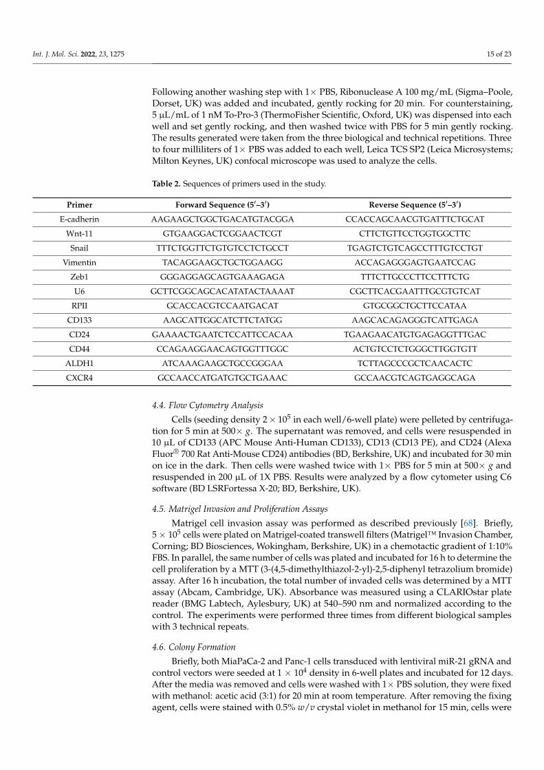
Table 2. Sequences of primers used in the study.
Primer Forward Sequence (5′–3′) Reverse Sequence (5′–3′)
E-cadherin AAGAAGCTGGCTGACATGTACGGA CCACCAGCAACGTGATTTCTGCAT
Wnt-11 GTGAAGGACTCGGAACTCGT CTTCTGTTCCTGGTGGCTTC
Snail TTTCTGGTTCTGTGTCCTCTGCCT TGAGTCTGTCAGCCTTTGTCCTGT
Vimentin TACAGGAAGCTGCTGGAAGG ACCAGAGGGAGTGAATCCAG
Zeb1 GGGAGGAGCAGTGAAAGAGA TTTCTTGCCCTTCCTTTCTG
U6 GCTTCGGCAGCACATATACTAAAAT CGCTTCACGAATTTGCGTGTCAT
RPII GCACCACGTCCAATGACAT GTGCGGCTGCTTCCATAA
CD133 AAGCATTGGCATCTTCTATGG AAGCACAGAGGGTCATTGAGA
CD24 GAAAACTGAATCTCCATTCCACAA TGAAGAACATGTGAGAGGTTTGAC
CD44 CCAGAAGGAACAGTGGTTTGGC ACTGTCCTCTGGGCTTGGTGTT
ALDH1 ATCAAAGAAGCTGCCGGGAA TCTTAGCCCGCTCAACACTC
CXCR4 GCCAACCATGATGTGCTGAAAC GCCAACGTCAGTGAGGCAGA
4.4. Flow Cytometry Analysis
Cells (seeding density 2× 10⁵ in each well/6-well plate) were pelleted by centrifuga-tion for 5 min at 500× g. The supernatant was removed, and cells were resuspended in10 µL of CD133 (APC Mouse Anti-Human CD133), CD13 (CD13 PE), and CD24 (AlexaFluor® 700 Rat Anti-Mouse CD24) antibodies (BD, Berkshire, UK) and incubated for 30 minon ice in the dark. Then cells were washed twice with 1× PBS for 5 min at 500× g andresuspended in 200 µL of 1X PBS. Results were analyzed by a flow cytometer using C6software (BD LSRFortessa X-20; BD, Berkshire, UK).
4.5. Matrigel Invasion and Proliferation Assays
Matrigel cell invasion assay was performed as described previously [68]. Briefly,5× 105 cells were plated on Matrigel-coated transwell filters (Matrigel™ Invasion Chamber,Corning; BD Biosciences, Wokingham, Berkshire, UK) in a chemotactic gradient of 1:10%FBS. In parallel, the same number of cells was plated and incubated for 16 h to determine thecell proliferation by a MTT (3-(4,5-dimethylthiazol-2-yl)-2,5-diphenyl tetrazolium bromide)assay. After 16 h incubation, the total number of invaded cells was determined by a MTTassay (Abcam, Cambridge, UK). Absorbance was measured using a CLARIOstar platereader (BMG Labtech, Aylesbury, UK) at 540–590 nm and normalized according to thecontrol. The experiments were performed three times from different biological sampleswith 3 technical repeats.
4.6. Colony Formation
Briefly, both MiaPaCa-2 and Panc-1 cells transduced with lentiviral miR-21 gRNA andcontrol vectors were seeded at 1 × 10⁴ density in 6-well plates and incubated for 12 days.After the media was removed and cells were washed with 1× PBS solution, they were fixedwith methanol: acetic acid (3:1) for 20 min at room temperature. After removing the fixingagent, cells were stained with 0.5% w/v crystal violet in methanol for 15 min, cells were
Int. J. Mol. Sci. 2022, 23, 1275 16 of 23
washed with distilled water, and cells were imaged using an EVOS FL Auto 2 ImagingSystem (ThermoFisher, Hemel Hempstead, UK).
4.7. Statistical Analysis
The expression levels of different miRs, proteins, and CSCs in PDAC cell lines wereexamined using ANOVA and Bonferroni multiple comparisons tests followed by Tukey’spost-hoc analysis. Specifically, free commercially available software packages GraphPadPrism v8.4 (La Jolla, CA, USA) was utilized for the statistical analysis. Statistical significancewas conducted as Tukey at p ≤ 0.05, while all the results were presented as mean ± SD.
5. Conclusions
In conclusion, our data indicate that miR-21 regulates key CSC markers and affectsEMT markers in PDAC. The EMT and Wnt-11 pathway was found to be modulated by miR-21 knockout, highlighting the importance of miR-21 as a potential target of cancer stemness.While further and in-depth studies will be needed to identify all related mechanisms forthe role of miR-21 in the poor prognosis and metastasis of PDAC; the data presented inthis study provide novel insights into roles for miR-21. Furthermore, our data supportprevious findings that show the importance of miR-21 and its potential as a therapeutictarget for PDAC.
Supplementary Materials: The following are available online at https://www.mdpi.com/article/10.3390/ijms23031275/s1.
Author Contributions: Conceptualization, M.M. and P.U.-O.; methodology, M.M., F.M., S.J., S.L. andP.U.-O.; software, M.M. and S.J.; validation, M.M., S.L., P.U.-O., A.D. and E.D.A.; formal analysis, M.M.and P.U.-O.; investigation, M.M.; writing—original draft preparation, M.M. and P.U.-O.; writing—review and editing, F.M., E.D.A., A.D., S.J., S.L. and P.U.-O.; supervision, S.L. and P.U.-O. All authorshave read and agreed to the published version of the manuscript.
Funding: This work was supported by the University of Westminster SLS Ph.D. studentships to M.M.and Research Enhancement Fund to P.U.-O.
Institutional Review Board Statement: Not applicable.
Informed Consent Statement: Not applicable.
Data Availability Statement: Data are contained within the article.
Acknowledgments: The authors would like to thank Junming Yue, University of Tennessee HealthScience Center, USA, for donating the lentiviral CRISPR/Cas9-mediated miR-21 gene editing vectorsand control vector; Hemant Kocher from Barts Cancer Institute, Queen Mary, University of Londonfor HDPE cells. Thanks are also due to The Guy Foundation for funding the purchase of equipmentutilized in this work.
Conflicts of Interest: The authors declare no conflict of interest.
Abbreviations
ALDH-1 Aldehyde dehydrogenase-1CA 19-9 Carbohydrate antigen 19-9CSCs Cancer stem cellsCXCR4 00 CX-C chemokine receptor type 4DMEM Dulbecco’s Modified Eagle’s MediumEMT Epithelial–mesenchymal transitionFBS Fetal bovine serumFoxM1 Forkhead box protein M1HPDE Human pancreatic ductal epithelial cell lineIDT Integrated DNA TechnologiesIPMN Intraductal papillary mucinous neoplasmKOs Knockouts
Int. J. Mol. Sci. 2022, 23, 1275 17 of 23
MAPK Mitogen-activated protein kinaseMDSCs Myeloid-derived suppressor cellsmiRs microRNAsmTOR Mammalian target of rapamycinnMDSCs Neutrophil-like heterogeneous myeloid-derived suppressor cellsNF-κB Nuclear factor kappa-light-chain-enhancer of activated B cellsoncomiR Oncogenic miRPanIN Pancreatic intraepithelial neoplasiaPCa Pancreatic cancerPCSCs Pancreatic cancer stem cellsPDAC Pancreatic ductal adenocarcinomaPDCD4 Programmed cell death 4PDGF Platelet-derived growth factorPI3K Phosphoinositide-3-kinasePNI Perineural invasionRPII RNA polymerase 2SD Standard DeviationSHH Sonic HedgehogTNM Tumor-Node-MetastasisUTRs Untranslated regionsVEGF Vascular endothelial growth factorZEB1 Zinc Finger E-box binding homeobox 1
References1. Carrato, A.; Falcone, A.; Ducreux, M.; Valle, J.W.; Parnaby, A.; Djazouli, K.; Alnwick-Allu, K.; Hutchings, A.; Palaska, C.;
Parthenaki, I.; et al. A systematic review of the burden of pancreatic cancer in. Europe: Real-world impact on survival, quality oflife and costs. J. Gastrointest. Cancer 2015, 46, 201–211. [CrossRef] [PubMed]
2. Von Hoff, D.D.; Korn, R.; Mousses, S. Pancreatic cancer—Could it be that simple? A different context of vulnerability. Cancer Cell2009, 16, 7–8. [CrossRef]
3. Gall, F.P.; Kessler, H.; Hermanek, P. Surgical treatment of ductal pancreatic carcinoma. Eur. J. Surg. Oncol. 1991, 17, 173–181.4. Srivastava, S.K.; Srivastava, S.K.; Arora, S.; Singh, S.; Bhardwaj, A.; Averett, C.; Singh, A.P. MicroRNAs in pancreatic malignancy:
Progress and promises. Cancer Lett. 2014, 347, 167–174. [CrossRef] [PubMed]5. Cartwright, T.; Richards, D.A.; Boehm, K.A. Cancer of the pancreas: Are we making progress? A review of studies in the US
Oncology Research Network. Cancer Control 2008, 15, 308–313. [CrossRef]6. Bartel, D.P. MicroRNAs: Genomics, biogenesis, mechanism, and function. Cell 2004, 116, 281–297. [CrossRef]7. Lin, S.; Gregory, R.I. MicroRNA biogenesis pathways in cancer. Nat. Rev. Cancer 2015, 15, 321–333. [CrossRef]8. Turchinovich, A.; Tonevitsky, A.G.; Burwinkel, B. Extracellular miRNA: A collision of two paradigms. Trends Biochem. Sci. 2016,
41, 883. [CrossRef]9. Friedman, R.C.; Farh, K.K.; Burge, C.B.; Bartel, D.P. Most mammalian mRNAs are conserved targets of microRNAs. Genome Res.
2009, 19, 92–105. [CrossRef] [PubMed]10. Yu, J.; Li, A.; Hong, S.M.; Hruban, R.H.; Goggins, M. MicroRNA alterations of pancreatic intraepithelial neoplasias. Clin. Cancer
Res. 2012, 18, 981–992. [CrossRef] [PubMed]11. Zhao, X.; Ren, Y.; Cui, N.; Wang, X.; Cui, Y. Identification of key microRNAs and their targets in exosomes of pancreatic cancer
using bioinformatics analysis. Medicine 2018, 97, e12632. [CrossRef]12. Slack, F.J.; Weidhaas, J.B. MicroRNA in cancer prognosis. N. Engl. J. Med. 2008, 359, 2720–2722. [CrossRef]13. Gilles, M.; Hao, L.; Huang, L.; Rupaimoole, R.; Lopez-Casas, P.P.; Pulver, E.; Jeong, J.C.; Muthuswamy, S.K.; Hidalgo, M.;
Bhatia, S.N.; et al. Personalized RNA medicine for pancreatic cancer. Clin. Cancer Res. 2018, 24, 1734–1747. [CrossRef] [PubMed]14. Galasso, M.; Sandhu, S.K.; Volinia, S. MicroRNA expression signatures in solid malignancies. Cancer J. 2012, 18, 238–243.
[CrossRef] [PubMed]15. Hussain, S.P. Pancreatic cancer: Current progress and future challenges. Int. J. Biol. Sci. 2016, 12, 270. [CrossRef] [PubMed]16. Kasaka, N.; Iguchi, H.; Ochiya, T. Circulating microRNA in body fluid: A new potential biomarker for cancer diagnosis and
prognosis. Cancer Sci. 2010, 101, 2087–2092. [CrossRef] [PubMed]17. Wang, J.; Chen, J.; Chang, P.; LeBlanc, A.; Li, D.; Abbruzzesse, J.L.; Frazier, M.L.; Killary, A.M.; Sen, S. MicroRNAs in plasma
of pancreatic ductal adenocarcinoma patients as novel blood-based biomarkers of disease. Cancer Prev. Res. 2009, 2, 807–813.[CrossRef]
18. Kojima, M.; Sudo, H.; Kawauchi, J.; Takizawa, S.; Kondou, S.; Nobumasa, H.; Ochiai, A. MicoRNA markers for diagnosis ofpancreatic and biliary-tract cancers. PLoS ONE 2015, 10, e0118220. [CrossRef]
19. Chan, A.; Prassas, I.; Dimitromanolakis, A.; Brand, R.E.; Serra, S.; Diamandis, E.P.; Blasutig, I.M. Validation of biomarkers thatcomplement ca19.9 in detecting early pancreatic cancer. Clin. Cancer Res. 2014, 20, 5787–5795. [CrossRef]

Int. J. Mol. Sci. 2022, 23, 1275 18 of 23
20. Sawabu, N.; Watanabe, H.; Yamaguchi, Y.; Ohtsubo, K.; Motoo, Y. Serum tumor markers and molecular biological diagnosis inpancreatic cancer. Pancreas 2004, 28, 263–267. [CrossRef]
21. Schultz, N.A.; Dehlendorff, C.; Jensen, B.V.; Bjerregaard, J.K.; Nielsen, K.R.; Bojesen, S.E.; Calatayud, D.; Nielsen, S.E.; Yilmaz,M.; Holländer, N.H.; et al. MicroRNA biomarkers in whole blood for detection of pancreatic cancer. JAMA 2014, 311, 392–404.[CrossRef]
22. Li, C.; Heidt, D.G.; Dalerba, P.; Burant, C.F.; Zhang, L.; Adsay, V.; Wicha, M.; Clarke, M.F.; Simeone, D.M. Identification ofpancreatic cancer stem cells. Cancer Res. 2007, 67, 1030–1037. [CrossRef] [PubMed]
23. Li, C.; Lee, C.J.; Simeone, D.M. Identification of human pancreatic cancer stem cells. Methods Mol. Biol. 2009, 568, 161–173.24. Niess, H.; Camaj, P.; Renner, A.; Ischenko, I.; Zhao, Y.; Krebs, S.; Mysliwietz, J.; Jäckel, C.; Nelson, P.J.; Blum, H.; et al. Side
population cells of pancreatic cancer show characteristics of cancer stem cells responsible for resistance and metastasis. TargetOncol. 2015, 10, 215–227. [CrossRef]
25. Kim, M.P.; Fleming, J.B.; Wang, H.; Abbruzzese, J.L.; Choi, W.; Kopetz, S.; McConkey, D.J.; Evans, D.B.; Gallick, G.E. ALDHactivity selectively defines an enhanced tumor-initiating cell population relative to CD133 expression in human pancreaticadenocarcinoma. PLoS ONE 2011, 6, e20636. [CrossRef] [PubMed]
26. Klonisch, T.; Wiechec, E.; Hombach-Klonisch, S.; Ande, S.R.; Wesselborg, S.; Schulze-Osthoff, K.; Los, M. Cancer stem cell markersin common cancers—Therapeutic implications. Trends Mol. Med. 2008, 14, 450–460. [CrossRef] [PubMed]
27. Marcato, P.; Dean, C.A.; Pan, D.; Araslanova, R.; Gillis, M.; Joshi, M.; Helyer, L.; Pan, L.; Leidal, A.; Gujar, S.; et al. Aldehydedehydrogenase activity of breast cancer stem cells is primarily due to isoform ALDH1A3 and its expression is predictive ofmetastasis. Stem Cells 2011, 29, 32–45. [CrossRef] [PubMed]
28. Miranda-Lorenzo, I.; Dorado, J.; Lonardo, E.; Alcala, S.; Serrano, A.G.; Clausell-Tormos, J.; Cioffi, M.; Megias, D.; Zagorac, S.;Balic, A.; et al. Intracellular autofluorescence: A biomarker for epithelial cancer stem cells. Nat. Methods 2014, 11, 1161–1169.[CrossRef]
29. Valle, S.; Martin-Hijano, L.; Alcalá, S.; Alonso-Nocelo, M.; Sainz, B., Jr. The Ever-Evolving Concept of the Cancer Stem Cell inPancreatic Cancer. Cancers 2018, 10, 33. [CrossRef]
30. Santamaria, S.; Delgado., M.; Kremer., L.; Garcia-Sanz, J.A. Will a mAb-Based Immunotherapy Directed against Cancer StemCells Be Feasible? Front. Immunol. 2017, 8, 1509. [CrossRef] [PubMed]
31. Plaks, V.; Kong, N.; Werb, Z. The cancer stem cell niche: How essential is the niche in regulating stemness of tumor cells? CellStem Cell 2015, 16, 225–238. [CrossRef]
32. Dawood, S.; Austin, L.; Cristofanilli, M. Cancer stem cells: Implications for cancer therapy. Oncology 2014, 28, 1101–1107, 1110.[PubMed]
33. Lei, Y.; Zhang, D.; Yu, J.; Dong, H.; Zhang, J.; Yang, S. Targeting autophagy in cancer stem cells as an anticancer therapy. CancerLett. 2017, 393, 33–39. [CrossRef]
34. Cojoc, M.; Mäbert, K.; Muders, M.H.; Dubrovska, A. A role for cancer stem cells in therapy resistance: Cellular and molecularmechanisms. Semin. Cancer Biol. 2015, 31, 16–27. [CrossRef] [PubMed]
35. Dalla Pozza, E.; Dando, I.; Biondani, G.; Brandi, J.; Costanzo, C.; Zoratti, E.; Fassan, M.; Boschi, F.; Melisi, D.; Cecconi, D.; et al.Pancreatic ductal adenocarcinoma cell lines display a plastic ability to bidirectionally convert into cancer stem cells. Int. J. Oncol.2015, 46, 1099–1108. [CrossRef]
36. Kalluri, R.; Weinberg, R.A. The basics of epithelial mesenchymal transition. J. Clin. Investig. 2009, 119, 1420–1428. [CrossRef]37. Felipe Lima, J.; Nofech-Mozes, S.; Bayani, J.; Bartlett, J.M.S. EMT in breast carcinoma—A review. J. Clin. Med. 2016, 5, 65.
[CrossRef]38. Lamouille, S.; Xu, J.; Derynck, R. Molecular mechanisms of epithelial–mesenchymal transition. Nat. Rev. Mol. Cell Biol. 2014, 15,
178–196. [CrossRef] [PubMed]39. Iacobuzio-Donahue, C.A.; Fu, B.; Yachida, S.; Luo, M.; Abe, H.; Henderson, C.M.; Vilardell, F.; Wang, Z.; Keller, J.W.; Banerjee, P.;
et al. DPC4 gene status of the primary carcinoma correlates with patterns of failure in patients with pancreatic cancer. J. Clin.Oncol. 2009, 27, 1806–1813. [CrossRef] [PubMed]
40. Winter, J.M.; Ting, A.H.; Vilardell, F.; Gallmeier, E.; Baylin, S.B.; Hruban, R.H.; Kern, S.E.; Iacobuzio-Donahue, C.A. Absence ofE-cadherin expression distinguishes noncohesive from cohesive pancreatic cancer. Clin. Cancer Res. 2008, 14, 412–418. [CrossRef]
41. Renz, B.W.; D’Haese, J.G.; Werner, J.; Westphalen, C.B.; Ilmer, M. Repurposing Established Compounds to Target PancreaticCancer Stem Cells (CSCs). Med. Sci. 2017, 5, 14. [CrossRef]
42. Arumugam, T.; Ramachandran, V.; Fournier, K.F.; Wang, H.; Marquis, L.; Abbruzzese, J.L.; Gallick, G.E.; Logsdon, C.D.; McConkey,D.J.; Choi, W. Epithelial to mesenchymal transition contributes to drug resistance in pancreatic cancer. Cancer Res. 2009, 69,5820–5828. [CrossRef]
43. Buck, E.; Eyzaguirre, A.; Barr, S.; Thompson, S.; Sennello, R.; Young, D.; Iwata, K.K.; Gibson, N.W.; Cagnoni, P.; Haley, J.D. Loss ofhomotypic cell adhesion by epithelial-mesenchymal transition or mutation limits sensitivity to epidermal growth factor receptorinhibition. Mol. Cancer Ther. 2007, 6, 532–541. [CrossRef] [PubMed]
44. Maier, H.J.; Schmidt-Strassburger, U.; Huber, M.A.; Wiedemann, E.M.; Beug, H.; Wirth, T. NF-kappaB promotes epithelial-mesenchymal transition, migration and invasion of pancreatic carcinoma cells. Cancer Lett. 2010, 295, 214–228. [CrossRef]
45. Uysal-Onganer, P.; Kawano, Y.; Caro, M.; WalkeR, M.M.; Diez, S.; Darrington, R.S.; Waxman, J.; Kypta, R.M. Wnt-11 promotesneuroendocrine-like differentiation, survival and migration of prostate cancer cells. Mol. Cancer. 2010, 9, 55. [CrossRef] [PubMed]
Int. J. Mol. Sci. 2022, 23, 1275 19 of 23
46. Arisan, E.D.; Rencuzogullari, O.; Freitas, I.L.; Radzali, S.; Keskin, B.; Kothari, A.; Warford, A.; Uysal-Onganer, P. UpregulatedWnt-11 and miR-21 Expression Trigger Epithelial Mesenchymal Transition in Aggressive Prostate Cancer Cells. Biology 2020, 9, 52.[CrossRef] [PubMed]
47. Guo, X.; Wang, X.F. Signaling cross-talk between TGF-beta/BMP and other pathways. Cell Res. 2009, 19, 71–88. [CrossRef][PubMed]
48. Wang, L.; Yao, M.; Fang, M.; Zheng, W.J.; Dong, Z.Z.; Pan, L.H.; Zhang, H.J.; Yao, D.F. Expression of hepatic Wnt5a andits clinicopathological features in patients with hepatocellular carcinoma. Hepatobiliary Pancreat. Dis. Int. 2018, 17, 227–232.[CrossRef]
49. Wang, X.; Liu, D.; Zhou, K.; Wang, B.; Liu, Q.; Deng, F.; Li, Q.; Ma, Y. Expression of Wnt-11 and Rock2 in esophageal squamous cellcarcinoma by activation of the WNT/PCP pathway and its clinical significance. Pathol. Res. Pract. 2016, 212, 880–885. [CrossRef][PubMed]
50. Hotz, B.; Arndt, M.; Dullat, S.; Bhargava, S.; Buhr, H.J.; Hotz, H.G. Epithelial to mesenchymal transition: Expression of theregulators snail, slug, and twist in pancreatic cancer. Clin. Cancer Res. 2007, 13, 4769–4776. [CrossRef]
51. De Craene, B.; Berx, G. Regulatory networks defining EMT during cancer initiation and progression. Nat. Rev. Cancer 2013, 13,97–110. [CrossRef] [PubMed]
52. Fedele, M.; Cerchia, L.; Chiappetta, G. The Epithelial-to-Mesenchymal Transition in Breast Cancer: Focus on Basal-Like Carcino-mas. Cancers 2017, 9, 134. [CrossRef] [PubMed]
53. Klymkowsky, M.W.; Savagner, P. Epithelial-mesenchymal transition: A cancer researcher’s conceptual friend and foe. Am. J.Pathol. 2009, 174, 1588–1593. [CrossRef]
54. Traub, P.G.; Perides, G.; Scherbarth, A.; Traub, U. Tenacious binding of lipids to vimentin during its isolation and purificationfrom Ehrlich ascites neoplastic cells. FEBS Lett. 1985, 193, 217–221. [CrossRef]
55. Naito, Y.; Iacobuzio-Donahue, C.A. Biomarker profiles associated with metastatic pancreatic cancer. Mod. Pathol. 2010, 23, 366A.56. Javle, M.M.; Gibbs, J.F.; Iwata, K.K.; Pak, Y.; Rutledge, P.; Yu, J.; Black, J.D.; Tan, D.; Khoury, T. Epithelial-mesenchymal transition
(EMT) and activated extracellular signal-regulated kinase (p-Erk) in surgically resected pancreatic cancer. Ann. Surg. Oncol. 2007,14, 3527–3533. [CrossRef]
57. Shibue, T.; Weinberg, R.A. EMT, CSCs, and drug resistance: The mechanistic link and clinical implications. Nat. Rev. Clin. Oncol.2017, 14, 611–629. [CrossRef]
58. Rhim, A.D.; Mirek, E.T.; Aiello, N.M.; Maitra, A.; Bailey, J.M.; McCallister, F.; Reichert, M.; Beatty, G.L.; Rustgi, A.K.; Vonderheide,R.H.; et al. EMT and dissemination precede pancreatic tumor formation. Cell 2012, 148, 349–361. [CrossRef]
59. Rao, C.V.; Mohammed, A. New insights into pancreatic cancer stem cells. World J. Stem Cells 2015, 7, 547–555. [CrossRef]60. Chalquest, R.R. Preveterinary requirements and admission to American veterinary colleges: Important changes. J. Am. Vet. Med.
Assoc. 1986, 189, 27–29.61. Kure, S.; Matsuda, Y.; Hagio, M.; Ueda, J.; Naito, Z.; Ishiwata, T. Expression of cancer stem cell markers in pancreatic intraepithelial
neoplasias and pancreatic ductal adenocarcinomas. Int. J. Oncol. 2012, 41, 1314–1324. [CrossRef] [PubMed]62. Hruban, R.H.; Adsay, N.V.; Albores-Saavedra, J.; Anver, M.R.; Biankin, A.V.; Boivin, G.P.; Furth, E.E.; Furukawa, T.; Klein, A.;
Klimstra, D.S.; et al. Pathology of genetically engineered mouse models of pancreatic exocrine cancer: Consensus report andrecommendations. Cancer Res. 2006, 66, 95–106. [CrossRef]
63. Oshima, Y.; Suzuki, A.; Kawashimo, K.; Ishikawa, M.; Ohkohchi, N.; Taniguchi, H. Isolation of mouse pancreatic ductal progenitorcells expressing CD133 and c-Met by flow cytometric cell sorting. Gastroenterology 2007, 132, 720–732. [CrossRef] [PubMed]
64. Wang, V.M.Y.; Ferreira, R.M.M.; Almagro, J.; Evan, T.; Legrave, N.; Zaw Thin, M.; Frith, D.; Carvalho, J.; Barry, D.J.; Snijders, A.P.;et al. CD9 identifies pancreatic cancer stem cells and modulates glutamine metabolism to fuel tumour growth. Nat. Cell Biol.2019, 21, 1425–1435. [CrossRef] [PubMed]
65. Lee, S.H.; Kim, H.; Hwang, J.-H.; Shin, E.; Lee, H.S.; Hwang, D.W.; Cho, J.Y.; Yoon, Y.S.; Han, H.S.; Cha, B.H. CD24 andS100A4 expression in resectable pancreatic cancers with earlier disease recurrence and poor survival. Pancreas 2014, 43, 380–388.[CrossRef]
66. Liu, Y.; Wu, T.; Lu, D.; Zhen, J.; Zhang, L. CD44 overexpression related to lymph node metastasis and poor prognosis of pancreaticcancer. Int. J. Biol. Markers 2018, 33, 308–313. [CrossRef] [PubMed]
67. Li, X.; Zhao, H.; Gu, J.; Zheng, L. Prognostic value of cancer stem cell marker CD133 expression in pancreatic ductal adenocarci-noma (PDAC): A systematic review and meta-analysis. Int. J. Clin. Exp. Pathol. 2015, 8, 12084–12092.
68. Yi, J.M.; Guzzetta, A.A.; Bailey, V.J.; Downing, S.R.; Van Neste, L.; Chiappinelli, K.B.; Keeley, B.P.; Stark, A.; Herrera, A.; Wolfgang,C.; et al. Novel methylation biomarker panel for the early detection of pancreatic cancer. Clin. Cancer Res. 2013, 19, 6544–6555.[CrossRef]
69. Deng, S.; Calin, G.A.; Croce, C.M.; Coukos, G.; Zhang, L. Mechanisms of microRNA deregulation in human cancer. Cell Cycle2008, 7, 2643–2646. [CrossRef]
70. Zhou, X.; Zhou, X.; Wang, X.; Huang, Z.; Wang, J.; Zhu, W.; Shu, Y.; Liu, P. Prognostic value of miR-21 in various cancers: Anupdating meta-analysis. PLoS ONE 2014, 9, e102413. [CrossRef]
71. Qu, K.; Zhang, X.; Lin, T.; Liu, T.; Wang, Z.; Liu, S.; Zhou, L.; Wei, J.; Chang, H.; Wang, Z.; et al. Circulating miRNA-21-5p as adiagnostic biomarker for pancreatic cancer: Evidence from comprehensive miRNA expression profiling analysis and clinicalvalidation. Sci. Rep. 2017, 7, 1692. [CrossRef] [PubMed]
Int. J. Mol. Sci. 2022, 23, 1275 20 of 23
72. Bao, B.; Ali, S.; Kong, D.; Sarkar, S.H.; Wang, Z.; Banerjee, S.; Aboukameel, A.; Padhye, S.; Philip, P.A.; Sarkar, F.H. Anti-tumoractivity of a novel compound-CDF is mediated by regulating miR-21, miR-200, and PTEN in pancreatic cancer. PLoS ONE 2011,6, e17850. [CrossRef] [PubMed]
73. Su, A.; He, S.; Tian, B.; Hu, W.; Zhang, Z. MicroRNA-221 mediates the effects of PDGF-BB on migration, proliferation, and theepithelial-mesenchymal transition in pancreatic cancer cells. PLoS ONE 2013, 8, e71309. [CrossRef] [PubMed]
74. Felli, N.; Fontana, L.; Pelosi, E.; Botta, R.; Bonci, D.; Facchiano, F.; Liuzzi, F.; Lulli, V.; Morsilli, O.; Santoro, S.; et al. MicroRNAs221 and 222 inhibit normal erythropoiesis and erythroleukemic cell growth via kit receptor down-modulation. Proc. Natl. Acad.Sci. USA 2005, 102, 18081–18086. [CrossRef] [PubMed]
75. Shahbazi, J.; Lock, R.; Liu, T. Tumor Protein 53-Induced Nuclear Protein 1 Enhances p53 Function and Represses Tumorigenesis.Front. Genet. 2013, 4, 80. [CrossRef]
76. Han, Z.; Chen, H.Y.; Fan, J.W.; Wu, J.Y.; Tang, H.M.; Peng, Z.H. Up-regulation of microRNA-155 promotes cancer cell invasionand predicts poor survival of hepatocellular carcinoma following liver transplantation. J. Cancer Res. Clin. Oncol. 2012, 138,153–161. [CrossRef]
77. Sasi, W.; Jiang, W.G.; Sharma, A.; Mokbel, K. Higher expression levels of SOCS 1,3,4,7 are associated with earlier tumour stageand better clinical outcome in human breast cancer. BMC Cancer 2010, 10, 178. [CrossRef]
78. Zhang, J.; Li, H.; Yu, J.P.; Wang, S.E.; Ren, X.B. Role of SOCS1 in tumor progression and therapeutic application. Int. J. Cancer2012, 130, 1971–1980. [CrossRef]
79. Ryu, J.K.; Hong, S.M.; Karikari, C.A.; Hruban, R.H.; Goggins, M.G.; Maitra, A. Aberrant MicroRNA-155 expression is an earlyevent in the multistep progression of pancreatic adenocarcinoma. Pancreatology 2010, 10, 66–73. [CrossRef]
80. Yuan, W.; Guo, Y.Q.; Li, X.Y.; Deng, M.Z.; Shen, Z.H.; Bo, C.B.; Dai, Y.F.; Huang, M.Y.; Yang, Z.Y.; Quan, Y.S.; et al. MicroRNA-126inhibits colon cancer cell proliferation and invasion by targeting the chemokine (C-X-C motif) receptor 4 and Ras homolog genefamily, member A, signaling pathway. Oncotarget 2016, 7, 60230–60244. [CrossRef]
81. Liu, Y.; Zhou, Y.; Feng, X.; An, P.; Quan, X.; Wang, H.; Ye, S.; Yu, C.; He, Y.; Luo, H. MicroRNA-126 functions as a tumor suppressorin colorectal cancer cells by targeting CXCR4 via the AKT and ERK1/2 signaling pathways. Int. J. Oncol. 2014, 44, 203–210.[CrossRef]
82. Li, N.; Tang, A.; Huang, S.; Li, Z.; Li, X.; Shen, S.; Ma, J.; Wang, X. miR-126 suppresses colon cancer cell proliferation and invasionvia inhibiting RhoA/ROCK signaling pathway. Mol. Cell. Biochem. 2013, 380, 107–119. [CrossRef]
83. Wang, S.C.; Lin, J.K.; Wang, H.S.; Yang, S.H.; Li, A.F.; Chang, S.C. Nuclear expression of CXCR4 is associated with advancedcolorectal cancer. Int. J. Colorectal Dis. 2010, 25, 1185–1191. [CrossRef]
84. Li, Z.; Li, N.; Wu, M.; Li, X.; Luo, Z.; Wang, X. Expression of miR-126 suppresses migration and invasion of colon cancer cells bytargeting CXCR4. Mol. Cell. Biochem. 2013, 381, 233–242. [CrossRef]
85. Hu, M.; Xiong, S.; Chen, Q.; Zhu, S.; Zhou, X. Novel role of microRNA-126 in digestive system cancers: From bench to bedside(Review). Oncol. Lett. 2019, 17, 31–41. [CrossRef]
86. Hasegawa, S.; Eguchi, H.; Nagano, H.; Konno, M.; Tomimaru, Y.; Wada, H.; Hama, N.; Kawamoto, K.; Kobayashi, S.;Nishida, N.; et al. MicroRNA-1246 expression associated with CCNG2-mediated chemoresistance and stemness in pancre-atic cancer. Br. J. Cancer 2014, 111, 1572–1580. [CrossRef]
87. Lu, Y.; Lu, J.; Li, X.; Zhu, H.; Fan, X.; Zhu, S.; Wang, Y.; Guo, Q.; Wang, L.; Huang, Y.; et al. MiR-200a inhibits epithelial-mesenchymal transition of pancreatic cancer stem cell. BMC Cancer 2014, 14, 85. [CrossRef]
88. Jiang, J.; Li, Z.; Yu, C.; Chen, M.; Tian, S.; Sun, C. MiR-1181 inhibits stem cell-like phenotypes and suppresses SOX2 and STAT3 inhuman pancreatic cancer. Cancer Lett. 2015, 356, 962–970. [CrossRef]
89. Jiang, J.; Yu, C.; Chen, M.; Zhang, H.; Tian, S.; Sun, C. Reduction of miR-29c enhances pancreatic cancer cell migration and stemcell-like phenotype. Oncotarget 2015, 6, 2767–2778. [CrossRef]
90. Zhao, Y.; Zhao, L.; Ischenko, I.; Bao, Q.; Schwarz, B.; Niess, H.; Wang, Y.; Renner, A.; Mysliwietz, J.; Jauch, K.W.; et al. Antisenseinhibition of microRNA-21 and microRNA-221 in tumor-initiating stem-like cells modulates tumorigenesis, metastasis, andchemotherapy resistance in pancreatic cancer. Target Oncol. 2015, 10, 535–548. [CrossRef]
91. Cioffi, M.; Trabulo, S.M.; Sanchez-Ripoll, Y.; Miranda-Lorenzo, I.; Lonardo, E.; Dorado, J.; Reis Vieira, C.; Ramirez, J.C.; Hidalgo,M.; Aicher, A.; et al. The miR-17-92 cluster counteracts quiescence and chemoresistance in a distinct subpopulation of pancreaticcancer stem cells. Gut 2015, 64, 1936–1948. [CrossRef] [PubMed]
92. Ma, C.; Ding, Y.C.; Yu, W.; Wang, Q.; Meng, B.; Huang, T. MicroRNA-200c overexpression plays an inhibitory role in humanpancreatic cancer stem cells by regulating epithelial-mesenchymal transition. Minerva Med. 2015, 106, 193–202. [PubMed]
93. Zhou, W.; Li, Y.; Gou, S.; Xiong, J.; Wu, H.; Wang, C.; Yan, H.; Liu, T. MiR-744 increases tumorigenicity of pancreatic cancer byactivating Wnt/beta-catenin pathway. Oncotarget 2015, 6, 37557–37569. [CrossRef]
94. Tsukasa, K.; Ding, Q.; Miyazaki, Y.; Matsubara, S.; Natsugoe, S.; Takao, S. miR-30 family promotes migratory and invasive abilitiesin CD133(+) pancreatic cancer stem-like cells. Hum. Cell 2016, 29, 130–137. [CrossRef]
95. Li, P.; Xu, Q.; Zhang, D.; Li, X.; Han, L.; Lei, J.; Duan, W.; Ma, Q.; Wu, Z.; Wang, Z. Upregulated miR-106a plays an oncogenic rolein pancreatic cancer. FEBS Lett. 2014, 588, 705–712. [CrossRef]
96. Lee, C.J.; Dosch, J.; Simeone, D.M. Pancreatic cancer stem cells. J. Clin. Oncol. 2008, 26, 2806–2812. [CrossRef]97. Ercan, G.; Karlitepe, A.; Ozpolat, B. Pancreatic cancer stem cells and therapeutic approaches. Anticancer Res. 2017, 37, 2761–2775.
[CrossRef]
Int. J. Mol. Sci. 2022, 23, 1275 21 of 23
98. Zhan, H.X.; Xu, J.W.; Wu, D.; Zhang, T.P.; Hu, S.Y. Pancreatic cancer stem cells: New insight into a stubborn disease. Cancer Lett.2015, 357, 429–437. [CrossRef]
99. Tesfaye, A.; Azmi, A.S.; Philip, P.A. miRNA and Gene Expression in Pancreatic Ductal Adenocarcinoma. Am. J. Pathol. 2019, 189,58–70. [CrossRef]
100. Singh, S.; Chitkara, D.; Kumar, V.; Behrman, S.W.; Mahato, R.I. miRNA profiling in pancreatic cancer and restoration ofchemosensitivity. Cancer Lett. 2013, 334, 211–220. [CrossRef]
101. Jung, D.E.; Wen, J.; Oh, T.; Song, S.Y. Differentially expressed microRNAs in pancreatic cancer stem cells. Pancreas 2011, 40,1180–1187. [CrossRef]
102. Ji, Q.; Hao, X.; Zhang, M.; Tang, W.; Yang, M.; Li, L.; Xiang, D.; Desano, J.T.; Bommer, G.T.; Fan, D.; et al. MicroRNA miR-34inhibits human pancreatic cancer tumor-initiating cells. PLoS ONE 2009, 4, e6816. [CrossRef] [PubMed]
103. Biondani, G.; Zeeberg, K.; Greco, M.R.; Cannone, S.; Dando, I.; Dalla Pozza, E.; Mastrodonato, M.; Forciniti, S.; Casavola, V.;Palmieri, M.; et al. Extracellular matrix composition modulates PDAC parenchymal and stem cell plasticity and behavior throughthe secretome. FEBS J. 2018, 285, 2104–2124. [CrossRef] [PubMed]
104. Hermann, P.C.; Huber, S.L.; Herrler, T.; Aicher, A.; Ellwart, J.W.; Guba, M.; Bruns, C.J.; Heeschen, C. Distinct populations of cancerstem cells determine tumor growth and metastatic activity in human pancreatic cancer. Cell Stem Cell 2007, 1, 313–323. [CrossRef]
105. Olempska, M.; Eisenach, P.A.; Ammerpohl, O.; Ungefroren, H.; Fandrich, F.; Kalthoff, H. Detection of tumor stem cell markers inpancreatic carcinoma cell lines. Hepatobiliary Pancreat. Dis. Int. 2007, 6, 92–97.
106. Marechal, R.; Demetter, P.; Nagy, N.; Berton, A.; Decaestecker, C.; Polus, M.; Closset, J.; Deviere, J.; Salmon, I.; Van Laethem, J.L.High expression of CXCR4 may predict poor survival in resected pancreatic adenocarcinoma. Br. J. Cancer 2009, 100, 1444–1451.[CrossRef]
107. Ikenaga, N.; Ohuchida, K.; Mizumoto, K.; Yu, J.; Kayashima, T.; Hayashi, A.; Nakata, K.; Tanaka, M. Characterization of CD24expression in intraductal papillary mucinous neoplasms and ductal carcinoma of the pancreas. Hum. Pathol. 2010, 41, 1466–1474.[CrossRef]
108. Huang, P.; Wang, C.Y.; Gou, S.M.; Wu, H.S.; Liu, T.; Xiong, J.X. Isolation and biological analysis of tumor stem cells from pancre-atic adenocarcinoma. World J. Gastroenterol. 2008, 14, 3903–3907. [CrossRef]
109. Li, C.; Wu, J.J.; Hynes, M.; Dosch, J.; Sarkar, B.; Welling, T.H.; Pasca di Magliano, M.; Simeone, D.M. c-Met is a marker of pancreaticcancer stem cells and therapeutic target. Gastroenterology 2011, 141, 2218–2227.e2215. [CrossRef]
110. Droz, D.; Zachar, D.; Charbit, L.; Gogusev, J.; Chretein, Y.; Iris, L. Expression of the human nephron differentiation molecules inrenal cell carcinomas. Am. J. Pathol. 1990, 137, 895–905.
111. Poncet, C.; Frances, V.; Gristina, R.; Scheiner, C.; Pellisier, J.F.; Branger, F.B. CD24, a glycosylphosphatidylinositol-anchoredmolecule, is transiently expressed during the development of human central nervous system and is a marker of human neuralcell lineage tumors. Acta Neuropathol. 1996, 91, 400–408. [CrossRef] [PubMed]
112. Gzil, A.; Zarebska, I.; Bursiewicz, W.; Antosik, P.; Grzanka, D.; Szylberg, L. Markers of pancreatic cancer stem cells and theirclinical and therapeutic implications. Mol. Biol. Rep. 2019, 46, 6629–6645. [CrossRef] [PubMed]
113. Jacob, J.; Bellach, J.; Grützmann, R.; Alldinger, I.; Pilarsky, C.; Dietel, M.; Kristiansen, G. Expression of CD24 in adenocarcinomasof the pancreas correlates with higher tumor grades. Pancreatology 2004, 4, 454–460. [CrossRef]
114. Boivin, D.; Labbé, D.; Fontaine, N.; Lamy, S.; Beaulieu, E.; Gingrans, D.; Beliveau, R. The stem cell marker CD133 (prominin-1) isphosphorylated on cytoplasmic tyrosine-828 and tyrosine-852 by Src and Fyn tyrosine kinases. Biochemistry 2009, 48, 3998–4007.[CrossRef] [PubMed]
115. Taïeb, N.; Maresca, M.; Guo, X.-J.; Garmy, N.; Fantini, J.; Yahi, N. The first extracellular domain of the tumour stem cell markerCD133 contains an antigenic ganglioside-binding motif. Cancer Lett. 2009, 278, 164–173. [CrossRef]
116. Röper, K.; Corbeil, D.; Huttner, W.B. Retention of prominin in microvilli reveals distinct cholesterol-based lipid micro-domains inthe apical plasma membrane. Nat. Cell Biol. 2000, 2, 582–592. [CrossRef] [PubMed]
117. Giebel, B.; Corbeil, D.; Beckmann, J.; Hohn, J.; Freund, D.; Giesen, K.; Fischer, J.; Kogler, G.; Wernet, P. Segregation of lipid raftmarkers including CD133 in polarized human hematopoietic stem and progenitor cells. Blood 2004, 104, 2332–2338. [CrossRef]
118. Zhan, T.; Rindtorff, N.; Boutros, M. Wnt signaling in cancer. Oncogene 2017, 36, 1461–1473. [CrossRef]119. Jiang, X.; Hao, H.-X.; Growney, J.D.; Woolfenden, S.; Bottiglio, C.; Ng, N.; Lu, B.; Hsieh, M.H.; Bagdasarian, L.; Meyer, R.; et al.
Inactivating mutations of RNF43 confer Wnt dependency in pancreatic ductal adenocarcinoma. Proc. Natl. Acad. Sci. USA 2013,110, 12649–12654. [CrossRef]
120. Chen, X.; Cheng, F.; Liu, Y.; Zhang, L.; Song, L.; Cai, X.; You, T.; Fan, X.; Wang, D.; Gong, A.; et al. Toll-like receptor 2 andToll-like receptor 4 exhibit distinct regulation of cancer cell stemness mediated by cell death-induced high-mobility group box 1.EBioMedicine 2019, 40, 135–150. [CrossRef]
121. Hou, Y.-C.; Chao, Y.-J.; Tung, H.-L.; Wang, H.C.; Shan, Y.S. Co-expression of CD44-positive/CD133-positive cancer stem cells andCD204-positive tumor-associated macrophages is a predictor of survival in pancreatic ductal adenocarcinoma. Cancer 2014, 120,2766–2777. [CrossRef] [PubMed]
122. Banerjee, S.; Nomura, A.; Sangwan, V.; Chugh, R.; Dudeja, V.; Vickers, S.M.; Saluja, A. CD133 + tumor initiating cells (TIC) in asyngenic murine model of pancreatic cancer respond to Minnelide. Clin. Cancer Res. 2014, 20, 2388–2399. [CrossRef] [PubMed]
Int. J. Mol. Sci. 2022, 23, 1275 22 of 23
123. Xin, B.; He, X.; Wang, J.; Cai, J.; Wei, W.; Zhang, T.; Shen, X. Nerve growth factor regulates CD133 function to promote tumorcell migration and invasion via activating ERK1/2 signaling in pancreatic cancer. Pancreatology 2016, 16, 1005–1014. [CrossRef][PubMed]
124. Huber, M.A.; Azoitei, N.; Baumann, B.; Grunert, S.; Sommer, A.; Pehamberger, H.; Kraut, N.; Beug, H.; Wirth, T. NF-κB is essentialfor epithelial-mesenchymal transition and metastasis in a model of breast cancer progression. J. Clin. Investig. 2004, 114, 569–581.[CrossRef]
125. Nomura, A.; Banerjee, S.; Sangwan, V.; Chugh, R.; Dudeja, V.; Vickers, S.M.; Saluja, A.K. CD133 initiates tumors, inducesepithelial-mesenchymal transition, and increases metastasis in pancreatic cancer. Cancer Res. 2015, 75, A24. [CrossRef]
126. Hashimoto, O.; Shimizu, K.; Semba, S.; Chiba, S.; Ku, Y.; Yokozaki, H.; Hori, Y. Hypoxia induces tumor aggressiveness and the ex-pansion of CD133-positive cells in a hypoxia-inducible factor- 1alpha-dependent manner in pancreatic cancer cells. Pathobiology2011, 78, 181–192. [CrossRef]
127. Siegel, R.; Ma, J.; Zou, Z.; Jemal, A. Cancer statistics. CA Cancer J. Clin. 2014, 64, 9–29. [CrossRef]128. World Health Organization. World Cancer Report; WHO: Geneva, Switzerland, 2014; Chapter 5.7; ISBN 978-92-832-0429-9.129. Chen, C.; Zhao, S.; Karnad, A.; Freeman, J.W. The biology and role of CD44 in cancer progression: Therapeutic implications. J.
Hematol. Oncol. 2018, 11, 64. [CrossRef]130. Cho, S.H.; Park, Y.S.; Kim, H.J.; Kim, C.H.; Lim, S.W.; Huh, J.W.; Lee, J.H.; Kim, H.R. CD44 enhances the epithelial-mesenchymal
transition in association with colon cancer invasion. Int. J. Oncol. 2012, 4, 211–218. [CrossRef]131. Ponta, H.; Sherman, L.; Herrlich, P.A. CD44: From adhesion molecules to signalling regulators. Nat. Rev. Mol. Cell Biol. 2003, 4,
33–45. [CrossRef]132. Brown, R.L.; Reinke, L.M.; Damerow, M.S.; Perez, D.; Chodosh, L.A.; Yang, J.; Cheng, C. CD44 splice isoform switching in human
and mouse epithelium is essential for epithelial-mesenchymal transition and breast cancer progression. J. Clin. Investig. 2011, 121,1064–1074. [CrossRef] [PubMed]
133. Bourguignon, L.Y.W.; Wong, G.; Earle, C.; Chen, L. Hyaluronan-CD44v3 interaction with Oct4-Sox2-Nanog promotes miR-302expression leading to self-renewal, clonal formation, and cisplatin resistance in cancer stem cells from head and neck squamouscell carcinoma. J. Biol. Chem. 2012, 287, 32800–32824. [CrossRef] [PubMed]
134. Bourguignon, L.Y.W.; Earle, C.; Wong, G.; Spevak, C.C.; Krueger, K. Stem cell marker (Nanog) and Stat-3 signaling promotemicroRNA-21 expression and chemoresistance in hyaluronan/CD44-activated head and neck squamous cell carcinoma cells.Oncogene 2012, 31, 149–160. [CrossRef] [PubMed]
135. Bourguignon, L.Y.W.; Spevak, C.C.; Wong, G.; Xia, W.; Gilad, E. Hyaluronan-CD44 interaction with protein kinase Cε promotesoncogenic signaling by the stem cell marker Nanog and the production of microRNA-21, leading to down-regulation of thetumor suppressor protein PDCD4, anti-apoptosis, and chemotherapy resistance in breast tumor cells. J. Biol. Chem. 2009, 284,26533–26546. [CrossRef]
136. Shiina, M.; Bourguignon, L.Y.W. Selective activation of cancer stem cells by size-specific hyaluronan in head and neck cancer. Int.J. Cell Biol. 2015, 2015. [CrossRef]
137. Cheng, W.; Liu, T.; Wan, X.; Gao, Y.; Wang, H. MicroRNA-199a targets CD44 to suppress the tumorigenicity and multidrugresistance of ovarian cancer-initiating cells. FEBS J. 2012, 279, 2047–2059. [CrossRef]
138. Grzywa, T.M.; Klicka, K.; Włodarski, P.K. Regulators at Every Step-How microRNAs Drive Tumor Cell Invasiveness andMetastasis. Cancers 2020, 12, 3709. [CrossRef]
139. Koshiba, T.; Hosotani, R.; Miyamoto, Y.; Ida, J.; Tsuji, S.; Nakajima, S.; Kawaguchi, M.; Kobayashi, H.; Doi, R.; Hori, T.; et al.Expression of stromal cell-derived factor 1 and CXCR106 ligand receptor system in pancreatic cancer: A possible role for tumorprogression. Clin. Cancer Res. 2000, 6, 3530–3535.
140. Gao, Z.; Wang, X.; Wu, K.; Zhao, Y.; Hu, G. Pancreatic stellate cells increase the invasion of human pancreatic cancer cells throughthe stromal cell-derived factor-1/CXCR107 axis. Pancreatology 2010, 10, 186–193. [CrossRef]
141. Marchesi, F.; Monti, P.; Leone, B.E.; Zerbi, A.; Vecchi, A.; Piemonti, L.; Mantovani, A.; Allavena, P. Increased survival, proliferation,and migration in metastatic human pancreatic tumor cells expressing functional CXCR108. Cancer Res. 2004, 64, 8420–8427.[CrossRef]
142. Deng, S.; Yang, X.; Lassus, H.; Liang, S.; Kaur, S.; Ye, Q.; Li, C.; Wang, L.P.; Roby, K.F.; Orsulic, S.; et al. Distinct expression levelsand pat- terns of stem cell marker, aldehyde dehydrogenase isoform 1 (ALDH1), in human epithelial cancers. PLoS ONE 2010,5, e10277. [CrossRef] [PubMed]
143. Jimeno, A.; Feldmann, G.; Suarez-Gauthier, A.; Rasheed, Z.; Solomon, A.; Zou, G.M.; Rubio-Viqueira, B.; Garcia-Garcia, E.;Lopez-Rios, F.; Matsui, W.; et al. A direct pancreatic cancer xenograft model as a platform for cancer stem cell therapeutic de-velopment. Mol. Cancer Ther. 2009, 8, 310–314. [CrossRef] [PubMed]
144. Rausch, V.; Liu, L.; Kallifatidis, G.; Baumann, B.; Mattern, J.; Gladkich, J.; Wirth, T.; Schemmer, P.; Buchler, M.W.; Zoller, M.; et al.Synergistic activity of sorafenib and sulforaphane abolishes pancreatic cancer stem cell characteristics. Cancer Res. 2010, 70,5004–5013. [CrossRef] [PubMed]
145. Zhang, J.; Xu, X.; Shi, M.; Chen, Y.; Yu, D.; Zhao, C.; Gu, Y.; Yang, B.; Guo, S.; Ding, G.; et al. CD13hi Neutrophil-likemyeloid-derived suppressor cells exert immune suppression through Arginase 1 expression in pancreatic ductal adenocarcinoma.Oncoimmunology 2017, 6, e1258504. [CrossRef] [PubMed]
Int. J. Mol. Sci. 2022, 23, 1275 23 of 23
146. Khaled, Y.S.; Ammori, B.J.; Elkord, E. Myeloid-derived suppressor cells in cancer: Recent progress and prospects. Immunol. CellBiol. 2013, 91, 493–502. [CrossRef]
147. Gabrilovich, D.I.; Ostrand-Rosenberg, S.; Bronte, V. Coordinated regulation of myeloid cells by tumours. Nat. Rev. Immunol. 2012,12, 253–268. [CrossRef]
148. Dart, D.A.; Arisan, D.E.; Owen, S.; Hao, C.; Jiang, W.G.; Uysal-Onganer, P. Wnt-11 Expression Promotes Invasiveness andCorrelates with Survival in Human Pancreatic Ductal Adenocarcinoma. Genes 2019, 10, 921. [CrossRef]
149. Arisan, E.D.; Rencuzogullari, O.; Cieza-Borrella, C.; Miralles Arenas, F.; Dwek, M.; Lange, S.; Uysal-Onganer, P. MiR-21 IsRequired for the Epithelial–Mesenchymal Transition in MDA-MB-231 Breast Cancer Cells. Int. J. Mol. Sci. 2021, 22, 1557.[CrossRef]
150. Moriyama, T.; Ohuchida, K.; Mizumoto, K.; Yu, J.; Sato, N.; Nabae, T.; Takahata, S.; Toma, H.; Nagai, E.; Tanaka, M. MicroRNA-21modulates biological functions of pancreatic cancer cells including their proliferation, invasion, and chemoresistance. Mol. CancerTher. 2009, 8, 1067–1074. [CrossRef]
151. Huo, W.; Zhao, G.; Yin, J.; Ouyang, X.; Wang, Y.; Yang, C.; Wang, B.; Dong, P.; Wang, Z.; Watari, H.; et al. Lentiviral CRISPR/Cas9vector mediated miR-21 gene editing inhibits the epithelial to mesenchymal transition in ovarian cancer cells. J. Cancer 2017, 8,57–64. [CrossRef]
152. Yue, J.; Sheng, Y.; Ren, A.; Penmatsa, S. A miR-21 hairpin structure-based gene knockdown vector. Biochem. Biophys. Res.Commun. 2010, 394, 667–672. [CrossRef]
153. Livak, K.J.; Schmittgen, T.D. Analysis of relative gene expression data using real-time quantitative PCR and the 2−∆∆CT Method.Methods 2001, 25, 402–408. [CrossRef]