microrna profiling in intraocular medulloepitheliomas
TRANSCRIPT

RESEARCH ARTICLE
MicroRNA Profiling in IntraocularMedulloepitheliomasDeepak P. Edward1,2*, Hind Alkatan1, Qundeel Rafiq2, Charles Eberhart2, Saleh AlMesfer1, Nicola Ghazi1, Leen Al Safieh1, Altaf A. Kondkar3, Khaled K. Abu Amero3,4
1 King Khaled Eye Specialist Hospital, Riyadh, Saudi Arabia, 2 Wilmer Eye Institute, John HopkinsUniversity School Of Medicine, Baltimore, MD, United States of America, 3 Department of Ophthalmology,College of Medicine, King Saud University, Riyadh, Saudi Arabia, 4 Department of Ophthalmology, Collegeof Medicine, University of Florida, Jacksonville, FL, United States of America
Abstract
Purpose
To study the differential expression of microRNA (miRNA) profiles between intraocular
medulloepithelioma (ME) and normal control tissue (CT).
Material and Methods
Total RNA was extracted from formalin fixed paraffin embedded (FFPE) intraocular ME
(n=7) and from age matched ciliary body controls (n=8). The clinical history and phenotype
was recorded. MiRNA profiles were determined using the Affymetrix GeneChip miRNA Ar-
rays analyzed using expression console 1.3 software. Validation of significantly dysregu-
lated miRNA was confimed by quantitaive real-time PCR. The web-based DNA Intelligent
Analysis (DIANA)-miRPath v2.0 was used to perform enrichment analysis of differentially
expressed (DE) miRNA gene targets in Kyoto Encyclopedia of Genes and Genomes
(KEGG) pathway.
Results
The pathologic evaluation revealed one benign (benign non-teratoid, n=1) and six malignant
tumors (malignant teratoid, n=2; malignant non-teratoid, n = 4). A total of 88 miRNAs were
upregulated and 43 miRNAs were downregulated significantly (P<0.05) in the tumor speci-
mens. Many of these significantly dysregulated miRNAs were known to play various roles in
carcinogenesis and tumor behavior. RT-PCR validated three significantly upregulated miR-
NAs and three significantly downregulated miRNAs namely miR-217, miR-216a, miR-216b,
miR-146a, miR-509-3p and miR-211. Many DEmiRNAs that were significant in ME tumors
showed dysregulation in retinoblastoma, glioblastoma, and precursor, normal and reactive
human cartilage. Enriched pathway analysis suggested a significant association of upregu-
lated miRNAs with 15 pathways involved in prion disease and several types of cancer. The
pathways involving significantly downregulated miRNAs included the toll-like receptor (TLR)
(p<4.36E-16) and Nuclear Factor kappa B (NF-κB) signaling pathways (p<9.00E-06).
PLOS ONE | DOI:10.1371/journal.pone.0121706 March 25, 2015 1 / 18
OPEN ACCESS
Citation: Edward DP, Alkatan H, Rafiq Q, Eberhart C,Al Mesfer S, Ghazi N, et al. (2015) MicroRNAProfiling in Intraocular Medulloepitheliomas. PLoSONE 10(3): e0121706. doi:10.1371/journal.pone.0121706
Academic Editor: Soheil S. Dadras, University ofConnecticut Health Center, UNITED STATES
Received: October 24, 2014
Accepted: February 3, 2015
Published: March 25, 2015
Copyright: © 2015 Edward et al. This is an openaccess article distributed under the terms of theCreative Commons Attribution License, which permitsunrestricted use, distribution, and reproduction in anymedium, provided the original author and source arecredited.
Data Availability Statement: All Cel files areavailable at the NCBI GEO database (Accession ID:GSE62367).
Funding: This study was funded by the KKESH-JHUcollaborative grant KKESHJHU/02-04. The fundershad no role in study design, data collection andanalysis, decision to publish, or preparation of themanuscript.
Competing Interests: The authors have declaredthat no competing interests exist.
Conclusions
We report significantly dysregulated miRNAs in intraocular ME tumors, which exhibited ab-
normal profiles in other cancers as well such as retinoblastoma and glioblastoma. Pathway
analysis of all dysregulated miRNAs shared commonalities with other cancer pathways.
IntroductionMiRNAs are short (approximately 22 nt), endogenous, non-coding, single-stranded RNA regu-latory molecules that regulate gene expression post-transcriptionally by degrading messengerRNA (mRNA) targets and/or by blocking their translation [1, 2]. Because of their unique post-transcription and protein-translation regulatory functions, miRNAs are known to regulate sev-eral key cellular and biological processes including tissue differentiation, development, growth,proliferation, and apoptosis [3]. MiRNAs regulate 30% to 90% of protein-coding human genes,thus seem to have the potential to modulate complex physiological or disease phenotypes.MiRNAs have been reportedly expressed in tissues, whole blood, serum plasma, and otherbody fluids [4]. Unlike mRNA, they are present in a stable form that is protected from endoge-nous RNase activity [5]. These features make miRNAs extremely attractive for genetic epidemi-ological research, where archived FFPE tissue, blood, or other biological fluids are most oftenavailable. In addition, miRNA expression profiles seem to correlate well between fresh andFFPE samples [6]. MiRNA dysregulation has been implicated in the development and progres-sion of various pathological conditions including cancer [7, 8] and is emerging as a potentialbiomarker in several diseases [9].
Intraocular medulloepithelioma is an uncommon congenital tumor of the undifferentiated,non-pigmented ciliary epithelial body, rarely arising from the iris, optic nerve head, or retina[10–12]. The tumor is predominantly diagnosed in children at the median age of 2 to 5 years[13, 14]. Although the tumor is rare as compared to retinoblastoma, it is still the second mostcommon primary intraocular neoplasm. Most patients with medulloepithelioma commonlypresent with visual symptoms, pain, protusion of the eye or ciliary body cystic mass, cataract,glaucoma, lens coloboma, and leukocoria. The ciliary body mass with cysts within the tumor isa classic feature of medulloepithelioma [11, 13, 15]. Ultrasonographic examination is ideal toanalyze the anterior segment [16] and radiological investigations can be especially beneficial ifthe ciliary body mass does not involve the retina [17]. Histologically, the tumor resembles themedullary epithelium of the embryonic neural tube or primitive retina. The pathologic findingsare similar to another rare tumor of the central nervous system (CNS) i.e. medulloepitheliomaof the CNS, but exhibits a different clinical behavior [18]. Ocular medullopeitheliomas can beclassified as benign or malignant [13]. Immunohistochemical markers have different patternsof reactivity depending on whether the tissue of interest is neuroepithelial or heteroplastic.Overall, the survival rate of patients with intraocular medulloepithelioma is excellent. In local-ized tumors, the eyes can often be salvaged by tumor resection. Malignant intraocular medul-loepitheliomas can cause significant ocular morbidity and extraocular extension, thusrequiring enucleation or exenteration for treatment.
Identification of miRNAs in ocular cells [19], aqueous humor [20], and vitreous fluid [21]suggests that miRNAs may have roles to play in the development and function of the eye andeye diseases. Many other studies have also reported miRNA dysregulation in several ocular dis-eases. These studies included differential miRNA expression in central epithelium of transpar-ent and cataractous lenses [22] with overexpression of let-7b in lenses with greater opacity
Medulloepithelioma miRNA Profiles
PLOS ONE | DOI:10.1371/journal.pone.0121706 March 25, 2015 2 / 18
[23]; downregulation of miR-29b increasing fibrosis risk in Tenon’s fibroblasts after glaucomafiltering surgery [24]; miR-24 blocking p53 tumor surveillance contributing to retinoblastoma[25]; and downregulation of miR-146a [26] and miR-200b [27] in retinal endothelial cellsin diabetics.
In recent years, many studies have shown that there are alterations in miRNA profiles intumor tissue when compared with normal tissue. Some reports noted a general downregulationof miRNAs in cancerous tissue suggesting that these non-coding RNAs may act as tumor sup-pressors. MiRNAs dysregulation can drive or antagonize tumorigenesis at various steps that in-clude miRNA biogenesis, post-transcriptional miRNA changes, and alterations in RNAsequences [28].
Based on the literature review, we hypothesized that the study of differential expression ofmiRNA profiles between medulloepithelioma tissues and normal control would give us cluesabout the role of miRNA dysregulation in the pathogenesis of this rare tumor.
Material and MethodsThe clinical history and histological features of the patients with intraocular medulloepithe-lioma were retrieved from the medical records in a de-identified fashion. The study was ap-proved by the Institutional Review Board at the Johns Hopkins University School of Medicine,USA and at the King Khaled Eye Specialist Hospital, KSA. The institutional review boardwaived the need for consent.
Tissue dissection and fixationThe tumor tissues (n = 7) were dissected from the FFPE tissue block using a dissecting micro-scope with a sterile No. 11 blade. For control tissue (n = 8), the ciliary body and epithelium ofenucleated eyes with retinoblastoma, where the tumor was confined to the posterior segment,were dissected.
Extraction of miRNAs from FFPE tissueTotal RNA including miRNA was extracted from FFPE samples using miRNeasy FFPE kit(Qiagen) following the manufacturer’s instructions. Each sample was deparaffinized with Qia-gen’s deparaffinization solution. Lysis buffer and proteinase K were added to release nucleicacid molecules. After the recommended heat treatments, DNase booster buffer and DNase Iwere added to the supernatant to remove genomic DNA and any small fragments of DNAsince the latter are found in FFPE samples after long-term fixation and storage. Followed bythe addition of buffer RBC and 100% ethanol, the entire sample volume was run on RNeasyMinElute spin columns, which were washed with buffer RPE twice according to the user manu-al. Control samples were eluted in 20 μl and ME samples were eluted in 60 μl of RNase freewater. RNA concentration was measured using NanoDrop ND 1000. RNA quality was deter-mined with Agilent bioanalyzer RNA 6000 Nano chip. Samples with RNA integrity number ofat least 7 were used for the miRNA arrays.
Affymetrix GeneChip miRNA ArraysFor analysis with Affymetrix GeneChip miRNA Arrays (Affymetrix), we prepared sampleswith 150 ng of RNA. Samples were labeled with the FlashTag Biotin HSR labeling kit accordingto the manufacturer’s instructions as follows. A tailing reaction was performed, RNA concen-tration was adjusted, and RNA Spike Control Oligos were added. ATP diluted in 1 mM Tris fortotal RNA was mixed with 10X reaction buffer, 25mMMnCl2, and PAP enzyme to make the
Medulloepithelioma miRNA Profiles
PLOS ONE | DOI:10.1371/journal.pone.0121706 March 25, 2015 3 / 18

Poly A Tailing Master Mix in a nuclease-free tube. The Poly A Trailing master mix, RNA sam-ple, and oligo mix were incubated. At this point, the 5X FlashTag Biotin HSR Ligation Mix andT4 DNA Ligase were added to each sample and the ligation mixtures were incubated. Ligationreactions were hybridized with Affymetrix GeneChip miRNA arrays. Hybridization cocktailwas added to each biotin-labelled ligation mixture and the resulting mix was applied to anarray and incubated overnight. Arrays were then washed and stained with Affymetrix kit andfluidics Station 450 according to protocol FS450_0002 and scanned with Affymetrix CommandConsole (AGCC) Software. Data was analyzed using expression console 1.3 software and up-loaded to the NCBI GEO database (Accession ID: GSE62367).
MiRNA validation by RT-PCREach miRNA sample (12 ng) was reverse transcribed to cDNA utilizing the TaqMan Micro-RNA Reverse Transcription Kit (Life Technologies). The product was then pre-amplified withTaqMan PreAmp master mix and pooled Taqman assays. The qPCR mixture was run on the7900HT fast real-time PCR system (Life Technologies). Each qPCR reaction contained previ-ously amplified template, TaqMan universal master mix II with no UNG and TaqMan miRNAprimer assays (Life Technologies). The six different primer assays used were for miR-211, miR-509-3p, miR-146a, miR-217, miR-216a, and miR-216b miRNAs. U6 snRNA was used as theendogenous control.
Statistical analysisMethod for differential profiling. Control (n = 8) and tumor (n = 7) samples were each
run on a separate Affymetrix GeneChip miRNA array and data was analyzed with Partek Ge-nomics Suite 6.6 (Partek, Inc, St. Louis, MO). Raw data was processed using Robust Multi-array Analysis (RMA). Quality control was performed by Principle Component Analysis(PCA). Tumor versus control fold change values were calculated. One-way ANOVA was usedto calculate p-values. Volcano plots were constructed with TIBCO Spotfire DecisionSite clientv9.1.2 with-Log10 (p-value) on the y-axis and log2 (fold change) on the x-axis.
Method for miRNA validation. Candidate miRNAs were quantified by the comparativecycle threshold (CT) method on an ABI PRISM 7900 HT Sequence Detection System. Real-time PCR reactions for each differentially expressed miRNA and template were done intriplicate. U6 snRNA was used as the endogenous control to normalize the data. The deltaCT (ΔCT), relative ΔΔCT, and fold change values were calculated in pooled samples. Statisticalanalysis was done using MannWhitney U test with significance level of 0.05 and2-tailed hypothesis.
MiRNA-targeted pathway analysis. The web-based DNA Intelligent Analysis (DIANA)-miRPath v2.0 was used (http://www.microrna.gr/miRPathv2) to perform enrichment analysis ofdifferentially expressed miRNA gene targets in Kyoto Encyclopedia of Genes and Genomes(KEGG) pathway v58.1. Following the inclusion of DEmiRNAs in the web tool the web serverutilizes miRNA targets predicted with high accuracy based on DIANA-microT-CDS and/or ex-perimentally validated transcripts from TarBase v6.0 and provides a p-value for each pathway byapplying Fisher’s method [29]. The default settings of the web server includes a score cutoff of0.8 for the target prediction that predicts around 350 mRNA targets per miRNA, a false discov-ery rate (FDR) method to correct multiple hypothesis testing, and a p-value threshold of 0.05.
ResultsThe clinical data of the patients are summarized in Table 1. Of the seven patients, one had a be-nign medulloepithelioma and six had malignant medulloepitheliomas.
Medulloepithelioma miRNA Profiles
PLOS ONE | DOI:10.1371/journal.pone.0121706 March 25, 2015 4 / 18
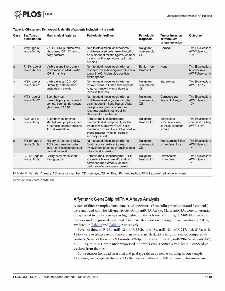
Affymetrix GeneChip miRNA Arrays AnalysisA total of fifteen samples from enucleated specimens (7 medulloepitheliomas and 8 controls)were analyzed with the Affymetrix GeneChip miRNA Arrays. Many miRNAs were differential-ly expressed in the two groups as highlighted in the volcano plot in Fig. 1. MiRNAs that wereover- or underexpressed by at least 2 standard deviations with a significant p-value (p< 0.05)are listed in Table 2 and Table 3, respectively.
Seven of these miRNAs–miR-124, miR-130b, miR-18a, miR-184, miR-217, miR-216a, miR-216b –were overexpressed by more than 6 standard deviations in tumors when compared tocontrols. Seven of these miRNAs–miR-509-3p, miR-146a, miR-145, miR-29b-2-star, miR-187,miR-133a, miR-211–were underexpressed in tumors versus controls by at least 6 standard de-viations from the mean.
Some tumors included neuronal and glial type tissue as well as cartilage in one sample.Therefore, we compared the miRNAs that were significantly different among tumor versus
Table 1. Clinical and Demogrpahic details of patients included in the study.
Case Sex/Age atpresentation
Main clinical features Pathologic findings Pathologicdiagnosis
Tumor invasionextraocular/scleral invasion
Outcome
1 M/4y; age attissue Dx: 9y
VA, OS HM; buphthalmos,glaucoma, IOP: 37mmHg;early cataract
Non-teratoid medulloepithelioma;undifferentiated cells resembling Rbcells; frequent mitotic figures; cornealinvasion with staphyloma; optic disccupping
Malignantnon-teratoid;OS
Corneal Tm: Enucleation;AW-FU period18y
2 F/13Y; age attissue Dx:17y
Visible grape like creamywhite mass in AC& uveitisIOP:41 mmHg
Non-teratoid medulloepithelioma;rosettes; few mitotic figures; cluster oftumor in AC; Alcian blue positivecystic spaces
Benign non-teratoid; OS
None Tm: Enucleation;buphthalmicAW-FU period 1y
3 M/6Y; age attissue Dx:6Y
Visible mass; ACG, IOP36mmHg; cataract/lenssublaxation, uveitis
Non-teratoid medulloepithelioma;myxoid areas in tumor, lens capsulerupture; frequent mitotic figures;invasive features
Malignantnon-teratoid;OD
Iris; corneal Tm: Enucleation;AW-FU: 11y
4 M/2Y; age attissue Dx:3Y
Buphthalmos;pseudohypopyon; cataract,corneal edema, iris neovas;glaucoma, IOP:40
Non-teratoid medulloepithelioma;undifferentiated large pleomorphiccells; frequent mitotic figures; Alcianblue positive cystic spaces; fewrosettes; staphyloma, break inDescemet’s membrane
Malignantnon-teratoid;OS
Corneoscleraltissue; AC angle
Tm: Enucleation;AW-FU period:10Y
5 F/3Y; age attissue Dx:5y
Buphthalmos, anteriorstaphyloma; proptosis; pain& redness; corneal opacity;TRD & exudation
Teratoid medulloepithelioma;neuroepithelial component; fibrillarcytoplasm & positive GFAP cells;moderate mitosis; Alcian blue positivecystic spaces; invasion: cornea/subconjunctival
Malignantteratoid; OD
Extrascleral:subconj nodule;corneal: posteriorstroma
Tm: Enucleation;chemo: 6 cycles;AW-FU: 4Y
6 M/1.5Y; age attissue Dx:2y
History of trauma; shallowAC; inferonasal vascularlesion on iris; distorted pupil;cortical material
Non-teratoid medulloepithelioma;focal necrosis; mitotic figures;involvement of ant segment/iris; focalintrascleral extension
Malignantnon-teratoid;OS
Ant segment & iris;intrascleral: focal
Tm: Enucleation;AW-FU period:1Y
7 F/ 0.5Y; age attissue Dx:5y
Ciliary body mass seenthrough pupil
Teratoid medulloepithelioma; TRD;absent iris & lens neural/glandular/cartilagenous elements; cornealperforation/extraocular extension
Malignantteratoid; OS
Extraocular;intrascleral
Tm: Enucleation;AW-FU period:1Y
M: Male; F: Female; Y: Years; AC: anterior chamber; OD: right eye; OS: left Eye; HM: hand motion; TRD: tractional retinal detachment.
doi:10.1371/journal.pone.0121706.t001
Medulloepithelioma miRNA Profiles
PLOS ONE | DOI:10.1371/journal.pone.0121706 March 25, 2015 5 / 18

control tissues to miRNA profiles described in retinoblastoma, glioblastoma, and cartilage.These comparisons are presented in Table 4. Since there was only one histologically benignmedulloepithelioma in our samples, we were unable to compare miRNA differential profilingbetween malignant and benign tumors. Since the sample size of teratoid and non-teratoid tu-mors was small, we did not compare miRNA expression differences between these two groups.
RT-PCR validationFor validation using RT-PCR, we chose three miRNAs (miR-217, miR-216a, and miR-216b) thatwere upregulated and three miRNAs (miR-146a, miR-509-3p, miR-211) that were downregu-lated by at least 6 standard deviations in tumor compared to control arrays. After normalizationwith U6 snRNA as the endogenous control, the RT-PCR data confirmed significant expression(P< 0.05) of all six miRNAs in all samples used in microarray analysis (Table 5).
Fig 1. Volcano plot of differentially expressedmiRNAs in seven cases of medulloepithelioma. The x-axis shows the log2 fold-change in miRNAexpression and y-axis shows the-Log10 of the p-value from tumor versus control miRNA expression counts. Labelled miRNAs have Log2 fold change greaterthan 2SD from the mean.
doi:10.1371/journal.pone.0121706.g001
Medulloepithelioma miRNA Profiles
PLOS ONE | DOI:10.1371/journal.pone.0121706 March 25, 2015 6 / 18

Table 2. MiRNAs significantly overexpressed (p< 0.05) by at least 2 standard deviations in tumors compared to control samples.
MiRNA Symbol ME vs. CT 15R (p-value) ME vs. CT 15R Lin(FC) ME vs. CT 15R Log2(FC) ME vs. CT Standard deviation
miR-4513 0.00083826 1.82565 0.868414 +2s
miR-331-5p 0.00107521 2.30358 1.20388 +2s
miR-1296 0.00400858 2.0393 1.02807 +2s
miR-4454 0.00506333 2.33973 1.22634 +2s
miR-181c-star 0.00864938 2.72817 1.44793 +2s
miR-3651 0.0104184 2.29852 1.2007 +2s
miR-654-5p 0.0144585 1.9331 0.950914 +2s
miR-550a 0.0155852 2.30192 1.20284 +2s
miR-324-5p 0.0156695 2.37185 1.24601 +2s
miR-99b-star 0.0203453 2.58034 1.36756 +2s
miR-4526 0.0254955 1.8913 0.919377 +2s
miR-4667-5p 0.0256011 1.90105 0.926796 +2s
miR-542-5p 0.0291441 2.25941 1.17595 +2s
miR-3175 0.030082 2.92621 1.54903 +2s
miR-151-3p 0.0310334 1.98682 0.99046 +2s
miR-452 0.0313143 2.47711 1.30866 +2s
miR-3065-5p 0.0315675 2.14354 1.09999 +2s
miR-887 0.0351422 1.89504 0.922229 +2s
miR-150-star 0.0386284 2.08094 1.05724 +2s
miR-629-star 0.0390014 2.00566 1.00408 +2s
miR-454 0.0394097 2.10381 1.073 +2s
miR-431 0.040385 2.88151 1.52683 +2s
miR-769-3p 0.0415066 1.83048 0.872224 +2s
miR-299-3p 0.0415905 1.90425 0.929225 +2s
miR-550a-star 0.0423132 2.12592 1.08809 +2s
miR-1287 0.0423992 1.91762 0.939317 +2s
miR-4783-3p 0.0452132 2.05298 1.03772 +2s
miR-337-5p 0.045312 2.05673 1.04036 +2s
miR-181a 0.045623 2.53466 1.34179 +2s
miR-296-3p 0.0465991 2.09545 1.06726 +2s
miR-489 0.0476999 2.73638 1.45227 +2s
miR-628-3p 0.0487057 2.37865 1.25014 +2s
miR-331-3p 0.00072146 4.77257 2.25477 +3s
miR-200c 0.00171416 3.74303 1.90421 +3s
miR-25 0.00228449 3.99073 1.99665 +3s
miR-483-5p 0.00374816 3.16618 1.66274 +3s
miR-210 0.0037929 4.45366 2.15499 +3s
miR-106b-star 0.00534338 3.76198 1.91149 +3s
miR-92a 0.00831397 2.99751 1.58377 +3s
miR-34a 0.009654 4.68914 2.22932 +3s
miR-19a 0.0105539 3.11789 1.64057 +3s
miR-425 0.0119682 3.46944 1.7947 +3s
miR-92a-1-star 0.0157259 3.31281 1.72806 +3s
miR-130a 0.0168223 3.05017 1.60889 +3s
miR-92b 0.0196283 4.24878 2.08705 +3s
miR-181a-star 0.0196711 4.49802 2.16929 +3s
miR-18a-star 0.0198721 3.17976 1.66892 +3s
(Continued)
Medulloepithelioma miRNA Profiles
PLOS ONE | DOI:10.1371/journal.pone.0121706 March 25, 2015 7 / 18

Pathway analysisFollowing the addition of upregulated miRNAs, the DIANA-miRPath v2.0 identified 31 pathways(see S1 Table) as significantly enriched (p< 0.05). Among these, the top 15 pathways includedthose involved in prion disease, infections (Hepatitis B, HTLV-1), PI3-AKT signaling pathway,
Table 2. (Continued)
MiRNA Symbol ME vs. CT 15R (p-value) ME vs. CT 15R Lin(FC) ME vs. CT 15R Log2(FC) ME vs. CT Standard deviation
miR-375 0.0297981 3.74662 1.90559 +3s
miR-370 0.0324271 3.04826 1.60799 +3s
miR-149 0.0341333 3.5908 1.8443 +3s
miR-629 0.0395607 4.6143 2.20611 +3s
miR-345 0.0412484 3.62454 1.8578 +3s
miR-376c 0.0428205 3.40819 1.76901 +3s
miR-135a-star 0.00010313 7.59616 2.92527 +6s
miR-769-5p 0.00019315 5.43666 2.44272 +6s
miR-4417 0.00023069 14.204 3.82823 +6s
miR-21 0.00031135 5.50629 2.46108 +6s
miR-20a 0.00034234 9.30564 3.21811 +6s
miR-106b 0.0004506 7.6127 2.92841 +6s
miR-106a 0.00053755 7.16934 2.84184 +6s
miR-17 0.00055035 7.07773 2.82329 +6s
miR-18b 0.00086543 10.1821 3.34796 +6s
miR-25-star 0.00097707 5.66118 2.5011 +6s
miR-20b 0.00101489 7.14623 2.83718 +6s
miR-93 0.00147512 5.09062 2.34784 +6s
miR-3687 0.00153801 7.4984 2.90658 +6s
miR-551b 0.00171105 5.46106 2.44918 +6s
miR-17-star 0.00202618 7.55628 2.91768 +6s
miR-19b 0.00440786 7.2896 2.86584 +6s
miR-7 0.00478394 12.5187 3.64601 +6s
miR-421 0.00618539 5.00526 2.32344 +6s
miR-93-star 0.0099438 5.25781 2.39446 +6s
miR-487a 0.011085 4.93567 2.30324 +6s
miR-15a 0.0139553 6.01482 2.58852 +6s
miR-363 0.0148836 6.16909 2.62506 +6s
miR-9-star 0.0166476 6.34056 2.66461 +6s
miR-382 0.0235705 5.15825 2.36688 +6s
miR-124-star 0.0273617 5.86526 2.5522 +6s
miR-891a 0.0330171 5.4209 2.43853 +6s
miR-487b 0.0383924 5.56007 2.4751 +6s
miR-182 0.0453814 15.8393 3.98544 +6s
miR-217 1.68E-05 166.311 7.37774 > +6s
miR-216a 3.53E-05 73.8658 6.20683 > +6s
miR-130b 7.78E-05 40.908 5.35431 > +6s
miR-216b 7.79E-05 71.4398 6.15866 > +6s
miR-18a 9.16E-05 30.4575 4.92873 > +6s
miR-184 0.00057019 28.0727 4.81109 > +6s
miR-124 0.00224884 54.4488 5.76683 > +6s
doi:10.1371/journal.pone.0121706.t002
Medulloepithelioma miRNA Profiles
PLOS ONE | DOI:10.1371/journal.pone.0121706 March 25, 2015 8 / 18

Table 3. MiRNAs significantly underexpressed (p< 0.05) by at least 2 standard deviations in tumors compared to control samples.
MiRNA Symbol ME vs. CT 15R (p-value) ME vs. CT 15R Lin(FC) ME vs. CT 15R Log2(FC) ME vs. CT Standard deviation
miR-4298 7.31E-05 -1.95551 -0.967547 -2s
let-7c 0.00038689 -1.91379 -0.936431 -2s
miR-574-5p 0.00404375 -1.82692 -0.86941 -2s
miR-509-3-5p 0.00509148 -1.841 -0.88049 -2s
miR-508-5p 0.0101842 -1.84679 -0.885021 -2s
miR-3921 0.01021 -1.7505 -0.807766 -2s
miR-30a-star 0.0113581 -2.45667 -1.2967 -2s
let-7d 0.0131867 -2.08921 -1.06296 -2s
miR-99a 0.0133064 -1.97959 -0.985198 -2s
miR-126 0.0142176 -1.75444 -0.811007 -2s
miR-3135b 0.0155168 -1.72398 -0.785747 -2s
miR-3609 0.0171527 -1.83873 -0.878707 -2s
miR-23a 0.0186938 -1.69122 -0.758066 -2s
miR-155 0.0217847 -1.63886 -0.712694 -2s
miR-3613-3p 0.0246578 -1.54502 -0.627627 -2s
miR-125b-2-star 0.0260984 -2.33265 -1.22197 -2s
miR-1184 0.0261281 -1.7633 -0.818276 -2s
miR-4329 0.0265162 -1.89642 -0.92328 -2s
miR-1207-5p 0.0270965 -1.52179 -0.605774 -2s
miR-455-3p 0.0363036 -1.66499 -0.735512 -2s
miR-4487 0.0397386 -1.54414 -0.626808 -2s
miR-4701-3p 0.044214 -1.67333 -0.742725 -2s
let-7b 0.00026306 -2.63302 -1.39672 -3s
miR-150 0.00080125 -3.03867 -1.60344 -3s
miR-328 0.0014731 -2.59841 -1.37763 -3s
miR-193a-5p 0.00215253 -3.28019 -1.71378 -3s
miR-214 0.00347545 -3.73469 -1.90099 -3s
miR-574-3p 0.00654517 -3.28345 -1.71521 -3s
miR-532-3p 0.0107958 -2.70874 -1.43762 -3s
miR-422a 0.0125962 -3.28801 -1.71722 -3s
miR-204 0.0129536 -3.31134 -1.72742 -3s
miR-378 0.020049 -2.93103 -1.55141 -3s
miR-1973 0.0215745 -2.51265 -1.32921 -3s
miR-378f 0.0268653 -3.42704 -1.77696 -3s
miR-133b 0.0396013 -3.55802 -1.83107 -3s
miR-1911 0.0473435 -3.19215 -1.67453 -3s
miR-509-3p 8.02E-06 -14.603 -3.8682 -6s
miR-146a 4.04E-05 -9.91478 -3.30958 -6s
miR-145 0.00090581 -4.41564 -2.14262 -6s
miR-29b-2-star 0.0034838 -5.02666 -2.3296 -6s
miR-187 0.010088 -4.60929 -2.20455 -6s
miR-133a 0.044119 -4.99992 -2.3219 -6s
miR-211 2.46E-07 -42.9191 -5.42355 -6s
doi:10.1371/journal.pone.0121706.t003
Medulloepithelioma miRNA Profiles
PLOS ONE | DOI:10.1371/journal.pone.0121706 March 25, 2015 9 / 18
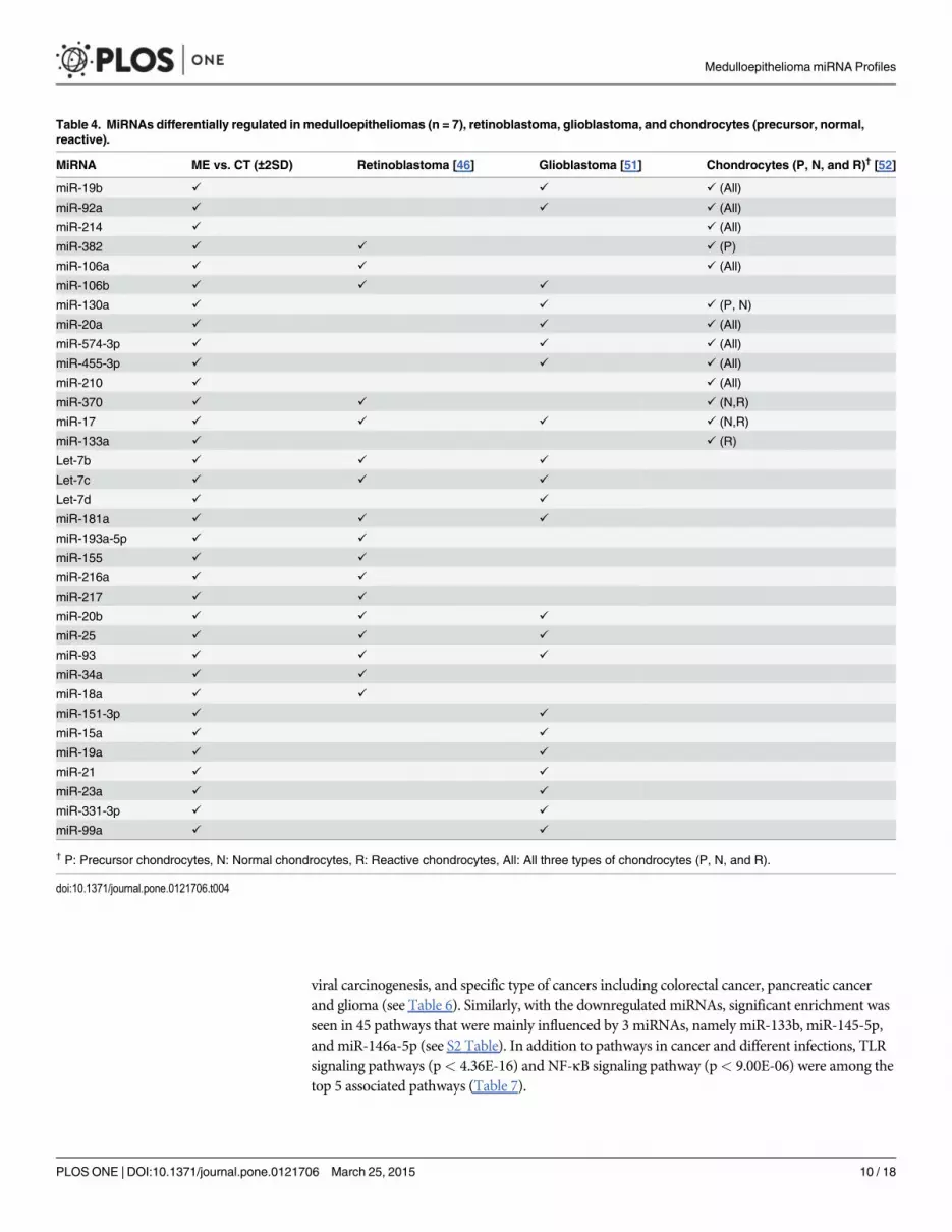
viral carcinogenesis, and specific type of cancers including colorectal cancer, pancreatic cancerand glioma (see Table 6). Similarly, with the downregulated miRNAs, significant enrichment wasseen in 45 pathways that were mainly influenced by 3 miRNAs, namely miR-133b, miR-145-5p,and miR-146a-5p (see S2 Table). In addition to pathways in cancer and different infections, TLRsignaling pathways (p< 4.36E-16) and NF-κB signaling pathway (p< 9.00E-06) were among thetop 5 associated pathways (Table 7).
Table 4. MiRNAs differentially regulated in medulloepitheliomas (n = 7), retinoblastoma, glioblastoma, and chondrocytes (precursor, normal,reactive).
MiRNA ME vs. CT (±2SD) Retinoblastoma [46] Glioblastoma [51] Chondrocytes (P, N, and R)† [52]
miR-19b ✓ ✓ ✓ (All)
miR-92a ✓ ✓ ✓ (All)
miR-214 ✓ ✓ (All)
miR-382 ✓ ✓ ✓ (P)
miR-106a ✓ ✓ ✓ (All)
miR-106b ✓ ✓ ✓
miR-130a ✓ ✓ ✓ (P, N)
miR-20a ✓ ✓ ✓ (All)
miR-574-3p ✓ ✓ ✓ (All)
miR-455-3p ✓ ✓ ✓ (All)
miR-210 ✓ ✓ (All)
miR-370 ✓ ✓ ✓ (N,R)
miR-17 ✓ ✓ ✓ ✓ (N,R)
miR-133a ✓ ✓ (R)
Let-7b ✓ ✓ ✓
Let-7c ✓ ✓ ✓
Let-7d ✓ ✓
miR-181a ✓ ✓ ✓
miR-193a-5p ✓ ✓
miR-155 ✓ ✓
miR-216a ✓ ✓
miR-217 ✓ ✓
miR-20b ✓ ✓ ✓
miR-25 ✓ ✓ ✓
miR-93 ✓ ✓ ✓
miR-34a ✓ ✓
miR-18a ✓ ✓
miR-151-3p ✓ ✓
miR-15a ✓ ✓
miR-19a ✓ ✓
miR-21 ✓ ✓
miR-23a ✓ ✓
miR-331-3p ✓ ✓
miR-99a ✓ ✓
† P: Precursor chondrocytes, N: Normal chondrocytes, R: Reactive chondrocytes, All: All three types of chondrocytes (P, N, and R).
doi:10.1371/journal.pone.0121706.t004
Medulloepithelioma miRNA Profiles
PLOS ONE | DOI:10.1371/journal.pone.0121706 March 25, 2015 10 / 18

DiscussionDuring tumorigenesis, it is suggested that dysregulations of miRNA-mediated gene regulatorynetworks, evident in many cancer models, play several roles. At the cellular level, miRNAsfunction as master regulators and signal modulators. They fine-tune gene expression in manycomplex pathways. When these pathways are disrupted, the resulting alterations permit tumor-igenesis within a particular tissue [28]. This study highlights significant changes in the miRNAexpression profile in intraocular medulloepitheliomas as compared to controls.
Many of the over- or underexpressed miRNA in medulloepithelioma were reported to playvarious roles in carcinogenesis. In this section of the discussion, we chose to highlight the func-tion(s) of some of the validated miRNAs that showed expression differences by several stan-dard deviations from the control. In this discussion we will focus on a few of the MiR-217 washighly overexpressed in medulloepitheliomas. Recent data suggests that miR-217 is an onco-gene that is overexpressed in aggressive human B cell lymphomas [30] and contradictarorilyfunctions as a potential tumor suppressor in hepatocellular carcinoma through direct suppres-sion of E2F3 [31]. On the other hand, levels of miR-217 are downregulated in pancreaticintraepithelial neoplasm and pancreatic ductal adenocarcinomas [32] and in clear cell renal
Table 5. qPCR validation Data.
Sample Control Medulloepithelioma Mann Whitney U
N Mean Fold-change Std. Dev. (Range) N Mean Fold-change Std. Dev. (Range) P-value
miR-217 8 0.304 0.450 (0.053–1.38) 7 13.529 13.460 (0.85–38.87) 0.002
miR-216a 8 0.312 0.435 (0.015–1.28) 7 23.055 22.109 (1.26–71.90) 0.002
miR-216b 8 0.633 1.235 (0.012–3.58) 7 22.614 24.237 (1.20–60.49) 0.002
miR-146a 8 2.427 1.834 (0.58–3.79) 7 0.525 0.209 (0.18–0.62) 0.005
miR-509-3p 8 4.557 3.214 (0.99–10.82) 7 0.311 0.203 (0.039–0.63) 0.001
miR-211 8 2.089 1.788 (0.41–3.67) 7 0.652 0.306 (0.35–1.25) 0.032
doi:10.1371/journal.pone.0121706.t005
Table 6. Top 15 pathways significantly influenced by upregulatedmiRNAs.
KEGG pathway p-value #genes† #miRNAs
Prion diseases 0 1 1
Colorectal cancer 0 11 8
Pancreatic cancer 0 13 12
Glioma 0 10 11
Chronic myeloid leukemia 0 24 11
Melanoma 0 20 12
Bladder cancer 0 17 12
Pathways in cancer 0 70 13
Prostate cancer 0 28 13
Hepatitis B 0 41 16
HTLV-I infection 2.22E-15 53 11
Small cell lung cancer 2.88E-13 26 9
Non-small cell lung cancer 2.95E-11 18 14
PI3K-Akt signaling pathway 3.20E-11 23 6
Viral carcinogenesis 2.83E-09 18 8
† Number of genes predicted by TarBase v6.0.
doi:10.1371/journal.pone.0121706.t006
Medulloepithelioma miRNA Profiles
PLOS ONE | DOI:10.1371/journal.pone.0121706 March 25, 2015 11 / 18

carcinomas [33]. Further studies are needed to determine whether higher expression of miR-217 in intraocular medulloepithelioma represents an oncogenic effect or the dysregulationfunctions as a potential tumor suppressor that might explain its low metastatic potential.
MiR-18a was also highly expressed in intraocular medulloepithelioma samples. Recently inan orthotopic metastatic breast cancer xenograft model, miR-18a suppressed distant metastasisvia the hypoxia-inducible factor 1-alpha pathway [34]. This observation is interesting since it iswell known that intraocular medulloepitheliomas typically spread locally and distant metasta-sis is rare [14]. It is possible that the overexpression of miR-18a may play a role in repressingthe metastatic behavior of intraocular medulloepitheliomas. Additionally, miR-216a and miR-216b in our study following qPCR validation demonstrated a twenty fold change in intraocularmedulloepithelioma as compared to the controls. In contrast, both these miRNAs are downre-gulated in a mouse model of pancreatic cancer [35]. However, overexpression of miR-216a wasshown to activate the PI3K/Akt and TGF-β pathways by targeting PTEN and SMAD7, contrib-uting to hepatocarcinogenesis and tumor recurrence in heptaocellular carcinoma. The exactoverexpression of these two miRNAs in medulloepithelioma needs further investigation [36].Interestingly miR-382 was significantly upregulated in ocular medulloepitheliomas in thisstudy. This miRNA functions as an oncogene under hypoxic conditions and also regulates tu-morigenesis through the PTEN/AKT/mTOR pathway [37].
Three miRNAs (miR-146a, miR-509-3p and miR-211) showed significantly reduced expres-sion in the intraocular medulloepithelioma specimens. Downregulation of miR-146a wasshown to play a role in migration and metastasis of breast carcinoma [38] and miR-509-3p wasshown to be a tumor suppressor in renal cell carcinoma with downregulation associated withcell invasion and migration in renal cell cancer [39]. MiR-211 downregulation has been shownin a variety of cancers and is associated with tumor progression invasion in breast carcinoma,tumors of the head and neck and melanomas [38, 40, 41]. Interestingly miR-211 is found inabundance in the vitreous humor and one might suggest that the source of this miRNA maypotentially be the ciliary epithelium or the sensory retina [21].
Table 7. Top 15 pathways significantly influenced by downregulated miRNAs.
KEGG pathway p-value #genes† #miRNAs
Toll-like receptor signaling pathway 4.36E-16 14 3
Hepatitis B 4.50E-10 14 3
Pathways in cancer 8.01E-07 20 3
Pancreatic cancer 7.90E-06 8 3
NF-kappa B signaling pathway 9.00E-06 7 2
Apoptosis 9.00E-06 9 3
Chagas disease (American trypanosomiasis) 1.52E-05 9 3
Tuberculosis 1.52E-05 13 3
Hepatitis C 1.52E-05 10 3
Bladder cancer 1.52E-05 6 3
RIG-I-like receptor signaling pathway 0.000161 7 2
Legionellosis 0.000229 6 3
Pertussis 0.000293 7 3
Measles 0.000293 9 3
Neurotrophin signaling pathway 0.000611 8 3
† Number of genes predicted by TarBase v6.0.
doi:10.1371/journal.pone.0121706.t007
Medulloepithelioma miRNA Profiles
PLOS ONE | DOI:10.1371/journal.pone.0121706 March 25, 2015 12 / 18
Though a panel of six miRNAs was highly over- or underexpressed and validated in themedulloepithelioma specimens, none of them were unique to the tumor. It is possible that acombination of this panel might be useful as biomarkers for medulloepitheliomas for undiffer-entiated neuronal tumors of the eye and adnexa. Both miR-216a and miR-216b are associatedwith various types of cancer in particular adenocarcinoma of the pancreas. MiR-146a is be-lieved to be involved in the regulation of inflammation and major pathways in cancer. As formiR-217, there is no specific pathway identified linked to this microRNA, but is indirectly asso-ciated with cancer and pancreatitis. Furthermore, miR-146a is directly involved in varioustypes of cancer including prostate, gastric, sarcoma, leukemia and pancreatic cancer; and miR-509-3p is involved in renal cell carcinoma with no direct link to a specific pathway. Finally,miR-211 is associated with cancer and stroke. So clearly, most of our differentially expressedmiRNAs are related to cancer and signaling pathways (see Tables 6 and 7).
Ocular medulloepithelioma has been associated with a DICER1 germline mutations in fa-milial pleuropulmonary blastoma (PPB) by genome-wide linkage analysis [42]. DICER1 islocated on chromosome 14q32 and encodes a ribonuclease that participates in miRNA forma-tion [43]. In this series, the ocular medulloepithelioma are localized in all patients and did notshow the phenotype typical of DICER mutations [42]. Furthermore dicer knockouts typicallyresult in a global depression in miRNA production [44]. In a patient with DICER mutationand mutated PPB elevated serum levels of miR-125a-3p and miR-125b-2-3p were described.These miRNAs were not upregulated in the tumor samples in this study [45].
Retinoblastoma like ocular medulloepthelioma is another ocular neoplasm that is commonamong children. Though the clinical features and behavior of retinoblastoma is quite differentfrom medulloepithelioma, they share some common pathologic features that include the undif-ferentiated primitive neuronal cells and presence of rosettes in the tumor. Our study detectedmany significantly up- and downregulated miRNAs in the tumor tissue of medulloepitheliomathat were also described in retinoblastoma as noted in Table 4. Of particular interest is the sig-nificant downregulation of the let-7 family of miRNAs described in retinoblastoma [46] andalso seen in the intraocular medulloepitheliomas in our study. The let-7 miRNAs are a familyof seven subtypes that play an important role in the development and differentiation of embry-onic cells into specific lineage in the central nervous system. In addition many members of thelet-7 family play a role in cancer as a tumor suppressing miRNA [47] but interacts with LIN28,which encodes an RNA binding protein, as is described later in this discussion.
Medulloepitheliomas are primitive neuroepithelial tumors that pathologically resembleprimitive neuroectodermal tumors of the central nervous system [48]. Detailed miRNA profil-ing in primitive neuroectodermal tumors is currently unavailable. In cell lines of primitive neu-roectodermal tumors, miR-125 was upregulated in response to chemotherapy [49]. Significantdysregulation of miR-125 was not seen in our tumor samples.
Embryonal tumor with multilayered rosettes (ETMR) is an aggressive primitive neuroecto-dermal neoplasm of the central nervous system. In the central nervous system, abundant neu-trophils and true rosettes in the embryonal tumors suggest that ependymoblastoma andmedulloepithelioma are the same entity [50]. Intraocular medulloepitheliomas share some ofthe histological features of these tumors; however, the result is rarely metastasis or mortality,even in the malignant histological variety [11]. LIN28, an RNA binding protein, is an impor-tant diagnostic marker, which is upregulated in many cancers including ETMR. In an ETMRcell line, LIN28 knockdown showed an increase in let-7 expression by activating downstreampathways. It is suggested that the LIN28/let-7 pathway plays a critical role in the pathophysiol-ogy of malignant neoplasms such as ETMR and germ cell tumors. Overexpression of LIN28 se-lectively inhibits let-7 biogenesis leading to decreased expression of let-7 in such neoplasms. Itis possible that the significant decrease in let-7 expression seen in intraocular
Medulloepithelioma miRNA Profiles
PLOS ONE | DOI:10.1371/journal.pone.0121706 March 25, 2015 13 / 18

medulloepitheliomas plays a significant role in the pathogenesis of this neoplasm. The less ag-gressive behavior of intraocular medulloepitheliomas, in contrast to those seen in the centralnervous system, are likely regulated by other mechanisms including some of the miRNAs de-scribed earlier in the discussion [47].
Medulloepitheliomas may either be benign or malignant neoplasms [13]. In this series, sixtumors were classified as malignant and one as benign. The miRNA differences between malig-nant versus benign samples could not be studied in view of the disparate numbers. In order toidentify specific miRNAs that might be dysregulated in tumors versus controls, the data wasanalyzed. As shown in Table 4, several miRNAs showed significant differential expression. Wealso compared this dysregulation to published reports on miRNA alterations in retinoblasto-ma, glioblastoma, and in embryonic, normal and hypertrophic cartilage [46, 51, 52]. Of thetotal thirty-four miRNAs that were significantly dysregulated in medulloepithelioma, twenty-two were reported to be dysregulated in malignant glioblastomas [51] suggesting common mo-lecular threads between these primitive neoplasms.
Furthermore, thirteen miRNAs that were dysregulated in intraocular medulloepitheliomaswere also expressed in normal, embryonic and/or hypertrophic cartilage. Based on this analy-sis, the role of these miRNAs, if any, in the formation of cartilaginous medulloepitheliomasneeds to be further investigated.
Several pathways appear to be enriched by the upregulated miRNAs. Among the top 15pathways, the pathway most significantly was associated with prion disease (p< 1e-16) wasfound to be influenced by a single miRNA, miR-130b-3p, which was predicted to target a singletranscript PRNP (prion protein gene). Mutations in PRNP are associated with human priondiseases [53]. The functional role of this pathway in medulloepithelioma is unclear, thus needsfurther investigation. Other pathways that may be of interest include transforming-growth-factor-beta (TGF-β) signaling pathway, which was most significantly influenced by miR-20a-5p (p< 9.6e-08) and miR-17-5p (p< 2.7e-07), and hypoxia-inducible factor (HIF)-1 signalingpathway, which was most significantly influenced by miR-20b-5p (p< 4.3e-07) (see S1 Tablefor the complete list). Upregulation of HIF1-alpha (HIF-1α) is associated with adverse out-comes in patients with neuroblastoma [54] and other tumors as previously described inthis report.
Similarly, 45 pathways corresponding to the downregulated miRNAs were noted (see S2Table). These pathways were mainly influenced by miR-133b, miR-145-5p, and miR-146a-5p.Besides pathways in cancer and different infections, TLR (p<4.36E-16) and NF-κB-signalingpathways (p< 9.00E-06) were among the top 5 associated pathways.
Most cancers are characterized by activation of the NF-κB pathway, resulting in cancer cellproliferation, survival, angiogenesis and metastasis. It appears that tumorigenesis in intraocularmedulloepitheliomas may also be influenced by the NF-κB pathway [55].
TLR stimulation can lead to up- or downregulation of various miRNA expressions. TLRsare known to trigger innate immune system and bolster adaptive immunity against antigensexpressed by pathogens and tumor cells [56], and can also modulate anti-cancer therapy [57].Patients with breast cancer carry the loss-of function allele for TLR4. The defective allele affectsthe binding of high-mobility group box (HMGB1) protein to TLR4. Patients with the loss-of-function TLR4 allele have been shown to relapse more quickly after radiotherapy or chemo-therapy indicating the clinical relevance of immunoadjuvant pathway triggered by tumor celldeath [58, 59]. MiR-146a, which was significantly downregulated in intraocular medulloepithe-liomas in this study, may interact with both the NF-κB pathway as well as the TLR pathway asshown in breast and thyroid cancer [60]. Studies have shown the pathological relevance ofNF-κB/miR-146 in human breast cancer, pancreatic cancer, anaplastic thyroid carcinomas,and brain tumors [61]; therefore, one might suggest that this pathway may play a role in the
Medulloepithelioma miRNA Profiles
PLOS ONE | DOI:10.1371/journal.pone.0121706 March 25, 2015 14 / 18

pathogeneis of medulloepitheliomas. In the CNS gliomas, TLR pathways are responsible forconverting microglia into a glioma supportive phenotype [62]. Such a role in medulloepithelio-mas needs further investigation.
There are some limitations of this particular study. The sample size of the study was smallmaking it difficult to analyze miRNA alterations in subsets of medulloepitheliomas. More spec-imens are needed to perform additional analysis. Also, intraocular medulloepitheliomas arelocalized tumors and determining mortality based prognostic factors based on miRNA expres-sion may not be relevant. We hope that future studies will be able to address the differences inthe biological behavior of central nervous system medulloepitheliomas and intraocular medul-loepitheliomas at the molecular level.
Supporting InformationS1 Table. DIANA-miRPath v2.0 identified significantly enriched pathways for upregulatedmiRNAs.(DOCX)
S2 Table. DIANA-miRPath v2.0 identified significantly enriched pathways for downregu-lated miRNAs.(DOCX)
Author ContributionsConceived and designed the experiments: DPE CE KKAA HK. Performed the experiments:NG AAK KKAA LAS QR HA DPE. Analyzed the data: DPE HA QR KKAA AAK LAS. Con-tributed reagents/materials/analysis tools: DPE CE HA KKAA NG AAK SAMQR LAS. Wrotethe paper: DPE HA QR SAM CE KKAA AAK NG LAS.
References1. He L, Hannon GJ. MicroRNAs: small RNAs with a big role in gene regulation. Nat Rev Genet. 2004
Aug; 5(7): 522–531. PMID: 15211354
2. Bartel DP, Chen CZ. Micromanagers of gene expression: the potentially widespread influence of meta-zoan microRNAs. Nat Rev Genet. 2004 May; 5(5): 396–400. PMID: 15143321
3. Krutzfeldt J, Poy MN, Stoffel M. Strategies to determine the biological function of microRNAs. NatGenet. 2006 Jun; 38 Suppl: S14–19. PMID: 16736018
4. Weber JA, Baxter DH, Zhang S, Huang DY, Huang KH, Lee MJ, et al. The microRNA spectrum in 12body fluids. Clin Chem. 2010 Nov; 56(11): 1733–1741. doi: 10.1373/clinchem.2010.147405 PMID:20847327
5. Mitchell PS, Parkin RK, Kroh EM, Fritz BR, Wyman SK, Pogosova-Agadjanyan EL, et al. CirculatingmicroRNAs as stable blood-based markers for cancer detection. Proc Natl Acad Sci U S A. 2008Jul 29; 105(30): 10513–8. doi: 10.1073/pnas.0804549105 PMID: 18663219
6. Xi Y, Nakajima G, Gavin E, Morris CG, Kudo K, Hayashi K, et al. Systematic analysis of microRNA ex-pression of RNA extracted from fresh frozen and formalin-fixed paraffin-embedded samples. RNA.2007 Oct; 13(10): 1668–74. PMID: 17698639
7. Dai R, Ahmed SA. MicroRNA, a new paradigm for understanding immunoregulation, inflammation, andautoimmune diseases. Transl Res. 2011 Apr; 157(4): 163–79. doi: 10.1016/j.trsl.2011.01.007 PMID:21420027
8. Croce CM. Causes and consequences of microRNA dysregulation in cancer. Nat Rev Genet. 2009Oct; 10(10): 704–14. doi: 10.1038/nrg2634 PMID: 19763153
9. Etheridge A, Lee I, Hood L, Galas D, Wanh K. Extracellular microRNA: a new source of biomarkers.Mutat Res. 2011 Dec 1; 717(1–2): 85–90. doi: 10.1016/j.mrfmmm.2011.07.020 PMID: 21889945
10. Zimmerman LE. Verhoeff's "terato-neuroma". A critical reappraisal in light of new observations andcurrent concepts of embryonic tumors. The Fourth Frederick H. Verhoeff Lecture. Am J Ophthalmol.1971 Dec; 72(6): 1039–57. PMID: 4110093
Medulloepithelioma miRNA Profiles
PLOS ONE | DOI:10.1371/journal.pone.0121706 March 25, 2015 15 / 18

11. Shields JA, Eagle RC Jr, Shields CL, Potter PD. Congenital neoplasms of the nonpigmented ciliary epi-thelium (medulloepithelioma). Ophthalmology. 1996 Dec; 103(12): 1998–2006. PMID: 9003333
12. Takei H, Florez L, Moroz K, Bhattacharjee MB. Medulloepithelioma: Two unusual locations. Pathol Int.2007 Feb; 57(2): 91–5. PMID: 17300673
13. BroughtonWL, Zimmerman LE. A clinicopathologic study of 56 cases of intraocular medulloepithelio-mas. Am J Ophthalmol. 1978 Mar; 85(3): 407–18. PMID: 655220
14. Kaliki S, Shields CL, Eagle RC Jr, Vemuganti GK, Almeida A, Manjandavida FP, et al. Ciliary bodymedulloepithelioma: analysis of 41 cases. Ophthalmology. 2013 Dec; 120(12): 2552–9. doi: 10.1016/j.ophtha.2013.05.015 PMID: 23796765
15. Canning CR, McCartney AC, Hungerford J. Medulloepithelioma (diktyoma). Br J Ophthalmol. 1988 Oct;72(10): 764–7. PMID: 3056510
16. Garcia-Feijoo J, Encinas JL, Mendez-Hernandez C, Ronco IS, Martinez de la Casa JM, Garcia San-chez J, et al. Medulloepithelioma of the ciliary body: ultrasonographic biomicroscopic findings. J Ultra-sound Med. 2005 Feb; 24(2): 247–250. PMID: 15661960
17. Vajaranant TS, Mafee MF, Kapur R, Rapoport M, Edward DP. Medulloepithelioma of the ciliary bodyand optic nerve: clinicopathologic, CT, and MR imaging features. Neuroimaging Clin N Am. 2005 Feb;15(1): 69–83. PMID: 15927861
18. Saunders T, Margo CE. Intraocular medulloepithelioma. Arch Pathol Lab Med. 2012 Feb; 136(2):212–6. doi: 10.5858/arpa.2010-0669-RS PMID: 22288972
19. Huang KM, Dentchev T, Stambolian D. MiRNA expression in the eye. MammGenome. 2008 Aug;19(7–8): 510–6.
20. Dunmire JJ, Lagouros E, Bouhenni RA, Jones M, Edward DP. MicroRNA in aqueous humor from pa-tients with cataract. Exp Eye Res. 2013 Mar; 108C: 68–71.
21. Ragusa M, Caltabiano R, Russo A, Puzzo L, Avitabile T, Longo A, et al. MicroRNAs in vitreus humorfrom patients with ocular diseases. Mol Vis. 2013; 19: 430–440. PMID: 23441115
22. WuC, Lin H, Wang Q, ChenW, Luo H, ChenW. et al. Discrepant expression of microRNAs in transpar-ent and cataractous human lenses. Invest ophthalmol vis sci. 2012 Jun 22; 53(7): 3906–12. doi: 10.1167/iovs.11-9178 PMID: 22562507
23. Peng CH, Liu JH, Woung LC, Lin TJ, Chiou SH, Tseng PC, et al. MicroRNAs and cataracts: correlationamong let-7 expression, age and the severity of lens opacity. Br J Ophthalmol. 2012 May; 96(5):747–51. doi: 10.1136/bjophthalmol-2011-300585 PMID: 22334139
24. Li N, Cui J, Duan X, Chen H, Fan F. Suppression of type I collagen expression by miR-29b via PI3K,Akt, and Sp1 pathway in human Tenon's fibroblasts. Invest ophthalmol vis sci. 2012 Mar 26; 53(3):1670–8. doi: 10.1167/iovs.11-8670 PMID: 22297492
25. To KH, Pajovic S, Gallie BL, Theriault BL. Regulation of p14ARF expression by miR-24: a potentialmechanism compromising the p53 response during retinoblastoma development. BMCCancer. 2012Feb 15; 12: 69. doi: 10.1186/1471-2407-12-69 PMID: 22336108
26. Feng B, Chen S, McArthur K, Wu Y, Sen S, Ding Q, et al. miR-146a-Mediated extracellular matrix pro-tein production in chronic diabetes complications. Diabetes. 2011 Nov; 60(11): 2975–84. doi: 10.2337/db11-0478 PMID: 21885871
27. McArthur K, Feng B, Wu Y, Chen S, Chakrabarti S. MicroRNA-200b regulates vascular endothelialgrowth factor-mediated alterations in diabetic retinopathy. Diabetes. 2011 Apr; 60(4): 1314–23. doi: 10.2337/db10-1557 PMID: 21357793
28. Adams BD, Kasinski AL, Slack FJ. Aberrant Regulation and Function of MicroRNAs in Cancer. CurrBiol. 2014 Aug 18; 24(16): R762–76. doi: 10.1016/j.cub.2014.06.043 PMID: 25137592
29. Vlachos IS, Kostoulas N, Vergoulis T, Georgakilas G, Reczko M, Maragkakis M, et al. DIANAmiRPathv.2.0: investigating the combinatorial effect of microRNAs in pathways. Nucleic Acids Res. 2012 Jul;40(Web Server issue): W498–504. doi: 10.1093/nar/gks494 PMID: 22649059
30. de Yebenes VG, Bartolome-Izquierdo N, Nogales-Cadenas R, Perez-Duran P, Mur SM, Martinez M,et al. miR-217 is an oncogene that enhances the germinal center reaction. Blood. 2014 Jul 10; 124(2):229–39. doi: 10.1182/blood-2013-12-543611 PMID: 24850757
31. Su J, Wang Q, Liu Y, Zhong M. miR-217 inhibits invasion of hepatocellular carcinoma cells through di-rect suppression of E2F3. Mol Cell Biochem. 2014 Jul; 392(1–2): 289–296. doi: 10.1007/s11010-014-2033-3 PMID: 24696420
32. Xue Y, Abou Tayoun AN, Abo KM, Pipas JM, Gordon SR, Gardner TB, et al. MicroRNAs asdiagnostic markers for pancreatic ductal adenocarcinoma and its precursor, pancreatic intraepithelialneoplasm. Cancer Genet. 2013 Jun; 206(6): 217–21. doi: 10.1016/j.cancergen.2013.05.020 PMID:23933230
Medulloepithelioma miRNA Profiles
PLOS ONE | DOI:10.1371/journal.pone.0121706 March 25, 2015 16 / 18

33. Li H, Zhao J, Zhang JW, Huang QY, Huang JZ, et al. MicroRNA-217, down-regulated in clear cell renalcell carcinoma and associated with lower survival, suppresses cell proliferation and migration. Neo-plasma. 2013; 60(5): 511–515. doi: 10.4149/neo_2013_066 PMID: 23790169
34. Krutilina R, SunW, Sethuraman A, Brown M, Seagroves TN, et al. MicroRNA-18a inhibits hypoxia-inducible factor 1-alpha activity and lung metastasis in basal breast cancers. Breast Cancer Res. 2014Jul 28; 16(4): R78. PMID: 25069832
35. Ali S, Banerjee S, Logna F, Bao B, Philip PA, Korc M, et al. Inactivation of Ink4a/Arf leads to deregu-lated expression of miRNAs in K-Ras transgenic mouse model of pancreatic cancer. J Cell Physiol.2012 Oct; 227(10): 3373–3380. doi: 10.1002/jcp.24036 PMID: 22213426
36. Xia H, Ooi LL, Hui KM. MicroRNA-216a/217-induced epithelial-mesenchymal transition targets PTENand SMAD7 to promote drug resistance and recurrence of liver cancer. Hepatology. 2013 Aug; 58(2):629–641. doi: 10.1002/hep.26369 PMID: 23471579
37. Seok JK, Lee SH, Kim JM, Lee YM. MicroRNA-382 induced by HIF-1alpha is an angiogenic miR target-ing the tumor suppressor phosphatase and tensin homolog. Nucleic Acids Res. 2014 Jul; 42(12):8062–72. doi: 10.1093/nar/gku515 PMID: 24914051
38. Vimalraj S, Miranda PJ, Ramyakrishna B, Selvamurugan N. Regulation of breast cancer and bone me-tastasis by microRNAs. Dis Markers. 2013; 35(5): 369–387. doi: 10.1155/2013/451248 PMID:24191129
39. Zhai Q, Zhou L, Zhou C, Wan J, Yu Z, Guo X, et al. Identification of miR-508-3p and miR-509-3p thatare associated with cell invasion and migration and involved in the apoptosis of renal cell carcinoma.Biochem Biophys Res Commun. 2012 Mar 23; 419(4): 621–626. doi: 10.1016/j.bbrc.2012.02.060PMID: 22369946
40. Chu TH, Yang CC, Liu CJ, Lui MT, Lin SC, Chang KW. miR-211 promotes the progression of head andneck carcinomas by targeting TGFbetaRII. Cancer Lett. 2013; 337(1): 115–124. doi: 10.1016/j.canlet.2013.05.032 PMID: 23726841
41. Kunz M. MicroRNAs in melanoma biology. Adv Exp Med Biol. 2013; 774: 103–120. doi: 10.1007/978-94-007-5590-1_6 PMID: 23377970
42. Slade I, Bacchelli C, Davies H, Murray A, Abbaszadeh F, Hanks S, et al. DICER1 syndrome: clarifyingthe diagnosis, clinical features and management implications of a pleiotropic tumour predisposition syn-drome. J Med Genet. 2011 Apr; 48(4): 273–8. doi: 10.1136/jmg.2010.083790 PMID: 21266384
43. FoulkesWD, Priest JR, Duchaine TF. DICER1: mutations, microRNAs and mechanisms. Nat Rev Can-cer. 2014 Ost; 14(10): 662–72. doi: 10.1038/nrc3802 PMID: 25176334
44. Chen S, Xue Y, Wu X, Le C, Bhutkar A, Bell EL, et al. Global microRNA depletion suppresses tumor an-giogenesis. Genes Dev. 2014 May 15; 28(10): 1054–67. doi: 10.1101/gad.239681.114 PMID:24788094
45. Murray MJ, Bailey S, Raby KL, Saini HK, de Kock L, Burke GA, et al. Serum levels of mature micro-RNAs in DICER1-mutated pleuropulmonary blastoma. Oncogenesis. 2014 Feb 10; 3: e87. doi: 10.1038/oncsis.2014.1 PMID: 24513630
46. Theriault BL, Dimaras H, Gallie BL, Corson TW. The genomic landscape of retinoblastoma: a review.Clin Experiment Ophthalmol. 2014 Jan-Feb; 42(1): 33–52. doi: 10.1111/ceo.12132 PMID: 24433356
47. Thornton JE, Gregory RI. How does Lin28 let-7 control development and disease? Trends Cell Biol.2012 Sep; 22(9): 474–82. doi: 10.1016/j.tcb.2012.06.001 PMID: 22784697
48. McLendon RE, Provenzale J. Glioneuronal tumors of the central nervous system. Brain Tumor Pathol.2002; 19(2): 51–8. PMID: 12622133
49. Iida K, Fukuchi J, Matsumoto Y, Oda Y, Takahashi Y, Fujiwara T, et al. miR-125b develops chemoresis-tance in Ewing sarcoma/primitive neuroectodermal tumor. Cancer Cell Int. 2013 Mar 4; 13(1): 21. doi:10.1186/1475-2867-13-21 PMID: 23497288
50. Korshunov A, Sturm D, Ryzhova M, Hovestadt V, Gessi M, Jones DT, et al. Embryonal tumor withabundant neuropil and true rosettes (ETANTR), ependymoblastoma, and medulloepithelioma sharemolecular similarity and comprise a single clinicopathological entity. Acta Neuropathol. 2014 Aug;128(2): 279–89. doi: 10.1007/s00401-013-1228-0 PMID: 24337497
51. Bradley BS, Loftus JC, Mielke CJ, Dinu V. Differential expression of microRNAs as predictors of glio-blastoma phenotypes. BMC Bioinformatics. 2014 Jan 18; 15: 21. doi: 10.1186/1471-2105-15-21 PMID:24438171
52. McAlinden A, Varghese N, Wirthlin L, Chang LW. Differentially expressedmicroRNAs in chondrocytesfrom distinct regions of developing human cartilage. PLoS One. 2013 Sep 9; 8(9): e75012. doi: 10.1371/journal.pone.0075012 PMID: 24040378
53. Jeong BH, Kim YS. Genetic Studies in Human Prion Diseases. J Korean Med Sci. 2014 May; 29(5):623–32. doi: 10.3346/jkms.2014.29.5.623 PMID: 24851016
Medulloepithelioma miRNA Profiles
PLOS ONE | DOI:10.1371/journal.pone.0121706 March 25, 2015 17 / 18

54. Dungwa JV, Hunt LP, Ramani P. HIF-1alpha up-regulation is associated with adverse clinicopathologi-cal and biological factors in neuroblastomas. Histopathology. 2012 Sep; 61(3): 417–27. doi: 10.1111/j.1365-2559.2012.04227.x PMID: 22571395
55. Sethi G, Sung B, Aggarwal BB. Nuclear factor-kappaB activation: from bench to bedside. Exp Biol Med(Maywood). 2008 Jan; 233(1): 21–31. PMID: 18156302
56. Janssens S, Beyaert R. Role of Toll-like receptors in pathogen recognition. Clin Microbiol Rev. 2003Oct; 16(4): 637–46. PMID: 14557290
57. Galluzzi L, Senovilla L, Zitvogel L, Kroemer G. The secret ally: immunostimulation by anticancer drugs.Nat Rev Drug Discov. 2012 Feb 3; 11(3): 215–33. doi: 10.1038/nrd3626 PMID: 22301798
58. Apetoh L, Ghiringhelli F, Tesniere A, Criollo A, Ortiz C, Lidereau R, et al. The interaction betweenHMGB1 and TLR4 dictates the outcome of anticancer chemotherapy and radiotherapy. Immunol Rev.2007 Dec; 220: 47–59. PMID: 17979839
59. Apetoh L, Ghiringhelli F, Tesniere A, Obeid M, Ortiz C, Criollo A, et al. Toll-like receptor 4-dependentcontribution of the immune system to anticancer chemotherapy and radiotherapy. Nat Med. 2007Sep 1; 13(9): 1050–59. PMID: 17704786
60. Aalaei-Andabili SH, Fabbri M, Rezaei N. Reciprocal effects of Toll-like receptors and miRNAs on bio-logical processes in human health and disease: a systematic review. Immunotherapy. 2013 Oct; 5(10):1127–1142. doi: 10.2217/imt.13.112 PMID: 24088081
61. Ma X, Becker Buscagila LE, Baker JR, Li Y. MicroRNAs in NF-kappaB signaling. J Mol Cell Biol. 2011Jun; 3(3): 159–66. doi: 10.1093/jmcb/mjr007 PMID: 21502305
62. Vinnakota K, Hu F, Ku MC, Georgieva PB, Szulzewsky F, Pohlmann A, et al. Toll-like receptor 2 medi-ates microglia/brain macrophageMT1-MMP expression and glioma expansion. Neuro Oncol. 2013Nov; 15(11): 1457–68. doi: 10.1093/neuonc/not115 PMID: 24014382
Medulloepithelioma miRNA Profiles
PLOS ONE | DOI:10.1371/journal.pone.0121706 March 25, 2015 18 / 18