from pancreatic to non-pancreatic insulin: a miraculous journey
TRANSCRIPT


International Journal of Biological Chemistry 9 (6): 302-317, 2015ISSN 1819-155X / DOI: 10.3923/ijbc.2015.302.317© 2015 Academic Journals Inc.
From Pancreatic to Non-Pancreatic Insulin: A Miraculous Journey
1Razique Anwer, 2Khalid I. Al Qumaizi, 3Waleed Mohammed Al Shaqha and4Faez Iqbal Khan1Department of Anatomy (Microbiology), College of Medicine, Al Imam Mohammad Ibn Saud IslamicUniversity, Riyadh, 13317-4233, Saudi Arabia2Department of Family Medicine, College of Medicine, Al Imam Mohammad Ibn Saud Islamic University,Riyadh, 13317-4233, Saudi Arabia3Department of Pharmacology, College of Medicine, Al Imam Mohammad Ibn Saud Islamic University,Riyadh, 13317-4233, Saudi Arabia4Department of Chemistry, Durban University of Technology, Durban, South Africa
Corresponding Author: Razique Anwer, Department of Anatomy (Microbiology), College of Medicine,Al Imam Mohammad Ibn Saud Islamic University, P.O. Box 7544, Riyadh, 13317-4233, Saudi Arabia
ABSTRACTThe improvement on insulin therapy since it was first administered in early of 20th century is
still hunting for better option to cure the metabolic disease the diabetes. Remarkable progressmade to date in regard to discovery of peptide hormones like recombinant insulin, insulinanalogues, oral insulin and inhaler insulin. But insulin therapy to the patients for long periodsresulted in immunological complications due to the development of insulin antibodies that causeinsulin allergy and many chronic disorders. Management of diabetes without any side effects is stilla challenge to the medical community. Therefore, it is prudent to look for options in herbalmedicine. Since its discovery a formidable amount of research, strongly suggest that plants producea protein with most of the characteristics of vertebrate insulins and plays similar roles in plantsas it does in animals. The Molecular Dynamics simulations studies of insulin suggesting aninteresting result. It has two constant values at different time scales. This may be due to differentchains present in the proteins or different conformations at different time scales of simulation. Therelative folding energy of insulin like protein from plant source suggested that it has maximumstability at neutral pH i.e., similar to pH of human blood.
Key words: Diabetes, insulin-like protein, insulin therapy, molecular dynamics simulations,Spirulina
INTRODUCTIONOver the past 3 decades, the prevalence of diabetes has shifted from being considered as old
people disorder to one of the major causes of morbidity and mortality affecting the youth andmiddle aged people. It occurs in all six inhabited continents of the world, though at a remarkablyvariable prevalence (Wild et al., 2004). Diabetes mellitus has reached epidemic proportions andaffected more than 382 million individuals worldwide. It is assumed to reach beyond 592 millionby 2035 (Table 1). In 2013, 5.1 million deaths occurred due to diabetes and every 6 sec a person diesfrom diabetes. Diabetes imposes a large economic burden with total healthcare spending of USD548 billion (11% of the total spent worldwide). Today’s emerging diabetes hotspots include countriesin the Middle East, Western Pacific, sub-Saharan Africa and South-East Asia where economic
302

Int. J. Biol. Chem., 9 (6): 302-317, 2015
Table 1: Number of people with diabetes in different regions and total world population in 2014 and 2035Region Population in 2014 (Millions) Population in 2035 (Millions) Increase (%)Western pacific 138.2 201.8 46South-East Asia 72.1 123.0 71Middle East and North Africa 36.8 67.9 84Africa 21.5 41.5 93Europe 52.0 68.9 32North America and Caribbean 38.8 50.4 29South and Central America 24.8 38.5 55South East-Asia 75.0 123.0 64Western pacific 137.8 201.8 46Total world 386.7 592.0 53Source: International Diabetes Federation 2014 Altas
development has transformed lifestyles. These rapid transitions are bringing previously unheardrates of obesity and diabetes in developing countries. Diabetes mellitus is a metabolic disordercharacterized by hyperglycemia, hyperlipidemia and hyper-aminoacidemia that due to reductionin both insulin secretion and insulin action. Consequently level of glucose in the blood goes beyondits threshold limits leading to chronic hyperglycaemia that result in disturbances of carbohydrate,fat and protein metabolism. Repeated and prolonged hyperglycemia can damage vascular andnervous system leading to devastating diabetes complications like retinopathy, neuropathy,nephropathy, amputation and cardiovascular diseases. On the basis of etiology, diabetes mellitusmay be categorized into several types. However, two major types are type 1 and type 2. In the type1 (insulin dependent diabetes or juvenile diabetes) patients who have little or no endogenousinsulin secretory capacity due to genetic disposition, environmental exposure to virus, toxin, stressand immunological destruction of pancreatic β-cells. Type 2 diabetes (Non-insulin dependentdiabetes mellitus or adult diabetes) occur due to resistivity of insulin to the receptor leads toincreased non-esterified fatty acids, inflammatory cytokines, adipokines and mitochondrialdysfunction for insulin resistance, glucotoxicity, lipotoxicity and amyloid formation for celldysfunction that causes insufficient production of insulin in response to blood glucose(Mohan et al., 2007). After removal of the precursor signal peptide, proinsulin ispost-translationally cleaved into three peptides: the B chain and A chain peptides, which arecovalently linked via two disulfide bonds to form insulin and C-peptide. Binding of insulin to theinsulin receptor (INSR) stimulates glucose uptake (Rutter et al., 2015). Neuroendocrine regulatorypeptides expression is increased in diabetic conditions (Kim et al., 2015). The acute type 2 diabeticsare treated with Oral Hypoglycemic Agents (OHA) like metformin, sulphonylureas etc., whereaschronic type 2 diabetics are treated with OHA as well as insulin. One recent study suggested thatthe both high and low birthweights are associated with an increased risk of β-cell dysfunction(Tam et al., 2015). In this review we have highlighted the use of insulin-like materials that ispresent in different species of plants, algae, fungi which has long been hidden in the literature.Molecular dynamics simulations were performed to understand the detailed insight into thestructure of insulin. Relative folding energy was calculated to check the optimum pH of insulin likeprotein from different plant source. The objective of this review is to summarize the currentupdates about insulin like activity from different herbal source that may be used as future insulinwithout any side effects.
HISTORICAL BACKGROUND OF DIABETESThe history of diabetes on the basis of clinical description and complication started in Egypt,
Arab and Asia some 3,500 years ago (Lasker et al., 2010). From the first century AD, the Greeks
303

Int. J. Biol. Chem., 9 (6): 302-317, 2015
described the disease as a melting down of the flesh and limbs into urine. Around 250 BC,Apollonius of Memphis probably coined the term “Diabetes” from Ionian Greek which means “Togo through” or siphon, reflecting the early understanding of a disease that drained patients of morefluid than they could consume (MacCracken and Hoel, 1997). In the 2nd century AD Aretaeus ofCappadocia concluded that it was due to a fault in the kidney, a generic description forconditions that are instantly recognizable today. Same notion was given by Roman physician Galen(131-201 AD), but he used an alternative term for diabetes including “Diarrhea urinosa” excessiveurinary output and “Dipsakos” excessive thirst and drinking. In 5th-6th centuries AD, it reportedthat sweet tasting substance present in the urine of patients with polyuria named it Madhumehameans honey urine being sticky to touch and strong enough to attract the ants (Tipton, 2008). Theyalso described two forms of diabetes one affecting older people, related to diet and the other thinpeople who did not survive long, that resemble the present day subdivision of type I and type IIdiabetes (Frank, 1957). Concurrently in China and Japan also described the sweetness of diabeticurine, which apparently attracted dogs. Their recommended treatments included the avoidance ofwine, sex and salty cereals. When Arabic medicine achievements blossomed in 9th-11th centuriessubsequently emphasized that diabetic urine tasted sweet and also described accurately the clinicalfeature of diabetes (Lasker et al., 2010). No further progress was made in the understanding ofdiabetes until 16th century, it suggested that urine of diabetic patients contains white powder, dueto deposition of salt in the kidney that causes thirst. The modern history of diabetes began with theexperiment of Willis (1674), he described diabetes as the ‘pissing evil’, made reference to the sweettaste of diabetic urine and reiterated the same observation that was first discovered by Indian physician over 1000 years before (Barnett and Krall, 2005). Dobson (1776) who providedexperimental evidence that people with diabetes pass sugar in their urine and finally suggestedthat the sweet substance in urine is sugar and that it is not present in the kidney but in serum. Itproposed that the sweetness first appeared in the blood and was later found in the urine. It gentlyheated two quarts of urine to dryness. It wrote “The remaining residue was a whitish cakegranulated and broke easily between the fingers, it smelled sweet like brown sugar, neither couldit be distinguished from sugar, except that the sweetness left a slight sense of coolness on thepalate” (Dobson, 1776). The term “Mellitus” was coined by Rollo (1809) a chemist, derived from theLatin and Greek word for honey and documented excess sugar in the blood as well as urine. Hedifferentiated diabetes mellitus from a polyuric disease which is tasteless (Insipidus) (MacCrackenand Hoel, 1997). In the late of 18th century it described that diabetes may be due to damage to thepancreas (Pickup and Williams, 2003). In the 19th century, knowledge of diabetes and itsassociated effect on metabolism improved due to much studied about glucoregulatory role of thepancreas became clear and known about the biochemical mechanism. Bernard (1865) showedthat, liver releases a substance that affects blood sugar levels and he isolated a starch likesubstance that he called “Glycogen” which was the precursor of glucose, “The internal secretion”of the liver. This observation established the liver’s role as a vital organ in diabetes (Sanders,2002). In Langerhans (1868), noticed small clusters of cells in the pancreas. Langerhans simplydescribed these structures without speculating as to their possible function and he thoughtwere lymph glands. Later became known as the islets of Langerhans thereby immortalizingthe scientist name. In Dickinson (1875), suggested diabetes as “A disease of the nervoussystem characterized by the secretion of saccharine containing urine” (Dickinson, 1875).Lancereaux (1877) demonstrated a causal relationship between diabetes and lesions of the
304

Int. J. Biol. Chem., 9 (6): 302-317, 2015
pancreas (Lancereaux, 1877). It reported that previously, removal of the pancreas from a dogresulted in displayed typical signs of severe diabetes with polyuria, polyphagia, thirst, wasting and death in ketoacidotic coma. This observation firmly established the role of the pancreatic disorders in causing diabetes (Von Mering and Minkowski, 1890). In De Meyer (1909), gave thename “Insulin” (latin, insula = island) to the glucose lowering hormone, whose existence at thattime was still hypothetical, which he postulated was produced by the islet tissue. In Schafer (1914)in Britain independently suggested the same name.
PHENOMENAL DISCOVERY: INSULINThe mission for the development of new drugs to combat diabetes is an ongoing continuous
process. Insulin discovery was one of the most important achievements and scientific milestonesof the 20th. The discovery of this peptide hormone insulin gave hope for millions of people whosuffered from the highly debilitating human disease diabetes mellitus. After recognizing the linkbetween the pancreas and diabetes, researchers focused on treating diabetes with pancreaticextracts. By Kleiner (1919) of Rockefeller Institute, successfully showed the hypoglycemic effect ofaqueous solutions of ground fresh pancreas when given intravenous. The toxic effects of his extractsprevented further contribution to this field, if this not happened would be judged real proof ofdiscovery. It was late night of 30 October 1920, Banting was preparing a talk to physiology studentsabout carbohydrate metabolism (Bliss, 1983), it had read the article in the November issue ofSurgery, Gynecology and Obstetrics, a discussion of “The Relation of the Islets of Langerhans toDiabetes with Special Reference to Cases of Pancreatic Lithiasis”, by Barron (1920). This reportgave an idea about insulin to Banting. Banting started his research, assisted by Best, supervisedby physiologist Banting et al. (1922) in a very well-equipped laboratory. James Collip, an expertbiochemist, later joined the team, developed a method of extraction and purification of insulin bychanging the concentrations of slightly acidic alcohol solutions of chilled beef pancreas. It was amajor improvement on Banting and Best’s methods, the most important step forward in thediscovery process. On 11 January 1922, at Toronto General Hospital injected pancreatic extractmade by Banting and Best to a 14 year old, severely diabetic boy, Leonard Thompson with 5-10 mL.This clinical test was a failure. On 23rd January a new series of injections began. Thompsonresponded immediately and his blood sugar to normal (from 520 to 120 mg dLG1) in about 24 h andabolished his glycosuria and ketonuria. It began to gain weight and regain strength (Lasker et al.,2010). For the first time in history there was clear, unambiguous evidence that scientists were ableto replace the function impaired in diabetes. This was the demonstration of the isolation of theinternal secretion of the pancreas that the world had awaited for many years. Eighteen monthslater, in one of the fastest recognitions of a medical discovery in its history, in the 1923 NobelCommittee awarded Nobel Prize in physiology or medicine to Banting and Macleod for thediscovery of insulin. Banting publicly acknowledged Best’s contribution and shared the prize moneyequally with Best while Macleod agreed to do the same with Collip (Pickup and Williams, 2003).Immediately after discovery University of Toronto gave pharmaceutical companies license toproduce insulin free of royalties. Nevertheless, the quality of the insulin administered at that timewas not very pure. Eli Lilly developed the method of producing large quantities of much purerinsulin through isoelectric precipitation. By the end of 1923, about one year after the first testinjection, insulin was being used commercially and became widely available saving countless lives.
305

Int. J. Biol. Chem., 9 (6): 302-317, 2015
AUXILIARY CONTRIBUTIONInsulin therapy (recombinant or animal insulin) is the cornerstone of treatment for all patients
with type 1 diabetes and for patients with type 2 diabetes who effected with severe beta cell failure.Since the discovery numerous papers have been documented in the field of insulin for optimalglucose control to prevent the devastating complications of the diabetes. Crystallize insulin wasfirst prepared by Abel (1926) at Johns Hopkins. It has been reported previously that insulin as aprotein composed of amino acids and finally it was accepted that the this hormone is a protein(Pefiffer and Kerner, 1984). Since the insulin of Banting and Best could not function for more than6 h, much research went into finding ways of prolonging action. In Hagedorn et al. (1936) fromDenmark discovered the action of insulin can be prolonged with the addition of protamine.Simultaneously Scott and Fisher in Toronto, learned that zinc also had a lengthening effect.Following these report gradual multiplication of insulin products that pave the path for furtherdevelopment, in 1950 the first insulin “Isophane NPH” (Neutral Protamine Hagedorn), obtainedcombining insulin and protamine in stoichiometric quantities (hence the term isophane from theGreek equal and manifest) at neutral pH was marketed by Danish company Novo Nordisk(Gualandi-Signorini and Giorgi, 2001). In 1952, the first Lente insulin, retarded with zinc andwithout protamine, was produced in Denmark (Hallas-Moller, 1956). Frederick Sanger proposedthe exact structure of bovine insulin that consists of two protein chains connected by a disulfidebond in 1945 and in 1955 insulin was sequenced by Frederick Sanger and was the first protein tobe fully sequenced. In 1958 Sanger received the Nobel Prize in Chemistry for his contribution inthis area (Pefiffer and Kerner, 1984) and the development of a radioimmunoassay for the insulinby Yalow and Berson, 1959. This work was also rewarded with the Nobel Prize (Yalow and Berson,1959). The biosynthesis pathway of insulin in pancreatic beta cells, specifically as a proinsulinprecursor was determined by Steiner and Oyer (1967) and Steiner and James (1992). Thethree-dimensional structure of insulin was analyzed by Adams et al. (1969) using X-raycrystallographic methods. Before the 1980s, either bovine or porcine insulin was administered tothe diabetic patients. Commercial production of human insulin began in 1982. This milestone inthe history of diabetes is of similar scientific relevance to the discovery of insulin itself. Once itbecame clear that it was impossible to obtain sufficient human insulin for world requirements fromcadavers, the techniques for synthesizing insulin were developed. Semisynthetic insulin wasobtained by an enzymatic method, in which trypsin catalyses the substitution of alanine, in positionB30 of pig insulin, with threonine. The insulin thus produced has the same amino acid sequencethat the human one and it was completely free of other pancreatic and gastroenteric hormones(Gualandi-Signorini and Giorgi, 2001). Despite many improvements in the management of diabetes,the non-physiological time-action profiles of conventional insulin formulations remain a significantobstacle. The limiting pharmacokinetic and pharmacodynamic features of standard insulins, whichfrequently lead to hypoglycemia as glycosylated hemoglobin (HbA1C) values approach the normalrange, renewed interest in producing safer insulin formulations that more closely mimic the basaland mealtime components of endogenous insulin secretion. This interest has yielded insulinanalogues that are characterized by action profiles that afford more flexible treatment regimenswith a lower risk of hypoglycemia (Hirsch, 2005). Insulin analogues have an alteration in the aminoacid sequence which changes the rate of insulin absorption and other structural change like proteinlinked to a fatty acid chain alters the time action curve. Regular insulin is modified to result in thevarious short-acting insulin analogies: insulin lispro (Humalog), insulin aspart (Novolog) andinsulin glulisine (Apidra), intermediate (Isophane, Lente) long-acting analogues: insulin glargineand insulin detemir.
306

Int. J. Biol. Chem., 9 (6): 302-317, 2015
HERBAL INSULIN: OTHER THAN PANCREASSince its discovery, a formidable amount of scientific research strongly suggest that plants
produce a protein with most of the characteristics of vertebrate insulins and plays similar roles inplants (Fig. 1) as it does in metazoans (Assaad, 2001; Jonak and Hirt, 2002; Ryan et al., 2002). Bestand Scott (1923) suggested that a hormone analogous to insulin must be present wherever glucoseis metabolized, i.e., it might be present in plants and reported to have insulin-like materials fromgerminating potatoes and rice. There was no prejudice as to whether plants would not have a needfor this hormone. On the contrary, one of these papers suggests that: “In view of these results webelieve that insulin may prove to be a constituent of every cell in which carbohydrate ismetabolized” (Best, 1924). Collip (1923), an expert biochemist developed the method for theextraction of insulin from pancreas, also obtained an active extract from green wheat leaves andordinary lawn grass. It performed experiment with extracted insulin-like substance from plantson normal rabbits and pancreatectomized dogs and observed measurable decreases in the levelsof blood glucose of the animals. The name “Glucokinin” is suggested by the writer as an appropriateterm, suggestive of its metabolic activity rather than its place of origin. Following this plant insulinextraction protocol, Best (1924), again reported its presence in plants. This time it presented resultson preparations from the beetroot showed blood sugar-lowering effect as rapid as those of animalinsulin doses subcutaneously (Best, 1924). After the discovery of insulin from pancreas and itsapproval as a new drug treatment for diabetes mellitus, many papers, including a review paperwere published by components of the Toronto group which unquestionably referred to the presenceof a hormone like insulin in plants (Macleod, 1924). A maize geneticist (Ellis and Eyster, 1923) atthe University of Missouri reported in Science 1923, that they call qualitative results on the actionof insulin and glucokinin on maize germination. They prepared a solution (glucokinin) from oniontops by the method developed by Collip and compared its effect on maize germination with insulin.They referred that the higher concentrations of glucokinin retarded growth while the less
Fig. 1: Different source of insulin-like protein and its function
307
Plant Extract Insulin in vitro activity in vivo activity
Insulin
Exhibit same function
Similar immunoreactivity in ELISA and Western bolt, some electrophoretic mobility in SDS PAGE, similar chromatographic behavior in HPLC, share similar insulin signaling pathways
Hypoglycemic activity in
streptozotocin-induced
diabetic rats

Int. J. Biol. Chem., 9 (6): 302-317, 2015
concentrated ones were beneficial (Eyster and Ellis, 1924). In the following year these authorspublished what they called their quantitative results on the action of insulin and glucokinin onmaize germination. This time they used glucokinin prepared from onion tops as well as from youngmaize seedlings and utilized inbred lines of maize in their experiments. They used hundreds ofseedlings in seven different types of experiments to come to the same results reported in theirprevious note that is, both insulin and glucokinin promote the growth of maize seedlings(Xavier-Filho et al., 2003). After this pioneering work no contribution attributed for a long time,this long silence was broken in 1970s by Khanna et al. (1974, 1976). They reported the presence ofinsulin in plants and immediately patented a process for its production from the fruits ofMomordica charantia (bitter gourd) in India. The isolated product, which was diversely namedv-insulin, polypeptide-p or p-insulin, showed hypoglycaemic activity but differed from insulin inimmunoactivity and immediately patented a process for its production from the fruits of Momordicacharantia (Khanna et al., 1974). Ng et al. (1986) utilized the seeds and employed gel filtration andionic exchange chromatography for isolation of active principle from Momordica charantia seedsin Hong Kong and showed properties similar to animal insulin. Again, hypoglycaemic activity ofMomordica charantia seeds in China was confirmed based on SDS-PAGE and amino acidcomposition (Sheng et al., 2004). In 1980s, endocrinologists at the Diabetes Branch (NIDDK,NIH/USA) detected insulin-like molecules in bacteria (Escherichia coli), protozoa (Tetrahymenapyriformis), fungi (Neurospora crassa, Aspergillus fumigatus) and proved that isolatedmolecules were similar to metazoan insulin in several aspects like solubility,chromatographic behavior, cross reactivity with anti-insulin antibodies and bioactivity(Le Roith et al., 1980, 1981, 1985). The results suggested to the authors “That insulin may havearisen earlier in evolution than had previously been thought” and pointed to the possibility of its presence in plants. In addition Collier et al. (1987) showed materials resembling insulin weredescribed in spinach, rye and Lemna gibba, which were recognized by broad spectrum anti-pork and anti-chicken insulin antibodies and had molecular masses (6 KDa), chromatographic properties (gel filtration, ion-exchange, reverse phase), immunological(quantitative radio immunoassay, column immunodepletion of immunoactivity) and biologicalactivities (stimulation of glucose incorporation to adipocytes) similar to those of vertebrate insulin.It address the possibility of contamination with animal insulin: “All equipment used for theseexperiments was new and then dedicated to use only with plant material or disposable”. They goon to speculate on the presence of insulin in prokaryotes and also in the animal and plant kingdoms: “This suggests that they are conserved, rather than the result of convergent evolutionor late transfer of DNA from vertebrate sources, although we cannot exclude these possibilities”(Collier et al., 1987). Insulin-like proteins was isolated from the leaves or aerial parts ofBryophyta (mosses), whisk ferns (Psilotum nudum), Lycopodophyta (Selaginella), Sphenopsida(horsetails, Equisetum, many gymnosperms (Coniferophyta, Cycadophyta, Ginkgophyta),angiosperms (flowering plants including monocotyledons and dicotyledons) Gracilariopsis (redalga), Spirulina maxima (Cyanobacterium) and yeast (Saccharomyces cerevisiae) using modifiedELISA and Western Blotting (Silva et al., 2002). Venancio et al. (2003) isolated insulin like proteinsfrom seeds coat of Vigna unguiculata at 16 and18 days after pollination whose RP-HPLC analysisand amino acid sequence was identical to that of bovine insulin (Table 2). Its highest concentrationwas found in the empty pods and seed coats and not in the embryo, suggesting its involvement incarbohydrate metabolism in facilitating glucose transport across membranes, similar to its role inanimals. Insulin potentiating activity was also reported in vitro from common spices such ascinnamon, clove and bay leaves (Rubin et al., 2004). Bauhinia variegate leaf showed partial
308
Int. J. Biol. Chem., 9 (6): 302-317, 2015
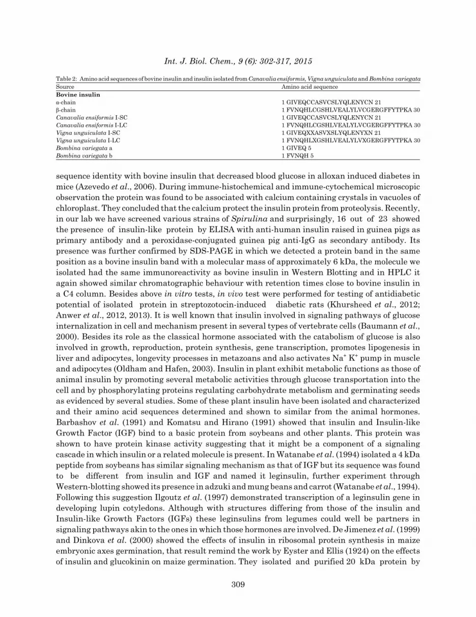
Table 2: Amino acid sequences of bovine insulin and insulin isolated from Canavalia ensiformis, Vigna unguiculata and Bombina variegataSource Amino acid sequenceBovine insulinα-chain 1 GIVEQCCASVCSLYQLENYCN 21β-chain 1 FVNQHLCGSHLVEALYLVCGERGFFYTPKA 30Canavalia ensiformis I-SC 1 GIVEQCCASVCSLYQLENYCN 21Canavalia ensiformis I-LC 1 FVNQHLCGSHLVEALYLVCGERGFFYTPKA 30Vigna unguiculata I-SC 1 GIVEQXXASVXSLYQLENYXN 21Vigna unguiculata I-LC 1 FVNQHLXGSHLVEALYLVXGERGFFYTPKA 30Bombina variegata a 1 GIVEQ 5Bombina variegata b 1 FVNQH 5
sequence identity with bovine insulin that decreased blood glucose in alloxan induced diabetes inmice (Azevedo et al., 2006). During immune-histochemical and immune-cytochemical microscopicobservation the protein was found to be associated with calcium containing crystals in vacuoles ofchloroplast. They concluded that the calcium protect the insulin protein from proteolysis. Recently,in our lab we have screened various strains of Spirulina and surprisingly, 16 out of 23 showed the presence of insulin-like protein by ELISA with anti-human insulin raised in guinea pigs asprimary antibody and a peroxidase-conjugated guinea pig anti-IgG as secondary antibody. Itspresence was further confirmed by SDS-PAGE in which we detected a protein band in the sameposition as a bovine insulin band with a molecular mass of approximately 6 kDa, the molecule weisolated had the same immunoreactivity as bovine insulin in Western Blotting and in HPLC itagain showed similar chromatographic behaviour with retention times close to bovine insulin ina C4 column. Besides above in vitro tests, in vivo test were performed for testing of antidiabeticpotential of isolated protein in streptozotocin-induced diabetic rats (Khursheed et al., 2012;Anwer et al., 2012, 2013). It is well known that insulin involved in signaling pathways of glucoseinternalization in cell and mechanism present in several types of vertebrate cells (Baumann et al.,2000). Besides its role as the classical hormone associated with the catabolism of glucose is alsoinvolved in growth, reproduction, protein synthesis, gene transcription, promotes lipogenesis inliver and adipocytes, longevity processes in metazoans and also activates Na+ K+ pump in muscleand adipocytes (Oldham and Hafen, 2003). Insulin in plant exhibit metabolic functions as those ofanimal insulin by promoting several metabolic activities through glucose transportation into thecell and by phosphorylating proteins regulating carbohydrate metabolism and germinating seedsas evidenced by several studies. Some of these plant insulin have been isolated and characterizedand their amino acid sequences determined and shown to similar from the animal hormones.Barbashov et al. (1991) and Komatsu and Hirano (1991) showed that insulin and Insulin-likeGrowth Factor (IGF) bind to a basic protein from soybeans and other plants. This protein wasshown to have protein kinase activity suggesting that it might be a component of a signalingcascade in which insulin or a related molecule is present. In Watanabe et al. (1994) isolated a 4 kDapeptide from soybeans has similar signaling mechanism as that of IGF but its sequence was foundto be different from insulin and IGF and named it leginsulin, further experiment throughWestern-blotting showed its presence in adzuki and mung beans and carrot (Watanabe et al., 1994).Following this suggestion Ilgoutz et al. (1997) demonstrated transcription of a leginsulin gene indeveloping lupin cotyledons. Although with structures differing from those of the insulin andInsulin-like Growth Factors (IGFs) these leginsulins from legumes could well be partners insignaling pathways akin to the ones in which those hormones are involved. De Jimenez et al. (1999)and Dinkova et al. (2000) showed the effects of insulin in ribosomal protein synthesis in maizeembryonic axes germination, that result remind the work by Eyster and Ellis (1924) on the effectsof insulin and glucokinin on maize germination. They isolated and purified 20 kDa protein by
309

Int. J. Biol. Chem., 9 (6): 302-317, 2015
G-50 Sephadex followed by affinity chromatography through a bovine insulin antibody-Sepharosecolumn from maize tissue showed IGF like activity in up-regulating maize germination andseedling growth. Maize IGF enhances phosphorylation of S6 ribosomal protein on the 40S ribosomalsubunit in a similar way as observed when bovine insulin is applied to maize axes duringgermination. Rapamycin, specific inhibitors of the insulin-stimulated phosphoinositide 3-kinase(PI3K) insulin-stimulated signal transduction pathway, prevented S6 phosphorylation. Anti-insulinantibody, heat treatment or trypsin digestion attenuated its activity which shows that this 20 kDaprotein from maize acts through a pathway similar to that of insulin or IGFs in animal tissues(Flores et al., 2001). Many proteins and subunit of insulin signaling pathways that has roles inmetazoan glucose transports are also present in plants and performed the same function astheir counterpart. Transmembrane protein Tyrosine kinase evidence for its presence in plantsand bacteria by Carpi et al. (2002) utilizing a bioinformatics approach by immunologicalanalysis, showing that ca. 3.5% of the protein kinase complement of A. thaliana is formed byTyr-phosphorylating enzymes. There are a number of molecule substrates for the insulin receptortyrosine kinase that provide an interface between the insulin receptor and downstream signalingmolecules such as Many insulin receptor substrate-1 (IRS-1), IRS-2, IRS-3, IRS-4, Shc and GAB-1that provide interface between the insulin receptor and downstream signaling molecules. The IRSseems to have been found although a zinc finger protein encoded by an Arabidopsis LSD1 geneshows high sequence homology in its carboxy-terminal with mammalian IRS-1 (Dietrich et al.,1997). Glucose transporter GLUT4 is expressed and distributed at high levels in muscle andadipocytes of animal cells. Plants express a number of monosaccharide (hexose/glucose)transporters (Williams et al., 2000) and one of these, which were cloned from sugar beet (Chiou andBush, 1996) guards a high sequence homology (36%) with a mammalian glucose transporter(Ibberson et al., 2000). Phosphatidyinositol-3-kinase (PI3K) is important part of thephosphoinositide pathway and most necessary step in the insulin stimulation of glucose transport.Hong and Verma (1994) have successfully cloned a PI3K cDNA from soybean that shows significantsequence homology to mammalian PI3K and is induced during nodule organogenesis. Presence ofPI3 and PI4-kinases in the outer envelope membrane of spinach chloroplasts was shown byBovet et al. (2001). Hexokinase a liver enzyme that phosphorylates glucose further metabolism(Brady et al., 1999). Plant hexokinase besides is also involved in sugar sensing processes that areregulated by glucose (Moore et al., 2003). MAPK pathway is evolutionarily conserved fromunicellular to eukaryote organisms and is one of the major signaling pathways involved inregulating many cellular activities and promotion of cellular growth (Agrawal et al., 2003). TheTOR (target of rapamycin) protein is a potential component of the phosphoinositide 3-kinase (PI3K)pathway in Drosophila and was also found in yeast (Oldham and Hafen, 2003) and Arabidopsis(Menand et al., 2002). The ribosomal protein S6 kinase involved in the regulation of cell growth andproliferation and is activated by the phosphoinositide-dependent protein kinase (PDK1) (Thomas,2002). Flores et al. (2001) result suggested that maize insulin-like growth factors ZmIGF-inducedsignal transduction pathway in maize, similar to the one described for insulin in animal tissuesthat selectively regulates protein synthesis. They also proved that ZmIGF was shown to stimulateS6 ribosomal protein phosphorylation on the 40 S ribosomal subunit and to enhance further thesynthesis of ribosomal proteins without a significant increase in general cytoplasmic proteinsynthesis.
MOLECULAR DYNAMICS APPROACHESThe MD simulations on insulin were performed at molecular mechanics level by GROMACS
4.6.5 packages (Van Der Spoel et al., 2005) and all atom functions by OPLS (optimized potential
310

Int. J. Biol. Chem., 9 (6): 302-317, 2015
for liquid simulation) in order to investigate its conformational profile (Khan et al., 2015;Anwer et al., 2015; Stephens et al., 2014; Gramany et al., 2015). Normally, the structure of insulinhas two helices. Insulin molecule was soaked in a cubic box of water molecules and spc216 templatewas used to solvate the protein and the charges were neutralized by the addition of Na+ and ClGions. The constant average fluctuation of temperature around 300 K suggested stable and accuratenature of the MD simulations performed. The average total energy and potential energy of insulinduring the simulations were -1233687.502 and -1458533.395 kJ molG1, respectively. The Radius ofgyration (Rg) analysis provided insight into the compactness of the protein during the simulations(Lobanov et al., 2008). The Rg values suggested that the insulin showed variations in itscompactness at different simulation time scale. This may be due to the presence of differentsmall chains of insulin. The overall Rg (nm) values found to be 2.25 nm for 5000 psec and 3.45 nmfor the rest of the periods. The steady and constant values during different time scales suggestedthe stable nature of insulin. This can be explained as the insulin has different Rg values at differenttime scales thus having a variation of compactness. The Root Mean Square Deviation (RMSD) isa measure of the deviation of the conformational stability of the proteins from back bone structureto the initial starting structure (Kuzmanic and Zagrovic, 2010). Initially, the rms values for insulinwere found to be <0.5 nm but the values increase as the simulation time scales increases. After 6nsec, the rmsd values become constant and steady suggesting the stable nature. The MDsimulation studies of insulin suggesting two constant values at different time scales due to differentchains present in the proteins or different conformations at different time scales of simulations(Fig. 2).
Fig. 2(a-d): (a) Average total energy, (b) Average potential energy (c) Radius of gyration and(d) RMS deviation values of insulin as a function of time. Black colour represents RMSdeviations obtained at 300 K. The duration of each simulation was 10 nsec
311
-1.22e+006
-1.225e+006
-1.23e+006
-1.235e+006
-1.24e+006
kJ m
olG1
(a) -1.45e+006
-1.455e+006
-1.46e+006
-1.465e+006
-1.47e+006
kJ m
olG1
4.0
3.5
3.0
2.5
2.0
4
3
2
1
0
Rg
(nm
)
RM
SD (n
m)
0 5000 10000 15000 20000 Time (p sec)
0 5000 10000 15000 20000 Time (n sec)
(b)
(c) (d)

Int. J. Biol. Chem., 9 (6): 302-317, 2015
Fig. 3(a-b):Relative folding energy calculations at each pH for the insulin like protein obtainedfrom (a) Canavalia ensiformis and (b) Vigna unguiculata, respectively
PROTEIN IONIZATION AND RELATIVE FOLDING ENERGYThe pKa values of the amino acid side chain aid in defining the pH-dependent characteristics
of a protein. The combination of the Generalized Born (GB) and Iterative Mobile Clustering (IMC)approaches utilized for pK calculations and protein ionization. The pKa and titration curvesfor each one of the titratable amino acid residues present in the Canavalia ensiformisand Vigna unguiculata insulin were calculated using modules present in Discovery studio 4.0(Spassov and Yan, 2008). The relative folding energy suggested that the insulin like proteinobtained from Canavalia ensiformis and Vigna unguiculata has maximum stability at neutral pHi.e., similar to pH of human blood (Fig. 3).
CONCLUSIONInsulin-like materials whose activity and structure resemblance to animal insulin hormones
thought to be present in plant, algae and fungi. Some of these have been isolated, characterized and
312
0 0.
4 0.
8 1.
2 1.
6 2.
0 2.
4 2.
8 3.
2 3.
6 4.
0 4.
4 4.
8 5.
2 5.
6 6.
0 6.
4 6.
8 7.
2 7.
6 8.
0 8.
4 8.
8 9.
2 9.
6 10
.0
10.4
10
.8
11.2
11
.6
12.0
12
.4
12.8
13
.2
pH
0
-2
-2
-3
-4
Rel
ativ
e fo
ldin
g en
ergy
(b)
0 0.
4 0.
8 1.
2 1.
6 2.
0 2.
4 2.
8 3.
2 3.
6 4.
0 4.
4 4.
8 5.
2 5.
6 6.
0 6.
4 6.
8 7.
2 7.
6 8.
0 8.
4 8.
8 9.
2 9.
6 10
.0
10.4
10
.8
11.2
11
.6
12.0
12
.4
12.8
13
.2
pH
0
-2
-4
-6
-8
Rel
ativ
e fo
ldin
g en
ergy
(a)

Int. J. Biol. Chem., 9 (6): 302-317, 2015
their biological properties were shown to share similarities with animal insulin. They are activemolecules, effectively communicating with animal cell’s signaling mechanism and also mediateinsulin action. The simulations studies suggesting stable nature of protein with differentconformations at different time scales of simulation. The relative folding energy of insulin likeprotein from plant source was thought to have maximum stability at neutral pH i.e., similar to pHof human blood.
REFERENCESAbel, J.J., 1926. Crystalline insulin. Proc. Natl. Acad. Sci. USA., 12: 132-136.Adams, M.J., T.L. Blundell, E.J. Dodson, G.G. Dodson and M. Vijayan et al., 1969. Structure of
rhombohedral 2 zinc insulin crystals. Nature, 224: 491-495.Agrawal, G.K., H. Iwahashi and R. Rakwal, 2003. Rice MAPKs. Biochem. Biophys. Res. Commun.,
302: 171-180.Anwer, R., S. Khursheed and T. Fatma, 2012. Detection of immunoactive insulin in Spirulina.
J. Applied Phycol., 24: 583-591.Anwer, R., A. Alam, S. Khursheed, S.M. Kashif, H. Kabir and T. Fatma, 2013. Spirulina: Possible
pharmacological evaluation for insulin-like protein. J. Applied Phycol., 25: 883-889.Anwer, K., R. Sonani, D. Madamwar, P. Singh and F. Khan et al., 2015. Role of N-terminal residues
on folding and stability of C-phycoerythrin: Simulation and urea-induced denaturation studies.J. Biomol. Struct. Dyn., 33: 121-133.
Assaad, F.F., 2001. Of weeds and men: What genomes teach us about plant cell biology.Curr. Opin. Plant Biol., 4: 478-487.
Azevedo, C.R., F.M. Maciel, L.B. Silva, A.T.S. Ferreira and M. da Cunha et al., 2006. Isolationand intracellular localization of insulin-like proteins from leaves of Bauhinia variegata.Braz. J. Med. Biol. Res., 39: 1435-1444.
Banting, F.G., C.H. Best, J.B. Collip, W.R. Campbell, A.A. Fletcher, J.J.R. Macleod and EC. Noble,1922. The effect produced on diabetes by extracts of pancreas. Trans. Assoc. Am. Phys.,37: 337-347.
Barbashov, S.F., T.A. Egorov and V.M. Kochkina, 1991. [Isolation and characteristics ofinsulin-binding proteins from soybeans]. Bioorg. Chem., 17: 421-423, (In Russian).
Barnett, D.M. and L.P. Krall, 2005. The History of Diabetes. In: Joslin's Diabetes Mellitus,4th Edn., Kahn, C.R., G.C. Weir, G.L. King, A.M. Jacobson, A.C. Moses and R.J. Smith (Eds.).,Wolters Kluwer Company, USA., pp: 3-15.
Barron, M., 1920. The relation of the islets of Langerhans to diabetes with special reference tocases of pancreatic lithiasis. Surg. Gynecol. Obstetr., 31: 437-448.
Baumann, C.A., V. Ribon, M. Kanzaki, D.C. Thurmond and S. Mora et al., 2000. CAP defines asecond signalling pathway required for insulin-stimulated glucose transport. Nature,407: 202-207.
Bernard, C., 1865. Introduction a l'Etude de la Medecine Experimentale. Balliere, Paris.Best, C.H. and D.A. Scott, 1923. Possible sources of insulin. J. Metab. Res., 3: 177-179.Best, C.H., 1924. Recent work on insulin. Endocrinology, 8: 617-629.Bliss, M., 1983. The Discovery of Insulin. University of Chicago Press, Chicago.Bovet, L., M.O. Muller and P.A. Siegenthaler, 2001. Three distinct lipid kinase activities
are present in spinach chloroplast envelope membranes: Phosphatidylinositol phosphorylationis sensitive to wortmannin and not dependent on chloroplast ATP. Biochem. Biophys.Res. Commun., 289: 269-275.
313
Int. J. Biol. Chem., 9 (6): 302-317, 2015
Brady, M.J., J.E. Pessin and A.R. Saltiel, 1999. Spatial compartmentalization in the regulation ofglucose metabolism by insulin. Trends Endocrinol. Metab., 10: 408-413.
Carpi, A., G. Di Maira, M. Vedovato, V. Rossi and T. Naccari et al., 2002. Comparative proteomebioinformatics: Identification of a whole complement of putative protein tyrosine kinases in themodel flowering plant Arabidopsis thaliana. Proteomics, 2: 1494-1503.
Chiou, T.J. and D.R. Bush, 1996. Molecular cloning, immunochemical localization to the vacuoleand expression in transgenic yeast and tobacco of a putative sugar transporter from sugar beet.Plant Physiol., 110: 511-520.
Collier, E., A. Watkinson, C.F. Cleland and J. Roth, 1987. Partial purification and characterizationof an insulin-like material from spinach and Lemna gibba G3. J. Biol.Chem., 262: 6238-6247.
Collip, J.B., 1923. Glucokinin. A new hormone present in plant tissue: Preliminary paper.J. Biol. Chem., 56: 513-543.
De Jimenez, E.S., E. Beltran-Pena and A. Ortiz-Lopez, 1999. Insulin-stimulated ribosomal proteinsynthesis in maize embryonic axes during germination. Physiol. Plant., 105: 148-154.
De Meyer, J., 1909. Action de la secretion interne du pancreas sur differents organes et enparticulier sur la secretion renale. Arch. Fisiol., 7: 96-99.
Dickinson, W.H., 1875. Diabetes, in his Diseases of the Kidney. Longmans Green and Co., London.Dietrich, R.A., M.H. Richberg., R. Schmidt, C. Dean and J.L. Dangl, 1997. A novel zinc finger
protein is encoded by the Arabidopsis LSD1 gene and functions as a negative regulator of plantcell death. Cell, 88: 685-694.
Dinkova, T.D., R. Aguilar and D.J.E. Sanchez, 2000. Expression of maize eukaryotic initiationfactor (eIF) iso4E is regulated at the translational level. Biochem. J., 351: 825-831.
Dobson, M., 1776. Experiments and observations on the urine in diabetes. Med. Obs. Inquiries,5: 298-316.
Ellis, M.M. and W.H. Eyster, 1923. Some effects of insulin and glucokinin on maize seedlings.Science, 58: 541-542.
Eyster, W.H. and M.M. Ellis, 1924. Growth of maize seedlings as affected by glucokinin and insulin.J. Gen. Physiol., 6: 653-670.
Flores, C.G., R. Aguilar, H.R. de la Cruz, M. Albores and E.S. de Jimenez, 2001. A maizeinsulin-like growth factor signals to a transduction pathway that regulates protein synthesisin maize. Biochem. J., 358: 95-100.
Frank, L.L., 1957. Diabetes mellitus in the text book of old Hindu medicine. Am. J. Gastroenterol.,27: 76-95.
Gramany, V., F.I. Khan, A. Govender, K. Bisetty, S. Singh and K. Permaul, 2015. Cloning,expression and molecular dynamics simulations of a xylosidase obtained from Thermomyceslanuginosus. J. Biomol. Struct. Dyn., 10.1080/07391102.2015.1089186
Gualandi-Signorini, A.M. and G. Giorgi, 2001. Insulin formulations: A review. Eur. Rev. Med.Pharmacol. Sci., 5: 73-84.
Hagedorn, H.C., B.N. Jensen, N.B. Krarup and I. Wodstrup, 1936. Protamine insulinate.J. Am. Med. Assoc., 106: 177-180.
Hallas-Moller, K., 1956. The lente insulin. Diabetes, 5: 7-14.Hirsch, I.B., 2005. Insulin analogues. N. Engl. J. Med., 352: 174-183.Hong, Z. and D.P. Verma, 1994. A phosphatidylinositol 3-kinase is induced during soybean
nodule organogenesis and is associated with membrane proliferation. Proc. Natl. Acad. Sci.USA., 91: 9617-9621.
314
Int. J. Biol. Chem., 9 (6): 302-317, 2015
Ibberson, M., M. Uldry and B. Thorens, 2000. GLUTX1, a novel mammalian glucose transporterexpressed in the central nervous system and insulin-sensitive tissues. J. Biol. Chem.,275: 4607-4612.
Ilgoutz, S.C., N. Knittel, J.M. Lin, S. Sterle and K.R. Gayler, 1997. Transcription of genes forconglutin γ and a leginsulin-like protein in narrow-leafed lupin. Plant Mol. Biol., 34: 613-627.
Jonak, C. and H. Hirt, 2002. Glycogen synthase kinase 3/SHAGGY-like kinases in plants:An emerging family with novel functions. Trends Plant Sci., 7: 457-461.
Khan, F.I., A. Govender, K. Permaul, S. Singh and K. Bisetty, 2015. Thermostable chitinase II fromThermomyces lanuginosus SSBP: Cloning, structure prediction and molecular dynamicssimulations. J. Theoret. Biol., 374: 107-114.
Khanna, P., T.N. Nag, S.C. Jain and S. Mohan, 1974. Extraction of insulin from a plant source.Proceedings of the 3rd International congress on plant tissue and cell cultures, July 21-26, 1974Leicester, UK,.
Khanna, P., T.N. Nag, S. Chandrajaia and S.V. Mohan, 1976. Process for isolation of insulin fromplant source. United States Patent, 3: 945-988.
Khursheed, S., R. Anwer, S. Zutshi and T. Fatma, 2012. Screening of photosynthetic O2-evolvingprokaryotes for an insulin-like antigen. J. Phycol., 48: 243-245.
Kim, J.W., M. Rhee, J.H. Park, H. Yamaguchi and K. Sasaki et al., 2015. Chronic effects ofneuroendocrine regulatory peptide (NERP-1 and-2) on insulin secretion and gene expressionin pancreatic β-cells. Biochem. Biophys. Res. Commun., 457: 148-153.
Kleiner, I.S., 1919. The action of intravenous injections of pancreas emulsions in experimentaldiabetes. J. Biol. Chem., 40: 153-170.
Komatsu, S. and H. Hirano, 1991. Plant basic 7 S globulin-like proteins have insulin andinsulin-like growth factor binding activity. FEBS Lett., 294: 210-212.
Kuzmanic, A. and B. Zagrovic, 2010. Determination of ensemble-average pairwise rootmean-square deviation from experimental B-factors. Biophys. J., 98: 861-871.
Lancereaux, E., 1877. Notes et reflixions a propos de deux cas de diabete sucre avec alteration dupancreas. Bull. Acad. Med. Paris, 6: 1215-1240.
Langerhans, P., 1868. Ueber die nerven der menschlichen haut. Archiv Pathologische AnatomiePhysiologie Klinische Medicin, 44: 325-325.
Lasker, S.P., C.S. McLachlan, L. Wang, S.M.K. Ali and H.F. Jelinek, 2010. Discovery, treatmentand management of diabetes. J. Diabetol., Vol. 1, No. 1.
Le Roith, D., J. Shiloach, J. Roth and M.A. Lesniak, 1980. Evolutionary origins of vertebratehormones: Substances similar to mammalian insulin are native to unicellular eukaryotes. Proc.Natl. Acad. Sci. USA., 77: 6184-6188.
LeRoith, D., J. Shiloach, J. Roth and M.A. Lesniak, 1981. Insulin or a closely related molecule isnative to Escherichia coli. J. Biol. Chem., 256: 6533-6536.
LeRoith, D., J. Shiloach, R. Heffron, C. Rubinovitz, R. Tanenbaum and J. Roth, 1985.Insulin-related material in microbes: Similarities and differences from mammalian insulin.Can. J. Biochem. Cell Biol., 63: 839-849.
Lobanov, M.Y., N.S. Bogatyreva and O.V. Galzitskaya, 2008. Radius of gyration as an indicator ofprotein structure compactness. Mol. Biol., 42: 701-706.
MacCracken, J. and D. Hoel, 1997. From ants to analogues. Puzzles and promises in diabetesmanagement. Postgrad. Med., 101: 138-140, 143-145, 149-150.
Macleod, J.J.R., 1924. Insulin. Physiol. Rev., 4: 21-67.
315

Int. J. Biol. Chem., 9 (6): 302-317, 2015
Menand, B., T. Desnos, L. Nussaume, F. Berger, D. Bouchez, C. Meyer and C. Robaglia, 2002.Expression and disruption of the Arabidopsis TOR (target of rapamycin) gene. Proc. Natl. Acad.Sci. USA., 99: 6422-6427.
Mohan, V., S. Sandeep, R. Deepa, B. Shah and C. Varghese, 2007. Epidemiology of type 2 diabetes:Indian scenario. Indian J. Med. Res., 125: 217-230.
Moore, B., L. Zhou, F. Rolland, Q. Hall and W.H. Cheng et al., 2003. Role of the arabidopsis glucosesensor HXK1 in nutrient, light, and hormonal signaling. Science, 300: 332-336.
Ng, T.B., C.M. Wong, W.W. Li and H.W. Yeung, 1986. Insulin-like molecules in Momordicacharantia seeds. J. Ethnopharmacol., 15: 107-117.
Oldham, S. and E. Hafen, 2003. Insulin/IGF and target of rapamycin signaling: A TOR de force ingrowth control. Trends Cell Biol., 13: 79-85.
Pefiffer, E. and W. Kerner, 1984. Biosynthetic Human Insulin. In: Diabetes Mellitus in DevelopingCountry. Bajaj, J.S. (Ed.)., Metita Offset Works, New Delhi, India.
Pickup, J.C. and G. Williams, 2003. The History of Diabetes Mellitus in Textbook of Diabetes.Blackwell Science Limited, Oxford, London.
Rollo, J., 1809. An account of two cases of the diabetes mellitus, with remarks as thry arose duringthe progress of the cure. London: C. Dilly, 1797. (A 2nd Edition with More Cases was Publishedin 1798), pp: 384.
Rubin, R.R., P. Ciechanowski, L.E. Egede, E.H.B. Lin and P.J. Lustman, 2004. Recognizing andtreating depression in patients with diabetes. Curr. Diab. Rep., 4: 119-125.
Rutter, G.A., T.J. Pullen, D.J. Hodson and A. Martinez-Sanchez, 2015. Pancreatic β-cell identity,glucose sensing and the control of insulin secretion. Biochem. J., 466: 203-218.
Ryan, C.A., G. Pearce, J. Scheer and D.S. Moura, 2002. Polypeptide Hormones. Plant Cell.,14: s251-s264.
Sanders, L.J., 2002. From Thebes to Toronto and the 21st century: An incredible journey. DiabetesSpectr., 15: 56-60.
Schafer, E., 1914. An introduction to the study of the endocrine glands and internal secretions.Stanford University Palo Alto, CA., pp: 84-86.
Sheng, Q., H. Yao, H. Xu, X. Ling and T. He, 2004. [Isolation of plant insulin from Momordicacharantia seeds by gel filtration and RP-HPLC]. Zhong Yao Cai, 27: 414-416.
Silva, L.B., S.S.S. Santos, C.R. Azevedo, M.A.L. Cruz and T.M. Venancio et al., 2002. The leavesof green plants as well as a cyanobacterium, a red alga and fungi contain insulin-like antigens.Braz. J. Med. Biol. Res., 35: 297-303.
Spassov, V.Z. and L. Yan, 2008. A fast and accurate computational approach to protein ionization.Protein Sci., 17: 1955-1970.
Steiner, D.F. and P.E. Oyer, 1967. The biosynthesis of insulin and a probable precursor of insulinby a human islet cell adenoma. Proc. Natl. Acad. Sci. USA., 57: 473-480.
Steiner, D.F. and D.E. James, 1992. Cellular and molecular biology of the beta cell. Diabetologia,2: S41-S48.
Stephens, D.E., F.I. Khan, P. Singh, K. Bisetty, S. Singh and K. Permaul, 2014. Creationof thermostable and alkaline stable xylanase variants by DNA shuffling. J. Biotechnol.,187: 139-146.
Tam, C.H.T., Y. Wang, J. Luan, H.M. Lee and A.O.Y. Luk et al., 2015. Non-linear relationshipbetween birthweight and cardiometabolic risk factors in Chinese adolescents and adults.Diabetic Med., 32: 220-225.
316
Int. J. Biol. Chem., 9 (6): 302-317, 2015
Thomas, G., 2002. The S6 kinase signaling pathway in the control of development and growth.Biol. Res., 35: 305-313.
Tipton, C.M., 2008. Susruta of India, an unrecognized contributor to the history of exercisephysiology. J. Applied Physiol., 104: 1553-1556.
Van Der Spoel, D., E. Lindahl, B. Hess, G. Groenhof, A.E. Mark and H.J.C. Berendsen, 2005.GROMACS: Fast, flexible and free. J. Comput. Chem., 26: 1701-1718.
Venancio, T.M., A.E.A. Oliveira, L.B. Silva, O.L.T. Machado, K.V.S. Fernandes and J. Xavier-Filho,2003. A protein with amino acid sequence homology to bovine insulin is present in the legumeVigna unguiculata (cowpea). Braz. J. Med. Biol. Res., 36: 1167-1173.
Von Mering, J. and O. Minkowski, 1890. Diabetes mellitus nach pancreas extripation. Arch. Exper.Path Pharmacol. Leipzig, 26: 371-381.
Watanabe, Y., S.F. Barbashov, S. Komatsu, A.M. Hemmings, M. Miyagi, S. Tsunasawa andH. Hirano, 1994. A peptide that stimulates phosphorylation of the plant insulin-binding proteinisolation, primary structure and cDNA cloning. Eur. J. Biochem., 224: 167-172.
Wild, S., G. Roglic, A. Green, R. Sicree and H. King, 2004. Global prevalence of diabetes: Estimatesfor the year 2000 and projections for 2030. Diabetes Care, 27: 1047-1053.
Williams, L.E., R. Lemoine and N. Sauer, 2000. Sugar transporters in higher plants: A diversityof roles and complex regulation. Trends Plant Sci., 5: 283-290.
Willis, T., 1674. Pharmaceutice Rationalis or An Exercitation of the Operation of Medicines inHumane Bodies. 2nd Edn., Trans. Samuel Pordage, Printed for T. Dring, C. Harper andJ. Leigh, London.
Xavier-Filho, J., A.E.A. Oliveira, L.B. da Silva, C.R. Azevedo and T.M. Venancio et al., 2003. Plantinsulin or glucokinin: A conflicting issue. Braz. J. Plant Physiol., 15: 67-78.
Yalow, R.S. and S.A. Berson, 1959. Assay of plasma insulin in human subjects by immunologicalmethods. Nature, 184: 1648-1649.
317