raman microspectroscopy detection and characterisation of
TRANSCRIPT
Polymers 2022, 14, 2700. https://doi.org/10.3390/polym14132700 www.mdpi.com/journal/polymers
Article
Raman Microspectroscopy Detection and Characterisation of
Microplastics in Human Breastmilk
Antonio Ragusa 1, Valentina Notarstefano 2,*, Alessandro Svelato 3, Alessia Belloni 2, Giorgia Gioacchini 2,
Christine Blondeel 3, Emma Zucchelli 3, Caterina De Luca 3, Sara D’Avino 3, Alessandra Gulotta 4,
Oliana Carnevali 2 and Elisabetta Giorgini 2
1 Department of Obstetrics and Gynecology, Università Campus Bio Medico di Roma,
Via Alvaro del Portillo, 200, 00128 Roma, Italy; [email protected] 2 Department of Life and Environmental Sciences, Università Politecnica delle Marche,
Via Brecce Bianche, 60131 Ancona, Italy; [email protected] (A.B.);
[email protected] (G.G.); [email protected] (O.C.); [email protected] (E.G.) 3 Department of Obstetrics and Gynecology, San Giovanni Calibita Fatebenefratelli Hospital, Isola Tiberina,
Via di Ponte Quattro Capi, 39, 00186 Roma, Italy; [email protected] (A.S.);
[email protected] (C.B.); [email protected] (E.Z.);
[email protected] (C.D.L.); [email protected] (S.D.) 4 Department of Obstetrics and Gynecology, Università degli Studi di Sassari, Viale S. Pietro,
07100 Sassari, Italy; [email protected]
* Correspondence: [email protected]; Tel.: +39-0712204723
Abstract: The widespread use of plastics determines the inevitable human exposure to its by-prod-
ucts, including microplastics (MPs), which enter the human organism mainly by ingestion, inhala-
tion, and dermal contact. Once internalised, MPs may pass across cell membranes and translocate
to different body sites, triggering specific cellular mechanisms. Hence, the potential health impair-
ment caused by the internalisation and accumulation of MPs is of prime concern, as confirmed by
numerous studies reporting evident toxic effects in various animal models, marine organisms, and
human cell lines. In this pilot single-centre observational prospective study, human breastmilk sam-
ples collected from N. 34 women were analysed by Raman Microspectroscopy, and, for the first
time, MP contamination was found in 26 out of 34 samples. The detected microparticles were clas-
sified according to their shape, colour, dimensions, and chemical composition. The most abundant
MPs were composed of polyethylene, polyvinyl chloride, and polypropylene, with sizes ranging
from 2 to 12 µm. MP data were statistically analysed in relation to specific patients’ data (age, use
of personal care products containing plastic compounds, and consumption of fish/shellfish, bever-
ages, and food in plastic packaging), but no significant relationship was found, suggesting that the
ubiquitous MP presence makes human exposure inevitable.
Keywords: microplastics; human breastmilk; Raman microspectroscopy; infants’ nutrition
1. Introduction
The global production of plastics has reached the impressive amount of more than
350 million tons per year. This is the result of the massive demand for this material, which
has been considered, until now, the golden choice in terms of durability, usability, and
versatility for a huge variety of applications and consumer products [1–3]. This wide-
spread use of plastics also led to their accumulation in landfills and in the natural envi-
ronment. In fact, as a consequence of the extensive production and employment of single-
use products, which represent >40% of manufactured plastics, 250,000 tons of plastic litter
is estimated to be floating in the oceans [4]. Although several countries are introducing
new regulations on plastic waste management and recycling strategies, it has to be noted
Citation: Ragusa, A.; Notarstefano, V.;
Svelato, A.; Belloni, A.; Gioacchini, G.;
Blondeel, C.; Zucchelli, E.; De Luca, C.;
D’Avino, S.; Gulotta, A.; et al. Raman
Microspectroscopy Detection and
Characterisation of Microplastics in
Human Breastmilk. Polymers 2022, 14,
2700. https://doi.org/10.3390/
polym14132700
Academic Editors: Xinfeng Wei,
Emmanuel Richaud and Andrew M.
Booth
Received: 13 June 2022
Accepted: 29 June 2022
Published: 30 June 2022
Publisher’s Note: MDPI stays neu-
tral with regard to jurisdictional
claims in published maps and institu-
tional affiliations.
Copyright: © 2022 by the authors. Li-
censee MDPI, Basel, Switzerland.
This article is an open access article
distributed under the terms and con-
ditions of the Creative Commons At-
tribution (CC BY) license (https://cre-
ativecommons.org/licenses/by/4.0/).
Polymers 2022, 14, 2700 2 of 13
that in 2018 in Europe, only 32.5% of post-consumer waste plastic was recycled, while
24.9% was accumulated in landfills [5].
Environmental plastic contamination derives from several factors, including mis-
managed plastic waste, fishing nets in the sea, and different household and commercial
activities, such as washing synthetic textiles, road markings, tires, marine coatings, per-
sonal care products, and plastic pellets [6,7]. In particular, after being released into the
environment, plastic products undergo a degradation process caused by the action of at-
mospheric agents, such as waves, abrasion, UV radiation, and photo-oxidation, in combi-
nation with biological processes, which leads to the formation of microplastics (MPs) [8].
MPs range from 5 millimetres to 100 nanometres and are classified as primary or second-
ary based on their source of release into the environment: primary MPs are purposely
manufactured at sizes <5 mm to be employed for commercial purposes (such as glitter in
cosmetic products and microbeads in cleansers, scrubs, and dish scrubbing pads), while
secondary MPs are generated by the previously described environmental degradation
processes of larger plastic items [9,10].
The ubiquitous occurrence of MPs in the environment determines inevitable human
exposure, mainly by three routes: ingestion, inhalation, and dermal contact. Among all of
them, ingestion is considered the major route, with an estimated intake of 39 52 thou-
sand MPs per person per year [11–13]. Once internalised, MPs may pass across cell mem-
branes [14], followed by accumulation or elimination by the onset of specific cellular
mechanisms. All of these processes are mainly related to MPs’ size, which cannot exceed
10–15 µm.
The potential health impairment caused by the internalisation and accumulation of
MPs is of prime concern. Although information is still lacking on this topic, several studies
reported evident toxic effects in various animal models, marine organisms, and human
cell lines [15–17], showing that MPs, once internalised, are not inert as previously sup-
posed and likely trigger local or systemic responses.
Given the strong concern related to the effects of MPs on animal and human health,
the use of reliable and objective techniques for MP detection and characterisation is cru-
cial. Among all of the exploited techniques, Raman Microspectroscopy (RMS) can be con-
sidered the gold standard, since it lets researchers characterise not only the morphological
features of microparticles but also their chemical composition in terms of both polymer
matrices and pigments. Moreover, RMS presents the advantage of enabling the analysis
of MPs as small as 2 µm directly on filtration membranes, thanks to the high potential of
light scattering [18–20]. Recently, our research group, for the first time, detected the pres-
ence of MPs in human placenta samples; this study, carried out by Raman Microspectros-
copy, received extensive attention, since the delicate role played by this organ may be
perturbed by the presence of MPs [21].
Based on these impressive results, we decided to investigate the contamination of
microplastics in breastmilk to assess another MP exposure route in the extremely vulner-
able population of infants. For this purpose, in the present study, milk samples collected
from 34 consenting patients were analysed by Raman Microspectroscopy, and, for the first
time, in most of the analysed samples, the presence of MPs was detected. The relevance
of this research lies in breastmilk being the gold standard for infants’ nutrition. Moreover,
it reflects both the mother’s and infant’s postnatal exposure, and hence, it represents an
optimal matrix for contaminant biomonitoring [22]. In fact, milk consists of protein and
fat globules in a carbohydrate-based suspension and represents a favourable environment
for the lipophilic nature of MPs and other chemicals. In this regard, it is noteworthy that
several studies reported the contamination of breastmilk by phthalates, heavy metals, and
perfluorinated compounds [23–25].
Polymers 2022, 14, 2700 3 of 13
2. Materials and Methods
2.1. Cohort Selection
This was a pilot observational descriptive study in a prospective and single-centre
cohort. It was approved by the Ethical Committee Lazio 1 (Protocol N. 708/CE Lazio 1; 24
May 2021), and it was carried out in full accordance with ethical principles, including The
Code of Ethics of the World Medical Association (Declaration of Helsinki) for experiments
involving humans. A dedicated cohort of N. 34 patients, all characterised by pregnancies
without complications, was enrolled at Ospedale Fatebenefratelli Isola Tiberina (Rome,
Italy). Exclusion criteria were: (a) medically prescribed special diets within 4 weeks prior
to delivery; (b) diarrhoea or severe constipation within 2 weeks prior to delivery; (c) use
of antibiotics within 2 weeks prior to delivery; (d) use of drugs affecting intestinal resorp-
tion, e.g., activated charcoal or cholestyramine, within 2 weeks prior to delivery; (e) diag-
nosis of a gastrointestinal pathology, e.g., ulcerative colitis or Crohn’s disease, except for
appendectomy; (f) cancer, HIV, or any other serious illness demanding medical treatment;
(g) invasive or abrasive dental treatments within 2 weeks prior to delivery; (h) alcohol
abuse (defined as Alcohol Use Disorder Identification test >10); and (i) current or recent
(within the previous 4 weeks) participation in a clinical trial. Patients who decided to par-
ticipate were asked to sign an informed consent form. Patients were also asked to fill in a
questionnaire to record their food consumption, with a special focus on fish, shellfish, and
foods employing packaging, and the use of personal care products from 7 days before the
expected date of delivery to 7 days after.
2.2. Sample Collection
Breastmilk samples were collected 1 week after delivery at the Department of Obstet-
rics and Gynaecology of San Giovanni Calibita Fatebenefratelli Hospital (Rome, Italy).
Patients were guided on a manual expression procedure, recommended by the World
Health Organization and described in a document released by the Italian Ministry of
Health [26], which uses manual expression to obtain the maximum milk output and to
avoid pain or damage to the breast tissue. No breast pumps were allowed to avoid con-
tamination from its plastic components. Briefly, the manual expression procedure consists
of cupping the breast with one hand, with the other forming a C-shape with the thumb
and the forefinger, 3–4 cm from the base of the nipple; then, pressure is applied by pushing
towards the ribcage, squeezing with the thumb and forefinger, and finally releasing the
pressure. The sequence of pressure, squeeze, and release was repeated until obtaining an
adequate amount of milk. Milk samples were placed into glass flasks, weighed, and then
stored at −20 °C until processing. Each sample contained an average amount of 4.16 ± 1.73
g of breastmilk.
2.3. Sample Digestion and Filtration
In order to remove organic components from milk samples, a digestion protocol was
set up and performed at the Laboratory of Vibrational Spectroscopy, Department of Life
and Environmental Sciences, Università Politecnica delle Marche (Ancona, Italy). A 10%
KOH solution prepared using 1.6 µm filtered deionised water and KOH tablets (Sigma-
Aldrich) was added to each flask in a ratio of sample to KOH of 1:10 (w/v). Flasks were
sealed and incubated at 40 °C for 48 h [27]. Digestates were then filtered through a 1.6 µm
pore-size filter membrane (Whatman GF/A) by a vacuum pump connected to a filter fun-
nel. Filter membranes were dried at room temperature and stored in glass Petri dishes
until Raman Microspectroscopy (RMS) analysis.
2.4. Detection and Identification of MPs by Raman Microspectroscopy
RMS analysis was performed by using an XploRA Nano Raman Microspectrometer
(Horiba Scientific) at the ARI Laboratory of Università Politecnica delle Marche (Ancona,
Italy). All filter membranes, including those deriving from the procedural blanks, were
Polymers 2022, 14, 2700 4 of 13
inspected by visible light using a ×10 objective (Olympus MPLAN10×/0.25). The detected
MPs were morphologically characterised by a ×100 objective (Olympus MPLAN100×/0.90)
and then directly analysed on the filter by RMS (spectral range 200–1800 cm−1, 532 nm or
785 nm laser diode, 600 lines per mm grating). Spectra were dispersed onto a 16-bit dy-
namic range Peltier-cooled CCD detector; the spectrometer was calibrated to the 520.7
cm−1 line of silicon prior to spectral acquisition. To reduce noise and enhance spectrum
quality, raw Raman spectra were subjected to polynomial baseline correction and vector
normalisation (Labspec 6 software, Horiba Scientific). The polymer matrix of the detected
particles was identified by comparing the collected Raman spectra with spectral libraries
of polymers and pigments obtained by measuring standard polymers/compounds
(KnowItAll software, John Wiley & Sons, Inc., Hoboken, NJ, USA) [28,29]. Similarities of
more than 80 of the Hit Quality Index (HQI) were considered satisfactory.
2.5. Quality Assurance and Control (QA/QC)
Efforts were adopted to avoid microplastic contamination during sample collection,
storage, processing, and analysis. To this aim, a plastic-free protocol was adopted during
all phases of the experiment, and a dedicated room was used for the digestion of milk
samples, filtration, and RMS analysis steps. Routinely employed plastic tools were re-
placed with sterilised glass ones. Cotton laboratory coats and single-use latex gloves were
worn during all phases of the experiment. All liquids, including ethanol for cleaning and
deionised water for cleaning and preparation of all solutions, were filtered through 1.6
µm pore-size filter membranes (Whatman GF/A). Work surfaces were thoroughly washed
with 70% ethanol prior to starting all procedures and during the experimental time. Glass-
ware and instruments, including scissors and tweezers, were washed using dishwashing
liquid, triple rinsed with 70% ethanol, and finally rinsed with 1.6 µm filtered deionised
water.
Moreover, environmental and procedural blanks were prepared and thoroughly an-
alysed to detect microplastic contamination deriving from the laboratory environment
and from other external sources. As regards environmental blanks, a filter membrane
soaked with 1.6 µm filtered deionised water was placed into an uncovered Petri dish and
positioned each day in the above-mentioned dedicated room. A procedural blank was also
prepared together with every batch of samples following the exact same procedure as
samples, but without adding milk. The filters deriving from environmental and proce-
dural blanks were first inspected by stereomicroscope.
2.6. Statistical Analysis
Data analysis was performed by using the statistical software package Prism6
(Graphpad Software, Inc., San Diego, CA, USA). Normality was checked by the D’Ago-
stino and Pearson omnibus normality test. Chi-square test, Student’s t-test, and one-way
analysis of variance (ANOVA) were performed to compare data accordingly. The signifi-
cance threshold was set at p < 0.05.
3. Results
In the present study, N. 34 breastmilk samples were investigated by RMS for the
presence of microplastics. MP contamination was found in 26 out of 34 women. As regards
QA/QC protocols, the analysis of environmental (N. 14) and procedural (N. 9) blanks was
performed. In the environmental blanks, only fibres, for a total of N. 16, ranging from 571
µm to 3000 µm, were found. Conversely, no MP contamination was found in the filters
from the procedural blanks. Given the dimensions of the fibres, which are not compatible
with the translocation into breastmilk, and the absence of fibres in the analysed milk sam-
ples, there was no need to blank-correct the results.
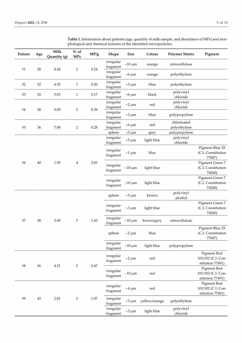
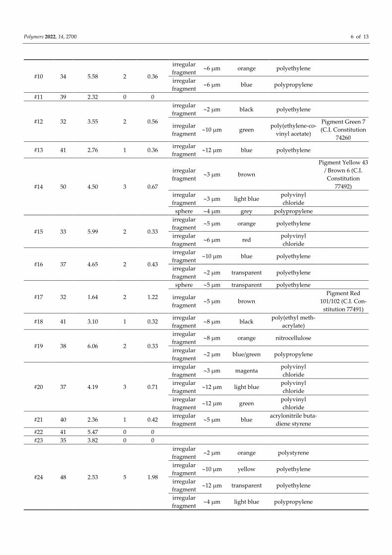
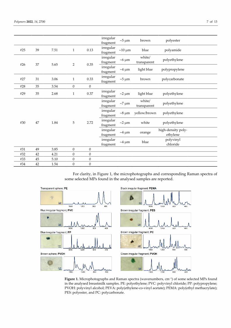
All details about the identified microparticles (such as morphology, dimensions, col-
our, polymer matrix, and pigment) are listed in Table 1.
Polymers 2022, 14, 2700 5 of 13
Table 1. Information about patients (age, quantity of milk sample, and abundance of MPs) and mor-
phological and chemical features of the identified microparticles.
Patient Age Milk
Quantity (g)
N. of
MPs MP/g Shape Size Colour Polymer Matrix Pigment
#1 28 8.44 2 0.24
irregular
fragment 10 µm orange nitrocellulose
irregular
fragment 6 µm orange polyethylene
#2 32 4.92 1 0.20 irregular
fragment 3 µm blue polyethylene
#3 32 5.83 1 0.17 irregular
fragment 6 µm black
polyvinyl
chloride
#4 38 5.09 2 0.39
irregular
fragment 2 µm red
polyvinyl
chloride
irregular
fragment 3 µm blue polypropylene
#5 36 7.08 2 0.28
irregular
fragment 6 µm red
chlorinated
polyethylene
sphere 5 µm grey polypropylene
#6 40 1.95 4 2.05
irregular
fragment 5 µm light blue
polyvinyl
chloride
irregular
fragment 1 µm blue
Pigment Blue 29
(C.I. Constitution
77007)
irregular
fragment 10 µm light blue
Pigment Green 7
(C.I. Constitution
74260)
irregular
fragment 10 µm light blue
Pigment Green 7
(C.I. Constitution
74260)
#7 38 3.49 5 1.43
sphere 5 µm brown polyvinyl
alcohol
irregular
fragment 3 µm light blue
Pigment Green 7
(C.I. Constitution
74260)
irregular
fragment 10 µm brown/grey nitrocellulose
sphere 2 µm blue
Pigment Blue 29
(C.I. Constitution
77007)
irregular
fragment 10 µm light blue polypropylene
#8 36 4.21 2 0.47
irregular
fragment 2 µm red
Pigment Red
101/102 (C.I. Con-
stitution 77491)
irregular
fragment 10 µm red
Pigment Red
101/102 (C.I. Con-
stitution 77491)
#9 45 2.81 3 1.07
irregular
fragment 4 µm red
Pigment Red
101/102 (C.I. Con-
stitution 77491)
irregular
fragment 5 µm yellow/orange polyethylene
irregular
fragment 3 µm light blue
polyvinyl
chloride
Polymers 2022, 14, 2700 6 of 13
#10 34 5.58 2 0.36
irregular
fragment 6 µm orange polyethylene
irregular
fragment 6 µm blue polypropylene
#11 39 2.32 0 0
#12 32 3.55 2 0.56
irregular
fragment 2 µm black polyethylene
irregular
fragment 10 µm green
poly(ethylene-co-
vinyl acetate)
Pigment Green 7
(C.I. Constitution
74260
#13 41 2.76 1 0.36 irregular
fragment 12 µm blue polyethylene
#14 50 4.50 3 0.67
irregular
fragment 3 µm brown
Pigment Yellow 43
/ Brown 6 (C.I.
Constitution
77492)
irregular
fragment 3 µm light blue
polyvinyl
chloride
sphere 4 µm grey polypropylene
#15 33 5.99 2 0.33
irregular
fragment 5 µm orange polyethylene
irregular
fragment 6 µm red
polyvinyl
chloride
#16 37 4.65 2 0.43
irregular
fragment 10 µm blue polyethylene
irregular
fragment 2 µm transparent polyethylene
#17 32 1.64 2 1.22
sphere 5 µm transparent polyethylene
irregular
fragment 5 µm brown
Pigment Red
101/102 (C.I. Con-
stitution 77491)
#18 41 3.10 1 0.32 irregular
fragment 8 µm black
poly(ethyl meth-
acrylate)
#19 38 6.06 2 0.33
irregular
fragment 8 µm orange nitrocellulose
irregular
fragment 2 µm blue/green polypropylene
#20 37 4.19 3 0.71
irregular
fragment 3 µm magenta
polyvinyl
chloride
irregular
fragment 12 µm light blue
polyvinyl
chloride
irregular
fragment 12 µm green
polyvinyl
chloride
#21 40 2.36 1 0.42 irregular
fragment 5 µm blue
acrylonitrile buta-
diene styrene
#22 41 5.47 0 0
#23 35 3.82 0 0
#24 48 2.53 5 1.98
irregular
fragment 2 µm orange polystyrene
irregular
fragment 10 µm yellow polyethylene
irregular
fragment 12 µm transparent polyethylene
irregular
fragment 4 µm light blue polypropylene
Polymers 2022, 14, 2700 7 of 13
irregular
fragment 5 µm brown polyester
#25 39 7.51 1 0.13 irregular
fragment 10 µm blue polyamide
#26 37 5.65 2 0.35
irregular
fragment 6 µm
white/
transparent polyethylene
irregular
fragment 4 µm light blue polypropylene
#27 31 3.06 1 0.33 irregular
fragment 5 µm brown polycarbonate
#28 35 3.54 0 0
#29 35 2.68 1 0.37 irregular
fragment 2 µm light blue polyethylene
#30 47 1.84 5 2.72
irregular
fragment 7 µm
white/
transparent polyethylene
irregular
fragment 8 µm yellow/brown polyethylene
irregular
fragment 2 µm white polyethylene
irregular
fragment 4 µm orange
high-density poly-
ethylene
irregular
fragment 4 µm blue
polyvinyl
chloride
#31 49 3.85 0 0
#32 42 4.21 0 0
#33 45 5.10 0 0
#34 42 1.54 0 0
For clarity, in Figure 1, the microphotographs and corresponding Raman spectra of
some selected MPs found in the analysed samples are reported.
Figure 1. Microphotographs and Raman spectra (wavenumbers, cm−1) of some selected MPs found
in the analysed breastmilk samples. PE: polyethylene; PVC: polyvinyl chloride; PP: polypropylene;
PVOH: polyvinyl alcohol; PEVA: poly(ethylene-co-vinyl acetate); PEMA: poly(ethyl methacrylate);
PES: polyester, and PC: polycarbonate.
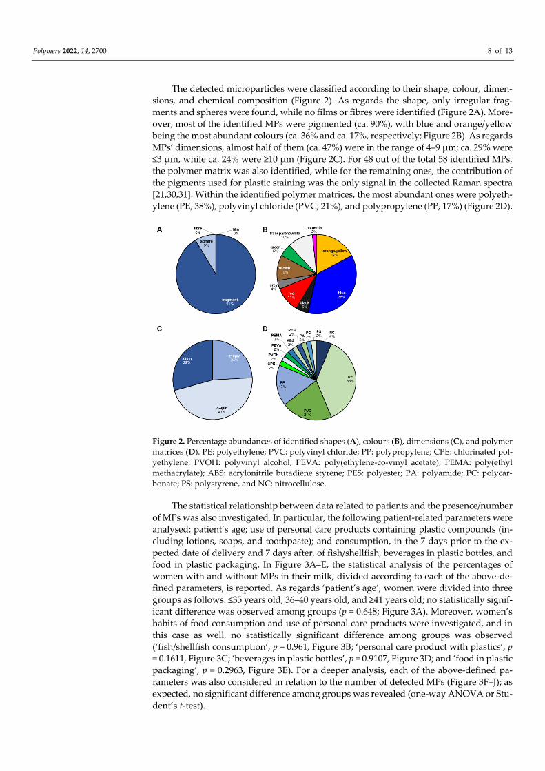
Polymers 2022, 14, 2700 8 of 13
The detected microparticles were classified according to their shape, colour, dimen-
sions, and chemical composition (Figure 2). As regards the shape, only irregular frag-
ments and spheres were found, while no films or fibres were identified (Figure 2A). More-
over, most of the identified MPs were pigmented (ca. 90%), with blue and orange/yellow
being the most abundant colours (ca. 36% and ca. 17%, respectively; Figure 2B). As regards
MPs’ dimensions, almost half of them (ca. 47%) were in the range of 4–9 µm; ca. 29% were
≤3 µm, while ca. 24% were ≥10 µm (Figure 2C). For 48 out of the total 58 identified MPs,
the polymer matrix was also identified, while for the remaining ones, the contribution of
the pigments used for plastic staining was the only signal in the collected Raman spectra
[21,30,31]. Within the identified polymer matrices, the most abundant ones were polyeth-
ylene (PE, 38%), polyvinyl chloride (PVC, 21%), and polypropylene (PP, 17%) (Figure 2D).
Figure 2. Percentage abundances of identified shapes (A), colours (B), dimensions (C), and polymer
matrices (D). PE: polyethylene; PVC: polyvinyl chloride; PP: polypropylene; CPE: chlorinated pol-
yethylene; PVOH: polyvinyl alcohol; PEVA: poly(ethylene-co-vinyl acetate); PEMA: poly(ethyl
methacrylate); ABS: acrylonitrile butadiene styrene; PES: polyester; PA: polyamide; PC: polycar-
bonate; PS: polystyrene, and NC: nitrocellulose.
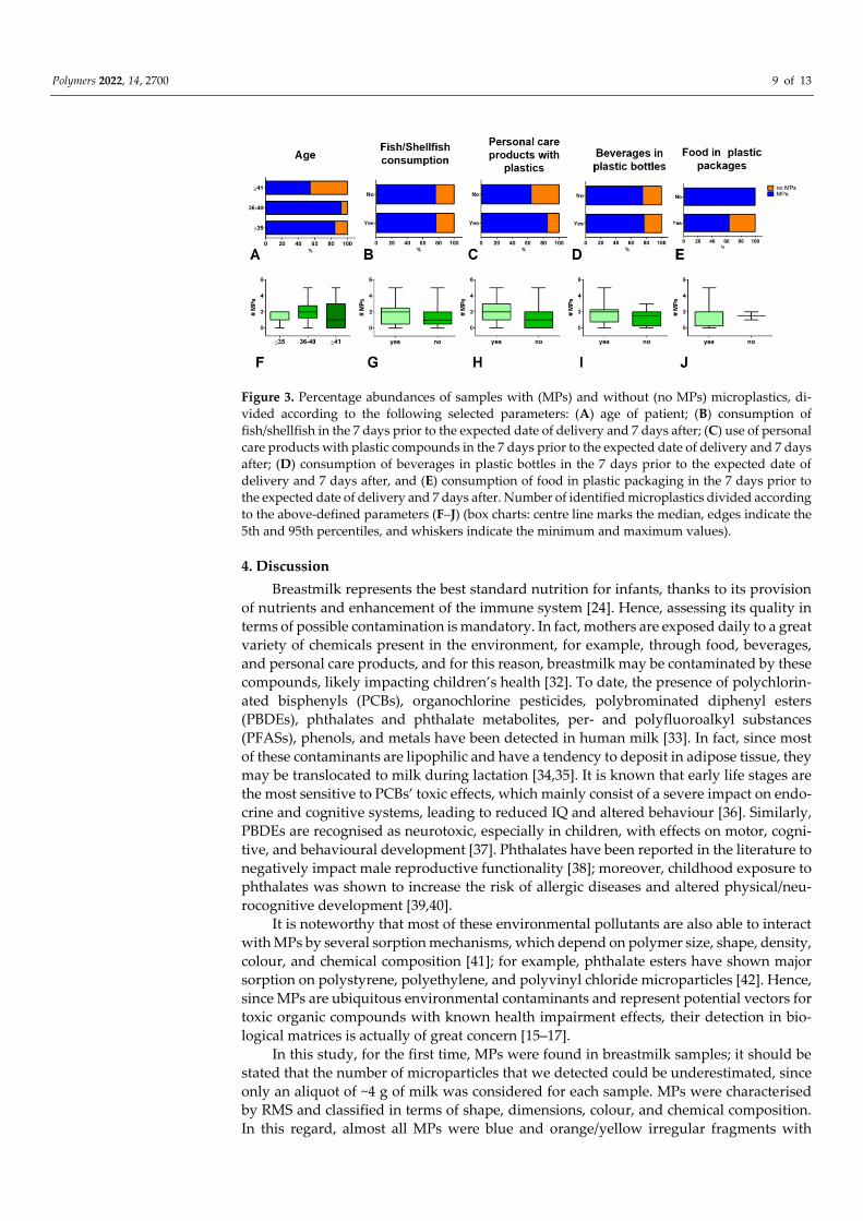
The statistical relationship between data related to patients and the presence/number
of MPs was also investigated. In particular, the following patient-related parameters were
analysed: patient’s age; use of personal care products containing plastic compounds (in-
cluding lotions, soaps, and toothpaste); and consumption, in the 7 days prior to the ex-
pected date of delivery and 7 days after, of fish/shellfish, beverages in plastic bottles, and
food in plastic packaging. In Figure 3A–E, the statistical analysis of the percentages of
women with and without MPs in their milk, divided according to each of the above-de-
fined parameters, is reported. As regards ‘patient’s age’, women were divided into three
groups as follows: ≤35 years old, 36–40 years old, and ≥41 years old; no statistically signif-
icant difference was observed among groups (p = 0.648; Figure 3A). Moreover, women’s
habits of food consumption and use of personal care products were investigated, and in
this case as well, no statistically significant difference among groups was observed
(‘fish/shellfish consumption’, p = 0.961, Figure 3B; ‘personal care product with plastics’, p
= 0.1611, Figure 3C; ‘beverages in plastic bottles’, p = 0.9107, Figure 3D; and ‘food in plastic
packaging’, p = 0.2963, Figure 3E). For a deeper analysis, each of the above-defined pa-
rameters was also considered in relation to the number of detected MPs (Figure 3F–J); as
expected, no significant difference among groups was revealed (one-way ANOVA or Stu-
dent’s t-test).
Polymers 2022, 14, 2700 9 of 13
Figure 3. Percentage abundances of samples with (MPs) and without (no MPs) microplastics, di-
vided according to the following selected parameters: (A) age of patient; (B) consumption of
fish/shellfish in the 7 days prior to the expected date of delivery and 7 days after; (C) use of personal
care products with plastic compounds in the 7 days prior to the expected date of delivery and 7 days
after; (D) consumption of beverages in plastic bottles in the 7 days prior to the expected date of
delivery and 7 days after, and (E) consumption of food in plastic packaging in the 7 days prior to
the expected date of delivery and 7 days after. Number of identified microplastics divided according
to the above-defined parameters (F–J) (box charts: centre line marks the median, edges indicate the
5th and 95th percentiles, and whiskers indicate the minimum and maximum values).
4. Discussion
Breastmilk represents the best standard nutrition for infants, thanks to its provision
of nutrients and enhancement of the immune system [24]. Hence, assessing its quality in
terms of possible contamination is mandatory. In fact, mothers are exposed daily to a great
variety of chemicals present in the environment, for example, through food, beverages,
and personal care products, and for this reason, breastmilk may be contaminated by these
compounds, likely impacting children’s health [32]. To date, the presence of polychlorin-
ated bisphenyls (PCBs), organochlorine pesticides, polybrominated diphenyl esters
(PBDEs), phthalates and phthalate metabolites, per- and polyfluoroalkyl substances
(PFASs), phenols, and metals have been detected in human milk [33]. In fact, since most
of these contaminants are lipophilic and have a tendency to deposit in adipose tissue, they
may be translocated to milk during lactation [34,35]. It is known that early life stages are
the most sensitive to PCBs’ toxic effects, which mainly consist of a severe impact on endo-
crine and cognitive systems, leading to reduced IQ and altered behaviour [36]. Similarly,
PBDEs are recognised as neurotoxic, especially in children, with effects on motor, cogni-
tive, and behavioural development [37]. Phthalates have been reported in the literature to
negatively impact male reproductive functionality [38]; moreover, childhood exposure to
phthalates was shown to increase the risk of allergic diseases and altered physical/neu-
rocognitive development [39,40].
It is noteworthy that most of these environmental pollutants are also able to interact
with MPs by several sorption mechanisms, which depend on polymer size, shape, density,
colour, and chemical composition [41]; for example, phthalate esters have shown major
sorption on polystyrene, polyethylene, and polyvinyl chloride microparticles [42]. Hence,
since MPs are ubiquitous environmental contaminants and represent potential vectors for
toxic organic compounds with known health impairment effects, their detection in bio-
logical matrices is actually of great concern [15–17].
In this study, for the first time, MPs were found in breastmilk samples; it should be
stated that the number of microparticles that we detected could be underestimated, since
only an aliquot of ~4 g of milk was considered for each sample. MPs were characterised
by RMS and classified in terms of shape, dimensions, colour, and chemical composition.
In this regard, almost all MPs were blue and orange/yellow irregular fragments with
Polymers 2022, 14, 2700 10 of 13
dimensions ranging from 2 µm to 12 µm, consistent with translocation mechanisms. In
accordance with other studies reported in the literature, the most abundant polymers
were polyethylene, polyvinyl chloride, and polypropylene [43,44].
Several MP routes of exposure have been reported in the literature, including inha-
lation, dermal contact, and ingestion, with the latter being considered the most impactful,
with an estimated total intake of around 39–52 thousand per person per year [12,13]. Once
ingested/inhaled, MPs can be internalised in human tissues [11]. At the gastrointestinal
level, they may pass through the epithelium by endocytosis mechanisms or by paracellu-
lar diffusion, after which they are translocated by dendritic cells through the lymphatic
circulation and reach the circulatory system [11]. As regards the respiratory system, in-
haled MPs likely penetrate the lower respiratory tract, characterised by a thin mucus layer,
and spread into the bloodstream after cellular uptake or paracellular diffusion [45].
Currently, there is growing scientific evidence about MPs in humans. Schwabl et al.
reported the detection of MPs in human stool [46], while, as a further measure, Ibrahim et
al. described the presence of MPs in human colectomy samples, proving that MPs in part
cross the intestinal barrier [47]. As evidence of inhalation exposure, Amato-Lourenço et
al. detected, in human lung tissue, <5.5 µm polymeric MPs and fibres ranging from 8.12
to 16.8 µm [48]. We recently found MPs in the human placenta, which represents the in-
terface between the foetus and the mother exposed to the external environment [21], re-
sults that were also confirmed by Braun et al. [49]. Very recently, the presence of plastic
particles in human blood finally proved the transport of MPs in the bloodstream to every
body site [50].
As regards the mammary gland, two hypothetical pathways have been suggested for
the translocation of exogenous particles from the bloodstream to breast milk: the mam-
mary epithelial cell-dependent and the immune cell-dependent pathways, with the latter
being particularly relevant in the case of inhaled particles [23–25,51,52]. Hence, a possible
association between the presence of MPs in breastmilk and specific information regarding
mothers’ habits (such as the consumption of fish and shellfish, beverages in plastic bottles,
and food in plastic packaging and the use of personal care products containing plastic
compounds in the 7 days prior to the expected date of delivery and the 7 days after) was
investigated, but no relationship was found between MP presence/number and each of
the above-mentioned parameters.
The lack of association with the use of personal care products is likely explained by
considering that dermal contact has a minor impact as an exposure route, since only par-
ticles <100 nanometres can cross the dermal barrier [11]. Conversely, the absence of a re-
lation with mothers’ food habits is more difficult to explain, since the major route of MP
exposure is represented by ingestion. In fact, numerous food-related sources of MPs have
been reported, including fish, shellfish, and human essential daily consumables, such as
table salt, sugar, bottled water, milk, honey, plastic teabags, and to a greater extent, plastic
kitchen tools, plates, and packaging [53]. Hence, our findings suggest that, since MPs are
ubiquitous in the environment, exposure to these microparticles is inevitable, and, for this
reason, it is impossible to isolate a specific source among the complex set of faced expo-
sures.
5. Conclusions
The evidence of MPs in human breastmilk, coupled with the previous discovery of
these microparticles in the human placenta, represents a great concern, since it impacts
the extremely vulnerable population of infants. In fact, the chemicals possibly contained
in foods, beverages, and personal care products consumed by breastfeeding mothers may
be transferred to the offspring, potentially exerting a toxic effect. Hence, it is mandatory
to increase efforts in scientific research to deepen the knowledge of the potential health
impairment caused by MP internalisation and accumulation, especially in infants, and to
assess innovative, useful ways to reduce exposure to these contaminants during preg-
nancy and lactation.
Polymers 2022, 14, 2700 11 of 13
Author Contributions: Conceptualisation, A.R. and A.S.; methodology, V.N.; validation, A.R. and
E.G.; formal analysis, V.N.; investigation, V.N., A.B., C.B., E.Z., C.D.L., S.D., and A.G.; resources,
A.R., A.S., O.C., and E.G.; data curation, V.N., A.B., G.G., C.B., E.Z., C.D.L., S.D., and A.G.; writing—
original draft preparation, V.N. and E.G.; writing—review and editing, A.R., A.S., G.G., and O.C.;
visualisation, V.N. and E.G.; supervision, A.R., O.C., and E.G.; project administration, E.G. All au-
thors have read and agreed to the published version of the manuscript.
Funding: This research was funded by Fondo di Ateneo, Università Politecnica delle Marche 2020–
2021 to E.G.
Institutional Review Board Statement: The study was conducted in accordance with the Declaration of
Helsinki, and approved by the Ethics Committee Lazio 1 (Protocol N. 708/CE Lazio 1; 24 May 2021).
Informed Consent Statement: Informed consent was obtained from all subjects involved in the
study.
Data Availability Statement: Data are contained within the article.
Conflicts of Interest: The authors declare no conflict of interest.
References
1. Jadhav, E.B.; Sankhla, M.S.; Bhat, R.A.; Bhagat, D.S. Microplastics from food packaging: An overview of human consumption,
health threats, and alternative solutions. Environ. Nanotechnol. Monit. Manag. 2021, 16, 100608.
https://doi.org/10.1016/j.enmm.2021.100608.
2. Lithner, D.; Larsson, Å .; Dave, G. Environmental and health hazard ranking and assessment of plastic polymers based on chem-
ical composition. Sci. Total Environ. 2011, 409, 3309–3324. https://doi.org/10.1016/j.scitotenv.2011.04.038.
3. Plastics Europe Plastics—The Facts 2019 An Analysis of European Plastics Production, Demand and Waste Data; 2019.
4. Eriksen, M.; Lebreton, L.C.M.; Carson, H.S.; Thiel, M.; Moore, C.J.; Borerro, J.C.; Galgani, F.; Ryan, P.G.; Reisser, J. Plastic Pol-
lution in the World’s Oceans: More than 5 Trillion Plastic Pieces Weighing over 250,000 Tons Afloat at Sea. PLoS ONE 2014, 9,
e111913. https://doi.org/10.1371/journal.pone.0111913.
5. Conti, I.; Simioni, C.; Varano, G.; Brenna, C.; Costanzi, E.; Neri, L.M. Legislation to limit the environmental plastic and micro-
plastic pollution and their influence on human exposure. Environ. Pollut. 2021, 288, 117708. https://doi.org/10.1016/j.en-
vpol.2021.117708.
6. Browne, M.A.; Crump, P.; Niven, S.J.; Teuten, E.; Tonkin, A.; Galloway, T.; Thompson, R. Accumulation of Microplastic on
Shorelines Woldwide: Sources and Sinks. Environ. Sci. Technol. 2011, 45, 9175–9179. https://doi.org/10.1021/es201811s.
7. Salvador Cesa, F.; Turra, A.; Baruque-Ramos, J. Synthetic fibers as microplastics in the marine environment: A review from
textile perspective with a focus on domestic washings. Sci. Total Environ. 2017, 598, 1116–1129. https://doi.org/10.1016/j.sci-
totenv.2017.04.172.
8. Hanun, J.N.; Hassan, F.; Jiang, J.-J. Occurrence, fate, and sorption behavior of contaminants of emerging concern to microplas-
tics: Influence of the weathering/aging process. J. Environ. Chem. Eng. 2021, 9, 106290. https://doi.org/10.1016/j.jece.2021.106290.
9. Kannan, K.; Vimalkumar, K. A Review of Human Exposure to Microplastics and Insights Into Microplastics as Obesogens.
Front. Endocrinol. 2021, 12, 724989. https://doi.org/10.3389/fendo.2021.724989.
10. Sridharan, S.; Kumar, M.; Singh, L.; Bolan, N.S.; Saha, M. Microplastics as an emerging source of particulate air pollution: A
critical review. J. Hazard. Mater. 2021, 418, 126245. https://doi.org/10.1016/j.jhazmat.2021.126245.
11. Prata, J.C.; da Costa, J.P.; Lopes, I.; Duarte, A.C.; Rocha-Santos, T. Environmental exposure to microplastics: An overview on
possible human health effects. Sci. Total Environ. 2020, 702, 134455. https://doi.org/10.1016/j.scitotenv.2019.134455.
12. Prata, J.C. Airborne microplastics: Consequences to human health? Environ. Pollut. 2018, 234, 115–126.
https://doi.org/10.1016/j.envpol.2017.11.043.
13. Cox, K.D.; Covernton, G.A.; Davies, H.L.; Dower, J.F.; Juanes, F.; Dudas, S.E. Human Consumption of Microplastics. Environ.
Sci. Technol. 2019, 53, 7068–7074. https://doi.org/10.1021/acs.est.9b01517.
14. Alimba, C.G.; Faggio, C.; Sivanesan, S.; Ogunkanmi, A.L.; Krishnamurthi, K. Micro(nano)-plastics in the environment and risk
of carcinogenesis: Insight into possible mechanisms. J. Hazard. Mater. 2021, 416, 126143. https://doi.org/10.1016/j.jhaz-
mat.2021.126143.
15. Danopoulos, E.; Twiddy, M.; West, R.; Rotchell, J.M. A rapid review and meta-regression analyses of the toxicological impacts
of microplastic exposure in human cells. J. Hazard. Mater. 2021, 127861. https://doi.org/10.1016/j.jhazmat.2021.127861.
16. Han, Y.; Lian, F.; Xiao, Z.; Gu, S.; Cao, X.; Wang, Z.; Xing, B. Potential toxicity of nanoplastics to fish and aquatic invertebrates:
Current understanding, mechanistic interpretation, and meta-analysis. J. Hazard. Mater. 2022, 427, 127870.
https://doi.org/10.1016/j.jhazmat.2021.127870.
Polymers 2022, 14, 2700 12 of 13
17. Yin, K.; Wang, Y.; Zhao, H.; Wang, D.; Guo, M.; Mu, M.; Liu, Y.; Nie, X.; Li, B.; Li, J.; et al. A comparative review of microplastics
and nanoplastics: Toxicity hazards on digestive, reproductive and nervous system. Sci. Total Environ. 2021, 774, 145758.
https://doi.org/10.1016/j.scitotenv.2021.145758.
18. Käppler, A.; Fischer, D.; Oberbeckmann, S.; Schernewski, G.; Labrenz, M.; Eichhorn, K.-J.; Voit, B. Analysis of environmental
microplastics by vibrational microspectroscopy: FTIR, Raman or both? Anal. Bioanal. Chem. 2016, 408, 8377–8391.
https://doi.org/10.1007/s00216-016-9956-3.
19. Ribeiro-Claro, P.; Nolasco, M.M.; Araújo, C. Characterization of Microplastics by Raman Spectroscopy. Compr. Anal. Chem. 2017,
75, 119–151. https://doi.org/10.1016/bs.coac.2016.10.001.
20. Di Renzo, L.; Mascilongo, G.; Berti, M.; Bogdanović, T.; Listeš, E.; Brkljača, M.; Notarstefano, V.; Gioacchini, G.; Giorgini, E.;
Olivieri, V.; et al. Potential Impact of Microplastics and Additives on the Health Status of Loggerhead Turtles (Caretta caretta)
Stranded Along the Central Adriatic Coast. Water Air Soil Pollut. 2021, 232, 98. https://doi.org/10.1007/s11270-021-04994-8.
21. Ragusa, A.; Svelato, A.; Santacroce, C.; Catalano, P.; Notarstefano, V.; Carnevali, O.; Papa, F.; Rongioletti, M.C.A.; Baiocco, F.;
Draghi, S.; et al. Plasticenta: First evidence of microplastics in human placenta. Environ. Int. 2021, 146, 106274.
https://doi.org/10.1016/j.envint.2020.106274.
22. Eidelman, A.I.; Schanler, R.J.; Johnston, M.; Landers, S.; Noble, L.; Szucs, K.; Viehmann, L. Breastfeeding and the Use of Human
Milk. Pediatrics 2012, 129, e827–e841. https://doi.org/10.1542/peds.2011-3552.
23. Llorca, M.; Farré, M.; Picó, Y.; Teijón, M.L.; Á lvarez, J.G.; Barceló, D. Infant exposure of perfluorinated compounds: Levels in
breast milk and commercial baby food. Environ. Int. 2010, 36, 584–592. https://doi.org/10.1016/j.envint.2010.04.016.
24. LaKind, J.S.; Verner, M.-A.; Rogers, R.D.; Goeden, H.; Naiman, D.Q.; Marchitti, S.A.; Lehmann, G.M.; Hines, E.P.; Fenton, S.E.
Current Breast Milk PFAS Levels in the United States and Canada: After All This Time, Why Don’t We Know More? Environ.
Health Perspect. 2022, 130. https://doi.org/10.1289/EHP10359.
25. Jian, J.M.; Chen, D.; Han, F.J.; Guo, Y.; Zeng, L.; Lu, X.; Wang, F. A short review on human exposure to and tissue distribution
of per- and polyfluoroalkyl substances (PFASs). Sci. Total Environ. 2018, 636, 1058–1069. https://doi.org/10.1016/j.sci-
totenv.2018.04.380.
26. Ministero della Salute Allattare al Seno-Un investimento Per la Vita; 2019. Available online:
https://www.salute.gov.it/portale/documentazione/p6_2_5_1.jsp?lingua=italiano&id=303 (accessed on 1 June 2022).
27. Karami, A.; Golieskardi, A.; Choo, C.K.; Romano, N.; Ho, Y.B.; Salamatinia, B. A high-performance protocol for extraction of
microplastics in fish. Sci. Total Environ. 2017, 578, 485–494. https://doi.org/10.1016/j.scitotenv.2016.10.213.
28. Dong, M.; Zhang, Q.; Xing, X.; Chen, W.; She, Z.; Luo, Z. Raman spectra and surface changes of microplastics weathered under
natural environments. Sci. Total Environ. 2020, 739, 139990. https://doi.org/10.1016/j.scitotenv.2020.139990.
29. SLOPP Library of Microplastics. Available online: https://rochmanlab.com/slopp-and-slopp-e-raman-spectral-libraries-for-mi-
croplastics-research (accessed on 1 June 2022).
30. Imhof, H.K.; Laforsch, C.; Wiesheu, A.C.; Schmid, J.; Anger, P.M.; Niessner, R.; Ivleva, N.P. Pigments and plastic in limnetic
ecosystems: A qualitative and quantitative study on microparticles of different size classes. Water Res. 2016, 98, 64–74.
https://doi.org/10.1016/j.watres.2016.03.015.
31. Stoye, D., Freitag, W. Paints, Coatings and Solvents; Wiley: Hoboken, NJ, USA, 1998; ISBN 9783527288632.
32. Karthikeyan, B.S.; Ravichandran, J.; Aparna, S.R.; Samal, A. ExHuMId: A curated resource and analysis of Exposome of Human
Milk across India. Chemosphere 2021, 271, 129583. https://doi.org/10.1016/j.chemosphere.2021.129583.
33. Lehmann, G.M.; LaKind, J.S.; Davis, M.H.; Hines, E.P.; Marchitti, S.A.; Alcala, C.; Lorber, M. Environmental Chemicals in Breast
Milk and Formula: Exposure and Risk Assessment Implications. Environ. Health Perspect. 2018, 126, 096001.
https://doi.org/10.1289/EHP1953.
34. Mead, M.N. Contaminants in Human Milk: Weighing the Risks against the Benefits of Breastfeeding. Environ. Health Perspect.
2008, 116, A426–A434. https://doi.org/10.1289/ehp.116–a426.
35. Vasios, G.; Kosmidi, A.; Kalantzi, O.-I.; Tsantili-Kakoulidou, A.; Kavantzas, N.; Theocharis, S.; Giaginis, C. Simple physicochem-
ical properties related with lipophilicity, polarity, molecular size and ionization status exert significant impact on the transfer
of drugs and chemicals into human breast milk. Expert Opin. Drug Metab. Toxicol. 2016, 12, 1273–1278.
https://doi.org/10.1080/17425255.2016.1230197.
36. Lundqvist, C.; Zuurbier, M.; Leijs, M.; Johansson, C.; Ceccatelli, S.; Saunders, M.; Schoeters, G.; Ten Tusscher, G.; Koppe, J. The
effects of PCBs and dioxins on child health. Acta Paediatr. 2006, 95, 55–64. https://doi.org/10.1080/08035320600886257.
37. Gibson, E.; Siegel, E.; Eniola, F.; Herbstman, J.; Factor-Litvak, P. Effects of Polybrominated Diphenyl Ethers on Child Cognitive,
Behavioral, and Motor Development. Int. J. Environ. Res. Public Health 2018, 15, 1636. https://doi.org/10.3390/ijerph15081636.
38. Main, K.M.; Mortensen, G.K.; Kaleva, M.M.; Boisen, K.A.; Damgaard, I.N.; Chellakooty, M.; Schmidt, I.M.; Suomi, A.-M.; Vir-
tanen, H.E.; Petersen, J.H.; et al. Human Breast Milk Contamination with Phthalates and Alterations of Endogenous Reproduc-
tive Hormones in Infants Three Months of Age. Environ. Health Perspect. 2006, 114, 270–276. https://doi.org/10.1289/ehp.8075.
39. Braun, J.M.; Sathyanarayana, S.; Hauser, R. Phthalate exposure and children’s health. Curr. Opin. Pediatr. 2013, 25, 247–254.
https://doi.org/10.1097/MOP.0b013e32835e1eb6.
Polymers 2022, 14, 2700 13 of 13
40. Huang, H.-B.; Chen, H.-Y.; Su, P.-H.; Huang, P.-C.; Sun, C.-W.; Wang, C.-J.; Chen, H.-Y.; Hsiung, C.A.; Wang, S.-L. Fetal and
Childhood Exposure to Phthalate Diesters and Cognitive Function in Children Up to 12 Years of Age: Taiwanese Maternal and
Infant Cohort Study. PLoS ONE 2015, 10, e0131910. https://doi.org/10.1371/journal.pone.0131910.
41. Rodrigues, J.P.; Duarte, A.C.; Santos-Echeandía, J.; Rocha-Santos, T. Significance of interactions between microplastics and
POPs in the marine environment: A critical overview. TrAC Trends Anal. Chem. 2019, 111, 252–260.
https://doi.org/10.1016/j.trac.2018.11.038.
42. Liu, F.; Liu, G.; Zhu, Z.; Wang, S.; Zhao, F. Interactions between microplastics and phthalate esters as affected by microplastics
characteristics and solution chemistry. Chemosphere 2019, 214, 688–694. https://doi.org/10.1016/j.chemosphere.2018.09.174.
43. Jones, J.I.; Vdovchenko, A.; Cooling, D.; Murphy, J.F.; Arnold, A.; Pretty, J.L.; Spencer, K.L.; Markus, A.A.; Vethaak, A.D.;
Resmini, M. Systematic Analysis of the Relative Abundance of Polymers Occurring as Microplastics in Freshwaters and Estu-
aries. Int. J. Environ. Res. Public Health 2020, 17, 9304. https://doi.org/10.3390/ijerph17249304.
44. Bajt, O. From plastics to microplastics and organisms. FEBS Open Bio 2021, 11, 954–966. https://doi.org/10.1002/2211-5463.13120.
45. Mowat, A.M. Anatomical basis of tolerance and immunity to intestinal antigens. Nat. Rev. Immunol. 2003, 3, 331–341.
https://doi.org/10.1038/nri1057.
46. Schwabl, P.; Köppel, S.; Königshofer, P.; Bucsics, T.; Trauner, M.; Reiberger, T.; Liebmann, B. Detection of Various Microplastics
in Human Stool. Ann. Intern. Med. 2019, 171, 453. https://doi.org/10.7326/M19-0618.
47. Ibrahim, Y.S.; Tuan Anuar, S.; Azmi, A.A.; Wan Mohd Khalik, W.M.A.; Lehata, S.; Hamzah, S.R.; Ismail, D.; Ma, Z.F.;
Dzulkarnaen, A.; Zakaria, Z.; et al. Detection of microplastics in human colectomy specimens. JGH Open 2021, 5, 116–121.
https://doi.org/10.1002/jgh3.12457.
48. Amato-Lourenço, L.F.; Carvalho-Oliveira, R.; Júnior, G.R.; dos Santos Galvão, L.; Ando, R.A.; Mauad, T. Presence of airborne
microplastics in human lung tissue. J. Hazard. Mater. 2021, 416, 126124. https://doi.org/10.1016/j.jhazmat.2021.126124.
49. Braun, T.; Ehrlich, L.; Henrich, W.; Koeppel, S.; Lomako, I.; Schwabl, P.; Liebmann, B. Detection of Microplastic in Human
Placenta and Meconium in a Clinical Setting. Pharmaceutics 2021, 13, 921. https://doi.org/10.3390/pharmaceutics13070921.
50. Leslie, H.A.; van Velzen, M.J.M.; Brandsma, S.H.; Vethaak, A.D.; Garcia-Vallejo, J.J.; Lamoree, M.H. Discovery and quantifica-
tion of plastic particle pollution in human blood. Environ. Int. 2022, 107199. https://doi.org/10.1016/j.envint.2022.107199.
51. Cai, J.; Zang, X.; Wu, Z.; Liu, J.; Wang, D. Translocation of transition metal oxide nanoparticles to breast milk and offspring: The
necessity of bridging mother-offspring-integration toxicological assessments. Environ. Int. 2019, 133, 105153.
https://doi.org/10.1016/j.envint.2019.105153.
52. Yang, L.; Kuang, H.; Zhang, W.; Wei, H.; Xu, H. Quantum dots cause acute systemic toxicity in lactating rats and growth re-
striction of offspring. Nanoscale 2018, 10, 11564–11577. https://doi.org/10.1039/C8NR01248B.
53. Senathirajah, K.; Attwood, S.; Bhagwat, G.; Carbery, M.; Wilson, S.; Palanisami, T. Estimation of the mass of microplastics in-
gested—A pivotal first step towards human health risk assessment. J. Hazard. Mater. 2021, 404, 124004.
https://doi.org/10.1016/j.jhazmat.2020.124004.