protein engineering and structural characterisation
TRANSCRIPT

Protein Engineering and Structural
Characterisation Approaches Aimed
Towards the Improvement of Biological
Therapies
Esteban Cruz
This thesis is submitted in fulfilment of the requirements for the degree of Doctor
of Philosophy
School of Pharmacy
Faculty of Medicine and Health
The University of Sydney
2019

This thesis is submitted to the University of Sydney in fulfilment of the requirements for the degree
of Doctor of Philosophy. The material included herein has not been submitted, either in full or in
part, for another degree at this institution or any other. The content of this thesis is original except
as acknowledged in the text accordingly.
Name: Esteban Cruz
Signature:
Date: 19th of August 2019
Statement of Authenticity

ii
It is hard to believe that four years have already gone by since I moved all the way across the world
from Costa Rica to undertake my PhD studies at the University of Sydney. It has been four years
full of challenges and sacrifice, being away from my family and friends, yet I was fortunate enough
to cross paths with amazing people along the way that I am now proud to call friends and mentors.
I would like to first acknowledge my PhD supervisor, Associate Professor Veysel Kayser, for
giving me the opportunity to embark on this journey and providing constant support and guidance
throughout my candidature. I deeply appreciate the mentorship and research opportunities you
provided. To my auxiliary supervisor Associate Professor Serdar Kuyucak. To my colleagues,
Vicki, Mouhamad and Ziya, with whom I worked and learned extensively in the lab and shared a
lot of fun moments. To our most valuable postdoc Zehra, whose contribution to the lab in the early
days of our PhDs was invaluable. To Christina, who has been my friend since the start and I now
consider my little sister. I’m so grateful to have met you and I’d like to thank you for all your
support and all the fun we’ve had together. To our honours students, Jacky, Emma and Fahmi, it
was great working with all of you guys. To all the new members of the group, Masoume, Qudsia,
Barbaros, Mohammad, Candice, Mariam, Dua’A, Rina, and Abdul, I wish you all the best in your
postgraduate studies. To all members of S114 for creating a great atmosphere to work in, especially
to Ophelia who always had a bright smile (or sweets) to cheer me up, and to Luke who is a legend
and taught me all about Australian culture.
I would like to express my sincerest gratitude to our Head of School, Professor Andrew
MacLachlan, and our HDR coordinator, Dr. Ingrid Gelissen, with whom I’ve liaised constantly
throughout my last year of candidature as an HDR student representative and member of the
committee of the AAPS Student Chapter. The support you have provided has been spectacular and
is truly inspiring for all students. To the incredible staff we have at the School of Pharmacy, who
greatly facilitate our work as students. Special mention to Shane, Suley and Tim, absolute legends
and some of the most generous and helpful people I’ve met.
Living abroad doing a PhD would have been extremely difficult without my friends’ support. I
would like to thank Ana and her family for their support at the start of my degree. To Ingrid, Astrid,
Acknowledgements

iii
Melvin and Mauricio for welcoming me to Australia and making me feel at home, I will always
be grateful for your kindness. I was also extremely lucky to make extraordinary friends at uni,
especially during this last year, that I would like to show my appreciation to. Thank you to our
small Petersham family, Alex and Jen, we’ve had amazing times together. All the gigs, all the
games, all the trivia nights, all the pizzas, all the ice cream, this could go on and on. Thank you so
much for your friendship. To the Lemon Tea Empire, Jono, Kamini, Hoi, Patrick and Miranda,
you guys are family now and I’ve had the most incredible time with all of you. To the
Hibbs/Groundwater group, Jin, Laith, Elias, Steve, you’re a bunch of crazy/amazing people. To
Associate Professor Wojciech Chrzanowski and his group, Huyen, Sarah, Ramya, Priyanka, and
minha irmã Taisinha. To Daniela (Ikhte) for being so incredibly encouraging and positive this
whole time, we made it! To Lin, Vincent, Jiaqi, William and my good friend Martin. To my
incredible friend Daniela López for being there for me when I most needed it.
Lastly, my heartfelt thanks to my family, for always being on my side, for always supporting my
dreams. To my mum and dad, forever my role models and my constant inspiration. Never will I
forget your commitment, your dedication, your faith in my abilities, and most importantly your
unconditional love.

iv
Published peer-reviewed journal articles in support of this thesis:
E. Cruz, J. Cain, B. Crossett, V. Kayser. Site-specific Glycosylation Profile of Influenza A (H1N1)
Hemagglutinin Through Tandem Mass Spectrometry. Human Vaccines and Immunotherapeutics,
14(3) (2018), 508-517. [Chapter 2]
Z. Elgundi, M. Reslan, E. Cruz, V. Sifniotis, V. Kayser. The State-of-play and Future of Antibody
Therapeutics. Advanced Drug Delivery Reviews, 122 (2017), 2-19. [Chapter 3]
E. Cruz, V. Kayser. Monoclonal Antibody Therapy of Solid Tumors: Clinical Limitations and
Novel Strategies to Enhance Treatment Efficacy. Biologics: Targets and Therapy, 13 (2019), 33-
51. [Chapter 6]
E. Cruz, V. Kayser. Synthesis and Enhanced Cellular Uptake In Vitro of Anti-HER2
Multifunctional Gold Nanoparticles. Cancers, 11 (2019), 870. [Chapter 7]
Submitted manuscripts in support of this thesis
M. Reslan, V. Sifniotis, E. Cruz, Z. Sumer-Bayraktar, S. Cordwell, V. Kayser (2019). Enhancing
the Stability of Adalimumab by Engineering Additional Glycosylation Motifs. Biotechnology and
Bioengineering. [Chapter 4]
Manuscripts prepared for submission in support of this thesis
E. Cruz, V. Sifniotis, Z. Sumer-Bayraktar, S. Cordwell, V. Kayser. (2019). Glycan Profile Analysis
of Engineered Trastuzumab with Rationally Added Glycosylation Sequons for Enhanced Physical
Stability. [Chapter 5]
List of Publications

v
Further published peer-reviewed publications during the candidature
V. Sifniotis, E. Cruz, B. Eroglu, V. Kayser. Currents Advancements in Addressing Key Challenges
of Therapeutic Antibody Manufacture and Formulation. Antibodies. 8(2) (2019), 36.
Z. Elgundi, V. Sifniotis, M. Reslan, E. Cruz, V. Kayser. Laboratory Scale Production and
Purification of a Therapeutic Antibody. Journal of Visualized Experiments, 2017 (119), 1-8.
Y. Moussa, Y. Ong, J. Perry, Z. Cheng, V. Kayser, E. Cruz, R. Kim, N. Sciortino, N. Wheate.
Demonstration of In Vitro Host-guest Complex Formation and Safety of Para-
sulfonatocalix[8]arene as a Delivery Vehicle for Two Antibiotic Drugs. Journal of Pharmaceutical
Sciences, 107(12) (2018), 3105-3111.
Further manuscripts prepared for submission during the candidature
Cruz E, Kayser V. Major Classes of Biotherapeutics. In: Ramzan I, ed. Biologics, Biosimilars, and
Biobetters: An Introduction for Pharmacists, Physicians and Other Health Practitioners. Wiley; To
be published in 2020.
M. Reslan, E. Cruz, V. Kayser. Analysis of the aggregation kinetics of Herceptin® (trastuzumab).
Cruz, E., Kayser, V. Multifunctional gold nanoparticles targeted against HER-2 amplified cells for
selective delivery of cytotoxic payloads. 29th Annual Queenstown Molecular Biology Meeting.
Queenstown, New Zealand. 2-4 September 2019. (Oral presentation).
Cruz, E., Kayser, V. Engineered Glycosylation Sites on Monoclonal Antibodies for Enhanced
Stability. AAPS 2019 PharmSci 360. San Antonio, Texas, United States. 3-6 November 2019.
(Oral presentation)
Conference presentations

vi
HA Hemagglutinin
RPLC Reversed-phase liquid chromatography
MS Mass spectrometry
ADC Antibody-drug conjugate
WT Wildtype
LC Liquid chromatography
SE-HPLC Size-exclusion high performance liquid chromatography
Tm Melting temperature
Tagg Onset temperature of aggregation
TNF-α Tumor necrosis factor alpha
FcγR Fc-gamma receptor
HER-2 Human epidermal growth factor receptor 2
Tmab Trastuzumab
SH Sulfhydryl
PEG Polyethylene glycol
HIV Human immunodeficiency virus
CPP Cell-penetrating peptide
UV Ultraviolet
Vis Visible
DLS Dynamic light scattering
ICP-MS Inductively couple plasma mass spectrometry
TEM Transmission electron microscope
DNA Deoxyribonucleic acid
CDR Complementarity determining regions
mAb Monoclonal antibody
ADCC Antibody-dependent cellular cytotoxicity
List of Abbreviations

vii
ESI Electrospray ionisation
MALDI Matrix-assisted laser desorption/ionisation
APCI Atmospheric pressure chemical ionisation
DDA Data-dependent acquisition
CID Collision-induced dissociation
HCD High-energy collision dissociation
ETD Electron-transfer dissociation
ECD Electron-capture dissociation
NA Neuraminidase
RNA Ribonucleic acid
NIAID National Institute of Allergy and Infectious Disease
IRD Influenza Research Database
PDB Protein Data Bank
VMD Visual Molecular Dynamics
PBS Phosphate-buffered saline
FA Formic acid
HPLC High performance liquid chromatography
ER Endoplasmic reticulum
RBS Receptor binding site
TIC Total ion current
FDA United Stated Food and Drug Administration
MDCK Madin-Darby Canine Kidney
SA Sialic acid
EGFR Epidermal growth factor receptor
RANK Receptor activator of nuclear factor kappa-B
NGS Next-generation sequencing
VH Heavy chain
LH Light chain
BLA Biologics license application
IL Interleukin
viii
NSCLC Non-small cell lung cancer
PD-L1 Programmed death-ligand 1
EPCAM Epithelial cell adhesion molecule
CD Cluster of differentiation
CFD Complement factor D
ACVR Activin receptor
CLL Chronic lymphocytic leukemia
vWF Von Willebrand factor
TTP Thrombotic thrombocytopenic purpura
kDa Kilodalton
sdAb Single-domain antibody fragments
SC Subcutaneous
CDC Complement dependent cellular cytotoxicity
PK Pharmacokinetics
CCR4 CC-chemokine receptor 4
PTCL Peripheral T cell lymphoma
NK Natural killer cells
MCC N-maleimidomethyl cyclohexane-1-carboxylate
DVD-Ig Dual variable domain immunoglobulin
DNL Dock-and-Lock
BiTE Bispecific T-cell engager
DART Dual affinity re-targeting
MMAE Monomethyl auristatin E
PSMA Protein-specific membrane antigen
NHL Non-Hodgkin's lymphoma
MMAF Monomethyl auristatin F
DM Maytansinoid
SMCC N-succinimidyl-4-(N-maleimidomethyl) cyclohexane-1-carboxylate
DAR Drug-to-antibody ratio
SEED Strand-exchange engineered domain

ix
DC Dendritic cells
EMA European Medicines Agency
HAMA Human anti-mouse antibodies
ALL Acute lymphoblastic leukemia
HLA Human leukocyte antigen
TCR Toll-like receptor
DAF Dual-action Fab
IV Intravenous
RSV Respiratory syncytial virus
SAP Surface Aggregation Propensity
BCC Basal cell carcinoma
APR Aggregation-prone region
AA Amino acid
CH1 Heavy chain constant region domain 1
CH2 Heavy chain constant region domain 2
CH3 Heavy chain constant region domain 3
VH Heavy chain variable domain
CL Light chain constant region
VL Light chain variable domain
PCR Polymerase chain reaction
PEI Polyethylenimine
SFM Serum free media
GE General Electric
PSM Peptide spectrum matches
GF Gel filtration
AUC Area under the curve
SD Standard deviation
SLS Static light scattering
BC< Barycentric mean
SPR Surface plasmon resonance

x
Ka Association rate constant
Kd Dissociation rate constant
KD Equilibrium dissociation constant
HEK Human embryonic kidney
MWCO Molecular weight cutoff
IAA Iodoacetamide
TFA Trifluoroacetic acid
DTT Dithiothreitol
PVDF Polyvinylidene difluoride
SPE Solid-phase extraction
PGC Porous graphitic carbon
EIC Extracted ion chromatogram
SASA Solvent accessible surface area
MD Molecular dynamics
CHO Chinese hamster ovary
ECM Extracellular matrix
IFP Interstitial fluid pressure
ADCP Antibody-dependent cellular phagocytosis
MAPK Mitogen activated protein kinase
CDK Cyclin-dependent kinase
IGF-IR Type 1 insulin-like growth factor receptor
TGF-α Tumor growth factor alpha
VEGF Vascular endothelial growth factor
AML Acute myeloid leukemia
CTLA-4 Cytotoxic T-lymphocyte associated protein 4
TCR T-cell receptor
APC Antigen presenting cell
TGF-β Tumor growth factor beta
IFN-γ Interferon gamma
NP Nanoparticle

xi
EPR Enhanced permeability and retention effect
AIDS Acquired immunodeficiency syndrome
ICI Immune checkpoint inhibitor
MRI Magnetic resonance imaging
EDTA Ethylenediaminetetraacetic acid
DTNB 5, 5’-dithiobis(2-nitrobenzoic acid)
DMSO Dimethyl sulfoxide
TOF Time-of-flight
FBS Fetal bovine serum
GR Growth rate
RES Reticuloendothelial system
MW Molecular weight
NHS N-Hydroxysuccinimide
AuNP Gold nanoparticles
PDI Polydispersity index

xii
Biological therapies are currently at the forefront of pharmaceutical innovation and drug discovery.
Seminal developments in molecular biology and recombinant technologies in the 1990’s brought
about a new era in biomedicine, inasmuch as they expanded the ability to manipulate the synthetic
machinery of biological systems for the production of recombinant macromolecules with exquisite
therapeutic potential and versatility. Most prominent among these developments were advances in
the manufacture of humanised and human monoclonal antibodies in a streamlined fashion.
Biotherapeutics then experienced a remarkable boom in the early 2000’s and currently remain the
fastest-growing class of therapeutics. As the field of molecular biology continues to advance at an
accelerated pace, manifold opportunities are presented to implement new tools and technologies
in the design of novel biotherapeutics and the refinement of existing biological therapies. The
empirical research contained in this thesis reports on various approaches to address crucial aspects
of the development of novel biotherapeutics, and demonstrates the application of modern mass
spectrometry methods to gain insight into the complex structural features of macromolecules.
Three main aims were defined and addressed in this work. The first aim consisted in employing a
tailored mass spectrometry method to acquire the site-specific glycan profile of an influenza
surface glycoprotein that plays a crucial role in infectivity and antigenicity. The structural data
obtained sheds light into antigenic properties of the virus that can aid in the design of recombinant
vaccines. The second aim sought to evaluate a protein engineering strategy to improve the
biophysical properties of monoclonal antibodies through the targeted insertion of glycosylation
sites in strategic positions on the protein. The data reported demonstrates the capabilities of the
technique to increase physical stability and underlines aspects that require further refinement
moving forward. The final aim focused on the synthesis of a nanoparticle-based drug delivery
platform that employs antibodies as targeting agents for increased treatment specificity. We
demonstrated the feasibility of chemically modifying the antibody using concomitant protein
functionalisation techniques to produce antibody-drug conjugates that bind to the surface of gold
nanoparticles to serve as targeting agents. The synthesized nanocarrier is designed to accumulate
preferentially in tumours through the enhanced permeability and retention effect, where it can
subsequently engage in specific interactions with tumour cells via active-targeting of the antibody.
Thesis Abstract

xiii
Altogether, the body of work presented in this thesis reports on novel experimental approaches
that will aid in the design and production of next-generation biotherapeutics.

xiv
STATEMENT OF AUTHENTICITY.......................................................................................... I
ACKNOWLEDGEMENTS ........................................................................................................ II
LIST OF PUBLICATIONS ....................................................................................................... IV
CONFERENCE PRESENTATIONS ......................................................................................... V
LIST OF ABBREVIATIONS .................................................................................................... VI
THESIS ABSTRACT ............................................................................................................... XII
CHAPTER 1 - THESIS INTRODUCTION ............................................................................... 1
PREFACE ...................................................................................................................................... 1
AIMS AND CHAPTER DESCRIPTION ................................................................................................ 3
Aim 1 (Chapter 2) – Studying the glycan profile of influenza hemagglutinin to contribute to
the design of improved vaccine platforms .............................................................................. 3
Aim 2 (Chapters 3, 4, and 5) – Targeted insertion of N-glycosylation sites to improve the
physical stability of antibody therapeutics ............................................................................. 5
Aim 3 (Chapter 6 and 7) – Chemical functionalisation of an antibody molecule for
employment as a targeting agent in the design of a nanoparticle drug delivery platform ..... 7
NOTES ON THE FORMAT OF THE CHAPTERS ................................................................................... 8
REFERENCES ................................................................................................................................ 9
CHAPTER 2 ................................................................................................................................ 12
CHAPTER 2 – AUTHORSHIP DECLARATION STATEMENT.............................................................. 13
ABSTRACT ................................................................................................................................. 14
INTRODUCTION .......................................................................................................................... 14
MATERIALS AND METHODS ....................................................................................................... 17
N-glycosylation sequon analysis of A/New Caledonia/20/1999 HA ..................................... 17
Homology modelling of A/New Caledonia/20/1999 HA ....................................................... 17
Hemagglutinin isolation through polyacrylamide electrophoresis and in-gel trypsin
digestion ................................................................................................................................ 17
Table of Contents

xv
Analysis of tryptic peptides and glycopeptides by nanoRPLC-MS/MS ................................ 17
Analysis of MS/MS spectra of intact N-glycopeptides .......................................................... 18
RESULTS .................................................................................................................................... 19
Prediction of N-glycosylation sites ....................................................................................... 19
Mass spectrometry analysis of electrophoretically-fractionated viral proteins ................... 20
Glycopeptide analysis ........................................................................................................... 22
Confirmation of glycosylated residues.................................................................................. 24
Glycan microheterogeneity profile ....................................................................................... 25
DISCUSSION ............................................................................................................................... 33
CONCLUSION .............................................................................................................................. 36
DISCLOSURE OF POTENTIAL CONFLICTS OF INTEREST ................................................................. 37
FUNDING .................................................................................................................................... 37
ACKNOWLEDGEMENTS ............................................................................................................... 37
REFERENCES .............................................................................................................................. 37
CHAPTER 3 ................................................................................................................................ 43
CHAPTER 3 – AUTHORSHIP DECLARATION STATEMENT.............................................................. 44
ABSTRACT ................................................................................................................................. 45
INTRODUCTION .......................................................................................................................... 45
ANTIBODY DISCOVERY STRATEGIES ........................................................................................... 46
NOVEL ANTIBODIES IN APPROVAL AND PRECLINICAL DEVELOPMENT STAGES ............................ 48
BIOBETTER ANTIBODIES ............................................................................................................. 53
Fc engineered antibodies for enhanced effector functions ................................................... 54
Antibody drug conjugates (ADC).......................................................................................... 56
Bispecifics ............................................................................................................................. 61
PHYSICAL AND CHEMICAL DEGRADATION OF ANTIBODIES ......................................................... 65
Aggregation........................................................................................................................... 66
Denaturation ......................................................................................................................... 67
Fragmentation....................................................................................................................... 67
Deamidation .......................................................................................................................... 68
Oxidation............................................................................................................................... 69
COMPUTATIONAL DESIGN TOOLS ............................................................................................... 69

xvi
OPTIMIZATION OF ANTIBODY BIOAVAILABILITY AND DELIVERY ................................................ 71
CONCLUSIONS ............................................................................................................................ 73
ACKNOWLEDGEMENTS ............................................................................................................... 74
DISCLOSURES ............................................................................................................................. 74
REFERENCES .............................................................................................................................. 74
CHAPTER 4 ................................................................................................................................ 96
CHAPTER 4 – AUTHORSHIP DECLARATION STATEMENT.............................................................. 97
ABSTRACT ................................................................................................................................. 98
INTRODUCTION .......................................................................................................................... 98
MATERIALS AND METHODS ..................................................................................................... 102
Cloning and mutation of AdmAb WT and variants ............................................................. 102
Expression and Purification of AdmAb WT and variants ................................................... 102
LC-MS/MS analysis of AdmAb variants ............................................................................. 103
Accelerated stability at elevated temperatures ................................................................... 105
SE-HPLC analysis of monomer loss ................................................................................... 105
AdmAb melting temperature (Tm) and onset temperature of aggregation (Tagg) ............. 106
Binding kinetics to TNF-α and Fcγ receptors (FcγRs) ....................................................... 106
RESULTS .................................................................................................................................. 107
Confirmation of mutation and glycan attachment through LC-MS/MS.............................. 107
Binding kinetics of AdmAb to TNF-α and FcγRs ................................................................ 111
DISCUSSION ............................................................................................................................. 114
CONCLUSION ............................................................................................................................ 116
ABBREVIATIONS ...................................................................................................................... 117
ACKNOWLEDGEMENTS ............................................................................................................. 118
DISCLOSURE OF POTENTIAL CONFLICTS OF INTEREST ............................................................. 118
REFERENCES ............................................................................................................................ 118
CHAPTER 5 .............................................................................................................................. 122
CHAPTER 5 – AUTHORSHIP DECLARATION STATEMENT............................................................ 123
ABSTRACT ............................................................................................................................... 124
INTRODUCTION ........................................................................................................................ 124

xvii
MATERIALS AND METHODS ...................................................................................................... 127
Materials ............................................................................................................................. 127
Cloning and mutation of Tmab WT and glycosylation mutants .......................................... 128
Expression and purification of Tmab WT and glycosylation mutants ................................ 128
LC-MS/MS analysis of Tmab variants ................................................................................ 129
Mass Spectrometry .............................................................................................................. 130
Data Analysis ...................................................................................................................... 131
Binding affinity to HER2 and Fc receptors ........................................................................ 132
RESULTS .................................................................................................................................. 133
Engineered glycosylation sites ............................................................................................ 133
Glycan occupancy prediction ............................................................................................. 134
Glycan attachment confirmation......................................................................................... 136
Glycan profile analysis ....................................................................................................... 138
Binding affinity to HER2 and FcγR1A ................................................................................ 146
DISCUSSION ............................................................................................................................. 148
Preliminary prediction and confirmation of glycan attachment ......................................... 148
Glycan profile analysis ....................................................................................................... 149
Alterations in HER2 and Fc receptor affinity ..................................................................... 151
CONCLUDING REMARKS ........................................................................................................... 152
REFERENCES ............................................................................................................................ 153
CHAPTER 6 .............................................................................................................................. 158
CHAPTER 6 – AUTHORSHIP DECLARATION STATEMENT............................................................ 159
ABSTRACT ............................................................................................................................... 160
INTRODUCTION ........................................................................................................................ 160
LIMITATIONS THAT IMPACT CLINICAL EFFICACY ...................................................................... 162
Poor penetration and heterogeneous distribution in solid tumors ..................................... 162
Resistance to monoclonal antibody therapy ....................................................................... 165
NOVEL APPROACHES TO ENHANCE EFFICACY ........................................................................... 168
Increasing the therapeutic index with antibody drug-conjugates....................................... 168
Engaging the immune system .............................................................................................. 172
Nanoparticle delivery vehicles to improve tumor delivery ................................................. 178

xviii
CONCLUSION ............................................................................................................................ 184
ACKNOWLEDGMENTS .............................................................................................................. 184
DISCLOSURE ............................................................................................................................ 184
REFERENCES ............................................................................................................................ 184
CHAPTER 7 .............................................................................................................................. 207
CHAPTER 7 – AUTHORSHIP DECLARATION STATEMENT............................................................ 208
ABSTRACT ............................................................................................................................... 209
INTRODUCTION ........................................................................................................................ 209
MATERIALS AND METHODS ..................................................................................................... 212
Materials ............................................................................................................................. 212
Synthesis of Spherical Citrate-Capped Gold Nanoparticles .............................................. 212
Tmab PEGylation (Tmab-PEG-SH) ................................................................................... 213
HIV-TAT Cell Penetrating Peptide (CPP) PEGylation (CPP-PEG-SH) ........................... 213
Tmab-vcMMAE Conjugate Synthesis ................................................................................. 214
Intact Mass Analysis ........................................................................................................... 215
Binding Kinetics to Recombinant HER2 through Surface Plasmon Resonance................. 215
Gold Nanoparticle Surface Functionalization .................................................................... 216
UV-Vis Spectroscopy .......................................................................................................... 216
Size-Exclusion High-Performance Liquid Chromatography (SE-HPLC) .......................... 216
DLS and Zeta Potential Measurements .............................................................................. 216
Cellular Uptake Quantification through Inductively Coupled Plasma Mass Spectrometry
(ICP-MS) ............................................................................................................................. 217
Cellular Uptake Evaluation by Transmission Electron Microscopy (TEM) ...................... 218
Cell Cytotoxicity Evaluation ............................................................................................... 218
Statistical Analysis .............................................................................................................. 219
RESULTS .................................................................................................................................. 219
Nanoparticle Design ........................................................................................................... 219
Antibody-Drug Conjugate (Tmab-vcMMAE) Synthesis...................................................... 221
Antibody and CPP PEGylation ........................................................................................... 225
Gold Nanoparticle Surface Functionalization .................................................................... 227
Cellular Uptake in Various Breast Cancer Cell Lines ....................................................... 228

xix
DISCUSSION ............................................................................................................................. 235
Trastuzumab and HIV-TAT PEGylation ............................................................................. 235
ADC Construction ............................................................................................................... 236
Gold Nanoparticle Surface Functionalization .................................................................... 236
Active Targeting and Cellular Uptake ................................................................................ 237
Cellular Uptake Enhancement with HIV-TAT .................................................................... 238
In Vitro Cytotoxicity of ADC-PEG-AuNP in HER2 Overexpressing Cancer Cell Lines ... 239
CONCLUSIONS .......................................................................................................................... 240
AUTHOR CONTRIBUTIONS ........................................................................................................ 241
FUNDING .................................................................................................................................. 241
CONFLICTS OF INTEREST .......................................................................................................... 241
REFERENCES ............................................................................................................................ 242
CONCLUDING REMARKS AND FUTURE DIRECTIONS .............................................. 248
APPENDICES ........................................................................................................................... 253
SUPPLEMENTARY INFORMATION FROM CHAPTER 2: SITE-SPECIFIC GLYCOSYLATION PROFILE OF
INFLUENZA A (H1N1) HEMAGGLUTININ THROUGH TANDEM MASS SPECTROMETRY .............. 254
SUPPLEMENTARY INFORMATION FROM CHAPTER 4: ENHANCING THE STABILITY OF ADALIMUMAB
BY ENGINEERING ADDITIONAL GLYCOSYLATION MOTIFS ......................................................... 257
SUPPLEMENTARY INFORMATION FROM CHAPTER 5: GLYCAN PROFILE ANALYSIS OF ENGINEERED
TRASTUZUMAB WITH RATIONALLY ADDED GLYCOSYLATION SEQUONS FOR ENHANCED STABILITY
................................................................................................................................................. 264
SUPPLEMENTARY INFORMATION FROM CHAPTER 7: SYNTHESIS AND ENHANCED CELLULAR
UPTAKE IN VITRO OF ANTI-HER2 MULTIFUNCTIONAL GOLD NANOPARTICLES ..................... 286

Preface
Reports of the use of biological therapies can be traced back to the 1st millennium BC, with the
employment of variolation in various regions of Africa and Asia. Variolation consisted in the
inoculation of patients with smallpox pathogens through the administration of dried smallpox
scabs into the nose of patients to prompt immunological protection – a rudimentary form of
vaccination [1, 2]. The technique eventually spread to other parts of the world and by the 18th
century it had reached Europe and America. By this point, it was common knowledge in Europe
that dairymaids that had been infected with cowpox (a closely related virus) from contact with the
cow’s udders could develop immunity against smallpox. Encouraged by this observation, English
physician and scientist Edward Jenner hypothesized that cowpox could deliberately be inoculated
into a patient to generate cross immunity against smallpox. Jenner tested his theory in a small
subset of patients and published his work in a booklet in 1798; which, although initially criticised
by the scientific community, eventually led to the spread of the technique of vaccination
throughout Europe in the early 1800s [3, 4]. The medical community then devoted efforts into
refining the method and developing vaccines against other infectious diseases. Louis Pasteur
would later make the technique safer by introducing the principle of viral attenuation [5].
Vaccination has since become the clinical intervention with the most important contribution to
global health, best reflected by the eradication of smallpox and dramatic reductions in the global
incidence of polio and measles [6-8].
Biological therapies (otherwise known as biotherapeutics, biopharmaceutics, and biologics) have
since come a long way. Presently, biologics comprise a wide spectrum of therapeutic classes
including vaccines, monoclonal antibodies, blood components, viruses, gene therapy, enzymes,
and cytokines. Seminal developments in molecular biology and recombinant DNA technologies
in the 1980’s and 1990’s enabled the manipulation of the synthetic machinery of living organisms
to efficiently produce engineered macromolecules. The field then experienced a remarkable
expansion since the late 1990’s with the approval of myriad recombinant products, and by 2016
biotherapeutics comprised 25% of the global pharmaceutical market [9, 10]. Most pivotal among
these developments was the invention of phage display technologies and complementarity-
Chapter 1 - Thesis Introduction

2
determining region (CDR) grafting for the production of humanised and fully-human monoclonal
antibodies (mAbs) against predetermined molecular targets [11-13]. Therapeutic monoclonal
antibodies now dominate the pharmaceutical market, featuring 7 of the top-10 best-selling drugs
in 2018; and they continue to expand as the fastest growing class of therapeutics [14]. The
astounding clinical success of mAbs has arisen from the structural and functional versatility of the
molecule and the exquisite molecular specificity that they confer. On that account, tremendous
research efforts are currently being devoted to implement recent technological advancements for
the improvement of currently available therapeutic mAbs and the development of enhanced next-
generation antibody therapeutics. Prime examples of next-generation mAbs that have already
reached the clinic include antibody fragments, fusion proteins, glycoengineered antibodies,
antibody-drug conjugates (ADC), and bispecific antibodies (described in further detail in chapters
3 and 6).
Most biotherapeutics are structurally complex macromolecules, among which proteins and
peptides are strongly predominant. In the case of proteins, the intrinsic complexity of their primary
and higher-order structures is further complicated by post-translational modifications (PTMs)
(e.g., glycosylation, phosphorylation, sulfation, hydroxylation, alkylation, N-acetylation, C-
terminal amidation) that have important implications in the biological activity and biophysical
properties of the protein [15, 16]. Arguably the most tangible illustration of the latter is the case of
mAb Fc glycosylation, wherein the absence of core fucosylation has been identified as a major
determinant of antibody-dependent cellular cytotoxicity (ADCC), which has led to the
development of cell lines engineered to have double knockout alleles of the enzyme responsible
for fucose addition [17-19]. PTMs are mostly enzymatic processes sensitive to metabolic
alterations, making it challenging to control structural heterogeneity and batch-to-batch variability.
This poses significant hurdles in the analysis and characterisation of therapeutic proteins and can
hinder the development of novel biotherapeutics.
In this context, mass spectrometry (MS) has become a cornerstone in structural biology and
currently plays a central role in the development of protein pharmaceuticals, biological therapies
and biomarker identification. Mass spectrometry methods are particularly suited for the detailed
study of post-translation modifications and are hence ubiquitously employed in such quality
analyses. Broadly, different MS techniques can be applied in the analysis of an extensive range of

3
biomolecules, ranging from small metabolites to large protein complexes [20-22]. The current
prominence of MS technologies in proteomics and protein analysis stems from numerous recent
advancements in instrumentation and methodologies, particularly the development of
macromolecule ionisation methods, such as electrospray ionisation (ESI), matrix-assisted laser
desorption/ionisation (MALDI), and atmospheric pressure chemical ionisation (APCI) [23, 24].
Modern mass spectrometers offer exquisite m/z resolution and sensitivity, being able to detect
molecules in the attomolar scale (10-18). The ability to couple MS instruments online to molecule
separation methods such as liquid and gas chromatography greatly improves the analysis of
complex samples, which can be aided by data-dependent acquisition methods (DDA) and tailored
sample preparation workflows [25-27]. DDA methods involve the selection of predetermined
precursor ions within allowed m/z ranges in an initial survey scan to be subjected to a subsequent
stage of mass spectrometry. This ion “filtering” step is particularly useful in the targeted detection
of low-abundance analytes in complex biological samples. Moreover, analysis of the
fragmentation patterns produced through various activation modes (e.g., collision-induced
dissociation (CID), high-energy collision dissociation (HCD), electron-transfer dissociation (ETD)
and electron-capture dissociation (ECD)) in tandem mass spectrometry allows the elucidation of
highly detailed structural information, as it is showcased in the experimental work in this thesis
[28, 29].
Considering the opportunities that the current wealth of knowledge in molecular biology and
biotechnology provide, the overarching aim of this thesis was to implement innovative protein
engineering approaches aimed towards the development of improved biotherapeutics. The
following body of work compiles a series of research manuscripts that delve into key aspects of
the development of new generation biological therapies, with a strong component of mass
spectrometry as a pivotal tool in the structural analysis of biomolecules.
Aims and chapter description
Aim 1 (Chapter 2) – Studying the glycan profile of influenza hemagglutinin to
contribute to the design of improved vaccine platforms
In contrast to the experience with antibody therapeutics and other biologicals, the development of
rationally-designed recombinant vaccines for numerous infectious diseases remains elusive. In the
4
case of influenza, further elucidation of antigenic determinants and viral adaptation mechanisms
are needed to improve rational design of recombinant platforms. Hence, we sought to make use of
state-of-the-art mass spectrometry methods to study an important structural feature of the virus –
the N-glycosylation profile.
Chapter 2 – “Site-specific Glycosylation Profile of Influenza A (H1N1) Hemagglutinin
Through Tandem Mass Spectrometry” – of this thesis describes a mass spectrometry-based
methodology for the analysis of the site-specific glycan microheterogeneity of an influenza H1N1
strain. Historically, the study of the evolution of the influenza virus dating back to the outbreak of
the Spanish flu in 1918 has focused primarily on antigenic alterations in the surface glycoproteins
(hemagglutinin and neuraminidase) caused by antigenic shift. These mechanisms indubitably play
a critical role in viral adaptation to human immunogenic responses; however, recent reports have
established that variations in the number and localisation of glycans on these proteins can have a
significant contribution to antigenic masking and modulation of strain infectivity. We thus present
a methodology that involves a simple preliminary fractionation and digestion step of the viral
proteins obtained from whole inactivated viruses, followed by a tandem mass spectrometry setup
that enables the analysis of digested glycopeptides. We employed this methodology on the
hemagglutinin protein of an H1N1 strain as a proof of principle, and we were able to establish two
important structural characteristics of the protein: (1) identification and confirmation of
glycosylation sites, and (2) the monosaccharide composition of the glycans obtained from specific
sites within the primary sequence. This site-specific analysis can easily be extended to the study
of other relevant viral strains in order to obtain further insight into viral evolution and adaptation
mechanisms. Chapter 2 was published in the Human Vaccines and Immunotherapeutics journal
as:
E. Cruz, J. Cain, B. Crossett, V. Kayser. Site-specific Glycosylation Profile of Influenza A
(H1N1) Hemagglutinin Through Tandem Mass Spectrometry. Human Vaccines and
Immunotherapeutics, 14(3) (2018), 508-517.
The implementation of state-of-the-art structural analysis techniques like the one discussed in
Chapter 2 will likely have pivotal contributions to the design of optimised treatment strategies
and vaccine design. Importantly, mass spectrometry techniques can be utilized to study an
extensive range of biomolecules, ranging from small metabolites to large protein assemblies. This

5
is further exemplified in Chapters 4, 5 and 7, where equivalent MS approaches were similarly
employed in the development of novel antibody-based therapeutics as discussed hereafter.
Aim 2 (Chapters 3, 4, and 5) – Targeted insertion of N-glycosylation sites to improve
the physical stability of antibody therapeutics
Chapter 3 – “The State-of-play and Future of Antibody Therapeutics” – is a comprehensive
review on the current landscape of antibody therapeutics, and it serves as an introduction to
Chapter 4 – “Enhancing the Stability of Adalimumab by Engineering Additional
Glycosylation Motifs” – and Chapter 5 – “Glycan Profile Analysis of Engineered
Trastuzumab with Rationally Added Glycosylation Sequons for Enhanced Physical
Stability” – that report on an innovative approach to produce improved “biobetter” antibodies.
The chapter gives an overview of important recent developments in antibody discovery and protein
engineering strategies that aim to improve the therapeutic potential of the conventional IgG
molecules that predominate in the market. Special focus is given to the limitations that derive from
the intrinsic propensity of antibody molecules to aggregate, which represents a recurring problem
in manufacturing and development of novel antibody therapeutics. The manuscript then discusses
recently developed computational tools to identify aggregation-prone regions, which can then be
targeted for replacement through mutagenesis as a means to improve the intrinsic physical stability
of the protein. The latter engineering approach was implemented in Chapter 4 – “Enhancing the
Stability of Adalimumab by Engineering Additional Glycosylation Motifs” – and Chapter 5
– “Glycan Profile Analysis of Engineered Trastuzumab with Rationally Added Glycosylation
Sequons for Enhanced Physical Stability” . This review was published in the Advanced Drug
Delivery Reviews journal as:
Z. Elgundi, M. Reslan, E. Cruz, V. Sifniotis, V. Kayser. The State-of-play and Future of
Antibody Therapeutics. Advanced Drug Delivery Reviews, 122 (2017), 2-19.
The next chapter of this thesis (Chapter 4 – “Enhancing the Stability of Adalimumab by
Engineering Additional Glycosylation Motifs”) explores a novel engineering approach to
enhance the intrinsic stability of antibodies against aggregation, using the blockbuster antibody
adalimumab (Humira®) as a proof-of-concept. The strategy involved the insertion of glycosylation
sequons on the primary structure of adalimumab to yield “hyperglycosylated” antibodies,

6
possessing glycans on engineered sites where the carbohydrate can shield identified aggregation-
prone regions. Various candidate mutants were produced and subsequently tested for their
tendency to aggregate through accelerated stability studies. A variation of the MS method
presented in Chapter 2 – “Site-specific Glycosylation Profile of Influenza A (H1N1)
Hemagglutinin Through Tandem Mass Spectrometry” – was employed for structural
characterisation of the mutants and confirmation of both the amino acid mutation and glycan
attachment. Several of the tested mutants displayed enhanced thermodynamic stability, reflected
by substantial increases in the melting temperature of the Fab domain where the glycan was
introduced. Importantly, the mutations were performed on conserved regions of the antibody,
meaning that they have potential for application on further IgG1 molecules. Chapter 2 has been
submitted to the Biotechnology and Bioengineering journal as:
M. Reslan, V. Sifniotis, E. Cruz, Z. Sumer-Bayraktar, S. Cordwell, V. Kayser (2019).
Enhancing the Stability of Adalimumab by Engineering Additional Glycosylation Motifs.
Biotechnology and Bioengineering.
Given the established potential of the “hyperglycosylation” approach presented in Chapter 4 –
“Enhancing the Stability of Adalimumab by Engineering Additional Glycosylation Motifs”,
we sought to analyse the structural characteristics of the introduced glycans to better understand
the conferred physicochemical properties. To achieve this, we performed equivalent mutations on
another blockbuster antibody – trastuzumab (Herceptin®) – and carried out a detailed structural
analysis of the glycan profile of the mutants. Once again, a mass spectrometry method similar to
the ones presented in Chapter 2 – “Site-specific Glycosylation Profile of Influenza A (H1N1)
Hemagglutinin Through Tandem Mass Spectrometry” and Chapter 4 – “Enhancing the
Stability of Adalimumab by Engineering Additional Glycosylation Motifs”, was employed,
however the technique utilised herein was modified to obtain structural information of the
carbohydrates, rather than glycopeptides. The global glycan profile of the “hyperglycosylated”
mutants revealed that the added Fab glycosylation site greatly enhances the heterogeneity of glycan
structures in contrast to the glycan microheterogeneity of the conserved Fc glycan. This
observation reveals an important feature that will likely have to be addressed if this strategy is to
be pursued in clinical development. Importantly, it was also established that most of these
mutations on trastuzumab have only a minor impact on the binding affinity to biologically relevant

7
receptors. The latter was similarly reported for adalimumab, suggesting that the biological activity
of antibodies that rely on effector functions is unlikely to be significantly altered through this
strategy. Chapter 5 is prepared for submission as:
E. Cruz, V. Sifniotis, Z. Sumer-Bayraktar, S. Cordwell, V. Kayser. (2019). Glycan Profile
Analysis of Engineered Trastuzumab with Rationally Added Glycosylation Sequons for
Enhanced Physical Stability.
Aim 3 (Chapter 6 and 7) – Chemical functionalisation of an antibody molecule for
employment as a targeting agent in the design of a nanoparticle drug delivery
platform
Chapter 6 – “Monoclonal Antibody Therapy of Solid Tumours: Clinical Limitations and
Novel Strategies to Enhance Treatment Efficacy” – is a literature review that introduces the aim
of Chapter 7 – “Synthesis and Enhanced Cellular Uptake In Vitro of Anti-HER2
Multifunctional Gold Nanoparticles” – that reports on an alternative approach to harness the
specificity of antibody molecules to produce antibody-targeted nanoparticle delivery systems. The
manuscript outlines critical challenges in the treatment of non-haematological cancers with
monoclonal antibodies related to the architectural features of solid tumours. It emphasizes the
importance of improving tumour penetration and distribution upon systemic delivery of an
anticancer agent to prevent exposure to sub-therapeutic concentrations. It then briefly describes
various novel strategies currently implemented or undergoing preclinical development to address
such challenges. These include: (1) antibody-drug conjugates that aim to enhance intrinsic potency
while maintaining selectivity, (2) antibody-based immune checkpoint inhibitors that can release
the breaks of the immune anti-cancer response, and (3) nanoparticle formats that preferentially
accumulate in solid tumours and can be granted further tumour-selectivity through the attachment
of antibody molecules. This chapter is published in the Biologics: Targets and Therapy journal as:
E. Cruz, V. Kayser. Monoclonal Antibody Therapy of Solid Tumours: Clinical Limitations
and Novel Strategies to Enhance Treatment Efficacy. Biologics: Targets and Therapy. 13
(2019), 33-51.
Chapter 7 describes the synthesis of antibody-targeted nanoparticle formats designed for
enhanced specificity towards HER2-positive tumours. Preferential accumulation of nanosized
8
materials has been validated in a wide range of mouse tumour models, and there is clinical
evidence in human patients to support this effect. Enhanced localisation stems from the increased
leakiness of tumour vasculature and concomitant impaired lymphatic drainage, leading to
nanoparticles extravasation and accumulation, respectively. Hence, nanoparticles constitute an
appealing vehicle for selective delivery of cytotoxic payloads to solid tumours. In this chapter, we
employed gold nanoparticles as drug carriers due to their proven biocompatibility and ease of
surface functionalisation. Taking advantage of the latter, the nanoparticles were granted further
tumour specificity via surface-attachment of a trastuzumab (anti-HER2) antibody-drug conjugate.
Trastuzumab was chemically modified to enable attachment of a potent cytotoxic agent
(monomethyl auristatin E) via a cleavable linker for selective intracellular release. The antibody
was then bound to the nanoparticle surface via a stable SH-gold bond through a polyethylene
glycol linker. The nanoparticle format was tested in in vitro experiments, where it displayed
enhanced cellular uptake and a potent cytotoxic activity in HER2-amplified cell lines. This
manuscript demonstrates the feasibility of employing antibody-drug conjugates as a potentially
superior active-targeting agent for payload delivery in solid tumours. Chapter 7 is published in
the Cancers journal as:
E. Cruz, V. Kayser. Synthesis and Enhanced Cellular Uptake In Vitro of Anti-HER2
Multifunctional Gold Nanoparticles. Cancers, 11 (2019), 870.
Notes on the format of the chapters
Chapters 2-7 have been published, submitted, or prepared for publication. In the case of published
and submitted material, the content included is identical to that of the published article, with the
exception that some formatting changes have been made to standardise the style throughout the
thesis. These formatting changes include a rearrangement of the order of the sections in some
chapters, so that all research manuscripts are organised in the following order:
1. Introduction
2. Methods
3. Results
4. Discussion

9
5. Conclusions
The format of the references has also been modified, so that all chapters follow the same style.
References
[1] S. Riedel, Edward Jenner and the history of smallpox and vaccination, Proc (Bayl Univ Med
Cent), 18 (2005) 21-25.
[2] A.J. Stewart, P.M. Devlin, The history of the smallpox vaccine, Journal of Infection, 52 (2006)
329-334.
[3] M.R. Hilleman, Vaccines in historic evolution and perspective: a narrative of vaccine
discoveries, Vaccine, 18 (2000) 1436-1447.
[4] P. Bonanni, J.I. Santos, Vaccine evolution, Perspectives in Vaccinology, 1 (2011) 1-24.
[5] S.A. Plotkin, S.L. Plotkin, The development of vaccines: how the past led to the future, Nature
Reviews Microbiology, 9 (2011) 889.
[6] B. Greenwood, The contribution of vaccination to global health: past, present and future, Philos
Trans R Soc Lond B Biol Sci, 369 (2014) 20130433-20130433.
[7] G.A. Shchelkunova, S.N. Shchelkunov, 40 Years without Smallpox, Acta naturae, 9 (2017) 4-
12.
[8] A.S. Bandyopadhyay, J. Garon, K. Seib, W.A. Orenstein, Polio vaccination: past, present and
future, Future Microbiology, 10 (2015) 791-808.
[9] R.L. Lalonde, P. Honig, Clinical Pharmacology in the Era of Biotherapeutics, Clinical
Pharmacology & Therapeutics, 84 (2008) 533-536.
[10] E. Moorkens, N. Meuwissen, I. Huys, P. Declerck, A.G. Vulto, S. Simoens, The Market of
Biopharmaceutical Medicines: A Snapshot of a Diverse Industrial Landscape, Front Pharmacol, 8
(2017) 314-314.
[11] B. Kotlan, M.C. Glassy, Antibody phage display: overview of a powerful technology that has
quickly translated to the clinic, Methods in molecular biology (Clifton, N.J.), 562 (2009) 1-15.

10
[12] P.T. Jones, P.H. Dear, J. Foote, M.S. Neuberger, G. Winter, Replacing the complementarity-
determining regions in a human antibody with those from a mouse, Nature, 321 (1986) 522-525.
[13] A. Harding, Profile: Sir Greg Winter—humaniser of antibodies, The Lancet, 368 (2006) S50.
[14] L. Urquhart, Top drugs and companies by sales in 2018, Nature Reviews Drug Discovery, 18
(2019) 145.
[15] F.M. Klis, A.F.J. Ram, R.C. Montijn, J.C. Kapteyn, L.H.P. Caro, J.H. Vossen, M.A.A. Van
Berkel, S.S.C. Brekelmans, H. Van den Ende, 13 Posttranslational Modifications of Secretory
Proteins, in: A.J.P. Brown, M. Tuite (Eds.) Methods in Microbiology, Academic Press1998, pp.
223-238.
[16] G. Walsh, R. Jefferis, Post-translational modifications in the context of therapeutic proteins,
Nature biotechnology, 24 (2006) 1241-1252.
[17] O. Popp, S. Moser, J. Zielonka, P. Ruger, S. Hansen, O. Plottner, Development of a pre-
glycoengineered CHO-K1 host cell line for the expression of antibodies with enhanced Fc
mediated effector function, mAbs, 10 (2018) 290-303.
[18] C. Ferrara, P. Brunker, T. Suter, S. Moser, U. Puntener, P. Umana, Modulation of therapeutic
antibody effector functions by glycosylation engineering: influence of Golgi enzyme localization
domain and co-expression of heterologous beta1, 4-N-acetylglucosaminyltransferase III and Golgi
alpha-mannosidase II, Biotechnology and bioengineering, 93 (2006) 851-861.
[19] K. Garber, No added sugar: antibody makers find an upside to 'no fucose', Nature
biotechnology, 36 (2018) 1025-1027.
[20] D. Rathore, A. Faustino, J. Schiel, E. Pang, M. Boyne, S. Rogstad, The role of mass
spectrometry in the characterization of biologic protein products, Expert review of proteomics, 15
(2018) 431-449.
[21] N. Iwamoto, T. Shimada, Recent advances in mass spectrometry-based approaches for
proteomics and biologics: Great contribution for developing therapeutic antibodies, Pharmacology
& therapeutics, 185 (2018) 147-154.

11
[22] R. O'Flaherty, I. Trbojevic-Akmacic, G. Greville, P.M. Rudd, G. Lauc, The sweet spot for
biologics: recent advances in characterization of biotherapeutic glycoproteins, Expert review of
proteomics, 15 (2018) 13-29.
[23] J.B. Fenn, M. Mann, C.K. Meng, S.F. Wong, C.M. Whitehouse, Electrospray ionization for
mass spectrometry of large biomolecules, Science (New York, N.Y.), 246 (1989) 64-71.
[24] R. Aebersold, D.R. Goodlett, Mass Spectrometry in Proteomics, Chemical Reviews, 101
(2001) 269-296.
[25] V. Vidova, Z. Spacil, A review on mass spectrometry-based quantitative proteomics: Targeted
and data independent acquisition, Analytica Chimica Acta, 964 (2017) 7-23.
[26] A. Lesur, B. Domon, Advances in high-resolution accurate mass spectrometry application to
targeted proteomics, 15 (2015) 880-890.
[27] L.C. Gillet, A. Leitner, R. Aebersold, Mass Spectrometry Applied to Bottom-Up Proteomics:
Entering the High-Throughput Era for Hypothesis Testing, Annual review of analytical chemistry
(Palo Alto, Calif.), 9 (2016) 449-472.
[28] P. Roepstorff, J. Fohlman, Proposal for a common nomenclature for sequence ions in mass
spectra of peptides, Biomedical mass spectrometry, 11 (1984) 601.
[29] J.S. Brodbelt, Ion Activation Methods for Peptides and Proteins, Anal Chem, 88 (2016) 30-
51.

12
Site-specific Glycosylation Profile of Influenza A (H1N1)
Hemagglutinin through Tandem Mass Spectrometry
Chapter 2
13
Chapter 2 – Authorship declaration statement
The following chapter is a full research article published in the journal Human Vaccines and
Immunotherapeutics as:
E. Cruz, J. Cain, B. Crossett, V. Kayser. Site-specific Glycosylation Profile of Influenza A (H1N1)
Hemagglutinin Through Tandem Mass Spectrometry. Human Vaccines and Immunotherapeutics,
14(3) (2018), 508-517.
E. Cruz co-designed the study, performed all experimental work and data analysis and wrote the
manuscript.
Permission to include the published material has been granted by the corresponding author.
Esteban Cruz, Signature: 19th of August, 2019
As corresponding author and supervisor for this candidature, I hereby confirm that this authorship
declaration statement is complete and accurate
Veysel Kayser, Signature: 19th of August, 2019

14
Abstract
The study of influenza virus evolution in humans has revealed a significant role of glycosylation
profile alterations in the viral glycoproteins – hemagglutinin (HA) and neuraminidase (NA), in the
emergence of both seasonal and pandemic strains. Viral antigenic drift can modify the number and
location of glycosylation sites, altering a wide range of biological activities and the antigenic
properties of the strain. In view of the key role of glycans in determining antigenicity, elucidating
the glycosylation profiles of influenza strains is a requirement towards the development of
improved vaccines. Sequence-based analysis of viral RNA has provided great insight into the role
of glycosite modifications in altering virulence and pathogenicity. Nonetheless, this sequence-
based approach can only predict potential glycosylation sites. Due to experimental challenges,
experimental confirmation of the occupation of predicted glycosylation sites has only been carried
out for a few strains. Herein, we utilized HCD/CID-MS/MS tandem mass spectrometry to
characterize the site-specific profile of HA of an egg-grown H1N1 reference strain (A/New
Caledonia/20/1999). We confirmed experimentally the occupancy of glycosylation sites identified
by primary sequence analysis and determined the heterogeneity of glycan structures. Four
glycosylation sequons on the stalk region (N28, N40, N303 and N497) and four on the globular
head (N71, N104, N142 and N177) of the protein are occupied. Our results revealed a broad glycan
microheterogeneity, i.e., a great diversity of glycan compositions present on each glycosite. The
present methodology can be applied to characterize other viruses, particularly different influenza
strains, to better understand the impact of glycosylation on biological activities and aid the
improvement of influenza vaccines.
Introduction
Influenza viruses undergo a high rate of antigenic drift, leading to gradual antigenic modifications
that are responsible for the persistent emergence of seasonal influenza strains. Occasional antigenic
shift (viral reassortments) can also lead to pandemic outbreaks that pose a serious public health
threat [1, 2]. The production of immunological memory after a primary exposure (through
infection or vaccination) to influenza is essential to trigger an accelerated and efficacious immune
response to subsequent infection [3, 4]. For this reason, it is crucial to elucidate the mechanisms
that prompt such response and determine the viral antigens that elicit the generation of
immunological memory.

15
The selection of annual strains for influenza vaccines relies on detailed characterization of the
genetic and antigenic features of circulating viruses [5]. Due to their surface exposure, the envelope
glycoproteins NA and HA play a prominent role in host immune cell recognition and are
considered to be the main antigenic determinants in the virus [6, 7]. Influenza A viruses are thus
further classified into subtypes according to the antigenic variants of their surface glycoproteins –
18 HA (H1-H18) and 11 NA (N1-N11) subtypes [8]. HA is the most abundant protein in the viral
envelope and is consequently also the focal point of virus surveillance [9].
The antigenic sites in HA are comprised mainly of polypeptide regions on the globular head;
however, it has been demonstrated that the presence of glycans in the proximity of these sites can
affect its biological activity, thereby altering immune cell recognition and receptor binding
specificity [10, 11]. HA undergoes N-linked glycosylation (no O-glycosylation has been reported),
whereby glycans are attached to asparagine residues within the consensus sequence Asn-Xaa-Ser
(Xaa can be any amino acid except proline) [12, 13]. Hemagglutinin assembles as a homotrimer
that displays a surface-exposed globular head formed by part of the HA1 chain, whereas the stalk
region is comprised mostly of α-helix coils and a transmembrane domain from HA2 [9]. Both the
stalk and the head region are often heavily glycosylated, and glycan attachment can affect a wide
spectra of biological properties, such as immunogenicity, virulence and receptor specificity [14,
15]. Overall, glycan attachment on the stalk region is highly conserved, and glycans on this area
play a critical role in correct protein folding and membrane transport [16, 17]. Conversely, the
globular head of HA exhibits a considerably higher rate of variation. Most antigenic sites are found
on the HA head, therefore modifications on this region usually impair immune recognition [18,
19].
Influenza subtype H1 has undergone extensive alteration over time in the number and position of
glycans attached, mostly on the globular head [20]. These modifications are associated with
adaptation mechanisms caused by antigenic drift, whereby novel virus subtypes avoid host cell
immune recognition by masking antigenic regions through the variation in the localization or
number of glycosites [20, 21].
The consensus sequons required for N-glycosylation make it possible to analyse glycosylation
occupancy profiles by searching for potential acceptor sites in the primary sequence of proteins.
Sequence-based analysis of potential glycosylation sites on H1 has revealed that in the early stages

16
of virus evolution after the outbreak of the 1918 pandemic, the H1N1 virus subtype predominantly
increased the number of glycosylation sites on its globular head. However, following 1950, the
number of sites remained somewhat constant and the alteration in position became the prominent
feature [20]. Strikingly, the H1 of the 2009 swine flu pandemic virus resembles the 1918 pandemic
H1 not only in its antigenic epitopes, but also in that they both lack glycosylation sites near the Sa
antigenic site [22, 23]. This absence of shielding glycans was an important factor contributing to
the pathogenicity of the 1918 pandemic strain [14].
Although sequence-based glycosylation analysis has substantially contributed to the understanding
of virus evolution, the information obtained through this method is only limited, as the sequons it
identifies are not necessarily occupied [24]. For this reason, there remains the need to confirm site
occupancy experimentally. Moreover, it is important to assess whether differences in glycoforms
exist among strains, and determine if the potential variations have an impact on biological
properties. To this end, mass spectrometry based methods, especially those based on collision-
induced dissociation (CID), higher-energy collision dissociation (HCD), electron-capture
dissociation (ECD) and electron-transfer dissociation (ETD), are particularly suited to analyse
protein posttranslational modifications [25, 26]. However, due to the experimental challenges that
N-glycoproteomic analysis poses, characterization of site-specific glycosylation profiles has only
been carried out for a few strains.
In this study, we employed HCD/CID tandem mass spectrometry to map glycosylation sites and
characterize the glycan microheterogeneity (i.e., the subset of glycan structures on each particular
position within the protein) of an egg-grown A/New Caledonia/20/1999 H1N1 reference strain.
We confirmed the occupancy of eight glycosylation sites on this reference strain – four on the stalk
and four on the globular head. This profile was consistent to the one obtained through sequence-
based analysis using the NetNGlyc server for prediction of glycosylation potential. Moreover, a
great diversity of glycan compositions was found on those positions close to the Sa antigenic site,
which could have important implications on the antigenicity of the strain.

17
Materials and Methods
N-glycosylation sequon analysis of A/New Caledonia/20/1999 HA
The amino acid sequence for the A/New Caledonia/20/1999 HA was obtained from the National
Institute of Allergy and Infectious Disease (NIAID) Influenza Research Database (IRD) [43].
through the web site at http://fludb.org. NetNGlyc 1.0 Server [24] was used for the prediction of
potential glycosylation sites. NetNGlyc 1.0 Server utilizes several artificial neural networks that
are able to predict the probability of an N-glycosylation motif to be occupied by analysing the
adjacent primary sequence. The server sets a default threshold of 0.5, whereby a higher value
indicates a predicted glycosylated residue.
Homology modelling of A/New Caledonia/20/1999 HA
The homology model of A/New Caledonia/20/1999 HA was created with Prime (Schrödinger)
using H2 (PDB entry 2WR3, chain A) as a template. The molecular structures were generated
using VMD 1.9.3.
Hemagglutinin isolation through polyacrylamide electrophoresis and in-gel trypsin
digestion
Egg-grown A/New Caledonia/20/1999 virus samples were provided by Sanofi-Pasteur (PA-US).
Viral proteins were separated by means of reducing SDS-PAGE by running whole virus samples
(suspended in PBS) on a 10% home-made bis-acrylamide gel. The virus suspension was diluted
with SDS-PAGE sample buffer containing 1% 2-Mercaptoethanol and 2% SDS. The samples were
boiled at 95 °C for 10 minutes before loading onto the gel. The gel was stained with Coomassie
Brilliant Blue R-250 overnight, and the bands corresponding to HA0 monomers, HA1 and HA2
were excised from the gel. The bands were subsequently destained using 40% (v/v) acetonitrile
and dried using a vacuum concentrator prior to incubation with 5 µL 12 mg/mL trypsin (Promega)
at 4 °C for 1 hour. 20 μL of NH4HCO3 50 mM pH 6.8 were added and incubated overnight to
extract the proteins from the gel.
Analysis of tryptic peptides and glycopeptides by nanoRPLC-MS/MS
The tryptic peptide fraction was first desalted using a C18 ZipTip (Millipore) and eluted with 80%
(v/v) acetonitrile. The desalted peptides were then dried by vacuum centrifugation and resuspended

18
in 0.1% formic acid (FA) prior to loading onto the nanoRPLC column. The glycopeptide solution
was loaded onto an in-house packed 20 cm x 75 μm Reprosil-Pur C18AQ (3 μm, 120 A; Dr.
Maisch GmbH) column/emitter using an easy nanoLC II HPLC (Proxeon). HPLC separation was
carried out over 140 minutes at a flow rate of 250 nL/min using a 0-40% solvent B gradient, where
solvent A consists in 0.1% (v/v) formic acid and solvent B is 90% (v/v) acetonitrile and 0.1%
formic acid. Instrument parameters were set up as follows: source voltage = 2.0 kV, S-lens RF
level = 68%, and capillary temperature = 275 °C. MS analysis was performed with an Orbitrap
Velos Pro MS (Thermo Scientific). The initial MS scan was collected in the Orbitrap mass analyser
(300-1,700 m/z; MS AGC = 1 x 10+6) with a resolution of 30,000 at 300 m/z. The three most
intense precursors were then selected for fragmentation using either data-dependent higher-energy
collisional dissociation (HCD) or collision induced (CID) fragmentation. HCD parameters were
as follows: activation time = 0.1 ms, resolution = 7,500, maximum injection time = 500 ms,
dynamic exclusion = enabled with repeat count 1, normalized energy = 45, exclusion duration =
60 s, default charge state = 2, and MSn AGC 2 x 10,000. CID parameters as follows: activation
time = 10 ms, maximum injection time = 300 ms, dynamic exclusion = enabled with repeat count
1, normalized energy = 35, exclusion duration = 30 s, default charge state = 2, and MSn AGC = 2
x 10+4.
Analysis of MS/MS spectra of intact N-glycopeptides
The MS data was processed with Proteome Discoverer 2.0 (Thermo Scientific), with HCD scans
searched using Byonic (Protein Metrics Inc.). The search was performed against an influenza A
virus (A/New Caledonia/20/1999) and Chicken protein sequence databases using the following
settings: Full trypsin specificity with a maximum of two missed cleavages, an MS tolerance of 20
ppm and a MS2 tolerance of 0.05 Da. A total of 3 common1 modifications (deamidation of
asparagine and glutamine – +0.984016, cysteine propionamide – +71.037114 and asparagine
glycosylation) were allowed and one total rare modification (oxidation of methionine –
+15.994915 Da). The N-glycan database (309 mammalian no sodium) available in the Byonic
search engine was chosen for N-linked modification. HCD spectra were manually inspected to
confirm the matched glycopeptide fragmentation pattern and the presence of diagnostic HexNAc
oxonium ions. CID spectra were manually inspected to validate the predicted glycan composition
by manual annotation.

19
Results
Prediction of N-glycosylation sites
Prior to tandem mass spectrometry analysis, the amino acid sequence of the A/New
Caledonia/20/1999 H1N1 strain was scanned to identify potential N-glycosylation sequons using
the NetNGlyc 1.0 server. Figure 1 shows the predicted sites and table 1 displays the potential
assigned for glycosylation for each sequon.
Figure 1. Location of potential N-glycosylation sites within A/New Caledonia/20/1999 H1.
Monomeric H2 (PDB entry, 2WR3) was used as a template to generate the homology model
of A/New Caledonia/20/1999 H1.

20
Table 1. Estimated glycosylation potential of the glycosylation sequons identified using the
NetNGlyc 1.0 server
Residue number Position Sequence Potential assigned
27 Stalk NNST 0.4009
28 Stalk NSTD 0.7964
40 Stalk NVTV 0.7477
71 Side of head NCSV 0.7470
104 Side of head NGTC 0.6300
142 Top of head NHTV 0.6911
177 Top of head NLSK 0.7380
303 Stalk NSSL 0.6707
498 Stalk NGTY 0.5190
557 Cytoplasmic domain NGSL 0.6833
A total of 10 sequons were identified within the amino acid sequence of the strain using the
NetNGlyc 1.0 server. Four of these sites are located on the globular head (N71, N104, N142 and
N177), five on the stalk (N27, N28, N71, N303 and N498) and one on the cytoplasmic tail (N557).
With the exception of asparagine N27, all other sequons were predicted to be glycosylated (Fig.
1). The case of N27 is exceptional as its adjacent N28 residue is also a potential acceptor site, but
only N28 yields a positive result (≥ 0.5).
Mass spectrometry analysis of electrophoretically-fractionated viral proteins
The first step in sample preparation for mass spectrometry involved a preliminary fractionation of
the viral proteins from a whole inactivated virus sample suspension through SDS-PAGE. This
isolation step was performed in order to reduce sample complexity prior to nanoLC-MS/MS
analysis.

21
Following gel extraction and trypsin digestion, bands 1-6 (Fig. 2) were subjected to a preliminary
nanoLC-MS/MS analysis to identify the bands where HA was present. The whole inactivated virus
samples used in this analysis are produced in embryonated chicken eggs, thus the Byonic search
was conducted against the strain’s proteome database and then against a chicken proteome
database. This last step allowed for the detection of remnant chicken proteins from the purification
steps of the whole virus stock suspension, which also improves the confidence of viral protein
matches in this case.
Figure 2. Reducing SDS-PAGE of whole inactivated A/New Caledonia/20/1999 H1N1
virus. The numbering on the virus lane indicates an arbitrarily-assigned order to identify the
prominent protein bands obtained through viral fractionation that were subjected to nanoLC-
MS/MS analysis.
HA was predominantly detected in band 4 (~60 kDa). This band also yielded the greater HA
protein coverage (53%) among all the bands (supplementary table 1). Subunits HA1 and HA2 were
detected in bands 5 (~50 kDa) and 6 (~22 kDa), respectively, and were subjected to glycopeptide
analysis. Moreover, peptides from HA were identified in bands 1 and 2, likely due to the presence
of hemagglutinin trimers and dimers. The overall protein coverage of HA across all bands was
71.5%.

22
Glycopeptide analysis
Bands 4-6 (Fig. 2) were then analysed to characterize the glycosylation profile of hemagglutinin.
Precursor glycopeptide ions subjected to HCD yield several diagnostic oxonium ions. Diagnostic
ions at 204.08 m/z (HexNAc + H) and 366.14 m/z (Hex(1)HexNAc(1) + H)) are typically present
in HCD spectra of glycosylated peptides. The HCD spectra were used in this study to assign
peptide identity. The higher dissociation energy of HCD allows for the fragmentation of the
peptide backbone, whereby the presence of b and y ions in the spectrum is matched by Byonic to
the theoretical fragmentation patterns of the tryptic peptides to score the PSMs (peptide spectral
matches) and assign identity. Figure 3A shows a representative HCD spectra displaying the
diagnostic oxonium ions at 186.076, 204.087 and 366.140 along with b and y ions for the reported
peptide. Other peptide + fragmented glycan ions (Y ions, e.g., peptide+HexNAc,
peptide+HexNAc(2)) detected through HCD can also be instrumental to manually validate the
peptide identity assigned by the search engine. The peptide backbone mass can be inferred from
the trimannosyl ions detected in the HCD/CID-MS/MS spectrum pairs. The theoretical Y ions are
then matched to the HCD/CID-MS/MS spectrum pair to validate the spectral matches. Byonic
derives the mass of the glycan by calculating the mass difference between the precursor ion
(peptide + glycan) and the identified tryptic peptide, which is then used to predict the composition
of the oligosaccharide by searching this value against the mammalian glycan database within the
engine. Byonic is thus able to predict glycoforms to the level of monosaccharide composition.

23
Figure 3. Representative CID and HCD spectra of H1 glycopeptides. (A) Annotated HCD
spectrum of glycopeptide ESSWPNHTVTGVSASCSHNGK. The spectrum exhibits the
diagnostic oxonium ion at 204.08 m/z and the presence of b and y ions derived from
backbone fragmentation. (B) Annotated CID spectrum of glycopeptide
ESSWPNHTVTGVSASCSHNGK with an attached glycan with a HexNAc(4)Hex(5)Fuc(1)
composition. Sequential glycan fragmentation is predominant in the spectrum and allows for
the validation of predicted glycan composition. The presence of a core fucose can be
confirmed by the detection of ion Pep+2HexNAc-Fuc in both the HCD and CID spectra.
A complementary CID fragmentation of each precursor ion also allows for the validation of the
predicted glycan composition from the Byonic server. Figure 3B displays the CID spectrum of the
same precursor ion as the one used for HCD in figure 3A. In this HCD/CID setup, CID causes
predominantly the cleavage of glycosidic bonds, resulting in the sequential fragmentation of the
glycan. The glycan information derived from low-energy CID is limited, since it does not generate
cross-ring fragmentation to elucidate linkage information, nor does it allow to differentiate
between glycan isomers in many cases or elucidate topology. Still, the sequential fragmentation
enables the elucidation of glycan monosaccharide compositions, by which the prediction from the
engine can be manually validated. Consequently, predicted glycan compositions from

24
glycopeptides with a high byonic score for the HCD spectrum were subjected to manual
interpretation.
Confirmation of glycosylated residues
Eight out of the nine predicted glycosylation sites were confirmed to be occupied. Four of these
sites are on the stalk region (N28, N40, N304 and N498) and four on the globular head (N71,
N104, N142 and N177). Figure 4 displays the location of these sequons on a 3D structure
homology model of the hemagglutinin monomer. Prediction for asparagine 557 was positive,
however, no peptide was detected for the cytosolic domain nor the transmembrane domain of HA,
suggesting that only the ectodomain of the protein can be characterized using this sample
preparation. Inasmuch as these domains are unlikely to play an important role in immunogenic
response – since they are not exposed to the surface to influence in host cell recognition and have
not been related to virulence, obtaining glycosylation information for N557 was deemed
unnecessary. Furthermore, it is not expected for glycosylation site N557 to be occupied given its
location in the cytosolic region of the protein.

25
Figure 4. Location of confirmed glycosylation sites in relation to the most relevant antigenic
sites on A/New Caledonia/20/1999 H1. (A) View of the globular head of H1 from the top.
(B) View of H1 from the side to highlight the distribution of glycosites along the stalk region
and the side of the globular head. Monomeric H2 (PDB entry, 2WR3) was used as a template
to generate the homology model of A/New Caledonia/20/1999 H1.
Glycan microheterogeneity profile
Table 2 shows the glycan monomeric compositions found through HCD/CID-MS/MS on each
glycosylation site within H1. Overall, glycan heterogeneity was quite broad, especially for sites
N142 and N498.

26
Table 2. Microheterogeneity of A/New Caledonia/20/1999 H1.
Position of glycan
attachment within
the protein
sequence
Predicted glycan
composition
Ion detected
(m/z)
Glycan
mass (Da)
Δm/z (Da)
Site 1: Asparagine 28
(NSTD)
Stalk
HexNAc(5)Hex(3) 1,389.59 (+6) 1, 501.55 -0.3369
Site 2: Asparagine 40
(NVTV)
Stalk
HexNAc(5)Hex(8)Fuc(1)
1,474.63 (+3) 2, 457.87 0.0079
HexNAc(5)Hex(6) 988.68 (+4) 1, 987.71 0.00022
Site 3: Asparagine 71
(NCSV)
Side of the head
HexNAc(3)Hex(5)NeuGc(1) 1,564.69 (+3) 1, 726.59 -0.36158
Site 4: Asparagine
104 (NGTC)
Side of the head
HexNAc(7)Hex(3)Fuc(1)
1,389.85 (+4) 2, 053.77 0.27418
HexNAc(2)Hex(3) 1,081.43 (+4) 892.31 -
0.001997
Site 5: Asparagine
142 (NHTV)
Top of the head
HexNAc(6)Hex(6)Fuc(1) 1,149.22 (+4) 2, 336.85 0.00096
HexNAc(6)Hex(5) 1,054.67 (+4) 2, 028.74 0.24239
HexNAc(6)Hex(4)NeuAc(1) 1,104.64 (+4) 2, 157.78 0.25827
HexNAc(6)Hex(4)Fuc(1) 1, 068.44 (4) 2, 012.74 0.24967

27
HexNAc(6)Hex(4) 953.60 (+5) 1, 866.68 0.00167
HexNAc(6)Hex(3)Fuc(1)Neu
Ac(1)
1,466.57 (+3) 2, 141.78 -0.35536
HexNAc(6)Hex(3)Fuc(2) 1,046.42 (+4) 1, 996.75 -0.00844
HexNAc(6)Hex(3) 991.16 (+4) 1, 704.63 0.00043
HexNAc(5)Hex(6)Fuc(2) 1,134.72 (+4) 2, 279.82 0.00087
HexNAc(5)Hex(6)Fuc(1) 1,098.45 (+4) 2, 133.77 0.00237
HexNAc(5)Hex(6) 1,061.70 (+4) 1, 987.71 -0.24352
HexNAc(5)Hex(5)Fuc(3) 1,130.96 (+4) 2, 263.83 0.00054
HexNAc(5)Hex(5)Fuc(2) 1,094.45 (+4) 2, 117.77 0.00123
HexNAc(5)Hex(5)Fuc(1) 1,057.94 (+4) 1, 971.71 0.0035
HexNAc(5)Hex(5) 1,021.42 (+4) 1, 825.66 -0.00022
HexNAc(5)Hex(4)NeuAc(1) 1,035.43 (+4) 1, 954.70 -0.24191
HexNAc(5)Hex(4)Fuc(1) 1,356.23 (+3) 1, 809.66 0.00052
HexNAc(5)Hex(4) 980.91 (+4) 1, 663.60 -0.00032
HexNAc(5)Hex(4)Fuc(2) 1,955.72 (+4) 1, 930.69 0.00039
HexNAc(5)Hex(3)Fuc(1)Neu
Gc(1)
1,404.24 (+3) 1, 954.70 -0.32956
HexNAc(5)Hex(3)Fuc(1) 976.91 (+4) 1, 647.61 -0.00067
HexNAc(4)Hex(7) 1,402.13 (+3) 1, 948.68 0.32814
HexNAc(4)Hex(6)NeuGc(1) 1,450.25 (+4) 2, 091.72 0.33191
HexNAc(4)Hex(6)NeuAc(1) 1,444.92 (+3) 2, 075.72 0.33262
HexNAc(4)Hex(6)Fuc(2) 1,084.19 (+4) 2, 076.75 0.00188
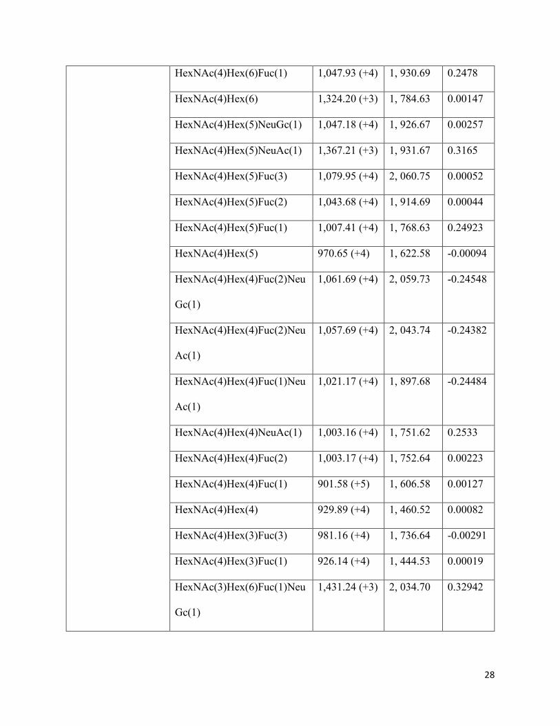
28
HexNAc(4)Hex(6)Fuc(1) 1,047.93 (+4) 1, 930.69 0.2478
HexNAc(4)Hex(6) 1,324.20 (+3) 1, 784.63 0.00147
HexNAc(4)Hex(5)NeuGc(1) 1,047.18 (+4) 1, 926.67 0.00257
HexNAc(4)Hex(5)NeuAc(1) 1,367.21 (+3) 1, 931.67 0.3165
HexNAc(4)Hex(5)Fuc(3) 1,079.95 (+4) 2, 060.75 0.00052
HexNAc(4)Hex(5)Fuc(2) 1,043.68 (+4) 1, 914.69 0.00044
HexNAc(4)Hex(5)Fuc(1) 1,007.41 (+4) 1, 768.63 0.24923
HexNAc(4)Hex(5) 970.65 (+4) 1, 622.58 -0.00094
HexNAc(4)Hex(4)Fuc(2)Neu
Gc(1)
1,061.69 (+4) 2, 059.73 -0.24548
HexNAc(4)Hex(4)Fuc(2)Neu
Ac(1)
1,057.69 (+4) 2, 043.74 -0.24382
HexNAc(4)Hex(4)Fuc(1)Neu
Ac(1)
1,021.17 (+4) 1, 897.68 -0.24484
HexNAc(4)Hex(4)NeuAc(1) 1,003.16 (+4) 1, 751.62 0.2533
HexNAc(4)Hex(4)Fuc(2) 1,003.17 (+4) 1, 752.64 0.00223
HexNAc(4)Hex(4)Fuc(1) 901.58 (+5) 1, 606.58 0.00127
HexNAc(4)Hex(4) 929.89 (+4) 1, 460.52 0.00082
HexNAc(4)Hex(3)Fuc(3) 981.16 (+4) 1, 736.64 -0.00291
HexNAc(4)Hex(3)Fuc(1) 926.14 (+4) 1, 444.53 0.00019
HexNAc(3)Hex(6)Fuc(1)Neu
Gc(1)
1,431.24 (+3) 2, 034.70 0.32942

29
HexNAc(3)Hex(6)Fuc(1)Neu
Ac(1)
1,402.56 (+3) 2, 018.70 0.33363
HexNAc(3)Hex(6)NeuGc(1) 1,037.41 (+4) 1, 888.64 0.24807
HexNAc(3)Hex(6)Fuc(1) 996.91 (+4) 1, 727.61 0.00239
HexNAc(3)Hex(6) 1,280.18 (+3) 1, 581.55 -0.01264
HexNAc(3)Hex(5)Fuc(1) 956.39 (+4) 1, 565.56 0.00357
HexNAc(3)Hex(5) 919.8 (+4) 1, 419.50 -0.00045
HexNAc(3)Hex(4)Fuc(2) 915.88 (+4) 1, 549.56 0.00085
HexNAc(3)Hex(4)Fuc(1) 915.64 (+4) 1, 403.50 0.00039
HexNAc(3)Hex(3)Fuc(1) 875.37 (+4) 1, 241.45 -0.00017
HexNAc(3)Hex(3) 838.85 (+4) 1, 095.39 0.00015
HexNAc(2)Hex(5) 869.11 (+4) 1, 216.42 0.00035
HexNAc(2)Hex(6) 909.62 (+4) 1, 378.47 0.00027
HexNAc(2)Hex(7) 950.13 (+4) 1, 540.52 -0.00177
HexNAc(2)Hex(8) 1,320.51 (+4) 1, 702.58 -0.01245
Site 6: Asparagine
177 (NLSK)
Top of the head
HexNAc(5)Hex(4)Fuc(1)Neu
Ac(1)
1,553.65 (+2) 2, 100.76 -0.0034
HexNAc(4)Hex(6) 1,166.17 (+3) 1, 784.63 0.00055
HexNAc(2)Hex(8) 1,354.56 (+2) 1, 702.58 0.00196
HexNAc(2)Hex(7) 1,273.53 (+2) 1, 540.52 0.00127
HexNAc(2)Hex(6) 1,193.00 (+2) 1, 378.47 0.0015
HexNAc(2)Hex(5) 1,111.98 (+2) 1, 216.42 0.00069
HexNAc(2) 585.06 (+4) 406.15 -0.00064

30
Site 7: Asparagine
303 (NSSL)
Stalk
HexNAc(6)Hex(4) 1,657.73 (+4) 1, 866.68 0.25101
HexNAc(6)Hex(3) 1,621.22 (+4) 1, 704.63 0.25377
HexNAc(4)Hex(5)NeuAc(1) 1,686.49 (+4) 1, 913.67 -0.24401
HexNAc(4)Hex(4)NeuGc(1) 1,649.97 (+4) 1, 767.61 -0.25517
HexNAc(3)Hex(6)NeuAc(1) 1,659.22 (+4) 1, 872.64 0.25161
HexNAc(3)Hex(4)Fuc(1)Neu
Ac(1)
1,614.70 (+4) 1, 694.60 0.2467
Site 8: Asparagine
498 (NGTY)
Stalk
HexNAc(8)Hex(8)Fuc(1) 1,342.18 (+3) 3, 067.11 -0.00606
HexNAc(7)Hex(7)Fuc(1)Neu
Ac(2)
1,414.88 (+3) 3, 284.17 0.3354
HexNAc(6)Hex(6) 1,050.08 (+3) 2, 190.79 0.00079
HexNAc(6)Hex(5)Fuc(1)Neu
Ac(2)NeuGc(1)
1,341.19 (+3) 3, 064.07 0.02
HexNAc(6)Hex(3)Fuc(2)Neu
Gc(1)
1,471.89 (+3) 2, 303.84 -0.34158
HexNAc(5)Hex(7) 1,415.21 (+3) 2, 149.76 -0.33692
HexNAc(5)Hex(5)Fuc(1)Neu
Ac(1)
1,458.23 (+3) 2, 262.81 -0.33147
HexNAc(5)Hex(5)NeuGc(1) 1,409.55 (+3) 2, 132.75 -0.32933
HexNAc(5)Hex(5)Fuc(1) 1,465.08 (+2) 1, 971.71 0.00223
HexNAc(5)Hex(5) 1,307.84 (+3) 1, 825.66 0.00225
HexNAc(5)Hex(4)Fuc(1)Neu
Ac(1)
1,401.21 (+3) 2, 100.76 -0.32961

31
HexNAc(5)Hex(4)Fuc(1)Neu
Gc(1)
1,409.55 (+3) 2, 116.75 -0.32763
HexNAc(5)Hex(4)NeuGc(1) 1,384.87 (+3) 1, 970.69 -0.00099
HexNAc(5)Hex(3)Fuc(1)Neu
Gc(1)
1,355.53 (+3) 1, 945.70 -0.32857
HexNAc(4)Hex(6) 1,293.83 (+3) 1, 784.63 -0.00351
HexNAc(4)Hex(5)Fuc(1)Neu
Gc(1)
1,390.54 (+4) 2, 075.72 -0.32938
HexNAc(4)Hex(5)NeuGc(1) 1,366.19 (+3) 1, 929.67 0.00128
HexNAc(4)Hex(5)Fuc(3) 1,386.20 (+3) 2, 060.75 -0.00084
HexNAc(4)Hex(5)Fuc(2) 1,006.74 (+3) 1, 914.69 0.00054
HexNAc(4)Hex(5)Fuc(1) 1,288.83 (+3) 1, 768.63 -0.00049
HexNAc(4)Hex(5) 1,101.78 (+3) 1, 622.58 0.00172
HexNAc(4)Hex(4)Fuc(2)Neu
Ac(1)
1,385.21 (+3) 2, 043.74 -0.32697
HexNAc(4)Hex(4)Fuc(1)Neu
Gc(1)
1,360.86 (+3) 1, 913.67 0.32938
HexNAc(4)Hex(4)Fuc(1)Neu
Ac(1)
1,336.52 (+3) 1, 987.68 -0.33167
HexNAc(4)Hex(4)Fuc(1) 943.87 (+4) 1, 606.58 -0.00836
HexNAc(3)Hex(6)Fuc(1)Neu
Gc(1)
1,382.18 (+3) 2, 034.70 -0.34415
HexNAc(3)Hex(6)Fuc(1) 1,343.03 (+2) 1, 727.61 0.00136

32
HexNAc(3)Hex(5)NeuGc(1) 1,298.49 (+3) 1, 726.59 -0.00482
HexNAc(3)Hex(4)Fuc(1)Neu
Ac(1)
1,287.83 (+2) 1, 694.60 0.00233
HexNAc(3)Hex(4)NeuAc(1) 1,239.15 (+3) 1, 548.54 0.00166
HexNAc(3)Hex(4)Fuc(2) 1,239.15 (+3) 1, 549.56 -0.01108
HexNAc(3)Hex(4) 1,107.95 (+2) 1, 257.44 0.00098
HexNAc(2)Hex(8) 1,330.49 (+2) 1, 702.58 -0.02006
The structural information obtained through CID fragmentation, albeit limited, still allows to
discriminate between high mannose, complex or hybrid glycans in some cases. A high mannose
structure can be assigned to the monosaccharide compositions in the form HexNAc(2)Hex(5-12),
as long as the sequential fragmentation confirms the neutral loss of only hexose residues in the
antennae from the glycopeptide Y ion series. Complex and hybrid glycans can be distinguished
when diagnostic Y ions are detected, such as Pep+HexNAc(2)Hex(4), for compositions with more
than 2 HexNAc residues. In such cases, at least one of the HexNAc residues must be in the
antennae, thus ruling out a high mannose glycan. Additionally, the Pep+HexNAc(2)Hex(4)
fragment confirms that one hexose must be attached to one glucose residue from the trimannosyl
core. Both these features combined can only occur in hybrid glycans, since only HexNAc residues
are attached to the trymannosyl core in complex glycans. Figure 3B shows a representative CID
spectrum with two diagnostic ions (Pep+HexNAc(3)Hex(5)+3 and
Pep+HexNAc(3)Hex(5)Fuc(1)+3) for a hybrid structure.
Other diagnostic ions shed further structural information. A 512 m/z peak in the lower m/z range
of the CID spectra corresponds to a Hex(1)HexNAc(1)Fuc(1) glycan B ion, characteristic of an
outer arm fucose. Conversely, core fucosylation can be determined from the detection of Y ions
such as Pep+HexNAc(1)Fuc(1) and Pep+HexNAc(2)Fuc(1). The latter fragments can also be
observed in some HCD spectra to further confirm fucose attachment to the chitobiose core. Special
consideration was given to glycans with a mass consistent with isobaric glycan compositions
containing either Fuc+NeuGc or Hex+NeuAc. In these cases, not all spectra contained sufficient

33
information to derive the correct composition. Nonetheless, diagnostic ions (NeuAc (292.10 m/z),
NeuAc-H2O (274.09 m/z), NeuGc (308.09 m/z), NeuGc-H2O (290.09 m/z)) were present in some
HCD spectra and were used to identify the correct composition. Moreover, glycan B fragments
corresponding to HexNAc-Gal-NeuAc (657 m/z) or HexNAc-Gal-NeuGc (673 m/z) detected in
CID were also present in some spectra.
Residues N142 and N498 displayed a great variety of glycan monomeric compositions.
Conversely, for sites N28, N40, N71 and N104, only a very limited number of compositions were
detected. Overall, complex and hybrid glycans were predominantly found. Nonetheless, high
mannose glycans are also present in N142 and N177, both located on the top of the globular head.
Fucosylation was observed in most glycopeptides and was confirmed by the presence of fucose
residues in the CID spectra or diagnostic ions in the HCD spectra. Core fucoses are predominant,
but outer arm fucoses were also found in some glycoforms. Highly bulky complex glycans were
found on N498.
Discussion
Sequence-based analysis has previously been employed by others to study the variation in the
glycosylation profile of several strains during virus evolution [27, 28]. Although N-glycosylation
prediction contributes greatly to the understanding of glycosite variation across strains, this method
only provides a prediction that relies on primary sequence context, and the actual occupancy of
glycosites must be confirmed experimentally. Moreover, it is known that the location of the sequon
can affect glycan maturation, thereby resulting in different sets of glycoforms for each site
depending on the accessibility of N-glycosyltransferases [29]. To this end, we used mass
spectrometry analysis to confirm the occupancy of glycosites predicted by the NetNGlyc 1.0
Server for the A/New Caledonia/20/1999 H1 protein. Others have shown that hemagglutinin from
egg-grown viruses and human cell lines display the same occupancy profile [30]. Therefore, the
results obtained in this study are relevant to the understanding of influenza virus adaptation in
humans.
The occupancy profile obtained through HCD/CID-MS/MS of tryptic glycopeptides was
consistent with the prediction by the NetNGlyc 1.0 server. All eight sequons confirmed
experimentally had been predicted by the server. Indeed, N28 (and not N27), was shown to be
glycosylated as was predicted. The transfer of N-glycan precursors to asparagine residues in

34
endoplasmic reticulum (ER) bound ribosomes is affected by conformational constraints as well as
inaccessibility due to steric hindrance from nearby residues. To this end, since N28 is occupied, it
is unlikely that an olygosaccharyltransferase can act upon asparagine N27. The positive prediction
for site N557 was not accurate, due to the location of the residue within the cytoplasmic domain
of the protein. Nonetheless, such inaccuracies can easily be identified to prevent misleading
interpretations when performing sequence-based analyses.
Glycosites N142 and N177 are of particular relevance for the study of immunogenicity and
virulence. These residues are both located on the receptor binding site (RBS) and the attachment
of oligosaccharides can mask the antigenic Sa region. Remarkably, both acceptor sites were absent
in the 1918 and 2009 pandemic strains. Wei et al. showed that mice display cross-neutralization
of 1918 and 2009 pandemic viruses after vaccination with the 1918 strain, whereas such antibodies
did not protect against seasonal strains – providing evidence that the introduction of N-
glycosylation sites on the vicinity of the RBS can mask antigenic recognition by pandemic
antibodies [31].
A vast heterogeneity of glycan compositions was found on N142 (Table 2), which is even more
notable taking into account that the predicted compositions can encompass several glycan
isoforms; meaning that the actual heterogeneity is presumably broader. Among the predicted
oligosaccharides attached to N142 are glycan compositions consistent with complex, high
mannose and hybrid glycans. The vast complexity encountered on this asparagine residue can be
attributed to the high surface exposure, providing accessibility for N-glycan processing and
maturation (Fig. 4).
In the case of glycosite N177, a much narrower variety of glycoforms was detected. However, the
intensities for these glycopeptides were much lower compared to those glycopeptides containing
site N142. Therefore, many glycopeptides for N177 could have been suppressed and missed in the
analysis, which constitutes a limitation of the method. Still, the MS/MS analysis predicted the
occupancy of high mannose, hybrid and complex glycoforms.
The presence of terminal sialic acid residues in several of the predicted glycoforms attached to
both N142 and N177 could play an important role in the immunogenicity of the strain. Ohuchi et
al. suggested that the presence of terminal NeuAc and NeuGc on HA glycans in the vicinity of the
receptor binding sites may impair binding to sialic acid-containing receptors [32]. Although the

35
effect of the presence of sialic acid residues on immune cell recognition in this case is unknown,
the bulkiness and negative charge of NeuAc and NeuGc would likely further impair an immune
response. Overall, terminal sialylation of both cell surfaces and glycoproteins has been shown to
decrease antigenicity [33, 34]. Therefore, the attachment of negatively charged terminal residues
in embryonated-egg vaccine production could be a drawback in terms of HA antigenicity
compared to other recombinant hemagglutinin based vaccines, where sialylation can be controlled
or non-existent, as is the case in the baculovirus expression system [35].
Glycosites N71 and N104 are located on the side of the globular head (Fig. 4). In the case of N71,
only one glycan with a composition consistent with a non-fucosylated hybrid glycan was found
(Table 2). For N104, one glycan with a complex-like composition was detected. Others have
proposed that the occupancy of site N104 might be sterically hindered by glycan attachment on
N71 due to their close proximity (Fig. 4) [20]. Moreover, it has been argued that since both
glycosites are able to shield antigenic site Ca2, glycosylation on N71 could render the occupancy
of N104 unnecessary to mask antigenic site Ca2 [20]. It was confirmed that both N71 and N104
are indeed glycosylated in egg-grown viruses. It was unexpected, however, to find such a limited
set of glycan compositions in these sites, in contrast to other positions such as N142, N177 and
N498. As discussed previously, these results are potentially due to a limited detection of the
glycopeptides containing these acceptor sites. However, the close proximity of both residues also
suggests that glycan processing might be impaired due to steric hindrance.
The detection of highly bulky complex glycans on N498 (e.g., HexNAc(8)Hex(8)Fuc(1) and
HexNAc(7)Hex(7)Fuc(1)NeuAc(2)) suggests the presence of poly-N-acetyllactosamine groups,
which are commonly found on membrane-proximal glycosites [36, 37]. The close proximity of
N498 to the viral membrane (Fig. 4B) does not seem to decrease the degree of glycan maturation
on this asparagine residue, resulting in a high complexity of structures.
In this mass spectrometry setup, some tryptic glycopeptides produce considerably higher
intensities than others. This causes that more information on glycan microheterogeneity can be
gathered on those glycopeptides that ionize with a higher efficiency and whose m/z value is better
detected under these experimental conditions, whereas only limited information can be derived
from other regions. This was likely the cause of the limited glycopeptide detection for sites N28,
N40, N71 and N104. Unfortunately, this drawback prevents an appropriate comparison of glycan

36
microheterogeneity among glycosites. Obtaining a more comprehensive profile is required to
better understand the role of glycan structures on the biological properties of the virus. Still, these
results allowed to confirm glycan attachment on these sites.
An advantage of utilizing reducing SDS-PAGE as a preliminary fractionation step is that analysing
HA0, HA1 and HA2 separately provides a more comprehensive characterization of glycan
microheterogeneity, owing to the fact that the lower overall presence of non-glycosylated peptides
in HA1 or HA2 isolated fractions (compared to HA0) could allow the detection of certain
glycopeptides with low total ion current (TIC). In fact, most of the distinct glycan compositions
detected on site N498 come from the analysis of HA2 and were not present in the analysis of HA0
(band 4 in figure 2). This is likely due to the large presence of non-glycosylated peptides in HA0,
which can suppress the signal of N498 glycopeptides.
The experimental confirmation of glycosylation sites is highly important in the study of influenza
viral evolution. Furthermore, there is evidence that egg and mammalian cell derived HA display
the same occupancy profile [30]. Consequently, characterizing further egg-derived strains with
this method would also yield site occupancy information relevant to other approved production
platforms, such as cell-based and recombinant technologies. Currently, egg-based vaccines
comprise the overwhelming majority of available influenza vaccine products. However, the FDA
has recently approved the cell-based Flucelvax (Seqirus), produced in Madin-Darby Canine
Kidney (MDCK) epithelial cells, and the recombinant Flublok (Protein Sciences Corporation),
produced in insect cells [38-40]. It is noteworthy to mention that the glycan structures found on
egg-derived viruses can differ significantly to those present on viruses that replicate in alternative
cell substrates [30, 41, 42].
Conclusion
The results presented in this study confirmed the glycan occupancy profile of A/New
Caledonia/20/1999 hemagglutinin, validated the prediction by the NetNGlyc 1.0 Server and
characterized the site-specific structural heterogeneity of the attached glycans. The mass
spectrometry approach employed was able to furnish information on sequon occupancy and glycan
structures in a single run. This efficient strategy can be extended to study further influenza
glycoproteins in order to attain deeper knowledge of the role of N-glycosylation in viral adaptation
mechanisms. Site-specific mass spectrometry methods, however, typically provide only partial

37
information on the structure of the oligosaccharides. To this regard, additional work should be
carried out to elucidate more detailed structural features to assess the impact of glycoforms on
parameters such as antigen masking, immunogenicity, SA receptor binding affinity and viral
membrane resistance to surfactant treatment. The latter is highly relevant for split-virus vaccine
manufacturing, where a surfactant is employed to disrupt the viral membrane. Gaining further
insight into the glycosylation characteristics of influenza viruses can therefore potentially improve
the manufacturing process of split-virus vaccines, the selection of strains for the composition of
seasonal vaccines and the development of recombinant products.
Disclosure of potential conflicts of interest
The authors declare that they have no conflict of interest.
Funding
N/A
Acknowledgements
The authors would like to thank Sanofi-Pasteur for financial support and for providing the virus
samples. We thank the Faculty of Pharmacy of The University of Sydney for financial
contribution. We would also like to thank Ben Parker and Stuart Cordwell for valuable discussions.
EC acknowledges the Ministry of Science, Technology and Telecommunications of the Republic
of Costa Rica for postgraduate scholarship.
References
[1]. J.K. Taubenberger, J.C. Kash, Influenza Virus Evolution, Host Adaptation, and Pandemic
Formation, Cell Host & Microbe, 7 (2010) 440-451. doi:410.1016/j.chom.2010.1005.1009.
[2]. M.I. Nelson, E.C. Holmes, The evolution of epidemic influenza, Nat Rev Genet, 8 (2007)
196-205. doi:110.1038/nrg2053.
[3]. C.W. Potter, J.S. Oxford, Determinants of immunity to influenza infection in man, British
medical bulletin, 35 (1979) 69-75. PMID:367490.

38
[4]. N. Mancini, L. Solforosi, N. Clementi, D. De Marco, M. Clementi, R. Burioni, A potential
role for monoclonal antibodies in prophylactic and therapeutic treatment of influenza, Antiviral
research, 92 (2011) 15-26. doi:10.1016/j.antiviral.2011.1007.1013.
[5]. K. Stohr, D. Bucher, T. Colgate, J. Wood, Influenza virus surveillance, vaccine strain
selection, and manufacture, Methods in molecular biology (Clifton, N.J.), 865 (2012) 147-162.
[6]. B.E. Johansson, D.J. Bucher, E.D. Kilbourne, Purified influenza virus hemagglutinin and
neuraminidase are equivalent in stimulation of antibody response but induce contrasting types of
immunity to infection, Journal of virology, 63 (1989) 1239-1246. PMID:2915381.
[7]. B.E. Johansson, T.M. Moran, C.A. Bona, S.W. Popple, E.D. Kilbourne, Immunologic
response to influenza virus neuraminidase is influenced by prior experience with the associated
viral hemagglutinin. II. Sequential infection of mice simulates human experience, Journal of
immunology (Baltimore, Md. : 1950), 139 (1987) 2010-2014. PMID:3624874.
[8]. R.K. Virk, V. Gunalan, P.A. Tambyah, Influenza infection in human host: challenges in
making a better influenza vaccine, Expert review of anti-infective therapy, 14 (2016) 365-375.
[9]. N. Sriwilaijaroen, Y. Suzuki, Molecular basis of the structure and function of H1
hemagglutinin of influenza virus, Proceedings of the Japan Academy. Series B, Physical and
biological sciences, 88 (2012) 226-249. PMID:22728439.
[10]. C.C. Wang, J.R. Chen, Y.C. Tseng, C.H. Hsu, Y.F. Hung, S.W. Chen, C.M. Chen, K.H.
Khoo, T.J. Cheng, Y.S. Cheng, J.T. Jan, C.Y. Wu, C. Ma, C.H. Wong, Glycans on influenza
hemagglutinin affect receptor binding and immune response, Proceedings of the National
Academy of Sciences of the United States of America, 106 (2009) 18137-18142.
doi:18110.11073/pnas.0909696106.
[11]. K. Wanzeck, K.L. Boyd, J.A. McCullers, Glycan shielding of the influenza virus
hemagglutinin contributes to immunopathology in mice, American journal of respiratory and
critical care medicine, 183 (2011) 767-773. doi:710.1164/rccm.201007-201184OC.
[12]. W. Chen, Y. Zhong, Y. Qin, S. Sun, Z. Li, The Evolutionary Pattern of Glycosylation Sites
in Influenza Virus (H5N1) Hemagglutinin and Neuraminidase, PLoS ONE, 7 (2012) e49224.
doi:49210.41371/journal.pone.0049224.

39
[13]. J.I. Kim, M.S. Park, N-linked glycosylation in the hemagglutinin of influenza A viruses,
Yonsei medical journal, 53 (2012) 886-893. doi:810.3349/ymj.2012.3353.3345.3886.
[14]. X. Sun, A. Jayaraman, P. Maniprasad, R. Raman, K.V. Houser, C. Pappas, H. Zeng, R.
Sasisekharan, J.M. Katz, T.M. Tumpey, N-Linked Glycosylation of the Hemagglutinin Protein
Influences Virulence and Antigenicity of the 1918 Pandemic and Seasonal H1N1 Influenza A
Viruses, Journal of virology, 87 (2013) 8756-8766. doi:8710.1128/jvi.00593-00513.
[15]. R.P. de Vries, E. de Vries, B.J. Bosch, R.J. de Groot, P.J. Rottier, C.A. de Haan, The
influenza A virus hemagglutinin glycosylation state affects receptor-binding specificity, Virology,
403 (2010) 17-25. doi:10.1016/j.virol.2010.1003.1047.
[16]. X. Zhang, S. Chen, D. Yang, X. Wang, J. Zhu, D. Peng, X. Liu, Role of stem glycans attached
to haemagglutinin in the biological characteristics of H5N1 avian influenza virus, The Journal of
general virology, 96 (2015) 1248-1257. doi:1210.1099/vir.1240.000082.
[17]. P.C. Roberts, W. Garten, H.D. Klenk, Role of conserved glycosylation sites in maturation
and transport of influenza A virus hemagglutinin, Journal of virology, 67 (1993) 3048-3060.
PMID:8497042.
[18]. P.L. Herve, V. Lorin, G. Jouvion, B. Da Costa, N. Escriou, Addition of N-glycosylation sites
on the globular head of the H5 hemagglutinin induces the escape of highly pathogenic avian
influenza A H5N1 viruses from vaccine-induced immunity, Virology, 486 (2015) 134-145.
110.1016/j.virol.2015.1008.1033.
[19]. R.A. Medina, S. Stertz, B. Manicassamy, P. Zimmermann, X. Sun, R.A. Albrecht, H. Uusi-
Kerttula, O. Zagordi, R.B. Belshe, S.E. Frey, T.M. Tumpey, A. Garcia-Sastre, Glycosylations in
the globular head of the hemagglutinin protein modulate the virulence and antigenic properties of
the H1N1 influenza viruses, Science translational medicine, 5 (2013) 187ra170.
doi:110.1126/scitranslmed.3005996.
[20]. S. Sun, Q. Wang, F. Zhao, W. Chen, Z. Li, Glycosylation Site Alteration in the Evolution of
Influenza A (H1N1) Viruses, PLoS ONE, 6 (2011) e22844.
doi:22810.21371/journal.pone.0022844.

40
[21]. M.D. Tate, E.R. Job, Y.M. Deng, V. Gunalan, S. Maurer-Stroh, P.C. Reading, Playing hide
and seek: how glycosylation of the influenza virus hemagglutinin can modulate the immune
response to infection, Viruses, 6 (2014) 1294-1316. doi:1210.3390/v6031294.
[22]. R. Xu, D.C. Ekiert, J.C. Krause, R. Hai, J.E. Crowe, I.A. Wilson, Structural basis of pre-
existing immunity to the 2009 H1N1 pandemic influenza virus, Science (New York, N.Y.), 328
(2010) 357-360. doi:310.1126/science.1186430.
[23]. M. Igarashi, K. Ito, R. Yoshida, D. Tomabechi, H. Kida, A. Takada, Predicting the Antigenic
Structure of the Pandemic (H1N1) 2009 Influenza Virus Hemagglutinin, PLoS ONE, 5 (2010)
e8553. doi:8510.1371/journal.pone.0008553.
[24]. NetNGlyc, NetNGlyc 1.0 Server, 2015.
[25]. S. Doll, A.L. Burlingame, Mass spectrometry-based detection and assignment of protein
posttranslational modifications, ACS chemical biology, 10 (2015) 63-71. doi:10.1021/cb500904b.
[26]. M. Thaysen-Andersen, N.H. Packer, Advances in LC–MS/MS-based glycoproteomics:
Getting closer to system-wide site-specific mapping of the N- and O-glycoproteome, Biochim.
Biophys. Acta, Proteins Proteomics, 1844 (2014) 1437-1452.
doi:1410.1016/j.bbapap.2014.1405.1002.
[27]. S.R. Das, S.E. Hensley, A. David, L. Schmidt, J.S. Gibbs, P. Puigbò, W.L. Ince, J.R.
Bennink, J.W. Yewdell, Fitness costs limit influenza A virus hemagglutinin glycosylation as an
immune evasion strategy, Proceedings of the National Academy of Sciences of the United States
of America, 108 (2011) E1417–E1422. doi:1410.1073/pnas.1108754108.
[28]. B.J. DeKosky, G.C. Ippolito, R.P. Deschner, J.J. Lavinder, Y. Wine, B.M. Rawlings, N.
Varadarajan, C. Giesecke, T. Dorner, S.F. Andrews, P.C. Wilson, S.P. Hunicke-Smith, C.G.
Willson, A.D. Ellington, G. Georgiou, High-throughput sequencing of the paired human
immunoglobulin heavy and light chain repertoire, Nat Biotechnol 31 (2013) 166-169.
doi:110.1038/nbt.2492.
[29]. M. Thaysen-Andersen, N.H. Packer, Site-specific glycoproteomics confirms that protein
structure dictates formation of N-glycan type, core fucosylation and branching, Glycobiology, 22
(2012) 1440-1452. doi:1410.1093/glycob/cws1110.

41
[30]. Y. An, J.A. Rininger, D.L. Jarvis, X. Jing, Z. Ye, J.J. Aumiller, M. Eichelberger, J.F. Cipollo,
Comparative Glycomics Analysis of Influenza Hemagglutinin (H5N1) Produced in Vaccine
Relevant Cell Platforms, J Proteome Res, 12 (2013) 3707-3720. doi:3710.1021/pr400329k.
[31]. C.-J. Wei, J.C. Boyington, K. Dai, K.V. Houser, M.B. Pearce, W.-P. Kong, Z.-y. Yang, T.M.
Tumpey, G.J. Nabel, Cross-Neutralization of 1918 and 2009 Influenza Viruses: Role of Glycans
in Viral Evolution and Vaccine Design, Science translational medicine, 2 (2010) 24ra21-24ra21.
doi:10.1126/scitranslmed.3000799.
[32]. M. Ohuchi, R. Ohuchi, A. Feldmann, H.D. Klenk, Regulation of receptor binding affinity of
influenza virus hemagglutinin by its carbohydrate moiety, Journal of virology, 71 (1997) 8377-
8384. PMID:9343193.
[33]. R. Schauer, Sialic acids as antigenic determinants of complex carbohydrates, Advances in
experimental medicine and biology, 228 (1988) 47-72. PMID:2459931.
[34]. R. Schauer, Sialic acids as regulators of molecular and cellular interactions, Current opinion
in structural biology, 19 (2009) 507-514. doi:510.1016/j.sbi.2009.1006.1003.
[35]. X. Shi, D.L. Jarvis, Protein N-Glycosylation in the Baculovirus-Insect Cell System, Curr
drug targets, 8 (2007) 1116-1125. PMID:17979671.
[36]. S.Y. Mir-Shekari, D.A. Ashford, D.J. Harvey, R.A. Dwek, I.T. Schulze, The glycosylation
of the influenza A virus hemagglutinin by mammalian cells. A site-specific study, The Journal of
biological chemistry, 272 (1997) 4027-4036. doi:4010.1074/jbc.4272.4027.4027.
[37]. M. Fukuda, J.L. Guan, J.K. Rose, A membrane-anchored form but not the secretory form of
human chorionic gonadotropin-alpha chain acquires polylactosaminoglycan, The Journal of
biological chemistry, 263 (1988) 5314-5318. PMID:2451668.
[38]. P.C. Soema, R. Kompier, J.-P. Amorij, G.F.A. Kersten, Current and next generation
influenza vaccines: Formulation and production strategies, Eur J Phar Biopharm, 94 (2015) 251-
263.
[39]. I. Manini, A. Domnich, D. Amicizia, S. Rossi, T. Pozzi, R. Gasparini, D. Panatto, E.
Montomoli, Flucelvax (Optaflu) for seasonal influenza, Expert review of vaccines, 14 (2015) 789-
804.

42
[40]. L.P. Yang, Recombinant trivalent influenza vaccine (flublok((R))): a review of its use in the
prevention of seasonal influenza in adults, Drugs, 73 (2013) 1357-1366.
[41]. S. Zhang, Comparative Characterization of the Glycosylation Profiles of an Influenza
Hemagglutinin Produced in Plant and Insect Hosts, Proteomics, 12 (2012) 1269-1288.
[42]. M. Butler, M. Spearman, The choice of mammalian cell host and possibilities for
glycosylation engineering, Curr Opin Biotechnol, 30 (2014) 107-112.
[43]. R.B. Squires, J. Noronha, V. Hunt, A. García‐Sastre, C. Macken, N. Baumgarth, D. Suarez,
B.E. Pickett, Y. Zhang, C.N. Larsen, A. Ramsey, L. Zhou, S. Zaremba, S. Kumar, J. Deitrich, E.
Klem, R.H. Scheuermann, Influenza Research Database: an integrated bioinformatics resource for
influenza research and surveillance, Influenza Other Respir Viruses, 6 (2012) 404-416.
doi:410.1111/j.1750-2659.2011.00331.x.

43
The State-of-Play and Future of Antibody Therapeutics
Graphical abstract
Chapter 3

44
Chapter 3 – Authorship declaration statement
The following chapter is a literature review published in the journal Advanced Drug Delivery
Reviews as:
Z. Elgundi, M. Reslan, E. Cruz, V. Sifniotis, V. Kayser. The State-of-play and Future of Antibody
Therapeutics. Advanced Drug Delivery Reviews, 122 (2017), 2-19.
E. Cruz wrote the “Antibody-Drug Conjugates” and “Optimization of antibody bioavailability and
delivery” sections, co-wrote the “Fc-engineered antibodies for enhanced effector functions” and
contributed to the “Bispecifics” section and to writing the abstract and conclusions. E. Cruz created
the figures featured in the review, including the graphical abstract. Additionally, the author
provided critical review of the manuscript.
The section “Biosimilars, guidelines and challenges” contained in the original article has been
omitted since it is not directly relevant to the subsequent chapters.
Permission to include the published material has been granted by the corresponding author.
Esteban Cruz, Signature: 19th of August, 2019
As corresponding author and supervisor for this candidature, I hereby confirm that this authorship
declaration statement is complete and accurate
Veysel Kayser, Signature: 19th of August, 2019

45
Abstract
It has been over four decades since the development of monoclonal antibodies (mAbs) using a
hybridoma cell line was first reported. Since then more than thirty therapeutic antibodies have been
marketed, mostly as oncology, autoimmune and inflammatory therapeutics. While antibodies are
very efficient, their cost-effectiveness has always been discussed owing to their high costs,
accumulating to more than one billion dollars from preclinical development through to market
approval. Because of this, therapeutic antibodies are inaccessible to some patients in both
developed and developing countries. The growing interest in biosimilar antibodies as affordable
versions of therapeutic antibodies may provide alternative treatment options as well potentially
decreasing costs. As certain markets begin to capitalize on this opportunity, regulatory authorities
continue to refine the requirements for demonstrating quality, efficacy and safety of biosimilar
compared to originator products. In addition to biosimilars, innovations in antibody engineering
are providing the opportunity to design biobetter antibodies with improved properties to maximize
efficacy. Enhancing effector function, antibody drug conjugates (ADC) or targeting multiple
disease pathways via multi-specific antibodies are being explored. The manufacturing process of
antibodies is also moving forward with advancements relating to host cell production and
purification processes. Studies into the physical and chemical degradation pathways of antibodies
are contributing to the design of more stable proteins guided by computational tools. Moreover,
the delivery and pharmacokinetics of antibody-based therapeutics are improving as optimized
formulations are pursued through the implementation of recent innovations in the field.
Introduction
Over the past twenty years, therapeutic antibodies have rapidly become the leading product within
the biopharmaceutical market. In 2013, therapeutic antibodies represented 50% of the $140 billion
taken by the biopharmaceutical market with sales growing from $39 billion in 2008 to $75 billion
in 2013 [1]. There are currently more than thirty therapeutic antibodies approved for established
markets such as the United States and Europe with over three hundred antibody-based products in
clinical development [1-3]. Therapeutic antibodies are no longer full-length, naked mouse
antibodies; advancements in antibody engineering technologies, novel antigen discovery strategies
and progress in deciphering disease pathways have all generated robust interest, resources and
investment in antibody development. In this review, we will discuss developments in the field of

46
therapeutic antibodies, the growth of biosimilars and pay particular attention to targeting
degradation pathways of antibodies to produce more stable biobetter antibodies and formulations.
Antibody discovery strategies
The generation of early antibodies relied on the immunization of mice or other mammals with the
desired antigen target. This resulted in multiple antibodies directed at different epitopes of the
antigen secreted by a mixed population of B cells with each cell secreting only one specific
antibody (i.e. polyclonal). Unfortunately, secreting B cells can only replicate a limited number of
times, therefore rendering mass production all but impossible. The ground-breaking hybridoma
technology developed by Kohler and Milstein allowed antibody secreting cells from the spleen of
immunized animals to be fused with immortalized non-antibody secreting cells, thus resulting in
cells that would divide continuously when cultivated in permissive conditions [4, 5]. Although
the first recombinant antibodies were produced using this technology, including the first approved
therapeutic antibody muromonab-CD3 (Orthoclone OKT®3) in 1986 for preventing kidney
transplant rejection, hybridoma production presented some drawbacks. Hybridomas can be labour
intensive, low yielding or genetically unstable [6]. More importantly though, the antibody
sequences originated from an immunized animal and consequently had the potential of triggering
an immune response in humans. Therefore, further improvements were needed to yield antibodies
more human-like and safe. These technologies have evolved from chimeric antibodies that is,
grafting essential mouse amino acids needed for antigen binding onto a human antibody
framework [7, 8] to both in vitro and in vivo techniques for generating humanized antibodies.
The XenoMouse™ (Abgenix) and HuMab-Mouse® (Medarex) are transgenic mice developed in
parallel and in both, the endogenous murine heavy and kappa light chain genes are inactivated and
replaced with the equivalent human germline sequences [9, 10]. Injection of antigens into these
mice leads to development of ‘fully human’ antibodies that have undergone mouse somatic
hypermutation and selection to relatively high affinity. Validation of this technology came with
the regulatory approval of panitumumab (Vectibix®) in 2006; a fully human antibody directed
against epidermal growth factor receptor (EGFR) as treatment for advanced colorectal cancer [11].
Since then, RANK ligand-specific denosumab (Prolia®) has been approved for bone loss and
TNFα-specific golimumab (Simponi®) for rheumatoid arthritis.
47
An in vitro method for generating fully human antibodies can be accomplished by cloning and
screening large libraries of sufficiently diverse human antibody genes in combination with display
technology. The concept of display technology provides a direct physical link between a gene
(genotype) and the encoding antibody fragment (phenotype) to allow selection of genes that
encode a protein with the desired binding function. Phage display technology remains the most
widely used in vitro method for the display of large repertoires and for the selection of high affinity
antibodies to biologically relevant targets [6]. Phage display involves the expression of proteins
on the surface of filamentous phage via fusion with phage coat protein with the genetic sequence
packaged within, linking phenotype to genotype selection. When combined with antibody
libraries, phage display allows for rapid in vitro selection of antigen-specific antibodies and
recovery of their corresponding coding sequence [12-14]. This system is highly effective, robust
and amenable to high throughput processes for screening of >10+10 specificities [15]. The
diversity of phage display libraries is distinguishable by source and design: naïve [16], immune
[12], synthetic [17] and semi-synthetic [18]. The technology was first demonstrated for a single
chain variable fragment (scFv) [13]. with screening of other formats also introduced including
human antigen binding fragments (Fabs) [19], domain antibodies [20], camelid domain antibodies
[21], single domain shark antibodies [22], diabodies [23] and even whole IgG [24]. The first
approved human antibody isolated by phage display technology was adalimumab (Humira®)
which binds the cytokine TNFα. This antibody was first selected as a scFv expressed on the
surface of phage and was further engineered in human IgG1 format, providing major validation
for phage display technologies [25]. Adalimumab remains the most lucrative antibody product
generating global sales of $11 billion in 2013 [1]. The number of phage display-derived candidates
currently in clinical development further demonstrates the value of phage display as an established
and reliable drug discovery platform [26].
Other cell surface methodologies such as bacterial, baculovirus and yeast display present
thousands of copies of the displayed protein on the cell surface thereby allowing for quantitative
screening such as flow cytometry [27-29]. Yeast display has the additional feature of eukaryotic
protein folding pathways and as a result, is perhaps the most ideally suited system for the surface
display of mammalian secreted proteins such as antibodies. Alternatively, ribosome display is
used to screen large scFv and single variable domain libraries containing up to >10+13 mutant
clones [30]. The main distinguishing feature of this method is that large libraries can be generated

48
because the entire procedure is performed in vitro without the need for cell transformations. The
genotype and phenotype are linked through ribosomal complexes, consisting of messenger RNA
(mRNA), ribosome and encoded protein. The in vitro selection of antibodies from antibody
libraries based on target affinity is termed as “panning”. The method is an iterative process
whereby the population of target specific antibodies is enriched relative to the number of panning
rounds.
With the advent of next-generation sequencing (NGS), analysis of the natural and synthetic
repertoires from which libraries have been constructed has become possible [31-33]. In one
application, NGS was used to identify antigen-specific IgG antibodies from polyclonal serum of
immunized rabbits and mice when used in combination with affinity chromatography coupled to
LC-MS/MS [34]. The technology has been used to demonstrate its utility in selecting antibodies
with favourable properties. For example, Reddy et al. found that 21/27 (or 78%) of scFv they
constructed could bind the target antigen with nanomolar affinity after using NGS data to pair
together the most abundant variable heavy (VH) and variable light (VL) genes from immunized
mice [35]. Other studies have seen similar findings with natural VH:VL pairing limitations [36].
and without the limitation [37]. Interestingly, the method of repertoire mining of VH and VL
abundances using NGS of splenocytes isolated from immunized mice was compared with a phage
panning approach of the same cDNA [38]. While both methods provided completely different sets
of antibodies, specificity and affinities of the antibody clones were comparable.
Novel antibodies in approval and preclinical development stages
In 2015, eight therapeutic antibodies were granted market approval by the US Food and Drug
Administration (FDA) (Table 1). While full-length and IgG1 antibodies still dominate, a
humanized Fab also gained approval. Five of the eight approvals were to non-oncology indications
with two antibodies entering ‘first-in-class’ for the treatment of hyperlipidemia. The outlook for
2016 has been positive with six antibodies approved (as of November 2016), including a first-in-
class antibody to bacterial target Bacillus anthraxis. A number of full-length antibodies are
currently under biologics license application (BLA) submission and awaiting FDA approval or
have completed Phase III trials with endpoints met (Table 1).

49
Table 1. Novel full-length antibodies and fragments developed in recent years and their clinical
status.
Name Commerci
al Name
Company Target Indication (FDA
approved)
Format
APPROVED BY FDA IN 2015
Secukinumab Costentyx
®
Novartis IL-17 Psoriasis Whole IgG1
(fully
human)
Dinutuximab Unituxin® United
Therapeutics
Corporation
GD2 Neuroblastoma
(pediatric
patients)
Whole IgG1
(chimeric)
Alirocumab Praulent® Sanofi PCSK9 Hyperlipidemia Whole IgG1
(fully
human)
Evolocumab Repatha® Amgen PCSK9 Hyperlipidemia Whole IgG2
(fully
human)
Idarucizumab Praxbind® Boehringer
Ingelheim
Dabigatran Anti-coagulation
reversal
Fab fragment
(humanized)
Mepolizumab Nucala® GlaxoSmithKline IL-5 Asthma Whole IgG1
(humanized)
Necitumumab Portrazza® Eli Lilly EGFR Non-small cell
lung cancer
(NSCLC)
Whole IgG1
(fully
human)
Daratumumab Darzalex® Janssen Biotech CD38 Multiple
myeloma
Whole IgG1
(fully
human)
APPROVED BY FDA IN 2016 (as of November)
Reslizumab Cinquil® Teva IL-5 Asthma Whole IgG4
(humanized)
Ixekizumab Taltz® Eli Lilly IL-17a Psoriasis Whole IgG4
(humanized)

50
Obiltoxaximab Anthim® Elusys
Therapeutics
Bacillus
anthrax
Anthrax Whole IgG3
(humanized)
Atezolizumab Tecentriq
®
Genentech PD-L1 NSCLC, Bladder
cancer
Whole IgG1
(humanized)
Olaratumab (+
Doxorubicin)
Lartruvo® Eli Lilly PDGFRalph
a
Soft tissue
carcinoma
Whole IgG1
Bezlotoxumab Zinplava® Merck Clostridium
difficile
toxin B
Clostridium
difficile infection
Whole IgG1
(fully
human)
NOT YET APPROVED, TRIALS COMPLETED
Brodalumab
Valeant
Pharmaceuticals
IL-17R Psoriasis Whole IgG1
(fully
human)
Bimagrumab
Novartis ACVR2B Muscle loss and
weakness
Whole IgG1
(fully
human)
MABp1 Xilonix XBiotech IL-1alpha Colorectal cancer,
Cachexia
Whole IgG1
(‘true’
human)
Catumaxomab Removab Trion Pharma EPCAM/CD
3
Malignant ascites Whole
hybrid of rat
IgG2
variable and
mouse IgG2a
Fc regions
Guselkumab Janssen Biotech IL-23/p19 Psoriasis Whole IgG1
(fully
human)
Dupilumab Sanofi/Regeneron IL-4R Atopic Dermatitis Whole IgG4
(fully
human)
ONGOING PHASE III TRIALS
Ocrelizumab Ocrevus Roche CD20 Multiple Sclerosis Whole IgG1

51
(humanized)
Sirukumab
GlaxoSmithKline IL-6 Rheumatoid
arthritis
Whole IgG1
Sarilumab
Sanofi/Regeneron IL-6R Rheumatoid
arthritis
Whole IgG1
(fully
human)
Tildrakizumab
Sun
Pharmaceutical
IL-23/p19 Psoriasis Whole IgG1
(humanized)
Romosozumab
Amgen/UCB
Pharma
Sclerostin Osteoporosis Whole IgG2
(humanized)
Racotumomab Vaxira Recombio NGcGM3 NSCLC Whole IgG1
(mouse)
Clivatuzumab
tetraxetan
hPAM4-
Cide
Immunomedics Mucin1 Pancreatic cancer Whole IgG1
(humanized)
Ublituximab
TG Therapeutics CD20 Chronic
Lymphocytic
Leukemia
Whole IgG1
(chimeric)
Benralizumab
AstraZeneca IL-5R Asthma Whole IgG1
(humanized)
Caplacizumab
Ablynx vWF Thrombotic
thrombocytopenic
purpura
VH-VH
(humanized)
Lampalizumab
Roche CFD Macular
degeneration
Fab fragment
(humanized)
Avelumab
Merck/Pfizer PD-L1 Merkel Cell
Carcinoma
Whole IgG1
(fully
human)
Aducanumab Biogen Beta
amyloid
Alzheimer’s
Disease
Whole IgG1
(fully
human)
Abbreviations: GD = disialoganglioside; PCSK = proprotein convertase subtilisin/kexin; EGFR =
epidermal growth factor receptor; PD-L = programmed death-ligand; PDGFR = platelet-derived
growth factor receptor; ACVR = activin receptor; EPCAM = epithelial cell adhesion molecule;

52
NGcGM = N-glycolyl (NGc) gangliosides; vWF = Von Willebrand factor; CF = complement
factor.
There is expected to be an influx of antibodies in the coming years for the treatment of
inflammatory skin conditions including psoriasis that encompass various cytokine targets or their
respective receptors; IL-17a, IL-17R, IL-1α, IL-4R and IL-23 (Table 1). Other promising
antibodies based on recent clinical trial results include ocrelizumab and atezolizumab.
Ocrelizumab is a humanized antibody which selectively targets CD20-positive B cells which based
on emerging evidence play a major role in activating T cells, the key cell type responsible for
inflammatory damage within central nervous system lesions in multiple sclerosis (MS) [39]. Data
from three Phase III studies (OPERA I, II and ORATORIO) show positive results in patients with
relapsing MS and primary progressive MS (PPMS) with superiority to well-established therapy
(Rebif®), by reducing the three major markers of disease activity. Atezolizumab is a monoclonal
antibody designed to target PD-L1 expressed on T cells and tumour-infiltrating immune cells,
preventing binding to PD-1 and B7.1 to mediate T cell immune responses. Positive results
announced from two Phase II studies (POPLAR and BIRCH) in patients with advanced non-small
cell lung cancer (NSCLC) show strong correlation between PD-L1 expression and response rates,
suggesting that measuring PD-L1 may help identify people most likely to respond to antibody
treatment. Atezolizumab has since been granted FDA approval (after priority review) for
treatment of a specific type of bladder cancer and NSCLC.
A number of novel formats and treatment options are also likely to enter the market in 2017. Of
note based on novelty, are racotumomab, ublituximab, MABp1 and caplacizumab. Racotumomab
is an anti-idiotypic mouse monoclonal antibody that mimics N-glycolyl (NGc) gangliosides, thus
triggering responses against tumor antigen NGcGM3 [40]. The idiotypic antibody acts as a
therapeutic vaccine by inducing the immune system to elicit a specific response. A Phase III
clinical trial is ongoing in advanced NSCLC patients.
Ublituximab is a chimeric monoclonal antibody which targets CD20 and has been fragment of
crystallization (Fc) engineered with low fucose content to enhance affinity to all allelic variants of
FcγIIIa receptors, demonstrating greater antibody dependent cell cytotoxicity (ADCC) than
rituximab (Rituxan®) and ofatumumab (Arzerra®). This will see the second glyco-engineered
antibody to enter the market (discussed later in Fc engineered section). Ublituximab is currently

53
being assessed in Phase III trials in combination with small molecule ibrutinib in patients with
previously treated high-risk Chronic Lymphocytic Leukemia (CLL). Indeed, 2015 witnessed the
first FDA approval of a combination regime of anti-PD-L1 antibodies nivolumab (Opdivo®) and
ipilimumab (Yervoy®) and is quickly becoming a focus of ongoing research with both emerging
and established therapeutics.
MABp1 is a ‘natural’ antibody cloned from an affinity-matured, in vivo human immune response,
with no sequence modifications. The antibody was derived from Epstein-Barr-virus-immortalized
B lymphocytes derived from an individual with circulating anti-interleukin-1α, a method
previously described by Garrone and colleagues [41]. This potential breakthrough first-in-class
‘true human™’ monoclonal antibody targeting IL-1α is being assessed in Phase III clinical trials
for late stage colorectal cancer. The safety results to date suggest a unique safety profile for this
antibody, among the best tolerated therapies used in oncology making it ideally suited for treating
advanced cancer patients with reduced tolerance for toxic therapy [42]. The antibody is also being
investigated in a Phase II trial to correct the metabolic dysregulation underlying the wasting
phenotype associated with malignancy by specifically targeting IL-1α signalling in the
hypothalamus.
A Phase III clinical trial (HERCULES) has been initiated for bivalent nanobody® (or VHH)
caplacizumab targeting anti-von Willebrand factor (vWF) to treat acquired Thrombotic
Thrombocytopenic Purpura (TTP). This could see the first nanobody® to be approved and the
smallest antibody fragment on the market at approximately fifteen kilodaltons (kDa).
Biobetter antibodies
Despite its success, antibody-based therapy still presents a long list of important shortcomings that
need to be overcome to fully exploit their full therapeutic potential. Typical drawbacks in antibody-
based therapy include limited efficacy due to poor tissue and tumor penetration, low in vivo
efficacy, cumbersome administration, antibody aggregation, solubility as well as high production
costs [43]. The use of antibody engineering to improve the properties of therapeutic antibodies has
advanced greatly in the last decades giving rise to a varied set of novel formats that offer enhanced
attributes for therapeutic and research purposes. These novel molecules are often referred to as
biobetters or next-generation antibodies and include platforms such as: engineered antibodies for

54
enhanced effector functions, antibody drug-conjugates (ADC), multi-specific antibodies and
single-domain antibody fragments (sdAb or nanobodies®) [44, 45].
A biobetter can have modifications to its chemical structure such as humanization,
fusion/conjugation or be glyco-engineered to be less immunogenic or more efficacious. For
instance, anti-CD20 ocrelizumab which is currently under review for FDA approval for MS is a
humanized version of rituximab (Rituxan®). A biobetter may be modified to have an improved
formulation for improved treatment regimen so treatment is less evasive or have a simplified
manufacturing process. This was demonstrated with novel subcutaneous (SC) formulations of
trastuzumab (Herceptin®) and rituximab (Mabthera®) developed by co-formulating the antibody
with hyaluronidase, an enzyme that increases the absorption and distribution of the injected
product [46]. A biobetter may be engineered to have higher target affinity, bind at a different
epitope or stronger effector function to enhance efficacy and potentially reduce any off-target side
effects. Any such improvement means that the therapeutic has been modified and incomparable
to the original therapeutic; it is therefore classified as a new biological therapeutic and requires
more laborious testing to obtain regulatory approval before market entry. In the next section, Fc
engineering, ADC and multi-specific antibody-based therapies will be discussed.
Fc engineered antibodies for enhanced effector functions
Antibody engineering has sought to improve the effector function of antibodies via the Fc region;
namely ADCC, complement dependent cytotoxicity (CDC) and PK profile. Specifically, this has
been mostly explored through modifications in the amino acid sequence or the glycosylation
pattern in the Fc region to enhance the affinity towards Fcγ receptors (FcγR) on effector cells [44,
47]. The most prominent and successful technology so far has been the glycosylation approach,
which has seen nearly twenty glyco-engineered antibodies enter clinical trials with two already
approved for clinical use [48-50]. The first success came with the approval of mogamulizumab
(Poteligeo®) in Japan in 2012, developed using the POTELLIGENT (Kyowa Hakko Kirin)
platform which is indicated for CC-chemokine receptor 4 (CCR4)-expressing T cell leukaemia-
lymphoma and peripheral T cell lymphoma (PTCL) in adult patients [51].
An ADCC enhanced version of rituximab, obinituzumab (Gazyva®) gained approval by the FDA
in 2013 under breakthrough therapy designation [52]. Obinituzumab is an anti-CD20 monoclonal
antibody that was originated by GlycArt Biotechnology (now GlycoMAb) and is approved for

55
CCL [49]. Both POTELLIGENT® and GlycArt platforms are based on the manufacture of
products using engineered cell lines that yield defucosylated antibodies; a structural modification
that removes the core 1,6 fucose from the N-glycans attached to asparagine at amino acid position
297 of human IgG1 thereby greatly enhancing the affinity towards FcγRIII, and increasing ADCC
induction by NK cells (Figure 1a) [53, 54]. NK cells are not abundant in the tumor site, and human
endogenous IgG can inhibit the elicitation of ADCC by therapeutic antibodies [55] therefore,
increasing the affinity towards FcγRIII to preferentially interact with the therapeutic antibody is a
valuable approach to improve the efficacy profile. This approach was demonstrated by Zhang et
al. by producing an anti-HER2 antibody with an identical sequence to trastuzumab but with
superior binding affinity to FcyRIIIa and greater ADCC activity [56]. The antibody was produced
using glyco-engineered Pichia pastoris and the differences were presumed to be due to absence of
fucose in the N-glycans attached to the IgG1 product.
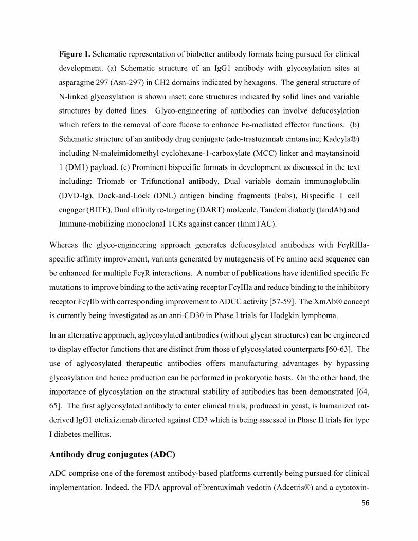
56
Figure 1. Schematic representation of biobetter antibody formats being pursued for clinical
development. (a) Schematic structure of an IgG1 antibody with glycosylation sites at
asparagine 297 (Asn-297) in CH2 domains indicated by hexagons. The general structure of
N-linked glycosylation is shown inset; core structures indicated by solid lines and variable
structures by dotted lines. Glyco-engineering of antibodies can involve defucosylation
which refers to the removal of core fucose to enhance Fc-mediated effector functions. (b)
Schematic structure of an antibody drug conjugate (ado-trastuzumab emtansine; Kadcyla®)
including N-maleimidomethyl cyclohexane-1-carboxylate (MCC) linker and maytansinoid
1 (DM1) payload. (c) Prominent bispecific formats in development as discussed in the text
including: Triomab or Trifunctional antibody, Dual variable domain immunoglobulin
(DVD-Ig), Dock-and-Lock (DNL) antigen binding fragments (Fabs), Bispecific T cell
engager (BITE), Dual affinity re-targeting (DART) molecule, Tandem diabody (tandAb) and
Immune-mobilizing monoclonal TCRs against cancer (ImmTAC).
Whereas the glyco-engineering approach generates defucosylated antibodies with FcγRIIIa-
specific affinity improvement, variants generated by mutagenesis of Fc amino acid sequence can
be enhanced for multiple FcγR interactions. A number of publications have identified specific Fc
mutations to improve binding to the activating receptor FcγIIIa and reduce binding to the inhibitory
receptor FcγIIb with corresponding improvement to ADCC activity [57-59]. The XmAb® concept
is currently being investigated as an anti-CD30 in Phase I trials for Hodgkin lymphoma.
In an alternative approach, aglycosylated antibodies (without glycan structures) can be engineered
to display effector functions that are distinct from those of glycosylated counterparts [60-63]. The
use of aglycosylated therapeutic antibodies offers manufacturing advantages by bypassing
glycosylation and hence production can be performed in prokaryotic hosts. On the other hand, the
importance of glycosylation on the structural stability of antibodies has been demonstrated [64,
65]. The first aglycosylated antibody to enter clinical trials, produced in yeast, is humanized rat-
derived IgG1 otelixizumab directed against CD3 which is being assessed in Phase II trials for type
I diabetes mellitus.
Antibody drug conjugates (ADC)
ADC comprise one of the foremost antibody-based platforms currently being pursued for clinical
implementation. Indeed, the FDA approval of brentuximab vedotin (Adcetris®) and a cytotoxin-

57
conjugated biobetter of trastuzumab, ado-trastuzumab emtansine (Kadcyla®) in 2011 and 2013
respectively, have eclipsed the clinical failures of first generation ADC. As of early 2016, in
addition to the two ADC available in the market, over forty antibody drug conjugates are
undergoing clinical trials and a plethora of such formats are in preclinical development [3]. (Table
3). The achievement of such milestones has been facilitated by the advent of novel ADC
technologies and designs.
Table 3. Overview of ADC developed in recent years and their clinical status.
ADC Format Target Payload Linker Lead
indications
FDA APPROVED
Brentuximab
vedotin
(Adcetris®)
IgG1
(Chimeric)
CD30 MMAE Valine-Citrulline Hodgkin
Lymphoma,
Anaplastic
large cell
lymphoma
Ado-trastuzumab
emtansine
(Kadcyla®)
IgG1
(Humanized)
HER2 DM1 Valine-Citrulline HER2+ breast
cancer
Gemtuzumab
ozogamicin
(Mylotarg® -
withdrawn)
IgG4
(Humanized)
CD33 Calicheamicin SS/hydrazone Acute
Myeloid
Leukemia
Phase III
Inotuzumab
ozogamicin
IgG4
(Humanized)
CD22 Calicheamicin SS/hydrazone Acute
Lymphoblastic
Leukemia
Phase II
Enfortumab
vedotin
(ASG-22ME)
IgG1
(Human)
Nectin-4 MMAE Valine-Citrulline Bladder
cancer

58
PSMA ADC IgG1
(Human)
PSMA MMAE Valine-Citrulline Prostate
cancer
Glembatumumab
vedotin (CDX-
011)
IgG2
(Human)
GPNMB MMAE Valine-Citrulline Breast cancer
Lifastuzumab
vedotin
(DNIB0600A)
IgG1
(Humanized)
NaPi2b MMAE Valine-Citrulline Ovarian
cancer
Pinatuzumab
vedotin
(DCDT2980S)
IgG1
(Humanized)
CD22 MMAE Valine-Citrulline Non-
Hodgkin’s
Lymphoma
(NHL)
Polatuzumab
vedotin
(DCDS4501A)
IgG1
(Humanized)
CD79b MMAE Valine-Citrulline NHL, Diffuse
large B cell
lymphoma
Lorvotuzumab
mertansine
(IMGN-901)
IgG1
(Humanized)
CD56 DM1 SPP Hematological
malignancies
IMMU-130
(Labetuzumab-
SN-38)
IgG1
(Humanized)
CEACAM5 SN-38 Carbonate Colorectal
cancer
IMMU-132
(hRS7-
SN38ADC)
IgG1
(Human)
TROP-2 SN-38 Carbonate Breast cancer
Milatuzumab
doxorubicin
(IMMU-110)
IgG1
(Humanized)
CD74 Doxorubicin Hydrazone Multiple
myeloma
Indatuximab
ravtansine (BT-
062)
IgG1
(Humanized)
CD138 DM4 SPDB Multiple
myeloma
Rovalpituzumab
tesirine
(SC16LD6.5)
Undisclosed Fyn3 D6.5 Undisclosed Small cell
lung cancer

59
Depatuxizumab
mafodotin
(ABT-414)
IgG1
(Human)
EGFRvIII MMAF Maleimidocaproyl Glioblastoma
Abbreviations: MMAE = monomethylauristatin; DM = maytansinoid; PSMA = prostate-
specific membrane antigen; GPNMB = transmembrane glycoprotein NMB; NaPi = sodium-
dependent inorganic phosphate; CEACAM = carcinoembryonic antigen-related cell
adhesion molecule; EGFR = epidermal growth factor receptor.
While the targeted nature of ADC therapy is expected to reduce treatment side effects, toxicity still
remains an important concern for ADC. In fact, gemtuzumab ozogamicin (Mylotarg®), a
humanized anti-CD33 antibody conjugated to calicheamicin was the first ADC to receive FDA
approval in 2000 for treatment of acute myeloid leukemia (AML). However, Mylotarg® was
voluntarily withdrawn from the market after results of a Phase III study raised toxicity concerns
and showed no benefit compared to standard therapies [66]. Brentuximab vedotin and ado-
trastuzumab emtansine also present significant toxicity; both are required by the FDA to carry a
black box warning due to the risk (although only seen in rare cases) of developing progressive
multifocal leukoencephalopathy (brentuximab vedotin) and risk of hepatotoxicity, cardiac toxicity,
and embryo-fetal toxicity (ado-trastuzumab emtansine). Consequently, there remains a challenge
in the field to further validate the superior therapeutic index that ADC can deliver over traditional
chemotherapy and other targeted therapies. In this regard, great efforts are being directed to
implement refinements in each of the components (targeted antigen, targeting moiety, linker,
payload and conjugation chemistry) to boost the therapeutic potential of ADC.
While most conjugates in clinical and preclinical development target cell surface receptors that are
overexpressed in solid tumours and B-cell malignancies (Table 3), novel approaches such as
targeting antigens present in neovasculature or extracellular stromal tissue are being explored. One
main advantage of such approach is that perfusion into the tumor is not required since blood serum
is in direct contact with vascular endothelial cells, with exposure of the conjugate to its target tissue
considerably greater than for traditional ADC targeting neoplastic cells [67-69]. Another attractive
feature is that vascular endothelial cells are less likely to develop resistance mechanisms. Others
have also explored the possibility of targeting cancer stromal cells, aiming to suppress tumor
proliferation, neovascularization, invasion, and metastasis which are typically promoted by the

60
tumor microenvironment [70, 71]. Overall, the factors affecting the internalization rate of ADC
suggest that antigen selection is of high importance in determining the efficacy of the conjugate in
a physiological environment.
Conversely, the efficacy of a conjugate is predominantly determined by its affixed payload.
Current ADC feature antineoplastic agents that have an extremely high potency, usually in the sub-
nanomolar range. Most of these molecules are derivatives of highly potent cytotoxic drugs that
have exquisite potency but at the same time, make them unamenable for development as
chemotherapeutic agents alone. The cytotoxic drugs being employed can be classified into three
major groups namely, calicheamicins (DNA disruption), maytansinoids and auristatins (cell cycle
interference) [72-75]. Monomethylauristatin E (MMAE) and monomethylauristatin F (MMAF)
are the most widely used payloads in the current pipeline with another twenty auristatin conjugates
under clinical development [76, 77]. Among the maytansine analogs, DM1 and DM4 are being
pursued with more than ten ADC in clinical trials (Table 3; Figure 1b). Alternative drug classes
such as duocarmycins and pyrrolobenzodiazepines may also gain popularity due to their
remarkable potency [78, 79].
The linker component is a major determinant of plasma stability of a conjugate, thus impacting on
the efficacy and safety profile. One of the main reasons for off-target effects is the premature
release of cytotoxic activity outside of target cells. A commonly used approach for coupling the
conjugate to an antibody is via a linker that contains a lysosomal-specific protease cleavage site.
In this format, the linker is intended to be cleaved in the lysosome by proteases such as cathepsin
B, plasmin and β-glucoronidase [80, 81]. Indeed, the valine-citrulline (Val-Cit) dipeptide linker,
recognized and cleaved by cathepsin B, is currently the most widely implemented linker
technology [82, 83]. A novel approach using non-cleavable linkers has gained popularity since
they could substantially increase blood serum stability. In this modality, the antibody is digested
inside the lysosome upon internalization, releasing the payload attached to an amino acid residue,
yet still retaining its cytotoxic activity [84, 85]. For example, ado-trastuzumab emtansine, employs
the hetero-bifunctional crosslinker N-succinimidyl-4-(N-maleimidomethyl) cyclohexane-1-
carboxylate (SMCC), creating a non-reducible thioether linker (MCC) as a spacer between the
anti-HER2 antibody and the maytansine DM1 [86]. Presumably due to increased stability, reduced
toxicity has been observed with non-cleavable linker design relative to conjugates with cleavable

61
linker [87]. Remarkably, ado-trastuzumab emtansine has displayed superior in vivo activity in
mouse breast cancer models compared to a similar trastuzumab-DM1 conjugate that differs only
in that it contains a disulphide reducible linker [88].
The chemical reactions used to couple ADC components can also play a role in the therapeutic
index. The first conjugation techniques developed to create ADC involved a chemical reaction
between a functional group in the linker and a reactive moiety in native amino acid residues,
predominantly the thiol side-chains of partially reduced cysteines and the epsilon-amino end of
lysine residues [89, 90]. In these cases, the conjugation sites and the number of drug molecules
affixed to the antibody are stochastic, resulting in heavily heterogeneous conjugates. This
heterogeneity undermines the clinical potential of ADC, since it impacts severely on their toxicity
and safety profiles. In terms of potency, the efficacy is compromised by the reduced subset of
ADC with an optimal amount of attached drug to exert a significant cytotoxic effect. Besides the
fact that conjugates with low drug-to-antibody ratios (DAR) have limited efficacy, high DAR also
possess higher toxicity and reduced plasma stability due to increased clearance rates [91]. This
clearly illustrates the critical importance of achieving an optimal ratio. Despite the shortcomings
of stochastic attachments methods, both cysteine and lysine conjugation techniques have been
utilized in the development of brentuximab vedotin and ado-trastuzumab emtansine, respectively.
A set of alternate novel techniques have been developed to allow for site-specific conjugation
including the THIOMAB platform [92], introduction of unnatural amino acids or enzymatic
modification of native amino acid side-chains [93-95] and even glycan attachment through glyco-
engineering [96].
Bispecifics
The next novel wave of therapeutic antibodies is predicted to be in the format of bispecific
antibodies. The concept of bispecific antibodies was first demonstrated more than twenty years
ago, initially by chemical conjugation of two antibodies [97] then by fusing two hybridoma
cultures [98]. There are now more than fifty different bispecific formats in development, enabling
researchers to adjust and control parameters such as size, half-life, stability, flexibility and
orientation to achieve the desired therapeutic outcome. The assembly of antibody chains to
accomplish the various antibody formats has been made possible with antibody engineering
techniques including variations of the general approach [99-101] to novel approaches such as

62
‘knobs-into-holes’ [102], CrossMab [103], ‘Dock-and-Lock’ (DNL) [104, 105] and even hybrid
domains derived from other immunoglobulin antibodies, via a technology referred to as strand-
exchange engineered domain (SEED) [106].
The majority of bispecific antibodies are currently being assessed in clinical trials and are detailed
in Table 4 with a few examples discussed below based on their mode of action. Referred to as a
trifunctional or triomab is catumaxomab (Removab®), bispecific to tumor antigen EPCAM and T
cell marker CD3 (Figure 1c). The antibody acts by recruiting and activating immune cells;
EPCAM targets the tumor, CD3 recruits T effector cells and the Fc region recruits and activates
monocytes, macrophages, dendritic cells (DC) and NK cells by FcγR binding [107].
Catumaxomab is a hybrid antibody comprised of rat IgG2b binding domains and mouse IgG2a Fc
region. Catumaxomab has not yet received FDA approval (BLA submitted) but has EMA approval
for the treatment of malignant ascites since 2009. The antibody is also in clinical trials for
application in ovarian cancer (II), gastric cancer (II) and epithelial cancer (I). Given that
catumaxomab is a rat-mouse hybrid, some anti-rat or anti-mouse IgG responses are observed in
patients, though treatment does not appear to be significantly affected. In fact, development of
human anti-mouse antibodies (HAMA) have been shown to contribute to greater clinical benefit
[108].
Table 4. Bispecific antibodies developed in recent years and their clinical status.
Name Company Targets Antibody
type
Mode of
Action
Indications
FDA approved
Blinatumoma
b Blincyto®
Amgen CD19/CD
3
BiTE T cell
recruitment
Philadelphia chromosome-negative
precursor B-cell acute
lymphoblastic leukemia
Clinical Trials Completed
Catumaxoma
b Removab®
Trion Pharma EPCAM/
CD3
Trifuncti
onal/
Triomab
T cell
recruitment/
Fc effector
Malignant ascites
Phase III
Emicizumab/
RG6013/AC
E910
Roche Factors
XI/X
CLC-IgG Two factor
dimerisation
Haemophilia A
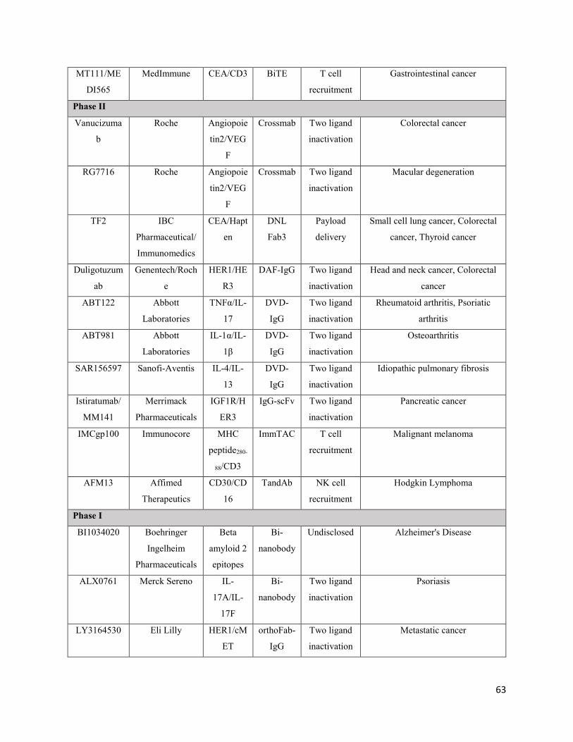
63
MT111/ME
DI565
MedImmune CEA/CD3 BiTE T cell
recruitment
Gastrointestinal cancer
Phase II
Vanucizuma
b
Roche Angiopoie
tin2/VEG
F
Crossmab Two ligand
inactivation
Colorectal cancer
RG7716 Roche Angiopoie
tin2/VEG
F
Crossmab Two ligand
inactivation
Macular degeneration
TF2 IBC
Pharmaceutical/
Immunomedics
CEA/Hapt
en
DNL
Fab3
Payload
delivery
Small cell lung cancer, Colorectal
cancer, Thyroid cancer
Duligotuzum
ab
Genentech/Roch
e
HER1/HE
R3
DAF-IgG Two ligand
inactivation
Head and neck cancer, Colorectal
cancer
ABT122 Abbott
Laboratories
TNFα/IL-
17
DVD-
IgG
Two ligand
inactivation
Rheumatoid arthritis, Psoriatic
arthritis
ABT981 Abbott
Laboratories
IL-1α/IL-
1β
DVD-
IgG
Two ligand
inactivation
Osteoarthritis
SAR156597 Sanofi-Aventis IL-4/IL-
13
DVD-
IgG
Two ligand
inactivation
Idiopathic pulmonary fibrosis
Istiratumab/
MM141
Merrimack
Pharmaceuticals
IGF1R/H
ER3
IgG-scFv Two ligand
inactivation
Pancreatic cancer
IMCgp100 Immunocore MHC
peptide280-
88/CD3
ImmTAC T cell
recruitment
Malignant melanoma
AFM13 Affimed
Therapeutics
CD30/CD
16
TandAb NK cell
recruitment
Hodgkin Lymphoma
Phase I
BI1034020 Boehringer
Ingelheim
Pharmaceuticals
Beta
amyloid 2
epitopes
Bi-
nanobody
Undisclosed Alzheimer's Disease
ALX0761 Merck Sereno IL-
17A/IL-
17F
Bi-
nanobody
Two ligand
inactivation
Psoriasis
LY3164530 Eli Lilly HER1/cM
ET
orthoFab-
IgG
Two ligand
inactivation
Metastatic cancer

64
Pasotuxizum
ab
Bayer PSMA/C
D3
BiTE T cell
recruitment
Prostate cancer
MGD006 Servier CD123/C
D3
DART T cell
recruitment
Acute Myeloid Leukemia
AFM11 Affimed
Therapeutics
CD19/CD
3
TandAb T cell
recruitment
Acute Lymphoblastic Leukemia,
Non-Hodgkin's Lymphoma
MGD007 Servier GPA33/C
D3
DART-
Fc
T cell
recruitment/
Fc effector
Colorectal cancer
Abbreviations: BiTE = bispecific T cell engager; CLC = common light chain; DNL= dock-
and-lock; Fab = antigen binding fragment; DAF = dual acting Fab; DVD-IgG = dual variable
domain immunoglobulin, scFv = single chain variable fragment; ImmTAC = immune-
mobilizing monoclonal TCRs against cancer; TandAb = tandem diabody; DART = dual
affinity re-targeting molecule; Fc = fragment of crystallization; EPCAM = epithelial cell
adhesion molecule; CEA = carcinoembryonic antigen; VEGF = vascular endothelial growth
factor; TNF = tumor necrosis factor; IGFR = insulin growth factor receptor; MHC = major
histocompatibility complex; PSMA = prostate-specific membrane antigen; GP =
glycoprotein; NK = natural killer.
Another example that achieves effector cell recruitment via a conceptually different format without
a Fc region is bispecific T cell engager (BiTE®). BiTEs are variable regions in the form of two
scFv connected by flexible linker peptides (Figure 1c) [109, 110]. One scFv is directed at a cell
surface tumor antigen (with higher affinity) and the other scFv binds CD3 (with lower affinity).
The first marketed BiTE was blinatumomab (Blincyto®) which targets CD19 on acute
lymphoblastic leukemia (ALL), approved by the FDA in late 2014 under the accelerated approval
program. Blinatumomab is currently undergoing multiple Phase II and Phase III clinical trials for
other B-cell related malignancies [111]. A major setback for BiTEs is the requirement of
continuous intravenous infusion due to the short half-life inherent to the small size of the molecule
(55 kDa) and the lack of Fc region. Additionally, the treatment produces significant side effects,
notably neurotoxicity and symptoms of cytokine-release syndrome [112]. Despite the challenges,
this strategy is being investigated for other targets (CEA, EPCAM, PSMA) and indications. Other
examples of molecules with immune cell recruitment are dual affinity re-targeting (DART®) [113,
114] and tandem diabodies (tandAb®) (Figure 1c) [115, 116].

65
Another novel class of bispecific molecules called immune-mobilizing monoclonal TCRs against
cancer (ImmTAC) further expands the variety of potential tumor-specific antigens, by
incorporating soluble T cell receptors (TCRs) that can target intracellular antigens through
recognition of peptide-HLA antigen complexes on the cell surface (Figure 1c) [117]. Under
current investigation in Phase II clinical trial for malignant melanoma is IMCgp100. The
engineered TCR portion of the molecule targets the gp100 peptide280-288 antigen, which is
overexpressed and presented by HLA-A2 on the surface of melanoma cells. The anti-CD3 scFv
portion captures and redirects T cells to kill the melanoma cells.
With the knowledge gained on validated targets and receptor signalling of existing therapeutic
antibodies, targeting two antigens to simultaneously interfere with two or more signaling pathways
is being pursued to improve therapeutic efficacy. The most advanced molecule in clinical
development is dugliotuzumab, a human IgG which targets the combination of HER1 and HER3
as a dual action Fab (DAF)-IgG [118]. Duligotuzumab is being evaluated in Phase II trial in
patients with head and neck cancer. The dual interference can also be applied to inflammatory
conditions. Dual variable domain immunoglobulin (DVD-Ig™) formats directed at TNFα + IL-
17 and IL-1α + IL-1β are undergoing assessment in Phase I studies in patients with rheumatoid
arthritis (Figure 1c) [119]. Other formats being investigated to interfere with signaling pathways
include CrossMabs [103] and bi-nanobodies [120].
Bispecifics can also serve as vehicles to deliver payloads to tumor cells. TF2 is a DNL(Fab3)
bispecific antibody that binds to CEA present on the surface of many solid tumours (Figure 1c).
The antibody comprises three Fab modules that are stably coupled to each other in a triangular
fashion using the ‘dock-and-lock’ technology. Without a Fc region, these molecules have a rather
short serum half-life which is an advantage for pre-targeting approaches. TF2 is being evaluated
in a Phase I trial in patients with colorectal cancer using Lutetium-177 or Indium-111 payload for
imaging.
Physical and chemical degradation of antibodies
During manufacturing and storage, therapeutic antibodies are at risk of degradation via a number
of pathways. Though these reactions may be kept under control by appropriate storage and

66
formulation conditions of the final product, degradation that occurs during culture, downstream
processing and in vivo cannot be controlled sufficiently. These degradation events may affect
antigen recognition, hamper functionality and in severe cases lead to immunogenic responses [121-
125]. Each antibody molecule seems to have a unique personality related to its requirements for
stability; a phenomenon derived from the fact that differences in the CDR between antibodies are
primarily dictated by the surface exposed amino acids that define antigen specificity [124]. The
identification of degradation prone or unstable regions early in the antibody development process
would ideally permit re-engineering of these problematic areas [126-129]. This approach is aided
by recent developments in computational modelling tools that predict regions of interest
susceptible to physical, chemical degradation or influence other biophysical properties of
antibodies. In the next section, degradation pathways commonly observed in therapeutic
antibodies and an overview of predictive tools are discussed.
Aggregation
Protein aggregation is the most common and significant type of physical degradation associated
with therapeutic antibodies, often leading to reduced activity and in some cases, formation of
immunogenic products [130, 131]. Initial preparations of therapeutic antibodies were administered
by intravenous (IV) infusion formulated at protein concentrations (1-25 mg/ml). As antibody-
based therapeutics have become more widely used, high concentration formulations that allow SC
injection (>50 mg/ml) became desirable giving rise to aggregation issues. Proteins are folded in
such a way as to internalize hydrophobic domains and surface expose more hydrophilic domains.
As protein-protein contact frequency increases at high concentrations, the opportunity for
aggregation formation increases proportionally. Changes in extrinsic conditions including
temperature, pH, salt, shaking, viscosity and concentration can transiently expose hydrophobic
domains which in doing so promotes protein-protein interactions that lead to aggregation events
[132-135]. Non-covalent aggregates can be formed via hydrophobic and/or electrostatic
interactions and may be reversible, while covalent aggregates are usually formed by disulphide
bonds and are difficult to reverse. Mechanisms of protein aggregation include: (1) aggregation of
native state monomers; (2) aggregation of monomers with a modified conformation (non-native);
(3) aggregation of chemically-modified monomers; (4) aggregation via a nucleation-dependant
67
process; and (5) surface-induced aggregation via adsorption of protein to glass-liquid or air-liquid
interfaces [136, 137].
Denaturation
Protein denaturation refers to the partial or complete unfolding of the native three-dimensional
folded protein structure. A denatured antibody often loses its tertiary and perhaps secondary
structure leading to loss of binding affinity and activity if an active site domain is affected, and
may expose aggregation-prone regions leading to further degradation [133, 138, 139]. Several
intermediate states may exist between the folded native structure of an antibody molecule and the
denatured state, with some intermediates thought to act as precursors or ‘nuclei’, attracting other
protein species to exposed hydrophobic sites and forming irreversible aggregates. Denaturation
may be induced by a number of stress conditions that arise during antibody manufacture including
changes in solution pH or temperature, use of organic solvents or chaotropes, high salt
concentrations, or shear force [132, 133, 140]. In general, the CH3 domain of an IgG antibody is
often the most stable against denaturation at high temperatures (highest Tm) while the IgG CH2
domain is least stable and denatures first (lowest Tm) [141].
Fragmentation
Fragmentation of therapeutic antibodies can be a product of enzymatic or non-enzymatic
hydrolysis that occurs at the peptide backbone of a number of regions, such as the hinge region,
the CH2-CH3 interface or a region containing aspartic acid (Asp) or tryptophan (Trp) residues
[132]. Asp-associated hydrolysis is affected by pH and the n+1 residue; for instance, a serine
(Ser), valine (Val) or tyrosine (Tyr) adjacent to an Asp may increase the rate of Asp-hydrolysis.
Hinge region hydrolysis can occur in the absence of Asp, and occurs most commonly in the IgG1
isoform [132, 142, 143]. The rate of hydrolysis is dependent on the flexibility the peptide sequence
at the hinge, and occurs within a narrow range of residues. Hinge hydrolysis rates are affected by
solution pH, with a minimum rate of hydrolysis observed near pH 6, and higher rates at a lower or
higher pH [132, 143]. Fragmentation of full-length antibodies is a common occurrence and
generally, cleaved forms are present in such low amounts that effect on efficacy would not likely
be seen.

68
Deamidation
Deamidation is the most common chemical degradation pathway of therapeutic antibodies and
results from the hydrolysis of the amide side-chain of amino acids glutamine (Gln) or asparagine
(Asn) [132, 136, 144, 145]. Hydrolysis of the side-chain can occur at acidic pH (pH <4) resulting
in the conversion of Asn to Asp and Gln to glutamic acid (Glu) [132]. However at higher pH,
deamidation occurs predominantly (and more slowly) via the formation of a cyclic imide
intermediate. For Asn, the cyclic intermediate (succinimide) leads to either the formation of Asp
or an isomer of Asp. A similar process results in the deamidation of Gln to Glu; although Gln
deamidation is much less frequent, due to the lower stability of the 6-membered cyclic intermediate
formed. In short, deamidation events lead to more acidic forms of the antibody through the
acquisition of additional carboxylic acid groups. Conversely, it is also possible for Asp residues
to undergo modification to a succinimide intermediate that produces a basic form of the antibody
by removal of a carboxylic acid group [144].
A number of factors can affect the rate of deamidation. For instance, Asn residues are more prone
to deamidation if they are present in solvent-accessible or structurally-flexible regions, especially
if followed by a small or flexible residue such as Gly, Ser, threonine (Thr) or Asn [132, 146].
Deamidation rate is also affected by extrinsic conditions including pH, temperature, buffer
composition and concentration [147]. Gln deamidation is thought to be less common than Asp
deamidation due to the lower stability of the 6-membered cyclic ring intermediate, which results
in a much slower reaction rate [132]. Although Gln deamidation occurs less frequently, a study
found that following incubation at pH 9, 7.8% of Gln82 of a recombinant IgG1 mAb had
undergone deamidation, despite no deamidation of this residue occurring at neutral pH [145].
Deamidation of therapeutic antibodies is well characterised both and in vivo, and has been shown
to decrease the potency, activity and stability of antibodies [144, 146-150]. The deamidation
events appear to be highly selective for individual antibodies. For example, Harris et al. performed
accelerated stability studies at elevated temperatures with rhuMAB HER2 antibody and found
three labile Asn residues in the CDR region (Asn55, Asn30 and Asn102) [144]. These residues
either formed aspartate, isoasparatate or a stable succinimide intermediate, resulting in a total of
seven species of the antibody being resolved. Deamidation of these Asn residues was shown to
significantly affect the specific in vitro activity and potency of rhuMAb HER2.

69
Oxidation
Oxidation is another common degradation pathway which can occur during antibody production,
formulation or storage. A number of amino acid residues may be affected, including methionine
(Met), cysteine (Cys), histidine (His), tyrosine (Tyr) and Trp [132]. Specific Met residues within
the Fc region (up to four residues) are prone to oxidation resulting in production of methionine
sulfoxide [136, 151, 152]. Oxidation of these residues may affect the stability of an antibody, Fc-
mediated effector function or Protein A binding affinity which is often used for purification from
cell culture supernatant [153]. Wang et al. demonstrated that oxidation of Met252 can result in >4-
fold reduction in the half-life of an antibody in transgenic mice expressing human Fc neonatal
receptor (FcRn). However, this was only observed when 80% of the antibody existed in the
oxidized form, and not at 40% [154].
Oxidation can be dependent on intrinsic factors such as the degree of surface exposed residues as
well as extrinsic factors including buffer composition, light exposure and pH, although Met
oxidation appears to be almost pH-independent [132, 153, 155]. Oxidative stress has been
observed during antibody production in mammalian expression systems where the formation of
reactive oxygen species as a result of hypoxic conditions caused fragmentation of an IgG1
antibody [156].
Tryptophan oxidation of antibodies has been reported following light exposure. Sreedhara et al.
found that light induced oxidation of surface exposed Trp residues (Trp53, Trp108 and Trp94) in
the Fab region of an IgG1 antibody leading to a loss in potency accompanied with a solution colour
change [151]. In another example, oxidation of Trp residue in the H3 CDR loop (Trp135) of a
humanized anti-respiratory syncytial virus (RSV) therapeutic antibody resulted in loss of antigen
binding and biological function [157].
Computational design tools
In recent years, computational methods used to simulate and develop structural models of proteins
have transformed into practical design tools for the development of biobetter and next-generation
antibody therapeutics [127, 158]. Current computational design tools have evolved to allow rapid
identification of specific amino acid sequences or regions on a protein of interest, that contribute
to its observed in vivo properties such as binding affinity, efficacy, stability and half-life [158].

70
Some of the early computational tools used for protein modelling include TANGO, PAGE,
AGGRESCAN, PASTA and Zyggregator all of which rely on the sequence of the protein of
interest (Table 5) [128, 159-164]. These computational tools use force fields such as CHARMM
or AMBER and exploit chemical properties of the amino acids such as hydrophobicity, β-sheet
propensity, charge, and aromatic content to predict aggregation hot-spots and residues susceptible
to chemical degradation. In some cases, multiple tools can be used in combination to improve the
predictive power. For example, Wang et al. combined TANGO and PAGE to identify aggregation-
prone motifs and residues susceptible to deamidation or oxidation of 22 commercial and 20 non-
commercial therapeutic antibodies [165].
Table 5. A representative list of computational tools for prediction of protein aggregation hot
spots.
Name Properties
Sequence-based methods
TANGO
[204].
Determines the secondary structure formation propensity
Aggrescan
[205].
Uses amino acid aggregation propensity value
Zyggregator
[207].
Compares a new peptide sequence to the database
PASTA
[208].
Predicts amyloid structure aggregation by looking into sequences
that are likely to stabilize the cross-beta core of fibrils
PAGE
[209].
Uses physicochemical properties for prediction
Structure-based methods
SAP
[173].
Determines spatial effective surface accessible area
LIP
[211].
Measures ratio of polar surface area to apolar surface area of buried
interfaces
AGGRESCAN3D
[212].
Based of original AGGRESCAN server with input from 3D structure
and spatial arrangement of residues

71
Some structure-based computational tools have also been developed. One such method, Spatial
Aggregation Propensity (SAP), predicts surface exposed aggregation-prone regions of a protein
based on hydrophobicity, dynamic fluctuations and solvent accessibility of residues and regions
[127]. The tool has been used to simulate entire antibodies and develop IgG1 antibody variants
with enhanced physical stability to the wild type, by performing single or multiple mutations in
either the Fab or Fc regions [127, 129].
Another method developed by Angarica and Sancho predicts aggregation propensity based on the
packing density and polarity ratio (ratio of polar surface area to apolar surface area) of buried
interfaces [166]. The tool was designed to characterise “Light Interfaces of high Polarity” (LIPs)
considered to be intrinsically unstable cores. The technology shows promise as a tool for
engineering antibody variants with increased aggregation-resistance, especially as a
complimentary method to surface- or sequence-based tools mentioned above.
One of the most recent developments in computational modelling is the evolution of
AGGRESCAN to AGGRESCAN3D (A3D), an improved server which addresses many of the
limitations of AGGRESCAN and other sequence-based methods. A3D takes into account the
three-dimensional structure of the protein and the spatial arrangement of the residues when the
protein is in its native folded state [167]. With the incorporation of a mutation module that allows
the easy modelling of the detected aggregation-prone and surrounding residues, A3D looks to be
a promising tool for predicting problematic regions and the same time, allowing for re-design of
more stable proteins.
Optimization of antibody bioavailability and delivery
Antibody-based therapies are predominantly delivered intravenously (IV), though an increasing
number are now being formulated and administered subcutaneously (SC). While the IV route
offers 100% bioavailability, systemic distribution and physiological barriers greatly reduce the
actual concentration of antibody achieved in target tissues [168]. What is more, IV infusions are
time-consuming and inconvenient. Ideally, an antibody formulation should be non-invasive and
increase local bioavailability. Limited alternatives have been implemented in the clinic since
formulation requirements for such delivery often pose significant hurdles. For other parenteral

72
administration routes, such as SC or intramuscular (IM), the most common limitation is poor
antibody solubility at the high concentrations required, given that maximum volume of injection
is restricted to 2 ml and 5 ml, respectively. When compared to IV formulations which range from
1-25 mg/ml, SC and IM products often require concentrations >100 mg/ml to deliver an effective
dose [169]. Furthermore, these delivery routes involve an absorption step to enable systemic
circulation. Two main approaches are being pursued to optimize bioavailability: 1) the design of
aggregation-resistant antibodies with higher solubility to prevent precipitation at higher
concentrations; 2) the use of polymer matrix systems to develop controlled release formulations
and improve PK profile.
As discussed in the previous section, the design of aggregation-resistant antibodies albeit
challenging, has become possible with the implementation of computational tools and increasing
understanding of antibody structure and degradation pathways. Formulation stability has also been
improved by using stabilizing additives such as salts (e.g. citrates, sulfates), amino acids (e.g.
glycine) and sugars (e.g. sorbitol, sucrose, trehalose) [132, 170-172]. The development of SC and
IM products has been successful in the last decade because of the formulation of large doses in
significantly smaller volumes without aggregation issues [46]. SC and IM formulations have
significantly improved patient convenience enabling self-administration through the use of pre-
filled syringes, a feature that is highly advantageous for treatment of chronic diseases. A novel
approach to improving SC formulation and PK has been proposed by Yang et al. whereby,
crystalline antibody preparations of infliximab (Remicade®) and trastuzumab (Herceptin®) were
formulated at 200 mg/ml while maintaining low viscosities suitable for this delivery route [173].
Animal studies in rats showed a 2-fold increase of antibody half-life compared to non-crystallized
antibody, demonstrating the potential of crystalline preparations as controlled release systems.
Alginate polymers have also been explored for controlled release. Schweizer et al. developed two
polyanionic alginate matrices loaded with antibody through electrostatic interactions [174]. Both
matrices were delivered to rats subcutaneously as hydrogels. After comparison with its liquid
antibody counterpart, no significant differences in bioavailability were reported.
For many pathologies, local delivery could increase efficacy and reduce systemic exposure. In
such cases, administration routes such as oral, topical, respiratory and intraocular become highly
relevant. Controlled release systems based on polymer matrices are also being tested in preclinical

73
studies for these delivery routes [168, 175]. For instance, liposomes and chitosan-alginate
microparticles have been employed for oral delivery in order to protect antibodies from gastric
inactivation and allow release in the small intestine [176, 177]. This is being explored in
combination with the conjugation of targeting ligands to improve delivery and absorption in the
gastro-intestinal tract. [178]. For topical application, a hydrofiber dressing/adhesive sheet has
been used to apply infliximab as a gel formulation for wound healing with improvements in 7/8
patients tested [179]. In another example, a Phase I trial has been completed with positive results
for BIL-010t, a topically administered, sheep antibody therapy to treat Basal Cell Carcinoma
(BCC). BIL-010t ointment was self-applied for 28 days; it was noted that 13/20 patients had
decreases in the sizes of their lesions with only mild localized skin reactions reported. The
respiratory route has been extensively studied for treatment of chronic obstructive pulmonary
disease, lung cancer, asthma and other pulmonary pathologies. Liposomes and microspheres have
shown potential to increase bioavailability of respiratory delivery by preventing proteolysis [180].
The PEGylation of antibody fragments was shown to increase lung lumen residence time in a
murine model through decreased clearance of alveolar macrophages and increased mucoadhesion
[181]. Other strategies being investigated to deliver antibodies via the respiratory tract include
IgG-loaded lipid microparticles and nano-micelles [182-184]. The most advanced molecule in
clinical development is ALX-0171, an anti-RSV nanobody administered through inhalation;
demonstrating a positive safety and tolerability profile in a first-in-infant Phase I/II study with an
anti-viral effect observed [185].
Overall, the IV route will likely remain the most prominent administration route in development
due to ease of formulation. Still, the improvement of local bioavailability is an obvious
requirement to fulfil the potential of antibody-based therapy, both in terms of efficacy and patient
convenience. As such, strategies such as pursuing alternative administration routes and developing
appropriate controlled release systems will gain relevance as their therapeutic potential continues
to be explored in preclinical studies.
Conclusions
Antibody-based therapeutics have evolved from murine antibodies to humanized and fully human
antibodies, developed with innovative technologies such as transgenic mice and phage display. In
the coming years, next-generation antibodies with improved properties and formats including

74
ADC and bispecifics are expected to gain popularity as biobetter antibody therapeutics. Major
hurdles are being overcome for biosimilar development following the marketing approval of the
first biosimilar antibody by the FDA earlier this year. The advancement of next-generation
biobetters and biosimilars is critical to; 1) reduce the high cost of therapeutic antibodies
encountered over the last thirty years, 2) address the shortcomings confronted with the use of these
antibodies such as poor efficacy and stability and most importantly, (3) provide greater patient
benefit.
Acknowledgements
The authors would like to acknowledge the Faculty of Pharmacy of The University of Sydney for
financial contribution. EC acknowledges the Ministry of Science, Technology and
Telecommunications of the Republic of Costa Rica for postgraduate scholarship.
Disclosures
The authors declare that they have no competing financial interests.
References
[1] D.M. Ecker, S.D. Jones, H.L. Levine, The therapeutic monoclonal antibody market, MAbs, 7
(2015) 9-14.
[2] R.E. Kontermann, U. Brinkmann, Bispecific antibodies, Drug Discovery Today, 20 (2015)
838-847.
[3] P.M. Drake, D. Rabuka, An emerging playbook for antibody-drug conjugates: lessons from the
laboratory and clinic suggest a strategy for improving efficacy and safety, Current Opinion in
Chemical Biology, 28 (2015) 174-180.
[4] G. Kohler, C. Milstein, Continuous cultures of fused cells secreting antibody of predefined
specificity, Nature, 256 (1975) 495-497.
[5] G. Kohler, C. Milstein, Derivation of specific antibody-producing tissue culture and tumor
lines by cell fusion, European Journal of Immunology, 6 (1976) 511-519.

75
[6] W. Li, N.B. Caberoy, New perspective for phage display as an efficient and versatile
technology of functional proteomics, Applied Microbiology and Biotechnology, 85 (2010) 909-
919.
[7] P.T. Jones, P.H. Dear, J. Foote, M.S. Neuberger, G. Winter, Replacing the complementarity-
determining regions in a human antibody with those from a mouse, Nature, 321 (1986) 522-525.
[8] L. Riechmann, M. Clark, H. Waldmann, G. Winter, Reshaping human antibodies for therapy,
Nature, 332 (1988) 323-327.
[9] N. Lonberg, Human antibodies from transgenic animals, Nature Biotechnology, 23 (2005)
1117-1125.
[10] C.T. Scott, Mice with a human touch, Nature Biotechnology, 25 (2007) 1075-1077.
[11] A. Jakobovits, R. Amado, X. Yang, L. Roskos, G. Schwab, From XenoMouse technology to
panitumumab, the first fully human antibody product from transgenic mice, Nature Biotechnology,
25 (2007) 1134-1143.
[12] W.D. Huse, L. Sastry, S.A. Iverson, A.S. Kang, M. Alting-Mees, D.R. Burton, S.J. Benkovic,
R.A. Lerner, Generation of a large combinatorial library of the immunoglobulin repertoire in phage
lambda, Science, 246 (1989) 1275-1281.
[13] J. McCafferty, A.D. Griffiths, G. Winter, D.J. Chiswell, Phage antibodies: filamentous phage
displaying antibody variable domains, Nature, 348 (1990) 552-554.
[14] C.F. Barbas, 3rd, A.S. Kang, R.A. Lerner, S.J. Benkovic, Assembly of combinatorial antibody
libraries on phage surfaces: the gene III site, Proceedings of the National Academy of Sciences,
88 (1991) 7978-7982.
[15] D.J. Schofield, A.R. Pope, V. Clementel, J. Buckell, S.D.J. Chapple, K.F. Clarke, J.S.
Conquer, A.M. Crofts, S.R.E. Crowther, M.R. Dyson, G. Flack, G.J. Griffin, Y. Hooks, W.J.
Howat, A. Kolb-Kokocinski, S. Kunze, C.D. Martin, G.L. Maslen, J.N. Mitchell, M. O'Sullivan,
R.L. Perera, W. Roake, S.P. Shadbolt, K.J. Vincent, A. Warford, W.E. Wilson, J. Xie, J.L. Young,
J. McCafferty, Application of phage display to high throughput antibody generation and
characterization, Genome Biology, 8 (2007).

76
[16] J.D. Marks, H.R. Hoogenboom, T.P. Bonnert, J. McCafferty, A.D. Griffiths, G. Winter, By-
passing immunization. Human antibodies from V-gene libraries displayed on phage, Journal of
Molecular Biology, 222 (1991) 581-597.
[17] C.F.I. Barbas, J.D. Bain, D.M. Hoekstra, R.A. Lerner, Semisynthetic combinatorial antibody
libraries: a chemical solution to the diversity problem, Proceedings of the National Academy of
Sciences, 89 (1992) 4457-4461.
[18] H.R. Hoogenboom, G. Winter, By-passing immunisation. Human antibodies from synthetic
repertoires of germline VH gene segments rearranged in vitro., Journal of Molecular Biology, 227
(1992) 381-388.
[19] H.R. Hoogenboom, A.D. Griffiths, K.S. Johnson, D.J. Chiswell, P. Hudson, G. Winter, Multi-
subunit proteins on the surface of filamentous phage: methodologies for displaying antibody (Fab)
heavy and light chains, Nucleic Acids Research, 19 (1991) 4133-4137.
[20] J. Davies, L. Riechmann, An antibody variable heavy domain with a lox-Cre site integrated
into its coding region: bacterial recombination within a single polypeptide chain, FEBS Letters,
377 (1995) 92-96.
[21] M.M. Harmsen, H.J. De Haard, Properties, production, and applications of camelid single-
domain antibody fragments, Applied Microbiology and Biotechnology, 77 (2007) 13-22.
[22] S.D. Nuttall, U.V. Krishnan, L. Doughty, K. Pearson, M.T. Ryan, N.J. Hoogenraad, M.
Hattarki, J.A. Carmichael, R.A. Irving, P.J. Hudson, Isolation and characterization of an IgNAR
variable domain specific for the human mitochondrial translocase receptor Tom70, European
Journal of Biochemistry, 270 (2003) 3543-3554.
[23] K. Nakano, T. Kojima, K. Kasutani, C. Senoh, O. Natori, S. Ishii, H. Tsunoda, K. Hattori,
Effective screening method of agonistic diabodies based on autocrine growth, Journal of
Immunological Methods, 347 (2009) 31-35.
[24] Y. Mazor, T. Van Blarcom, R. Mabry, B.L. Iverson, G. Georgiou, Isolation of engineered,
full-length antibodies from libraries expressed in Escherichia coli, Nature Biotechnology, 25
(2007) 563-565.

77
[25] L.S. Jespers, A. Roberts, S.M. Mahler, G. Winter, H.R. Hoogenboom, Guiding the selection
of human antibodies from phage display repertoires to a single epitope of an antigen,
Biotechnology (N Y), 12 (1994) 899-903.
[26] A.E. Nixon, D.J. Sexton, R.C. Ladner, Drugs derived from phage display: from candidate
identification to clinical practice, MAbs, 6 (2014) 73-85.
[27] P.S. Daugherty, G. Chen, M.J. Olsen, B.L. Iverson, G. Georgiou, Antibody affinity maturation
using bacterial surface display, Protein Engineering, 11 (1998) 825-832.
[28] M.J. Feldhaus, R.W. Siegel, L.K. Opresko, J.R. Coleman, J.M. Feldhaus, Y.A. Yeung, J.R.
Cochran, P. Heinzelman, D. Colby, J. Swers, C. Graff, H.S. Wiley, K.D. Wittrup, Flow-cytometric
isolation of human antibodies from a non immune Sacchromyces cerevisiae surface display library,
Nature Biotechnology, 21 (2003) 163-170.
[29] D.G. Mottershead, K. Alfthan, K. Ojala, K. Takkinen, C. Oker-Blom, Baculoviral display of
functional scFv and synthetic IgG-binding domains, Biochem Biophys Res Commun, 275 (2000)
84-90.
[30] J. Hanes, C. Schaffitzel, A. Knappik, A. Pluckthun, Picomolar affinity antibodies from a fully
synthetic naive library selected and evolved by ribosome display, Nat Biotechnol, 18 (2000) 1287-
1292.
[31] G. Georgiou, G.C. Ippolito, J. Beausang, C.E. Busse, H. Wardemann, S.R. Quake, The
promise and challenge of high-throughput sequencing of the antibody repertoire, Nature
Biotechnology, 32 (2014) 158-168.
[32] U. Ravn, G. Didelot, S. Venet, K.T. Ng, F. Gueneau, F. Rousseau, S. Calloud, M. Kosco-
Vilbois, N. Fischer, Deep sequencing of phage display libraries to support antibody discovery,
Methods, 60 (2013) 99-110.
[33] P. Mathonet, C.G. Ullman, The application of next generation sequencing to the
understanding of antibody repertoires, Frontiers in Immunology, 4 (2013).
[34] W.C. Cheung, S.A. Beausoleil, X. Zhang, S. Sato, S.M. Schieferl, J.S. Wieler, J.G. Beaudet,
R.K. Ramenani, L. Popova, M.J. Comb, J. Rush, R.D. Polakiewicz, A proteomics approach for the

78
identification and cloning of monoclonal antibodies from serum, Nature Biotechnology, 30 (2012)
447-452.
[35] S.T. Reddy, X. Ge, A.E. Miklos, R.A. Hughes, S.H. Kang, K.H. Hoi, C. Chrysostomou, S.P.
Hunicke-Smith, B.L. Iverson, P.W. Tucker, A.D. Ellington, G. Georgiou, Monoclonal antibodies
isolated without screening by analyzing the variable-gene repertoire of plasma cells, Nature
Biotechnology, 28 (2010) 965-969.
[36] U. Ravn, F. Gueneau, L. Baerlocher, M. Osteras, M. Desmurs, P. Malinge, G. Magistrelli, L.
Farinelli, M.H. Kosco-Vilbois, N. Fischer, By-passing in vitro screening--next generation
sequencing technologies applied to antibody display and in silico candidate selection, Nucleic
Acids Research, 38 (2010) e193.
[37] B.J. DeKosky, G.C. Ippolito, R.P. Deschner, J.J. Lavinder, Y. Wine, B.M. Rawlings, N.
Varadarajan, C. Giesecke, T. Dorner, S.F. Andrews, P.C. Wilson, S.P. Hunicke-Smith, C.G.
Willson, A.D. Ellington, G. Georgiou, High-throughput sequencing of the paired human
immunoglobulin heavy and light chain repertoire, Nature Biotechnology, 31 (2013) 166-169.
[38] I. Saggy, Y. Wine, L. Shefet-Carasso, L. Nahary, G. Georgiou, I. Benhar, Antibody isolation
from immunized animals: comparison of phage display and antibody discovery via V gene
repertoire mining, Protein Engineering Design and Selection, 25 (2012) 539-549.
[39] K. Lehmann-Horn, H.C. Kronsbein, M.S. Weber, Targeting B cells in the treatment of
multiple sclerosis: recent advances and remaining challenges, Therapeutic Advances in
Neurological Disorders, 6 (2013) 161-173.
[40] A.M. Vazquez, A.M. Hernandez, A. Macias, E. Montero, D.E. Gomez, D.F. Alonso, M.R.
Gabri, R.E. Gomez, Racotumomab: an anti-idiotype vaccine related to N-glycolyl-containing
gangliosides - preclinical and clinical data, Frontiers in Oncology, 2 (2012) 150.
[41] P. Garrone, O. Djossou, F. Fossiez, J. Reyes, S. Ait-Yahia, C. Maat, S. Ho, T. Hauser, J.M.
Dayer, J. Greffe, P. Miossec, S. Lebecque, F. Rousset, J. Banchereau, Generation and
characterization of a human monoclonal autoantibody that acts as a high affinity interleukin-1
alpha specific inhibitor, Molecular Immunology, 33 (1996) 649-658.

79
[42] D.S. Hong, D. Hui, E. Bruera, F. Janku, A. Naing, G.S. Falchook, S. Piha-Paul, J.J. Wheler,
S. Fu, A.M. Tsimberidou, M. Stecher, P. Mohanty, J. Simard, R. Kurzrock, MABp1, a first-in-
class true human antibody targeting interleukin-1alpha in refractory cancers: an open-label, phase
1 dose-escalation and expansion study, The Lancet Oncology, 15 (2014) 656-666.
[43] P. Chames, M. Van Regenmortel, E. Weiss, D. Baty, Therapeutic antibodies: successes,
limitations and hopes for the future, British Journal of Pharmacology, 157 (2009) 220-233.
[44] R. Niwa, M. Satoh, The current status and prospects of antibody engineering for therapeutic
use: focus on glycoengineering technology, Journal of Pharmaceutical Sciences, 104 (2015) 930-
941.
[45] J.B. Evans, B.A. Syed, From the analyst's couch: Next-generation antibodies, Nat Rev Drug
Discov, 13 (2014) 413-414.
[46] O. Shpilberg, C. Jackisch, Subcutaneous administration of rituximab (MabThera) and
trastuzumab (Herceptin) using hyaluronidase, British Journal of Cancer, 109 (2013) 1556-1561.
[47] S. Derer, C. Kellner, S. Berger, T. Valerius, M. Peipp, Fc engineering: design, expression,
and functional characterization of antibody variants with improved effector function, Methods Mol
Biol, 907 (2012) 519-536.
[48] A. Beck, J.M. Reichert, Marketing approval of mogamulizumab: a triumph for glyco-
engineering, MAbs, 4 (2012) 419-425.
[49] M. Sachdeva, S. Dhingra, Obinutuzumab: A FDA approved monoclonal antibody in the
treatment of untreated chronic lymphocytic leukemia, Int J Appl Basic Med Res, 5 (2015) 54-57.
[50] M. Ratner, Genentech's glyco-engineered antibody to succeed Rituxan, Nature
Biotechnology, 32 (2014) 6+.
[51] J.M. Subramaniam, G. Whiteside, K. McKeage, J.C. Croxtall, Mogamulizumab: first global
approval, Drugs, 72 (2012) 1293-1298.
[52] A.K. Kakkar, S. Balakrishnan, Obinutuzumab for chronic lymphocytic leukemia: promise of
the first treatment approved with breakthrough therapy designation, J Oncol Pharm Pract, 21
(2015) 358-363.

80
[53] S.D. Liu, C. Chalouni, J.C. Young, T.T. Junttila, M.X. Sliwkowski, J.B. Lowe, Afucosylated
antibodies increase activation of FcgammaRIIIa-dependent signaling components to intensify
processes promoting ADCC, Cancer Immunol Res, 3 (2015) 173-183.
[54] T. Mizushima, H. Yagi, E. Takemoto, M. Shibata-Koyama, Y. Isoda, S. Iida, K. Masuda, M.
Satoh, K. Kato, Structural basis for improved efficacy of therapeutic antibodies on defucosylation
of their Fc glycans, Genes to Cells, 16 (2011) 1071-1080.
[55] S. Preithner, S. Elm, S. Lippold, M. Locher, A. Wolf, A.J. da Silva, P.A. Baeuerle, N.S. Prang,
High concentrations of therapeutic IgG1 antibodies are needed to compensate for inhibition of
antibody-dependent cellular cytotoxicity by excess endogenous immunoglobulin G, Mol
Immunol, 43 (2006) 1183-1193.
[56] N. Zhang, L. Liu, C. Dan Dumitru, N.R.H. Cummings, M. Cukan, Y. Jiang, Y. Li, F. Li, T.
Mitchell, M.R. Mallem, Y. Ou, R.N. Patel, K. Vo, H. Wang, I. Burnina, B.-K. Choi, H.E. Huber,
T.A. Stadheim, D. Zha, Glycoengineered Pichia produced anti-HER2 is comparable to
trastuzumab in preclinical study, MAbs, 3 (2011) 289-298.
[57] R.L. Shields, A.K. Namenuk, K. Hong, Y.G. Meng, J. Rae, J. Briggs, D. Xie, J. Lai, A.
Stadlen, B. Li, J.A. Fox, L.G. Presta, High resolution mapping of the binding site on human IgG1
for Fc gamma RI, Fc gamma RII, Fc gamma RIII, and FcRn and design of IgG1 variants with
improved binding to the Fc gamma R, The Journal of Biological Chemistry, 276 (2001) 6591-
6604.
[58] G.A. Lazar, W. Dang, S. Karki, O. Vafa, J.S. Peng, L. Hyun, C. Chan, H.S. Chung, A. Eivazi,
S.C. Yoder, J. Vielmetter, D.F. Carmichael, R.J. Hayes, B.I. Dahiyat, Engineered antibody Fc
variants with enhanced effector function, Proceedings of the National Academy of Sciences, 103
(2006) 4005-4010.
[59] J.B. Stavenhagen, S. Gorlatov, N. Tuaillon, C.T. Rankin, H. Li, S. Burke, L. Huang, S. Vijh,
S. Johnson, E. Bonvini, S. Koenig, Fc optimization of therapeutic antibodies enhances their ability
to kill tumor cells in vitro and controls tumor expansion in vivo via low-affinity activating
Fcgamma receptors, Cancer Research, 67 (2007) 8882-8890.

81
[60] S.T. Jung, S.T. Reddy, T.H. Kang, M.J. Borrok, I. Sandlie, P.W. Tucker, G. Georgiou,
Aglycosylated IgG variants expressed in bacteria that selectively bind FcgammaRI potentiate
tumor cell killing by monocyte-dendritic cells, Proceedings of the National Academy of Sciences,
107 (2010) 604-609.
[61] S.L. Sazinsky, R.G. Ott, N.W. Silver, B. Tidor, J.V. Ravetch, K.D. Wittrup, Aglycosylated
immunoglobulin G1 variants productively engage activating Fc receptors, Proceedings of the
National Academy of Sciences, 105 (2008) 20167-20172.
[62] M.J. Borrok, S.T. Jung, T.H. Kang, A.F. Monzingo, G. Georgiou, Revisiting the role of
glycosylation in the structure of human IgG Fc, ACS Chemical Biology, 7 (2012) 1596-1602.
[63] R. Stewart, G. Thom, M. Levens, G. Guler-Gane, R. Holgate, P.M. Rudd, C. Webster, L.
Jermutus, J. Lund, A variant human IgG1-Fc mediates improved ADCC, Protein Engineering
Design and Selection, 24 (2011) 671-678.
[64] V. Kayser, N. Chennamsetty, V. Voynov, K. Forrer, B. Helk, B.L. Trout, Glycosylation
influences on the aggregation propensity of therapeutic monoclonal antibodies, Biotechnology
Journal, 6 (2011) 38-44.
[65] Y. Mimura, S. Church, R. Ghirlando, P.R. Ashton, S. Dong, M. Goodall, J. Lund, R. Jefferis,
The influence of glycosylation on the thermal stability and effector function expression of human
IgG1-Fc: properties of a series of truncated glycoforms, Molecular Immunology, 37 (2000) 697-
706.
[66] J.L. Zaro, Mylotarg: Revisiting Its Clinical Potential Post-Withdrawal, in: J. Wang, W.-C.
Shen, J.L. Zaro (Eds.) Antibody-Drug Conjugates, Springer International Publishing2015, pp.
179-190.
[67] F.J. Burrows, P.E. Thorpe, Eradication of large solid tumors in mice with an immunotoxin
directed against tumor vasculature, Proceedings of the National Academy of Sciences, 90 (1993)
8996-9000.
[68] A. Palumbo, F. Hauler, P. Dziunycz, K. Schwager, A. Soltermann, F. Pretto, C. Alonso, G.F.
Hofbauer, R.W. Boyle, D. Neri, A chemically modified antibody mediates complete eradication

82
of tumours by selective disruption of tumour blood vessels, British Journal of Cancer, 104 (2011)
1106-1115.
[69] A. Visintin, K. Knowlton, E. Tyminski, C.I. Lin, X. Zheng, K. Marquette, S. Jain, L.
Tchistiakova, D. Li, C.J. O'Donnell, A. Maderna, X. Cao, R. Dunn, W.B. Snyder, A.K. Abraham,
M. Leal, S. Shetty, A. Barry, L. Zawel, A.J. Coyle, H.F. Dvorak, S.C. Jaminet, Novel Anti-
TM4SF1 Antibody-Drug Conjugates with Activity against Tumor Cells and Tumor Vasculature,
Molecular Cancer Therapeutics, 14 (2015) 1868-1876.
[70] E. Ostermann, P. Garin-Chesa, K.H. Heider, M. Kalat, H. Lamche, C. Puri, D. Kerjaschki,
W.J. Rettig, G.R. Adolf, Effective immunoconjugate therapy in cancer models targeting a serine
protease of tumor fibroblasts, Clinical Cancer Research, 14 (2008) 4584-4592.
[71] D. Quail, J. Joyce, Microenvironmental regulation of tumor progression and metastasis,
Nature Medicine, 19 (2013) 1423-1437.
[72] G.R. Pettit, Y. Kamano, C.L. Herald, A.A. Tuinman, F.E. Boettner, H. Kizu, J.M. Schmidt,
L. Baczynskyj, K.B. Tomer, R.J. Bontems, The isolation and structure of a remarkable marine
animal antineoplastic constituent: dolastatin 10, Journal of the American Chemical Society, 109
(1987) 6883-6885.
[73] W.M. Maiese, M.P. Lechevalier, H.A. Lechevalier, J. Korshalla, N. Kuck, A. Fantini, M.J.
Wildey, J. Thomas, M. Greenstein, Calicheamicins, a novel family of antitumor antibiotics:
taxonomy, fermentation and biological properties, The Journal of Antibiotics, 42 (1989) 558-563.
[74] J.M. Cassady, K.K. Chan, H.G. Floss, E. Leistner, Recent developments in the maytansinoid
antitumor agents, Chemical and Pharmaceutical Bulletin, 52 (2004) 1-26.
[75] R.V. Chari, Targeted cancer therapy: conferring specificity to cytotoxic drugs, Accounts of
Chemical Research, 41 (2008) 98-107.
[76] S.O. Doronina, B.A. Mendelsohn, T.D. Bovee, C.G. Cerveny, S.C. Alley, D.L. Meyer, E.
Oflazoglu, B.E. Toki, R.J. Sanderson, R.F. Zabinski, A.F. Wahl, P.D. Senter, Enhanced activity
of monomethylauristatin F through monoclonal antibody delivery: effects of linker technology on
efficacy and toxicity, Bioconjugate Chemistry, 17 (2006) 114-124.

83
[77] T.V. Jerjian, A.E. Glode, L.A. Thompson, C.L. O'Bryant, Antibody-Drug Conjugates: A
Clinical Pharmacy Perspective on an Emerging Cancer Therapy, Pharmacotherapy, 36 (2016) 99-
116.
[78] R.C. Elgersma, R.G. Coumans, T. Huijbregts, W.M. Menge, J.A. Joosten, H.J. Spijker, F.M.
de Groot, M.M. van der Lee, R. Ubink, D.J. van den Dobbelsteen, D.F. Egging, W.H. Dokter, G.F.
Verheijden, J.M. Lemmens, C.M. Timmers, P.H. Beusker, Design, Synthesis, and Evaluation of
Linker-Duocarmycin Payloads: Toward Selection of HER2-Targeting Antibody-Drug Conjugate
SYD985, Molecular Pharmaceutics, 12 (2015) 1813-1835.
[79] J.A. Hartley, The development of pyrrolobenzodiazepines as antitumour agents, Expert
Opinion on Investigational Drugs, 20 (2011) 733-744.
[80] J.E. Koblinski, M. Ahram, B.F. Sloane, Unraveling the role of proteases in cancer, Clin Chim
Acta, 291 (2000) 113-135.
[81] S.O. Doronina, B.E. Toki, M.Y. Torgov, B.A. Mendelsohn, C.G. Cerveny, D.F. Chace, R.L.
DeBlanc, R.P. Gearing, T.D. Bovee, C.B. Siegall, J.A. Francisco, A.F. Wahl, D.L. Meyer, P.D.
Senter, Development of potent monoclonal antibody auristatin conjugates for cancer therapy, Nat
Biotechnol, 21 (2003) 778-784.
[82] C. Vaklavas, A. Forero-Torres, Safety and efficacy of brentuximab vedotin in patients with
Hodgkin lymphoma or systemic anaplastic large cell lymphoma, Ther Adv Hematol, 3 (2012) 209-
225.
[83] J. Bendell, M. Saleh, A.A. Rose, P.M. Siegel, L. Hart, S. Sirpal, S. Jones, J. Green, E.
Crowley, R. Simantov, T. Keler, T. Davis, L. Vahdat, Phase I/II study of the antibody-drug
conjugate glembatumumab vedotin in patients with locally advanced or metastatic breast cancer,
J Clin Oncol, 32 (2014) 3619-3625.
[84] H.K. Erickson, P.U. Park, W.C. Widdison, Y.V. Kovtun, L.M. Garrett, K. Hoffman, R.J. Lutz,
V.S. Goldmacher, W.A. Blattler, Antibody-maytansinoid conjugates are activated in targeted
cancer cells by lysosomal degradation and linker-dependent intracellular processing, Cancer
Research, 66 (2006) 4426-4433.

84
[85] S. Alley, X. Zhang, N. Okeley, M. Anderson, E. Oflazoglu, I. Stone, C.-L. Law, P. Senter, D.
Benjamin, Effects of linker chemistry on tumor targeting by anti-CD70 antibody-drug conjugates,
Cancer Research, 67 (2007) 916.
[86] S. Girish, M. Gupta, B. Wang, D. Lu, I.E. Krop, C.L. Vogel, H.A. Burris Iii, P.M. LoRusso,
J.H. Yi, O. Saad, B. Tong, Y.W. Chu, S. Holden, A. Joshi, Clinical pharmacology of trastuzumab
emtansine (T-DM1): an antibody-drug conjugate in development for the treatment of HER2-
positive cancer, Cancer Chemotherapy and Pharmacology, 69 (2012) 1229-1240.
[87] A.G. Polson, J. Calemine-Fenaux, P. Chan, W. Chang, E. Christensen, S. Clark, F.J. de
Sauvage, D. Eaton, K. Elkins, J.M. Elliott, G. Frantz, R.N. Fuji, A. Gray, K. Harden, G.S. Ingle,
N.M. Kljavin, H. Koeppen, C. Nelson, S. Prabhu, H. Raab, S. Ross, D.S. Slaga, J.P. Stephan, S.J.
Scales, S.D. Spencer, R. Vandlen, B. Wranik, S.F. Yu, B. Zheng, A. Ebens, Antibody-drug
conjugates for the treatment of non-Hodgkin's lymphoma: target and linker-drug selection, Cancer
Research, 69 (2009) 2358-2364.
[88] H.K. Erickson, G.D. Lewis Phillips, D.D. Leipold, C.A. Provenzano, E. Mai, H.A. Johnson,
B. Gunter, C.A. Audette, M. Gupta, J. Pinkas, J. Tibbitts, The effect of different linkers on target
cell catabolism and pharmacokinetics/pharmacodynamics of trastuzumab maytansinoid
conjugates, Mol Cancer Ther, 11 (2012) 1133-1142.
[89] R.P. Lyon, D.L. Meyer, J.R. Setter, P.D. Senter, Conjugation of anticancer drugs through
endogenous monoclonal antibody cysteine residues, Methods Enzymol, 502 (2012) 123-138.
[90] M.P. Brun, L. Gauzy-Lazo, Protocols for lysine conjugation, Methods Mol Biol, 1045 (2013)
173-187.
[91] C.R. Behrens, E.H. Ha, L.L. Chinn, S. Bowers, G. Probst, M. Fitch-Bruhns, J. Monteon, A.
Valdiosera, A. Bermudez, S. Liao-Chan, T. Wong, J. Melnick, J.W. Theunissen, M.R. Flory, D.
Houser, K. Venstrom, Z. Levashova, P. Sauer, T.S. Migone, E.H. van der Horst, R.L. Halcomb,
D.Y. Jackson, Antibody-Drug Conjugates (ADCs) Derived from Interchain Cysteine Cross-
Linking Demonstrate Improved Homogeneity and Other Pharmacological Properties over
Conventional Heterogeneous ADCs, Molecular pharmaceutics, 12 (2015) 3986-3998.

85
[92] J.R. Junutula, H. Raab, S. Clark, S. Bhakta, D.D. Leipold, S. Weir, Y. Chen, M. Simpson,
S.P. Tsai, M.S. Dennis, Y. Lu, Y.G. Meng, C. Ng, J. Yang, C.C. Lee, E. Duenas, J. Gorrell, V.
Katta, A. Kim, K. McDorman, K. Flagella, R. Venook, S. Ross, S.D. Spencer, W. Lee Wong, H.B.
Lowman, R. Vandlen, M.X. Sliwkowski, R.H. Scheller, P. Polakis, W. Mallet, Site-specific
conjugation of a cytotoxic drug to an antibody improves the therapeutic index, Nature
Biotechnology, 26 (2008) 925-932.
[93] J.Y. Axup, K.M. Bajjuri, M. Ritland, B.M. Hutchins, C.H. Kim, S.A. Kazane, R. Halder, J.S.
Forsyth, A.F. Santidrian, K. Stafin, Y. Lu, H. Tran, A.J. Seller, S.L. Biroc, A. Szydlik, J.K.
Pinkstaff, F. Tian, S.C. Sinha, B. Felding-Habermann, V.V. Smider, P.G. Schultz, Synthesis of
site-specific antibody-drug conjugates using unnatural amino acids, Proceedings of the National
Academy of Sciences, 109 (2012) 16101-16106.
[94] E.S. Zimmerman, T.H. Heibeck, A. Gill, X. Li, C.J. Murray, M.R. Madlansacay, C. Tran,
N.T. Uter, G. Yin, P.J. Rivers, A.Y. Yam, W.D. Wang, A.R. Steiner, S.U. Bajad, K. Penta, W.
Yang, T.J. Hallam, C.D. Thanos, A.K. Sato, Production of site-specific antibody-drug conjugates
using optimized non-natural amino acids in a cell-free expression system, Bioconjug Chem, 25
(2014) 351-361.
[95] A.M. Sochaj, K.W. Swiderska, J. Otlewski, Current methods for the synthesis of
homogeneous antibody-drug conjugates, Biotechnology Advances, 33 (2015) 775-784.
[96] R. van Geel, M.A. Wijdeven, R. Heesbeen, J.M. Verkade, A.A. Wasiel, S.S. van Berkel, F.L.
van Delft, Chemoenzymatic Conjugation of Toxic Payloads to the Globally Conserved N-Glycan
of Native mAbs Provides Homogeneous and Highly Efficacious Antibody-Drug Conjugates,
Bioconjugate Chemistry, 26 (2015) 2233-2242.
[97] A. Nisonoff, M.M. Rivers, Recombination of a mixture of univalent antibody fragments of
different specificity, Arch Biochem Biophys, 93 (1961) 460-462.
[98] U.D. Staerz, M.J. Bevan, Hybrid hybridoma producing a bispecific monoclonal antibody that
can focus effector T-cell activity, Proceedings of the National Academy of Sciences, 83 (1986)
1453-1457.
86
[99] K. Gunasekaran, M. Pentony, M. Shen, L. Garrett, C. Forte, A. Woodward, S.B. Ng, T. Born,
M. Retter, K. Manchulenko, H. Sweet, I.N. Foltz, M. Wittekind, W. Yan, Enhancing antibody Fc
heterodimer formation through electrostatic steering effects: applications to bispecific molecules
and monovalent IgG, The Journal of Biological Chemistry, 285 (2010) 19637-19646.
[100] T.S. Von Kreudenstein, E. Escobar-Carbrera, P.I. Lario, I. D'Angelo, K. Brault, J. Kelly, Y.
Durocher, J. Baardsnes, R.J. Woods, M.H. Xie, P.A. Girod, M.D. Suits, M.J. Boulanger, D.K.
Poon, G.Y. Ng, S.B. Dixit, Improving biophysical properties of a bispecific antibody scaffold to
aid developability: quality by molecular design, MAbs, 5 (2013) 646-654.
[101] S.M. Lewis, X. Wu, A. Pustilnik, A. Sereno, F. Huang, H.L. Rick, G. Guntas, A. Leaver-
Fay, E.M. Smith, C. Ho, C. Hansen-Estruch, A.K. Chamberlain, S.M. Truhlar, E.M. Conner, S.
Atwell, B. Kuhlman, S.J. Demarest, Generation of bispecific IgG antibodies by structure-based
design of an orthogonal Fab interface, Nature Biotechnology, 32 (2014) 191-198.
[102] J.B. Ridgway, L.G. Presta, P. Carter, 'Knobs-into-holes' engineering of antibody CH3
domains for heavy chain heterodimerization, Protein Engineering Design and Selection, 9 (1996)
617-621.
[103] W. Schaefer, J.T. Regula, M. Bahner, J. Schanzer, R. Croasdale, H. Durr, C. Gassner, G.
Georges, H. Kettenberger, S. Imhof-Jung, M. Schwaiger, K.G. Stubenrauch, C. Sustmann, M.
Thomas, W. Scheuer, C. Klein, Immunoglobulin domain crossover as a generic approach for the
production of bispecific IgG antibodies, Proceedings of the National Academy of Sciences, 108
(2011) 11187-11192.
[104] E.A. Rossi, D.M. Goldenberg, T.M. Cardillo, W.J. McBride, R.M. Sharkey, C.H. Chang,
Stably tethered multifunctional structures of defined composition made by the dock and lock
method for use in cancer targeting, Proceedings of the National Academy of Sciences, 103 (2006)
6841-6846.
[105] E.A. Rossi, D.M. Goldenberg, C.H. Chang, The dock-and-lock method combines
recombinant engineering with site-specific covalent conjugation to generate multifunctional
structures, Bioconjugate Chemistry, 23 (2012) 309-323.

87
[106] J.H. Davis, C. Aperlo, Y. Li, E. Kurosawa, Y. Lan, K.M. Lo, J.S. Huston, SEEDbodies:
fusion proteins based on strand-exchange engineered domain (SEED) CH3 heterodimers in an Fc
analogue platform for asymmetric binders or immunofusions and bispecific antibodies, Protein
Engineering Design and Selection, 23 (2010) 195-202.
[107] R. Zeidler, J. Mysliwietz, M. Csanady, A. Walz, I. Ziegler, B. Schmitt, B. Wollenberg, H.
Lindhofer, The Fc-region of a new class of intact bispecific antibody mediates activation of
accessory cells and NK cells and induces direct phagocytosis of tumour cells, British Journal of
Cancer, 83 (2000) 261-266.
[108] M.G. Ott, F. Marme, G. Moldenhauer, H. Lindhofer, M. Hennig, R. Spannagl, M.M. Essing,
R. Linke, D. Seimetz, Humoral response to catumaxomab correlates with clinical outcome: results
of the pivotal phase II/III study in patients with malignant ascites, International Journal of Cancer,
130 (2012) 2195-2203.
[109] E. Wolf, R. Hofmeister, P. Kufer, B. Schlereth, P.A. Baeuerle, BiTEs: bispecific antibody
constructs with unique anti-tumor activity, Drug Discovery Today, 10 (2005) 1237-1244.
[110] P.A. Baeuerle, P. Kufer, R. Bargou, BiTE: Teaching antibodies to engage T-cells for cancer
therapy, Current Opinion in Molecular Therapeutics, 11 (2009) 22-30.
[111] M.J. Newman, D.J. Benani, A review of blinatumomab, a novel immunotherapy, Journal of
Oncology Pharmacy Practice, (2015).
[112] M.S. Topp, N. Gokbuget, A.S. Stein, G. Zugmaier, S. O'Brien, R.C. Bargou, H. Dombret,
A.K. Fielding, L. Heffner, R.A. Larson, S. Neumann, R. Foa, M. Litzow, J.M. Ribera, A.
Rambaldi, G. Schiller, M. Bruggemann, H.A. Horst, C. Holland, C. Jia, T. Maniar, B. Huber, D.
Nagorsen, S.J. Forman, H.M. Kantarjian, Safety and activity of blinatumomab for adult patients
with relapsed or refractory B-precursor acute lymphoblastic leukaemia: a multicentre, single-arm,
phase 2 study, Lancet Oncol, 16 (2015) 57-66.
[113] S. Johnson, S. Burke, L. Huang, S. Gorlatov, H. Li, W. Wang, W. Zhang, N. Tuaillon, J.
Rainey, B. Barat, Y. Yang, L. Jin, V. Ciccarone, P.A. Moore, S. Koenig, E. Bonvini, Effector cell
recruitment with novel Fv-based dual-affinity re-targeting protein leads to potent tumor cytolysis
and in vivo B-cell depletion, Journal of Molecular Biology, 399 (2010) 436-449.

88
[114] P.A. Moore, W. Zhang, G.J. Rainey, S. Burke, H. Li, L. Huang, S. Gorlatov, M.C. Veri, S.
Aggarwal, Y. Yang, K. Shah, L. Jin, S. Zhang, L. He, T. Zhang, V. Ciccarone, S. Koenig, E.
Bonvini, S. Johnson, Application of dual affinity retargeting molecules to achieve optimal
redirected T-cell killing of B-cell lymphoma, Blood, 117 (2011) 4542-4551.
[115] S.M. Kipriyanov, G. Moldenhauer, J. Schuhmacher, B. Cochlovius, C.W. Von der Lieth,
E.R. Matys, M. Little, Bispecific tandem diabody for tumor therapy with improved antigen binding
and pharmacokinetics, J Mol Biol, 293 (1999) 41-56.
[116] F. McAleese, M. Eser, RECRUIT-TandAbs: harnessing the immune system to kill cancer
cells, Future Oncology, 8 (2012) 687-695.
[117] N. Liddy, G. Bossi, K.J. Adams, A. Lissina, T.M. Mahon, N.J. Hassan, J. Gavarret, F.C.
Bianchi, N.J. Pumphrey, K. Ladell, E. Gostick, A.K. Sewell, N.M. Lissin, N.E. Harwood, P.E.
Molloy, Y. Li, B.J. Cameron, M. Sami, E.E. Baston, P.T. Todorov, S.J. Paston, R.E. Dennis, J.V.
Harper, S.M. Dunn, R. Ashfield, A. Johnson, Y. McGrath, G. Plesa, C.H. June, M. Kalos, D.A.
Price, A. Vuidepot, D.D. Williams, D.H. Sutton, B.K. Jakobsen, Monoclonal TCR-redirected
tumor cell killing, Nature Medicine, 18 (2012) 980-987.
[118] G. Schaefer, L. Haber, L.M. Crocker, S. Shia, L. Shao, D. Dowbenko, K. Totpal, A. Wong,
C.V. Lee, S. Stawicki, R. Clark, C. Fields, G.D. Lewis Phillips, R.A. Prell, D.M. Danilenko, Y.
Franke, J.P. Stephan, J. Hwang, Y. Wu, J. Bostrom, M.X. Sliwkowski, G. Fuh, C. Eigenbrot, A
two-in-one antibody against HER3 and EGFR has superior inhibitory activity compared with
monospecific antibodies, Cancer Cell, 20 (2011) 472-486.
[119] C. Wu, H. Ying, C. Grinnell, S. Bryant, R. Miller, A. Clabbers, S. Bose, D. McCarthy, R.R.
Zhu, L. Santora, R. Davis-Taber, Y. Kunes, E. Fung, A. Schwartz, P. Sakorafas, J. Gu, E. Tarcsa,
A. Murtaza, T. Ghayur, Simultaneous targeting of multiple disease mediators by a dual-variable-
domain immunoglobulin, Nature Biotechnology, 25 (2007) 1290-1297.
[120] K.E. Conrath, M. Lauwereys, M. Galleni, A. Matagne, J.M. Frere, J. Kinne, L. Wyns, S.
Muyldermans, Beta-lactamase inhibitors derived from single-domain antibody fragments elicited
in the camelidae, Antimicrobial Agents and Chemotherapy, 45 (2001) 2807-2812.

89
[121] M.K. Joubert, M. Hokom, C. Eakin, L. Zhou, M. Deshpande, M.P. Baker, T.J. Goletz, B.A.
Kerwin, N. Chirmule, L.O. Narhi, V. Jawa, Highly aggregated antibody therapeutics can enhance
the in vitro innate and late-stage T-cell immune responses, The Journal of Biological Chemistry,
287 (2012) 25266-25279.
[122] S. Hermeling, D.J. Crommelin, H. Schellekens, W. Jiskoot, Structure-immunogenicity
relationships of therapeutic proteins, Pharmaceutical Research, 21 (2004) 897-903.
[123] D.S. Rehder, D. Chelius, A. McAuley, T.M. Dillon, G. Xiao, J. Crouse-Zeineddini, L.
Vardanyan, N. Perico, V. Mukku, D.N. Brems, M. Matsumura, P.V. Bondarenko, Isomerization
of a single aspartyl residue of anti-epidermal growth factor receptor immunoglobulin gamma2
antibody highlights the role avidity plays in antibody activity, Biochemistry, 47 (2008) 2518-2530.
[124] A.L. Daugherty, R.J. Mrsny, Formulation and delivery issues for monoclonal antibody
therapeutics, Adv Drug Deliv Rev, 58 (2006) 686-706.
[125] Y. Yan, H. Wei, Y. Fu, S. Jusuf, M. Zeng, R. Ludwig, S.R. Krystek, G. Chen, L. Tao, T.K.
Das, Isomerization and Oxidation in the Complementarity-Determining Regions of a Monoclonal
Antibody: A Study of the Modification–Structure–Function Correlations by Hydrogen–Deuterium
Exchange Mass Spectrometry, Analytical Chemistry, 88 (2016) 2041-2050.
[126] B. Moorthy, B. Xie, E. Moussa, L. Iyer, S. Chandrasekhar, J. Panchal, E. Topp, Structure of
Monoclonal Antibodies, in: A. Rosenberg, B. Demeule (Eds.) Biobetters, Springer New
York2015, pp. 81-89.
[127] N. Chennamsetty, V. Voynov, V. Kayser, B. Helk, B.L. Trout, Design of therapeutic proteins
with enhanced stability, Proceedings of the National Academy of Sciences, 106 (2009) 11937-
11942.
[128] N. Chennamsetty, V. Voynov, V. Kayser, B. Helk, B.L. Trout, Prediction of aggregation
prone regions of therapeutic proteins, The Journal of Physical Chemistry B, 114 (2010) 6614-
6624.
[129] F. Courtois, C.P. Schneider, N.J. Agrawal, B.L. Trout, Rational Design of Biobetters with
Enhanced Stability, Journal of Pharmaceutical Sciences, 104 (2015) 2433-2440.

90
[130] M. Ahmadi, C.J. Bryson, E.A. Cloake, K. Welch, V. Filipe, S. Romeijn, A. Hawe, W.
Jiskoot, M.P. Baker, M.H. Fogg, Small Amounts of Sub-Visible Aggregates Enhance the
Immunogenic Potential of Monoclonal Antibody Therapeutics, Pharmaceutical Research, 32
(2015) 1383-1394.
[131] J. Bessa, S. Boeckle, H. Beck, T. Buckel, S. Schlicht, M. Ebeling, A. Kiialainen, A. Koulov,
B. Boll, T. Weiser, T. Singer, A.G. Rolink, A. Iglesias, The Immunogenicity of Antibody
Aggregates in a Novel Transgenic Mouse Model, Pharmaceutical Research, 32 (2015) 2344-2359.
[132] M.C. Manning, D.K. Chou, B.M. Murphy, R.W. Payne, D.S. Katayama, Stability of Protein
Pharmaceuticals: An Update, Pharmaceutical Research, 27 (2010) 544-575.
[133] E. Sahin, A.O. Grillo, M.D. Perkins, C.J. Roberts, Comparative effects of pH and ionic
strength on protein–protein interactions, unfolding, and aggregation for IgG1 antibodies, Journal
of Pharmaceutical Sciences, 99 (2010) 4830-4848.
[134] P. Arosio, S. Rima, M. Morbidelli, Aggregation Mechanism of an IgG2 and two IgG1
Monoclonal Antibodies at low pH: From Oligomers to Larger Aggregates, Pharmaceutical
Research, 30 (2013) 641-654.
[135] S.N. Telikepalli, O.S. Kumru, C. Kalonia, R. Esfandiary, S.B. Joshi, C.R. Middaugh, D.B.
Volkin, Structural Characterization of IgG1 mAb Aggregates and Particles Generated Under
Various Stress Conditions, Journal of Pharmaceutical Sciences, 103 (2014) 796-809.
[136] F. Depreter, G. Pilcer, K. Amighi, Inhaled proteins: Challenges and perspectives,
International Journal of Pharmaceutics, 447 (2013) 251-280.
[137] V. Kayser, N. Chennamsetty, V. Voynov, B. Helk, K. Forrer, B.L. Trout, Evaluation of a
non-Arrhenius model for therapeutic monoclonal antibody aggregation, Journal of Pharmaceutical
Sciences, 100 (2011) 2526-2542.
[138] N. Kim, R.L. Remmele Jr, D. Liu, V.I. Razinkov, E.J. Fernandez, C.J. Roberts, Aggregation
of anti-streptavidin immunoglobulin gamma‐1 involves Fab unfolding and competing growth
pathways mediated by pH and salt concentration, Biophysical Chemistry, 172 (2013) 26-36.
[139] H. Wu, R. Kroe-Barrett, S. Singh, A.S. Robinson, C.J. Roberts, Competing aggregation
pathways for monoclonal antibodies, FEBS Letters, 588 (2014) 936-941.

91
[140] S.B. Mehta, J.S. Bee, T.W. Randolph, J.F. Carpenter, Partial Unfolding of a Monoclonal
Antibody: Role of a Single Domain in Driving Protein Aggregation, Biochemistry, 53 (2014)
3367-3377.
[141] V.I. Razinkov, M.J. Treuheit, G.W. Becker, Accelerated Formulation Development of
Monoclonal Antibodies (mAbs) and mAb-Based Modalities: Review of Methods and Tools,
Journal of Biomolecular Screening, 20 (2015) 468-483.
[142] H. Liu, G. Gaza-Bulseco, J. Sun, Characterization of the stability of a fully human
monoclonal IgG after prolonged incubation at elevated temperature, Journal of Chromatography
B, 837 (2006) 35-43.
[143] J. Vlasak, R. Ionescu, Fragmentation of monoclonal antibodies, MAbs, 3 (2011) 253-263.
[144] R.J. Harris, B. Kabakoff, F.D. Macchi, F.J. Shen, M. Kwong, J.D. Andya, S.J. Shire, N.
Bjork, K. Totpal, A.B. Chen, Identification of multiple sources of charge heterogeneity in a
recombinant antibody, Journal of Chromatography B: Biomedical Sciences and Applications, 752
(2001) 233-245.
[145] H. Liu, G. Gaza-Bulseco, C. Chumsae, Glutamine deamidation of a recombinant monoclonal
antibody, Rapid Communications in Mass Spectrometry, 22 (2008) 4081-4088.
[146] Y.T. Zhang, J. Hu, A.L. Pace, R. Wong, Y.J. Wang, Y.-H. Kao, Characterization of
asparagine 330 deamidation in an Fc-fragment of IgG1 using cation exchange chromatography
and peptide mapping, Journal of Chromatography B, 965 (2014) 65-71.
[147] A.L. Pace, R.L. Wong, Y.T. Zhang, Y.-H. Kao, Y.J. Wang, Asparagine deamidation
dependence on buffer type, pH, and temperature, Journal of Pharmaceutical Sciences, 102 (2013)
1712-1723.
[148] L. Yi, N. Beckley, B. Gikanga, J. Zhang, Y.J. Wang, H.-W. Chih, V.K. Sharma,
Isomerization of Asp–Asp motif in model peptides and a Monoclonal Antibody Fab Fragment,
Journal of Pharmaceutical Sciences, 102 (2013) 947-959.
[149] V. Timm, P. Gruber, M. Wasiliu, H. Lindhofer, D. Chelius, Identification and
characterization of oxidation and deamidation sites in monoclonal rat/mouse hybrid antibodies,
Journal of Chromatography B, 878 (2010) 777-784.
92
[150] L. Huang, J. Lu, V.J. Wroblewski, J.M. Beals, R.M. Riggin, In Vivo Deamidation
Characterization of Monoclonal Antibody by LC/MS/MS, Analytical Chemistry, 77 (2005) 1432-
1439.
[151] A. Sreedhara, J. Yin, M. Joyce, K. Lau, A.T. Wecksler, G. Deperalta, L. Yi, Y. John Wang,
B. Kabakoff, R.S.K. Kishore, Effect of ambient light on IgG1 monoclonal antibodies during drug
product processing and development, European Journal of Pharmaceutics and Biopharmaceutics,
100 (2016) 38-46.
[152] A. Sreedhara, K. Lau, C. Li, B. Hosken, F. Macchi, D. Zhan, A. Shen, D. Steinmann, C.
Schöneich, Y. Lentz, Role of Surface Exposed Tryptophan as Substrate Generators for the
Antibody Catalyzed Water Oxidation Pathway, Molecular Pharmaceutics, 10 (2013) 278-288.
[153] R. Torosantucci, C. Schöneich, W. Jiskoot, Oxidation of Therapeutic Proteins and Peptides:
Structural and Biological Consequences, Pharmaceutical Research, 31 (2014) 541-553.
[154] W. Wang, J. Vlasak, Y. Li, P. Pristatsky, Y. Fang, T. Pittman, J. Roman, Y. Wang, T.
Prueksaritanont, R. Ionescu, Impact of methionine oxidation in human IgG1 Fc on serum half-life
of monoclonal antibodies, Mol Immunol, 48 (2011) 860-866.
[155] W. Wang, S. Singh, D.L. Zeng, K. King, S. Nema, Antibody structure, instability, and
formulation, Journal of Pharmaceutical Sciences, 96 (2007) 1-26.
[156] B. Yan, Z. Yates, A. Balland, G.R. Kleemann, Human IgG1 Hinge Fragmentation as the
Result of H2O2-mediated Radical Cleavage, Journal of Biological Chemistry, 284 (2009) 35390-
35402.
[157] Z. Wei, J. Feng, H.-Y. Lin, S. Mullapudi, E. Bishop, G.I. Tous, J. Casas-Finet, F. Hakki, R.
Strouse, M.A. Schenerman, Identification of a Single Tryptophan Residue as Critical for Binding
Activity in a Humanized Monoclonal Antibody against Respiratory Syncytial Virus, Analytical
Chemistry, 79 (2007) 2797-2805.
[158] C.J. Wilson, Rational protein design: developing next-generation biological therapeutics and
nanobiotechnological tools, Wiley Interdisciplinary Reviews: Nanomedicine and
Nanobiotechnology, 7 (2015) 330-341.

93
[159] N. Cerda-Costa, A. Esteras-Chopo, F.X. Aviles, L. Serrano, V. Villegas, Early kinetics of
amyloid fibril formation reveals conformational reorganisation of initial aggregates, Journal of
Molecular Biology, 366 (2007) 1351.
[160] N. de Groot, I. Pallares, F. Aviles, J. Vendrell, S. Ventura, Prediction of "hot spots" of
aggregation in disease-linked polypeptides, BMC Structural Biology, 5 (2005) 18.
[161] U. Ogmen, O. Keskin, S. Aytuna, R. Nussinoiv, A. Gursoy, PRISM: Protein interactions by
structural matching, Nucleic Acids Research, 33 (2005) W331-W336.
[162] G.G. Tartaglia, M. Vendruscolo, The Zyggregator method for predicting protein aggregation
propensities, Chemical Society Reviews, 37 (2008) 1395-1401.
[163] A. Trovato, F. Seno, S.C.E. Tosatto, The PASTA server for protein aggregation prediction,
Protein Engineering Design and Selection, (2007) gzm042.
[164] G.G. Tartaglia, A. Cavalli, R. Pellarin, A. Caflisch, Prediction of aggregation rate and
aggregation-prone segments in polypeptide sequences, Protein Science, 14 (2005) 2723-2734.
[165] X. Wang, T.K. Das, S.K. Singh, S. Kumar, Potential aggregation prone regions in
biotherapeutics, MAbs, 1 (2009) 254-267.
[166] V.E. Angarica, J. Sancho, Protein Dynamics Governed by Interfaces of High Polarity and
Low Packing Density, PLoS ONE, 7 (2012) e48212.
[167] R. Zambrano, M. Jamroz, A. Szczasiuk, J. Pujols, S. Kmiecik, S. Ventura, AGGRESCAN3D
(A3D): server for prediction of aggregation properties of protein structures, Nucleic Acids
Research, 43 (2015) W306-W313.
[168] D. Schweizer, T. Serno, A. Goepferich, Controlled release of therapeutic antibody formats,
European Journal of Pharmaceutics and Biopharmaceutics, 88 (2014) 291-309.
[169] A.L. Daugherty, R.J. Mrsny, Formulation and delivery issues for monoclonal antibody
therapeutics, Advanced Drug Delivery Reviews, 58 (2006) 686-706.
[170] L. Chang, D. Shepherd, J. Sun, X. Tang, M.J. Pikal, Effect of sorbitol and residual moisture
on the stability of lyophilized antibodies: Implications for the mechanism of protein stabilization
in the solid state, Journal of Pharmaceutical Sciences, 94 (2005) 1445-1455.

94
[171] L. Platts, R.J. Falconer, Controlling protein stability: Mechanisms revealed using
formulations of arginine, glycine and guanidinium HCl with three globular proteins, International
Journal of Pharmaceutics, 486 (2015) 131-135.
[172] S. Ohtake, Y. Kita, T. Arakawa, Interactions of formulation excipients with proteins in
solution and in the dried state, Adv Drug Deliv Rev, 63 (2011) 1053-1073.
[173] M.X. Yang, B. Shenoy, M. Disttler, R. Patel, M. McGrath, S. Pechenov, A.L. Margolin,
Crystalline monoclonal antibodies for subcutaneous delivery, Proceedings of the National
Academy of Sciences, 100 (2003) 6934-6939.
[174] D. Schweizer, K. Schonhammer, M. Jahn, A. Gopferich, Protein-polyanion interactions for
the controlled release of monoclonal antibodies, Biomacromolecules, 14 (2013) 75-83.
[175] D.W. Grainger, Controlled-release and local delivery of therapeutic antibodies, Expert
Opinion on Biological Therapy, 4 (2004) 1029-1044.
[176] M. Shimizu, Y. Miwa, K. Hashimoto, A. Goto, Encapsulation of chicken egg yolk
immunoglobulin G (IgY) by liposomes, Bioscience Biotechnology and Biochemistry, 57 (1993)
1445-1449.
[177] X.Y. Li, L.J. Jin, T.A. McAllister, K. Stanford, J.Y. Xu, Y.N. Lu, Y.H. Zhen, Y.X. Sun, Y.P.
Xu, Chitosan-alginate microcapsules for oral delivery of egg yolk immunoglobulin (IgY), Journal
of Agricultural and Food Chemistry, 55 (2007) 2911-2917.
[178] Y. Yun, Y.W. Cho, K. Park, Nanoparticles for oral delivery: Targeted nanoparticles with
peptidic ligands for oral protein delivery, Advanced Drug Delivery Reviews, 65 (2013) 822-832.
[179] M. Streit, Z. Beleznay, L.R. Braathen, Topical application of the tumour necrosis factor-
alpha antibody infliximab improves healing of chronic wounds, International Wound Journal, 3
(2006) 171-179.
[180] M. Smola, T. Vandamme, A. Sokolowski, Nanocarriers as pulmonary drug delivery systems
to treat and to diagnose respiratory and non respiratory diseases, International Journal of
Nanomedicine, 3 (2008) 1-19.

95
[181] S.J. Koussoroplis, G. Paulissen, D. Tyteca, H. Goldansaz, J. Todoroff, C. Barilly, C.
Uyttenhove, J. Van Snick, D. Cataldo, R. Vanbever, PEGylation of antibody fragments greatly
increases their local residence time following delivery to the respiratory tract, Journal of
Controlled Release, 187 (2014) 91-100.
[182] L. Dellamary, D.J. Smith, A. Bloom, S. Bot, G.R. Guo, H. Deshmuk, M. Costello, A. Bot,
Rational design of solid aerosols for immunoglobulin delivery by modulation of aerodynamic and
release characteristics, Journal of Controlled Release, 95 (2004) 489-500.
[183] A.I. Bot, T.E. Tarara, D.J. Smith, S.R. Bot, C.M. Woods, J.G. Weers, Novel lipid-based
hollow-porous microparticles as a platform for immunoglobulin delivery to the respiratory tract,
Pharmaceutical Research, 17 (2000) 275-283.
[184] I.M. El-Sherbiny, N.M. El-Baz, M.H. Yacoub, Inhaled nano- and microparticles for drug
delivery, Global Cardiology Science and Practice, 2015 (2015) 2.
[185] L. Detalle, T. Stohr, C. Palomo, P.A. Piedra, B.E. Gilbert, V. Mas, A. Millar, U.F. Power,
C. Stortelers, K. Allosery, J.A. Melero, E. Depla, Generation and Characterization of ALX-0171,
a Potent Novel Therapeutic Nanobody for the Treatment of Respiratory Syncytial Virus Infection,
Antimicrobial Agents and Chemotherapy, 60 (2016) 6-13.

96
Enhancing the Stability of Adalimumab by Engineering
Additional Glycosylation Motifs
CHAPTER 4

97
Chapter 4 – Authorship declaration statement
The following chapter is a manuscript prepared for submission as:
M. Reslan, V. Sifniotis, E. Cruz, Z. Sumer-Bayraktar, S. Cordwell, V. Kayser (2019). Enhancing
the Stability of Adalimumab by Engineering Additional Glycosylation Motifs. E. Cruz
E. Cruz and Sumer-Bayraktar performed all mass spectrometry experiments and E. Cruz
performed all mass spectrometry data analysis. E. Cruz wrote the sections in the manuscript related
to the structural characterisation of the antibody mutants through mass spectrometry. E. Cruz
provided critical review of the manuscript.
Esteban Cruz, Signature: 19th of August, 2019
As corresponding author and supervisor for this candidature, I hereby confirm that this authorship
declaration statement is complete and accurate
Veysel Kayser, Signature: 19th of August, 2019

98
Abstract
Monoclonal antibodies (mAbs) are of high value in the diagnostic and treatment of many
debilitating diseases such as cancers, auto-immune disorders and infections. Unfortunately, protein
aggregation is one of the ongoing challenges, limiting the development and application of mAbs
as therapeutic products by decreasing half-life, increasing immunogenicity and reducing activity.
We engineered an aggregation-prone region of adalimumab, the top selling mAb product
worldwide - with additional glycosylation sites to enhance its resistance to aggregation by steric
hindrance as a next generation biologic. We found that the addition of N-glycans in the Fab domain
significantly enhanced its conformational stability, with some variants increasing the melting
temperature of the Fab domain by more than 6 °C. The mutations tested had minimal impact on
antigen binding affinity, or affinity to Fcγ receptors responsible for effector function. Our findings
highlight the significant utility of this rational engineering approach for enhancing the
conformational stability of therapeutic mAbs and other next-generation antibody formats.
Introduction
Monoclonal antibodies (mAbs) are amongst the top-selling therapeutic prescription products
worldwide. In 2017, Humira® (adalimumab) alone, generated over 18 billion US dollars,
maintaining its position as the top-selling drug worldwide [1, 2]. Antibody therapeutics are
revolutionary biopharmaceuticals used to treat cancers, auto-immune and inflammatory
conditions, and even some types of asthma. Approximately 80 antibody products have been
approved and marketed by the FDA and EMA with hundreds more in early and advanced clinical
trials [3]. Advances in antibody engineering technology have led to the development of a multitude
of novel, next-generation antibody-derived products often termed biobetters, which include
bispecifics, nanobodies, antibody-drug conjugates, and nanoparticles functionalised with
antibodies [3, 4].
Despite the four-decade-long progress and ever-growing interest in antibody research, one of the
major challenges in the development of antibody therapeutics remains unsolved: protein
aggregation [4-7]. Protein aggregation is one of the most common form of antibody degradation,
having significant implications for a therapeutic product under development; aggregation can
result in reduced therapeutic efficacy, and may trigger severe immunogenic side effects when

99
administered [8, 9]. Formulations of biologics including mAbs and other protein products are
stabilized with the use of additives such as salts, sugars, amino acids and surfactants [10-12]. Some
novel formulation additives have also been investigated to inhibit protein aggregation, including
deuterated solvents [13], hydrogels [14, 15], peptide dendrons [16] and ionic liquids [17].
However, the type and amount of additives allowed in any given formulation is limited, especially
in parenteral formulations – which are often used to administer mAbs. Thus, other approaches to
enhance the aggregation-resistance of mAbs are needed.
Another approach to significantly enhance the stability of a mAb is to modify the antibody
structure itself. A single amino acid mutation in an aggregation-prone region (APR) can be
sufficient to significantly improve the stability of an antibody against aggregation and/or increase
its melting temperature. Predictions of aggregation-prone regions are often aided by computational
tools – these include TANGO, AGGRESCAN3D, SAP [18-20], docking studies and/or molecular
dynamics simulations [21]. For instance, the spatial aggregation propensity (SAP) tool has
previously been used to guide mutations of aggregation-prone motifs of rituximab. These motifs
predicted by SAP were ‘neutralised’ by single-point mutations, and multiple mutations were then
combined to increase the overall antibody stability [22].
A variation of this approach (single-point mutation of high SAP regions) involves engineering an
additional N-glycosylation site in a spatially proximate region to an APR, to sterically hinder
aggregation in that APR [23, 24]. This strategy involves the introduction of amino acid sequons
containing the sequence N-X-S/T (N-glycosylation sequon), that can be recognised by
oligosaccharyltransferases and result in the enzymatic attachment of a glycan to the asparagine (N)
residue within the N-glycosylation sequon. Naturally occurring N-linked glycans in the Fc domain
are known to significantly influence antibody stability and effector function and to increase half-
life in patient serum. The introduction of extra N-linked glycosylation sites in the Fab domain may
not only improve resistance to aggregation, but also enhance solubility and conformational
stability of the antibody, offering additional benefits to a single-point mutation in a hydrophobic
region.
Courtois, Agrawal [23] recently tested a series of Fab-glycosylated bevacizumab variants and
reported significant improvements in overall stability. Nakamura, Oda-Ueda [24] also tested a
glycan-introducing mutation in the Fab of adalimumab expressed in Pichia pastoris and found that

100
it inhibited protease digestion and precipitation under pH stress. However, its impact on soluble
aggregation of the full antibody is not clear.
Table 1. Surface exposed amino acid (AA) residues identified in adalimumab Fab region for
substitution.
AA Residue Position Region Mutation Proposed Function
L 178 CH1 K Remove APR by introducing polar amino acid
[23].
N
Remove APR and introduce glycosylation site [23].
Q 160 Ckappa
Sterically hinder APR by introducing glycosylation site [23]. L 116
Cjoin T 118
A 122
CH1
Improve solubility by introducing glycosylation site [25].
Q 179 Sterically hinder APR by introducing glycosylation site [23, 25].
L 183
T 199 Improve solubility and sterically hinder self-association by introducing glycosylation site [25, 26].
In this study, we report on and discuss the stability of Fab-glycosylated adalimumab variants that
have spatial proximity to a previously determined APR in the IgG1 CH1 region of rituximab [22]
and bevacizumab [23]. Specifically, the amino acid of the APR transposed to the adalimumab
IgG1 peptide sequence is leucine 178. The substitution of this hydrophobic amino acid to lysine
in bevacizumab [23], or to serine in rituximab [22] in the equivalent position, had experimentally
demonstrated increased stability. This amino acid can also be substituted with asparagine (e.g.
L178N) to introduce an N-linked glycosylation site. For this study, surface exposed amino acids
were identified that can be substituted to asparagine to introduce N-linked glycosylation sites i.e.

101
Asn-X-Ser/Thr, where X is any amino acid except proline (Fig. 1). All substitutions except T199N,
were previously identified to have 10 Angstrom spatial proximity to the APR of interest [22, 23].
T199N was specifically chosen as it had been reported to dramatically increase the solubility of a
poorly soluble antibody [25], however it does not have spatial proximity to the APR. This
particular substitution also has the potential to sterically hinder reversible self-association [26]
which warranted further investigation through this study. The substitutions that were identified
and pursued for this study are listed in Table 1.
Figure 1. Crystal structure 3wd5 of adalimumab Fab fragment complexed to partial TNF-α
fragment. Adalimumab VH/CH1 (green), VL/CL (aqua), TNF-α fragment (orange), APR
site (purple) and surface exposed amino acids identified for substitution to an N-linked
glycosylation site (pink).
In addition to studying the effect of these Fab N-linked glycosylations on the aggregation
propensity of adalimumab, we also investigated the conformational stability of the variants, and

102
characterised their binding affinity to TNF-α, and Fcγ receptors (FcγRs) responsible for effector
function, to understand the broader impact of these additional glycans.
Materials and Methods
Cloning and mutation of AdmAb WT and variants
The blank gWiz expression vector (Genlantis, P000200) was used to construct separate AdmAb
IgG1 and kappa chain expression vectors. Firstly, secretion peptide genes, IgG1 and kappa chain
genes were sourced from genes contained in vector pVITRO1_trastuzumab_IgG1/κ (Addgene
plasmid # 61883), a kind donation from Andrew Beavil. These genes were cloned separately into
the empty gWiz vector through conventional restriction cloning techniques (all materials from
GeneSearch). The variable regions of AdmAb genes were designed and synthetically produced
(Integrated DNA Technologies). These genes were exchanged with the trastuzumab variable
regions in the gWiz vectors through In-Fusion cloning (Scientifix, 639648) to produce gWiz
AdmAb IgG1 and gWiz AdmAb kappa vectors.
Mutations were then introduced to the gWiz AdmAb IgG1 and kappa vectors through high fidelity
inverse PCR to produce a library of AdmAb mutant expression vectors. AdmAb WT and mutant
variants were confirmed through sequencing and expressed through transfection of vector pair
combinations.
Expression and Purification of AdmAb WT and variants
AdmAb WT and mutants were transiently expressed in suspension HEK293F culture. HEK293F
culture was maintained and transfected in Freestyle 293F media (Life Technologies, 12338-018),
120 rpm, 37 °C, 5% CO2, humidified incubation conditions. 24 hours prior to transfection,
HEK293F culture was seeded to 6 - 7 x 10+5 cells/mL in a 60 mL culture and the culture was
adjusted to 1 x 10+6 cells/mL on the day of transfection.
The complexing DNA for transfection was prepared as 1 µg total DNA per 1 x 10+6 cells. The
gWiz AdmAb IgG1 and kappa vectors were combined in a w/w ratio of 1:1, and the complexing
w/w ratio of PEI (BioScientific, 23966-2 (POL)) to total DNA was 2:1. The vector DNA and PEI
were separately diluted in OptiPRO SFM (Life Technologies, 12309-050). The dilutions were
combined and left to complex for 15 minutes before adding dropwise to the transfection culture.

103
After 24-hour incubation of the transfection culture, the culture was scaled up to 120 mL with
Freestyle 293F media and supplemented to a total concentration of 0.5% tryptone w/v. After a
further 24 hours of incubation, the culture was scaled up to 240 mL with Freestyle 293F media
and supplemented to a total concentration of 0.5% tryptone w/v. The transfection culture remained
in incubation for a total of 8 days before the media containing secreted antibody was harvested by
centrifugation, and the culture supernatant was clarified by filtration.
AdmAb WT and variants were purified from the culture supernatants using protein A affinity with
a 1 mL HiTrap MabSelect SuRe column (GE Healthcare, 29-0491-04). PBS pH 7.4 was used as
the binding and wash buffer, and glycine-HCl pH 3 was used to elute bound antibody. Eluted
antibody samples were neutralized in 1 M Tris-HCl pH 8 and concentrated to <1 mL using 50 kDa
centrifugal filters (Sigma-Aldrich, Z740193). To purify the samples from any aggregates and
obtain pure monomer samples, concentrated samples were fractioned using SE-HPLC (see SE-
HPLC section) and the monomer fractions were collected and concentrated in 150 mM potassium
phosphate buffer, pH 6.5, for analysis.
LC-MS/MS analysis of AdmAb variants
Successful mutation and introduction of glycosylation motifs of the AdmAb variants was
confirmed through site-specific liquid chromatography tandem mass spectrometry (LC-MS/MS).
Briefly, AdmAb variants were separated on reducing SDS-PAGE and digested with trypsin
(Sigma-Aldrich, T6567) and chymotrypsin (Sigma-Aldrich, 11418467001). A fraction of the
digestions was further treated with PNGase F (GeneSearch, P0708S) for glycan release. The
digestion products were analysed by LC-MS/MS using higher-energy collisional dissociation
(HCD), collision induced dissociation (CID) and electron transfer dissociation (ETD) methods.
The AdmAb variants (10 μg) were run on a reducing SDS-PAGE, in-gel trypsin digestion was
performed on the heavy chain band (50 - 60 kDa) of variants L116N, T118N, A122N, L178K,
L178N, Q179N, L183N, T199N and the light chain band (25 - 30 kDa) of Q160N. The bands were
excised, diced and washed with 50% (v/v) acetonitrile in 100 mM NH4HCO3. The gel pieces were
then reduced with 10 mM dithiothreitol at 56 °C for 45 minutes, followed by alkylation by addition
of 55 mM iodoacetamide in 100 mM NH4HCO3, and incubation at 25 °C for 30 minutes. The
samples were subsequently washed with 50% (v/v) acetonitrile in 100 mM NH4HCO3.
104
The alkylated polypeptides were incubated with trypsin (1 μg trypsin per 50 μg antibody) in 50
mM NH4HCO3, pH 6.8 at 37 °C overnight. The resulting tryptic peptides were further digested
with chymotrypsin in 100 mM Tris-HCl, 10 mM CaCl2, pH 7.8 at room temperature, overnight.
Glycan release was performed to confirm amino acid substitution; a fraction of the chymotryptic
peptides (excluding L178K) was incubated with recombinant PNGase F (400 units per 1 μg of
peptide) in 50 mM sodium phosphate buffer pH 7.5 at 37 °C, overnight. The chymotryptic peptides
and the de-glycosylated fractions were desalted with C18 ZipTips (Millipore, ZTC18S096), eluted
in 80% (v/v) acetonitrile, dried by vacuum centrifugation then re-dissolved in 0.1% formic acid
for LC-MS/MS analysis.
Glycopeptides and de-glycosylated peptides were separated on an in-house packed 20 cm × 75 μm
Reprosil-Pur C18AQ (3 μm, 120 A; Dr. Maisch GmbH) column using a Dionex 3500RS HPLC
over a 90-minute 0-40% solvent B gradient at a flow rate of 300 nL/min, at 60°C (solvent A was
0.1% (v/v) formic acid; solvent B was 80% (v/v) acetonitrile and 0.1% formic acid). MS analysis
was performed on an Orbitrap Fusion MS (Thermo Scientific) in positive mode. Instrument
parameters were set up as follows: source voltage = 2.3 kV, S-lens RF level = 68%, and capillary
temperature = 275 °C. The initial MS scan was acquired in the Orbitrap mass analyser (350–2000
m/z; MS AGC = 6 × 10+5) with a resolution of 60,000 at 400 m/z. MS1 was followed by data-
dependent HCD using top speed, where as many dependent scans as possible are acquired in a
specified time. HCD fragmentation was followed by re-isolation of the top two most intense
precursors, and fragmentation with CID and ETD MS/MS. HCD parameters were as follows:
activation time = 0.1 ms, resolution = 30,000, maximum injection time = 200 ms, dynamic
exclusion = enabled with repeat count 1, normalized energy = 40, exclusion duration = 20 s, default
charge state = 2, and MSn AGC 2.0e5. CID parameters were as follows: 30,000 resolution in
orbitrap, activation time = 10 ms, max injection time = 300 ms, dynamic exclusion = enabled with
repeat count 1, normalized energy = 35, exclusion duration = 20 s, default charge state = 2, and
MSn AGC 5.0e4. ETD parameters were as follows: 30,000 resolution in orbitrap, 5.0e4 AGC,
ETD reaction time = 50 ms, Max injection time = 300 ms.
Peptide spectrum matches (PSM) were generated through a search against the corresponding
FASTA file (wild-type or variant Adalimumab amino acid sequence) utilizing the search engine
Byonic (Protein Metrics Inc.). Annotation of the HCD and ETD spectra were automated through

105
Byonic and the PSM assignments were validated by manually annotating the aforementioned
spectra and the complementary CID spectrum. Further confirmation of the identity assignment was
achieved through analysis of the de-glycosylated chymotryptic peptides utilizing the same LC-
MS/MS approach as for the glycosylated fraction.
Accelerated stability at elevated temperatures
We performed accelerated studies at an elevated temperature as previously described [7]. Humira®
40 mg/0.8 mL subcutaneous injections (Symbion) were used for reference. Briefly, 20 µL of each
AdmAb variant (1 mg/mL) was transferred to a 0.2 mL PCR vial and incubated at 65 °C for 1, 2
or 3 hours in a Thermal Cycler with heated lid (Applied Biosystems, CA, USA) to induce
aggregation. Each experiment was repeated in triplicate. A thermal cycler was used to minimize
sample amount and prevent evaporation of the sample and consequent changes in antibody
concentration during incubation (lid/cover of thermal cycler is heated above incubation
temperature). Following incubation, samples were immediately transferred to an ice bath as to halt
aggregation. The aggregated samples were then characterized by size exclusion-high performance
liquid chromatography (SE-HPLC).
SE-HPLC analysis of monomer loss
Size exclusion-high performance liquid chromatography (SE-HPLC) was used to quantify AdmAb
monomer loss following incubation at elevated temperature as previously described [7]. Analysis
was performed using an Agilent 1200 Liquid Chromatography system (Agilent Technologies) with
a Zorbax GF-250 column (Agilent Technologies, 884973-701) coupled to a guard column (Agilent
Technologies, 82095-911) at 22 °C, using a 150 mM potassium phosphate pH 6.5 mobile phase,
and a flow rate of 0.5 mL/min. 5 μL of each incubated sample was injected in triplicate. Monomer
peaks were detected using an in-line UV signal detector set at 280 nm. The area under the curve
(AUC) of the monomer peak was averaged over the three runs. The percentage mean relative
monomer was calculated for each sample by setting the monomer AUC of the non-incubated
samples as 100% and calculating the change in monomer AUC accordingly. The standard
deviations (SD) were plotted as error bars in the figures.

106
AdmAb melting temperature (Tm) and onset temperature of aggregation (Tagg)
The Tm and Tagg of Humira®, AdmAb WT, and variants were simultaneously measured using
intrinsic tryptophan fluorescence and static light scattering (SLS), respectively, on the UNcle
system (Unchained Labs). A linear temperature ramp from 15 to 95 °C at a 1 °C/minute scan rate
was performed whilst measuring tryptophan fluorescence and SLS simultaneously through laser
excitation at 266 nm. 8.5 µL of each AdmAb sample (1 mg/mL) was transferred undiluted in
triplicate, into the UNcle sample holding unit. A holding time was not used as to maximize the
frequency of measurements. Tm and Tagg were determined by the UNcle Analysis software. The
barycentric mean (BCM) was used to plot the Tm curves, which is defined by the following
equation:
𝜆𝜆𝐵𝐵𝐵𝐵𝐵𝐵 =∑ 𝜆𝜆𝜆𝜆(𝜆𝜆)𝜆𝜆
∑ 𝜆𝜆(𝜆𝜆)𝜆𝜆
The equation is defined over the range 300 - 450 nm; each wavelength value (λ) is multiplied by
the tryptophan fluorescence intensity (I) at that wavelength, and the sum of that value for all
wavelengths from 300 - 450 is divided by the sum of the intensities at those wavelengths. This
results in an ‘averaged’ peak wavelength (λBCM) for a given spectrum.
Binding kinetics to TNF-α and Fcγ receptors (FcγRs)
The binding kinetics of Humira®, AdmAb WT and variants were assessed to TNF-α and FcγR1A,
2B and 3A through surface plasmon resonance (SPR) to confirm that the mutations did not
contribute to a loss of biological function. Briefly, a Biacore T200 system (GE Healthcare) was
used, running single cycle kinetic assays. Monomeric AdmAb samples were captured on a ProA
chip (GE Healthcare, 29-1275-55) and increasing dilutions of TNF-α (Sigma-Aldrich, H8916)
were injected, with a 60-minute dissociation. HIS-tagged FcγR1A (Life Technologies, 10256-
H08H-5), FcγR2B (Sigma-Aldrich, SRP6396) and FcγR3A (Sigma-Aldrich, SRP6436) were
captured on an anti-HIS chip and increasing dilutions of monomeric AdmAb samples were
injected, with a 30-minute dissociation. The sensograms were analysed to determine association
rate (Ka), dissociation rate (Kd) and equilibrium dissociation constants (KD). All runs were
performed at 25 °C with 10 mM HEPES, 150 mM NaCl, 0.05% v/v Tween 20 running buffer, pH
adjusted to 7.4.

107
Soluble homotrimeric TNF-α was assayed against monomeric AdmAb samples on a ProA chip
under continuous flow rate of 20 µL/min. Monomeric AdmAb samples were prepared to 10 nM
and captured on the sensor chip surface with a contact time of 10 seconds. TNF-α was injected in
2-fold increasing concentrations (2.5 – 40 nM) with a contact time of 120 seconds at each
concentration, then a dissociation time of 60 minutes. The chip surface was regenerated with 50
mM NaOH for 30 seconds at a flow rate of 30 µL / min.
Monomeric AdmAb samples were assayed against HIS tagged FcγRs. Anti-HIS antibody was
covalently attached to a CM5 biosensor chip (GE Healthcare, 29-1049-88) using an amine
coupling kit (GE Healthcare, BR-1000-50) and HIS capture kit (GE Healthcare, 28-9950-56). HIS
tagged FcγR1A, 2B and 3A were prepared to 4 nM and captured to the sensor chip surface with a
contact time of 300 seconds at 5 µL/min flow rate. AdmAb samples were injected on FcγR1A
coated surface in 2-fold increasing concentrations (2.5 – 40 nM) at a flow rate of 20 µL/min with
a contact time of 120 seconds at each concentration, followed by 30-minute dissociation time.
AdmAb samples were injected on FcγR2B and FcγR3A-coated surfaces in 2-fold increasing
concentrations (25 – 400 nM unless otherwise stated) at a flow rate of 10 µL/min with a contact
time of 120 seconds at each concentration, followed by 1800 second dissociation time. The chip
surface was regenerated with 10 mM glycine-HCl, pH 1.5 for 60 seconds at a flow rate of 30
µL/min.
The sensograms were analysed globally using a Langmuir 1:1 binding model to determine Ka, Kd
and KD. All samples were conducted in duplicate to obtain average values and standard deviation.
Kinetic rate and equilibrium constants determined from sensograms were accepted if the goodness
of fit value (χ2) was within 5% of the maximum response level (Rmax) of the sensograms and KD
was calculated as Kd/Ka.
Results
Confirmation of mutation and glycan attachment through LC-MS/MS
Detailed analysis of LC-MSMS data confirmed that all AdmAb variants had successful amino acid
substitution. Incorporation of HCD, ETD and CID fragmentation techniques further provided
compositional glycan information of the new N-glycosylation sites. Except for Q160N mutant, all
other AdmAb glycosylation-site variants were found to contain an N-glycan (data not shown).

108
Here, we show the characterization of glycan site-engineered AdmAb variant T199N as a
representation of the analysis performed for all AdmAb variants.
Figure 2. (A) HCD, (B) ETD and (C) CID spectra of chymotryptic T199N glycopeptide
observed at m/z 909.65 (4+). Peptide sequence SLSSVVTVPSSSLGNQTY and attached
complex type N-glycan HexNAc5Hex4Fuc1 are shown. Blue square; N-acetylglucosamine,
red triangle; fucose, green circle; mannose, yellow circle; galactose, Hex; hexose, HexNAc;
N-acetylhexosamine, M; precursor ion.
HCD spectrum of SLSSVVTVPSSSLGNQTY peptide observed at m/z 909.65(4+) showed b- and
y-ion series confirming amino acid sequence and the asparagine substitution was shown
specifically by y4-y11 fragment ions (Fig. 2A). HCD of T199N mutant also contained diagnostic

109
HexNAc (m/z 204.09) and HexNAcHex (m/z 366.14) oxonium ions, strongly indicating
glycosylation on the mutant peptide (Fig. 2A). ETD further confirmed the structure of T199N
glycopeptide by multiple fragment ions corresponding to dissociated peptide backbone with intact
glycan (Figure 2B, c-15, c-16, c-17 and z-16 ions). Theoretical monosaccharide composition of
the N-glycan was derived using Byonic (Protein Metrics) search results and GlycoMod
(https://web.expasy.org). CID data in Figure 2C shows sequential loss of monosaccharides from
glycopeptide fragments of T199N mutant and contains complementary oxonium ions m/z 366.14,
m/z 528.19, and m/z 569.22 of the observed glycan in the low mass region. Extensive glycosidic
bond cleavages (Fig. 2C, y-ions, m/z 1616.2(2+), m/z 1535.19(2+), m/z 1461.67(2+)) observed in
CID confirmed that SLSSVVTVPSSSLGNQTY peptide is occupied by a complex type N-glycan
with HexNAc5Hex4Fuc1 composition. CID spectra of deglycosylated T199N peptide m/z
609.64(3+) showed enhanced peptide backbone fragmentation and provided additional
information for the expected mass shift from threonine to asparagine (y4-y18 ions and b17 ion,
Fig. 3). Same ion series also displayed deamidation (+1Da) of the new asparagine residue which
is a characteristic result of PNGaseF treatment of a formerly N-glycosylated peptide.
Figure 3. CID spectrum of de-glycosylated chymotryptic T199N mutant peptide
SLSSVVTVPSSSLGNQTY observed at m/z 609.64(3+). b- and y- ions show CID fragments
of the peptide.
Monomer loss following accelerated studies at elevated temperature
Incubation at 65 °C revealed modest differences in aggregation propensity between the reference
Humira®, WT and single-mutant variants (Fig. 4). Variants L116N and T118N were completely
degraded after an hour of incubation and were therefore not included in the figure (Fig. 4). Variant
Q160N had very similar monomer loss data at all incubation temperatures to the WT, while L183N

110
had a noticeably higher monomer loss. Variants A122N, Q179N and T199N retained higher
monomer content than the control at most incubation conditions, with A122N having the least
monomer loss (Fig. 4). While Q179N had comparable monomer loss after 1 hour, monomer loss
was lower at 2 and 3 hours, when compared to the WT. Interestingly, variants L178N and L178K
had comparable monomer loss profiles which were not significantly different to the WT (Fig. 4).
Humira® reference had slightly lower monomer loss than the WT.
Figure 4. Monomer loss of Humira®, AdmAb wild-type (WT) and variants following
incubation at 65 °C for 1, 2 and 3 hours. Error bars represent the SD.
Tm and Tagg
The WT AdmAb had a very similar Tm1 and Tm2 to the Humira® reference product; a slightly
lower Tm1 was measured at 69.6 °C (WT) compared with 70.8 °C (Table 2). The Tm2 was
comparable at 82 °C compared with 82.5 °C for Humira®. Variant Q160N had a similar melting
profile to the WT, with an identical Tm1 and comparable Tm2 of 69.6 and 82 °C, respectively.
Variants T118N and L116N had a significantly lower Tm1 at 59.7 and 63.6 °C, respectively; the

111
Tm2 was also lower at 68 and 78.6 °C, respectively. Interestingly, a third Tm was recorded for
variants T118N and L116N at 87.8 and 88.3 °C, respectively. Variants L178K, A122N and Q179N
had similar Tms and were comparable to the WT. Variants T199N and L178N had similar Tm1
values to the WT (70.6 and 69.9 °C, respectively), but significantly higher Tm2 values (88.2 and
89.3 °C, respectively). Variant L183N had a similar Tm1 to the WT, but had a consistent minor
unfolding event at ~65 °C (not shown); Tm2 was higher than the WT (88.3 °C, Table 2). The Tagg
of the WT and variants ranged from 70.6 to 72.5 °C, except for variants T118N and L116N, which
had significantly lower Tagg values (61.7 and 64.7 °C, respectively; Table 2). Most variants had
a slightly higher Tagg than the WT (70.7 °C) by approximately 1-2 °C, while the Tagg of Q160N
and L178K were unchanged. Compared with L178K, the Tagg of glycosylated variant L178N was
1.7 °C higher.
Table 2. The melting (Tm) and onset of aggregation temperatures (Tagg) of the AdmAb variants
compared with the wild-type (WT) and reference Humira® product (± SD). ^see discussion
AdmAb Tm1 (°C) Tm2 (°C) Tm3 (°C) Tagg @266 nm
Humira® 70.8 ± 0.2 82.5 ± 0.8 - 71.3 ± 0.4
WT 69.6 ± 0.5 82 ± 1.4 - 70.7 ± 0.1
Q160N 69.6 ± 0.3 81.1 ± 1.4 - 71 ± 0.4
T199N 70.6 ± 0.2 88.2 ± 2.2 - 71.7 ± 0.1
T118N 59.7 ± 0.5 68 ± 0.7 87.8 ± 2.5 61.7 ± 0.6
L178K 68.4 ± 0.6 83.5 ± 0.5 - 70.6 ± 0.3
L178N 69.9 ± 0.5 89.3 ± 0.2 - 72.3 ± 0.3
A122N 69.7 ± 0.5 83.4 ± 0.7 - 71.8 ± 0.4
Q179N 69.4 ± 0.2 82.7 ± 0.9 - 71.7 ± 0.5
L118N 63.6 ± 0.5 78.6 ± 0.5 88.3 ± 1.2 64.7 ± 0.3
L183N 70^ ± 0.5 88.3 ± 1.1 - 72.5
Binding kinetics of AdmAb to TNF-α and FcγRs
The binding kinetics of reference Humira®, monomeric AdmAb WT and variants to TNF-α and
FcγR1A, 2B and 3A were assessed using SPR single cycle kinetic analysis. Samples were run in

112
duplicate and sensograms were fitted to a Langmuir 1:1 binding model to obtain Ka, Kd and KD
values for comparison as per previous studies [27, 28]. It was determined that in general, the
majority of AdmAb variants did not experience a significant loss of biological activity.
The KD range determined for monomeric AdmAb variants binding to soluble homotrimeric TNF-
α spanned 36.69 – 110.27 pM, while Humira® had a mean KD of 30.77 pM (Table 3). Monomeric
AdmAb WT had a KD of 66.43 ± 2.37 pM which lies within the range, with no significant outlier
(Table 3). Of all variants tested, T199N had the strongest KD to TNF- α (36.69 pM) -
approximately 1.8-fold stronger than the WT - while Q179N had the weakest KD (110.27 pM).
Table 3. Kinetics and binding affinity of reference (Humira®) and monomeric AdmAb mutants
to soluble homotrimeric TNF-α (± SD).
AdmAb TNF-α
Ka (x 10+5) M-1.s-1 Kd (x 10-5) s-1 KD (pM)
Humira® 11.7 ± 0.01 3.59 ± 0.80 30.77 ± 6.83
WT 6.08 ± 0.07 4.04 ± 0.10 66.43 ± 2.37
L178K 4.84 ± 0.01 4.61 ± 0.64 95.33 ± 13.3
L178N 5.33 ± 0.03 5.42 ± 0.67 101.7 ± 12.1
Q160N 6.2 ± 0.07 2.86 ± 0.05 46.05 ± 1.31
L116N 5.19 ± 0.09 2.84 ± 0.1 54.77 ± 2.87
T118N 5.04 ± 0.02 3.59 ± 0.21 71.19 ± 4.50
A122N 6.47 ± 0.03 2.87 ± 0.05 44.32 ± 0.57
Q179N 5.02 ± 0.40 5.53 ± 0.1 110.3 ± 6.56
L183N 5.58 ± 0.14 3.05 ± 0.03 55.37 ± 0.86
T199N 7.15 ± 0.69 2.62 ± 0.01 36.69 ± 3.54
The KD range determined for FcγR1A binding to monomeric AdmAb variants spanned 2.1 – 6.81
nM, while Humira® had a mean KD of 1.84 nM (Table 4). Monomeric AdmAb WT had a KD of
4.4 ± 0.01 nM which lies within the range with no significant outlier (Table 4). Of all variants
tested, the strongest KD was recorded for mutant T118N (2.1 nM), while the weakest KD was
recorded for L178N (6.81 nM).

113
Due to sample limitations, monomeric AdmAb Q160N was not tested for the cycle assays against
FcγR2B (Table 4).
The KD range determined for FcγR2B binding to monomeric AdmAb variants spanned 1.81 –
9.61 nM, while Humira® had a mean KD of 2.55 nM. Monomeric AdmAb WT had a KD of 4.47
± 0.1 nM which lies within the range with no significant outlier (Table 4). T199N had the strongest
KD to FcγR2B (1.81 nM), while T118N had the weakest KD (9.61 nM). However, due to the large
variation in KD values for the WT and several mutants, most differences were not deemed
significant.
The KD range determined for FcγR3A binding to monomeric AdmAb variants spanned 1.48 –
18.01 nM, while Humira® had a mean KD of 1.84 nM. Monomeric AdmAb WT had a KD of 5.96
± 1.62 nM which lies within the range with no significant outlier (Table 4). The strongest KD was
recorded for L183N (1.48 nM), while the weakest KD was recorded for A122N (18.01 nM) with
large ranges. Overall, all differences in KD recorded compared to the WT, were no larger or
smaller than 3-fold for TNF-α and all receptors tested.
Table 4. Binding affinity of reference Humira® and monomeric AdmAb mutants to FcγR1A,
FcγR2B and FcγR3A.
AdmAb KD (nM)
FcγR1A FcγR2B FcγR3A
Humira® 1.84 ± 0.18 2.55 ± 1.67 1.84 ± 0.68
WT 4.4 ± 0.01 4.47 ± 1.00 5.96 ± 1.62
L178K 4.14 ± 0.06 9.16 ± 6.86 2.37 ± 1.19
L178N 6.81 ± 0.45 3.67 ± 0.93 2.15 ± 0.73
Q160N 5.3 ± 0.01 N.D. 16.2 ± 3.05
L116N 3.87 ± 0.14 6.15 ± 1.10 6.71 ± 1.91
T118N 2.1 ± 0.01 9.61 ± 1.38 13.3 ± 0.45
A122N 5.58 ± 0.18 7.53 ± 2.74 18.01 ± 7.4
Q179N 2.74 ± 0.01 5.27 ± 0.11 2.92 ± 0.16
L183N 2.6 ± 0.04 2.52 ± 2.05 1.48 ± 0.84

114
T199N 6.49 ± 0.06 1.81 ± 1.25 11.80 ± 0.2
Discussion
We rationally designed and produced a library of glycan-engineered AdmAb variants to enhance
stability through steric hindrance of an APR. Successful mutation and glycan addition was
confirmed for all mutants aside from Q160N, through LC-MS/MS. Consequently, the stability and
binding kinetics results for the Q160N variant are directly related to the amino acid substitution
alone. Since Q160N had similar stability and antigen binding affinity to the WT, it was concluded
that the amino acid substitution did not have any significant impact on the antibody properties
tested. The KD values for the AdmAb WT produced in our study (Table 3 and 4) are comparable
to Humira® KD values observed, and as reported in the literature; TNF-α (30-110 pM), FcγR1A
(15-23 nM), FcγR2B (4.3-14.2 nM) and FcγR3A (5.8-7.9 nM) [29, 30].
Likewise, most measured KD values for the mutants tested lay within a close range to reported
literature values for the reference AdmAb, showing that these mutations had minimal impact on
binding properties.
The highest improvement in resistance to aggregation was observed in mutant A122N with a slight
increase in Tagg and reduction in monomer loss (Table 2 and Fig. 4). The A122N mutation resulted
in 1.5-fold increased binding affinity to TNF-α with slightly reduced binding to FcγRs tested and
with no impact on conformational stability. Pepinsky, et al [25] reported that a mutation equivalent
in position to AdmAb variant A122N significantly improved the solubility of an anti-LINGO
antibody, with no impact on binding affinity or conformational stability. Improvements in
solubility often correlate with reduced precipitation and insoluble aggregate formation.
This may explain the minor improvement in Tagg we observed with mutant A122N which reflects
a higher resistance to precipitation at higher temperatures. Although only a minor improvement in
monomer loss, the improvement is consistent at higher concentrations tested (15 mg/mL, data not
shown) and is significant in a clinical setting, where typically no more than 5% of aggregates are
considered acceptable by regulatory standards.
No significant improvements in monomer loss were observed with other mutants (e.g. T199N and
Q179N). Courtois, et al [22] hypothesised that a mutant equivalent in position to Q179N in

115
bevacizumab could not be glycosylated as it was inaccessible to glycosylases. However, we
confirmed that Q179N was glycosylated; likewise, Pepinsky, et al [25] were able to express a
glycosylated variant with a mutation in an equivalent position to Q179N which mildly improved
antibody solubility. Accessibility to glycosylases does not determine whether the initial glycan is
added or not, as this addition occurs in the endoplasmic reticulum prior to the folding of the protein.
Thus, it may have been possible to introduce a glycan in this position with bevacizumab as well.
Nevertheless, with the additional N-glycan, Q179N was expected to sterically hinder Fab-mediated
aggregation in the targeted APR; while T199N was reported to significantly improve solubility
[25] and sterically hinder self-association [26], however this was not observed for AdmAb. The
Q179N mutation also mildly reduced antigen binding affinity by ~1.6-fold.
Interestingly, Pepinsky, et al [25] reported no change in the Tm of the mutation equivalent in
position to AdmAb variant T199N, while we observed a significant improvement in Fab Tm with
variant T199N (~6 degrees higher). Additionally, T199N had 1.8-fold improved KD to TNF-α
with minimal differences in KD to FcγRs. The lack of improvement in resistance to aggregation,
despite the improvement in Fab stability, may be related to the mechanism of aggregation in
AdmAb. Since the CH2 has the lowest Tm in most IgG1s, it is expected that most aggregation
steps are at least initiated with unfolding of the CH2; thus, an improvement in Fab stability may
not significantly impact overall resistance to soluble aggregation.
Similarly, we observed an improvement in Fab Tm for mutants L178N and L183N. L178N is an
interesting example, because when compared to the non-glycosylated variant L178K, there is an
improvement in Tagg and Fab Tm through glycosylation. This emphasises the impact of the N-
glycans added via mutation on the conformational stability of the Fab and its resistance to
precipitation, and highlights that these changes are not due to the amino acid substitution. The
mutation significantly improved the conformational stability of the Fab domain. Furthermore,
despite suppressing pH-induced precipitation when tested on the Fab alone [24], the L178N
mutation had minimal impact on soluble aggregation when tested on the full AdmAb. Surprisingly,
the non-glycosylated variant L178K which was reported to significantly suppress aggregation in
bevacizumab [23], but enhance aggregation in rituximab [22], also had minimal impact on the
stability of AdmAb. This finding highlights that framework regions differ significantly between
IgG1 antibodies despite having identical sequences.

116
Variants L116N and T118N had multiple Tms, with a lower Tm1, higher Tm3 and a significantly
reduced Tagg; additionally, they were completely degraded at 65 °C. The Tms of these mutants
are harder to interpret, and do not necessarily represent the CH2, Fab and CH3 in that order. As
previously reported [31], disturbances in the joining region between the Fab domains can create
additional Tms for each separate domain. Thus, we expect that the Tm1 value of these mutants is
representative of only part of the Fab (for e.g. the CH1 or VL); since the Tm of this part of the Fab
is now less stable than the CH2, aggregation proceeded more rapidly at 65 °C. Again, our results
do not correspond with findings reported for the mutation equivalent to AdmAb L116N in
bevacizumab, which showed improved resistance to aggregation with no destabilization of the Fab
[23]. Some perturbation in Fab stability was also observed with mutant L183N, resulting in
increased monomer loss. The perturbation was minor and hard to detect; however consistently
appeared in temperature ramps for Tm measurement. It is likely that the amino acid substation in
this position impaired the conformational stability of part of the Fab domain as observed with
mutants L116N and T118N.
Conclusion
Protein aggregation is a complex challenge hindering the development and long-term stability of
therapeutic mAbs. Using advanced computational tools, we identified aggregation prone regions
and engineered adalimumab, a marketed therapeutic mAb, with additional N-glycans in the Fab
domain, with the aim to sterically hinder aggregation and increase long-term stability. Our glycan-
engineered variants displayed a range of different stabilities, with some mutations increasing
susceptibility to aggregation, others enhancing the conformational stability of the Fab domain, and
one showing minor improvements in resistance to aggregation. This approach has potential
application for other antibodies where the Fab domain has poor stability or is a pre-cursor to
aggregation. We demonstrated that the conformational stability of blockbuster therapeutic
antibodies such as AdmAb can still be improved with glycan-engineering without compromising
its antigen and Fc receptor binding affinity; these findings highlight the significant value of this
engineering approach in the quest for developing more stable therapeutic mAbs with improved
half-lives and therapeutic outcomes.

117
Abbreviations
AdmAb adalimumab
APR aggregation-prone region
AUC area under the curve
BCM barycentric mean
CID collision induced dissociation
ETD electron transfer dissociation
HCD higher-energy collisional dissociation
Ka association rate constant
Kd dissociation rate constant
KD equilibrium dissociation constant
LC-MS/MS liquid chromatography tandem mass spectrometry
mAb monoclonal antibody
PEI polyethylenimine, linear, 25 kDa
PSM peptide spectrum matches
rpm rotations per minute
SAP spatial aggregation propensity
SD standard deviation
SDS-PAGE sodium dodecyl sulfate polyacrylamide gel electrophoresis
SE-HPLC size exclusion-high performance liquid chromatography
SLS static light scattering
SPR surface plasmon resonance
Tagg onset temperature of aggregation

118
Tm melting temperature (protein)
UV ultraviolet
v/v volume for volume
WT wild-type
w/v weight for volume
Acknowledgements
The authors would like to acknowledge the Bosch Institute, Australia for technical help. E. Cruz
would like to acknowledge the Ministry of Science, Technology and Telecommunications of the
Republic of Costa Rica for the awarded postgraduate scholarship. M. Reslan is a recipient of the
Research Training Program stipend provided by the University of Sydney on behalf of the
Department of Education and Training to support his research training.
Disclosure of Potential Conflicts of Interest
The authors declare no financial or commercial conflict of interest.
References
[1]. L. Urquhart, Top drugs and companies by sales in 2017, Nature Reviews: Drug Discovery,
17 (2018) 232.
[2]. D. Ecker, S. Dana Jones, H. L Levine, The Therapeutic Monoclonal Antibody Market, mAbs,
7 (2015) 9-14.
[3]. C. Klein, Special Issue: Monoclonal Antibodies, Antibodies, 7 (2018) 17.
[4]. Z. Elgundi, M. Reslan, E. Cruz, V. Sifniotis, V. Kayser, The state-of-play and future of
antibody therapeutics, Advanced Drug Delivery Reviews, 122 (2017) 2-19.
[5]. M.L. Fleischman, J. Chung, E.P. Paul, R.A. Lewus, Shipping-Induced Aggregation in
Therapeutic Antibodies: Utilization of a Scale-Down Model to Assess Degradation in Monoclonal
Antibodies, Journal of Pharmaceutical Sciences, 106 (2017) 994-1000.

119
[6]. A. Beck, L. Goetsch, C. Dumontet, N. Corvaïa, Strategies and challenges for the next
generation of antibody–drug conjugates, Nature Reviews: Drug Discovery, 16 (2017) 315.
[7]. V. Kayser, N. Chennamsetty, V. Voynov, B. Helk, K. Forrer, B.L. Trout, Evaluation of a
Non-Arrhenius Model for Therapeutic Monoclonal Antibody Aggregation, Journal of
Pharmaceutical Sciences, 100 (2011) 2526-2542.
[8]. J. Bessa, S. Boeckle, H. Beck, T. Buckel, S. Schlicht, M. Ebeling, A. Kiialainen, A. Koulov,
B. Boll, T. Weiser, T. Singer, A.G. Rolink, A. Iglesias, The Immunogenicity of Antibody
Aggregates in a Novel Transgenic Mouse Model, Pharmaceutical Research, 32 (2015) 2344-2359.
[9]. M. Ahmadi, C.J. Bryson, E.A. Cloake, K. Welch, V. Filipe, S. Romeijn, A. Hawe, W. Jiskoot,
M.P. Baker, M.H. Fogg, Small Amounts of Sub-Visible Aggregates Enhance the Immunogenic
Potential of Monoclonal Antibody Therapeutics, Pharmaceutical Research, 32 (2015) 1383-1394.
[10]. M. Reslan, Y.K. Demir, B.L. Trout, H.-K. Chan, V. Kayser, Lack of a synergistic effect of
arginine–glutamic acid on the physical stability of spray-dried bovine serum albumin,
Pharmaceutical Development and Technology, 22 (2017) 785-791.
[11]. J. Zhang, V. Frey, M. Corcoran, J. Zhang-van Enk, J.A. Subramony, Influence of Arginine
Salts on the Thermal Stability and Aggregation Kinetics of Monoclonal Antibody: Dominant Role
of Anions, Molecular Pharmaceutics, 13 (2016) 3362-3369.
[12]. R. Respaud, D. Marchand, C. Parent, T. Pelat, P. Thullier, J.-F. Tournamille, M.-C. Viaud-
Massuard, P. Diot, M. Si-Tahar, L. Vecellio, N. Heuzé-Vourc’h, Effect of formulation on the
stability and aerosol performance of a nebulized antibody, mAbs, 6 (2014) 1347-1355.
[13]. M. Reslan, V. Kayser, The effect of deuterium oxide on the conformational stability and
aggregation of bovine serum albumin, Pharmaceutical Development and Technology, (2016) 1-7.
[14]. B.V. Sridhar, J.R. Janczy, Ø. Hatlevik, G. Wolfson, K.S. Anseth, M.W. Tibbitt, Thermal
Stabilization of Biologics with Photoresponsive Hydrogels, Biomacromolecules, 19 (2018) 740-
747.
[15]. J. Lee, J.H. Ko, K.M. Mansfield, P.C. Nauka, E. Bat, H.D. Maynard, Glucose-Responsive
Trehalose Hydrogel for Insulin Stabilization and Delivery, Macromolecular Bioscience, 18 (2018)
1700372.

120
[16]. R. Bansal, S. Dhawan, S. Chattopadhyay, G.P. Maurya, V. Haridas, A.S. Rathore, Peptide
Dendrons as Thermal-Stability Amplifiers for Immunoglobulin G1 Monoclonal Antibody
Biotherapeutics, Bioconjugate Chemistry, 28 (2017) 2549-2559.
[17]. M. Reslan, V. Kayser, Ionic liquids as biocompatible stabilizers of proteins, Biophysical
Reviews, 10 (2018) 781-793.
[18]. N. Chennamsetty, V. Voynov, V. Kayser, B. Helk, B.L. Trout, Design of therapeutic proteins
with enhanced stability, Proceedings of the National Academy of Sciences, USA, 106 (2009)
11937-11942.
[19]. S. Kulshreshtha, V. Chaudhary, G.K. Goswami, N. Mathur, Computational approaches for
predicting mutant protein stability, Journal of Computer-Aided Molecular Design, 30 (2016) 401-
412.
[20]. R. Zambrano, M. Jamroz, A. Szczasiuk, J. Pujols, S. Kmiecik, S. Ventura, AGGRESCAN3D
(A3D): server for prediction of aggregation properties of protein structures, Nucleic Acids
Research, 43 (2015) W306-W313.
[21]. S. Kuyucak, V. Kayser, Biobetters From an Integrated Computational/Experimental
Approach, Computational and Structural Biotechnology Journal, 15 (2017) 138-145.
[22]. F. Courtois, C.P. Schneider, N.J. Agrawal, B.L. Trout, Rational Design of Biobetters with
Enhanced Stability, Journal of Pharmaceutical Sciences, 104 (2015) 2433-2440.
[23]. F. Courtois, N.J. Agrawal, T.M. Lauer, B.L. Trout, Rational design of therapeutic mAbs
against aggregation through protein engineering and incorporation of glycosylation motifs applied
to bevacizumab, mAbs, 8 (2016) 99-112.
[24]. H. Nakamura, N. Oda-Ueda, T. Ueda, T. Ohkuri, Introduction of a glycosylation site in the
constant region decreases the aggregation of adalimumab Fab, Biochemical and Biophysical
Research Communications, 503 (2018) 752-756.
[25]. R.B. Pepinsky, L. Silvian, S.A. Berkowitz, G. Farrington, A. Lugovskoy, L. Walus, J.
Eldredge, A. Capili, S. Mi, C. Graff, E. Garber, Improving the solubility of anti‐LINGO‐1
monoclonal antibody Li33 by isotype switching and targeted mutagenesis, Protein Science, 19
(2010) 954-966.

121
[26]. T.F. Lerch, P. Sharpe, S.J. Mayclin, T.E. Edwards, E. Lee, H.D. Conlon, S. Polleck, J.C.
Rouse, Y. Luo, Q. Zou, Infliximab crystal structures reveal insights into self-association, mAbs, 9
(2017) 874-883.
[27]. R. Karlsson, E. Pol, Å. Frostell, Comparison of surface plasmon resonance binding curves
for characterization of protein interactions and analysis of screening data, Analytical Biochemistry,
502 (2016) 53-63.
[28]. J.M. Hayes, A. Frostell, R. Karlsson, S. Muller, S.M. Martín, M. Pauers, F. Reuss, E.F.
Cosgrave, C. Anneren, G.P. Davey, P.M. Rudd, Identification of Fc Gamma receptor glycoforms
that produce differential binding kinetics for rituximab, Mol. Cell. Proteomics, 16 (2017) 1770-
1788.
[29]. Z. Kaymakcalan, P. Sakorafas, S. Bose, S. Scesney, L. Xiong, D.K. Hanzatian, J. Salfeld,
E.H. Sasso, Comparisons of affinities, avidities, and complement activation of adalimumab,
infliximab, and etanercept in binding to soluble and membrane tumor necrosis factor, Clinical
Immunology, 131 (2009) 308-316.
[30]. L. Magnenat, A. Palmese, C. Fremaux, F. D'Amici, M. Terlizzese, M. Rossi, L. Chevalet,
Demonstration of physicochemical and functional similarity between the proposed biosimilar
adalimumab MSB11022 and Humira®, mAbs, 9 (2017) 127-139.
[31]. R.M. Ionescu, J. Vlasak, C. Price, M. Kirchmeier, Contribution of Variable Domains to the
Stability of Humanized IgG1 Monoclonal Antibodies, Journal of Pharmaceutical Sciences, 97
(2008) 1414-1426.

122
Glycan Profile Analysis of Engineered Trastuzumab with
Rationally Added Glycosylation Sequons for Enhanced
Physical Stability
CHAPTER 5

123
Chapter 5 – Authorship declaration statement
The following chapter is a manuscript prepared for submission as:
E. Cruz, V. Sifniotis, Z. Sumer-Bayraktar, S. Cordwell, V. Kayser. (2019). Glycan Profile Analysis
of Engineered Trastuzumab with Rationally Added Glycosylation Sequons for Enhanced Physical
Stability.
E. Cruz co-designed the study. E. Cruz and Z. Sumer-Bayraktar performed all mass spectrometry
experiments and data analysis. E. Cruz obtained all additional results with the exception of the
surface-plasmon resonance data and wrote the manuscript.
Esteban Cruz, Signature: 19th of August, 2019
As corresponding author and supervisor for this candidature, I hereby confirm that this authorship
declaration statement is complete and accurate
Veysel Kayser, Signature: 19th of August, 2019

124
Abstract
Protein aggregation constitutes a recurring complication in the manufacture and clinical use of
therapeutic monoclonal antibodies (mAb) and mAb derivatives. Antibody aggregates can reduce
production yield, cause immunogenic reactions, decrease the shelf-life of the pharmaceutical
product and impair the capacity of the antibody monomer to bind to its cognate antigen. A common
strategy to tackle protein aggregation involves the identification of surface-exposed aggregation-
prone regions (APR) for replacement through protein engineering. Others have shown that the
insertion of N-glycosylation sequons on amino acids proximal to an aggregation-prone region can
increase the physical stability of the protein by shielding the APR, thus preventing self-association
of antibody monomers. We recently implemented this approach in the Fab region of full-size
adalimumab and demonstrated that the thermodynamic stability of the Fab domain increases upon
N-glycosite addition. Previous experimental data reported for this technique has lacked
appropriate confirmation of glycan occupancy and structural characterization of the ensuing glycan
profile. Here, we mutated previously identified candidate positions on the Fab domain of
trastuzumab and employed tandem mass spectrometry to confirm attachment and obtain the N-
glycosylation profile of the mutants. The trastuzumab glycomutants displayed a glycan profile
with significantly higher structural heterogeneity compared to the WT antibody, which contains a
single N-glycosylation site per heavy chain located in the CH2 domain of the Fc region. These
findings suggest that Fab N-glycosites have higher accessibility to enzymes responsible for glycan
maturation, shedding light into mAb glycobiology and potential implications in the application of
this technique for the development of “biobetter” antibodies.
Introduction
The advent of therapeutic monoclonal antibodies (mAb) reshaped the pharmaceutical industry and
has enabled a wide array of therapeutic avenues. Antibody therapeutics have dominated the market
for the past two decades, featuring seven mAbs or mAb-based therapeutics in the top-10 list of
best-selling drugs in 2018; and are now implemented in the treatment of an extensive range of
pathologies, including oncological, inflammatory, cardiovascular, neurodegenerative and
infectious diseases [1-3]. Moreover, mAbs comprise the largest class of molecules undergoing
clinical development, thus it is forecasted that their market size will grow further as their clinical
applications expand [3].

125
The coming of age of mAbs as therapeutics entailed substantial efforts aimed at addressing pitfalls
of first-generation molecules, most notably the immunogenicity caused by their non-human origin
[4]. Yet, despite the myriad advancements in antibody technology, protein aggregation is a
recurring complication that continues to hinder their manufacture and clinical properties [2].
Furthermore, the physicochemical factors determining thermodynamic and colloidal stability
remain poorly understood. Aggregation can have a negative impact on the therapeutic efficacy of
monoclonal antibodies by compromising their biological function, increasing clearance rates, and
triggering immunogenic reactions. Moreover, the shelf-life of the pharmaceutical product can be
severely reduced by the accelerated formation of protein aggregates [5, 6].
Although it is believed that evolution has yielded improvements in the thermodynamic states of
native antibodies relative to aggregated species, the manufacturing process and storage exposes
recombinant therapeutic mAbs to a combination of “non-physiological” stress factors that hamper
their physical and chemical stability [7, 8]. These factors include high protein concentrations,
mechanical stress, exposure to air-water interfaces, and variations in pH, temperature, and ionic
strength. To tackle this issue, numerous strategies have been developed and are the subject of
intense exploration. These approaches can be broadly classified into two main categories: those
that enhance the intrinsic stability of the protein to prevent aggregation throughout the
manufacturing process [9-11], and those that improve the formulation of the final product by the
optimization of pH, ionic strength and the incorporation of stabilizing agents [12, 13].
Excipients commonly used in protein stabilization include surfactants, sugars, amino acids,
polymers, and other proteins. Recently, ionic liquids have emerged as promising additives in this
context [14, 15]. Whilst highly valuable, formulation strategies are limited in their applicability in
that the amount of excipient added is restricted by several factors, including toxicity and increases
in viscosity and tonicity [14]. Furthermore, optimizing formulation conditions for particular
proteins is challenging, given the lack of understanding of aggregation mechanisms and how
excipients impact these processes. In light of these limitations, strategies to improve the intrinsic
physical stability of mAbs become highly valuable as a way to avoid physical degradation in the
manufacturing process and/or decrease the necessity to incorporate excipients in the final
formulation. This has been particularly relevant in recent times due to the growing trend to
formulate antibody therapeutics as sub-cutaneous (SC) syringes for self-injection, wherein high

126
protein concentrations are required to deliver effective doses in the limited injection volumes
allowed by the SC route [2, 16].
Approaches to improve intrinsic aggregation resistance rely on rational modifications of the
antibody structure. It is widely believed that surface-exposed hydrophobic regions in proteins can
lead to self-association and initiate the formation of larger aggregates [5, 9]. Considering this, in
silico methods have been developed to screen and identify aggregation-prone regions (APR).
Exposed hydrophobic amino acids in APRs can then be replaced with more hydrophilic or charged
residues through mutagenesis [7, 9, 10]. Pepinsky et al. introduced an innovative variation to this
approach, wherein the mutations pursued inserted an N-glycosylation sequon (Asn-X-Ser/Thr) in
the Fab region of an anti-LINGO antibody, enabling the attachment of an additional glycan on the
antibody molecule – IgG1 molecules possess a conserved N-glycosylation motif in the Fc region
[17]. The substantial increments (50-fold) in solubility obtained with two such mutants were
attributed to the large hydrodynamic size and hydrophilic character of the glycan, preventing self-
association through steric hindrance. Later, Courtois et al. employed an advanced computational
tool, called Spatial Aggregation Propensity (SAP), that performs full antibody molecular dynamic
simulations to identify APRs in bevacizumab; and proposed a panel of residues that could be
mutated to insert a glycosylation site where the glycan can shield an aggregation prone region and
deter protein-protein interactions [18]. In the latter study, significant enhancements were reported
in accelerated stability studies with the glyco-engineered bevacizumab variants, reporting up to 3-
fold improvements in monomer preservation. More recently, our group implemented several of
the identified mutations in adalimumab and demonstrated improvements in thermodynamic
stability, reflected by increments in the second melting temperature (Tm2).
Altogether, the experimental results obtained with the glycosylation site insertion approach have
highlighted the potential of this technique to improve shelf-life and pharmacokinetic profiles of
approved therapeutic antibodies and mAbs in preclinical development. Notwithstanding, crucial
aspects neglected in the aforementioned studies have been the appropriate confirmation of glycan
attachment; and more importantly, the structural analysis of the glycans attached to the engineered
site. The latter is essential in the context of regulatory approval and in elucidating the molecular
mechanisms that drive improvements in stability. In view of the foregoing, we sought to ascertain

127
differences in the glycosylation profiles of a wild-type antibody compared to its glycoengineered
mutants.
Herein, we implemented several glycosite insertions in the Fab region of the blockbuster antibody
trastuzumab and characterized the full glycan profile of the trastuzumab variants by glycan
enzymatic release followed by LC-MS/MS analysis. In addition, we employed algorithms for
prediction of successful glycan attachment to compare with experimental data in order to gain
insight into structural features governing glycan transfer during translation. Furthermore, we
evaluated the binding kinetics of the trastuzumab variants to its biological target (HER2) and to
Fc receptors involved in eliciting effector functions to assess potential effects on therapeutic
efficacy.
Materials and methods
Materials
Herceptin® was a generous donation from Genentech (San Francisco, California). The pVITRO-
1-trastuzumab-IgG1/κ expression vector (containing trastuzumab heavy and light chain genes) was
a generous gift from Andrew Beavil (Addgene plasmid # 61883). The gWiz expression vector was
purchased from Genlantis (San Diego, USA). The primers utilized for mutagenesis and sequencing
were acquired from Geneworks (Adelaide, Australia). The DpnI enzyme (R0176S) and the
Phusion high-fidelity PCR kit (E0553S) were purchased from Genesearch (Gold Coast, Australia).
StellarTM competent E. coli cells were purchased from Scientifix (Melbourne, Australia). Tryptone
(LP0042B) was purchased from Thermo Fisher Scientific (Melbourne, Australia). Sodium
chloride (S9888) and Kanamycin solution (K0254) were purchased from Sigma-Aldrich (Sydney,
Australia). The miniprep (PLN70) and maxiprep (NA0310) kits were obtained from Sigma-
Aldrich (Sydney, Australia).
Freestyle 293F cells were provided by Dr. Mario Torrado del Rey of Prof. Joel Mackay’s research
group, School of Life and Environmental Sciences, The University of Sydney. FreeStyleTM 293
media (12338018), RPMI media (21870-076), OptiPROTM serum free media (SFM) (12309050)
and other reagents used for tissue culture were purchased from Life Technologies (Melbourne,
Australia). 25 kDa linear polyethylenimine (PEI) (23966-2) was purchased from BioScientific
(Sydney, Australia). Disposable baffled tissue-culture flasks (CLS431405), glycine hydrochloride

128
(G2879), and HiTrap® Protein A HP affinity column (GE17-0403-01) were obtained from Sigma-
Aldrich (Sydney, Australia). The Reprosil-Pur C18AQ (3µm, 120 A) was purchased from Dr.
Maisch (Amerbuch, Germany).
PBS tablets (09-2051-100), 1 M Tris-HCl pH 9 (BIOSD814), and all chemicals employed for SDS-
PAGE were acquired from Astral Scientific (Sydney, Australia). Precision Plus Protein™ Dual
Color Molecular Weight Marker (1610374) was obtained from Bio-Rad Laboratories (Sydney,
Australia). Trypsin (T6567), Chymotrypsin (1141847001) were purchased from Sigma-Aldrich
(Sydney, Australia). C18 ZipTip® pipette tips (ZTC18S096) were obtained from Merck Millipore.
PNGase F (P0708) was purchased from Genesearch (Gold Coast, Australia).
HEPES (54457), Tween 20 (P9416) and the HIS-tagged HER2 (SRP6405), FcγR2B (SRP6396),
and FcγR3A (SRP6436) receptors were purchased from Sigma-Aldrich (Sydney, Australia). HIS-
tagged FcγR1A (10256-H08H-5) were obtained from Life Technologies (Melbourne, Australia).
The CM5 chips (29-1049-88), anti-HIS capture kit (28-9950-56), and amine coupling kit (BR-
1000-50) were purchased from GE Healthcare (Sydney, Australia).
Cloning and mutation of Tmab WT and glycosylation mutants
A section of the pVITRO-1-trastuzumab-IgG1/κ expression vector containing the secretion signal
and Tmab heavy chain gene, and another section containing the secretion signal and Tmab light
kappa chain gene were amplified separately employing high fidelity PCR (Phusion High Fidelity
PCR kit) with a 2-step PCR cycling method. These amplified sections were then inserted separately
into the blank gWiz vector through restriction enzyme cloning to produce separate vectors for
trastuzumab heavy chain (HC) and light chain (LC) expression. Mutagenesis was performed
through high fidelity inverse PCR to obtain mutated trastuzumab expression vectors. Successful
mutation was confirmed by sequencing. HEK-293F cells were co-transfected with heavy and light
chain vectors for transient expression.
Expression and purification of Tmab WT and glycosylation mutants
The trastuzumab variants were expressed transiently in HEK-293F cells in suspension at 120 rpm,
using Freestyle 293F media. The cell culture was maintained at 37 °C, 5% CO2. Transfection was
performed at a cell density of 1 x 10+6 cells/mL (60 mL cultures). Vector DNA and PEI in OptiPRO
SFM were added to the cells in a 2:1 PEI/DNA w/w ratio. Total DNA added per 1 x 10+6 cells was

129
1 µg (1:1 w/w HC to LC ratio). Following 24 hours after transfection, the cultures were scaled up
to 120 mL with Freestyle 293F media containing tryptone (0.5% w/v final concentration). At 48
hours post transfection the cultures were further scaled up to 240 mL with Freestyle 293F media
containing tryptone (0.5% w/v final concentration). The cell culture supernatant was harvested by
centrifugation at day 8 following transfection and subsequently clarified by filtration (0.22 µm).
Purification of the secreted antibody was performed employing a HiTrap® Protein A HP affinity
column. PBS was used for the binding and washing steps. Elution of bound antibody was
performed with 0.1 M glycine HCl pH 2.7 and the eluted fractions were immediately neutralized
with 1 M Tris-HCl pH 9. Eluted fractions containing antibody were pooled and buffer exchanged
to PBS using 50 kDa molecular weight cutoff (MWCO) centrifugal filters. Antibody
concentrations were derived from absorbance readings at 280 nm using a molar extinction
coefficient ε = 2.25 x 10+6 M-1 cm-1.
LC-MS/MS analysis of Tmab variants
Glycopeptide analysis
Tmab WT, Herceptin and Tmab mutants were run on a reducing 10% SDS-PAGE. The HC bands
(Tmab WT, Herceptin and all mutants except Q160N) and the LC band (Q160N) were excised
from the gel and diced. The proteins in the gel were reduced by incubation with 10 mM DTT at
56 °C for 45 minutes and then alkylated with 55 mM iodoacetamide (IAA) in 100 mM NH4HCO3
at 25 °C for 30 minutes. The alkylated proteins were then digested with trypsin (1 µg per 50 µg of
protein) in 50 mM NH4HCO3 (pH 6.8) at 37 °C overnight. The tryptic peptides were subsequently
treated with chymotrypsin in 100 mM Tris-HCl and 10 mM CaCl2 (pH 7.8) at 25 °C overnight.
C18 ZipTips were used for desalting the tryptic/chymotryptic peptides, using 80% (v/v)
acetonitrile in 0.1% TFA for elution. Solvent was removed by vacuum centrifugation and the
peptides were dissolved in 0.1% formic acid for LC-MS/MS analysis.
Free glycan analysis
The glycans were released from the antibody and analysed through tandem mass spectrometry as
described before [19]. Briefly, antibody aliquots were reduced and alkylated using 1 M DTT and
500 mM IAA, respectively. Subsequently, 10 µg of protein was blotted on a PVDF membrane
previously wetted with ethanol and left to dry overnight. The membrane was then washed with

130
methanol, followed by a wash with water to remove salts. The membrane was then stained briefly
with Direct Blue 71 solution (1 part 0.1% direct blue 71 stain (w/v) in MilliQ water and 12 parts
wash solution (40% ethanol (v/v), 10% acetic acid (v/v)) to visualize the spots. The spots were
then briefly destained using wash solution and washed with water. The spots were cut from the
membrane and transferred to 96-well plates and blocked with 1% (w/v) polyvinylpyrrolidone 40
(PVP40). The spots were washed three times with water and placed in new wells containing water.
PNGase F was subsequently added to the wells (1 unit per 5 ug of protein) and the plate was
incubated overnight at 37 °C. The resulting solution containing the released glycans was collected
and transferred to a low-protein binding tube. 10 µL of 100 mM ammonium acetate pH 5 was
added and incubation was done for 1 hour at 25 °C to remove glycosylamines from the reducing
end. The samples were dried by vacuum centrifugation followed by reduction with 1 M NaBH4 in
50 mM KOH for 3 hours at 50 °C. The reaction was neutralized with 1 µL glacial acetic acid.
An AG 50W X8 cation exchange resin loaded onto a ZipTip C18 tip for packing and was used for
desalting the reduced glycans. Prior to sample loading, the columns were washed three times with
50 µL of 1 M HCl followed by three washes with 50 µL of methanol. The columns were then
transferred to new collection tubes and washed three times with 50 µL of water. The samples were
loaded onto the column and eluted with water. The eluted glycans were dried with a SpeedVac
concentrator and redissolved in 100 µL methanol to remove residual methyl borate and dried once
more. Methanol redissolution was performed three times.
Finally, a carbon cleanup step was performed with a carbon solid-phase extraction (SPE) slurry
packed into a TopTip. The carbon SPE columns were initially washed three times with 50 µL of
acetonitrile in 0.1% (v/v) TFA. The columns were then washed three times with 50 µL of 0.1%
(v/v) TFA in water. The desalted reduced glycans were then dissolved in 0.1% (v/v) TFA in water
and loaded onto the column. The columns were washed three times with 50 µL of 0.1% (v/v) TFA
in water and the glycans were eluted with 50% (v/v) acetonitrile in 0.1% (v/v) TFA. The glycans
were dried in a SpeedVac concentrator and dissolved in 10 mM NH4HCO3 for LC-MS/MS
analysis.
Mass Spectrometry
Glycopeptide analysis was done using an in-house packed 20 cm x 75 µm Reprosil-Pur C18AQ (3
µm, 120 A) coupled to an Orbitrap Fusion MS (Thermo Scientific) was used for LC-MS/MS.

131
HPLC solvent A was 0.1% (v/v) formic acid and solvent B was 80% (v/v) acetonitrile in 0.1%
formic acid. The peptides were separated running a 90-minute 0-40% solvent B gradient at 300
nL/min at 60 °C. The Orbitrap Fusion mass spectrometer was used in positive mode, with source
voltage = 2.3 kV, S lens RF level = 68%, and a capillary temperature of 275 °C. Initial MS scan
was acquired from 350-2000 m/z at a resolution of 60000 at 400 m/z (MS AGC = 6 x 10⁵).
Following MS1, data-dependent higher-energy collisional dissociation (HCD) was used at top
speed. HCD parameters were set up as: activation time = 0.1 ms, maximum injection time = 200
ms, dynamic exclusion = enabled with repeat count 1, resolution = 30000, normalized energy =
40, exclusion duration = 20 s, default charge state = 2 and MSn AGC = 2.0 x 10⁵. The two most
intense precursors were re-isolated and subjected to collision induced fragmentation (CID). CID
parameters were: resolution = 30000 in orbitrap, activation time = 10 ms, dynamic exclusion =
enabled with repeat count 1, normalized energy = 35, exclusion duration = 20 s, default charge
state = 2, MSn AGC = 5.0 x 10⁴.
MS analysis of the free glycans was done on a VelosPro (Thermo) mass spectrometer with an
Agilent 1260 HPLC using a Hypercarb porous graphitised carbon capillary column (3 µm particle
size, 100 mm x 180 µm, pore size 250 Å, Thermo). The glycans were separated using a 60 min 5-
45% (v/v) solvent B (80% (v/v) acetonitrile in 10mM NH4HCO3) gradient on a flow rate of 2
μl/min. Column was washed with 100% solvent B and equilibrated with solvent A (10mM
ammonium bicarbonate) after each run. ESI–MS was performed in negative ion mode at a
resolution of 30000 with a mass range of m/z 500–2000. Transfer capillary temperature was at 275
°C and the capillary voltage was at 3 kV. MS2 of the top 9 most intense ions was done using
collusion induced dissociation (CID) at a normalized collision energy of 35 in the ion trap with an
activation time of 10 ms.
Data Analysis
The analysis of the peptide and glycopeptide data was done using Byonic (Protein Metrics Inc.)
software. Peptide spectrum matches (PSM) were obtained through a search against the FASTA
files of the wild-type trastuzumab and its variants. CID spectra annotation was automated using
Byonic and the spectral matches were inspected manually. The Byonic search was conducted with
a semi specific digestion specificity allowing two missed cleavages. The fragment mass tolerance
was 0.05 Dalton (Da). A maximum of two common modifications (Asparagine and glutamine

132
deamidation +0.984016 Da, Cysteine carbamidomethylation +57.021464) and one rare
(Methionine oxidation +15.994915) modifications were enabled.
Free glycans were analysed manually using Thermo Xcalibur Qual Browser version 3.0.63. The
theoretical monosaccharide compositions were determined using the monoisotopic masses of the
detected ions using GlycoMod (http://web.expasy.org/glycomod/) with mass tolerance of ±0.5 Da.
The glycan structures were manually assigned using PGC retention time and MS2 diagnostic
fragment ion information. The percentage relative quantitation of each glycan structure per Tmab
variant was calculated using the extracted ion chromatogram (EIC) areas upon peak smoothing
using Gaussian Algorithm (15 points). The area values for the glycan structures were summed and
normalised to 100% and each glycan peak was expressed as percentage of the total.
Binding affinity to HER2 and Fc receptors
The wild type trastuzumab references (Tmab WT and Herceptin) and the hyperglycosylated
mutants were tested for their capacity to bind their molecular target (HER2) and Fc receptors
(FcγR1A, FcγR2B, and FcγR3A). A Biacore T200 instrument (GE Healthcare, Australia) was used
to obtain the binding kinetic constants through surface-plasmon resonance (SPR) using single-
cycle kinetic analysis.
For HER2 binding experiments, a CM5 sensor chip was functionalized with an anti-HIS antibody
through amine coupling chemistry. The HIS-tagged HER2 receptor (4 nM) was then bound to the
anti-HIS at 5 µL/min flow rate for 5 minutes. The trastuzumab variants were assayed in single-
cycle kinetic titrations in 2-fold serial dilutions spanning 0.5 – 8 nM using HBS-T running buffer
(10 mM HEPES, 150 mM NaCl, 0.05% (v/v) Tween 20, pH 7.4). Analyte runs were performed at
20 µL/min for 2 min with 60 min dissociation times. All experiments were conducted in duplicate.
HIS-tagged Fc receptors were bound to the anti-HIS antibody on the CM5 sensor following the
same procedure as for HER2. The analyte runs were performed as described above for HER2, with
the exception of a 30 min dissociation time and that analyte concentrations ranged from 2.5 – 40
nM for FcγR1A, and from 25 – 400 nM for FcγR2B and FcγR3A.
The sensorgrams were fitted to a Langmuir 1:1 binding model to derive the binding kinetic
constants (Ka, Kd, and KD). The fit was deemed acceptable if χ2 was lower than 5% of the maximum
response level (Rmax).

133
Results
Engineered glycosylation sites
Table 1 lists the surface exposed amino acid residues that were mutated herein in trastuzumab with
the respective proposed mechanism to increase physical stability. One residue (L115) lies on the
variable region of the heavy chain (VH), five (A121, L177, Q178, L182 and T198) on the constant
heavy (CH1) and one on the constant region of the light kappa chain (Ck). The amino acid
substitutions L115N, Q160N, Q178N and L182N were derived from previous studies by Courtois
et al. that identified sites on the tertiary structure of the Fab region of IgG1 antibodies where the
incorporation of a glycan could shield hydrophobic surface-exposed regions. These aggregation-
prone regions (APRs), or aggregation hotspots, were mapped with the Spatial Aggregation
Propensity (SAP) technology, wherein individual amino acids are assigned an aggregation
propensity value based on side chain hydrophobicity, dynamically-exposed solvent accessible
surface area (SASA), and the hydrophobic contributions of adjacent amino acids within a given
radius [18]. For more details on this technique see [20].
Table 1. Surface exposed amino acid (aa) residues identified in Tmab Fab region for glycosylation
sequon addition.
aa Substitution
(Position) Region Proposed function
L115N VH Mask APR by introducing glycosylation site [18].
A121N
CH1
Improve solubility by introducing glycosylation site [17].
L177N Mutate aa with high spatial-aggregation propensity and introduce
glycosylation site [18, 21].
Q178N Sterically hinder self-association by introducing glycosylation site and
increase solubility [18, 17].
L182N Mask APR by introducing glycosylation site [18].
T198N Improve solubility and sterically hinder self-association by introducing
glycosylation site [17].
Q160N Ck Mask APR by introducing glycosylation site [18].
Aside from T198, all other aforementioned substitutions were engineered to shield the APR
generated by the high SAP residue L177. Mutations L115N and Q160N have previously

134
demonstrated to improve resistance to aggregation through accelerated stability studies in
bevacizumab – a therapeutic monoclonal antibody with a pronounced tendency to aggregate
through self-association [18]. Q178N and L182N were identified in the same study as positions in
the vicinity of L177N that can be mutated to generate an N-glycosylation sequon (Asn-X-Ser/Thr)
through a single amino acid substitution, however these were excluded from experimental tests in
bevacizumab due to Q178 having reduced SASA and L182 not being oriented on the same face as
the APR. In this work, L177 was also mutated to substitute the high SAP residue with an
asparagine within a consensus motif for glycan addition. The latter mutation was recently tested
in an adalimumab Fab expressed in Pichia pastoris, conferring improved resistance to aggregation
and proteinase K digestion relative to wild-type adalimumab Fab.
A121N, Q178N and T198N were identified by Pepinsky et al. [17] as glycosite additions that could
prevent self-association through steric hindrance in an anti-LINGO antibody with reduced
solubility. In this case, all three modifications granted a dramatic improvement in solubility.
Glycan occupancy prediction
To assess the validity of employing existing algorithms for the prediction of glycan attachment,
the primary sequence of the trastuzumab variants was subjected to an in-silico analysis using two
available N-glycosylation prediction servers. Both servers, NetNGlyc 1.0 and NGlycPred, assign
a 0-1 score, wherein values greater than 0.5 indicate a predicted glycosylated asparagine residue.
Figure 1 depicts the location and surface exposure of the amino acid substitutions and the high
SAP L177. Table 2 reports the predicted potential for glycan occupancy obtained with the
NetNGlyc 1.0 server, which relies on adjacent primary sequence to assign a probability for N-
glycan transfer during protein translation. This server has been employed successfully in various
studies to predict glycosylation in identified Asn-X-Ser/Thr motifs within primary amino acid
sequences [22-24]. NetNGlyc predicted all residues, except Q178N, to undergo glycosylation.
Further, an additional algorithm (NGlycPred) that incorporates structural features (e.g., adjacent
secondary structure, surface accessibility, local contact order) into the analysis was utilised for
prediction based on fundamentally distinct inputs [25]. NGlycPred assigned a negative value
(<0.5) to L115N and Q178N. Herein, all listed mutations were implemented and expressed in
HEK-293F human embryonic kidney cells. The attached glycans were subsequently released by

135
PNGase F digestion and analysed by capillary/nanoLC-ESI-MS/MS to confirm glycan attachment
and obtain structural features of the oligosaccharides.
Figure 1. Location of the mutated amino acid residues for glycosite addition. Crystal
structure of the HER2 extracellular domain complexed with the Fab of trastuzumab (1N8Z)
visualised in PyMOL. The mutated amino acids are highlighted in red. The high SAP L177
is displayed in yellow. VH and CH1 domains are shown in light grey and dark grey
respectively. VL and CL domains are shown in light green and dark green respectively. The
HER2 ECD is displayed in blue.
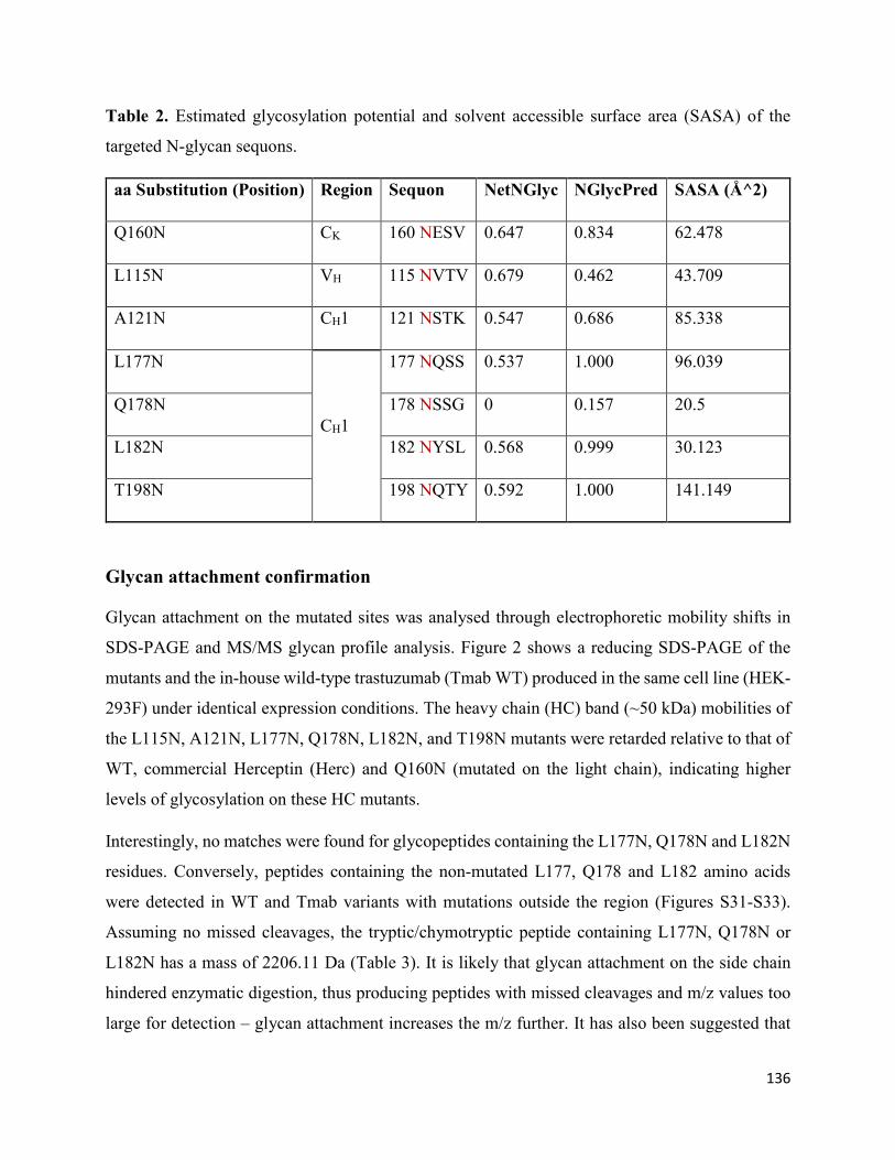
136
Table 2. Estimated glycosylation potential and solvent accessible surface area (SASA) of the
targeted N-glycan sequons.
aa Substitution (Position) Region Sequon NetNGlyc NGlycPred SASA (Å^2)
Q160N CK 160 NESV 0.647 0.834 62.478
L115N VH 115 NVTV 0.679 0.462 43.709
A121N CH1 121 NSTK 0.547 0.686 85.338
L177N
CH1
177 NQSS 0.537 1.000 96.039
Q178N 178 NSSG 0 0.157 20.5
L182N 182 NYSL 0.568 0.999 30.123
T198N 198 NQTY 0.592 1.000 141.149
Glycan attachment confirmation
Glycan attachment on the mutated sites was analysed through electrophoretic mobility shifts in
SDS-PAGE and MS/MS glycan profile analysis. Figure 2 shows a reducing SDS-PAGE of the
mutants and the in-house wild-type trastuzumab (Tmab WT) produced in the same cell line (HEK-
293F) under identical expression conditions. The heavy chain (HC) band (~50 kDa) mobilities of
the L115N, A121N, L177N, Q178N, L182N, and T198N mutants were retarded relative to that of
WT, commercial Herceptin (Herc) and Q160N (mutated on the light chain), indicating higher
levels of glycosylation on these HC mutants.
Interestingly, no matches were found for glycopeptides containing the L177N, Q178N and L182N
residues. Conversely, peptides containing the non-mutated L177, Q178 and L182 amino acids
were detected in WT and Tmab variants with mutations outside the region (Figures S31-S33).
Assuming no missed cleavages, the tryptic/chymotryptic peptide containing L177N, Q178N or
L182N has a mass of 2206.11 Da (Table 3). It is likely that glycan attachment on the side chain
hindered enzymatic digestion, thus producing peptides with missed cleavages and m/z values too
large for detection – glycan attachment increases the m/z further. It has also been suggested that

137
large glycopeptides can be missed in the workflow utilized for this analysis during peptide
extraction following SDS-PAGE, as the large size could prevent them from extraction due to gel
shrinkage caused by the organic solvents employed [19]. Similarly, glycan attachment in the
vicinity of the cleavage sites has been shown to prevent enzymatic activity of the serine protease
proteinase K in the L177N mutant in adalimumab Fab [21]. Still, successful glycosylation of
L177N, Q178N, and L182N was confirmed by mobility shifts in SDS-PAGE and an increased
heterogeneity observed in the released glycan profile compared to WT as discussed in the next
section.
Figure 2. Reducing SDS-PAGE of the trastuzumab mutants for confirmation of glycan
attachment. Commercial Herceptin (Herc) and the wild-type (non-mutated) trastuzumab
produced in-house were run together with the mutants to compare electrophoretic mobility
shifts in the ~50 kDa band corresponding to the heavy chain.
Despite the positive prediction from the algorithms (NGlycPred score of 0.834 (Q160N), Table 2),
the light chain (LC) band (~25 kDa) of Q160N showed no apparent mobility shift compared to
WT LC region, suggesting a lack of glycosylation on the mutated asparagine residue. Although
the glycan profiling of the Q160N showed very similar structures to WT Tmab, the glycan release
was performed on the whole protein but not on the light chain only and therefore provided an

138
overall glycoprofile of the protein (figure 3, table 4). While the visual similarity of the light chain
gel mobilities of WT Tmab and Q160N and the information of the overall glycan profile of Q160N
do not entirely exclude the possibility of glycan occupancy on the 160N, the MS analysis of the
Q160N light chain peptide clearly showed the presence of a non-glycosylated LC region. To
confirm the lack of glycan attachment, the Q160N mutant was subjected to trypsin and
chymotrypsin digestion to generate the corresponding peptides for nanoLC-ESI-MS/MS analysis.
Spectral matches were found for non-glycosylated peptides encompassing the Q160N residue, thus
confirming successful Q to N conversion and reaffirming the lack of glycan attachment (Figure
S34). This apparent lack of glycan attachment was also observed in the corresponding Q160N
mutant when expressed in adalimumab (Q161N) (data to be published).
Table 3. Peptide sequences after trypsin/chymotrypsin digestion allowing one missed cleavage
(MC) site
Mass (Da) Position MC Sequence
4000.01 162-201 1 NSGALTSGVHTFPAVLQSSGLYSLSSVVTVPSSSLGTQTY
3248.63 153-183 1 FPEPVTVSWNSGALTSGVHTFPAVLQSSGLY
3148.59 184-213 1 SLSSVVTVPSSSLGTQTYIC NVNHKPSNTK
2206.11 162-183 0 NSGALTSGVHTFPAVLQSSG LY
1812.92 184-201 0 SLSSVVTVPSSSLGTQTY
Glycan profile analysis
To analyse the global glycan profile of the trastuzumab variants, the proteins were immobilized
on PVDF membranes and treated with PNGase F to release glycans attached to both the conserved
WT N297 and the added glycosylation site. The released oligosaccharides were run on capillary
LC-ESI-MS/MS, wherein a PGC column was used for separation of distinct glycoforms and
MS/MS analysis (CID fragmentation) was performed to elucidate glycan structure. Figure 3 shows
the annotated full MS spectra of Tmab WT, Herceptin, Q160N, and L177N displaying the
identified glycan structures found on each variant. The global glycan profile reflects contrasting
differences in glycoform heterogeneity and the relative abundance of the glycans found on the
glycomutants. MS1 and MS2 (where possible) spectra of all the glycomutants can be found in

139
supplementary materials. The released glycan profiles displayed in figure 3 and figures S1-S9
come from both the conserved Fc N-glycosylation sequon and the added N-glycosylation site.
Table 4 lists the glycan structures found on all Tmab variants through this MS methodology; and
Figure 4 reports the glycan relative abundances on specific Tmab variants determined by
quantifying the area under the curve (AUC) of the extracted ion chromatograms.
Figure 3. Full MS spectra of N-glycans released from Tmab WT, Q160N, L177N and
Herceptin contrasting the heterogeneity found on the mutant.

140
Glycan microheterogeneity at the conserved N297 in Tmab WT is similar to that reported in
previous analysis of commercial Herceptin and other monoclonal therapeutic antibodies, where
complex G0F, G1F and G2F glycoforms (Figure 4 and table 4) are predominant [26, 27]. Minor
species (i.e., Man5, G0 and G2S1) were detected in commercial Herceptin but not in Tmab WT
(Figure 3). An important difference between commercial Herceptin and Tmab WT lies in the
relative abundance of the three main glycoforms (G0F, G1F and G2F), where the originator
contained the pattern G0F>G1F>G2F (60%, 31%, and 2%) and Tmab WT possessed
G1F>G0F>G2F (26%, 57%, and 17%) (Figure 4). Glycosylation differences are expected between
these two samples, as the originator is produced in CHO cells and our in-house Tmab in HEK-
293F. Moreover, sialic acid-containing species (17a table 4) were present in the originator and not
detected in Tmab WT. This observation is consistent with previous studies reporting higher
sialylation of N-glycans in recombinant proteins expressed in CHO when compared to HEK cells
[28].
The G1F>G0F>G2F distribution observed in Tmab WT was conserved in all the glycomutants
(Figure 4), although the abundance of G1F seemed to decrease in comparison to that observed for
Tmab WT. Figure 5 shows the relative abundances considering only the main complex glycoforms
found on the Fc domain (G0F, G1F, and G2F). The decrease in G1F and concomitant increase in
G0F was more pronounced in the Q178N mutant where G1F (40.47%) was only slightly less
abundant than G0F (42.98%) (Figure 5).

141
Figure 4. Relative abundances of the glycan found on each specific mutant. Highlighted in
blue are G0F, G1F and G2F which were also found on the WT tmab. High mannose glycans
are displayed in greed, hybrid glycans in yellow and sialic acid containing glycans in red.

142
Figure 5. Relative abundances of the main glycoforms found on all Tmab variants.
Remarkably, a much wider heterogeneity of glycan structures were detected on most Tmab
variants relative to wild type trastuzumab (Tmab WT and Herceptin) (Figures 3 and 4). The fact
that G0F, G1F, and G2F were still the most abundant structures in all samples indicates that the
increased variety of glycans come from the Fab region. The wider array of structures included high
mannose, hybrid, and complex biantennary, triantennary and tetrantennary glycans with varied
degrees of galactosylation and sialylation (Table 4). Variants L177N and L182N displayed the
most varied sets of glycoforms with 16 and 12 different structures detected, respectively.
Importantly, sialic acid-containing glycans were particularly abundant in L115N, L182N, and
T198N. Q178N had a significant presence of high mannose glycans.

143
Table 4. Full list of glycans found on all mutants
#
Structure Type Composition Structure
[M-
2H]2-
Mutants
containing
the glycan
1a high
mannose HexNAc(2)Hex(5)
617.22 Q178N
1b high
mannose HexNAc(2)Hex(5)
617.22
L177N
Q178N
L182N
2a (G0) complex HexNAc(4)Hex(3)
658.24 L177N
Herceptin
2b (G0) complex HexNAc(4)Hex(3)
658.24 L177N
3 high
mannose HexNAc(2)Hex(6)
698.25 Q178N
L182N
4 (G0F) complex HexNAc(4)Hex(3)Fuc(1)
731.27
All Tmab
variants
5 complex HexNAc(5)Hex(3)
759.78 L177N
6a hybrid HexNAc(3)Hex(4)Man(2)
Fuc(1)
791.79 L177N
6b hybrid HexNAc(3)Hex(4)Man(2)
Fuc(1)
791.79 L177N
6c hybrid HexNAc(3)Hex(4)Man(2)
Fuc(1)
791.79 L177N
7 hybrid HexNAc(3)Hex(5)Man(1)
799.79 Q178N
L182N

144
8a (G1F) complex HexNAc(4)Hex(3)Man(1)
Fuc(1)
812.3 L177N
8b (G1F) complex HexNAc(4)Hex(3)Man(1)
Fuc(1)
812.3 All Tmab
variants
9 complex HexNAc(5)Hex(3)Man(1)
840.81 L177N
10 high
mannose HexNAc(2)Hex(8)
860.3 L115N
11 hybrid HexNAc(3)Hex(4)Man(1)
NeuAc(1)
864.31 L182N
12 hybrid HexNAc(3)Hex(5)Man(1)
Fuc(1)
872.81 L177N
13 (G2F) complex HexNAc(4)Hex(3)Man(2)
Fuc(1)
893.33 All Tmab
variants
14a complex HexNAc(5)Hex(3)Man(1)
Fuc(1)
913.84
A121N
L177N
Q178N
L182N
T198N
14b complex HexNAc(5)Hex(3)Man(1)
Fuc(1)
913.84
A121N
L177N
Q178N
L182N
T198N
15 high
mannose HexNAc(2)Hex(9)
941.33 L115N
16 hybrid HexNAc(3)Hex(5)Man(1)
NeuAc(1)
944.84
A121N
L177N
L182N
T198N

145
17a complex HexNAc(4)Hex(3)Man(2)
NeuAc(1)
965.84 L177N
L182N
17b complex HexNAc(4)Hex(3)Man(2)
NeuAc(1)
965.84 L177N
18 complex HexNAc(5)Hex(3)Man(1)
Fuc(2)
986.87 A121N
L177N
19a complex HexNAc(5)Hex(3)Man(2)
Fuc(1)
994.87
L115N
A121N
L177N
Q178N
L182N
T198N
19b complex HexNAc(5)Hex(3)Man(2)
Fuc(1)
994.87 L177N
20a complex HexNAc(4)Hex(3)Man(2)
Fuc(1)NeuAc(1)
1038.87
L115N
A121N
L177N
Q178N
L182N
T198N
20b complex HexNAc(4)Hex(3)Man(2)
NeuAc(1)
1038.87
A121N
L177N
Q178N
L182N
T198N
21a complex HexNAc(5)Hex(3)Man(1)
NeuAc(1)
1059.39
A121N
L177N
T198N
21b complex HexNAc(5)Hex(3)Man(1)
NeuAc(1)
1059.39 L177N
21c complex HexNAc(5)Hex(3)Man(1)
NeuAc(1)
1059.39 L177N

146
Binding affinity to HER2 and FcγR1A
To assess potential alterations in biological activity caused by the structural modifications of
glycan addition, we tested the binding affinity of the Tmab variants on biologically relevant
receptors in vitro. Surface-plasmon resonance (SPR) single-cycle kinetic assays were performed
to determine binding kinetic constants of the Tmab variants to their molecular target HER2, and
to Fc receptors FcγR1A, FcγR2B, and FcγR3A (Table 5 and Table 6). Sensorgrams displaying
typical curvatures with satisfactory curve fitting were obtained across all receptors tested with the
exception of mutants A121N, Q178N, L182N, and T198N on FcγR2B, thus the corresponding
binding kinetic constants to this receptor were not determined (Figures S35-S38). Chi-square
values and residuals were inspected to validate curve fitting.
Table 5. Binding kinetic constants of the Tmab variants to HER2 and FcγR1A
HER2 FcγR1A
Ka Kd KD Ka Kd KD
(x 10+6) M-
1.s-1 (x 10-5) s-1 pM
(x 10+5) M-
1.s-1 (x 10-4) s-1 nM
Tmab WT 2.50 ± 0.03 3.03 ± 0.72 12.11 ± 2.72 2.11 ± 0.03 10.64 ± 0.56 5.05 ± 0.33
L115 > N 3.82 ± 3.80 18.16 ±
9.14
47.56 ±
25.89 1.79 ± 0.20 4.20 ± 0.06 2.34 ± 0.26
A121 > N 4.40 ± 1.71 23.84 ± 4.35 54.24 ±
10.24 3.83 ± 0.36 8.77 ± 1.14 2.29 ± 0.08
Q160 > N 0.83 ± 0.02 2.78 ± 1.26 33.53 ±
15.86 2.55 ± 0.02 6.62 ± 0.11 2.59 ± 0.02
L177 > N 1.84 ± 0.13 6.32 ± 0.08 34.28 ± 2.01 5.01 ± 0.18 15.04 ± 1.27 3.00 ± 0.15
Q178 > N 1.35 ± 0.27 4.29 ± 0.67 31.88 ± 2.11 7.38 ± 0.11 9.23 ± 0.30 1.25 ± 0.02
L182 > N 1.1 * 3.2 * 29.1 * 3.67 ± 0.55 12.37 ± 2.64 3.37 ± 0.22
T198 > N 6.11 ± 1.17 50.97 ± 3.23 83.44 ±
77.77 4.71 ± 0.02 8.09 ± 0.10 1.72 ± 0.03

147
Table 6. Binding kinetic constants of the Tmab variants to FcγR2B and FcγR3A
FcγR2B FcγR3A
Ka Kd KD Ka Kd KD
(x 10+4) M-
1.s-1 (x 10-4) s-1 nM
(x 10+4) M-1.s-
1 (x 10-4) s-1 nM
PBS-Herc 3.05 ± 0.04 1.99 ± 0.13 6.55 ± 0.35 10.51 ± 0.29 1.90 ±
0.54 1.81 ± 0.47
Tmab WT 5.76 ± 1.01 4.75 ± 0.41 8.25 ± 0.69 11.41 ± 2.06 1.29 ±
0.64 1.13 ± 0.32
L115 > N 0.65 ± 0.26 0.78 ± 0.44 11.92 ± 3.57 3.35 ± 0.85 2.74 ±
0.23 8.17 ± 1.60
T117 > N ND ND
A121 > N ND 2.78 ± 1.99 1.95 ± 0.43 7.01 ± 3.84
Q160 > N 4.01 ± 0.32 1.85 ± 0.27 4.62 ± 0.32 21.17 ± 4.95 8.01 ±
2.95 3.78 ± 0.46
L177 > N 2.95 ± 1.82 6.88 ± 4.26 23.28 ±
44.62 2.83 ± 0.22
3.02 ±
0.05 10.67 ± 1.03
Q178 > N ND 1.95 ± 0.20 0.98 ±
0.20 5.04 ± 1.47
L182 > N ND 3.12 ± 0.25 1.95 ±
0.20 6.24 ± 1.30
T198 > N ND 2.25 ± 0.14 0.86 ±
0.15 3.81 ± 0.41
SPR-derived binding kinetic parameters of the mutants on immobilized HER2 displayed a modest
decrease in the binding affinity constant (KD) compared to wild-type trastuzumab (Table 5). KD
values for the Tmab variants ranged from 29.10 – 83.44 pM compared to 12.11 ± 2.72 pM for
Tmab WT, which represents a 2.4 – 6.9-fold reduction in binding affinity. Most variants displayed
a decrease in binding rate constant (Ka) and an increase in dissociation rate constant (Kd), with
the exception of A121N and T198N that had higher Ka values, yet they also possessed the highest
increase in Kd constants. Thus, the A121N and T198N mutations displayed the lowest binding
affinity constants to HER2.

148
Conversely, binding affinity to captured FcγR1A was improved for all mutants relative to both
Tmab WT and Herceptin, exhibiting KD values 1.5 – 4 times lower than Tmab WT. Mutants
Q178N and T198N recorded the highest binding affinity to FcγR1A, with KD constants of 1.25
and 1.72 nM respectively. SPR assays with immobilized FcγR2B yielded curves with lower
similarity to the wild-type references. Dissociation times for mutants A121N and Q178N were too
long to obtain reliable kinetic binding affinity parameters. L182N showed a drastic decrease in
binding affinity. Mutants L115N, Q160N, and L177N exhibited modest increases in binding
affinity constants to FcγR2B. Binding affinity to FcγR3A was reduced across all mutants,
displaying 3.35 – 9.44-fold increases in KD values.
Discussion
The insertion of novel N-glycosylation sites on the primary structure of IgG1 molecules is a highly
promising approach to enhance the physicochemical properties of therapeutic proteins, having
previously demonstrated important enhancements in solubility and physical stability in
bevacizumab, adalimumab, and the Li33 anti-LINGO antibody. However, implementation of this
approach in clinical development could be challenging due to the concomitant increase in
structural heterogeneity. This is especially relevant when considering mAbs whose therapeutic
efficacy relies, at least partially, on eliciting effector functions; given that the technique entails the
incorporation of a relatively large molecule that should not alter detrimentally the binding affinity
towards the various receptors involved in its biological activity (i.e., cognate antigen, Fc receptors,
C1q). Further considerations include potential modifications in immunogenicity and
pharmacokinetic profiles. Moreover, elucidation of the underlying mechanisms that drive
improvements in stability demands thorough structural analysis of the glycan structures attached
to the engineered site. In light of this, we inserted N-glycosylation motifs in trastuzumab in several
amino acid positions previously identified by others on conserved regions of the IgG1 molecule
and performed a detailed structural analysis of the ensuing glycan profile.
Preliminary prediction and confirmation of glycan attachment
N-glycosylation is a complex biochemical process, for which the requirements for glycan
attachment are not yet fully understood. Three key factors determining glycan occupancy have
been identified: (1) location of the asparagine residue within the consensus motif Asn-X-Ser/Thr,
(2) location of the N residue in the ER lumen during translation, and (3) the adjacent secondary

149
structure of the protein must enable glycan transfer. Based on these, numerous servers have been
developed to map N-glycosylation sequons and predict the probability of glycan attachment, and
have been instrumental in the study of the glycosylation profile of proteomes. We thus employed
two such servers to assess the validity of their prediction values applied to this antibody
engineering strategy. This is particularly important considering the fact that previous studies have
not pursued potential APR shielding regions experimentally due to low glycan transfer probability.
Specifically, residue Q178 was deemed ineligible by Courtois et al. based on low solvent
accessible surface area obtained from MD simulations. Our preliminary evaluation of glycan
attachment probability on trastuzumab shed similar results (Table 2) to those obtained by Courtois
et al. on their panel of glycosylation sites identified through SAP, wherein Q178N had negative
predictions by both NetNGlyc and NGlycPred, and SASA was amongst the lowest calculated (20.5
Å2). Importantly, L115N also obtained a negative prediction value from NGlycPred. It was thus
remarkable that our experimental data contradicted the negative prediction for residues L115N and
Q178N. The Q178N mutant had previously been implemented by Pepinsky et al. in the anti-
LINGO IgG1 antibody with important improvements in solubility, although no compelling data to
confirm glycan attachment was reported. Moreover, our data strongly suggest that Q160N does
not undergo glycan attachment, despite the high SASA and prediction values calculated for the
residue. This was equally observed in the equivalent mutation in adalimumab (data to be
published). These results emphasize the importance of confirming glycan attachment and
performing structural analysis of the resulting structural modifications, to better interpret the
results derived from aggregation experiments. Furthermore, they reveal a conspicuous need for
further understanding of the factors that determine successful glycan transfer to refine this antibody
engineering approach.
Glycan profile analysis
Compared to plasma proteins and other recombinant therapeutic proteins, monoclonal antibodies
display limited heterogeneity in their Fc N-glycan profile, as reported herein. This observation is
ascribed to the decreased surface exposure of Asn N297 and the attached glycan as it engages in
several interactions with the protein backbone and residue side chains at the inner face of the
domain [28, 29]. Although most plasma IgG molecules possess only the conserved N-
glycosylation sequon in the Fc region, roughly 15-25% also express additional glycosylation sites

150
on the variable domain of the Fab region [30, 31]. Glycan profiling of Fab-glycosylated serum
IgGs has revealed contrasting structural characteristics between Fc-derived and Fab-derived
glycans, most notably higher overall heterogeneity and sialylation [32, 33]. All mutations
evaluated in this study are located in the Fab region, although only L115N lies on a variable domain
– in close proximity to the VH-CH1 interface. The global glycan profile acquired through
capillary/nanoLC-ESI-MS/MS demonstrated that increased structural heterogeneity is similarly
obtained when engineered glycosylation sites are incorporated through mutagenesis in the Fab
region of a recombinant mAb.
There was no evident association between the degree of heterogeneity or apparent glycan
maturation and the solvent accessible surface area calculated. For instance, low-SASA mutant
L115N possessed a relatively high abundance of sialylated glycans, indicative of higher glycan
processing. Remarkably, high mannose glycans (HexNAc(2)Man(8) and HexNAc(2)Man(9))
were also detected in L115N. Similarly, residue Q178N displayed high glycoform heterogeneity
despite having the lowest SASA among the mutants, including sialylated and triantennary glycans.
As the en bloc transfer of the dolichol oligosaccharide precursor occurs on nascent proteins
undergoing translation, low surface exposure calculated from the WT structure might not preclude
glycan attachment and the resulting orientation of the glycan could be surface-exposed for enzyme
accessibility. Ideally, SASA would be calculated from the crystal structure of the mutated protein,
but performing such analyses for all mutations entails prominent experimental challenges.
Overall, the degree of sialylation was markedly higher in most engineered mutants relative to both
wild-type references, i.e., Tmab WT and Herceptin. Similar findings have consistently been
reported for both polyclonal and recombinant monoclonal IgG proteins containing naturally-
occurring N-glycosylation sequons in the Fab region [34]. The higher abundance of sialic acid
glycans in Fab glycosites has been attributed to increased accessibility to enzymes involved in
glycan maturation relative to the Fc glycan. In this case, however, we also found various high
mannose and hybrid glycans not detected in Tmab WT. In the seminal exploration of SAP
reduction by Courtois et al., a G0 glycan was selected for molecular dynamic simulations of the
Fab region to shed light into the factors driving resistance to aggregation upon glycosite insertion
[18]. Here, it was posited that the added glycans on some of the mutated sites produce limited APR
shielding and hence the improvements in physical stability could stem from steric hindrance due

151
to the large hydrodynamic size and hydrophilic character of the added glycans. The diversity of
glycoforms reported here indicates that this structural heterogeneity must be accounted for when
performing these analyses, particularly considering the important presence of negatively charged
sialic acid residues on some of the mutants.
HEK-293F cells were chosen for transient expression of the Tmab variants due to high transient
transfection efficiency and yield relative to CHO cells. Comparison of the glycan profile of CHO-
produced Herceptin with HEK-derived Tmab WT shows that galactosylation was more abundant
in HEK-293F expression. This observation was also consistent across the mutant panel when
considering uniquely Fc glycans. Galactosylation of the conserved Fc glycan is not considered to
affect significantly mAb binding to FcγR receptors, yet there is consistent evidence that the
presence of terminal galactose promotes activation of the classical complement route [35]. Hence,
the higher content of G1F and G2F glycoforms could lead to potency advantages in mAbs with
significant modulation of complement activity.
Alterations in HER2 and Fc receptor affinity
A crucial consideration in this engineering approach must be the conservation of biological
activity. The proximity of the mutations to the CDRs and to regions involved in Fc receptor binding
warrants examination of alterations in receptor binding affinity. Taking this into consideration, we
evaluated the binding kinetics of the panel of mutants to their molecular target (HER2) and to Fc
receptors involved in effector function, using single-cycle SPR.
The kinetic constants obtained indicate that HER2 binding affinity is moderately diminished for
all mutants, reflected by 2.4 – 6.9-fold reductions in the binding affinity constant (KD). All KD
values, however, were subnanomolar – demonstrating that strong binding to HER2 is retained
despite modest decreases in affinity. Remarkably, the highest reduction in affinity was obtained
with T198N, which is located farthest from the HER2 binding region. This reduction in binding
affinity could have resulted from conformational changes affecting the antigen binding region. KD
values derived herein for wild-type Tmab binding to HER2 were in agreement with those reported
in previous literature, spanning 19 pM – 5.48 nM [36-38].
Overall, the mutant library exhibited minor modifications in Fc receptor binding affinity. Binding
to FcγR1A was marginally improved in all Tmab variants. Curve fitting was less reliable for

152
FcγR2B, and it was not possible to calculate these parameters for mutants A121N, Q178N, L182N,
and T198N. Still, sensorgram interpretation suggests that L182N binding to FcγR2B was
drastically disrupted. Binding affinity to FcγR3A also showed moderate disruption across all Tmab
variants, with 3.35 – 9.44-fold increases in KD values.
The affinity screening to biologically relevant receptors performed in this work served as a proof-
of-principle that N-glycosylation sites can be incorporated in the Fab region of therapeutic
monoclonal antibodies without major modifications in antigen and Fc receptor binding.
Nonetheless, evaluation of ADCC, CDC and inhibition of cell proliferation is still required to
ascertain potential variations in biological activity.
Concluding remarks
The rational addition of N-glycosylation sequons represents a highly promising strategy to
improve the physical stability of therapeutic antibodies, as is highlighted by the important
enhancements in physical stability obtained in previous studies with bevacizumab, adalimumab
Fab, and the Li33 anti-LINGO antibody. Moreover, we have previously performed the panel of
mutations reported here in full-size adalimumab and demonstrated enhancements in
thermodynamic stability through this approach. However, our data established that for this strategy
to move forward in the clinical development pathway, there is an evident need to control or
minimize the structural heterogeneity that results from glycan addition. The latter is also required
to obtain further insight into the conferred physicochemical properties responsible for the
improvements in stability. Native Fc glycosylation, albeit less diverse than most glycoproteins,
already constitutes a challenge concerning the assessment of critical quality attributes. This,
however, could be overcome with glyco-engineering strategies to curtail glycoform heterogeneity.
Technologies to reduce glycan diversity in recombinant expression are being pursued in the
context of refining the production of biopharmaceuticals. Prime examples of these include
engineered Pichia pastoris (GlycoSwitch) and human (GlycoDelete) cell lines. We foresee great
value in combining these two glyco-engineering strategies towards the development of therapeutic
antibodies with enhanced physicochemical and biological properties. Albeit introducing
heterogeneity, molecular engineering approaches such as this one offer a great deal of flexibility
and control over preparation of new generation therapies. This glycoengineering strategy can also

153
be employed to enhance the biophysical stability of other protein-based therapies with short half-
life and low formulation stabilities.
References
[1] L. Urquhart, Top drugs and companies by sales in 2018, Nature Reviews Drug Discovery, 18
(2019) 245.
[2] Z. Elgundi, M. Reslan, E. Cruz, V. Sifniotis, V. Kayser, The state-of-play and future of
antibody therapeutics, Advanced drug delivery reviews, 122 (2017) 2-19.
[3] H. Kaplon, J.M. Reichert, Antibodies to watch in 2019, mAbs, 11 (2019) 219-238.
[4] J.K.H. Liu, The history of monoclonal antibody development – Progress, remaining challenges
and future innovations, Annals of Medicine and Surgery, 3 (2014) 113-116.
[5] V. Kayser, N. Chennamsetty, V. Voynov, B. Helk, B.L. Trout, Conformational stability and
aggregation of therapeutic monoclonal antibodies studied with ANS and Thioflavin T binding,
mAbs, 3 (2011) 408-411.
[6] M. Vazquez-Rey, D.A. Lang, Aggregates in monoclonal antibody manufacturing processes,
Biotechnology and bioengineering, 108 (2011) 1494-1508.
[7] R. van der Kant, A.R. Karow-Zwick, J. Van Durme, M. Blech, R. Gallardo, D. Seeliger, K.
Aßfalg, P. Baatsen, G. Compernolle, A. Gils, J.M. Studts, P. Schulz, P. Garidel, J. Schymkowitz,
F. Rousseau, Prediction and Reduction of the Aggregation of Monoclonal Antibodies, Journal of
Molecular Biology, 429 (2017) 1244-1261.
[8] J. Hernandez-Jimenez, A. Martinez-Ortega, A. Salmeron-Garcia, J. Cabeza, J.C. Prados, R.
Ortiz, N. Navas, Study of aggregation in therapeutic monoclonal antibodies subjected to stress and
long-term stability tests by analyzing size exclusion liquid chromatographic profiles, International
journal of biological macromolecules, 118 (2018) 511-524.
[9] V. Voynov, N. Chennamsetty, V. Kayser, B. Helk, B.L. Trout, Predictive tools for stabilization
of therapeutic proteins, mAbs, 1 (2009) 580-582.

154
[10] R. Zambrano, M. Jamroz, A. Szczasiuk, J. Pujols, S. Kmiecik, S. Ventura, AGGRESCAN3D
(A3D): server for prediction of aggregation properties of protein structures, Nucleic Acids
Research, 43 (2015) W306-W313.
[11] J. Van Durme, G. De Baets, R. Van Der Kant, M. Ramakers, A. Ganesan, H. Wilkinson, R.
Gallardo, F. Rousseau, J. Schymkowitz, Solubis: a webserver to reduce protein aggregation
through mutation, Protein engineering, design & selection : PEDS, 29 (2016) 285-289.
[12] M. Reslan, V. Kayser, The effect of deuterium oxide on the conformational stability and
aggregation of bovine serum albumin, Pharmaceutical Development and Technology, 23 (2018)
1030-1036.
[13] V.I. Razinkov, M.J. Treuheit, G.W. Becker, Accelerated formulation development of
monoclonal antibodies (mAbs) and mAb-based modalities: review of methods and tools, Journal
of biomolecular screening, 20 (2015) 468-483.
[14] M. Reslan, V. Kayser, Ionic liquids as biocompatible stabilizers of proteins, Biophysical
reviews, 10 (2018) 781-793.
[15] M. Reslan, V. Ranganathan, D.R. Macfarlane, V. Kayser, Choline ionic liquid enhances the
stability of Herceptin(R) (trastuzumab), Chemical communications (Cambridge, England), 54
(2018) 10622-10625.
[16] R. Mathaes, A. Koulov, S. Joerg, H.-C. Mahler, Subcutaneous Injection Volume of
Biopharmaceuticals—Pushing the Boundaries, Journal of Pharmaceutical Sciences, 105 (2016)
2255-2259.
[17] R.B. Pepinsky, L. Silvian, S.A. Berkowitz, G. Farrington, A. Lugovskoy, L. Walus, J.
Eldredge, A. Capili, S. Mi, C. Graff, E. Garber, Improving the solubility of anti-LINGO-1
monoclonal antibody Li33 by isotype switching and targeted mutagenesis, Protein science : a
publication of the Protein Society, 19 (2010) 954-966.
[18] F. Courtois, N.J. Agrawal, T.M. Lauer, B.L. Trout, Rational design of therapeutic mAbs
against aggregation through protein engineering and incorporation of glycosylation motifs applied
to bevacizumab, MAbs, 8 (2016) 99-112.
155
[19] P.H. Jensen, N.G. Karlsson, D. Kolarich, N.H. Packer, Structural analysis of N- and O-glycans
released from glycoproteins, Nature protocols, 7 (2012) 1299-1310.
[20] N. Chennamsetty, V. Voynov, V. Kayser, B. Helk, B.L. Trout, Design of therapeutic proteins
with enhanced stability, Proceedings of the National Academy of Sciences, 106 (2009) 11937.
[21] H. Nakamura, N. Oda-Ueda, T. Ueda, T. Ohkuri, Introduction of a glycosylation site in the
constant region decreases the aggregation of adalimumab Fab, Biochemical and biophysical
research communications, 503 (2018) 752-756.
[22] S. Chattopadhyay, P. Bagchi, D. Dutta, A. Mukherjee, N. Kobayashi, M. Chawlasarkar,
Computational identification of post-translational modification sites and functional families reveal
possible moonlighting role of rotaviral proteins, Bioinformation, 4 (2010) 448-451.
[23] A. Eshaghi, V.R. Duvvuri, R. Lai, J.T. Nadarajah, A. Li, S.N. Patel, D.E. Low, J.B. Gubbay,
Genetic Variability of Human Respiratory Syncytial Virus A Strains Circulating in Ontario: A
Novel Genotype with a 72 Nucleotide G Gene Duplication, PLOS ONE, 7 (2012) e32807.
[24] E. Cruz, J. Cain, B. Crossett, V. Kayser, Site-specific glycosylation profile of influenza A
(H1N1) hemagglutinin through tandem mass spectrometry, Human vaccines &
immunotherapeutics, 14 (2018) 508-517.
[25] G.-Y. Chuang, J.C. Boyington, M.G. Joyce, J. Zhu, G.J. Nabel, P.D. Kwong, I. Georgiev,
Computational prediction of N-linked glycosylation incorporating structural properties and
patterns, Bioinformatics, 28 (2012) 2249-2255.
[26] H. Xie, A. Chakraborty, J. Ahn, Y.Q. Yu, D.P. Dakshinamoorthy, M. Gilar, W. Chen, S.J.
Skilton, J.R. Mazzeo, Rapid comparison of a candidate biosimilar to an innovator monoclonal
antibody with advanced liquid chromatography and mass spectrometry technologies, MAbs, 2
(2010) 379-394.
[27] K.H. Lee, J. Lee, J.S. Bae, Y.J. Kim, H.A. Kang, S.H. Kim, S.J. Lee, K.J. Lim, J.W. Lee, S.K.
Jung, S.J. Chang, Analytical similarity assessment of rituximab biosimilar CT-P10 to reference
medicinal product, mAbs, 10 (2018) 380-396.
[28] A. Croset, L. Delafosse, J.P. Gaudry, C. Arod, L. Glez, C. Losberger, D. Begue, A.
Krstanovic, F. Robert, F. Vilbois, L. Chevalet, B. Antonsson, Differences in the glycosylation of

156
recombinant proteins expressed in HEK and CHO cells, Journal of biotechnology, 161 (2012) 336-
348.
[29] E. Girardi, M.D. Holdom, A.M. Davies, B.J. Sutton, A.J. Beavil, The crystal structure of
rabbit IgG-Fc, The Biochemical journal, 417 (2009) 77-83.
[30] F.S. van de Bovenkamp, N.I.L. Derksen, P. Ooijevaar-de Heer, K.A. van Schie, S. Kruithof,
M.A. Berkowska, C.E. van der Schoot, H. Ijspeert, M. van der Burg, A. Gils, L. Hafkenscheid,
R.E.M. Toes, Y. Rombouts, R. Plomp, M. Wuhrer, S.M. van Ham, G. Vidarsson, T. Rispens,
Adaptive antibody diversification through <em>N</em>-linked glycosylation of the
immunoglobulin variable region, Proceedings of the National Academy of Sciences, 115 (2018)
1901.
[31] R. Jefferies, Glycosylation of Antibody Molecules, in: S. Dübel, J.M. Reichert (Eds.)
Handbook of Therapeutic Antibodies2014, pp. 171-200.
[32] K.R. Anumula, Quantitative glycan profiling of normal human plasma derived
immunoglobulin and its fragments Fab and Fc, Journal of immunological methods, 382 (2012)
167-176.
[33] A. Bondt, Y. Rombouts, M.H.J. Selman, P.J. Hensbergen, K.R. Reiding, J.M.W. Hazes,
R.J.E.M. Dolhain, M. Wuhrer, Immunoglobulin G (IgG) Fab Glycosylation Analysis Using a New
Mass Spectrometric High-throughput Profiling Method Reveals Pregnancy-associated Changes,
Molecular &amp; Cellular Proteomics, 13 (2014) 3029.
[34] M. Holland, H. Yagi, N. Takahashi, K. Kato, C.O. Savage, D.M. Goodall, R. Jefferis,
Differential glycosylation of polyclonal IgG, IgG-Fc and IgG-Fab isolated from the sera of patients
with ANCA-associated systemic vasculitis, Biochimica et biophysica acta, 1760 (2006) 669-677.
[35] P.N. Boyd, A.C. Lines, A.K. Patel, The effect of the removal of sialic acid, galactose and total
carbohydrate on the functional activity of Campath-1H, Molecular immunology, 32 (1995) 1311-
1318.
[36] P. Dakshinamurthy, P. Mukunda, B. Prasad Kodaganti, B.R. Shenoy, B. Natarajan, A.
Maliwalave, V. Halan, S. Murugesan, S. Maity, Charge variant analysis of proposed biosimilar to
Trastuzumab, Biologicals, 46 (2017) 46-56.

157
[37] D. Lakayan, R. Haselberg, R. Gahoual, G.W. Somsen, J. Kool, Affinity profiling of
monoclonal antibody and antibody-drug-conjugate preparations by coupled liquid
chromatography-surface plasmon resonance biosensing, Anal Bioanal Chem, 410 (2018) 7837-
7848.
[38] W.-H. Lua, S.K.-E. Gan, D.P. Lane, C.S. Verma, A search for synergy in the binding kinetics
of Trastuzumab and Pertuzumab whole and F(ab) to Her2, Npj Breast Cancer, 1 (2015) 15012.

158
Monoclonal Antibody Therapy of Solid Tumors: Clinical
Limitations and Novel Strategies to Enhance Treatment
Efficacy
Chapter 6

159
Chapter 6 – Authorship declaration statement
The following chapter is a literature review published in the journal Biologics: Targets and
Therapy as:
E. Cruz, V. Kayser. Monoclonal Antibody Therapy of Solid Tumors: Clinical Limitations and
Novel Strategies to Enhance Treatment Efficacy. Biologics: Targets and Therapy. 13 (2019), 33-
51.
E. Cruz carried out the literature searches and wrote the review.
Permission to include the published material has been granted by the corresponding author.
Esteban Cruz, Signature: 19th of August, 2019
As corresponding author and supervisor for this candidature, I hereby confirm that this authorship
declaration statement is complete and accurate
Veysel Kayser, Signature: 19th of August, 2019

160
Abstract
Abstract: Monoclonal antibodies (mAbs) have become a cornerstone in the therapeutic guidelines
of a wide range of solid tumors. The targeted nature of these biotherapeutics has improved
treatment outcomes by offering enhanced specificity to reduce severe side effects experienced with
conventional chemotherapy. Notwithstanding, poor tumor tissue penetration and the
heterogeneous distribution achieved therein are prominent drawbacks that hamper the clinical
efficacy of therapeutic antibodies. Failure to deliver efficacious doses throughout the tumor can
lead to treatment failure and the development of acquired resistance mechanisms. Comprehending
the morphological and physiological characteristics of solid tumors and their microenvironment
that affect tumor penetration and distribution is a key requirement to improve clinical outcomes
and realize the full potential of monoclonal antibodies in oncology. This review summarizes the
essential architectural characteristics of solid tumors that obstruct macromolecule penetration into
the targeted tissue following systemic delivery. It further describes mechanisms of resistance
elucidated for blockbuster antibodies for which extensive clinical data exists, as a way to illustrate
various modes in which cancer cells can overcome the anticancer activity of therapeutic antibodies.
Thereafter, it describes novel strategies designed to improve clinical outcomes of mAbs by
increasing potency and/or improving tumor delivery; focusing on the recent clinical success and
growing clinical pipeline of antibody-drug conjugates (ADC), immune checkpoint inhibitors and
nanoparticle-based delivery systems.
Introduction
Therapeutic monoclonal antibodies (mAbs) successfully entered the clinic over 25 years ago and
have become one of the central components of the healthcare system [1, 2]. Their arrival brought
about a therapeutic revolution due to their capacity to target specific molecular components, with
a large number of mAbs already approved in oncology, autoimmune disorders, chronic diseases
and many more conditions. Currently, over 80 antibody therapeutics have received regulatory
approval in Europe and/or the United States and just in 2017 sales of therapeutic antibodies
exceeded $100 billion worldwide [3].
In oncology, therapeutic antibodies offer the possibility to treat tumors in a targeted fashion and
reduce the severe side effects of conventional chemotherapy. Recent developments in cancer

161
biology have aided the discovery of molecular biomarkers in a wide range of solid malignancies
that can be used as targets with beneficial therapeutic outcomes. At present, over 15 distinct
monoclonal antibodies are indicated for the treatment of solid tumors [4]. Notwithstanding, in spite
of their remarkable clinical success some patients do not benefit from the treatment due to intrinsic
resistance mechanisms or the emergence of acquired resistance following treatment initialisation
[5, 6].
In solid tumors, the development of acquired resistance mechanisms is thought to emerge primarily
from continuous genetic alterations that modify the cellular phenotype and undermine the initial
therapeutic efficacy. This capacity of cancer cells to overcome the anticancer effect of the antibody
is facilitated by the exposure to subtherapeutic concentrations of the drug [7, 8]. The tumor
microenvironment poses physical barriers, most notably a markedly increased hydrostatic
pressure, that hinder penetration of macromolecules into the tumor following systemic
administration [9, 10]. This reduces the overall amount of antibody molecules that reach the target
tissue and exposes areas of the tumor that are difficult to penetrate to marginal doses of the
antibody, leading to acquired resistance and treatment failure [8]. In fact, therapeutic mAbs in
oncology are more commonly administered as combination therapy in conjunction with
chemotherapeutics due to relatively limited efficacy as single agents [11].
Identifying and understanding primary and acquired resistance mechanisms and overcoming the
barriers that impair efficient delivery of the drug into the tissue is critical to enhance therapeutic
outcomes. Most of the understanding regarding primary and acquired resistance comes from the
evaluation of clinical data available for early-approved blockbuster antibodies, such as
trastuzumab and cetuximab. This review gives an overview of the key factors affecting tumor
distribution upon systemic delivery and describes relevant mechanisms of resistance identified in
trastuzumab (anti-HER2) and cetuximab (anti-EGFR) therapy. Additionally, it describes recent
developments in the implementation of novel antibody-based therapeutics, such as antibody-drug
conjugates, immune checkpoint inhibitors, and antibody-targeted nanoparticles that have the
potential to increase therapeutic outcomes of solid tumors.

162
Limitations that impact clinical efficacy
Poor penetration and heterogeneous distribution in solid tumors
Therapeutic IgG antibodies must overcome pronounced physical and physiological obstacles in
order to penetrate and distribute uniformly throughout the tumor. In solid malignancies, impaired
lymphatic drainage due to the sparse presence of lymphatic vessels leads to the accumulation of
macromolecules in the interstitial tissue and a consequent increase in hydrostatic pressure [9, 12-
14]. Hence, the altered pressure differential from vascular vessels to the interstitial compartment
limits convection and extravasation of macromolecules from the vascular lumen into the tumor
(Figure 1) [15]. Moreover, antibody distribution following extravasation is further impeded by
cellular internalization and subsequent endocytic clearance at the tumor edge (an effect coined the
“binding-site barrier”), leading to poor penetration and regions of marginal antibody
concentrations [10, 16, 17]. The binding-site barrier suggests that higher affinity and higher
antigen expression, especially at the tumor edge, can retard mAb tumor percolation and impair
homogeneous distribution; although this barrier can be overcome by increasing the administered
dose.

163
Figure 1. Structural features of the tumor microenvironment that increase interstitial
pressure and hinder mAb extravasation and distribution. (A) Blood vessels that irrigate
healthy normal tissue possess a continuous inner lining of endothelial cells, enveloped by
perivascular cells called pericytes that grant integrity to the vascular tube. The extracellular

164
matrix (ECM) contains a lax network of collagen and proteoglycan fibers, and the presence
of macrophages and fibroblasts is scarce. Lymph vessels efficiently remove and prevent the
accumulation of macromolecules and interstitial fluid. (B) Increased demand of oxygen and
nutrients in tumor tissues causes blood vessels to form defectively and irregularly shaped.
The lack of pericytes makes the vascular tube unstable and leakier. The abundant presence
of fibroblasts and infiltrating macrophages promote the formation of a dense extracellular
matrix, with a condensed network of collagen and proteoglycan fibers. The paucity of lymph
vessels leads to the accumulation of macromolecules and an increase in interstitial fluid
pressure (IFP). The fibrotic nature of the ECM and the altered pressure differential between
the vascular lumen and the tumor hinder antibody convection into the targeted tissue.
A vast body of research studying some of the blockbuster therapeutic mAbs has highlighted the
significance of increasing tissue penetration to improve the outcome of antibody therapy [15, 18].
A study on cetuximab and trastuzumab in mouse xenografts confirmed that tumor distribution can
be improved with an increase in dose; however, hypoxic areas remained difficult to reach even at
higher doses. Moreover, xenografts expressing intermediate levels of ErbB1 (cognate antigen for
cetuximab) displayed more homogeneous distribution of cetuximab compared to xenografts with
higher ErbB1 expression [19].
An alternative approach consists in improving diffusion by employing smaller antibody fragments,
such as Fab fragments (~50 kDa), single-chain variable fragments (scFv ~30 kDa) and single-
domain antibodies (sdAb 12-15 kDa). Yet, while these formats indeed possess higher diffusion
rates, the tumor distribution achieved in physiological settings is poor because the clearance rates
for smaller fragments is markedly higher relative to full size antibody molecules [15, 20]. IgG
immunoglobulins undergo salvage recycling through interaction of the Fc region with the neonatal
Fc receptor (FcRn), leading to prolonged half-lives of more than 20 days for most therapeutic
mAbs [21]. Conversely, antibody fragments lacking an Fc region display half-lives of hours, or
even minutes for formats below the glomerular filtration cutoff (30-50 kDa). The high elimination
rates upon systemic delivery prevent most antibody fragments from saturating the tumor and
achieving uniform distributions [22, 23]. Increasing tumor tissue penetration thus poses significant
challenges given the intricate pharmacokinetic properties of IgGs.

165
Resistance to monoclonal antibody therapy
Understanding the resistance mechanisms that affect monoclonal antibody therapy in cancer has
proven to be a strenuous task, insofar as the antitumor activity of mAbs stems from a multiplicity
of molecular mechanisms – eg, signaling pathway disruption, antibody dependent cellular
cytotoxicity (ADCC), antibody dependent cellular phagocytosis (ADCP) and complement
dependent cytotoxicity (CDC). To this day, the clinical contribution of the various modes of action
involved in the anticancer activity of most mAbs remains controversial [24-26]. On that account,
intrinsic phenotypic variations in tumor cells or tumor-related cells affecting any of the involved
modes of action can compromise treatment efficacy. Moreover, adaptive phenotypic modifications
can arise following repeated exposure to sub-optimal doses of the biotherapeutic resulting in
acquired resistance [27, 28].
Most of the current understanding of the contributing factors in the development of intrinsic or
acquired resistance and their clinical significance comes from preclinical and clinical trials of
benchmark therapeutic antibodies. Notwithstanding that the modes of action of different mAbs are
not identical, the vast clinical data available for these benchmark antibodies are pivotal to
comprehend host response and optimize monoclonal antibody therapy. The next sections briefly
discuss resistance mechanisms identified in clinical settings for trastuzumab in HER2 positive
breast cancer, and for cetuximab in colorectal cancer as archetypes of solid tumor treatment.
Resistance to trastuzumab (anti-HER2 therapy)
Trastuzumab was the first therapeutic monoclonal antibody to be approved for a solid carcinoma
(FDA approval in the year 1998) [29]. Trastuzumab targets the extracellular domain of the human
epidermal growth factor receptor 2 (HER2/Neu or ErbB2) that is overexpressed in a broad range
of malignancies. HER2 overexpression is detected in 15-20% of breast cancers and this subset is
associated with poor prognosis and higher rates of recurrence [30, 31].
HER2 exists primarily as a monomeric receptor that can form heterodimers with other members
of the ErbB family of receptors (HER1, HER3 and HER4) upon ligand-mediated activation of the
latter. Heterodimerization activates the MAPK and PI3K/AKT/mTor intracellular pathways,
inducing cell proliferation and inhibition of apoptosis, respectively [32, 33]. Direct binding of
trastuzumab with HER2 can hinder heterodimerization and promote proteolysis of the receptor

166
through receptor-mediated endocytosis. This interaction inhibits downstream signaling and causes
cell cycle arrest by accumulation of the cyclin-dependent kinase (CDK) inhibitor p27 [34].
Additionally, trastuzumab can mediate antibody-dependent cellular cytotoxicity (ADCC) [35, 36]
and antibody-dependent cellular phagocytosis (ADCP) [36-38]. Induction of complement
dependent cytotoxicity (CDC) has also been documented in in vitro experiments, but it is thought
to contribute only minimally to the anticancer effect in patients [36, 39].
Intrinsic alterations of the HER2 receptor involving regions associated with the binding epitope of
trastuzumab have been linked to intrinsic (or primary) resistance mechanisms. For instance,
alternate transcription initiation sites can result in the expression of a truncated variant of the
receptor (p95-HER2) that lacks the cognate epitope for trastuzumab [40]. Insertions and point
mutations in the tyrosine kinase domain of HER2 have been identified in various cancers, some of
them associated with resistance to trastuzumab and lapatinib, however evidence of such mutations
in HER2 overexpressing breast cancers has not been reported to date [41, 42]. A further alteration
resulting in impaired target binding comes from the overexpression of mucin-4, which has been
shown to induce association with HER2 causing steric hindrance to abrogate trastuzumab binding
to HER2 [43].
Additional intrinsic and acquired resistance mechanisms predominantly involve alterations in the
P13K/Akt/mTOR axis, activation of other ErbB receptors (especially EGFR and HER3) by
increased ligand production, and circumvention of HER2 binding by activation of the PI3K
cascade through alternative pathways. Mutations in PIK3CA and function impairment of PTEN
(both downstream of HER2 signaling) have been implicated in bypassing HER2 blockade [44, 45].
Overexpression of the insulin-like growth factor (IGF-IR) has been documented as an adaptive
response to trastuzumab by some tumors, resulting in resistance to the antibody. IGF-IR can form
heterodimers and heterotrimers with HER2 and HER3 in breast cancer cells resistant to
trastuzumab [46, 47]. Similarly, increased levels of EpoR, EpHA2 and RTK MET can activate
P13K/Akt/mTOR by interacting with other members of the ErbB family or through activation of
intracellular kinases [47-49].
Resistance to cetuximab (anti-EGFR therapy)
The epidermal growth factor receptor (EGFR; HER1; ErbB1) forms part of the ErbB family of
receptors. EGFR is pivotal in modulating proliferative mechanisms and has been implicated in a

167
broad range of cancers [50-52]. Cetuximab (chimeric IgG1) was the first anti-EGFR mAb to
receive regulatory approval in 2004 [53]. Since then, two more anti-EGFR mAbs (panitumumab
and necitumumab) and six anti-EGFR small molecule inhibitors (gefitinib, erlotinib, lapatinib,
neratinib, vandetanib and osimertinib) have obtained regulatory approval for various cancers [4].
The anticancer activity of cetuximab partially resembles that of trastuzumab in that it targets
another member of the ErbB family of receptors with intrinsic protein tyrosine kinase activity.
Accordingly, dimerization of EGFR can activate the PI3K/AKT/mTOR, RAS/RAF/MAPK and
JAK/STAT signaling pathways to promote cell growth and proliferation [54, 55]. In contrast to
HER2, EGFR can undergo a conformational transition triggered by binding of specific ligands,
predominantly EGF and TGFα, that promotes the formation of homodimers and heterodimers with
other members of the HER family [56]. Cetuximab can block ligand activation of EGFR by binding
directly to the extracellular domain III of the receptor and inducing receptor internalization and
proteolysis [57]. A further contributing mechanism of action involves suppression of VEGF (a
pro-angiogenic factor) production resulting in impaired angiogenesis [58]. Moreover, ADCC and
CDC are also believed to contribute to cetuximab efficacy in EGFR over-expressing cancers [59,
60].
There is vast documentation of primary and acquired resistance to anti-EGFR therapy in patients
with colorectal and head and neck cancer. Indeed, roughly 80% of metastatic colorectal cancer
patients do not display susceptibility to EGFR blockade [61]. This low response rate has been
linked to a broad spectrum of alterations in several of the components of the downstream signaling
pathways. Specifically, mutations in the PIK3CA [62], NRAS, BRAF and KRAS [63]. genes that
confer constitutive activation of the EGFR are amongst the best studied contributing factors in
intrinsic and acquired resistance. Further alterations such as low EGFR copy numbers or low
expression of specific EGFR-ligands (eg, EREG and AREG) have been implicated in resistance
to EGFR therapy [64, 65].
EGFR downregulation and structural modifications in the binding region can also compromise
treatment efficacy [66]. The role of mutations in the extracellular domain (ECD) of EGFR in
cetuximab resistance remains unclear. Recent publications have identified several point mutations
that abrogate cetuximab binding to the receptor [67]. Still, RAS mutations are found more

168
frequently in refractory patients than ECD mutations and have been associated with worst clinical
outcomes [68].
Novel approaches to enhance efficacy
Increasing the therapeutic index with antibody drug-conjugates
Antibody drug conjugates (ADCs) were conceived as an approach to enhance the therapeutic
window of its primary components, namely the targeted antibody and a cytotoxin or an
immunotoxin covalently attached to the antibody. Endowing the drug with specificity towards a
molecular target – by virtue of the attachment of an antibody – allows for the utilization of highly
potent cytotoxic compounds, that otherwise display intolerable systemic toxicity.
ADCs increase the intrinsic potency of the targeted treatment – relative to the antibody agent,
therefore lower doses are required to reach the tumor to effectively destroy the targeted cells.
Moreover, depending on the chemical nature of the drug and its release in the tumor (either
intracellular or extracellular), some payloads can subsequently diffuse and kill surrounding cells
(‘bystander killing’) [69, 70]. Consequently, these features could ameliorate the drawbacks of the
heterogeneous tumor distributions of therapeutic antibodies and decrease the risk of developing
resistance.
Despite the potential of the concept, the clinical implementation of ADCs has met with significant
challenges, mostly regarding off-target toxicity. To date, only four ADCs (Mylotarg, Adcetris,
Kadcyla and Besponsa) have received regulatory approval. Gemtuzumab ozogamicin (Mylotarg)
(anti-CD33) was the first to enter the market in 2000 under an accelerated approval process [71].
It was originally approved as stand-alone treatment for refractory CD33-positive acute myeloid
leukemia (AML), but it was voluntarily withdrawn in 2010 after failure to display benefits relative
to standard therapies in a phase III comparative controlled clinical trial (NCT00085709 or SWOG-
0106) [72]. Moreover, Mylotarg caused a significantly higher rate of fatal induction toxicity in this
confirmatory trial. Gentuzumab ozogamicin had previously raised hepatotoxicity concerns due to
high incidence (~20%) of Grade 3 or 4 liver transaminitis and hyperbilirubinemia, and reports of
hepatic veno-occlusive disease [73]. Mylotarg received FDA approval once again in 2017
following a careful review of the dosing regimen, whereby fractionated lower-dose regimens
demonstrated a decrease in early mortality without compromise in complete remission rate [74,

169
75]. Brentuximab vedotin (Adcetris) (anti-CD30 for Hodgkin lymphoma and anaplastic large cell
lymphoma) and ado-trastuzumab emtansine (Kadcyla) (anti-HER2 for HER2-positive metastatic
breast cancer) gained approvals in 2011 [76] and 2013 [77], respectively. More recently, the FDA
granted approval to inotuzumab ozogamicin (Besponsa) (anti-CD22) for treatment of relapsed or
refractory B-cell precursor acute lymphoblastic leukemia (ALL) in 2017 [78].
The clinical development of ADCs has been hampered predominantly by systemic toxicity due to
off-target release of the payload. Most adverse effects reported in clinical reports are ascribed to
the potent cytotoxicity of the payload, underlining the importance of improving ADC design to
enhance therapeutic index [79, 80]. On that account, the linker chemistry plays a crucial role in
determining plasma stability to prevent premature release.
Linker chemistry
Earlier ADC formats carried mostly chemically-labile linkers, such as pH-labile moieties intended
to be released within the cell. These linkers should be stable at the neutral pH of the blood (pH
7.3-7.5) and undergo hydrolysis once they are internalized within the cell by receptor-mediated
endocytosis, where the more acidic environment of the endosome (pH 5.0-6.5) or the lysosome
(pH 4.5-5.0) trigger the release of the payload [81-83]. Both Mylotarg and Besponsa employ a pH-
labile hydrazone linker. Other early constructs bore reducible disulfide linkers that enable payload
delivery in the intracellular reducing environment. The higher concentrations of glutathione in the
intracellular compartment induce disulfide bond reduction and cytotoxin release [84].
Since then, plasma stability has been improved by the implementation of alternative release
strategies. Most commonly, the linker is designed to possess a dipeptide sequence that is
recognized and cleaved by lysosomal proteases following receptor-mediated endocytosis. Most
ADCs currently in development employ this approach [83, 85]. Specifically, the dipeptide valine-
citrulline group – recognized and cleaved by cathepsin B (lysosomal protease) – is the most widely
implemented technology in the current clinical pipeline [85]. A further approach consists in
utilizing non-cleavable linkers, whereby release of the drug requires cellular uptake and proteolysis
(Figure 2).

170
Figure 2. Structural components of an antibody drug conjugate. Trastuzumab emtansine is
a commercially approved anti-Her2 antibody with a potent maytansinoid payload attached
to lysines in the mAb polypeptide chain through a non-cleavable linker.
Conjugation methods
Most of the ADC formats that have entered clinical trials employ stochastic conjugation methods
to lysine residues in the antibody, or to free SH groups in cysteines obtained by partial reduction
of the interchain disulfide bonds. These techniques, although widely used, suffer from several
disadvantages. In IgG molecules, lysine side chains are abundant and lysine conjugation yields
consequently highly heterogeneous drug attachments, some of them occurring on residues where
attachment can be detrimental to the physicochemical stability of the antibody [86]. Additionally,
highly heterogeneous drug-to-antibody-ratios (DAR) are obtained, where the ADCs with high
DARs (>8) show more narrow therapeutic indices [87]. Conjugation to free SH groups offers
greater homogeneity as the maximum amount of available SH groups after partial reduction of the
interchain disulfide bonds is limited to 8. Nonetheless, the disruption of these bonds can result in
alterations in the quaternary structure of the IgG molecule [88]. The impact of these conjugation
techniques on the physicochemical stability of ADCs is thoroughly described in [89].
Novel developments in linker technologies intend to enhance the homogeneity of ADCs by
providing site-specific attachment of the drug-linker to the antibody, thereby controlling the

171
number of drugs affixed as well as preventing attachment to regions in the antibody that may
impair binding to the cognate epitope or to Fc receptors on immune effector cells. The THIOMAB
platform, developed by Genentech, was the first site-specific technique to be implemented and it
consists in the insertion of engineered unpaired cysteines on protein surface [90]. Site-specific
methods also include recombinant techniques to introduce unnatural amino acids – eg, p-
acetylphelylalanine, N6-((2-azidoethoxy)carbonyl)-L-lysine, selenocysteine – in the primary
sequence of the antibody that can be readily modified [90-92]. Furthermore, other formats have
employed short peptide tags or specific attachment to the glycan moiety in the CH2 domain [93,
94]. Several preclinical studies have reported superiority in efficacy and safety of site-specific
homogeneous ADCs compared to conventional lysine or cysteine-conjugation chemistry [95, 96].
Site-specific conjugates currently account for approximately 15% of ADC formats in development
[97].
Cytotoxic payloads
A further increasing trend in ADC optimization focuses on the development and employment of
more potent payloads. In particular, DNA alkylators – predominantly calicheamycins,
pyrrolobenzodiazepines (PBDs) and duocarmycins – have seen a significant increase in popularity
in the development of novel ADC platforms [98]. This strategy gained relevance after several
ADCs failed to demonstrate adequate efficacy in clinical trials early in the decade. In 2013, 80%
of the clinical pipeline was made up of conjugates bearing antimitotic agents, namely auristatins
or maytansinoids (mostly DM1, DM4, MMAE and MMAF). Since then, this fraction has dropped
by more than 15% owing to the introduction of novel formats carrying DNA alkylating agents and
other novel cytotoxic compounds, e.g. trastuzumab deruxtecan [99], trastuzumab duocarmazine
[100], vadastuximab talirine [101]. [102].
Further optimization of ADC design is sure to bring about major improvements to the field of
antibody therapeutics and precision medicine. The field has grown dramatically in recent years
and will likely continue to experience major developments in the near future as novel technologies
and strategies are implemented in preclinical and clinical development. The ADC field will also
benefit from advancements in the identification of novel target antigens.

172
Engaging the immune system
Immune checkpoint blockade
One of the most important recent developments in antibody therapy in oncology has been the
introduction of immune checkpoint inhibitors (ICI) in the clinic. ICI therapy consists in the
utilization of monoclonal antibodies to disrupt key signaling pathways involved in the suppression
of immune effector cells [103]. Releasing the brakes of the immune system in this way can trigger
potent and durable antitumor responses. One of the most advantageous features of ICI therapy is
the capability of eliciting antitumor responses in a wide range of malignancies, since the treatment
engages the immune machinery as opposed to traditional targeted therapy that is specific to
antigens expressed in cancer cells. A further key feature of immune checkpoint blockade is the
observed long-term durability of the anticancer response [104].
Two crucial inhibitory pathways have been exploited in the development of these therapeutics,
namely the cytotoxic T-lymphocyte associated protein 4 (CTLA-4) and the programmed cell death
(PD-1) receptor or its ligand PD-L1. The first FDA approval was granted in 2011 to ipilimumab
(anti-CTLA-4) for late-stage melanoma, following the review of a phase III randomized,
controlled trial that included 676 melanoma patients (stage III or IV) and demonstrated an increase
in overall survival rate. This was the first drug to achieve a significant improvement in overall
survival in advanced melanoma and it marked a key development in the field of cancer
immunotherapy [105]. Following the first approval of ipilimumab the field has experienced a
remarkable expansion. Anti-PD-1 antibodies pembrolizumab and nivolumab received regulatory
approval in 2014. More recently, the anti-PD-L1 atezolizumab entered the clinic in 2016 and anti-
PD-L1 mAbs avelumab and durvalumab in 2017 (table 1).
Table 1. Approved immune checkpoint inhibitors and FDA indications
Antibody Target FDA indications FDA
approval
date
Ipilimumab
(Yervoy)
CTLA-4 Unresectable or metastatic melanoma 2011

173
Adjuvant treatment in cutaneous melanoma
following surgery
2015
Unresectable or metastatic melanoma in
paediatric patients 12 years of age or older
2017
Nivolumab
(Opdivo)
PD-1 Unresectable or metastatic melanoma 2014
Advanced (metastatic) squamous non-small
cell lung cancer (NSCLC)
2015
Advanced (metastatic) renal cell carcinoma 2015
Classical Hodgkin lymphoma 2016
Metastatic squamous cell carcinoma of the
head and neck (HNSCC)
2016
Metastatic urothelial carcinoma 2017
Microsatellite instability-high (MSI-H) or
mismatch repair deficient (dMMR)
metastatic colorectal cancer (mCRC)
2017
Hepatocellular carcinoma (HCC) 2017
Pembrolizumab
(Keytruda)
PD-1 Advanced or unresectable melanoma 2014
Advanced (metastatic) NSCLC 2015
Metastatic HNSCC 2016
Refractory classic Hodgkin Lymphoma 2017
Metastatic urothelial carcinoma 2017
Metastatic solid tumors with microsatellite
instability-high or mismatch repair
deficient*
2017
Metastatic gastric or gastroesophageal
junction adenocarcinoma with PD-L1
expression
2017

174
Metastatic cervical cancer with PD-L1
expression
2018
Refractory primary mediastinal large B-cell
lymphoma
2018
Hepatocellular carcinoma 2018
Metastatic Merkel cell carcinoma 2018
Atezolizumab
(Tecentriq)
PD-L1 Urothelial carcinoma 2016
Metastatic NSCLC 2016
Avelumab
(Bavencio)
PD-L1 Metastatic Merkel cell carcinoma 2017
Urothelial carcinoma 2017
Durvalumab
(Imfinzi)
PD-L1 Metastatic urothelial carcinoma 2017
Advanced NSCLC 2018
Ipilimumab +
Nivolumab
CTLA-4 +
PD-1
BRAF V600 wild-type unresectable or
metastatic melanoma
2015
BRAF V600 wild-type and BRAF V600
mutation-positive metastatic melanoma
2016
Intermediate- and poor-risk advanced renal
cell carcinoma
2018
Microsatellite instability-high or mismatch
repair deficient metastatic colorectal cancer
2018
*First approval based on the presence of a biomarker instead of the tissue affected
CTLA-4, cytotoxic T-lymphocyte-associated antigen; PD-1, programmed cell-death 1;
programmed cell-death ligand 1; NSCLC, squamous non-small cell lung cancer; HNSCC,
squamous cell carcinoma of the head and neck.
CTLA-4 therapy
The CTLA-4 and PD-1 immunosuppressive checkpoints are key regulatory mechanisms in
immune response modulation and self-tolerance. In cancer, the presentation of neoantigens by
antigen presenting cells (mainly dendritic cells) in the lymph nodes induces an initial activation of
naïve T cells that leads to expansion and proliferation of cytotoxic and helper T cells specific to
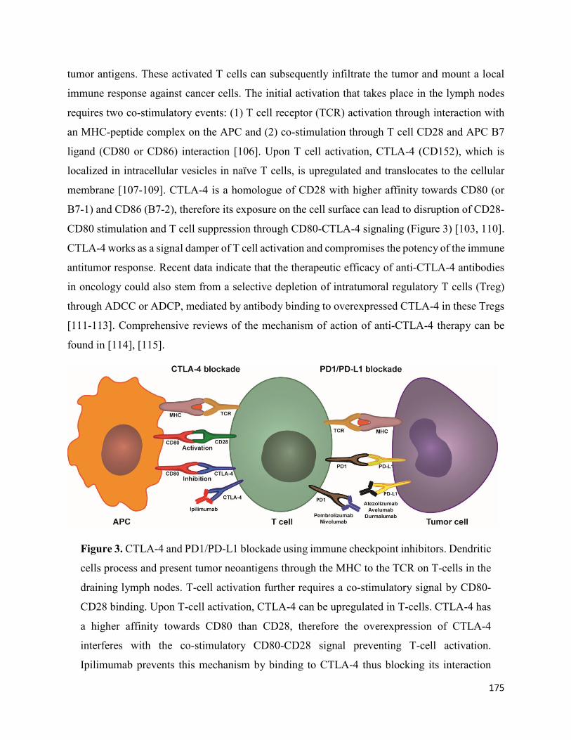
175
tumor antigens. These activated T cells can subsequently infiltrate the tumor and mount a local
immune response against cancer cells. The initial activation that takes place in the lymph nodes
requires two co-stimulatory events: (1) T cell receptor (TCR) activation through interaction with
an MHC-peptide complex on the APC and (2) co-stimulation through T cell CD28 and APC B7
ligand (CD80 or CD86) interaction [106]. Upon T cell activation, CTLA-4 (CD152), which is
localized in intracellular vesicles in naïve T cells, is upregulated and translocates to the cellular
membrane [107-109]. CTLA-4 is a homologue of CD28 with higher affinity towards CD80 (or
B7-1) and CD86 (B7-2), therefore its exposure on the cell surface can lead to disruption of CD28-
CD80 stimulation and T cell suppression through CD80-CTLA-4 signaling (Figure 3) [103, 110].
CTLA-4 works as a signal damper of T cell activation and compromises the potency of the immune
antitumor response. Recent data indicate that the therapeutic efficacy of anti-CTLA-4 antibodies
in oncology could also stem from a selective depletion of intratumoral regulatory T cells (Treg)
through ADCC or ADCP, mediated by antibody binding to overexpressed CTLA-4 in these Tregs
[111-113]. Comprehensive reviews of the mechanism of action of anti-CTLA-4 therapy can be
found in [114], [115].
Figure 3. CTLA-4 and PD1/PD-L1 blockade using immune checkpoint inhibitors. Dendritic
cells process and present tumor neoantigens through the MHC to the TCR on T-cells in the
draining lymph nodes. T-cell activation further requires a co-stimulatory signal by CD80-
CD28 binding. Upon T-cell activation, CTLA-4 can be upregulated in T-cells. CTLA-4 has
a higher affinity towards CD80 than CD28, therefore the overexpression of CTLA-4
interferes with the co-stimulatory CD80-CD28 signal preventing T-cell activation.
Ipilimumab prevents this mechanism by binding to CTLA-4 thus blocking its interaction

176
with CD80. Once activated T-cells migrate to the tumor to mount an immune anti-tumor
response, tumor cells and macrophages can upregulate PD-L1 and suppress the immune
response by interacting with the upregulated PD-1 on T-cells. Anti-PD1 and anti-PD-L1
antibodies inhibit this adaptive immune resistance mechanism.
PD-1/PD-L1 therapy
The PD-1/PD-L1 pathway plays a crucial role in adaptive immune responses. PD-1 is expressed
by activated T cells, B cells, macrophages, natural killer (NK) cells and several APCs [116]. PD-
1 expression on naïve T cells is induced upon TCR stimulation or TGF-β and cytokine (eg, IL-2,
IL-7, IL-15, IL-21) autocrine/paracrine signaling. When activated tumor-specific T cells infiltrate
the tumor, TCRs are triggered by recognition of the MHC-cognate antigen complex, resulting in
the release of interferon-γ (IFN-γ) and other inflammatory cytokines. Secretion of IFN-γ can
induce the expression of PD-L1, and PD-L2 to a lesser extent (PD-1 ligands), on the cell surface
of tumor cells and tumor macrophages [117]. PD-1 binding to PD-L1 suppresses the T cell
response of previously activated T cells at the tumor-invasive margin, leading to adaptive immune
resistance (Figure 3) [118, 119]. The proposed mechanism of action of PD-1/PD-L1 inhibitors thus
consists in suppression of the PD-1 regulatory signal exerted on activated tumor-infiltrating T cells
[120]. Nonetheless, further mechanisms of action have been suggested and are reviewed elsewhere
[120-122].
Targeting PD-1 or PD-L1 has been presumed to be a more tumor-specific approach than CTLA-4
blockade, given the involvement of the former in restoring T cell function at the effector stage
which requires previous tumor specific T-cell activation. This is supported by clinical data
showing improved outcomes and a lower rate of grade 3-4 adverse events with anti-PD-1 therapy
compared to ipilimumab (anti-CTLA-4) [123]. The open-label, randomized, phase III clinical trial
KEYNOTE-006 provided a head-to-head comparison of advanced melanoma treatment with
ipilimumab or two different dose regimens of pembrolizumab (anti-PD-1). Pembrolizumab
treatment achieved a more than two-fold increase in 24-month progression-free survival rates
compared to ipilimumab while the 24-month overall survival rate was 55% (pembrolizumab) to
43% (ipilimumab) [123, 124]. Moreover, pembrolizumab has shown clinical efficacy in advanced
melanomas refractory to ipilimumab by increasing progression-free survival [125]. A clinical trial

177
comparing nivolumab (anti-PD-1) to ipilimumab in advanced melanoma also reported substantial
improvements in overall survival and progression free survival rates with PD-1 therapy [126].
Moving forward with ICI
Despite the remarkable clinical outcomes of ICI therapy, immune checkpoint blockade is still a
relatively new concept and is undergoing extensive efforts for optimization. Key limitations being
addressed include low objective response rates and primary and acquired resistance to treatment.
Low objective response rates are presumably associated with primary resistance mechanisms.
Achieving higher response rates will likely come from a better understanding of tumor biology
and the elucidation of biomarkers that can identify patients that are more likely to respond to
specific immunotherapeutics.
Moreover, since CTLA-4 and PD-1 are non-redundant inhibitory mechanisms, combination
therapy targeting both pathways can significantly increase objective response rates. This was
shown in a phase II trial where nivolumab plus ipilimumab therapy displayed a 61% objective
response rate compared to 11% with ipilimumab mono-therapy [127]. Dual immune checkpoint
inhibition has shown great promise in increasing therapeutic efficacy and nivolumab plus
ipilimumab combination has already gained approval for metastatic melanoma, renal cell
carcinoma and microsatellite instability-high or mismatch repair deficient metastatic colorectal
cancer (table 1). Notwithstanding, combined therapy also seems to increase the frequency of
immune-related toxicities [128]. Thorough reviews on strategies and novel concepts for
combination therapy can be found in [129, 130]. Additionally, alternative inhibitory pathways of
the antitumor immune response are also being targeted for clinical development; for example,
blockade of LAG-3, TIM-3, TIGIT, VISTA, and others have started early clinical trials; and are
reviewed elsewhere [131].
Bispecific antibodies (BsAbs)
A conceptually different strategy to engage the immune system in tumor cell depletion consists in
the use of bispecific antibodies (BsAbs), wherein one arm of the BsAb targets a tumor cell antigen
while the other arm recruits and activates T cells, or other immune effector cells. Additionally,
various BsAb formats have been designed for therapeutic approaches that do not involve direct
immunomodulation; eg, cross-linking or inhibition of two different receptors [132, 133]. A

178
plethora of bispecific antibody formats have started clinical development, and have been reviewed
by others [134-136]. Nonetheless, only two bispecific formats have obtained approval by
established regulatory agencies for cancer therapy. The first case – catumaxomab – comprises a
hybrid rat-mouse full-size mAb with specificity towards tumor-expressed EpCAM and to the CD3
T cell co-receptor. Catumaxomab was approved by the EMA in 2009 for treatment of malignant
ascites in EpCAM positive carcinomas [137]. Conversely, the other marketed BsAb –
blinatumomab – comprises two scFv proteins connected by a peptide linker; a BsAb format called
Bispecific T cell Engagers (BiTE). Blinatumomab binds to CD19 expressed on malignant B
lymphocytes, while also engaging the CD3 co-receptor to recruit T cells. Blinatumomab was
approved by the FDA in 2014 under the accelerated approval program, for use in precursor B-cell
acute lymphoblastic leukemia [123].
Another flourishing strategy in cancer immunotherapy with bispecifics involves the recruitment
and activation of Natural Killer (NK) cells. Analogous to BiTEs, Bispecific Killer cell Engagers
(BiKEs) possess two scFv fragments; one directed towards a tumor antigen and another scFv that
engages FcγRIIIa (CD16) on NK cells. Moreover, trispecific formats (TriKEs) have been created
by incorporating an additional scFv fragment targeting another tumor antigen [138].; or
alternatively containing IL-15 to induce NK cell expansion [139]. Several BiKEs and TriKEs are
undergoing preclinical development [140, 141]. Other strategies to target NK cells for tumor
eradication have been reviewed in [142].
Nanoparticle delivery vehicles to improve tumor delivery
In cancer therapy, nanoparticle (NP) delivery systems offer the possibility to modify the
pharmacokinetic profile of small molecule cytotoxins and increase tumor targeting as a means to
improve therapeutic indices and safety profiles. Nanoparticle delivery systems are typically in the
10-100 nm range, making them susceptible to accumulation in tumor tissues as a consequence of
the EPR effect. The EPR refers to the enhanced accumulation of nanostructures in tumor tissue
following extravasation through the endothelium that irrigates the neoplasm. The vasculature in
these sites is formed rapidly due to an increased demand of oxygen and nutrients and secretion of
vascular effectors, leaving large fenestrations or endothelial gaps that allow diffusion of
nanoparticles (NPs) that are otherwise too large to penetrate through healthy capillaries [143-145].
Moreover, accumulation of NPs is further enhanced by a decrease in lymphatic drainage [143,

179
146]. Preferential accumulation due to the EPR effect is termed passive targeting and is an inherent
property of nano-sized materials. Importantly, the contribution of the EPR effect in preclinical and
clinical settings has been debated and it is known to depend on myriad factors relating to tumor
characteristics, including localization, stage, vascular density, fibrotic tumor microenvironment,
lymphatic drainage and vascular architecture [144, 147, 148]. Still, the EPR remains a fundamental
principle behind the design and development of nanoparticle delivery strategies for solid tumors.
Several nanoparticles have also been developed as imaging agents, however this section discusses
only those formats intended for therapeutic purposes in oncology.
Since the first reports of the EPR effect in 1986 [143], interest in the development of nanoparticle-
delivery platforms has increased substantially and has led to the approval of several NP
formulations. At present, liposomal delivery systems comprise the vast majority of nanoparticle-
based therapeutics approved for clinical use in oncology and those undergoing clinical
development [149]. Doxil (doxorubicin encapsulated in PEGylated liposomes) was the first nano-
carrier to be licensed in the US in 1995 for treatment of AIDS-related Kaposi’s sarcoma. The first
approval of Doxil served as a benchmark for the validation of NP systems in oncology; and the
formulation is currently also FDA approved in ovarian cancer and multiple myeloma [150].
Importantly, the approval granted for the aforementioned indications was based on superior safety
profiles compared to established therapy; and it also demonstrated superior efficacy in Kaposi’s
sarcoma [151-153]. Thereafter, ten other nanotherapeutics have entered the clinic (table 2). Except
for Abraxane (albumin-bound paclitaxel) and NanoTherm (iron oxide nanoparticles), all other
approved nanomedicines consist of liposomal chemotherapeutics [149, 154].
Table 2. Approved nanoparticles in oncology
Name NP carrier Targetin
g
Payload Indications Approval
date (FDA)
Doxil/Caely
x [149].
Pegylated
liposome
Passive Doxorubicin • HIV associated Kaposi’s sarcoma
• Ovarian cancer • Multiple
myeloma
1995 (FDA)
1996 (EMA)

180
Daunoxome
[171].
Non-
pegylated
liposome
Passive Daunorubicin • HIV associated Kaposi’s sarcoma
1996 (FDA)
Discontinue
d
DepoCyt
[172].
Non-
pegylated
liposome
Passive Cytarabine • Lymphomatous meningitis
1999 (FDA)
Discontinue
d
Myocet
[173].
Non-
pegylated
liposome
Passive Doxorubicin • Metastatic breast cancer
2000 (EMA)
Abraxane
[174].
Albumin
nanoparticl
e
Passive Paclitaxel • Advanced non-small-cell lung cancer
• Metastatic breast cancer
• Metastatic pancreatic adenocarcinoma
2005 (FDA)
2008 (EMA)
Oncaspar
[175].
PEG protein
conjugate
Passive L-
Asparaginase
• Acute Lymphoblastic Leukemia
2006 (FDA)
MEPACT
[176].
Non-
pegylated
liposome
Passive Mifamurtide • Non-metastatic resectable osteosarcoma
2009 (EMA)
Nanotherm
[177].
Iron oxide
nanoparticl
e
Passive Thermal
ablation*
• Glioblastoma 2010 (EMA)
Marqibo
[178].
Non-
pegylated
liposome
Passive Vincristine • Philadelphia chromosome-negative acute lymphoblastic leukemia
2012 (FDA)
Onivyde
[179].
Pegylated
liposome
Passive Irinotecan • Metastatic pancreatic adenocarcinoma
2015 (FDA)
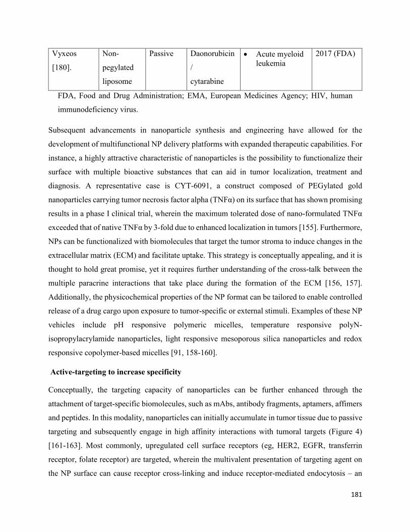
181
Vyxeos
[180].
Non-
pegylated
liposome
Passive Daonorubicin
/
cytarabine
• Acute myeloid leukemia
2017 (FDA)
FDA, Food and Drug Administration; EMA, European Medicines Agency; HIV, human
immunodeficiency virus.
Subsequent advancements in nanoparticle synthesis and engineering have allowed for the
development of multifunctional NP delivery platforms with expanded therapeutic capabilities. For
instance, a highly attractive characteristic of nanoparticles is the possibility to functionalize their
surface with multiple bioactive substances that can aid in tumor localization, treatment and
diagnosis. A representative case is CYT-6091, a construct composed of PEGylated gold
nanoparticles carrying tumor necrosis factor alpha (TNFα) on its surface that has shown promising
results in a phase I clinical trial, wherein the maximum tolerated dose of nano-formulated TNFα
exceeded that of native TNFα by 3-fold due to enhanced localization in tumors [155]. Furthermore,
NPs can be functionalized with biomolecules that target the tumor stroma to induce changes in the
extracellular matrix (ECM) and facilitate uptake. This strategy is conceptually appealing, and it is
thought to hold great promise, yet it requires further understanding of the cross-talk between the
multiple paracrine interactions that take place during the formation of the ECM [156, 157].
Additionally, the physicochemical properties of the NP format can be tailored to enable controlled
release of a drug cargo upon exposure to tumor-specific or external stimuli. Examples of these NP
vehicles include pH responsive polymeric micelles, temperature responsive polyN-
isopropylacrylamide nanoparticles, light responsive mesoporous silica nanoparticles and redox
responsive copolymer-based micelles [91, 158-160].
Active-targeting to increase specificity
Conceptually, the targeting capacity of nanoparticles can be further enhanced through the
attachment of target-specific biomolecules, such as mAbs, antibody fragments, aptamers, affimers
and peptides. In this modality, nanoparticles can initially accumulate in tumor tissue due to passive
targeting and subsequently engage in high affinity interactions with tumoral targets (Figure 4)
[161-163]. Most commonly, upregulated cell surface receptors (eg, HER2, EGFR, transferrin
receptor, folate receptor) are targeted, wherein the multivalent presentation of targeting agent on
the NP surface can cause receptor cross-linking and induce receptor-mediated endocytosis – an

182
advantageous feature for intracellular drug delivery [164]. Monoclonal antibodies play a pivotal
role in this strategy due to their exquisite specificity; as well as to the existence of well-established
techniques – primarily phage display – that allow for high-throughput development of mAbs to
specific antigens. Additionally, the prevailing clinical success of monoclonal antibodies is
favorable for regulatory approval.
Figure 4. Harnessing the EPR effect to improve tumor delivery using nanoparticle carriers.
Blood vessels that irrigate the tumor tissue are defective. The lack of pericytes and altered
structural features make the vessels less stable and leakier. Larger fenestrations between the

183
endothelial cells allow nanoparticles to extravasate into the tumor. The fibrotic ECM lacks
proper lymphatic drainage, therefore nanoparticles can accumulate in the tissue following
extravasation (passive targeting). Nanoparticles can be functionalized with monoclonal
antibodies, or other active targeting agents, to promote specific internalization and drug
delivery into targeted cells (cancer cells or other cells in the tumor microenvironment) once
they accumulate in the tumor through passive targeting.
At present, a plethora of active-targeted nanoparticles have undergone preclinical development,
however only a few have initiated clinical trials. A notable example is BIND-014, a docetaxel-
containing polymeric nanoparticle targeting the prostate-specific membrane antigen (PSMA) that
has recently completed phase II clinical trials for various cancers, where it has demonstrated
clinical efficacy and acceptable safety profiles [165]. Recently, paclitaxel solid lipid nanoparticles
conjugated to various antibodies as targeting agents have demonstrated remarkable
pharmacokinetic properties and efficacy in mouse models, and have started clinical development
[166].
In its inception, active targeting was intended to aid tumor localization and retention in conjunction
with passive targeting. Notwithstanding, experimental data has demonstrated that while
engagement and internalization within cancer cells are significantly increased, tumor accumulation
is only marginally improved [20, 163, 167]. An extensive analysis of in vivo data published from
2005-2015 showed that passive targeting results in 0.6% (median) of the injected dose
accumulating in tumor tissue, compared to 0.9% with active-targeted nanoparticles [147]. It is
noteworthy to underscore that these data were obtained with several different nanoparticle formats
administered in a wide variety of solid tumors. Still, it suggests that accumulation via passive
targeting is essential for enhanced delivery of payloads through active-targeting. Consequently,
successful clinical implementation of both passive and active-targeted nanoparticles will require a
better understanding of the physiological factors that determine the extent of EPR accumulation in
order to identify patients that can benefit from this approach [168]. Alternatively, therapeutic
strategies to increase EPR-related accumulation can be implemented, such as administration of
angiotensin-II receptor blockers to increase vessel perfusion, or sonoporation to promote vascular
permeability [169, 170].

184
Conclusion
Improving tumor penetration and distribution upon systemic delivery are crucial requirements in
mAb therapy to improve clinical outcomes and prevent the emergence of acquired resistance
mechanisms. Preventing treatment failure due to intrinsic resistance will require a better
understanding of cancer biology and the identification of novel biomarkers for a better selection
of therapeutic agents and treatment regimens. The clinical pipeline of alternative mAb based
approaches to enhance clinical efficacy has experienced a marked expansion in the last decade.
The formats discussed in this review – ADC, immune-checkpoint inhibitors and nanoparticle-
delivery systems – are among key strategies with demonstrated clinical benefits in the treatment
of solid tumors. Of note is the accelerated growth of the ICI class having obtained regulatory
approval for 6 distinct antibodies since the year 2011 (first approval) and a remarkable broadening
of clinical indications. Despite their clinical success, these therapeutics are based on relatively new
technologies that are still undergoing extensive efforts to optimize therapeutic potential. Numerous
ICI antibodies targeting alternative targets for immune inhibition (eg, LAG-3, TIM-3, TIGIT,
VISTA, B7-H3) are in phase I/II clinical trials. Safety concerns inherent to the high potency and
structural versatility of ADCs and nanoparticles have been prominent barriers in their
implementation, but clinical validation of novel designs could bring about major breakthroughs in
these fields in the coming years.
Acknowledgments
EC acknowledges the Ministry of Science, Technology and Telecommunications of the Republic
of Costa Rica for postgraduate scholarship.
Disclosure
The authors declare no conflicts of interest in this work.
References
[1]. Z. Elgundi, M. Reslan, E. Cruz, V. Sifniotis, V. Kayser, The state-of-play and future of
antibody therapeutics, Advanced drug delivery reviews, 122 (2017) 2-19.
[2]. J. Chung, Special issue on therapeutic antibodies and biopharmaceuticals, Experimental
&Amp; Molecular Medicine, 49 (2017) e304.

185
[3]. A.L. Grilo, A. Mantalaris, The Increasingly Human and Profitable Monoclonal Antibody
Market, Trends in Biotechnology, (2018).
[4]. U.S. Food and Drug Administration, Drugs@FDA: FDA Approved Drug Products, 2018.
[5]. W. De Roock, B. Claes, D. Bernasconi, J. De Schutter, B. Biesmans, G. Fountzilas, K.T.
Kalogeras, V. Kotoula, D. Papamichael, P. Laurent-Puig, F. Penault-Llorca, P. Rougier, B.
Vincenzi, D. Santini, G. Tonini, F. Cappuzzo, M. Frattini, F. Molinari, P. Saletti, S. De Dosso, M.
Martini, A. Bardelli, S. Siena, A. Sartore-Bianchi, J. Tabernero, T. Macarulla, F. Di Fiore, A.O.
Gangloff, F. Ciardiello, P. Pfeiffer, C. Qvortrup, T.P. Hansen, E. Van Cutsem, H. Piessevaux, D.
Lambrechts, M. Delorenzi, S. Tejpar, Effects of KRAS, BRAF, NRAS, and PIK3CA mutations
on the efficacy of cetuximab plus chemotherapy in chemotherapy-refractory metastatic colorectal
cancer: a retrospective consortium analysis, The Lancet Oncology, 11 (2010) 753-762.
[6]. M.H. Jin, A.R. Nam, J.E. Park, J.H. Bang, Y.J. Bang, D.Y. Oh, Resistance Mechanism against
Trastuzumab in HER2-Positive Cancer Cells and Its Negation by Src Inhibition, Molecular cancer
therapeutics, 16 (2017) 1145-1154.
[7]. J.H.E. Baker, A.H. Kyle, S.A. Reinsberg, F. Moosvi, H.M. Patrick, J. Cran, K. Saatchi, U.
Hafeli, A.I. Minchinton, Heterogeneous distribution of trastuzumab in HER2-positive xenografts
and metastases: role of the tumor microenvironment, Clinical & experimental metastasis, 35
(2018) 691-705.
[8]. C.P. Pallasch, I. Leskov, C.J. Braun, D. Vorholt, A. Drake, Y.M. Soto-Feliciano, E.H. Bent,
J. Schwamb, B. Iliopoulou, N. Kutsch, N. van Rooijen, L.P. Frenzel, C.M. Wendtner, L. Heukamp,
K.A. Kreuzer, M. Hallek, J. Chen, M.T. Hemann, Sensitizing protective tumor microenvironments
to antibody-mediated therapy, Cell, 156 (2014) 590-602.
[9]. L.T. Baxter, R.K. Jain, Transport of fluid and macromolecules in tumors. I. Role of interstitial
pressure and convection, Microvascular Research, 37 (1989) 77-104.
[10]. M. Juweid, R. Neumann, C. Paik, M.J. Perez-Bacete, J. Sato, W. van Osdol, J.N. Weinstein,
Micropharmacology of monoclonal antibodies in solid tumors: direct experimental evidence for a
binding site barrier, Cancer research, 52 (1992) 5144-5153.

186
[11]. D. Bhutani, U.N. Vaishampayan, Monoclonal antibodies in oncology therapeutics: present
and future indications, Expert opinion on biological therapy, 13 (2013) 269-282.
[12]. P.A. Netti, L.T. Baxter, Y. Boucher, R. Skalak, R.K. Jain, Time-dependent behavior of
interstitial fluid pressure in solid tumors: implications for drug delivery, Cancer research, 55
(1995) 5451-5458.
[13]. J.S. Young, C.E. Llumsden, A.L. Stalker, The significance of the “tissue pressure” of normal
testicular and of neoplastic (Brown-Pearce carcinoma) tissue in the rabbit, The Journal of
Pathology and Bacteriology, 62 (1950) 313-333.
[14]. J.R. Less, M.C. Posner, Y. Boucher, D. Borochovitz, N. Wolmark, R.K. Jain, Interstitial
hypertension in human breast and colorectal tumors, Cancer research, 52 (1992) 6371-6374.
[15]. G.M. Thurber, M.M. Schmidt, K.D. Wittrup, Antibody tumor penetration: transport opposed
by systemic and antigen-mediated clearance, Advanced drug delivery reviews, 60 (2008) 1421-
1434.
[16]. K. Fujimori, D.G. Covell, J.E. Fletcher, J.N. Weinstein, A modeling analysis of monoclonal
antibody percolation through tumors: a binding-site barrier, Journal of nuclear medicine : official
publication, Society of Nuclear Medicine, 31 (1990) 1191-1198.
[17]. T. Saga, R.D. Neumann, T. Heya, J. Sato, S. Kinuya, N. Le, C.H. Paik, J.N. Weinstein,
Targeting cancer micrometastases with monoclonal antibodies: a binding-site barrier, Proceedings
of the National Academy of Sciences of the United States of America, 92 (1995) 8999-9003.
[18]. G.M. Thurber, S.C. Zajic, K.D. Wittrup, Theoretic criteria for antibody penetration into solid
tumors and micrometastases, Journal of nuclear medicine : official publication, Society of Nuclear
Medicine, 48 (2007) 995-999.
[19]. C.M. Lee, I.F. Tannock, The distribution of the therapeutic monoclonal antibodies
cetuximab and trastuzumab within solid tumors, BMC cancer, 10 (2010) 255.
[20]. M.M. Schmidt, K.D. Wittrup, A modeling analysis of the effects of molecular size and
binding affinity on tumor targeting, Molecular cancer therapeutics, 8 (2009) 2861-2871.

187
[21]. R.J. Keizer, A.D. Huitema, J.H. Schellens, J.H. Beijnen, Clinical pharmacokinetics of
therapeutic monoclonal antibodies, Clinical pharmacokinetics, 49 (2010) 493-507.
[22]. G.M. Thurber, M.M. Schmidt, K.D. Wittrup, Factors determining antibody distribution in
tumors, Trends in pharmacological sciences, 29 (2008) 57-61.
[23]. A.P. Chapman, P. Antoniw, M. Spitali, S. West, S. Stephens, D.J. King, Therapeutic
antibody fragments with prolonged in vivo half-lives, Nature Biotechnology, 17 (1999) 780.
[24]. G.J. Weiner, Monoclonal antibody mechanisms of action in cancer, Immunologic research,
39 (2007) 271-278.
[25]. R. Gennari, S. Menard, F. Fagnoni, L. Ponchio, M. Scelsi, E. Tagliabue, F. Castiglioni, L.
Villani, C. Magalotti, N. Gibelli, B. Oliviero, B. Ballardini, G.D. Prada, A. Zambelli, A. Costa,
Pilot Study of the Mechanism of Action of Preoperative Trastuzumab in Patients with Primary
Operable Breast Tumors Overexpressing HER2, Clinical Cancer Research, 10 (2004) 5650.
[26]. R.A. Clynes, T.L. Towers, L.G. Presta, J.V. Ravetch, Inhibitory Fc receptors modulate in
vivo cytoxicity against tumor targets, Nature Medicine, 6 (2000) 443.
[27]. L. Reslan, S. Dalle, C. Dumontet, Understanding and circumventing resistance to anticancer
monoclonal antibodies, mAbs, 1 (2009) 222-229.
[28]. A. Viloria-Petit, T. Crombet, S. Jothy, D. Hicklin, P. Bohlen, J.M. Schlaeppi, J. Rak, R.S.
Kerbel, Acquired resistance to the antitumor effect of epidermal growth factor receptor-blocking
antibodies in vivo: a role for altered tumor angiogenesis, Cancer research, 61 (2001) 5090-5101.
[29]. D.J. Slamon, B. Leyland-Jones, S. Shak, H. Fuchs, V. Paton, A. Bajamonde, T. Fleming, W.
Eiermann, J. Wolter, M. Pegram, J. Baselga, L. Norton, Use of chemotherapy plus a monoclonal
antibody against HER2 for metastatic breast cancer that overexpresses HER2, The New England
journal of medicine, 344 (2001) 783-792.
[30]. D.J. Slamon, G.M. Clark, S.G. Wong, W.J. Levin, A. Ullrich, W.L. McGuire, Human breast
cancer: correlation of relapse and survival with amplification of the HER-2/neu oncogene, Science
(New York, N.Y.), 235 (1987) 177-182.

188
[31]. S.M. Tovey, S. Brown, J.C. Doughty, E.A. Mallon, T.G. Cooke, J. Edwards, Poor survival
outcomes in HER2-positive breast cancer patients with low-grade, node-negative tumours, British
journal of cancer, 100 (2009) 680-683.
[32]. Z. Mitri, T. Constantine, R. O'Regan, The HER2 Receptor in Breast Cancer:
Pathophysiology, Clinical Use, and New Advances in Therapy, Chemotherapy research and
practice, 2012 (2012) 743193-743193.
[33]. T. Yamamoto, S. Ikawa, T. Akiyama, K. Semba, N. Nomura, N. Miyajima, T. Saito, K.
Toyoshima, Similarity of protein encoded by the human c-erb-B-2 gene to epidermal growth factor
receptor, Nature, 319 (1986) 230-234.
[34]. X.F. Le, F.X. Claret, A. Lammayot, L. Tian, D. Deshpande, R. LaPushin, A.M. Tari, R.C.
Bast, Jr., The role of cyclin-dependent kinase inhibitor p27Kip1 in anti-HER2 antibody-induced
G1 cell cycle arrest and tumor growth inhibition, The Journal of biological chemistry, 278 (2003)
23441-23450.
[35]. M. Barok, J. Isola, Z. Palyi-Krekk, P. Nagy, I. Juhasz, G. Vereb, P. Kauraniemi, A. Kapanen,
M. Tanner, G. Vereb, J. Szollosi, Trastuzumab causes antibody-dependent cellular cytotoxicity-
mediated growth inhibition of submacroscopic JIMT-1 breast cancer xenografts despite intrinsic
drug resistance, Molecular cancer therapeutics, 6 (2007) 2065-2072.
[36]. B. Petricevic, J. Laengle, J. Singer, M. Sachet, J. Fazekas, G. Steger, R. Bartsch, E. Jensen-
Jarolim, M. Bergmann, Trastuzumab mediates antibody-dependent cell-mediated cytotoxicity and
phagocytosis to the same extent in both adjuvant and metastatic HER2/neu breast cancer patients,
Journal of Translational Medicine, 11 (2013) 307-307.
[37]. R.A. Clynes, T.L. Towers, L.G. Presta, J.V. Ravetch, Inhibitory Fc receptors modulate in
vivo cytotoxicity against tumor targets, Nat Med, 6 (2000) 443-446.
[38]. Y. Shi, X. Fan, H. Deng, R.J. Brezski, M. Rycyzyn, R.E. Jordan, W.R. Strohl, Q. Zou, N.
Zhang, Z. An, Trastuzumab triggers phagocytic killing of high HER2 cancer cells in vitro and in
vivo by interaction with Fcgamma receptors on macrophages, Journal of immunology (Baltimore,
Md. : 1950), 194 (2015) 4379-4386.

189
[39]. S. Mamidi, M. Cinci, M. Hasmann, V. Fehring, M. Kirschfink, Lipoplex mediated silencing
of membrane regulators (CD46, CD55 and CD59) enhances complement-dependent anti-tumor
activity of trastuzumab and pertuzumab, Molecular oncology, 7 (2013) 580-594.
[40]. M. Scaltriti, F. Rojo, A. Ocana, J. Anido, M. Guzman, J. Cortes, S. Di Cosimo, X. Matias-
Guiu, S. Ramon y Cajal, J. Arribas, J. Baselga, Expression of p95HER2, a truncated form of the
HER2 receptor, and response to anti-HER2 therapies in breast cancer, Journal of the National
Cancer Institute, 99 (2007) 628-638.
[41]. N.E. Ben–Baruch, R. Bose, S.M. Kavuri, C.X. Ma, M.J. Ellis, HER2 mutated breast cancer
responds to treatment with single agent neratinib, a second generation HER2/EGFR tyrosine
kinase inhibitor, Journal of the National Comprehensive Cancer Network : JNCCN, 13 (2015)
1061-1064.
[42]. B.N. Rexer, C.L. Arteaga, Intrinsic and Acquired Resistance to HER2-Targeted Therapies
in HER2 Gene-Amplified Breast Cancer: Mechanisms and Clinical Implications, Critical Reviews
in Oncogenesis, 17 (2012) 1-16.
[43]. P. Nagy, E. Friedlander, M. Tanner, A.I. Kapanen, K.L. Carraway, J. Isola, T.M. Jovin,
Decreased accessibility and lack of activation of ErbB2 in JIMT-1, a herceptin-resistant, MUC4-
expressing breast cancer cell line, Cancer research, 65 (2005) 473-482.
[44]. J.D. Black, S. Lopez, E. Cocco, S. Bellone, G. Altwerger, C.L. Schwab, D.P. English, E.
Bonazzoli, F. Predolini, F. Ferrari, E. Ratner, D.-A. Silasi, M. Azodi, P.E. Schwartz, A.D. Santin,
PIK3CA oncogenic mutations represent a major mechanism of resistance to trastuzumab in
HER2/neu overexpressing uterine serous carcinomas, British Journal of Cancer, 113 (2015) 1020-
1026.
[45]. C. Kim, C.-K. Lee, H.J. Chon, J.H. Kim, H.S. Park, S.J. Heo, H.J. Kim, T.S. Kim, W.S.
Kwon, H.C. Chung, S.Y. Rha, PTEN loss and level of HER2 amplification is associated with
trastuzumab resistance and prognosis in HER2-positive gastric cancer, Oncotarget, 8 (2017)
113494-113501.

190
[46]. R. Nahta, L.X. Yuan, B. Zhang, R. Kobayashi, F.J. Esteva, Insulin-like growth factor-I
receptor/human epidermal growth factor receptor 2 heterodimerization contributes to trastuzumab
resistance of breast cancer cells, Cancer research, 65 (2005) 11118-11128.
[47]. G. Zhuang, D.M. Brantley-Sieders, D. Vaught, J. Yu, L. Xie, S. Wells, D. Jackson, R.
Muraoka-Cook, C. Arteaga, J. Chen, Elevation of receptor tyrosine kinase EphA2 mediates
resistance to trastuzumab therapy, Cancer research, 70 (2010) 299-308.
[48]. K. Liang, F.J. Esteva, C. Albarracin, K. Stemke-Hale, Y. Lu, G. Bianchini, C.Y. Yang, Y.
Li, X. Li, C.T. Chen, G.B. Mills, G.N. Hortobagyi, J. Mendelsohn, M.C. Hung, Z. Fan,
Recombinant human erythropoietin antagonizes trastuzumab treatment of breast cancer cells via
Jak2-mediated Src activation and PTEN inactivation, Cancer cell, 18 (2010) 423-435.
[49]. D.L. Shattuck, J.K. Miller, K.L. Carraway, 3rd, C. Sweeney, Met receptor contributes to
trastuzumab resistance of Her2-overexpressing breast cancer cells, Cancer research, 68 (2008)
1471-1477.
[50]. J.A. Foekens, J.G.M. Klijn, P.I.M. Schmitz, P.M.J.J. Berns, The Clinical Significance of
Epidermal Growth Factor Receptor (EGF-R) in Human Breast Cancer: A Review on 5232
Patients*, Endocrine Reviews, 13 (1992) 3-17.
[51]. S.H. Lu, L.L. Hsieh, F.C. Luo, I.B. Weinstein, Amplification of the EGF receptor and c-myc
genes in human esophageal cancers, International journal of cancer, 42 (1988) 502-505.
[52]. A. Ochiai, A. Takanashi, N. Takekura, K. Yoshida, S. Miyamori, T. Harada, E. Tahara,
Effect of human epidermal growth factor on cell growth and its receptor in human gastric
carcinoma cell lines, Japanese journal of clinical oncology, 18 (1988) 15-25.
[53]. P. Kirkpatrick, J. Graham, M. Muhsin, Cetuximab, Nature Reviews Drug Discovery, 3
(2004) 549.
[54]. C.D. Andl, T. Mizushima, K. Oyama, M. Bowser, H. Nakagawa, A.K. Rustgi, EGFR-
induced cell migration is mediated predominantly by the JAK-STAT pathway in primary
esophageal keratinocytes, American journal of physiology. Gastrointestinal and liver physiology,
287 (2004) G1227-1237.

191
[55]. P. Wee, Z. Wang, Epidermal Growth Factor Receptor Cell Proliferation Signaling Pathways,
Cancers, 9 (2017) 52.
[56]. F. Walker, J. Rothacker, C. Henderson, E.C. Nice, B. Catimel, H.H. Zhang, A.M. Scott,
M.F. Bailey, S.G. Orchard, T.E. Adams, Z. Liu, T.P. Garrett, A.H. Clayton, A.W. Burgess, Ligand
binding induces a conformational change in epidermal growth factor receptor dimers, Growth
factors (Chur, Switzerland), 30 (2012) 394-409.
[57]. S. Li, K.R. Schmitz, P.D. Jeffrey, J.J.W. Wiltzius, P. Kussie, K.M. Ferguson, Structural basis
for inhibition of the epidermal growth factor receptor by cetuximab, Cancer cell, 7 (2005) 301-
311.
[58]. R.B. Luwor, Y. Lu, X. Li, J. Mendelsohn, Z. Fan, The antiepidermal growth factor receptor
monoclonal antibody cetuximab/C225 reduces hypoxia-inducible factor-1 alpha, leading to
transcriptional inhibition of vascular endothelial growth factor expression, Oncogene, 24 (2005)
4433-4441.
[59]. S. Chen, X. Li, R. Chen, M. Yin, Q. Zheng, Cetuximab intensifies the ADCC activity of
adoptive NK cells in a nude mouse colorectal cancer xenograft model, Oncology Letters, 12 (2016)
1868-1876.
[60]. G.A. Milano, ADCC and treatment outcome in colorectal cancer patients receiving
cetuximab-based therapy, Journal of Clinical Oncology, 34 (2016) e15085-e15085.
[61]. B. Zhao, L. Wang, H. Qiu, M. Zhang, L. Sun, P. Peng, Q. Yu, X. Yuan, Mechanisms of
resistance to anti-EGFR therapy in colorectal cancer, Oncotarget, 8 (2016) 3980-4000.
[62]. J.M. Xu, Y. Wang, Y.L. Wang, Y. Wang, T. Liu, M. Ni, M.S. Li, L. Lin, F.J. Ge, C. Gong,
J.Y. Gu, R. Jia, H.F. Wang, Y.L. Chen, R.R. Liu, C.H. Zhao, Z.L. Tan, Y. Jin, Y.P. Zhu, S. Ogino,
Z.R. Qian, PIK3CA Mutations Contribute to Acquired Cetuximab Resistance in Patients with
Metastatic Colorectal Cancer, Clinical cancer research : an official journal of the American
Association for Cancer Research, 23 (2017) 4602-4616.
[63]. H.C. Hsu, T.K. Thiam, Y.J. Lu, C.Y. Yeh, W.S. Tsai, J.F. You, H.Y. Hung, C.N. Tsai, A.
Hsu, H.C. Chen, S.J. Chen, T.S. Yang, Mutations of KRAS/NRAS/BRAF predict cetuximab
resistance in metastatic colorectal cancer patients, Oncotarget, 7 (2016) 22257-22270.

192
[64]. M. Moroni, S. Veronese, S. Benvenuti, G. Marrapese, A. Sartore-Bianchi, F. Di
Nicolantonio, M. Gambacorta, S. Siena, A. Bardelli, Gene copy number for epidermal growth
factor receptor (EGFR) and clinical response to antiEGFR treatment in colorectal cancer: a cohort
study, The Lancet. Oncology, 6 (2005) 279-286.
[65]. S. Khambata-Ford, C.R. Garrett, N.J. Meropol, M. Basik, C.T. Harbison, S. Wu, T.W.
Wong, X. Huang, C.H. Takimoto, A.K. Godwin, B.R. Tan, S.S. Krishnamurthi, H.A. Burris, 3rd,
E.A. Poplin, M. Hidalgo, J. Baselga, E.A. Clark, D.J. Mauro, Expression of epiregulin and
amphiregulin and K-ras mutation status predict disease control in metastatic colorectal cancer
patients treated with cetuximab, Journal of clinical oncology : official journal of the American
Society of Clinical Oncology, 25 (2007) 3230-3237.
[66]. Y. Lu, X. Li, K. Liang, R. Luwor, Z.H. Siddik, G.B. Mills, J. Mendelsohn, Z. Fan, Epidermal
growth factor receptor (EGFR) ubiquitination as a mechanism of acquired resistance escaping
treatment by the anti-EGFR monoclonal antibody cetuximab, Cancer research, 67 (2007) 8240-
8247.
[67]. F. Braig, M. Marz, A. Schieferdecker, A. Schulte, M. Voigt, A. Stein, T. Grob, M. Alawi,
D. Indenbirken, M. Kriegs, E. Engel, U. Vanhoefer, A. Grundhoff, S. Loges, K. Riecken, B. Fehse,
C. Bokemeyer, M. Binder, Epidermal growth factor receptor mutation mediates cross-resistance
to panitumumab and cetuximab in gastrointestinal cancer, Oncotarget, 6 (2015) 12035-12047.
[68]. B.O. Van Emburgh, S. Arena, G. Siravegna, L. Lazzari, G. Crisafulli, G. Corti, B. Mussolin,
F. Baldi, M. Buscarino, A. Bartolini, E. Valtorta, J. Vidal, B. Bellosillo, G. Germano, F.
Pietrantonio, A. Ponzetti, J. Albanell, S. Siena, A. Sartore-Bianchi, F. Di Nicolantonio, C.
Montagut, A. Bardelli, Acquired RAS or EGFR mutations and duration of response to EGFR
blockade in colorectal cancer, Nature communications, 7 (2016) 13665.
[69]. F. Li, K.K. Emmerton, M. Jonas, X. Zhang, J.B. Miyamoto, J.R. Setter, N.D. Nicholas, N.M.
Okeley, R.P. Lyon, D.R. Benjamin, C.L. Law, Intracellular Released Payload Influences Potency
and Bystander-Killing Effects of Antibody-Drug Conjugates in Preclinical Models, Cancer
research, 76 (2016) 2710-2719.
[70]. N.M. Okeley, J.B. Miyamoto, X. Zhang, R.J. Sanderson, D.R. Benjamin, E.L. Sievers, P.D.
Senter, S.C. Alley, Intracellular activation of SGN-35, a potent anti-CD30 antibody-drug
193
conjugate, Clinical cancer research : an official journal of the American Association for Cancer
Research, 16 (2010) 888-897.
[71]. P.F. Bross, J. Beitz, G. Chen, X.H. Chen, E. Duffy, L. Kieffer, S. Roy, R. Sridhara, A.
Rahman, G. Williams, R. Pazdur, Approval summary: gemtuzumab ozogamicin in relapsed acute
myeloid leukemia, Clinical cancer research : an official journal of the American Association for
Cancer Research, 7 (2001) 1490-1496.
[72]. S.H. Petersdorf, K.J. Kopecky, M. Slovak, C. Willman, T. Nevill, J. Brandwein, R.A.
Larson, H.P. Erba, P.J. Stiff, R.K. Stuart, R.B. Walter, M.S. Tallman, L. Stenke, F.R. Appelbaum,
A phase 3 study of gemtuzumab ozogamicin during induction and postconsolidation therapy in
younger patients with acute myeloid leukemia, Blood, 121 (2013) 4854.
[73]. F.J. Giles, H.M. Kantarjian, S.M. Kornblau, D.A. Thomas, G. Garcia-Manero, T.A.
Waddelow, C.L. David, A.T. Phan, D.E. Colburn, A. Rashid, E.H. Estey, Mylotarg™
(gemtuzumab ozogamicin) therapy is associated with hepatic venoocclusive disease in patients
who have not received stem cell transplantation, Cancer, 92 (2001) 406-413.
[74]. K.J. Norsworthy, C.W. Ko, J.E. Lee, J. Liu, C.S. John, D. Przepiorka, A.T. Farrell, R.
Pazdur, FDA Approval Summary: Mylotarg for Treatment of Patients with Relapsed or Refractory
CD33-Positive Acute Myeloid Leukemia, The oncologist, (2018).
[75]. E.Y. Jen, C.W. Ko, J.E. Lee, P.L. Del Valle, A. Aydanian, C. Jewell, K.J. Norsworthy, D.
Przepiorka, L. Nie, J. Liu, C.M. Sheth, M. Shapiro, A.T. Farrell, R. Pazdur, FDA Approval:
Gemtuzumab Ozogamicin for the Treatment of Adults with Newly Diagnosed CD33-Positive
Acute Myeloid Leukemia, Clinical cancer research : an official journal of the American
Association for Cancer Research, 24 (2018) 3242-3246.
[76]. C. Deng, B. Pan, O.A. Connor, Brentuximab Vedotin, Clinical Cancer Research, 19 (2013)
22.
[77]. FDA Approves Kadcyla for Breast Cancer, Cancer Discovery, 3 (2013) 366.
[78]. I.R. Yurkiewicz, L. Muffly, M. Liedtke, Inotuzumab ozogamicin: a CD22 mAb-drug
conjugate for adult relapsed or refractory B-cell precursor acute lymphoblastic leukemia, Drug
design, development and therapy, 12 (2018) 2293-2300.

194
[79]. H. Saber, J.K. Leighton, An FDA oncology analysis of antibody-drug conjugates,
Regulatory toxicology and pharmacology : RTP, 71 (2015) 444-452.
[80]. J.C. Masters, D.J. Nickens, D. Xuan, R.L. Shazer, M. Amantea, Clinical toxicity of antibody
drug conjugates: a meta-analysis of payloads, Investigational new drugs, 36 (2018) 121-135.
[81]. Y.-B. Hu, E.B. Dammer, R.-J. Ren, G. Wang, The endosomal-lysosomal system: from
acidification and cargo sorting to neurodegeneration, Translational neurodegeneration, 4 (2015)
18-18.
[82]. R.S. Greenfield, T. Kaneko, A. Daues, M.A. Edson, K.A. Fitzgerald, L.J. Olech, J.A.
Grattan, G.L. Spitalny, G.R. Braslawsky, Evaluation <em>in Vitro</em> of
Adriamycin Immunoconjugates Synthesized Using an Acid-sensitive Hydrazone Linker, Cancer
research, 50 (1990) 6600.
[83]. P.R. Hamann, L.M. Hinman, I. Hollander, C.F. Beyer, D. Lindh, R. Holcomb, W. Hallett,
H.-R. Tsou, J. Upeslacis, D. Shochat, A. Mountain, D.A. Flowers, I. Bernstein, Gemtuzumab
Ozogamicin, A Potent and Selective Anti-CD33 Antibody−Calicheamicin Conjugate for
Treatment of Acute Myeloid Leukemia, Bioconjugate Chemistry, 13 (2002) 47-58.
[84]. G.D. Lewis Phillips, G. Li, D.L. Dugger, L.M. Crocker, K.L. Parsons, E. Mai, W.A. Blättler,
J.M. Lambert, R.V.J. Chari, R.J. Lutz, W.L.T. Wong, F.S. Jacobson, H. Koeppen, R.H. Schwall,
S.R. Kenkare-Mitra, S.D. Spencer, M.X. Sliwkowski, Targeting HER2-Positive Breast Cancer
with Trastuzumab-DM1, an Antibody–Cytotoxic Drug Conjugate, Cancer research, 68 (2008)
9280.
[85]. Y. Wang, S. Fan, W. Zhong, X. Zhou, S. Li, Development and Properties of Valine-Alanine
based Antibody-Drug Conjugates with Monomethyl Auristatin E as the Potent Payload,
International journal of molecular sciences, 18 (2017).
[86]. A.A. Wakankar, M.B. Feeney, J. Rivera, Y. Chen, M. Kim, V.K. Sharma, Y.J. Wang,
Physicochemical stability of the antibody-drug conjugate Trastuzumab-DM1: changes due to
modification and conjugation processes, Bioconjug Chem, 21 (2010) 1588-1595.
[87]. X. Sun, J.F. Ponte, N.C. Yoder, R. Laleau, J. Coccia, L. Lanieri, Q. Qiu, R. Wu, E. Hong,
M. Bogalhas, L. Wang, L. Dong, Y. Setiady, E.K. Maloney, O. Ab, X. Zhang, J. Pinkas, T.A.

195
Keating, R. Chari, H.K. Erickson, J.M. Lambert, Effects of Drug-Antibody Ratio on
Pharmacokinetics, Biodistribution, Efficacy, and Tolerability of Antibody-Maytansinoid
Conjugates, Bioconjug Chem, 28 (2017) 1371-1381.
[88]. G.W. Seegan, C.A. Smith, V.N. Schumaker, Changes in quaternary structure of IgG upon
reduction of the interheavy-chain disulfide bond, Proceedings of the National Academy of
Sciences of the United States of America, 76 (1979) 907-911.
[89]. P.L. Ross, J.L. Wolfe, Physical and Chemical Stability of Antibody Drug Conjugates:
Current Status, Journal of pharmaceutical sciences, 105 (2016) 391-397.
[90]. J.Y. Axup, K.M. Bajjuri, M. Ritland, B.M. Hutchins, C.H. Kim, S.A. Kazane, R. Halder,
J.S. Forsyth, A.F. Santidrian, K. Stafin, Y. Lu, H. Tran, A.J. Seller, S.L. Biroc, A. Szydlik, J.K.
Pinkstaff, F. Tian, S.C. Sinha, B. Felding-Habermann, V.V. Smider, P.G. Schultz, Synthesis of
site-specific antibody-drug conjugates using unnatural amino acids, Proceedings of the National
Academy of Sciences, 109 (2012) 16101.
[91]. M.P. VanBrunt, K. Shanebeck, Z. Caldwell, J. Johnson, P. Thompson, T. Martin, H. Dong,
G. Li, H. Xu, F. D'Hooge, L. Masterson, P. Bariola, A. Tiberghien, E. Ezeadi, D.G. Williams, J.A.
Hartley, P.W. Howard, K.H. Grabstein, M.A. Bowen, M. Marelli, Genetically Encoded Azide
Containing Amino Acid in Mammalian Cells Enables Site-Specific Antibody-Drug Conjugates
Using Click Cycloaddition Chemistry, Bioconjug Chem, 26 (2015) 2249-2260.
[92]. T. Hofer, J.D. Thomas, T.R. Burke, Jr., C. Rader, An engineered selenocysteine defines a
unique class of antibody derivatives, Proc Natl Acad Sci U S A, 105 (2008) 12451-12456.
[93]. P. Strop, S.H. Liu, M. Dorywalska, K. Delaria, R.G. Dushin, T.T. Tran, W.H. Ho, S. Farias,
M.G. Casas, Y. Abdiche, D. Zhou, R. Chandrasekaran, C. Samain, C. Loo, A. Rossi, M. Rickert,
S. Krimm, T. Wong, S.M. Chin, J. Yu, J. Dilley, J. Chaparro-Riggers, G.F. Filzen, C.J. O'Donnell,
F. Wang, J.S. Myers, J. Pons, D.L. Shelton, A. Rajpal, Location matters: site of conjugation
modulates stability and pharmacokinetics of antibody drug conjugates, Chemistry & biology, 20
(2013) 161-167.

196
[94]. X. Li, T. Fang, G.-J. Boons, Preparation of well-defined antibody-drug conjugates through
glycan remodeling and strain-promoted azide-alkyne cycloadditions, Angewandte Chemie
(International ed. in English), 53 (2014) 7179-7182.
[95]. C.R. Behrens, E.H. Ha, L.L. Chinn, S. Bowers, G. Probst, M. Fitch-Bruhns, J. Monteon, A.
Valdiosera, A. Bermudez, S. Liao-Chan, T. Wong, J. Melnick, J.W. Theunissen, M.R. Flory, D.
Houser, K. Venstrom, Z. Levashova, P. Sauer, T.S. Migone, E.H. van der Horst, R.L. Halcomb,
D.Y. Jackson, Antibody-Drug Conjugates (ADCs) Derived from Interchain Cysteine Cross-
Linking Demonstrate Improved Homogeneity and Other Pharmacological Properties over
Conventional Heterogeneous ADCs, Molecular pharmaceutics, 12 (2015) 3986-3998.
[96]. P. Bryant, M. Pabst, G. Badescu, M. Bird, W. McDowell, E. Jamieson, J. Swierkosz, K.
Jurlewicz, R. Tommasi, K. Henseleit, X. Sheng, N. Camper, A. Manin, K. Kozakowska, K. Peciak,
E. Laurine, R. Grygorash, A. Kyle, D. Morris, V. Parekh, A. Abhilash, J.W. Choi, J. Edwards, M.
Frigerio, M.P. Baker, A. Godwin, In Vitro and In Vivo Evaluation of Cysteine Rebridged
Trastuzumab-MMAE Antibody Drug Conjugates with Defined Drug-to-Antibody Ratios,
Molecular pharmaceutics, 12 (2015) 1872-1879.
[97]. J.M. Lambert, A. Berkenblit, Antibody–Drug Conjugates for Cancer Treatment, Annual
Review of Medicine, 69 (2018) 191-207.
[98]. A. Beck, L. Goetsch, C. Dumontet, N. Corvaia, Strategies and challenges for the next
generation of antibody-drug conjugates, Nature reviews. Drug discovery, 16 (2017) 315-337.
[99]. H. Iwata, K. Tamura, T. Doi, J. Tsurutani, S. Modi, H. Park, I.E. Krop, Y. Sagara, C.H.
Redfern, R.K. Murthy, R.A. Redman, K. Shitara, Y. Fujisaki, M. Sugihara, L. Zhang, J. Shahidi,
A. Yver, S. Takahashi, Trastuzumab deruxtecan (DS-8201a) in subjects with HER2-expressing
solid tumors: Long-term results of a large phase 1 study with multiple expansion cohorts, Journal
of Clinical Oncology, 36 (2018) 2501-2501.
[100]. C. Saura, F. Thistlethwaite, U. Banerji, S. Lord, V. Moreno, I. MacPherson, V. Boni, C.D.
Rolfo, E.G.E. de Vries, C.M.L. Van Herpen, S. Rottey, J.J.J. Geenen, F. Eskens, M. Gil Martin,
E. Mommers, N.P. Koper, R. Mulder, P.G. Aftimos, A phase I expansion cohorts study of SYD985
in heavily pretreated patients with HER2-positive or HER2-low metastatic breast cancer, Journal
of Clinical Oncology, 36 (2018) 1014-1014.

197
[101]. E.M. Stein, R.B. Walter, H.P. Erba, A.T. Fathi, A.S. Advani, J.E. Lancet, F. Ravandi, T.
Kovacsovics, D.J. DeAngelo, D. Bixby, S. Faderl, A.P. Jillella, P.A. Ho, M.M. O'Meara, B. Zhao,
C. Biddle-Snead, A.S. Stein, A phase 1 trial of vadastuximab talirine as monotherapy in patients
with CD33-positive acute myeloid leukemia, Blood, 131 (2018) 387-396.
[102]. P.M. Drake, D. Rabuka, Recent Developments in ADC Technology: Preclinical Studies
Signal Future Clinical Trends, BioDrugs : clinical immunotherapeutics, biopharmaceuticals and
gene therapy, 31 (2017) 521-531.
[103]. D.R. Leach, M.F. Krummel, J.P. Allison, Enhancement of antitumor immunity by CTLA-
4 blockade, Science (New York, N.Y.), 271 (1996) 1734-1736.
[104]. E. Pons-Tostivint, A. Latouche, P. Vaflard, F. Ricci, D. Loirat, S. Hescot, M.-P. Sablin, R.
Rouzier, M. Kamal, C. Morel, C. Lecerf, V. Servois, X. Paoletti, C. Le Tourneau, Comparative
Analysis of Durable Responses on Immune Checkpoint Inhibitors Versus Other Systemic
Therapies: A Pooled Analysis of Phase III Trials, JCO Precision Oncology, (2019) 1-10.
[105]. F.S. Hodi, S.J. O'Day, D.F. McDermott, R.W. Weber, J.A. Sosman, J.B. Haanen, R.
Gonzalez, C. Robert, D. Schadendorf, J.C. Hassel, W. Akerley, A.J. van den Eertwegh, J. Lutzky,
P. Lorigan, J.M. Vaubel, G.P. Linette, D. Hogg, C.H. Ottensmeier, C. Lebbe, C. Peschel, I. Quirt,
J.I. Clark, J.D. Wolchok, J.S. Weber, J. Tian, M.J. Yellin, G.M. Nichol, A. Hoos, W.J. Urba,
Improved survival with ipilimumab in patients with metastatic melanoma, The New England
journal of medicine, 363 (2010) 711-723.
[106]. A.H. Sharpe, Mechanisms of costimulation, Immunological reviews, 229 (2009) 5-11.
[107]. D. Perkins, Z. Wang, C. Donovan, H. He, D. Mark, G. Guan, Y. Wang, T. Walunas, J.
Bluestone, J. Listman, P.W. Finn, Regulation of CTLA-4 expression during T cell activation,
Journal of immunology (Baltimore, Md. : 1950), 156 (1996) 4154-4159.
[108]. M.L. Alegre, P.J. Noel, B.J. Eisfelder, E. Chuang, M.R. Clark, S.L. Reiner, C.B.
Thompson, Regulation of surface and intracellular expression of CTLA4 on mouse T cells, Journal
of immunology (Baltimore, Md. : 1950), 157 (1996) 4762-4770.
[109]. J.G. Egen, J.P. Allison, Cytotoxic T lymphocyte antigen-4 accumulation in the
immunological synapse is regulated by TCR signal strength, Immunity, 16 (2002) 23-35.

198
[110]. P.S. Linsley, J.L. Greene, W. Brady, J. Bajorath, J.A. Ledbetter, R. Peach, Human B7-1
(CD80) and B7-2 (CD86) bind with similar avidities but distinct kinetics to CD28 and CTLA-4
receptors, Immunity, 1 (1994) 793-801.
[111]. X. Du, F. Tang, M. Liu, J. Su, Y. Zhang, W. Wu, M. Devenport, C.A. Lazarski, P. Zhang,
X. Wang, P. Ye, C. Wang, E. Hwang, T. Zhu, T. Xu, P. Zheng, Y. Liu, A reappraisal of CTLA-4
checkpoint blockade in cancer immunotherapy, Cell Research, 28 (2018) 416-432.
[112]. Y. Bulliard, R. Jolicoeur, M. Windman, S.M. Rue, S. Ettenberg, D.A. Knee, N.S. Wilson,
G. Dranoff, J.L. Brogdon, Activating Fc gamma receptors contribute to the antitumor activities of
immunoregulatory receptor-targeting antibodies, The Journal of experimental medicine, 210
(2013) 1685-1693.
[113]. J.R. Ingram, O.S. Blomberg, M. Rashidian, L. Ali, S. Garforth, E. Fedorov, A.A. Fedorov,
J.B. Bonanno, C. Le Gall, S. Crowley, C. Espinosa, T. Biary, E.J. Keliher, R. Weissleder, S.C.
Almo, S.K. Dougan, H.L. Ploegh, M. Dougan, Anti–CTLA-4 therapy requires an Fc domain for
efficacy, Proceedings of the National Academy of Sciences, 115 (2018) 3912.
[114]. S.C. Wei, C.R. Duffy, J.P. Allison, Fundamental Mechanisms of Immune Checkpoint
Blockade Therapy, Cancer Discovery, (2018).
[115]. J.A. Seidel, A. Otsuka, K. Kabashima, Anti-PD-1 and Anti-CTLA-4 Therapies in Cancer:
Mechanisms of Action, Efficacy, and Limitations, Frontiers in Oncology, 8 (2018).
[116]. S. Simon, N. Labarriere, PD-1 expression on tumor-specific T cells: Friend or foe for
immunotherapy?, Oncoimmunology, 7 (2017) e1364828-e1364828.
[117]. S.H. Kil, R. Estephan, J. Sanchez, J.M. Zain, M.E. Kadin, J.W. Young, S.T. Rosen, C.
Querfeld, PD-L1 Is Regulated By Interferon Gamma and Interleukin 6 through STAT1 and STAT3
Signaling in Cutaneous T-Cell Lymphoma, Blood, 130 (2017) 1458.
[118]. M.E. Keir, S.C. Liang, I. Guleria, Y.E. Latchman, A. Qipo, L.A. Albacker, M. Koulmanda,
G.J. Freeman, M.H. Sayegh, A.H. Sharpe, Tissue expression of PD-L1 mediates peripheral T cell
tolerance, The Journal of experimental medicine, 203 (2006) 883-895.
[119]. G.J. Freeman, A.J. Long, Y. Iwai, K. Bourque, T. Chernova, H. Nishimura, L.J. Fitz, N.
Malenkovich, T. Okazaki, M.C. Byrne, H.F. Horton, L. Fouser, L. Carter, V. Ling, M.R. Bowman,

199
B.M. Carreno, M. Collins, C.R. Wood, T. Honjo, Engagement of the PD-1 immunoinhibitory
receptor by a novel B7 family member leads to negative regulation of lymphocyte activation, The
Journal of experimental medicine, 192 (2000) 1027-1034.
[120]. L.T. Nguyen, P.S. Ohashi, Clinical blockade of PD1 and LAG3 — potential mechanisms
of action, Nature Reviews Immunology, 15 (2014) 45.
[121]. W. Zou, J.D. Wolchok, L. Chen, PD-L1 (B7-H1) and PD-1 pathway blockade for cancer
therapy: Mechanisms, response biomarkers, and combinations, Science Translational Medicine, 8
(2016) 328rv324.
[122]. M.W. LaFleur, Y. Muroyama, C.G. Drake, A.H. Sharpe, Inhibitors of the PD-1 Pathway in
Tumor Therapy, Journal of immunology (Baltimore, Md. : 1950), 200 (2018) 375-383.
[123]. D. Przepiorka, C.-W. Ko, A. Deisseroth, C.L. Yancey, R. Candau-Chacon, H.-J. Chiu, B.J.
Gehrke, C. Gomez-Broughton, R.C. Kane, S. Kirshner, N. Mehrotra, T.K. Ricks, D. Schmiel, P.
Song, P. Zhao, Q. Zhou, A.T. Farrell, R. Pazdur, FDA Approval: Blinatumomab, Clinical Cancer
Research, 21 (2015) 4035.
[124]. J. Schachter, A. Ribas, G.V. Long, A. Arance, J.J. Grob, L. Mortier, A. Daud, M.S. Carlino,
C. McNeil, M. Lotem, J. Larkin, P. Lorigan, B. Neyns, C. Blank, T.M. Petrella, O. Hamid, H.
Zhou, S. Ebbinghaus, N. Ibrahim, C. Robert, Pembrolizumab versus ipilimumab for advanced
melanoma: final overall survival results of a multicentre, randomised, open-label phase 3 study
(KEYNOTE-006), Lancet (London, England), 390 (2017) 1853-1862.
[125]. D. Schadendorf, R. Dummer, A. Hauschild, C. Robert, O. Hamid, A. Daud, A. van den
Eertwegh, L. Cranmer, S. O'Day, I. Puzanov, J. Schachter, C. Blank, A. Salama, C. Loquai, J.M.
Mehnert, D. Hille, S. Ebbinghaus, S.P. Kang, W. Zhou, A. Ribas, Health-related quality of life in
the randomised KEYNOTE-002 study of pembrolizumab versus chemotherapy in patients with
ipilimumab-refractory melanoma, European journal of cancer (Oxford, England : 1990), 67 (2016)
46-54.
[126]. M.T. Wan, M.E. Ming, Nivolumab versus ipilimumab in the treatment of advanced
melanoma: a critical appraisal: ORIGINAL ARTICLE: Wolchok JD, Chiarion-Sileni V, Gonzalez

200
R et al. Overall survival with combined nivolumab and ipilimumab in advanced melanoma. N Engl
J Med 2017; 377:1345-56, The British journal of dermatology, 179 (2018) 296-300.
[127]. J. Larkin, V. Chiarion-Sileni, R. Gonzalez, J.J. Grob, C.L. Cowey, C.D. Lao, D.
Schadendorf, R. Dummer, M. Smylie, P. Rutkowski, P.F. Ferrucci, A. Hill, J. Wagstaff, M.S.
Carlino, J.B. Haanen, M. Maio, I. Marquez-Rodas, G.A. McArthur, P.A. Ascierto, G.V. Long,
M.K. Callahan, M.A. Postow, K. Grossmann, M. Sznol, B. Dreno, L. Bastholt, A. Yang, L.M.
Rollin, C. Horak, F.S. Hodi, J.D. Wolchok, Combined Nivolumab and Ipilimumab or
Monotherapy in Untreated Melanoma, New England Journal of Medicine, 373 (2015) 23-34.
[128]. J.D. Wolchok, V. Chiarion-Sileni, R. Gonzalez, P. Rutkowski, J.J. Grob, C.L. Cowey, C.D.
Lao, J. Wagstaff, D. Schadendorf, P.F. Ferrucci, M. Smylie, R. Dummer, A. Hill, D. Hogg, J.
Haanen, M.S. Carlino, O. Bechter, M. Maio, I. Marquez-Rodas, M. Guidoboni, G. McArthur, C.
Lebbe, P.A. Ascierto, G.V. Long, J. Cebon, J. Sosman, M.A. Postow, M.K. Callahan, D. Walker,
L. Rollin, R. Bhore, F.S. Hodi, J. Larkin, Overall Survival with Combined Nivolumab and
Ipilimumab in Advanced Melanoma, The New England journal of medicine, 377 (2017) 1345-
1356.
[129]. R. Zappasodi, T. Merghoub, J.D. Wolchok, Emerging Concepts for Immune Checkpoint
Blockade-Based Combination Therapies, Cancer cell, 33 (2018) 581-598.
[130]. S.A. Patel, A.J. Minn, Combination Cancer Therapy with Immune Checkpoint Blockade:
Mechanisms and Strategies, Immunity, 48 (2018) 417-433.
[131]. J.A. Marin-Acevedo, B. Dholaria, A.E. Soyano, K.L. Knutson, S. Chumsri, Y. Lou, Next
generation of immune checkpoint therapy in cancer: new developments and challenges, Journal of
hematology & oncology, 11 (2018) 39-39.
[132]. S. Huang, C. Li, E.A. Armstrong, C.R. Peet, J. Saker, L.C. Amler, M.X. Sliwkowski, P.M.
Harari, Dual targeting of EGFR and HER3 with MEHD7945A overcomes acquired resistance to
EGFR inhibitors and radiation, Cancer research, 73 (2013) 824-833.
[133]. A. Patnaik, M. Gordon, F. Tsai, K. Papadopoulous, D. Rasco, S.M. Beeram, S. Fu, F. Janku,
S.M. Hynes, S.R. Gundala, M.D. Willard, W. Zhang, A.B. Lin, D. Hong, A phase I study of

201
LY3164530, a bispecific antibody targeting MET and EGFR, in patients with advanced or
metastatic cancer, Cancer chemotherapy and pharmacology, 82 (2018) 407-418.
[134]. C. Spiess, Q. Zhai, P.J. Carter, Alternative molecular formats and therapeutic applications
for bispecific antibodies, Molecular Immunology, 67 (2015) 95-106.
[135]. R.E. Kontermann, U. Brinkmann, Bispecific antibodies, Drug Discovery Today, 20 (2015)
838-847.
[136]. U. Brinkmann, R.E. Kontermann, The making of bispecific antibodies, mAbs, 9 (2017)
182-212.
[137]. M.M. Heiss, P. Murawa, P. Koralewski, E. Kutarska, O.O. Kolesnik, V.V. Ivanchenko,
A.S. Dudnichenko, B. Aleknaviciene, A. Razbadauskas, M. Gore, E. Ganea-Motan, T. Ciuleanu,
P. Wimberger, A. Schmittel, B. Schmalfeldt, A. Burges, C. Bokemeyer, H. Lindhofer, A. Lahr,
S.L. Parsons, The trifunctional antibody catumaxomab for the treatment of malignant ascites due
to epithelial cancer: Results of a prospective randomized phase II/III trial, International journal of
cancer, 127 (2010) 2209-2221.
[138]. M.K. Gleason, M.R. Verneris, D.A. Todhunter, B. Zhang, V. McCullar, S.X. Zhou, A.
Panoskaltsis-Mortari, L.M. Weiner, D.A. Vallera, J.S. Miller, Bispecific and trispecific killer cell
engagers directly activate human NK cells through CD16 signaling and induce cytotoxicity and
cytokine production, Molecular cancer therapeutics, 11 (2012) 2674-2684.
[139]. U.S. Arvindam, P. van Hauten, C. Hallstrom, D.A. Vallera, H. Dolstra, J.S. Miller, M.
Felices, CD16-IL15-CLEC12A Trispecific Killer Engager (TriKE) Drives NK Cell Expansion,
Activation, and Antigen Specific Killing of Cancer Stem Cells in Acute Myeloid Leukemia, Blood,
132 (2018) 1454.
[140]. A. Wiernik, B. Foley, B. Zhang, M.R. Verneris, E. Warlick, M.K. Gleason, J.A. Ross, X.
Luo, D.J. Weisdorf, B. Walcheck, D.A. Vallera, J.S. Miller, Targeting natural killer cells to acute
myeloid leukemia in vitro with a CD16 x 33 bispecific killer cell engager and ADAM17 inhibition,
Clinical cancer research: an official journal of the American Association for Cancer Research, 19
(2013) 3844-3855.

202
[141]. H. Don Yun, M. Felices, D.A. Vallera, P. Hinderlie, S. Cooley, M. Arock, J. Gotlib, C.
Ustun, J.S. Miller, Trispecific killer engager CD16xIL15xCD33 potently induces NK cell
activation and cytotoxicity against neoplastic mast cells, Blood Advances, 2 (2018) 1580.
[142]. C. Guillerey, N.D. Huntington, M.J. Smyth, Targeting natural killer cells in cancer
immunotherapy, Nature Immunology, 17 (2016) 1025.
[143]. Y. Matsumura, H. Maeda, A new concept for macromolecular therapeutics in cancer
chemotherapy: mechanism of tumoritropic accumulation of proteins and the antitumor agent
smancs, Cancer research, 46 (1986) 6387-6392.
[144]. H. Maeda, Toward a full understanding of the EPR effect in primary and metastatic tumors
as well as issues related to its heterogeneity, Advanced drug delivery reviews, 91 (2015) 3-6.
[145]. F. Danhier, O. Feron, V. Préat, To exploit the tumor microenvironment: Passive and active
tumor targeting of nanocarriers for anti-cancer drug delivery, Journal of Controlled Release, 148
(2010) 135-146.
[146]. K. Greish, Enhanced permeability and retention (EPR) effect for anticancer nanomedicine
drug targeting, Methods in molecular biology (Clifton, N.J.), 624 (2010) 25-37.
[147]. S. Wilhelm, A.J. Tavares, Q. Dai, S. Ohta, J. Audet, H.F. Dvorak, W.C.W. Chan, Analysis
of nanoparticle delivery to tumours, Nature Reviews Materials, 1 (2016) 16014.
[148]. J.W. Nichols, Y.H. Bae, EPR: Evidence and fallacy, Journal of Controlled Release, 190
(2014) 451-464.
[149]. S. Tran, P.-J. DeGiovanni, B. Piel, P. Rai, Cancer nanomedicine: a review of recent success
in drug delivery, Clinical and translational medicine, 6 (2017) 44-44.
[150]. Y. Barenholz, Doxil(R)--the first FDA-approved nano-drug: lessons learned, Journal of
controlled release: official journal of the Controlled Release Society, 160 (2012) 117-134.
[151]. D.W. Northfelt, B.J. Dezube, J.A. Thommes, B.J. Miller, M.A. Fischl, A. Friedman-Kien,
L.D. Kaplan, C. Du Mond, R.D. Mamelok, D.H. Henry, Pegylated-liposomal doxorubicin versus
doxorubicin, bleomycin, and vincristine in the treatment of AIDS-related Kaposi's sarcoma: results
of a randomized phase III clinical trial, Journal of Clinical Oncology, 16 (1998) 2445-2451.

203
[152]. A.N. Gordon, J.T. Fleagle, D. Guthrie, D.E. Parkin, M.E. Gore, A.J. Lacave, Recurrent
epithelial ovarian carcinoma: a randomized phase III study of pegylated liposomal doxorubicin
versus topotecan, Journal of clinical oncology : official journal of the American Society of Clinical
Oncology, 19 (2001) 3312-3322.
[153]. R.Z. Orlowski, A. Nagler, P. Sonneveld, J. Blade, R. Hajek, A. Spencer, J. San Miguel, T.
Robak, A. Dmoszynska, N. Horvath, I. Spicka, H.J. Sutherland, A.N. Suvorov, S.H. Zhuang, T.
Parekh, L. Xiu, Z. Yuan, W. Rackoff, J.L. Harousseau, Randomized phase III study of pegylated
liposomal doxorubicin plus bortezomib compared with bortezomib alone in relapsed or refractory
multiple myeloma: combination therapy improves time to progression, Journal of clinical
oncology : official journal of the American Society of Clinical Oncology, 25 (2007) 3892-3901.
[154]. C.L. Ventola, Progress in Nanomedicine: Approved and Investigational Nanodrugs, P & T
: a peer-reviewed journal for formulary management, 42 (2017) 742-755.
[155]. S.K. Libutti, G.F. Paciotti, A.A. Byrnes, H.R. Alexander, Jr., W.E. Gannon, M. Walker,
G.D. Seidel, N. Yuldasheva, L. Tamarkin, Phase I and pharmacokinetic studies of CYT-6091, a
novel PEGylated colloidal gold-rhTNF nanomedicine, Clinical cancer research : an official journal
of the American Association for Cancer Research, 16 (2010) 6139-6149.
[156]. S. Yang, H. Gao, Nanoparticles for modulating tumor microenvironment to improve drug
delivery and tumor therapy, Pharmacological research, 126 (2017) 97-108.
[157]. B. Zhang, Y. Hu, Z. Pang, Modulating the Tumor Microenvironment to Enhance Tumor
Nanomedicine Delivery, Frontiers in Pharmacology, 8 (2017).
[158]. Z.-S. Liao, S.-Y. Huang, J.-J. Huang, J.-K. Chen, A.-W. Lee, J.-Y. Lai, D.-J. Lee, C.-C.
Cheng, Self-Assembled pH-Responsive Polymeric Micelles for Highly Efficient, Noncytotoxic
Delivery of Doxorubicin Chemotherapy To Inhibit Macrophage Activation: In Vitro Investigation,
Biomacromolecules, 19 (2018) 2772-2781.
[159]. P.C. Naha, K. Bhattacharya, T. Tenuta, K.A. Dawson, I. Lynch, A. Gracia, F.M. Lyng, H.J.
Byrne, Intracellular localisation, geno- and cytotoxic response of polyN-isopropylacrylamide
(PNIPAM) nanoparticles to human keratinocyte (HaCaT) and colon cells (SW 480), Toxicology
letters, 198 (2010) 134-143.
204
[160]. S. Chai, Y. Guo, Z. Zhang, Z. Chai, Y. Ma, L. Qi, Cyclodextrin-gated mesoporous silica
nanoparticles as drug carriers for red light-induced drug release, Nanotechnology, 28 (2017)
145101.
[161]. A.D. Friedman, S.E. Claypool, R. Liu, The smart targeting of nanoparticles, Current
pharmaceutical design, 19 (2013) 6315-6329.
[162]. R. Bazak, M. Houri, S. El Achy, S. Kamel, T. Refaat, Cancer active targeting by
nanoparticles: a comprehensive review of literature, Journal of cancer research and clinical
oncology, 141 (2015) 769-784.
[163]. D.B. Kirpotin, D.C. Drummond, Y. Shao, M.R. Shalaby, K. Hong, U.B. Nielsen, J.D.
Marks, C.C. Benz, J.W. Park, Antibody targeting of long-circulating lipidic nanoparticles does not
increase tumor localization but does increase internalization in animal models, Cancer research,
66 (2006) 6732-6740.
[164]. W. Jiang, B.Y.S. Kim, J.T. Rutka, W.C.W. Chan, Nanoparticle-mediated cellular response
is size-dependent, Nature Nanotechnology, 3 (2008) 145.
[165]. K.A. Autio, R. Dreicer, J. Anderson, J.A. Garcia, A. Alva, L.L. Hart, M.I. Milowsky, E.M.
Posadas, C.J. Ryan, R.P. Graf, R. Dittamore, N.A. Schreiber, J.M. Summa, H. Youssoufian, M.J.
Morris, H.I. Scher, Safety and Efficacy of BIND-014, a Docetaxel Nanoparticle Targeting
Prostate-Specific Membrane Antigen for Patients With Metastatic Castration-Resistant Prostate
Cancer: A Phase 2 Clinical Trial, JAMA oncology, 4 (2018) 1344-1351.
[166]. J.-H. Kim, Y. Kim, K.H. Bae, T.G. Park, J.H. Lee, K. Park, Tumor-Targeted Delivery of
Paclitaxel Using Low Density Lipoprotein-Mimetic Solid Lipid Nanoparticles, Molecular
pharmaceutics, 12 (2015) 1230-1241.
[167]. E.A. Sykes, J. Chen, G. Zheng, W.C.W. Chan, Investigating the Impact of Nanoparticle
Size on Active and Passive Tumor Targeting Efficiency, ACS Nano, 8 (2014) 5696-5706.
[168]. D. Rosenblum, N. Joshi, W. Tao, J.M. Karp, D. Peer, Progress and challenges towards
targeted delivery of cancer therapeutics, Nature communications, 9 (2018) 1410.
[169]. B. Theek, M. Baues, T. Ojha, D. Mockel, S.K. Veettil, J. Steitz, L. van Bloois, G. Storm,
F. Kiessling, T. Lammers, Sonoporation enhances liposome accumulation and penetration in

205
tumors with low EPR, Journal of controlled release : official journal of the Controlled Release
Society, 231 (2016) 77-85.
[170]. V.P. Chauhan, J.D. Martin, H. Liu, D.A. Lacorre, S.R. Jain, S.V. Kozin, T. Stylianopoulos,
A.S. Mousa, X. Han, P. Adstamongkonkul, Z. Popovic, P. Huang, M.G. Bawendi, Y. Boucher,
R.K. Jain, Angiotensin inhibition enhances drug delivery and potentiates chemotherapy by
decompressing tumour blood vessels, Nature communications, 4 (2013) 2516.
[171]. E.A. Forssen, The design and development of DaunoXome® for solid tumor targeting in
vivo, Advanced drug delivery reviews, 24 (1997) 133-150.
[172]. M.J. Glantz, K.A. Jaeckle, M.C. Chamberlain, S. Phuphanich, L. Recht, L.J. Swinnen, B.
Maria, S. LaFollette, G.B. Schumann, B.F. Cole, S.B. Howell, A randomized controlled trial
comparing intrathecal sustained-release cytarabine (DepoCyt) to intrathecal methotrexate in
patients with neoplastic meningitis from solid tumors, Clinical cancer research : an official journal
of the American Association for Cancer Research, 5 (1999) 3394-3402.
[173]. G. Batist, J. Barton, P. Chaikin, C. Swenson, L. Welles, Myocet (liposome-encapsulated
doxorubicin citrate): a new approach in breast cancer therapy, Expert opinion on pharmacotherapy,
3 (2002) 1739-1751.
[174]. E. Miele, G.P. Spinelli, E. Miele, F. Tomao, S. Tomao, Albumin-bound formulation of
paclitaxel (Abraxane ABI-007) in the treatment of breast cancer, International journal of
nanomedicine, 4 (2009) 99-105.
[175]. P.A. Dinndorf, J. Gootenberg, M.H. Cohen, P. Keegan, R. Pazdur, FDA drug approval
summary: pegaspargase (oncaspar) for the first-line treatment of children with acute lymphoblastic
leukemia (ALL), The oncologist, 12 (2007) 991-998.
[176]. J.E. Frampton, Mifamurtide, Pediatric Drugs, 12 (2010) 141-153.
[177]. K. Maier-Hauff, F. Ulrich, D. Nestler, H. Niehoff, P. Wust, B. Thiesen, H. Orawa, V.
Budach, A. Jordan, Efficacy and safety of intratumoral thermotherapy using magnetic iron-oxide
nanoparticles combined with external beam radiotherapy on patients with recurrent glioblastoma
multiforme, Journal of Neuro-Oncology, 103 (2011) 317-324.

206
[178]. J.A. Silverman, S.R. Deitcher, Marqibo® (vincristine sulfate liposome injection) improves
the pharmacokinetics and pharmacodynamics of vincristine, Cancer chemotherapy and
pharmacology, 71 (2013) 555-564.
[179]. Y.N. Lamb, L.J. Scott, Liposomal Irinotecan: A Review in Metastatic Pancreatic
Adenocarcinoma, Drugs, 77 (2017) 785-792.
[180]. A.C. Krauss, X. Gao, L. Li, M.L. Manning, P. Patel, W. Fu, K.G. Janoria, G. Gieser, D.A.
Bateman, D. Przepiorka, Y.L. Shen, S.S. Shord, C.M. Sheth, A. Banerjee, J. Liu, K.B. Goldberg,
A.T. Farrell, G.M. Blumenthal, R. Pazdur, FDA Approval Summary: (Daunorubicin and
Cytarabine) Liposome for Injection for the Treatment of Adults with High-Risk Acute Myeloid
Leukemia, Clinical cancer research : an official journal of the American Association for Cancer
Research, (2018).

207
Synthesis and Enhanced Cellular Uptake In Vitro of Anti-
HER2 Multifunctional Gold Nanoparticles
Chapter 7

208
Chapter 7 – Authorship declaration statement
The following chapter full research article published in the journal Cancers as:
E. Cruz, V. Kayser. Synthesis and Enhanced Cellular Uptake In Vitro of Anti-HER2
Multifunctional Gold Nanoparticles. Cancers, 11 (2019), 870.
E. Cruz co-designed the study and performed all experimental work and data analysis. E. Cruz
wrote the manuscript.
Permission to include the published material has been granted by the corresponding author.
Esteban Cruz, Signature: 19th of August, 2019
As corresponding author and supervisor for this candidature, I hereby confirm that this authorship
declaration statement is complete and accurate
Veysel Kayser, Signature: 19th of August, 2019

209
Abstract
Nanoparticle carriers offer the possibility of enhanced delivery of therapeutic payloads in tumor
tissues due to tumor-selective accumulation through the enhanced permeability and retention effect
(EPR). Gold nanoparticles (AuNP), in particular, possess highly appealing features for
development as nanomedicines, such as biocompatibility, tunable optical properties and a
remarkable ease of surface functionalization. Taking advantage of the latter, several strategies have
been designed to increase treatment specificity of gold nanocarriers by attaching monoclonal
antibodies on the surface, as a way to promote selective interactions with the targeted cells—an
approach referred to as active-targeting. Here, we describe the synthesis of spherical gold
nanoparticles surface-functionalized with an anti-HER2 antibody-drug conjugate (ADC) as an
active targeting agent that carries a cytotoxic payload. In addition, we enhanced the intracellular
delivery properties of the carrier by attaching a cell penetrating peptide to the active-targeted
nanoparticles. We demonstrate that the antibody retains high receptor-affinity after the structural
modifications performed for drug-conjugation and nanoparticle attachment. Furthermore, we show
that antibody attachment increases cellular uptake in HER2 amplified cell lines selectively, and
incorporation of the cell penetrating peptide leads to a further increase in cellular internalization.
Nanoparticle-bound antibody-drug conjugates retain high antimitotic potency, which could
contribute to a higher therapeutic index in high EPR tumors.
Introduction
Most solid malignancies display a tumor microenvironment with increased interstitial fluid
pressures (IFP) that significantly impairs tumor penetration of conventional anticancer agents
following systemic delivery. This effect hinders movement of the therapeutic agent from the
vascular lumen to the tumor tissue, requiring higher doses to achieve therapeutic efficacy [1, 2].
Consequently, the therapeutic index is reduced, and off-target side-effects compromise clinical
outcomes. Moreover, inefficient localization in the target tissue can lead to tumor regions exposed
to subtherapeutic doses of the drug, whereby cancer cells can undergo phenotypic alterations that
render them resistant to the agent administered [3].
In this context, nanoparticles (NPs) have emerged as drug delivery vehicles that can harness the
preferential accumulation of nanosized materials in the tumor due to the well-described enhanced

210
permeability and retention (EPR) effect [4]. Several liposome-encapsulated cytotoxic drugs have
received regulatory approval on the basis of superior therapeutic indices relative to the free drug
[5]. A further attractive feature of nanoparticles is their functional versatility, as their design can
be tailored to confer diverse physiological and physicochemical properties to broaden treatment
modalities. Myriad distinct NP formats are undergoing preclinical development for various
therapeutic and diagnostic applications, e.g., gene delivery, thermal ablation therapy, magnetic
resonance imaging (MRI), photoacoustic imaging [6–9].
Among the diverse range of inorganic NPs, gold nanoparticles (AuNP) have been widely appraised
as attractive systems for therapeutic applications, e.g., drug delivery, photothermal therapy and
radiosensitization [9, 10]. AuNPs are easy to synthesize with tunable shapes and sizes, and the
strong gold-sulfur (Au-S) interaction allows for the modification of the nanoparticle surface with
sulfhydryl containing linkers, through which functional groups can be incorporated to confer
biological properties for therapeutic purposes [11]. An analysis of nanoparticle tumor delivery
efficiency in in vivo models derived from published data from the year 2005 to 2015 showed that
AuNPs had the highest median delivery efficiency among the analysed inorganic nanoparticle
types (including iron oxide, silica, quantum dots and others) [12]. Moreover, a PEGylated AuNP
format coated with TNF-α has already shown a promising safety profile and enhanced
accumulation in various solid tumors in a phase I dose escalation trial, setting a clinical precedent
for gold nanoparticles [13, 14].
Adding to the inherent passive accumulation of NPs in solid tumors, the targeting capacity of a
nanoparticle carrier can potentially be enhanced by the incorporation of an active targeting agent
on the NP surface. Active targeting moieties—e.g., antibodies, peptides, aptamers, affimers—can
engage in high-affinity specific interactions with biomolecules overexpressed in cancer subtypes
to increase treatment specificity [15]. Within this concept, systemic delivery of the nanocarrier
results in passive accumulation in the tumor microenvironment, where the subsequent interaction
of the affixed targeting agent with cancer cells can induce receptor crosslinking, receptor-mediated
endocytosis and intracellular cargo delivery [16].
In this work, we employed an anti-HER2 antibody, Trastuzumab (Tmab), as an active targeting
agent on spherical gold nanoparticles. Tmab is a therapeutic monoclonal antibody that binds to the
human epidermal growth factor receptor 2 (HER2) and is approved for the treatment of HER2-

211
positive breast cancer and metastatic gastric cancer [17, 18]. Moreover, HER2 overexpression has
been documented in esophageal [19], ovarian [20]. and endometrial cancer [21]. and has been
identified as a negative prognostic factor in several of these malignancies [22–24]. Trastuzumab
exerts its anticancer activity by binding to the extracellular domain of HER2 to prevent
dimerization with other ErbB receptors, thereby inhibiting its key function in cell proliferation and
migration. In addition, immune effector components can be engaged through the Fc region of the
antibody to destroy cancer cells via antibody-dependent cellular cytotoxicity (ADCC), antibody-
dependent cellular phagocytosis (ADCP) and complement-dependent cytotoxicity (CDC) [25, 26].
Monoclonal antibodies (mAbs) have become a cornerstone of cancer care since the first therapeutic
mAb market approval in 1997 (Rituximab) by virtue of their enhanced treatment specificity. As of
early 2019, more than 20 distinct mAbs are indicated for a wide array of solid malignancies,
predominantly administered through systemic routes [27]. This notwithstanding, poor tumor
penetration and distribution are prominent obstacles that compromise the therapeutic index of
mAbs [28, 29]. To this end, enhancing drug accumulation in the tumor through the employment
of enhanced delivery systems could provide major improvements in therapeutic safety and
efficacy.
Conventional designs of active-targeted nanoparticles for drug delivery typically consist of
nanoparticles carrying a surface-incorporated targeting agent and a cytotoxic payload either
encapsulated within the NP core or loaded onto the surface. In this work, we sought to employ a
novel strategy, wherein a cytotoxic drug is conjugated to the antibody initially, and the resulting
antibody-drug conjugate (ADC) is employed as a targeting agent-drug carrier on the nanoparticles,
thereby broadening the functionality of the active-targeting agent. Herein, we describe the
synthesis and physicochemical characterization of ADC-targeted spherical gold nanoparticles. The
ADC was produced by chemical attachment of monomethyl auristatin E (MMAE) to Tmab
through a cathepsin-cleavable valine-citrulline linker; and further reacted with a sulfhydryl-
containing linker for surface conjugation to the gold nanoparticles. We demonstrate that
Trastuzumab can be chemically modified in this fashion while retaining high affinity towards its
cognate receptor. Since the valine-citrulline linker must be internalized for payload release, we
analysed the intracellular uptake of active-targeted AuNPs on various cancer cell lines.

212
Furthermore, we evaluated the effect of surface incorporation of a cell penetrating peptide to the
active-targeted nanoparticle on intracellular uptake.
Materials and Methods
Materials
Herceptin® (Trastuzumab) was a generous donation from Genentech (San Francisco, CA, USA).
Thiol PEG NHS (NHS-PEG-SH) (5 kDa) linker (Cat. No. PG2-NSTH-5k) was purchased from
Nanocs (Boston, MA, USA). The MC-Val-Cit-PAB-MMAE (vcMMAE) linker (Cat. No.
BP23969) was obtained from Broadpharm (San Diego, CA, USA). The HIV-1 TAT protein (47-
57) (HIV-TAT or CPP) (Cat. No. H0292) was purchased from Sigma-Aldrich (Castle Hill, NSW,
Australia). The Series S Sensor Chip CM5 (Cat. No. 29-1049-88), the amine coupling kit (Cat.
No. BR-1000-50) and the anti-HIS capture kit (Cat. No. 28-9950-56) employed in the Biacore SPR
instrument were purchased from GE Healthcare (Parramatta, NSW, Australia). The recombinant
HIS-tagged soluble HER2 (Cat. No. SRP6405) was obtained from Sigma-Aldrich (Australia).
Phosphate buffered saline (PBS) was purchased from Astral Scientific (Gymea, NSW, Australia).
Amicon 3 kDa (Cat. No. Z740168) and 50 kDa (Cat. No. Z740177) cutoff centrifugal filter units
were acquired from Sigma-Aldrich (Australia). Millex-GV syringe filters (0.22 µm, PVDF, Cat.
No. SLGV033RS) were purchased from purchased from Sigma-Aldrich (Castle Hill, NSW,
Australia). RPMI 1640 and DMEM (high glucose) media were obtained from Life Technologies
(Mulgrave, VIC, Australia). All other chemicals and reagents were purchased from Sigma Aldrich
(Australia).
Synthesis of Spherical Citrate-Capped Gold Nanoparticles
Spherical gold nanoparticles were synthesized by citrate reduction of gold chloride in aqueous
solution as described by Turkevich [45], and revised by Frens [46]. All glassware employed in this
procedure was soaked in aqua regia (3:1 HCl/HNO3 molar ratio) for 3 h prior to the reaction and
rinsed with double distilled H2O. Briefly, 100 mL of a 254 µM HAuCl4 solution in double distilled
H2O was heated to boiling under stirring. Once boiling, 2 mL or 1 mL of a 1% w/v (34 µM) sodium
citrate solution was added to prepare 20 nm and 50 nm, respectively. Following citrate addition,
the solution was boiled for 15 min, then cooled to room temperature under stirring for 2 h.
Unreacted citrate was removed by decanting after centrifugation at 10,000 g or 3,500 g for 30 min

213
to pellet the 20 nm and 50 nm nanoparticles. The synthesized gold nanoparticles were resuspended
in double distilled water. Nanoparticle size, size distribution and morphology were assessed
through transmission DLS (hydrodynamic size) electron microscopy (size, size distribution and
morphology) and shifts in the surface plasmon resonance (SPR) absorption band.
Tmab PEGylation (Tmab-PEG-SH)
Trastuzumab 21 mg/mL in formulation buffer (L-histidine 4.64 mM, α,α-Trehalose 52.86 mM,
polysorbate 20 concentration 73.31 µM, HCl 2.58 mM) was buffer exchanged to sodium
bicarbonate (NaHCO3) 0.1 M pH 8.0 using 50 kDa cutoff centrifugal filters to a final antibody
concentration of 10 mg/mL (6.87 × 10⁻⁵ M). The extinction coefficient ε280 = 2.25 × 10+5 M−1
cm−1 was used for all antibody concentration determinations. Buffer exchange was carried out
thoroughly to reduce to a minimum the concentration of L-histidine in the formulation buffer, as
the primary amine in L-histidine will react readily with the NHS group in the linker. A 5 mg/mL
(1 mM) NHS-PEG-SH (5 kDa) linker stock solution was prepared in NaHCO3 0.1 M pH 8.0 and
immediately added to Trastuzumab in 2:1, 5:1, 10:1, 20:1 and 25:1 NHS-linker/Tmab ratios and
incubated at 4 °C overnight under stirring. The NHS-linker stock solution in NaHCO3 pH 8.0 was
prepared immediately before adding to the Trastuzumab sample, since the NHS ester can undergo
rapid hydrolysis at basic pH. Following PEGylation, unreacted NHS-PEG-SH linker was removed
by centrifugation through 50 kDa cutoff filters and the PEGylated Trastuzumab (Tmab-PEG-SH)
was buffer exchanged to phosphate buffered saline (PBS) 0.01 M pH 7.4 with 1 mM EDTA to a
final antibody concentration of 5 mg/mL. EDTA 1 mM was added to inhibit disulfide bond
formation between the free SH groups in the linker [47, 48].
HIV-TAT Cell Penetrating Peptide (CPP) PEGylation (CPP-PEG-SH)
HIV-TAT (47–57) peptide was dissolved in NaHCO3 0.1 M pH 8.0 to a 1 mg/mL (641 µM)
concentration. A 10 mg/mL (2 mM) NHS-PEG-SH (5 kDa) solution in NaHCO3 0.1 M pH 8.0
was added to the HIV-TAT peptide in a 4:1 NHS-linker/CPP molar ratio and incubated overnight
at 4 °C under stirring. Unreacted CPP was removed by centrifugation through 3 kDa cutoff filters
and the PEGylated CPP (CPP-PEG-SH) was buffer exchanged to phosphate buffered saline 0.01
M pH 7.4 with 1 mM EDTA.

214
Tmab-vcMMAE Conjugate Synthesis
Antibody Partial Reduction
Trastuzumab in formulation buffer was buffer exchanged to PBS 0.01 M with 10 mM EDTA in a
final concentration of 5 mg/mL (34 uM). A freshly prepared 10 mM stock solution of dithiothreitol
(DTT) in PBS 0.01 M EDTA 1 mM was added to the antibody in a 3:1 DTT/Tmab ratio and the
reaction was incubated at 37 °C for 90 min under stirring. DTT was then removed by buffer
exchanging the partially reduced Tmab with 50 kDa cutoff centrifugal filters to PBS 0.01 M
containing 10 mM EDTA to a 10 mg/mL (34 µM) concentration. After partial reduction, the
integrity of the full-size IgG molecule was confirmed by SE-HPLC. In addition, free sulfhydryl
(SH) groups per antibody were quantified by reaction with DTNB (5, 5’-dithiobis(2-nitrobenzoic
acid)) and determination of the absorbance at 412 nm for free SH concentration. The final
flowthrough of the buffer exchange prior to the DTNB reaction was used as a blank to subtract the
potential contribution of residual DTT in the solution. The extinction coefficient ε412 = 1.42 ×
10+5 M−1 cm−1 for the TNB2− reaction product was employed for sulfhydryl quantification.
Conjugate Synthesis
vcMMAE was dissolved in DMSO at a 1.26 mM concentration and added to a chilled 10 mg/mL
partially reduced Tmab solution in a 4.6:1 vcMMAE/Tmab ratio. The reaction mixture was
incubated at 4 °C with stirring for 1 h. A 20-fold molar excess of cysteine—relative to maleimide—
was added to quench the reaction. Unreacted vcMMAE and cysteine were removed by
centrifugation through 50 kDa cutoff centrifugal filters and buffer exchanged to PBS 0.01 M pH
7.4 for storage, or NaHCO3 0.1 M pH 8.0 for subsequent PEGylation. The average drug-antibody
ratio (DAR) was calculated based on absorbance values at 248 nm and 280 nm as has been
described previously [49]. The following formula was employed:
𝐷𝐷𝐷𝐷𝐷𝐷 = 𝜀𝜀248𝑇𝑇𝑇𝑇𝑇𝑇𝑇𝑇 − 𝐹𝐹𝜀𝜀280𝑇𝑇𝑇𝑇𝑇𝑇𝑇𝑇
𝐹𝐹𝜀𝜀280𝐵𝐵𝐵𝐵𝑀𝑀𝑀𝑀 − 𝜀𝜀248𝐵𝐵𝐵𝐵𝑀𝑀𝑀𝑀
F=A248/A280 and the extinction coefficients utilized are listed in Table 4.
Table 1. Extinction coefficients of Trastuzumab and monomethyl auristatin E (MMAE) employed
for the calculation of drug-to-antibody ratio (DAR) based on UV-Vis spectroscopy

215
Sample 248 nm 280 nm
Trastuzumab 7.75 × 10+4 2.25 × 10+5
MMAE 1.59 × 10+4 1.50 × 10+3
PEGylation of Tmab-vcMMAE (ADC-PEG-SH) was achieved following the same procedure as
for the unconjugated antibody.
Intact Mass Analysis
Trastuzumab and Tmab-vcMMAE were concentrated using 50 kDa cutoff centrifugal filters and
buffer exchanged to 10% acetonitrile with 0.1% formic acid. The antibody samples were analysed
through direct injection into a Triple TOF 6600 mass spectrometer (Sciex, Framingham, MA,
USA). Infusion was performed at 50 µL/min. The mass range for detection was 100–5,000 m/z.
Deconvolution of the raw data was achieved using SCIEX Peakview 2.2 (Concord, ON, Canada)
and Bruker BioTools software packages (Billerica, MA, USA).
Binding Kinetics to Recombinant HER2 through Surface Plasmon Resonance
The binding kinetics of derivatized Trastuzumab (Tmab-PEG-SH, Tmab-vcMMAE and ADC-
PEG-SH) were tested against a recombinant HER-2 protein using surface plasmon resonance
(SPR) in a Biacore T200 instrument (GE Healthcare, Parramatta, NSW, Australia). Briefly, an
anti-HIS antibody was bound to a CM5 sensor chip through amine coupling chemistry.
Subsequently, a recombinant HIS-tagged HER-2 (4 nM) was bound to the anti-HIS antibody on
the sensor chip at a 5 µL/min flow rate for 5 min. 2-fold serial dilutions of the Trastuzumab variants
ranging from 8–0.5 nM in HBS-T running buffer (10 mM HEPES, 150 mM NaCl, 0.05% (v/v)
Tween 20, pH 7.4) were assayed at 25 °C as single cycle kinetic titrations. The analytes were
applied to the sensor surface at 20 µL/min for 2 min, followed by 60 min dissociation times.
Analyses of the sensorgrams were performed by fitting a Langmuir 1:1 binding model to derive
the association constant (Ka), the dissociation constant (Kd) and the binding affinity (KD—
calculated as Ka/Kd). The analytes were run in duplicate to calculate average values and standard
deviation. A goodness of fit (χ2) value within 5% of the maximum response level (Rmax) was
used as acceptance criteria.

216
Gold Nanoparticle Surface Functionalization
Trastuzumab-coated (Tmab-PEG-AuNP), OH-PEG coated (OH-PEG-AuNP) and CPP-coated
(CPP-PEG-AuNP) gold nanoparticles were produced by incubating citrate-capped gold
nanoparticles (OD = 1) with a 1 × 10+5 molar excess of SH-PEG-Tmab, SH-PEG-OH or SH-PEG-
CPP in NaHCO3 0.01 M pH for 2 h at room temperature while stirring. The unconjugated reagents
were removed by pelleting the nanoparticles at 3,500 g for 30 min and removing the supernatant.
The conjugated nanoparticles were centrifuged four times and resuspended in PBS 0.01 M pH 7.4
for storage at 4 °C. ADC-PEG-AuNP were produced by incubating the nanoparticles with ADC-
PEG-SH following the same procedure. CPP+Tmab-PEG-AuNP were obtained by incubation with
a 1 × 10+5 molar excess of CPP-PEG-SH for 5 min followed by the addition of a 1 × 10+5 molar
excess of Tmab-PEG-SH, and further incubation under stirring for 2 h.
UV-Vis Spectroscopy
UV-Vis absorption spectra were obtained over a wavelength range of 800–200 nm for gold
nanoparticles or 400–200 nm for protein samples, using a Shimadzu 2600 UV-Vis
spectrophotometer (Shimadzu, Japan). AuNP samples in RPMI media were corrected by blank
subtraction of the RPMI.
Size-Exclusion High-Performance Liquid Chromatography (SE-HPLC)
Size-exclusion chromatograms were obtained with a Zorbax GF-250 column connected to an
Agilent 1200 Liquid Chromatography system (Agilent Technologies, Santa Clara, CA, USA),
running potassium phosphate buffer 150 mM pH 6.5 as a mobile phase at a 0.5 mL/min flow rate.
Peak absorption was detected at 280 nm with an in-line UV signal detector (Agilent Technologies,
Santa Clara, CA, USA).
DLS and Zeta Potential Measurements
DLS and zeta potential measurements of the functionalized gold nanoparticles were conducted
with a Malvern Zetasizer Nano ZS (Malvern Instruments, Worcestershire, UK) with a 633 nm
Helium Neon Laser and an avalanche photo diode (APD) detector. The measurements were
conducted in triplicate and the values are reported as mean Z-average ± standard deviation. For
zeta potential measurements, the functionalized nanoparticles suspended in PBS 1X (phosphate

217
buffer 0.01 M, NaCl 0.137 M, KCl 0.0027 M, pH 7.4) were diluted 1:10 in deionized water. Cit-
AuNPs were directly resuspended in PBS 0.1X. The zeta potential was derived from the Henry
equation using an f(Ka) of 1.5.
Cellular Uptake Quantification through Inductively Coupled Plasma Mass
Spectrometry (ICP-MS)
The SKBR-3 cell line was provided by Dr. Thomas Grewal. The DLD-1 cell line was purchased
from the American Type Culture Collection (ATCC). The MDA-MB-231 and MCF-7 cell lines
were obtained from Dr. Fanfan Zhou. SKOV-3 cells were provided by Dr. Pegah Varamini.
To compare the cellular uptake of gold nanoparticles coated with OH-PEG and Tmab-PEG,
SKBR-3 cells were seeded at density of 1 × 10+5 cells/well in 24-well plates in RPMI media
containing 10% FBS. Following incubation at 37 °C for 48 h, the cell media was removed, the
cells were washed twice with PBS, and fresh RPMI media (10% FBS) containing 50 µg/mL 20
nm and 50 nm gold nanoparticles (coated with OH-PEG or Tmab-PEG) was added, using 6 wells
per AuNP sample. The cells were further incubated for 24 h. The AuNP containing media was
removed and the cell monolayer washed 4 times with PBS. The cells were detached from the plate
using 0.05% trypsin and collected in 1.5 mL centrifuge tubes. Trypsin was removed by pelleting
the cells at 300 g for 5 min and the cells were washed twice more with PBS. The cell pellet was
digested with 200 µL concentrated HNO3 (15.9 M) overnight at room temperature. 800 µL
concentrated HCl (12.1 M) was then added to dissolve the gold nanoparticles. A 1:4 dilution in
Milli-Q water was performed for quantification of gold content through ICP-MS. ICP-MS
measurements were carried out with a Perkin Elmer Nexion 300× ICP-MS instrument (Perkin-
Elmer, Waltham, MA, USA), calibrated with 5, 10 and 20 parts per billion (ppb) gold standard
solutions.
To compare cellular uptake in SKBR-3, DLD-1, MDA-MB-231 and MCF-7 cells, the uptake
assays were carried out following the same procedure as described above albeit with the following
modifications: (1) 25 µg/mL AuNP concentrations were used, (2) DLD-1, MDA-MB-231 and
MCF-7 cells were seeded at 3 × 10+4 cells/well, (3) MDA-MB-231 and MCF-7 cell lines were
cultured in DMEM media containing 10% FBS, (4) each nanoparticle sample was run in triplicate.

218
The concentration of the gold nanoparticles was determined based on their absorbance at 450 nm
using ε450 = 5.41 × 10+8 M−1 cm−1 and ε450 = 9.92 × 10+9 M−1 cm−1 for 20 nm and 50 nm,
respectively, according to previous determinations [32]. ICP-MS quantification of gold content in
the AuNP suspensions was utilized to corroborate that the extinction coefficients used in this
method provide appropriate estimations of gold concentrations. The nanoparticles in cell culture
medium were filter sterilized through 0.22 µM filters prior to addition to the cells.
Cellular Uptake Evaluation by Transmission Electron Microscopy (TEM)
SKBR-3 cells were seeded at a density of 1 × 10+5 cells/well on collagen-coated Thermanox plastic
coverslips placed inside each well (24-well plates) and incubated at 37 °C for 48 h in RPMI media
containing 10% FBS. Fresh RPMI containing 50 ug/mL 50 nm OH-PEG-AuNP or Tmab-PEG-
AuNP was added to the wells and further incubated at 37 °C for 24 h. The wells were washed
thrice with PBS. The cells were fixed with 2.5% glutaraldehyde in 0.1 M phosphate buffer pH 7.4.
The cell monolayers were subsequently fixed with osmium tetroxide 1% (w/v) in phosphate buffer
0.1 M pH 7.4, then embedded into an epon resin. The monolayers were microtomed into 70 nm
sections and stained with uranyl acetate 2% and Reynold’s lead citrate. TEM images were obtained
with a JEOL JEM-1400 (Tokyo, Japan) microscope with an accelerating voltage of 120 kV.
Cell Cytotoxicity Evaluation
SKBR-3 and SKOV-3 cells were seeded at 5 × 10+3 and 3 × 10+3 cells/well on 96-well plates and
incubated at 37 °C for 24 h in RPMI media containing 10% FBS. Fresh RPMI media containing
free MMAE, ADC, ADC-PEG-AuNP, Trastuzumab or OH-PEG-AuNP were added to the wells
at the corresponding concentrations in triplicates. RPMI media was replenished for negative
control samples. Images (10x magnification) of four different regions per well were acquired at 2-
h intervals for 72 h after addition of the antimitotic or control sample using an Incucyte® ZOOM
Live-cell Analysis System (Essen BioScience, Ann Arbor, MI, USA). Cell confluence was
analysed with the Incucyte® ZOOM integrated analysis software (v2016A) to generate cell growth
curves over time. Growth rate inhibition metrics were employed to assess the antimitotic effect of
the samples. Growth rate inhibition metrics have been developed recently to provide more robust
and biologically relevant drug response parameters [50]. GR values were calculated as:
𝐺𝐺𝐷𝐷(𝑑𝑑) = 2𝑙𝑙𝑙𝑙𝑙𝑙2�
𝑥𝑥(𝑑𝑑)𝑓𝑓𝑥𝑥(𝑑𝑑)0
�/𝑙𝑙𝑙𝑙𝑙𝑙2�𝑥𝑥(𝑐𝑐)𝑓𝑓𝑥𝑥(𝑐𝑐)0
�− 1

219
Where 𝑥𝑥(𝑑𝑑)0 and 𝑥𝑥(𝑑𝑑)𝑓𝑓 are the confluence values of cells treated with a cytotoxic agent at time t
= 0 h and t = 72 h, respectively. 𝑥𝑥(𝑐𝑐)0 and 𝑥𝑥(𝑐𝑐)𝑓𝑓 are confluence values of control wells at t = 0 h
and t = 72 h.
GR values were plotted against treatment concentration and the data was fitted to a four-parameter
dose-response curve. GR50 was obtained by interpolating the treatment concentration at which
GR = 0.5.
Statistical Analysis
Gold uptake quantification was analysed with a two-tailed, unpaired Student t-test. Values are
denoted as mean ± standard deviation, and p < 0.05 was established as statistical significance.
Results
Nanoparticle Design
Figure 1 displays an outline of the nanoparticle design and conjugation strategy. Attachment of
the bioactive moieties—anti-HER2 mAb and HIV-TAT cell penetrating peptide (CPP)—to the
gold surface was achieved through the covalent thiol-gold interaction using a bifunctional 5 kDa
poly ethylene glycol (PEG) linker with a thiol (SH) and an N-hydroxysuccinimide (NHS) ester
end groups (NHS-PEG-SH). The NHS group reacts with ε-amines in lysine residues (and with α-
amines present at the N-terminals to a lesser extent) under slightly alkaline conditions to produce
stable amide bonds with the protein or peptide [30]. The 5 kDa PEG linker was employed to
increase exposure of the functional groups and prevent non-specific interactions between the
bioactive groups and the gold surface. Furthermore, PEGylation of gold nanoparticles has proven
to be highly beneficial in increasing circulation half-life by preventing adhesion of serum proteins
that facilitate uptake by the reticuloendothelial system (RES)—an effect that drastically decreases
the amount of nanoparticles that can eventually reach the tumor site [31].
Trastuzumab (anti-HER2 mAb) was employed as an active targeting agent to confer specificity
towards HER2 overexpressing cancer cell lines. To enhance the anticancer potency of the
antibody, MMAE was attached to Trastuzumab (Tmab) through a valine-citrulline dipeptide
cleavable linker (vcMMAE) to produce Tmab-vcMMAE (Figure 1). The valine-citrulline moiety
is cleaved by the lysosomal protease cathepsin B; thus, MMAE release occurs primarily following

220
endocytosis and subsequent localization into endosomes or lysosomes. The HIV-TAT cell
penetrating peptide was further added onto the surface via the same NHS-PEG-SH linker to
increase cellular uptake for intracellular release of the drug cargo (Figure 2B, C).
Figure 1. Molecular structures of the bioactive agents utilized for gold nanoparticle (AuNP)
surface functionalization. (A) Valine-citrulline momomethyl auristatin E (vcMMAE) linker
for antibody-drug conjugate (ADC) construction. (B) Human immunodeficiency virus twin-
arginine translocation (HIV-1 TAT 47–57) protein.
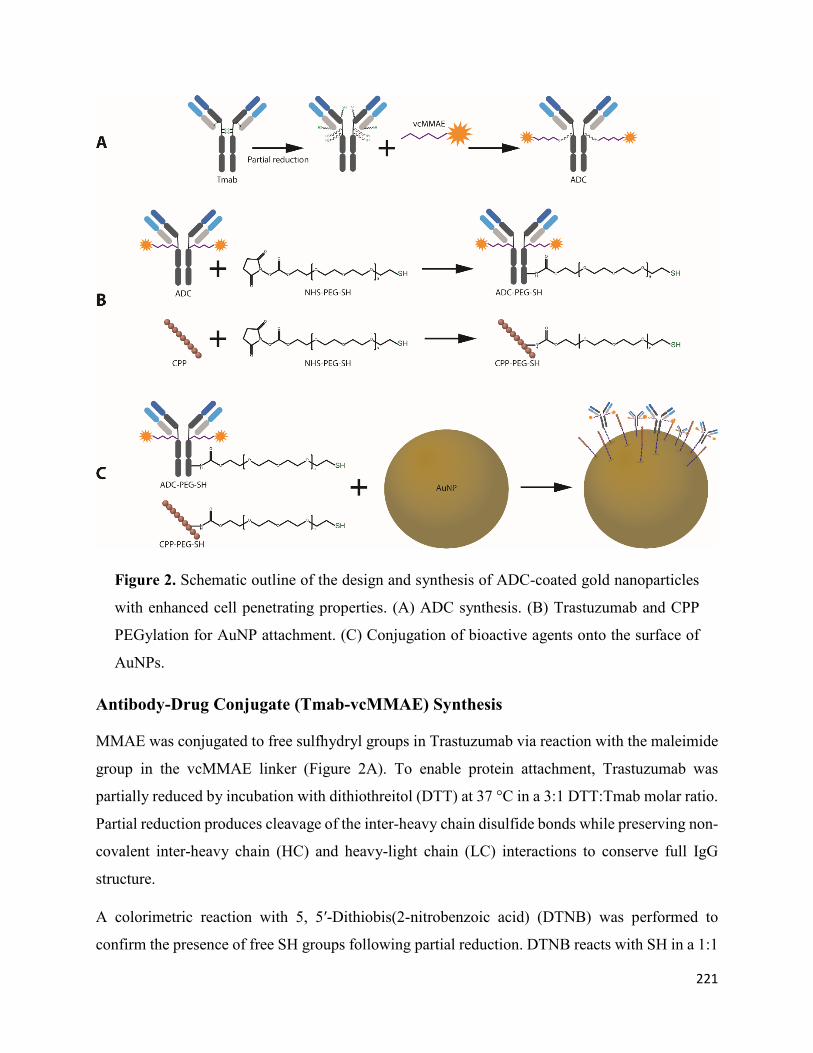
221
Figure 2. Schematic outline of the design and synthesis of ADC-coated gold nanoparticles
with enhanced cell penetrating properties. (A) ADC synthesis. (B) Trastuzumab and CPP
PEGylation for AuNP attachment. (C) Conjugation of bioactive agents onto the surface of
AuNPs.
Antibody-Drug Conjugate (Tmab-vcMMAE) Synthesis
MMAE was conjugated to free sulfhydryl groups in Trastuzumab via reaction with the maleimide
group in the vcMMAE linker (Figure 2A). To enable protein attachment, Trastuzumab was
partially reduced by incubation with dithiothreitol (DTT) at 37 °C in a 3:1 DTT:Tmab molar ratio.
Partial reduction produces cleavage of the inter-heavy chain disulfide bonds while preserving non-
covalent inter-heavy chain (HC) and heavy-light chain (LC) interactions to conserve full IgG
structure.
A colorimetric reaction with 5, 5′-Dithiobis(2-nitrobenzoic acid) (DTNB) was performed to
confirm the presence of free SH groups following partial reduction. DTNB reacts with SH in a 1:1

222
molar ratio to produce 2-nitro-5-thiobenzoic acid (TNB2−). Absorption at 412 nm (λ max of
TNB2−) can be utilized to calculate the amount of free SH groups per antibody monomer (Figure
S1A).
After confirmation and quantification of the presence of free SH groups, the intact structure (no
chain dissociation) was confirmed through size exclusion–high performance liquid
chromatography (SE-HPLC), whereby the elution time of the reduced antibody shifted by 0.0128
min but did not display peaks at longer elution times indicative of chain dissociation (Figure S1B).
The drug-linker (vcMMAE) was then attached to free SH groups in the partially reduced Tmab as
described in the methods section. Successful attachment of vcMMAE was confirmed through
intact protein mass spectrometry analysis (Figure 3). The deconvoluted mass spectrum of the ADC
displayed up to 3 vcMMAE attachments per antibody heavy chain in the G0F or G1F glycoforms
(Figure 3A). Light chain analysis (Figure 3B) showcased a single attachment per LC monomer,
consistent with a single free SH in LC obtained from partial reduction of the interchain disulfide
bonds.

223
Figure 3. Protein intact mass analysis of Tmab-vcMMAE. (A) Deconvoluted spectrum of
Tmab-vcMMAE heavy chain. (B) Deconvoluted spectrum of Tmab-vcMMAE light chain.

224
Figure 4. UV-Vis spectra of unmodified Trastuzumab, Tmab-vcMMAE (ADC) and
MMAE. The contribution of MMAE to the absorption spectrum of the antibody-drug
conjugate enables an estimation of the DAR based on the distinct A280/A248 ratios obtained
with the unmodified antibody and the ADC.
An average drug-to-antibody ratio (DAR) of 2.91 was obtained from analysis of the UV-Vis
spectrum of the ADC as described in the methods section (Figure 4).

225
Figure 5. SE-HPLC chromatograms of PEGylated Trastuzumab variants obtained by
employing varying ratios of PEG:Tmab ratios. Inset shows the zoomed region displaying
small shifts in elution time.
Antibody and CPP PEGylation
Structural Characterization
Trastuzumab and HIV-TAT were PEGylated via lysine conjugation chemistry (Figure 1).
Antibody PEGylation was confirmed through SE-HPLC (Figure 5). The SE-HPLC
chromatograms of the PEGylated Trastuzumab show an increasing shift towards earlier elution
times as the PEG-linker/tmab ratio increases (Figure 5). Resolving the exact number of PEG
polymers attached per antibody molecule through SE-HPLC separation and other standard protein
characterization techniques is challenging, given that the expected molecular weight (MW)
increment per individual attachment corresponds to less than 4% of the MW of unmodified
Trastuzumab, namely 5 kDa increments to the ~148 kDa expected MW of the antibody. Moreover,
PEG molecules display heterogeneity in the number of ethylene glycol units—albeit with only
small differences in MW—adding to the ensuing heterogeneity. Nonetheless, detailed
characterization and homogeneity are not crucial for subsequent use as targeting agents, as long as
the bulk of the protein monomers have been modified and functionality is conserved. A single

226
Trastuzumab monomer possesses 88 lysine residues and 4 amino-terminal groups available for
reaction with the NHS group. Hence, reaction with a large number of linkers can potentially impair
receptor binding. Consequently, conservation of the functionality of the PEGylated derivative was
assessed prior to subsequent surface functionalization of the nanoparticles.
Binding Kinetics of Functionalized Trastuzumab
The chemically modified Trastuzumab variants (Tmab-PEG-SH, ADC and ADC-PEG-SH) were
tested for their capacity to retain the binding affinity and binding kinetics to a recombinant HER2
protein after functionalization through surface plasmon resonance (SPR) single cycle kinetic
analysis Figure S5).
The binding kinetics to the HER2 receptor were not significantly altered under the assay
conditions. The affinity constant (KD) of the PEGylated antibodies, ranging from 5.46–6.91 pM,
showed only minor differences compared to the mean KD of unmodified Trastuzumab—6.07 pM
(Table 1). Kinetic constants for the antibody drug conjugate were also highly similar to the
unmodified Trastuzumab. The PEGylated ADC, on the other hand, recorded a slight increase in
binding rate constant (Ka) accompanied by a 32-fold increase in the dissociation rate constant
(Kd), for a net 14-fold decrease KD.
The varying molar ratios of PEG-linker utilized for derivatization were deemed appropriate for
subsequent attachment to the surface of gold nanoparticles, as the modified antibody did not
display significant alterations in binding affinity to the cognate receptor. Henceforth, the highest
molar excess (25:1 PEG-mAb ratio) for reaction was employed in order to maximize Trastuzumab
attachment to AuNPs. PEGylation of Tmab-vcMMAE caused a significant decrease in KD;
however, the affinity constant remains in the picomolar range, thus it is still expected to exert
active targeting capacity.
Table 2. Kinetics and affinity analysis of functionalized Trastuzumab variants.
Trastuzumab Variant Ka (× 10+6) M−1·s−1 Kd (× 10+5) s−1 KD (pM)
Tmab 3.24 ± 0.15 1.98 ± 0.50 6.07 ± 1.27
Tmab-PEG-SH 2X 3.53 ± 0.15 2.47 ± 0.24 6.83 ± 0.68

227
Tmab-PEG-SH 5X 2.86 ± 0.03 1.97 ± 0.20 6.91 ± 0.78
Tmab-PEG-SH 10X 2.87 ± 0.11 1.98 ± 0.14 6.89 ± 0.21
Tmab-PEG-SH 25X 2.05 ± 0.03 1.12 ± 0.11 5.46 ± 0.44
ADC 2.25 ± 0.01 1.58 ± 0.09 7.05 ± 0.41
ADC-PEG-SH 7.45 ± 0.07 61.80 ± 0.05 85.01 ± 10.92
Gold Nanoparticle Surface Functionalization
The surface of 50 nm citrate-capped gold nanoparticles (Cit-AuNP) was functionalized with OH-
PEG-SH (OH-PEG-AuNP), Tmab-PEG-SH (Tmab-PEG-AuNP), CPP-PEG-SH (CPP-PEG-
AuNP), or a combination of CPP-PEG-SH and Tmab-PEG-SH (CPP+Tmab-PEG-AuNP) through
a sequential addition of the bioactive agents.
Transmission electron micrograph (TEM) analysis of the synthesized Cit-AuNPs displayed a mean
diameter of 48.29 ± 5.58 nm showing a narrow size distribution and uniform spherical morphology
(Table 2). The mean hydrodynamic diameter obtained by DLS was 60.62 ± 0.19 nm (Z-average)
with a polydispersity index (PDI) of 0.29. The SPR absorption band of the AuNPs had an
absorption maximum (λ max) at 530.5 nm, consistent with the expected λ max for ~50 nm gold
nanoparticles according to previously reported determinations of SPR bands of spherical AuNPs
[32]. Upon surface functionalization, the λ max shifted towards longer wavelengths (red-shift)—
a well-described spectral shift caused by an increase in the local refractive index on the NP surface.
In increasing order, the λ max shifts were +1.9 nm for OH-PEG-AuNPs, +2.7 nm for CPP-PEG-
AuNP, +3.3 nm for CPP+Tmab-PEG-AuNPs and +3.7 for Tmab-PEG-AuNPs.
The change in SPR absorption maximum was accompanied by an increase in hydrodynamic
diameter (Table 2), where Tmab-PEG functionalization showed the highest increment (87.35 ±
0.41 nm). The PDIs of all surface-functionalized samples decreased relative to Cit-AuNP,
indicative of enhanced colloidal stability and a consequent reduction of nanoparticle aggregation.
Surface functionalization caused marked alterations in the zeta potential (ζ) of the colloidal
dispersions (Table 2). Citrate-capped AuNPs displayed a mean ζ of -34.60 ± 0.91 mV, consistent
with a negatively charged surface due to the negatively charged OH- groups of the citrate moiety.
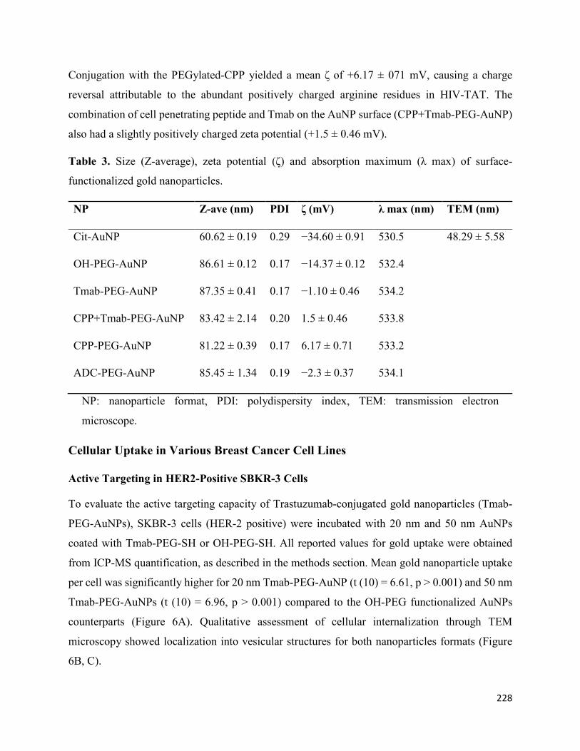
228
Conjugation with the PEGylated-CPP yielded a mean ζ of +6.17 ± 071 mV, causing a charge
reversal attributable to the abundant positively charged arginine residues in HIV-TAT. The
combination of cell penetrating peptide and Tmab on the AuNP surface (CPP+Tmab-PEG-AuNP)
also had a slightly positively charged zeta potential (+1.5 ± 0.46 mV).
Table 3. Size (Z-average), zeta potential (ζ) and absorption maximum (λ max) of surface-
functionalized gold nanoparticles.
NP Z-ave (nm) PDI ζ (mV) λ max (nm) TEM (nm)
Cit-AuNP 60.62 ± 0.19 0.29 −34.60 ± 0.91 530.5 48.29 ± 5.58
OH-PEG-AuNP 86.61 ± 0.12 0.17 −14.37 ± 0.12 532.4
Tmab-PEG-AuNP 87.35 ± 0.41 0.17 −1.10 ± 0.46 534.2
CPP+Tmab-PEG-AuNP 83.42 ± 2.14 0.20 1.5 ± 0.46 533.8
CPP-PEG-AuNP 81.22 ± 0.39 0.17 6.17 ± 0.71 533.2
ADC-PEG-AuNP 85.45 ± 1.34 0.19 −2.3 ± 0.37 534.1
NP: nanoparticle format, PDI: polydispersity index, TEM: transmission electron
microscope.
Cellular Uptake in Various Breast Cancer Cell Lines
Active Targeting in HER2-Positive SBKR-3 Cells
To evaluate the active targeting capacity of Trastuzumab-conjugated gold nanoparticles (Tmab-
PEG-AuNPs), SKBR-3 cells (HER-2 positive) were incubated with 20 nm and 50 nm AuNPs
coated with Tmab-PEG-SH or OH-PEG-SH. All reported values for gold uptake were obtained
from ICP-MS quantification, as described in the methods section. Mean gold nanoparticle uptake
per cell was significantly higher for 20 nm Tmab-PEG-AuNP (t (10) = 6.61, p > 0.001) and 50 nm
Tmab-PEG-AuNPs (t (10) = 6.96, p > 0.001) compared to the OH-PEG functionalized AuNPs
counterparts (Figure 6A). Qualitative assessment of cellular internalization through TEM
microscopy showed localization into vesicular structures for both nanoparticles formats (Figure
6B, C).

229
Trastuzumab coated AuNPs did not display enhanced uptake in two other breast cancer cell lines
(MCF-7 and MDA-MB-231) that are not reported to upregulate HER-2 expression (Figure 7) [33].
Uptake into DLD-1 cells (colorectal cancer HER-2 negative cell lines) showed a small increase in
mean uptake per cell with no statistical significance. ADC conjugated gold nanoparticles were not
employed for cellular uptake assays as the high potency of the drug can cause significant cell death
at the concentrations used; thus, evaluation of cellular uptake is not comparable to the other
formats.
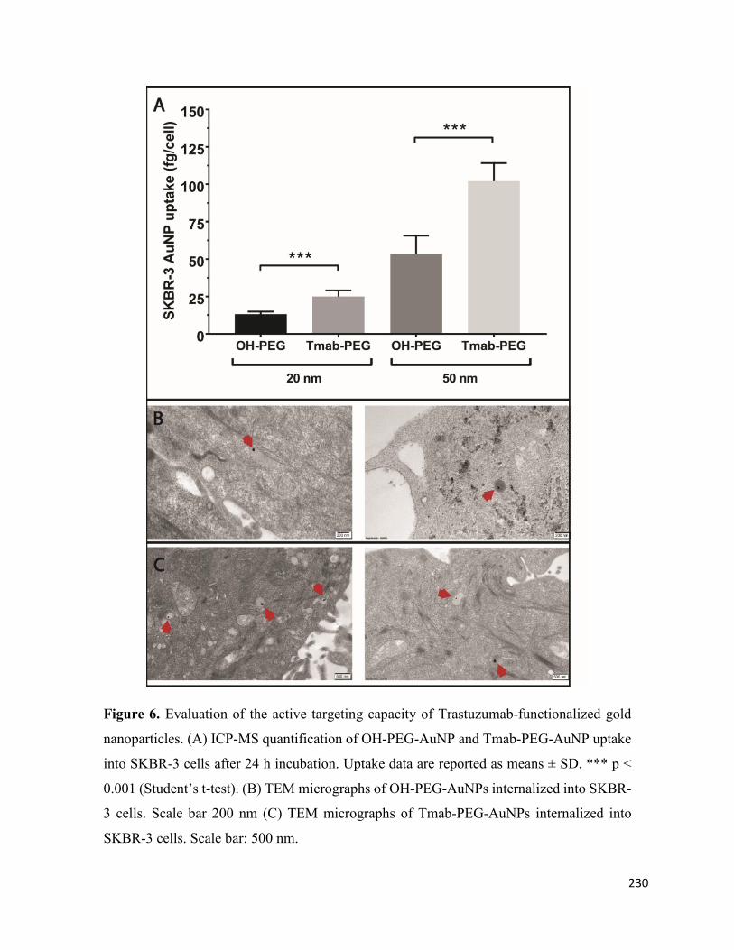
230
Figure 6. Evaluation of the active targeting capacity of Trastuzumab-functionalized gold
nanoparticles. (A) ICP-MS quantification of OH-PEG-AuNP and Tmab-PEG-AuNP uptake
into SKBR-3 cells after 24 h incubation. Uptake data are reported as means ± SD. *** p <
0.001 (Student’s t-test). (B) TEM micrographs of OH-PEG-AuNPs internalized into SKBR-
3 cells. Scale bar 200 nm (C) TEM micrographs of Tmab-PEG-AuNPs internalized into
SKBR-3 cells. Scale bar: 500 nm.

231
Figure 7. Active targeting of Trastuzumab functionalized gold nanoparticles in various
cancer cell lines. Uptake data are reported as means ± SD. * p < 0.05 (Student’s t-test).
Differences between OH-PEG and Tmab-PEG functionalised gold nanoparticle uptake in
DLD-1 and MCF-7 cell lines were not significant.
CPP-Driven Enhanced Internalization
To assess the effect on cell internalization using the HIV-TAT cell penetrating peptide as a coating
functional group on the surface of the nanoparticles, 4 different cancer cell lines (SKBR-3, DLD-
1, MDA-MB-231 and MCF-7) were treated with 25 µg/mL 50 nm gold nanoparticles
functionalized with OH-PEG-SH, Tmab-PEG-SH, CPP-PEG-SH, or a combination of Tmab-PEG-
SH and CPP-PEG-SH (CPP+Tmab-PEG-AuNP).
A significant increase in uptake, relative to OH-PEG-AuNP, obtained by attachment of the anti-
HER2 antibody (Tmab-PEG-AuNP) was recorded for the SKBR-3 cell line (t (4) = 2.22, p > 0.05)
only (Figure 7). In the same SKBR-3 cell line, AuNP functionalized with the cell penetrating
peptide (CPP-PEG-AuNPs) showed approximately 1000-fold increase compared to the Tmab-

232
PEG-AuNP (Figure 8). Similarly, CPP-PEG-AuNPs displayed a high increase in cell uptake
relative to OH-PEG-Tmab in all cell lines tested (Figure 8). CPP+Tmab-PEG-AuNP also recorded
markedly higher uptake across all cell lines compared to OH-PEG-AuNP. CPP-PEG-AuNP
displayed significantly higher internalization than the combination of CPP+Tmab-PEG-AuNP in
SKBR-3 and MCF-7 cells, and no statistical difference was observed between these two formats
in the DLD-1 and MDA-MB-231 cell lines.
Figure 8. Cellular uptake of cell penetrating peptide (CPP) functionalized gold nanoparticles
into various cancer cell lines. Uptake data are reported as means ± SD. * p < 0.05, ** p <
0.01, *** p < 0.001 (Student’s t-test).
In Vitro Cytotoxicity of ADC-PEG-AuNP in HER2 Overexpressing Cancer Cell Lines
To assess the capacity for intracellular release of the drug payload, the in vitro cytotoxic activity
of the antibody-drug conjugate bound to the nanoparticles (ADC-PEG-AuNP) was evaluated in
two HER2 amplified cell lines: (1) SKBR-3 and (2) SKOV-3 (ovarian adenocarcinoma). Growth
rate inhibition (GR) metrics derived from cell growth curves were determined to compare the

233
GR50 value of free MMAE, Tmab-vcMMAE, ADC-PEG-AuNP and Trastuzumab (Figure 9 and
Figure S6). Growth rate calculations are specified in the methods section. GR50 corresponds to
the concentration at which GR(c) = 0.5. ADC and ADC-PEG-AuNP concentrations reported in
Figure 9 correspond to MMAE concentrations based on DAR and antibody per AuNP estimations.
The in vitro cytotoxic activity of free MMAE was higher in both cell lines relative to ADC and
ADC-PEG-AuNP (Table 3). MMAE GR50 values were subnanomolar for both cell lines. SKOV-
3 displayed slightly higher sensitivity to MMAE (GR50 = 0.14 nM) compared to SKBR-3 cells
(GR50 = 0.33 nM). ADC and ADC-PEG-AuNP displayed similar GR50 values for both cell lines.
Trastuzumab showed a dramatically decreased potency relative to MMAE containing formats,
particularly in SKOV-3. Hence, the GR50 value was not determined for this cell line due to the
high concentration of antibody required to obtain an appropriate dose-response curve. The effect
on growth rate inhibition was also determined for OH-PEG-AuNP as a control. The stabilized gold
nanoparticles only caused small reductions in growth rate at high nanoparticle concentrations
(Figure 9).

234
Figure 9. Growth rate (GR) inhibition of (A) free MMAE, (B) ADC, (C) ADC-PEG-AuNP,
(D) Trastuzumab and (E) OH-PEG-AuNP in SKBR-3 and SKOV-3 cell lines. Data are
reported as means ± SD. 95% confidence bands are displayed as dotted lines. Concentration
of ADC and ADC-PEG-PEG are reported as molar concentrations of MMAE according to
the estimated DAR and number of ADC per AuNP, respectively.

235
Table 4. GR50 values with confidence intervals (CI) obtained from dose-response curves in Figure
9.
Sample SKBR-3 SKOV-3
Agent GR50 (nM) GR50 95% CI R2 SKOV-3 GR50 95% CI R2
Free MMAE 0.33 (0.28–0.37) 0.9986 0.14 (0.11–0.17) 0.9851
ADC 34.91 (29.04–41.02) 0.9847 4.81 (3.56–6.32) 0.9636
ADC-PEG-AuNP 19.45 (16.52–22.80) 0.9913 10.14 (8.55–11.83) 0.9878
Tmab 2118.36 (1849.27–2426.61) 0.9931 N.D. N.D. N.D.
N.D.: not defined, CI: confidence intervals, GR50: concentration required to achieve a
growth rate inhibition of 0.5.
Discussion
The lack of clinical precedent for inorganic nanoparticles has hindered their implementation in
cancer therapy. However, the results of the Phase I clinical trial (NCT00356980) of CYT-6091
(PEGylated colloidal gold-rhTNF) published in 2009 were highly promising with regards to safety
profile and the capacity to accumulate effectively in a wide range of solid tumors [34]. Considering
the remarkable therapeutic potential of gold nanoparticles and the validation of the EPR effect for
colloidal gold in human patients, we were prompted to assess three strategies; or a combination
thereof, to further enhance the potential of AuNPs for clinical implementation: (1) surface
attachment of PEGylated Trastuzumab for targeted treatment of HER2-positive tumors (active
targeting), (2) employment of an antibody-drug conjugate as targeting agent to increase the
anticancer potency of the system, and (2) surface coating with the cationic HIV-TAT cell
penetrating peptide to enhance intracellular delivery.
Trastuzumab and HIV-TAT PEGylation
Attachment of poly ethylene glycol has become a conventional strategy to increase circulation
times and distribution of nanosized structures. PEGylation prevents opsonization and uptake by
the RES system—a biological mechanism that severely impedes tumor localization by premature
clearance [35, 36]. Herein, our results support that Trastuzumab PEGylation for subsequent gold
surface attachment can be readily achieved without significant modifications in HER-2 affinity or

236
binding kinetics as was reflected by SPR binding measurements to a recombinant HER2 protein.
The same NHS-linker was used for HIV-TAT PEGylation, taking advantage of the two lysine
residues in its amino acid composition (Figure 1).
ADC Construction
MMAE is a cytotoxic payload with exceptionally high potency that has frequently been employed
in the construction of antibody drug conjugates. Under our experimental conditions, we obtained
an average drug-to-antibody ratio of 2.91, as per UV-Vis spectroscopy analysis, consistent with
DARs reported for similar ADC synthesis methods [37, 38]. For further structural characterization
and confirmation of vcMMAE attachment, the ADC was analysed through intact protein mass
spectrometry analysis. The ADC was buffer exchanged to MeCN 10% v/v to induce inter-heavy
and heavy-light chain dissociation, in order to analyse the number of drugs attached to each
polypeptide chain. Chain dissociation in MeCN 10% v/v was confirmed by SE-HPLC
chromatograms showing the appearance of two peaks at longer elution times (Figure S2). The
deconvoluted mass spectra confirmed that vcMMAE can attach to all possible free sulfhydryl
groups formed upon partial reduction, i.e., a maximum of three attachments on the heavy chain
and one attachment on the light chain.
Herein, our results report on the feasibility of combining two common bioconjugation techniques
(lysine and cysteine attachment) to PEGylate Tmab-vcMMAE for nanoparticle attachment.
Furthermore, our data show that HER-2 binding affinity decreases by an order or magnitude with
ADC PEGylation; yet, the binding affinity remains within the picomolar range. Several studies
have combined targeting agents and cytotoxic drugs on nanoparticles; however, the added
complexity of the systems also complicates appropriate characterization for implementation,
especially in regard to dosage determination as the amount of each individual component requires
quantification. To this end, the use of antibody-drug conjugates as targeting agents carrying the
payload could simplify this—provided that the DAR is determined, quantification of protein
content would be sufficient to estimate drug dosage per nanoparticle.
Gold Nanoparticle Surface Functionalization
Adding to improved biodistribution and tumor targeting, PEGylation also increases the colloidal
stability of gold nanoparticles—a key requirement for long-term storage. Attachment of the

237
bioactive groups was achieved through the thiol moiety of the PEG linker, which, at high pH, can
form covalent gold-sulfur (Au-S) bonds, providing stable conjugation to the surface [11]. Indeed,
surface functionalization had a pronounced enhancement in nanoparticle stability upon addition of
1% NaCl and cell culture media (Figure S3). Attachment was confirmed by an increase in
hydrodynamic size (DLS) and SPR absorption maxima, and most importantly by alterations in the
zeta potential that allow to discriminate the presence of the bioactive groups. For instance, the
positively charged HIV-TAT caused a charge reversal in zeta potential (+6.17 ± 0.71 mV) for a
+40.77 mV shift compared to the citrate-capped gold nanoparticles (−34.60 mV). In contrast,
coating with the neutral OH-PEG caused a smaller +20.23 mV shift. Zeta potential values closer
to the isoelectric point are generally detrimental to colloidal stability; however, the hydrophilic
PEG polymer on the surface impedes nanoparticle aggregation by steric hindrance to prevent
surface interactions between AuNPs. The large exclusion volume of the hydration cloud of the
PEG linkers is known to prevent interactions between nanoparticle surfaces that lead to
aggregation [35].
Quantification of the average number of antibodies that coat individual nanoparticles is
challenging, insofar as common colorimetric methods for protein quantitation are difficult to
perform due to the much stronger absorption coefficients of gold nanoparticles throughout the
wavelength ranges used for protein concentration measurements. Instead, we quantified the
amount of antibody by accounting for the ensuing decrease in antibody concentration following
attachment, after removal of the functionalized nanoparticles through centrifugation. According to
these measurements, an average of 156 antibodies covered the surface of 50 nm AuNPs and 40
antibodies on 20 nm AuNPs (Figure S4).
Active Targeting and Cellular Uptake
The multivalent presentation of Trastuzumab on gold nanoparticles has been shown previously to
promote HER2 receptor crosslinking, leading to enhanced cellular internalization in HER2
overexpressing cell lines [39]. In our experimental setup, Trastuzumab-coated gold nanoparticles
were compared to the AuNPs coated with the SH-linker without antibody derivatization, to
maximize the similarity in physicochemical properties, excluding the presence of protein. Indeed,
the mean hydrodynamic diameter of both formats differed by less than 1 nm according to DLS
measurements (Table 2). Electrophoretic mobility determinations, on the other hand, recorded

238
negative zeta potential values for OH-PEG-AuNPs and close to neutral values for Tmab-PEG-
AuNP. The drift towards more neutral values—relative to citrate-capped nanoparticles—is
consistent with antibody attachment, as Trastuzumab (isoelectric point (pI) 8.7) possesses a net
positive charge when dissolved in PBS. A small net positive charge is also expected when
suspended in cell culture media (pH 7.4). The effect of nanoparticle surface charge on cellular
uptake is well-documented, whereby positively charged nanoparticles have consistently displayed
higher uptake rates in nonphagocytic cells [40]. The increase in internalization with positively
charged surfaces has generally been ascribed to favorable electrostatic nanoparticle/cell
interactions due to the net negative charge of the plasma membrane [40]. In view of the foregoing,
it is difficult to rule out a contribution of the more neutral zeta potential of Tmab-PEG-AuNP in
enhancing cellular uptake. This notwithstanding, the observation that internalization enhancement
was only recorded in a HER2 overexpressing cell line (SKBR-3)—and not in the HER2 basal
counterparts (DLD-1, MDA-MB-231 and MCF-7)—supports cellular uptake increase through
Trastuzumab-mediated HER2 receptor crosslinking. Interestingly, TEM micrographs of SKBR-3
cells did not show clear distinction between both formats in subcellular localization—i.e., both
AuNP designs were primarily localized within vesicular structures, presumably coated
preendosomal and carrier vesicles (early endosomes and lysosomes). Alternatively, it is possible
that some of these structures are autophagosomes, as gold nanoparticles have been shown to induce
autophagosome accumulation [41]. This observation warrants further elucidation of the effect of
surface functionalization on uptake mechanism and localization.
Cellular Uptake Enhancement with HIV-TAT
Due to the relatively low loading capacity of spherical gold nanoparticles, it is essential to ensure
maximum cellular internalization when developed as drug delivery vehicles. Having improved
selective uptake into HER2 overexpressing cell lines through active targeting, we sought to
evaluate the effect of combining a cell penetrating peptide with the antibody targeting agent. HIV-
TAT internalization mechanism remains a topic of debate; however, evidence of uptake
saturability and energy dependency suggest an endosomal pathway [42]. Endosomal and
subsequent lysosomal localization is required for effective drug release of cathepsin B-cleavable
linkers, such as those containing the valine-citrulline dipeptide. Enhancing uptake is thus
paramount in HER2-targeted conjugates for intracellular release, considering that most ErbB

239
receptors have shown impaired ligand-induced receptor trafficking [43]. To this end,
functionalization with the cell penetrating peptide caused a dramatic increase in cellular uptake
across all cell lines tested. This enhancement was considerably more significant than that obtained
by antibody functionalization only. Conversely, our results did not show improvement in uptake
upon combination of both bioactive agents compared to CPP-PEG-AuNP. In fact, uptake was
significantly higher with CPP functionalization in SKBR-3 and MCF-7 cells. We presume that this
observation stems from the more positive zeta potential of CPP-PEG-AuNPs, in which case
engagement through cell membrane/nanoparticle electrostatic interactions is a stronger
determinant of uptake rate than antibody-mediated receptor cross-linking.
These findings warrant further investigation into the effect of the highlighted physicochemical and
physiological attributes in a more physiological setting. While higher uptake may be desirable in
delivery applications, internalization must be specific to the targeted tumor cells. Previous studies
have reported that uptake, rather than diffusion, could be the primary mechanism for nanoparticle
tumor delivery. Consequently, surface charge has been proposed as a major determinant in tumor
distribution upon systemic administration [44]. If indeed transcellular transport has a crucial
impact in tumor penetration, then enhancing cellular internalization through strategies such as the
attachment of a cell penetrating peptide might provide improved tumor tissue distribution, thus
enhancing efficacy and therapeutic index.
In Vitro Cytotoxicity of ADC-PEG-AuNP in HER2 Overexpressing Cancer Cell Lines
Growth rate inhibition sensitivity in SKOV-3 and SKBR-3 cell lines was markedly higher for free
MMAE than for the antibody-drug conjugate and for ADC-carrying gold nanoparticles (Figure 8).
It is plausible that the requirement of linker cleavage and self-immolation of the p-aminobenzyl
carbamate group in the antibody-drug conjugate hinders conjugated vcMMAE activity compared
to the free drug. Additionally, although HER2 binding and cross-linking can induce receptor-
mediated endocytosis, free MMAE likely penetrates more readily into the intracellular
compartment. Nonetheless, the structural characteristics that presumably hinder conjugated
vcMMAE cytotoxicity in isolated carcinoma cells are expected to provide selectivity advantages
in more physiological settings.
Comparison of the cytotoxic activity of free ADC and nanoparticle-conjugated ADC displayed
similar GR50 for both SKBR-3 and SKOV-3 cells (Table 3). GR50 values for ADC-PEG-AuNP
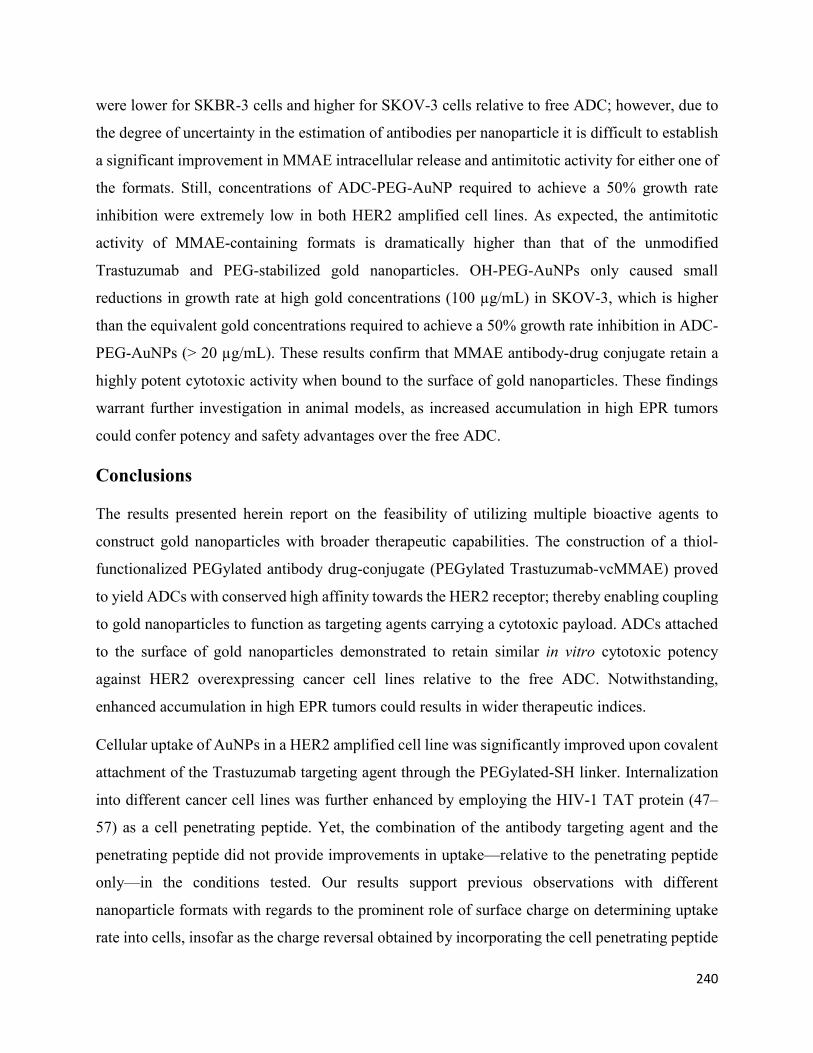
240
were lower for SKBR-3 cells and higher for SKOV-3 cells relative to free ADC; however, due to
the degree of uncertainty in the estimation of antibodies per nanoparticle it is difficult to establish
a significant improvement in MMAE intracellular release and antimitotic activity for either one of
the formats. Still, concentrations of ADC-PEG-AuNP required to achieve a 50% growth rate
inhibition were extremely low in both HER2 amplified cell lines. As expected, the antimitotic
activity of MMAE-containing formats is dramatically higher than that of the unmodified
Trastuzumab and PEG-stabilized gold nanoparticles. OH-PEG-AuNPs only caused small
reductions in growth rate at high gold concentrations (100 µg/mL) in SKOV-3, which is higher
than the equivalent gold concentrations required to achieve a 50% growth rate inhibition in ADC-
PEG-AuNPs (> 20 µg/mL). These results confirm that MMAE antibody-drug conjugate retain a
highly potent cytotoxic activity when bound to the surface of gold nanoparticles. These findings
warrant further investigation in animal models, as increased accumulation in high EPR tumors
could confer potency and safety advantages over the free ADC.
Conclusions
The results presented herein report on the feasibility of utilizing multiple bioactive agents to
construct gold nanoparticles with broader therapeutic capabilities. The construction of a thiol-
functionalized PEGylated antibody drug-conjugate (PEGylated Trastuzumab-vcMMAE) proved
to yield ADCs with conserved high affinity towards the HER2 receptor; thereby enabling coupling
to gold nanoparticles to function as targeting agents carrying a cytotoxic payload. ADCs attached
to the surface of gold nanoparticles demonstrated to retain similar in vitro cytotoxic potency
against HER2 overexpressing cancer cell lines relative to the free ADC. Notwithstanding,
enhanced accumulation in high EPR tumors could results in wider therapeutic indices.
Cellular uptake of AuNPs in a HER2 amplified cell line was significantly improved upon covalent
attachment of the Trastuzumab targeting agent through the PEGylated-SH linker. Internalization
into different cancer cell lines was further enhanced by employing the HIV-1 TAT protein (47–
57) as a cell penetrating peptide. Yet, the combination of the antibody targeting agent and the
penetrating peptide did not provide improvements in uptake—relative to the penetrating peptide
only—in the conditions tested. Our results support previous observations with different
nanoparticle formats with regards to the prominent role of surface charge on determining uptake
rate into cells, insofar as the charge reversal obtained by incorporating the cell penetrating peptide

241
had a more pronounced impact than the addition of the antibody targeting agent. Efficient cleavage
of the valine-citrulline moiety for drug release requires cellular internalization for exposure to
cathepsin B in lysosomes or endosomes; therefore, incorporation of the CPP might provide
improved intracellular delivery of the MMAE payload in this format.
Supplementary Materials: The following are available online at www.mdpi.com/xxx/s1, Figure
S1: Analysis of the presence of sulfhydryl groups and conservation of intact structure of
Trastuzumab after partial reduction with DTT, Figure S2: SE-HPLC chromatograms of partially
reduced Trastuzumab in 1 mM EDTA, Tmab-vcMMAE in H2O and Tmab-vcMMAE in
acetonitrile 10% with formic acid 1%, Figure S3: AuNP stability upon surface functionalization
with Tmab-PEG-SH. Addition of 1% NaCl to citrate capped AuNPs caused aggregation as
evidenced by a broad absorption band in the 700–800 nm range, Figure S4: Representative
tryptophan fluorescence emission spectra for the estimation of Trastuzumab:AuNP ratio for 20 nm
AuNPs, Figure S5: Representative sensorgrams of (A) Trastuzumab, (B) Tmab-PEG-SH 25X and
(C) Tmab-vcMMAE binding to recombinant HER2 receptor, Figure S6: Representative SKBR-3
cell growth curves employed to analyse growth rate inhibition activity of MMAE-containing
agents at equivalent MMAE concentrations.
Author Contributions
E.C. and V.K. conceived the design. E.C. designed the study, performed all experimental work
and data analysis and wrote the manuscript. V.K. supervised the development of the study,
contributed to data interpretation and manuscript evaluation and editing.
Funding
This research received no external funding.
Acknowledgments: The authors would like to thank Genentech for their kind donation of
Herceptin®. The authors would like to acknowledge the School of Pharmacy of The University of
Sydney for financial support. EC would like to acknowledge the Ministry of Science, Technology
and Telecommunications of the Republic of Costa Rica for postgraduate scholarship.
Conflicts of Interest
The authors declare no conflict of interest

242
References
[1]. Heldin, C.H.; Rubin, K.; Pietras, K.; Ostman, A. High interstitial fluid pressure-an obstacle
in cancer therapy. Nat. Rev. Cancer, 4 (2004), 806–813, doi:10.1038/nrc1456.
[2]. Netti, P.A.; Baxter, L.T.; Boucher, Y.; Skalak, R.; Jain, R.K. Time-dependent behavior of
interstitial fluidpressure in solid tumors: Implications for drug delivery. Cancer Res, 55 (1995)
5451–5458.
[3]. Tredan, O.; Galmarini, C.M.; Patel, K.; Tannock, I.F. Drug resistance and the solid tumor
microenvironment. J Natl Cancer Inst, 99 (2007), 1441–1454, doi:10.1093/jnci/djm135.
[4]. Matsumura, Y.; Maeda, H. A new concept for macromolecular therapeutics in cancer
chemotherapy: Mechanism of tumoritropic accumulation of proteins and the antitumor agent
smancs. Cancer Res, 46 (1986), 6387–6392.
[5]. Tran, S.; DeGiovanni, P.-J.; Piel, B.; Rai, P. Cancer nanomedicine: A review of recent success
in drug delivery. Clin Transl Med, 6 (2017), 44–44, doi:10.1186/s40169-017-0175-0.
[6]. Jin, S.; Leach, J.C.; Ye, K. Nanoparticle-mediated gene delivery. Methods Mol. Biol. 544
(2009), 547–557, doi:10.1007/978-1-59745-483-4_34.
[7]. Manthe, R.L.; Foy, S.P.; Krishnamurthy, N.; Sharma, B.; Labhasetwar, V. Tumor ablation
and nanotechnology. Mol Pharm, 7 (2010), 1880–1898, doi:10.1021/mp1001944.
[8]. Will, O.; Purkayastha, S.; Chan, C.; Athanasiou, T.; Darzi, A.W.; Gedroyc, W.; Tekkis, P.P.
Diagnostic precision of nanoparticle-enhanced MRI for lymph-node metastases: A meta-analysis.
Lancet Oncol, 7 (2006), 52–60, doi:10.1016/s1470-2045(05)70537-4.
[9]. Lukianova-Hleb, E.Y.; Ren, X.; Sawant, R.R.; Wu, X.; Torchilin, V.P.; Lapotko, D.O. On-
demand intracellular amplification of chemoradiation with cancer-specific plasmonic
nanobubbles. Nat Med, 20 (2014), 778, doi:10.1038/nm.3484.
[10]. Kyriazi, M.-E.; Giust, D.; El-Sagheer, A.H.; Lackie, P.M.; Muskens, O.L.; Brown, T.;
Kanaras, A.G. Multiplexed mRNA Sensing and Combinatorial-Targeted Drug Delivery Using
DNA-Gold Nanoparticle Dimers. ACS Nano, 12 (2018), 3333–3340,
doi:10.1021/acsnano.7b08620.

243
[11]. Xue, Y.; Li, X.; Li, H.; Zhang, W. Quantifying thiol–gold interactions towards the efficient
strength control. Nat Commu, 5 (2014), 4348, doi:10.1038/ncomms5348.
[12]. Wilhelm, S.; Tavares, A.J.; Dai, Q.; Ohta, S.; Audet, J.; Dvorak, H.F.; Chan, W.C.W.
Analysis of nanoparticle delivery to tumours. Nat Rev Mater 1 (2016), 16014,
doi:10.1038/natrevmats.2016.14.
[13]. Alkilany, A.M.; Murphy, C.J. Toxicity and cellular uptake of gold nanoparticles: What we
have learned so far? J. Nanopart. Res. Interdiscip. Forum Nanoscale Sci Technol, 12 (2010), 2313–
2333, doi:10.1007/s11051-010-9911-8.
[14]. Goel, R.; Shah, N.; Visaria, R.; Paciotti, G.F.; Bischof, J.C. Biodistribution of TNF-alpha-
coated gold nanoparticles in an in vivo model system. Nanomedicine, 4 (2009), 401–410,
doi:10.2217/nnm.09.21.
[15]. Byrne, J.D.; Betancourt, T.; Brannon-Peppas, L. Active targeting schemes for nanoparticle
systems in cancer therapeutics. Adv Drug Deliv Rev, 60 (2008), 1615–1626,
doi:10.1016/j.addr.2008.08.005.
[16]. Rosenblum, D.; Joshi, N.; Tao, W.; Karp, J.M.; Peer, D. Progress and challenges towards
targeted delivery of cancer therapeutics. Nat Commu, 9 (2018), 1410, doi:10.1038/s41467-018-
03705-y.
[17]. Slamon, D.J.; Leyland-Jones, B.; Shak, S.; Fuchs, H.; Paton, V.; Bajamonde, A.; Fleming,
T.; Eiermann, W.; Wolter, J.; Pegram, M.; et al. Use of Chemotherapy plus a Monoclonal Antibody
against HER2 for Metastatic Breast Cancer That Overexpresses HER2. N Engl J Med, 344 (2001),
783–792, doi:10.1056/NEJM200103153441101.
[18]. Bang, Y.J.; Van Cutsem, E.; Feyereislova, A.; Chung, H.C.; Shen, L.; Sawaki, A.; Lordick,
F.; Ohtsu, A.; Omuro, Y.; Satoh, T.; et al. Trastuzumab in combination with chemotherapy versus
chemotherapy alone for treatment of HER2-positive advanced gastric or gastro-oesophageal
junction cancer (ToGA): A phase 3, open-label, randomised controlled trial. Lancet, 376 (2010),
687–697, doi:10.1016/s0140-6736(10)61121-x.

244
[19]. Fléjou, J.F.; Paraf, F.; Muzeau, F.; Fékété, F.; Hénin, D.; Jothy, S.; Potet, F. Expression of
c-erbB-2 oncogene product in Barrett's adenocarcinoma: Pathological and prognostic
correlations. J Clin Pathol, 47 (1994), 23, doi:10.1136/jcp.47.1.23.
[20]. Berchuck, A.; Kamel, A.; Whitaker, R.; Kerns, B.; Olt, G.; Kinney, R.; Soper, J.T.; Dodge,
R.; Clarke-Pearson, D.L.; Marks, P.; et al. Overexpression of HER-2/neu Is Associated with Poor
Survival in Advanced Epithelial Ovarian Cancer. Cancer Res, 50 (1990), 4087.
[21]. Rolitsky, C.D.; Theil, K.S.; McGaughy, V.R.; Copeland, L.J.; Niemann, T.H. HER-2/neu
amplification and overexpression in endometrial carcinoma. Int J Gynecol Pathol, 18 (1999), 138–
143, doi:10.1097/00004347-199904000-00007.
[22]. Nakajima, M.; Sawada, H.; Yamada, Y.; Watanabe, A.; Tatsumi, M.; Yamashita, J.;
Matsuda, M.; Sakaguchi, T.; Hirao, T.; Nakano, H. The prognostic significance of amplification
and overexpression of c-met and c-erb B-2 in human gastric carcinomas. Cancer, 85 (1999), 1894–
1902, doi:10.1002/(SICI)1097-0142(19990501)85:9<1894:AID-CNCR3>3.0.CO;2-J.
[23]. Santin, A.D.; Bellone, S.; Van Stedum, S.; Bushen, W.; Palmieri, M.; Siegel, E.R.; De Las
Casas, L.E.; Roman, J.J.; Burnett, A.; Pecorelli, S. Amplification of c-erbB2 oncogene. Cancer,
104 (2005), 1391–1397, doi:10.1002/cncr.21308.
[24]. Yonemura, Y.; Ninomiya, I.; Yamaguchi, A.; Fushida, S.; Kimura, H.; Ohoyama, S.;
Miyazaki, I.; Endou, Y.; Tanaka, M.; Sasaki, T. Evaluation of Immunoreactivity for erbB-2 Protein
as a Marker of Poor Short Term Prognosis in Gastric Cancer. Cancer Res, 51 (1991), 1034.
[25]. Hudis, C.A. Trastuzumab--mechanism of action and use in clinical practice. N Engl J Med,
357 (2007), 39–51, doi:10.1056/NEJMra043186.
[26]. Liu, M.; Yang, Y.J.; Zheng, H.; Zhong, X.R.; Wang, Y.; Wang, Z.; Wang, Y.G.; Wang, Y.P.
Membrane-bound complement regulatory proteins are prognostic factors of operable breast cancer
treated with adjuvant trastuzumab: A retrospective study. Oncol Rep, 32 (2014), 2619–2627,
doi:10.3892/or.2014.3496.
[27]. Elgundi, Z.; Reslan, M.; Cruz, E.; Sifniotis, V.; Kayser, V. The state-of-play and future of
antibody therapeutics. Adv Drug Deliv Rev, 122 (2017), 2–19, doi:10.1016/j.addr.2016.11.004.

245
[28]. Thurber, G.M.; Schmidt, M.M.; Wittrup, K.D. Antibody tumor penetration: Transport
opposed by systemic and antigen-mediated clearance. Adv Drug Deliv Rev, 60 (2008), 1421–
1434, doi:10.1016/j.addr.2008.04.012.
[29]. Lee, C.M.; Tannock, I.F. The distribution of the therapeutic monoclonal antibodies
cetuximab and trastuzumab within solid tumors. BMC Cancer, 10 (2010), 255–255,
doi:10.1186/1471-2407-10-255.
[30]. Hermanson, G.T. Chapter 18-PEGylation and Synthetic Polymer Modification. In
Bioconjugate Techniques, 3rd ed.; Hermanson, G.T., Ed.; Academic Press: Boston, MA, USA,
2013, doi:10.1016/B978-0-12-382239-0.00018-2pp. 787-838.
[31]. Gustafson, H.H.; Holt-Casper, D.; Grainger, D.W.; Ghandehari, H. Nanoparticle Uptake:
The Phagocyte Problem. Nano Today, 10 (2015), 487–510, doi:10.1016/j.nantod.2015.06.006.
[32]. Haiss, W.; Thanh, N.T.K.; Aveyard, J.; Fernig, D.G. Determination of Size and
Concentration of Gold Nanoparticles from UV−Vis Spectra. Anal Chem, 79 (2007), 4215–4221,
doi:10.1021/ac0702084.
[33]. Holliday, D.L.; Speirs, V. Choosing the right cell line for breast cancer research. Breast
Cancer Res, 13 (2011), 215, doi:10.1186/bcr2889.
[34]. Libutti, S.K.; Paciotti, G.F.; Byrnes, A.A.; Alexander, H.R., Jr.; Gannon, W.E.; Walker, M.;
Seidel, G.D.; Yuldasheva, N.; Tamarkin, L. Phase I and pharmacokinetic studies of CYT-6091, a
novel PEGylated colloidal gold-rhTNF nanomedicine. Clin Cancer Res, 16 (2010), 6139–6149,
doi:10.1158/1078-0432.ccr-10-0978.
[35]. Suk, J.S.; Xu, Q.; Kim, N.; Hanes, J.; Ensign, L.M. PEGylation as a strategy for improving
nanoparticle-based drug and gene delivery. Adv Drug Deliv Rev, 99 (2016), 28–51,
doi:10.1016/j.addr.2015.09.012.
[36]. Gref, R.; Minamitake, Y.; Peracchia, M.T.; Trubetskoy, V.; Torchilin, V.; Langer, R.
Biodegradable long-circulating polymeric nanospheres. Science, 263 (1994), 1600–1603.
[37]. Adem, Y.T.; Schwarz, K.A.; Duenas, E.; Patapoff, T.W.; Galush, W.J.; Esue, O. Auristatin
Antibody Drug Conjugate Physical Instability and the Role of Drug Payload. Bioconj Chem, 25
(2014), 656–664, doi:10.1021/bc400439x.

246
[38]. Li, F.; Emmerton, K.K.; Jonas, M.; Zhang, X.; Miyamoto, J.B.; Setter, J.R.; Nicholas, N.D.;
Okeley, N.M.; Lyon, R.P.; Benjamin, D.R.; et al. Intracellular Released Payload Influences
Potency and Bystander-Killing Effects of Antibody-Drug Conjugates in Preclinical Models.
Cancer Res, 76 (2016), 2710–2719, doi:10.1158/0008-5472.can-15-1795.
[39]. Jiang, W.; Kim, B.Y.S.; Rutka, J.T.; Chan, W.C.W. Nanoparticle-mediated cellular response
is size-dependent. Nat Nanotechnol, 3 (2008), 145, doi:10.1038/nnano.2008.30.
https://www.nature.com/articles/nnano.2008.30#supplementary-information.
[40]. Cho, E.C.; Xie, J.; Wurm, P.A.; Xia, Y. Understanding the Role of Surface Charges in
Cellular Adsorption versus Internalization by Selectively Removing Gold Nanoparticles on the
Cell Surface with a I2/KI Etchant. Nano Lett, 9 (2009), 1080–1084, doi:10.1021/nl803487r.
[41]. Ma, X.; Wu, Y.; Jin, S.; Tian, Y.; Zhang, X.; Zhao, Y.; Yu, L.; Liang, X.-J. Gold
Nanoparticles Induce Autophagosome Accumulation through Size-Dependent Nanoparticle
Uptake and Lysosome Impairment. ACS Nano, 5 (2011), 8629–8639, doi:10.1021/nn202155y.
[42]. Tkachenko, A.G.; Xie, H.; Liu, Y.; Coleman, D.; Ryan, J.; Glomm, W.R.; Shipton, M.K.;
Franzen, S.; Feldheim, D.L. Cellular Trajectories of Peptide-Modified Gold Particle Complexes:
Comparison of Nuclear Localization Signals and Peptide Transduction Domains. Bioconj. Chem,
15 (2004), 482–490, doi:10.1021/bc034189q.
[43]. Baulida, J.; Kraus, M.H.; Alimandi, M.; Di Fiore, P.P.; Carpenter, G. All ErbB receptors
other than the epidermal growth factor receptor are endocytosis impaired. J Biol Chem, 271 (1996),
5251–5257.
[44]. Kim, B.; Han, G.; Toley, B.J.; Kim, C.-k.; Rotello, V.M.; Forbes, N.S. Tuning payload
delivery in tumour cylindroids using gold nanoparticles. Nat. Nanotechnol, 5 (2010), 465,
doi:10.1038/nnano.2010.58. https://www.nature.com/articles/nnano.2010.58#supplementary-
information.
[45]. Turkevich, J.; Stevenson, P.C.; Hillier, J. The Formation of Colloidal Gold. J Phys Chem,
57 (1953), 670–673, doi:10.1021/j150508a015.
[46]. Frens, G. Controlled Nucleation for the Regulation of the Particle Size in Monodisperse
Gold Suspensions. Nat Phys Sci, 241 (1973), 20, doi:10.1038/physci241020a0.

247
[47]. Kelly, S.T.; Zydney, A.L. Effects of intermolecular thiol–disulfide interchange reactions on
bsa fouling during microfiltration. Biotechnol Bioeng, 44 (1994), 972–982,
doi:10.1002/bit.260440814.
[48]. Trivedi, M.V.; Laurence, J.S.; Siahaan, T.J. The role of thiols and disulfides on protein
stability. Curr Protein Pept Sci, 10 (2009), 614–625.
[49]. Hamblett, K.J.; Senter, P.D.; Chace, D.F.; Sun, M.M.; Lenox, J.; Cerveny, C.G.; Kissler,
K.M.; Bernhardt, S.X.; Kopcha, A.K.; Zabinski, R.F.; et al. Effects of drug loading on the
antitumor activity of a monoclonal antibody drug conjugate. Clin Cancer Res, 10 (2004), 7063–
7070, doi:10.1158/1078-0432.ccr-04-0789.
[50]. Hafner, M.; Niepel, M.; Chung, M.; Sorger, P.K. Growth rate inhibition metrics correct for
confounders in measuring sensitivity to cancer drugs. Nat Methods, 13 (2016), 521–527,
doi:10.1038/nmeth.3853.

248
Landmark discoveries in the field of molecular biology in the 1990’s ushered in a new era in the
biomedical field with the introduction of a plethora of recombinant products into the
pharmaceutical market. Biotherapeutics have since experienced tremendous growth, best
exemplified by the current dominance of antibody therapeutics in the pharmaceutical market. The
field continues to capitalize on these discoveries, as novel bio-engineering approaches are sought
to improve established therapies based on further advancements in molecular biology and other
disciplines. While the latter provide manifold opportunities, these also entail prominent challenges
towards clinical development and regulatory approval. The experimental work contained in this
thesis reports on various novel approaches that make use of recent progress in the field aimed
towards the improvement of biological therapies, showcasing the application of state-of-the-art
mass spectrometry techniques to address key aspects in various stages of preclinical development.
The first aim of this thesis consisted in exploiting recently developed methodologies in mass
spectrometry to study a structural feature of influenza hemagglutinin that has recently gained
notoriety in the study of viral evolution, namely the alterations in the number and position of N-
glycosylation sequons on the viral glycoprotein. Remarkably, the implementation of rationally-
designed vaccines for influenza and other infectious diseases has remained elusive, despite myriad
advancements in recombinant technologies and an imminent need to improve currently available
options. This, in many cases, stems from insufficient understanding of immune response
mechanisms, antigenic determinants, and viral adaptation mechanisms. Thus, we sought to apply
a targeted workflow on whole viruses of a specific H1N1 strain to obtain the glycan profile of
influenza hemagglutnin as a proof-of-concept. Notably, the MS experimental setup employs
complementary activation modes that enable the determination of glycan structures on specific
sites of the protein. Hence, not only does it provide identification of glycosylation sites, but by
deriving structural features of the glycans it allows to assess the impact of localisation on glycan
maturation. Through this method, we acquired extensive site-specific structural data on H1
hemagglutinin without the need for protein purification or complex sample preparation. This work
CONCLUDING REMARKS AND FUTURE DIRECTIONS

249
prompts implementation of such analytical workflows to obtain glycosylation profiles of further
relevant influenza glycoproteins. This information will contribute to the study of influenza
antigenicity and serve as an important complement to biological activity and comprehensive
sequence-data based studies for this aim. Notably, understanding the role of N-glycans in
antigenicity provides opportunities for glycoprotein engineering towards the development of
optimized vaccines platforms. For instance, protein glycoengineering strategies, such as the one
covered in chapters 4 and 5, could be employed to modulate the antigenicity of recombinant
vaccine formats.
The succeeding sections of this thesis address further facets in the rational design of biomolecules
for therapeutic purposes. The second aim of this thesis – addressed in chapters 3, 4, and 5 – sought
to put forward and evaluate an innovative approach to alter the physicochemical properties of the
conserved structural regions of IgG1 molecules, with the intent of developing a generalised
strategy to enhance the clinical potential of antibody therapeutics by increasing their physical
stability. Chapter 3 serves as an introductory chapter to the subsequent research manuscripts
(chapters 4 and 5), presenting an overview of the complications that physical degradation poses
on the development and manufacture of antibody therapeutics. The chapter provides a brief
description of various computational tools that have been developed to enable in silico
identification of aggregation-prone regions. The structural modifications implemented in chapters
4 and 5 were derived from one such tool, namely the Spatial Aggregation Propensity (SAP)
prediction.
Chapters 4 and 5 report on the targeted insertion of glycosylation sites on monoclonal antibodies
to deter aggregation propensity and enhance solubility by shielding aggregation-prone regions
identified by SAP. Rationally designed mutations were introduced on the primary structure of
blockbuster therapeutic antibodies – adalimumab in chapter 4 and trastuzumab in chapter 5 – to
explore the possibility of enhancing the clinical properties of established therapeutics mAbs.
Naturally, this technique can equally be applied to mAb leads in preclinical development. We
demonstrated in chapter 4 that the hyperglycosylation strategy can confer important improvements
in the thermodynamic stability of the Fab region, reflected by increases in the corresponding
melting temperature. Importantly, this was achieved without compromising the high binding
affinity to the molecular target of adalimumab (TNF-α) or to Fc-receptors involved in effector

250
functions, indicating that the biological activity of an IgG molecule is unlikely to be impaired by
glycosylation of the targeted sites. Contrary to previous reports employing this technique in other
antibodies, it was not possible to establish enhancements in aggregation resistance in accelerated
stability studies when incorporated in adalimumab. To understand these seemingly contradictory
observations, further insight is needed into the mechanisms that drive antibody aggregation and
into the structural modifications that emerge upon glycosite addition. Moreover, additional long-
term experiments evaluating the physicochemical stability of the protein at lower temperatures are
needed to ascertain the colloidal stability of the protein under conditions that better reflect the
storage of antibody therapeutics in a clinical context.
Hyperglycosylation of trastuzumab in chapter 5 revealed an important consequence of the
technique: the overall structural heterogeneity of the protein increases markedly due to extensive
glycan maturation in the Fab glycosylation sites. In comparison, glycan heterogeneity in the
conserved Fc-region is relatively limited as a result of the atypical orientation of these glycans
towards the core of the protein. Although controlling Fc glycosylation is presumably more
important, due to the well-studied interactions that glycoforms in this position establish with Fc-
receptors, the large increase in heterogeneity obtained through hyperglycosylation is likely to have
important implications for regulatory approval if this strategy is to be pursued in therapeutic
antibodies. These findings prompt further study of the impact of individual glycoforms on self-
association mechanisms and enhancements in thermodynamic stability of the domain.
Altogether, the experimental data reported herein for the glycosite insertion strategy highlight the
potential of this method to enhance the physical stability of therapeutic antibodies, yet it also
underscores aspects of the technique that require further refinement for implementation. We
presume that combining this approach with other glycoengineering technologies that minimise
glycan heterogeneity could have great potential. It is important to note that inserting engineered
glycosylation sites can be employed to enhance the biophysical properties of further therapeutic
proteins. Moreover, the method enables protein functionalisation in a targeted fashion, since the
glycan can be used as a chemical handle with a predetermined localisation that grants site-
specificity and homogeneity relative to stochastic protein labelling chemistry. For instance, this
could be advantageous for the construction of site-specific protein-drug conjugates.

251
The third aim of this thesis involved the exploration of alternative applications of protein
engineering in the design of novel therapeutic agents. Chapter 7 showcases the feasibility of
chemically modifying an IgG molecule (trastuzumab) to expand the versatility of the protein. The
two most common protein labelling techniques (i.e., lysine and native cysteine chemistry) were
employed concomitantly on trastuzumab to produce an antibody-drug conjugate capable of
binding covalently to the surface of gold nanoparticles. We showed that the antibody retains its
antigen binding capacity upon chemical functionalisation, and that it is capable of enhancing
targeting to tumor cells in vitro. The antibody-targeted nanocarriers synthesized in this chapter are
expected to improve selective tumour delivery by harnessing the preferential accumulation of
nanosized materials combined with the active-targeting capacity of the antibodies exposed on its
surface. The utilisation of an antibody-drug conjugate as a targeting agent provides several
advantages, including better control of drug loading and the use of commercially available “smart”
linkers that enable localised release of the payload to diminish off-target toxicity. Future work will
involve the assessment of safety and efficacy in mouse xenograft models with overexpression of
HER2. Furthermore, the nanoparticle system presented in chapter 7 provides remarkable
versatility. A promising possibility consists in tagging the nanoparticle surface with two (or more)
antibodies with distinct specificities. This “multispecific” format could conceptually allow
circumvention of resistance mechanisms that evoke downregulation of one of the molecular
targets.
Various mass spectrometry methodologies were employed to tackle the different aims of this
thesis, highlighting the numerous applications that novel MS tools offer in protein engineering.
Data-dependent acquisition employing HCD and CID activation modes was utilised to derive site-
specific structural information of influenza hemagglutinin glycosylation. For the second aim, a
combination of workflows and activation modes, including CID, HCD, and ETD, were performed
to confirm unambiguously the amino acid mutations and the attachment of glycans on the inserted
asparagine residues, and were subsequently focused on the structural analysis of the glycan
molecules. In the last aim, intact protein mass analysis was used to confirm the chemical
attachment of the drug-linker moiety to trastuzumab in the construction of the antibody-drug
conjugate. The latter also provided information concerning the number of drug molecules attached
per antibody chain. Mass spectrometry has stood out as a key analytical technique in the current
era of biopharmaceuticals; and constant technological advancements will continue to extend the

252
capabilities of these techniques to address crucial considerations in the development of novel
biotherapeutics.

253
Appendices

254
Supplementary information from Chapter 2: Site-specific Glycosylation Profile
of Influenza A (H1N1) Hemagglutinin through Tandem Mass Spectrometry
Supplementary table 1. Influenza protein coverage across all bands obtained by SDS-PAGE
fractionation (Fig. 2) and subsequent mass spectrometry analysis.
Description Coverage
(%)
#
Peptide
s
#
PSMs
# Unique
Peptides
#
AAs
MW
[kDa].
FDR
A2
hemagglutinin 71.50 62 2, 018 62 565 63.2 0%
nucleocapsid
protein
79.72 59 1, 050 59 498 56.2 0%
polymerase PB2 65.88 64 251 63 759 86 0%
matrix protein 1 78.57 29 1, 238 29 252 27.9 0%
neuraminidase 38.59 19 217 19 469 51.5 0%
polymerase PB1 44.85 50 231 49 758 86.6 0%
polymerase PA 58.66 57 196 57 716 82.5 0%
nonstructural
protein 1
64.35 15 69 15 230 25.8 0%
PB1-F2 protein 24.44 4 20 4 90 10.8 23.10
%
nuclear export
protein
24.79 3 9 3 121 14.4 21.40
%
matrix protein 2 38.14 2 9 2 97 11.1 50%
Supplementary table 2. Influenza protein coverage for band 4 (Fig. 2) obtained by SDS-PAGE
fractionation and subsequent mass spectrometry analysis.
Description Coverage
(%)
#
Peptides
#
PSMs
# Unique
Peptides
#
AAs
MW
[kDa].
FDR
A2
hemagglutinin 56.81 48 855 48 565 63.2 0%
nucleocapsid
protein
71.08 49 121 49 498 56.2 0%

255
matrix protein 1 78.17 27 104 27 252 27.9 0%
neuraminidase,
partial
26.44 16 76 16 469 51.5 0%
polymerase PB1 32.59 27 42 27 758 86.6 0%
polymerase PA 37.29 23 31 23 716 82.5 0%
polymerase PB2 32.81 24 28 24 759 86 0%
PB1-F2 protein 8.89 1 4 1 90 10.8 20%
nonstructural
protein 1
24.78 3 3 3 230 25.8 0%
matrix protein 2 38.14 2 3 2 97 11.1 18.80
%
Supplementary table 3. Influenza protein coverage for band 5 (Fig. 2) obtained by SDS-PAGE
fractionation and subsequent mass spectrometry analysis.
Description Coverage
(%)
#
Peptides
#
PSMs
# Unique
Peptides
#
AAs
MW
[kDa].
FDR
A2
nucleocapsid
protein
80.12 56 556 56 498 56.2 0%
hemagglutinin 48.85 37 162 37 565 63.2 0%
matrix protein
1
66.67 22 123 22 252 27.9 0%
neuraminidase 20.64 12 45 12 470 51.6 0%
polymerase
PB2
31.62 21 23 21 759 86 0%
polymerase
PB1
16.36 14 39 14 758 86.6 0%
polymerase PA 19.55 11 12 11 716 82.5 0%
polymerase
PB1
3.83 3 5 3 757 86.4 18.80
%

256
matrix protein
2
10.31 1 1 1 97 11.1 23.50
%
PB1-F2 protein 8.89 1 1 1 90 10.8 22.20
%
Supplementary table 4. Influenza protein coverage for band 6 (Fig. 2) obtained by SDS-PAGE
fractionation and subsequent mass spectrometry analysis.
Description Coverag
e (%)
#
Peptid
es
#
PSM
s
# Unique
Peptides
# Protein
Groups
#
AA
s
MW
[kDa].
FDR
A2
hemagglutinin 37.35 34 429 34 1 565 63.2 0%
nucleocapsid
protein
51.81 34 59 34 1 498 56.2 0%
matrix protein
1
59.52 19 80 19 1 252 27.9 0%
nonstructural
protein 1
36.52 8 10 8 1 230 25.8 0%
polymerase
PA
11.73 10 11 10 1 716 82.5 0%
neuraminidase 17.87 8 15 8 1 470 51.6 0%
polymerase
PB2
7.11 5 5 5 1 759 85.9 0%
nuclear export
protein
5.79 1 1 1 1 121 14.3 16.70
%
matrix protein
2
10.31 1 1 1 1 97 11.1 15.40
%
polymerase 1.98 2 3 2 1 757 86.4 14.30
%

257
Supplementary information from Chapter 4: Enhancing the stability of
adalimumab by engineering additional glycosylation motifs
Figure S1. HCD spectrum of the chymotryptic peptide
LSTASSLDYWGQGTNVTVSSASTK for the L116N mutant.
Figure S2. ETD spectrum of the chymotryptic peptide
LSTASSLDYWGQGTNVTVSSASTK for the L116N mutant.

258
Figure S3. HCD spectrum of the de-glycosylated chymotryptic peptide
GQGTNVTVSSASTKGPAVFPLAPSSK for the L116N mutant.
Figure S4. HCD spectrum of the chymotryptic peptide
LSTASSLDYWGQGTLVNVSSASTK for the T118N mutant.
Figure S5. ETD spectrum of the chymotryptic peptide
LSTASSLDYWGQGTLVNVSSASTK for the T118N mutant.
Figure S6. CID spectrum of the de-glycosylated chymotryptic peptide
LSTASSLDYWGQGTLVNVSSASTK for the T118N mutant.

259
Figure S7. ETD spectrum of the de-glycosylated chymotryptic peptide
LSTASSLDYWGQGTLVNVSSASTK for the T118N mutant.
Figure S8. HCD spectrum of the chymotryptic peptide GQGTLVTVSSNSTK for the
A122N mutant.
Figure S9. ETD spectrum of the chymotryptic peptide GQGTLVTVSSNSTK for the
A122N mutant.

260
Figure S10. CID spectrum of the de-glycosylated chymotryptic peptide
LSTASSLDYWGQGTLVTVSSNSTK for the A122N mutant.
Figure S11. ETD spectrum of the de-glycosylated chymotryptic peptide
LSTASSLDYWGQGTLVTVSSNSTK for the A122N mutant.
Figure S12. HCD spectrum of the chymotryptic peptide
KVDNALQSGNSNESSVTEQDSKDSTY for the Q160N mutant

261
Figure S13. ETD spectrum of the chymotryptic peptide
KVDNALQSGNSNESSVTEQDSKDSTY for the Q160N mutant
Figure S14. CID spectrum of the chymotryptic peptide NSGALTSGVHTFPAVK for the
L178K mutant.
Figure S15. ETD spectrum of the chymotryptic peptide NSGALTSGVHTFPAVK for the
L178K mutant.

262
Figure S16. SHCD spectrum of the chymotryptic peptide PAVNQSSGLY for the L178N
mutant.
Figure S17. ETD spectrum of the chymotryptic peptide PAVNQSSGLY for the L178N
mutant.
Figure S18. HCD spectrum of the chymotryptic peptide
NSGALTSGVHTFPAVLQSSGNY for the L183N mutant.

263
Figure S19. ETD spectrum of the chymotryptic peptide NSGALTSGVHTFPAVLQSSGNY
for the L183N mutant.
Figure S20. ETD spectrum of the chymotryptic peptide SLSSVVTVPSSSLGNQTY for the
T198N mutant.
Figure S21. ETD spectrum of the de-glycosylated chymotryptic peptide
SLSSVVTVPSSSLGNQTY for the T198N mutant.

264
Supplementary information from Chapter 5: Glycan profile analysis of
engineered trastuzumab with rationally added glycosylation sequons for
enhanced stability
Full MS spectra of the Tmab variants
Figure S1. Full MS spectra of released glycans from Tmab WT.
Figure S2. Full MS spectra of released glycans from Herceptin.

265
Figure S3. Full MS spectra of released glycans from L115N.
Figure S4. Full MS spectra of released glycans from A121N.

266
Figure S5. Full MS spectra of released glycans from Q160N.
Figure S6. Full MS spectra of released glycans from L177N.

267
Figure S7. Full MS spectra of released glycans from Q178N.
Figure S8. Full MS spectra of released glycans from L182N.

268
Figure S9. Full MS spectra of released glycans from T198N.

269
MS2 spectra
Figure S10. MS2 spectra of glycan 1 (table 4).
Figure S11. MS2 spectra of glycan 2 (table 4).

270
Figure S12. MS2 spectra of glycan 3 (table 4).
Figure S13. MS2 spectra of glycan 4 (table 4).

271
Figure S14. MS2 spectra of glycan 5 (table 4).
Figure S15. MS2 spectra of glycan 6 (table 4).
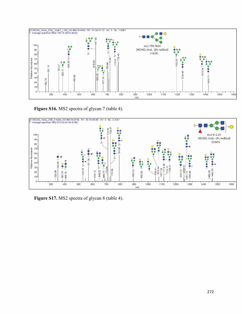
272
Figure S16. MS2 spectra of glycan 7 (table 4).
Figure S17. MS2 spectra of glycan 8 (table 4).

273
Figure S18. MS2 spectra of glycan 9 (table 4).
Figure S19. MS2 spectra of glycan 10 (table 4).

274
Figure S20. MS2 spectra of glycan 11 (table 4).
Figure S21. MS2 spectra of glycan 12 (table 4).

275
Figure S22. MS2 spectra of glycan 13 (table 4).
Figure S23. MS2 spectra of glycan 14 (table 4).

276
Figure S24. MS2 spectra of glycan 15 (table 4).
Figure S25. MS2 spectra of glycan 16 (table 4).

277
Figure S26. MS2 spectra of glycan 17 (table 4).
Figure S27. MS2 spectra of glycan 18 (table 4).

278
Figure S28. MS2 spectra of glycan 19 (table 4).
Figure S29. MS2 spectra of glycan 20 (table 4).

279
Figure S30. MS2 spectra of glycan 21 (table 4).

280
Glycopeptide spectra
Figure S31. CID spectrum of the chymotryptic peptide
DYFPEPVTVSWNSGALTSGVHTFPAVLQSSGLY from the L115N mutant containing
amino acid positions 177, 178 and 182.
Figure S32. CID spectrum of the chymotryptic peptide
DYFPEPVTVSWNSGALTSGVHTFPAVLQSSGLY from the A121N mutant amino acid
positions 177, 178 and 182.

281
Figure S33. CID spectrum of the chymotryptic peptide SGALTSGVHTFPAVLQSSGLY
from Tmab WT amino acid positions 177, 178 and 182.
Figure S34. CID spectrum of the chymotryptic peptide
VDNALQSGNSNESVTEQDSKDSTYSLSSTLTLSK from the Q160N mutant confirming
the mutation at position 160.

282
SPR sensorgrams
Figure S35. Analysed SPR sensorgrams of single cycle kinetic assays of Tmab variants to
surface captured HER2. (A) Herceptin, (B) Tmab WT, (C) L115N, (D) A121N, (E) L177N,
(F) Q178N, (G) L182N, (H) T198N, and (I) Q160N.

283
Figure S36. Analysed SPR sensorgrams of single cycle kinetic assays of Tmab variants to
surface captured FcγR1A. (A) Herceptin, (B) Tmab WT, (C) L115N, (D) A121N, (E)
L177N, (F) Q178N, (G) L182N, (H) T198N, and (I) Q160N.

284
Figure S37. Analysed SPR sensorgrams of single cycle kinetic assays of Tmab variants to
surface captured FcγR2A. (A) Herceptin, (B) Tmab WT, (C) L115N, (D) A121N, (E)
L177N, (F) Q178N, (G) L182N, (H) T198N, and (I) Q160N.

285
Figure S38. Analysed SPR sensorgrams of single cycle kinetic assays of Tmab variants to
surface captured FcγR3A. (A) Herceptin, (B) Tmab WT, (C) L115N, (D) A121N, (E)
L177N, (F) Q178N, (G) L182N, (H) T198N, and (I) Q160N.

286
Supplementary information from Chapter 7: Synthesis and Enhanced Cellular
Uptake In Vitro of Anti-HER2 Multifunctional Gold Nanoparticles
Figure S1. Analysis of the presence of sulfhydryl groups and conservation of intact structure
of Trastuzumab after partial reduction with DTT. (A) UV-Vis spectra of Ellman’s reagent
(DTNB) and of partially reduced antibody after reaction with DTNB. (B) SE-HPLC
chromatogram of intact Trastuzumab and reduced Trastuzumab.

287
Figure S2. SE-HPLC chromatograms of partially reduced Trastuzumab in 1 mM EDTA,
Tmab-vcMMAE in H2O and Tmab-vcMMAE in acetonitrile 10% with formic acid 1%.
Figure S3. AuNP stability upon surface functionalization with Tmab-PEG-SH. Addition of
1% NaCl to citrate capped AuNPs caused aggregation as evidenced by a broad absorption
band in the 700-800 nm range. Tmab-PEG-SH attachment prevented aggregation upon
addition of NaCl.

288
Figure S4. Representative tryptophan fluorescence emission spectra for the estimation of
Trastuzumab:AuNP ratio for 20 nm AuNPs. The estimation is based on the loss of
fluorescence intensity following AuNP surface attachment and centrifugation of the
functionalized gold nanoparticles. The decrease in intensity is assumed to come from
removal of the Trastuzumab monomers attached on AuNPs through nanoparticle
sedimentation. C denotes controls and S the reacted samples. The ratio (R) is calculated as:
𝐷𝐷 = �1 −𝜆𝜆𝑆𝑆𝜆𝜆0� × 𝐹𝐹
Where:
Is = fluorescence intensity of the supernatant after AuNP removal by centrifugation post reaction
I0 = fluorescence intensity of the control (identical original concentration of antibody as that in the
reaction)
F = Tmab:AuNP molar ratio in the reaction

289
Figure S5. Representative sensorgrams of (A) Trastuzumab, (B) Tmab-PEG-SH 25X and
(C) Tmab-vcMMAE binding to recombinant HER2 receptor. The sensorgram is shown in
red and the derived 1:1 binding model fit in black.

290
Figure S6. Representative SKBR-3 cell growth curves employed to analyse growth rate
inhibition activity of MMAE-containing agents at equivalent MMAE concentrations. (A)
Cell confluence change over time monitored every 2 hours. Data are reported as means ±
SD. (B) Representative images of SKBR-3 cells obtained with the Incucyte® ZOOM Live-
cell Analysis System showing decrease in confluence and morphological changes for
MMAE treated cells.