quantitative proteomic strategies for the identification of microrna targets
TRANSCRIPT

10.1586/EPR.12.49 549ISSN 1478-9450© 2012 Expert Reviews Ltdwww.expert-reviews.com
Review
MicroRNAs (miRNAs) are short noncoding RNAs found in eukaryotic cells. A miRNA molecule has very few nucleotides (~19–25 nucleotides in length) compared with other RNAs. Since their initial discovery in Caenorhabditis elegans in the early 1990s, miRNA-dependent gene regulation has been widely investigated in various eukaryotic organisms [1–3]. Most miRNAs are initially transcribed by RNA polymerase II as capped and polyadenylated primary miRNA transcripts (pri-miRNA). Once formed in the nucleus, the enzymes Drosha and Pasha cleave pri-miRNAs to a stem-loop precursor structure (precursor [pre]-miRNA) of approximately 60–70 nucleotides in length [4]. Pre-miRNAs are exported from the nucleus into the cytoplasm by binding to the nucleocytoplasmic transport factor Exportin-5, where they are cleaved by the RNase III enzyme Dicer to release mature miRNAs that are RNA duplexes of approximately 22-nt in length [5]. One strand of the duplex (miRNA strand) is selectively loaded into the miRNA-induced
silencing complex (miRISC), which contains the proteins argonaute (Ago) and trinucleotide repeat-containing 6 (Tnrc6) [6–9]. The other strand is usually degraded. Mature miRNAs work essentially by one of two modes. In plants, miRNAs base pair with mRNA targets by precise or nearly precise complementarity, and directly cleave or destruct the target mRNA through a mechanism involving the RNAi machinery [10,11]. In animals, on the other hand, most miRNAs bind to partly complementary sequences located on their target mRNAs and mediate the repression of protein synthesis [12]. Due to this mode of recognition, miRNA may regulate many of its target mRNAs at the translational level without affecting mRNA abundance [13]. Therefore, miRNAs are post-transcriptional regulators that regulate gene expression by either translational repression or degradation of a target mRNA. Up to now, more than 1800 miRNAs have been found in humans [101] and are predicted to regulate the expression of 30–50% of the genome [14]. By
Chongyang Li1,2, Qian Xiong1, Jia Zhang1,3, Feng Ge*1 and Li-Jun Bi31Institute of Hydrobiology, Chinese Academy of Sciences, Wuhan, 430072, China 2Graduate School, Chinese Academy of Sciences, Beijing 100039, PR China 3Key Laboratory of Non-coding RNA, Institute of Biophysics, Chinese Academy of Sciences, Beijing 100101, China*Author for correspondence: Tel.: +86 27 687 80500 Fax: +86 27 687 80500 [email protected]
MicroRNAs (miRNAs) are small noncoding RNAs, approximately 22 nucleotides in length, found in diverse organisms. They have emerged in recent years as key regulators of a broad spectrum of cellular functions. miRNAs regulate biological processes by inducing translational inhibition and degradation of their target mRNAs through base pairing to partially or fully complementary sites. In the field of miRNA research, the identification of the targets of individual miRNAs is of utmost importance. Our understanding of the molecular mechanisms by which individual miRNAs modulate cellular functions will remain incomplete until a full set of miRNA targets is identified and validated. Since a miRNA may regulate many of its targets at the translational level without affecting mRNA abundance, proteomic methods are best suited for revealing the full spectrum of miRNA targets. Quantitative proteomics is emerging as a powerful toolbox for identifying miRNA targets and for quantifying the contribution of translational repression by miRNAs. In this review, the authors summarize the quantitative proteomic approaches that have been employed for identification of miRNA targets and discuss current challenges as well as possible ways of overcoming them.
Quantitative proteomic strategies for the identification of microRNA targetsExpert Rev. Proteomics 9(5), 549–559 (2012)
Keywords: 2D difference gel electrophoresis • isobaric tags for relative and absolute quantification • isotope-coded affinity tags • microRNAs • pulsed stable-isotope labeling by amino acids in cell culture • quantitative proteomics • stable-isotope labeling by amino acids in cell culture
Expert Review of Proteomics
2012
9
5
549
559
© 2012 Expert Reviews Ltd
10.1586/EPR.12.49
1478-9450
1744-8387
Quantitative proteomic strategies for the identification of microRNA targets
Li, Xiong, Zhang, Ge & Bi
Expert Rev. Proteomics
Review
For reprint orders, please contact [email protected]
Expert Rev. Proteomics 9(5), (2012)550
Review
affecting gene regulation, miRNAs are known to control a wide range of biological functions such as proliferation, differentiation, migration, apoptosis, antiviral defense and metabolism [1–3]. The expression of miRNAs is cell type and tissue-specific in human. A number of miRNAs have been implicated in some types of cancer and have the potential to become therapeutic targets or diagnostic and prognostic biomarkers [15,16]. It has been reported that a single miRNA could regulate hundreds of mRNAs and a single mRNA may be regulated by multiple miRNAs [17]. However, very few miRNA targets have been identified by experimental methods.
miRNA target identificationIn the field of miRNA research, the mechanism underlying the potential role of miRNAs has still not been resolved. The first step toward understanding the functions of a particular miRNA is the identification of its mRNA targets. This step is one of the most important and toughest goals and requires different tech-nical approaches [18]. Some computational programs based on sequence alignment, including miRBase [19], TargetScan [14] and PicTar [20], have been applied to predict targets of miRNAs. These programs are strong tools for identifying the putative targets of miRNAs and have contributed significantly to the development of miRNA research. However, these computational programs have their inherent limitations. For example, in animals, the comple-mentarity between miRNAs and mRNA targets is usually not perfect, making it inaccurate to predict target sites. The three most commonly used bioinformatic target prediction tools such as miRBase [19], TargetScan [14] and PicTar [20], search for miRNA targets exclusively in mRNA 3 -́untranslated regions and do not take into account such factors as functional targeting within the 5́ -untranslated region and protein coding region [13]. Moreover, most programs do not consider the coexpression of miRNA and target, which is proposed to be an effective way of improving predictions [21]. Therefore, many predicted targets may be false and many genuine targets can be missed [22]. It has been estimated that the false positive rate of these programs is approximately 24–70% [23–25]. Furthermore, these various miRNA target predic-tion programs use different algorithms of targeting and produce rather different lists of predicted targets [13]. This underscores the requirement for experimental techniques such as proteomics [26] or argonaute (AGO) coimmunoprecipitation (IP) [27,28] to identify targets with high validity. Details about computational analysis of potential miRNA targets have been reviewed exten-sively [29,30] and are beyond the scope of this review. Instead, proteomic identification of miRNA targets will be focused on.
Experimental approaches are essential to identify genuine mRNA targets and understand miRNA functions. Since mi RNAs can bind to complementary sequences on target mRNAs and these targets are repressed at the mRNA and/or protein expres-sion level, the dysregulation of a miRNA may lead to specific alterations in the transcriptome and/or proteome. Several high-throughput methods have been developed to measure such alterations at a global level. The fact that miRNAs down regulate the level of a number of their target mRNAs has been used to identify such targets through gene expression analysis [31,32].
However, targets that are repressed at the protein level without being affected at the mRNA level would be missed. For exam-ple, Leivonen et al. reported that among proteins repressed by miR-193b, only a minority (13%) showed a similar effect at the mRNA level according to microarray gene expression data [33]. Thus, proteomic methods may turn out to be necessary to reveal the full spectrum of miRNA targets. In the following section, how quantitative proteomic approaches have been applied to identify mRNA targets are explained.
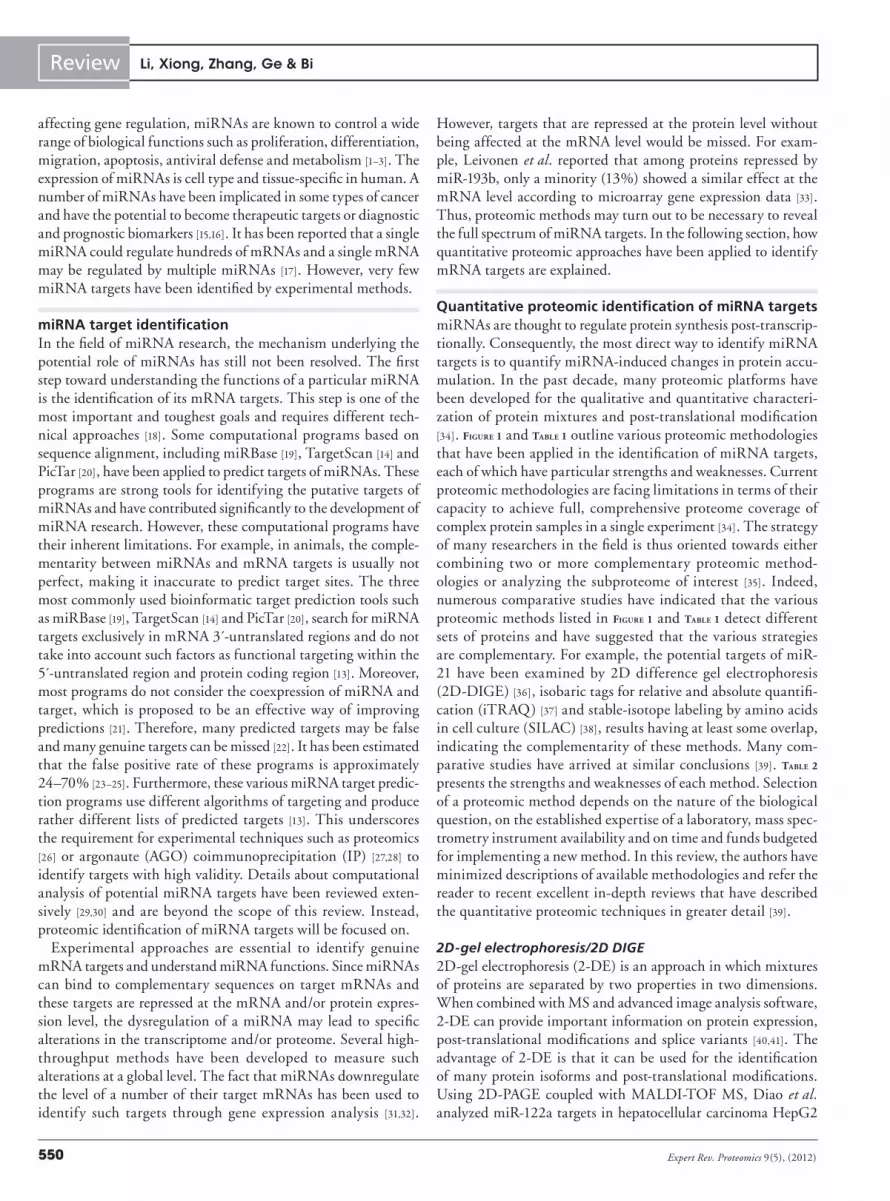
Quantitative proteomic identification of miRNA targetsmiRNAs are thought to regulate protein synthesis post-transcrip-tionally. Consequently, the most direct way to identify miRNA targets is to quantify miRNA-induced changes in protein accu-mulation. In the past decade, many proteomic platforms have been developed for the qualitative and quantitative characteri-zation of protein mixtures and post-translational modification [34]. Figure 1 and Table 1 outline various proteomic methodologies that have been applied in the identification of miRNA targets, each of which have particular strengths and weaknesses. Current proteomic methodologies are facing limitations in terms of their capacity to achieve full, comprehensive proteome coverage of complex protein samples in a single experiment [34]. The strategy of many researchers in the field is thus oriented towards either combining two or more complementary proteomic method-ologies or analyzing the subproteome of interest [35]. Indeed, numerous comparative studies have indicated that the various proteomic methods listed in Figure 1 and Table 1 detect different sets of proteins and have suggested that the various strategies are complementary. For example, the potential targets of miR-21 have been examined by 2D difference gel electrophoresis (2D-DIGE) [36], isobaric tags for relative and absolute quantifi-cation (iTRAQ) [37] and stable-isotope labeling by amino acids in cell culture (SILAC) [38], results having at least some overlap, indicating the complementarity of these methods. Many com-parative studies have arrived at similar conclusions [39]. Table 2 presents the strengths and weaknesses of each method. Selection of a proteomic method depends on the nature of the biological question, on the established expertise of a laboratory, mass spec-trometry instrument availability and on time and funds budgeted for implementing a new method. In this review, the authors have minimized descriptions of available methodologies and refer the reader to recent excellent in-depth reviews that have described the quantitative proteomic techniques in greater detail [39].
2D-gel electrophoresis/2D DIGE2D-gel electrophoresis (2-DE) is an approach in which mixtures of proteins are separated by two properties in two dimensions. When combined with MS and advanced image analysis software, 2-DE can provide important information on protein expression, post-translational modifications and splice variants [40,41]. The advantage of 2-DE is that it can be used for the identification of many protein isoforms and post-translational modifications. Using 2D-PAGE coupled with MALDI-TOF MS, Diao et al. analyzed miR-122a targets in hepatocellular carcinoma HepG2
Li, Xiong, Zhang, Ge & Bi

551www.expert-reviews.com
Review
2D-D
IGE
Tota
l cel
l lys
ate
Den
atur
e &
Dig
est
miR
-223
, -1,
-12
4,-1
81, -
7, -
21, -
373,
-143
, -15
5, -
200s
miR
-1, -
16, -
30a,
-155
, let
-17b
, -34
a
Tota
l cel
l lys
ate
Tryp
sin
dige
st
Tryp
sin
dige
stH
/L r
atio
Intensity
Intensity
H/M
rat
io
Tota
l cel
l lys
ate
Mix
ext
ract
s 1:
1
Mix
ext
ract
s 1:
1
Cy3
labe
l Mix
, run
2D
gel
Mix
Imag
e an
alys
is
Spo
ts p
icki
ng,
in-g
el d
iges
tion
MS
iden
tific
atio
n
2D c
hrom
atog
raph
icse
para
tion
of p
eptid
es
Qua
ntita
tion
by M
S/M
Sm
/zQ
uant
itatio
n by
MS
m/z
Qua
ntita
tion
by M
S
Cy5
labe
l11
5 or
117
labe
l11
4 or
116
labe
l
Con
trol
miR
-21,
-19
3b
iTR
AQ
Con
trol
Con
trol
Con
trol
SIL
AC
pSIL
AC
miR
-372
,-2
1, -
29a
Ligh
tLi
ght
Ligh
tH
eavy
Hea
vyH
eavy
Fig
ure
1. K
ey p
rote
om
ic m
eth
od
olo
gie
s ap
plie
d in
th
e id
enti
fica
tio
n o
f m
iRN
A t
arg
ets.
2D
-DIG
E: 2
D d
iffe
renc
e in
-gel
ele
ctro
phor
esis
; H: H
eavy
; iTR
AQ
: Iso
baric
tag
s fo
r re
lativ
e an
d ab
solu
te q
uant
itatio
n; L
: Lig
ht; M
: Med
ium
; MS:
Mas
s sp
ectr
omet
ry;
pSIL
AC
: Pul
sed
SILA
C; S
ILA
C: S
tabl
e is
otop
e la
belin
g of
am
ino
acid
s in
cel
l cul
ture
.
Quantitative proteomic strategies for the identification of microRNA targets

Expert Rev. Proteomics 9(5), (2012)552
Review
cells [42]. They developed a lentiviral vector (Lenti-miR-122a) for the overexpression of miR-122a in HepG2 cells. Changes at the protein level were compared between HepG2 cells infected with control vector and Lenti-miR-122a. Ten proteins were identified that were differentially expressed on miR-122a overexpression. Among them, peroxiredoxin 2 (PRDXII) was validated as one of the direct targets of miR-122a in HepG2 cells using western blotting and Luciferase reporter assays. Recently, Kanzaki et al. analyzed miR-17–92 target profiles in small-cell lung cancer SBC-3 cells using 2-DE [43]. The expression of endogenous miR-19a, miR-20a and miR-92-1 was knocked down using anti-miRNA locked nucleic acid. Using 2-DE and MS, the group identified 112 putative targets of miR-17–92 in SBC-3 cells. Further validation studies based on luciferase assays, western blotting and quantitative reverse transcriptase(RT)-PCR demonstrated that miR-92-1 regulates the RAB14 gene at the translational level without affecting the mRNA level. These reports suggest that 2-DE is valuable for identifying miRNA targets. However, 2-DE has some inherent limitations, especially with respect to hydrophobic and alkaline proteins that are often underrepresented in 2-DE. In addition, this technique has a low dynamic range and suffers from gel-to-gel variability [40].
As an improvement, 2D-DIGE overcomes some of the problems associated with traditional 2-DE and allows more accurate and
sensitive quantitative proteomics studies [44]. Several studies using 2D-DIGE to identify miRNA targets have recently been reported. Muniyappa et al. examined downstream targets of miR-29a using 2D-DIGE [45]. They conducted comparative proteomic profiling of lysates from lung carcinoma DLKP-A cells transfected transiently with exogenous pre-miR-29a or anti-miR-29a and cells treated with negative controls, identifying over 100 differentially-regulated proteins. Comprehensive studies using short interfering RNA to knock down several candidate proteins revealed that miR-29a inf luences the invasive and proliferative capacity of cancer cells upon regulation of RAN (RAS-related nuclear protein). Schramedei et al. took a proteomic approach to identify proteins downregulated upon enhanced miR-21 expression in human prostate carcinoma LNCaP cells [36]. Using 2D-DIGE, they identified 16 proteins either directly or indirectly suppressed by enhanced miR-21 expression. Further functional analysis revealed that tumor suppressor acidic nuclear phosphoprotein 32 family member A (ANP32A) is one of direct targets of miR-21 and downregulation of ANP32A contributes to various miR-21-mediated effects. Recently, Lai et al. performed a comparative proteomic analysis of non-
small-cell lung cancer CL 1–0 cells expressing miRNA-372 and/or its vector alone [46]. Using 2D-DIGE and Q-TOF MS/MS, they were able to identify 19 downregulated proteins. Most of these proteins were associated with lung cancer and tumor invasion and some of them may be used as potential diagnostic biomarkers of non-small-cell lung cancer.
While 2-DE/DIGE-based method has some inherent limita-tions and there has been significant progress in non-gel-based methods for coupling fractionation methods directly online with MS analysis, 2-DE/DIGE remains an important technique for conducting proteomic studies. Furthermore, recent studies dem-onstrated that miR-21 can regulate STAT3 pathway and its phos-phorylation [38,47]. Therefore, it is expected that 2-DE/DIGE may play a role in the identification of post-translational modifications caused by miRNAs. Though 2-DE/DIGE has its advantages and disadvantages, there is no doubt that it will remain an essential technique for the characterization of proteomes for many years to come.
Isotope-coded affinity tags/iTRAQIsotope-coded affinity tags (ICATs), introduced by Gygi et al. in 1999, is an in vitro strategy for quantitative proteomics [48]. The ICAT reagent is a sulphydryl-directed alkylating agent, which
Table 1. Published studies in which quantitative proteomic approaches are applied to identify miRNA targets.
Study (year) miRNA(s) Methodology Cells Ref.
Lai et al. (2012) miR-372 2D-DIGE Lung adenocarcinoma [46]
Kaller et al. (2011)Chen et al. (2011)
miR-34a pSILACICAT
Colorectal cancerNeuroblastoma
[64][50]
Leivonen et al. (2011) miR-193b iTRAQ Breast cancer [33]
Yang et al. (2009)Schramedei et al. (2011)Xiong et al. (2012)
miR-21 iTRAQ2D-DIGESILAC
Breast cancerProstate cancerMultiple myeloma
[37][36][38]
Diao et al. (2010) miR-122a 2-DE HCC [42]
Chou et al. (2010) miR-7 SILAC Lung cancer [61]
Kanzaki et al. (2011) miR-17–92 2-DE Lung cancer [43]
Yan et al. (2011) miR-373 SILAC Breast cancer [59]
Muniyappa et al. (2009) miR-29a 2D-DIGE Lung cancer [45]
Vinther et al. (2006) miR-1 SILAC Cervical cancer [56]
Yang et al. (2010) miR-143 SILAC Pancreatic cancer [57]
Lössner et al. (2011) miR-155 SILAC 293T cells [60]
Baek et al. (2008) miR-223, -1, -124, -181
SILAC Cervical cancer [25]
Korpal et al. (2011) miR-200s SILAC Breast and bladder cancer
[58]
Selbach et al. (2008) miR-1, -16, -30a, -155 and let-7b
pSILAC Cervical cancer [62]
2D-DIGE: 2D difference gel electrophoresis; 2-DE: 2D-gel electrophoresis; HCC: Hepatocellular carcinoma; ICAT: Isotope-coded affinity tag; iTRAQ: Isobaric tags for relative and absolute quantitation; pSILAC: Pulsed SILAC; SILAC: Stable-isotope labeling by amino acids in cell culture.
Li, Xiong, Zhang, Ge & Bi

553www.expert-reviews.com
Review
can be used to label cysteine residues on the protein. It comprises a reactive group (i.e., thiol), an affinity group (i.e., biotin) and a linker group carrying light or heavy isotopes. The protein reactive groups (i.e., cysteine residues) are labeled separately with either light or heavy reagent, and then the labeled proteins are com-bined, digested by enzymes. The labeled peptides are enriched, quantified and identified by LC-MS/MS [49]. Since the first appli-cation of ICAT reagents, a number of key technological advances have been introduced to improve the recovery of peptides and chromatographic properties [49]. Using ICAT-based quantitative technology, Chen et al. examined global protein changes caused by miR-34a in neuroblastoma IMR32 cells [50]. ICAT analysis of miR-34a transfected IMR32 cells resulted in identification of 1495 unique proteins, of which 143 were upregulated and 192 proteins were downregulated by miR-34a compared with nega-tive control-transfected cells. They further validated that YY1 is a direct target of miR-34a. Since YY1 is a ubiquitous transcription factor that negatively regulates p53 and plays an essential role in cancer biology, the elucidation of the role of YY1 in the p53-miR-34a regulatory loop may provide novel insights into the tumor suppressive function of miRNA-34a.
However, ICAT requires the laborious biotin–avidin enrich-ment process and incurs nonspecific binding with such enrich-ment. As cysteine is a rare amino acid, its derivatization has the advantage of simplifying peptide mixtures since only tagged peptides are enriched. However, the choice of cysteine makes it impossible to detect changes in the approximately 14% of pro-teins that do not contain cysteine residues in Escherichia coli [51]. Furthermore, quantification is based on a very limited number
of peptides for each protein, which reduces the reliability of the measurement [51]. Hence, iTRAQ has been established as a more comprehensive and efficient method for proteomic quantifica-tion [52]. This method was first developed by Ross et al. in 2004 [52] and was subsequently commercialized by Applied Biosystems (CA, USA). The iTRAQ reagent comprises a reporter group (variable mass of 114–117 Da or 113–121 Da), a balance group and an amino-reactive group, which specifically reacts with the NH
2-terminal and side-chain lysine amino groups. The reporter
groups carry stable isotopes with different combinations of iso-topes allowing the simultaneous labeling of up to eight samples using the 8-plex kit. The iTRAQ reagents can be used to perform both relative quantification and absolute quantification by adding an internal standard peptide. The advantages of iTRAQ include: the identification of low-abundance proteins in complex samples and quantification of up to eight samples in parallel, which at least overcomes the issue of run-to-run reproducibility. Therefore, it holds the greatest promise for biomarker discovery and can easily be applied to proteomic identification of miRNA targets [53].
Using an iTRAQ based quantitative proteomics strategy, the Pandey group carried out global proteomic profiling to identify potential targets of miR-21 in breast cancer cells [37]. By knock-ing down the expression of endogenous miR-21 in MCF-7 breast cancer cells, they identified 58 potential targets of miR-21. Using luciferase assays, they demonstrated that six of these candidate targets are likely direct targets of miR-21. Importantly, mRNA-based validation experiments clearly demonstrated that miR-21 regulates many of its targets through translational inhibition without affecting mRNA abundance, emphasizing the utility of
Table 2. Comparison of various quantitative proteomic approaches available to identify miRNA targets.
Methodology Advantages Limitations Ref.
2-DE Established technology; resolves same proteins with different PTMs and splice variants
Low proteomic coverage; poor reproducibility; low dynamic range; systematic bias against detecting proteins with extreme MW and pI values
[40,41]
2D-DIGE More accurate and sensitive than 2-DE; eliminating gel-to-gel variation
Requires expensive reagents and instruments; labeling variability
[44]
ICAT Complex samples are simplified; MS-based quantitation can reduce costly analysis time
Low confidence and poor statistics; laborious biotin–avidin enrichment process; incomplete labeling; cannot analyze more than two samples at a time; difficulty detecting acidic proteins or those with few or no cysteines
[48,49]
iTRAQ High confidence and quantitation statistics; identification of low-abundance proteins in complex samples; quantification of up to eight samples in parallel
Fractionation required; costly in terms of MS time; requires expensive reagents; variability in labeling efficiencies; complex sample preparation procedures; requires high sample concentration
[52,53]
SILAC The availability of several labels; a predictable mass shift; less prelabeling variability; greater number of proteins measured
Only applicable to cultured cells and cannot be used for autotrophic organisms, tissue and body fluid samples; conversion of Arg to Pro leading to potential inaccuracies and complicating the final quantitation
[54,55]
pSILAC Permitting the quantification of differences in protein translation and synthesis rates; providing high sensitivity
Relatively expensive; not practical for analyzing biological samples that cannot be grown in culture; time consuming
[62,63]
2D-DIGE: 2D difference gel electrophoresis; 2-DE: 2D-gel electrophoresis; ICAT: Isotope-coded affinity tag; iTRAQ: Isobaric tags for relative and absolute quantitation; MS: Mass spectrometry; MW: Molecular weight; pI: Isoelectric point; pSILAC: Pulsed SILAC; PTM: Post-translational modification; SILAC: Stable-isotope labeling by amino acids in cell culture.
Quantitative proteomic strategies for the identification of microRNA targets

Expert Rev. Proteomics 9(5), (2012)554
Review
proteomic approaches for identifying miRNA targets. Using the same iTRAQ strategy, Leivonen et al. found that the expression of 39 out of 743 identified proteins was repressed by miR-193b in breast cancer MCF-7 cells. Some of the significantly repressed proteins were selected for validation experiments [33]. Of these, only a minority (13%) showed a similar effect at the mRNA level according to microarray gene expression data, illustrat-ing the importance of detecting miRNA targets at the protein level. Functional RNAi assays showed that specific combina-tions of knock-downs of these key target genes could mimic the phenotypic effects of miR-193b overexpression in MCF-7 cells. However, iTRAQ technology has some disadvantages such as variability in labeling efficiencies, complex sample preparation procedures, and a requirement for high sample concentration [53]. Thus, the iTRAQ technology is more suitable as a primary screen-ing method for miRNA target identification rather than for target verification or validation.
Stable-isotope labeling by amino acids in cell cultureA good alternative to iTRAQ and other isobaric tagging tech-niques is SILAC. SILAC is an easy, reliable and robust MS-based differential proteomics technique and has become a common technique in quantitative proteomics [54]. In SILAC, isotopically labeled amino acids are added to the growth medium and cells are cultivated in media containing either normal amino acids (light) or amino acids labeled with heavy isotopes (heavy). Then, cells are pooled, digested and analyzed by tandem MS [55]. The most commonly used amino acids are lysine and arginine. They can generate tags with different mass shifts by various combinations of 13C, 15N and 3H, allowing comparison of proteomes of either two or three different cell populations in a single SILAC experi-ment [55]. In addition, SILAC has the advantages of incorporating the isotope tags before sample preparation, reducing potential biases due to separate handling of the samples [55]. An important drawback of SILAC is that it relies on cells growing in culture in adapted media and it is not effective for autotrophic organisms such as bacteria [53]. Currently, SILAC has proved useful for quan-titative proteomics in the field of miRNA target identification in cell culture models.
Vinther et al. were the first to publish a SILAC- or proteom-ics-based study on the identification of miRNA targets [56]. In this study, a SILAC-based quantitative proteomic strategy was performed to systematically identify potential targets of miR-1 in human cervical cancer HeLa cells. They compared control-transfected HeLa cells with SILAC labeled HeLa cells trans-fected with a specific miR-1 duplex. Expression of 12 proteins (from a set of 504 detected proteins) was repressed by miR-1 transfection. Their findings clearly showed that a single miRNA can regulate many proteins and that SILAC can be used for miRNA target identification. In this study, the influence of miRNAs on protein output was limited to a medium-size pro-teomics analysis involving the detection of 504 proteins, but as technologies have improved this has become less problematic. Bartel and coworkers were able to improve the proteome cover-age by examining more than 2000 distinct proteins, although
this is still some way from a full proteome [25]. They performed SILAC experiments with three miRNAs miR-1, miR-124 and miR-181 in HeLa cells and examined the effects of the mir-223 gene knockout in mouse neutrophils. Bone marrow hematopoietic progenitors were isolated from wild-type and mir-223-deficient mice and cultured in SILAC media differentiating into mature neutrophils in vitro. SILAC results demonstrated that a single miRNA is capable of repressing the production of hundreds of proteins and that individual miRNAs may act as rheostats to make fine-scale adjustments to protein output. In addition to providing information on specific targets, identification of this large cohort of genes regulated by a single miRNA might help us learn more about the sequence features that define miRNA targets and thus improve the accuracy of computational target prediction. Using SILAC-based quantitative proteomics, the Pandey group compared miR-143 mimic- to control-transfected pancreatic cancer MiaPaCa2 cells [57]. They identified 94 puta-tive targets of miR-143. Luciferase reporter assays for 34 of these showed that ten were likely to be direct miR-143 targets. These data coupled with results from gene expression profiling of the same cells clearly show that miR-143 regulates many of its targets at the translational level without affecting mRNA abundance, confirming the importance of quantitative proteomic approaches for identifying miRNA targets. Korpal et al. globally identified the genes regulated by miR-200s [58]. They compared protein and gene expression differences between miR-200s-overexpressing mouse breast cancer 4TO7 cells with negative control cells. Of the 3769 proteins quantified by MS, 1562 protein abundance values were cross referenced with gene expression data. Of these, nine showed reduced abundance at both the mRNA and protein level. Further functional and clinical correlation studies revealed that miR-200s promotes metastatic colonization partly through direct targeting of Sec23a. Their findings suggest a pleiotropic role of miR-200s in promoting metastatic colonization of breast cancer. Furthermore, Yan et al. revealed that miR-373 regulates 335 proteins in human breast cancer MCF-7 cells and TXNIP and RABEP1 are the direct targets of miR-373 [59]. Lössner et al. identified 46 putative miR-155 target proteins and CKAP5 was confirmed as a new target of miR-155 in HEK293T cells [60]. Chou et al. demonstrated that ERF is a direct target of miR-7 in lung cancer CL1-5 cells [61]. Xiong et al. found that the expression of 178 proteins was upregulated significantly by miR-21 inhibi-tion in human multiple myeloma U266 cells and confirmed the protein inhibitor of activated STAT3 as a direct miR-21 target [38].
However, since different proteins have different turnover times and miRNAs in most cases only cause modest decreases in pro-tein translation, miRNA-mediated regulation of proteins with long half-lives may not be detected by measuring steady-state protein levels using SILAC. Thus, it is necessary to quantify the differences in protein translation rates caused by miRNAs on a proteome-wide scale.
Pulsed SILACA new variant of SILAC, pulsed SILAC (pSILAC), has been devised to quantify the differences in protein translation rates
Li, Xiong, Zhang, Ge & Bi

555www.expert-reviews.com
Review
caused by miRNAs [62]. As shown in Figure 1, in the pSILAC method, cells are first cultivated in standard growth medium with the normal light (L) amino acids. Concomitantly with differen-tial treatment, cells are transferred to culture medium contain-ing heavy (H) isotope-coded amino acids (13C
615N
4-L-arginine
and 13C615N
2-L-lysine; in red) or medium-heavy (M) isotopes-
coded amino acids (13C6-L-arginine and 2H
4-L-lysine; in green).
Therefore, cells are ‘pulse’ labeled since all newly synthesized proteins incorporate either the ‘heavy’ or the ‘medium heavy’ amino acids. Two cell populations are mixed and analyzed by MS at different time points. The abundance ratio of ‘heavy’ versus ‘medium heavy’ peptides reflects differences in translation of the corresponding proteins. Only intensity differences between newly synthesized proteins (medium-heavy and heavy) are considered. Hence, pSILAC measures changes in the amount of newly syn-thesized proteins between two samples over the measurement time after the pulse [63]. Therefore, pSILAC is the best technique for detecting primary miRNA targets. Using this approach, Rajewsky and colleagues identified a substantial number of targets of five miRNAs (miR-1, miR-155, miR-16, miR-30a, let-7b) in HeLa cells [62]. They performed transfections to individually overex-press five human miRNAs (miR-1, miR-155, miR-16, miR-30a and let-7b) in HeLa cells. Changes in protein production were measured by pulse-labeling 8 h post-transfection over a time period of 24 h. In total, they measured changes in the produc-tion of approximately 5000 proteins in HeLa cells. Their results showed that pSILAC can be used for assessing the early effects of miRNAs on rates of protein synthesis and for quantifying the contribution of translational repression by miRNAs. Using the same approach, Kaller et al. combined pSILAC and microarrays to identify miR-34a-induced changes in protein and mRNA expression [64]. They quantified the de novo protein synthesis of 1206 proteins after overexpression of miR-34a in colorectal cancer SW480 cells. In accordance with a previous pSILAC study of miRNA-mediated changes in protein expression [62], overexpres-sion of miR-34a caused moderate changes in protein translation. Therefore, the filtering criteria established in static abundance studies (fold change >2) cannot be applied. Of these proteins, 228 (~19%) were differentially regulated with log2 fold changes ≤ −0.3 or ≥0.3. These two reports have provided a wealth of data on potential miRNA targets and clues for future research directions. Furthermore, pSILAC has also been applied to determine transla-tion rates [63] and identify new proteins in species for which only limited sequence information exists [65]. However, this method is relatively expensive, time consuming and not practical for analyz-ing biological samples that cannot be grown in culture, such as tissues or body fluids.
Label-free quantitative proteomicsMore recently, researchers are increasingly turning to label-free quantitative proteomics techniques for faster, cleaner and simpler results. MS-based label-free quantitative proteomics is generally based on two general categories [66]. In the first are the meas-urements of changes in chromatographic ion intensity such as peptide peak areas or peak heights. The second is based on the
spectral counting of identified proteins after MS/MS analysis. Peptide peak intensity or spectral count is measured for indi-vidual LC-MS/MS or LC/LC-MS/MS runs and changes in pro-tein abundance are calculated via a direct comparison between different analyses [66]. Compared with isotope-labeling methods, label-free experiments need to be carefully controlled, due to pos-sible errors caused by run-to-run variations in performance of LC and MS [39]. However, the improved chromatographic retention time calculators, high resolution mass spectrometer and com-mercially available data processing software have greatly improved the reliability and accuracy of label-free measurements [39]. The rapid development of label-free quantitative proteomic techniques has provided fast and low-cost measurement of protein expres-sion levels in complex biological samples. These techniques, though not being utilized yet in the field of miRNA research, are showing promise for miRNA research. It is expected that these techniques will provide rigorous, powerful tools for analyzing miRNA-induced protein changes in the next 5 years.
Expert commentary & five-year viewmiRNAs are important regulators of cell growth, differentiation and apoptosis, and dysregulation of miRNA function may lead to certain diseases [3]. Identification of miRNA targets is central to understanding the biological roles of miRNAs and remains a fundamental challenge [67]. Strategies based on detecting mRNA expression changes have been used to identify miRNA targets [68]. Since some genes may be repressed at the protein level with-out being affected at the mRNA level, such mRNA approaches may be inherently limited and proteomic methods may turn out to be necessary to reveal the full spectrum of miRNA targets [26]. As listed in Table 1, different proteomic strategies have been used to define and identify miRNA targets in a more or less comprehensive manner.
There is general consensus that transcriptomics and proteomics provide different but complementary information about miRNA regulated genes [13,69,70]. Genomic profiling is comprehensive since almost the entire genome of commonly studied species can be completely arrayed, though gene annotations are still not com-plete. On the other hand, proteomic technologies with supportive analytical and biochemical methods can provide information about protein features such as protein expression, protein–protein interactions and post-translational modifications that genom-ics cannot offer. Combining genomic and proteomic strategies provides a powerful toolbox for the identification of miRNA targets. The growing evidence of poor correlations between mRNA and protein abundance further suggests that these two approaches, mRNA and protein profiling, should be applied in parallel to globally decipher miRNA targets [13]. More proteomic methods should therefore be developed in the future to com-plement present transcriptome strategies. Wider availability of experimentally-validated miRNA targets and their action mech-anisms will no doubt improve the accuracy of computational target prediction. However, on their own, data generated from these techniques have limited usefulness. The advent of genomic and proteome microarrays has made gleaning information from
Quantitative proteomic strategies for the identification of microRNA targets

Expert Rev. Proteomics 9(5), (2012)556
Review
biological systems easy and quick, but this mountain of data have yet to be fully mined and validated. Integration of infor-mation from these sources using powerful analytical computing platforms, open-source databases, and advances in technology represents the next step in the identification and application of novel miRNA targets.
It is the authors’ belief that proteomics has become an indispen-sable tool for the identification of miRNA targets. However, there are still challenges ahead. Among the most important ones is that current quantitative proteomic techniques must be improved so that they can generate more sensitive and reproducible data. Given the increasing sensitivity of modern mass spectrometers, it is antici-pated that such technical issues will be overcome in the near future. There is also a need to standardize and validate the protocols that are developed, and to maintain rigorous quality control. Currently, it remains problematic to avoid false targets and distinguish pri-mary and secondary miRNA targets. As far as we can see, there are at least two ways to overcome these problems. First, the identified target genes should be validated by using extensive experimental validation or on computational predictions, such as appropriate statistical filtering, western blotting (proteins), RT-PCR (mRNA), luciferase report assays and AGO-IP. Without subsequent down-stream functional analyses, proteomic experiments merely provide lists of protein data with little practical value. Second, pSILAC provided a powerful means to identify miRNA targets and to distinguish between primary and secondary miRNA targets [26]. Given the increasing sensitivity of modern mass spectrometers, we expect that there will be many opportunities to apply cutting-edge proteomic technologies to discriminate between primary and secondary miRNA targets in the next 5 years.
A single miRNA is capable of downregulating the abundance and/or translation of many mRNAs. Compounding the complex-ity of miRNA regulation, multiple miRNAs can act together on individual mRNAs to produce diverse effects on protein produc-tion [17]. It is expected that in addition to identifying individ-ual miRNA targets, more and more proteomics-based miRNA research will focus on the exploration of miRNA-regulatory path-ways. Thus, it is necessary to combine multiple strategies to obtain a comprehensive description of the miRNA-regulatory network with high-confidence. To obtain a comprehensive system-wide view of miRNA regulation, multiple levels of regulation should ideally be studied in parallel. This can be achieved by combin-ing quantitative proteomics with other omics-based approaches, including transcriptomics, metabolomics and modificomics. Since there are significant ongoing efforts, many new methodologies
from other scientific fields may be translated for use in miRNA research in the future. Many of the present studies can be consid-ered as proof-of-principle and they provide a glimpse of methods that may become available in the next 5 years. In combination with other systems-biology-based approaches, proteomics can be suc-cessfully applied on a global scale to decipher miRNA targets and expand our current knowledge of miRNA-regulatory pathways.
Due to the early stage of research, current proteomic studies on miRNAs have largely relied on established cell lines. No doubt, results derived from cell lines can guide future investigations on model organisms and further translational research; the interpreta-tion of these results, however, needs to be cautious as not all results from cell lines are relevant to entire model organisms. Therefore, it would be desirable to study miRNA targets in whole organ-isms rather than making use of established cell lines that have only a limited potential to reveal miRNA-regulatory mechanisms. Stable isotope labeling of entire model organisms like mice, rats, worms, zebrafish and flies presents an ideal way to use quantitative proteomics for the identification of miRNA targets in vivo [71–74].
In addition to the role of miRNAs in cellular signaling, other regulatory RNAs like long noncoding RNAs (lncRNAs) have emerged as important regulatory factors in mammalian genomics [75]. Differential expression of lncRNAs is becoming recognized as a hallmark feature of cancer; however, functional roles for the vast majority of these unique RNAs are still in question [76]. As new lncRNAs are being discovered at a rapid pace, our under-standing of the molecular mechanisms of lnc RNAs is likely to be enriched and diversified. Proteomic strategies are useful for identifying lncRNA targets and expanding our knowledge of the function of lncRNAs. So far, there are no reports on proteomic studies for the identification of lncRNA targets. We predict that there will be a surge in publications relating proteomic analysis of lncRNAs in the next 5 years. In particular, the identifica-tion of lncRNA targets may provide insightful information on their functions and is one of the most important next steps for understanding the functions of lncRNAs in human cancers.
Financial & competing interests disclosureThis work was supported by the National Basic Research Program of China (973 Program, 2012CB518700), and the Hundred Talents Program of the Chinese Academy of Sciences. The authors have no other relevant affiliations or financial involvement with any organization or entity with a financial interest in or financial conflict with the subject matter or materials discussed in the manuscript apart from those disclosed.
No writing assistance was utilized in the production of this manuscript.
Key issues
• Each individual miRNA may downregulate the abundance and/or translation of many mRNAs and multiple miRNAs can work together on individual mRNA to produce synergistic effects on protein production.
• Identification of miRNA targets is central to understanding the biological roles of miRNAs and remains a fundamental challenge.
• Proteomic methods are essential for identifying genuine miRNA targets. Current quantitative proteomic technologies (2D difference gel electrophoresis, isobaric tags for relative & absolute quantification, stable-isotope labeling by amino acids in cell culture [SILAC] and pulsed SILAC) each have inherent strengths and weaknesses but can be used in complementary ways to reveal the full spectrum of miRNA targets.
Li, Xiong, Zhang, Ge & Bi

557www.expert-reviews.com
Review
ReferencesPapers of special note have been highlighted as:• of interest•• of considerable interest
1 Lee RC, Ambros V. An extensive class of small RNAs in Caenorhabditis elegans. Science 294(5543), 862–864 (2001).
2 Ambros V, Lee RC, Lavanway A, Williams PT, Jewell D. MicroRNAs and other tiny endogenous RNAs in C. elegans. Curr. Biol. 13(10), 807–818 (2003).
3 Bartel DP. MicroRNAs: genomics, biogenesis, mechanism, and function. Cell 116(2), 281–297 (2004).
• ProvidesacomprehensiveoverviewofmicroRNAs(miRNAs).
4 Lee Y, Ahn C, Han J et al. The nuclear RNase III Drosha initiates microRNA processing. Nature 425(6956), 415–419 (2003).
5 Yi R, Qin Y, Macara IG, Cullen BR. Exportin-5 mediates the nuclear export of pre-microRNAs and short hairpin RNAs. Genes Dev. 17(24), 3011–3016 (2003).
6 Gregory RI, Chendrimada TP, Cooch N, Shiekhattar R. Human RISC couples microRNA biogenesis and posttranscriptional gene silencing. Cell 123(4), 631–640 (2005).
7 Rana TM. Illuminating the silence: understanding the structure and function of small RNAs. Nat. Rev. Mol. Cell Biol. 8(1), 23–36 (2007).
8 Kim VN. MicroRNA biogenesis: coordinated cropping and dicing. Nat. Rev. Mol. Cell Biol. 6(5), 376–385 (2005).
9 Wiemer EA. The role of microRNAs in cancer: no small matter. Eur. J. Cancer 43(10), 1529–1544 (2007).
10 Tang G, Reinhart BJ, Bartel DP, Zamore PD. A biochemical framework for RNA silencing in plants. Genes Dev. 17(1), 49–63 (2003).
11 Rhoades MW, Reinhart BJ, Lim LP, Burge CB, Bartel B, Bartel DP. Prediction of plant microRNA targets. Cell 110(4), 513–520 (2002).
12 Berezikov E. Evolution of microRNA diversity and regulation in animals. Nat. Rev. Genet. 12(12), 846–860 (2011).
13 Thomson DW, Bracken CP, Goodall GJ. Experimental strategies for microRNA target identification. Nucleic Acids Res. 39(16), 6845–6853 (2011).
•• SummarizesanddiscussestheexistingexperimentaltechniquesformiRNAtargetidentification.
14 Lewis BP, Burge CB, Bartel DP. Conserved seed pairing, often flanked by adenosines, indicates that thousands of human genes are microRNA targets. Cell 120(1), 15–20 (2005).
15 Ryan BM, Robles AI, Harris CC. Genetic variation in microRNA networks: the implications for cancer research. Nat. Rev. Cancer 10(6), 389–402 (2010).
16 Broderick JA, Zamore PD. MicroRNA therapeutics. Gene Ther. 18(12), 1104–1110 (2011).
17 Wu S, Huang S, Ding J et al. Multiple microRNAs modulate p21Cip1/Waf1 expression by directly targeting its 3´ untranslated region. Oncogene 29(15), 2302–2308 (2010).
18 Thomas M, Lieberman J, Lal A. Desperately seeking microRNA targets. Nat. Struct. Mol. Biol. 17(10), 1169–1174 (2010).
19 Griffiths-Jones S, Saini HK, van Dongen S, Enright AJ. miRBase: tools for microRNA genomics. Nucleic Acids Res. 36(Database issue), D154–D158 (2008).
20 Krek A, Grün D, Poy MN et al. Combinatorial microRNA target predictions. Nat. Genet. 37(5), 495–500 (2005).
21 Ritchie W, Flamant S, Rasko JE. Predicting microRNA targets and functions: traps for the unwary. Nat. Methods 6(6), 397–398 (2009).
22 Alexiou P, Maragkakis M, Papadopoulos GL, Reczko M, Hatzigeorgiou AG. Lost in translation: an assessment and perspective for computational microRNA target identification. Bioinformatics 25(23), 3049–3055 (2009).
23 Bentwich I. Prediction and validation of microRNAs and their targets. FEBS Lett. 579(26), 5904–5910 (2005).
24 Sethupathy P, Megraw M, Hatzigeorgiou AG. A guide through present computational approaches for the identification of mammalian microRNA targets. Nat. Methods 3(11), 881–886 (2006).
25 Baek D, Villén J, Shin C, Camargo FD, Gygi SP, Bartel DP. The impact of microRNAs on protein output. Nature 455(7209), 64–71 (2008).
• Inthispaper,theglobaleffectofmiRNAsonproteinabundanceisstudiedusingastable-isotopelabelingbyaminoacidsincellculture(SILAC)-basedquantitativeproteomicsapproach.
26 Grosshans H, Filipowicz W. Proteomics joins the search for microRNA targets. Cell 134(4), 560–562 (2008).
27 Hendrickson DG, Hogan DJ, Herschlag D, Ferrell JE, Brown PO. Systematic identification of mRNAs recruited to argonaute 2 by specific microRNAs and corresponding changes in transcript abundance. PLoS ONE 3(5), e2126 (2008).
28 Karginov FV, Conaco C, Xuan Z et al. A biochemical approach to identifying microRNA targets. Proc. Natl Acad. Sci. USA 104(49), 19291–19296 (2007).
29 Bartel DP. MicroRNAs: target recognition and regulatory functions. Cell 136(2), 215–233 (2009).
•• AcomprehensivereviewofmiRNAtargetrecognitionandregulation.
30 Brodersen P, Voinnet O. Revisiting the principles of microRNA target recognition and mode of action. Nat. Rev. Mol. Cell Biol. 10(2), 141–148 (2009).
31 Grimson A, Farh KK, Johnston WK, Garrett-Engele P, Lim LP, Bartel DP. MicroRNA targeting specificity in mammals: determinants beyond seed pairing. Mol. Cell 27(1), 91–105 (2007).
32 Elmén J, Lindow M, Schütz S et al. LNA-mediated microRNA silencing in non-human primates. Nature 452(7189), 896–899 (2008).
33 Leivonen SK, Rokka A, Ostling P et al. Identification of miR-193b targets in breast cancer cells and systems biological analysis of their functional impact. Mol. Cell Proteomics 10(7), M110.005322 (2011).
34 Schiess R, Wollscheid B, Aebersold R. Targeted proteomic strategy for clinical biomarker discovery. Mol. Oncol. 3(1), 33–44 (2009).
35 Rajcevic U, Niclou SP, Jimenez CR. Proteomics strategies for target identification and biomarker discovery in cancer. Front. Biosci. 14, 3292–3303 (2009).
36 Schramedei K, Mörbt N, Pfeifer G et al. MicroRNA-21 targets tumor suppressor genes ANP32A and SMARCA4. Oncogene 30(26), 2975–2985 (2011).
37 Yang Y, Chaerkady R, Beer MA, Mendell JT, Pandey A. Identification of miR-21 targets in breast cancer cells using a quantitative proteomic approach. Proteomics 9(5), 1374–1384 (2009).
38 Xiong Q, Zhong Q, Zhang J et al. Identification of novel miR-21 target proteins in multiple myeloma cells by quantitative proteomics. J. Proteome Res. 11(4), 2078–2090 (2012).
Quantitative proteomic strategies for the identification of microRNA targets

Expert Rev. Proteomics 9(5), (2012)558
Review
39 Coombs KM. Quantitative proteomics of complex mixtures. Expert Rev. Proteomics 8(5), 659–677 (2011).
• Providesacomprehensiveoverviewofquantitativeproteomicsandtheirapplications.
40 Issaq H, Veenstra T. Two-dimensional polyacrylamide gel electrophoresis (2D-PAGE): advances and perspectives. BioTechniques 44(5), 697–698, 700 (2008).
41 Marko-Varga G, Lindberg H, Löfdahl CG et al. Discovery of biomarker candidates within disease by protein profiling: principles and concepts. J. Proteome Res. 4(4), 1200–1212 (2005).
42 Diao S, Zhang JF, Wang H et al. Proteomic identification of microRNA-122a target proteins in hepatocellular carcinoma. Proteomics 10(20), 3723–3731 (2010).
43 Kanzaki H, Ito S, Hanafusa H et al. Identification of direct targets for the miR-17–92 cluster by proteomic analysis. Proteomics 11(17), 3531–3539 (2011).
44 Unlü M, Morgan ME, Minden JS. Difference gel electrophoresis: a single gel method for detecting changes in protein extracts. Electrophoresis 18(11), 2071–2077 (1997).
45 Muniyappa MK, Dowling P, Henry M et al. MiRNA-29a regulates the expression of numerous proteins and reduces the invasiveness and proliferation of human carcinoma cell lines. Eur. J. Cancer 45(17), 3104–3118 (2009).
46 Lai JH, She TF, Juang YM et al. Comparative proteomic profiling of human lung adenocarcinoma cells (CL 1-0) expressing miR-372. Electrophoresis 33(4), 675–688 (2012).
47 Yang CH, Yue J, Fan M, Pfeffer LM. IFN induces miR-21 through a signal transducer and activator of transcription 3-dependent pathway as a suppressive negative feedback on IFN-induced apoptosis. Cancer Res. 70(20), 8108–8116 (2010).
48 Gygi SP, Rist B, Gerber SA, Turecek F, Gelb MH, Aebersold R. Quantitative analysis of complex protein mixtures using isotope-coded affinity tags. Nat. Biotechnol. 17(10), 994–999 (1999).
49 Sethuraman M, McComb ME, Heibeck T, Costello CE, Cohen RA. Isotope-coded affinity tag approach to identify and quantify oxidant-sensitive protein thiols. Mol. Cell Proteomics 3(3), 273–278 (2004).
50 Chen QR, Yu LR, Tsang P et al. Systematic proteome analysis identifies transcription factor YY1 as a direct target of miR-34a. J. Proteome Res. 10(2), 479–487 (2011).
51 Schmidt A, Kellermann J, Lottspeich F. A novel strategy for quantitative proteomics using isotope-coded protein labels. Proteomics 5(1), 4–15 (2005).
52 Ross PL, Huang YN, Marchese JN et al. Multiplexed protein quantitation in Saccharomyces cerevisiae using amine-reactive isobaric tagging reagents. Mol. Cell Proteomics 3(12), 1154–1169 (2004).
53 Elliott MH, Smith DS, Parker CE, Borchers C. Current trends in quantitative proteomics. J. Mass Spectrom. 44(12), 1637–1660 (2009).
•• Providesdetailedinformationonquantitativeproteomicsandtheirtrends.
54 Mann M. Functional and quantitative proteomics using SILAC. Nat. Rev. Mol. Cell Biol. 7(12), 952–958 (2006).
55 Gruhler S, Kratchmarova I. Stable isotope labeling by amino acids in cell culture (SILAC). Methods Mol. Biol. 424, 101–111 (2008).
56 Vinther J, Hedegaard MM, Gardner PP, Andersen JS, Arctander P. Identification of miRNA targets with stable isotope labeling by amino acids in cell culture. Nucleic Acids Res. 34(16), e107 (2006).
57 Yang Y, Chaerkady R, Kandasamy K et al. Identifying targets of miR-143 using a SILAC-based proteomic approach. Mol. Biosyst. 6(10), 1873–1882 (2010).
58 Korpal M, Ell BJ, Buffa FM et al. Direct targeting of Sec23a by miR-200s influences cancer cell secretome and promotes metastatic colonization. Nat. Med. 17(9), 1101–1108 (2011).
59 Yan GR, Xu SH, Tan ZL, Liu L, He QY. Global identification of miR-373-regulated genes in breast cancer by quantitative proteomics. Proteomics 11(5), 912–920 (2011).
60 Lössner C, Meier J, Warnken U et al. Quantitative proteomics identify novel miR-155 target proteins. PLoS ONE 6(7), e22146 (2011).
61 Chou YT, Lin HH, Lien YC et al. EGFR promotes lung tumorigenesis by activating miR-7 through a Ras/ERK/Myc pathway that targets the Ets2 transcriptional repressor ERF. Cancer Res. 70(21), 8822–8831 (2010).
62 Selbach M, Schwanhäusser B, Thierfelder N, Fang Z, Khanin R, Rajewsky N. Widespread changes in protein synthesis induced by microRNAs. Nature 455(7209), 58–63 (2008).
• Inthispaper,aparticularimplementationofSILACtermedpulsedSILACisused
tostudytheglobaleffectsofmiRNAsonproteinsynthesis.
63 Schwanhäusser B, Gossen M, Dittmar G, Selbach M. Global analysis of cellular protein translation by pulsed SILAC. Proteomics 9(1), 205–209 (2009).
64 Kaller M, Liffers ST, Oeljeklaus S et al. Genome-wide characterization of miR-34a induced changes in protein and mRNA expression by a combined pulsed SILAC and microarray analysis. Mol. Cell Proteomics 10(8), M111.010462 (2011).
65 Looso M, Borchardt T, Krüger M, Braun T. Advanced identification of proteins in uncharacterized proteomes by pulsed in vivo stable isotope labeling-based mass spectrometry. Mol. Cell Proteomics 9(6), 1157–1166 (2010).
66 Zhu W, Smith JW, Huang CM. Mass spectrometry-based label-free quantitative proteomics. J. Biomed. Biotechnol. 2010, 840518 (2010).
67 Fiedler J, Gupta SK, Thum T. Identification of cardiovascular microRNA targetomes. J. Mol. Cell. Cardiol. 51(5), 674–681 (2011).
68 Jin H, Tuo W, Lian H, Liu Q, Zhu XQ, Gao H. Strategies to identify microRNA targets: new advances. N. Biotechnol. 27(6), 734–738 (2010).
69 Lehrbach NJ, Miska EA. Functional genomic, computational and proteomic analysis of C. elegans microRNAs. Brief Funct. Genomic Proteomic 7(3), 228–235 (2008).
70 Ørom UA, Lund AH. Experimental identification of microRNA targets. Gene 451(1–2), 1–5 (2010).
71 Krüger M, Moser M, Ussar S et al. SILAC mouse for quantitative proteomics uncovers kindlin-3 as an essential factor for red blood cell function. Cell 134(2), 353–364 (2008).
• DescribedthefirstmouselabelledwithSILACin vivo.
72 Gouw JW, Pinkse MW, Vos HR et al. In vivo stable isotope labeling of fruit flies reveals post-transcriptional regulation in the maternal-to-zygotic transition. Mol. Cell Proteomics 8(7), 1566–1578 (2009).
73 Liao L, McClatchy DB, Park SK, Xu T, Lu B, Yates JR 3rd. Quantitative analysis of brain nuclear phosphoproteins identifies developmentally regulated phosphorylation events. J. Proteome Res. 7(11), 4743–4755 (2008).
74 Westman-Brinkmalm A, Abramsson A, Pannee J et al. SILAC zebrafish for quantitative analysis of protein turnover
Li, Xiong, Zhang, Ge & Bi

559www.expert-reviews.com
Review
and tissue regeneration. J. Proteomics 75(2), 425–434 (2011).
75 Wang KC, Chang HY. Molecular mechanisms of long noncoding RNAs. Mol. Cell 43(6), 904–914 (2011).
76 Wapinski O, Chang HY. Long noncoding RNAs and human disease. Trends Cell Biol. 21(6), 354–361 (2011).
• ProvidesacomprehensiveoverviewoflongnoncodingRNAsandtheirmolecularmechanisms.
Website
101 miRBase: the microRNA database v18.0. www.mirbase.org
Quantitative proteomic strategies for the identification of microRNA targets