phase calibration of multi-baseline pol-insar data stacks
TRANSCRIPT

Phase Calibration of Multi-Baseline Pol-InSAR Data Stacks Matteo Pardini Vittorio Bianco Konstantinos Papathanassiou


...
Deutsches Zentrum für Luft- und Raumfahrt e.V.
Institut für Hochfrequenztechnik und Radarsysteme Institutsdirektor Prof. Dr. Alberto Moreira
Oberpfaffenhofen - 82234 Weßling
Institut für Hochfrequenztechnik und Radarsysteme
INTERNER BERICHT
DLR-HR-IB-551-1/2014Version: 1.0 Datum: 05.02.2014
Dokument Nummer: DLR-IB-551-1/2014 Titel: Phase Calibration of Multi-Baseline Pol-InSAR Data Stacks Verfasser: Matteo Pardini Projekt: Fachgruppe: Information Retrieval Abteilungsleiter: Dr. Gerhard Krieger Kurzbeschreibung: New opportunities are arising in Earth observation with the use of
multibaseline (MB) SAR being able to combine data stacks for an improved imaging and characterization of the observed scene. Prior to any MB coherent processing, it is necessary to correct the data stack for the phase contributions due to residual non-compensated platform motions (typical in airborne SAR applications) and/or atmospheric propagation delays (e.g. in spaceborne SAR), which in general are different from track to track. The classical calibration methods tackle the MB phase calibration problem by means of the detection of point-like scatterers. However, the effectiveness of these techniques is scenario-dependent, as both the number and spatial distribution of such scatterers varies. Especially in natural scenarios, the MB phase calibration is particularly challenging given the volumetric nature of the scattering. Moreover, a limited number of baselines can impair the detection performance, which in turn results into a degraded calibration quality. In this work we propose a new calibration method based on the minimization of the entropy applied on the vertical profile of the backscattered power to improve the performance. This allows to potentially exploit the MB SAR signal independently of the imaged scattering mechanism. With particular reference to SAR Tomography applications, the proposed method has been tested under controlled conditions by processing a simulated dataset of a forest stand. Performance results are shown in this presentation. The calibration accuracy has been also evaluated with an airborne L-Band dataset acquired by the DLR E-SAR platform over the forest site of Traunstein (Germany), and comparisons with the classical calibration techniques have been carried out.
Schlüsselworte: SAR, tomography, entropy minimization, phase calibration, baseline
errors, atmospheric propagation

Deutsches Zentrum für Luft- und Raumfahrt e.V.
Institut für Hochfrequenztechnik und Radarsysteme Institutsdirektor Prof. Dr. Alberto Moreira
Oberpfaffenhofen - 82234 Weßling
Institut für Hochfrequenztechnik und Radarsysteme
INTERNER BERICHT
DLR-HR-IB-551-1/2014Version: 1.0 Datum: 05.02.2014
Verteiler: 1 Stück Bibliothek Forschungszentrum Oberpfaffenhofen 1 Stück Institutsleiter Prof. Dr. Alberto Moreira 1 Stück Prof. Dr. Helmut Süß 1 Stück Dr. Gerhard Krieger 1 Stück Dr. Manfred Zink 1 Stück Dr. Andreas Reigber 5 Stück Information Retrieval and PolInSAR Gruppen

1
PHASE CALIBRATION OF MULTI-BASELINE POL-INSAR
DATA STACKS1
Technical Report
M. Pardini1, V. Bianco2, K. Papathanassiou1
1 Deutsches Zentrum für Luft- und Raumfahrt (DLR)
Microwave and Radar Institute (HR) – Weßling (Germany)
2 University of Naples “Federico II” – Naples (Italy)
November 2013
The experiments presented in this report were carried out during the stay of V. Bianco in DLR-HR for his M.Eng. thesis work, supervised by M. Pardini (DLR-HR), K. Papathanassiou (DLR-HR) and A. Iodice (Univ. of Naples) between June and December 2011.

2
- This page intentionally left blank -

3
ABSTRACT
New opportunities are arising in Earth observation with the use of multibaseline (MB) SAR being
able to combine data stacks for an improved imaging and characterization of the observed scene. Prior to
any MB coherent processing, it is necessary to correct the data stack for the phase contributions due to
residual non-compensated platform motions (typical in airborne SAR applications) and/or atmospheric
propagation delays (e.g. in spaceborne SAR), which in general are different from track to track. The classical
calibration methods tackle the MB phase calibration problem by means of the detection of point-like
scatterers. However, the effectiveness of these techniques is scenario-dependent, as both the number and
spatial distribution of such scatterers varies. Especially in natural scenarios, the MB phase calibration is
particularly challenging given the volumetric nature of the scattering. Moreover, a limited number of
baselines can impair the detection performance, which in turn results into a degraded calibration quality. In
this work we propose a new calibration method based on the minimization of the entropy applied on the
vertical profile of the backscattered power to improve the performance. This allows to potentially exploit the
MB SAR signal independently of the imaged scattering mechanism. With particular reference to SAR
Tomography applications, the proposed method has been tested under controlled conditions by processing a
simulated dataset of a forest stand. Performance results are shown in this presentation. The calibration
accuracy has been also evaluated with an airborne L-Band dataset acquired by the DLR E-SAR platform
over the forest site of Traunstein (Germany), and comparisons with the classical calibration techniques have
been carried out.

4
- This page intentionally left blank -

5
TABLE OF CONTENTS
CHAPTER 1 – INTRODUCTION …………………………………………………………………………. 7
1.1 Synthetic aperture radar imaging ………………………………………………………………… 7
1.2 SAR Interferometry ………………………………………………………………………………. 8
1.3 The layover phenomenon and multidimensional imaging ……………………………………… 10
1.4 PolInSAR: scatterer separation with different polarizations ……………………………………… 11
1.5 Tomo-SAR: scatterer separation with multiple baselines ………………………………………… 14
1.6 The MB phase calibration problem ………………………………………………………………. 17
1.7 Objective of the report ………………………………………………………………………………. 19
CHAPTER 2 – MB PHASE CALIBRATION ...…………………………………………………………….. 21
2.1 Theoretical limits of phase calibration ……………………………………………………………… 21
2.1.1 Known location of one sensor and direction from that sensor to a second sensor ……………... 22
2.1.2 Known location of one sensor ………………………………………………………………….. 23
2.1.3 All sensors locations are unknown ……………………………………………………………… 25
2.1.4 Calibration algorithms in the array processing literature ……………………………………….. 25
2.2 Available calibration methods in MB SAR ………………………………………………………….. 26
2.3 The proposed calibration chain ……………………………………………………………………… 31
2.3.1 Entropy minimization …………………………………………………………………………... 33
2.3.2 Estimation of ( ),x re …………………………………………………………………………… 37
2.3.3 Statistical model of ( ), x rd …………………………………………………………………….. 38
2.3.4 Estimation of 0z ………………………………………………………………………………... 39
2.3.5 Relationship between the proposed calibration methods and the array calibration theory …….. 39
CHAPTER 3 – PERFORMANCE ANALYSIS WITH SIMULATED DATA 41
3.1 The simulated data set ……………………………………………………………………………….. 41
3.2 Performance analysis ………………………………………………………………………………... 42

6
3.2.1 Entropy minimization …………………………………………………………………………... 42
3.2.2 Estimation of 0z : performance analysis ………………………………………………………... 66
3.2.3 Global performance analysis …………………………………………………………………… 70
3.3 Conclusions ………………………………………………………………………………………….. 72
APPENDIX 3.A – ENTROPY MINIMIZATION: COMPUTATIONAL LOAD ………………………………... 76
CHAPTER 4 – EXPERIMENTS WITH REAL AIRBORNE DATA ……………………………………………. 77
4.1 The TempoSAR 2008 dataset ……………………………………………………………………….. 77
4.2.1 Experiments with corner reflectors ……………………………………………………………... 79
4.2.2 ABF tomograms ………………………………………………………………………………… 79
4.2.3 Vertical profiles …………………………………………………………………………………. 82
4.3 Results with a dual-baseline configuration ………………………………………………………….. 88
CHAPTER 5 – CONCLUSIONS AND OUTLOOK …………………………………………………………... 91
REFERENCES ……………………………………………………………………………………………. 95

7
CHAPTER 1
INTRODUCTION
1.1 Synthetic aperture radar imaging The term “remote sensing” refers to the process of detection, acquisition, recording and analysis of
physical properties of an area by measuring its reflected or emitted radiation, with devices not in physical
contact with the surface being analysed [1]. Generally speaking, sensors are classified as passive and active.
In the field of microwaves, a passive sensor, named radiometer, measures the electromagnetic radiation
reflected or emitted by the investigated surfaces, operating as an incoherent receiver. Conversely, an active
sensor is able to transmit a signal and to (coherently) receive the back-scattered echo, which will be affected
by the physical features of the object under study [1]. In particular, a Real Aperture Radar (RAR) is an active
device which generates short duration phase modulated pulses (chirps) in the microwave region of the
electromagnetic spectrum, being able to return images either during the day and the night in a weather-
independent way (the Earth’s atmosphere is almost transparent in this region), thus overcoming the main
limitation of passive sensors. For a RAR the achievable along-track resolution shows a range distance
dependent behavior, which unavoidably constrains these systems to poor resolution1 values, so limiting their
employment [2].
Born to face this problem, the Synthetic Aperture Radar (SAR) notion was firstly introduced by Carl
Wiley in 1965 [2]. He stated that it is possible to simulate an antenna array behavior moving the transmitter
and collecting the received pulses from several positions along the platform flight track. The coherent
integration of the compressed pulses produces an output equivalent to a very large narrow beam antenna,
referred to as synthetic, thus enabling to achieve an azimuth resolution independent from the surface-sensor
distance; on the other hand, the range resolution capability is guaranteed by the short duration of the
transmitted pulse.
Nowadays, SAR is deeply employed for land use and soil moisture mapping, urban areas growth
monitoring, weather changes detecting by means of glaciers dissolution observation, forest classification,
1 The radar resolution is defined as the minimum distance at which two point-like scatterers have to be placed in order

8
biomass measurements and anomalous events reporting. Moreover, the range-azimuth high resolution,
weather independence, day and night working capabilities make SAR systems suited for the continuous and
global monitoring of the Earth surface [2]-[5].
The SAR imaging process provides a geometric projection of the 3-D radar reflectivity function
( ), ,x y zβ into a 2-D cylindrical coordinates ( ),x r (where x denotes the azimuth direction, i.e. the one
parallel to the flight track, r is the slant-range, and z is the vertical height), followed by a 2-D convolution
with the system azimuth-range point-spread function (PSF) ( ),g x r [2], [5]:
( ) ( ) ( )4, , , exp ,y x r x y z Rd r g x rpβ θλ
= − ⊗⊗ ∫ (1.1)
where ( ),y x r is the complex SAR image, R is the distance between the sensor and the target, θ is the look
angle, and λ is the wavelength.
1.2 SAR Interferometry The product of a SAR acquisition is the complex image ( ),y x r which collects the complex
reflectivity information filtered by the system PSF. SAR Interferometry (InSAR) is a well-established
technique to achieve the elevation information and to produce topography surface models [2]-[5]. The first
applications of interferometry to radar dates back to 1974 by L.C. Graham [3], followed by experiments
made by R.M. Goldstein and H.A. Zebker during the ‘80s, till in the 1991 the European Space Agency
(ESA) launched the ERS-1 sensor which provided continuous and nearly global Earth surface mapping.
After several attempts, on February 2000 the Shuttle Radar Topography Mission (SRTM) set the basis for the
deep development of research and commercial applications based on InSAR products by generating a global
Earth surface digital elevation model (DEM) with about 90m cell spacing.
The main concept behind InSAR is to introduce some kind of diversity in the acquisition system in
order to get knowledge about the missing third dimension [2]. When viewing angle diversity is exploited
InSAR is normally called across-track interferometry (XTI-SAR). A cross-track InSAR system acquires two
complex SAR images from two antennas slightly separated by a cross-track baseline B⊥ . The phase
difference ϕ between the echoes collected by the two antennas, called interferometric phase, can be used to
determine the terrain height for each pixel according to the following relationship [2], [4]:
1cos cos4
z H rB
ϕλξp
−
⊥
= − − −
(1.2)
where H is the platform altitude, and ξ is the baseline tilt angle (the angle between the baseline and the
ground range axis). The basic InSAR processing can be summarized as follows [2],[4]:
1. Firstly, two focused SAR images has to be acquired and focused preserving the phase information
and the images have to be co-registered in order to compensate the range difference existing between
the two acquisitions;
to be discriminated by the system.

9
2. A calibration procedure has to be set up to compensate the platform deviations from the nominal
track;
3. Since the sensor receives from each resolution cell a back-scattered wave which is the result of the
coherent superposition of many microscopic contributions, SAR images are affected by the so-called
speckle2 noise. Furthermore, an additive thermal noise contribution on the received signal is always
present. Hence, a multi-looking3 step is needed to counteract the consequent degradation effects.
Basically, it consists in taking several looks of a target in a single radar sweep (a simple way to get
this can be to separate the synthetic antenna in sub-apertures) and in averaging the obtained images;
4. Denoting with N the number of looks, for each fixed pixel ( ),x r the corresponding interferometric
phase can be estimated as follows:
( ) ( ) ( )*1, 2,
1
ˆ , arg , ,N
n nn
x r y x r y x rϕ=
=
∑ (1.3)
5. The achieved phase has to be unwrapped to remove the 2π ambiguities.
6. Finally the height information can be retrieved by means of (1.2).
In the last two decades, XTI-SAR has found many applications in topographic and urban mapping,
geophysics, forestry, as well as hydrology and glaciology [4].
When the diversity parameter is the acquisition time, the technique is referred to as along-track
interferometry (ATI-SAR). It can be used to detect additional non-topographic information such as velocity
measurements on both short and large time scales [2]. In the first case, the interferogram is built with two
temporally close acquisitions along the same track, so their phase difference is mainly due to scatterer
motion in range direction. By measuring the Doppler shift, the velocity of the target can be easily obtained,
making ATI-SAR a useful instrument for ocean currents and waves monitoring. On the other hand, very
small variations can be registered by increasing the temporal distance between the acquisitions. In this case,
if the same nominal track could be assumed for both of the acquisitions, the phase difference would be only
related to the scene changes. Unluckily, platform motion is always affected by oscillations with respect to the
nominal track, hence the interferometric phase has a topographic contribution which has to be taken into
account. To overcome this limitation Differential SAR interferometry (D-InSAR) has been developed in the
last two decades. The idea is to utilize three images (or two images with addition of a DEM) to subtract the
topographic content from the interferogram, thus revealing differential fringes indicating surface
displacements on a scale smaller than the radar wavelength, usually in the millimetre range. This enables a
monitoring of ecological stress-changes processes [6]-[7] like sudden co-seismic displacements, long-term
tectonic movements, volcanic bulging before eruptions, land sliding in mountainous areas and glaciers
movements.
2 The speckle is a multiplicative noise characterizing each SAR image, which is due to the interferences between the backscattered echoes coming from different elementary scatterers in the same resolution cell. Due to the typical effect of random walk in the complex plane, the coherent combination of these contributions leads to amplitude fluctuations of the resulting received signal, even for close-by pixel belonging to the same homogeneous area. 3 Individual looks are groups of signal samples in a SAR processor that splits the full synthetic aperture into several sub-apertures, each representing an independent look of the same scene. The resulting multi-look image formed by

10
Moreover, by using multi-frequency or multi-polarised repeat-pass data sets, other kinds of diversity
can be exploited to detect changing processes in natural surfaces or vegetation layers. In the last framework,
Polarimetric SAR Interferometry (PolInSAR) is a powerful mean which combines the capability of
interferometry to extract height information with polarimetric decomposition techniques, in order to achieve
a limited volumetric imaging and to allow an estimation of canopy heights or underlying ground topography
[8]-[11].
1.3 The layover phenomenon and multidimensional imaging As already mentioned, the SAR imaging process results into a mapping on the ( ),x r plane of the
scene reflectivity function. As a consequence, when a high slope topography is present, or when the radar
wave penetrates into a volume, the sensor receives in the same instant backscattered echoes from a number
of surface locations characterized by different heights, which will be mapped in the same range-azimuth
resolution cell [2]. The corresponding pixel will be then associated to an “integrated” intensity value. This
well-known phenomenon, named layover, strongly gives rise to interpretation ambiguities and affects
acquisitions on urban areas as well as forested or glacier ones, where a typical residual volumetric scattering
contribution at low frequencies is present. InSAR processing provides height information, but it has no
resolving capability along the third dimension since it is unable to separate multiple scattering mechanisms
in the same cell [2], [5].
To solve this problem, several approaches have been proposed in literature. Multifrequency methods
try to exploit the frequency-dependent behavior of the penetration coefficient in order to get returns from
different layers. For instance, in forested areas it would be possible, to resolve the canopy and the ground
contribution. Anyway, it has been widely demonstrated that there is no frequency sensitive only to the
ground under a vegetation layer without being affected by any volume scattering contribution [11]. In this
scenario, a different solution requires the use of fully-polarimetric imaging, which takes advantage of
polarization diversity to separate the scattering sources (typically, volume backscattering is the strongest one
in the HV channel). As it will be discussed in the next Section, PolInSAR, exploits information retrieved
from different polarimetric channels and combines them using across track interferometric techniques [8]-
[10]. In this framework, the successful results in tree height and underlying ground topography estimation
also highlight the key role played by the cross-polarized (HV) channel measurement, making fully
polarimetric data sets necessary for this purpose.
However, contributions distributed over different heights cannot be resolved if they belong to the
same scattering mechanism. In order to enhance the height resolution capability and to generate 3-D images
of complex scenarios, SAR 3-D tomography (Tomo-SAR) techniques have been developed in the last decade
starting from the first airborne tomographic experiment at the German Aerospace Center (DLR) of [12]. The
main idea is to perform an additional aperture synthesis along the vertical plane (i.e., a beam forming in the
elevation direction) exploiting multi-baseline (MB) data sets. As it will be discussed more in detail in the
incoherent combination of these looks is characterized by reduced speckle at the cost of degraded spatial resolution.

11
following, a Fourier transform relation exists between data collected with multiple tracks and the reflectivity
function ( ), ,γ x r z . Thus, the coherent combination of MB data allows to produce vertical profiles
representing the distribution of the power backscattered by targets distributed along the third dimension.
Thanks to its innovative features, Tomo-SAR constitutes a promising mean for biomass estimation, forest
and urban settlement classification, tree and building height retrieval, ice thickness monitoring and other risk
control problems (see [13] and references therein). Other enhancements to MB 3-D imaging methods have
been recently introduced. In the Polarimetric Tomo-SAR, the polarimetric information is employed in order
to improve the estimation accuracy of the vertical position of the imaged scatterers and the physical
interpretation of the vertical profiles. In [14] several algorithms, already deeply exploited in the Tomo-SAR
framework, have been generalized in order to determine height-dependent optimal polarization
combinations.
Recently, a new multidimensional imaging mode has been proposed to adapt 3-D imaging to
scenarios in which scattering can vary in time, such as urban areas (due to e.g. terrain displacements) and
forest areas, in which scattering changes produce temporal decorrelation. This mode, generally referred as
Differential SAR Tomography [15] or 4-D imaging, is based on the synergic combination of Tomo-SAR and
D-InSAR in order to jointly get scatterer separation in height and scatterer temporal characterization. For
instance, in scenarios affected by subsidence, they return a power distribution in the height-deformation
velocity plane, thus enabling a differential InSAR at each height. In this way, layover ambiguities in
extremely variable urban environments can be solved or temporal decorrelating volumetric scatterers (for
example moving glaciers) can be monitored [13], [15]. A Polarimetric Differential Tomo-SAR imaging has
been proposed and firstly tested in [16]. In this way, the polarimetric characterization and interpretation is
possible of temporal-varying scattering phenomena at different heights.
1.4 PolInSAR: scatterer separation with different polarizations PolInSAR methods can separate scattering contribution in the same resolution cell by joining
polarimetric techniques to InSAR and by relating the polarimetric-interferometric coherences to scattering
models. Especially in forest scenarios, PolInSAR is nowadays a well-assessed and mature technique able to
estimate forest height with a precision in the order of magnitude of 1m.
In [8] the polarization-dependent behaviour of scattering was demonstrated and exploited for an
improved interferometry. For a better understanding, a generalization is possible of the classical scalar
interferometry by starting from the following 3-dimensional Pauli scattering vector in a single pixel in the
case that a quad-pol system is available4:
[ ]1 , ,22
THH VV HH VV HVy y y y y= + −k (1.4)
where subscript T denotes the transpose operator. The resulting 6 6× interferometric multi-polarimetric
covariance matrix is [8]:
4 The dependency on pixel coordinates (x,r) has been dropped for the sake of notation simplicity.

12
1 11 126 1 2
2 12 22
H HHE
= =
k T ΩT k k
k Ω T (1.5)
where the subscript H denotes the Hermitian transpose, {}⋅E indicates the operator performing the
statistical expectation, and:
{ }11 1 1= HET k k
{ }22 2 2= HET k k
{ }12 1 2= HEΩ k k
(1.6)
The objective is to generalize the concept of interferogram to embed all the possible combinations of
polarization states. It is then useful to introduce a couple of normalized complex column vectors ( )1 2,ω ω
representing two generic scattering mechanisms. The projections of ( )1 2,k k are:
[ ]1 2 1 1 2 2, ,µ µ = H Hω k ω k . (1.7)
In this way, for different choices of ( )1 2,ω ω , it possible to generate any kind of interferogram with
corresponding interferometric phase given by
( )12 1 12 2arg Hϕ = ω Ω ω . (1.8)
Thus, assuming that at least two orthogonal scattering mechanisms i and j can be separated by means of a
proper choice of the combination vectors5, an additional information can be obtained by computing the phase
difference between the corresponding interferograms:
( ) ( )* *1 2 1 2arg argij i i j jϕ µ µ µ µ∆ = − , (1.9)
which represents the phase difference between the effective phase centers of the related scattering
mechanisms [8]. For instance, if in the forest under analysis two orthogonal scattering mechanisms could be
received from the ground and the vegetation layer, the difference between the corresponding height centroids
could be simply obtained from (1.9), thus allowing to estimate the elevation of the top of the canopy.
Unfortunately, experimental analyses showed the presence of both volume and scattering contributions in all
polarizations, so that orthogonal scattering mechanisms, even though present, are not the only ones received
from the polarimetric sensors. Recent advances have shown different ways to obtain more significant
scattering mechanisms in terms of separation between ground and volume scattering [11].
To estimate the parameters of different scatterers in the same cell, more complex model-based
inversion algorithms have then been developed [9]-[10]. For the particular case of SAR acquisitions over
forest stands, a simple but reliable model is the Random Volume Over Ground (RVOG), which relates the
observed multipolarimetric-interferometric coherences with the forest parameters. Basically, the investigated
area is divided in two layers in the height dimension, as shown in Fig. 1.1. A ground scatterer is located at
height 0=z z with scattering amplitude per volume unit Gm , whereas the vegetation layer is seen as a
5 It is possible to demonstrate that two orthogonal scattering mechanisms can be obtained through the

13
volume of thickness Vh , filled with randomly oriented particles, and scattering amplitude Vm . According to
the model, it is possible to express the complex interferometric coherence ( )γ ω associated to a generic
scattering mechanism ω as follows [7]:
( ) ( ) ( )( )0exp
1γ
γ φ+
=+
V mj
mω
ωω
, (1.10)
where 0φ denotes the ground phase (which is a linear function of the ground height through the baseline-
dependent height-to-phase conversion factor zk ) and
( ) ( )( ) 0
2expcos
G V
V
m hmm
eθ
= −
ωω
ω (1.11)
is the effective ground-to-volume amplitude ratio. In (1.10) and (1.11) γV and e indicate the complex
coherence accounting for the volumetric contribution alone and the extinction coefficient through the
volume, respectively, Vh is the forest height, and 0θ is the incidence angle. In [10] a three-stage inversion
method has been tested by employing a simulated coherent electromagnetic scattering model and its
effectiveness in parameter estimation, especially forest height, has been validated when at least one
polarimetric channel can be found where the ratio ( )m ω is small. Since then, forest height estimation
matured and developed from a pre-operational to an operational PolInSAR product that has been validated in
the frame of several campaigns over a wide range of forest, terrain and environmental conditions. The
limitations of the individual inversion approaches and the error propagation through them are today well
understood as well as the options available to compensate for them.
As a final note, MB PolInSAR inversion approaches have been investigated recently. In [17], it has
been shown that MB approaches are useful to reduce constraints for single baseline PolInSAR inversion such
as non-volumetric decorrelation effects and the height sensitivity of the vertical wavenumber. Moreover, MB
PolInSAR inversion could contribute to compensate or filter out inversion results with strong height errors,
improving significantly the accuracy of the estimation of the forest height.
eigendecomposition of the polarimetric-interferometric coherence matrix [8].
Figure 1.1 – Schematic representation of the RVOG model (reproduced from [9]).

14
1.5 Tomo-SAR: scatterer separation with multiple baselines As already discussed, InSAR mainly assumes that just one single target is present within the
resolution cell, and estimates the height of its scattering centroid and outputs a map which establishes a
correspondence between each pixel of the scene and the height of the scatterer present inside it, i.e. a DEM.
However, in real scenarios the area under test can be characterized by multiple scatterers superimposing their
responses in the same resolution cell. As a result, the single-scatterer hypothesis is not sufficient to
characterize the radar signal
For instance, in urban areas it would be useful to resolve scattering mechanisms coming from the
basis and the top of the buildings, or to separate different layers of man-made structures. In natural areas like
forest stands different contributions could come from ground, tree trunks (double-bounce), or canopy (this
contribution is generally modeled as volume scattering). In these cases, as a result of the combination of
multiple contributions, InSAR processing returns just an estimate of a height centroid, being not able to
resolve them. The main idea to overcome this limitation is to generate height resolution capability by relying
on a synthetic aperture along the cross-range direction. This can be obtained by exploiting information
acquired by K passes of the radar platform, and combining them in such a way to return a profile of the
power backscattered from targets placed at different heights inside the same range-azimuth resolution cell.
These kind of techniques of coherent data combination are commonly referred to as SAR Tomography
(Tomo-SAR), and they are intrinsically multibaseline. In the last decade, Tomo-SAR has been growing fast,
and Tomo-SAR techniques have been developed by different European and not research groups in the SAR
community. Papers [5], [12]-[14], [19]-[31] are an incomplete, yet representative, set of references about
Tomo-SAR.
Consider the acquisition geometry of Fig. 1.2, where a set of K SAR images is acquired,
corresponding to K nominal sensor flight tracks shifted along the cross-range direction. 1K − images
(slaves) are supposed to be co-registered with respect to a reference master. The flat-Earth phase is assumed
to be compensated. The complex amplitude received by the k-th sensor of this interferometric array can be
Figure 1.2 – MB acquisition with K sensors (reproduced from [18]).

15
expressed as [22]
( ) ( ) ( ) 4, ', ' ', ', exp ' 'kk
B zy x r g x r x r z j dx dr dzr
pβ
λ⊥ =
∫ ∫ ∫ , (1.12)
where ( )', 'g x r is the post-focusing 2-D SAR impulse response, ( ),x r are the azimuth-slant range
coordinates, ⊥kB is the k-th baseline orthogonal to the line of flight, λ is the radar wavelength and
( )', ',β x r z the scene reflectivity function. After some manipulations (see [13] and [22] for details),
( ),ky x r can be rewritten as:
( ) ( ) 4, , , exp kk
B zy x r x r z j dzr
pβ
λ⊥ =
∫ (1.13)
Equation (1.13) shows that a Fourier relation exists between SAR data and the target projection. Hence,
( ), ,x r zβ can be obtained by collecting all the samples ( ),ky x r , with 1, ,= k K , and performing a
Discrete Fourier Transform of the observable vector ( ) ( )1 , , , ,T
Ky x r y x r = y in the height-dependent
spatial frequencies domain, as follows [22]:
( ) ( ) ( )1
ˆ , , , exp 2K
k k zk
x r z y x r j Bγ p ω⊥=
= −∑ , (1.14)
where
2ωλ
=zzr
(1.15)
is a spatial frequency in the height dimension. Supposing that an uniform baseline distribution is available,
let d and L be the sampling distance and the overall length of the synthetic aperture along the cross-range
dimension, respectively. The achievable (geometric) Rayleigh resolution in the z-direction is given by [12]:
2λδ =z
rL
. (1.16)
According to the Nyquist criterion, an upper bound can be found to the sampling distance d in order to avoid
height ambiguities [12]:
2λ
≤∆
rdH
(1.17)
where ∆H is height interval in which the scattering of the target of interest is included.
At least in principle, the sampling distance should be uniform to properly reconstruct the vertical
profiles of the scene. However, due to practical constraints, it is almost impossible to keep a constant
baseline separation for all the tracks both in airborne and in the spaceborne acquisitions. Furthermore, the
limited number of sensor passes often results in a strongly undersampled observable vector, leading to
increasing sidelobes. As a consequence, the imaging quality worsens with respect to the ideal case, in terms
of contrast and height ambiguities, as anomalous side and grating-lobes can arise [5], [12], [13], [24]. Many
approaches have been proposed in the literature to solve this problem. In particular, research efforts have
been spent to cast the problem of the height profile estimation in the more general signal processing
framework [12], [13], [22], [23], [26], [27], [30], [31]. Spectral estimation approaches in the array processing

16
framework have also proposed and successfully tested [5], [13], [23]. In this latter framework, the problem is
traced back to the one of finding the best spectral estimator of the intensity distribution in a statistical sense:
( ) ( ){ }2γ γ=P z E z . (1.18)
Here, the discussion is limited to the most common spectral estimators, which will also be useful for the
purposes of this work. The simplest way to do this is to apply a classical Fourier-based beamforming (BF)
filtering to the observable data vector y [5], [13], [32]. In a few words, the key idea is to design a filter
which passes the spatial frequency of interest undistorted while scanning the spatial bandwidth of interest.
More in details, let
( ) ( ) ( ),1 ,1,exp , ,expT
z z Kz jk z jk z = a (1.19)
be the so-called K -dimensional steering vector for a generic track distribution, whose elements represent
the spatial phase history generated by the scatter position in height at the different baselines. The k-th
element of the steering vector is given by:
( ) ( ),4exp exp k
z kk
Bz jk z j zr
pλ
⊥ = = a . (1.20)
where it has been defined ,4pλ
⊥= kz k
Bkr
. ,z kk is the height-to-phase conversion factor relative to the k-th
baseline; of course, at the master track it results ,1 0=zk . The N -look estimate6 of the covariance matrix yR
of vector y is given by:
( ) ( )1
1ˆ=
= ∑N
Hy
nn n
NR y y , (1.21)
from which the beamforming spectrum can be obtained as [5]:
( ) ( ) ( )1ˆˆ
1
Hz
BFz z
P zK
−
=−
a R a (1.22)
The heights of the scatterers in the considered multilook cell are estimated as the heights associated with the
highest peaks of ( )B̂FP z .
Although the BF estimate of (1.22) was used since the early beginning to demonstrate the potentials
of the Tomo-SAR principle [19], this spectral estimator does not enjoy good imaging capabilities, due to its
low height resolution (which approximately equals (1.16) independently of the baseline sampling) and a low
capability to reduce the sidelobe amplitudes. In this scenario, a powerful tool to improve the estimation
performance is the nonparametric data-adaptive Capon filter, hereafter referred to as Adaptive Beam
Forming (ABF), firstly proposed for Tomo-SAR in [23] and extensively experimented in [13]. ABF is based
on the filtering of the observable vector through a complex finite impulse response (FIR) filter ( )zh of order
K , in such a way to leave undistorted the signal at height z and to reject all the other contributions different
from the one of interest. From an algebraic point of view, the filter is designed in order to solve the following
6 The expression in (1.21) coincides with the maximum likelihood estimate of the data covariance matrix in the case in

17
optimization problem [32]:
( )( ) ( )ˆmin H
yzz z
hh R h subject to ( ) ( ) 1=H z zh a . (1.23)
The filter impulse response optimizing (1.23) can be expressed as:
( ) ( )( ) ( )
1
1
ˆ
ˆ
−
−= y
Hy
zz
z zR a
ha R a
, (1.24)
and the resulting ABF estimator of the intensity distribution is given by
( ) ( ) ( )ˆ ˆ= HABF yP z z zh R h . (1.25)
The data-adaptivity of the Capon filter is apparent from (1.24) and (1.25), and it allows to improve 3-
D imaging quality, to imrove the height resolution and to increase the peak sidelobe level with respect to the
BF estimator [23]. Thanks to its sidelobe suppressing capabilities, in the last years ABF has been deeply
employed in Tomo-SAR applications and its efficiency has been tested both with simulated data and with
real data sets, becoming a standard imaging method as a matter of fact.
1.6 The MB phase calibration problem Being a radar-based technique, the SAR MB processing is based on multiple delay measurements,
converted into distances by multiplying them for the propagation velocity of the electromagnetic wave into
the medium. Due to atmospheric disturbances, the propagation conditions are subjected to changes both
during the same acquisition and between different passes. This results in random variations of the measured
delay and, as a consequence, of the measured sensor-targets distances [33]. Moreover, under realistic
conditions, in airborne acquisitions the effective flight tracks deviate from the nominal trajectories, due to
the uncertainty which unavoidably affects the Global Positioning System (GPS), resulting in baseline
estimation errors. The first kind of error becomes significant in space borne SAR applications, due to the role
played by the ionosphere, whereas the second type is strictly related to airborne SAR acquisition geometries.
Although these disturbances come from different phenomena, both can be handled as the result of
uncompensated propagation delays of the radar echoes as they travel back to the SAR sensor [33]-[34]. More
in details:
• atmosphere propagation delays: let ( ),kd x r be the atmospheric one-way path delay in λ -units.
The phase error at each baseline is simply given by:
( ) ( ), 4 ,k kx r d x rψ p= ; (1.26)
• baseline estimation errors: let ( ),∆ kz x r and ( ),k x rη∆ be the k-th baseline error components in the
vertical and horizontal direction, respectively, in the height-ground range plane for fixed azimuth-
range coordinate ( , )x r . The corresponding uncompensated baseline error in the line-of-sight
direction is given by:
which the data vector is complex Gaussian.

18
( ) ( ) ( ) ( ) ( ) ( ), sin , cos , ,4k k k kB x r r x r r z x r x rλθ η θ ψp⊥ ∆ ≈ ∆ − ∆ = . (1.27)
As a consequence, the phase error at each baseline can be written again as:
( ) ( ) ( ), 4 , 4 ,k k kx r B x r d x rψ p p⊥= ∆ = . (1.28)
We can then conclude that ( ) ( ), 4 ,k kx r d x rψ p= is a model of the phase error which embeds all the
possible phase errors which can be found in airborne and spaceborne acquisitions. The quantities
( ){ } 1
1,
−
=
Kk k
d x r can be statistically modeled as a set of independent random variables [33], [35]. A reasonable
choice is ( ) ( )( )2, 0, ,σ∼x r N x rd I , i.e. a set of independent real-valued Gaussian variables, where ( , )x rd is
a ( 1)−K -dimensional vector whose elements are ( ){ } 1
1,
−
=
Kk k
d x r . Analogously, it results
( ) ( )( )2, 0, ,x r N x rψσ∼ψ I , where ( , )x rψ is a ( 1)−K -dimensional vector whose elements are
( ){ } 1
1,
Kk k
x rψ−
=Accounting for these disturbances, the MB observable vector can be written as:
( ) ( ) ( )0, , ,=x r x r x ry y e (1.29)
where 0 ( , )x ry represents the perfectly calibrated MB data vector and “” indicates the Hadamard product,
performing an element by element product. Furthermore, assuming the first image to be the master, it results:
( ) ( )( )1, for 1
,exp 4 , , for 1 1, , .p
= = = − =
nk
nx r
j d x r k n Ke (1.30)
It is easy to show the impact of phase errors by rearranging (1.14) as follows [33]:
( ) ( ) ( )
( ) ( ){ }1
0,
ˆ , , , exp 2
, exp , ,
K
k k zk
k k
x r z y x r j B
F y x r je x r
γ p ω⊥=
= − =
=
∑ (1.31)
where {}F ⋅ denotes the imaging operator. Apparently, the disturbance effect results in an image defocusing
due to the convolution of the desired projection with the Fourier transform of ( )exp ,kje x r [33]. The
effect of these phase contributions is severe in tomographic applications, leading to a strong worsening in the
estimation of the vertical profiles which are characterized by a loss of height resolution and inflated
sidelobes. To get a flavor, in Fig. 1.3 these effects are shown by means of simulated data, as reported in [13].
The data vector received from a point-like scatterer located at zero height and with very high signal-to-noise
ratio has been simulated according to the signal model reported in [21]. Residual path delays have been
simulated as independent Gaussian random variables with standard deviation 0.04λ. The BF and ABF
spectral estimators have been considered, and the obtained profiles have been reported in dB with respect to
the peak amplitude. It is apparent that the effects of the miscalibration are almost negligible in the BF profile,
while they have detrimental effects on the ABF one, which exhibit a much lower peak sidelobe level and a
reduced mainlobe width with respect to the error-free case. In fact, due to its data adaptive features, the ABF
estimator is known to be very sensitive to residual miscalibrations. As a matter of fact, despite its better

19
performance with respect to a traditional BF filter, in presence of phase errors the ABF filter could see the
useful spatial harmonics as interfering contributions and reject them instead of leave unaltered. As a
consequence, the filter will not be able anymore to preserve the sharpness of the profile, with also a loss of
radiometric fidelity, i.e. the main lobe amplitude do not correspond to the real backscattered power. A
common way to mitigate this phenomenon, commonly referred to as self-cancellation, is to apply a diagonal
loading of the observable covariance matrix, in order to simulate a fictitious increase of the data noise level
and limit the adaptivity of the ABF filter, at the cost of reduced super resolution and sidelobe suppression.
However, the diagonal loading cannot be thought as method solving at the root the problem of phase
calibration, i.e. MB data must be properly phase calibrated before being processed. In other words, the
diagonal loading can only be seen as a feasible way to make ABF robust to miscalibration residuals.
A final remark is in order. Until now, the discussion about phase calibration was focused on the
tomographic processing. It should be noted that MB data need a careful phase calibration before any
processing, as residual miscalibration can impair any following parameter estimation procedure.
1.7 Objective of the report The objective of this work is to propose and test a new method in order to calibrate in phase a MB
data set. In particular, the performance of the proposed processing chain will be assessed by processing a
simulated and real MB data set, over forest scenarios for tomographic applications.
This report is structured as follows.
In Chapter 2 the calibration problem will be addressed in the framework of the array signal
processing, showing the main theoretical developments available in literature. In particular, theoretical limits
of phase estimation will be discussed and study case of interest will be placed in this context. Then, an
overview of the main available calibration methods in MB SAR processing will be given, pointing out their
pro and cons. The proposed method will be presented and its advantages with respect to the existing
techniques will be discussed.
In Chapter 3 a performance analysis with simulated airborne L-band data will be carried out and its
results will be shown. In particular, firstly each part of the proposed processing chain will be analyzed
individually and then the performance of the whole algorithm will be evaluated. Performance analysis with
Figure 1.3 – Examples of estimated vertical profiles of the backscattered power, in absence and in presence of phase
mismatch for a single scatterer at zero height (reproduced from [13]). Left panel: BF; right panel: ABF.

20
both uniform and non-uniform baseline configurations will be presented.
In Chapter 4 the performance analysis will be carried out with a real E-SAR data stack acquired over
the forest site of Traunstein (Germany) and the results will be presented. The results of a comparison with
the other calibration techniques will be shown.
Finally, Chapter 5 will draw some conclusions and some guidelines for future developments and
improvements will be suggested.

21
CHAPTER 2
MB PHASE CALIBRATION
2.1 Theoretical limits of phase calibration To investigate the theoretical limits of the MB phase calibration, it is worth remembering the
parallelism between MB processing and multicomponent array signal processing [5]. Many authors faced the
issue of source localization using a sensor array whose effective trajectories were affected by random
displacements with respect to the nominal ones. In this last framework, the phase calibration problem has
been deeply studied since the ‘80s, and can be formally stated as the estimation of a set of unknown
parameters (i.e. the calibration phases) relying on some kinds of auxiliary information, if available. In
particular, in a seminal paper of 1987 Y. Rockah and P. Schultheiss derived Cramer-Rao lower bounds1
(CRLB) on calibration and source location accuracy achievable with far-field radiating sources whose
positions are not initially known [37]. They proved that the calibration becomes possible only when the
number of sources exceeds a certain minimum and when the relative geometry between the sources and the
array avoids certain particular configurations.
Following the discussion in [37], in Fig. 2.1 the geometry of acquisition of a sensor array with
uncertainty in sensor location is shown. This array represents the classical MB acquisition geometry (with
angles and distances not in scale) in the classical reference system in the ground range, azimuth, height space
at fixed azimuth coordinate. A second orthogonal reference system (x, y)2 is then defined in the ground
range-height plane, to which the coordinates of the sensor positions are referred. White circles identify the
nominal positions of the antennas, while the black circles identify the actual ones. Without loose of
generality, for the sake of simplicity x axis is parallel to the ground range axis. α j indicates the so-called
1 Information theory states that any unbiased estimator has an error variance greater than or equal to the CRLB value, at least in asymptotic conditions (i.e. high number of observables and high signal to noise ratio) [38]. 2 Notice that with a slight abuse of notation same symbols have been used to indicate quantities which are different with respect to Chapter 1. In the Author opinion, this non considered a limit, as quantities have been defined unambiguously in the text. Moreover, this abuse of notation is limited only to this section.

22
bearing of the j -th source, i.e. an angle measured with respect to the y-axis. Moreover, for the sake of
simplicity, we suppose that scatterers are point-like. No hypotheses are made about their number in the same
cell. Nevertheless, the analysis could be generalized also to non point-like scatterer by means of the hybrid
CRLB derived in [39] with the same conclusions.
Let ( , )∆ ∆i ix y be the random, time-invariant, displacements of the i-th sensor with respect to its
nominal track ( ),i ix y . The displacements are modeled as independent zero-mean Gaussian random
variables with standard deviation σ . In the analysis of [37], three different cases have been examined,
depending on the number of unknown parameters, which are reported in the following subsections 2.1.1,
2.1.2 and 2.1.3.
2.1.1 Known location of one sensor and direction from that sensor to a second sensor
In parallel with MB SAR, supposing known the location of one sensor is equivalent to choose the
position of the first sensor as the master track and to estimate the relative locations of the other sensors (i.e.
the slave tracks) with respect to this one.
If a second assumption stands that the direction from a master track to a second track is known, the
problem geometry can be simplified by placing the x-axis in correspondence of the sensor whose direction
relative to the first one is known, as shown in Fig. 2.2 where a three-sensor array configuration is depicted.
The following discussion is focused on a 3-sensor case, but it can be generalized to a set of K antennas by
considering 3-sensor subsets, each one composed by any two of already calibrated sensors and a third one in
unknown location, in order to form a three-element structure satisfying the same assumption. In this case, it
results respectively from the two assumptions
Figure 2.1 – Geometry of estimation problem examined by Rockah and Schultheiss in [37], re-casted to a MB
acquisition geometry.

23
1 1 1 1 0= = ∆ = ∆ =x y x y , (2.1)
and
2 2 0= ∆ =y y , (2.2)
so that the unknown parameters reduces to
2 3 3( , , , )α ∆ ∆ ∆i x x y . (2.3)
It can be demonstrated that the CRLB on the uncertainty in sensor positions is upper-bounded as follows:
( ) 12 3 3, ,CRLB x x y −∆ ∆ ∆ ≤ M (2.4)
where M is a matrix which is a function of SNR3, nominal sensor position coordinates, source bearing
(direction of arrival) and the standard deviation σ of the sensor unknown displacements with respect to the
nominal positions. It can be proved that ( )2 3 3, ,CRLB x x y∆ ∆ ∆ → 0 when →∞SNR unless M is singular
[37]. It has been demonstrated that M is singular when the shape of the array is linear and/or at least three
non-collinear sources cannot be found. As a consequence, in this case, with the exception of particular array
and/or source configurations, the estimation of the displacements can be achieved with an arbitrary degree of
accuracy by increasing the SNR.
2.1.2 Known location of one sensor
This study case corresponds to the MB SAR acquisition case, in which the known sensor is assumed
to belong to the master track and no other assumptions are made on the other sensor positions, i.e. a
directional reference is not available anymore. Referring to Fig. 2.3, for simplicity one can place the master
sensor on the reference system origin and the nominal position of the second sensor on the x-axis. As a
consequence, an angle ϕ between the x-axis and the actual position of the second sensor can be defined to
3 The signal-to-noise ratio (SNR) is defined as the ratio between the power of the information signal and the power of the noise, measured in correspondence of the receiver. Here, for the sake of simplicity, we can refer to a mean of the SNRs measured on the K sensors.
Figure 2.2 – Three-sensor array configuration with known location of one sensor and direction from that sensor to a
second sensor.

24
indicate a rotation ambiguity of the actual sensor distribution. The problem can then be rewritten in terms of
the estimation of the following unknown parameter set:
[ ]1 2 3, , , TL L L φ=p , (2.5)
where, with reference to Fig. 2.3,
( )2 2 21 1 2 2
1 12 2
= = +L a x y ,
( ) ( )2 221 2 3 2 3 2
1 12 2
= = − + − L a x x y y ,
( )2 2 23 3 3 3
1 12 2
= = +L a x y ,
1 2
2 2
tanφ − ∆= + ∆
yx x
.
(2.6)
If [ ]1 2 3, , , TL L L φ∆ = ∆ ∆ ∆p are the uncertainties in the estimation of the sensor positions with respect to the
master track, the desired CRLBs are given by:
( ) ( )3 3 3 12
12 2 21 3 2 3 3
limSNR
CRLBx x y
σ× ×
−→∞
×
∆ = + +
0 0p
0, (2.7)
where M N×0 indicates a ( ),M N -dimensional matrix full of zeros. In the general case of K sensors, the
bound on the orientation ϕ can be easily developed, at least asymptotically:
( )2
21
2
lim σφ→∞
=
= ≠ 0
∑KSNR
ii
CRLBr
, (2.8)
where 1ir is the nominal distance of the generic i-th sensor from sensor 1, given by:
2 21 = +i i ir x y . (2.9)
Equations (2.7) and (2.8) show that in this study case three scatterers of sufficient strength can calibrate the
array shape to any desired degree of accuracy, but even a large number of sources (in unknown locations)
Figure 2.3 – Three-sensor array configuration with known location of one sensor.

25
cannot resolve a rotational ambiguity in array location. There will remain a residual component of bearing
error due to sensor location uncertainty even when the SNRs of the calibrating sources are arbitrarily high
[16]. This is valid independently from the array shape. In a MB SAR framework, the array rotational
ambiguity corresponds to an unknown additional phase ramp along the baselines, which in turn corresponds
to an height shift. According to [37], this residual interferometric array rotation could be minimized e.g. by
choosing the origin on a sensor at one extreme of the array as remote as possible from other sensors. This is
apparent from (2.8). However, this degree of freedom could not be available in MB SAR.
2.1.3 All sensors locations are unknown
In this scenario, the origin of the coordinate system can be placed at the nominal location of the first
sensor, as shown in Fig. 2.4. In the general case of K sensors the unknown parameter vector becomes:
[ ]1 1, , , , , , TK Kx x y y φ∆ = ∆ ∆ ∆ ∆p . (2.10)
It can be demonstrated [37] that in high SNR conditions the array geometry can be determined only up to a
rotation angle φ and a translational shift ( )1 1,∆ ∆x y corresponding to the uncertainty affecting the
knowledge of the master track. Anyway, an exhaustive treatment of this case is out of scope, since this is not
the case of MB applications.
2.1.4 Calibration algorithms in the array processing literature
In 1994 Viberg and Swindlehurst [40] exploited a Bayesian approach to the self-calibration4
problem. They found that in principle, without relying on any assumption about the array shape (i.e. linear or
circular), self-calibration is possible if prior information is available. In this way, rotation ambiguities can be
removed (see Section 2.1.2) to a certain degree of accuracy without posing constraints on the distances
between the master element and the others elements of the array. In particular, they verified the efficiency of
4 Generally speaking, the term self-calibration refers to the possibility to calibrate a data set by using the set itself. In the following of this work, this expression is used to point out how the proposed method does not rely on external information to estimate the phase errors, but it acts as a blind technique and the only corrupted stack is employed to
Figure 2.4 – General array geometry in the case that no sensor positions are known.

26
their method in enabling the simultaneous estimation of all the parameters of interest when the variance of
perturbations is low, allowing the linearization of the phase error. Many approaches to face the problem have
been carried out and a number of calibration methods have been proposed in literature. In general, a pre-
processing step is needed in order to retrieve prior information to be exploited so that the number of
unknown parameters can be reduced and the estimation performance improves. For further information, the
interested reader can refer to [40]-[42] and references therein. It is worth noting that in the mentioned works,
the calibration targets are assumed not to decorrelate from acquisition to acquisition. In the MB SAR
framework this is almost equivalent to scattering from man-made structures, while it is not true when
observing volume and/or temporal decorrelating scenarios.
2.2 Available calibration methods in MB SAR The phase calibration problem is known since the very beginning of InSAR, and an intensive
research on this topic has been carried out in the last 20 years. Here, the literature review is limited only to
those contributions which have been specifically designed for MB SAR. From a signal processing point of
view, the MB phase calibration can be carried out by following two different classes of algorithms. A first
class makes use of a grid of targets of opportunity which remain stable during the entire acquisition time
span. On the other hand, a second class of algorithms is based on some kind of MB autofocus.
Concerning the first class, the simplest method consists in phase-locking the data on a reference
point. As the name itself suggests, a very stable reference point in the acquisition area is employed; generally
it is a corner reflector (CR) of which the exact location 0 0( , )x r is known. Let 0z be the height of the CR,
known as well. Assuming ,z kk and the phase errors constant (or at least very slowly varying) in range and in
azimuth on the whole image stack, the phase of a generic point P with respect to the one of the CR (located
in point P0) can be obtained as follows:
0,
PP P Pk k k z kk zϕ ϕ ϕ∆ = − = ∆ . (2.11)
The method is easy to be implemented, the computational time is null, it does not need for multi-polarimetric
data sets (a single channel can be employed) and assures good performance in height estimation with respect
to 0z . Unfortunately, in airborne SAR applications the vertical wavenumber depends on the looking angle.
As a matter of fact, the target-platform distance is too small to neglect the variations of the incidence angle
along the whole image and the space-varying geometry leads zk to be variable too. As a consequence, (2.11)
has to be rearranged as follows [33]:
0, , 0
PP P P Pk z k z kk z k zϕ∆ = ∆ + ∆ . (2.12)
Therefore, due to the additional term in (2.12), different estimates would be obtained by choosing different
reference points, a condition which bounds the method performance and hinders its employment in airborne
applications when the carrier frequency gets higher (e.g. when operating at L-band) [33]. It should also be
extract the required information.

27
considered that the phase error varies in space, which constitutes a limitation for CR-based calibration also
for spaceborne acquisitions.
All in all, it has been shown experimentally that at very long wavelengths (e.g. P-band) the CR-
based calibration is effective until a maximum distance from the CR of about 500m [13]. This distance is
expected to decrease with L-band. As a consequence, a grid of CRs would be an optimal solution, which is
actually unfeasible. To circumvent this shortcoming, a feasible way is to detect stable (viz. persistent) targets
in the observed scenario. This method was firstly developed in [43] and it is nowadays an operational tool
for calibration and subsidence motion estimation in urban scenarios with spaceborne data. In [33], it has been
experimented also in forest scenarios with P-band airborne acquisitions. A persistent scatterer (PS) is defined
as an amplitude and phase stable scatterer, where the concept of stability is referred to the whole MB stack.
The way in which the PS detection algorithm operates is reported in Fig. 2.5. In a first step, the dispersion
factor has to be calculated for each pixel of the image, defined as:
( ) ( )( )
,,
,A
A
x rD x r
x rµσ
= , (2.13)
where ( ),A x rµ and ( ),A x rσ are respectively the mean and the standard deviation of the MB-varying
amplitudes of the pixel at coordinates ( , )x r . The pixels whose dispersion factors are higher than a fixed
threshold are classified as Persistent Scatterer Candidates (PSCs). Then, for each PSC the ensemble
coherence is evaluated as follows:
( ) ,1
1( , ) max exp ,K
ensemble k z kz kx r x r k z
Kγ ϕ
=
= − ∑ (2.14)
where ( , )k x rϕ is the phase of the pixel at coordinates ( , )x r in the k -th image [10]. The PSCs whose
ensemble coherence is higher than a threshold are finally declared to be PSs. The evaluation of ( , )γ ensemble x r
is needed both to select the PSs from the subset of PSCs and to find the corresponding height estimates. In
this way a sparse grid of phase error estimates is built as
( ) ( ) ( ) ( )0 0 0 0 , 0 0 0 0ˆ ˆ, , , ,k k z kx r x r k x r z x rψ ϕ= − , (2.15)
where 0 0ˆ( , )z x r are the height estimates from (2.14), being 0 0( , )x r the coordinates of the PSs. At this point,
a 2-dimensional interpolation is needed to obtain the error estimates for the whole image.
Figure 2.5 – Detection of the PSs in an interferogram set: concept scheme.

28
In [34], a similar method has been introduced, based on the detection of a set of reliable targets as
well. The main difference with respect to the previous technique stands in the selection criterion of the most
reliable scatterers, which in this case are named Coherent Scatterer (CSs). A way to detect CS is based on the
calculation of two (or even more) sublook spectra for each image in the MB stack. Then, the coherence of
the sublook images is computed for each track, and the sublook coherences averaged. Only those pixels
whose average sublook coherence exceeds a fixed threshold will be selected as CS candidates (CSCs). The
subsequent estimation of the phase errors proceeds as in the PS case.
A comparison between the PS and the CS techniques has been carried out in [34] by processing a
multibaseline PolInSAR L-Band dataset acquired by the DLR E-SAR airborne system. The stack is
composed by 6 fully polarimetric images obtained in repeat-pass InSAR mode, acquired in 2008 over the
boreal forest area of Krycklan (Sweden) in the frame of the ESA project BioSAR-2. Fig. 2.6 shows the
locations of the calibration targets (the threshold values have been chosen in order to obtain the same number
of PSs and CSs). It is apparent that the distribution of PSs is range dependent (more detections in far range
due to the range dependency of the dispersion factor), whereas no dependencies can be observed in the CS
displacement. Furthermore, only 20 points have been found in common, suggesting the possible application
of both detection methods in cascade to increase the number of reliable calibration points.
Some comments are in order. First of all, the detection performance of targets of opportunity exhibit
a non-negligible dependency on the selection of the threshold to be applied to the test statistic, and no rule-
of-thumb exists. A small threshold values could lead to the detection of unreliable scatterers (false alarms),
whereas a too high value would not allow to find a proper number of PSCs/CSCs for the calibration
processing (missed detection). In other words, a trade-off exists between false alarm minimization and
number of PS/CS detected. Actually, this feature could potentially impair the use of every methods based on
targets of opportunity. Moreover, a low number of available acquisitions (in the order of 5 or less, as
typically happens in temporal decorrelating scenarios) can constitute a limiting factor for the detection
Figure 2.6 – Red points: detected stable targets over Krycklan (Sweden). Range direction is on the horizontal axis.
Left panel: CS map. Right panel: PS map (reproduced from [34]).

29
performance even in the case in which the threshold is optimally selected. Furthermore, independently from
the goodness of the detection, the final calibration performance is strongly scenario-dependent. In fact,
PS/CS techniques are deeply employed and perform well when the number of detected PS/CS is high and
when their spatial distribution is dense enough to account for the spatial variability of the calibration phases.
For this reason, they appear to be suitable to calibrate data acquired over urban environment, reaching high
calibration accuracy, as the buildings and the other man-made structure constitute a rich source of stable
scatterers. This effectiveness has been demonstrated with different carrier frequencies and different sensors.
The situation is different in natural areas. In fact, the scattering e.g. from a forest stand has a volumetric
contribution that, depending on carrier frequency, can mask out coherent scattering mechanisms coming
from the interaction between ground and tree trunks, thus reducing the number of detections. This problem
becomes more significant with L-band acquisitions with respect to P-band acquisitions [44]. As a
consequence, the spatial density of calibration targets worsens as well.
To sum up, two limitations affect the calibration methods relying on targets of opportunity: the
implementation of the detection step, and the resulting spatial density of the detected targets. For this reason,
research efforts have been spent in the development of a second class of algorithms, i.e. those performing a
MB autofocusing (or self-calibration) independently of the kind of scattering in the cell under analysis. As a
consequence, no detection is needed and the spatial coverage becomes (almost) full.
In [45] a method is proposed which is based on a statistical approach relying on the maximization of
a likelihood function. The main assumption is that the phase offset to be estimated is constant along the
single image5, while it varies from acquisition to acquisition. 1−K interferograms are calculated from the
MB stack, and M pixels exhibiting the highest coherence values are considered. In the hypothesis that a
single powerful scatterer is present for each of them, the unknown height vector [ ]1, , TM Mz z=z is
considered in which the heights of the scatterers in the selected cells are contained. The phases of the
selected scatterers can be written as: '
, ,φ φ ψ= +m k m k k , for 1, ,= m M and 1, , 1= −k K . (2.16)
where ψ k is the constant phase error (to be estimated) for the k-th interferogram, and ,φm k is the
interferometric phase signal corresponding to the m-th pixel of the k-th interferogram [13]. From (2.16), the
estimation problem is well-posed as the number of unknowns is 1+ −M K and the available observables are
( 1)−M K . In order to estimate the phase offsets ψ k , a Maximum Likelihood (ML) approach can be
followed. If we assume the observed phase values to be statistically independent from pixel to pixel and from
acquisition to acquisition, the likelihood function can be factorized as follows [45]:
( ) ( )1 1
',
1 1
', ,K M
M m k m kk m
L f zφ φ ψ− −
= =
=∏∏z ψ , (2.17)
5 Notice that this not a limitation, as the images can be divided into range-azimuth areas in which the phase error can be assumed constant.

30
where ( )', ,m k m kf zφ ψ is the single likelihood function for the m-th pixel of the k-th interferogram, and
[ ]1 1, ,ψ ψ −=
TKψ is the unknown phase offset vector [see (1.28)]. Thus, the phase errors are estimated by
maximizing expression (2.17) with respect toψ :
( )ˆ arg max ', ML φ=ψ
ψ z ψ . (2.1)
If the vector Mz is known, the ML estimation can be decoupled in 1−K scalar maximization problems
which have a lower complexity than the initial optimization problem. Unfortunately, this is not always true,
and a more complex iterative solution should be adopted, e.g. involving an assumption on the prior
distribution of the height vector. This algorithm looks not to be completely suitable for Tomo-SAR
applications. In fact, in general the a-priori knowledge of the M heights could not be available, thus
increasing the complexity of the estimation problem with nuisance parameters. Moreover, the method still
rely on the detection of a set of single scatterers, thus not really overcoming the limitations of PS/CS-based
algorithms. Last but not least, the hypothesis of (almost) constant phase errors on a range-azimuth area is
more likely to be verified in spaceborne data for the very high spatial correlation of the athomospheric path
delay.
In [46], an algebraic approach has been proposed with particular reference to forest scenarios (the
most challenging ones), and it has been called ‘ALGAE’. Through an algebraic decomposition of the MB-
multipolarimetric covariance matrix [29], the ground scatterer can be separated from the volume. In this way,
an autofocusing on the ground scatterer can be carried out. In brief, the ALGAE algorithm enjoys the
following advantages with respect to the existing methods. First of all, the solution takes account of the
spatial variability of ,z kk . Secondly, no detection of particular scatterers is needed. Thus, the performance is
affected by neither false/missed detections nor the spatial density of reliable scatterers, and the method
results to be scenario-dependent at a much lower extent. To perform well, ALGAE needs a clear distinction
between the ground scatterer and the volume contribution, although it could not happen in natural scenarios
with a dense vegetation cover and/or a poor height resolution of the acquisitions. To counteract this possible
shortcoming, some kind of spatial interpolation and averaging procedures can be employed, possibly relying
on a set of PS, if available [47]. Moreover, a two dimensional phase unwrapping has to be performed of the
ground phases, with a non-negligible computational load. Finally, a fully polarimetric MB data set is needed
to separate the ground scatterer from the volume scattering contribution.
In [48], a MB technique has been conceived which maximizes an ad-hoc defined contrast of the
vertical profiles estimated in the area under test. The use of vertical profiles independently of the imaged
scatterers makes this method to be scenario-independent. In brief, an area of interest in the imaged scene is
selected, and the ABF tomograms are calculated for each pixel, and by applying to the data a baseline-
dependent phase correction ,OFF kφ , 1, , 1k K= − . As a result, a set of vertical profiles ( )OFFP φ can
obtained as a function of the correction offset and in a range-azimuth area. Supposing that the imaged scene
is homogeneous in range-azimuth, some statistics of the spectral amplitudes can be derived as a function of
OFFφ . In this way, a tomographic contrast is obtained as:

31
( )( )( )OFF
OFFOFF
std Pf
mean P
=
φφ
φ. (2.19)
Thus, the phase correction can be chosen as the one maximizing the contrast in (2.25), i.e.:
( )ˆ arg max OFF
OFF OFFf=φ
φ φ . (2.2)
This method has the remarkable advantage of fully exploiting the whole amplitude-phase information
contained in the MB stack to estimate the phase errors. However, although the method has not been fully
experimentd, the contrast measure looks to be very dependent on the dimension of the used range-azimuth
window employed to calculate the spectral statistics, i.e. on the variability of the scattering characteristics in
the cell under analyis.
From this literature review, it is apparent that until now no definitive solution exists for the MB
phase calibration, having each solution their own pros and cons. In particular, a strong scenario dependency
has been observed as a common feature. The presented work is intended to give its contribution on this topic
by proposing a new calibration chain with the following advantages: (i) it acts as blind technique avoiding
the detection of targets of opportunity or autofocusing on a particular scatterer, i.e. it fully exploits the
information in the vertical profile; in this way it accounts for the spatial variability of the phase errors
without being dependent on the features of the imaged scenario, and it enjoys a full processing coverage; (ii)
unlike the technique described in [48], the proposed method works as a coherent processor and it directly
maximizes the profile contrast according to a formulation from the information theory; (iii) unlike [45], no
statistical models are assumed; (iv) the proposed method is able to operate also with single polarization data.
Nevertheless, the four mentioned advantages are obtained at the cost of a non-negligible computational load.
2.3 The proposed calibration chain The proposed processing chain exploits the low variability in the range-azimuth directions of the
phase errors ( ),x rψ . The calibration chain, schematically depicted in Fig. 2.7, is composed by the steps
discussed in detail in the following.
Step 1
Figure 2.7 – Block scheme of the proposed calibration chain.

32
The first step consists in the calculation of the interferogram at each baseline with respect to the
master image. Supposing that the multilook extent is such that the phase errors remain unaltered, the
following complex signal is obtained:
( ) ( ) ( ) ( ) ( )0, exp , , exp , ,D zx r j x r z x r j x r x r = y k δ e . (2.21)
where ( ),z x rk is the K-dimensional vector containing the vertical wavenumber for each baseline, ( )0 ,z x r
is the height of the dominant scatterer (i.e. the DEM height), and ( ),x rδ is a set of K phases resulting from
the incoherent estimation of the DEM along the baselines (i.e., each baseline independently from each other).
Of course, ( )1
, 0 = z x rk and ( )1
, 0 = x rδ .
Step 2
The second step consists in the compensation of the interferometric complex signal (2.21) from the
phase-corrupted data to obtain:
( ) ( ) ( ) ( ) ( )*0, , , , exp , = = − C C Dx r x r x r x r j x ry y y y δ . (2.22)
Notice that, apart from the height shift, the observed MB signal (1.29) differs from the signal (2.22) only for
the fact that the phases ( ),x rδ corrupt the useful error-free signal at a lower extent. Therefore, their
estimation results in general to be simpler than the estimation of the original phase miscalibrations. Other
advantages resulting from the compensation of the interferograms will be explained in subsection 2.3.1,as
they depend on the specific algorithm used for estimating ( ),x rδ .
Step 3
The third step consists in the estimation of the vector ( ),x rδ from ( ),C x ry . In this work, a
minimum entropy criterion is used (details are given in the next subsection 2.3.1) to maximize the contrast of
the vertical profile extracted from ( ),C x ry . Assuming that no estimation error is introduced, the following
complex signal is obtained with the minimum entropy estimates of ( ),x rδ :
( ) ( ), exp ,δ = x r j x ry δ . (2.23)
Step 4
The fourth step consists in the compensation of the estimated incoherent phases in (2.23) from
( ), D x ry to obtain:
( ) ( ) ( ) ( )0, exp , , ,D zx r j x r z x r x r = y k e . (2.24)
Step 5
In the fifth and last block, exploiting the low variability of ( ),x rψ in the azimuth and range
direction, an estimation of ( )0 ,z x r and ( ),x re is obtained from the signal ( ),D x ry .
It is worth to point out the primary role of Step 3 in the proposed processing chain. Indeed, after
estimating the contribution of the incoherent phase variations ( ),x rδ from the signal ( ),C x ry , they can be
compensated from it in order to isolate the complex reflectivities for each acquisition. In this way, since the

33
phase errors ( ),x re have been already compensated in ( ),C x ry after Step 2, the retrieved reflectivities can
be used to get already an estimate of the vertical profiles for tomographic applications, even if the height
reference varies from cell to cell. It is expected that, since the data are not corrupted, the profiling can be
performed even by means of an ABF spectral estimator, with the consequent improvement of height
resolution and sidelobe rejection with respect to a classical BF. However, the contribution of the phase errors
is still not known after Step 3, making Step 4 and Step 5 mandatory for different non-tomographic
applications and to retrieve the correct height positioning of the scattering.
As already emphasized, the main advantage of the proposed calibration approach is the possibility to
exploit the whole information contained in the MB stack without facing the problem of the spatial
distribution of the calibration targets. In this way, the technique results to be independent from the features
of the imaged area and the frequency band employed. Moreover, avoiding the detection step, the number of
available acquisitions does not affect, at least in principle, the performance as no statistics are required to
perform the calibration. As it will be shown in the performance analysis, the resulting phase calibration is
effective also with a dual-baseline configuration, which is a very limit case in MB applications.
2.3.1 Entropy minimization
Consider a fixed range-azimuth resolution cell (x, r) and let ( )f z be the corresponding vertical
profile. When the MB data stack is not affected by miscalibrations, the profile is supposed to exhibit a
number of maxima located in correspondence of the heights of the most powerful height-resolved scatterers
in the cell under test. In the ideal case in which a discrete distribution of M point-like scatterers is present in
the scene, located at heights ( )1, Nz z , the spectrum is filled with a number of M Dirac delta. In natural
scenarios like forest stands, multiple close-by scattering echoes are received by the sensor array and the
profile becomes a continuous signal which accounts for every contributions coming from a continuous set of
heights. If residual miscalibrations corrupt the data the profile will look, in general, with a lower contrast, i.e.
less sharp with inflated sidelobes and possibly height mislocations. As observed in [33], this behavior is
more critical in cells where a volume scattering is present, since the effect of phase errors on the profile
sharpness is more significant. Very importantly, the ABF profile result very sensitive to phase errors, as a
consequence its sharpness loss turns out to be a good indicator of the presence of phase errors. From an
algebraic point of view, an useful mean to get a measure of profile sharpness, hence an indication of the
extent of residual miscalibration, is represented in information theory by the concept of entropy. In
particular, the Renyi entropy [49]-[50] is a function of the estimated vertical profile ( )f z in the height
interval of interest as follows:
[ ] ( ) ( )2 2
1 1
1( ) ln ln1 1
N Nr
r n nn n
rS f z f z f zr r= =
= −− −∑ ∑ . (2.25)
where nz are the height at which the profile is sampled. When 2r = , the Renyi entropy can be used to
measure the sharpness of the vertical profile and (2.31) becomes:

34
[ ] ( ) ( )4 22
1 1
( ) ln 2lnN N
n nn n
S f z f z f z= =
= − +∑ ∑ . (2.26)
In Chapter 3 a simulated analysis will be carried out to prove that in tomographic applications the
Renyi entropy increases at the increase of the standard deviation of the phase errors affecting the data. It will
also be shown that entropy minimization constitutes a reliable criterion to estimate the phase errors, thus
retrieving the original profile sharpness.
It is worth noting that the entropy is measured inside a fixed height interval, and different positions
of this interval with respect to same vertical profile lead to different entropy values. As a consequence, the
entropy minimization algorithm could introduce a height shift while restoring the profile sharpness. This
phenomenon is visualized in Fig. 2.8. On the left side, a profile ( )f z is depicted, while on the right side it is
shown a height-shifted version of it, i.e. ( ) ( )2 = − ∆f z f z z . Recalling (2.26) that the entropy is a measure of
the spectral content (i.e. the integral of the vertical profile), from Fig. 2.8 it is apparent that using the same
height window for both spectra it results:
( ) [ ]2 2 2 2 ( ) < S f z S f z . (2.27)
In this case the same profile shifted in height exhibits a lower entropy, and it could be the result of the
entropy minimization. However, this height shift can be neglected in the proposed calibration chain. In fact,
after the entropy minimization and the compensation of the vector ( ),x rδ in Step 4, a possible height shift
would add to the scattering centroid height. The resulting height reference is then retrieved in Step 5, when
separating the height information from the original phase errors. This shed a new light on Step 2, making it
of key importance: the compensation of the interferograms (i) reduce the calibration problem to a better
conditioned one, (ii) allow to compensate for the height shifts introduced by the entropy minimization, and
(iii) from an implementation point of view, makes easier the choice of a height interval of interest in which
the scattering is supposed to be included. As a further comment, one could apply the entropy minimization
directly to the original MB signal (1.29), but the correct height reference would be lost. The entropy
minimization (or, equivalently, sharpness maximization) problem can be formalized as follows. It
Figure 2.8 – Example of entropy measures employing a fixed height window. The same profile with a height shift can
exhibit a lower entropy.

35
corresponds to the problem of estimating a set of 1−K phase correction parameters6 [ ]1 1, , Kδ δ −=δ
chosen in order to minimize the function (2.32) i.e.
[ ]2ˆ arg min ( , )ABFS f z=
δδ δ (2.28)
with ( ],kδ p p∈ − . In the previous expression, [ ]2 ( , )ABFS f z δ is calculated by using ( , )ABFf z δ , i.e. the ABF
profile (given its sensitivity to phase errors) obtained from the covariance matrix ˆδR , defined as:
( ) ( ){ }ˆ ˆ exp , exp ,Hy j x r j x rδ = R R δ δ . (2.29)
In any case, a closed-form solution of (2.28) does not exist, thus a numerical procedure must be
adopted. When 3=K , the optimization problem could also be implemented as an exhaustive search in a two
dimensional space. Although computationally heavy, the exhaustive minimization leads to the global
minimum of [ ]2 ( , )ABFS f z δ . When 3>K , the problem consists in a minimization of a non-linear, in general
non-convex, ( )1−K -dimensional functional, and an exhaustive search becomes unfeasible. The only
possibility left is to resort to numerical iterative methods, with no guarantee of global optimization. As
suggested in [50] and in many other multi-dimensional optimizations in applied signal processing, a possible
way to tackle (2.28) is to decompose the hard problem in a sequence of easier problems. In this work, two
procedures have been tested, and they are presented in the following.
A first minimization option is called Coordinate Descent (CD), and it constitutes a faster and easier
alternative to the joint minimization of the entropy functional with respect to every parameters kδ , e.g. like a
gradient descent-based optimization. In a few words, a CD algorithm minimizes the entropy functional with
respect to a single parameter at a time while holding the others fixed like an update process. Suppose to be at
the generic step of the algorithm, that means to be at the i -th iteration and that the first 1k − phases have
already been updated. As a consequence, the estimated phase vector at the present status is: ( ), 1 ( ) ( ) ( 1) ( 1) ( 1)
1 1 1 1ˆ ˆ ˆ ˆ ˆ ˆ, , , , , ,i k i i i i i
k k k Kδ δ δ δ δ− − − −− + −
= δ . (2.30)
The next parameter to be updated is the k -th phase. Define:
( , ) ( ) ( ) ( ) ( 1) ( 1)1 1 1 1ˆ ˆ ˆ ˆ, , , , , ,i k i i i i i
k k k Kδ δ δ δ δ− −− + −
= δ . (2.31)
In this way, ( )ikδ is estimated by solving the following scalar minimization:
( )( ) ( , )2
ˆ arg min ,k
i i kk ABFS f z
αδ = δ , (2.32)
By repeating the minimization of (2.32) for 1, , 1= −k K , a sequence of scalar problem is obtained in lieu
of the complex multidimensional problem of (2.28), and any of them can be solved exhaustively. Fig. 2.9
shows in a pictorial way the path from the initial values to the entropy minimum in a simple 3-image case. In
Chapter 3, the results of a simulated performance analysis for this algorithm will be shown. Here, it is worth
to point out that it constitutes a more feasible way to find a solution of (2.28) since its computational
complexity is far below the one of exhaustive methods. Like all the iterative optimization methods, the final
6 Dependence on (x,r) has been dropped for notation simplicity.

36
performance will depend on the initialization values. In other words, the convergence to a global minimum
of the functional [ ]2 ( , )ABFS f z δ is not guaranteed, unless a reliable initialization is found. Experiments have
shown that a good starting point is the one with a set of K identical phases, whose value is chosen in order to
minimize the entropy functional according to a one-dimensional exhaustive search.
A second implementation of the optimization (2.28) has been considered, and from here on it will be
called Sequential Exhaustive algorithm (SE). For a better understanding, refer to Fig. 2.10 . The rationale of
the SE procedure is based on the simple observation that the less critical scenario for the minimization is for
3=K , where the most reliable solution can be found by simply employing an exhaustive search (still
feasible in the 2-D case). Starting from the initialization vector init =δ 0 , let
[ ](1)1 2, ,0, ,0δ δ=δ (2.33)
be the parameter vector to be used for a first optimization of (2.33). A 2-dimensional exhaustive search is
performed starting from initα , which leads to:
(1)1 2
ˆ ˆ ˆ, ,0, ,0δ δ = δ , (2.34)
which represents the estimate of the compensation vector at the second iteration, and will be a fixed
parameter for the following step. Thus, the free vector at the second iteration becomes: (2)
1 2 3ˆ ˆ, , , ,0δ δ δ = δ . (2.35)
This time, a one-dimensional exhaustive search is sufficient to get
(2)1 2 3
ˆ ˆ ˆ ˆ, , , ,0δ δ δ = δ . (2.36)
Therefore, the procedure can be iterated until obtaining
Figure 2.9 – Coordinate descent sequence superimposed to an entropy functional in the case K=3. The entropy
functional has been calculated in a forested cell of the simulated data set (see Chapter 3).

37
( 2)1 2 3 1
ˆ ˆ ˆ ˆ ˆ, , , ,KKδ δ δ δ−−
= δ , (2.37)
which will be the final estimate of the compensation phases needed to minimize the entropy functional. In
summary, the SE procedures needs K–3 one dimensional and one bi-dimensional optimization. The
computational burden is lower than the one of a traditional exhaustive algorithm, but it is higher than the one
of a CD.
Now, all the elements are available to compute the signal (2.23) and to compensate for the
incoherent phase variations from ( , ) D x ry in order to get ( , )D x ry , from which the phase offset vector
( , )x re will be estimated.
2.3.2 Estimation of ( ),x re
The estimation of the centroid height and of the original acquisition-dependent phase errors (Step 5
of the calibration chain) can be easily tackled by reasonably assuming that ( , )x re is slowly variable with x
and r. Suppose that a reliable scatterer (e.g. a corner reflector) can be found in 0 0( , )x r . As a consequence,
one can compensate the phase error assuming this scatterer to be located at a known height (without losing
generality, one can also suppose this scatterer to be located at null height), thus obtaining:
( ) ( ) ( ) ( )0 0 0 0 0 0 0 0 0, , exp , ,D zx r x r j x r z x r = − e y k . (2.38)
Given the hypothesis of slow range-azimuth variability of the phase errors, one can move one pixel further in
range (or, equivalently, in azimuth), correct ( )0 0, 1+D x ry with ( )0 0,x re , and estimate ( )0 0 0, 1z x r + . In
general, ( )0 ,z x r is estimated from the phase vector
( ) ( ) ( ) ( ) ( ) ( ) ( )* *0, , , 1 exp , , , , 1D zx r x r x r j x r z x r x r x r = − = − s y e k e e . (2.39)
By further manipulating the complex exponential, one obtains:
Figure 2.10 – Schematic view of the sequential exhaustive optimization procedure. Example with K = 6.

38
( ) ( ) ( ) ( ) ( ){ }( ) ( ) ( )
0
0
, exp , , exp 4 , , 1
exp , , exp 4 , ,
z
z
x r j x r z x r j x r x r
j x r z x r j x r
p
p
= − − =
=
s k d d
k d
(2.3)
having posed ( ) ( ) ( ), , , 1= − − x r x r x rd d d . The goal of the remaining subsections is then to estimate
( )0 ,h x r from ( ),x rs .
2.3.3 Statistical model of ( ), x rd
According to Section 1.6, it reasonable to assume ( ) ( )( )2, 0, ,σ∼x r N x rd I ; analogously, it will
result ( ) ( )( )2, 1 0, , 1σ− ∼ −x r N x rd I . From the basic theory of Gaussian vectors, it turns out that:
( ) ( )( ), , ,∼ x r N x rd 0 C , (2.41)
where ( ), x rC is the covariance matrix to be determined in order to complete the stochastic characterization
of ( ), x rd . By definition of covariance matrix:
( ) ( ) ( ) ( ) ( ){ }( ) ( ){ }2 2
2 2
, , , 1 , , 1
( , ) ( , 1) 2 , , 1
( , ) ( , 1) '.
σ σ
σ σ
= − − − − =
= + − − − =
= + − +
T
T
x r E x r x r x r x r
x r x r E x r x r
x r x r
C d d d d
I I d d
I I C
(2.42)
Concerning the calculation of the elements of 'C , two cases must be distinguished:
• diagonal terms: in this case it results
( ) ( ){ },
' 2 , , 1 = − −
l ll lE d x r d x rC . (2.43)
Given the starting hypothesis, it is reasonable to assume a correlation coefficient ( ), 1χ −r r among the two
variables, turning out into:
( ) ( ) ( ),
' 2 , 1 , , 1χ σ σ = − − −
l lr r x r x rC . (2.44)
• off-diagonal terms: in this case it results
( ) ( ){ },
' 2 , , 1 = − −
l ml mE d x r d x rC . (2.45)
Given the assumption of independence of the errors between different tracks, it results:
,' 0 =
l mC . (2.46)
Finally, ( ), x rC can be written as:
( ) ( ) ( ) ( ) ( ) ( )2 2, , , 1 2 , 1 , , 1σ σ χ σ σ = + − − − − x r x r x r r r x r x rC I . (2.47)
It is reasonable to assume further that ( ) ( )2 2, , 1σ σ= −x r x r , and dropping the dependence on ( ),x r for the
sake of notation simplicity:
( )22 1σ χ= −C I . (2.48)

39
The expression of C in (2.48) states that the higher the correlation χ between adjacent pixels, the lower the
resulting error standard deviation, tending to 0 with 1χ = , as it is reasonable to expect.
2.3.4 Estimation of 0z
According to the previous Subsection, 0z must be estimated from:
( ) ( )0exp exp 4zj h j p=s k d . (2.49)
where ( ),∼ Nd 0 C , with ( )2 22 1σ χ σ= − =
C I I .
It is worth noting that the problem of estimating 0z from the signal (2.49) is rather atypical.
Nevertheless, it can be considered as a single-look deterministic signal backscattered from the height 0z ,
corrupted by a residual phase error and in the asymptotic case for signal-to-noise ratio tending to infinite. In
absence of phase miscalibration, it is well-known that for such a signal the maximum likelihood estimator of
0z is given by the height corresponding to the highest peak of the Fourier transform of the data (i.e. the BF
spectral estimate). The estimation performance obtainable with this estimator will be analyzed in the next
Chapter.
2.3.5 Relationship between the proposed calibration methods and the array calibration theory
As previously affirmed, the proposed phase calibration method can be placed in the theoretical
framework of (interferometric) array self-calibration. With reference to the theoretical calibration limits
analyzed in Section 2.1, in the MB SAR framework the nominal interferometric array shape is linear and
only the position of the master track can be assumed known (case 2 examined by Rockah and Schultheiss in
[37]). This ensures the calibration to be limited by a residual height shift due to an array rotational
ambiguity. However, since the phase offsets can be reasonably assumed slowly variable in range and
azimuth, it is possible to compensate for the offset in each pixel by using the phase error estimated from the
previous one, in order to reduce the problem to a “small variance” one and to compensate for as much as
possible the residual height shifts. As a final remark, even in the “small variance” problem a global height
shift of the scattering can appear, depending on whether the height of the reference pixel is known or not.
However, this height shift is constant on the whole image, hence it is not of concern in the applications (as
usually happens in SAR interferometry).

40
- This page intentionally left blank -

41
CHAPTER 3
PERFORMANCE ANALYSIS WITH
SIMULATED DATA
3.1 The simulated data set The proposed processing chain has been tested in controlled conditions by using a simulated dataset.
In particular, a MB stack has been generated with the PolSARProSim software1 [51]. PolSARProSim makes
use of a detailed (but limited) physical description of the scene, including in addition to geometry such
quantitites as surface roughness and the frequency-dependent dielectic behavior of the materials. Theoretical
attenuation and scattering models for each element of the artificial 3-D scene are combined with a model for
coherent SAR imaging. Images simulated in this way display properties consistent with those observed in
real SAR imagery, with high level of realism. Although many unavoidable approximations are used in the
1 The PolSARProSim software is freely available in the educational software PolSARPro distributed by ESA under the GNU licence.
Figure. 3.1 – Interferometric coherence with respect to the master acquisition for the first 5 baselines. The
red line indicates the range coordinate selected for the tomographic analysis.

42
generation of the electromagnetic scattering, the calibration performance obtained over this data set can be
considered reliable, both qualitatively and quantitatively.
The acquisition parameters have been set according to the typical values of the DLR E-SAR
platform, with an incidence angle of 30deg and a central frequency of 1.3GHz (L-Band). The complete stack
consists of 10 images, with an overall horizontal baseline length of 45m and a minimum distance between
the tracks (sampling distance) equal to 5m. The simulated area is a forest site with deciduous and
broadleaves tree species, with a mean tree height of 20m. The area has been simulated in absence of
topography, and the ground height is 0m over the whole scene. In Fig. 3.1 the interferometric coherences are
shown with respect to the master acquisition for the first 5 baselines. As expected, the coherence reduces at
the increase of the baseline, and this happens at a higher extent where volumetric scattering is present. A
tomographic analysis of these data has been performed by selecting the range coordinate at mid-range (see
red line in Fig. 3.1), thus obtaining the tomograms in the height-azimuth plane.
3.2 Performance analysis In this Section the results of a simulated performance analysis of the whole proposed calibration
chain will be shown. As already deeply discussed in Chapter 2, this chain consists of two main blocks: the
first one minimizes the incoherent phase variations arising after the compensation of the complex InSAR
phase from the multi-baseline data set; the second one retrieves the original phase errors in the MB stack.
These blocks will be analyzed individually, in order to quantify their performance. Then, the calibration
results will be shown in Section 3.2.3, where the whole chain will be applied to PolSARProSim data.
3.2.1 Entropy minimization
First of all, a simulated analysis has been carried out to prove in an empirical way that the
minimization of the Renyi entropy is a good criterion to reduce the phase miscalibrations which corrupt the
data. In particular, the purpose of this initial set of simulation has been to prove that the Renyi entropy of the
ABF vertical profile increases monotonously at the increase of the standard deviation of the phase errors. If
so, the minimization of the entropy of the ABF profile (i.e. the maximization of its sharpness) leads to the
desired error-free profile.
It is worth to point out that the simulated miscalibration phase vector according to the Gaussian
model could contain a component linear with ( ),z x rk , causing a mere height shift of the profile. Since the
objective of this simulations is to measure the sharpness loss only, without loss of generality, this linear
contribution has been estimated in a least squares sense and compensated from the corrupted data. Notice
that this initial analysis is valid in each point of the calibration chain. For this reason, phase errors will be
measured generically in radians. More specific interpretations will be proposed in the course of the analysis.
After their generation, PolSARProSim MB data have been corrupted using a ( 1)K − -dimensional
vector α with random Gaussian phase noise independent from baseline to baseline, with zero mean and with
standard deviation expressed in radians { }0.08 ,0.16 ,0.32 ,0.4 ,0.6ασ p p p p p∈ . A realization of this pahse

43
noise has been generated for each pixel at constant range under analysis. The number of realizations has been
set to 1000=trialN realizations, which is a good compromise between the reliability of the measures
extracted from the simulation and the feasibility in acceptable times. For each error realization, the following
×K K covariance matrix has been built:
( ) ( ) ( ) ( ){ }ˆ ˆ, , exp , ,Hcorrupted yx r x r j x r x r=R R α α . (3.1)
Where not otherwise stated, the covariance matrices have been estimated by averaging a 5×5 range-azimuth
window. Starting from ( )ˆ ,corrupted x rR , the ABF profile have been calculated and the corresponding entropy
values have been measured for each pixel of the data stack in the selected line at constant range. As already
discussed, the entropy measure depends on the employed height window and an inappropriate interval would
lead to unreliable estimates. For this first analysis, the height interval [ ]10m,40m− has been considered
because it contains the whole scattering contributions coming from ground, tree trunks and canopy (volume
scattering). The entropy measures have been collected for each realization and for each azimuth coordinate,
and their statistics have been calculated. In particular, for each pixel the minimum, the mean, and the
maximum of the obtained entropy values have been measured and their histogram has been derived for a
better visualization and understanding of their distribution along the image, i.e. depending of the imaged
scatterers.
At first, the full uniform baseline distribution with 10 SAR images has been processed. In Fig. 3.2
the ABF tomogram in the height-azimuth plane corresponding to the error-free data set at the selected range
line is shown obtained with HH polarization. In the bare soil area, the main scattering contribution is located
at zero height (i.e. at the ground height), whereas in the forested area the volume scattering plays the most
significant role, with a maximum height of 20m. In some cells it is also possible to distinguish multiple
different contributions in height, e.g. one coming from the ground and some from the canopy. In Fig. 3.3 the
ABF tomogram corrupted by a simulated phase error with 0.08ασ p= is reported averaged over trialN
trials. With such an error standard deviation, the discrimination is still possible between the bare and the
Figure 3.2 – ABF tomogram in the azimuth-height plane for the range line under test in absence of phase errors,
uniform full baseline configuration (K = 10). Top left :minimum and maximum spectral amplitudes.

44
forested areas. It is possible to recognize inflated sidelobes both in the bare soil area and in the forested one.
Moreover, it is worth noting a strong decrease of the maximum value in the corrupted tomogram with respect
to the error-free one, from 0.414 to 0.101. This is due to the previously discussed self-cancellation effect of
the ABF filter which tends to reject the most powerful spatial harmonics as they are seen as interferences,
and to enhance the spurious sidelobe contributions. In Fig. 3.4, the histograms of the measured entropies
over the error realizations still with 0.08ασ p= are shown at the variation of the azimuth coordinate. The
minimum and the maximum measured entropies have been superimposed with red lines, while a black line
denotes the entropy of the profiles in absence of error. It is apparent that this curve is always lower than or
equal to the minimum measured entropy of the phase-corrupted spectrum. Furthermore, the mean entropy is
higher in the central part of the tomographic slice, which corresponds to the canopy area, in comparison with
the bare soil area whose entropy values never reach 4. This happens because the degradation effect of errors
in the canopy area is stronger due to the volume scattering, thus reducing the profile sharpness at a higher
extent, as shown by the histograms. This is a confirmation of the theoretical trends derived in [33]. For a
better visualization, in Fig. 3.5 the error-free, the measured minimum/maximum and the average entropies
are plotted as a function of azimuth in the same panel. It is apparent that the entropy of the error-free profile
constitutes a lower bound for the ones corresponding to the miscalibrated data, and that the minimum
measured entropy is very close to it. Nevertheless, an unexpected very small difference exists between the
error-free and the minimum measured entropy plots. It has been verified that this is an effect of the limited
number of realizations, i.e. a “finite sample effect”. In fact, the probability of having an error realization of
10-element vector of independent random variables very close to 0 is very small. In this sense, a measured
minimum entropy totally superimposing to the error-free one could be found simply by increasing the
number of realization.
This analysis has been repeated by gradually increasing the standard deviation ασ while holding the
baseline configuration fixed. Figs. 3.6-3.17 show the averaged corrupted spectra, the histograms and the
entropy comparisons for values of ασ belonging to { }0.16 ,0.32 ,0.4 ,0.6p p p p . From these plots, the results
of an increasing phase error are more apparent, with higher sidelobes and with a stronger self-cancellation
effect resulting in an overall decrease of the 95.4% of the maximum peak amplitude which reaches 0.019
with the maximum considered ασ . Noteworthy, the relative radiometry among the peaks of the same profile
is totally mislead, again as a consequence of the self-cancellation effect. From the comparison of the
histograms and the entropy statistics, the trends already underlined before become more apparent, with the
mean entropy values moving away from the error free case; the higher is ασ , the higher is the distance
between the red and the black line. Nevertheless, the green line corresponding to the minimum entropy is
still close to the one corresponding to the error-free case. Notice that the finite sample effect get worse at the
increase of ασ , as a higher and higher number of realization is needed to generate a small phase error. To
conclude this analysis, in Fig. 3.18 the mean entropy values are plotted for different phase error standard
deviations ασ . From this figure, it is apparent that the entropy of an ABF spectrum monotonically increases
with the increase of ασ .

45
Figure 3.3 – ABF tomogram in the azimuth-height plane for the range line under test averaged over 1000 realization of
the miscalibration error vector (σα = 0.08p), uniform full baseline configuration (K = 10). Top left: minimum and
maximum spectral amplitudes.
Figure 3.4 – Entropy histograms calculated over 1000 realization of the miscalibration error vector (σα = 0.08p),
uniform full baseline configuration (K = 10). Black line: entropy corresponding to the error-free data set. Red lines:
measured minimum and maximum entropies corresponding to the corrupted data set.
Figure 3.5 – Entropy statistics calculated over 1000 realization of the miscalibration error vector (σα = 0.08p), uniform
full baseline configuration (K = 10). Black line: entropy corresponding to the error-free data set.

46
Figure 3.6 – ABF tomogram in the azimuth-height plane for the range line under test averaged over 1000 realization of
the miscalibration error vector (σα = 0.16p), uniform full baseline configuration (K = 10). Top left: minimum and
maximum spectral amplitudes.
Figure 3.7 – Entropy histograms calculated over 1000 realization of the miscalibration error vector (σα = 0.16p),
uniform full baseline configuration (K = 10). Black line: entropy corresponding to the error-free data set. Red lines:
measured minimum and maximum entropies corresponding to the corrupted data set.
Figure 3.8 – Entropy statistics calculated over 1000 realization of the miscalibration error vector (σα = 0.16p), uniform
full baseline configuration (K = 10). Black line: entropy corresponding to the error-free data set.

47
Figure 3.9 – ABF tomogram in the azimuth-height plane for the range line under test averaged over 1000 realization of
the miscalibration error vector (σα = 0.32p), uniform full baseline configuration (K = 10). Top left: minimum and
maximum spectral amplitudes.
Figure 3.10 – Entropy histograms calculated over 1000 realization of the miscalibration error vector (σα = 0.32p),
uniform full baseline configuration (K = 10). Black line: entropy corresponding to the error-free data set. Red lines:
measured minimum and maximum entropies corresponding to the corrupted data set.
Figure 3.11 – Entropy statistics calculated over 1000 realization of the miscalibration error vector (σα = 0.32p),
uniform full baseline configuration (K = 10). Black line: entropy corresponding to the error-free data set.

48
Figure 3.12 – ABF tomogram in the azimuth-height plane for the range line under test averaged over 1000 realization
of the miscalibration error vector (σα = 0.4p), uniform full baseline configuration (K = 10). Top left: minimum and
maximum spectral amplitudes.
Figure 3.13 – Entropy histograms calculated over 1000 realization of the miscalibration error vector (σα = 0.4p),
uniform full baseline configuration (K = 10). Black line: entropy corresponding to the error-free data set. Red lines:
measured minimum and maximum entropies corresponding to the corrupted data set.
Figure 3.14 – Entropy statistics calculated over 1000 realization of the miscalibration error vector (σα = 0.4p), uniform
full baseline configuration (K = 10). Black line: entropy corresponding to the error-free data set.

49
Figure 3.15 – ABF tomogram in the azimuth-height plane for the range line under test averaged over 1000 realization
of the miscalibration error vector (σα = 0.6p), uniform full baseline configuration (K = 10). Top left: minimum and
maximum spectral amplitudes.
Figure 3.16 – Entropy histograms calculated over 1000 realization of the miscalibration error vector (σα = 0.6p),
uniform full baseline configuration (K = 10). Black line: entropy corresponding to the error-free data set. Red lines:
measured minimum and maximum entropies corresponding to the corrupted data set.
Figure 3.17 – Entropy statistics calculated over 1000 realization of the miscalibration error vector (σα = 0.6p), uniform
full baseline configuration (K = 10). Black line: entropy corresponding to the error-free data set.

50
Figure 3.18 – Average entropies calculated over 1000 realizations of the miscalibration error vector, as a function of
the azimuth coordinate and for different values of σα , uniform full baseline configuration (K = 10). Black line: entropy
corresponding to the error-free data set.
The next issue to be addressed is to verify if the entropy of the vertical profiles increases at the
increase of the standard deviation of the error vector in the case in which a lower number of baselines are
available, still in uniform baseline configurations. For this reason, the same experiment has been repeated
with K = 4, { }0m,5m,10m,15mB = , and K = 5, { }0m,5m,10m,15m,20mB = . Notice that in the considered
cases the height resolution of the MB acquisition is lower than the full baseline case, leading to profiles with
a lower sharpness (i.e. a higher entropy) even in an error-free case. The simulated analysis (not reported here
for the sake of conciseness) has shown again that the entropy increases at the increase of ασ .
As a final experiment, it is worth to investigate the role of the non-uniformity of the baseline
distribution. As a consequence, this simulated analysis has been repeated by fixing the error standard
deviation to 0.08ασ p= and by changing the baseline configuration, with the only requirement that it is not
uniform. Here, results are reported for two baseline configurations of particular interest. In the first one,
{ }0m,5m,10m,15m,25m=B and it resembles the nominal baseline configuration of the MB data set
acquired during the DLR airborne TempoSAR 2008 campaign that will be processed in Chapter 4. Actually,
this baseline set is common to other different airborne campaigns, thus the obtained results are representative
for other different data sets. Of course, the scattering could change from scenario to scenario, however, the
simulated data set contains many different profiles which could also be in common. The second considered
baseline configuration is composed by three image only with { }0m,5m,15m=B . These numbers have been
chosen to limit as much as possible the baseline sampling employing a rather high degree of non-uniformity
(the smallest baseline is 1/3 of the total baseline). Figs. 3.19 and 3.20 show the error-free ABF tomograms
obtained in the 5-image case and in the 3 image case, respectively. Due to a reduction of total baseline, the
height resolution results to be worse than the height resolution of the tomogram in Fig. 3.2. Moreover,
sidelobes are more visible due to the non-uniform baseline sampling, although if the adaptivity of the ABF
filtering is able to reject them even with a very poor dual baseline sampling. Notice that also the profile
themselves and the profile peak amplitudes change from case to case. This is due in part to the fact that no

51
normalization with respect to K is employed in the calculation of the ABF filter (see [32]), and in part to
radiometric biases characterizing the ABF spectral estimate itself. However, a thorough discussion on this
topic is beyond the purpose of the entire work. In Figs. 3.21-3.23 the corrupted tomogram, the entropy
histograms and the entropy statistics are shown for the 5-image baseline configuration, while the same
quantities are shown in Figs. 3.24-3.26 for the 3-image configuration. In both cases, it is possible to affirm
that the profile entropy increases with the presence of miscalibration errors. The amount of this increase
depends on the number of images, being higher with higher K. Notice that differently from before in this
study case the minimum measured entropy is superposed to the error-free one. This means that 1000
realization are enough here to characterize statistically the entropy functional. In fact, it can be easily seen
that with a lower number of independent Gaussian samples the probability to have a realization of the error
vector in a very small interval around the vector filled with 0s increases with respect to the case with K = 10.
Notice also that in some points the minimum measured entropy is lower than the error-free one, even if of a
negligible quantity. This is particularly apparent for the case K = 3. This phenomenon can be explained to be
due to small inaccuracies in the estimation of the linear component of the generated error vector, which
results into a height shift that in turn could reduce the profile entropy, as seen in Fig. 2.8. The lower is K, the
(relatively) higher the inaccuracy.
Figure 3.19 – ABF tomogram in the azimuth-height plane for the range line under test in absence of phase errors,
baseline configuration B={0m, 5m, 10m, 15m, 25m}. Top left: minimum and maximum spectral amplitudes.
Figure 3.20 – ABF tomogram in the azimuth-height plane for the range line under test in absence of phase errors,
baseline configuration B={0m, 5m, 15m}. Top left :minimum and maximum spectral amplitudes.

52
Figure 3.21 – ABF tomogram in the azimuth-height plane for the range line under test averaged over 1000 realization
of the miscalibration error vector (σα = 0.08p), baseline configuration B={0m, 5m, 10m, 15m, 25m}. Top left:
minimum and maximum spectral amplitudes.
Figure 3.22 – Entropy histograms calculated over 1000 realization of the miscalibration error vector (σα = 0.08p),
baseline configuration B={0m, 5m, 10m, 15m, 25m}. Black line: entropy corresponding to the error-free data set. Red
lines: measured minimum and maximum entropies corresponding to the corrupted data set.
Figure 3.23 – Entropy statistics calculated over 1000 realization of the miscalibration error vector (σα = 0.08p),
baseline configuration B={0m, 5m, 10m, 15m, 25m}. Black line: entropy corresponding to the error-free data set

53
Figure 3.24 – ABF tomogram in the azimuth-height plane for the range line under test averaged over 1000 realization
of the miscalibration error vector (σα = 0.08p), baseline configuration B={0m, 5m, 15m}. Top left: minimum and
maximum spectral amplitudes.
Figure 3.25 – Entropy histograms calculated over 1000 realization of the miscalibration error vector (σα = 0.08p),
baseline configuration B={0m, 5m, 15m}. Black line: entropy corresponding to the error-free data set. Red lines:
measured minimum and maximum entropies corresponding to the corrupted data set.
Figure 3.26 – Entropy statistics calculated over 1000 realization of the miscalibration error vector (σα = 0.08p),
baseline configuration B={0m, 5m, 10m, 15m, 25m}. Black line: entropy corresponding to the error-free data set.

54
The simulated experiments carried out until now have been useful to assess that the entropy of the
ABF profile increases at the increase of the standard deviation of the phase errors corrupting the data,
assuming a minimum value for small (tending to 0) error vectors which corresponds to the error-free
entropy. This is true independently of the baseline number and their uniformity. As a consequence, it is
reasonable to conclude that the minimization of the entropy is a good criterion for phase calibration. The
next objective is to verify the degree of accuracy that can be achieved in the reconstruction of the profile,
especially considering the implementation of the iterative search of the solution. Since the interest is here in
measuring the accuracy in the retrieval of the profile shape, after the entropy minimization the resulting
height shifts have been compensated by maximizing the correlation between the reconstructed profile and the
error-free profile. Notice that in what follows the interest is in evaluating the performance that can be
obtained in Step 3 of the calibration chain. As a consequence, here vectors δ have been simulated as
Gaussian random variables expressing the DEM errors independent from baseline to baseline, thus their
standard deviation δσ will be expressed in meters. Moreover, to resemble the multilook averaging with
which interferograms are estimated at Step1, phase errors have been boxcar- averaged in range and azimuth
with a 11×11 window.
To get a reconstruction performance which is independent from the inaccuracies intrinsic with an
iterative procedure, an exhaustive minimization has been implemented and taken as a reference. Due to
feasibility problems, this means that K is constrained to be equal to 3. Three different double-baseline
configurations have been considered, by holding the phase error standard deviation fixed at 10mδσ = . The
first considered baseline distribution is uniform with a ratio between the minimum and the maximum
baseline length equal to 0.5. The ratios corresponding to the second and the third non-uniform configurations
are respectively equal to 0.3 and 0.25. For the three of them, the overall baseline length is 20m. In other
words, the degrees of non-uniformity increases from the first to the third distribution. In Figs. 3.27-3.29 the
ABF tomograms are shown for three configurations obtained from the error-free dataset, from the corrupted
one, and after the minimum entropy (hereafter referred as ME) correction. It is possible to observe that even
in the most unfavorable case the minimization algorithm is able to return a profile very similar (if not
identical) to the error-free one, once again validating the effectiveness of entropy minimization. It also worth
noting that the both the peak and relative profile amplitudes are accurately recovered, i.e. the ABF filtering is
not affected by self-cancellation. This is already an indication that ME is a good candidate for the calibration
of MB data set over forested areas, the analysis of which strongly relies on radiometric fidelity of the
imaging technique. As a further comment, these kinds of scenarios are challenging for the conventional MB
calibration techniques.
As already emphasized, the exhaustive method becomes unfeasible when 3>K and an iterative
algorithm is needed to solve the minimization problem. Both the coordinate descent (hereafter referred as
CD-ME) and the sequential exhaustive (SE-ME) have then been implemented. The CD-ME iterations have
been initialized with that vector which contains equal correction phases2 and that minimize the profile
2 This is equivalent to a very rough scan of the multi-dimensional space of the solution along its multi-dimensional diagonal.

55
entropy. The first objective is to understand until which extent the profile reconstruction performance of an
iterative method degrades with respect to the exhaustive one. This comparison is possible for K = 3, and the
same 3-image acquisitions considered before have been used. Notice that in such a case the SE-ME solution
coincides with the exhaustive one, thus only the CD-ME optimization has been taken into account. To get a
performance measure, the following normalized error energies have been calculated:
( ) ( )
( )
2
11
2
1
=
=
− =∑
∑
N
CD n ef nn
N
ef nn
f z f zEn
f z,
( ) ( )
( )
2
12
2
1
=
=
− =∑
∑
N
EX n ef nn
N
ef nn
f z f zEn
f z,
( ) ( )
( )
2
13
2
1
=
=
− =∑
∑
N
CD n EX nn
N
EX nn
f z f zEn
f z,
(3.2)
where ( )CDf z and ( )EXf z denote the ABF profiles obtained after the CD-ME optimization and the
exhaustive one, respectively, while ( )eff z denotes the error-free profile. While En1 and En2 express
respectively the reconstruction error between CD-ME and the exhaustive solution with respect to the error-
free profile, En3 expresses the error between the iterative and the exhaustive solutions. Notice that En1 and
En2 already take into account radiometric inaccuracies in the profile reconstruction. The obtained error
energies are shown in Fig. 3.30 reported in percentage in the interval between 0% and 2%. It is apparent that
both the CD-ME and the exhaustive algorithms reach a good performance with error energy lower than 0.3%
if the tracks are uniform [Fig. 3.30(a)], and no relevant differences can be observed between the two
solutions. In the second analyzed case, in which the ratio between the minimum and the total baseline equals
0.3 [Fig. 3.30(b)], both the methods performs still very well when the non-uniformity extent increases, with
error energies lower than 0.5% in the most of the azimuth lines and lower than 3% in some isolated points. In
these points it is possible to recognize a local performance worsening when the ratio between the minimum
and the maximum baseline increases further [Fig. 3.30(c)]. In particular, in the third configuration some error
peaks arise in a few pixels for which En1 is between 4% and 10%. Anyway, in the most of the analyzed
coordinates the error energy approaches the null value and no relevant differences between the exhaustive
and the CD-ME solutions can be seen in this first comparison. To sum up, the CD-ME solution already
demonstrated to be a good candidate for the entropy minimization and, with the chosen initialization, it leads
to the global minimum of entropy functional, at least for 3K = . Moreover, already in this very poor baseline
sampling, CD-ME constitutes a fast alternative to the exhaustive solution.
The second objective of this analysis is to compare the two individuated candidate algorithms CD-
ME and SE-ME. The SE-ME algorithm tries to solve the trade-off existing between computational-burden

56
and method reliability. A performance analysis has been carried out by measuring the following error
energy:
( ) ( )
( )
2
1
2
1
=
=
− =∑
∑
N
SE n ef nn
SE N
ef nn
f z f zEn
f z, (3.3)
where ( )SEf z is the ABF vertical profile obtained after the minimum entropy optimization by means of the
sequential exhaustive algorithm. EnSE has then been compared with En1. The performance has been
evaluated with different baseline configurations by fixing the phase error standard deviation to 10δσ = m.
For this analysis the very important TempoSAR 2008 baseline configuration has been investigated
( { }0m,5m,10m,15m,25m=B ), together with a 6=K uniform baseline distribution
( { }0m,5m,10m,15m,20m,25mB = ) and the full 10K = uniform baseline distribution. Notice that the first
one can be considered as a thinned non-uniform version of the second with the same total baseline.
ABF tomograms for the TempoSAR baselines are reported in Fig. 3.31, normalized with respect to
the peak amplitude at each azimuth coordinate for better visualization. The CD-ME and SE-ME calibrated
tomograms are very similar to the error-free one, although some residual miscalibrations are still present at a
few isolated coordinates. Just from a visual comparison, the SE-ME solution looks slightly better than the
CD-ME. Fig. 3.32 reports the same plots, but for the full uniform baselines. In this case, the corrupted
tomograms is worse than in the previous, even if obtained with the same δσ . This is due not only to a
different realization of the phase error vector, but also to the increased number of baselines [33].
Nevertheless, both CD-ME and SE-ME reconstructions are very good and similar (if not identical) to the
error free tomogram. Differently from before, in this case CD-ME is better than SE-ME. This is likely to be
due to a sort of error propagation starting from the first two dimensional searches and propagating through
the next one dimensional searches. In other words, with SE-ME sharpness is not maximized jointly, but it is
maximized at each baseline conditionally to the maximization reached at the previous baselines. Conversely,
through the iterated update process, CD-ME always tries to maximize the sharpness jointly for all the
baselines. This trend becomes apparent at the increase of the number of images in the stack. However, it is
still possible to affirm that, although its sub-optimality, the conditioned optimization still can reach a good
reconstruction quality in the analyzed cases.
Fig. 3.33 compares EnSE has then been compared with En1 for the three baseline configurations
under test. Error energies have been found to follow the same trend independently of the baselines. First of
all, both EnSE and En1 are much lower than 1% for the majority of azimuth coordinates, and a few number of
isolated points, they are below 3%. In general, SE-ME is slightly better than CD-ME in those points, and this
happens mainly with a lower number of baselines. When the number of baselines reach the full
configuration, as already pointed out, CD-ME is better than SE-ME. It is worth stressing the spiky nature of
these errors, meaning that they could be removed with a post-filtering. It also worth noting that CD-ME
presents higher errors especially around azimuth bins 40 and 200, and this is true independently of the

57
baseline configuration. These two coordinates correspond approximately to the borders between bare area
and forested area, and vice versa. This might suggestion a (low) dependency of the entropy minimization on
the analyzed profile, even if not at a high extent. Nevertheless, this topic is far from the purpose of this
report.
Fig 3.34 compares EnSE with En1 by fixing the baseline configuration to the TempoSAR 2008 one,
and by varying δσ . In particular, { }2.5m,5m,15mδσ = have been considered. It is possible to observe that
for both CD-ME and SE-ME the obtained error energies are not sensitive to the error standard deviation, and
they look almost identical. This is a clear indication of the fact that the full profile information is used in the
calibration, independently from the imaged target. In other words, the method do not make preferences of
profiles which could be less be affected by lower miscalibration levels. Not only, but a confirmation is given
tha the entropy (i.e. the sharpness) of the ABF profile represents a parameter which really can discriminate
between a calibrated and a non-calibrated data vector, and its minimization is able to recover the scattering
information.
To conclude this section, it is worth to sum up and stress the results of this simulated analysis:
1. the entropy/sharpness of the ABF vertical profile increases/decreases at the increase of the standard
deviation of the phase errors;
2. the minimization/maximization of the entropy/sharpness is a good criterion for phase calibration, and
allows to recover the error-free profile;
3. CD-ME and SE-ME optimization procedures are able to reconstruct the error-free profile with a high
accuracy. However, SE-ME performance degrades at the increase of the number of baselines as it is
based on a conditioned optimization. Moreover, in the cases in which SE-ME outperforms CD-ME,
the performance gain is no worth compared to the increase of the computational load. In fact, as it is
shown in Appendix 3.A, the computational load of SE-ME is at least one order of magnitude bigger
than the computational load of CD-ME;
4. CD-ME is globally the best performing optimization procedure, and it will be used in the following.
It leads to error energies lower than 1% in the majority of the analyzed cases, independently from the
MB acquisition configuration (uniform/not-uniform, with poor/rich baseline sampling) and the
standard deviation of the phase errors. This is due to the fact that entropy expresses the
miscalibration degree of the data vector, independently of the imaged scatterer and the different
effects that a miscalibration can cause on its response. Moreover, the chosen initialization (one
identical phase correction for all the baselines minimizing the profile entropy) has demonstrated to
be accurate enough to guarantee the convergence of the iterations to a global minimum in the most
of the cases. Nevertheless, a slight profile dependency on the optimization results has been observed,
even if the error energy there do not exceed the 10%. These cases correspond to isolated pixels in the
range-azimuth plane, whose calibration inaccuracies can be compensated by means of basic local
averages.

58
(a)
(b)
(c)
Figure 3.27 – ABF tomograms in the azimuth-height plane for the range line under test, baseline configuration B={0m,
5m, 10m}. (a) Error-free; (b) corrupted by incoherent phase variations with σδ = 10m (single realization); (c)
reconstructed by means of an exhaustive search.

59
(a)
(b)
(c)
Figure 3.28 – ABF tomograms in the azimuth-height plane for the range line under test, baseline configuration B={0m,
5m, 15m}. (a) Error-free; (b) corrupted by incoherent phase variations with σδ = 10m (single realization); (c)
reconstructed by means of an exhaustive search.

60
(a)
(b)
(c)
Figure 3.29 – ABF tomograms in the azimuth-height plane for the range line under test, baseline configuration B={0m,
5m, 20m}. (a) Error-free; (b) corrupted by incoherent phase variations with σδ = 10m (single realization); (c)
reconstructed by means of an exhaustive search.
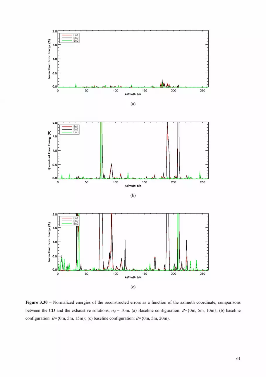
61
(a)
(b)
(c)
Figure 3.30 – Normalized energies of the reconstructed errors as a function of the azimuth coordinate, comparisons
between the CD and the exhaustive solutions, σδ = 10m. (a) Baseline configuration: B={0m, 5m, 10m}; (b) baseline
configuration: B={0m, 5m, 15m}; (c) baseline configuration: B={0m, 5m, 20m}.

62
Figure 3.31 – ABF tomograms in the azimuth-height plane for the range line under test, B={0m,5m,10m,15m,25m},
with σδ = 10m. From top to bottom: (a) Error-free; (b) corrupted; (c) CD-ME; (d) SE-ME.

63
Figure 3.32 – ABF tomograms in the azimuth-height plane for the range line under test, B={0m,5m,10m,15m,20m,
25m,30m,35m,40m,45m}, with σδ = 10m.. From top to bottom: (a) Error-free; (b) corrupted; (c) CD-ME; (d) SE-ME.

64
(a)
(b)
(c)
Figure 3.33 – Normalized energies of the reconstructed errors as a function of the azimuth coordinate, comparisons
between the CD and the SE solutions, incoherent phase variations with σδ = 10m. (a) Baseline configuration: B={0m,
5m,10m,15m,25m}; (b) baseline configuration: B={0m, 5m,10m,15m,20m,25m}; (c) baseline configuration: B={0m,
5m,10m,15m,20m,25m,30m,35m,40m,45m}.

65
(a)
(b)
(c)
Figure 3.34 – Normalized energies of the reconstructed errors as a function of the azimuth coordinate, comparisons
between the CD and the SE solutions, baseline configuration: B={0m, 5m, 10m, 15m, 25m}. (a) σδ = 2.5m; (b) σδ =
5m; (c) incoherent phase variations with σδ = 15m.

66
3.2.2 Estimation of 0z : performance analysis
The objective of this Section is to evaluate the performance obtainable in the estimation of 0z and,
consequently, of the original calibration errors in Step 5 of the calibration chain. As explained in Section
2.3.2, these operations are performed by the last processing block of the proposed chain by means of a
“forward-backward” procedure starting from a reference pixel and exploiting the slow variability of the
phase errors we want to estimate. Noteworthy, this operation is not strictly needed for tomographic purposes,
since after the minimum entropy step it is already possible to isolate the complex reflectivities and to
estimate the vertical profiles by means of an ABF filter. Anyway, as already discussed in the previous
chapter, the contribution of the phase errors have been compensated in the signal Cy but it is still an
unknown of the problem. On the other hand, for applications different from SAR Tomography it would be
useful to know the extent of these phase offsets. Thus, the following analysis is worth to be performed in
order to assess the goodness of the last processing block in isolating the DEM contribution from the
undesired calibration errors.
Here the performance is derived by means of a simulated analysis evaluating the RMSE obtainable
in the estimation of 0z with a simple BF, as proposed in Section 2.3.4. 10000 realizations of the
miscalibration phase vectors have been generated for each variation of the parameter of interest. At each
realization, 0z has been estimated through a BF spectral estimate. Then, the root mean square error (RMSE)
of the estimation has been evaluated in each analyzed case. As a benchmark, the hybrid Cramér-Rao bound
(HCRB) was calculated using the formulation in [52] assuming an infinite signal-to-noise ratio. For the sake
of analytical simplicity and generality of the results, it is useful to express zk normalized with respect to the
maximum value of its elements, say ,maxzk , and 0z normalized with respect to the height resolution unit
,max2δ p=z zk . With this choice, the obtained results are independent from the baseline lengths and the
incidence angle (which affect the absolute value of zk ). They only depend on the baseline distribution and
2σ .
In Fig. 3.35 the estimation RMSE (continuous lines) have been plotted as a function of σ and for
different values of K (indicated with different colors). Baselines have been designed uniform for the sake of
simplicity. The HCRB has been superimposed for each K (dashed lines). Of course, 0z is perfectly
estimated without errors for 0σ = (no miscalibration) independently of the value of K . At the increase of
σ , the RMSE increases, still reaching the HCRB. It has been verified that the estimation bias is null. This
means that BF is the best estimator of 0z in that zone, and no other better performing estimator can be found.
Dependently on K , for a further increase of 0h , the RMSE starts to deviate from the HCRB, in a way
resembling a threshold effect. The value of σ in which this deviation happens depends on K : the higher
K , the higher the “cut-off” value of σ . To make an example, in real applications with airborne data
,maxzk approaches approximately 0.5 (at least in near-mid range), leading to 15mδ =z . As a consequence, a
RMSE of 0.1 resolution units (r.u.) corresponds to 1.5m, a still acceptable value. From the curves of Fig.

67
3.35, a RMSE of 0.1 r.u. is reached for instance at 0.05σ = for 5K = and 0.075σ = for 20K = .
Nevertheless, it is expected that σ does not exceed 0.01 in the hypothesis of high χ . In this case, 0z can be
estimated with a RMSE lower than about 20cm independently from K . Not only, but BF is the best
estimator there, as it reaches the HCRB.
So far, we uniform baseline distributions have been considered. With non-uniform distributions and
5>K (what happens in practice), it is not expected a dramatic worsening of the performance, even if,
depending on the non-uniformity degree, sidelobes with a poor amplitude level could be amplified by the
miscalibration. This trend has been verified by comparing the RMSE obtained by a 5K = uniform
distribution and the 5-element distribution of the TempoSAR 2008 campaign. Results are plotted in Fig.
3.36. The non-uniform baseline distribution leads to a negligible worsening of the RMSE, which is not of
concern.
A very particular case is found with 3=K , to which the curves in Fig. 3.37 are referred. As already
emphasized many times this is a very interesting limit case. Here, the signal generated by a baseline
distribution with ,mid0, ,1 = z zkk has been simulated, and the related RMSE has been obtained as a function
of ,midzk and for different σ . In Fig. 3.37, the continuous lines are the RMSEs obtained by processing the
non-uniform data stack. It is apparent that in general the RMSE decreases at the increase of ,midzk , i.e. at the
reduction of the non-uniformity degree of the baselines. Again, we observe that for 0.01σ < the RMSE
reaches acceptable levels in the order of magnitude of 0.01 r.u. For 0.04σ = , no acceptable RMSE values
are reached anymore.
In order to improve the estimation performance and to reduce the dependency of the RMSE on the
baseline non-uniformity, a feasible approach is to interpolate the available MB signal from a non-uniform
distribution to a (virtual) uniform one. Details on this signal interpolation can be found in [27], [53]. This
interpolator is very easy to be implemented. Indeed, in [53] it has been demonstrated that this interpolator
can be expressed as least-square estimator with a proper loading in the case of small errors, while it assumes
a different form for big errors. Nevertheless, concerning the estimation of scatterer height, the performance
of the small-scale solution has been found to be equivalent to the one of the complete solution. In Fig. 3.37
the RMSE obtained after baseline interpolation has been plotted for each analyzed case in dashed lines. It is
possible to observe that the acceptable performing regions of ,midzk have enlarged. In particular, for
0.05σ = (which is of interest for large χ ), an acceptable RMSE can be obtained independently of the non
uniformity.
In conclusion, from this simulated analysis in the cases of interest, i.e. when it is possible to reduce
our problem to a “small variance” one, the DEM estimation can be effectively performed through a simple
BF spectral estimation. As a consequence, the original calibration errors will also be well estimated.

68
Figure 3.35 – RMSE obtained by estimating z0 by means of BF as a function of the standard deviation of the residual
error and for different values of K, uniform baseline configuration. Continuous lines: RMSE; dashed lines: HCRB.
Figure 3.36 – RMSE obtained by estimating z0 by means of BF as a function of the standard deviation of the residual
error for K=5: comparison between an uniform baseline configuration (black) and the TempoSAR baseline
configuration (green). Continuous lines: RMSE; dashed lines: HCRB.
Figure 3.37 – Case K = 3, RMSE obtained by estimating z0 by means of BF as a function of the mid kz and for different
values of the standard deviation of the residual error. Continuous lines: RMSE with the non-uniform baselines; dashed
lines: RMSE after baseline interpolation.

69
As a final experiment, an example of estimated DEMs from the simulated PolSARProSim data stack
is reported in Fig. 3.38. The full uniform baseline distribution has been considered, i.e. the one already used
many times before. Data have been corrupted with random phase noise vector ψ , see model in (1.28), with
standard deviation 0.32ψσ p= . In particular, in Fig. 3.38(a) is shown obtained from the error free DEM
stack Dy , where it is possible to recognize clearly the bare areas, with a height centroid located at 0m, and to
distinguish it from the forest area in the middle of the slice, whose height centroid is located at around 18m.
From the figure it is also possible to recognize the presence of inflated sidelobes due to residuals of baseline
to baseline differences in the estimated DEM height, i.e. the vector δ . Fig. 3.38(b) refers to vector Dy
corrupted by the phase noise, where both the disturbances are present coming from phase miscalibrations and
vector δ . Here, ambiguities arise and the extent of the sidelobes could lead to a wrong estimate of the height
centroid. In Fig. 3.38(c), after the estimation of the vector δ through the minimization of the entropy, the BF
spectrum of the signal ( ),D x ry , which is the input of the last processing block and only contain the original
pahse perturbation to be estimated. Finally, Fig. 3.38(d) shows the BF spectrum obtained from the fully
compensated DEM, where the phase miscalibrations have been suppressed and the sidelobes due to residual
DEM estimation errors have been rejected, i.e. after Step 5. The maxima of the obtained “clean” MB DEM
in Fig. 3.38(d) perfectly matche with the maxima of the DEM in Fig. 3.38(a), thus demonstrating the
capability of Step 5 in estimating the miscalibration phases.
Figure 3.38 – BF tomograms of MB interferograms, uniform full baseline configuration (K = 10). (a) Error-free; (b)
miscalibrated, σψ = 0.32π; (c) after ME optimization; (d) fully calibrated.

70
3.2.3 Global performance analysis
In the previous sections each part of the proposed calibration chain has been analyzed as a stand-
alone block and tests with simulated data have been carried out in order to assess its performance from both
a qualitative and a quantitative point of view. Here, we link together every parts previously described and we
analyze the global performance of the proposed calibration algorithm. In other words, the calibration chain is
considered as a black box and the calibration performance is measured at its output. The algorithm has been
implemented in a single IDL code consisting in the following main functions, corresponding to the Step
discussed in Section 2.3:
Step 1
Estimation of the interferogram at each baseline with respect to the master track, starting from the
corrupted MB data stack.
Step 2
Compensation of the complex interferogram at each baseline from the corrupted data. The output of
this processing step is a MB signal containing either the scattering information free from miscalibrations and
the residual incoherent phase variations from baseline to baseline. This spurious contribution is due to the
processing itself and has to be properly compensated.
Step 3
Estimation of the incoherent phase contributions by means of CD-ME algorithm.
Step 4
Compensation of these quantities from the estimated complex MB interferograms. The output signal
from this block is the one used to estimate to miscalibration phases.
Step 5
Miscalibration phase vector estimation from the DEMs, by means of a forward-backward procedure
from a starting reference point.
Figure 3.39 – Phase screens estimated from TempoSAR’08 data.

71
Finally the estimated miscalibration phases are compensated from the MB corrupted data stack to get
the calibrated data stack ready for any other processing.
In this Section, the results of a simulated analysis aimed to test the whole processing chain will be
shown and discussed. The analysis has been carried out considering again the baseline distribution of the
TempoSAR campaign, as it is of particular interest for the next experiments with real data. In particular, to
corrupt the PolSAR-ProSim stack we used phase screens previously estimated with the CS technique from
the real data acquired during the TempoSAR’08 campaign. For each track, a cut of them has been extracted
in order to adapt them to the dimensions of the PolSARProSim dataset. The resulting phase screens are
shown in Fig. 3.39.
The results obtained in the TempoSAR case are shown in Fig. 3.40. First of all, the ABF tomograms
are reported of the error-free and the corrupted data respectively. From the error-free tomogram it is possible
to clearly distinguish the bare soil area from the vegetation layer. In the azimuth coordinate interval [50, 200]
the volume scattering contribution comes from the top of the canopy, with its maximum located at around
18m height. In this case, the ABF filter is able to properly suppress the sidelobes and multiple maxima can
be due to different scattering mechanisms, which could be imaged with a lower quality estimator. In the bare
area, the imaged scatterers have spectral maxima to the zero height, where the ground is located. If the ABF
filter receives in input the phase corrupted data, then the retrieved spectra look very different. A shift of the
maxima can be noticed in both the bare area and in the forested one. Furthermore, the spectrum is
characterized by inflated sidelobes at each azimuth coordinate which makes the image ambiguous. The width
of the main lobe enlarges, worsening the height resolution. As a consequence, it is no longer possible to
estimate with reasonable accuracy the parameters of interest. The estimated interferograms in Step 1 are
affected by both the data miscalibration and the contribution of the incoherent phase variations from baseline
to baseline. The overall results of the whole calibration chain are in Fig. 3.40, i.e. the ABF tomograms
obtained from the data after the compensation of the estimated phase screens. In order to emphasize the
importance of CD-ME, the final output which could be obtained without relying on this processing block,
i.e. avoiding Steps 3 and 4 is reported as well. It is apparent from the obtained tomograms that the proposed
calibration chain is able to estimate the calibration phases and to reconstruct properly the vertical profile.
With the exception of some azimuth coordinates, the obtained profiles look very similar, if not identical, to
the ABF tomograms obtained from the error free dataset. It is possible to distinguish again the zero-height
ground from the vegetation layer and to properly estimate the height centroid where multiple volume
scattering contributions occur.
In order to confirm the validity of the proposed method, this simulated analysis has been repeated
with the dual baseline configuration { }0m,5m,15mB = . Fig. 3.41 shows the tomograms corresponding to
the error-free data set, the corrupted data set, the data set calibrated without CD-ME, and finally the overall
calibration result. For this three-image configuration we can repeat the same considerations and get the same
conclusions of the previously analyzed case.
In order to evaluate quantitatively the performance, the normalized energy of the error with respect
to the error free profile has been calculated. In Figs. 3.42 and 3.43 the obtained error power percentages for

72
the corrupted dataset (black dashed line), the one calibrated without ME processing (green continuous line)
and the final calibrated data, i.e. the output of the whole chain (red continuous line) are compared. As
regards the miscalibrated data the error power is higher than 100% for the most of the azimuth lines, whereas
the line corresponding to the final calibrated profile stands in general below the 5%. In the TempoSAR case,
the only exceptions to this trend are represented by a few azimuth coordinates where the CD algorithm did
not converge to the correct point. Furthermore, the analysis of the green line highlights the strong necessity
of Step 3 and Step 4, since the mean error percentage is still higher than 30% with error peaks reaching the
100%. When 3=K , an higher gain can be obtained by applying this processing block in this case. This is
apparent from Fig. 3.43 where the reconstruction error corresponding to the final calibration output stands
below the 2% for each azimuth coordinate.
3.3 Conclusions From the simulated analysis carried out in this Chapter, it is possible to affirm that that the proposed
calibration chain exhibits good performance in the estimation of the phase miscalibrations. This is clear from
both the qualitative analysis of the tomograms and the quantitative evaluation of the reconstruction error.
The primary role of the minimization of the entropy, i.e. of Steps 3 and 4 of the calibration chain, has also
been demonstrated numerically. The CD-ME algorithm has been shown to be effective in entropy
minimization.
The error plots of Figs. 3.42 and 3.43 have also shown that especially for 3K > the proposed
algorithm is not immune to isolated errors in the estimation of the calibration phases. However, as previously
emphasized, their removal e.g. by a median filtering is considered to be a feasible way to increase further the
calibration performance.

73
Figure 3.40 – ABF tomograms in the azimuth-height plane for the range line under test, baseline configuration B={0m,
5m, 10m, 15m, 25m}. From top to bottom: (a) Error-free; (b) corrupted; (c) calibrated, no ME; (d) fully calibrated, CD-
ME.

74
Figure 3.41 – ABF tomograms in the azimuth-height plane for the range line under test, baseline configuration B={0m,
5m, 15m}. From top to bottom: (a) Error-free; (b) corrupted; (c) calibrated, no ME; (d) fully calibrated, CD-ME.

75
Figure 3.42 – Error power of the ABF tomograms as a function of azimuth with respect to the error-free ones, baseline
configuration B={0m, 5m, 10m, 15m, 25m}. Black dashed line: miscalibrated data. Green continuous line: calibrated
data without ME. Red continuous line: calibrated data after entropy minimization (CD-ME).
Figure 3.43 – Error power of the ABF tomograms as a function of azimuth with respect to the error-free ones, baseline
configuration B={0m, 5m, 15m}. Black dashed line: miscalibrated data. Green continuous line: calibrated data without
ME. Red continuous line: calibrated data after entropy minimization (CD-ME).

76
APPENDIX 3.A
ENTROPY MINIMIZATION: COMPUTATIONAL LOAD
From the available literature, the number of real-valued multiplications needed to calculate the ABF
profile in a single range-azimuth cell is:
( )3 22 4 1ABFC K K M K K= − + + + , (A.3.1)
being K the number of images in the data stack and M the number of height samples at which the profile is
calculated. The entropy of the profile can then be calculated by using 4 2M + real valued multiplications.
It is easy to demonstrate that the computational loads in terms of real valued multiplications needed
in entropy minimization by means of the exhaustive, the SE-ME and the CD-ME algorithms are respectively
given by:
( ) 14 2
KEXH ABFC M C L
− = + + , (A.3.2)
( ) ( )4 2 4 2 1SE ME ABF ABFC M C L M C L K− = + + + + + − , (A.3.3)
( )( )1 4 2CD ME ABF iterC K M C LN− = − + + , (A.3.4)
where L is the number of phase samples and iterN is the number of minimizations needed by CD-ME to
reach the minimum of the entropy functional.
Quantities (A.3.2)-(A.3.4) have been plotted in Fig. A.3.1 as a function of K , for 201M L= = and
5iterN = , the latter being a typical value observed in the analyses reported in Chapter 3. It is apparent that
while EXHC increases rapidly with K , SE MEC − and CD MEC − are smaller at least of 4 orders of magnitude.
Moreover, CD MEC − is at least one order of magnitude smaller than SE MEC − , and this difference increases at
the increase of K .
Figure A.3.1 – Computational load calculated in terms of number of real-valued multiplications for different entropy
optimization methods, plotted as a function of the number of images in the stack.

77
CHAPTER 4
EXPERIMENTS WITH
REAL AIRBORNE DATA
4.1 The TempoSAR 2008 dataset After having employed a simulated data to assess the effectiveness of the proposed phase calibration
method the processing chain has been applied to real airborne data. In particular, an E-SAR dataset acquired
by DLR during the TempoSAR 2008 campaign over the forest stand of Traunstein (Germany) has been
processed In Fig. 4.1 the Pauli RGB image and the LIDAR DTM are shown.
The MB stack consists of 5 fully polarimetric L-band images obtained with 5 passes of the airborne
platform carrying the radar (the time span is about 1 hour). The baseline configuration is non-uniform, with a
minimum nominal sampling distance 5m=d and an overall nominal synthetic aperture length 25m=L in
the horizontal direction. This test site is very interesting for applications of forest observation since a wide
vegetation cover can be recognized with a variety of tree species (e.g. spruces, beeches and white firs). The
vegetation height ranges in between 10m and 40m and, noteworthy, the biomass level is varies between
40t/ha and 450t/ha. For this reason, the vegetation layer is often very dense, so the main contribution to the
backscattered power comes from the canopy. Hence, the calibration of this kind of data could be a limit case
in which an algebraic decomposition (e.g. like in the ALGAE method in [46]) could not be successfully
applied everywhere. Nevertheless, since the proposed calibration method exploits the whole vertical profile
independently on its shape, it can be applied even in this boundary conditions. As regards the existing
methods based on the detection of reliable calibration targets, no theoretical reasons impair, at least in
principle, their effectiveness when the full multibaseline stack is available. As already discussed, problems
arise in the detection of CSs and PSs when the number of available acquisitions decreases since the
evaluation of both the dispersion factor ( ),D x r and the ensemble coherence ( ),γ ensemble x r becomes

78
unreliable. On the other hand, the proposed calibration method has been conceived in order to avoid these
criticalities. To check this potentiality, it has been tested also with a dual-baseline configuration. This
constitutes a very limit case in which the phase calibration problem becomes very challenging. Anyway, the
dual-baseline case is also a very interesting solution to reduce the degrading effects of the temporal
decorrelation in repeat-pass acquisitions over natural scenarios. In the following sections processing results
will be shown to assess the effectiveness of the proposed method and comparisons with the existing
techniques will be carried out.
4.2 Processing results: full baseline configuration This section is devoted to analyze the performance of the whole processing chain achieved when the
full baseline stack is employed to calibrate the data. In particular, at first results are presented of experiments
carried out over corner reflectors, where the knowledge of their theoretical response is exploited as a way to
validate the proposed method. Then, an area of interest is selected and ABF tomograms will be reported to
show the calibration performance. Finally, in order to make comparisons between the obtained results and
the ones we can get with the existing techniques, some representative pixels are selected and the respective
vertical profiles after calibration will be examined.
Figure 4.1 – Test site of Traunstein (Germany). Range increases from the bottom to the top, azimuth increases from
the left to the right. Top panel: Pauli RGB radar image; bottom panel: LIDAR DTM.

79
4.2.1 Experiments with corner reflectors
Prior to any performance analysis, a reliable calibration method has to be tested in correspondence of
points in which a reference is available. When the performance analysis has been carried out with simulated
data, this reference was the error free profile for each cell of the image stack. Moving to real data, a good
reference is the response of the system to a point-like scatterer, i.e. the tomographic PSF. For this reason, in
Fig. 4.2 the BF profiles are plotted obtained after calibration in correspondence of three corner reflectors
(CRs). In these points, the expected BF profile is the PSF of the system. BF profiles have been reported after
the calibration with PSs, CSs, and with CD-ME. The mainlobe width of the PSF enlarges when moving from
the first CR to the second and the third ones. In fact, CR2 and CR3 are located in far range, while CR1 is in
near range. Thus, the horizontal baseline reduces and the height resolution worsens.
The CD-ME profile fits very well with the PSF for each of the examined points. As regards the other
methods, the corresponding profiles are still in good agreement with the PSF. From the plots referred to the
first two corner reflectors we can recognize a mismatch in correspondence of the sidelobes of the function
between the PSF and the lines referring to the PS and CS, while for the third corner reflector all the lines are
superimposed to the PSF.
4.2.2 ABF tomograms
An area of interest has been selected and the proposed calibration algorithm has been applied to two
fixed azimuth lines in the HH channel. In particular, two azimuth bins x1=2750 and x2=3500 have been
chosen and tomograms along the range direction have been considered for a total distance in range of 1425m
and 1710m, respectively. In order to make comparisons with the existing techniques, the CS-based
calibration has been applied to the corrupted stack. Similar results have been obtained with the PS-based
calibration. The resulting tomograms at azimuth x1 are shown in Fig. 4.3. As already stressed, since the ABF
filter is data adaptive, it is very sensitive to miscalibrations, and their effect on the retrieved spectrum is
apparent. It is possible to recognize recognize ambiguities and sidelobes in each range coordinate. Moreover,
Figure 4.2 – BF profiles in correspondence of corner reflectors. Black dashed line: PSF. Blue line: PS-based method.
Red line: CS-based method. Black continuous line: CD-ME.

80
the effect of the residual phase contributions results in a strong loss of height resolution. Due to these
phenomena, it is impossible to distinguish between the bare and the forested areas and to exactly locate the
scatterers in height, because of sidelobes which can be as powerful as the main peak. Figs. 4.3(b) and 4.3(c)
show respectively the ABF spectra obtained after the CS-based and CD-ME calibrations, respectively. When
the full-baseline stack is available, the CS method performs well. In particular, it is possible to distinguish
again between the ground and the vegetation layers and to get an accurate estimate of the parameters of
interest. The height resolution capability is recovered, since the most of the sidelobes are rejected and the
mainlobe width reduces for each range coordinate. A dense vegetation is present in near range, with the top
of the canopy located at around 20m with respect to the ground scatterer, which is placed at -5m according to
the LIDAR measure. In the middle of the tomogram, a bare area can be seen with a moderate slope, while in
far-range a forested area whose extension in height is 25m can be distinguished.
Figure 4.3 – ABF tomograms, full baseline case, azimuth coordinate x1=2750. Top panel: corrupted data; middle panel:
calibrated data with CS method; bottom panel: calibrated data with CD-ME method.

81
Figure 4.4 – ABF tomograms, full baseline case, azimuth coordinate x2=3500. Top panel: corrupted data; middle panel:
calibrated data with CS method; bottom panel: calibrated data with CD-ME method.
As a further experiment, each polarization has been processed independently, and the estimated
calibration phases have been compared. In particular, for each baseline the correlation coefficients have been
calculated. The obtained values of the correlation coefficients are higher than 0.85, i.e. a strong correlation
exists between the phases estimated from the different polarimetric channels. This means that the estimated
phase errors are actually the ones coming from uncompensated platform motions, which are not affected by
phase information related to scattering in different polarizations. This constitutes a good indication of the
effectiveness of the method in isolating the phase terms of interest from the scattering information.
The same considerations can be repeated for the azimuth coordinate x2 (see Fig. 4.4). In particular,
after calibration the contributions from the ground and the canopy can be separated very clearly. This is
achieved thanks to the gain in terms of height resolution after the compensation of the phase errors. These
improvements can be actually recognized after the application of both the examined calibration methods,
whose performance are not impaired when the full MB stack can be exploited. Anyway, some differences
can be noticed between the two calibrated tomograms, which are the result of a better compensation of the
miscalibration phases after applying the CD-ME. In order to underline these differences and to compare
better the calibration performance, in the following section some representative points will be selected and
the corresponding vertical profiles will be shown.

82
4.2.3 Vertical profiles
In this section the results of a comparison between the proposed method and the one based on the
CSs will be shown by analyzing of ABF and BF vertical profiles. In particular, the reported profiles are
obtained in correspondence of pixels which are representative of a huge set of resolution cells showing the
same features. The range locations of these representative points are shown in Fig. 4.5.
The aim of this analysis is to employ the spectral estimators like probes to highlight the potential
presence of residual miscalibrations corrupting the data. In fact, the classical BF is not very sensitive to
residual phase contributions, hence it is more stable. Nevertheless, where required, it can be used as a
reference to evaluate the capability of the calibration methods in recovering the radiometric fidelity of the
ABF profiles. In Fig. 4.6 the ABF profiles are plotted after calibration corresponding to pixels A and B.
These cells correspond to bare areas, in which a scattering contribution from the ground is the only one
present. In particular, in this and in the following figures the red line plots the profiles obtained CS
calibration method, while the black line is referred to CD-ME. As regards the pixel A, the CD-ME profile is
able to locate better the ground scatterer with respect to the CS one, since the height corresponding to its
maximum peak is closer to the LIDAR measurement (around 14m). The CS profile shows a height shift of
5m. In pixel B, an improvement is apparent in terms of height resolution. In particular, the CD-ME method
shows a gain of 1.1m in -3dB mainlobe width, which improves from 6.4m (CS) to 5.3m (CD-ME). As a
consequence, the better CD-ME calibration enhances super-resolution properties of ABF, as expected.
The following analysis refers to cells in which two scattering contributions could be recognized,
coming from both the ground and the vegetation layer. In Fig. 4.7 the profiles are reported corresponding to
the cells C, D, and E respectively. In the first cell (pixel C) a volume layer is present, which is located at 5m
height, over a ground scatterer placed at -25m, while in the second one (pixel D) the ground contribution is
located in height at -20m. From both the CS and CD-ME profiles it is possible to distinguish two main peaks
representing the two scattering mechanisms, meaning that the phase calibration has been performed
successfully. Anyway, some differences can be noticed clearly between the profiles. After the calibration
with the CD-ME approach the two peaks are better resolved in both pixels C and D, while the CS-based
profiles are less sharp. This behavior can be due to residual miscalibrations still affecting the MB stack after
processing. The same considerations can be repeated for the profiles corresponding to the pixel E, where the
height centroid of the volume scatterer is located at around 40m, while the ground contribution is present at
15m (according to LIDAR measure). In particular, by analyzing the CS profile it is worth to point out the
presence of a strong sidelobe located at -20m which is almost as powerful as the ground contribution. In this
case some ambiguities could arise in the ground height estimation, as it is difficult to distinguish the surface
scatterer from the sidelobe. This sidelobe could be due to a residual phase error, such that the ABF filter
emphasizes the corresponding spatial frequency with height -20m, rejecting the one corresponding to the
ground contribution. This sidelobe is well suppressed in the profile obtained after the calibration with the
ME. In this case, a gain of 5dB in terms of sidelobe rejection can be measured with respect to the CS profile,
which means that the residual phase contribution has been compensated as well.

83
As regards the radiometric fidelity, the analysis of the pixels F and G is noteworthy. Unfortunately,
in absence of a ground truth, it is not possible to state exactly which is the inaccurate profile (i.e. the one still
affected by miscalibrations), unless a further analysis is carried out. However, a possible reference can be the
BF profile, as it is less prone to miscalibration residuals. In Fig. 4.8 both the BF and ABF profiles are
reported after calibration with the ME and the CS method. Figure refers to the cell F, where a main volume
contribution is present at 15m height and a less powerful echo from the ground scatterer is located at 5m.
The BF profiles are almost superimposed (at least in a height interval close to the mainlobe) and the power
relation between the two maxima is almost the same for both the ME and CS profiles. Since the BF is
radiometrically linear, this power relation can be assumed as reference. Considering the ABF filtering, the
CD-ME profile exhibits a power relation between the peaks in the order of the one in the corresponding BF
profile, with a stronger contribution from the volume and a weaker one from the surface. The profile appears
sharp. On the other hand, the shape of the CS profile is very different, and the two peaks are almost
identically powerful. In this case the radiometric fidelity is not preserved, and the explanation of the change
in the profile behavior has to be found in a residual contribution of phase errors which are well compensated
after the ME-based calibration. In order to better investigate this features and to have a clear validation of the
last conclusions, a further simulated analysis has been performed. In particular, an error free profile with two
scatterers has been simulated in such a way that the BF spectrum of the simulated profile matches with the
one retrieved from real data [see Fig. 4.9(a)]. Then, the ABF profile has been calculated and compared with
the one obtained after CD-ME calibration. A good agreement between the two profiles has been found [see
Fig. 4.9(b)]. Since the simulated one has been obtained in absence of errors, this means that the proposed
method has been able to estimate the calibration phases better than the CS-based.
Several cells with these features have been found in the examined azimuth lines. As a last example,
Fig. 4.10 reports a case in which is very apparent the effect of residual miscalibrations in the CS-based ABF
profile. In fact, in this cell a stronger contribution from the volume is present at 15m height, while a weaker
contribution from the ground comes from 5m. This is apparent from the BF plots, where both the CS and the
CD-ME match. Again, with the ABF filter the behavior of the red line totally changes and the power relation
between the peaks is inverted, with a more powerful peak from the surface, while the CD-ME profile shows
the same power relation of the BF. This constitutes a clear indication of the better capabilities of the
proposed method in recovering the radiometric fidelity of the profiles, a very important feature for the
analysis of forest scenarios.

84
Figure 4.5 – HH range-azimuth image. Red lines: analyzed lines at constant azimuth. Green dots: locations of the
analyzed cells.
MARKER AZ. BIN RG. BIN TYPE
A 3500 1000 Bare
B 2750 850 Bare
C 2750 620 Forested
D 2750 648 Forested
E 2750 1125 Forested
F 3500 577 Forested
G 3500 578 Forested
Table 4.1 – Range-azimuth position and characteristics of the analyzed cells.
Figure 4.6 – ABF vertical profiles after calibration. Red line: CS method. Black line: CD-ME method. Figure refers to
bare areas. Left panel: pixel A; right panel: pixel B.

85
Figure 4.7 – ABF vertical profiles after calibration. Red line: CS method. Black line: CD-ME method. Figure refers to
forested areas. Left panel: pixel C; middle panel: pixel D; right panel: pixel E.
Figure 4.8 – Vertical profiles of the pixel F after calibration. Red line: CS method. Black line: CD-ME method. Figure
refers to forested areas. Left panel: BF profiles; right panel: ABF profiles.
Figure 4.9 – Vertical profiles of the pixel F after calibration. Green line: simulated. Black line: CD-ME method. Figure
refers to forested areas. Left panel: BF profiles; right panel: ABF profiles.

86
Figure 4.10 – Vertical profiles of the pixel G after calibration. Red line: CS method. Black line: CD-ME method.
Figure refers to forested areas. Left panel: BF profiles; right panel: ABF profiles.
4.3 Results with a dual-baseline configuration The last part of the performance analysis presents the calibration results obtainable when a few
acquisition are available for processing. In particular, the dual-baseline configuration is a limit case because
of the low number of acquisitions which can impair the detection of reliable scatterers in the PS/CS methods.
On the other hand it is worth to remind that this kind of baseline configuration is particularly appealing in
repeat-pass SAR sensing of natural areas where the scattering is subjected to temporal decorrelation effects.
As for the full baseline case, two azimuth coordinates of interest have been selected and the proposed
calibration chain has been applied together with the other calibration methods (PS-based and CS-based) in
order to make some comparisons.
In particular, the azimuth bins x3=3000 and x4=5200 have been chosen (see Fig. 4.11, where the
amplitude SAR image of the test site is reported) and again tomograms have been extracted for a total range
distance of 1595m and 1760m respectively. As regards the coordinate x3, Fig. 4.12 shows respectively the
ABF tomograms obtained from the corrupted data, from the data calibrated with the CS-based method and
the one obtained after applying the proposed scheme. The corrupted tomogram shows pseudo-ambiguities
and sidelobes; moreover, some height shifts can be recognized and it becomes very difficult to place
properly the main scattering mechanisms. In this case, the CS method is not able to estimate the calibration
phases, as the quality of the retrieved spectrum is very low and variable in space, depending on the
performance of the CS detection. While this method fails with a few acquisitions, the proposed CD-ME still
works well, as shown in Fig. 4.12(c). From the ABF tomograms the ground, the vegetation layer and the
scattering from the top of the canopy can be distinguished. The ambiguities disappears and also the sidelobe
rejection improves. Some residual lobes in near-range are still present, which are actually due to the PSF in
height. The ABF after CD-ME is also able to recover the loss of height resolution, demonstrating not to be
strongly affected by the low number of available baselines. The same considerations can be repeated for the
analysis of the azimuth coordinate x4 (see Fig. 4.13) where the proposed method with the PS-based
technique, whose corresponding tomographic spectrum is shown in Fig. 4.13(b). This azimuth line is

87
characterized by a big slope corresponding to a hill with a low vegetation cover. As expected, the PS
techniques totally fails in this challenging case. On the contrary, a good quality of the ABF tomograms after
CD-ME from which we can still recognize the vertical structure of the investigated area. Again, some
residual lobes are still present which are due to the system PSF. However, the gain in terms of ambiguity
suppression, super-resolution and height shift compensation is apparent.
Figure 4.11 – HH range-azimuth image. Red lines: lines at constant azimuth used for the dual baseline analysis.

88
Figure 4.12 – ABF tomograms, dual baseline case, azimuth coordinate x3=2750. Top panel: corrupted data; middle
panel: calibrated data with CS method; bottom panel: calibrated data with CD-ME method.

89
Figure 4.13 – ABF tomograms, dual baseline case, azimuth coordinate x4=5200.Top panel: corrupted data; middle
panel: calibrated data with PS method; bottom panel: calibrated data with CD-ME method.

90
- This page intentionally left blank -

91
CHAPTER 5
CONCLUSIONS AND OUTLOOK
The processing of MB-SAR data exploits the coherent combination of complex SAR data to retrieve
the information of interest about the observed scene. In this framework, this work is focused on SAR
tomography which allows resolving along the height dimension different scatterers belonging to the same
range-azimuth cell. In this way it is possible to extract information about the vertical structure of the
observed area. Unfortunately, especially in airborne SAR acquisitions, residual non-compensated platform
motions result in baseline estimation errors. On the other hand, atmospheric propagation delays have to be
accounted for in space borne acquisitions. In both cases, the MB data stack is affected by unknown phase
contributions different from track to track. As a consequence, prior to any MB coherent processing, it is
necessary to correct the data stack for these phase miscalibrations. From a signal processing perspective, the
phase calibration can be carried out by following two different classes of algorithms. A first class makes use
of a grid of targets of opportunity (generally point-like) which remain stable during the entire acquisition
time span. Depending on the stability criterion, these scatterers are defined “persistent” (PS) or “coherent”
(CS). MB calibration methods based on the detection of PS and CS have been developed and tested so far.
However, the effectiveness of this class of techniques is scenario-dependent. In fact, in natural scenarios the
presence of stable point-like scatterers is more reduced than in urban scenarios. Moreover, as the number of
baselines is typically kept low to avoid temporal decorrelation problems, the detection performance worsen
and the calibration quality degrades dramatically. As a consequence, the MB phase calibration in presence of
a few tracks turns out to be a challenging task. To overcome all these problems, a second class of algorithms
has been introduced based on some kind of MB autofocus. However, the common feature of the existing
methods is that their performance is very scenario dependent.
This work proposes and investigates a new calibration method based on the minimization of the
entropy of the adaptive beamforming (ABF) tomogram in the SAR cell under test. Indeed, in the context of
the information theory the minimization of the entropy of the ABF tomogram is equivalent to the
maximization of its sharpness, as it get reduced when phase miscalibrations corrupt the data, especially with

92
ABF. It is worth noting that the proposed method exploits the whole MB information, and there is no need to
focus on a particular target or on a scatterer in the profile. In this way the spatial coverage of calibration
targets is full and a single polarimetric channel is sufficient to correct properly the whole MB data stack.
The proposed calibration chain operates on a MB single-pol data stack in 5 steps. For each baseline,
the interferometric phase is estimated and compensated from the complex MB data. So doing, the remaining
MB complex data stack is a function of the complex backscattering at each baseline (i.e. the vertical profile)
translated around the zero height, with additional phase contributions due to the DEM variations from
baseline to baseline. These phase residuals can be estimated by minimizing the entropy of the ABF
tomogram for each SAR cell. Then, if the original miscalibration phases (i.e. those affecting the original MB
dataset) are of interest, they can be estimated from the interferometric phases calculated at the beginning by
applying local averages. Actually, this last block relies on the reasonable assumption of low variability in the
range-azimuth space of the phase errors.
It is worth noting that the entropy minimization is a non-convex optimization problem which does
not admit a closed form solution. To this purpose, two different iterative methods have been investigated
which are different ways to try to overcome the trade-off existing between reliability and complexity. Of
course, the exhaustive method is the most reliable one because the convergence to the global minimum of the
entropy functional is guaranteed. Unfortunately, it is unfeasible when the number of images increases (i.e.
when K > 3). The first proposed iterative method proceed through exhaustive optimizations sequentially. In
this way, the method is faster than the exhaustive one and it is still feasible when the number of images to
process exceeds 3. A faster iterative solution has been implemented, called coordinate descent. The solution
is found iteratively by optimizing the phases one by one while keeping the others fixed. In this way the
minimization problem turns out to be scalar at each iteration, and the computational complexity reduces. For
both algorithms, the convergence to the global minimum is not granted. These two kind of optimizations
have been implemented and tested with a simulated stack. In particular, PolSARProSim data have been
corrupted with a random phase noise and the algorithms have been applied in order estimate the error-free
vertical profiles. As a performance measure, the normalized error energy has been computed between the
reference ABF profile and the one after optimization. From the comparisons carried out in Chapter 3, the
coordinate descent algorithm results also to be the best one in recovering the right vertical profile, with errors
approaching the null value in each simulation. This result is obtained by choosing as a starting point the one
with a set of identical phases, whose value is determined according to a one-dimensional exhaustive
optimization. For these reasons, the coordinate descent solution has been chosen to constitute the core
processing of the proposed scheme.
The whole calibration chain has then been implemented and tested with both simulated and real data.
In the first case the reference stack has been corrupted with residual motion phases estimated from the DLR
E-SAR TempoSAR 2008 dataset. A performance analysis has been carried out measuring the reconstruction
errors of the profiles. The chain has been tested with both the full baseline stack, other thinned baseline
configurations and with a poor dual baseline configuration. Results show the effectiveness of the proposed
method in estimating the calibration phases and, hence, in reconstructing properly the vertical profiles. In

93
particular, in the dual baseline case the error approaches the null value for the most of the analyzed azimuth
coordinates. The method has also been shown to be able to recover the exact amplitudes of the backscattered
powers in the SAR cells under test, thus preserving the radiometric fidelity of the tomographic imaging.
As regards the test with real data, a dataset acquired by means of the DLR’s E-SAR platform over
the forest site of Traunstein has been processed. The experiments carried out over corner reflectors, whose
tomographic response is known, have shown a better match of the obtained minimum entropy profiles with
the system PSF. Then, an area of interest has been selected and tomograms have been analyzed to show the
calibration performance. Finally, some representative cells have been selected and the respective vertical
profiles after calibration have been examined. Other tomographic experiments have demonstrated the
superiority of the proposed method with respect to the existing techniques. In particular, from the vertical
profiles gains in terms of height resolution and sidelobe suppression have been shown in the ABF profiles. In
the bare areas the ground scatterers are better located with respect to the reference LIDAR measures), and
the mainlobe width reduces (1m gain). In the cells where two scattering contributions are present, coming
from the surface and the volume layer, they are better resolved and the peak to sidelobe level increases (up to
5dB gain in the examined pixels). This means that the phase errors have been properly compensated, as the
remarkable properties of the ABF imaging are restored.
Future work on this topic could be aimed at both further theoretical developments and experiments.
The following lines have been individuated:
1. In the airborne case, a model is available for the phase errors due to non-compensated platform
motions, see equation (1.27). As a consequence, for each azimuth coordinate, the problem reduces to
the estimation of the vertical and horizontal platform displacements from the nominal track, repeated
for all the tracks. One could consider to minimize the entropy of the 2-D tomogram in the range-
height plane. In this way, the calibration of the entire dataset would be faster than calibrating each
single cell, at the cost of an increased dimensionality of the optimization problem (2 parameters per
track instead of one);
2. A slight dependence of the calibration performance on the specific profile has been observed in
Chapter 3. Further simulated experiments should be devoted to verify this tendence;
3. Further experiments could be carried out to verify an improvement in the estimation of the
polarimetric scattering mechanisms estimated by means of polarimetric Tomo-SAR as a
consequence of an improved phase calibration;
4. Further experiments should be devoted to assess the retrieved radiometric fidelity;
5. Other airborne/spaceborne real data sets should be employed to assess the calibration performance
with different scenarios (e.g. other forests, urban, ice covered…);
6. The performance obtainable in the calibration of temporally varying scenarios (e.g. due to
deformations and/or temporal decorrelation) should be investigated. In this case, the entropy of the
differential tomographic profile could be a useful functional to be considered.

94
- This page intentionally left blank -

95
REFERENCES
[1] J. B. Campbell, Introduction to Remote Sensing, Taylor & Francis, London, 2002.
[2] R. Bamler, P. Hartl, “Synthetic Aperture Radar Interferometry”, Inverse Problems, vol.14, pp. R1-
R54, August 1998.
[3] L. C. Graham, “Synthetic Interferometer Radar for Tomographic Mapping”, Proceedings of the IEEE,
vol. 62, pp. 763-764, June 1974.
[4] P. Rosen, S. Hensley, I. R. Joughin, F. K. Li, S. N. Madsen, E. Rodirguez, and R. M. Goldstein,
“Synthetic Aperture Radar Interferometry”, Proceedings of the IEEE, vol. 88, pp. 331-382, March
2000.
[5] F. Gini, F. Lombardini, “Multibaseline Cross-Track SAR Interferometry: A Signal Processing
Perspective”, IEEE Aerospace and Electronic Systems Magazine, vol. 20, no. 8, pp. 71-93, August
2005.
[6] A. Ferretti, C. Prati, and F. Rocca, “Nonlinear Subsidence Rate Estimation Using the Permanent
Scatterers in Differential SAR Interferometry”, IEEE Transactions on Geoscience and Remote
Sensing, vol. 38, no. 5, pp. 2202-2212, May 2000.
[7] P. Berardino, G. Fornaro, R. Lanari, and E. Sansosti, “A New Algorithm for Surface Deformation
Monitoring Based on Small Baseline Differential SAR Interferograms”, IEEE Transactions on
Geoscience and Remote Sensing, vol. 40, no.11, pp. 2375-2383, November 2002.
[8] S. R. Cloude, K. P. Papathanassiou, “Polarimetric SAR Interferometry”, IEEE Transactions On
Geoscience And Remote Sensing, vol. 36, no. 5, pp. 1551-1563, September 1998.
[9] K. P. Papathanassiou, S. R. Cloude, “Single-Baseline Polarimetric SAR Interferometry”, IEEE
Transactions On Geoscience And Remote Sensing, vol. 39, no. 11, pp. 2352-2363, November 2001.
[10] S. R. Cloude, K. P. Papathanassiou, “Three-stage inversion process for polarimetric SAR
interferometry”, IEEE Proc.-Radar Sonar Navig., vol. 150, no. 3, pp. 125-134, June 2003.
[11] C. López-Martínez, A. Alonso, X. Fàbregas, K. P. Papathanassiou, “Ground topography estimation
over forests considering Polarimetric SAR Interferometry”, pp. 3612-3615, IGARSS, Honolulu,
Hawaii, USA, July 2010.

96
[12] A. Reigber, A. Moreira, “First Demonstration of Airborne SAR Tomography Using Multibaseline L-
Band Data”, IEEE Transactions On Geoscience And Remote Sensing, vol. 38, no. 5, pp. 2142-2152,
September 2000.
[13] M. Pardini, Advances and Experiments of Tomographic SAR Imaging for the Analysis of Complex
Scenarios, Ph.D. Thesis, University of Pisa, Italy, June 2010.
[14] S. Sauer, L. Ferro-Famil, A. Reigber, E. Pottier, “Three-Dimensional Imaging and Scattering
Mechanism Estimation Over Urban Scenes Using Dual-Baseline Polarimetric InSAR Observations at
L-band”, IEEE Transactions On Geoscience And Remote Sensing, vol. 49, no. 11, pp. 4616-4629,
November 2011.
[15] F. Lombardini, “Differential Tomography: A New Framework For SAR Interferometry”, IEEE
Transactions on Geoscience and Remote Sensing, vol. 43, no. 1, pp. 37-44, January 2005.
[16] F. Lombardini, M. Pardini, F. Cai, “Polarimetric Differential-TomoSAR Imaging”, Proc. Of European
SAR Conference, EUSAR, Aachen (Germany), June 2010.
[17] S.K. Lee, F. Kugler, K. Papathanassiou, I Hajnsek, “Multibaseline Polarimetric SAR Interferometry
Forest Height inversion Approaches”, Proc. of ESA POLINSAR Workshop, Frascati (Italy), January
2011.
[18] S. Tebaldini, A. Monti Guarnieri, Methods and Performance for Multipass SAR Interferometry, in
Geoscience and Remote Sensing. New Achievements, Intech Online Publisher.
[19] P. Pasquali, C. Prati, F. Rocca, M. Seymour, J. Fortuny, E. Olmer, and A. J. Sieber, “A 3D SAR
Experiment with EMSL Data”, Proc. of 1995 IEEE International Geoscience and Remote Sensing
Symposium (IGARSS), Florence (Italy), July 19955.
[20] J. Homer, I. D. Longstaff, Z. She, and D. Gray, “High Resolution 3-D Imaging Via Multi-Pass SAR”,
IEEE Proceedings, Part F-Radar, Sonar and Navigation, vol. 149, pp. 45-50, February 2002.
[21] F. Gini, F. Lombardini, and M. Montanari, “Layover Solution in Multibaseline SAR Interferometry”,
IEEE Transactions on Aerospace and Electronic Systems, vol. 38, pp. 1344-1356, October 2002.
[22] G. Fornaro, F. Serafino, and F. Soldovieri, “Three Dimensional Focusing With Multi-pass SAR”,
IEEE Transactions on Geoscience and Remote Sensing, vol. 41, pp. 507-517, March 2003.
[23] F. Lombardini and A. Reigber, “Adaptive Spectral Estimation for Multibaseline SAR Tomography
with L-Band Data”, Proc. of 2003 IEEE International Geoscience and Remote Sensing Symposium
(IGARSS), Tolouse (France), July 2003.
[24] G. Fornaro, F. Lombardini and F. Serafino, “Three-Dimensional Multipass SAR Focusing:
Experiments with Long Term Data”, IEEE Transactions on Geoscience and Remote Sensing, vol. 43,
no. 5, pp. 702-714, May 2005.
[25] I. Hajnsek, R. Scheiber, L. Ulander, A. Gustavsson, G. Sandberg, S. Tebaldini, A. M. Guarnieri, F.
Rocca, F. Lombardini, and M. Pardini, Technical Assistance for the Development of Airborne SAR and
Geophysical Measurements During the BioSAR 2007 Experiment, tech. rep., ESA contract no.
20755/07/NL/CB.

97
[26] F. Lombardini and M. Pardini, “3-D SAR Tomography: The Multibaseline Sector Interpolation
Approach”, IEEE Geoscience and Remote Sensing Letters, vol. 5, pp. 630-634, October 2008.
[27] F. Lombardini, M. Pardini, and L. Verrazzani, “A Robust Multibaseline Sector Interpolator for 3D
SAR Imaging”, in Proc. of 2008 European Synthetic Aperture Radar Conference (EUSAR),
Friedrichshafen (Germany), June 2008.
[28] S. Tebaldini, “Forest SAR Tomography: A Covariance Matching Approach”, in Proc. of 2008 IEEE
RADAR Conference (RADARCon).
[29] S. Tebaldini, “Algebraic Synthesis of Forest Scenarios from Multibaseline PolInSAR Data”, IEEE
Transactions on Geoscience and Remote Sensing, vol. 47, pp. 4132-4142, December 2009.
[30] X. X. Zhu, R. Bamler, “Very High Resolution Spaceborne SAR Tomography in Urban Environment”,
IEEE Transactions on Geoscience and Remote Sensing, vol. 48, no. 12, pp. 4296-4308, December
2009.
[31] E. Aguilera, M. Nannini, and A. Reigber, “Multi-Signal Compressed Sensing for Polarimetric SAR
Tomography”, Proc. of IEEE International Geoscience and Remote Sensing Symposium (IGARSS),
Vancouver, Canada, July 2011.
[32] P. Stoica, R. L. Moses, Introduction to Spectral Analysis, Prentice-Hall, 1997.
[33] S. Tebaldini, A. Monti Guarnieri, “On the Role of Phase Stability in SAR Multibaseline Applications”,
IEEE Transactions On Geoscience And Remote Sensing, vol. 48, no. 7, pp. 2953-2966, July 2010.
[34] K. Iribe, K. Papathanassiou, I. Hajnsek, M. Sato, Y. Yokota, “Coherent Scatterer in forest
environment: detection, properties and its applications”, Proc. of IEEE International Geoscience and
Remote Sensing Symposium (IGARSS), Honolulu, Hawaii (USA), July 2010.
[35] A. Jacoksson, F. Gini, F. Lombardini, “Robust estimation of Radar Reflectivities in Multibaseline
InSAR”, IEEE Transactions on Aerospace and Electronic Systems, vol. 41, no. 2, pp. 751-758, April
2005.
[36] H. L. Van Trees, Optimum Array Processing. Part IV of Detection, Estimation, and Modulation
Theory. Wiley, 2002.
[37] Y. Rockah, P. M. Schultheiss, “Array Shape Calibration Using Sources in Unknown Locations – Part
I: Far-Field Sources”, IEEE Transactions On Acoustics, Speech, And Signal Processing, vol. ASSP-
35, no.3, pp. 286-299, March 1987.
[38] S. M. Kay, Fundamentals of Statistical Signal Processing – Estimation Theory, Prentice-Hall, 1993.
[39] M. Pardini, F. Lombardini, F. Gini, “ The Hybrid Cramér-Rao Bound on Broadside DOA Estimation
of Extended Sources in Presence of Array Errors”, IEEE Transactions on Signal Processing, vol. 56,
no. 4, pp. 1726-1730, April 2008.
[40] M. Viberg, A. L. Swindlehurst, “A Bayesian Approach to Auto-Calibration for Parametric Array
Signal Processing”, IEEE Transactions On Signal Processing, vol. 42, no.12, pp. 3495-3507,
December 1994.

98
[41] M. P. Wylie, S. Roy, H. Messer, “Joint DOA Estimation and Phase Calibration of Linear Equispaced
(LES) Arrays”, IEEE Transactions On Signal Processing, vol. 42, no.12, pp. 3449-3459, December
1994.
[42] B. P. Flanagan, K. L. Bell, “Array Self-Calibration With Large Sensor Position Errors”, Signal
Processing, vol. 81, pp. 2201-2214, 2001.
[43] A. Ferretti, C. Prati, F. Rocca, “Permanent Scatterers in SAR Interferometry”, IEEE Transactions on
Geoscience and Remote Sensing, vol. 39, no. 1, pp. 8-20, January 2001.
[44] K. Iribe, Investigations on Coherent Scatterers in Natural Environments for SAR Multi-Image
Applications, Ph.D. Thesis, Graduate School of Environmental Studies, Tohoku University, August
2010.
[45] G. Ferraioli, G. Ferraiuolo, V. Pascazio, “Phase-Offset Estimation in Multichannel SAR
Interferometry”, IEEE Geoscience And Remote Sensing Letters, vol. 5, no. 3, pp. 458-562, July 2008.
[46] G. Gatti, S. Tebaldini, M. Mariotti d’ Alessandro, F. Rocca, “ALGAE: A Fast Algebraic Estimation of
Interferogram Phase Offsets in Space-Varying Geometries”, IEEE Transactions On Geoscience And
Remote Sensing, vol. 49, no. 6, pp. 2343-2353, June 2011.
[47] S. Tebaldini, F. Rocca, “Multibaseline Polarimetric SAR Tomography of a Boreal Forest at P- and L-
Bands”, IEEE Transactions on Geoscience And Remote Sensing, vol. 50, no. 1, pp. 232-246, January
2012.
[48] Y. Huang, L. Ferro-Famil, F. Lombardini,“Improved Tomographic SAR Focusing Using Automatic
Baseline Error Compensation”, in Proc. Of ESA PolInSAR Workshop, Frascati, Italy, January 2011.
[49] R. L. Morrison, M. N. Do, D. C. Munson, “SAR Image Autofocus By Sharpness Optimization: A
Theoretical Study”, IEEE Transactions on Image Processing, vol. 16, no. 9, pp. 2309-2321, September
2007.
[50] T. J. Kragh, “Monotonic Iterative Algorithm for Minimum-Entropy Autofocus”, Proc. of 14th Annual
ASAP Workshop, June 2006.
[51] M. L. Williams, PolSARproSim – A Coherent, Polarimetric SAR Simulation of Forests for PolSARPro
– Design Document and Algorithm Specification, December 2006, available at
http://envisat.esa.int/polsarpro/Manuals/PolSARproSim_Design.pdf
[52] M. Pardini, F. Lombardini, F. Gini, “The Hybrid Cramer-Rao Bound on Broadside DOA Estimation of
Extended Sources in Presence of Array Errors”, IEEE Transactions on Signal Processing, vol. 56, no.
4, pp. 1726-1730, April 2008.
[53] M. Pardini, Array Interpolation Methods for Non-Uniform Multibaseline SAR Interferometers, M.Eng.
Thesis, University of Pisa, Italy, June 2006