december, 1991 - cdc stacks
TRANSCRIPT

December, 1991
TABLE OF CONTENTS
Editor's comments
Page
i
ACAV Treasury Report (as of August 31, 1991) ii
"Classification and nomenclature of viruses" iii(reviewed by R. Akkina)
"Studies on the natural history of yellow fever in vTrinidad" (reviewed by C.H. Calisher)
Royal Society of Tropical Medicine and Hygiene vi(Objectives, Fellowships, Subscriptions, and Publications)
(Continued on next page)
PURPOSE OF THE ARBOVIRUS INFORMATION EXCHANGE:To exchange information on a timely basis. The recipients are
those who study various aspects of arboviruses. The Exchange contains preliminary reports, summaries, observations, and comments submitted voluntarily by qualified agencies and individual investigators. The appearance in the Exchange of any information, data, opinions, or views does not constitute formal publication and should not be referred to in "Reference" sections of papers or included in lists of publications. The Exchange is not a "peer reviewed" publication; in fact, it is not a publication at all. Any reference to or quotation of any part of the Exchange must be authorized directly by the agency or person submitting the text.

DENGUE
Dengue-2 outbreak in Araguaina, Tocantins State, Brazil.(P.F.C. Vasconcelos, E.S. Travassos da Rosa,J.F.S. Travassos da Rosa, R.B. Freitas, A.P.A. Travassos da Rosa)
A 10 iiL ultramicroELISA for detection of human IgM immunoglobulin against dengue virus. (J.L. Pelegrino, J. Lafert£, M.G. Guzmdn, S. Vdzquez, G. Gonzalez, A. Valdivia)
Purification of specific human IgG and IgM for the detection of dengue antibody levels in human serum. (A. Otero, J.L. Pelegrino,S. Vazquez, J. Lafert6)
Monoclonal antibodies that recognize a Cuban dengue strain.(C. Hermida, G. Guzman, M. Pupo, M. Gonzalez, R. Marcet,S. Vazguez, Y. Caballero, I. Delgado)
Pathological changes of liver in dengue infections (S. Hotta)
Nuclear localization of the core protein of dengue virus.(R. Bulich, J. Haig, J. Aaskov)
Dengue in French Guiana, (letter from J.M. Reynes) 1
VECTOR COMPETENCE AND VECTOR BIOLOGY
Experimental transmission of Venezuelan equine 1encephalomyelitis virus by Aedes albopictus (Diptera: Culicidae) from New Orleans, Louisiana. (M.J. Turell, J.R. Beaman)
Effect of environmental temperature on the vector competence of 1 Aedes taeniorhynchus for Venezuelan equine encephalomyelitis and Rift Valley fever viruses. (M.J. Turell)
First confirmation of breeding populations of Aedes albopictus 1 in Continental Africa. (H.M. Savage, B.R. Miller, V.I. Ezike,A.C.N. Nwankwo, R. Spiegel)
Culicoides (Diptera: Ceratopogonidae) vector species in Israel 1 trapped from burrows of rodents that might be potential reservoirs of bluetongue and Akabane viruses. (Y. Braverman, N. Messaddeq, and M Kremer)
DEVELOPMENT AND APPLICATION OF TECHNIQUES
The use of nucleic acid probes for the detection of bluetongue 1 virus. (E.H. Venter, A.A. van Dijk, H. Huismans)
Expression of the bacterial gene, Chloramphenicol 1acetyltransferase (CAT) in mosquitoes and mosquito cells using recombinant Sindbis virus vectors. (K. Olson, S. Higgs,J. Carlson, B. Beaty)

Detection of genetic variation between populations of 2Aedes aeaypti mosquitoes by amplification of genomic DNA using random primers. (M.E. Ballinger-Crabtree, B.R. Miller)
MOLECULAR BIOLOGY AND MOLECULAR IMMUNOLOGY
The synthesis of dengue virus type 2 structural proteins 2containing deletions in hydrophobic domains. (A. Gruenberg,P.J. Wright)
Nucleotide and encoded amino acid sequences of the structural 2 and nonstructural NS1 porotein genes of a Malaysian dengue-2 virus from Aedes albopictus. (G. Subramaniam, I.M. Kautner,C.L. Koh, S.K. Lam)
Close relationship of Machupo virus to another pathogenic 2South American arenavirus, Junin. (C. Griffiths, S. Wilson,C. Clegg)
EPIDEMICS AND SURVEILLANCE
Venezuelan hemorrhagic fever. (R. Salas, N. de Manziyne, 2R.B. Tesh, R. Rico-Hesse, R.E. Shope, A. Betancourt, 0. Godoz,R. Bruzual, M.E. Pacheco, B. Ramos, M.E. Taibo,J. Garcia Tamayo, E. Jaimes, C. Vasquez, F. Araoz, J. Querales)
Arboviruses in the Brazilian Amazon region. 2(A.P.A. Travassos da Rosa, J.F.S. Travassos da Rosa, P.F.C. Vasconcelos, N. Dbgallier, C.S.F. Gregbrio)
Oropouche virus outbreaks in Rondonia State, Brazil, 1991. 3(A.P.A. Travassos da Rosa, J.F.S. Travassos da Rosa,P.F.C. Vasconcelos, E.S. Travassos da Rosa, S.G. Rodrigues)
Concomitant infections by malaria and arboviruses in the 3Brazilian Amazon region. (P.F.C. Vasconcelos,A.P.A. Travassos da Rosa, J.F.S. Travassos da Rosa, N. Degallier)
Update on yellow fever in central Nigeria, 1991. (0. Tomori, 3A. Anoja, A. Nasidi, F. Oyewole, Y. Saka, V. Eziki, R. Spiegel,P. Moore, D. Gubler)
Arbovirus surveillance using multiple small sentinel chicken 3flocks. (J.L. Hardy, L.D. Kramer, M.M. Milby, S.B. Presser,W.C. Reeves, W.K. Reisen)
Arbovirus surveillance in NSW, Australia, 1989-91. (R. Russell) 4
Central European encephalitis virus activity in Styria, Austria. 4 (D. Stiinzner, W. Sixl, R. Schaffler, M. Labuda, 0. Kozuch,E. Kocidnov£, V. Vyrostekov&)
Arboviral surveillance guidelines (C.G. Moore, R.G. McLean, 4T.F. Tsai, C.J. Mitchell, C.H. Calisher, D.J. Gubler)

Progress Report: Central American and Caribbean Bluetongue Epidemiology Project, (submitted by E. Jane Homan)Arbovirus laboratory testing in Delaware, 1990. (J. Jong-Ho,P. Randolph, S. Dee, M. Willey)
Tick-borne encephalitis in Hungary in the last ten years.(E. Ferenczi, E. Molnar)
Rift valley fever in central highlands of Madagascar.(J. Morvan, J. Roux)
A report from the Virus Reference Laboratory. (S.S. Kalter)
Preliminary summary of surveillance for mosquito-borne encephalitis virus activity in California, 1991. (R.W. Emmons)
Report from Department of Virus Ecology, The D.I. Ivanovsky Institute of virology, Academy of Medical Sciences, Moscow,U. S.S.R. (M.S. Nedyalkova, A.M. Butenko, D.K. Lvov)
A survey of suspected mosquito vectors of Japanese encephalitis on Saipan. (C.J. Mitchell, H.M. Savage, G.C. Smith, S.P. Flood, L.T. Castro, M. Roppul)
Arbovirus activity in Colorado during 1991. (G.C. Smith,C.J. Mitchell, C.G. Moore, H.M. Savage, A.B. Thapa,S.L. Shrestha)
Birds as arbovirus hosts in Brazilian Amazonia. (N. Degallier, A.P.A. Travassos da Rosa, J.-M.C. da Silva, S.G. Rodrigues, P.F.C. Vasconcelos, J.F.S. Travassos da Rosa, G.P. da Silva, R.P. da Silva)
Serologic evidence for tick-borne encephalitis (TBE) in North American military stationed in Germany. (J. Clement, H. Leirs,V. Armour, D. Ward, J. Groen, A. Osterhaus, C. Kunz)
Avian hosts of St. Louis encephalitis in Pine Bluff, Arkansas, 1991. (R.G. McLean, M. Townsend, L.J. Kirk, R.B. Shriner)
Murray Valley encephalitis, Kunjin, dengue-1, and Ross River virus infections in Queensland, Australia. (D. Phillips,C. Atkins, M. Wieraers)
Evidence for persisting high level enzootic activity of certain orbiviruses and Malpais Spring virus in wild ungulates of central New Mexico. (C.H. Calisher, D.E. Taylor)
HANTAVIRUSES
Hantaviruses in Wisconsin and Duluth, Minnesota. (K.A. Burek,C. Rossi, J.W. LeDuc, T.M. Yuill)

Serologically verified hantavirus infections in Hungary.(G. Faludi)Endemic foci of hemorrhagic fever with renal syndrome (HFRS) in Germany. (J. Pilaski, C. Ellerich, 0. Gorschewsky, R. Peceny, T. Kreutzer, A. Lang, W. Benik)
Epizootiological aspects of hantavirus infection in Belgium,The Netherlands, and Germany: Of Mice and Men. (J.P. Clement,A. Lefevre, J. Groen, A. Osterhaus, R. Verhagen, H. Leirs,G. van der Groen)
EXPERIMENTAL INFECTIONS
A low virulence strain of JBE virus isolated from Culex tritaeniorhynchus mosquitoes in Indonesia. (B.Q. Chen)
Study on the experimental infection of tree shrew with chikungunya virus. (H. Zhang, Z. Mi, H. Shi, D. Zi)
The natural infection rate of mosquitoes by Japanese encephalitis B virus in Yunnan Province, China. (H. Zhang,D. Zi, H. Shi, Z. Mi, Z. Gong, J Zhang, Z. Hou)
Interferon studies in Mayaro virus-infected TC7 cells.(M.C.S. Rebello, J.L. Oliveira, M.E.F. Fonseca, M.A. Rebello)
Inability of Barmah Forest virus to cross the placenta in mice. (P. Birt, J. Haig, J. Aaskov)
Effect of pH on infectivity of Eyach virus in Vero cells.(G. Dobler, H. Meier-Ewert)
PATHOLOGY
Axoplasmic retrograde transport of Jurona virus.(C.W. Picanco-Dinez, J.C. Souza, A.P.A. Travassos da Rosa,R. Araujo, M.T.F. Araujo, J.F.S. Travassos da Rosa)
LYME DISEASE
Serological studies on the infection of dogs in Ontario, Canada, with Borrelia burgdorferi. (H. Artsob, I. Barker,R. Fister, G. Sephton)
A serosurvey for antibody to Borrelia burgdorferi in an "at risk” population of southern Portugal. (A.R. Filipe)
Ixodes pacificus and Borrelia burgdorferi in Arizona.(C.A. Olson, E.W. Cupp, S. Luckhart, J.M.C. Ribeiro, C. Levy)
Borreliae in culicine mosquitoes. (J. Halouzka)
Sample controlled seroprevalence study of Lyme infection in Belgium. (J. Clement, A. Ramon, W. Vranckx, A. Lefevre,A. Fain, G.R.F. Krueger, R. Ackermann)

POEMS
Bunyaviridae (by Dra. Norma E. Mettler) 101
Fourth International course on dengue and dengue hemorrhagic 104 fever (Cuba)First International symposium on bovine ephemeral fever and 105related rhabdoviruses (China/Australia)
NOTE: As noted in "PURPOSE OF THE ARBOVIRUS INFORMATION EXCHANGE",which is found on the front page of each issue, you are encouraged to submit a brief summary of your work. The summary need not be in manuscript style, the results do not have to be definitive, you need not include tables (unless you want to). This is not a peer-reviewed publication. The intent is to communicate among ourselves and to let each other know what we are doing.
The next issue will likely be mailed June 1, 1992 (probable deadline for submissions: May 15, 1992). There is nothing that requires you to wait until the last minute. If you have something to communicate in January, February, March or April, please send it. Also, there is nothing that prevents you from submitting a report to every issue. There are no pages charges either but, then again, this is not a publication.
PLEASE !!!Follow the directions for submitting reports. Double-spaced pages take twice as much space as single-spaced pages. Do not double-space or number pages. Single-space them and leave them unnumbered. Do not staple pages together.
Charles H. Calisher, Ph.D.DVBID/NCID/CDCP.0. Box 2087Ft. Collins, CO 80522
GUIDELINES FOR SUBMITTING REPORTS
We want to keep this mechanism timely and viable. Therefore, submit only recent news and summaries of your work. PLEASE limit the submission to 1 or a very few sheets (21.59 cm x 27.94 cm =8.5 x 11 inches) plus a table or two? condense as much as you can (single space the text); do not staple pages together; do not number pages.

Editor's commentsThis issue of the Arbovirus Information Exchange is of considerable size. Thanks to a multitude of contributors around the world, we are able to print 56 reports and a poem. Reports on the discovery of Aedes albopictus in Africa, expression of a bacterial gene in mosguitoes, detection of genetic variation in mosquitoes using random primers, and Venezuelan hemorrhagic fever, plus books reviews, announcements, and other pieces of information demonstrates the viability of arbovirology. The poem demonstrates its romance.
Thanks to the continued succor of the Centers for Disease Control (specifically, the financial support and enthusiasm of the staff of the Division of Vector-Borne Infectious Diseases and its Director, Duane Gubler) and the enthusiasm of the American Committee on Arthropod-borne Viruses (ACAV), the Arbovirus Information Exchange continues to allow arbovirologists to swap information, let others know what each group is doing, stay in touch personally, and help stay professionally abreast. Since this newsletter was established, a revolution in information exchange has taken place, one that is far beyond what anyone could have imagined 30 years ago. Nevertheless, it is personal interchange that has served as a foundation of arbovirology. I trust that this will continue.
For the time being, and at the request of the Chairman of the Executive Council of the ACAV (Joel Dalrymple, re-elected to the post during the annual meeting earlier this month), I will remain as Editor. However, there are so many competent, enthusiastic, and hardworking people active on the ACAV that I cannot imagine there would be a problem replacing me very quickly, should the situation change. I am gratified that so many have responded to my requests and reminders to submit reports? the Arbovirus Information Exchange exists for and because of you.
ANNOUNCEMENT
The Third International Symposium on Arboviruses and Hantaviruses in Mediterranean Countries will be held in Cortina, Italy 25-29 March, 1992. Here is an opportunity to discuss the latest information about these viruses and to do a bit of skiing. For further information, write: Organizing Secretariat, Third International Symposium on Arboviruses, MACI, P.O. Box 6164, Roma Prati 00100, Italy (telephone: 06-589-7084? fax 06-574-1762 or 06-445-3904.
i

AMERICAN SOCIETY OF TROPICAL MEDICINE AND HYGIENE
September 23, 1991
Mr. Thomas M. Yuill Assoc. Dean and Professor University of Wisconsin-Madison School of Veterinary Medicine 2015 Linden Drive West Madison, Wisocnsin 53706
Dear Mr. Yui 11 :
RICHARD J. BURK. JR., Executive Director8000 Westpark Drive. Suite 130 McLean. VA 22102 Telephone (703)790-1745
Telex 90-1811 FAX (703) 790-9063
OCT 2 199IV
Veterinary Medicine ,
Below please find the breakdown for the ACAV Treasury, Scherer Memorial, and the Young Memorial. I hope that these figures will help you with your report to the committee.
BALANCES AS OF 8/31/91
BALANCE ACTIVITY ACTIVITY CURRENTACCOUNT 12/31/89 1990 JAN-AUG 91 BALANCE
ACAV 6722.91 TIES 2120.00 1125.00 7462.73PURCH (3278.38) .00INT 403.37 369.83
SCHERER 3325.91 INT 199.55 183.32 3708.78MEMORIAL
YOUNG 8 12 . 31 INT 48.74 44.77 905.82MEMORIAL
Please feel free to call me concerning any of the breakdown figures.
Sincerely,
Deborah A. Reitz Fiscal Administrator
cc: R. BurkJ. Ravdin li

ReviewClassification and Nomenclature of Viruses:Fifth Report of the International Committee
on Taxonomy of Viruses
Archives of Virology/Supplementum 2
R.I.B. Francki, C.M. Fauquet, D.L. Knudson, and F. Brown (Eds) Springer-Verlag, Wien, New York, 1991, 450 p., DM 110
With ever increasing number of viruses discovered and characterized every year it is an obvious necessity that they are given appropriate names and classified in a timely fashion. The International Committee on Taxonomy of Viruses (ICTV) is charged with the immense task of updating virus taxonomy every three years based on evolving knowledge on virus structure, genetic organization, replication strategies, epidemiological data, etc. The last official ICTV report was published in 1982, nearly a decade ago in Interviroloev. Understandably, the present report has been much awaited by the community of virologists as a single current authentic source of information on viral taxonomy.
The 5th report has been published as a supplementura to Archives of Virology. the newly designated official journal of the virology division of the International Union of Microbial Societies. While it generally follows similar format as that of its predecessor, several improvements including stylistic were made in the page layouts, sub-headings, etc. In addition to taxonomic description of viruses, the book's beginning sections contain relevant information about the rules for virus classification, mechanistic aspects of the workings of the sub-committees, format for submission of new taxonomic proposals, names of members of ICTV, etc. This information is very educative and useful for any virologist who is considering placing a newly discovered virus into its proper taxonomic position.
Classified are 2430 different viruses into 73 families and groups. Of these, 19 are new families or groups which encompass viruses of plants, animals, fungi and bacteria. The descriptions for each virus family/group under the subheadings "properties of the virus particle, replication and biological aspects" have been greatly expanded and some were completely rewritten. The presentations are concise, up to the point and current. Where available, English vernacular names were provided for each virus group and genera. Each genus is typified by a well characterized prototype virus, and where appropriate each genus is subdivided into subgroups. List of other members of the genera are included in addition to possible members. Overall, the data are well organized and logically presented.
Animal virologists will quickly realize many changes, additions, and deletions in the present report. A few examples: A new order Mononegavirales was created and a new family Hepadnaviridae came into existence. Subfamilies Paramyxovirinae and Pneumovirinae were established under the family Paramyxoviridae. Pestivirus genus was transferred to the Flaviviridae family. A new genus Tospovirus which infects plants via thrips has been established and an old genus, Ukuvirus was consolidated into the Phlebovirus in the family Bunyaviridae. Based on the new information about the genomic organization of
i l l

hepatitis C virus, a new genus was created under the Flaviviridae family. Similarly, Dhori and Thogoto viruses were formally initiated into the Orthomyxoviridae family. In the family Retroviridae, the three previously existing subfamilies Onocovirinae, Lentivirinae, and Spumavirinae were eliminated, and instead, the existing viruses were grouped into seven genera.
The individual viruses and virus families are well indexed. For the first time, names of contributors responsible for each family are indicated. There are numerous typographical errors and occasional not-so-minor errors: for example, in the virus index on page 424, bovine immunodeficiency and leukemia viruses are listed under Reoviridae when they should be listed as belonging to the family Retroviridae. Overall, the fifth report presents the classification of viruses in a systematic manner with a lot of useful information. The book is moderately priced and deserves shelf space in every virologist's office.
Ramesh Akkina Dept. Microbiology
Colorado State University Fort Collins, Colorado
IV

Book: "Studies on the natural history of yellow fever in Trinidad", 1991, edited by Elisha S. Tikasingh.
This 188 page summary is the first in a series of monographs to be published by the Caribbean Epidemiology Centre. Dedicated to the late Dr. Wilbur G. Downs, with a Preface by Franklin White and an Introduction by the Editor, the book is divided into six parts: History, The 1954 Outbreak, The 1978-1980 Outbreak, The Interepidemic/ Interepizootic Years 1980-1988, The 1988-1989 Epizootic, and The Future.
From the titles of these sections one can begin to understand the remarkable and well-studied epidemiology of this disease in Trinidad. Epidemic yellow fever was first described in Trinidad in 1793, with periodic outbreaks since then. Beginning with a brief history of yellow fever in Trinidad this book methodically and succinctly takes the reader from then to now, stopping along the way to provide pertinent data, references, and summaries. For those of us interesting in this fascinating and important disease of the Americas and Africa, this is a well edited and useful publication, if not handbook.
Cost of the soft cover edition is a nominal $12.00 U.S. (checks made payable to CAREC) and the book can be obtained through the CAREC Librarian, CAREC (PAHO/WHO), P.O. Box 164, Port of Spain, Trinidad, West Indies.
(reviewed by C.H. Calisher)
v

ROYAL SOCIETY OF TROPICAL MEDICINE AND HYGIENE
Objectives of the SocietyThe objectives of the Society are to promote health and to advance the study, control and prevention of disease in man and other animals in warm climates, to facilitate discussion and the exchange of information among those who are interested in tropical diseases, and generally to promote the work of those interested in these objectives.
FellowshipAll registered medical and veterinary practitioners, scientists and others interested in the objectives of the Society whose qualifications are deemed satisfactory by the Council, are eligible for election as Fellows. The names of candidates should be submined on the form provided for the purpose, which must be signed by two Fellows, one of whom must have personal knowledge of the nominee.
Anyone who desires to become a candidate for the Fellowship of the Society should write to the Honorary Secretaries at the address below, or to the Editor, Arbovirus Information Exchange.
SubscriptionsThe annual subscription by Fellows becomes due in advance on 1st April each year.There is a reduced subscription for bone fide students which is limited to 3 years.The annual subscripton for 1992 is £40.00 and for students is £25.00.
Payment of the subscription may be made by:a) cheque in sterling (including European Community cheques);b) cheque in equivalent U.S. or Canadian dollars (but not sterling cheques drawn on US banks);c) draft direct to our bank (sterling or dollars):National Westminster Bank pic, 1 Cavendish Square, London W1A 4NU.Quote Sortcode 60-40-02, Account No. 24814237;d) standing order (please write to us for a form);e) credit card: Access, Eurocard, Mastercard, Visa.
MeetingsMeetings of the Society are normally held on the third Thursday of every month (except July, August and September) usually at Manson House unless otherwise notified.
PublicationsThe Society has two regular publications - the Transactions which is issued 6 times annually (No.l of each volume in February) and the "Yearbook" issued annually in October. These are sent without charge to each Fellow of the Society whose annual subscription is not in arrears. Both publications are available for purchase by non-Fellows, libraries and departments.
Manson HouseThe headquarters and offices of the Society are at Manson House, 26 Portland Place, London WIN 4EY (Tel: 071 580 2127: Fax: 071 436 1389), where any Fellow may obtain information from the staff who are in attendance from 9.00 a.m. to 4.00 p.m. Monday to Friday.
Local SecretariesLocal Secretaries are appointed by Council to act on behalf of the Society in various parts of the world; the Editor of Arbovirus Information Exchange is the Local Secretary for Colorado, USA.
vi

DENGUE 2 OUTBREAK IN ARAGUAINA, TOCANTINS STATE, BRAZIL.Pedro F.C. VASCONCELOS, ELIZABETH S. TRAVASSOS DA ROSA, Jorge F. S. TRAVASSOS DA ROSA, Ronaldo B. FREITAS and Aielia P.A. TRAVASSOS DA ROSA. Secao de Virus, Instituto Evandro Chagas. Fundacao Nacional de Saude-HS. Av. Alairante Barroso, 492, 66065, Belei-PA.
We report the first outbreak of dengue fever caused by dengue £ (DEN £) in Araguaina, Tocantins State (Figure i). Four hundred people of 74 families, living at S. Joao, Araguaina Sul and Neblina districts were questioned and then bled, in order to obtain sera to test for anti-dengue antibodies. If a person was sick, a smallquantity of blood was collected for virus isolation. The mainclinical picture of disease was characterized by fever, headache, myalgias, arthralgias and skin rash. The diagnosis of infection was made by both virus isolation into a Ibopictus (C6/36) cellsand serology, by Hemagglutmat ion-inhibition (HI) and Igh capture enzyme immuno assay (MAC ELISA). Five strains of DEN £ were isolated, and another iii infections were diagnosed serologically (Igh positive). The positivity rate of the samples was £9% (116/400), while that of the families was 66.£% (45/7£) , where atleast one member of the each family was infected. It was alsodetected i0.£% assymptomatic infections. With regard to age all groups were affected. By other hand it was detected further significative differences forward the female sex. Assuming that the sample of 400 persons is representative of the entire population of Araguaina, it may be concluded that there were 90,000 infections among the 300,000 inhabitants of Araguaina, between march 15 and may 31, 1991.This is the first epidemic of DEN £ in the Brazilian Amazonian region, as well as the first evidence of the spread of the serotype outside Rio de Janeiro State.
Note’. In addition, seventeen more strains of DEN £ were isolated from people not included in the familiar survey. Aecf&s aegypti was widely distributed in the usual domestic breeding sites.
1

Fiqure Is Map of Tocantins State, showinq Araquaina CitvDenque 2 outbreak occurred.
2

A 10 Ml. ULTRAMICROELISA FOR DETECTION OF HOMAN IgM IMMUNOGLOBULIN AGAINST DENGUE VIRUS
Pelegrino, J.L., Laferte. J., Guzman, M.G., Vazquez, S.Gonzalez, G. and Valdivia, A.
PEDRO KOURI INSTITUTE OF TROPICAL MEDICINE. P.O. Box 601, LA HABANA, CUBA.
During last years an increasing in Dengue virus infection cases have been observed even in areas under previous efficient control.
Dengue serological diagnosis have been focused on the classic haemagglutination inhibition technique, complement fixation and viral neutralisation; nevertheless, the use the third generation immunoassays as ELISA have played an important role due a remarkable easy performance and sensitivity. On the other hand, assays based on IgM capture nave the advantage of detection of recently acquired infections.
We have set up a capture 10 m L ultramieroELISA in automated microtitration system using a 96 (25 m L ) well PVC platescoated with goat affinity purified polyclonal anti human IgM antibody. A blocking step consisting of bovine albumin 4% in carbonate/bicarbonate buffer was introduced.
The antigen preparation was obtained from suckling mouse brains and extracted by the sucrose-acetone method and a d j u s t e d t o 16 haemagg 1 u tin a t i on u n its. As c o n 3 u g a t e we u. s e d a monoclonal anti Dengue complex antibody bound to alkaline phosphatase. A fluorigenic substrate was used for developing the reaction (4 methyl umbelyphery1 phosphate).
The automatic fluorescence readings were analyzed by a coupled microcomputer in which a parallel statistic quality control processing have been done. The test positive criteria have been established 260 human sera from Dengue patiens and blood donors were studied. Results were cpmpared. with MAC ELISA2.A 100% of coincidence in both tests was obtained. The UltramicroElisa has the advantage of massive screening using ultra micro volumes of reaction.
REFERENCES:1. -Otero, A., Sarracent, .J, Fernandes Yero, J.L. and
Rodriguez, I. (1984) A 10 m L ultramieroELISA for the detection of monoclonal antibodies against human alphafetoprotein. Hybridoma 3: 391.
2. -Fernandes, R.J. and Vasques, S. (1990) Serologicaldiagnosis of Dengue by an ELISA inhibition method (EIM). Mem. Inst. Oswaldo Crus, Rio de Janeiro vol 85 < 3 ): 347.
3
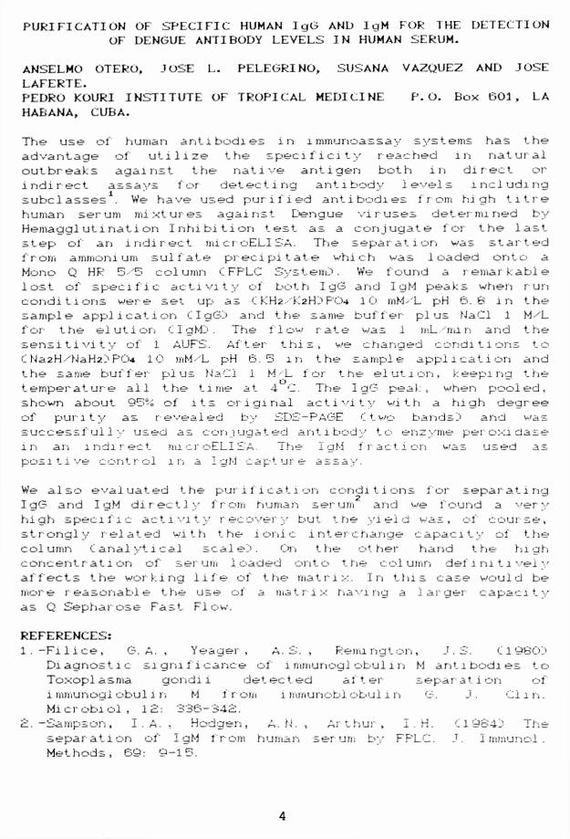
PURIFICATION OF SPECIFIC HUMAN IgG AND IgM FOR THE DETECTION OF DENGUE ANTIBODY LEVELS IN HUMAN SERUM.
ANSELMO OTERO. JOSE L. PELEGR1NO. SUSANA VAZOUEZ AND JOSE LAFERTE.PEDRO KOURI INSTITUTE OF TROPICAL MEDICINE P. O. Box 601, LA HABANA. CUBA.
The use ol human antibodies in immunoassay systems has the advantage of utilize the speci 1"i ci ty reached in natural outbreaks against the native antigen both in direct or i ndi r ect assays f or detecti ng ant 1 body Ievels 1 ncluding subc 1 asses1. We have used pur i f i ed ant i bodi es 1 r om hi gh titre human serum mixtures against Dengue viruses determined by Hemagglutination Inhibition test as a conjugate for the last step of an indirect microELISA. The separation was started from ammonium sulfate precipitate which was loaded onto a Mono Q HR 5-'53 column CFPLC System}. We found a remarkable lost of' specific activity of both IgG and IgM peaks when run conditions were set up as O.H2 -'K2HJ PO-* lO mM- L pH 6. b in the sample application CI gGj and the same buffer plus NaCl 1 M/L for the elution C I gMJ. The flow rate was 1 mL 'mi n and the sensitivity of 1 AUFS. After this, we changed conditions to C Na2H 'NaH2j PO-* 10 mM-'L p>H 6. 53 in the sample application and the same buffer plus NaCl 1 M-'L for the elution, keeping the temperature all the time at 4 C. The IgG peak, when pooled, shown about 953-£ of its original activity with a high degree of pun ty as revealed by SDS-PAGE C two bands j and was success 1 ul 1 y used as conjugated antibody to enzyme p^eroxidase m an indirect microELISA. The IgM fraction w as used as positive control in a IgM capture assay.
We also evaluat ed the puri 1 i cat i on condit i ons f or separ ati ng IgG and IgM directly from human serum and we found a very high specific activity recovery but the yield was, of course, strongly related with the ionic interchange capacity of the column C analytical seal el>. On the other hand t lie high concentration of serum loaded onto trie column definitively affects the working life of the matrix. In this case would be more reasonable the use ol a matrix having a larger capacity as Q Sepharose Fast F1 ow.
REFERENCES:1. -Filice, G. A . , Yeager , A . S. , Remington, J.S. 11980J
Diagnostic significance of immunoglobulin M antibodies to Toxoplasma gondii detected after separation ofi mmunogl obul l n M f r om lmmunobi obulln G. J . Cl i n.Mi c r obi ol , 1 £: 336 ~ 34S.
3. —Samp-son, I . A. , Hibdgen, A. N. , Ar thur , I . H. t19S4j Thes e pa rati on of I gM f r om human serum by FF'LC. J. I mmunc-1 .Methods, 69: 9-15.
4

MONOCLONAL ANTIBODIES THAT RECOGNIZE A CUBAN DENGUE STRAIN.
Hermida, C. , Guzmnan, G. , Pupo. , M. , Gonzalez, M. , Marcet, R. , Vazquez, S. , Caballero, Y. and Delgado, I.
PEDRO KOURI INSTITUTE OF TROPICAL MEDICINE. P. O. Box 601, LAHABANA, CUBA.
In May 1981 , Cuba report a great Dengue Hemorrhagic Fever C DHF!) epidemic caused by Dengue 2 virus. It was the first Dengue Hemorrhagic outbreak in the America Region. Since May to October 1981, some Dengue £ strains were isolated in Tropical Medicine Institute of Havana, Cuba, from Dengue patients and infected mosquitoes.
Taking into account the usefulness of monoclonal antibodies CMabs!) for diagnosis and molecular characterization, we have generated two different Mabs against A33 strain of Dengue 2 virus which was isolated from a patient with DHF grade II using newborn mice.
For the immunization step female Balb/c mice were injected with 200 i j I of macerated mice brain <! 20 % in Hank’s saline solution) from infected mice with A33 strain. The first immunization was done with complete Freund’s adjuvant, while the other ones were done with incomplete Freund’s adjuvant. The three immunizations were carried out by intraperitoneal route and every 1‘3 days. Monoclonal antibodies - secretin hybridomas have been obtained from the fusion between murin myeloma cell line P3 X63 —Ag8 6. 3. 3 and spleen cells I rom an i mmuni zed Ba 1 b• 'c mi ce.
For the characterization of the specificity of Mabs the following strains were used in our research: DEN-1 (Hawaii),DEN-2 CNew Guinea!) , DEN—2 C A33_) , DEN—3 CH—87!) , DEN—4C H-247!), Saint Louis Encephalitis C SLE!) and Yellow f ever t'YF). The specific antibodies produced by those hybridomas were simultaneously detected by an indirecti nmunof'l uor escence CIFIJ and the Enzyme Linked Immunosorbent Assay <! ELI SAD . In the I FI test, fixed cells from Aedes albopictus <!C6.--'36D line previously infected with different Dengue virus serotypes were used.
For the ELISA "sandwich" test we used a human polyclonal against Dengue also isolated by us and the different Dengue virus serotypes. Two positive hybrids were chosen by their strong recognition in both methods: A33vi and A35/2. MabA35 xi specifically recognized the Dengue 2 virus standard serotype and the Cuban A35 strain, while did not recognize any other serotypes. On the other hand, Mab A33x2 showed an extensi ve r ecogni tion of all vir us serot ypes tested.
5
at 4i

Our- results suggests that the recognition of M a b A 3*5- 1 1 - strongly directed to serotype £ of Dengue virus, v-hi 1 e Mab A35'c "seems to recognize a group antigen of Dengue virus. At this moment these Mabs are been used m molecular biology experiments for the characterization of Dengue strain* and in the standardization for diagnostic purposes.
6

REPORT FROM VIRUS LABORATORY, DEPARTMENT OF TROPICAL MEDICINE, MEDICAL RESEARCH INSTITUTE, KANAZAWA MEDICAL UNIVERSITY, UCHINADA, ISHIKAWA, JAPAN 920-02
Pathological Changes of Liver in Dengue Infections
SUSUMU HOTTA
Histopathological findings of the liver in DV infection of experimental animals (monkeys and mice) and humans (fatal cases) will be reviewed and discussed.
Monkeys, especially Japanese monkeys (Macacus fuscatus) , infected intracutan. or subcutan. with DV show pyrexia and viremia which however are variable and which therefore cannot necessarily be used as markers of infection. Histopathological alterations of the liver are inevitable and constant; changes of varying degrees such as fatty degeneration and cellular infiltration are manifest. It seems that the cellular damages are parallel with virulence of infecting virus; when an "attenuated" strain of DV (e.g., Mochizuki strain) is inoculated, the pathological pictures manifested are mild, whereas the changes shown in monkeys inoculated with a newly isolated (human- virulent) virus are much severer. The liver damages, when evaluated by a "scoring" method, may be a "marker of virulence" of DV.
Tissues from DHF/DSS patients (fatal cases) exhibit pathol o g i c a l changes of varying sorts and degrees. Particularly, liver tissues show marked changes. Pictures to be called "midzonal necrosis" are manifest. Liver cells undergo degeneration and/or necrosis. Kupffer cells contain eosinophilic granular structures which cannot be distinguished from Councilman bodies seen in the liver of yellow fever cases. DEN antigen in the Kupffer cells can be detected by applying immunoenzyme antibody stain method. By use of anti-DEN type- specific monoclonal antibodies, types of infecting viruses can be determined. Pictures of "macrophage activation" are revealed in various tissues.
Mice succumb to encephalitis after being inoculated with DEN virus intracerebrally. This is one of the DEN virus isolation methods. However, liver damages of ic-infected mice are not marked. Moreover, an extraneural (e.g. intraperitoneal) inoculation of DV can hardly infect ordinary mice. Contrarily "nude mice" can be infected with DV through ip route, although such infectivity may differ from a particular virus strain to
7

another. The active virus can be detected in various organs and tissues. The liver cells reveal degeneration and Kupffer cells contain eosinophilic granular structures which resemble the Councilman bodies found in the liver of humans. Specific DV a n t i g e n is d e t e c t e d in t h o s e c e l l s by a p p l y i n g immunofluorescent or immunoenzyme stain techniques. It is to be added that similar changes are seen in liver tissues of nude mice inoculated ip with YF virus 17D strain.
In summary, the liver damage is one of the characteristic signs of DV infection commonly in monkeys, humans and nude mice. Hence DV may be called a "hepatotropic" agent and the damage of liver is perhaps a key phenomenon underlying the pathogenesis of dengue. The hepatotropism is shared by DV and YFV from the histopathological viewpoint. It has already been known that both viruses are near in their immunological properties as well as in molecular-biological characteristics such as nucleotides and/or amino acids sequences of genomic RNA.
(The human specimens were taken from collection of USA-AFIP, Washington,D.C. during the writer's visit there. He is grateful to Dr. Wear, Mr. Duckett and Mr. Bratthauer for their kindness in providing the materials. His deep thanks are also due to Dr. Russell and the scientific staff of WRAIR for their helpful suggestions and favorable arrangements.)
8

QUEENSLAND HEALTH - QUEENSLAND UNIVERSITY OF TECHNOLOGY CENTER FOR ARBOVIRUS REFERENCE AND RESEARCH
63 GEORGE STREET, BRISBANE, 4000 AUSTRALIA
Nuclear localization of the core protein of dengue virus.
As part of a vaccine project we developed a panel of monoclonal antibodies against the core protein of dengue 2. These react with dengue 2, dengue 4 and weakly with dengue 1 in western blots but they do not react at all with dengue3. In indirect IFA and ELISA a similar pattern of reactivity was seen - a strong reaction with dengue 2 infected cells, a weak reaction will dengue 4, and no detectable reaction with dengue 1, dengue 3, Murray Valley encephalitis or Kunjin virus infected cells.
A noticeable feature in the IFA studies was the intense staining in the perinuclear region and in, or on, the nuclear membrane. This nuclear staining was even more distinct in CV-1 cells infected with a vaccinia construct containing cDNA coding for the dengue 2 core protein.
Another noticeable feature of the histological studies was the distinct staining patterns produced by anti-envelope and anti-core protein antibodies. While the anti-core protein antibodies produced some cytoplasmic staining and a strong granular pattern over the nucleus, the anti-envelope protein antibody stained only the cytoplasm.
A PEPSCAN assay of the reactivity of the above monoclonals with octapeptides composed of overlapping amino acid sequences from the dengue 2 core protein indicated they all reacted with the region ’RNTPFNMLKRE19. Although the sequence NMLKR is common to all the viruses used in the indirect IFA discussed above, Chou and Fassman and "Surface Plot" predictions suggest that secondary structure and surface accessibility in this region varies between dengue 2 and the other flaviviruses studied.
We have not been able to demonstrate, convincingly, core protein j_n the nucleus of infected cells. Although gold labelled antibody could be seen, by electron microscopy, localised, apparently specifically, to the nucleus of dengue 2 infected cells, the pattern of the deposition was unlike that seen by light microscopy.
The different patterns of localisation of the dengue envelope and core proteins raises a number of questions as to the possible role - particularly of the nuclear associated core protein - of proteins not incorporated into the virion.
R. Bulich, J. Haig, J. Aaskov
9

Knstitut ftsstcur Cayenne, 1991 October 24hir« ffiuyatw
BP 601097306 CAYENNE CEDEX French Guiana T el.: 19(594) 30.00.60 Fax : 19(594) 30.94.16
Docteur REYNES J-Marc Laboratoire d'Entomologie
Charles H CALISHER, Ph. D. EditorArbovirus Information Exchange
Dear Sir,
A dengue outbreak occured in July 1991 and still continues in F rench G u iana . The se ro ty p e DEN-2 w as iso la ted (in d irec t immunofluorescence with serotype specific. MAb on serum - infected AP 61 cell cultures). The dengue activity is restricted to Cayenne, chief town of the departm ent. For the first time, few hemorrhagic cases were reported and confirm ed by virus isolation or serologically (commu. in press). An epidemiologic survey is in progress.
Sincerely yours
J.M. REYNES 10

EXPERIMENTAL TRANSMISSION OF VENEZUELAN EQUINE ENCEPHALOMYELITIS VIRUS BY AEDES ALBOPICTUS (DIPTERA: CULICIDAE) FROM
NEW ORLEANS, LOUISIANAMICHAEL J. TURELL and JOSEPH R. BEAMAN
Virology Division, U.S. Army Medical Research Institute of Infectious Diseases, Fort Detrick, Frederick, MD 21702-5011
Experimental studies were undertaken to ascertain the vector competence of a strain of Aedes albopictus (Skuse) collected in New Orleans, LA (Gentilly strain) for an epizootic (Trinidad donkey) strain of Venezuelan equine encephalomyelitis (VEE) virus. This strain of Ae. albopictus was significantly more susceptible to infection with VEE virus than were any of the four strains tested previously (Table 1), including two from North America and two from South America. Likewise, dissemination and transmission rates were significantly higher in the Gentilly strain. This strain was also more susceptible to infection with a second alphavirus, chikungunya (CHIK) virus, than were any of the other strains of Ae. albopictus tested. Although all three strains of Ae. albopictus tested were more susceptible to VEE virus than to CHIK virus, susceptibility to infection and dissemination with one alphavirus appeared to be directly related to susceptibility to infection and dissemination with the other virus, and may indicate shared receptor sites for the two alphaviruses in Ae. albopictus.
Table 1. Infection, dissemination, and transmission rates by day of extrinsic incubation in the Gentilly strain of Aedes albopictus after ingestion of 104‘5 PFU of VEE virus.
Day of extrinsic incubation
Criteria 7 14 21 >28 Totals
Number tested 40 40 40 60 180
Infection 78% 95% 83% 85% 85%
Dissemination 73% 93% 78% 85% 82%
Dissemination (I)+ 94% 97% 94% 100% 97%
Transmission 50% (8)+ 70% (20) 38% (26) 35% (34) 45% (88
^Percentage of all mosquitoes with virus in their legs percentage of infected mosquitoes with virus in their legs
"^Percentage transmitting (total no. feeding)11

Table 1. Effect of environmental temperature on the susceptibility of Aedes taeniorhvnchus to Rift Valley fever virus after ingesting
plaque-forming units of virus.
Temperature of
Rearing IncubationNumbertested
Infectionrate*
DisseminationRate+
19° C 19° C 119 72% 42%
19° C 26° C 107 67% 60%
ro o\ 0 o 19° C 140 51% 18%
26° C 26° C 140 45% 37%
Percentage of mosquitoes containing virus
"Percentage of mosquitoes with virus recovered from their legs
Table 2. Effect of environmental temperature on the susceptibility of Aedes taeniorhvnchus to Venezuelan equine encephalomyelitis virus after ingesting 104•5 plaque-forming units of virus.
Temperature of
Rearing IncubationNumbertested
Infectionrate*
DisseminationRate+
19° C 19° C 128 97% 76%
19° C 26° C 115 97% 87%
26° C 19° C 140 80% 41%
26° C 26° C 140 75% 44%
Percentage of mosquitoes containing virus
^Percentage of mosquitoes with virus recovered from their legs
12

EFFECT OF ENVIRONMENTAL TEMPERATURE ON THE VECTOR COMPETENCE OF AEDE8 TAENIORHYNCHUS FOR VENEZUELAN EQUINE ENCEPHALOMYELITIS
AND RIFT VALLEY FEVER VIRUSESMICHAEL J. TURELL
Department of Arboviral Entomology, Virology Division, USAMRIID, Fort Detrick, Frederick, Maryland 21702
Environmental temperature has long been known to affect the ability of arthropods to transmit an arbovirus, with most studies indicating decreased periods of extrinsic incubation and increased transmission rates when mosquitoes are held at warmer temperatures after an infectious blood meal. However, recent studies indicate that the temperature at which mosquito larvae are reared may influence their susceptibility to infection as adults. Therefore, we evaluated the effect of environmental temperature, both during mosquito rearing and after virus exposure, on the vector competence of Aedes taeniorhvnchus mosquitoes for Venezuelan equine encephalomyelitis (VEE) and Rift Valley fever (RVF) viruses. Infection rates were significantly higher in Ae. taeniorhvnchus that were exposed to VEE virus than in those exposed to RVF virus, regardless of temperature, even though mosquitoes were exposed to a 10-fold higher infectious dose with RVF virus.
Environmental temperature had a dramatic effect on the vector competence of Ae. taeniorhvnchus for both viruses. Mosquitoes reared at low temperature (19° C) were significantly (P < 0.001) more susceptible to infection with either virus than were those mosquitoes reared at standard temperature (2 6° C) , regardless of the temperature at which mosquitoes were held after virus exposure (19 or 26° C) (Tables 1 and 2) . In contrast, in infected mosquitoes, virus disseminated from the midgut to the hemocoel more rapidly in those mosquitoes held at 26° C than in those held at19° C, regardless of the rearing temperature. However, once a disseminated infection was attained, environmental temperature did not appear to affect subsequent transmission of virus by bite to a susceptible hamster. Thus, a combination of low rearing temperature and warm holding temperature produced the most efficient vectors for both viruses.
13

FIRST CONFIRMATION OF BREEDING POPULATIONS OF AEDES ALBOPICTP8 IN CONTINENTAL AFRICA
Delta State, Nigeria experienced a yellow fever (YF) epidemic from April 15 to July 20, 1991. A rapid assessment team estimated that 600-1200 cases of YF occurred in Ika Local Government Area (LGA), Delta State, with human fatalities of 300- 600 (1).
In September, 1991, mosquito oviposition cups were placed in four rural communities of Ika and Aniocha LGAs, Delta State, that had experienced YF activity. The vegetation of the study area is deciduous forest referred to as derived savanna. In the four surveyed communities, areas immediately surrounding human dwellings, typically at a radius of 5-10 meters, are cleared and farmed as large gardens. Secondary forests still exist as strips stretching from the edge of each village to extensive farmlands located 2-3 kilometers away from the village. These forests are traversed by foot paths and jeep roads and include economically important trees such as kola nut, pear, breadfruit and oil-palm trees.
Mosquito oviposition cups with cloth liners were placed on the ground in the forest edge at an average distance of 200 m from human dwellings. Liners were collected after 48 hours and those with eggs of Aedes mosquitoes were sent to the Division of Vector-Borne Infectious Diseases (DVBID), Centers for Disease Control (CDC). Eggs were hatched and larvae mass reared, by locality, to the adult stage for specific identification. Ae. albopictus were present in collections from three communities: Igbodo, Owa-Alero, and Egbudu-Akah. The composition of the 271 specimens in combined collections from Igbodo and Owa-Alero follows: Ae. aegypti. 7 3.8 %; Ae. albopictus. 18.1 %; Ae. apicoaraenteus. 4.0 %; Ae. lilii. 2.6 %; and Ae. simpsoni subgroup, 1.5 %. The 14 specimens from Egbudu-Akah represented only two taxa: Ae. albopictus. 64%; and Ae. africanus, 36%. An additional eight adults were reared from Mbiri and all were identified as Ae. africanus.
Egg collections of Ae. albopictus from three separate localities, none of which are associated with harbors or with tire dumps, indicate that this species is well established in this area. These collections represent the first record of breeding populations of Ae. albopictus in continental Africa.
Field and/or laboratory data indicate that Ae. albopictus is susceptible to or an efficient vector of a number of viruses that currently cause human disease in Africa including dengue (DEN), YF, chikungunya, Rift Valley fever and West Nile viruses (2,3). Ae. albopictus is highly anthropophilic, able to utilize both artificial and natural containers for oviposition, and is an aggressive colonizer as demonstrated by its rapid spread and establishment in North America, Brazil and various islands of the South Pacific (4). The further spread and establishment of Ae. albopictus in Africa from established populations in Nigeria and other undetected populations seems likely based on the previous colonization history of this species. Indeed, Ae. albopictus may displace Ae. aegypti in some ardafc as has occurred in some areas
14

of the southern U.S. (5) and alter established arbovirus transmission cycles.
Of particular concern to public health officials is the potential role of Ae. albopictus in DEN and YF transmission. Strains of Ae. albopictus from Asia and strains introduced into North America are efficient vectors of DEN (6,7,3). In addition to its role in epidemic transmission, Ae. albopictus may act as a reservoir host and facilitate endemic transmission due to its ability to transmit dengue viruses vertically (8).
The potential role of Ae. albopictus in the more complex urban and sylvan YF transmission cycles is of particular concern. Vector competence studies on different strains of Ae. albopictus introduced into North and South America indicate that Ae. albopictus is a competent vector of YF virus (9,10). Virus transmission studies with African strains of YF virus and Ae. albopictus from Nigeria are in progress. Due to its ability to utilize a wide variety of oviposition sites, its biting habits, and its competence as a YF virus vector in the laboratory, Ae. albopictus may link the sylvan and urban YF transmission cycles. Our data indicate that Ae. albopictus already has colonized both urban and rural forested areas in Delta State. This, inconjunction with the recent YF epidemic in Delta State, suggests that the components of the above scenario are already in place in some areas of Nigeria.
References1. Moore P, Gubler D. DVBID, CDC. Personal communication.2. Shroyer DA. J. Am. Mosq. Contr. Assoc. 1986;2:424-428.3. Mitchell CJ. J. Am. Mosq. Contr. Assoc. 1991;7:446-451.4. Hawley WA. J. Am. Mosq. Contr. Assoc. Suppl. 1988;1:1-40.5. Hobbs JH, Hughes EA, Eichold BH, II. J. Am. Mosq. Contr.
Assoc. 1991;7:488-489.6. Gubler DJ, Rosen L. Am. J. Trop. Med. Hyg. 1976;25:318-
325.7. Rosen L, Roseboom LE, Gubler DJ, Lien JC, Chaniotis BN.
Am. J. Trop. Med. Hyg. 1985;34:603-615.8. Rosen L, Shroyer DA, Tesh RB, Freier JE, Lien JC. Am. J.
Trop. Med. Hyg. 1983;32:1108-1119.9. Mitchell CJ, Miller BR, Gubler DJ. J. Am. Mosq. Cont.
Assoc. 1987;3:460-465.10. Miller BR, Ballinger ME. Trans. R. Soc. Trop. Med. Hyg.
1988;82:476-477.
Reported by H.M. SAVAGE & B.R. MILLER (Medical Entomology- Ecology Branch, DVBID, NCID, CDC, P.O. Box 2087, Ft. Collins,CO 80522), V.I. EZIKE, & A.C.N. NWANKWO (National Arbovirus and Vectors Research Division, Federal Ministry of Health, P.O. Box 104, Enugu, Nigeria) and R. SPIEGEL (International Health Programs Office, CDC, and Combatting Childhood Communicable Diseases, USAID, 1601 Adeola Hopewell Street, Lagos, Nigeria
15

CULICQIDES (DIPTERA: CERATOPGGONIDAE) VECTOR SPECIES IN I SR TRAPPED FROM BURROWS OF RODENTS THAT MIGHT BE POTENTIAL
RESERVOIRS OF BLUETONGUE AND AKABANE VIRUSES
V. BRAVERMAN1. N. MESS ADDED27 AND M. KEENER- 1. Kimron Veterinary Institute, P.O.B. 12, Bet Dagan 50250
Israel and 2. Laboratoire de Parasitologie et R'athologie Tropicale, Universite Louis Pasteur de Strasbourg, Faculte
Medecine, 3, rue \ oeberle, 67000 Strasbourg, France.
The cyclic occurence of bluetonque in Israel isthat of Akabane cause considerate breeding materia1
Bluetonque and Med i terranean. So
>hort , whei b 3 uetoi
estrictlonis longer. Both viruses, es|
ec onomic 1oss, mainly b yexportation.probably Akabane are endemic diseases jh thi ■far their reservoir animals were not evidei
established, despite the long time that these pathogens known. In Israel and other Mediterranean countries the taxoi o-f C. schu.l t z ei - o:< vst oma gp. was not studied in beta:Therefore it is not known if the species which exist in Mediterranean is identical to C. ox vstoma Kieffer in Japan, wl is a known vector of Akabane. This study was uridertal en < first step in order to elucidate the role of C. schultztok ystoma in the epidemi o1ogy of these diseases.
Emergence traps put over rodent holes in June Pezael in the Jordan Valley yielded several = chu3 tzei - o', -/stoma gp (s u b q e n u s R em m i. a) , f e m a 3. e;and also as breed:
O’nq
heror
C er at o poqonidae. Sugges11 n gr es11 nq s i. t es . C-ul i coi dos ok v storm
v e •_ t o ? ofprovenand a suspected reported here. between ok vst oma gp. and rodents between a reservoir, a 1ivestock .
Akabane, ephemeral fever and ? ago vec tor of bluetonque. The c1o
a member o- Culicoides suggest a possible cycle vector and susceptib1e
y 1970 a lC j 1J co:mosul er
ow : : j £» f e rr ef f eri ma v i rU!1 assoc ia1sc hul txe;
of t h e animals
i r i sue '
16

THE USE OF NUCLEIC ACID PROBES FOR THE DETECTION OF BLUETONGUE VIRUS★ESTELLE H VENTER, A.A. van DIJK and H.HUISMANS★Department of Infectious Diseases, Faculty Veterinary Science University of Pretoria, Onderstepoort Republic of South Africa
The Orbiviruses constitute a large and diverse genus of the Reoviridae family. They all have a genome of segmented double- stranded RNA (Gonzalez and Knudson,1988).
Viruses belonging to the Orbivirus genus infect a wide range of domestic and wild ruminants including sheep, cattle, deer, antelope,goats, buffalo, horses and others (Squire et_al.,1987). Consequently orbivirus infections could lead to the loss of domestic animals adversely affecting the agricultural industry.
Currently diagnosis of orbivirus infections is based on a variety of serological techniques as well as on the isolation of the virus in tissue culture and embryonated eggs (Osburn et al.,1981, Squire et al.,1987). These techniques are very time consuming.
Little progress has been made, however in the application of new technology to identify orbiviruses in tissue culture cells, blood, animal tissue or insect vectors.
The emphasis of research in our laboratory is on the orbivirus group of organisms and my personal research is more on bluetongue virus (BTV), the prototype of the Orbivirus genus. Recently all the segments of BTV have been cloned and compared with respect to their use as either serogroup-specific or serotype-specific probes (Huismans and Cloete, 1987).
A dot spot assay of ds RNA of all 24 serotypes of BTV were hybridized to 6 cloned segments of BTV. Segment 5 detected almost all the serotypes and looked as a possible group specific probe. Segments 1, 3,4 and 8 are also suitable candidates.
These investigations have shown that genome segments that encode non-structural proteins as well as proteins on the inside of the core particle are generally the most highly conserved within a particular serogroup and are therefore the most suitable candidates for a group specific probe. Roy et al (1985) have used segment 3, that codes for a major core protein, as probe to identify BTV in infected cells.
During BTV replication m-RNA, coded by the different genome segments, are not synthesized in equimolar amounts (Huismans and Verwoerd,1973). It can therefore be predicted that the different probes will detect virus-specified RNA with a sensitivity that reflects the relative amount of target m-RNA in the infected cell.
17

We have used segment 5 of BTV serotype 4, coding for the non- stuctural protein NS1, as a probe to detect BTV in infected tissue culture cells. Since during replication, m-RNA of this segment is transcribed in large excess compare to all the other genome segments of the virus.
This segment was compared to other cloned BTV segments in the detection of BTV m-RNA during replication of the virus in a synchronised tissue culture system. Segment 5 detected the m-RNA at 4h p.i.in a pool of lxlO5 infected cells and in only 600 infected cells after 24h replication. Segments 3 and 8 detected m-RNA from 8h p.i. and all the other probes from 12h p.i. This result suggests that segment 5 is the probe of choice.
The probe was applied further in the detection of BTV in infected tissue culture cells. Cells were infected with a series of different pfu and harvested after certain times p.i. Segment 5 was used as probe and at the lowest moi (lxlO"5 pfu/cell) the RNA was first detected at 48h p.i.
The serogroup-specificity of the NS1 gene probe was investigated by cross-hybridization to cells infected with heterologous serogroups. It was found that the probe is group-specific under conditions of high stringency and should be able to distinguish between the different viruses even in the case of mixed infections.
At present we are using the probe in In situ hybridization studies both on infected tissue culture cells and on infected mouse brain sections. Good results are obtained and will soon be published.
REFERENCES
GONZALEZ,H.A. and KNUDSON,D.L. (1988) Intra-and inter-serogroup genetic relatedness to orbiviruses.I: Blot hybridization of viruses of Australian serogroups. J. gen.Virol. 69,125-134
HUISMANS,H . and CLOETE,M.(1987) A comparison of different cloned bluetongue virus genome segments as probes for the detection of virus-specified RNA. Virology 158, 373-380
HUISMANS,H. and VERWOERD,D .W .(1973)Control of transcription during expression of the bleutongue virus genome. Virology 52, 81-88
OSBURN,B.I.,MCGOWAN,B.,HERON,B.,LOOMIS,E.,BUSHNELL,R.,STOTT,J.and UTTERBACK,W. (1981) Epizootiologic study of bluetongue:virologic and serologic results. Am.J.Vet. Res. 42, 884-887
ROY,P ., RITTER,G.D., AKASHI,H .,COLLISSON,E . and INABA,Y .(1985) A genetic probe for identifying bluetongue virus infections in vivo and in vitro.J. gen.Virol. 66, 1613 - 1619
SQUIRE,K.R.E.,STOTT,J.L.,DANGLER,C.A. and OSBURN,B .I.(1987) Application of molecular techniques to the diagnosis of bluetongue virus infection.Prog. Vet. Microbiol. Immun. 3, 235- 250 18

REPORT FROM THE ARTHROPOD-BORNE AND INFECTIOUS DISEASES LABORATORY, COLORADO STATE UNIVERSITY, FORT COLLINS, COLORADO.
EXPRESSION OF THE BACTERIAL GENE, CHLORAMPHENICOL ACETYL- TRANSFERASE (CAT) IN MOSQUITOES AND MOSQUITO CELLS USING
RECOMBINANT SINDBIS VIRUS VECTORS.
Sindbis (SIN; Togaviridae) virus expression vectors, developed by C.M. Rice et al. (Washington University, St. Louis, MO), have been used to express CAT protein in mosquito cells (C6/36; Aedes albopictus) and adult female mosquitoes (Aedes triseriatus and Aedes aegypti). The pTRCAT expression vector is non-infectious and transcribes a self-replicating RNA containing the non-structural coding sequences of SIN infectious clone TOTO 1002. The structural genes were removed and replaced with the CAT gene. Expression vector pTE/3'2J/CAT transcribes RNA containing both SIN virus structural and nonstructural genes as well as the CAT gene. The genome of this vector contains two subgenomic RNA promoters which transcribe mRNA for the SIN structural genes (26S RNA) and the CAT gene. Genomic RNA from pTE/3'2J/CAT generates infectious recombinant virus within the appropriate cell lines.
TRCAT RNA was transfected into C6/36 cells using lipofectin; CAT protein within the cells was detected by an indirect immunofluorescence assay. Approximately one in 300 cells were shown to express the CAT polypeptide. Genomic RNA transcribed from pTE/312J/CAT was transfected into BHK-21 (hamster) cells to generate recombinant virus. The virus in turn has been used to infect C6/36 cells and Aedes triseriatus adult female mosquitoes. C6/36 cells were infected with an MOI of 1.0 and peak virus titer (7-8 log10TCID50) was detected within 24 h. Virtually all cells expressed CAT protein by indirect immunofluorescence. SIN Virus titers and CAT protein levels remained constant at 72 h postinfection. Approximately 3.0 log10 TCID50 of recombinant virus was intrathoracically inoculated into adult female Aedes triseriatus mosquitoes. Titers greater than 6.0 log10 TCID50 could be detected within four days post-infection. CAT polypeptide was detected at this time by indirect immunofluorescence of mosquito squashes. Virus and CAT polypeptide can be detected in mosquitoes up to 16 days after infection. CAT polypeptide production has been further confirmed by assaying CAT enzyme activity. We are currently quantifying the levels of CAT expression in C6/36 cells and adult mosquitoes. These expression vectors should be useful for introducing novel genes into mosquito cell lines as well as adult mosquitoes.
(Report from K. Olson, S. Higgs, J. Carlson, and B. Beaty)
19

DETECTION OF GENETIC VARIATION BETWEEN POPULATIONS OF Aedes aegypti MOSQUITOES BY AMPLIFICATION OF GENOMIC DNA USING RANDOM PRIMERS
Reported by: M. E. Ballinger-Crabtree and B. R. Miller, Medical Entomology-Ecology Branch, Division of Vector-Borne Infectious Diseases, Centers for Disease Control, P. 0. Box 2087, Fort Collins, Colorado, USA, 80522.
Amplification of mosquito genomic DNA with random 10-base primers was used to detect genetic differences between two subspecies and eleven geographic populations of Aedes aegypti mosquitoes.1(Table 1)
TABLE 1
Ae aegyptl MOSQUITOES USED IN THIS STUDY
SUBSPECIES POPULATION ORIGIN SOURCE DESCRIPTION CODEFormosus Ogbomosho, Nigeria V.I. Ezike Colonized 1990, F15 OG
Enugu, Nigeria V.I. Ezike Colonized 1989 S2Enugu, Nigeria V.I. Ezike Ugbo-Owa rural area;
colonized 1989S3
Enugu, Nigeria V.I. Ezike Colonized 1989 S4Agbor, Nigeria V.I. Ezike Field collected,
rural townshipAG
Igbodo, Nigeria V.I. Ezike Field collected, deciduous forest
IG
Entebbe, Uganda L.G. Mukwaya Colonized 1991, F3 EN
Aegypti San Juan, Puerto Rico (Rexville)
D.J. Gubler Colonized 1985 RX
San Antonio, Texas D.A. Eliason Colonized 1984 SAChachoengsao Province, Thailand (Village 6)
J . Edman Colonized 1991, FI V6
Townsville, Australia I. Fanning Colonized TN
* Mosquitoes were originally collected from urban areas unless otherwised noted. Unless specified, year of colonization or number of colonized generations unknown.
Forty primers (Operon Technologies, Inc., Alameda, CA) were screened for usefulness in distinguishing between Ae. aegypti aegypti and Ae. aegypti formosus subspecies. Each primer was used to amplify genomic DNA from three individual mosquitoes each from the Ogbomosho and Rexville populations. Primers which generated fragments unique to all three individuals of only one of these populations were then used to amplify DNA from 10 individuals from each of the populations listed in Table 1. Certain useful fragments were selected and base pair size ranges were determined to allow for measurement error. The presence or absense of fragments within these size ranges for each individual was compared using nearest neighbor statistical analysis, a nonparametric multivariate discriminant analysis (PROC NEIGHBOR IDENTITY in SAS2) .
Analysis of the frequency of occurence of 16 DNA fragments resulting from amplification of each individual with three different primers uncovered enough genetic diversity to allow 100% discrimination between the aegypti and formosus subspecies (Table 2) and 89% discrimination between the populations listed above (Table 3). This technique will be applied in the future to studies involving molecular taxonomy where sibling and cryptic species/subspecies are difficult to discriminate using existing techniques and in determination of genetic similarity between mosquito populations which will be useful in studies of the geographic movement of vector populations.
20

Table 2Nearest neighbor analysis: Classification of known individual mosquitoesbased on 16 RAPD bands generated by amplification with primers A2, B3, and B13
FROM
# CLASSIFIED INTO GROUPS/TOTAL (PERCENT)
A F
A 60/60 0/60(100) (0)
F 0/70 70/70(0) (100)
A"Ae, aenypti aegypti; F-Ae. aeaypti forroosus
Table 3
Nearest neighbor analysis: Percent classification of known individual mosquitoes intopopulations based on the presence or absence of 16 RAPD bands generated by amplification with primers A2, B3, and B13
FROM
PERCENT CLASSIFIED INTO POPULATION-
OG S2 S3 S6 AG IG EN RX SA V6 TN OTHER
OG 90 0 0 0 0 0 0 0 0 0 0 10
S2 0 90 0 0 0 0 0 0 0 0 0 10
S3 0 0 80 0 0 0 0 0 0 0 0 20
S6 0 0 0 100 0 0 0 0 0 0 0 0
AG 0 0 0 0 90 0 0 0 0 0 0 10
IG 0 0 0 0 0 90 0 0 0 0 0 10
EN 0 0 0 0 0 0 100 0 0 0 0 0
RX 0 0 0 0 0 0 0 100 0 0 0 0
SA 0 0 0 0 0 0 0 0 70 0 0 30
V6 0 0 0 0 0 0 0 0 0 100 0 0
TN 0 0 0 0 0 0 0 0 0 0 70 30
’ Ten individuals from each population were tested. Individuals with unclassifiable band patterns were classified as other. * 1 2
References:1. Williams, J.G.K., Kubeklik, A.R., Livak, K.J, Rafalski, J.A., and Tingey, S.V. 1991. DNA polymorphisims amplified by arbitrary primers are useful genetic markers. Nucleic Acids Res. 18:6531- 6535.
2. SAS Institute. 1989. SAS User's Guide: Statistics. SASInstitute, Cary, NC.
21

The Synthesis of Dengue Virus Type 2 Structural Proteins Containing Deletions in Hydrophobic Domains
Allen Gruenberg and Peter J. Wright
Department of Microbiology, Monash University, Clayton, Victoria, 3168, Australia
We investigated the role of the hydrophobic domains of C and prM in the processing of DEN-2 structural proteins in intact cells. The 5' end of the genome of DEN-2 encoding the structural proteins was expressed using recombinant vaccinia virus. Three additional recombinants derived by deletion of selected dengue sequences were also expressed.
. C prM ^ M ^ E NS1
ii m- -«r m m //— m i
□ ........ m m m — / / w m \
i i m m m
i i m m \■ ■ - i f M l
encoded hydrophobic domains
The first construct contained a deletion of nucleotides encoding most of the C protein; nucleotides encoding the hydrophobic domain at the carboxy terminus were retained.The second and third constructs contained smaller deletions of 72 bp and 129 bp encoding hydrophobic domains at the carboxy termini of C and prM respectively. We used indirect immunofluorescence and radioimmunoprecipitation to detect prM and E in cells infected with the recombinant viruses. We found that deletion of 90% of C had no apparent effect on the processing of prM and E, and that the signal sequence for E at the carboxy terminus of prM was active in the absence of 65% of the upstream signal sequence for prM located at the carboxy terminus of C. Deletion of the hydrophobic sequences preceding the amino terminus of E prevented cleavage at the prM/E junction. The results demonstrated the importance of membrane association in the cleavage of structural proteins from the flavivirus polyprotein. Cells infected with the parental recombinant virus or with the construct containing the large C deletion released the E glycoprotein into the culture medium.
22

Nucleotide and encoded amino acid sequences of the structural and nonstructural NS1 protein genes of a
Malaysian dengue-2 virus from Aedes albopictus
G Subramaniam, I M Kautner, C L Koh and S K Lam Departments of Medical Microbiology and Genetics and Cellular Biology,
University of Malaya, 59100 Kuala Lumpur.
We report here the nucleotide and encoded amino acid sequences of the capsid (C), membrane precursor (prM), membrane (M), envelope (E) and nonstructural NS1 protein genes of a Malaysian dengue-2 (DEN-2) virus, strain P8-377. This virus was isolated in 1968 by the Hooper Foundation from a pool of Aedes albopictus mosquitoes from Malacca.
V'e determined the nucleotide sequences by direct RNA sequencing achieved by primer extension with synthetic oligonucleotide primers and the Sanger dideoxyribonucleotide chain termination method. Total viral RNA was isolated from infected A. albopictus C6/36 cells.
The 15-mer synthetic oligonucleotide primers used for RNA sequencing were based on the sequence of DEN-2 JAM (Deubel et al., 1986) for the structural region and on the sequence of the Puerto Rican Vaccine Strain, PR 159/S1 (Hahn et al., 1988) for the NS1 region.
However, because certain primers were not able to hybridize with the template RNA, three new primers were synthesized to enable the completion of the sequencing of the structural and NS1 protein genes of P8-377.
A comparative analysis was carried out at nucleotide and encoded amino acid levels between P8-377 and other DEN-2 viruses which included three Malaysian human isolates (M1, M2 and M3), NGC (New Guinea C), JAM, and S1.
At the nucleotide level, P8-377 - S1 had 95.6% similarity in the C region; P8-377 - NGC and P8-377 - JAM had 94.5% similarity in the prM region; P8-377 - NGC had 97.3% similarity in the M region; P8-377 - JAM had 94.2% similarity in the E region; and P8-377 - S1 showed 94.5% similarity in the NS1 region.
At the amino acid level, P8-377 - JAM had 100% similarity in the C region; P8-377 - S1 had 92.3% similarity in the prM region; P8-377 - JAM had more than 96.0% similarity in the M region and the E region; and P8-377 - NGC had 95.6% similarity in the NS1 region.
From the data generated, we observed that most of the base changes were transitions rather than transversions, and that codon usage was non-random. Cysteine residues and glycosylation sites were conserved.
Results also indicated that the Malaysian mosquito isolate showed greater similarity with other DEN-2 viruses at both the nucleotide and amino acid levels than with the three Malaysian human isolates.
23

The differences that exist between the Malaysian mosquito isolate and the Malaysian human isolates (M1, M2 and M3) could be attributed to a number of factors including host difference (arthropod and human), time of isolation [P8-377 (1968) and M1, M2 and M3 (1987)] and laboratory passage level.
Further studies will be carried out if we can isolate DEN-2 virus from mosquitoes during the current outbreak in order to explain the genetic variabilities among species of DEN-2 viruses at the level of host difference and temporal variability.
References
1. V Deubel, R M Kinney and D W Trent (1986). Nucleotide sequence and deduced amino acid sequence of the structural proteins of dengue type two virus, Jamaica genotype. Virology 155, 365-377.
2. Y S Hahn, R Galler, T Hunkapillar, J M Dalrymple, J H Strauss and E G Strauss (1988). Nucleotide sequence of dengue 2 RNA and comparison of the encoded proteins with those of other flaviviruses. Virology 162, 167-181.
24

Close Relationship of Machupo Virus to Another Pathogenic South American Arenavirus, JuiunReport from the Special Pathogens Group, Public Health Laboratory Service Centre for Applied Microbiology and Research, Porton Down, Salisbury SP4 OJG, U.K.
Machupo virus is the aetiological agent of Bolivian haemorrhagic fever, a severe disease presently confined to a sparsely populated agricultural area in the northeast of the country. It is identified as a member of the New World group of the Arenaviridae by its serological cross-reactivity in immunofluorescence and complement fixation tests, but is clearly distinguishable from the other viruses in neutralization assays. We have now cloned and sequenced the nucleocapsid (N) protein gene of Machupo virus, and have defined more precisely its relationship to other members of the family by comparison of the derived amino acid sequences.
A cDNA library was made in the phage Xgtll using as template RNA extracted from CV-1 cells infected 4 days previously with Machupo virus (strain AA288-77) at a multiplicity of 0.1. Reverse transcription was primed with a 19 base oligonucleotide complementary to the conserved 3'-terminus of the arenavirus S RNA segment (5'CGCACAGTGGATCCTAGGC). The library was screened at low stringency using a 32P-labelled PCR product of 1.7 kb containing most of the sequence of the N gene of Junin virus. Hybridizing recombinant viruses were plaque-purified, and insert DNA transferred into phagemid vectors for dideoxy sequencing. The phylogenetic relationship of Machupo virus to other arenaviruses was investigated using the progressive alignment tree-building programs of Feng and Doolittle.
The cloned sequence contains 1814 nt (EMBL Accession no. X62616). In the viruscomplementary sense an open reading frame starts at nt68, coding for a protein of 564 amino acid residues which shows amino acid sequence similarities to the N proteins of other arenaviruses. The figure shows a phylogenetic tree for the arenavirus N proteins constructed by the progressive alignment method. The scale bar indicates the horizontal distance corresponding to a 10% divergence in amino acid sequence. Machupo virus N protein is most closely related to that of Junin virus, the aetiological agent of Argentine haemorrhagic fever. The Tacaribe virus N protein also appears on this branch of the tree, while that of Pichinde virus appears to be considerably more divergent.
The tree mirrors relation- LCM Arm ships among the New WorldLCM WE viruses defined by patterns of
cross-reactivity with monoclonal antibodies as well as heterologous cross-protection of experimentally infected animals. It has long been known that prior infection of
— Machupo guinea pigs or primates with-Tocanbe Tacaribe virus protects against
subsequent challenge with Jurun virus. It has also been reported that
|_________! guinea pigs which survive10* experim ental M achupo virus
infection are protected againstJunin virus challenge, while previous Pichinde virus infection confers no resistance.
Two interesting possibilities arise from the relationships among Machupo, Junin and Tacaribe viruses which are apparent when the molecular, serological, and cross-protection data are correlated. First, it seems likely that Tacaribe virus infection would protect experimental animals against Machupo virus as well as against Junin virus challenge. Secondly, a vaccine strain of Junin virus may well also confer immunity to Machupo virus challenge. An attenuated vaccine strain (Candid-1) developed for human use is currently undergoing large-scale clinical trial in the endemic region of Argentina. There would be important implications for the control of any future outbreak of Bolivian haemorrhagic fever if it could be confirmed that a vaccine at this advanced stage of development was indeed effective in protecting against Machupo virus infection.
Old WorldC
nLasso (Sisrra Loons)
La33a (Nigeria)
Mopeia
New World
-Junin
-Pichinde
Caroline Griffiths, Stuart Wilson, Christopher Clegg25

VENEZUELAN HEMORRHAGIC FEVERR. Salas1, N. de Manziyne2, R.B.Tesh3, R.Rico-Hesse3, R.E.Shope ,A. Betancourt2, O.Godoz2, R.Bruzual4, M.E.Pacheco1, B.Ramos , M.E. Taibo1, J. Garcia Tamayo^, E.Jaimes1, C. Vasquez1, F.Araoz2 and J. Querales1. (1) National Institute of Hygiene "Rafael Rangel", Caracas; (2) Div. of Transmissible Diseases, Venezuelan Ministry of Health and Social Assistance? (3) Yale Arbovirus Research Unit, Yale University School of Medicine, New Haven, CT, USA?(4) Miguel Oraa Hospital, Guanare; and (5) Central University, Caracas, Venezuela.
In September 1989, an outbreak of a severe hemorrhagic illness, initially thought to be dengue hemorrhagic fever, was first recognized by local physicians in the Municipality of Guanarito, Portuguesa State, Venezuela. Cases of the disease have continued to be present, with a few patients seen each month. In the period from May 1, 1990 until March 30, 1991, a total of 104 presumed cases of the disease with 26 deaths were reported to the Ministry of Health. Most of the patients have been adults? to date, all have come from rural areas in the Municipality of Guanarito or from adjacent regions of neighboring Barinas State.
The Muncipality of Guanarito is located in the central plains (llanos) of Venezuela, is about 2,481 km in total area, and comprises most of the southeastern portion of Portuguesa State. The climate is tropical with a mean annual temperature of 28°C and annual rainfall of 1,300mm. Most of the 24,000 inhabitants of the municipality live in rural zones and are involved in agriculture or cattle raising.
Sera from acutely ill patients and tissue samples taken at autopsy from fatal cases were submitted to the National Institute of Hygiene in Caracas for virus isolation. Some of these samples were also examined at the Yale Arbovirus Research Unit in New Haven. Clinical specimens for virus isolation were inoculated into cultures of mosquito (C6/36) and Vero cells. After 7 to 10 days, spot slides of the cultures were prepared and examined for viral antigen by indirect fluorescent antibody test (IFAT), using dengue serotype-specific mouse monoclonal antibodies and a polyclonal hyperimmune mouse ascitic fluid made against Guanarito virus. Guanarito virus, the presumed etiologic agent of this newly recognized disease, was originally isolated by us from a fatal case in September 1990. Serological studies (CF,IFAT and PRNT) done at the Yale Arbovirus Research Unit and at the United States Army Medical Research Institute of Infectious Diseases (P.B. Jahrling, personal communication) indicate that Guanarito virus is a new member of the Tacaribe complex of the family Arenaviridae? it will be more fully described in a future publication.
26

Paired acute and convalescent phase sera from surviving patients were tested for antibodies to dengue and Guanarito viruses by IFAT and by IgM antibody capture enzyme immunoassay (MACEIA).
Fifteen cases of Guanarito virus infection were confirmed by virus isolation and/or antibody conversion between September 1990 and April 1991. All of the patients were rural residents of the Municipality of Guanarito. The patients ranged in age from 6 to 54 years; there were 8 males and 7 females. Fourteen of the cases were hospitalized; the duration of their illness at the time of admission ranged from 3 to 12 days (medium = 6). The most common presenting symptoms were fever, prostration, headache, arthralgia, cough, sore throat, nausea/vomiting, diarrhea, epistaxis, bleeding gums, menorrhagia (in females) and melena.In general, the initial physical findings were compatible with a viral illness and usually revealed an acutely ill patient, often dehydrated and somnolent, with one or more of the following signs: pharyngitis/tonsilitis, marked conjunctival injection, cervical lymphadenopathy, facial edema, scattered pulmonary rales and petechiae. Most of the patients had thrombocytopenia (86%) and leucopenia (79%) on admission, but their initial laboratory tests were otherwise not particularly revealing. The initial clinical impressions of the examining physicians of 14 patients were: viral syndrome (5), dengue hemorrhagic fever (4),classical dengue fever (1), viral hemorrhagic syndrome (2), lobas pneumonia (1), and convulsive syndrome (1).
Nine (60%) of the 15 confirmed cases ultimately died, despite vigorous treatment with blood, fresh plasma, concentrated platelets, fibrinogen, vitamin K, intravenous fluids and electrolytes, antibiotics, oxygen and other supportive measures. Death occurred within 1 to 6 days after hospitalization.Autopies were done on 6 of the fatal cases. Although their clinical courses varied, the gross and histopathological autopsy findings on these patients were remarkably similar and generally showed the following: (1) diffuse pulmonary edema and congestionwith intraparenchymal and subpleural hemorrhages; (2) the liver was congested with focal hemorrhages and was yellow in color;(3) the heart was enlarged and showed punctiform epicardial hemorrhages; (4) the spleen was enlarged and congested; (5) the kidneys were edematous with loss of the corticomedular border; and (6) blood was present in the stomach, large and small intestines, rectum, bladder and uterus.
The 6 surviving patients recovered without serious sequellae. One reported temporary alopecia and another had a mild hearing loss. Convalescence was prolonged; one survivor reported that he was very weak and unable to work for 2 months.
Guanarito virus was recovered from the sera and/or spleens from all of the fatal cases and from 2 of the surviving patients. Specimens for culture were not obtained from the other 4
27

survivors; however, specific antibodies to the virus were detected in their convalescent sera and were absent in their acute phase sera. None of the patients showed cultural or serological evidence of recent dengue virus infection.
Based on the epidemiology characteristics of these cases, their clinical and laboratory features, and their association with a previously unrecognized arenavirus (Guanarito), the 15 patients appear to represent a new disease entity which we have designated "Venezuelan hemorrhagic fever" (VHF). Although VHF is quite similar in its clinical manifestations, laboratory findings and pathology to Lassa fever and to Argentine and Bolivian hemorrhagic fevers, its etiology and currently recognized geographic distribution are distinct. To date, all recognized cases of the disease have come from rural areas of 2 states (Portuguesa and Barinas) in Central Venezuela. Epidemiologic studies are now in progress to determine the prevalence, full geographic distribution, mode of transmission and risk factors for Guanarito virus infection in the Republic.
28

ARBOVIRUSES IN THE BRAZILIAN AMAZON REGION
Amelia P.A. TRAVASSOS DA ROSA(l), Jorge F.S. TRAVASSOS DA R0SA(1), Pedro F.C. VASCONCELOS(l), Nicolas DeGALLIER(E) & Gregorio, C. Sa FILHO (1).
(1) Inst it ato Evandro Chagas, Fundacao Nacional de Saude, Centro Colaborador da OHS ea Arboviroses e Centro de Referenda Nacional para Arboviroses, Laboratono de Arbovirus; Caixa Postal 1128, 66065, Belem, Para, Brasil. (2) Entoaologista 0RST0H/IEC, Cx. Postal 75, Belea, Para, Brasil.
Studies in the Amazon region of Brazil carried out between November 1954 and June i99i revealed the presence of 181 different types of arboviruses, belonging to several antigenic and taxonomic groups, some of which include certain viruses not transmitted by arthropods. Of these arboviruses, 157 were isolated for the first time in Brazil and 87 of them have been confirmed to be new to the world. Thirty-three of these viruses are known to cause illness in man. Clinical manifestations range from mild febrile, which may or may not be accompagnied by skin rash and by arthralgia to severe and often fatal haemorrhagic fever. Four arboviral disease considered at present as the most important in public health are discussed: Oropouche (ORO), Mayaro (MAY), Yellow fever (YF) and Dengue (DEN). ORO virus has caused extensive outbreaks involving thousands of people. Although some patients were severely ill, there were no fatalities. MAY virus has caused fever with arthralgia and skin rash. Cases of jungle Yellow fever are recorded almost every year; they may occur sporadically or in outbreaks. DEN outbreaks have been reported in Boa Vista, Roraima, involving the serotypes i and 4 (Nate: this was the first time for 50 years that DEN has been confirmed in Brazil) and in Araguaina, Tocantins where the serotype 8 was incriminated. It is important to emphasize that DEN virus type E was first isolated in Brazil from a human travelling from Luanda, Angola, in february 1989. Antibodies to eastern equine encephalomyelitis (EEE), Western equine encephalomyelitis (WEE), and St. Louis encephalitis (SLE), have been found in inhabitants of the region but only SLE .has been isolated from humans, without encephalitis.
The maintenance cycles of arboviruses in the Amazon region are often complex, involving wild vertebrates such as primates, birds, rodents, bats, marsupials, sloths as well as reptiles and amphibians. The mosquitoes genera Haearagagus, Ca lex, Aeries, Psarophara, Sabethes and Myeomyia are important vectors of arboviruses in the region. Haemagogus janthinomys is the principal vector of both MAY and YF. Culicoiries paraensis is the primary vector of ORO virus during urban epidemics. The only means to solve the problem of epidemic dengue is actually the control of its vector Aeries aegypti, since a vaccine is not yet disponible.
29

Fig. 1: Areas of YF, MAY, 0R0, DEN, SLE, EEE & WEE viruses distribution in the Brazilian Amazon according to data from virological and serological studies at Instituto Evandro Chagas.
30

H 0 S T SINVERTEBRATE VERTEBRATE DISEASE IN m
WILD NATURAL LABORATORYHOSOUITO SANDFLIES NIDGES OTHERS HUHAHS HAHHALS BIRDS REPTILES OTHERS SENTINELS INFICTION INFECTION t
n+
*+
44
■»♦ ♦

4 + 44 4 44 4 44 44 4 44 4 4
4 4 444
4 4 44 4 44 4 44 4 44 4 4+ 4 4
4
4 4
4 4 44 44 4 44 4 44 44 4
4
+4 4 4 4 4
4
+
4
44
4
4 4 44
+3 24

asimti* ♦PACUi* 4TAPARA* +IES« ♦URIURAHA* 4iami*AR 485678* *H 5t5?4e*
UngroupedBELeK*nQJU: M S CAftPOS* °ARA*SMTAReH*
A£r b iv iru s
6roup ChanguinolaALKPIRIH* 4ALTAKIRA* 4ARACA:* 4ARATAU* 4ARUANS* 4ASSURINIS* 4EACAJA:* 4PAKAJA* 4BAlBIM* 4C A O H e* 4CAHOAL* 4COARI* 4GORCTIRE* 4GIMP?* 4IRIT'JIA*JftMAWXH 4JAWDIA* 4JAR I*JATUARANAt 4Jt'TAl* 4BMisacsaftM*1UCURA* 4GUReW 4£,ACAjA* 4parakans* 4PARAHATH 4PARAUAPEBAS* 4£,ARU* 4PEPENDAKA* 4PIHB0BAI* 4PIRAT'JBA* 4GIRL'S* 4SARACA* 4SURUBIHi 4TAPIROPe* 4TIHBOZAL* 4TOCAHTIHS* 4TRACAHBE* 4TUERE* 4TUHOHAQUE* 4UATUNX* 4’JXITUBA* 4
+
44
4
4
4
4
4
33

44
4
4
4
4
4 34

I 4 +
£IA« ♦
IKE* ♦
l* +
RA*i 4
♦HAS* ♦
4*
4
* 492* 447* 4
T O T A L
*• I s o l a t e d sit 1 s t . t ime in By
- Con-Firmed sis new ty p e s t o t
4i
4
47* 4
T O T A L
35

OROPOUCHE VIRUS OUTBREAKS IN RONDONIA STATE, BRAZIL, 1991.
Amelia P.A. Travassos da Rosa, Jorge F. S. Travassos da Rosa, P£dro F.C. Vasconcelos, Elizabeth S. Travassos da Rosa and Sueli G. Rodrigues. Secao de Virus, Instituto Evandro Chagas, Av. Almirante Barroso, 49?, 66065, Belem-PA
An extensive outbreak of Oropouche (ORO) virus disease broke out in the Rondonia State, Brazil, during the -First quarter o-F 1991. The epidemic was recognized in early -February, in the villages o-F Ariquemes (9s 56' i8''S, 63s i' 24''U) and Ouro Preto do Oeste ( 1 0 2 39' 30''s, 62s 16' 24''W). Clinical manifestations were characterized by fever, chills, headache, myalgias, arthralgias, dizziness and nausea. The cases in general were mild, although, a few patients became severely ill and were hospitalized. No fatalities have been recorded. From 15 to 28 march a serological survey for ORO was carried out in those two localities. A total of 229 people were bled, 152 of them reported an ORO-like illness. The diagnosis procedures used were virus isolation in suckling mice and serologic tests. Both Hemagglutination inhibition (HI) as screening and an enzyme immuno assay (I1AC ELISA) as a confirmatory were used. ORO was isolated from 19 febrile patients (16 from Ariquemes and 3 from Ouro Preto do Oeste). The survey revealed and HI antibody prevalence rate of 52.8% (121/229) to ORO. When tested by MAC ELISA there was a concordance of 96.2% with the HI results. THe incidence among females was 46.3% (56/121) and among males it reached 61.1% (65/108). All age groups were affected. A randomized survey carried out in Ariquemes by Health Secretary of Rondonia estimate in 58,574 cases of ORO infections. This is first outbreak of ORO disease reported in Rondonia State.
Note - Between 1961 and 1988, ORO virus, has caused extensive outbreaks in Para, Amazonas, Amapa, liaranhao and Goias states, Brazil, involving thousands of people.
36

CONCOMITANT INFECTIONS BY MALARIA AND ARBOVIRUSES IN THE BRAZILIAN AMAZON REGION.
Pedro Fernando da Costa Vasconcelos («), A«elia Paes de Andrade Travassos da Rosa (*), Jorge Fernando Soares Travassos da Rosa (») and Nicolas Degalller (»#). («) Laboratono de arbovirus, Secao de Virus, Evandro Chagas Institute* SESP Foundation* Av. Alairante Barroso 492* CP 1128* 66065* Belea* Para* Brazil. (•*) ORSTOH, CP 75* 66065* Belea, Para, Brazil* & Instituto Evandro Chagas.
The authors describe six cases? in which infections of arboviruses and malaria were observed in the same patient and at the same time? in the Amazon region of Brazil. The arboviruses isolated are included in family Bunyaviridae? genus Bunyavirus. Plasmodium falciparum (diagnosed by thick and thin smears) was associated with the following arboviruses: Guaroa (California serogroup) 3 times? Tacaiuma (Anopheles A serogroup) twice? Catu (Guama serogroup) once. The latter was also infected by P. vivax*
Five patients were male and one female. All were seventeen years old or more. None were born in Para State? although all were living there. The female was a domestic help? while four men were agricultural workers and one was a commercial traveller.
The main clinical history of disease was fever with headache? chills? myalgia and arthralgia. Sometimes we noted abdominal pain? nausea? vomiting and dizziness. Jaundice was recorded in two cases of P. falciparum in association with Tacaiuma. The typical periodic fever associated with malaria was not observed. It was continuous. Patients were treated by SUCAh with chloroquine? primaquine? quinine? or other drugs when necessary. Five recovered quickly? but one died.
These cases are important because in Amazonia thousands of people are diagnosed and treated for malaria. About 10% of the strains of P. falciparum are considered to be drug resistant. The possibility (probably much underestimated) of concomitant infections with arboviruses may obscure the effectiveness of the treatment? or may lead to an erroneus diagnosis.
It is supposed that the joint infections with malaria and Guaroa or Tacaiuma viruses were due to the fact that in Amazonia? mosquito vectors may be in either cases Anopheles nuneztovan or An. triannulatus.
37

In mid-July, Nigerian Federal Ministry of Health (FMOH) officials were alerted to an outbreak of severe hemorrhagic fever centered in the village of Mbiri in Bendel State (now Delta State), Nigeria, approximately 300 km east of Lagos. On July 24-27, a rapid assessment team travelled to Bendel State to determine the extent and the etiology of the outbreak. This is a preliminary report of the findings.
The outbreak occurred between April 15 and July 20, 1991. Patients presented with a history sudden onset of prefrontal headache, fever, nausea, vomiting and abdominal pain. These early symptoms were often treated with chloroquine for suspected malaria. Over the next two to six days of illness, patients commonly developed marked jaundice, oliguric kidney failure, hematemesis and melena. Many severly ill patients lapsed into coma and developed intractable myoclonic seizures before death. Because of the prominence of jaundice among the patients, yellow fever (YF) virus was considered to be the most likely cause of the outbreak.
During the rapid assessment, 11 hospitals and health centers were visited. From May 1 to July 27, 169 patients with jaundice were admitted to these 11 health centers; 69 died (case fatality ratio=40%). However, only a fraction of the health centers in the affected area were visited during this initial assessment and many moribund patients reportedly left hospital and died elsewhere. These patients were not counted among the 69 deaths. A suvey of village chiefs in the area suggested that as many as 300-400 deaths and 800-1200 cases may have occurred during this outbreak.
Initial studies performed on serum samples from acutely ill patients were not diagnostic for yellow fever infection because of a high level of heterotypic flaviviral antibodies; both IgM- capture ELISA and complement-fixation tests for antibody to YF virus showed high levels of cross-reactivity and were not interpretable. Inoculations of sera into C6/36 and Vero cells failed to grow any detectable viruses. However, YF virus was cultured and identified from the sera of three acutely ill patients after intracerebral inoculation into newborn mice. Identification of these viruses was confirmed by YF-specific monclonal antibody IFA and by polymerase chain-reaction.
The areas of Bendel State affected by this outbreak were primarily agricultural. There was no evidence of transmission in nearby cities, nor was this outbreak characteristic for a sylvatic outbreak of YF: most affected villages were not located near forests and monkeys were not reported to occur in the effected area. A serosurvey of an affected village was performed and an analysis of risk factors for YF infection is ongoing. The unusual ecology of this YF outbreak suggests that YF transmission in Nigeria may differ from that of classical sylvatic YF.
(Reported by the rapid assessment team— University of Ibadan: Oyewole Tomori, A. Anoja; Nigerian Federal Ministry of Health: Abdul Nasidi, Femi Oyewole, Y. Saka, V. Eziki; USAID/CCCD, Lagos: Richard Spiegel, U.S. Centers for Disease Control, Division of Vector-Borne Infectious Diseases: Patrick Moore, Duane Gubler)
Update on yellow fever in Central Nigeria, 1991
38

Report from the Arbovirus Research Unit, Department of Biomedical and Environmental Health Sciences, School of Public Health, University of California, Berkeley, CA 94720
JL Hardy, LD Kramer, MM Milby, SB Presser, WC Reeves and WK Reisen
A major component of the California Encephalitis Virus Surveillance Program is the monthly testing of serum samples from flocks of 20-25 sentinel chickens located throughout the state. Recent outbreaks of St. Louis encephalitis (SLE) in urban areas indicated the need to find alternate methods to detect transmission of arboviruses in ecological settings where large sentinel chicken flocks are not practical. In 1989 we began to evaluate the usefulness of smaller sentinel flocks of 5-10 chickens as a means to detect more accurately the occurrence and spread of virus activity in a populated region. Although initial efforts were only marginally successful, due to very low levels of virus activity, our experience in 1991 has demonstrated the effectiveness of small sentinel flocks in tracing the spread of both SLE and western equine encephalomyelitis (WEE) viruses from an enzootic transmission focus in the Coachella Valley of southeastern California.
Three "regular" sentinel flocks (20 chickens each) at Mecca, Thermal and Indio in Riverside County were augmented this year by 12 "miniflocks" (10 chickens each) placed along the north shore of the Salton Sea and extending outward to surround the city of Mecca. WEE virus was detected initially in a pool of Culex tarsalis mosquitoes collected at a miniflock site in Salton Sea State Park, a known focus of WEE and SLE virus activity. By late June, chickens in 3 neighboring flocks within 13 km of the initial WEE virus detection had become seropositive for WEE virus. In the July bleeding, WEE virus infection was detected at 3 additional sites at distances of up to 28 km. The spread of WEE virus activity appeared to follow the lake shore and extend northward along the Whitewater River channel, and encompassed 12 flocks at distances of up to 34 km by late August; 85 chickens in 13 of the total 15 flocks had seroconverted to WEE virus by late September.
A similar pattern of spread was observed for SLE virus, which was first detected in July at the Salton Sea State Park site. Three neighboring flocks were infected by August, and another 4 by September, for a total of 60 SLE- positive chickens within 20 km of the Park.
Most seropositive chickens were replaced with uninfected chickens the month after they tested positive, thus allowing 2 consecutive samples to confirm the test results. In a year of widespread virus activity, replacement chickens which have not been exposed to virus infection must be brought in from distant sources, adding to the cost of the program. However, if the goal of the surveillance program is simply to detect the onset of virus activity in a known focus of enzootic transmission, routine replacement of positive chickens would not be necessary.
Arbovirus Surveillance Using Multiple Small Sentinel Chicken Flocks
39

A rbovirus su rveillariQg_ia_N£j!L_ A ustralia-L_l& 8£-£tl.
Richard Russell, Medical Entomology Unit, University of Sydney at Westmead Hospital, Westmead, NSW 2145, Australia
The annual arbovirus surveillance programme for the state of NSW in southeastern Australia monitors mosquito populations and activity of arboviruses through virus isolation from mosquitoes, serological testing of sentinel chickens, and collation of data on human cases. The field and laboratory programme covers both inland and coastal regions (although the sentinel chickens are confined to the inland localities) from October through April.
Weekly collections of mosquitoes from 2 sites at each of up to 30 localities (see map) are received alive for identification, counting and processing for viruses. Population profiles of the dominant species are constructed for each site.
The mosquito collections are pooled for species, sex, date and collection site. The pools are tested for the presence of flavivirus antigen by ELISA, and processed for virus isolation through mosquito and vertebrate cell culture. The identification of isolates is undertaken by ELISA and neutralisation tests for alpha- (Barmah Forest, Ross River, Sindbis), bunya- (Gan Gan, Mapputta, Termeil, Trubanaman), and flavi- (Alfuy, Edge Hill, Kokobera, Kunjin, Murray Valley Encephalitis, Stratford) viruses.
Sentinel chickens at up to 14 inland sites are bled weekly or monthly (depending on site). The sera are tested for the presence of flavivirus antibodies by indirect ELISA; seroconversions are confirmed and the eliciting virus is identified by neutralisation tests. In 1990/91 the chickens were also tested for antibodies to Sindbis and Barmah Forest viruses, although not for Ross River virus infection because chickens do not exhibit an appropriate antibody response to this virus.
Human serological diagnosis is based on detection of specific IgM antibodies by ELISA; data relating to locality, age, sex, and symptoms are collected from various laboratories and collated for epidemiological analysis.
The virus isolates from mosquitoes, and the chicken and human infection data for the past 3 years according to locality are summarised in Table 1.
Ross River virus has been active throughout the state each year since 1988/89 as evidenced by both human serology and mosquito isolations. Human infections were recorded in all surveillance areas, although the north coast and the southwest of the state (e.g. Griffith) were the regions with greatest incidence.
40

Sindbis virus was recovered from "the mosquitoes each year and was widely distributed in the inland of the state although there was only a single isolation from the coast (at Maclean on the far north coast) during the 3 years; the chicken sentinels were tested for antibody to this virus in 1990/91 only and seroconversions were shown in the Darling (Menindee), Lachlan (Condobolin, Lake Cargelligo), Murrumbidgee (Griffith, Hay, Leeton) and Murray (Wentworth) River basins, confirming the mosquito results. Sindbis virus infection in clinically-derived human sera was not reported in any year.
Barmah Forest virus infection in humans was confirmed from the Byron Bay (Brunswick Heads), Wee Waa (Narrabri), Leeton (Wagga Wagga) and Walgett (Lightning Ridge) localities; no isolates were obtained from mosquitoes and no sentinel chickens seroconverted to this virus.
Kunjin virus was detected during the 1990/91 period only, when it was active in the Darling, Lachlan, Murrumbidgee and Murray River regions as evidenced by its isolation from or detection in mosquitoes (Bourke, Condoblin, Lake Cargelligo, Leeton and Wentworth), the detection of antibodies in sentinel chickens (Bourke, Hay, Leeton, Wentworth), and confirmed human cases from the Wentworth and Griffith (Colearabally) areas. This is the first time that Kunjin virus has been detected in NSW since a seroconversion in sentinel chickens in early 1982.
There was also an unspecified flavivirus detected in mosquitoes from Wentworth and a human infection with unspecified flavivirus from Deniliquin.
Trubanaman virus activity was detected once only, it was isolated from mosquitoes collected at Wentworth in 1989/90. No human infections were reported.
A number of virus isolates that could not be identified as any of the above-mentioned alpha-, flavi-, and bunyaviruses were obtained. These are undergoing investigation by Dr C.Calisher at the CDC arbovirus reference laboratory in Fort Collins, USA.
The mosquitoes Aedes vigilax and Culex annulirostris continue to be implicated as the most important arbovirus vectors in the coastal and inland regions, respectively, of NSW. Ross River, Sindbis and Kunjin viruses were most often obtained from Cx annulirostris for the inland collection areas, while As vigilax was the most common source of Ross River virus in coastal areas.
Mosquito populations over the past 3 years have not been unusually high in regional terms but it is difficult to generalise for the whole state. Overall numbers and species composition vary with locality. Both Ae vigilax and Cx annulirostris exhibit seasonal activity peaking generally from mid-summer through early-autumn. A sample of the collection data reflecting relative abundance of the principal vector species is presented in Table 2.

Cx annulirostris populations in inland areas with extensive irrigated agriculture can be relatively high in drier summers when mosquito numbers elsewhere are relatively low. Thus, notwithstanding adverse environmental conditions, some localities have substantial numbers of vectors during mid-summer each year. The prime example is Griffith, in the southwest, where nightly trap samples of annulirostris during summer were consistently much higher than at other localities (one trap at Griffith collected >6000 female annul irostris in one night in February 1991); the incidence of Ross River virus cases in the Griffith region appears to be higher than in other inland localities, probably as a consequence of the substantial vector populations generated by local irrigation practices.
Ag vitfilax populations in coastal areas are primarily determined by tidal inundation of estuarine mudflats and associated marshlands (although rainfall can be an influential factor in some localities), and monthly peaks related to tide cycles can be detected within the seasonal pattern of activity. Trap numbers from individual sites following emergence of a new generation can be substantial (one trap at Batemans Bay collected >800 female Ae vigjlax in one night in February 1991) although this may not be reflected in monthly trap averages.
Many people contribute to this programme. I wish to acknowledge the Environmental Health Officers of the NSW Health Department's Public Health Units who organise the regional activities, and those various local personnel who collect the mosquitoes and chicken sera at the surveillance sites; the success of the programme is dependent upon these field workers as well as the following who are involved in the laboratories: John Clancy and Peter Wells in my laboratory at Westmead Hospital identify the mosquitoes, and isolate and culture the viruses; Helen Naim and Royle Hawkes at the University of New South Wales identify the flaviviruses from mosquitoes; Michael Fennell, Ian Carter and Michael Cloonan at the Prince Henry Hospital undertake human serology for Ross River and Barmah Forest viruses and identify the alphaviruses and bunyaviruses from mosquitoes; Linda Hueston and Tony Cunningham at Westmead Hospital undertake the sentinel chicken serology, human serology for Ross River virus and flaviviruses, and collect human case data for epidemiology; we are indebted to the laboratories of Barrett & Smith, Douglass Pathology, Gribbles Pathology, Hampson and Partners, IMVS, Macquarie Pathology, Mansfield Pathology, Queensland State Health Department, Sullivan and Nicolaides, VDRL, the Victorian Department of Health, and many local practitioners for providing human case data.
42

IABLE 1: Arbovirus surveillance in NSW, 1988/89 - 90/91, by locality and method (detection of virus/antigen in mosquitoes, detection of IgG antibody in sentinel chickens, detection of IgM antibody in humans).
LOCALITY VIRUS IN MOSQUITOES ANTIBODY IN CHICKENS ANTIBODY IN HUMANS88/89 89/90 90/91 88/89 89/90 90/91 88/89 89/90 90/91
Ini andA1bury -# - s** nc*** nc nc R R RBalr ana 1 d nc - nc nc nc nc R R RBour ke - R K - - K R R RCondoboli n nc - K,S nc nc S R R RDeni 1i qui n R , S X - - - - R R F,RForbes nc - - nc nc - R R RGriffith R , S , X S , X S , X - - - B , R R K , RHay - - S - - K,S R R RLk. Cargelligo nc - K,S nc nc S R R RLeeton S,X - K,X - - K , S R R B,RMeni ndee - s S,X - - S - - RMoree - - - - - - B ,R R RWalgett nc nc - nc nc - R R B , RWarren - - - - - - R R RWee Waa S - X - - - R R B , RWentworth X S,T,X K,F,R,S,X - - K , S R R K ,R
CoastalBal1i na nc X X nc nc nc R R RBatemans Bay - - R nc nc nc R R RBel 1i ngen nc nc - nc nc nc R R RByron Bay - - - nc nc nc R R B , RCoffs Harbour - nc nc nc nc nc R R REden nc - X nc nc nc R R REvans Head - nc nc nc nc nc R R RForster - - - nc nc nc R R RBosford - - - nc nc nc R R RKempsey - - - nc nc nc R R RMa ck sv i11e - - - nc nc nc R R RMaclean - s , x - nc nc nc R R RHeri mbula nc nc R nc nc nc R R RFt Macquarie nc nc - nc nc nc R R RFt Stephens R R - nc nc nc R R RShoal haven - - - nc nc nc R R RTaree - R - nc nc nc R R RTathra - - X nc nc nc R R RTweed Heads — nc nc nc R R R
* - = no virus activity detected* * B = Barmah Forest virus , K = Kunjin virus , R = Ross River virus i
S = Sindbis virus , T = Trubanamtv* virus, F * flavivirus unspecified,X = unidentified virus not f l a v W i r u s nor Ross River, Sindbis,
Barmah Forest , Getah, Gan Gan , Mapputta, Termei1 , nor Trubanaman.*** nc = no collections

LOCATION OF MOSQUITO TRAPPING AND CHICKEN SENTINEL SITES ARBOVIRUS VECTOR MONITORING PROGRAMME
IABLE 2: Average number of fe male Cu^ex a n n u H r o s t r i s and Aedes y i .g i . lax in samples per n i g h t at some of t h e s u r v e i l l a n c e l o c a l i t i e s in th e i n l a n d and c o a s t a l r e g i o n s f o r Jan-Mar 1 9 8 8 /8 9 , 1989/90 and 1990 /9 1.
L o c a l i t y 1988/89 1989/90 l<?90/(
I n l a n d - Cx a n n u l i r o s t r i sBour 1: e 58 110 69Deni 1 i qu i n 502 439 128G r i f f i t h 1522 1037 1 952Leet on 669 171 386M o r e e 34 10 105Wee Waa 28 36 45Wentwor th 169 107 213
C o a s ta l - Ae v i q i l a xBatemans Bay 163 39 250P o r t Stephens 17 84 nn
4. X.
Maclean 11 7344

Central European encephalitis virus activity in Styria, Austria
D. StUnzner, W. Sixl, R. SchflfflerInstitute of Hygiene, University of Graz, Graz, Austria M. Labuda, 0. Kozuch, E. KocianovaInstitute of Virology, Slovak Academy of Sciences, Bratislava V. Vyrostekova'Institute of Epidemiology, Faculty of Medicine, Comenius University, Bratislava, Czechoslovakia
[Report from the WHO Collaborating Centre for Arbovirus Reference and Research, Bratislava, Czechoslovakia]
In collaboration between Institute of Virology, Bratislava and Institute of Hygiene, Graz ticks and mammals were collected in May, 1990 for the detection of tick-borne pathogens.For a survey of Central European encephalitis [CEE] virus, Ixodes ricinus adults and nymphs were collected by flagging the vegetation with a white cotton blanket. In total, 3,404 Ixodes ricinus ticks were collected in 12 localities of Styria. From 2,445 nymphs, 9 CEE virus isolates were recovered. From 451 tick females, 4 isolates and from 508 males, 2 isolates were obtained. Minimal field infection rate reached 4.4 virus containing ticks in 1,000 collected ticks [Table 1],A serosurvey was performed based on virus neutralizing antibodies detected in small mammals collected in two localities [Grambach and Wagnitz] of Styria, Austria. A high prevalence of CEE virus was demonstrated in Apodemus flavicollis [47.9%], the most abundant collected mammal species. The second abundant rodent species, Clethrionomys glareolus was also frequently in contact with CEE virus [29.4%]. Of 83 mammals tested, 57.8% were Apodemus flavicollis and 41.0% were Clethrionomys glareolus [Table 2].These results show a much higher CEE virus activity in Styria, Austria than previously demostrated in natural foci of CEE virus in Slovakia, Czechoslovakia.
45

Table 1Central European encephalitis virus isolations from Ixodes ricinus ticks collected in Styria, Austria [May 14—18, 1990]
Locality Nymphs Females Males Total MFIR*Grambach 1/501 0/ 84 1/117 2/702+ 2.9Fernitz 1/ 10 0/ 1 0/ 5 1/ 16 62.5Keinach 1/344 0/ 72 0/ 73 1/489 2.0Preding 0/159 0/ 29 0/ 28 0/216 -
Zwaring 1/154 0/ 13 0/ 21 1/188 5.3Unterpremstatten 0/ 9 0/ 27 0/ 19 0/ 55 -
Wagnitz 0/ 50 0/ 15 0/ 31 0/ 96 -
Fasslberg 0/245 0/ 12 0/ 11 0/268 -
Not 0/136 0/ 10 0/ 12 0/158 -
Kleinsemmering 3/412 1/106 0/106 4/624 6.4Mortantsch 2/310 2/ 45 1/ 55 5/410 12.2Kirchdorf 0/115 1/ 37 0/ 30 1/182 5.5Grand total 9/2,445 4/451 2/508 15/3,404 4.4
+ Number of isolated strains / number of examined ticks *MFIR - minimal field infection rate; minimal number of infected ticks calculated for 1,000 collected ticks
Table 2
Central European encephalitis virus neutralizing antibodies in small mammals collected in two localities of Styria, Austria [May 14-18, 1990]
Locality G r a m b a c h W a g n i t zSpecies Animals
posit/tested% positive Animals %
posit/testedpositive
Clethrionomysglareolus 1/ 4 25.0 9/30 30.0Apodemusflavicollis 6/16 37.5 17/32 51.3Crociduraleucodon - 0/ 1 0
In total 7/20 35.0 26/63 41.3
46

Arboviral Surveillance GuidelinesThe Division of Vector-Borne Infectious Diseases, NCID, CDC,
is developing a document tentatively titled "Guidelines for Arboviral Surveillance Programs." The Guidelines will partially replace CDC's Vector Topics series, written in the mid-1970's. That series has not been revised, and some sections are badly out of date.
In the new Guidelines, which deal with arboviruses of major public health importance in the United States, we describe surveillance methods for arboviruses, for vectors, and for vertebrate hosts. There are brief descriptions of the ecology of each disease, the biology and behavior of the major vectors, and recommendations of appropriate trapping and sampling schemes. The target date for distribution is spring, 1992.
Report submitted by C.G. Moore, R.G. McLean, T.F. Tsai, C.J. Mitchell, C.H. Calisher, and D.J. Gubler, Division of Vector-Borne Infectious Diseases, National Center for Infectious Diseases, Centers for Disease Control, Fort Collins, Colorado, USA.
47

Progress Report: Central American and Caribbean Bluetongue Epidemiology Project
The Interamerican Bluetongue Team*
A regional study of bluetongue (BLU) epidemiology in the Caribbean Basin has been in progress since 1986 with the cooperation of the Central American Animal and Plant Health Organization (OIRSA), the Interamerican Institute for Cooperation in Agriculture, the Universities of Wisconsin-Madsion and Florida, the Arthropod-borne Animal Disease Research Laboratory of USDA-ARS, and the Office of International Cooperation and Development of USDA and the Ministries of Agriculture of Barbados, Costa Rica, the Dominican Republic El Salvador, Guatemala, Honduras, Jamaica, Nicaragua, Panama, Puerto Rico, Trinidad and Tobago.
In addition to elucidating the epidemiology of BLU virus in this endemic area, a long term objective is to provide information to assist regional and national veterinary services in development of procedures and policies to reduce the economic impact of bluetongue in the hemisphere.
The project design revolves around a sentinel herd system. Sentinel herds are located in all participating countries. Young ruminants are selected to join sentinel groups of 25 animals at approximately 3 months of age. Blood samples are taken monthly for serologic and virologic testing. Samples are shipped from each country via air freight to the central project laboratory in Costa Rica, where group specific antibody testing is done. Serologic results are communicated back to field personnel in time for the next monthly sampling. Samples from animals in which recent BLU virus infection is detected serologically are processed for virus isolation. BLU virus isolate positive samples are shipped to USDA ABADRL for serotyping. Vector populations are monitored on sentinel farms by use of light traps. Virus isolation is attempted from potential C u lico ides vectors. Surveillance for suspected cases of disease is coordinated with field personnel supervising sentinel herds and with vesicular disease surveillance in the Central American countries.
As previously reported to the Arbovirus Information Exchange, BLU virus serotypes isolated from the Caribbean islands in the period 1987-1989 included 1, 3, and 12. BLU virus serotypes isolated from the Central American isthmus in the same period included 1, 3, and 6. Since 1989 we have isolated BLU virus serotypes 3, 4, 6, 8, and 17 in the Caribbean Islands and BLU serotypes 1, 3, 6, and 17 in Central America.
No confirmed cases of BLU disease have been observed and no clinical signs attributable to BLU infection have been observed in animals with lifetime residence in the region. However BLU virus has been isolated from animals coincidently with VSV virus, as well as from two groups of cattle experiencing abortions following importation from North America.
Isolates of BLU virus serotype 6 have been made from parous C u lico ides insignis and C. pusillu s at sites where BLU is actively circulating in sentinel animals. Additional isolates have come from from blood fed C .filarifer.
BLU viruses appear to have a stable endemic distribution in the region with their distribution dependent on the availability of vector habitat rather than that of ruminant animal populations.
* Report submitted by E. Jane Homan, International Agricultural Programs, 1450 Linden Drive West, University of Wisconsin, Madison, WI 53706
48

ARBOVIRUSLABORATORY TESTING
IN DELAWARE 1990
A result of rainfall patterns with humid spring and early summer, Delaware provided favorable environment for multiplication of mosquitoes in the year of 1990. The arbovirus surveillance for 1990 season was highlighted by extensive Eastern Equine Encephalitis (EEE) virus activity which was reflected from the results of our laboratory testing below.
In all 3 counties in Delaware where significant numbers of EEE cases were detected, from early July through late October, by either virus isolations from mosquito pools or seroconversions of sentinel birds.
Eighteen of 101 mosquito pools were positive for EEE virus isolation represented 3 of 4 locations which were designated for mosquito collection. Three of the 12 sites throughout the state where sentinel chicken were bled biweekly for testing antibody to the virus, showed seroconversion, indicating a current infection during this surveillance season.
Activity of EEE virus of the year 1990 was at the highest level since the Delaware Public Health Laboratory started the testing program with the collaboration of the Mosquito Control Agent of Delaware in 1981. For years EEE viral activities were detected almost at the same areas such as Cedar Neck, Cool Spring, Wooden Hawk, Cypress Swamp, etc. The observations suggested that indigenous virus likely overwintered and maintained in nature by cycles involving wild birds, or rural mosquitoes.Results of the study clearly suggest that the threat of an EEE virus activity in Delaware. Our results help define the area where special care of mosquito control should be focussed.
Jong-Ho Jean,Patricia Randolph,Susan Dee and Margaret WilleyVirology SectionDelaware Public Health LaboratorySmyrna, DE.
49

TICK-BORNE ENCEPHALITIS IN HUNGARY IN THE LAST TEN YEARS
B. Ferenczi and E. MolnarDepartment of Virology, "B. Johan" National Institute of Public Health, Budapest, Hungary
About 300 patients have been hospitalized because of tick-borne encephalitis /TBE/ virus infection every year in Hungary. Nine tenths of the infections occured in the vicinity of endemic forests. /Figure 1/The number of serologically verified cases between 1969 and 1978 was 1391# Between 1981 and 1990 2838 cases have been registered.Nevertheless, the increase in number is probably due to the improved diagnostic procedures. There are no indications which would suggest a modification in the geographical distribution. Incidence or case fatality rate of the disease. Minor changes in the frequency of TBE in different counties were observed. The highest prevalence was observed in the counties of Zala, Somogy and Vas in both decades. In fourth place was Nograd county which has replaced Komarom in the last 10 years.The long-term character of the clinical disease, its prolonged convalescent period, the possible sequelae and the 1.0 to 1.5 per cent fatality rate of cases render TBE to one of the important social and national economic problems in the country. Vaccination campaigns have been organized since 1977. Between 1977 end 1990 about 147,240 doses of FSME- Immun and FSME-Immun Inject have been used for the va.ccination of professionally endangered inhabitants of the endemic areas. Two week long vaccination campaigns were carried out every year in the spring. These were organized and controlled by the local sanitary-epidemiological stations. The amount of the distributed doses was proportional to the incidence of TBE in the counties. Nineteen per cent of the total amount was distributed in Zala county, 12 per cent in Somogy whereas Vas and Nograd obtained only 8 and 4 per cent respectively.The epidemiological data up to now are insufficient for the evaluation of vaccination effectiveness. It has to be mentioned, however, that 50 per cent of the Hungarian cases between 1969 and 1978 were diagnosed in Zala county, but this county only accounted for 22 per cent of all cases in the last ten years. In addition to other factors such as improvement of diagnostics in regions of lower prevalence, post-vaccination immunity may play a role in the relative reduction of the frequency of TBE in this county.The seroconversion rate after vaccination with FSME-Immun was 75 per cent between 1977 and 1979. It has to be emphasized, however, that no clinical TBE was observed among individuals who had been vaccinated at that time. In contrast, several verified TBE cases could be registered among individuals who were vaccinated between 1980 and 1989 with the complete series of three vaccine doses. The number of such cases is 20 according to our vaccination data obtained from physicians of these patients or territorial sanitary-epidemiological stations, or both.Twelve of them contracted TBE within 3 years after the 3rd dose of the vaccine, eight of these were vaccinated between 1979 end 1983. This fact is consonant with the results of a retrospective serological study made in 1987 and 1988, involving a group of people vaccinated between 1980 and 1983. This group only showed a 25 per cent seropositivity. These observations raise the need of regular control of vaccination efficacy, examinations of the actual antibody responses and additional tests for maintenance of antibody levels upon revaccination.
50

Literature
Molnar E. , Sz.Gulyds M. , Kubinyi L., Nosek J. , Kozuch 0*,Ernek E. f Labuda M. , Grulich I*Studies on the occurrence of tick-borne encephalitis in Hungary. Acta Veterinaria Hung. 26s 419-437 /1976/Molnar E., Erdos L., Pornosi P.Field trials with the tick-borne encephalitis vaccine "FSME-IMMUN" in Hungary.Internat.Symp.Tick-borne Encephalitis Baden/Vienna 19th-20th October 1979. Facultas, Wien 180-192 /1981/Perenczi E.Retrospective serological control of persons previously vaccinated against tick-borne encephalitis.2nd Internat.Symp. Positive Strand RNA Viruses.Vienna 26th-30th June 1989.
i
Figure 1. SEROLOGICALLY VERIFIED TICK-BORNE ENCEPHALITIS CASES IN HUNGARY BETWEEN 1981 end 1990 (W.2B38)
(Nu.bere Indicate the per cent of ceeee in different geogrephicel ereee)
51

REPORT OF THE ARBOVIRUS RESEARCH UNIT
INSTITUT PASTEUR DE MADAGASCAR. BP 1274. TANANARIVE
RIFT VALLEY FEVER IN CENTRAL HIGHLANDS OF MADAGASCAR (Jacques Morvan, Jean Roux)
Ri-ft Valley -fever virus (RVFV) was isolated in Madagascar in 1979 •from pools o-f mosquitoes captured in a primary rain -forest. Serological investigations in man and cattle showed a low viral circulation (prevalence o-f IgG antibody to RVFV inferior to 1 percent). In 1990 we described the -first epizootic outbreak observed in Madagascar, among zebu cattle in the district o-f Fenerive on the East-coast. Since February 1991, several abortion -foci in bovines were observed around Tananarive in Central Highlands (abortion rate average: 12 percent o-f pregnant -females). For virus isolation 40 samples (organs o-f still-born cal-f, products o-f abortion) were inoculated into tissue culture (Vero E6, Aedes pseudoscute 11 ar is cells, Aedes albopictus cells) and intracerebral1y into 1—2 day old suckling mice. They were also tested -for ELISA antigene detection using an IgM trapping. A total o-f 460 human sera and 873 bovine sera were collected between March and April 1991 and tested -for IgM specific RVFV antibody by an ELISA IgM capture assay, and -for IgG antibody by an indirect immunofluorescence assay (IFA).
22 samples were con-firmed positive -for Ri-ft Valley -fever by virus isolation, and viral antigene was detected -from 25 samples. No virus was detected -from the 120 pools o-f mosquitoes captured in the same areas. The results o-f serological investigation in man and cattle are presented ir. the Table.
Since the viral isolations in 1979, the RVFV circulation was low. Epizootic outbreaks appeared during 1990 with an extension to breeding countries o-f the Central Highlands. In the same time beside we observed a constant increase in the prevalence o-f RVFV speci-fic antibodies.The reasons o-f this recent and intensive spread o-f RVFV infection among livestock are still not well explained. Further studies are needed to assess the extent ot the a-f-fected area, and to identity the cause o-f the epizootic.
52

Table: RVFV speci-ficHighlands o-f Madagascar 1991.
antibodies in man and cattle in Central during epidemic outbreaks. February—Apri1
IgG (IFA)
IgM(ELISA)
HUMAN SERAT a n a n a r i v e• March 1991Hospital patients 2/135 (1.5)healthy contacts 19/220 (8) 11/220 (5)Mangamila, March 1991 11/55 (20) 4/55 (7)Ts i r o a n o m a n d i d y . March 1991 2/31 (6) -/31 (0)A n t s i r a b e . April 1991 3/19 (15)BOVINE S ERAT a n a n a r i v e. March 1991Aborted -females 23/28 (82) 21/28 (75)Other bovines 119/295 (40) 111/295 (37)Mart garni la. March 1991 41/1B0 (22)Manjakandrina. March 1991 3/23 (13) 9/23 (39)T s i r o a n o m a n d i d y . March 1991 3/10 (30) 4/10 (40)Antsirabe, April 1991 71/337 (21)
no/n positive/total sera () percentage o-f positive sera
53

A REPORT FROM THE VIRUS REFERENCE LABORATORY, INC 7540 Louis Pasteur, Suite 205
San Antonio TX 78229Tel. (512) 614 7350 FAX (512) 614 7355
The Virus Reference Laboratory, Inc. was established approximately 3 years ago in order to continue the activities of the WHO Collaborating Center for Reference and Research in Simian Viruses and the NIH Simian Virus Reference Laboratory. The principal activity of VRL was to provide a viral diagnostic service to those nonhuman primate facilities without this capability. The greatest need was testing both human and non-human primates for evidence of B virus (Herpesvirus simiae). However, over the years a multitude of other zoonotic conditions occurred and VRL provided diagnostic testing to determine the etiology. Accordingly, in addition to B virus and other herpesvirus infections (SA8,H .tamarinus, H. saimiri, CMV, varicel1 a/zoster, 1ymphotrophicherpes, etc.) there were outbreaks of hepatitis, Marburg, poxvirus and the more recently retrovirus (simian AIDS) infection, simian hemorrhagic fever and filovirus infection. Although arboviruses in general have never been a major source of concern among investigators using monkeys and apes, several human-nonhuman arbovirus diseases have been recognized: yellow fever, Kyasanurforest, etc. In addition to the loss of animal life, downtime because of illness, enhanced costs due to therapy and disease control, etc., there was the continuing concern over human infection. Compounding the direct effect of infection/disease on the animal colony was the environmental impact, conservation concerns and the marked increase in costs per animal.
In an attempt to ascertain the potential threat of newly captured animals to other animals and possibly to humans, VRL has developed a test kit for the detection of antibody at the time of animal capture in the field. This kit now permits detecting the presence of antibody to B virus, retroviruses and measles virus on blood collected on provided filter paper and may be performed in the non-1aboratory setting. Preliminary studies with such viruses as SLE, WEE, EEE and VEE indicates the suitability of this system for detecting the presence of arbovirus antibody. The kit is self- contained requiring only a liter of water, forceps and a discard pan. Individuals testing the animals should be aware of the potential danger to themselves principally from B virus and take the precautions recommended for all individuals working with non human primates. Tests are completed in approximately 60 minutes and read visually. An insert supplied with the kit provides directions for performing the test along with interpretation of the results. A kit for filovirus testing will be available soon.
For information regarding availability of these kits, please contact VRL.
(Submitted by S.S. Kalter, PhD.)
54

Preliminary Summary of Surveillance for Mosquito-borne Encephalitis VirusActivity in California. 1991
As usual, an extensive surveillance program was conducted, involving cooperative efforts by many groups and individuals from local mosquito control agencies; the Arbovirus Research Program at the University of California at Berkeley; the California Mosquito and Vector Control Association (CMVCA) and the CMVCA Research Foundation; county and local public health departments; the California Department of Food and Agriculture; physicians and veterinarians throughout California; and three branches of the California Department of Health Services - the Infectious Disease Branch, the Environmental Management Branch, and the Viral and Rickettsial Disease Laboratory of the Division of Laboratories. A complete summary will be published in the proceedings of the California Mosquito and Vector Control Association annual conference. The CMVCA is now providing a large portion of the funding for this effort, to replace the loss of a significant part of state-funded resources.
Clinical and laboratory surveillance for human and equine cases of arboviral encephalitis, meningoencephalitis and meningitis has so far detected 1 human case of St. Louis encephalitis (SLE): a 39 year old man from Valinda, Los Angeles County, with onset 10/11/91 and place of exposure most probably the Sante Fe flood control basin area near Irwindale, where 14 of a 13-bird sentinel chicken flock have converted to SLE antibody-positive during Septeraber/October. There have been no confirmed cases of WEE in equines or humans.
The Arbovirus Research Program, UCB, detected numerous WEE and SLE isolates from Culex tarsalis during July in the Seeley area of Imperial county, and subsequent seroconversions to SLE in Seeley, Derrick, Scurlock, and Gracey in the Imperial Valley, and the Salton Sea area; and seroconversions to WEE at Adohr Farms, Thermal, Artesian Acres, Gonzalez, and Jessup in the Coachella Valley, have been confirmed by the ARPUCB and the VRDL. The VRDL also detected 3 WEE in Culex quinquefasciatus . 4 WEE in C. tarsalis. and 1 SLE in Culex stigmatosoma. from Los Angeles County sites. There has been no evidence thus far of activity in central or northern California.
Laboratory and field research this winter will seek even more cost-effective and rapid, early detection methods, such as filter-paper blood collections, using the best combination of mosquito pools and sentinel chicken bleedings that can be devised.
Richard W. Emmons, M.D., Ph.D.Viral and Rickettsial Disease Laboratory
55

REPORT FROM DEPARTMENT OF VIRUSES ECOLOGY,
THE D.I.IVANOVSKY INSTITUTE OF VIROLOGY, ACADEMY
OF MEDICAL SCIENCES, MOSCOW, USSR.
Accoding to current antigenic classification of the California
(CAL) serogroup viruses (family Bunyaviridae, genus Bunyavirus), Inkoo
(INK) virus has been placed in the California encephalitis complex
within the serogroup as a subtype of the first main type of the
complex, California encephalitis type, together with other four
subtypes: California encephalitis (CE) virus, La Cross (LC) virus, San
Angelo (SA) virus and Tahyna (TAH) virus. The second main type (virus)
within the complex, Melao, is composed of 4 subtypes: Melao (MEL)
virus, Jamestown Canyon (JC) virus, Keyston (KEY) virus, and Serra do
Navio (SN) virus (Calisher, 1983).
An attempt was made to investigate antigenic relationship of INK
virus (strain KN-3641) with other members of California encephalitis
type: TAH virus (strain 92) and snowshoe hare (SSH) virus (original
strain) and with members of Melao type: KEY virus (strain C 14031-33)
and JC virus (strain C 14001). For this purpose double immunodiffusion
precipitation (Ouhterlony) test in agar was used and considered to be
succesful for distinguishing within the complex. This test was
performed with antigens prepared from infected cell culture
suppernatant fluid (Pig Kidney cell line) by precipitation of virus
with polyethilene glycol (PEG m. w. 6000) and hyperimmune mouse
ascitic fluids (HMAF). Cross-titration of two-fluid dilututions of
antigens and HMAF was used Microtiter Serum Dilution Neutralization
(SDN) tests with HMAF for subtyping and with selected sera from
patients were performed in the same cell culture using inhibition of
cytopatic effect (CPE) as indicator, direct sandwich Enzyme-Linked

Immunosorbent Assay (ELISA) typing system to TAH and INK viruses was
also used. ELISA typing method was performed as previously discribed
(Artsob et al., 1984).
When CAL serogroup viruses were assayed by precipitation and
ELISA tests (Fig. 1, 2, 3, 4), INK virus exhibited patterns of cross
reactivity obvious different from other members of California
encephalitis type (TAH and SSH viruses) and more similar to members of
Melao type (KEY and JC viruses). When tested by SDN test, HMAF to INK
virus had heterologous titer against JC virus the same as its
homologous was (Tabl. 1). Results of SDN test with TAH, INK, JC and
SSH viruses using sera taken from Inkoo virus infected individuals,
supported the above data (Tabl. 2).
These preliminary results suggest that INK virus is probably a
member of Melao type and it is antigenically closely related to JC
virus.
References:
H. Artsob, L.P Spence & C. Th'ng. (1984). Enzyme-Linked Immunosorbent
Assay typing of California serogroup viruses isolated in Canada. J.
Clin. Microbiol, v. 20, n. 2, pp. 276-280.
Calisher C.H. (1983). taxonomy, calssification, and geografic
distribution of California Serogroup Bunyaviruses.
In California Serogroup viruses, pp. 1-16. Edited by C.H. Calisher &
W.H. Thompson. New York: Alan R. Liss.
(M.S. Nedyalkova, A. M. Butenko, D.K. Lvov).
S7

/
TABLE 1. COMPARISON OF TAHYNA (TAH), INKOO (INK), JAMESTOWN CANYON ( JC) AND KEYSTONE VIRUSES BY SERUM DILUTION NEUTRALIZATION ( SDN) TEST. ( approximate viruses dose in test was 100 TCID 50 )
Titer of hyperimmune mouse ascitic fluids to:
Virus TAH INK JC KEY
TAHYNA (strain 92)
2560 160 80 nd
INKOO(strainKN-3641)
20 1280 320 20
JAMESTOWNCANYON ( strain 61V-2235 )
<10 1280 5120 nd
KEYSTONE ( strain B64-5587 )
nd 40
\
nd 320
nd - not done.
58 1 °
K

O'-'-C
hFig. 1
16
„ 11 te r o-f HMRF o226
24
2016
12
84
0TfiH
1616m
1
B: 1®=1
4 4
SSH
HMRF-INKIHMRF-KEY
'HMRF-SSH
HMflF-TRH
antigens
titer o-f anugene
Rq-1RH
HMRF
COMPARISON OF THE CALIFORNIA SEROGROUP TAH, SSH. KEY AND INK VIRUSES BY DOUBLE IMMUNODIFFUSION PRECIPITATION TEST IN AGAR.Fig. 1. Comumns show reciprocals of highest HMAF dilution giving a visible precipitin line with homologous and heterologous antigens. Fig. 2. Columns show reciprocals of highest antigen dilution giving visible precipitin line with homologous and heterologous antigens.- only undiluted HMAF or antigen gives a visible line.- no reaction.
59

Fig.3
..Graph DBHKcontrol
••••■Graph C JC
— Graph B INK
— — Graph fl TRH
Fig. 4
.. Graph DBHK contrl
— Graph C TRH
••••■Graph B JC
— Graph fl INK
antigen dilution
ELISA TYPING OF CALIFORNIA SEROGROUP TAHYNA ( TAH), INKOO ( INK) AND JAKGTOWN CANYON ( JC ) VIRUSES.
Homologous and heterologous reaction of TAH, INK and JC viruses by ELISA typing system to TAH ( Fig. 3 ) and INK ( Fig.4 )viruses. Plates were coated with mouse IgG prepared from anti- TAH or anti * INK hyperimmune mouse ascitic fluid. The enzyme conjugate was horseradish peroxidasae -conjugated the same anti-TAH or anti-INK mouse IgG.
60

Table 2. Antibody titers to four California serogroup Tahyna(TAH), Inkoo( INK), Jamestown Canyon( JC) and snowshoe hare ( SSH) viruses in sera from selected patients suspected to be Inkoo virus infection by serum dilution neutralization (SDN) test.
aSerum Days SDN titer
NN codeNo
Region Age postonset TAH INK JC SSH
1. 1150 Moscow 19 7 20 40 40 012 20 160 160 0
2. 1346 _ M _ 13 3 40 160 80 2011 40 320 160 2022 40 80 160 10
3. 1357 _ II _ 17 6 40 320 160 1014 40 160 160 10
4. 1391 - " - 17 5 20 00 40 013 20 320 160 10
5. 1412 _ M _ 30 5 80 80 80 1010 80 160 160 0
a - approximate virus dose in test was 100 TCID 50
Patients N Patient N 2
1, 3, 4 and 5 had an infuenza-like disease, had meningitis.
61

A SURVEY OF SUSPECTED MOSQUITO VECTORS OF JAPANESE ENCEPHALITIS ON SAIPAN
A discrete outbreak of Japanese encephalitis (JE) occurred on Saipan, Commonwealth of Northern Mariana Islands (CNMI), during October, 1990. This is the first documented incursion of JE into Oceania since an outbreak on Guam during 1947-48. Eleven human cases were identified, all with onset within a period of four weeks ( William S. Paul, personal communication). The prevalence of JE antibody in swine (962, n-52) suggests that swine were important amplifying hosts (Wm. S. Paul, per. comm.)
We collected adult mosquitoes during September 21-27, 1991, at the same time of year that virus amplification could be expected to have occurred last year. Our collection sites are located in the general areas of case occurrence and swine seroconversions. The mosquito collection data are summarized in Table 1. Almost all of our specimens came from CDC light traps baited with dry ice (C02) and collections made from swine from sunset until midnight. Among the known vectors of JE from other areas where epidemics have occurred, Culex tritaeniorhvnchus was the predominant species in our collections. Culex annulirostris. the probable vector on Guam during the 1947-48 outbreak made up a negligible proportion (<12) of our collection. Almost 112 of our total collection was identified only as Culex sitiens group which includes both Cx. annulirostris and Cx. tritaeniorhvnchus. An additional 352 was identified only as Culex (Culex) species because of the poor condition of the specimens. However, there is no reason to believe that the proportion of Cx. annulirostris would have been greater in these groups than in the group that was identifiable to species. Subsequent collections, currently being processed, support this conclusion.
Virus was not isolated from 119 pools of mosquitoes collected during 1991 (Table 1). We may never be certain which mosquito species was the principal vector of JE on Saipan during 1990, but results from our retrospective studies strongly suggest that it was Cx. tritaeniorhvnchus. To our knowledge, the only previous reference to the presence of this species on Saipan, is in an unpublished trip report for July 6-17, 1981, by Dr. K. Ando, an Associate Entomologist assigned to the Pacific Trust Territories by VBC/WHO. Our results indicate that Cx. tritaeniorhvnchus currently is an abundant and widely distributed species on Saipan, at least on the southern half of the island.
Report submitted by Carl J. Mitchell, Harry M. Savage, and Gordon C. Smith, Medical Entomology-Ecology Branch, Division of Vector-Borne Infectious Diseases, National Center for Infectious Diseases, Centers for Disease Control, Fort Collins; Sean P. Flood, Medical Director, Public Health, CNMI, Saipan; and Luis T. Castro and Mario Roppul, Sanitarians, CNMI, Saipan.
6 2

Table 1. Summary of adult mosquito collections on Saipan, September 21-27, 1991.
Collection Method
Species or groupHumanNo.
bait(%)
PigNo.
bait*(%)
CDC light trap& co2**
No. (%) No.Totals
(%)No. pools tested
for virusAedes alboDictus 1 (<0.l) 7 (0.1) 8 (0.1) 3Ae. oaklevi 5 (0.2) 32 (0.7) 37 (0.5) 5Ae. saioanensis 37 (0.8) 37 (0.5) 2Ae. vexans 123 (100) 338 (13.9) 583 (12.4) 1044 (14.4) 16Culex annulirostris 4 (0.2) 9 (0.2) 13 (0.2) 5Cx. tritaeniorhvnchus 995 (40.8) 1820 (38.8) 2815 (38.8) 36Cx. sitiens group 467 (19.1) 308 (6.6) 775 (10.7) 11Cx. (Cux.) spp. 630 (25.8) 1889 (40.3) 2519 (34.7) 39Cx. fuscanus 2 (<0.1) 2 (<0.1) 2Totals 123 (100) 2440 (100) 4687 (100) 7250 (100) 119*Principally night-time biting/resting collections in pig sties; a few specimens collected immediately adjacent to sties with a sweep net.
**In addition to mosquitoes, 181 Ceratopogonidae were collected and tested for virus in 2 pools with negative results.

Arbovirus Activity in Colorado During 1991
During the summer of 1991 mosquitoes were collected in northern Colorado using CDC light traps baited with C02. Weekly collections were made between July 26 and August 29, 1991. Each collection was sorted and identified for species composition. The laboratory work load did not allow for testing of all the mosquitoes collected, therefore, only species of Culex and Culiseta were routinely pooled and tested for virus in vero cell culture (table 1).
A total of 62 virus strains were isolated during the season including 21 WEE, 4 TUR, 35 HPV, 1 TUR/HPV, and a CAL serogroup virus. All 62 virus isolates were from pools identified as Cx. tarsalis. The seasonal distribution of virus isolations is shown in table 2.
No suspected WEE human or horse cases occurred in the area where mosquitoes were collected and tested. A clinically diagnosed horse case of WEE occurred in the metropolitan Denver area.
Report submitted by Gordon C. Smith, Carl J. Mitchell, Chester G. Moore, Harry M. Savage, Medical Entomology-Ecology Brunch, Division of Vector-Borne Infectious Diseases, National Center for Infectious Diseases, Centers for Disease Control, Fort Collins, Colorado and Arun Bhadra Thapa and Shambhu Lall Shrestha, Division of Epidemiology, Ministry of Health, Kathmandu, Nepal.
64

Table 1.Summary of Colorado mosquitoes tested for
arbovirus during 1991.
j Mosq. j Pools Species |tested J tested
----------------------+ ---------+ ---------Cx.p.pipiens | 49| 4
!Cx.tarsalis 4,537| 146 j
!Cx.(Cux) spp. 11 256 J 18 j
|Cs.inornata 11 140 j 19 j
Total j 4,982| 187 j
Table 2.Seasonal distribution of virus isolations from Cx. tarsalis collected in Colorado during 1991.
ii|Collection | datei
iiiiiit___ i
11 Virus Strains Isolated ii___ i1 ri u d • ||tested ji i
Pools j —— ■ tested J CAL_ i gr | HPV J TUR | WEEi i i i_i _ *mix
iii- ii| Junei 26 205 j
i5 |
ij i2 1 i !iii
iiiij July 1 2 341 j 7! ! 2 ! ii 1 J
iii. ij July 1 _
9 1 364 j 9! ii 6 ! 1 jiiii
'July1 16 11 645 j 14 j i 5 1D i j 3 1ii
i !i! Julyi
23 j 313 j 10 j ii 2! i ! 2 !iii- i
! Julyi
30 ! 1,065j 42 | i i o | 7 !iiiii
| Aug.i
8 j 412 | 11\ 1 1 2! i ! 2 1 c iiiiii
| Aug.i
13 254 | llj 11 3! !iiiii
| Aug.ii|Aug.i
20
29!
!
754 |
184 |
30 j
7! 1
2 1 ^ i
1 j2 !11
3 ! l!
iiiiiii111 Total ii 4,537] 14 6| i ! 35| 4 j 21! 1iii
*mix— TUR/HPV isolated in same pool.
65

Birds as arboviruses' hosts in Brazilian AmazoniaL
Nicolas Degallier2, Amelia P. A. Travassos da Rosa3, Jose-Maria C. da Silva\ Suely Guerreiro Rodrigues3, Pedro F. C. Vasconcelos3, Jorge F. S. Travassos da Rosa3, Geraldo Pereira da Silva3 e Raimundo Pereira da Silva3
12423 samples of birds' sera or viscera, representing 40 families, 193 genera and 304 species, were collected in 18 localities in brazilian Amazonia. 70 strains of 14 distinct types of arboviruses were Isolated and 1743 sera were found with antibodies against at least one arbovirus type. Formlcarlidae family furnished most strains (1,20 %) and positive sera (30,21 %). The most prevalent arboviruses in birds were Oropouche (3,86 %), Western Equine Encephalitis (3,06 %), Saint Louis Encephalitis (2,80 %), Turlock (1,31 %), Itaporanga (1,00 %), Tacaiuma (0,73 %), Mayaro (0,49 %) and Eastern Equine Encephalitis (0,48 %). Arboviruses were classified according to the preferred vegetation types and strates of their birds' hosts.
The ecological distribution of the arboviruses seems to localize along a continuum from 0-15 m levels in terra firme forest (Rocio, Utinga, Kwatta, Gamboa and Icoaraci), intermediate levels of the same forest and secondary growth areas (Oropouche, Turlock, Itaporanga, Guaroa, Tnn iti, Caraparu, Jurona, Una, Encefalite de Saint Louis and Encefalite equina do oeste), canopy of the forest and secondary growth vegetation (Cacipacore, Mayaro, llheus, Candiru and Tacaiuma), the latter vegetation type and inundated forest, and finally extremely versatile viruses like Eastern Equine Encephalitis which were encountered In birds from every vegetation types and levels (fig. 1).
The important role of birds in the sylvatic cycles of viruses Jurona, Itaporanga, Mayaro, Oropouche, Belem, Pixuna, Una and Tacaiuma is confirmed. Bird sera positive for Rocio virus is the f irs t indication of the presence of this agent in the amazoman region. Birds are as yet the only known hosts for the viruses Cacipacore, Candiru and Pacora- 1 ike.
The ecological niche concept is discussed in relation with ecology of arboviruses. The adopted definition is that of a hyperdimensioned volume, of which each dimension represents one ecological variable. One of the most lim iting factors in the evolution of arboviruses' cycles may be the group-reacting antibodies produced by the vertebrate host and resulting in the impossibility for the same individual to be viremic for more than one virus type in the same serological group, at least during some time However, more studies are needed to quantify the relative importance of numerous vectors, and determine the width and superposition of the econiches of neotropical arboviruses.
!Work done w ith financial funds from Fundagao Nacional de Saude (M in is try of Health), CNPq, ORSTOM. 20RST0M, C. P. 7 5 ,6 6 0 0 0 Bel6m PARA BRASIL, and Instltuto EvandroCh8gas.3lnstituto Evandro Chagas-Fundagao Nacional de Saude, C P 1 1 2 8 ,6 6 0 6 5 Belem PARA ^Museu Paraense Emilio Goeldi/CNPq, C. P. 399, 66040 Belem PARA.
66

n A r i it i A n n ti in* r n r m a v/ u -------1 z .

883 r iv e r side
K3 bushy land on sand
S3 secundary forest
m inundated forest: 5 - 1 5 m
5 inundated forest: 1 - 5 m
■ forest: >30 m
■ forest: 1 5 - 3 0 m
HID forest: 5 - 15 m
D forest: 1 - 5 m
■ forest ground

SEROLOGIC EVIDENCE FOR TICK-BORNE ENCEPHALITIS (TBE) IN NORTH-AMERICANMILITARY STATIONED IN GERMANY.
l 1 9 9 3 3 4J. Clement . H Leirs , V. Armour . D. ward , J. Groen , A. Osterhaus . Chr. Ku.nz .
1. Seiglar Zoonosis Workgroup. Military Hospital, Brussels. Be'glum2. 1 Qth US Medical Laboratory, Landstuh.’, Germany.3. institute of Public Health and Environmental Protection, Bilthoven.
The Netherlands4. Institute of Virology, University of Vienna, Austria
14/11 *91 17 08 *T02 2687249 CV1MD - CTISM *
INTRODUCTION
Tick-borne Encephalitis (TBE). also known as Fr jh-Sommer-Meningo-Encephalitis (FSME) is a tick-borne viral cisease endemic in many Darts of Central Europe. Sweden. Finland and the USSR, in Western Europe, the spread of the virus is poorly documented, except for South-Sermany and Austria. The disease is unknown however In Norfh-Amerlca. This pcper presents preliminary da ta of a serosurvey conducted In North-Amerlcan military stationed in South-Germany
MATE^'AL AND METHODS
A total of 1,125 serum samples of U.S. soldiers, selected for training in outdoor environments, were collected in 1989 from different localities in Bayern anc Rhein and-Pfalz in Germany, “ he majority of these sera were paired, taken from the same 511 individuals with an interval of about six months. Additionally, 89 samp'es were collected from Canadian military, newly arrived in Baden-Wurttemberg Serumi samples were sent to the Military Hospital in Brussels (Belgium), the Institute of Public Health in Bilthoven (Netherlands) and the University of Vienna (Austria).
Presence of cnti-TBE IgG antibodies was tested fist in a home-made ELISA >n Bilthoven using a Central European TBE-strain as antigen. Confirmation of positives was performed in the same ELISA system comparing C.D. values on TBE-Infected and uninfected Vero E6 cells. All positives, borderlire positives and a part of the negatives were retested in Brussels, using the commercial y available IMMUNOZYM FSME IgG ELISA kit (Immune AG). The some sera were sent to the TBE reference laboratory in Vienna for confirmation with a home-made more specific ELISA kit, using a highly purified antigen of a Western subtype TBE-stram (Neudorfl prototype).
RESULTS
All sera negative In the Bilthoven test were also negative in Brussels and in Vienna. The first Bilthoven ELISA test however showed 26 positives, but only 17 of them were confirmed In the Bilthoven TBE/Vero system. All these sera were clearly positive in the Brussels and Vienna tests, whereas the other 9 were negative or borderline. In addition, 35 of the 109 Bilthoven borderlines proved to be clearly positive in the Vienna confirmation tests, giving a total of 52 (17+35) confirmed positives.
Based on the final Vienna results, a tetai serooreva'ence o ' 4.62% clearly positives (52/1.125) was thus registered; if borderline positives are a so included, the overall TBE-seroprevalence increases even up to 5.69% (64/1,125) Five of the positive sera were not paired. Of the dhers, one individual showed a positive and another one
68

14/11 ’91 17-09 ©02 2687249 CVIMD - CTISM
a borderline positive result In the first sample, whereas a negative result was found in the second serum of the pair. 4 u.S. individuals seroconverted trom negative to positive, 5 others to borderline positive. All other sera were positive in the first sampleand remained so during the studied interval of ± 6 months. Of the Canadian sera, only 2.25% (2/89) were positive.
DISCUSSION
A TBE seroprevaience of 4.62% is relatively low. when compared to the contiguous local populations ; a study from an adjacent area (Thuringia) reported seroprevalences up to 25% (Storch et ai. 1991). The seroprevaience in an endemic focus in Sweden was 12% (Gustafson et al. 1991). in contrast, a study in Spain revealed only one seropositive case of 700 fested (Jusfe 1991) and In Switzerland, seroprevaience for TBE was only 0.6% (de Marval 1991)This seroprevaience is unexpectedly high however, taking into account that the studied population consists of healthy young military, residing in Scuih-Germany for a limited period of time (months to years), and originating from the U.S. or from Canada, where TBE has not been documented so far.
The figures found In this study are com parable with another tick-borne disease assessed in the same study group, namely Lyme borreliosis (7.8%), whereas another rodent-borne zoonosis namely Hantavirus disease, scores much tower (1.4%) (unpublished own data).
The three different laboratory tests used showed a slightly different specificity. There Is a need tor a narrower “grey zone" of ELISA borderline positives. It is suggestea that TBE-serology should be pe'formed more routinely when clinical neurological symptoms are preceded by open air activities. The possibly Increased risk for octlve military or other outdoor professions must be evaluated
R E F E R E N C E S1. de Morvai F, Gem L, Aeschllman A. Seroepldemiological sludy on Tick-Borne Encephalitis (TBE) and Lyme borreliosis in Switzerland. Abstract of the 2nd International Symposium on ‘ ick-Borne Encephalitis Baden, Ausrria, June 6-7, 1991 pag 24.2. Gustcfson R, Fo'sgren M, Gardulf A, Svenungssori B. TBE in an endemic a rea In Sweden during a 2-year period Abstract of the 2nd International Symposium on Tick- Borne Encephalitis. Baden, Austla, June 6-7,1991 pag 16.3. Stoch H, Gruel H. Epidemiological da ta about TBE In South Thuringia. Abstract of the 2nd International Symposium on Tick-Borne Encephalitis. Baden, Austria, June 6-7, 1991 pag.28.4. Juste R.A, Fernandez de Luco D, Gomez M, Gonzalez L. Saez de Ocariz C, Sanzo J.M. Dehesa V. A survey on flavivirus Infections In the Basque country (Spain) : Louping-ill and Tick-Borne Encephalitis. Baden, Austria, June 6-7. 1991 pag 12.
080
69

Report from the Vertebrate Ecology Section, Medical Entomology and Ecology Branch, Division of Vector-borne Infectious Diseases, NCID, CDC, Fort Collins, Colorado.(R.G. McLean, M.Townsend*, L.J. Kirk, and R.B. Shriner)♦Jefferson County Health Department, Pine Bluff, Arkansas
AVIAN HOSTS OF ST. LOUIS ENCEPHALITIS IN PINE BLUFF, ARKANSAS, 1991
An investigation of the avian hosts of St. Louis encephalitis (SLE) virus was conducted in Pine Bluff, Arkansas during August 30- September 5, 1991 following an SLE epidemic which resulted in 24 confirmed or probable human cases (Centers for Disease Control, 1991) . A total of 363 birds of 33 species were captured with ground-level mist nets at 4 sites along the northern edge of the city. No viruses were isolated from the serum of these birds but 91 birds (25%) of 11 species had detectable neutralizing antibody against the TBH-28 strain of SLE virus in the constant-virus serum- dilution plaque reduction test in VERO cell culture. The antibody prevalence varied among the sites from 11% to 44% (Table 1) but the prevalence at each site was influenced by the avian species composition. The two most abundant species captured and present in the city were also the species with the highest prevalence of antibody, American robin (4 3%) and house sparrow (42%) . Therefore, the sites containing either of these two species had the highest prevalences of antibody (Table 1) . Nine other species were infected but at significantly lower rates for the abundant species.
70

Table 1. Prevalence of neutralizing antibody against SLE virus in birdsby species captured and sites sampled in Pine Bluff, Arkansas, 1991.
AVIAN SPECIES TOTALPARK
S I T E S GRANARY CEMETERY STOCKYARD
House Sparrow 129* 0 108 0 21(42%) (44%) (33%)
American Robin 54 51 0 3 0(43%) (45%)
Cardinal 54 46 0 6 2( 6%) ( 2%) (17%) (50%)
Carolina Wren 29 21 0 8 0( 7%) (10%)
Blue Jay 14 11 0 3 0(14%) ( 9%) (33%)
Brown-headed Cowbird 9 9 0 0 0
Tufted Titmouse 9 3 0 6 0(n%) (17%)
Starling 7 5 0 0 2(14%) (20%)
Mockingbird 6 4 0 0 2(33%) (25%) (50%)
Downy Woodpecker 5 2 0 3 0(20%) (33%)
Red-bellied Woodpecker 5 5 0 0 0(20%) (20%)
Gray catbird 1 0 0 0 1(100%) (100%)
Other species (21) 41 35 0 6 0
TOTAL (33 Species) 363 192 108 35 28(25%) (16%) (44%) (n%) (36%)
♦Total number tested (percentage positive)
REFERENCES
Centers for Disease Control. 1991. St. Louis encephalitis outbreak - Arkansas, 1991. Mortality and Morbidity Weekly Report 40: 605-607.
71

QUEENSLAND HEALTH - QUEENSLAND UNIVERSITY OF TECHNOLOGY CENTER FOR ARBOVIRUS REFERENCE AND RESEARCH
63 GEORGE STREET, BRISBANE, 4000 AUSTRALIA
Epidemiology
Two children from Arakun (North Queensland) were diagnosed with Murray Valley encephalitis virus infections. Both children suffered a period of encephalitis, with one child having residual paralysis. These are the first cases of MVE infection in Queensland since 1982 which have demonstrated CNS involvement.
During the period July 1990 - June 1991, 11 cases of Kunjin infection have been diagnosed serologically. The most common symptoms were fever and polyarthralgia. Encephalitis was not recorded in any of these patients. Kunjin virus was isolated from the acute phase serum of one patient. The patient developed fever, rigors, headache, photophobia, nausea, muscle weakness, fatigue and lethargy. A convalescent serum sample collected 14 days after the first specimen, contained IgM antibodies to Kunjin only. This is the first isolation of Kunjin from a patient with a natural infection.
Other isolations recorded from acute phase serum samples include 16 Dengue 1 and 12 Ross River virus isolates.
D. Phillips, C. Atkins, M. Wiemers
72

Evidence for persisting high level enzootic activity of certain orbiviruses and Malpais Spring virus in wild ungulates of central New Mexico.
The White Sands Missile Range (WSMR), in south central New Mexico, has a high population of Mule Deer (Odocoileus hemionus) , Pronghorn (Antilocapra americana), and Oryx (Oryx gazella); feral horses, coyotes, and other species compose the remainder of the large mammal fauna. The indigenous and feral wildlife at WSMR are monitored for population densities and overt disease but are allowed to range freely, public access to the area is limited but periodic hunts are used to cull the burgeoning populations of wild ungulates indigenous to the area, and habitat is protected. A description of WSMR has been published.1 In previous publications we reported the isolation of a newly recognized vesiculovirus, Malpais Spring (MPS) virus, from mosguitoes collected in south central New Mexico, high prevalences of antibodies to MPS virus and to orbiviruses bluetongue (BLU)-IO, BLU-11, BLU-13, BLU-17, epizootic hemorrhagic disease (EHD) New Jersey, and EHD Alberta, and lower prevalences of antibodies to vesiculoviruses vesicular stomatitis (VS) Indiana and VS New Jersey viruses and to the togavirus western equine encephalitis (WEE) virus.1' 2 As a follow-up we determined whether high antibody prevalences to the orbiviruses, to MPS virus, and to WEE virus remain at remarkably high levels in the populations indigenous to the area. Serum samples from Deer, Pronghorn, and Oryx were collected in the period 1987-1991 and tested for neutralizing antibody to eight of the viruses with which the 1985-1986 collection was tested.
Prevalences of antibody in Oryx, Pronghorn, and Deer remained high during the 4 years of the study. For example, as shown in Table 1, antibody to BLU viruses in Oryx differed somewhat by virus but ranged from 40.1% to as high as 94.7%, antibody to EHD viruses ranged from about 14.0% to 63.2%, antibody to MPS ranged from 55.3% to 78.9%, and antibody to WEE virus usually was, with the exception of 1989, relatively low. Similar high antibody prevalence rates (low for WEE virus) in Pronghorn and Deer, age, and other data indicate that the orbiviruses for which we tested, and MPS virus, infect most of the large wild mammals at WSMR and infect them as young, essentially as soon as maternal antibody wanes. Given that antibody from prior infection protects against superinfection with the homologous virus and that neutralizing antibody persists for the life of most animals, it is unlikely that these results are due to recurrent epizootics. Type-specific antibody titers indicate that BLU-10, -11, -13, and - 17, EHD New Jersey, and EHD Alberta viruses are all present at WSMR and that the broad reactivity of antibody detected is not due to cross-reactivities between closely related viruses. Complete results are available and are intended for publication.
References: (1) Clark, G.G., C.L. Crabbs, C.L. Bailey, C.H. Calisher, and G.B. Craig, Jr. J. Am. Mosq. Control Assoc. 2:529-534, 1986. (2) Clark, G.G., C.H. Calisher, C.L. Crabbs, K.M. Kanestorp, R.B. Tesh, R.A.Bowen, and D.E. Taylor. Am. J. Trop. Med. Hyg. 39:586-592, 1988.
C.H. Calisher (Division of Vector-Borne Infectious Diseases, C.D.C., Ft. Collins, Colorado 80522.) and D.E. Taylor (WSMR, Environmental Office, Department of the Army, N.M. 88002-5076.)
73

Virus 19852 1986 1987 1988 1989 1990
bluetongue-10 55.3 78.0 49.6 41.1 57.8 68.4
bluetongue-11 84.2 65.9 51.1 40.1 59.2 78.9
bluetongue-13 65.8 81.7 71.9 85.6 80.3 94.7
bluetongue-17 78.9 75.6 54.7 72.3 46.3 87.7
EHD New Jersey 63.2 35.4 20.1 46.5 26.5 42.1
EHD Alberta 42.1 36.6 44.6 62.4 20.4 14.0
Malpais Spring 55.3 70.7 71.9 65.8 78.9 64.9
western eq. enc. 2.6 6.1 6.5 6.9 10.2 1.8
Number tested 38 82 139 202 147 57

Report from Department of Veterinary Science and School of Veterinary Medicine, University of Wisconsin, Madison, Wisconsin 53706.1
United States Army Medical Research Institute for Infectious Diseases, Ft. Detrick, Frederick, Maryland, 21701-50112
HANTAVIRUSES IN WISCONSIN AND DULUTH, MINNESOTA
Hantaviruses cause a complex of human diseases known as hemorrhagic fever with renal syndrome (HFRS) throughout the Eurasian continent. These viruses are carried persistently in their rodent hosts and are transmitted to people by aerosols of contaminated urine, feces and saliva. The discovery of pathogenic hantaviruses in rats in maritime ports of Korea and Japan raised the concern for international dispersal of rat-associated hantaviruses.Global serologic and virologic studies uncovered evidence for rat-associated hantaviruses world-wide, including the United States. During the course of this hantavirus search, two other members of this genus were isolated in the United States; Prospect Hill virus (PH) from Microtus pennsvlvanicus and Leaky virus from Mui musculus.
The purpose of the present study was to determine if a hantavirus is present in Wisconsin and Duluth, MN. Rodents were trapped in the Great Lakes port cities of Wisconsin including Green Bay, Milwaukee, and Superior and Duluth, Minnesota, all of which are ports involved in international shipping.A total of 675 wild rodents were trapped, including 310 meadow voles (Microtus pennsvlvanicus). 173 norway rats (Rattus norveai cus). 179 deer mice (Peromvscus spp.. including leucopus. and two subspecies of f\ maniculatus). and 13 house mice (Mui musculus). Twenty percent of the rats, 177. of the field voles, 87. of the house mice, and 37. Peromvscus spp. were seropositive to a hantavirus by immunof1uorescent antibody assay (IFA). By the plaque reduction neutralization test (PRNT), sera from 9 field voles, 1 Peromvscus leucopus. and 1 rat were positive with highest titer to PH virus. All of the PRNT seropositive individuals were from the twin cities of Superior, WI and Duluth, MN. Presence of hantavirus antigen was detected in lung tissue by IFA in M. pennsvlvanicus and P. sj)£., but not in rats. Two hantaviruses, designated SD-1 and SD-2, were isolated from M. pennsvlvanicus captured in Duluth, MN and found to be very similar to prototype PH virus by cross-IFA and cross-PRNT. SD-1 can be differentiated from prototype PH virus by monoclonal antibody analysis. There was no convincing evidence for the presence of a typical rat-associated hantavirus since IFA serology was not supported by PRNT, nor was antigen detected, nor virus isolated from rat tissues. In summary, we have serologic and virologic evidence of a PH-like hantavirus, designated SD virus in Superior, WI and Duluth, MN. The human disease potential is unknown although in other studies, people with antibody to PH virus have been found with no history of HFRS-like disease.
(K.A. Burekl, C. Rossi^, J. W. LeDuc^, T. M. Yui111)
75
2579H

orszAgos kOzisg£szs£g Ogyi intGzetNATIONAL INSTITUTE OF HYGIENE INSTITUT NATIONAL DE LA SANTg STAATLICHES INSTITUT FOR HYGIENErOCyMPCTBEHHHfl MHCTHTYT rMTKEHH
H-1097 BUDAPEST, Gyill lit 2-6.Tel: 142-250Tlx: 22-5349 ok! h. Telejr.; BUDOKI
r
Budapest,
Our / Notre Ref.:
Unier Zelchen / Haill N? 1
Your / Votre Ref.:
Ihr Zeichen / Bam N?:io n '
Re.: / a. i.: / Betr. / TlO f lp e A M e T y
Serological ver fied Hantavirus infections in Hungary.
"Bela Johan" National Institute of Public-Health,Department of Medical Virology.Health Service of Hungarian Army.
The Haemorrhagic Fever with Renal Syndrome (HFRS) has been a well known syndrome in Hungary from the early 50's .Formerly both epidemic and sporadic cases were mainly diagnosed on the basis of clinical symptoms and epidemiological characteristics.The laboratory testing was prevented by the lack of the biohazard level working conditions (P4).Since 1989 138 sera of suspect cases have been selected fro routine samples in our laboratory.The samples were tested by indirect immunofluorescence antibody test (IFAT) using infected rat splenocyte antigen provided and specified by the Military Medical Academy of Bulgarian Army/Sofia.Recently tests are carried out by "Hantavirus Antibody IF Assay Kit"of Progen/Heidelberg of infected Vero E6 cell origin.During the last three years altogether 17 cases (of 138) have been verified as Hantavirus infection.Five cases were positive both for Hantaan-virus specific IgG and IgM antibodies.The others were diagnosed as Puumala-virus positiv cases.The results supposed our working hypothesis that there is cocirculation of a few different Hantavirus strains in Hungary.
76

REPORT FROM THE ARBOVIRUS LABORATORY,
MEDICAL INSTITUTE OF ENVIRONMENTAL HYGIENE,
P-4000 DUESSELDORF, FED. REP. GERMANY
Endemic foci of hemorrhagic fever with renal syndrome (HFRS)
in Germany
We had started our search for Hemorrhagic Fever w ith Renal Syndrome (HFRS) in Germany with seroepidemiological surveys in humans and different species of free-living and laboratory-bred rodents in 1984. The indirect immunofluorescence antibody (IFA) technique was applied starting with spot slides kindly supplied to us by Dr. J. LE DUC, Fort Detrick/USA. These slides contained vero E6 cells infected with hantaan virus strain 76-118. Subsequently we could prepare our own spot slides when Dr. G. VAN DER GROEN, Antwerpe/ Belgium, had kindly supplied us with different hantavirus strains.
Different seroepidemiogical surveys in humans revealed the presence of hantavirus circulating in Germany:a) 7 out of 303 (2.3 %) sera from soldiers,b) 10 out of 820 (1.3 %) sera from in-door patients of 3 different hospitals at Dusseldorf,c) 27 out of 989 (2.7 %) sera from in-door patients of hospitals at Wurzburg,d) 12 out of 202 (5.9 %) sera from patients of a nephrology department at Munich, ande) 41 out of 1106 (3.7 %) sera from wood-cutters collected within a distance of 150 km
around Ulm were found positive by IFA.
A total of 23 out of 308 (7.5 %) sera from laboratory rats collected from different breeding stocks in Germany and other European countries was found positive by IFA, titers ranging between 1 : 10 and 1 : 1280. Dr. H.W. LEE, Seoul/Korea, could demonstrate an IgM antibody titer of 1 : 32 against hantavirus strains Seoul and 76-118 in one serum from a laboratory rat collected in July 1986 in a German breeding colony. So far no laboratory-associated clinical cases of HFRS have been observed in our country.
We have demonstrated hantavirus antibodies in sera from 4 different species of w ild-living rodents: Rattus norvegicus, Clethrionomys glareolus, Microtus arvalis and Mus musculus (Table 1). A hantavirus strain, named "Polle 18", was isolated from Rattus norvegicus captured in Lower Saxony.
The first HFRS case in Germany was described by ZEIER and coworkers. In 1986 we had informations about 3 clinical cases. However this figure increased every year: 1987 to 8 cases, 1988 to 16, 1989 to 18, 1990 to 24, and 1991 to 46 cases, consisting out of 33 cases which had been serologically confirmed by IFA, IgM ELISA and Western blot analysis mainly at Dusseldorf and additional 13 cases which had been diagnosed at Gottingen (data of 7 cases kindly supplied by Dr. B. KRONE) and at Reutlingen (data of 6 cases kindly supplied by Dr. U. SCHUBERT). The homes of all 46 patients are mapped in Fig. 1.
77

Thus it can be demonstrated that different HFRS foci exist in Germany. The "Schwabische A lb” , a low-mountain area in the south-western part of Germany, seems to be the most important endemic focus. Sero-survey data from wood-cutters in this area revealed that the hantavirus antibody prevalence rates in wood-cutters differed in 32 forest districts. The highest prevalence rate of 26 % was found in the district Geislingen in the center of the natural focus (PILASKI et al., 1991). The HFRS outbreak in February 1990 in US soldiers camping in the "Schwabische Alb" area stresses the active role of this focus (UNDERWOOD et al., 1991).
Table 1: Results of IFA serosurveys in 494 wild-living rodents (77 Rattusnorvegicus; 129 Apodemus sylvaticus; 88 Apodemus flavicollis; 30 Mus musculus; 2 Microtus agrestis; 20 Microtus arvalis;145 Clethrionomys glareolus ; 3 Arvicola terrestris) trapped at different locations in Western Germany
Location Species Date of sample (month, year)
No. pos./ No. test.
TotalNo. positive/ No. tested
% positive
Polle R. norvegicus 7.85 7/22Stade R. norvegicus 7.85 4/25Polle R. norvegicus 6.86 10/29 21/77 27.3Hannover R. norvegicus 1.86 0/1Munster zoo Ap. sylvaticus 2.78-9.80 0/21Munster Ap. sylvaticus 7.85 0/20Daun/Eifel Ap. sylvaticus 9.-10.85 0/53 0/129 0Celle Ap. sylvaticus 10.85 0/3Lonsee Ap. sylvaticus 7.86-10.88 0/5Amstetten Ap. sylvaticus 7.88-10.88 0/27Daun/Eifel Ap. flavicollis 9.-10.85 0/52Lonsee Ap. flavicollis 7.85 0/3 0/88 0Amstetten Ap. flavicollis 7.,10.88 0/23Eppingen Ap. flavicollis 8.86 0/10Munster zoo Mus musculus 2.78-9.80 3/13Celle Mus musculus 7.,10.85 1/7 4/30 13.3Amstetten Mus musculus 7.,10.88 0/10Eppingen Micr. agrestis 8.86 0/2 0/2 0Oldenbrok Micr. arvalis 9.86 0/8 6/20 30.0Oldenbrok Micr. arvalis 11.86 6/12Daun/Eifel Cl. glareolus 9.-10.85 0/98Melbtal/Bonn Cl. glareolus 9.85 0/5Celle Cl. glareolus 10.85 0/1 1/145 0.7Lonsee Cl. glareolus 7.,8.86 1/22Amstetten Cl. glareolus 7.,10.88 0/10Eppingen Cl. glareolus 8.86 0/9Lonsee Arv. terrestris 10.88 0/3 0/3 0
Total 32/494 6.5
78

Fig. 1: Map of Germany indicating homes of 46 HFRS patients (• serologically confirmed at Dusseldorf; o diagnosed at Gottingen and Reutlingen) and the location of the HFRS outbreak in US soldiers in February 1990 described by UNDERWOOD et al. (*).
References
PILASKI, J . , C. ELLERICH, T. KREUTZER, A. LANG, W. BENIK, A. POHL-KOPPE, L. BODE,E. VANEK, I.B. AUTENRIEWTH, K. BIGOS, and H.W. LEE (1991): Haemorrhagic fever with renal syndrome in Germany. Lancet 337: 111.
UNDERWOOD, P.K., D.L. WARD, J. W.Y. QUAN, J.W. LE DUC, and J. CLEMENT (1991): An Outbreak of Hemorrhagic Fever with Renal Syndrome in West Germany (submitted to Amer.J. Trop. Med.).
ZEIER, M., ANDRASSY, K., WALDHERR, R., and E. RITZ (1986): Akutes Nierenversagen durch Hantavirus. Dtsch. Med. Wschr. 111: 207-210.
(J. Pilaski, C. Ellerich, 0 . Gorschewsky, R. Peceny, T. Kreutzer, A. Lang, W. Benik)
1*1

1 4 / 1 1 ‘ 9 1 1 7 : 0 5 0 0 2 2 6 5 7 2 4 9 C V I M D - C T I S M E t
EPIZOOTIOLOGICAL ASPECTS OF HANTAVIRUS INFECTION IN BELGIUM, THE NETHERLANDS AND GERMANY : OF MICE AND MEN
1 1 2 2 3J.P. C le m e n t , A. Le fevre , J. G ro e n , A. O sterhaus , R. V e rh a g e n , H.
1 3 ^Leirs + , G. van der Groen
1. Military Hospital Brussels , Belgium.2. National Institute of Public Health and Environmental Protection, Bilthoven, The Netherlands.3. University of Antwerp (RUCA), Antwerp, Belgium.4. Institute of Tropical Medicine, Antwerp, Belgium.
Except for Scandinavia, little is Known concerning epizootiology of Hantavirus (HV)-infection in European rodent populations, and the ways of transmission of Puumala-HV, the etiologic agent of Nephrcpathla epl- demica (NE), to humans through infected wild rodents.On a total of 5,038 small mammals captured in Belgium, the Netherlands and Germany, 6.88% (153/2,225) showed IPA or ELISA presence of Puumala-like antigen in the lungs, and 5.22% (194/3,718) had serological evidence of IPA IgG Hantaviral antibodies. Clethrlonomys glareolus (Cl. gl.) (the red bank vole) was the second most abundant (total N captured : 2,012), and in each country by far the most infected rodent species, showing a Puumala-llke antigen presence in 14.7% (129/872) for Belgium, 12.5% (4/32) for the Netherlands and 12.2% (11/90) for Germany. Gl. gl. captured around habitats of recent human NE-cases in these 3 countries appeared however more abundant and significantly more infected with HV than the average in each country, showing Puumala-like antigen in up to 35,5%.High density regions (HDR) for Cl. gl. in Belgium were defined as areas where the incidence of Cl. gl. skulls in owl pellets (total N of examined preys : 268,423) exceeded the average for Belgium (2.1%). Hobitats of 81 asymptomatic human HV-seropositives of 2 previously published Belgian epidemiological studies were equally distributed inside (54%) and outside (46%) these HDR. In contrast, 73% of a total of 22 serologically confirmed recent human NE-cases in Belgium appeared to live and work inside these HDR vs only 27% (p<0.05) cutside. The majority of HV- positive rodents in Belgium were also found in these regions.Cl. gl. is confirmed as the main rodent resevoir for Puum.ala-like virus in Belgium, the Netherlands and Germany. Sporadic or clustered human cases of NE may appear when the local Cl. gl. population is abundant and/or highly infected with Puumaia.
80

A low virulemce strain of JBE virus isolated from Culex Tri taeniorhvnchus mosquitoes in Indonesia
Chen Bo Quan
Institute of Virology. Chinese Academy of Preventive Medicine , Beijing 100052 . China
A study on virulence and immunogenicity of 5 strain of JBE virus isolated from Culex Tritaeniorhvnchus mosquitoes bv Dr. Olson in Indonesia was carried out .1. viral strain :Jak - 1 Lot 974 BHK 21 P-2 SMB2
Jak o Lot 2380 Vero P-3 SMB1
Jak - 3 Lo 1451 Vero P-3 SMB 1
Jak - -U Lo t 6577 Vero P-3 SMB1Jak 5 Lot 788 Vero P-2 SMB1
2. The v Table
irulence of JBE virusl. The virulence of JBE virus strain isolated in
Indones i a
strain PEL'/ml . I.C .tlter PFU/LD50 Sub titer PFU /LD50
J a k - 1
Log 1 0
9.0
LD50
6.67 2.33
LD50
3.3 5.7Jak - 2 9.0 5.67 3.23 3.3 5.7Jak- 3 S. 0 6.50 1.50 5.0 3 . OJak - V x . 3 7.33 1.17 5.0 3.5Jak 5 7.5 6.67 0 . S3 4.3 3.2
Table l showed two Of them£ Jak-1 and Jak-2)to have 1 owi ru 1 ence . Th i er ratioes of PFU/LD50 t i ter were 2.33
and 3.23 respectively..
81

Table 2. The virulence of iBE virus strain seleted from Jak-2 strain by plaque seleted method
No. of st ra i n
PFU ml Logl o
LD50.ml Log 1 0
PFU/LD50
1 7.00 3.50 3.50O 7.00 3.50 3.50j 7 . f»6 3.50 4.064 7.19 3.50 3.695 6.95 3.50 3.456 7.13 1 . 50 5.637 7.13 2.00 5.138 7.12 2.00 5.129 7.45 2.00 5.451 0 7.16 3.50 3.66Table 2 showed strain 6 .7, 8. 9. to have low virulence than others. The ratio of PFU/LD50 were greater than5.00Secondary cloning was carried out by plaque seleted method from plaque 6. 7. 8. 9. . Avirulence strian 6-2was obtained. 6-2 strain did not kill the 3 week-old mice by i.c. i nocu 1 at i on, bu t the titer of PFL' was 6.0-7.0 The rmnimun dose of 50* protection rate of 6-2 strain against Eiei jing A2 strain challenging is 0.00001 and 0.000001 when viral titer is PFU/ml log 10 4.0 and 7.0 respecti ve1y .The relatoinship between a high rate of low virulent strain exisred in nature and the occurrence of inapparant infection was discussed..
82

STUDY ON THE EXPERIMENTAL INFECTION OF TREE SHREW WITH CHIKUNGUNYA VIRUS
Zhang- Hailin Mi Zhuqing Shi Huafang Zi Dengyun (Yunnan Provincial Institute of Epidemic Diseases Control and Research, Dali, Yunnan 671000, China)
Adult Tree shrews (Tupaia belangeri chinensis) have been tested by inoculation with three strains of Chikungunya (CHIK) virus isolated from Yunnan, China and Africa, for the susceptibility. These animals showed viremia for 2-6 days after injection. Specific hemagglutination inhibition (HI) antibody appeared on the 6th day following infection and reached peak value on the 30th to 40th day. Neutralization antibody response occured 10 days after infection and reached peak value on the 30th day, coexisted with HI antibody. Complement fixation antibody response occured on the 21th day and reached peak value on the 40th to 50th day after infection. CHIK viruses were detected in the brain, lung, liver, kidney and spleen tissues 4-12 days after infection and disapeared on the 14th day. Pathological examination could find changes of inflammation and hemorrhage in the organs of these animals. The results indicated that Tree shrew is a susceptible animal to CHIK virus, and this animal might be used in some studies of CHIK virus.
83

THE NATURAL INFECTION RATE OF MOSQUITOES BY JAPANESE ENCEPHALITIS B VIRUS IN YUNNAN PROVINCE, CHINA
Zhang Hailin, Zi Dengyun, Shi Huafang, Mi Zhuqing, Gong Zhengda, Zhang Jiayu and Hou Zongliu, Yunnan Provincial Institute of Epidemic Diseases Control and Research, Dali, Yunnan 671000,P.R. of China
In July-September 1983-1937, 57898 adult female mosquitoes belonging to 29 species of 8 genera were collected from Japanese B encephalitis(JE) epidemic areas in Yunnan, China, and were examined by C6/36 cell method and suckling mouse method. Twenty-eight strains of JE virus were isolated from 12 species of mosquitoes(table). The positive isolation rates by the two methods were 6.19% and 3#33% respectively. The highest positive isolation rate 12.3% (per 100 pools), mosiquito body virus carrier rate 1:208 or mosquito body virus carrier rate 4.81%o(per 1000mosquitoes) were found in the middle ten days of August, corresponding to epidemic peak of JE, Nine strains of JE virus were isolated from Culex tritaeniorhynchus with an isolation rate of 7.44%, natural isolation ratio was 1:584 and mosquito body virus carrier rate 1.71%e. These results indicated that C. tritaeniorhynchus might be the main vector of JE virus in Yunnan, while C. whitmorei, C. pseudovishnui and Anophees sinensis are also unportant vectors of JE virus in Yunnan.
84

TABLE Isolation of Japanese B Encephalitis Virus from Mosquitoes Collected in Yunnan, China, 1983-1987
Species of mosq. Mosq.pools
No.of mosq.
No.positive pools
Positive rates of pools
Isolationratioes
(%)
Isolatio rates(%c
Culex tritaeniorhychus 121 5260 9 7.44 1:584 1.71C. whitmorei 88 3517 5 5.68 1:703 1.42C. pseudovishnui 80 2609 3 3.75 1:869 1.15C. fuscocephala 149 6622 1 0.67 1:6622 0.15C. annulus 21 467 1 4.76 1:467 2.14C. gelidus 19 502 1 5.26 1:502 1.99Anophees sinensis 81 3830 3 3.70 1:1276 0.78Mansonia uniformis 35 1195 1 2.86 1:1195 0.84Aedes albopictus 176 6138 1 0.57 1 :6138 0.16Ae. vexans 61 1920 1 1.64 1:1 920 0.52Ae. lineatopennis 19 683 1 5.26 1:683 1.46Ae. assamensis 22 456 1 4.55 1:456 2.19Other species 656 24699 0
Total 1528 57898 28 1.83 1:2068 0.48
85

M.C.S. Rebello, J.L. Oliveira, M.E.F. Fonseca, M.A. Rebello (Universidade Federal do Rio de Janeiro, 21949, Rio de Janeiro, R.J., Brasil)
Treatment of TC7 cells with interferon (IFN) drastically reduced the yield of infectious Mayaro virus under experimental conditions which did not detect modification of virus attachment and penetration into the cells. In IFN-treated cells, synthesis of Mayaro virus proteins was inhibited and cellular protein synthesis restored. This phenomenon is dependent on IFN concentration and multiplicity of infection. Electron microscopy of these cells revealed anomalous viral particles and normal ones inside cytoplasmic vacuoles. This suggests that IFN also interferes with Mayaro virus morphogenesis and inhibits the release of virions from treated cells. Studies on second infection cycles with virions obtained from IFN-treated cells indicated that both viral populations inhibit host protein synthesis and synthesized the same spectrum of viral proteins. However, replication of this second viral population is impaired when cells are treated with much lower doses of IFN.
Interferon studies in Mayaro virus-infected TC7 cells
86

QUEENSLAND HEALTH - QUEENSLAND UNIVERSITY OF TECHNOLOGY CENTER FOR ARBOVIRUS REFERENCE AND RESEARCH
63 GEORGE STREET, BRISBANE, 4000 AUSTRALIA
Inability of Barmah Forest virus to cross the placenta in mice.
A number of alphaviruses including two found in Australia (Ross River virus and Getah) cross the placenta in pregnant mice. Ross River virus also crosses the placenta in pregnant women but at a lower rate that seen in mice.
Barmah Forest virus, an alphavirus serologically related to Sindbis, has been isolated from patients in Australia with rash, pyrexia and polyarthralgia. There is also convincing serological evidence (Barmah Forest virus specific IgM in acute phase sera) that this virus causes disease in humans.
The prototype strain of BFV prepared as a suckling mouse carcass and brain pool failed to produce any viraemia in either pregnant CBA or Balb C mice infected iv., although these animals did produce a classical primary immune response (IgM antibody production beginning about day 3 and IgG about day 6). No live virus could be detected in maternal muscle or in placentas or foetuses using either C6-36 Aedes albopictus mosquito cells or veros.
Similar results were obtained with two other strains of BFV which had been prepared as separate suckling mouse brain and carcass pools. These consisted of one strain (Murweh) isolated from mosquitoes in Charleville in central Australia and one of the human isolates of BFV.
Although these latter four virus pools all grew well in the C6-36 cells, none produced cytopathic effects in vero cells - in contrast to the prototype strain which produced clear CPE in veros.
Since humans certainly are viraemic following infection with Barmah Forest virus we conclude that there remains a possibility that BFV could cross the placenta in humans but that mice are probably not the experimental model in which to study this.
P. Birt, J. Haig, J. Aaskov
87

8 9 3 8 4 9 3 2 4 3 1 5 . i t . 1 9 9 1 1 4 : 18
Report from the Abt. f j r Virologie, Technische Univorsitflt Munchen
Effect o f pH on Infectivity o f EVA CH virus in Vero celtsi
EYACH virus Is e member of the newly created genus Coltivirus in the family Reoviridae. It was Isolated in Germany and up to date is the only European virus, serologically related to Colorado tick fever virus. Due to difficulties of cultivation of Eyach virus In cell culture, little information is available concerning conditions of virus Infection and replication In cell culture.
We succeeded in establishing a plaque test assay for EYACH virus in Vero Cells. Using this system we Investigated the effect of pH on virus stability end plaque formation.
Medium from infected Vero cells used as stock virus with a titer of 6x10®pfu/ml pvas diluted in buffer with various pH values. Confluent monolayers of Vero cells in Petri dishes were infected for plaque titration. After virus adsorption of two hours at 3j7°C virus inoculum was removed and cells were washed once with buffer of , the appropriate pH. For plaque formation cells were overlayed with 1% agarose lend Incubated as usual in a 6% CO2 atmosphere. Dally microscopic examination showed no Ri0n£ nf eHvAree effects in cells inoculated with inooulo of unphysiologic pi I. After 8 days of incubation, cells were fixed with formaldehyde and stained with 0,5% Coomasie Blue.
1The inocula pH values and the resulting titers were os follows;
pH 6.0 no plaquespH 6,5 2x10⁴ pfu/mlpH 7.0 1x10® pfu/mlpH 7,6 4x10® pfu/mlpH 8,0 1x10® pfu/ml
The results show thst maximal EYACH virus infectivity is dependant on alkaline pH.
In addition we tested whether the effect of low pH on virus infectivity was irreversible.
Virus was kept at pH 6,0 for one hour at 37°C and then pH was readjusted to pH 7,4. Infectivity of the Inoculum was essayed in tlte same manner as described above.
Titer of the virus control showed 6x10® jpfu/ml. Exposure to pH 6 0 resulted in an Infectivity reduction of three logs to 6x10* pfu/ml. Raising the pH to pH 7,4, had no effect on infectivity.
iThe experiment shows that infectivity of EYACH virus in Vero cells is sensitive to acidic pH values, and also that infectivity could not be restored by raising the pH to normal. Acidic pH seemed to irreversibly destroy infectivity of this coltivirus.
(G. Dobler, H. Meier-Ewert)
88

AXOPLASMIC RETROGRADE TRANSPORT OF JURONA VIRUS (BE AR 40578)»Picanco-Diniz, C.U., *Souza, J.C. »*Travassos da Rosa, A.P.A., ***Araujo, R., ***Araujo, M.T.F. and oTravassos da Rosa, J.F.S. *Dept. de Fisiologia/UFPa, «iInstitato Evandro Chagas, Av. Aliirante Barroso, 492 - P.O.Box 1128, «»«Depto. de Anatolia Patologica e NRPH/UFPA, Praca Caailo Salgado, s/n
The prototype strain of Jurona (JUR) virus* BE AR 40578* was isolated from a pool of 89 females Haemagogus sp. captured by hand off human bait in Belem-Brasi1ia highway* km 87* Para* Brazil on 9 March 1962. Optical and electromycroscopy* of experimentally inoculated animals has shown an encephalitis with ne&ronal necrosis 48h P.I.. Virus particles bullet shaped (typical Rabdhovirus) were founded in cytoplasmic budding from cell membranes. Serological studies has classified JUR as Vesiculovirus.
Identification of transport mechanisms of arbovirus in the Central Nervous System is important for pathogeny and pathology studies. Using unmunocytocheinical procedures we have been able to trace the pathways of the virus particles in visual cortex of newborn swiss mice. Injections of 0.5ul of a virus suspension were inoculated in the Area 17 of the brain mice (Uagor et a/.* 1981). Animals were perfused 24 and 48 hours later with saline* followed by 4% paraformaldehyde diluted in 0.1li phosphate buffer* pH 7.2-7.4, and cryoprotector solutions (25% and 50%). 150 urn thick sections from freezing microtome were washed 20min three times in 0.05h Tr is buffer saline* 0.1% triton (TBST)* pH 7.4. Sections were incubated 48 hours in primary antibody (1:1000) in TBST and serum block (4%). After 20 min three washes in TBST sections were incubated with biotin labed secondary antibody in TBST* pH 7.4. A new overnight incubation with ABC complex in TBST was followed by an histochemical reaction with Ni enhanced glucose- oxidase/diaminobenzidin technique (Shu et a/, 1989). In extrastriated visual areas projecting to Area 17 we were able to find labeled degenerating neurons in supragranular area. Axons were also labeled in white cortical matter. The label thopographic specificity is compatible with the view that retrograde axoplasmic transport can be one of the paths virus use to disseminate in central nervous system.
A'ote: JUR1abor at ory from the Brazi1.
virus since that time has not been isolated in our until 1989* when we get the second strain (BE H 493533)
blood of a patient living in Costa barques* Rondonia*
89

ShROLOGI CAL SlUUlt.S UN THb lNhtCllUN Ul- UOUb IN ONIARIO, CANADA, WITH BORRtLlA BURGDORFERI
Harvey Artsob', Ian Barker2, Richard Fisterj, Gregory Sephton4
'Zoonotic Diseases, National Laboratory for Special Pathoqens, LCDC, Ottawa, Canada, K1A 0L2 ^Department of Pathology,
Ontario Veterinary College, University of Guelph,Guelph, Canada, N1G 2W1 JTufts Veterinary Diagnostic Laboratory,
North Grafton, Massachusetts, U.S.A. 01536 4Post Office Box 8, Powassan, Ontario, Canada, POH 1Z0
Lyme disease has been documented in Canada but the true extent of its occurrence is presently unclear. To date tne only area in Canada where BorrelJ.a burgdorferi has been definitively shown is at Long Point in southern Ontario1. Between January 1988 and August 1989 a serosurvey was undertaken on 1,095 Ontario dogs to determine whether the results might provide further insight into the distribution of B. burgdorferi in the province. The sera were arbitrarily chosen from specimens that passed through a veterinary clinic with no selection bias based on age, sex, breed, geographic location or clinical symptoms of the dogs.
All dog sera were tested by enzyme-1inked immunosorbent assay (ELISA) with all ELISA positives subsequently tested by the immunof1uorescent antibody (IFA) test. Western blot (WB) tests were undertaken on reactive sera.
Sixty-five (5.9%) of the 1,095 sera tested were positive by ELISA. IFA reactivity of the ELISA positive sera was as follows: < 1:32-4-3 sera, 1:32-6 sera, 1:64-8 sera, 1:128-2 sera, 1 :256-4 sera, 1:512-1 serum and 1:1024-1 serum. Twenty-one of the 22 ELISA positive, IFA reactive and ten ELISA positive, IFA negative sera were tested by WB with eight WB positive and three WB eauivocal reactors obtained (Table 1).
Two of the eight WB confirmed reactors had been to known Lyme endemic areas of Long Point, Ontario and the northeastern United States. Travel histories for five of the remaining six dogs were not available while the sixth dog, from North bay (4t>O20*N,7 9025 ’ w ) , had lived in this part of northern Ontario for its entire life. Three of the six dogs resided in communities in soutnern Ontario within approximately 20 miles of each other (43055’N, 8000’N) .
90

Given the fact that a positive seroloqical test can be expected to have a low predictive value in areas where Lyme disease does not occur' and that the only conclusive evidence to date tor the occurrence of B. ourqdorferi in Ontario is at Lonq Point, it was felt that WB confirmatory tests were necessary. Usinq WB as the standard, this serosurvey did not provide evidence to suggest that B. burgdorferi is widespread in Ontario. These findings are consistent with field studies undertaken in Ontario from 1 987- 1 990.:
REFERENCES
1 . Barker I. K ., Surgeoner G .A ., Artsob H . , McEwen S . A . , Elliot L . , Robinson J.T. Distribution of the Lyme disease vector, Ixodes dammini (Acari: Ixooiaae) and isolation of Borrelia burgdorferi in Ontario, Canada. J Med Entomol In press.
2. Sorvillo F.J., Nahlen B. Lyme disease. Letter to the Editor. N Eng J Med 1990; 322:474-475.
91

Table 1. Pattern of reactivity to Borrelia burqaorteri bv immunotluorescent antibody and western blot assays of KISA positive serum from Ontario doqs.
seiujw tiumueh j f >\ t i t h e JE^fUVl ULUf J(£!iULT1l
DL6 1:256 positive
DLS78 i 1:64i
negative
DLS203I
- 1 | negative
DLS286i l
positive
DLS289 negative
DLS307 1:64 equivocal
DLS337 1:64i
negative
DLS345 - negative
DLS355 - i negative
DLS377 1:64ii negative
IDLS384 1:1024
!i positive
10LS456 i 1:256
:I positive |
10LS464 1:512 j positiveiDLS485 - -- - I 1:256
!; negative
1DLS1137 ! 1:128
1i negative
IDLS1170 . . • 1:32
-----j-------------------------negative
r0LS1244 1:32 positive
fDLS1269 1:32
-----1----------------------
! negative1
DLS1273 -
inegative
1DLS1368 1:32 | positive
lDLS1373 1:64
j--------positive
DLS1393 -i
negative1
DLS1416 - equivocal |i
DLS1447 1:256 negative $
DLS1496 1:128 negative §1
0LS1551 j 1:32 negative §l
DLS1573 I 1:641
l negative |1
DLS1600 I - equivocal ji
DLS1610 { 1:32 i negative §*
DLS1651 ! - negative s
DLS1724 1:64 I negative |
- = IFA H i r e <1:32
92

A SEROSURVEY FOR ANTIBODY TO Borrelia burgdorferi IN AN "AT RISK"
POPULATION OF SOUTHERN PORTUGAL.
Having found the first clinical case of Lyme disease in
Portugal, it was decided to organize a serologic survey of a sample
of the human population living in the southern part of the country
(Portalegre, Evora, Beja, and Setubal). Samples were from people
who farm; physicians collecting these samples asked whether the
patient remembered being bitten by ticks and whether the patient
hunted (usually for recreation). People who reported being bitten
by ticks were considered "at risk".
Serologic tests were done using an immunofluorescence test
that is commercially available, considering titers >256 positive;
positive and negative controls were included in all tests. All
positive sera were screened by the suggested VDRL test.
Three of 9 people who had been clinically diagnosed as having
Lyme disease had antibody to Borrelia burgdorferi; two of the 9
were considered guestionably positive serologically.
In people with a known history of tick contact 11/45 (24.4%)
had antibody to Borrelia burgdorferi. Thus, there is clinical and
epidemiologic evidence for the presence of Borrelia burgdorferi, or
a closely related organism, in southern Portugal.
REPORT FROM THE CENTER FOR ZOONOSES RESEARCH, NATIONAL INSTITUTE OF
HEALTH, AGUAS DE MOURA, PORTUGAL (Armindo R. Filipe)
93

i i : ■* -±» V fc. I w • _ X fc . I C _ • ' I I j .— s — S* 1 F R 1
Ixodes pacificus and Bonalia burgdorferi in ArizonaC.A. Olson, E.W. Cupp, S. Luckhart, J.M.C. Ribeiro (Department of Entomology, University of Arizona? Tucson) and C. Levy (Division of Disease Prevention, Arizona Department of Health Services; Phoenix)On 10 February 1991, two X. pacificus adults were collected by two hikers from their legs after painful bites alerted them to the presence of these ticks. The incident occurred on the Aspen Peak trail in the Hualapai Mountain County Park south of Kingman, Mohave County, Arizona, at an altitude of approximately 7,200 feet. On 17-19 April 1991, an examination of the trail area was made, resulting in the collection of 65 adult I. pacificus from along the south-facing slopes in a scrub oak zone. The altitude in this area of the park was approximately 7,700 feet. Adult ticks were swept from the dry grass understory along the edges of the trail. Temperatures during the second collection were in the high 50*s F. The gut/salivary gland extracts were examined by indirect immunofluorescence for £. burgdorferi using a monoclonal antibody (H5332) that recognizes a 31 kDa outer surface protein of this spirochete followed by anti-mouse sera conjugated with fluorescein isothiocyanate. Of the 48 extracts examined, two were strongly positive, indicating a minimal adult tick infection rate of 4% along the Aspen Peak trail.Two subsequent trips to the area in May and June also resulted in collection of both adults and immatures in other locations. In May, seven adult ticks were collected from vegetation in a narrow glen located at a 7,000 feet near Flag Mine, outside the park. Some adults (n=3) were also found questing along a deer trail which was located on a hillside above the mine in a more open and sparsely vegetated zone. In early June, immatures (n=27) were collected from noosed fence lizards and adults (n=9) were again taken along the hiking trail in the park.This is the first record of both X, pacif icus and B. burgdorferi in Arizona. The infection rate of the single field-collected sample (4%) is within the range of those reported for this tick in northern California. However, much more survey works is required before the ecology of I. pacificus/B. burgdorferi in Arizona can be validly compared with other locations in the western USA.
\94

Report from the Institute of Systematical and Ecological Biology, Czechoslovak Academy of Sciences, 603 65 Brno, Czechoslovakia
Borreliae in culicine mosquitoes
Borrelia burgdorferi (the Lyme disease spirochete) is considered to be transmitted primarily by ticks of the Ixodes ricinus complex (Burgdorfer et al. 1982: Science 216: 1317-1319). However, biting insects (deer flies, black flies and mosquitoes) have been found to be infected with borreliae occasionally (Magnarelli et al. 1986; J.Inf.Dis. 154: 355-358, Magnarelli and Anderson 1988: J .Cl in.Microbiol. 26: 1482-1486, M. PejCoch - personal communication). Unidentified spirochetes were also observed in the digestive tract of anopheline mosquitoes (Sinton and Shute 1939; J .Trop.Med.Hyg. 42: 125-126, Laird 1959: Ecology 40: 206-221). This paper demonstrates the presence of borreliae in mosquitoes of the genera Aedes Meig. and Culex L.
Female mosquitoes were sampled with an aspirator in two localities in southern Moravia (CSFR). Locality I. A lowland forest near the town Lednice: 98 Aedes vexans Meig., 21 Aedes cantans (Meig.) and 21 Aedes spp. mosquito specimens were collected in August and September 1990. Locality II. Basement rooms of the buildings in the town Valtice. Two samples were collected here: 480 females of Culex pipiens molestus Forsk. (in September and October 1990) and 400 females of the same species in March and April 1991. All specimens were kept in the laboratory at 18 C alive until examined. Digestive tract was examined by dark-field microscopy at 200x (400x) magnification for the presence of borreliae.
Borreliae were detected in 4.1% of Ae. vexans collected in the locality I, and in 4.3% and 3.5% of C. pipiens molestus mosquitoes collected in the locality II in autumn 1990 and spring 1991, respectively. Borreliae were found in numbers from two spirochetes up to several thousands in the positive mosquitoes (Table).
The borreliae detected in the mosquitoes were morphologically similar or identical to those observed in I.ricinus L. ticks collected in the same areas which were identified as Borrelia burgdorferi (Hubaiek et al. 1990: Folia, parasitol. 37: 359-362, Kryuchechnikov et al. 1990: Zh. Mikrobiol.Epid.Immunobiol.(Moskva) 6; 10-13). The detection of borreliae in naturally infected Ae. vexans mosquitoes is in accordance with literature references as mentioned above. The presence of borreliae in the mosquito head and digestive tract observed after experimental infection (in Ae.
95

aegypti, As. atropalpus and Ae. trissriatus mosquitoes) seems to be ephemeral (Magnarelli et. al. 1987: J.Inf.Dis. 156: 694-695). However, our results indicate persistence of borreliae in C. pipiens molestus mosquitoes over the winter season. Findings of motile borreliae (occasionally in large numbers) in hibernating anthropophi1ic C. pipiens molestus mosquitoes show one possible way of overwintering borreliae under natural conditions.
Mosquito species Ae. Ae. Aedes C. pipiensvexans cantans spp. molestus
Season of collection A A A A s
Infection rate "4/98 0/21 0/21 21/480 14/400
No. of borreliae per mosquito
1 - 1 0 ** 2 — — 14 1010 - 100 1 - - 4 4
100 - 1 000 1 - - 2 -
» 1 000 — — 1 —
‘''No. infected/No. examined ‘l"+No. of individual mosquitoesA - August-October 1990, S - March-April 1991
( J. Halouzka )
9(o

•01 17:06 002 2687249 CVIMD - a ISM 21003
A-2 SAMPLE CONTROLLED SERQPREVALENCE STUDY OF LYME INFECTION IN BELGIUM
J. C lem ent' A. Ramon 2*4‘s, W. Vranckx A. Lefevre *, A. Fain \ GRF. Krueger 4, R. Ackermann 5.
1. Military Hospital Brussels, Belgium - 2. Military Hospital Cologne, Germany - 3. Royal Belgian Institute for Natural Sciences, Brussels,Belgium - 4. International Institute of
bmnunopathology, Cologne, Germany - 5. Biotechnica - Scientific Institute of Biotechnology,Cologne, Germany.
AIM OF THE STUDY
Little is known concerning epidemiology and geographical spread of Lyme infcc-tion in large Belgian standard population groups, the clinical relevance of seroposirivity and its possible correlation with frequent open air activities and/or liistory of a previous tick bite. We tried to answer thes: questions with a two-sample prospective serosurvey performed in Belgium between 1987 and 1988 in the Belgian Army.
MATERIAL AND M ETHODS
A/ QQMPS&JJXOX OF THE STUDY GROUPSA group of Belgian paratroopers (“Para”)(total N° of sera : 1,126) with frequent open field activities was considered as a high-risk g r o u p and compared with an age- and sex-matched control group of the Belgian Medical Service (“MS*') (total N° of sera : 1,569) with less or no open air activities All study subjects w-ere male asymptomatic draftees between 18 and 28 years (mean age 20.9 >cars ± 4.2 years) considered at good health after having passed initial conscription medical screening procedures. In 200 “Para", respectively 401 “MS", a double s c r u m sampling with a mean interval period of 10 months could be carried out.
B/ SEROLOGICAL TECHNIQUES
Screening of the sera fo ' lgG antibodies against Borrelia burgdorferi (Bb) was performed with a home-made FLISA kit and confirmed with JFA. The antigen used in both kits was gained from an European N34 Bb-strain. For ELISA, the antigen was further sonicated, centrifuged and Filtrated, before addition of conjugate (Sigma anti-human IgG) and substrate (Sigfnu Phosphatase n° 104). O.D. reading was done at 405 11M. Results were expressed as the ratio of the observed O.D. value divided by the cut-off O.D. value.ELISA ratio values >1.5 were considered as borderline positive, and >2.0 as clearly positive.
C l TICK COLLECTION TECHNIQUES
Ticks were obtained by “flagging" different grass or low brush areas in Belgium during summer months.
97

1 '91 17-06 ©02 2687249 CVIMD - CTISM
RESULTSOn a total of 2,695 sera, 95 (3.25%) appeared clearly positive (ELISA ratio >2.0) whereas 76 appeared borderline positive (2.0 < ELISA ratio > 1.5), giving an overall seroprevalencc of 6.35% (171 out of 2,695). There was no statistically significant difference between seroprevalencc in the risk group “Para** (2.9%) vs the control group “MS'* (3.9%) (X2 - 2.16 p>0.10). However, a liigher rate of seroconversions was noted in the risk group “Para” (3 out of 200 or 1.49%) vs the control group “MS” ( l out of 401 or0. 2 5 . ) . None o f the scrojxistive nor of the seroconverted study subjects presented clinical symptoms or a suggestive illness history.When seroprevalenccs were calculated according to the provincial distribution of the subjects* habitat and/or the finding places of the vector IXODES RICLMUS in Belgium, southern provinces appeared slightly more preponderant for Bb-seroprevalence and tick density (Fig 1).No correlations were found between scropositivjty and recent (3 years) open field activities, the presence or not of house pets etc. in a total of 1,469 study subjects who fully and adequately completed their computer questionnaires. Of those, 116 (7.9%) mentioned a tick bite in their recent history (Table 1). However, Lyme scroposiriviiy (ELISA ratio >2.0) was NOT significantly higher in the group with a previous tick bite (5/116 or 4 3%) vs the group without (51/1,353 or 3.8%), nor was there any significant difference between the 2 subgroups “Para** and “MS” of the sludy (Table 1) or between those with a tick bite, followed by Erythema clironicum migrans (ECM) or not (data nor shown).
CONCLUSIONS1. In Belgium, seroprevalencc of IgG Borrclia burgdorferi Ab (overall positivity 6.35 %, clear positivity [ELISA ratio > 2.0] 3.52%) in asymptomatic young Belgian males was correlated with the geographical localisation of their habitats, but not with limited periods of open air activities before (3 years) or during (10 months) the study.2. Clear seropositivity (ELISA ration > 2.0) nor even seroconversion (2-sample study) had any clinical relevance, nor could a suggestive illness history be elicited in either of these 2 groups (total N° of subjects 95, respectively 4).3. A previous history of a tick bite could NOT be confirmed as nn important factor for developing Bb-seropositivity.
98

Total
N°
With Tick biteN° %
ELISApositiveN° %
Without Tick biteN° %
ELISApositiveN° %
Para 563 59 10.5 5.1 504 89.5 23 4.6
MS 906 57 6.3 2 3.5 849 93.7 28 3.3
Total 1,469 116 7.9 5 4.3 1,353 92.1 51 3.8
LEGEND : Comparison between a high-risk group “Para” and a - control group “MS”, with a previous tick bite history or not, and the presence in their sera of Bb-antibodies (ELISA ratio > 2.0).

100
Fig. 1. Borrelia burgdorferi IgG HL1SA prevalence in Belgium according to the study subjects’ habitat per province and the finding places of the vector Ixodes Ricinus. Data from a total of 2,695 sera of healthy young male Belgian military examined between 1987 and 1988.
©02 2687249 CV1MD - CTISM
2)006

Cra. 1x0rma Lvangelina pettierKi none re viene at Uganda«uonae se sis-o ex prototipo ce uns :amilia °ue ■= s granae, xa mas granae ae ios v* m s y :un:ue casi todos tienen el r^iuxo de aroovirus nay un genero que es nuevo.sxii artropoao stnouiao.;..is vi nones son esfencos,ae capsiae nexicoidal con genoma as n. -tli cue en tres segmentos estan que son monocs tenanos en negativo uaemas oue acompanan croteinas que pueden ser tres o mas.Como mis miembros son mucnos,mas de un cuarto ae iixiar vanos t an an o s registro de acuerdo a especie viral rero en proaeaio les digo cue mis viriones ostentan ae 90 a cientoveinte nanimetros con cuoierta.Cmco generos componen a esta lamilia viral los 3Ui;YA,I«AI3C y los UUrvI que con el PHI130 iran todos ellos aroovims cue acoplaron rueaas ai genero ae xos KANTA y a otros s m agrupara alxdn &enero asignaao y a putativos nomasaunque algunos ya pc seen un antigeno grupal j esoe* an ser eiiiOxazndos,Lias tarce,si,io seran si no en uno ce estos cmco, otros generos crearan.Los que no son aroovirus xos maera ex H.-NTm^N que es el que causo en Corea tsa epiuemia fatal que iue fiebre nenorragica con un sincrome renal y para desgracia nuestra tiene distnbucion mundiai.l-ues estos ::-NTPVI rlUS cue ya tienen varies miencrosrios de eiLos ae corea,uno de India.otro europeotiene en nuestre continente ai Prospect m i l que fud aiSxadoae uno s ra tone s silvestres que en prauos lueron na^Iaaos.
3U> Y.- V- Ax DAE
Los cue estan como a roovi rus que y2 .es ne mencionadoson fo cel tfUL’YA; cien uenos a^ rliiCJO d: aos24 para el ii.-lHC, sei s a JUue xe pusierony 42 quedaron en fam x x x a sin un genero.?ero lo mas iraportante cue deoo conunicar es acuelm . ue interest ai nondre o al nmmcl causcnac las epidemias,las zoonosis o quizas axgun c a so que esporacico os casuistica nomas.: sienco tantes los miemoros pars poaer estudiar se a^ruparon por sntigenos comunes para encontrar y se pusieron en grupcs cntigemcos que ya por simple serologia los puede catalogar.~n el genero primero.me remero ai bcLYA'IRJ; son Giecioenc estos grupos que les deoo meiiwionar lo encabeza el A L lP r im L n S , tiene 3 y tiene n son toaos americenos,-i-5 LAYCxAo es de aca.
101 it

Le siguen ios 3UA’YA-*i'»tSRA» que cgrupa unos 2« ei nombre suena ai'ncano,Ia membresig no lo es Amin y Leguari son ue el ios y c.ri xa Argentina tones aislaaos y ae cosqui ;os en ambience ccrijoes.Los BWAKBA g o s airicanos.Los ^rupo G de estas sierras seis de ellos crasileftos y seis ael resto de America a:ec:an a Ios nuiaanos con eniermedad sisteiaica ai iguai cue CilFORNIA cue son once de esias t-erras.y otros q o s aiembros del grupo la Europe tiene con ells y estos virus CALIFORNIA son laportantes coiegas cues a veces nos producen enfermedad con secuelas ence:ailtis llamauas cues ai encefaio aiectrnsienco el virus de LA CROSSE ei cue mas nos aa proclemas ios transmiten ios ocscuicos y me esses oioiios ai ado s ya s^ na . is_ado en banta Fe ei que ilaaamos MEL AC y contra ei virus CL AREA anti cuer_cs ne ancons race.hay otros grupos cel ^enero: ios cilAKA cue son docena ios CArlh cue ya son ocno,jLOS GAKBGA ya son tres xos PATOIS llegan a siese.son tocos a^ericanos y el 3AK JUaK de ios OaMBCa ya Cue aislaao en Santa Fe.0LIFAN7SVLEI j MY.-..ILC son gru. os de ctrss regiones como 71' R_0CK, BAkAU, e. k GCa’iCv.jj y ios TETm y ae ios tres cue sin grupo se encueruran en BGNYAYIRUB me interesa MOJU CCS CAM?C3,ae Brasil,en porsugu£s.Y naolanco oe brasilerios, epidemias lmpcrtar.tes produce ei C ROPOL CHE que persenece ai 3IM.BU lie naliaao primeramente en isia de^ mar Can be y l ios nermanos veemos .es cuebranta su salud.El CIM3U es grupo grande, tiene 21 inaivicuos pero es oueno que recueraen de este conjunto a dos uno es el cRCPOUCIiE cue mencione en epidemias y otro es ex .hKj- que en Japon surgio.Ex AhABAKE produce peraica para el ganaaero por suerte no en este sueIotpero de el se ratio y oiro cue quierc traer^es ai recuerdo ae la mente es el PARA que en eA Cnacc de la r.rgensir.a se aiSxOY al iinal en BULT-VxRLS les raolo cel foI!iA7ITL*N ctro grupo ae este ^nero que e.i .-.G ico y Ecuador essmn y del gene ro LUkUYI RU 3 foio les dire ai parar cue so;i seis de garrapatas y eso es toco a mencionar.Lcs tl ?.CYIRU5 cci-tienen seis comp^ejos agrupacos ci e ron ei nombre a esse genero xo a N A IRC 31 one e.i ovejas producen enfermedaa cue es zconosis v cue las deja con gioneruionefritis,xeucopenia y otras quejasPer suerte se cescribieron en A:rica y en la Ir.aia ai igiix rue Ios m.*-_YUJ y xos ER.- GHAEI KH.-E\ que estan en esos iugares y sanoien en PaKissar. utros son Ios SaKH.* LIN cue essan nasta en Ganadc
102

pero al igual rue tu UGA sc^o en j.xoces se ca.En cambio el grupo cue es riUJH33 uene virus en Aneri uno de elxOs en Peru cue es aex sur y cue esta cerca. Pero el cue import a en la c-er.^ia es el grupo de CRIi-ique causa en esos iugares eni’ermedad y cue es sena es una fiebre nemorragica llamaua la de Crimea cientos de oasos anuaj.es (.nuu.e en areas ruraies auncue es ta-xuien ce te»~er mieccion en nospitales.Nos cueuan ios PHL-3CVIRUS que les uije cuantos son y ce estes treinta y ocno y uomo el nomcre es razon son moscas de las arenas las que actu;-n de vector y nas ve vemte en America re^uerao como xectcrIn ita^ie es cue proaucen en Napoles y en Sicilia ias necres ae i'ieoc tomes, craa una con su es ti gma pero lo mas importance ce^ genero a conocer e s rj._jrtE xsi x. V .u.-j-Lt ij_ rtxi x • ccnue suex e aparecerEsta es una gran zoonosis que ceces ce pues tiene equivalentes entre nosotros mata ovinos y oovinos y a .cs ceprinos y ios cuaaros proteiformes en el i.omore
retener tamoidn tancienouedas ver
y si liras el listaco ce este genero iinc.1 encontraras cue cel nomcre se pudo recuperar ex ,*vLmNLnR y el cAnml PL , ei v*H/*GRx«S y ex PUKTn tocos exios en America,ce 3rasil al norte todo.
TuKd
Aoemas deoo lniormar cue xa Argentina agreed un grupo ae virus nuevos cue esta Camilla reunid con el nombre RE SI 3 TEN Cl A cel xUgar en que se nallo 3A R R A N E R A y ANTju-hULRA, son del Cnaco,lo inte^rd.Y coy por iina^izaco este re_ato colega pues me llevo mueno txempc ui.cerlo de esta manera cue es muy lacil cie leer ce esta u r m a presentaca iSi vieras xos cocumentos usauos utra extractnrla;
f u z i E ra . »<o F oie l. . i*.e 1 1 xe r**j • xj a rum be p / x r. a r t i n e z x • 0 _3udj0s Ai res
*<o i •*de
nvestigaaorv ce ^a rcia.de Eue-no
a Jom^sidn de InvestigcCione 1•»6o y ^oud.to nuton zt ua,
103

LETTER TO THE EDITOR
The Institute of Tropical Medicine "Pedro Kouri" is pleased to announce to all professionals vmculated to Arthropod Borne Viral Diseases the celebration of its IVth International Course on Dengue Virus and Dengue hemorrhagic fever which is due to be field in t fie cadre of the IVth Cuban Congress on Microbiology and Parasitology during the second quarter of march, 1992.
Sponsored by the Cuban Ministry ol Health, the Institute of Tropical Medicine "Pedro Kouri" and the Pan American Health Or gam za t i on , t hi s congr ess wi 11 gat her ar ound 600 del ega t es from Cuba and abroad and will have the participation of distinguished investigators from Europe, Latin America and t h e Un i t ed States.
The scientific program will include lectures to be delivered by outstanding personalities, oral presentations, and posters as well as round tables covering Infectious and Tropical diseases, AIDS, Immunological Diagnosis and Molecular Biology and the Cuban experience in the production, development and application of the BC anti—meningococcal vaccine. A social, cultural and sigthseeing programme wi11 also be arranged for the delegates and accompanying persons.
The Program of the International Course of Dengue will include such topics Virological Diagnosis, Vector Control, Clinical Manifestations, Epidemiology and Molecular Biology applied to the study- diagnosis of Dengue strains; having in thi s opportunit y the valuabie participatlon of Prof V. Deubel CPasteur Institute of Pan s j . As in previously celebrated workshops, registrations lees are affordable and include transportation and accommodation facilities.
Those interested should contact to: Dr. Nereyda Cantelar deFrancisco. Institute de Medicina Tropical "Pedro Kouri" Apartado 601, Marianao 13. Ciudad de La Habana, Cuba. Telex: CU IPK 51190£, FAX CU £15957. Medical Doctors, Biologist or Technologists can participate. We will meet you all in Havana with our traditional warm welcome.
Professor Dr. Gustavo KouriDirector Pedro Kouri Institute of Tropical Medicine 15th Avenue at £00 street. Si honey. La Habana, CUBA
10^

BULLETIN No 1
The Chinese Academy of Agricultural Sciences and the Australian Centre for International Agricultural Research are co-sponsoring the first international review of the arboviral disease, bovine ephemeral fever. Although this disease is usually not fatal it has a severe effect on milk production, draught cattle and buffalo performance and loss of meat production. In recent years it has become apparent that this virus has antigenic relatives and probably related diseases. Its impact is felt throughout Asia, Australia and Africa.
THE VENUE
The Symposium will be held in Beijing, China at the Shangri-La hotel which is where delegates will be lodged. Beijing is a focal point for international airlines. It is also a major centre for domestic airlines.
PROGRAM CONTRIBUTIONS
The symposium will cover bovine ephemeral fever in the broadest sense, to bring together the various facets of epidemiology, pathogenesis, diagnostic methods, entomology and the emerging detail of the virion. Ephemeral fever-
'W ascs are poorly understood but include Kotonkan and Puchong. The-• - innvhincauscs problems

for international contributors and to>
First International BEF Symposium Harbin Veterinary Research Institute 101 Maduan Street HarbinPeople's Republic of China
for Chinese contributors.
Two copies of full manuscripts must be in the hands of the Organisers by 15 April 1992. There is a formidable problem of translation to be done by die Organising Committee. This lead lime is essential. Where possible it should be accompanied by a 5 1/4 or 3 1/2 inch lloppy diskette in Microsoft Word, ASCII or WordPerfect 5.0 or 5.1. Manuscripts will be limited to b-8 pages of single spaced typing equivalent, including all illustrations and references.
POSTERSThese are very welcome. Dimensions will be circulated to anyone who
expresses an interest in presenting a poster.
LANGUAGEIn general terms, the symposium will be based on English. The abstracts
will be made available in Chinese and English. Experience has shown the difficulty of attempting simultaneous translation of presentations involving
<— * i n n Q ii js im do riant that complex orcrowded slides be avoided.
105

MORE QUOTES
Isaac Bashevis Singer: "We must believe in free will. We have no choice."
Russian proverb: "A promised horse will not pull the cart."
Chinese proverb: "In multitudes of words, surely some mistakes."
Edward Abbey: "What do the coyotes mean when they yodel at the moon? What are the dolphins trying so patiently to tell us? Precisely what did those two enraptured gopher snakes have in mind when they came gliding toward my eyes over the naked sandstone? If I had been as capable of trust as I am susceptible to fear I might have learned something new or some truth so very old we have all forgotten it.They do not sweat and whine about their condition, they do not lie awake in the dark and weep for their sins."
Woody Allen: "I took a course in speed reading and was able to read 'War and Peace' in twenty minutes. It's about Russia."
Woody Allen: "If only God would give me some clear sign! Like makinga large deposit in my name in a Swiss bank."
"The Girls Next Door": "Love'll get you through times with no moneybetter'n money will get you through times with no love."
Bill (The Winery), describing two cabernets: "The difference betweenthe two wines is like the difference between having a conversation with Broderick Crawford and having one with Leslie Howard."
Melvin Gilliam of Oklahoma State University, about free throws: "Ithink the trouble is the name. If they're free, they ought to give them to you."
Arthur M. Schlesinger on Aaron Burr: "A man of undoubted talents who, however, was trusted by no one in the long course of American history except for his daughter Theodosia and Gore Vidal."
Ashley Montagu: "Science has proof without any certainty.Creationists have certainty without any proof."
Robert A. Heinlein ("The Notebooks of Lazarus Long"): "A human beingshould be able to change a diaper, plan an invasion, butcher a hog, con a ship, design a building, write a sonnet, balance accounts, build a wall, set a bone, comfort the dying, take orders, give orders, cooperate, act alone, solve equations, analyze a new problem, pitch manure, program a computer, cook a tasty meai, fight efficiently, die gallantly. Specialization is for insects."
Jay Leno: "The Suzuki Samurai is on its back so often its a wonderJimmy Swaggert doesn't buy one."

Homer D. King: "Outlawing all atomic weapons could be a magnificent gesture. However, it should be remembered that Gettysburg had a local ordinance forbidding the discharge of firearms."
tenebrific (adj.) causing or producing darkness
A. Bartlett Giamatti: "Baseball is about homecoming. It is a journey by theft and strength, guile and speed, out around first to the far island of second, where foes lurk in the reeds and the green sea suddenly grows deeper, then to turn sharply, skimming the shallows, making for a shore that will show a friendly face, a color, a familiar language and at third, to proceed, no longer by paths indirect but straight to home."
A. Bartlett Giamatti: "The appeal of baseball is intimately wrapped up with one's youth. Baseball is very much about being young again in a harmless way. And one of its core appeals is to remind America of a time when it was young. You fly over a major city at night in the summer and suddenly you'll see that green oasis that reminds everybody of baseball's basic mythology: We come from a rural, simpler America. What's home? Home is longing for when you were happy because you were younger."
A. Bartlett Giamatti: "The hunger for home makes the green geometry of the baseball field more than simply a metaphor for the American experience and character. The baseball field and the game that sanctifies boundaries, rules, and law, and appreciates cunning, theft, and guile; that exalts energy, opportunism, and execution, while paying lip service to management, strategy, and long-range planning, is finally closer to an embodiment of American life than the mere sporting image of it."
A. Bartlett Giamatti: "Baseball began in a bright green field with an ancient name when this country was new and raw and without shape, and it has shaped America by linking every summer from 1846 to this one, through wars and depressions and seasons of rain. Baseball is one of the few enduring institutions in America that has been continuous and adaptable and in touch with its origins. As a result, baseball is not simply an essential part of this country; it is a living memory of what American culture at its best wishes to be."
Gabriel Garcia Mdrquez (Chronicle of a Death Foretold): "In that smile, for the first time since her birth, Angela Vicario saw her as she was: a poor woman devoted to the cult of her defects."
Oscar Wilde: "Horse sense is what keeps horses from betting on what people will do."
"How many New Yorkers does it take to screw in a light bulb?""None of your damned business."
Marshall Lumsden: "There is only one thing that can keep growing without nourishment: the human ego."
Alex Comfort: "We may eventually come to realize that chastity is no more a virtue than malnutrition."

Beilby Porteus: MOne murder makes a villain, millions a hero."Edward Abbey: "We need more predators. The sheepmen complain, it is true, that the coyotes eat some of their lambs. This is true but do they eat enough? I mean, enough lambs to keep the coyotes sleek, healthy and well fed. That is my concern. As for the sacrifice of an occasional lamb, that seems to me a small price to pay for the support of the coyote population. The lambs, accustomed by tradition to their role, do not complain; and the sheepmen, who run their hooved locusts on the public lands and are heavily subsidized, most of them as hog-rich as they are pigheaded, can easily afford these trifling losses."
H.L. Mencken: "A cynic is a man who, when he smells flowers, looks around for a coffin."
Oscar Wilde: "A cynic is a man who knows the price of everything and the value of nothing."
Woody Allen: "I'm astounded by people who want to "know" the universe when they can't find their way around Chinatown."
Ed Sanders: "The writing of more than 75 poems in any fiscal year should be punishable by a fine of $500."
Unknown: "The difference between genius and stupidity is that genius has its limits."
Unknown: "There's nothing wrong with teenagers that reasoning withthem won't aggravate."
Unknown: "A manuscript, like a foetus, is never improved by showingit to somebody before it is completed."
Unknown: "They were such a progressive couple they tried to adopt agay baby."
Mark Twain: "I would rather be with Lillian Russell stark naked thanUlysses S. Grant in full military regalia."
Jean Giraudoux: "Only the mediocre are always at their best."
Oscar Wilde: "Seriousness is the only refuge of the shallow."
Ambrose Bierce: "Man: An animal whose chief occupation is extermination of other animals and his own species, which, however, multiplies with such insistent rapidity as to infect the whole habitable earth and Canada." (For Harvey and Leslie O)
David Steinberg: "My father never lived to see his dream come true of an all Yiddish-speaking Canada." (For Harvey and Leslie O)
Elayne Boosler: "I'm just a person trapped inside a woman's body."
David Letterman: "Fall is my favorite season in Los Angeles, watching the birds change color and fall from the trees."

Samuel Butler: "Man is the only animal that laughs and has a state legislature."Frank Layden: "We got a pretty good reading on one player when we looked at the information sheet he had filled out for the public relations department. On the line for church preference he had written 'red brick'."Henry Miller: "To enter life by way of the vagina is as good a way asany."
Russian proverb: "Since when does the fiddle pick the tune?"
When Pastor Niemoeller was released, he was asked, "How did the world let this happen?" He responded: "In Germany, the Nazis first camefor the Communists, and I didn't speak up because I wasn't a Communist. Then they came for the Jews, and I didn't speak up because I wasn't a Jew. Then they came for the trade unionists, and I didn't speak up because I wasn't a trade unionist. Then they came for the Catholics, and I didn't speak up because I was a Protestant. Then they came for me, and by that time there was no one left to speak for
Percy Bysshe Shelley:
"I met a traveler from an antique land Who said, 'Two vast and trunkless legs of stone Stand in the desert. Near them, on the sand,Half sunk, a shattered visage lies, whose frown,And wrinkled lip, and sneer of cold command,Tell that its sculptor well those passions read Which yet survive. Stamped on those lifeless things,The hand that mocked them and the heart that fed.And on the pedestal these words appear:"My name is Ozymandias, King of kings:Look on my works, ye mighty, and despair."Nothing beside remains. Round the decay Of the colossal wreck, boundless and bare,The lone and level sands stretch far away.'"
Russian proverb: "Lie down with dogs and you will get up with fleas."
(A Canticle for Leibowitz) Non cogitamus, ergo nihil suraus: We do notknow, therefore we are not.
Harry the Horse: "A guy must pay his bookmaker, no matter what."
--- and by the way, the thoughtful Patricia Nuttall has given me acopy of "The Oxford Dictionary of Modern Quotations".