nmr analysis of caenorhabditis elegans flp-18 neuropeptides: implications for npr-1 activation †
TRANSCRIPT

NMR Analysis of C. elegans FLP-18 Neuropeptides: Implicationsfor NPR-1 Activation
Aaron T. Dossey‡,§, Vincenzina Reale‖,§, Heather Chatwin‖, Cherian Zachariah‡, MariodeBono¶, Peter D. Evans‖, and Arthur S. Edison‡,*
‡ McKnight Brain Institute, University of Florida, 100 S. Newell Drive, Bld. 59, Rm. LG-150, Gainesville,FL 32611
‖ The Inositide Laboratory, The Babraham Institute, Cambridge, CB2 4AT, UK
¶ The MRC Laboratory Of Molecular Biology, Hills Road, Cambridge, CB2 2QH, UK
AbstractFMRFamide-Like-Peptides (FLPs) are the largest neuropeptide family in animals, particularlyinvertebrates. FLPs are characterized by a C-to-N-terminal gradient of decreasing amino acidconservation. NPR-1 is a GPCR (G Protein Coupled Receptor) which has been shown to be a strongregulator of foraging behavior and aggregation responses in Caenorhabditis elegans. Recently,ligands for NPR-1 were identified as neuropeptides coded by the precursor genes flp-18 and flp-21in C. elegans. The flp-18 gene encodes eight FLPs including DFDGAMPGVLRF-NH2 andEMPGVLRF-NH2. These peptides exhibit considerably different activities on NPR-1, the longershowing lower potency. We have used NMR and biological activity to investigate structural featuresthat may explain these activity differences. Our data demonstrate that long range electrostaticinteractions exist between N-terminal aspartates and the C-terminal penultimate arginine as well asN-terminal H-bonding interactions that form transient loops within DFDGAMPGVLRF-NH2. Wehypothesize that these loops, along with peptide charge, diminish this peptide's activity on NPR-1relative to that of EMPGVLRF-NH2. These results provide some insight into the large amino aciddiversity in FLPs.
FMRFamide was first discovered in 1977 by Price and Greenberg as a cardioexcitatory peptidefrom the clam Macrocallista nimbosa (1). FMRFamide-Like-Peptides (FLPs) are the largestfamily of neuropeptides found in invertebrates (2-5), but mammalian (even human) FLPs havealso been identified (6-9). These peptides are characterized by a C- to N-terminal gradient ofdecreasing sequence conservation, and most end in RF-NH2. This is true when FLP peptidesequences as a whole, from within taxa, within species, or even on specific precursor proteinsare compared (2,4,5,10). The peptides, like most neuropeptides and hormones, are synthesizedas part of larger precursor proteins and processed in the secretory pathway (11). Peptides on aparticular precursor have conserved regions in the mature peptides that are often associatedwith receptor binding and make up a subfamily (4). For many neuropeptides, including FLPs,this region is the C-terminus. However, other peptide families have different patterns ofconservation; for example, in insect orcokinins the N-terminus is the conserved region (12)
Address correspondence to: Dr. Arthur S. Edison, Box 100245, Department of Biochemistry and Molecular Biology, University ofFlorida, Gainesville, FL 32610-0245, Telephone: 352-392-4535, Fax: 352-392-3422.§These authors contributed equally to this workAuthor Contributions – A.T.D and C.Z. did the NMR data analysis. V.R. did electrophysiological activity measurements. H.C. preparedcDNA and cRNA for the NPR-1 receptor, which was supplied by M.deB. A.T.D., P.D.E., and A.S.E. formulated the hypotheses,interpreted the results, and developed the conclusions.
NIH Public AccessAuthor ManuscriptBiochemistry. Author manuscript; available in PMC 2008 August 17.
Published in final edited form as:Biochemistry. 2006 June 20; 45(24): 7586–7597.
NIH
-PA Author Manuscript
NIH
-PA Author Manuscript
NIH
-PA Author Manuscript

and in insulin, the cystine framework and other central residues portions are conserved (13).Two examples of FLP precursor proteins, flp-18 from the nematode Caenorhabditis elegans(4) and afp-1 from the nematode Ascaris suum (14) are shown in Table 1.
The first nematode FLP, AF1, was isolated from Ascaris suum (15), and most subsequent earlynematode FLP work was done on this species (14,16-21). FLPs are highly expressed innematodes, and thus are likely important chemical components of their anatomically simplenervous systems (3,5,18). In C. elegans, 28 different genes encoding well over 60 possibleFLPs have been identified using bioinformatic approaches (4,5,22) and 28 of the putativeprocessed peptides have been detected biochemically (23-27).
FLPs are involved in a wide range of biological processes that have been reviewed previously(10,28-31). Some of the more prominent functional studies have focused on their role incardioexcitation (1), muscle contraction (19), modulation of the action of morphine (32), egglaying (33) and feeding behavior (31) in nematodes. Also, disruption of the flp-1 gene in C.elegans resulted in a number of phenotypes (34). Though much work has been done to elucidatethe activities of FLPs, the definitive biological functions of the vast majority of FLPs are stillunknown.
Two types of receptors for FLPs have been identified: GPCRs (G-Protein Coupled Receptors)(35-40) and a sodium channel gated by FMRF-NH2 (41-43). Other human/mammalian ionchannel receptors have been identified whose activities are modulated by FLPs, including theAcid Sensing Ion Channels (ASICs) and Epithelial Na+ Ion Channels (ENaCs) (8,44). NPR-1,a GPCR that modulates feeding behavior in C. elegans, is activated by two subfamilies of FLPsin C. elegans, including the FLP-18 peptides and FLP-21 peptide (35). All of the FLP-18peptides occur on the same precursor and are presumably processed and releasedsimultaneously (4). The most active of these peptides is EMPGVLRF-NH2; by comparisonDFDGAMPGVLRF-NH2 is significantly less active (35). These observations motivated us tofurther investigate the structural properties of these peptides relative to their biologicalactivities.
In previous studies, we have suggested that N-terminal hydrogen bonding can influence FLPactivity (45). Structural interactions in small peptides such as FLPs are generally invisible totechniques such as X-ray crystallography, because small peptides are dynamic in solution.Also, some NMR parameters such as NOE correlations for distance measurements are oflimited value on small peptides due to their dynamic properties in solution (46). However,these structural properties can modulate their activities (45). Although a high resolution X-raycrystallographic structure for a FLP-18 peptide bound to NPR-1 would be extremely useful, itwould not provide any information on the unbound state of the peptides. As shown below, wehave identified significant structural differences between different unbound FLP-18 peptides,and this work seeks to illuminate the relationship between free ligand conformations and theiractivities on the NPR-1 receptor.
In the present study we have used NMR for pH titrations, temperature titrations, and chemicalshift analyses to identify transient long-range interactions within flp-18 peptides and designedanalogues. The sequence and activity diversity among these peptides have motivated us toexamine the structural properties of two extreme cases. We have focused on structural featuresof EMPGVLRF-NH2 and DFDGAMPGVLRF-NH2 that may influence the activity of eachpeptide on NPR-1. The material presented in this paper examines the hypothesis that localstructure in the variable N-terminal regions of flp-18 peptides can modulate their binding toNPR-1.
Dossey et al. Page 2
Biochemistry. Author manuscript; available in PMC 2008 August 17.
NIH
-PA Author Manuscript
NIH
-PA Author Manuscript
NIH
-PA Author Manuscript

Experimental ProceduresPeptide Synthesis
Peptides listed in Table 2 were synthesized using standard Fmoc solid phase methods, purifiedby HPLC, and verified by MALDI-TOF mass spectrometry at the University of FloridaInterdisciplinary Center for Biotechnology Research (UF ICBR) protein core facility.
Peptide Sample PreparationLyophilized peptides were weighed and dissolved to ∼1 mM in 95% H2O and 5% D2O, andthe pH was adjusted to 5.5. These were aliquoted and frozen at -20 °C until being used forbiological assays or NMR experiments. Small aliquots of each sample were submitted foramino acid analysis at the UF ICBR protein core to determine more accurate concentrations.For NMR spectroscopy, the pH-stable chemical shift standard DSS (2,2-dimethyl-2-silapentane-5-sulfonic acid) was added to 600 μL aliquots to a final concentration of 0.17 mM.
Biological Activity Assays - Expression in Xenopus laevis oocytesSense cRNA was prepared in vitro using the mCAPTM RNA Capping Kit (Stratagene, LaJolla, CA) from plasmid DNA containing full-length npr-1 215V cDNA cloned in pcDNA3(Invitrogen Ltd., Paisley UK). RNA transcripts were synthesized using T7 RNA Polymerase(Stratagene, La Jolla, CA) after linearizing the plasmid with Apa I (Promega UK, Southampton,UK) and blunting the 3′ overhangs with T4 DNA Polymerase (Amersham Pharmacia Biotech,Little Chalfont, Bucks, UK). T7 RNA transcripts synthesized in vitro with the mCAPTM RNACapping Kit are initiated with the 5′ 7MeGpppG 5′ cap analog. Sense cRNA was prepared ina similar manner from the GIRK1 and GIRK2 clones in pBS-MXT (47) (kindly donated by Drs.S.K. Silverman and H.A. Lester, California Institute of Technology, Pasedena, USA) afterlinearizing the plasmid with Sal I (Promega).
All experiments using Xenopus laevis were carried out under a Home Office (UK) ProjectLicense. Stage V and VI oocytes from virgin female adult X. laevis were prepared usingstandard procedures (35,48-50). Oocytes were then injected with 50 ng of npr-1 receptor sensecRNA, either alone, or together with 0.5 ng each of GIRK 1 and GIRK 2 sense cRNA andincubated at 19oC for 2 - 5 days. Uninjected oocytes were used as controls.Electrophysiological recordings were made from oocytes using a two-microelectrode voltage-clamp technique, (35,48-50).
NMR SpectroscopyNMR data were collected at 600 MHz on a Bruker Advance (DRX)-600 console in a 14.1 Teslamagnet equipped with a 5 mm TXI Z-Gradient CryoProbe. Unless otherwise stated, all NMRexperiments were collected at 288 K, and spectra were collected with a 6600 Hz spectral widthand were referenced by setting the methyl proton resonance peak from DSS protons to 0.0ppm. The 1H carrier frequency was centered on water which was reduced using aWATERGATE sequence (51) or presaturation. Two-dimensional TOCSY (52) experimentswere collected using a DIPSI-2 mixing sequence with a 60 ms mixing time. Two-dimensionalROESY (53) experiments were collected using a 2.27 kHz cw field spinlock applied for 250ms.
Processing of 1D NMR spectra and creation of stack plots of pH and temperature titrationswas done using Bruker XWINNMR and XWINPLOT ver 4.0 software. Two-dimensionalNMR datasets were processed with NMRPipe (54) using standard methods: removing residualwater by deconvolution, multiplying the data with a squared cosine function, zero-filling,Fourier transformation, and phase correction. Data were analyzed and assigned withNMRView (55) using standard 1H-based methods (56).
Dossey et al. Page 3
Biochemistry. Author manuscript; available in PMC 2008 August 17.
NIH
-PA Author Manuscript
NIH
-PA Author Manuscript
NIH
-PA Author Manuscript

One-dimensional pH titration experiments were performed for all peptides in Table 2 thatcontain aspartate and/or glutamate residue(s), as well as PGVLRF-NH2 andSGSGAMPGVLRF-NH2 as controls. One-dimensional NMR spectra were collected atincrements of about 0.2 pH units from 5.5 to 1.9 by adding 1-3 μL of 0.01-0.1 M HCl for eachpH value. pKa values and effective populations (c in Equation 1) of pH dependant resonancepeaks were calculated using Origin 7.0 software and a modified version of the Henderson-Hasselbach equation below as previously described (57):
Equation 1
where δ(pH) is the experimental chemical shift, δb is the chemical shift at the least acidiccondition, δa is the chemical shift at the more acidic condition, pKai is the negative commonlog of the acid/base equilibrium constant for the ith titration event, and ci is the contribution ofthe ith titration event to the total pH dependence of chemical shift.
One-dimensional NMR temperature titrations were collected on a standard TXI probe at 5Kelvin (K) increments from 278 to 328 K, then ramped back to 278 K to check for sampleintegrity. The temperature for each experiment was calibrated using methanol (for 278.15 –298.15 K) and ethylene glycol (for 308.15 – 328.15 K) and the corrected temperatures wereused for the determination of all temperature coefficients (TC).
ResultsPeptide Design Rationale and Physiological Responses
Three major considerations have motivated this study. First, we have been intrigued for sometime by the amino acid diversity in FLPs (2,10,14-16,45,58,59). In particular, as describedabove, FLPs display patterns of decreasing amino acid conservation from the C- to the N-termini, and the comparison of the C. elegans flp-18 (4) and A. suum afp-1 (14) genes suggeststhat the longer peptides produced by these genes are unique (see Table 1). Second, the activityat NPR-1 of the long FLP-18 peptide, DFDGAMPGVLRF-NH2, is significantly lower thanthe shorter EMPGVLRF-NH2 (35). Finally, in previous work on FLPs from mollusks, we foundthat different amino acid substitutions significantly changed the conformations of the peptides(45,58) and that these conformational differences are correlated with their differences inactivity (60).
In designing the peptides for this study, we considered several possibilities to explain thedifference in NPR-1 activity between two native FLP-18 peptides, EMPGVLRF-NH2 andDFDGAMPGVLRF-NH2: First, the N-terminus could have intrinsic activity or act as acompetitive inhibitor; second, a glutamic acid might be required in a position correspondingto the first residue of the more active EMPGVLRF-NH2; third, the added bulk due to the extraamino acids could prevent the active portion of the peptide from efficiently binding to NPR-1;fourth, the N-terminal extension of DFDGAMPGVLRF-NH2 could be involved in structuralinteractions that cause it to be less potent on NPR-1 than EMPGVLRF-NH2. To address thesepossibilities, two native FLP-18 peptides, and a range of substituted and derived analogues(Table 2), were tested for their ability to activate the NPR-1 215V receptor expressed asdescribed in the Experimental Methods. In the following, we use the term “activity” to indicatethe magnitude of the potassium current evoked by a 10-6 M pulse of peptide as a percentageof the response of the same oocyte to a 10-6 M control pulse of peptide 1 (EMPGVLRF-NH2). We will refer to peptides by their peptide number shown in Table 2.
Dossey et al. Page 4
Biochemistry. Author manuscript; available in PMC 2008 August 17.
NIH
-PA Author Manuscript
NIH
-PA Author Manuscript
NIH
-PA Author Manuscript

It was observed that that the long native FLP-18 peptide 2 was much less effective than theshorter FLP-18 peptide 1 at activating the receptor (Table 2), confirming previous observations(35). To test if the N-terminus of peptide 2 could have intrinsic activity or act as a competitiveinhibitor, we designed and analyzed the effect of peptide 3. This peptide had no intrinsicactivity on the receptor (Table 2) and did not block the effects of 1 μM pulses of peptide 1 (n= 3) (data not shown). To test the possibility that a glutamic acid might be required in a positioncorresponding to the first residue peptide 1, we examined peptide 4, where a glutamic acidresidue is substituted for the alanine at position 5 in peptide 2. However, this substitution didnot improve, and in fact weakened, the effectiveness of the long peptide. It also seemed possiblethat the added bulk of peptide 2 due to the extra amino acids could be preventing access to theNPR-1 binding site. Thus, we analyzed peptides 5 and 6. Peptide 5 was designed to eliminateany potential structure in the N-terminus based on commonly used flexible linker sequencesin fusion protein constructs (pET fusion constructs, Novagen Inc.). The N-terminus of peptide6 was designed to induce a nascent helical structure in the same region (61,62). It can be seenthat peptide 5 completely restored activity compared to peptide 2, whilst peptide 6 onlypartially restored activity in comparison with the short native peptide.
As shown in Table 1 the longest native peptide from afp-1 in A. suum (19), peptide 7, is twoamino acids longer than the corresponding longest peptide from the C. elegans flp-18 gene(4), peptide 2. Thus, we also synthesized and tested peptide 7, as well as its N-terminus,peptide 8. Peptide 7 was slightly more effective than the peptide 2. However, the short N-terminal peptide sequence was again inactive (Table 2) and did not block the effects of 1 μMpulses of peptide 1 (n = 3) (data not shown). In addition, we also made chimeras of the longC. elegans and A. suum sequences, peptides 9 and 10. Peptide 9, in which two extra aminoacids (SM) are introduced into the center of peptide 4 to give it the same number of aminoacids as peptide 7, showed similar activity to that of the long native Ascaris peptide itself,GFGDEMSMPGVLRF-NH2. However, peptide 10 showed similar activity to that of peptide1.
As shown later, our results indicate that the conserved C-terminal PGVLRF-NH2 is largelyunstructured in solution. Thus, we tested peptide 11 and also peptide 12, in which theconserved sequence was duplicated. The activity of the peptide 11 was less than that of peptide1 and similar to that of peptides 7 and 9. When compared to peptides 1 and 5, the reducedactivity of peptide 11 could indicate that methionine preceding proline is important for activity.However, the C-terminal duplicated peptide with no methionine was approximately twice asactive as peptide 1.
To further investigate the effects of changing the structure of the N-terminal sequence of theC. elegans long FLP-18 peptide, we determined full dose response curves for the peptides 1,2, and 5 (Fig. 1).
From Figure 1, both peptides 2 and 5 are less potent compared to peptide 1. This suggeststhat elimination of structure at the N-terminus of peptide 2 can increase its potency on thereceptor. Also, peptides 2 and 5 are more efficacious at higher concentrations than peptide1, suggesting that longer peptides might be more efficacious on NPR-1 than shorter peptides.
NMR Chemical Shifts Reveal Regions of flp-18 Peptides with Significant StructureChemical shifts are extremely sensitive to molecular and electronic environments and thusprovide unique atomic probes in molecules. Specifically, in peptides and proteins, chemicalshifts of many nuclei along the polypeptide backbone have been shown to be dependent onsecondary structure (46,63-68). Thus, the first step in NMR analysis is the assignment ofresonance peaks in spectra to atoms in the molecule. For all peptides in Table 2, nearly complete
Dossey et al. Page 5
Biochemistry. Author manuscript; available in PMC 2008 August 17.
NIH
-PA Author Manuscript
NIH
-PA Author Manuscript
NIH
-PA Author Manuscript

NMR resonance assignments were made using standard two-dimensional 1H-based methods(56) (Supplementary Material S2).
Short, linear peptides are often very dynamic, lack a 3D hydrophobic core, and interconvertrapidly between many different conformations. Despite this inherent flexibility, numerousstudies have demonstrated that regions of short peptides can be highly populated in specifictypes of secondary structure (69-72). A fundamental hypothesis of this study is that differencesin local structure of variable N-termini of free FLPs could partially explain differences in theirpotencies on receptors.
In order to compare chemical shifts from one peptide to another and to identify regions thatcontain significant populations of secondary structure, it is useful to compare experimentalchemical shifts to random-coil values (64-66,68). Fig. 2 plots the difference betweenexperimental and random-coil values for some of the peptides analyzed in this study. The whiteand black bars represent deviations from random-coil values for amide and alpha protons,respectively, and the magnitude of the deviations reflects the population of local structure alongthe backbone of the peptides (64,68). All data were compared to random coil values at pH 2.3.
Several features in Figure 2 are worth noting. First, the chemical shifts of residues in theconserved PGVLRF-NH2 regions of each peptide are close to random-coil values, and rathersimilar among all the peptides examined. This suggests that this conserved sequence isunstructured in solution and that flexibility is important for binding to NPR-1. This flexibilitymay help the ligand diffuse/maneuver more effectively into a binding pocket on the receptor.Second, the N-terminal extension of peptide 2 shows significant deviation from random-coilvalues. In particular, the G4 amide proton has a very large deviation, suggesting significantstructure. Third, the chemical shift deviations of amide and alpha protons of peptides 3 and8 are nearly identical to the corresponding regions of the full-length peptides 2 and 7,respectively. This indicates that these N-terminal extensions are behaving as independentstructural units. Finally, peptide 5, designed to lack N-terminal structure, indeed shows aconsistent very small deviation from random-coil values in its first five residues.
pH Dependence of Amide Proton Chemical Shifts Reveal Regions of flp-18 Peptides withSignificant Structure
The sensitivity of NMR chemical shifts to electronic structure and hydrogen bonding makethem ideal probes of longer-range interactions with titratable side-chains. NMR studies ofpeptides that utilize amide protons often need to be conducted below ∼pH 6 to prevent amideproton exchange (56,73). By varying the pH from about 5.5 to 2, both aspartic and glutamicacid side-chains will be converted from negatively charged and deprotonated to neutral andprotonated. These different charge states of the carboxylate groups will produce changes in theelectronic environment in interacting atoms proportional to 1/R3, where R is the distancebetween the charged group and chemical shift probe. Backbone amide resonances areparticularly sensitive to interactions such as H-bonding (57,66) and thus provide ideal probesof long-range interactions with side-chain carboxylates. This phenomenon provides a powerfulmechanism to study long-range hydrogen bonding and salt bridge interactions in small peptides(45,73,74).
Many of the FLP-18 peptides contain aspartic or glutamic acids, so we performed 1D NMRpH titration experiments on all peptides in Table 2 containing these residues and, as controls,on peptides 5 and 11, neither of which showed any pH dependence in the proton chemicalshifts. Stackplots of the amide region for a representative set of peptides are shown in Fig. 3.
No chemical shifts in peptide 5 (Figs 3E and 4E) or peptide 11 (data not shown) have any pHdependence, demonstrating that backbone amide proton chemical shifts are not intrinsically
Dossey et al. Page 6
Biochemistry. Author manuscript; available in PMC 2008 August 17.
NIH
-PA Author Manuscript
NIH
-PA Author Manuscript
NIH
-PA Author Manuscript

pH-dependent in this pH range. Second, several resonances in other peptides have large pH-dependent shifts. To our knowledge no systematic study has been undertaken to identify themaximum change in chemical shift of backbone amide protons as a function of pH, butWüthrich and coworkers showed that in a small protein with a well-defined (∼70-90%populated) hydrogen-bond between an aspartic acid side chain and a backbone amide protonled to a change of 1.45 ppm over the titratable range of the aspartic acid (75). Thus, in Figure3, some amide proton resonances of non-titratable amino acids have pH-dependent shifts thatare characteristic of significant H-bond interactions. Others have smaller pH-dependentchanges, suggesting either more transient dynamic interactions or much longer and weaker H-bonds. In contrast, several resonances in peptides with titratable groups show little or no pHdependence, showing that these effects are relatively specific. Next, the spectra from the N-terminal truncated peptides 3 and 8 are highly dependent on pH and are nearly perfect subsetsof the same regions in their full length counterparts. Consistent with chemical shift data inFigure 2, this demonstrates that the N-terminal extensions of peptides 2 and 7 behave asindependent structural units. The extensions also do not interact significantly with the more C-terminal backbone atoms, which show relatively little pH dependence, indicating that theconserved C-termini are less structured than the N-terminal extensions.
pH Dependence of Arginine Side-Chains Reveal Long-Range InteractionsThe penultimate arginine residue is highly conserved and found in the same position in allFLPs. This arginine is at least 7 residues away from any carboxyl groups, so we were surprisedto find in several FLP-18 peptides that its epsilon proton (Arg Hε) is pH dependant (Fig. 4).
Peptide 5 demonstrates that there is no intrinsic pH dependence Arg Hε over the pH rangeinvestigated, and we conclude that in other peptides there are long-range interactions betweenthe Arg and the N-terminal carboxylates. Such an interaction would indicate a non-covalentring structure. These interactions show up in most of the FLP-18 analogues having N-terminalcarboxyl sidechains and, at first glance, do not appear to relate to the activity of the peptides(Table 2). For example, peptide 1 (one of the more active peptides) has nearly the same ArgHε pH dependence as peptide 4 (the least active PGVLRF-NH2 containing examined).Moreover, peptide 10, with similar activity on to peptide 1, has nearly no Arg Hε pHdependence. Thus, Arg interaction with acidic residues alone is not sufficient to explain thedifference in activity among the FLP-18 analogues tested.
Quantitative Determination of pKa Reveals Multiple InteractionsSeveral of the peptides in Table 2 have more than one carboxylate, so it is not always obviouswhich is responsible for the pH dependence of a particular resonance. If the titrating groupshave distinct pKa values, then it should be possible to determine the contribution of eachcarboxylate on each titrating resonance using Equation 1. Every peak that exhibited pH-dependent chemical shifts was fitted using first one, then two, then three pKa values. In allcases we used the minimum number of interacting pKa values to get a good quality of fit andmaximum linear regression coefficient (R2) to the experimental data. In the peptides with threetitrating groups, inclusion of three interacting groups in the calculation did not improve the fitsmore so than including only two.
The complete table of relative pKa contributions (c from Eq 1) and pKa values are providedas supplementary material (Table S1), and the interactions are represented graphically in Fig.5. As we discuss below, the interactions between titrating groups and resonances in thesepeptides is rather complicated and dynamic. The data presented here illustrate that, thoughthere is a heterogeneous ensemble of H-bonding interactions between various backbone amideprotons, certain ones are prominent.
Dossey et al. Page 7
Biochemistry. Author manuscript; available in PMC 2008 August 17.
NIH
-PA Author Manuscript
NIH
-PA Author Manuscript
NIH
-PA Author Manuscript

Using the pKas calculated for the pH dependent resonances for the peptides in this study wecan assign most H-bonding interactions between titrating carboxyl side-chains and eitherbackbone amide or Arg Hε protons. Figure 5 also illustrates the relative strength of theseinteractions. The most significant interaction (the largest shift from a long range interaction)is from a hydrogen bond between the D1 carboxylate and G4 amide in all peptides containingthe N-terminal DFDG sequence. It contributes 40% to the observed titration of the G4 amidein peptides 2 and 3, and 55% to that of peptide 4 (supplementary data). The calculated pKaof D1 (∼3.0) is significantly lower than that of D3 (∼4.0), indicating that D1 is likely interactingwith the positively charged amino terminus and stabilizing its negative charge. This is alsoseen in peptide 1, as E1 also has an unusually low pKa (∼3.5). Additional support for this pKaassignment comes from the pKa of the alpha protons of D1 and D3 of peptide 3 and D1 ofpeptide 2, which are 3.23, 4.09, and 2.97, respectively (supplementary data).
The interactions observed from pH titrations of peptides beginning in GFGD are different fromand less substantial than those beginning in DFDG. For example, the largest pH dependantchemical shift change of the amide proton of a non-acidic amino acid for peptide 7 is ∼0.12ppm (G3), whereas G4 in peptide 2 is ∼0.28 ppm. Also, the arginine sidechain of peptides7 and 9 show a rather small chemical shift change in the pH titration (Fig. 4D) compared thesame resonance for peptides 2 and 4. Additionally, the D4 sidechain of GFGD containingpeptides seems to interact primarily with backbone amides N-terminal to it (SupplementalMaterial S1). This is a different conformation entirely than that observed in DFDG containingpeptides, where D1 has a substantial interaction with G4.
Temperature Dependence of Amide Chemical Shifts Corroborates Regions with H-BondingAlthough complicated and often over-interpreted, the temperature dependence of amide protonchemical shifts in polypeptides can be associated with hydrogen bonding (46,76). Additionally,some peptides analyzed here lacked carboxyl side-chains, so pH titration results were not validin determining possible structural interactions in these peptides. We therefore measuredtemperature coefficients (TCs) for several relevant peptides (Fig. 5). A rough guideline tointerpreting TCs is that an absolute value less than 4 indicates an internal hydrogen bond, valuesbetween 4 and 6 indicate weak hydrogen bonding, and values greater than 6 are not involvedin hydrogen bonding (46,76,77). The magnitudes of the temperature coefficients for all amideprotons in this study are inversely correlated with the magnitude of chemical shift pHdependence for those resonances (Fig. 6), which is consistent with H-bonding interactions asdescribed above.
Overall Peptide Charge is Correlated With Activity on NPR-1The experimental data presented above demonstrate that acidic residues in the N-terminalregions of FLP-18 peptides can interact with numerous amide protons and the conservedpenultimate Arg. Although there are many additional factors influencing activity as addressedbelow, there appears to be a qualitative relationship between their charge properties(particularly of the N-terminus) and activities on NPR-1. This relationship is demonstrated inFigure 7 where the overall net charge at pH 7 of the entire peptide is plotted against its activityon NPR-1.
DiscussionThe goal of this work has been to determine the conformational properties of unbound FLP-18neuropeptides from C. elegans and how these may affect their potencies on NPR-1. The startingpoint for this study was the knowledge that two of the peptides encoded by the flp-18 genehave significantly different potencies on NPR-1 (35). The major findings reported above canbe summarized as follows:
Dossey et al. Page 8
Biochemistry. Author manuscript; available in PMC 2008 August 17.
NIH
-PA Author Manuscript
NIH
-PA Author Manuscript
NIH
-PA Author Manuscript

• The backbone of the conserved PGVLRF-NH2 is predominantly unstructured.• DFDG forms a structural loop stabilized by H-bonding.• Another loop forms when N-terminal acidic residue(s) interact with the conserved C-
terminal penultimate arginine side-chain.• The DFDG loop interacts with the second loop to form a dynamic bicyclic structure
that might influence binding to NPR-1.• Charge also affects the activity of FLP-18 peptides on NPR-1.
The backbone structure of the conserved PGVLRF-NH2 is predominantly unstructuredAll NMR structural parameters measured in this study for the PGVLRF-NH2 region of FLP-18peptides indicate that the peptide backbone of this conserved sequence is predominantlyunstructured. The only significant evidence for any kind of structural motif is the interactionbetween the conserved penultimate arginine side-chain and acidic residues in the N-termini.These results suggest that the primary receptor-binding region of FLP-18 peptides is highlyflexible before interacting with NPR-1.
DFDG forms a structural loop stabilized by H-bondingWe observe transient H-bonding and ionic interactions within FLP-18 peptides beginning inthe sequence DFDG. Specifically, acidic residues in the variable N-termini form substantialH-bonds to backbone amides N-terminal to the conserved proline (Figures 5 and 8).
In the DFDG containing peptides, G4 has the smallest temperature coefficient of all amides inthe study; this is characteristic of involvement in a significant H-bonding interaction (46,76,78). This phenomenon is particularly prominent in peptide 4, where the D1 pKa rather thanthat of D3 is the most significant contributor to the G4 amide proton titration. It is also the leastactive PGVLRF-NH2 containing peptide tested. Also, weak ROESY peaks were observedbetween D1 beta protons and G4 alpha protons in both peptides 2 and 3 (data not shown). Thisfurther corroborates the pH titration results that indicate significant long-range H-bondingbetween the D1 sidechain and G4 backbone amide proton of peptides beginning with DFDG.In contrast, the N-terminal SGSG region of peptide 5 is unstructured based on our NMR results,and is one of the most active peptides analyzed.
The DFDG loop may interact with the second loop to form a dynamic bicyclic structure whichreduces binding to NPR-1
There is no direct or simple correlation between the activity data and any one set of NMR data.However, the two carboxylate residues in peptides 2 and 4 allow both the N-terminal loops aswell as the ionic interaction between the conserved arginine and the aspartates (Fig. 8A). Theincreased activities of peptides 7 and 9, along with their apparent weaker interaction betweenthe penultimate arginine and acidic residues relative to peptides 2 and 4, illustrate that theresidues SM inserted in the middle of these peptides can interfere with loop formation betweenthe N- and C- termini. FLP-18 peptides are short and flexible, and both loop interactions arelikely dynamic. However, there is a possibility that the bulk of the N-terminal loop in DFDGcontaining peptides is brought into proximity of the conserved receptor-binding region by theaction of the second loop involving the penultimate arginine. We propose that this bicyclicstructure reduces binding to NPR-1.
Charge is also important in determining the activity of flp-18 peptides on NPR-1There is a significant correlation between charge and activity such that more positively chargedpeptides tend to activate NPR-1 better than more negatively charged ones. Interestingly, the
Dossey et al. Page 9
Biochemistry. Author manuscript; available in PMC 2008 August 17.
NIH
-PA Author Manuscript
NIH
-PA Author Manuscript
NIH
-PA Author Manuscript

vast majority of predicted FLPs in C. elegans tend to be positively charged (4), including thepeptide encoded by flp-21, which has an overall charge of +3 and is active on both naturallyoccurring isoforms of NPR-1 (215F and 215 V). However, peptides 4 and 9 have the samecharge but different activities on NPR-1. The acidic residues of peptides 4 and 9 differsubstantially in their interaction with the C-terminal arginine. This is likely due to the insertionof the residues SM in the middle of peptide 9. Thus, the N-terminal DFDG loop in peptide9 does not interact well with the penultimate arginine, whereas that of peptide 4 does. Thisfurther supports the bicyclic model and the affect of a two-loop conformation on the activitiesof DFDG containing peptides.
Peptides 6 and 12 were often outliers in our attempts to correlate specific NMR data parametersto activity results. Peptide 6 was designed to possess a helix in the N-terminus, and wepredicted reduced binding to NPR-1 resulting in activity similar to that of peptide 2. Thisprediction was incorrect, and peptide 6 had more activity than peptide 2. However, with nocarboxylates, peptide 6 lacks the ability to form sidechain mediated H-bonding loops, whichour model suggests should give it an activity more like that of peptides 1 and 5. Thus, theactivity of peptide 6 (intermediate between peptides 1 and 2) suggests that other propertiesof its structure modulate its potency.
Peptide 12 unexpectedly had nearly exactly twice the activity of peptide 1. It is composed oftwo copies of the conserved PGVLRF sequence that is responsible for FLP-18 activity onNPR-1. Previous studies on FLP receptors show that the C-terminal amide group is necessaryfor activity (40), so it is extremely unlikely that the C-terminal PGVLRF in peptide 12 caninteract with the active site of NPR-1. However, this peptide is also the most positively chargedof all among those tested. This is consistent with our observation that a peptide's chargeinfluences its activity on NPR-1.
Both native FLP-18 peptides in this study, DFDGAMPGVLRF-NH2 and EMPGVLRF-NH2,differ in both potency and efficacy. We have shown that N-terminal structure, peptide charge,loop formation and backbone flexibility in PGVLRF-NH2 all modulate the activity of FLP-18peptides on NPR-1. One interesting feature of the dose response curves in Figure 1 is that thetwo longer peptides have a larger maximal response and a steeper linear portion than the shorterpeptide. This suggests that the native peptides could induce different configurations of theNPR-1 receptor with different abilities to couple the G-protein pathway under study andperhaps to other additional second messenger pathways as yet untested (79-82). Both the A.suum and C. elegans long peptides have been isolated (19,23), demonstrating that these existin vivo. However, other studies (83,84) have shown that many peptide degradation productscan also be found in cells. Perhaps multiple forms of FLP-18 peptides could shape thebehavioral response to NPR-1 activity in a way that could not be achieved by any one peptidealone. It is possible that the ensemble of peptides functions as a bouquet to achieve a unique,beneficial, fine tuned response (2,45).
Supplementary MaterialRefer to Web version on PubMed Central for supplementary material.
Acknowledgements
Peptides were synthesized and purified by Alfred Chung and amino acid analyses were done by Scott McClung in theICBR Protein Core facility at the University of Florida. We thank Jim Rocca and Dan Plant in the McKnight BrainInstitute AMRIS facility for excellent NMR support. We thank Omjoy Ganesh for writing a program used to implementpublished sequence dependant corrections to the random coil chemical shifts. A.S. Edison thanks Tony Stretton formany helpful and pleasant discussions.
Dossey et al. Page 10
Biochemistry. Author manuscript; available in PMC 2008 August 17.
NIH
-PA Author Manuscript
NIH
-PA Author Manuscript
NIH
-PA Author Manuscript

This work was supported by a NSF CAREER grant to A.S. Edison, NIH 5P41RR016105, and the Human FrontierScience Program.
References1. Price DA, Greenberg MJ. Structure of a Molluscan Cardioexcitatory Neuropeptide. Science
1977;197:670–671. [PubMed: 877582]2. Greenberg MJ, Price DA. Relationships among the FMRFamide-like peptides. Prog Brain Res
1992;92:25–37. [PubMed: 1302879]3. Li C, Nelson LS, Kim K, Nathoo A, Hart AC. Neuropeptide gene families in the nematode
Caenorhabditis elegans. Ann N Y Acad Sci 1999;897:239–52. [PubMed: 10676452]4. Li C, Kim K, Nelson LS. FMRFamide-related neuropeptide gene family in Caenorhabditis elegans.
Brain Res 1999;848:26–34. [PubMed: 10612695]5. McVeigh P, Leech S, Mair GR, Marks NJ, Geary TG, Maule AG. Analysis of FMRFamide-like peptide
(FLP) diversity in phylum Nematoda. Int J Parasitol 2005;35:1043–60. [PubMed: 16076468]6. Vilim FS, Aarnisalo AA, Nieminen ML, Lintunen M, Karlstedt K, Kontinen VK, Kalso E, States B,
Panula P, Ziff E. Gene for pain modulatory neuropeptide NPFF: induction in spinal cord by noxiousstimuli. Mol Pharmacol 1999;55:804–11. [PubMed: 10220558]
7. Perry SJ, Yi-Kung Huang E, Cronk D, Bagust J, Sharma R, Walker RJ, Wilson S, Burke JF. A humangene encoding morphine modulating peptides related to NPFF and FMRFamide. FEBS Lett1997;409:426–30. [PubMed: 9224703]
8. Perry SJ, Straub VA, Schofield MG, Burke JF, Benjamin PR. Neuronal expression of an FMRFamide-gated Na+ channel and its modulation by acid pH. J Neurosci 2001;21:5559–67. [PubMed: 11466427]
9. Kivipelto L, Panula P. Comparative Distribution of Neurons Containing FLFQPQRFamide-like(morphine-modulating) Peptide and Related Neuropeptides in the Rat Brain. Eur J Neurosci1991;3:175–185. [PubMed: 12106216]
10. Espinoza E, Carrigan M, Thomas SG, Shaw G, Edison AS. A statistical view of FMRFamideneuropeptide diversity. Mol Neurobiol 2000;21:35–56. [PubMed: 11327149]
11. Fisher JM, Scheller RH. Prohormone processing and the secretory pathway. J Biol Chem1988;263:16515–8. [PubMed: 3053690]
12. Pascual N, Castresana J, Valero ML, Andreu D, Belles X. Orcokinins in insects and otherinvertebrates. Insect Biochem Mol Biol 2004;34:1141–6. [PubMed: 15522610]
13. Conlon JM. Evolution of the insulin molecule: insights into structure-activity and phylogeneticrelationships. Peptides 2001;22:1183–93. [PubMed: 11445250]
14. Edison AS, Messinger LA, Stretton AO. afp-1: a gene encoding multiple transcripts of a new classof FMRFamide-like neuropeptides in the nematode Ascaris suum. Peptides 1997;18:929–35.[PubMed: 9357048]
15. Cowden C, Stretton AO, Davis RE. AF1, a sequenced bioactive neuropeptide isolated from thenematode Ascaris suum. Neuron 1989;2:1465–73. [PubMed: 2627377]
16. Cowden C, Stretton AO. AF2, an Ascaris neuropeptide: isolation, sequence, and bioactivity. Peptides1993;14:423–30. [PubMed: 8332542]
17. Sithigorngul P, Cowden C, Guastella J, Stretton AO. Generation of monoclonal antibodies against anematode peptide extract: another approach for identifying unknown neuropeptides. J Comp Neurol1989;284:389–97. [PubMed: 2754042]
18. Cowden C, Sithigorngul P, Brackley P, Guastella J, Stretton AO. Localization and differentialexpression of FMRFamide-like immunoreactivity in the nematode Ascaris suum. J Comp Neurol1993;333:455–68. [PubMed: 8349852]
19. Cowden C, Stretton AO. Eight novel FMRFamide-like neuropeptides isolated from the nematodeAscaris suum. Peptides 1995;16:491–500. [PubMed: 7651904]
20. Bowman JW, Friedman AR, Thompson DP, Ichhpurani AK, Kellman MF, Marks N, Maule AG,Geary TG. Structure-activity relationships of KNEFIRFamide (AF1), a nematode FMRFamide-related peptide, on Ascaris suum muscle. Peptides 1996;17:381–7. [PubMed: 8735963]
21. Bowman JW, Winterrowd CA, Friedman AR, Thompson DP, Klein RD, Davis JP, Maule AG, BlairKL, Geary TG. Nitric oxide mediates the inhibitory effects of SDPNFLRFamide, a nematode
Dossey et al. Page 11
Biochemistry. Author manuscript; available in PMC 2008 August 17.
NIH
-PA Author Manuscript
NIH
-PA Author Manuscript
NIH
-PA Author Manuscript

FMRFamide-related neuropeptide, in Ascaris suum. J Neurophysiol 1995;74:1880–8. [PubMed:8592181]
22. Nathoo AN, Moeller RA, Westlund BA, Hart AC. Identification of neuropeptide-like protein genefamilies in Caenorhabditiselegans and other species. Proc Natl Acad Sci U S A 2001;98:14000–5.[PubMed: 11717458]
23. Husson SJ, Clynen E, Baggerman G, De Loof A, Schoofs L. Discovering neuropeptides inCaenorhabditis elegans by two dimensional liquid chromatography and mass spectrometry. BiochemBiophys Res Commun 2005;335:76–86. [PubMed: 16061202]
24. Rosoff ML, Doble KE, Price DA, Li C. The flp-1 propeptide is processed into multiple, highly similarFMRFamide-like peptides in Caenorhabditis elegans. Peptides 1993;14:331–8. [PubMed: 8483810]
25. Marks NJ, Shaw C, Maule AG, Davis JP, Halton DW, Verhaert P, Geary TG, Thompson DP. Isolationof AF2 (KHEYLRFamide) from Caenorhabditis elegans: evidence for the presence of more than oneFMRFamide-related peptide-encoding gene. Biochem Biophys Res Commun 1995;217:845–51.[PubMed: 8554607]
26. Marks NJ, Maule AG, Geary TG, Thompson DP, Davis JP, Halton DW, Verhaert P, Shaw C.APEASPFIRFamide, a novel FMRFamide-related decapeptide from Caenorhabditis elegans:structure and myoactivity. Biochem Biophys Res Commun 1997;231:591–5. [PubMed: 9070852]
27. Marks NJ, Maule AG, Geary TG, Thompson DP, Li C, Halton DW, Shaw C. KSAYMRFamide (PF3/AF8) is present in the free-living nematode, Caenorhabditis elegans. Biochem Biophys Res Commun1998;248:422–5. [PubMed: 9675153]
28. Mercier AJ, Friedrich R, Boldt M. Physiological functions of FMRFamide-like peptides (FLPs) incrustaceans. Microsc Res Tech 2003;60:313–24. [PubMed: 12539161]
29. Rastogi RK, D'Aniello B, Pinelli C, Fiorentino M, Di Fiore MM, Di Meglio M, Iela L. FMRFamidein the amphibian brain: a comprehensive survey. Microsc Res Tech 2001;54:158–72. [PubMed:11458399]
30. Brownlee DJ, Fairweather I. Exploring the neurotransmitter labyrinth in nematodes. Trends Neurosci1999;22:16–24. [PubMed: 10088995]
31. Dockray GJ. The expanding family of -RFamide peptides and their effects on feeding behaviour. ExpPhysiol 2004;89:229–35. [PubMed: 15123557]
32. Yang HY, Fratta W, Majane EA, Costa E. Isolation, sequencing, synthesis, and pharmacologicalcharacterization of two brain neuropeptides that modulate the action of morphine. Proc Natl AcadSci U S A 1985;82:7757–61. [PubMed: 3865193]
33. Waggoner LE, Hardaker LA, Golik S, Schafer WR. Effect of a neuropeptide gene on behavioral statesin Caenorhabditis elegans egg-laying. Genetics 2000;154:1181–92. [PubMed: 10757762]
34. Nelson LS, Rosoff ML, Li C. Disruption of a neuropeptide gene, flp-1, causes multiple behavioraldefects in Caenorhabditis elegans. Science 1998;281:1686–90. [PubMed: 9733518]
35. Rogers C, Reale V, Kim K, Chatwin H, Li C, Evans P, de Bono M. Inhibition of Caenorhabditiselegans social feeding by FMRFamide-related peptide activation of NPR-1. Nat Neurosci2003;6:1178–85. [PubMed: 14555955]
36. Kubiak TM, Larsen MJ, Nulf SC, Zantello MR, Burton KJ, Bowman JW, Modric T, Lowery DE.Differential activation of “social” and “solitary” variants of the Caenorhabditis elegans G protein-coupled receptor NPR-1 by its cognate ligand AF9. J Biol Chem 2003;278:33724–9. [PubMed:12821653]
37. Mertens I, Vandingenen A, Meeusen T, Janssen T, Luyten W, Nachman RJ, De Loof A, Schoofs L.Functional characterization of the putative orphan neuropeptide G-protein coupled receptor C26F1.6in Caenorhabditis elegans. FEBS Lett 2004;573:55–60. [PubMed: 15327975]
38. Mertens I, Meeusen T, Janssen T, Nachman R, Schoofs L. Molecular characterization of two Gprotein-coupled receptor splice variants as FLP2 receptors in Caenorhabditis elegans. BiochemBiophys Res Commun 2005;330:967–74. [PubMed: 15809090]
39. Kubiak TM, Larsen MJ, Zantello MR, Bowman JW, Nulf SC, Lowery DE. Functional annotation ofthe putative orphan Caenorhabditis elegans G-protein-coupled receptor C10C6.2 as a FLP15 peptidereceptor. J Biol Chem 2003;278:42115–20. [PubMed: 12937167]
Dossey et al. Page 12
Biochemistry. Author manuscript; available in PMC 2008 August 17.
NIH
-PA Author Manuscript
NIH
-PA Author Manuscript
NIH
-PA Author Manuscript

40. Johnson EC, Bohn LM, Barak LS, Birse RT, Nassel DR, Caron MG, Taghert PH. Identification ofDrosophila neuropeptide receptors by G protein-coupled receptors-beta-arrestin2 interactions. J BiolChem 2003;278:52172–8. [PubMed: 14555656]
41. Lingueglia E, Champigny G, Lazdunski M, Barbry P. Cloning of the amiloride-sensitive FMRFamidepeptide-gated sodium channel. Nature 1995;378:730–3. [PubMed: 7501021]
42. Cottrell GA. The first peptide-gated ion channel. J Exp Biol 1997;200:2377–86. [PubMed: 9343851]43. Furukawa Y, Miyawaki Y, Abe G. Molecular cloning and functional characterization of the Aplysia
FMRFamide-gated Na(+) channel. Pflugers Arch. 200544. Xie J, Price MP, Wemmie JA, Askwith CC, Welsh MJ. ASIC3 and ASIC1 mediate FMRFamide-
related peptide enhancement of H+-gated currents in cultured dorsal root ganglion neurons. JNeurophysiol 2003;89:2459–65. [PubMed: 12612000]
45. Edison AS, Espinoza E, Zachariah C. Conformational ensembles: the role of neuropeptide structuresin receptor binding. J Neurosci 1999;19:6318–26. [PubMed: 10414961]
46. Andersen NH, Neidigh JW, Harris SM, Lee GM, Liu Z, Tong H. Extracting Information from theTemperature Gradients of Polypeptide NH Chemical Shifts. 1. The Importance of ConformationalAveraging. J Am Chem Soc 1997;119:8547–8561.
47. Silverman SK, Lester HA, Dougherty DA. Subunit stoichiometry of a heteromultimeric G protein-coupled inward-rectifier K+ channel. J Biol Chem 1996;271:30524–8. [PubMed: 8940021]
48. Van Renterghem C, Bilbe G, Moss S, Smart TG, Constanti A, Brown DA, Barnard EA. GABAreceptors induced in Xenopus oocytes by chick brain mRNA: evaluation of TBPS as a use-dependentchannel-blocker. Brain Res 1987;388:21–31. [PubMed: 2437999]
49. Brown NA, McAllister G, Weinberg D, Milligan G, Seabrook GR. Involvement of G-protein alphail subunits in activation of G-protein gated inward rectifying K+ channels (GIRK1) by human NPY1receptors. Br J Pharmacol 1995;116:2346–8. [PubMed: 8581266]
50. Reale V, Hannan F, Hall LM, Evans PD. Agonist-specific coupling of a cloned Drosophilamelanogaster D1-like dopamine receptor to multiple second messenger pathways by syntheticagonists. J Neurosci 1997;17:6545–53. [PubMed: 9254667]
51. Piotto M, Saudek V, Sklenar V. Gradient-tailored excitation for single-quantum NMR spectroscopyof aqueous solutions. J Biomol NMR 1992;2:661–5. [PubMed: 1490109]
52. Schweiger A, Braunschweiler L, Fauth J, Ernst RR. Coherent and incoherent echo spectroscopy withextended-time excitation. Physical Review Letters 1985;54:1241–1244. [PubMed: 10030974]
53. Bothner-By AA, Stephens RL, Lee J, Warren CD, Jeanloz RW. Structure determination of atetrasaccharide: transient nuclear Overhauser effects in the rotating frame. J Am Chem Soc1984;106:811–813.
54. Delaglio F, Grzesiek S, Vuister GW, Zhu G, Pfeifer J, Bax A. NMRPipe: a multidimensional spectralprocessing system based on UNIX pipes. J Biomol NMR 1995;6:277–93. [PubMed: 8520220]
55. Johnson BA, B R. a computer program for the visualization and analysis of NMR data. J BiomolNMR 1994;4:603–614.
56. Wuthrich, K. NMR of Proteins and Nucleic Acids. New York, NY: 1986.57. Betz M, Lohr F, Wienk H, Ruterjans H. Long-range nature of the interactions between titratable
groups in Bacillus agaradhaerens family 11 xylanase: pH titration of B. agaradhaerens xylanase.Biochemistry 2004;43:5820–31. [PubMed: 15134456]
58. Carlacci L, Edison AS. Computational analysis of two similar neuropeptides yields distinctconformational ensembles. Proteins 2000;40:367–77. [PubMed: 10861928]
59. Stretton AO, Cowden C, Sithigorngul P, Davis RE. Neuropeptides in the nematode Ascaris suum.Parasitology 1991;102 Suppl:S107–16. [PubMed: 2057216]
60. Payza K, Greenberg MJ, Price DA. Further characterization of Helix FMRFamide receptors: kinetics,tissue distribution, and interactions with the endogenous heptapeptides. Peptides 1989;10:657–61.[PubMed: 2550913]
61. Marqusee S, Robbins VH, Baldwin RL. Unusually stable helix formation in short alanine-basedpeptides. Proc Natl Acad Sci U S A 1989;86:5286–90. [PubMed: 2748584]
Dossey et al. Page 13
Biochemistry. Author manuscript; available in PMC 2008 August 17.
NIH
-PA Author Manuscript
NIH
-PA Author Manuscript
NIH
-PA Author Manuscript

62. Chakrabartty A, Kortemme T, Baldwin RL. Helix propensities of the amino acids measured in alanine-based peptides without helix-stabilizing side-chain interactions. Protein Sci 1994;3:843–52.[PubMed: 8061613]
63. Wishart DS, Sykes BD, Richards FM. Relationship between nuclear magnetic resonance chemicalshift and protein secondary structure. J Mol Biol 1991;222:311–33. [PubMed: 1960729]
64. Schwarzinger S, Kroon GJ, Foss TR, Wright PE, Dyson HJ. Random coil chemical shifts in acidic 8M urea: implementation of random coil shift data in NMRView. J Biomol NMR 2000;18:43–8.[PubMed: 11061227]
65. Schwarzinger S, Kroon GJ, Foss TR, Chung J, Wright PE, Dyson HJ. Sequence-dependent correctionof random coil NMR chemical shifts. J Am Chem Soc 2001;123:2970–8. [PubMed: 11457007]
66. Wishart DS, Case DA. Use of chemical shifts in macromolecular structure determination. MethodsEnzymol 2001;338:3–34. [PubMed: 11460554]
67. Lin JC, Barua B, Andersen NH. The helical alanine controversy: an (Ala)6 insertion dramaticallyincreases helicity. J Am Chem Soc 2004;126:13679–84. [PubMed: 15493925]
68. Andersen NH, Cao B, Chen C. Peptide/protein structure analysis using the chemical shift indexmethod: upfield alpha-CH values reveal dynamic helices and alpha L sites. Biochem Biophys ResCommun 1992;184:1008–14. [PubMed: 1575719]
69. Dyson HJ, Rance M, Houghten RA, Lerner RA, Wright PE. Folding of immunogenic peptidefragments of proteins in water solution. I. Sequence requirements for the formation of a reverse turn.J Mol Biol 1988;201:161–200. [PubMed: 2843644]
70. Dyson HJ, Rance M, Houghten RA, Wright PE, Lerner RA. Folding of immunogenic peptidefragments of proteins in water solution. II. The nascent helix. J Mol Biol 1988;201:201–17. [PubMed:3418697]
71. Blanco FJ, Serrano L, Forman-Kay JD. High populations of non-native structures in the denaturedstate are compatible with the formation of the native folded state. J Mol Biol 1998;284:1153–64.[PubMed: 9837733]
72. Osterhout JJ Jr, Baldwin RL, York EJ, Stewart JM, Dyson HJ, Wright PE. 1H NMR studies of thesolution conformations of an analogue of the C-peptide of ribonuclease A. Biochemistry1989;28:7059–64. [PubMed: 2819049]
73. Bundi A, Wuthrich K. 1H NMR titration shifts of amide proton resonances in polypeptide chains.FEBS Lett 1977;77:11–4. [PubMed: 15871]
74. Bundi A, Wuthrich K. Use of 1H-NMR Titration Shifts for Studies of Polypeptide Conformation.Biopolymers 1979;18:299–311.
75. Szyperski T, Antuch W, Schick M, Betz A, Stone SR, Wuthrich K. Transient hydrogen bondsidentified on the surface of the NMR solution structure of Hirudin. Biochemistry 1994;33:9303–10.[PubMed: 8049231]
76. Baxter NJ, Williamson MP. Temperature dependence of 1H chemical shifts in proteins. J BiomolNMR 1997;9:359–69. [PubMed: 9255942]
77. Silva Elipe, Maria Victoria; M, RT.; Bednarek, Maria A.; Arison, Byron H. 1H-NMR Studies on aPotent and Selective Antagonist at Human Melanocortin Receptor 4 (hMC-4R). Biopolymers2003;68:512–527. [PubMed: 12666176]
78. Higashijima T, Tasumi M, Miyazawa T. H nuclear magnetic resonance studies of melanostatin:dependence of the chemical shifts of NH protons on temperature and concentration. FEBS Lett1975;57:175–8. [PubMed: 240740]
79. Kenakin T. Principles: receptor theory in pharmacology. Trends Pharmacol Sci 2004;25:186–92.[PubMed: 15063082]
80. Evans PD, Robb S, Cheek TR, Reale V, Hannan FL, Swales LS, Hall LM, Midgley JM. Agonist-specific coupling of G-protein-coupled receptors to second-messenger systems. Prog Brain Res1995;106:259–68. [PubMed: 8584662]
81. Robb S, Cheek TR, Hannan FL, Hall LM, Midgley JM, Evans PD. Agonist-specific coupling of acloned Drosophila octopamine/tyramine receptor to multiple second messenger systems. Embo J1994;13:1325–30. [PubMed: 8137817]
Dossey et al. Page 14
Biochemistry. Author manuscript; available in PMC 2008 August 17.
NIH
-PA Author Manuscript
NIH
-PA Author Manuscript
NIH
-PA Author Manuscript

82. Spengler D, Waeber C, Pantaloni C, Holsboer F, Bockaert J, Seeburg PH, Journot L. Differentialsignal transduction by five splice variants of the PACAP receptor. Nature 1993;365:170–5. [PubMed:8396727]
83. Floyd PD, Li L, Rubakhin SS, Sweedler JV, Horn CC, Kupfermann I, Alexeeva VY, Ellis TA,Dembrow NC, Weiss KR, Vilim FS. Insulin prohormone processing, distribution, and relation tometabolism in Aplysia californica. J Neurosci 1999;19:7732–41. [PubMed: 10479677]
84. Li L, Moroz TP, Garden RW, Floyd PD, Weiss KR, Sweedler JV. Mass spectrometric survey ofinterganglionically transported peptides in Aplysia. Peptides 1998;19:1425–33. [PubMed: 9809658]
The abbreviations used areFMRFamide
Phe-Met-Arg-Phe-NH2
FLP FMRFamide-Like-Peptide
NMR nuclear magnetic resonance
AAA Amino Acid Analysis
TOCSY total correlation spectroscopy
NOESY nuclear Overhauser effect spectroscopy
ROESY rotating frame Overhauser effect spectroscopy
D2O deuterium oxide
GPCR G-Protein Coupled Receptor
C. elegans Caenorhabditis elegans
TC Temperature Coefficient
K Kelvin
NPR-1 neuropeptide resemblance receptor-1
flp-n gene name
flp-n precursor protein
FLP-N peptide name
Dossey et al. Page 15
Biochemistry. Author manuscript; available in PMC 2008 August 17.
NIH
-PA Author Manuscript
NIH
-PA Author Manuscript
NIH
-PA Author Manuscript
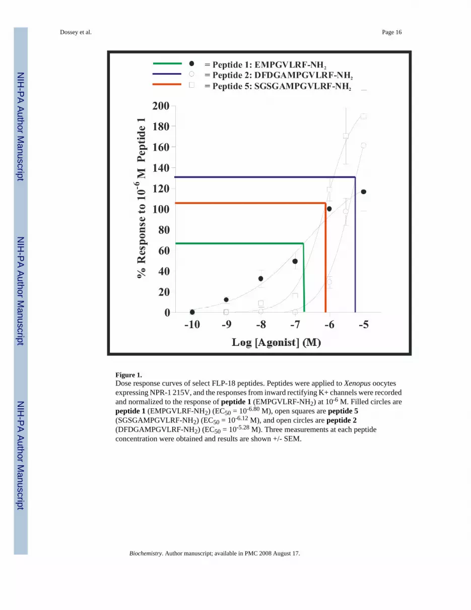
Figure 1.Dose response curves of select FLP-18 peptides. Peptides were applied to Xenopus oocytesexpressing NPR-1 215V, and the responses from inward rectifying K+ channels were recordedand normalized to the response of peptide 1 (EMPGVLRF-NH2) at 10-6 M. Filled circles arepeptide 1 (EMPGVLRF-NH2) (EC50 = 10-6.80 M), open squares are peptide 5(SGSGAMPGVLRF-NH2) (EC50 = 10-6.12 M), and open circles are peptide 2(DFDGAMPGVLRF-NH2) (EC50 = 10-5.28 M). Three measurements at each peptideconcentration were obtained and results are shown +/- SEM.
Dossey et al. Page 16
Biochemistry. Author manuscript; available in PMC 2008 August 17.
NIH
-PA Author Manuscript
NIH
-PA Author Manuscript
NIH
-PA Author Manuscript

Figure 2.NMR chemical shift deviations from random coil values. Experimental chemical shifts at pH∼2.3 were subtracted from sequence corrected random coil values (64,65). Filled and openbars represent alpha and amide protons, respectively.
Dossey et al. Page 17
Biochemistry. Author manuscript; available in PMC 2008 August 17.
NIH
-PA Author Manuscript
NIH
-PA Author Manuscript
NIH
-PA Author Manuscript

Figure 3.Amide region of one-dimensional NMR data, collected as a function of pH from about 1.9 to5.5. Peaks are labeled with their assigned amino acids and the panels correspond to thefollowing peptides: (ADD peptide sequences here as well) A. Peptide 3 = DFDGAM-NH2,B. Peptide 2 = DFDGAMPGVLRF-NH2, C. Peptide 8 = GFGDEM-NH2, D. Peptide 7 =GFGDEMSMPGVLRF-NH2, E. Peptide 5 = SGSGAMPGVLRF-NH2, F. Peptide 1 =EMPGVLRF-NH2. pH dependent interactions are summarized in Figure 5, and complete pKaanalyses are provided in supplementary material.
Dossey et al. Page 18
Biochemistry. Author manuscript; available in PMC 2008 August 17.
NIH
-PA Author Manuscript
NIH
-PA Author Manuscript
NIH
-PA Author Manuscript

Figure 4.Arg Hε region of one-dimensional NMR data, collected as a function of pH from 1.9 to 5.5.These long-range interactions on the penultimate C-terminal Arg result from the titratablecarboxylate groups on the N-termini. pH dependent interactions are summarized in Figure 5,and complete pKa analyses are provided in supplementary material. Legend: A. Peptide 4 =DFDGEMPGVLRF-NH2, B. Peptide 2 = DFDGAMPGVLRF-NH2, C. Peptide 10 =GFGDAMPGVLRF-NH2, D. Peptide 7 = GFGDEMSMPGVLRF-NH2, E. Peptide 5 =SGSGAMPVLRF-NH2, F. Peptide 1 = EMPGVLRF-NH2
Dossey et al. Page 19
Biochemistry. Author manuscript; available in PMC 2008 August 17.
NIH
-PA Author Manuscript
NIH
-PA Author Manuscript
NIH
-PA Author Manuscript

Figure 5.Proposed H-bonding Interactions Between Backbone Amide Protons and Carboxyl Side-chains: DFDGAM-NH2 = Peptide 3, DFDGAMPGVLRF-NH2 = Peptide 2,DFDGEMPGVLRF-NH2 = Peptide 4, SGSGAMPGVLRF-NH2 = Peptide 5, EMPGVLRF-NH2 = Peptide 2. Each H-bond acceptor residue is color coded to match the arrows leadingfrom it to its H-bond donors. The arrow widths are proportional to the relative extent to whichthat particular interaction affects the chemical shift of the amide proton at the point end of thearrow. The bar plots show the temperature coefficient of the backbone amide proton resonances
Dossey et al. Page 20
Biochemistry. Author manuscript; available in PMC 2008 August 17.
NIH
-PA Author Manuscript
NIH
-PA Author Manuscript
NIH
-PA Author Manuscript

Figure 6.Relationship between Temperature Coefficients and pH dependence of chemical shift amongBackbone Amide Protons: Plotted here is the chemical shift change with pH vs withTemperature for backbone amide resonances. R2 for the linear fit is 0.58. All data from peptides1-4 are represented.
Dossey et al. Page 21
Biochemistry. Author manuscript; available in PMC 2008 August 17.
NIH
-PA Author Manuscript
NIH
-PA Author Manuscript
NIH
-PA Author Manuscript

Figure 7.Relationship between overall peptide charge and activity on the NPR-1 receptor. The overallpeptide charge at neutral pH is plotted against activity for all of the peptides analyzed in thisstudy. For the linear fit, R2 = 0.67. Legend: Filled circle = 4, Open Circle = 2, Filled Square =9, Open Square = 5, Filled Triangle = 6, Open Triangle = 7, Filled Diamond = 1, Open Diamond= 10, Filled Hexagon = 11, Open Hexagon = 12 (numbers correspond to peptide numbers inTable 2)
Dossey et al. Page 22
Biochemistry. Author manuscript; available in PMC 2008 August 17.
NIH
-PA Author Manuscript
NIH
-PA Author Manuscript
NIH
-PA Author Manuscript

Figure 8.Model of interactions thought occurring within native FLP-18 peptides. This figure shows themost significant H-bonding interactions supported by NMR data. A: For DFDGAMPGVLRF-NH2, H-bonds between the D1 side-chain carboxylate and G4/A5 backbone amide protons aswell as H-bonding/ionic interaction between the D3 side-chain by red dashed lines. The N-terminal loop structure implicated in inhibiting binding to NPR-1 is circled in black. B:EMPGVLRF-NH2 is shown with the most significant H-bonding and ionic interactions forwhich we have evidence. Notice that it has no N-terminal loop, in contrast toDFDGAMPGVLRF-NH2. Also, the same unstructured region for both peptides is shown inribbon view. The N- and C- termini are also labeled on both peptides.
Dossey et al. Page 23
Biochemistry. Author manuscript; available in PMC 2008 August 17.
NIH
-PA Author Manuscript
NIH
-PA Author Manuscript
NIH
-PA Author Manuscript

NIH
-PA Author Manuscript
NIH
-PA Author Manuscript
NIH
-PA Author Manuscript
Dossey et al. Page 24
Table 1Sequence comparison of flp-18 and afp-1 precursor proteinsa
aPeptide sequences red, processing sites blue, * denotes amino acid gap in peptide, _ denotes gap. Note that this analysis is not an alignment (4,14).
bThe longer peptides for each precursor are underlined.
Biochemistry. Author manuscript; available in PMC 2008 August 17.

NIH
-PA Author Manuscript
NIH
-PA Author Manuscript
NIH
-PA Author Manuscript
Dossey et al. Page 25
Table 2Peptides used in this study
Name Sequencea % Response (n)b
Peptide 1c EMPGVLRF-NH2 100Peptide 2d 29.1±5.7 (16)
Peptide 3 0 (3)
Peptide 4 19.0±2.6 (8)
Peptide 5 SGSGAMPGVLRF-NH2 118.7±11.0 (4)Peptide 6 AAAAAMPGVLRF-NH2 62.4±3.2 (10)Peptide 7e 49.3±16.5 (4)
Peptide 8 0 (3)
Peptide 9f 45.0±10.5 (6)
Peptide 10g 104.8±2.8 (8)
Peptide 11 PGVLRF-NH2 43.0±5.5 (14)Peptide 12 PGVLRFPGVLRF-NH2 198.1±33.3 (10)aNaturally occurring sequences are underlined. The conserved PGVLRF-NH2 sequence is in bold. N-terminal “extension” sequences of native long flp-18
peptides are bold and: Red for C. elegans based sequences and Blue for A. suum based sequences.
bPeptides (10-6 M) were applied in 2 m pulses to Xenopus oocytes expressing NPR-1 215V. Results expressed as a % of response to 10-6 M Peptide 1
(EMPGVLRF-NH2) +/- SEM
cMost active native C. elegans FLP-18 peptide
dLongest and least active native FLP-18 peptide
eLongest Ascaris AFP-1 peptide
fChimera of long FLP-18 + long AFP-1
gChimera of long AFP-1 + long FLP-1
Biochemistry. Author manuscript; available in PMC 2008 August 17.