anxiety and depression: what do we know of neuropeptides?
TRANSCRIPT
Citation: Kupcova, I.; Danisovic, L.;
Grgac, I.; Harsanyi, S. Anxiety and
Depression: What Do We Know of
Neuropeptides? Behav. Sci. 2022, 12,
262. https://doi.org/10.3390/
bs12080262
Academic Editor: Dario Siniscalco
Received: 7 June 2022
Accepted: 27 July 2022
Published: 29 July 2022
Publisher’s Note: MDPI stays neutral
with regard to jurisdictional claims in
published maps and institutional affil-
iations.
Copyright: © 2022 by the authors.
Licensee MDPI, Basel, Switzerland.
This article is an open access article
distributed under the terms and
conditions of the Creative Commons
Attribution (CC BY) license (https://
creativecommons.org/licenses/by/
4.0/).
behavioral sciences
Review
Anxiety and Depression: What Do We Know of Neuropeptides?Ida Kupcova 1, Lubos Danisovic 1 , Ivan Grgac 2 and Stefan Harsanyi 1,*
1 Institute of Medical Biology, Genetics and Clinical Genetics, Faculty of Medicine, Comenius University inBratislava, Sasinkova 4, 811 08 Bratislava, Slovakia; [email protected] (I.K.);[email protected] (L.D.)
2 Institute of Anatomy, Faculty of Medicine, Comenius University in Bratislava, Sasinkova 4, 811 08 Bratislava,Slovakia; [email protected]
* Correspondence: [email protected]; Tel.: +421-2-59357-299
Abstract: In modern society, there has been a rising trend of depression and anxiety. This trendheavily impacts the population’s mental health and thus contributes significantly to morbidity and,in the worst case, to suicides. Modern medicine, with many antidepressants and anxiolytics at hand,is still unable to achieve remission in many patients. The pathophysiology of depression and anxietyis still only marginally understood, which encouraged researchers to focus on neuropeptides, as theyare a vast group of signaling molecules in the nervous system. Neuropeptides are involved in theregulation of many physiological functions. Some act as neuromodulators and are often co-releasedwith neurotransmitters that allow for reciprocal communication between the brain and the body.Most studied in the past were the antidepressant and anxiolytic effects of oxytocin, vasopressin orneuropeptide Y and S, or Substance P. However, in recent years, more and more novel neuropeptideshave been added to the list, with implications for the research and development of new targets,diagnostic elements, and even therapies to treat anxiety and depressive disorders. In this review, wetake a close look at all currently studied neuropeptides, their related pathways, their roles in stressadaptation, and the etiology of anxiety and depression in humans and animal models. We will focuson the latest research and information regarding these associated neuropeptides and thus picturetheir potential uses in the future.
Keywords: anxiety; depression; neuropeptides; melanocortins; CRH; NPY; galanin family; spexin;Substance P
1. Introduction
Anxiety disorder and depressive disorders present a broad spectrum of diagnoseswith a wide range of symptoms. The risk of depression is prevalent in younger patients(18–29 years old) but is not limited to this group, and even children with no indicationof etiology suffer from the depressive disorder [1,2]. The Global Burden of Diseases,Injuries, and Risk Factors Study (GBD) 2019 showed that the two most disabling mentaldisorders were depressive and anxiety disorders [3]. Before 2020, the prevalence of thedepressive disorder in European countries ranged from 2.58% to 10.33% with a meanoverall prevalence of 6.38 [4]. Studies from the USA reported prevalences of 10.4% and20.6% for 12-month and lifetime prevalences, respectively [5]. Global disease outbreaksin the past, or recently the COVID-19 pandemic, have significantly affected global mentalhealth. However, psychological factors are often neglected, even though pandemics arepsychological phenomena [6]. A systematic review on the effect of the COVID-19 pandemicon mental health in South Asian countries reported anxiety and depression rates of 34.1%to 41.3% in pooled patient samples [7]. In a recent meta-analysis on the global effect of theCOVID-19 pandemic on the general population, Salari et al. reported a pooled prevalenceof 31.9% for anxiety and 33.7% for depression, with a predominance in females [8]. Evenbefore the pandemic, countries reported a rising incidence of depression in females [9]. The
Behav. Sci. 2022, 12, 262. https://doi.org/10.3390/bs12080262 https://www.mdpi.com/journal/behavsci
Behav. Sci. 2022, 12, 262 2 of 34
impact of the pandemic had an amplifying effect on mental health issues among the generalpopulation of low- and middle-income countries [10]. Adjusted for COVID-19, globalprevalence showed similar results with elevation in anxiety (25.6%) and major depressioncases (27.6%), which is a prominent reminder that mental health throughout the globe is aneverlasting issue [11].
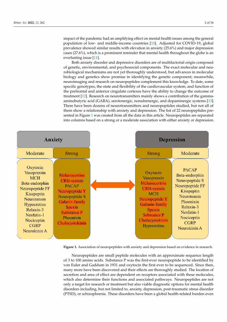
Both anxiety disorder and depressive disorders are of multifactorial origin composedof genetic, environmental, and psychosocial components. The exact molecular and neu-robiological mechanisms are not yet thoroughly understood, but advances in molecularbiology and genetics show promise in identifying the genetic component; meanwhile,neuroimaging and research on neuropeptides complement this knowledge. To date, somespecific genotypes, the state and flexibility of the cardiovascular system, and function ofthe prefrontal and anterior cingulate cortexes have the ability to change the outcome oftreatment [12]. Research on neurotransmitters mainly shows a contribution of the gamma-aminobutyric acid (GABA), serotonergic, noradrenergic, and dopaminergic systems [13].There have been dozens of neurotransmitters and neuropeptides studied, but not all ofthem show a relationship with anxiety and depression. The list of 22 neuropeptides pre-sented in Figure 1 was created from all the data in this article. Neuropeptides are separatedinto columns based on a strong or a moderate association with either anxiety or depression.
Behav. Sci. 2022, 12, x FOR PEER REVIEW 2 of 36
a pooled prevalence of 31.9% for anxiety and 33.7% for depression, with a predominance in females [8]. Even before the pandemic, countries reported a rising incidence of de-pression in females [9]. The impact of the pandemic had an amplifying effect on mental health issues among the general population of low- and middle-income countries [10]. Adjusted for COVID-19, global prevalence showed similar results with elevation in anx-iety (25.6%) and major depression cases (27.6%), which is a prominent reminder that mental health throughout the globe is an everlasting issue [11].
Both anxiety disorder and depressive disorders are of multifactorial origin com-posed of genetic, environmental, and psychosocial components. The exact molecular and neurobiological mechanisms are not yet thoroughly understood, but advances in molec-ular biology and genetics show promise in identifying the genetic component; mean-while, neuroimaging and research on neuropeptides complement this knowledge. To date, some specific genotypes, the state and flexibility of the cardiovascular system, and function of the prefrontal and anterior cingulate cortexes have the ability to change the outcome of treatment [12]. Research on neurotransmitters mainly shows a contribution of the gamma-aminobutyric acid (GABA), serotonergic, noradrenergic, and dopaminer-gic systems [13]. There have been dozens of neurotransmitters and neuropeptides stud-ied, but not all of them show a relationship with anxiety and depression. The list of 22 neuropeptides presented in Figure 1 was created from all the data in this article. Neuro-peptides are separated into columns based on a strong or a moderate association with ei-ther anxiety or depression.
Figure 1. Association of neuropeptides with anxiety and depression based on evidence in research.
Neuropeptides are small peptide molecules with an approximate sequence length of 3 to 100 amino acids. Substance P was the first-ever neuropeptide to be identified by von Euler and Gaddum in 1931 and oxytocin the first ever to be sequenced. Since then, many more have been discovered and their effects are thoroughly studied. The location of secretion and area of effect are dependent on receptors associated with these mole-cules, which also determine their functions and associated pathways. Neuropeptides are not only a target for research or treatment but also viable diagnostic options for mental health disorders including, but not limited to, anxiety, depression, post-traumatic stress
Figure 1. Association of neuropeptides with anxiety and depression based on evidence in research.
Neuropeptides are small peptide molecules with an approximate sequence lengthof 3 to 100 amino acids. Substance P was the first-ever neuropeptide to be identified byvon Euler and Gaddum in 1931 and oxytocin the first ever to be sequenced. Since then,many more have been discovered and their effects are thoroughly studied. The location ofsecretion and area of effect are dependent on receptors associated with these molecules,which also determine their functions and associated pathways. Neuropeptides are notonly a target for research or treatment but also viable diagnostic options for mental healthdisorders including, but not limited to, anxiety, depression, post-traumatic stress disorder(PTSD), or schizophrenia. These disorders have been a global health-related burden even

Behav. Sci. 2022, 12, 262 3 of 34
before the COVID-19 pandemic, which only exacerbated and enhanced all contributingfactors, causing mental health issues to become one of the dominant issues.
Several studies were aimed at addressing a multitude of individual objective and sub-jective symptoms, using diverse methods. These disorders exhibit, indeed, gender and occu-pational differences and higher strength in patients suffering from seriousdiseases [14–16]. Substances of abuse are often used to cope with anxiety and depres-sion, leading to increased susceptibility to nicotinism, alcoholism, or drug misuse andaddiction. Nicotine creates a sense of relaxation, and ethanol has sedative and anxiolyticeffects, so there is no wonder these substances are often used in self-treatment. Theserelatively frequent habits are used to relieve self-perceived stress or anger, blunt feelings,and relieve the sensation of anxiety, even though over time they may have rather oppositeeffects. The use of these coping mechanisms is frequently seen in clinical practice eventhough a recent systematic review that collected 148 studies concerning the associationof cigarette smoking behavior with depression and anxiety reported contrasting resultsbecause of inconsistencies in directions and strengths of the associations between thesevariables [17]. Many pathways with involvement in anxiety, depression, or addictionare mediated or affected by various neuropeptides, which could prove a viable path forresearch and the treatment of patients.
All the aforementioned facts urge more attention to mental health since these disordershave a rising tendency despite various available treatment methods. Research aimed notonly at treatment but also at creating more effective diagnostic tools and markers areneeded. Research suggests that neuropeptides could change the management of anxiety anddepression [18]. The importance lies not only in the immediate future but also as a lessonfrom the past, to prepare for the future situation where population mobility reductionswill be implemented and patients will be unable to visit a specialist or even benefit fromtelemedicine. This problem is of cardinal importance in all fields of clinical medicine.
The latest research in the field of neuropeptides presents promising results for thediagnosis, management, and treatment of patients with anxiety or depression. In thisreview, we take a close look at all currently studied neuropeptides, their related pathways,receptors, and their roles in the etiology of anxiety and depression in humans and animalmodels. We will focus on the latest research and information regarding these associatedneuropeptides and thus picture their potential uses in the future.
2. Neuropeptides in Anxiety and Depression
Neuropeptides are small polypeptides of size ranging from 3 to 100 amino acids.Secretion is localized in neurons and the effect, depending on receptors, is local or distant.Affected are both neurons and other somatic cells, indicating a wide array of effects [19,20].Some are synthesized as precursors and later cleaved into mature forms, while some haveno post-translational changes [21]. Stress, pain, or emotional distress often leads to anxietyand depression. Studies of neuropeptides and their related receptors pose a high potentialfor a better understanding of the etiology/etiopathogenesis of these disorders, leadingto better, more targeted surveillance and treatment, especially as diagnostic markers. InTable 1, we summarize the neuropeptides mentioned in this article with their relatedreceptors and encoding genes. Data were acquired using the OMIM® database accessed on25 May 2022 [22].

Behav. Sci. 2022, 12, 262 4 of 34
Table 1. Anxiety and depression-related neuropeptides, encoding genes, and related receptors.(Receptors of neuropeptides without any relation to anxiety or depression are not mentioned inthis table.)
Neuropeptide Gene (Symbol) Cytogenetic Location Related Receptors
Oxytocin OXYTOCIN (OXT) 20p13 OXTR
Vasopressin ARGININE VASOPRESSIN (AVP) 20p13 AVPR1A; AVPR1B; AVPR2
Adrenocorticotropic hormone PROOPIOMELANOCORTIN(POMC) 2q23.3 MCR1; MCR2; MCR3; MCR4;
MCR5
Corticotropin-releasing hormone CORTICOTROPIN-RELEASINGHORMONE (CRH) 8q13.1
CRHR1; CRHR2Urocortin 1 UROCORTIN (UCN) 2p23.3
Urocortin 2 UROCORTIN II (UCN2) 3p21.31
Urocortin 3 UROCORTIN III (UCN3) 10p15.1
Pituitary adenylatecyclase-activating polypeptide
ADENYLATECYCLASE-ACTIVATING
POLYPEPTIDE 1 (ADCYAP1)18p11.32 ADCYAP1R1; VIPR1
Melanocyte stimulating hormone PROOPIOMELANOCORTIN(POMC) 2p23.3 MC3R; MC4R
Melanin-concentrating hormonePRO-MELANIN-
CONCENTRATINGHORMONE (PMCH)
12q23.2 MCH-R1; MCH-R2
Beta-endorphin PROOPIOMELANOCORTIN(POMC) 2q23.3 OPRM1
other µ-opioid receptors
Neuropeptide Y NEUROPEPTIDE Y (NPY) 7p15.3 NPY1R; NPY2R; NPY5R
Neuropeptide S NEUROPEPTIDE S (NPS) 10q26.2 NPSR1
Neuropeptide FF NEUROPEPTIDE FF-AMIDEPEPTIDE (NPFF) 12q13.13 NPFFR1; NPFFR2
Galanin GALANIN (GAL) 11q13.2 GALR1; GALR2; GALR3;GPR151
Galanin-like peptide GALANIN-LIKE PEPTIDE (GALP) 19q13.43 GALR1; GALR2
Spexin SPEXIN HORMONE (SPX) 12p12.1 GALR2; GALR3
Kisspeptin KISS1 METASTASIS SUPPRESSOR(KISS1) 1q32.1 KISS1R
Substance P TACHYKININ 1 (TAC1) 7q21.3 TACR1; TACR2; TACR3
Neurotensin NEUROTENSIN (NTS) 12q21.31 NTSR1; NTSR2
Hypocretin HYPOCRETIN (HCRT) 17q21.2 HCRTR1; HCRTR2
Phoenixin SMALL INTEGRAL MEMBRANEPROTEIN-20 (SMIM20) 4p15.2 GPR17
Relaxin 3 RELAXIN 3 (RLN3) 19p13.12 RXFP3
Nesfatin-1 NUCLEOBINDIN 2 (NUCB2) 11p15.1 Not discovered
Nociceptin PREPRONOCICEPTIN (PNOC) 8p21.1 OPRL-1
Cholecystokinin CHOLECYSTOKININ (CCK) 3p22.1 CCKAR; CCKBR
Calcitonin gene-related peptide CGRP RECEPTOR COMPONENT(CGRP) 7q11.21 CGRPR
Neurokinin A TACHYKININ 1 (TAC1) 7q21.3 TACR1; TACR2; TACR3
Signaling axes (or pathways) studied in relation to anxiety and depression are thehypothalamic–pituitary–adrenal (HPA) axis, the hypothalamic–pituitary–gonadal (HPG)axis, and the gut–brain axis (GBA). The GBA is newer, and less thoroughly studied, al-though reports indicate that the microbiome and gut hormones may play a role in theetiology of depression [23]. More studied are the HPA and HPG axes. Interactions of theseaxes are presented in Figure 2.

Behav. Sci. 2022, 12, 262 5 of 34Behav. Sci. 2022, 12, x FOR PEER REVIEW 5 of 36
Figure 2. Gut-brain axis, HPA axis, HPG axis and their interactions.
The HPA axis affects many different systems in the mammalian body, with a mod-ern link to neuropeptides, psychiatry, and neurology, on which we will focus in this re-view [24]. The HPA axis is controlled by the limbic system depending on stimuli (stress-ors) received by the latter, and so inducing release of the CRH. CRH stimulates the re-lease of ACTH (adrenocorticotropic hormone), which affects the adrenal glands and the production of glucocorticoids. Stress-adaptation responses are mediated by corti-costerone in rodents and by cortisol in humans.
Circulating levels of gluco- and mineralo-corticoids are essential for negative feed-back on the hypothalamus, which has been reported faulty in patients suffering from depression [25]. A systematic review and meta-analysis on the effects of aging and the HPA axis in patients with depression reported that older patients suffering from depres-sion showed a higher degree of dysregulation in the HPA axis [26]. A bipolar disorder- (BD) focused systematic review and meta-analysis found a possible association of pro-gressive HPA axis dysfunction with the cognitive deterioration of BD patients, which seemed rather a risk factor than a determinant, but nonetheless an important one [27]. In a recent review, an association between early-life stress in the HPA axis and anxiety has been found, followed by hyperactivity in the axis acting as a risk factor for relapses [28]. Effects of the microbiota on the HPA axis, stress response, anxiety-like behavior, endo-crine abnormalities, and wider neuropsychiatric disorders have been observed, calling for further investigation [29–32]. In a systematic review, Juruena et al., based on the ac-tivity of the HPA axis, found a difference between melancholic and atypical depressive subtypes [33]. However, they attributed these findings to hypercortisolism in melancho-lia and rather normal, than decreased, function in atypical depression [33].
The HPG axis is represented by neurons of the gonadotropin-releasing hormone (GnRH), which in mammals is a 10-amino acid peptide. GnRH stimulates the synthesis and secretion of luteinizing hormone (LH) and follicle-stimulating hormone (FSH), thus having an important role in the regulation of fertility and reproduction. GnRH is inhibit-ed by the gonadotropin-inhibitory hormone (GnIH), which belongs to the RFRP family. The RFRPs (RF-amide-related peptides) act in regulatory regard to the HPG axis [34]. Studies on the GnIH have been associated with different behaviors in male rats [35].
Figure 2. Gut-brain axis, HPA axis, HPG axis and their interactions.
The HPA axis affects many different systems in the mammalian body, with a modernlink to neuropeptides, psychiatry, and neurology, on which we will focus in this review [24].The HPA axis is controlled by the limbic system depending on stimuli (stressors) receivedby the latter, and so inducing release of the CRH. CRH stimulates the release of ACTH(adrenocorticotropic hormone), which affects the adrenal glands and the production ofglucocorticoids. Stress-adaptation responses are mediated by corticosterone in rodents andby cortisol in humans.
Circulating levels of gluco- and mineralo-corticoids are essential for negative feedbackon the hypothalamus, which has been reported faulty in patients suffering from depres-sion [25]. A systematic review and meta-analysis on the effects of aging and the HPA axisin patients with depression reported that older patients suffering from depression showeda higher degree of dysregulation in the HPA axis [26]. A bipolar disorder- (BD) focusedsystematic review and meta-analysis found a possible association of progressive HPA axisdysfunction with the cognitive deterioration of BD patients, which seemed rather a riskfactor than a determinant, but nonetheless an important one [27]. In a recent review, anassociation between early-life stress in the HPA axis and anxiety has been found, followedby hyperactivity in the axis acting as a risk factor for relapses [28]. Effects of the microbiotaon the HPA axis, stress response, anxiety-like behavior, endocrine abnormalities, and widerneuropsychiatric disorders have been observed, calling for further investigation [29–32]. Ina systematic review, Juruena et al., based on the activity of the HPA axis, found a differencebetween melancholic and atypical depressive subtypes [33]. However, they attributed thesefindings to hypercortisolism in melancholia and rather normal, than decreased, function inatypical depression [33].
The HPG axis is represented by neurons of the gonadotropin-releasing hormone(GnRH), which in mammals is a 10-amino acid peptide. GnRH stimulates the synthesisand secretion of luteinizing hormone (LH) and follicle-stimulating hormone (FSH), thushaving an important role in the regulation of fertility and reproduction. GnRH is inhibitedby the gonadotropin-inhibitory hormone (GnIH), which belongs to the RFRP family. TheRFRPs (RF-amide-related peptides) act in regulatory regard to the HPG axis [34]. Studies

Behav. Sci. 2022, 12, 262 6 of 34
on the GnIH have been associated with different behaviors in male rats [35]. Stress-induced increase in adrenal glucocorticoids increased the RFRPs, which contributed tothe suppression of reproductive function through the HPG axis [36]. In birds, a smalldose of interfering RNA targeted against the GnIH precursor mRNA caused elevation inaggressive and sexual behaviors [37]. Iwasa et al. reviewed the field of stress-inducedreproductive dysfunction and found an association with the GnIH/RFRP-3 system [38].These findings are important as stress, whether physical or emotional, is related to anxietyand depression in a reciprocal character, and one may affect or over time induce theother. The use of GnRH analogs caused upregulation in the expression of phoenixin inhypothalamic, hypophyseal, and ovarian regions, while the GPR173 expressions weredownregulated in the hypothalamus and pituitary [39].
2.1. Oxytocin
Oxytocin (OXT) is composed of nine amino acids and was the first polypeptide hor-mone to be sequenced. OXT is synthesized in the supraoptic and paraventricular nucleiof the hypothalamus [40]. It became first known for its role in lactation, parturition, andmaternal behavior, yet over the last several decades it has also been implicated in memory,regulation of anxiety, mood, and social behavior [41]. In reaction to acute stress, oxytocinis released from the nucleus centralis of the amygdala, which leads to the local activa-tion of GABA-ergic interneurons [42]. While the amygdala is responsible for acute stressreactions, stria terminalis is the structure involved in the transition to chronic anxiousstates. A portion of oxytocinergic neurons from the periventricular nucleus projects di-rectly into this structure [43]. The anterior cingulate cortex likely facilitates learning andsocial adaptation [44,45]. Oxytocin receptors (OXTR) are present across the whole brain,and it seems that the research of polymorphisms and epigenetic markers for OXTR isa promising area for future research [46]. Myers et al. reported two single nucleotidepolymorphisms (SNP) in the OXTR gene in depressed patients [41]. Costa et al. describeda relationship between separation anxiety in adulthood and SNP rs53576 of the third in-tron of the OXTR gene, which seems of substance for the epigenetic regulation of thisgene [47]. An allele of rs53576 is also related to increased suicide risk, according to someauthors [48]. Oxytocin interacts with various neurotransmitters and neuroendocrine sys-tems [49]. The most prominent ones are the serotonergic and GABA-ergic systems and theHPA axis. Mottolese et al. have proven the interplay between oxytocin and serotonin usinga 5-HT1A receptor antagonist 2’methoxyphenyl-fluorobenzamidoethylpiperazine (MPFF).Intranasal administration of oxytocin resulted in increased levels of MPPF in nucleus dor-salis raphes, the right amygdala-hippocampus-parahippocampus complex, insula, andright medioventral prefrontal cortex, measured using PET MRI [50]. The interplay betweenthe oxytocinergic and serotoninergic systems is further supported by the fact that MDMA(3,4-methylenedioxymethamphetamine) causes oxytocin-mediated behavioral changes viaserotoninergic 5HT1A receptors [51]. Furthermore, oxytocin is also connected to the HPAaxis. Part of the neurons in the paraventricular nucleus co-expresses oxytocin and CRH, aportion of CRH-releasing neurons expresses mRNA for the OXTR, and clusters of OXTRexpress CRH receptor CRHR2. Centrally administered oxytocin reduces the excitability ofCRH neurons, inhibits their spontaneous excitability, and lowers the expression of CRHmRNA and neuroendocrine response to stress [42]. In animals, the central administration ofoxytocin reduces anxiety-like behaviors and lowers plasmatic cortisol levels [52]. Further-more, the medial prefrontal cortex contains oxytocinergic interneurons synthetizing CRHbinding protein (CRHBP) and GABA. In male rodents, the stimulation of these interneuronsleads to the release of CRHBP and the reduction of CRH activity and anxiety, while infemale rodents CRHBP was not able to effectively inhibit CRH activity, which may bedue to higher levels of CRH in paraventricular nuclei (PVN) [53]. Antagonistic activity onGABAA receptors in the PVN blocks the suppression of stress-induced CRH release andthe application of oxytocin into the PVN results in a significant increase in GABA releasefrom the PVN [52]. Meisenberg was the first to describe the antidepressant-like effect of

Behav. Sci. 2022, 12, 262 7 of 34
oxytocin when oxytocin application had an effect similar to the tricyclic antidepressantimipramine in rodents—it resulted in reduced immobility during the forced swim test [51].Later, Scantamburlo et al. reported an inverse relationship between plasma oxytocin levelsand the severity of symptoms of depression and anxiety in depressed patients [54]. Jobstet al. measured plasmatic levels of oxytocin in chronically depressed patients before andafter psychotherapy. In their research, increased levels of oxytocin correlated with a re-duction of subjectively reported depressive symptoms according to the Beck DepressionInventory-II [55]. Lancaster et al. found that higher endogenous oxytocin levels are associ-ated with a reduced central amygdala volume and blood oxygen level-dependent activityin response to aversive stimuli [56]. However, the relationship between oxytocin and de-pressive and anxiety symptoms is not as straightforward as it may seem, and the literatureis full of contradictory results. For example, Parker et al. found increased oxytocin levelsin depressed subjects, and more recently Tabak et al., examining the role of oxytocin insocial anxiety in humans, reported increased plasma concentrations of oxytocin followingthe Trier Social Stress Test (TSST), but this increase was specific to women, and that socialanxiety moderated the OXT increase with more socially anxious participants showinggreater increases than those with lower levels of social anxiety [57]. This can be the resultof the so-called oxytocin paradox, which has been described as the context-dependentfeedback loop of OXT signaling based on socio-psychological settings leading to positiveor adverse psychological consequences [58]. The functioning of oxytocin varies accordingto contextual factors and individual differences, including gender, age, and psychosocialfunctioning [59]. In the past years, research allowed us to better understand signaling waysaffected by OXT and its complex regulation, and further multi-peptide-centered researchwill reveal this intricate web of regulation.
2.2. Vasopressin
Vasopressin, also known as the antidiuretic hormone (ADH) or arginine vasopressin(AVP), consists of 9 amino acids, belongs to nonapeptides, and is synthesized by magno-cellular cells of the hypothalamic supraoptic nucleus and PVN whose axons project tothe posterior pituitary [60,61]. After release into the bloodstream, it is transported pre-dominantly to kidneys and blood vessels, where it regulates physiological functions (e.g.,resorption of water, vasoconstriction, . . . ). Vasopressin has amino acid sequences andtertiary structures similar to oxytocin, probably due to the shared origin of their encodinggenes [62]. In addition, it is possible that an interplay between oxytocin and vasopressin isinvolved in mood and stress-response regulation [63]. It binds to four types of G-protein-coupled receptors: AVPR1A, AVPR1B, AVPR2 (for clarity reasons further as V1A, V1B,and V2, respectively), and oxytocin receptor [64]. V2 receptor binds to vasopressin in thekidney, where it participates in water homeostasis [65]. On the other hand, V1B and V1Areceptor subtypes are promising sites of interest for psychiatry. The V1B subtype is highlyexpressed in the anterior pituitary gland, where it stimulates corticotropin release and isalso localized in the amygdala, hippocampus, and hypothalamus [66]. Purba et al. alreadyin 1996 reported increased numbers of V1B receptors in the postmortem analysis of brainsof depressed patients compared to controls [67]. In depression and anxiety, vasopressinbinds to V1 receptors in the brain. Hoghson et al. used V1B-30N-A potent V1B receptorantagonist in rat pups to examine the potential role of V1B in anxiety. They reported thatV1B-30N had a dose-dependent anxiolytic effect in the separation-induced vocalization testin rat pups, moreover without causing sedation [64]. The interaction between vasopressinand oxytocin has already been mentioned, but vasopressin co-operates at least with oneother neuromodulator system—the serotonergic. Ishizuka et al. used V1B knockout micetreated with a selective serotonin reuptake inhibitor (SSRI) and serotonin noradrenalinereuptake inhibitor (SNRI) to examine whether at least part of the SSRI´s effects might bemediated via the V1B receptor. The effects were evaluated in experiments using an elevatedplus-maze (EPM) test and a hole-board (HB) test. Chronic treatment of V1bR KO mice withSSRI did not change the amount of time spent on the open arms, the number of head dips,

Behav. Sci. 2022, 12, 262 8 of 34
or the number of readings, while chronic treatment with SNRI significantly increased thetime spent on the open arms and the number of head dips. These results suggest that theanxiety action of 5-HT reuptake inhibitors might partly involve V1bR regulating the anxietybehaviors [66]. While the V1B receptor likely plays a role in depression and anxiety, theV1A subtype seems to be related to a different type of psychopathology. Vollegbregt et al. intheir study reported an association between RS3 microsatellite repeats within the promoterregion of the V1A gene and extreme childhood aggression, while no association was foundbetween childhood aggression and RS1 repeats [68]. However, in addition to factors relatedto the V1 receptor, the levels of vasopressin also likely play a role in the pathophysiologyof depression and anxiety. It was shown that chronically elevated plasma vasopressinlevels may induce depressive symptomatology [69]. Another research project pointingto the positive correlation between vasopressin levels and depression was performed byMlynarik et al., which showed reduced depression-like behavior in vasopressin-deficientBattleboro rats during the forced swim test [70]. The results of human studies agree withanimal research. Goekoop et al. measured plasma levels of vasopressin in 89 patientswith a highly anxious-retarded subtype of depression with a family history of depression.Depression with above-normal plasma AVP, as well as familial depression with above-normal plasma AVP, showed a high correlation between anxiety and retardation and thiscorrelation was significantly higher than that found in the depressed patient control groups.The data support the delimitation of a largely familial depression with above-normalplasma AVP, vasopressinergic activation of the HPA axis, and a variable anxious-retardedphenotype [71]. As already mentioned above, the current situation regarding the efficienttreatment of anxiety and depression is far from satisfactory despite the fact that there aremultiple pharmacological agents on the market. The rate at which novel antidepressantsare developed and introduced into clinical practice is very slow. This motivates researchinto different treatment modalities, of which V1B antagonists seem to be one promisingoption [72,73].
2.3. Melanocortins
Melanocortins are a group of peptide hormones derived from pro-opiomelanocortin(POMC). This group includes the adrenocorticotropic hormone (ACTH), alfa-, beta-, andgamma-melanocyte stimulating hormones (MSH), beta-endorphin, and corticotropin-likeintermediate peptide (CLIP), the adrenocorticotropic hormone fragment [74]. Melanocortinreceptors are associated with obesity, erectile dysfunction, cachexia, pain, depression,and anxiety [75]. Melanocortins work through the melanocortin receptors. The fivemelanocortin receptors are GPCRs identified as MC1 to five receptors. To date, onlyMC3R and MC4R with their ligand, the alfa-MSH (also gamma-MSH in the case of MC3R)were connected to anxiety and depression [76]. The main expression sites of melanocortinneurons are located in the brainstem and the hypothalamus, in the arcuate nucleus (ARC),closely communicating with AgRP- and Neuropeptide Y- (NPY) expressing neurons, thusindicating their participation in the system [77,78]. The alpha-MSH was reported to sup-press the effects of NPY, thus reducing its antidepressant effect [79]. The administration ofalpha-MSH antagonist with NPY shows a synergistic anxiolytic effect [80]. The inhibitionof MC4 receptors in the dorsal raphe nucleus (DRN) using alpha-MSH induces anxietyand depression and reduces feeding in mice [81]. PVN, ARC, and DRN are predominantlocations of the alpha-MSH effect [82]. The circle of regulation in the melanocortin systemis closed by the receptor antagonist, the Agouti-related peptide (AgRP), with an orexigeniceffect [83]. Chronic administration of a high-fat diet blunts AgRP response to anxiety anddepression signals, as well as hunger by reducing GABA-ergic outputs from AgRP aimedat MC4R. However, GABA-ergic stimulation and suppression of the 5-HT3R within theMC4R neurons in the bed nucleus causes cessation of the effect of the high-fat diet-inducedanxiety and depression [84]. The combined effect also reduces food intake and thus bodyweight. These findings indicate that initially AgRP regulates appetite, but later with aloss of effect, MC4Rs are a viable option for further research on anxiety and depression

Behav. Sci. 2022, 12, 262 9 of 34
treatment [85,86]. Further associations are discussed in individual sections. A specificregulation protein, which affects the melanocortin system, is the melanocyte-stimulatinghormone release-inhibiting factor-1 (MIF-1) with its analog Nemifitide. Both show lowaffinity to µ-opioid receptors and function as blockers of alpha-MSH release, thus reducingits inhibitory effect on NPY. Preclinical studies have shown effects in depressive disorder,but further research into this particular peptide is necessary to prove its efficacy.
2.4. Corticotropin-Releasing Hormone
Corticotropin-releasing hormone (CRH) is also known as the corticotropin-releasingfactor (CRF); however, for precise nomenclature reasons, further on, the term CRH willbe used. CRH is the central regulator of the hypothalamic–pituitary–adrenal (HPA) axis,which is the main organizer of the body’s response to stress. CRH consists of 41 aminoacids and binds to CRHR1 and CRHR2, two GPCRs (not counting alpha and beta splicingvariants), both in the central nervous system and the R2 also in peripheral tissues [87].CHR receptors are expressed in the pituitary, amygdala, hippocampus, brain stem, andcingulate cortex (20). The CRH system consists of CRH itself, then Urocortin 1, 2, and 3,which all serve as ligands for the CRH receptors [88]. Effects of this system lie not onlyin the mediation of stress-related responses but also in the regulation of inflammation,with a close relationship with inflammation-related pain, as macrophages, monocytes,or mast cells do express both CRH receptors, thus serving as targets of this system [89].CRH-induced stress-related responses through the HPA axis have a wide area of effect,recently measured in human hair follicles connected to hair loss interestingly antagonizableby caffeine [90]. Here we can observe how deteriorative an effect the CRH-HPA systemcan have on the human body and how broad the effect is. It is not surprising that manyhuman and animal studies report CRHs’ effect on the HPA axis regulation. Chronicstress activates the CRH-related signaling in the bed nucleus and induces maladaptivebehavior in mice [91]. This goes in line with observed HPA axis dysfunction in patientswith major depression or schizophrenia, where the continuous effect of CRH on the HPAaxis causes disbalance (increased cortisol, increased ACTH, reduced feedback) and resultsin a pathology [92,93]. This disbalance is targeted by antidepressants, which after a periodof time have the potential to revert changes in HPA axis hyperactivation. The CRHsystem is long associated with serotonergic mediation, emotional disbalance, behavioralchanges, anxiety, and depression [94–96]. CRHR1 gene variants have been linked to ahigher susceptibility to depression or panic disorder [97,98]. A sex-specific stress reactionin females has been observed in an animal model, with sex hormones affecting CRHregulation [99]. Transgenic mice with induced CRH expression show hyperactivation ofthe HPA axis and an increase in stress-mediated processes and behaviors, treatable withCRHR1 antagonists [100].
Further members of the CRH family are the urocortins. Urocortin 1, 2, and 3 are agroup of three peptides (UCN1, UCN2, and UCN3). UCN1 is a 40-amino acid peptide,while UCN2 and 3 are paralogous 38-amino acid peptides that bind to CRHR2 [101]. UCN1is produced by the hypothalamus, substantia nigra, and the pituitary gland and has a highaffinity for both CRH receptors, suggesting its role in behavior and interestingly also regu-lation of food intake [102]. The intracerebrovascular injection of UCN1 has anxiogenic and“anti-social” effects antagonizable by CRHR1 antagonists [103,104]. However, the CRHR1antagonist had no effects on social interaction on its own but antagonized the decreases insocial interaction induced by stress or UCN1 [105]. Summarized by Tanaka et al., UCN2and UCN3 show an association with depression-like behavior, where stimulation by UCN1(CRHR1-2) is antagonized by UCN2 and UCN3 (CRHR2), thus resulting in no effect [106].
The latest members associated with the CRH family are four teneurins, or teneurin C-terminal associated peptides (TACPs), which share homology with the amino acid sequenceof CRH and bind to latrophilins (a group of highly conserved GPCRs). Their effect is tissue-specific, and reports show an ability of TACP-1 to reduce anxiety, addiction, and depressionin stress-induced behaviors [107].
Behav. Sci. 2022, 12, 262 10 of 34
On the other hand, studies and preclinical models also show mixed results [108]. Insome cases, CRH or TACP receptor antagonists have different effects on anxiety, basedon the animal test line, the baseline of depression, or perceived anxiety or the treatmentregimen, but also depending on the test, as these therapeutics do not work under basalconditions. However, if depression- or anxiety-related symptoms or signaling are present,these therapeutics do have a significant effect [109]. In other words, a stressor (depressionor anxiety) must be present for antidepressants and anxiolytics to work [110]. Theseconclusions are not surprising, as urocortins and their over-expression in CRH-deficientindividuals are often induced as a compensatory mechanism in absence of CRH. The sameresult is for models with CRH over-expression antagonizable by CRHR1 antagonists, wheresome lines do exhibit higher anxiety and vulnerability to stress, but in other lines that donot exhibit these traits, a UCN1 downregulation was found [111,112].
2.5. Pituitary Adenylate Cyclase-Activating Peptide
Pituitary adenylate cyclase-activating peptide (PACAP) is a neuropeptide existingin two isoforms, either 27 or 38 amino acids long. PACAP is relatively well-conservedacross many species [113]. Wide expression in central and peripheral tissues indicates arole in multiple physiological functions, such as the modulation of nociception, regulationof prolactin release, and food intake, along with stress, anxiety, and depression [114,115].PACAP has the highest affinity for the ADCYAP1R1 receptor, known also as the PAC1receptor [116]. Studies have associated this neuropeptide with stress-related mood dis-orders and adaptation [19,117]. PASAP is predominately expressed in the amygdala andthe bed nucleus [118,119]. Dore et al. observed pro-depressive and anxiety-like effectson rats after intracerebroventricular PACAP administration [120]. Human studies showan association of PACAP/PAC1 receptor with behavioral and endocrine responses, alsoin neuropsychiatric disorders such as schizophrenia or PTSD [121–123]. A recent animalmodel of PACAP-mutated mice under chronic variable mild stress exhibited viable re-sults for future depression-targeted tests [124]. PACAP-KO mice exhibit decreased c-Fosexpression, which shows a close connection not only with CRH and wider melanocortinsignaling [125,126]. These results go in line with previous results on PACAP’s influenceon CRH [127]. Additionally, a study on the consequences of chronic stress on PACAPexpression in the bed nucleus goes in hand with CRH, indicating that synergic modulationexists in anxiety-like behavior [128]. The anxiety-associated effect of PACAP was furtherstudied in mice, where the induction of PACAP-firing neurons aimed at the bed nucleusenhanced anxiety-like behavior [129].
2.6. Melanin-Concentrating Hormone
Melanin-concentrating hormone (MCH), or pro-melanin-concentrating hormone(PMCH), exists in a cyclic form and consists of 19 amino acids. MCH binds to two GPCRs,the MCH-1R and MCH-2R, of which only the 1R type is expressed in rodents, while both arepresent in humans [130]. Receptors are widely expressed in the brain, but MCH secretion,depending on species, is limited to sections of the hypothalamus and zona increta [131].MCH is involved in the regulation of feeding and energy homeostasis, also stress-relatedadaptation, emotions, and cognitive functions, some of which are due to the MCH inter-action with neurotransmitters of serotonergic and cholinergic systems [132,133]. MCHreverses activation of the HPA axis and also interacts with associated serotonergic andcholinergic pathways [134–136]. MCH-1R exhibits a potential for anxiety and depressiontreatment because even in animal models, MCH-1R knockouts exhibit an anxiety-resistantphenotype [137,138]. This potential has been studied by the administration of differentMCH-1R antagonists that produced antidepressant results in rats [139,140]. MCH-1Rreceptor antagonists infused in the nucleus accumbens are producing faster anxiolyticand antidepressant effects in mice and rats compared to the selective serotonin reuptakeinhibitors (SSRIs), while receptor agonists produce opposite effects [141]. This happensconcurrently with induced sensitivity of dopamine D2 and D3 receptors [141]. The nucleus

Behav. Sci. 2022, 12, 262 11 of 34
accumbens has a relation to the mediation of relative motivation for rewards. A recentanimal study observed reduced stress-induced anxiety and depression after intranasaladministration of MCH [142]. These results in animal studies are further proof of theimportant role of MCH in the etiology of anxiety and depression with connections to theHPA axis via CRH, where the MCH-1R antagonists act as inhibitors of this connection.
2.7. Beta-Endorphin
Beta-endorphin (β-endorphin) is a 31-amino acid endogenous opioid neuropeptidethat binds to opioid receptors. The mature form is derived from precursor POMC. Therehave been conflicting findings in research regarding its potential role in depression, but re-cent studies show decreased β-endorphin activity during negative moods, and a decreasedquantity of its µ receptors has been found in the brains of depressed suicide victims [143].There is also a suggestion that its plasmatic levels might correlate with the response todepression treatment [144]. There has also been evidence that the opioid system is involvedin the regulation of anxiety, and its activation has been demonstrated to have an anxiolyticeffect in humans [145]. Savic et al. created a model on plasma beta-endorphin in whichonly anxiety and hyperarousal were directly associated with peripheral beta-endorphinfluctuations [146]. Patients with Meniere’s disease or dyscirculatory encephalopathy withthe vestibular ataxic syndrome were examined for beta-endorphin levels before and afterpharmacological and physical rehabilitative treatment where the levels correlated withthe degree of anxiety and depression, but not the vestibular-related symptoms [147]. Theintracerebroventricular administration of beta-endorphin antagonized suppressive effectsof alfa-MSH on food intake and weight gain for a limited time and later lost effect, whichindicates an intricate cross-regulation in the melanocortin system [148].
In a recent study on the effects of auricular point sticking therapy in patients un-dergoing partial lung resection, beta-endorphin concentration was used as a marker ofperi- and post-operative pain, anxiety, and depression. Authors attributed the analgesicmechanism to the increase in plasma concentration of β-endorphin [149]. In a modelon opiate-mediated analgesia in rats suffering from osteoarthritis (OA) -like symptomscombined with normal or anxiety-like behavior, baseline plasma levels of β-endorphinwere significantly lower in the OA + anxiety group [150]. However, this effect was alsoobserved after an intra-articular injection of saline in both groups, which is possible since,in a meta-analysis, the efficacy of saline injections in articular pain management of OApatients was found to be on par with other injectable options [151].
2.8. Neuropeptide Y
Neuropeptide Y (NPY), a 36-amino acid neuropeptide, has a wide distribution in thecentral nervous system (CNS) with connections to the melanocortin system. NPY belongsto the most conserved proteins in evolution. In the past, NPY levels were linked to affectivedisorders, and measured plasma levels were low in suicidal depressed patients [152,153].The effect of NPY through its receptors has been reviewed by Morales-Medina et al., withthe conclusion that animal models on Y1 and Y2 receptors provided robust data on theirrole in emotional responses and stress [154]. Holzer et al. reported the role of NPY, peptideYY (PYY), and pancreatic polypeptide (PP) on depression-related behavior through the gut–brain axis [155]. A recent study on a murine model demonstrated that depression increasedIL6 levels and promoted myeloid cell infiltration by a sympathetic-NPY signal [156]. Adifferent murine model reported depression-like behavior associated with the decreasedexpression of NPY in the hypothalamus and hippocampus, while also inducing changes inthe microbiome and brain metabolome in mice on a high-fat diet [157]. NPY is partiallyconnected to the HPG axis via kisspeptin neuron fibers on the ARC neurons that expressPOMC, the precursor of melanocortins, and indirectly inhibits the ARC neurons expressingNPY. A review by Dr. Domin suggested neuroprotective and antidepressant propertiesof ligands of NPY receptors Y2R and Y5R, currently backed by the murine model [158].A correlation between rats with anxiety-like behavior on levels of NPY, calcitonin gene-

Behav. Sci. 2022, 12, 262 12 of 34
related peptide, and neurokinin has been reported by Carboni et al. 2022. A most recentstudy studied transcriptional and translational levels of NPY receptors in the prefrontalcortex and hippocampus of normal brains (control subjects) and suicidal subjects (studygroup) [159]. In both studied parts of the brain, a significant decrease in NPY mRNA andalso upregulation of Y1R and Y2R mRNA was observed in the study group, along with asignificant decrease in expression of the NPY protein in the prefrontal cortex of the studygroup [160].
2.9. Neuropeptide S
Neuropeptide S (NPS), a 20-amino acid neuropeptide, is implicated in sleep, arousal,feeding behavior, anxiety, and stress adaptation [19,20]. In a study using Flinders SensitiveLine (FSL) versus Flinders Resistant Line (FRL) rats, the intracerebral application of NPSresulted in reduced depression and anxiety-related behaviors in FSL animals, suggesting itsanxiolytic and antidepressant role [161]. In humans, a polymorphism of the NPS receptorin the amygdala was identified in males with panic disorder [162]. The mechanism ofthe anxiolytic effect of NPS was examined in a study, which investigated the interactionsbetween NPS and other mediators, namely noradrenaline, serotonin, glutamate, GABA,dopamine, and acetylcholine, using mouse frontal cortex synaptosomes labeled with ra-dioactive neurotransmitters. They found out that NPS binds to neurons in the frontal cortex,reducing evoked serotonin and norepinephrine release [163]. A later review described theinterplay between NPS and oxytocinergic systems [164]. More recent evidence supportsthe anxiolytic role of NPS. Tillmann et al. used an adeno-associated viral vector to inducethe overexpression of NPS in rat amygdala. The NPS overexpression had a massive anxi-olytic effect in rats. However, the study did not confirm the antidepressant properties ofNPS [165].
2.10. Neuropeptide FF
Neuropeptide FF (NPFF), an 8-amino acid neuropeptide, belongs to RF-amide-relatedpeptides—the RFRPs, which play a role in physiological processes, such as the modulationof morphine-induced analgesia, blood pressure elevation, and increased pancreatic somato-statin secretion [166,167]. NPFF utilizes two receptors, NPFFR1 and NPFFR2, which alsorepresent new therapeutic targets [168]. NPFF and different RFRPs do have similar effects:they do share the NPFFR1; however, further effects in mammals are not well researchedyet [169]. A connection with the HPA and HPG axis in relation to NPFF receptors hasbeen studied. NPFFR2 has been found to activate the HPA axis and induce anxiety inrats and mice [170]. NPFF has a role in the reproductive system, the HPG axis, the auto-nomic nervous system, and also in pathways affecting stress response, food intake, andenergy balance [171,172]. A recent study reported stronger negative feedback on the HPAaxis (trend toward resistance) after a single prolonged stress event in NPFFR2 knockoutmice [173]. The prevention of lipopolysaccharide-induced depressive-like responses inmice was achieved in NPFFR2 knockout mice [174].
2.11. The Galanin Family
Galanin (GAL) in humans is a 30-amino acid neuropeptide. In other species, it consistsof 29 amino acids, and the mature forms are derived from a 123- or 124-peptide precursor forhumans and other species, respectively [175]. The precursor peptide contains sequences forother galanin family members, the galanin-message-associated peptide (GMAP) and GAL(1–15), the galanin N-terminal fragment [176]. Other members of the galanin family aregalanin-like peptide—GALP (encoded by a GALP gene)—and alarin (encoded by a splicevariant of GALP) [177]. Galanin family members bind to specific GPCRs—GALR1, GALR2,and GALR3, of which, at least in rodents, R1s are mainly centrally expressed, and R2s andR3s are both in central and peripheral tissues [178]. The 1 and 3 receptors cause K+ efflux,while GALR2 elevates Ca2+; thus, their effects induce different pathways. Receptors 1 and3 activate Gi-coupled inhibitory signaling, and GALR2 activates Gq-coupled inhibitory

Behav. Sci. 2022, 12, 262 13 of 34
signaling [179]. GAL and GAL (1–15) exhibit a high affinity for type 1 and 2 receptors, andspexin (discussed earlier) has an affinity for type 2 and 3 [180]. GAL is a “multitalentedpeptide”, involved in a wide network of functions, such as the regulation of feeding,learning, and memory, as well as nociception and changes in mood and behavior, thusposing a potential therapeutic target for affective disorders or drug addiction [181–183].GAL is expressed in many species, both in the central and peripheral nervous system,gastrointestinal tract, and endocrine organs, with effects reaching even processes suchas the control of prolactin, growth hormone, and luteinizing hormone release [175,184].The GAL regulation of glucose and energy homeostasis was studied in animal models,where GAL-KO mice were glucose intolerant; data suggests that GAL might also playan inhibitory role in insulin secretion on a neuronal level [185]. In mice, an increasein metabolism and energy usage is caused by the ∆FosB over-expression mediated byhypothalamic neurons, which need GAL as the inducer [186,187]. In the central nervoussystem, GAL is co-expressed with serotonin and norepinephrine, of which both are involvedin depressive disorder [188]. GAL modulates the norepinephrine in the locus coeruleusand serotonin in DRN [188]. This effect is likely to occur because parts of the brainstem(e.g., DRN—dorsal raphe nucleus) neurons co-express serotonin (5-HT) and galanin [189].Microinjections of 3 nmol galanin into the dorsal periaqueductal grey matter impairedlearned anxiety in rats without changing unconditioned fear, suggesting an inhibition ofacquisition of anxiety-like responses [190]. However, GALR1 and GALR2 were found tohave an opposite effect on anxiety, being -genic vs. -lytic, respectively [191]. Studies haveshown the effects of galanin on anxiety and depression; however, a correlation betweendepression and plasma levels of GAL proved positive only in women [192,193]. Resultsof animal studies show an upregulated GALR1 expression in the prefrontal cortex in apostnatal depression model, while 5-HT and 5-HIAA expression were downregulated [194].A mice anxiety model with GALR3-KO mice reported elevated anxiety-like behavior andreluctance toward social behavior, which seems a result of GAL-mediated regulation, eitherthrough GAL (1–15) or spexin [195]. As the prefrontal cortex is an important part of thebrain affecting mood and behavior, results suggest that galanin may play an important role.A possible connection between stress-related anxiety and the resulting depression lies inpain. In humans and animals alike, chronic pain leads to changes in mood and behavior andthus a coexisting feeling of anxiety and depression. As a response to inflammation and painin rodents, expressions of type 1 and 2 receptors are upregulated in the nucleus accumbens,serving as a confirmation of the previous theory [196,197]. Recently, the genotyping ofnon-coding variants of galanin found a presence of a T allele at rs1042577 to be associatedwith greater levels of anxiety, while haplotype analysis pointed to a significant associationof rs948854_C-rs4432027_C combination with anxiety [198].
Galanin (1–15) enhanced the antidepressant effects induced by the 5-HT1AR agonist8-OH-DPAT in the forced swimming test [199], indicating a viable diagnostic and therapeu-tic path targeting the galanin-serotonin receptor interaction [200]. Tested by a proximityligation assay, a possible heterocomplex of GALR1 and GALR2 in the dorsal hippocampusand DRN was proposed, as knockout (KO) of the first caused a disappearance of GAL(1–15) effect, while KO of the second caused a reduction of effect, possibly by dissipation ofthe receptor heterocomplex [201]. A different angle suggests that GAL has a higher potencyof signaling activation via the GALR2, while GAL (1–15) has a high affinity to the GALR1-2heteroreceptor, and this disbalance in signaling could lead to depression-like effects [202].
GALP is a 60-amino acid peptide with a homologous sequence to galanin that en-ables the binding to and activation of all three GAL receptors. GALP however, is almostexclusively expressed in ARC and the posterior pituitary [203]. To date, GALP has notbeen connected to anxiety or depression. Data suggest relations to metabolism, energyhomeostasis, and reproduction; changes in these processes are observed only after acute(not chronic) stress exposure in rats [204,205]. Alarin does not bind to GAL receptors,which means its function is mediated through an alarin-specific receptor, which has not yetbeen discovered.

Behav. Sci. 2022, 12, 262 14 of 34
2.12. Spexin
Spexin (SPX) or neuropeptide Q (NPQ) is an endogenous neuropeptide consisting of14 amino acids in its mature form—the same for humans and mice, but not rats [206]. SPXis produced in the pancreas and to date does not have a specific receptor; however, it wasfound to interact with galanin receptors GALR2 and GALR3180. The presence of SXP inCNS and endocrine, gastrointestinal, urinary, and reproductive organs has been recordedin multiple species, suggesting a wide array of pathways connected to this neuropep-tide [207–210]. Spexin-based GALR2 agonist with increased stability created by D-Asn1substitution produced an anxiolytic effect in mice after intracerebroventricular administra-tion [211]. Studies have also linked spexin to the regulation of glycemia (by a reciprocalinhibitory relationship between insulin and spexin) and also to body weight regulation, asobserved in a study comparing obese children to their average-weight peers [175,212]. Astudy on rats by Palasz et al. demonstrated that the prolonged intraperitoneal administra-tion of escitalopram caused an upregulation of the SPX gene in the hippocampus and stria-tum, while expression in the hypothalamus was downregulated [213]. A subsequent studyby the same team found that the prolonged intraperitoneal administration of haloperidoland chlorpromazine increased the SPX and proopiomelanocortin (POMC) mRNA expres-sion in the rat amygdala, while the expression of kisspeptin-1 mRNA decreased [214]. Allaforementioned results indicate an anxiolytic, antidepressant, and antipsychotic effect ofSPX-related pathways in the amygdala [215].
Animal studies on the effects of spexin in anxiety and depression indicate a connectionbetween SPX and the CRH system, while the CRH system has an established connection tothe serotonergic system [216–218]. Collectively, in animal studies, the location of spexin-producing neurons is often in close proximity to the serotonergic (5-HT) neuron fibers,which complements theories on SPX-CRH-5-HT inter-actions and thus a possible effect ofSPX on mood and behavior. A 3.5-fold increase in the local mRNA expression of CRH and a30% lower SPX mRNA expression in mice extensively exposed to unpredictable stress wasobserved. This effect deepens by a CRH injection into the hippocampus-mediated by theCRHR2 [219]. Overexpression of SPX1 has an anxiolytic effect in transgenic zebrafish, whichis mediated by galanin 2a and 2b orthologous receptors [220–222]. Fish under chronic socialdefeat stress had a 2.4-fold increase in plasma cortisol levels and upregulated expression ofSPX1a and SPX1b paralogues in the optic tectum, hypothalamus, and mid-brain [223].
2.13. Kisspeptin
Kisspeptins are a family of four peptides (kisspeptin-10, 13, 14, and 54) cleaved froma 145-amino acid precursor which interacts with the KISS1 receptor also known as theGPR54 or metastin receptor [224,225]. Kisspeptins play roles in multiple non-hormonaland non-mood-related pathologies ranging from pulmonary fibrosis and amyloid-betapathology to breast cancer or metastasis process [226–230]. In the hormonal-related area,researchers observed effects on the stimulation of GnRH secretion, regulation of energybalance, and the effect on fat tissue or the beta cells in the pancreas [231–236]. Theseconnections to puberty and the reproductive system are caused by interaction with theHPG axis, which indicates a two-way relation with energetic metabolism not limitedonly to developmental stages but also later in life. Proof of this theory was observed inpostmenopausal women and ovariectomized monkeys with the highest concentration ofkisspeptin located in the hypothalamic infundibular nucleus, which is due to hypertrophyand upregulated KISS1 gene expression, reversible with estrogen administration [237].High concentration locations in rodents are the anteroventral periventricular nucleus andthe arcuate nucleus [238]. In rodents, expression in the amygdala has been observed withinduced expression in puberty stimulated either by sex-related steroid hormones or justgoing independently on par with their secretion [239].
Questions about KISS1 and its relation to mood or social behavior, resulting in anxietyand depression, are only recently being addressed, even though stress-induced increases inadrenal glucocorticoids are known to suppress the HPG axis, and reproductive dysfunctions

Behav. Sci. 2022, 12, 262 15 of 34
lead to mood changes in humans. Even without psychological effects, GnRH affects moodand regulates social behavior through the HPG axis in humans and rodents alike [240–243].Furthermore, a stress-induced desire for high-sugar foods affects the energetic metabolism,thus over time resulting in obesity and, again, in humans, to anxiety-like or depressive-like behavior. Hofmann et al. positively correlated kisspeptin with BMI and body fatmass in patients with anorexia nervosa; however, no relation to anxiety or depression wasobserved [244]. Kisspeptin-13 stimulates the HPA axis by interaction with α2 receptorswhere intracerebroventricular administration induces hyperthermia and causes anxiety-likebehavior in rats, while interaction with 5-HT2 receptors causes antidepressant-like effectsin mice [245,246].
2.14. Substance P
Substance P (SP) is an 11-amino acid neuropeptide of the tachykinin family that com-prises neurokinins A and B along with hemokinin-1. Tachykinins share three neurokininreceptors, the NK-1R, NK-2R, and NK-3R (also known as TACR1; TACR2; and TACR3).Substance P is present in the central nervous system and primarily secreted in the amygdala,hippocampus, or basal forebrain; however, cells connected to the inflammatory processesalso exhibit the capacity for its secretion. Substance P has been found to interact withthe serotonergic, dopaminergic, and noradrenergic systems, which connects it to manybiological processes such as stress regulation, nociception, or homeostasis regulation [247].As these systems also play a major role in the pathophysiology of depression, a poten-tial role of Substance P as a co-factor and biomarker in depressive disorders and anxietyis suggested. It is supposed to be involved in the activation of the sympathetic systemand HPA axis in response to stressors [248]. This is supported by the observation thatthe central administration of Substance P and NK-1R (neurokinin type 1 receptor) leadsto depression-like and anxious behaviors in animals, while NK-1R antagonists cause adecrease in c-Fos expression in PVN along with an anxiolytic effect [249,250]. In humanswith major depression, increased serum levels of substance P were identified, comparedto healthy controls, and after treatment, a reduction in serum Substance P levels wasobserved [247]. The NK-1R antagonists have been demonstrated to have antidepressanteffects, and they also reduce neuronal activity in the brain areas involved in the responseto stress [251]. In rats, stress induction was accompanied by the release of Substance Pin the medial amygdala and induced anxiety-like behavior antagonizable by the receptorantagonists [252]. However, a recent study on the availability of the NK-1R in patientssuffering from major depressive disorder did not find any correlation in baseline expressioncompared to healthy controls [253]. Even though NK-1R receptor antagonists were primar-ily created as antidepressants showing sometimes mixed, but mostly promising, results inthe treatment of depression and anxiety, research on these antagonists reported various con-current effects, of which antiemetic and antipruritic effects were further studied [254–256].Upregulation of the tachykinergic system (Substance P included) has been seen in skinbiopsies of patients with atopic dermatitis [257]. Advancements in pain management withthe use of capsaicin or β-caryophyllene (BCP) were also reported [258–260].
2.15. Neurotensin
Neurotensin (NT), a 13-amino acid neuropeptide, is present in the bed nucleus of thestria terminalis, which is a structure supposed to be involved in anxiety. Its potential rolein the pathophysiology of anxiety is so far insufficiently understood. NT in rat brains hasbeen found to be associated with dopaminergic and glutamatergic systems [261,262]. Inexperiments, neurotensin has been found to increase the transmission in the bed nucleus ofthe stria terminalis together with corticotropin-releasing factor, whereas a blockade of NTreceptors prevented stress-related anxiety-like behaviors [263].
NTSR1 (neurotensin receptor 1) knockout mice showed increased despair and anxiety.Additionally in dark phases, they spent a lower percentage of time in REM sleep [264]. TheNTSR2 (neurotensin receptor 2) was marked as one of the viable targets for fear-inhibiting

Behav. Sci. 2022, 12, 262 16 of 34
neural pathways [265]. In a study on patients with acne vulgaris and their quality oflife, anxiety, and depression, neurotensin levels were significantly higher in the studygroup compared to controls [266]. NT and xenin in obese patients displayed univariableconcentrations in males and females; however, females exhibited higher psychometricvalues, as well as a positive correlation with stress, anxiety, depression, and eating disorders,but men did not [267].
2.16. Hypocretins
Hypocretins are neuropeptides originating in the lateral hypothalamus, first discov-ered in 1998. Hypocretin neurons provide interactions within the whole central nervoussystem, interact with other neurotransmitters, and activate the HPA axis. The hypocretinsystem consists of two neuropeptides, the 33-amino acid Orexin-A (OrxA = Hcrt-1) andthe 28-amino acid Orexin-B (OrxB = Hcrt-2), and two types of receptors, the HCRT-R1and the HCRT-R2. Hypocretins are involved in the regulation of arousal, homeostasis,and circadian rhythms, which if dysregulated lead to daytime drowsiness and even nar-colepsy [268–271]. Orexin type 1 receptors have been associated with the promotion ofanxiety and depressive-like behavior, while the type 2 receptors exhibit anxiolytic andantidepressant effects [272]. HCRT-2R knockout enhances anxiety and depression [272].
Their potential role in anxiety via the HPA axis activation was investigated shortlyafter their discovery on animal models when Kuru et al. had centrally administered bothorexins into rat brains, which led to an increase in plasma levels of ACTH presumably viaCRH and arginine vasopressin release and c-Fos gene induction [273]. This suggestionwas later supported by the observation that humans with panic disorder had increasedorexin levels in cerebrospinal fluid compared to controls [274]. A reciprocal relationshipwith CRH is suggested, as neurons of both neuropeptides are innervating regions in thelimbic system, especially regions associated with anxiety and depression. The connectionto the HPA axis leads us to believe that in reaction to stress, the orexin system increases asa reactive and coping mechanism for animals in threat adaptation. Dysregulation in theadaptation process and hyperactivation of the HPA axis is a potential connection to anxietyand depression. Additionally, the potential role of orexinergic neurons in depression wasdescribed in rodents, when unpredictable chronic mild stress led to the increased activityof orexinergic neurons and reduced expression of HCRT type 2 receptor, but this effectwas reversed by the treatment with antidepressant agent fluoxetine [275]. More recently, acontradictory finding was reported in an animal study, in which intracerebral applicationof an orexin receptor agonist resulted in a reduction of anxious and depressive behaviors,whilst its antagonists induced anxious and depressive behavior [268]. The latest reviewssuggest different roles of orexins in anxiety and depression, concluding that orexins areanxiogenic, while they also probably play a role in depression, but this role is to be furtherelucidated [271,276].
2.17. Phoenixin
Phoenixin (PNX) exists in two isoforms: PNX-14 and PNX-20, which consist of 14and 20 amino acids, respectively. Both are coupled with G-protein-coupled receptor-173(GPR173) [277]. Jiang et al. described its dose-dependent anxiolytic-like behavior in amale Kunming strain of Swiss mice [278]. PNX has a connection to the HPG axis andstimulates the GnRH-stimulated LH release from pituitary cells [279]. A similar effectwas observed in the open field test, which is often used for testing anxiety-like behaviorand locomotion [280]. Prinz et al. mapped the expression sites of the 14-amino acidphoenixin in the brain and peripheral tissues of Sprague Dawley rats using a specificphoenixin 14 antibody and found a high density of phoenixin 14 reactivity in the centralamygdaloid nucleus, which might be involved in its anxiolytic activity [281]. Later, inresearch using restraint stress in Sprague Dawley rats, Friedrich et al. were able to provethat phoenixin expression in the dorsal motor nucleus of the vagus nerve, raphe pallidus,and nucleus of the solitary tract positively correlated with C-fos expression in these nuclei

Behav. Sci. 2022, 12, 262 17 of 34
in response to restraint stress [282]. A recent review also suggests that PNX modulatesthe HPA axis during chronic stress [277]. There are also human studies indicating therole of phoenixin-14 in the modulation of anxiety. For instance, a study performed on 68male obese inpatients who were treated for obesity and associated comorbidities showedthat phoenixin is negatively associated with self-reported anxiety as assessed using theGeneralized Anxiety Disorder-7 form [283].
2.18. Relaxin-3
Relaxin-3 (RLN-3) is a newly discovered 51-amino acid neuropeptide. Primary re-search in the past has been performed on rats with a recent inclusion in human research.This neuropeptide is mainly located in the nucleus incertus (NI), with efferent ways withinthe amygdala [284,285]. The relaxin-3 receptor (RXFP3) is located in the brain and has beenlinked to stress, arousal, and feeding [286,287]. Relaxin-3 knockout mice showed similarresults to relaxin-3 knockout (KO) rats in the past, where no presence of the protein in braintissue was found compared to wild-type (WT) mice [288]. Relaxin-3 neurons located in themidbrain, and pons were associated with dysfunctional modalities (e.g., stress, arousal,anxiety) in neuropsychiatric diseases [289]. A murine model on relaxin-3 KO mice did notshow differences in depression-like behavior, social interactions, or fear conditioning, butshowed a slight increase in anxiety-like behavior compared to WT mice [290]. However, alater murine model testing an RXFP3 agonist demonstrated decreased cumulative neuro-genic stressors and anxiety-like behavior, namely in mice with former exposure to anxietytests [291]. RLN-3 KO mice were also hypoactive during the dark/active phase and onhome-cage running wheels [292,293]. RLN-3 and related pathways, via a stress-inducedincrease in mRNA expression, are connected to a stress-related increase in food intake,especially high-sugar foods, with higher effects in females [294,295]. This behavior isdecreased with RXFP3-antagonists [296]. Habitual eating over time can lead to furtherobesity and metabolic disorders that even deepen the stress and anxiety-related symp-toms [297]. RLN-3/RXFP3 deficiency did not prove effective in altering anxiety, anhedonia,and social interactions in a model of cessation of exposure after chronic methamphetamineadministration [298]. Recently, therapy of anxiety, depression, and related disorders inrats was targeting the RLN-3/RXFP3 system via intranasal delivery of an RLN-3 mimeticwith positive results [299]. A novel study on female rats with mRNA-induced RLN-3depletion led to alterations in food intake, a 2% decrease in body weight, and increase inanxiety-like behavior, although only in a large open field but not in an elevated plus-mazeor light/dark box [300]. RLN-3-depleted rats had also disrupted corticosterone regulationand increased oxytocin and AVP, but not CRH, which indicates more of a fine-tuning effectof RLN3 neurons on stress, food intake regulation, and neuroendocrine responses [300]. Ina systematic review, Wong et al., 2021 concluded that there is evidence in the literature onthe RLN-3/RXFP3 to promote arousal and suppress anxiety- and depression-like behavior,but this field lacks high-quality clinical studies [301]. A most recent study associated theRLN-3/RXFP3 system as an important factor in molecular aging and multiple forms ofaging-associated diseases [302].
2.19. Nesfatin-1
Nesfatin-1 a novel neuropeptide made of 82 amino acids, is cleaved from its precursornucleobindin 2 (NUCB2) and was first discovered as an anorexigenic protein in rat hy-pothalamus [303]. Expressions are located predominantly in the brainstem, but also in theforebrain, hindbrain, and spinal cord [304,305]. In female rats, activity-induced anorexiaand restricted feeding resulted in elevated expression of nesfatin-1 in the hypothalamicnuclei, locus coeruleus, and the rostral part of the solitary tract, indicating a possible role inthe pathology of anorexia and stress-related mood changes [306]. The sex-specific expres-sion of NUCB2 mRNA compared to controls is 1.8-fold elevated in male suicide victims,however 2.7-fold decreased in female suicide victims [307]. In obese subjects, the femalegender is more associated with nesfatin-1 elevation than males, especially in perceived

Behav. Sci. 2022, 12, 262 18 of 34
stress, anxiety, and depressiveness, but does not necessarily correlate with BMI [308–310].Although in an inpatient setting after treatment and improved self-evaluated anxiety scores,levels of nesfatin-1 did not change [311]. Positive correlations in females and even inversecorrelations in males could indicate different reactions to cope with stress in both sexes,and slow changes in expression levels do indicate a coping mechanism. However, a ques-tion stands what results would show a comparison to human subjects without obesity, ifthe correlation is caused rather by effect than by being the cause. Normal weight rats, ifintracerebroventricularly administered doses of the protein, exhibited increases in anxiety,depression-like behavior, and anhedonia [312]. Not directly addressed, but in a study onfemale patients with fibromyalgia syndrome (FMS), subjects and controls were in the sameweight/BMI category (+4 kg median weight in subjects), and measured serum nesfatin-1concentrations were higher in subjects with FMS and anxiety [313]. These results couldpoint to the nesfatin expression levels elevated in hypersensitive patients mediated throughthe brain–gut axis (visceral sensitivity) or while feeling discomfort and pain mediatedthrough CRH neurons [314,315]. Suppression of this elevation has anxiolytic effects butdoes not prevent the effect of the CRH system [316]. Nesfatin-1 levels in subjects withantisocial personality disorder chosen upon involvement in at least one crime incidentdemonstrated higher impulsivity and aggression, while expression levels of nesfatin-1in serum were lower than in controls [317]. In relation to the first depressive episode inadolescents, nesfatin-1 levels were lower in the study group equally in both sexes [318].Alcohol dependency in patients in their first month of abstinence was not correlated withnestafin-1; however, the scores on the self-rating depression scale were [319]. These resultsindicate that mood, anxiety, and depression are the targets for nesfatin-1 and not addiction.The connection of nesfatin-1 to the HPG axis through CRH upregulation could be a possiblepathway to mood and behavior changes [320].
2.20. Nociceptin
Nociceptin (Orphanin FQ—N/OFQ) is a 17-amino acid neuropeptide that has beenstudied in association with body weight regulation, emotions, stress, anxiety, depression,and substance dependence [321–323]. High expression levels of the nociceptin receptor(NOP or opioid receptor-like 1—OPRL1) have been recorded in the limbic system, thus theaforementioned associations [324,325]. ORL1 receptors are located on serotonergic neuronsin the dorsal raphe nucleus, suggesting an anti-depressive/anxiolytic effect through thispathway [326]. In-escapable stress situations stimulate N/OFQ expression, inducing stressand anxiogenic behavior. This effect is modulated by the OPRL1 antagonist in malerats [327]. N/OPQ antagonist efficacy in controlling depression symptoms proved positivein a pre-clinical rodent model. In humans, these effects were lesser, although positive intotal compared to placebo [328]. The central administration of OPRL1 antagonists inducesanxiolytic effects in mice [329]. However, subsequent studies reported OPRL1 agonistsinducing aggressive behavior, while antagonists prevented this behavior in mice [330].Only a marginal relevance of N/OPQ in aversive learning in mice was recorded, althoughmemory impairment is suggested [331,332]. New data complements the claim of functionalheterogeneity in these receptors [333,334]. The OPRL1 system is suggested a standalone ofother opioid receptors, as no compensatory induction was recorded in N/OPQ knockoutrats [335]. A connection to the HPA axis and related neuropathic pain shows the wideutilization of the OPRL1 receptors, affecting even stress-related mechanisms in patientswith post-traumatic stress disorder and traumatic brain injury [336,337].
2.21. Cholecystokinin
As the name suggests, cholecystokinin (CCK) was first discovered in the gastroin-testinal tract as a digestive peptide hormone that participates in the processing of fats andproteins by stimulating the pancreas and gallbladder [338]. Later, CCK was also describedas a neurotransmitter found in many areas of the mammalian brain, including the limbicsystem [339]. CCK is of a variable length, depending on the posttranslational modification

Behav. Sci. 2022, 12, 262 19 of 34
of preprocholecystokinin, the 150-amino acid precursor [340]. The expression of CCK wasfound to be ubiquitous, making it one of the most widely expressed neuropeptides inthe brain [341]. The role of CCK as an anxiogenic substance was studied in both humanand animal studies. In 1991, Bradwejn et al. performed a clinical trial with the mainaim to study CCK tetrapeptide (CCK-4) activity in patients with panic disorder. Uponinjection of 50 micrograms of CKK-4, 12 out of 12 patients in the experimental groupexperienced a panic attack. Interestingly, 7 out of 15 subjects in the control group hadsimilar symptoms [342]. By the end of the 1990s, another clinical trial on human subjectsinvestigated the CCK concentration in the cerebrospinal fluid (CSF). The authors foundan inverse correlation between CSF levels of CCK and proneness to anxiety, depression,and suicidal behavior [343]. In a knock-down mouse model, Del Boca et al. concludedthat the experimental reduction of CCK mainly in the basolateral amygdala producedanxiolytic and antidepressant-like effects [344]. CCK activity has been also linked to socialstress-induced anxiety and depression. Establishing a mouse model of social defeat stress,Vialou et al. demonstrated the overexpression of ∆FosB in model animals. ∆FosB is atranscription factor promoting stress susceptibility, whose signaling was mediated via theCCK-B receptor. The role of CCK-ergic neurotransmission in the pathogenesis of anxietyand depression was also evidenced by the infusion of CCK-B receptor agonists, producingsimilar effects [345]. The correlation between CCK-4 levels and anxiety and depression wasshown repeatedly in a mouse model. Based on the known action of NPY as the substancewith virtually opposing effects on anxiety and depression, Desai et al. highlighted that NPYand CCK-4 attenuate each other upon experimental intervention, producing the respectiveeffects on the parameters of anxiety and depression [346]. Despite all the aforementioned,as of today, no clinical trial has been able to show that CCK-B receptor antagonists haveclinically significant effects in humans in terms of diminishing the symptoms of anxietyand depression [347]. Moreover, the authors also discovered that the CCK role in anxietyand depression is far more complex. Whereas the acute administration of CCK-B receptorantagonist Ly225.910 to mice was indeed anxiolytic, the prolonged carry-over effect wasdifferent, causing reluctance to perform a given task. Surprisingly, the endogenous CCKreactivation of the CCK-B receptor restored the benchmark anxiety levels [347]. In summary,CCK is a promising target in the treatment of anxiety and depression, though a more robustbody of clinical trials has to be at hand to implement this approach to the evidence-basedcomplex management of these two conditions [348].
2.22. Other Neuropeptides
Calcitonin gene-related peptide (CGRP) is a 37-amino acid neuropeptide that existsin two forms (alpha and beta) and binds to the CGRP receptor [349]. The intracerebroven-tricular administration of CGRP receptor antagonists and CGRP resulted in increasedlocomotor activity, and decreased depression-like behavior, but failed to show effects inanxiety-oriented tests [350]. The administration of CGRP to adult mice resulted in in-creased depression-like behavior that could be reversed by the receptor antagonists [351].CGRP is linked to immunity and the gut–brain axis with antimicrobial and antifungalproperties [352].
Neurokinin A (also known as Tachykinin 1 or substance K) is a 10-amino acid neu-ropeptide encoded by the TAC1 gene that belongs to the tachykinin family and binds totachykinin receptors. The deletion of the TAC1 gene results in decreased anxiety-like anddepression-like behaviors in mice [353]. As in the case of Substance P, NK-1R and NK-2Rreceptor antagonists decrease anxiety and depressive-like behavior in rats [354]. A recentstudy reported significant correlations between CGRP, NKA, NPY, NTS, and anxiety-likebehavior in rats [159]. Elevated levels of NPY in the cingulate cortex and striatum, whileCGRP was elevated in the frontal cortex and hippocampus and NKA in the entorhinalcortex [159].

Behav. Sci. 2022, 12, 262 20 of 34
3. Conclusions
Anxiety and depression have been accompanying humanity and animals throughouthistory. Mental health was a big unknown for a long time and even nowadays, withregiments of medical procedures or drugs, we are unable to cure all patients, or evenachieve long-lasting remission in many. The principle of medical practice is to improve thequality of life for patients. However, in patients with mental disorders, this is not an easyfeat. As these disorders have a very variable clinical presentation, we often misinterpretsome symptoms or miss them entirely. In these cases, an examinable, measurable levelof some molecule or molecules could greatly improve the often-abstract symptomatologyof psychiatric disorders. Often, pain or fear leads to anxiety or depression, emotionaldisbalance, and slowly even to physical deterioration, starting from benign symptoms,through hair loss, digestive problems, and lack of appetite, but it can even lead to suicide.
The world of neuropeptides is vast, and addressing every single one would be no easyfeat. In this review, we have taken a close look at this topic, and we can conclude that, basedon available data, some neuropeptide families and systems are clearly associated withboth anxiety and depression. These groups include the melanocortins, the CRH system,and the galanin family. Individual neuropeptides with effect on both disorders are NPY,SPX, Substance P, and CCK. Targeting these molecules and their receptors will improveour diagnostic capability, and therefore, our targets of treatment will be more focused andhopefully effective. Understanding these pathways and using their receptor agonists orantagonists could prove an irreplaceable tool for treating many disorders, not only mental;as we have seen, energy metabolism, reproduction, and many other physiological functionsare also mediated or affected by neuropeptides.
Decades into research and several pathways are yet only partially uncovered andtheir reciprocal regulation not thoroughly understood. As changes in mediation betweenspecies exist, we have to overcome these obstacles and move the research forward. So,in the end, we praise all researchers who have contributed to the enormous effort in theresearch of neuropeptides and the continuation of this research; especially, clinical researchin the future will play a pivotal role in understanding mental disorders such as anxiety anddepression, revealing their etiology and pathogenesis, and improve upon treatment.
Author Contributions: Conceptualization, S.H. and I.K.; methodology, S.H.; validation, S.H andL.D.; investigation, I.K. and I.G.; resources, L.D.; data curation, I.K. and L.D.; writing—original draftpreparation, I.K., I.G. and S.H.; writing—review and editing, all authors; supervision, S.H.; projectadministration, L.D.; All authors have read and agreed to the published version of the manuscript.
Funding: This research received no external funding.
Institutional Review Board Statement: Not applicable.
Informed Consent Statement: Not applicable.
Data Availability Statement: Not applicable.
Conflicts of Interest: The authors declare no conflict of interest.
References1. Wittchen, H.-U.; Knäuper, B.; Kessler, R.C. Lifetime Risk of Depression. Br. J. Psychiatry 1994, 165, 16–22. [CrossRef]2. Mathet, F.; Martin-Guehl, C.; Maurice-Tison, S.; Bouvard, M.-P. Prevalence of depressive disorders in children and adolescents
attending primary care. A survey with the Aquitaine Sentinelle Network. Encephale 2003, 29, 391–400.3. Vos, T.; Lim, S.S.; Abbafati, C.; Abbas, K.M.; Abbasi, M.; Abbasifard, M.; Abbasi-Kangevari, M.; Abbastabar, H.; Abd-Allah, F.;
Abdelalim, A.; et al. Global Burden of 369 Diseases and Injuries in 204 Countries and Territories, 1990–2019: A SystematicAnalysis for the Global Burden of Disease Study 2019. Lancet 2020, 396, 1204–1222. [CrossRef]
4. Arias-de la Torre, J.; Vilagut, G.; Ronaldson, A.; Serrano-Blanco, A.; Martín, V.; Peters, M.; Valderas, J.M.; Dregan, A.; Alonso, J.Prevalence and Variability of Current Depressive Disorder in 27 European Countries: A Population-Based Study. Lancet PublicHealth 2021, 6, e729–e738. [CrossRef]
5. Hasin, D.S.; Sarvet, A.L.; Meyers, J.L.; Saha, T.D.; Ruan, W.J.; Stohl, M.; Grant, B.F. Epidemiology of Adult DSM-5 MajorDepressive Disorder and Its Specifiers in the United States. JAMA Psychiatry 2018, 75, 336. [CrossRef] [PubMed]

Behav. Sci. 2022, 12, 262 21 of 34
6. Taylor, S. The Psychology of Pandemics. Annu. Rev. Clin. Psychol. 2022, 18, 581–609. [CrossRef] [PubMed]7. Hossain, M.M.; Rahman, M.; Trisha, N.F.; Tasnim, S.; Nuzhath, T.; Hasan, N.T.; Clark, H.; Das, A.; McKyer, E.L.J.;
Ahmed, H.U.; et al. Prevalence of Anxiety and Depression in South Asia during COVID-19: A Systematic Review andMeta-Analysis. Heliyon 2021, 7, e06677. [CrossRef]
8. Salari, N.; Hosseinian-Far, A.; Jalali, R.; Vaisi-Raygani, A.; Rasoulpoor, S.; Mohammadi, M.; Rasoulpoor, S.; Khaledi-Paveh, B.Prevalence of Stress, Anxiety, Depression among the General Population during the COVID-19 Pandemic: A Systematic Reviewand Meta-Analysis. Glob. Health 2020, 16, 57. [CrossRef]
9. Markkula, N.; Suvisaari, J. Prevalence, Risk Factors and Prognosis of Depressive Disorders. Duodecim 2017, 133, 275–282.10. Kola, L.; Kohrt, B.A.; Hanlon, C.; Naslund, J.A.; Sikander, S.; Balaji, M.; Benjet, C.; Cheung, E.Y.L.; Eaton, J.; Gonsalves, P.; et al.
COVID-19 Mental Health Impact and Responses in Low-Income and Middle-Income Countries: Reimagining Global MentalHealth. Lancet Psychiatry 2021, 8, 535–550. [CrossRef]
11. Santomauro, D.F.; Mantilla Herrera, A.M.; Shadid, J.; Zheng, P.; Ashbaugh, C.; Pigott, D.M.; Abbafati, C.; Adolph, C.; Amlag, J.O.;Aravkin, A.Y.; et al. Global Prevalence and Burden of Depressive and Anxiety Disorders in 204 Countries and Territories in 2020Due to the COVID-19 Pandemic. Lancet 2021, 398, 1700–1712. [CrossRef]
12. Ge, R.; Downar, J.; Blumberger, D.M.; Daskalakis, Z.J.; Vila-Rodriguez, F. Functional Connectivity of the Anterior CingulateCortex Predicts Treatment Outcome for RTMS in Treatment-Resistant Depression at 3-Month Follow-Up. Brain Stimul. 2020, 13,206–214. [CrossRef] [PubMed]
13. Liu, Y.; Zhao, J.; Fan, X.; Guo, W. Dysfunction in Serotonergic and Noradrenergic Systems and Somatic Symptoms in PsychiatricDisorders. Front. Psychiatry 2019, 10, 286. [CrossRef] [PubMed]
14. Yi, J.C.; Syrjala, K.L. Anxiety and Depression in Cancer Survivors. Med. Clin. N. Am. 2017, 101, 1099–1113. [CrossRef]15. Cohen, B.E.; Edmondson, D.; Kronish, I.M. State of the Art Review: Depression, Stress, Anxiety, and Cardiovascular Disease.
Am. J. Hypertens. 2015, 28, 1295–1302. [CrossRef]16. Dyrbye, L.N.; Thomas, M.R.; Shanafelt, T.D. Systematic Review of Depression, Anxiety, and Other Indicators of Psychological
Distress among U.S. and Canadian Medical Students. Acad. Med. 2006, 81, 354–373. [CrossRef]17. Fluharty, M.; Taylor, A.E.; Grabski, M.; Munafò, M.R. The Association of Cigarette Smoking With Depression and Anxiety:
A Systematic Review. Nicotine Tob. Res. 2017, 19, 3–13. [CrossRef]18. Madaan, V.; Wilson, D.R. Neuropeptides: Relevance in Treatment of Depression and Anxiety Disorders. Drug News Perspect 2009,
22, 319. [CrossRef]19. Kormos, V.; Gaszner, B. Role of Neuropeptides in Anxiety, Stress, and Depression: From Animals to Humans. Neuropeptides 2013,
47, 401–419. [CrossRef]20. Rana, T.; Behl, T.; Sehgal, A.; Singh, S.; Sharma, N.; Abdeen, A.; Ibrahim, S.F.; Mani, V.; Iqbal, M.S.; Bhatia, S.; et al. Exploring the
Role of Neuropeptides in Depression and Anxiety. Prog. Neuro-Psychopharmacol. Biol. Psychiatry 2022, 114, 110478. [CrossRef]21. Hoyer, D.; Bartfai, T. Neuropeptides and Neuropeptide Receptors: Drug Targets, and Peptide and Non-Peptide Ligands:
A Tribute to Prof. Dieter Seebach. Chem. Biodivers. 2012, 9, 2367–2387. [CrossRef]22. OMIM—Online Mendelian Inheritance in Man, OMIM®. McKusick-Nathans Institute of Genetic Medicine, Johns Hopkins
University (Baltimore, MD, USA). 25 May 2022. Available online: https://omim.org/ (accessed on 3 June 2022).23. Lach, G.; Schellekens, H.; Dinan, T.G.; Cryan, J.F. Anxiety, Depression, and the Microbiome: A Role for Gut Peptides. Neurothera-
peutics 2018, 15, 36–59. [CrossRef]24. Miller, W.L. The Hypothalamic-Pituitary-Adrenal Axis: A Brief History. Horm. Res. Paediatr. 2018, 89, 212–223. [CrossRef]25. Daskalakis, N.P.; Meijer, O.C.; de Kloet, E.R. Mineralocorticoid Receptor and Glucocorticoid Receptor Work Alone and Together
in Cell-Type-Specific Manner: Implications for Resilience Prediction and Targeted Therapy. Neurobiol. Stress 2022, 18, 100455.[CrossRef]
26. Belvederi Murri, M.; Pariante, C.; Mondelli, V.; Masotti, M.; Atti, A.R.; Mellacqua, Z.; Antonioli, M.; Ghio, L.; Menchetti, M.;Zanetidou, S.; et al. HPA axis and Aging in Depression: Systematic Review and Meta-Analysis. Psychoneuroendocrinology 2014, 41,46–62. [CrossRef]
27. Belvederi Murri, M.; Prestia, D.; Mondelli, V.; Pariante, C.; Patti, S.; Olivieri, B.; Arzani, C.; Masotti, M.; Respino, M.;Antonioli, M.; et al. The HPA axis in Bipolar Disorder: Systematic Review and Meta-Analysis. Psychoneuroendocrinology2016, 63, 327–342. [CrossRef]
28. Juruena, M.F.; Bocharova, M.; Agustini, B.; Young, A.H. Atypical Depression and Non-Atypical Depression: Is HPA axis Functiona Biomarker? A Systematic Review. J. Affect. Disord. 2018, 233, 45–67. [CrossRef]
29. Petra, A.I.; Panagiotidou, S.; Hatziagelaki, E.; Stewart, J.M.; Conti, P.; Theoharides, T.C. Gut-Microbiota-Brain Axis and Its Effecton Neuropsychiatric Disorders With Suspected Immune Dysregulation. Clin. Ther. 2015, 37, 984–995. [CrossRef]
30. Rieder, R.; Wisniewski, P.J.; Alderman, B.L.; Campbell, S.C. Microbes and Mental Health: A Review. Brain Behav. Immun. 2017, 66,9–17. [CrossRef]
31. Huo, R.; Zeng, B.; Zeng, L.; Cheng, K.; Li, B.; Luo, Y.; Wang, H.; Zhou, C.; Fang, L.; Li, W.; et al. Microbiota Modulate Anxiety-likeBehavior and Endocrine Abnormalities in Hypothalamic-Pituitary-Adrenal Axis. Front. Cell. Infect. Microbiol. 2017, 7, 489.[CrossRef]
32. Frankiensztajn, L.M.; Elliott, E.; Koren, O. The Microbiota and the Hypothalamus-Pituitary-Adrenocortical (HPA) Axis, Implica-tions for Anxiety and Stress Disorders. Curr. Opin. Neurobiol. 2020, 62, 76–82. [CrossRef] [PubMed]

Behav. Sci. 2022, 12, 262 22 of 34
33. Juruena, M.F.; Eror, F.; Cleare, A.J.; Young, A.H. The Role of Early Life Stress in HPA axis and Anxiety. In Anxiety Disorders;Kim, Y.-K., Ed.; Advances in Experimental Medicine and Biology; Springer: Singapore, 2020; Volume 1191, pp. 141–153,ISBN 978-981-329-704-3.
34. Mohapatra, S.S.; Mukherjee, J.; Banerjee, D.; Das, P.K.; Ghosh, P.R.; Das, K. RFamide Peptides, the Novel Regulators of MammalianHPG Axis: A Review. Vet. World 2021, 14, 1867–1873. [CrossRef] [PubMed]
35. Johnson, M.A.; Tsutsui, K.; Fraley, G.S. Rat RFamide-Related Peptide-3 Stimulates GH Secretion, Inhibits LH Secretion, and HasVariable Effects on Sex Behavior in the Adult Male Rat. Horm. Behav. 2007, 51, 171–180. [CrossRef] [PubMed]
36. Kirby, E.D.; Geraghty, A.C.; Ubuka, T.; Bentley, G.E.; Kaufer, D. Stress Increases Putative Gonadotropin Inhibitory Hormone andDecreases Luteinizing Hormone in Male Rats. Proc. Natl. Acad. Sci. USA 2009, 106, 11324–11329. [CrossRef]
37. Ubuka, T.; Mizuno, T.; Fukuda, Y.; Bentley, G.E.; Wingfield, J.C.; Tsutsui, K. RNA Interference of Gonadotropin-InhibitoryHormone Gene Induces Aggressive and Sexual Behaviors in Birds. Gen. Comp. Endocrinol. 2013, 181, 179–186. [CrossRef]
38. Iwasa, T.; Matsuzaki, T.; Yano, K.; Irahara, M. Gonadotropin-Inhibitory Hormone Plays Roles in Stress-Induced ReproductiveDysfunction. Front. Endocrinol. 2017, 8, 62. [CrossRef]
39. Suszka-Switek, A.; Pałasz, A.; Filipczyk, Ł.; Menezes, I.C.; Mordecka-Chamera, K.; Angelone, T.; Bogus, K.; Bacopoulou, F.;Worthington, J.J.; Wiaderkiewicz, R. The Gn RH Analogues Affect Novel Neuropeptide SMIM 20/Phoenixin and GPR 173Receptor Expressions in the Female Rat Hypothalamic–Pituitary–Gonadal ( HPG ) Axis. Clin. Exp. Pharm. Physiol. 2019, 46,350–359. [CrossRef]
40. Gebert, D.; Auer, M.K.; Stieg, M.R.; Freitag, M.T.; Lahne, M.; Fuss, J.; Schilbach, K.; Schopohl, J.; Stalla, G.K.; Kopczak, A. De-Masking Oxytocin-Deficiency in Craniopharyngioma and Assessing Its Link with Affective Function. Psychoneuroendocrinology2018, 88, 61–69. [CrossRef]
41. Myers, A.J.; Williams, L.; Gatt, J.M.; McAuley-Clark, E.Z.; Dobson-Stone, C.; Schofield, P.R.; Nemeroff, C.B. Variation in theOxytocin Receptor Gene Is Associated with Increased Risk for Anxiety, Stress and Depression in Individuals with a History ofExposure to Early Life Stress. J. Psychiatr. Res. 2014, 59, 93–100. [CrossRef]
42. Love, T.M. The Impact of Oxytocin on Stress: The Role of Sex. Curr. Opin. Behav. Sci. 2018, 23, 136–142. [CrossRef]43. MacDonald, K.; Feifel, D. Oxytocin’s Role in Anxiety: A Critical Appraisal. Brain Res. 2014, 1580, 22–56. [CrossRef]44. Boccia, M.L.; Petrusz, P.; Suzuki, K.; Marson, L.; Pedersen, C.A. Immunohistochemical Localization of Oxytocin Receptors in
Human Brain. Neuroscience 2013, 253, 155–164. [CrossRef]45. Grace, S.A.; Rossell, S.L.; Heinrichs, M.; Kordsachia, C.; Labuschagne, I. Oxytocin and Brain Activity in Humans: A Systematic
Review and Coordinate-Based Meta-Analysis of Functional MRI Studies. Psychoneuroendocrinology 2018, 96, 6–24. [CrossRef]46. Engel, S.; Laufer, S.; Knaevelsrud, C.; Schumacher, S. The Endogenous Oxytocin System in Depressive Disorders: A Systematic
Review and Meta-Analysis. Psychoneuroendocrinology 2019, 101, 138–149. [CrossRef]47. Costa, B.; Pini, S.; Baldwin, D.S.; Silove, D.; Manicavasagar, V.; Abelli, M.; Coppedè, F.; Martini, C. Oxytocin Receptor and
G-Protein Polymorphisms in Patients with Depression and Separation Anxiety. J. Affect. Disord. 2017, 218, 365–373. [CrossRef]48. Parris, M.S.; Grunebaum, M.F.; Galfalvy, H.C.; Andronikashvili, A.; Burke, A.K.; Yin, H.; Min, E.; Huang, Y.; Mann, J.J. Attempted
Suicide and Oxytocin-Related Gene Polymorphisms. J. Affect. Disord. 2018, 238, 62–68. [CrossRef]49. Acevedo-Rodriguez, A.; Mani, S.K.; Handa, R.J. Oxytocin and Estrogen Receptor β in the Brain: An Overview. Front. Endocrinol.
2015, 6, 160. [CrossRef]50. Mottolese, R.; Redouté, J.; Costes, N.; Le Bars, D.; Sirigu, A. Switching Brain Serotonin with Oxytocin. Proc. Natl. Acad. Sci. USA
2014, 111, 8637–8642. [CrossRef]51. Broadbear, J.H.; Kabel, D.; Tracy, L.; Mak, P. Oxytocinergic Regulation of Endogenous as Well as Drug-Induced Mood. Pharmacol.
Biochem. Behav. 2014, 119, 61–71. [CrossRef]52. Smith, A.S.; Tabbaa, M.; Lei, K.; Eastham, P.; Butler, M.J.; Linton, L.; Altshuler, R.; Liu, Y.; Wang, Z. Local Oxytocin Tempers
Anxiety by Activating GABAA Receptors in the Hypothalamic Paraventricular Nucleus. Psychoneuroendocrinology 2016, 63, 50–58.[CrossRef]
53. Li, K.; Nakajima, M.; Ibañez-Tallon, I.; Heintz, N. A Cortical Circuit for Sexually Dimorphic Oxytocin-Dependent AnxietyBehaviors. Cell 2016, 167, 60–72. [CrossRef]
54. Scantamburlo, G.; Hansenne, M.; Fuchs, S.; Pitchot, W.; Maréchal, P.; Pequeux, C.; Ansseau, M.; Legros, J.J. Plasma OxytocinLevels and Anxiety in Patients with Major Depression. Psychoneuroendocrinology 2007, 32, 407–410. [CrossRef]
55. Jobst, A.; Sabaß, L.; Hall, D.; Brücklmeier, B.; Buchheim, A.; Hall, J.; Sarubin, N.; Zill, P.; Falkai, P.; Brakemeier, E.-L.; et al.Oxytocin Plasma Levels Predict the Outcome of Psychotherapy: A Pilot Study in Chronic Depression. J. Affect. Disord. 2018, 227,206–213. [CrossRef]
56. Lancaster, K.; Goldbeck, L.; Pournajafi-Nazarloo, H.; Connelly, J.J.; Carter, C.S.; Morris, J.P. The Role of Endogenous Oxytocin inAnxiolysis: Structural and Functional Correlates. Biol. Psychiatry Cogn. Neurosci. Neuroimaging 2018, 3, 618–625. [CrossRef]
57. Tabak, B.A.; Rosenfield, D.; Sunahara, C.S.; Alvi, T.; Szeto, A.; Mendez, A.J. Social Anxiety Is Associated with Greater PeripheralOxytocin Reactivity to Psychosocial Stress. Psychoneuroendocrinology 2022, 140, 105712. [CrossRef]
58. Bethlehem, R.A.I.; Baron-Cohen, S.; van Honk, J.; Auyeung, B.; Bos, P.A. The Oxytocin Paradox. Front. Behav. Neurosci. 2014, 8, 48.[CrossRef]
59. Gadassi Polack, R.; Joormann, J.; Orbach, M.; Silverman, W.K.; Lebowitz, E.R. Maternal Depression and Mother-Child OxytocinSynchrony in Youth with Anxiety Disorders. Res. Child Adolesc. Psychopathol. 2021, 49, 381–392. [CrossRef]

Behav. Sci. 2022, 12, 262 23 of 34
60. Young, W.S., 3rd; Gainer, H. Transgenesis and the Study of Expression, Cellular Targeting and Function of Oxytocin, Vasopressinand Their Receptors. Neuroendocrinology 2003, 78, 185–203. [CrossRef] [PubMed]
61. Caldwell, H.K.; Lee, H.-J.; Macbeth, A.H.; Young, W.S. Vasopressin: Behavioral Roles of an “Original” Neuropeptide. Prog.Neurobiol. 2008, 84, 1–24. [CrossRef] [PubMed]
62. Zheng, H.; Lim, J.Y.; Kim, Y.; Jung, S.T.; Hwang, S.W. The Role of Oxytocin, Vasopressin, and Their Receptors at Nociceptors inPeripheral Pain Modulation. Front. Neuroendocrinol. 2021, 63, 100942. [CrossRef] [PubMed]
63. Neumann, I.D.; Landgraf, R. Balance of Brain Oxytocin and Vasopressin: Implications for Anxiety, Depression, and SocialBehaviors. Trends Neurosci. 2012, 35, 649–659. [CrossRef]
64. Hodgson, R.A.; Mullins, D.; Lu, S.X.; Guzzi, M.; Zhang, X.; Bleickardt, C.J.; Scott, J.D.; Miller, M.W.; Stamford, A.W.;Parker, E.M.; et al. Characterization of a Novel Vasopressin V1b Receptor Antagonist, V1B-30N, in Animal Models of Anxiety-likeand Depression-like Behavior. Eur. J. Pharmacol. 2014, 730, 157–163. [CrossRef]
65. Ranieri, M.; Di Mise, A.; Tamma, G.; Valenti, G. Vasopressin Type 2 Receptor Agonists and Antagonists. In Reference Module inBiomedical Sciences; Elsevier: Amsterdam, The Netherlands, 2021; pp. 656–669, ISBN 978-0-12-801238-3.
66. Ishizuka, Y.; Abe, H.; Tanoue, A.; Kannan, H.; Ishida, Y. Involvement of Vasopressin V1b Receptor in Anti-Anxiety Action of SSRIand SNRI in Mice. Neurosci. Res. 2010, 66, 233–237. [CrossRef]
67. Purba, J.S. Increased Number of Vasopressin- and Oxytocin-Expressing Neurons in the Paraventricular Nucleus of the Hypotha-lamus in Depression. Arch. Gen. Psychiatry 1996, 53, 137. [CrossRef] [PubMed]
68. Vollebregt, O.; Koyama, E.; Zai, C.C.; Shaikh, S.A.; Lisoway, A.J.; Kennedy, J.L.; Beitchman, J.H. Evidence for Association ofVasopressin Receptor 1A Promoter Region Repeat with Childhood Onset Aggression. J. Psychiatr. Res. 2021, 140, 522–528.[CrossRef] [PubMed]
69. Müller, M.B.; Landgraf, R.; Keck, M.E. Vasopressin, Major Depression, and Hypothalamic–Pituitary–Adrenocortical Desensitiza-tion. Biol. Psychiatry 2000, 48, 330–333. [CrossRef]
70. Mlynarik, M.; Zelena, D.; Bagdy, G.; Makara, G.B.; Jezova, D. Signs of Attenuated Depression-like Behavior in VasopressinDeficient Brattleboro Rats. Horm. Behav. 2007, 51, 395–405. [CrossRef] [PubMed]
71. Goekoop, J.G.; de Winter, R.P.F.; de Rijk, R.; Zwinderman, K.H.; Frankhuijzen-Sierevogel, A.; Wiegant, V.M. Depressionwith Above-Normal Plasma Vasopressin: Validation by Relations with Family History of Depression and Mixed Anxiety andRetardation. Psychiatry Res. 2006, 141, 201–211. [CrossRef]
72. Louis, C.; Cohen, C.; Depoortère, R.; Griebel, G. Antidepressant-like Effects of the Corticotropin-Releasing Factor 1 ReceptorAntagonist, SSR125543, and the Vasopressin 1b Receptor Antagonist, SSR149415, in a DRL-72 s Schedule in the Rat. Neuropsy-chopharmacol 2006, 31, 2180–2187. [CrossRef]
73. Hodgson, R.A.; Higgins, G.A.; Guthrie, D.H.; Lu, S.X.; Pond, A.J.; Mullins, D.E.; Guzzi, M.F.; Parker, E.M.; Varty, G.B. Comparisonof the V1b Antagonist, SSR149415, and the CRF1 Antagonist, CP-154,526, in Rodent Models of Anxiety and Depression. Pharmacol.Biochem. Behav. 2007, 86, 431–440. [CrossRef]
74. Rocha, A.; Godino-Gimeno, A.; Cerdá-Reverter, J.M. Evolution of Proopiomelanocortin. In Vitamins and Hormones; Elsevier:Amsterdam, The Netherlands, 2019; Volume 111, pp. 1–16, ISBN 978-0-12-818858-3.
75. Fosgerau, K.; Raun, K.; Nilsson, C.; Dahl, K.; Wulff, B.S. Novel α-MSH Analog Causes Weight Loss in Obese Rats and Minipigsand Improves Insulin Sensitivity. J. Endocrinol. 2014, 220, 97–107. [CrossRef]
76. Copperi, F.; Kim, J.D.; Diano, S. Melanocortin Signaling Connecting Systemic Metabolism With Mood Disorders. Biol. Psychiatry2022, 91, 879–887. [CrossRef]
77. Mountjoy, K.G. Distribution and Function of Melanocortin Receptors within the Brain. In Melanocortins: Multiple Actions andTherapeutic Potential; Catania, A., Ed.; Advances in Experimental Medicine and Biology; Springer: New York, NY, USA, 2010;Volume 681, pp. 29–48, ISBN 978-1-4419-6353-6.
78. Nyamugenda, E.; Griffin, H.; Russell, S.; Cooney, K.A.; Kowalczyk, N.S.; Islam, I.; Phelan, K.D.; Baldini, G. Selective Survival ofSim1/MC4R Neurons in Diet-Induced Obesity. iScience 2020, 23, 101114. [CrossRef]
79. Goyal, S.; Kokare, D.; Chopde, C.; Subhedar, N. Alpha-Melanocyte Stimulating Hormone Antagonizes Antidepressant-like Effectof Neuropeptide Y in Porsolt’s Test in Rats. Pharmacol. Biochem. Behav. 2006, 85, 369–377. [CrossRef]
80. Kokare, D.M.; Dandekar, M.P.; Chopde, C.T.; Subhedar, N. Interaction between Neuropeptide Y and Alpha-Melanocyte Stimulat-ing Hormone in Amygdala Regulates Anxiety in Rats. Brain Res. 2005, 1043, 107–114. [CrossRef]
81. Bruschetta, G.; Jin, S.; Liu, Z.-W.; Kim, J.D.; Diano, S. MC4R Signaling in Dorsal Raphe Nucleus Controls Feeding, Anxiety, andDepression. Cell Rep. 2020, 33, 108267. [CrossRef]
82. Kokare, D.M.; Dandekar, M.P.; Singru, P.S.; Gupta, G.L.; Subhedar, N.K. Involvement of α-MSH in the Social Isolation InducedAnxiety- and Depression-like Behaviors in Rat. Neuropharmacology 2010, 58, 1009–1018. [CrossRef]
83. Fu, L.-Y.; van den Pol, A.N. Agouti-Related Peptide and MC3/4 Receptor Agonists Both Inhibit Excitatory HypothalamicVentromedial Nucleus Neurons. J. Neurosci. 2008, 28, 5433–5449. [CrossRef]
84. Xia, G.; Han, Y.; Meng, F.; He, Y.; Srisai, D.; Farias, M.; Dang, M.; Palmiter, R.D.; Xu, Y.; Wu, Q. Reciprocal Control of Obesity andAnxiety–Depressive Disorder via a GABA and Serotonin Neural Circuit. Mol. Psychiatry 2021, 26, 2837–2853. [CrossRef]
85. Chaki, S.; Okubo, T. Melanocortin-4 Receptor Antagonists for the Treatment of Depression and Anxiety Disorders. Curr. Top. Med.Chem. 2007, 7, 1145–1151. [CrossRef]
Behav. Sci. 2022, 12, 262 24 of 34
86. Sternson, S.M.; Atasoy, D. Agouti-Related Protein Neuron Circuits That Regulate Appetite. Neuroendocrinology 2014, 100, 95–102.[CrossRef] [PubMed]
87. Dermitzaki, E.; Venihaki, M.; Tsatsanis, C.; Gravanis, A.; Avgoustinaki, P.D.; Liapakis, G.; Margioris, A.N. The Multi-FacetedProfile of Corticotropin-Releasing Factor (CRF) Family of Neuropeptides and of Their Receptors on the Paracrine/Local Regulationof the Inflammatory Response. Curr. Mol. Pharmacol. 2018, 11, 39–50. [CrossRef] [PubMed]
88. Vuppaladhadiam, L.; Ehsan, C.; Akkati, M.; Bhargava, A. Corticotropin-Releasing Factor Family: A Stress Hormone-ReceptorSystem’s Emerging Role in Mediating Sex-Specific Signaling. Cells 2020, 9, 839. [CrossRef] [PubMed]
89. Tapp, Z.M.; Godbout, J.P.; Kokiko-Cochran, O.N. A Tilted Axis: Maladaptive Inflammation and HPA axis Dysfunction Contributeto Consequences of TBI. Front. Neurol. 2019, 10, 345. [CrossRef]
90. Fischer, T.W.; Bergmann, A.; Kruse, N.; Kleszczynski, K.; Skobowiat, C.; Slominski, A.T.; Paus, R. New Effects of Caffeine onCorticotropin-releasing Hormone (CRH)-induced Stress along the Intrafollicular Classical Hypothalamic–Pituitary–Adrenal(HPA) Axis (CRH-R1/2, IP3 -R, ACTH, MC-R2) and the Neurogenic Non-HPA axis (Substance P, P75 NTR and TrkA) in Ex VivoHuman Male Androgenetic Scalp Hair Follicles. Br. J. Dermatol. 2021, 184, 96–110. [CrossRef]
91. Hu, P.; Liu, J.; Maita, I.; Kwok, C.; Gu, E.; Gergues, M.M.; Kelada, F.; Phan, M.; Zhou, J.-N.; Swaab, D.F.; et al. Chronic StressInduces Maladaptive Behaviors by Activating Corticotropin-Releasing Hormone Signaling in the Mouse Oval Bed Nucleus of theStria Terminalis. J. Neurosci. 2020, 40, 2519–2537. [CrossRef]
92. Sze, Y.; Brunton, P.J. Sex, Stress and Steroids. Eur. J. Neurosci. 2020, 52, 2487–2515. [CrossRef]93. Mikulska, J.; Juszczyk, G.; Gawronska-Grzywacz, M.; Herbet, M. HPA axis in the Pathomechanism of Depression and Schizophre-
nia: New Therapeutic Strategies Based on Its Participation. Brain Sci. 2021, 11, 1298. [CrossRef]94. Forster, G.L.; Pringle, R.B.; Mouw, N.J.; Vuong, S.M.; Watt, M.J.; Burke, A.R.; Lowry, C.A.; Summers, C.H.; Renner, K.J.
Corticotropin-Releasing Factor in the Dorsal Raphe Nucleus Increases Medial Prefrontal Cortical Serotonin via Type 2 Receptorsand Median Raphe Nucleus Activity. Eur. J. Neurosci. 2008, 28, 299–310. [CrossRef]
95. Lukkes, J.L.; Forster, G.L.; Renner, K.J.; Summers, C.H. Corticotropin-Releasing Factor 1 and 2 Receptors in the Dorsal RaphéDifferentially Affect Serotonin Release in the Nucleus Accumbens. Eur. J. Pharmacol. 2008, 578, 185–193. [CrossRef]
96. Keen-Rhinehart, E.; Michopoulos, V.; Toufexis, D.J.; Martin, E.I.; Nair, H.; Ressler, K.J.; Davis, M.; Owens, M.J.; Nemeroff, C.B.;Wilson, M.E. Continuous Expression of Corticotropin-Releasing Factor in the Central Nucleus of the Amygdala Emulates theDysregulation of the Stress and Reproductive Axes. Mol. Psychiatry 2009, 14, 37–50. [CrossRef]
97. Liu, Z.; Zhu, F.; Wang, G.; Xiao, Z.; Wang, H.; Tang, J.; Wang, X.; Qiu, D.; Liu, W.; Cao, Z.; et al. Association of Corticotropin-Releasing Hormone Receptor1 Gene SNP and Haplotype with Major Depression. Neurosci. Lett. 2006, 404, 358–362. [CrossRef]
98. Keck, M.E.; Kern, N.; Erhardt, A.; Unschuld, P.G.; Ising, M.; Salyakina, D.; Müller, M.B.; Knorr, C.C.; Lieb, R.; Hohoff, C.; et al.Combined Effects of Exonic Polymorphisms in CRHR1 and AVPR1B Genes in a Case/Control Study for Panic Disorder. Am. J.Med. Genet. 2008, 147B, 1196–1204. [CrossRef]
99. Rosinger, Z.J.; Jacobskind, J.S.; Bulanchuk, N.; Malone, M.; Fico, D.; Justice, N.J.; Zuloaga, D.G. Characterization and GonadalHormone Regulation of a Sexually Dimorphic Corticotropin-Releasing Factor Receptor 1 Cell Group. J. Comp. Neurol. 2019, 527,1056–1069. [CrossRef]
100. Lu, X.; Ross, B.; Sanchez-Alavez, M.; Zorrilla, E.P.; Bartfai, T. Phenotypic Analysis of GalR2 Knockout Mice in Anxiety- andDepression-Related Behavioral Tests. Neuropeptides 2008, 42, 387–397. [CrossRef]
101. Vaughan, J.; Donaldson, C.; Bittencourt, J.; Perrin, M.H.; Lewis, K.; Sutton, S.; Chan, R.; Turnbull, A.V.; Lovejoy, D.;Rivier, C.; et al. Urocortin, a Mammalian Neuropeptide Related to Fish Urotensin I and to Corticotropin-Releasing Factor. Nature1995, 378, 287–292. [CrossRef]
102. Pan, W.; Kastin, A. Urocortin and the Brain. Prog. Neurobiol. 2008, 84, 148–156. [CrossRef]103. Gehlert, D.R.; Shekhar, A.; Morin, S.M.; Hipskind, P.A.; Zink, C.; Gackenheimer, S.L.; Shaw, J.; Fitz, S.D.; Sajdyk, T.J. Stress and
Central Urocortin Increase Anxiety-like Behavior in the Social Interaction Test via the CRF1 Receptor. Eur. J. Pharmacol. 2005, 509,145–153. [CrossRef]
104. Spiga, F.; Lightman, S.L.; Shekhar, A.; Lowry, C.A. Injections of Urocortin 1 into the Basolateral Amygdala Induce Anxiety-likeBehavior and c-Fos Expression in Brainstem Serotonergic Neurons. Neuroscience 2006, 138, 1265–1276. [CrossRef]
105. de Groote, L.; Penalva, R.G.; Flachskamm, C.; Reul, J.M.H.M.; Linthorst, A.C.E. Differential Monoaminergic, Neuroendocrine andBehavioural Responses after Central Administration of Corticotropin-Releasing Factor Receptor Type 1 and Type 2 Agonists.J. Neurochem. 2005, 94, 45–56. [CrossRef]
106. Tanaka, M.; Telegdy, G. Antidepressant-like Effects of the CRF Family Peptides, Urocortin 1, Urocortin 2 and Urocortin 3 in aModified Forced Swimming Test in Mice. Brain Res. Bull. 2008, 75, 509–512. [CrossRef]
107. Woelfle, R.; D’Aquila, A.L.; Pavlovic, T.; Husic, M.; Lovejoy, D.A. Ancient Interaction between the Teneurin C-Terminal AssociatedPeptides (TCAP) and Latrophilin Ligand-Receptor Coupling: A Role in Behavior. Front. Neurosci. 2015, 9, 146. [CrossRef]
108. Holsboer, F.; Ising, M. Central CRH System in Depression and Anxiety—Evidence from Clinical Studies with CRH1 ReceptorAntagonists. Eur. J. Pharmacol. 2008, 583, 350–357. [CrossRef] [PubMed]
109. Rotzinger, S.; Lovejoy, D.A.; Tan, L.A. Behavioral Effects of Neuropeptides in Rodent Models of Depression and Anxiety. Peptides2010, 31, 736–756. [CrossRef] [PubMed]
110. Overstreet, D.H.; Knapp, D.J.; Breese, G.R. Can CRF1 Receptor Antagonists Become Antidepressant and/or Anxiolytic Agents?Drug Dev. Res. 2005, 65, 191–204. [CrossRef]

Behav. Sci. 2022, 12, 262 25 of 34
111. Weninger, S.C.; Peters, L.L.; Majzoub, J.A. Urocortin Expression in the Edinger-Westphal Nucleus Is Up-Regulated by Stress andCorticotropin-Releasing Hormone Deficiency1. Endocrinology 2000, 141, 256–263. [CrossRef] [PubMed]
112. Kozicz, T.; Korosi, A.; Korsman, C.; Tilburg-Ouwens, D.; Groenink, L.; Veening, J.; van Der Gugten, J.; Roubos, E.; Olivier, B.Urocortin Expression in the Edinger-Westphal Nucleus Is down-Regulated in Transgenic Mice over-Expressing NeuronalCorticotropin-Releasing Factor. Neuroscience 2004, 123, 589–594. [CrossRef] [PubMed]
113. Gargiulo, A.T.; Curtis, G.R.; Barson, J.R. Pleiotropic Pituitary Adenylate Cyclase-Activating Polypeptide (PACAP): Novel Insightsinto the Role of PACAP in Eating and Drug Intake. Brain Res. 2020, 1729, 146626. [CrossRef]
114. Vaudry, D.; Falluel-Morel, A.; Bourgault, S.; Basille, M.; Burel, D.; Wurtz, O.; Fournier, A.; Chow, B.K.C.; Hashimoto, H.;Galas, L.; et al. Pituitary Adenylate Cyclase-Activating Polypeptide and Its Receptors: 20 Years after the Discovery. Pharm. Rev.2009, 61, 283–357. [CrossRef]
115. Stojakovic, A.; Ahmad, S.M.; Malhotra, S.; Afzal, Z.; Ahmed, M.; Lutfy, K. The Role of Pituitary Adenylyl Cyclase-ActivatingPolypeptide in the Motivational Effects of Addictive Drugs. Neuropharmacology 2020, 171, 108109. [CrossRef]
116. Liao, C.; de Molliens, M.P.; Schneebeli, S.T.; Brewer, M.; Song, G.; Chatenet, D.; Braas, K.M.; May, V.; Li, J. Targeting the PAC1Receptor for Neurological and Metabolic Disorders. Curr. Top. Med. Chem. 2019, 19, 1399–1417. [CrossRef]
117. Hashimoto, R.; Hashimoto, H.; Shintani, N.; Ohi, K.; Hori, H.; Saitoh, O.; Kosuga, A.; Tatsumi, M.; Iwata, N.; Ozaki, N.; et al.Possible Association between the Pituitary Adenylate Cyclase-Activating Polypeptide (PACAP) Gene and Major DepressiveDisorder. Neurosci. Lett. 2010, 468, 300–302. [CrossRef]
118. Hashimoto, H.; Shintani, N.; Tanaka, K.; Mori, W.; Hirose, M.; Matsuda, T.; Sakaue, M.; Miyazaki, J.; Niwa, H.; Tashiro, F.; et al.Altered Psychomotor Behaviors in Mice Lacking Pituitary Adenylate Cyclase-Activating Polypeptide (PACAP). Proc. Natl. Acad.Sci. USA 2001, 98, 13355–13360. [CrossRef]
119. Hannibal, J. Pituitary Adenylate Cyclase-Activating Peptide in the Rat Central Nervous System: An Immunohistochemical andin Situ Hybridization Study. J. Comp. Neurol. 2002, 453, 389–417. [CrossRef]
120. Dore, R.; Iemolo, A.; Smith, K.L.; Wang, X.; Cottone, P.; Sabino, V. CRF Mediates the Anxiogenic and Anti-Rewarding, But Not theAnorectic Effects of PACAP. Neuropsychopharmacol 2013, 38, 2160–2169. [CrossRef]
121. Lutfy, K.; Shankar, G. Emerging Evidence for the Role of Pituitary Adenylate Cyclase-Activating Peptide in NeuropsychiatricDisorders. In Progress in Molecular Biology and Translational Science; Elsevier: Amsterdam, The Netherlands, 2019; Volume 167,pp. 143–157, ISBN 978-0-12-818855-2.
122. Ressler, K.J.; Mercer, K.B.; Bradley, B.; Jovanovic, T.; Mahan, A.; Kerley, K.; Norrholm, S.D.; Kilaru, V.; Smith, A.K.;Myers, A.J.; et al. Post-Traumatic Stress Disorder Is Associated with PACAP and the PAC1 Receptor. Nature 2011, 470, 492–497.[CrossRef]
123. Almli, L.M.; Mercer, K.B.; Kerley, K.; Feng, H.; Bradley, B.; Conneely, K.N.; Ressler, K.J. ADCYAP1R1 Genotype Associates withPost-traumatic Stress Symptoms in Highly Traumatized African-American Females. Am. J. Med. Genet. 2013, 162, 262–272.[CrossRef]
124. Farkas, J.; Kovács, L.Á.; Gáspár, L.; Nafz, A.; Gaszner, T.; Ujvári, B.; Kormos, V.; Csernus, V.; Hashimoto, H.;Reglodi, D.; et al. Construct and Face Validity of a New Model for the Three-Hit Theory of Depression Using PACAPMutant Mice on CD1 Background. Neuroscience 2017, 354, 11–29. [CrossRef]
125. Gaszner, B.; Kormos, V.; Kozicz, T.; Hashimoto, H.; Reglodi, D.; Helyes, Z. The Behavioral Phenotype of Pituitary Adenylate-Cyclase Activating Polypeptide-Deficient Mice in Anxiety and Depression Tests Is Accompanied by Blunted c-Fos Expression inthe Bed Nucleus of the Stria Terminalis, Central Projecting Edinger–Westphal Nucleus, Ventral Lateral Septum, and Dorsal RapheNucleus. Neuroscience 2012, 202, 283–299. [CrossRef]
126. Iemolo, A.; Seiglie, M.; Blasio, A.; Cottone, P.; Sabino, V. Pituitary Adenylate Cyclase-Activating Polypeptide (PACAP) inthe Central Nucleus of the Amygdala Induces Anxiety via Melanocortin Receptors. Psychopharmacology 2016, 233, 3269–3277.[CrossRef]
127. Agarwal, A.; Halvorson, L.M.; Legradi, G. Pituitary Adenylate Cyclase-Activating Polypeptide (PACAP) Mimics Neuroendocrineand Behavioral Manifestations of Stress: Evidence for PKA-Mediated Expression of the Corticotropin-Releasing Hormone (CRH)Gene. Mol. Brain Res. 2005, 138, 45–57. [CrossRef] [PubMed]
128. Hammack, S.E.; Roman, C.W.; Lezak, K.R.; Kocho-Shellenberg, M.; Grimmig, B.; Falls, W.A.; Braas, K.; May, V. Roles for PituitaryAdenylate Cyclase-Activating Peptide (PACAP) Expression and Signaling in the Bed Nucleus of the Stria Terminalis (BNST) inMediating the Behavioral Consequences of Chronic Stress. J. Mol. Neurosci. 2010, 42, 327–340. [CrossRef] [PubMed]
129. Boucher, M.N.; Aktar, M.; Braas, K.M.; May, V.; Hammack, S.E. Activation of Lateral Parabrachial Nucleus (LPBn) PACAP-Expressing Projection Neurons to the Bed Nucleus of the Stria Terminalis (BNST) Enhances Anxiety-like Behavior. J. Mol. Neurosci.2022, 72, 451–458. [CrossRef] [PubMed]
130. Chung, S.; Parks, G.S.; Lee, C.; Civelli, O. Recent Updates on the Melanin-Concentrating Hormone (MCH) and Its ReceptorSystem: Lessons from MCH1R Antagonists. J. Mol. Neurosci. 2011, 43, 115–121. [CrossRef]
131. Bencze, J.; Pocsai, K.; Murnyák, B.; Gergely, P.A.; Juhász, B.; Szilvássy, Z.; Hortobágyi, T. The Melanin-Concentrating HormoneSystem in Human, Rodent and Avian Brain. Open Med. 2018, 13, 264–269. [CrossRef]
132. Alldredge, B. Pathogenic Involvement of Neuropeptides in Anxiety and Depression. Neuropeptides 2010, 44, 215–224. [CrossRef]133. Al-Massadi, O.; Dieguez, C.; Schneeberger, M.; López, M.; Schwaninger, M.; Prevot, V.; Nogueiras, R. Multifaceted Actions of
Melanin-Concentrating Hormone on Mammalian Energy Homeostasis. Nat. Rev. Endocrinol. 2021, 17, 745–755. [CrossRef]

Behav. Sci. 2022, 12, 262 26 of 34
134. Millan, M.J.; Gobert, A.; Panayi, F.; Rivet, J.-M.; Dekeyne, A.; Brocco, M.; Ortuno, J.-C.; Di Cara, B. The Melanin-ConcentratingHormone1 Receptor Antagonists, SNAP-7941 and GW3430, Enhance Social Recognition and Dialysate Levels of Acetylcholine inthe Frontal Cortex of Rats. Int. J. Neuropsychopharm. 2008, 11, 1105. [CrossRef]
135. Smith, D.G.; Hegde, L.G.; Wolinsky, T.D.; Miller, S.; Papp, M.; Ping, X.; Edwards, T.; Gerald, C.P.; Craig, D.A. The Effects ofStressful Stimuli and Hypothalamic–Pituitary–Adrenal Axis Activation Are Reversed by the Melanin-Concentrating Hormone 1Receptor Antagonist SNAP 94847 in Rodents. Behav. Brain Res. 2009, 197, 284–291. [CrossRef]
136. Presse, F.; Conductier, G.; Rovere, C.; Nahon, J.-L. The Melanin-Concentrating Hormone Receptors: Neuronal and Non-NeuronalFunctions. Int. J. Obes. Suppl. 2014, 4, S31–S36. [CrossRef]
137. Shimazaki, T.; Yoshimizu, T.; Chaki, S. Melanin-Concentrating Hormone MCH1 Receptor Antagonists: A Potential New Approachto the Treatment of Depression and Anxiety Disorders. CNS Drugs 2006, 20, 801–811. [CrossRef]
138. García-Fuster, M.J.; Parks, G.S.; Clinton, S.M.; Watson, S.J.; Akil, H.; Civelli, O. The Melanin-Concentrating Hormone (MCH)System in an Animal Model of Depression-like Behavior. Eur. Neuropsychopharmacol. 2012, 22, 607–613. [CrossRef]
139. Georgescu, D. The Hypothalamic Neuropeptide Melanin-Concentrating Hormone Acts in the Nucleus Accumbens to ModulateFeeding Behavior and Forced-Swim Performance. J. Neurosci. 2005, 25, 2933–2940. [CrossRef]
140. Chaki, S.; Yamaguchi, J.; Yamada, H.; Thomsen, W.; Tran, T.-A.; Semple, G.; Sekiguchi, Y. ATC0175: An Orally Active Melanin-Concentrating Hormone Receptor 1 Antagonist for the Potential Treatment of Depression and Anxiety. CNS Drug Rev. 2006, 11,341–352. [CrossRef]
141. Marsteller, D.A.; Gerald, C.P.G.; Kong, R.; Cajina, M.; Craig, D.A.; Swanson, C.J. The MCH1 Receptor Antagonist SNAP 94847Induces Sensitivity to Dopamine D2/D3 Receptor Agonists in Rats and Mice. Eur. J. Pharmacol. 2009, 602, 66–72. [CrossRef]
142. Oh, J.-Y.; Liu, Q.F.; Hua, C.; Jeong, H.J.; Jang, J.-H.; Jeon, S.; Park, H.-J. Intranasal Administration of Melanin-ConcentratingHormone Reduces Stress-Induced Anxiety- and Depressive-like Behaviors in Rodents. Exp. Neurobiol. 2020, 29, 453–469.[CrossRef]
143. Hegadoren, K.M.; O’Donnell, T.; Lanius, R.; Coupland, N.J.; Lacaze-Masmonteil, N. The Role of β-Endorphin in the Pathophysiol-ogy of Major Depression. Neuropeptides 2009, 43, 341–353. [CrossRef]
144. Kubryak, O.; Emelyanova, I. 269—Increase of Beta-Endorphin Plasma Level as a Potential Marker of Positive Response toTreatment of Depression. Eur. Psychiatry 2013, 28, 1. [CrossRef]
145. Colasanti, A.; Rabiner, E.; Lingford-Hughes, A.; Nutt, D. Opioids and Anxiety. J. Psychopharmacol. 2011, 25, 1415–1433. [CrossRef]146. Savic, D.; Knezevic, G.; Matic, G.; Damjanovic, S.; Spiric, Z. Posttraumatic and Depressive Symptoms in β-Endorphin Dynamics.
J. Affect. Disord. 2015, 181, 61–66. [CrossRef]147. Kunel’skaya, N.L.; Guseva, A.L.; Chistov, S.D. The Level of β-Endorphin, Chronic Stress, and Depression Associated with
Vestibular Pathology. Vestn. Otorinolaringol. 2015, 80, 12. [CrossRef]148. Dutia, R.; Meece, K.; Dighe, S.; Kim, A.J.; Wardlaw, S.L. β-Endorphin Antagonizes the Effects of α-MSH on Food Intake and Body
Weight. Endocrinology 2012, 153, 4246–4255. [CrossRef]149. Li, Y.; Du, J.-L.; Hao, P.-L.; Zhang, M.-X.; Jiang, Y.-B.; Shu, M.; Guan, L. Effect of auricular point sticking therapy on perioperative
pain in patients with partial lung resection. Zhongguo Zhen Jiu 2021, 41, 603–607. [CrossRef]150. Lillywhite, A.; Woodhams, S.G.; Gonçalves, S.V.; Watson, D.J.G.; Li, L.; Burston, J.J.; Gowler, P.R.W.; Canals, M.; Walsh, D.A.;
Hathway, G.J.; et al. Anxiety Enhances Pain in a Model of Osteoarthritis and Is Associated with Altered Endogenous OpioidFunction and Reduced Opioid Analgesia. Pain Rep. 2021, 6, e956. [CrossRef] [PubMed]
151. Gazendam, A.; Ekhtiari, S.; Bozzo, A.; Phillips, M.; Bhandari, M. Intra-Articular Saline Injection Is as Effective as Corticosteroids,Platelet-Rich Plasma and Hyaluronic Acid for Hip Osteoarthritis Pain: A Systematic Review and Network Meta-Analysis ofRandomised Controlled Trials. Br. J. Sports Med. 2021, 55, 256–261. [CrossRef] [PubMed]
152. Redrobe, J.P.; Dumont, Y.; Quirion, R. Neuropeptide Y (NPY) and Depression: From Animal Studies to the Human Condition.Life Sci. 2002, 71, 2921–2937. [CrossRef]
153. Widerlöv, E.; Lindström, L.H.; Wahlestedt, C.; Ekman, R. Neuropeptide Y and Peptide YY as Possible Cerebrospinal Fluid Markersfor Major Depression and Schizophrenia, Respectively. J. Psychiatr. Res. 1988, 22, 69–79. [CrossRef]
154. Morales-Medina, J.C.; Dumont, Y.; Quirion, R. A Possible Role of Neuropeptide Y in Depression and Stress. Brain Res. 2010, 1314,194–205. [CrossRef] [PubMed]
155. Holzer, P.; Reichmann, F.; Farzi, A. Neuropeptide Y, Peptide YY and Pancreatic Polypeptide in the Gut–Brain Axis. Neuropeptides2012, 46, 261–274. [CrossRef]
156. Cheng, Y.; Tang, X.-Y.; Li, Y.-X.; Zhao, D.-D.; Cao, Q.-H.; Wu, H.-X.; Yang, H.-B.; Hao, K.; Yang, Y. Depression-InducedNeuropeptide Y Secretion Promotes Prostate Cancer Growth by Recruiting Myeloid Cells. Clin. Cancer Res 2019, 25, 2621–2632.[CrossRef]
157. Hassan, A.M.; Mancano, G.; Kashofer, K.; Fröhlich, E.E.; Matak, A.; Mayerhofer, R.; Reichmann, F.; Olivares, M.;Neyrinck, A.M.; Delzenne, N.M.; et al. High-Fat Diet Induces Depression-like Behaviour in Mice Associated with Changes inMicrobiome, Neuropeptide Y, and Brain Metabolome. Nutr. Neurosci. 2019, 22, 877–893. [CrossRef]
158. Domin, H. Neuropeptide Y Y2 and Y5 Receptors as Potential Targets for Neuroprotective and Antidepressant Therapies: Evidencefrom Preclinical Studies. Prog. Neuro-Psychopharmacol. Biol. Psychiatry 2021, 111, 110349. [CrossRef]

Behav. Sci. 2022, 12, 262 27 of 34
159. Carboni, L.; El Khoury, A.; Beiderbeck, D.I.; Neumann, I.D.; Mathé, A.A. Neuropeptide Y, Calcitonin Gene-Related Peptide, andNeurokinin A in Brain Regions of HAB Rats Correlate with Anxiety-like Behaviours. Eur. Neuropsychopharmacol. 2022, 57, 1–14.[CrossRef]
160. Sharma, A.; Ren, X.; Zhang, H.; Pandey, G.N. Effect of Depression and Suicidal Behavior on Neuropeptide Y (NPY) and ItsReceptors in the Adult Human Brain: A Postmortem Study. Prog. Neuro-Psychopharmacol. Biol. Psychiatry 2022, 112, 110428.[CrossRef]
161. Wegener, G.; Finger, B.C.; Elfving, B.; Keller, K.; Liebenberg, N.; Fischer, C.W.; Singewald, N.; Slattery, D.A.; Neumann, I.D.;Mathé, A.A. Neuropeptide S Alters Anxiety, but Not Depression-like Behaviour in Flinders Sensitive Line Rats: A Genetic AnimalModel of Depression. Int. J. Neuropsychopharm. 2012, 15, 375–387. [CrossRef]
162. Okamura, N.; Hashimoto, K.; Iyo, M.; Shimizu, E.; Dempfle, A.; Friedel, S.; Reinscheid, R.K. Gender-Specific Association of aFunctional Coding Polymorphism in the Neuropeptide S Receptor Gene with Panic Disorder but Not with Schizophrenia orAttention-Deficit/Hyperactivity Disorder. Prog. Neuro-Psychopharmacol. Biol. Psychiatry 2007, 31, 1444–1448. [CrossRef]
163. Raiteri, L.; Luccini, E.; Romei, C.; Salvadori, S.; Calò, G. Neuropeptide S Selectively Inhibits the Release of 5-HT and Noradrenalinefrom Mouse Frontal Cortex Nerve Endings: Neuropeptide S and Neurotransmitter Release. Br. J. Pharmacol. 2009, 157, 474–481.[CrossRef]
164. Gupta, P.R.; Prabhavalkar, K. Combination Therapy with Neuropeptides for the Treatment of Anxiety Disorder. Neuropeptides2021, 86, 102127. [CrossRef]
165. Tillmann, S.; Skibdal, H.E.; Christiansen, S.H.; Gøtzsche, C.R.; Hassan, M.; Mathé, A.A.; Wegener, G.; Woldbye, D.P.D. SustainedOverexpression of Neuropeptide S in the Amygdala Reduces Anxiety-like Behavior in Rats. Behav. Brain Res. 2019, 367, 28–34.[CrossRef]
166. Gouardères, C.; Sutak, M.; Zajac, J.-M.; Jhamandas, K. Antinociceptive Effects of Intrathecally Administered F8Famide andFMRFamide in the Rat. Eur. J. Pharmacol. 1993, 237, 73–81. [CrossRef]
167. Fehmann, H.C.; McGregor, G.; Weber, V.; Eissele, R.; Göke, R.; Göke, B.; Arnold, R. The Effects of Two FMRFamide RelatedPeptides (A-18-F-Amide and F-8-F-Amide; ‘Morphine Modulating Peptides’) on the Endocrine and Exocrine Rat Pancreas.Neuropeptides 1990, 17, 87–92. [CrossRef]
168. Nguyen, T.; Marusich, J.; Li, J.-X.; Zhang, Y. Neuropeptide FF and Its Receptors: Therapeutic Applications and Ligand Develop-ment. J. Med. Chem. 2020, 63, 12387–12402. [CrossRef] [PubMed]
169. Kim, J.S. What’s in a Name? Roles of RFamide-Related Peptides Beyond Gonadotrophin Inhibition. J Neuroendocr. 2016, 28.[CrossRef] [PubMed]
170. Lin, Y.-T.; Yu, Y.-L.; Hong, W.-C.; Yeh, T.-S.; Chen, T.-C.; Chen, J.-C. NPFFR2 Activates the HPA axis and Induces AnxiogenicEffects in Rodents. Int. J. Mol. Sci. 2017, 18, 1810. [CrossRef]
171. Kovács, A.; László, K.; Gálosi, R.; Tóth, K.; Ollmann, T.; Péczely, L.; Lénárd, L. Microinjection of RFRP-1 in the Central Nucleus ofAmygdala Decreases Food Intake in the Rat. Brain Res. Bull. 2012, 88, 589–595. [CrossRef] [PubMed]
172. Lin, Y.-T.; Chen, J.-C. Neuropeptide FF Modulates Neuroendocrine and Energy Homeostasis through Hypothalamic Signaling.Chin. J. Physiol. 2019, 62, 47. [CrossRef] [PubMed]
173. Lin, Y.-T.; Huang, Y.-L.; Tsai, S.-C.; Chen, J.-C. Ablation of NPFFR2 in Mice Reduces Response to Single Prolonged Stress Model.Cells 2020, 9, 2479. [CrossRef]
174. Yu, Z.; Lin, Y.-T.; Chen, J.-C. Knockout of NPFFR2 Prevents LPS-Induced Depressive-like Responses in Mice. Int. J. Mol. Sci. 2021,22, 7611. [CrossRef]
175. Mills, E.G.; Izzi-Engbeaya, C.; Abbara, A.; Comninos, A.N.; Dhillo, W.S. Functions of Galanin, Spexin and Kisspeptin inMetabolism, Mood and Behaviour. Nat. Rev. Endocrinol 2021, 17, 97–113. [CrossRef]
176. Lang, R.; Gundlach, A.L.; Holmes, F.E.; Hobson, S.A.; Wynick, D.; Hökfelt, T.; Kofler, B. Physiology, Signaling, and Pharmacologyof Galanin Peptides and Receptors: Three Decades of Emerging Diversity. Pharm. Rev. 2015, 67, 118–175. [CrossRef]
177. Santic, R.; Fenninger, K.; Graf, K.; Schneider, R.; Hauser-Kronberger, C.; Schilling, F.H.; Kogner, P.; Ratschek, M.; Jones, N.;Sperl, W.; et al. Gangliocytes in Neuroblastic Tumors Express Alarin, a Novel Peptide Derived by Differential Splicing of theGalanin-like Peptide Gene. J. Mol. Neurosci. 2006, 29, 145–152. [CrossRef]
178. Waters, S.M.; Krause, J.E. Distribution of Galanin-1, -2 and -3 Receptor Messenger RNAs in Central and Peripheral Rat Tissues.Neuroscience 1999, 95, 265–271. [CrossRef]
179. Webling, K.E.B.; Runesson, J.; Bartfai, T.; Langel, Ü. Galanin Receptors and Ligands. Front. Endocrin. 2012, 3, 146. [CrossRef]180. Kim, D.-K.; Yun, S.; Son, G.H.; Hwang, J.-I.; Park, C.R.; Kim, J.I.; Kim, K.; Vaudry, H.; Seong, J.Y. Coevolution of the
Spexin/Galanin/Kisspeptin Family: Spexin Activates Galanin Receptor Type II and III. Endocrinology 2014, 155, 1864–1873.[CrossRef]
181. Karlsson, R.-M.; Holmes, A. Galanin as a Modulator of Anxiety and Depression and a Therapeutic Target for Affective Disease.Amino Acids 2006, 31, 231–239. [CrossRef]
182. Holmes, A.; Picciotto, M. Galanin: A Novel Therapeutic Target for Depression, Anxiety Disorders and Drug Addiction? CNSNeurol. Disord. Drug Targets 2006, 5, 225–232. [CrossRef]
183. Hökfelt, T.; Tatemoto, K. Galanin—25 Years with a Multitalented Neuropeptide: Galanin—25 Years with a MultitalentedNeuropeptide. Cell. Mol. Life Sci. 2008, 65, 1791–1795. [CrossRef]

Behav. Sci. 2022, 12, 262 28 of 34
184. Lang, R.; Gundlach, A.; Kofler, B. The Galanin Peptide Family: Receptor Pharmacology, Pleiotropic Biological Actions, andImplications in Health and Disease. Pharmacol. Ther. 2007, 115, 177–207. [CrossRef]
185. Ahrén, B.; Pacini, G.; Wynick, D.; Wierup, N.; Sundler, F. Loss-of-Function Mutation of the Galanin Gene Is Associated withPerturbed Islet Function in Mice. Endocrinology 2004, 145, 3190–3196. [CrossRef]
186. Idelevich, A.; Sato, K.; Nagano, K.; Rowe, G.; Gori, F.; Baron, R. Neuronal Hypothalamic Regulation of Body Metabolism andBone Density Is Galanin Dependent. J. Clin. Investig. 2018, 128, 2626–2641. [CrossRef]
187. Idelevich, A.; Sato, K.; Nagano, K.; Rowe, G.; Gori, F.; Baron, R. ∆FosB Requires Galanin, but Not Leptin, to Increase Bone Massvia the Hypothalamus, but Both Are Needed to Increase Energy Expenditure. J. Bone Min. Res. 2019, 34, 1707–1720. [CrossRef][PubMed]
188. Kuteeva, E.; Hökfelt, T.; Wardi, T.; Ögren, S.O. Galanin—25 Years with a Multitalented Neuropeptide: Galanin, Galanin ReceptorSubtypes and Depression-like Behaviour. Cell. Mol. Life Sci. 2008, 65, 1854–1863. [CrossRef] [PubMed]
189. Funck, V.R.; Fracalossi, M.P.; Vidigal, A.P.P.; Beijamini, V. Dorsal Hippocampal Galanin Modulates Anxiety-like Behaviours inRats. Brain Res. 2018, 1687, 74–81. [CrossRef]
190. Soares, F.R.C.; Silote, G.P.; Almeida-Santos, A.F.; Aguiar, D.C.; Schenberg, L.C.; Beijamini, V. Galanin Microinjection into theDorsal Periaqueductal Gray Matter Produces Paradigm-Dependent Anxiolytic Effects. Brain Res. Bull. 2016, 121, 42–47. [CrossRef][PubMed]
191. Morais, J.S.; Souza, M.M.; Campanha, T.M.N.; Muller, C.J.T.; Bittencourt, A.S.; Bortoli, V.C.; Schenberg, L.C.; Beijamini, V. GalaninSubtype 1 and Subtype 2 Receptors Mediate Opposite Anxiety-like Effects in the Rat Dorsal Raphe Nucleus. Behav. Brain Res.2016, 314, 125–133. [CrossRef] [PubMed]
192. Barde, S.; Rüegg, J.; Prud’homme, J.; Ekström, T.J.; Palkovits, M.; Turecki, G.; Bagdy, G.; Ihnatko, R.; Theodorsson, E.;Juhasz, G.; et al. Alterations in the Neuropeptide Galanin System in Major Depressive Disorder Involve Levels of Transcripts,Methylation, and Peptide. Proc. Natl. Acad. Sci. USA 2016, 113, E8472–E8481. [CrossRef]
193. Wang, Y.-J.; Yang, Y.-T.; Li, H.; Liu, P.-Z.; Wang, C.-Y.; Xu, Z.-Q.D. Plasma Galanin Is a Biomarker for Severity of Major DepressiveDisorder. Int. J. Psychiatry Med. 2014, 48, 109–119. [CrossRef]
194. Li, H.; Wang, T.; Shi, C.; Yang, Y.; Li, X.; Wu, Y.; Xu, Z.-Q.D. Inhibition of GALR1 in PFC Alleviates Depressive-like Behaviors inPostpartum Depression Rat Model by Upregulating CREB-BNDF and 5-HT Levels. Front. Psychiatry 2018, 9, 588. [CrossRef]
195. Brunner, S.M.; Farzi, A.; Locker, F.; Holub, B.S.; Drexel, M.; Reichmann, F.; Lang, A.A.; Mayr, J.A.; Vilches, J.J.; Navarro, X.; et al.GAL 3 Receptor KO Mice Exhibit an Anxiety-like Phenotype. Proc. Natl. Acad. Sci. USA 2014, 111, 7138–7143. [CrossRef]
196. Yang, Y.; Zhang, Y.; Li, X.H.; Li, Y.; Qian, R.; Li, J.; Xu, S.L. Involvements of Galanin and Its Receptors in Antinociception inNucleus Accumbens of Rats with Inflammatory Pain. Neurosci. Res. 2015, 97, 20–25. [CrossRef]
197. Zhang, Y.; Gao, Y.; Li, C.-Y.; Dong, W.; Li, M.-N.; Liu, Y.-N.; Dong, Y.; Xu, S.-L. Galanin Plays a Role in Antinociception via Bindingto Galanin Receptors in the Nucleus Accumbens of Rats with Neuropathic Pain. Neurosci. Lett. 2019, 706, 93–98. [CrossRef]
198. Keszler, G.; Molnár, Z.; Rónai, Z.; Sasvári-Székely, M.; Székely, A.; Kótyuk, E. Association between Anxiety and Non-CodingGenetic Variants of the Galanin Neuropeptide. PLoS ONE 2019, 14, e0226228. [CrossRef]
199. Millón, C.; Flores-Burgess, A.; Narváez, M.; Borroto-Escuela, D.O.; Santín, L.; Gago, B.; Narváez, J.A.; Fuxe, K.; Díaz-Cabiale, Z.Galanin (1–15) Enhances the Antidepressant Effects of the 5-HT1A Receptor Agonist 8-OH-DPAT: Involvement of the Raphe-Hippocampal 5-HT Neuron System. Brain Struct. Funct. 2016, 221, 4491–4504. [CrossRef]
200. Millón, C.; Flores-Burgess, A.; Narváez, M.; Borroto-Escuela, D.O.; Gago, B.; Santín, L.; Castilla-Ortega, E.; Narváez, J.Á.; Fuxe, K.;Díaz-Cabiale, Z. The Neuropeptides Galanin and Galanin(1–15) in Depression-like Behaviours. Neuropeptides 2017, 64, 39–45.[CrossRef]
201. Millón, C.; Flores-Burgess, A.; Narváez, M.; Borroto-Escuela, D.O.; Santín, L.; Parrado, C.; Narváez, J.A.; Fuxe, K.; Díaz-Cabiale, Z.A Role for Galanin N-Terminal Fragment (1–15) in Anxiety- and Depression-Related Behaviors in Rats. Int. J. Neuropsychopharmacol.2015, 18, pyu064. [CrossRef]
202. Borroto-Escuela, D.O.; Narvaez, M.; Di Palma, M.; Calvo, F.; Rodriguez, D.; Millon, C.; Carlsson, J.; Agnati, L.F.; Garriga, P.;Díaz-Cabiale, Z.; et al. Preferential Activation by Galanin 1–15 Fragment of the GalR1 Protomer of a GalR1–GalR2 HeteroreceptorComplex. Biochem. Biophys. Res. Commun. 2014, 452, 347–353. [CrossRef]
203. Kerr, N.C.H.; Holmes, F.E.; Wynick, D. Galanin-like Peptide (GALP) Is Expressed in Rat Hypothalamus and Pituitary, But Not inDRG. NeuroReport 2000, 11, 3909–3913. [CrossRef]
204. Lawrence, C.; Fraley, G.S. Galanin-like Peptide (GALP) Is a Hypothalamic Regulator of Energy Homeostasis and Reproduction.Front. Neuroendocrinol. 2011, 32, 1–9. [CrossRef]
205. Saito, J.; Ozaki, Y.; Kawasaki, M.; Ohnishi, H.; Okimoto, N.; Nakamura, T.; Ueta, Y. Induction of Galanin-like Peptide GeneExpression in the Arcuate Nucleus of the Rat after Acute but Not Chronic Inflammatory Stress. Mol. Brain Res. 2005, 133, 233–241.[CrossRef]
206. Porzionato, A.; Rucinski, M.; Macchi, V.; Stecco, C.; Malendowicz, L.K.; De Caro, R. Spexin Expression in Normal Rat Tissues.J. Histochem. Cytochem. 2010, 58, 825–837. [CrossRef]
207. Sonmez, K.; Zaveri, N.T.; Kerman, I.A.; Burke, S.; Neal, C.R.; Xie, X.; Watson, S.J.; Toll, L. Evolutionary Sequence Modeling forDiscovery of Peptide Hormones. PLoS Comput. Biol. 2009, 5, e1000258. [CrossRef] [PubMed]
208. Liu, Y.; Li, S.; Qi, X.; Zhou, W.; Liu, X.; Lin, H.; Zhang, Y.; Cheng, C.H.K. A Novel Neuropeptide in Suppressing LuteinizingHormone Release in Goldfish, Carassius Auratus. Mol. Cell. Endocrinol. 2013, 374, 65–72. [CrossRef] [PubMed]

Behav. Sci. 2022, 12, 262 29 of 34
209. Gu, L.; Ma, Y.; Gu, M.; Zhang, Y.; Yan, S.; Li, N.; Wang, Y.; Ding, X.; Yin, J.; Fan, N.; et al. Spexin Peptide Is Expressed in HumanEndocrine and Epithelial Tissues and Reduced after Glucose Load in Type 2 Diabetes. Peptides 2015, 71, 232–239. [CrossRef][PubMed]
210. Sassek, M.; Kolodziejski, P.A.; Szczepankiewicz, D.; Pruszynska-Oszmalek, E. Spexin in the Physiology of Pancreatic Islets—Mutual Interactions with Insulin. Endocrine 2019, 63, 513–519. [CrossRef]
211. Reyes-Alcaraz, A.; Lee, Y.-N.; Son, G.H.; Kim, N.H.; Kim, D.-K.; Yun, S.; Kim, D.-H.; Hwang, J.-I.; Seong, J.Y. Development ofSpexin-Based Human Galanin Receptor Type II-Specific Agonists with Increased Stability in Serum and Anxiolytic Effect in Mice.Sci. Rep. 2016, 6, 21453. [CrossRef]
212. Behrooz, M.; Vaghef-Mehrabany, E.; Ostadrahimi, A. Different Spexin Level in Obese vs Normal Weight Children and ItsRelationship with Obesity Related Risk Factors. Nutr. Metab. Cardiovasc. Dis. 2020, 30, 674–682. [CrossRef]
213. Pałasz, A.; Suszka-Switek, A.; Filipczyk, Ł.; Bogus, K.; Rojczyk, E.; Worthington, J.; Krzystanek, M.; Wiaderkiewicz, R. Escitalo-pram Affects Spexin Expression in the Rat Hypothalamus, Hippocampus and Striatum. Pharmacol. Rep. 2016, 68, 1326–1331.[CrossRef]
214. Pałasz, A.; Pałka, M.; Filipczyk, Ł.; Menezes, I.C.; Rojczyk, E.; Worthington, J.J.; Piwowarczyk-Nowak, A.; Krzystanek, M.;Wiaderkiewicz, R. Effect of Long-Term Treatment with Classical Neuroleptics on NPQ/Spexin, Kisspeptin and POMC MRNAExpression in the Male Rat Amygdala. J. Neural. Transm. 2018, 125, 1099–1105. [CrossRef]
215. Lv, S.-Y.; Zhou, Y.-C.; Zhang, X.-M.; Chen, W.-D.; Wang, Y.-D. Emerging Roles of NPQ/Spexin in Physiology and Pathology. Front.Pharmacol. 2019, 10, 457. [CrossRef]
216. Gallagher, J.P.; Orozco-Cabal, L.F.; Liu, J.; Shinnick-Gallagher, P. Synaptic Physiology of Central CRH System. Eur. J. Pharmacol.2008, 583, 215–225. [CrossRef]
217. Lam, D.D.; Garfield, A.S.; Marston, O.J.; Shaw, J.; Heisler, L.K. Brain Serotonin System in the Coordination of Food Intake andBody Weight. Pharmacol. Biochem. Behav. 2010, 97, 84–91. [CrossRef]
218. Donner, N.C.; Siebler, P.H.; Johnson, D.T.; Villarreal, M.D.; Mani, S.; Matti, A.J.; Lowry, C.A. Serotonergic Systems in the Balance:CRHR1 and CRHR2 Differentially Control Stress-Induced Serotonin Synthesis. Psychoneuroendocrinology 2016, 63, 178–190.[CrossRef]
219. Zhuang, M.; Lai, Q.; Yang, C.; Ma, Y.; Fan, B.; Bian, Z.; Lin, C.; Bai, J.; Zeng, G. Spexin as an Anxiety Regulator in MouseHippocampus: Mechanisms for Transcriptional Regulation of Spexin Gene Expression by Corticotropin Releasing Factor. Biochem.Biophys. Res. Commun. 2020, 525, 326–333. [CrossRef]
220. Jeong, I.; Kim, E.; Seong, J.Y.; Park, H.-C. Overexpression of Spexin 1 in the Dorsal Habenula Reduces Anxiety in Zebrafish. Front.Neural Circuits 2019, 13, 53. [CrossRef]
221. Kim, E.; Jeong, I.; Chung, A.-Y.; Kim, S.; Kwon, S.-H.; Seong, J.Y.; Park, H.-C. Distribution and Neuronal Circuit of Spexin 1/2Neurons in the Zebrafish CNS. Sci. Rep. 2019, 9, 5025. [CrossRef]
222. Okamoto, H.; Agetsuma, M.; Aizawa, H. Genetic Dissection of the Zebrafish Habenula, a Possible Switching Board for Selectionof Behavioral Strategy to Cope with Fear and Anxiety. Dev. Neurobiol. 2012, 72, 386–394. [CrossRef]
223. Lim, C.H.; Soga, T.; Levavi-Sivan, B.; Parhar, I.S. Chronic Social Defeat Stress Up-Regulates Spexin in the Brain of Nile Tilapia(Oreochromis Niloticus). Sci. Rep. 2020, 10, 7666. [CrossRef]
224. Lee, D.K.; Nguyen, T.; O’Neill, G.P.; Cheng, R.; Liu, Y.; Howard, A.D.; Coulombe, N.; Tan, C.P.; Tang-Nguyen, A.-T.; George, S.R.;et al. Discovery of a Receptor Related to the Galanin Receptors. FEBS Lett. 1999, 446, 103–107. [CrossRef]
225. Kotani, M.; Detheux, M.; Vandenbogaerde, A.; Communi, D.; Vanderwinden, J.-M.; Le Poul, E.; Brézillon, S.; Tyldesley, R.;Suarez-Huerta, N.; Vandeput, F.; et al. The Metastasis Suppressor Gene KiSS-1 Encodes Kisspeptins, the Natural Ligands of theOrphan G Protein-Coupled Receptor GPR54. J. Biol. Chem. 2001, 276, 34631–34636. [CrossRef]
226. Lei, Z.; Bai, X.; Ma, J.; Yu, Q. Kisspeptin-13 Inhibits Bleomycin-induced Pulmonary Fibrosis through GPR54 in Mice. Mol. Med.Rep. 2019, 20, 1049–1056. [CrossRef]
227. Khonacha, S.E.; Mirbehbahani, S.H.; Rahdar, M.; Davoudi, S.; Borjkhani, M.; Khodagholi, F.; Motamedi, F.; Janahmadi, M.Kisspeptin-13 Prevented the Electrophysiological Alterations Induced by Amyloid-Beta Pathology in Rat: Possible Involvementof Stromal Interaction Molecules and PCREB. Brain Res. Bull. 2022, 184, 13–23. [CrossRef] [PubMed]
228. Ji, K.; Ye, L.; Mason, M.D.; Jiang, W.G. The Kiss-1/Kiss-1R Complex as a Negative Regulator of Cell Motility and Cancer Metastasis(Review). Int. J. Mol. Med. 2013, 32, 747–754. [CrossRef]
229. Ulasov, I.V.; Borovjagin, A.V.; Timashev, P.; Cristofanili, M.; Welch, D.R. KISS1 in Breast Cancer Progression and Autophagy.Cancer Metastasis Rev. 2019, 38, 493–506. [CrossRef]
230. Trevisan, C.M.; Montagna, E.; de Oliveira, R.; Christofolini, D.M.; Barbosa, C.P.; Crandall, K.A.; Bianco, B. Kisspeptin/GPR54System: What Do We Know About Its Role in Human Reproduction? Cell. Physiol. Biochem. 2018, 49, 1259–1276. [CrossRef][PubMed]
231. Gottsch, M.L.; Cunningham, M.J.; Smith, J.T.; Popa, S.M.; Acohido, B.V.; Crowley, W.F.; Seminara, S.; Clifton, D.K.; Steiner, R.A. ARole for Kisspeptins in the Regulation of Gonadotropin Secretion in the Mouse. Endocrinology 2004, 145, 4073–4077. [CrossRef][PubMed]
232. Topaloglu, A.K.; Tello, J.A.; Kotan, L.D.; Ozbek, M.N.; Yilmaz, M.B.; Erdogan, S.; Gurbuz, F.; Temiz, F.; Millar, R.P.; Yuksel, B.Inactivating KISS1 Mutation and Hypogonadotropic Hypogonadism. N. Engl. J. Med 2012, 366, 629–635. [CrossRef]

Behav. Sci. 2022, 12, 262 30 of 34
233. Navarro, V.M. Metabolic Regulation of Kisspeptin—the Link between Energy Balance and Reproduction. Nat. Rev. Endocrinol.2020, 16, 407–420. [CrossRef]
234. Tolson, K.P.; Garcia, C.; Yen, S.; Simonds, S.; Stefanidis, A.; Lawrence, A.; Smith, J.T.; Kauffman, A.S. Impaired KisspeptinSignaling Decreases Metabolism and Promotes Glucose Intolerance and Obesity. J. Clin. Investig. 2014, 124, 3075–3079. [CrossRef]
235. Harter, C.J.L.; Kavanagh, G.S.; Smith, J.T. The Role of Kisspeptin Neurons in Reproduction and Metabolism. J. Endocrinol. 2018,238, R173–R183. [CrossRef]
236. Tolson, K.P.; Marooki, N.; Wolfe, A.; Smith, J.T.; Kauffman, A.S. Cre/Lox Generation of a Novel Whole-Body Kiss1r KO MouseLine Recapitulates a Hypogonadal, Obese, and Metabolically-Impaired Phenotype. Mol. Cell. Endocrinol. 2019, 498, 110559.[CrossRef]
237. Rometo, A.M.; Krajewski, S.J.; Lou Voytko, M.; Rance, N.E. Hypertrophy and Increased Kisspeptin Gene Expression in theHypothalamic Infundibular Nucleus of Postmenopausal Women and Ovariectomized Monkeys. J. Clin. Endocrinol. Metab. 2007,92, 2744–2750. [CrossRef]
238. Kim, J.; Semaan, S.J.; Clifton, D.K.; Steiner, R.A.; Dhamija, S.; Kauffman, A.S. Regulation of Kiss1 Expression by Sex Steroids inthe Amygdala of the Rat and Mouse. Endocrinology 2011, 152, 2020–2030. [CrossRef]
239. Stephens, S.B.Z.; Chahal, N.; Munaganuru, N.; Parra, R.A.; Kauffman, A.S. Estrogen Stimulation of Kiss1 Expression in the MedialAmygdala Involves Estrogen Receptor-α But Not Estrogen Receptor-β. Endocrinology 2016, 157, 4021–4031. [CrossRef]
240. Warnock, J.K.; Bundren, J.C. Anxiety and Mood Disorders Associated with Gonadotropin-Releasing Hormone Agonist Therapy.Psychopharmacol. Bull. 1997, 33, 311–316.
241. Smeenk, J.M.J. The Effect of Anxiety and Depression on the Outcome of In-Vitro Fertilization. Hum. Reprod. 2001, 16, 1420–1423.[CrossRef]
242. Umathe, S.N.; Bhutada, P.S.; Jain, N.S.; Dixit, P.V.; Wanjari, M.M. Effects of Central Administration of Gonadotropin-ReleasingHormone Agonists and Antagonist on Elevated plus-Maze and Social Interaction Behavior in Rats. Behav. Pharmacol. 2008, 19,308–316. [CrossRef]
243. Parhar, I.S.; Ogawa, S.; Ubuka, T. Reproductive Neuroendocrine Pathways of Social Behavior. Front. Endocrinol. 2016, 7, 28.[CrossRef]
244. Hofmann, T.; Elbelt, U.; Haas, V.; Ahnis, A.; Klapp, B.F.; Rose, M.; Stengel, A. Plasma Kisspeptin and Ghrelin Levels AreIndependently Correlated with Physical Activity in Patients with Anorexia Nervosa. Appetite 2017, 108, 141–150. [CrossRef]
245. Csabafi, K.; Jászberényi, M.; Bagosi, Z.; Lipták, N.; Telegdy, G. Effects of Kisspeptin-13 on the Hypothalamic-Pituitary-AdrenalAxis, Thermoregulation, Anxiety and Locomotor Activity in Rats. Behav. Brain Res. 2013, 241, 56–61. [CrossRef]
246. Tanaka, M.; Csabafi, K.; Telegdy, G. Neurotransmissions of Antidepressant-like Effects of Kisspeptin-13. Regul. Pept. 2013, 180,1–4. [CrossRef]
247. Bondy, B.; Baghai, T.C.; Minov, C.; Schüle, C.; Schwarz, M.J.; Zwanzger, P.; Rupprecht, R.; Möller, H.-J. ürgen Substance P SerumLevels Are Increased in Major Depression: Preliminary Results. Biol. Psychiatry 2003, 53, 538–542. [CrossRef]
248. Iftikhar, K.; Siddiq, A.; Baig, S.G.; Zehra, S. Substance P: A Neuropeptide Involved in the Psychopathology of Anxiety Disorders.Neuropeptides 2020, 79, 101993. [CrossRef] [PubMed]
249. Schwarz, M.J.; Ackenheil, M. The Role of Substance P in Depression: Therapeutic Implications. Dialogues Clin. Neurosci. 2002, 4,21–29. [CrossRef] [PubMed]
250. Borbély, É.; Hajna, Z.; Nabi, L.; Scheich, B.; Tékus, V.; László, K.; Ollmann, T.; Kormos, V.; Gaszner, B.; Karádi, Z.; et al.Hemokinin-1 Mediates Anxiolytic and Anti-Depressant-like Actions in Mice. Brain Behav. Immun. 2017, 59, 219–232. [CrossRef][PubMed]
251. Kramer, M.S.; Winokur, A.; Kelsey, J.; Preskorn, S.H.; Rothschild, A.J.; Snavely, D.; Ghosh, K.; Ball, W.A.; Reines, S.A.;Munjack, D.; et al. Demonstration of the Efficacy and Safety of a Novel Substance P (NK1) Receptor Antagonist in MajorDepression. Neuropsychopharmacol 2004, 29, 385–392. [CrossRef]
252. Ebner, K.; Muigg, P.; Singewald, G.; Singewald, N. Substance P in Stress and Anxiety. Ann. N. Y. Acad. Sci. 2008, 1144, 61–73.[CrossRef]
253. Nyman, M.; Eskola, O.; Kajander, J.; Jokinen, R.; Penttinen, J.; Karjalainen, T.; Nummenmaa, L.; Hirvonen, J.; Burns, D.;Hargreaves, R.; et al. Brain Neurokinin-1 Receptor Availability in Never-Medicated Patients with Major Depression—A PilotStudy. J. Affect. Disord. 2019, 242, 188–194. [CrossRef]
254. Keating, G.M. Netupitant/Palonosetron: A Review in the Prevention of Chemotherapy-Induced Nausea and Vomiting. Drugs2015, 75, 2131–2141. [CrossRef]
255. He, A.; Alhariri, J.M.; Sweren, R.J.; Kwatra, M.M.; Kwatra, S.G. Aprepitant for the Treatment of Chronic Refractory Pruritus.BioMed Res. Int. 2017, 2017, 4790810. [CrossRef]
256. Graefe, S.B.; Mohiuddin, S.S. Biochemistry, Substance P. In StatPearls; StatPearls Publishing: Treasure Island, FL, USA, 2022.257. Lönndahl, L.; Rasul, A.; Lonne-Rahm, S.-B.; Holst, M.; Johansson, B.; El-Nour, H.; Radu Djurfeldt, D.; Nordlind, K. Tachykinin
Upregulation in Atopic Dermatitis. Immunopharmacol. Immunotoxicol. 2019, 41, 117–122. [CrossRef]258. Sharma, S.K.; Vij, A.S.; Sharma, M. Mechanisms and Clinical Uses of Capsaicin. Eur. J. Pharmacol. 2013, 720, 55–62. [CrossRef]259. Han, D.-S.; Lee, C.-H.; Shieh, Y.-D.; Chen, C.-C. Involvement of Substance P in the Analgesic Effect of Low-Level Laser Therapy
in a Mouse Model of Chronic Widespread Muscle Pain. Pain Med. 2019, 20, 1963–1970. [CrossRef]

Behav. Sci. 2022, 12, 262 31 of 34
260. Aguilar-Ávila, D.S.; Flores-Soto, M.E.; Tapia-Vázquez, C.; Pastor-Zarandona, O.A.; López-Roa, R.I.; Viveros-Paredes, J.M.β -Caryophyllene, a Natural Sesquiterpene, Attenuates Neuropathic Pain and Depressive-like Behavior in Experimental DiabeticMice. J. Med. Food 2019, 22, 460–468. [CrossRef]
261. St-Gelais, F.; Jomphe, C.; Trudeau, L.-E. The Role of Neurotensin in Central Nervous System Pathophysiology: What Is theEvidence? J. Psychiatry Neurosci. 2006, 31, 229–245.
262. Boules, M.; Li, Z.; Smith, K.; Fredrickson, P.; Richelson, E. Diverse Roles of Neurotensin Agonists in the Central Nervous System.Front. Endocrinol. 2013, 4, 36. [CrossRef]
263. Normandeau, C.P.; Ventura-Silva, A.P.; Hawken, E.R.; Angelis, S.; Sjaarda, C.; Liu, X.; Pêgo, J.M.; Dumont, É.C. A Key Role forNeurotensin in Chronic-Stress-Induced Anxiety-like Behavior in Rats. Neuropsychopharmacol. 2018, 43, 285–293. [CrossRef]
264. Fitzpatrick, K.; Winrow, C.J.; Gotter, A.L.; Millstein, J.; Arbuzova, J.; Brunner, J.; Kasarskis, A.; Vitaterna, M.H.; Renger, J.J.; Turek,F.W. Altered Sleep and Affect in the Neurotensin Receptor 1 Knockout Mouse. Sleep 2012, 35, 949–956. [CrossRef]
265. McCullough, K.M.; Choi, D.; Guo, J.; Zimmerman, K.; Walton, J.; Rainnie, D.G.; Ressler, K.J. Molecular Characterization of Thy1Expressing Fear-Inhibiting Neurons within the Basolateral Amygdala. Nat. Commun. 2016, 7, 13149. [CrossRef]
266. Mikhael, N.W.; Mansour, A.I.; Salah El Din, E.M.; El Azab, M.H.; Salem, R.M. Serum Neurotensin: An Objective Mirror toAcne-Induced Quality of Life and Psychological Impairment. J. Clin. Aesthetic Dermatol. 2021, 14, E69–E73.
267. Wölk, E.; Stengel, A.; Schaper, S.J.; Rose, M.; Hofmann, T. Neurotensin and Xenin Show Positive Correlations With PerceivedStress, Anxiety, Depressiveness and Eating Disorder Symptoms in Female Obese Patients. Front. Behav. Neurosci. 2021, 15, 629729.[CrossRef]
268. Staton, C.D.; Yaeger, J.D.W.; Khalid, D.; Haroun, F.; Fernandez, B.S.; Fernandez, J.S.; Summers, B.K.; Summers, T.R.; Sathyanesan,M.; Newton, S.S.; et al. Orexin 2 Receptor Stimulation Enhances Resilience, While Orexin 2 Inhibition Promotes Susceptibility, toSocial Stress, Anxiety and Depression. Neuropharmacology 2018, 143, 79–94. [CrossRef] [PubMed]
269. Schmidt, F.M.; Arendt, E.; Steinmetzer, A.; Bruegel, M.; Kratzsch, J.; Strauß, M.; Baum, P.; Hegerl, U.; Schönknecht, P. CSF-Hypocretin-1 Levels in Patients with Major Depressive Disorder Compared to Healthy Controls. Psychiatry Res. 2011, 190,240–243. [CrossRef] [PubMed]
270. Li, B.; Chang, L.; Peng, X. Orexin 2 Receptor in the Nucleus Accumbens Is Critical for the Modulation of Acute Stress-InducedAnxiety. Psychoneuroendocrinology 2021, 131, 105317. [CrossRef] [PubMed]
271. Vaseghi, S.; Zarrabian, S.; Haghparast, A. Reviewing the Role of the Orexinergic System and Stressors in Modulating Mood andReward-Related Behaviors. Neurosci. Biobehav. Rev. 2022, 133, 104516. [CrossRef]
272. Summers, C.H.; Yaeger, J.D.W.; Staton, C.D.; Arendt, D.H.; Summers, T.R. Orexin/Hypocretin Receptor Modulation of Anxiolyticand Antidepressive Responses during Social Stress and Decision-Making: Potential for Therapy. Brain Res. 2020, 1731, 146085.[CrossRef]
273. Kuru, M.; Ueta, Y.; Serino, R.; Nakazato, M.; Yamamoto, Y.; Shibuya, I.; Yamashita, H. Centrally Administered Orexin/HypocretinActivates HPA axis in Rats. NeuroReport 2000, 11, 1977–1980. [CrossRef]
274. Johnson, P.L.; Truitt, W.; Fitz, S.D.; Minick, P.E.; Dietrich, A.; Sanghani, S.; Träskman-Bendz, L.; Goddard, A.W.; Brundin, L.;Shekhar, A. A Key Role for Orexin in Panic Anxiety. Nat. Med. 2010, 16, 111–115. [CrossRef]
275. Nollet, M.; Gaillard, P.; Minier, F.; Tanti, A.; Belzung, C.; Leman, S. Activation of Orexin Neurons in Dorsomedial/PerifornicalHypothalamus and Antidepressant Reversal in a Rodent Model of Depression. Neuropharmacology 2011, 61, 336–346. [CrossRef]
276. Lin, C.-C.; Huang, T.-L. Orexin/Hypocretin and Major Psychiatric Disorders. In Advances in Clinical Chemistry; Elsevier:Amsterdam, The Netherlands, 2022; p. S0065242322000312.
277. Gołyszny, M.; Obuchowicz, E.; Zielinski, M. Neuropeptides as Regulators of the Hypothalamus-Pituitary-Gonadal (HPG) AxisActivity and Their Putative Roles in Stress-Induced Fertility Disorders. Neuropeptides 2022, 91, 102216. [CrossRef]
278. Jiang, J.H.; He, Z.; Peng, Y.L.; Jin, W.D.; Mu, J.; Xue, H.X.; Wang, Z.; Chang, M.; Wang, R. Effects of Phoenixin-14 on Anxiolytic-likeBehavior in Mice. Behav. Brain Res. 2015, 286, 39–48. [CrossRef]
279. Mcilwraith, E.K.; Belsham, D.D. Phoenixin: Uncovering Its Receptor, Signaling and Functions. Acta Pharm. Sin 2018, 39, 774–778.[CrossRef]
280. Seibenhener, M.L.; Wooten, M.C. Use of the Open Field Maze to Measure Locomotor and Anxiety-like Behavior in Mice. JoVE2015, 96, e52434. [CrossRef]
281. Prinz, P.; Scharner, S.; Friedrich, T.; Schalla, M.; Goebel-Stengel, M.; Rose, M.; Stengel, A. Central and Peripheral Expression Sitesof Phoenixin-14 Immunoreactivity in Rats. Biochem. Biophys. Res. Commun. 2017, 493, 195–201. [CrossRef]
282. Friedrich, T.; Schalla, M.A.; Lommel, R.; Goebel-Stengel, M.; Kobelt, P.; Rose, M.; Stengel, A. Restraint Stress Increases theExpression of Phoenixin Immunoreactivity in Rat Brain Nuclei. Brain Res. 2020, 1743, 146904. [CrossRef]
283. Hofmann, T.; Weibert, E.; Ahnis, A.; Elbelt, U.; Rose, M.; Klapp, B.F.; Stengel, A. Phoenixin Is Negatively Associated with Anxietyin Obese Men. Peptides 2017, 88, 32–36. [CrossRef]
284. Ryan, P.J.; Ma, S.; Olucha-Bordonau, F.E.; Gundlach, A.L. Nucleus Incertus—An Emerging Modulatory Role in Arousal, Stressand Memory. Neurosci. Biobehav. Rev. 2011, 35, 1326–1341. [CrossRef]
285. Santos, F.N.; Pereira, C.W.; Sánchez-Pérez, A.M.; Otero-García, M.; Ma, S.; Gundlach, A.L.; Olucha-Bordonau, F.E. ComparativeDistribution of Relaxin-3 Inputs and Calcium-Binding Protein-Positive Neurons in Rat Amygdala. Front. Neuroanat. 2016, 10, 36.[CrossRef]

Behav. Sci. 2022, 12, 262 32 of 34
286. Smith, C.M.; Ryan, P.J.; Hosken, I.T.; Ma, S.; Gundlach, A.L. Relaxin-3 Systems in the Brain—The First 10 Years. J. Chem. Neuroanat.2011, 42, 262–275. [CrossRef]
287. Ganella, D.E.; Ryan, P.J.; Bathgate, R.A.D.; Gundlach, A.L. Increased Feeding and Body Weight Gain in Rats after Acute andChronic Activation of RXFP3 by Relaxin-3 and Receptor-Selective Peptides: Functional and Therapeutic Implications. Behav.Pharmacol. 2012, 23, 516–525. [CrossRef]
288. Smith, C.M.; Shen, P.-J.; Ma, S.; Sutton, S.W.; Gundlach, A.L. Verification of a Relaxin-3 Knockout/LacZ Reporter Mouse as aModel of Relaxin-3 Deficiency. Ann. N. Y. Acad. Sci. 2009, 1160, 259–260. [CrossRef]
289. Smith, C.M.; Walker, A.W.; Hosken, I.T.; Chua, B.E.; Zhang, C.; Haidar, M.; Gundlach, A.L. Relaxin-3/RXFP3 Networks: AnEmerging Target for the Treatment of Depression and Other Neuropsychiatric Diseases? Front. Pharmacol. 2014, 5, 46. [CrossRef][PubMed]
290. Watanabe, Y.; Tsujimura, A.; Takao, K.; Nishi, K.; Ito, Y.; Yasuhara, Y.; Nakatomi, Y.; Yokoyama, C.; Fukui, K.; Miyakawa, T.; et al.Relaxin-3-Deficient Mice Showed Slight Alteration in Anxiety-Related Behavior. Front. Behav. Neurosci. 2011, 5, 50. [CrossRef][PubMed]
291. Ryan, P.J.; Büchler, E.; Shabanpoor, F.; Hossain, M.A.; Wade, J.D.; Lawrence, A.J.; Gundlach, A.L. Central Relaxin-3 Receptor(RXFP3) Activation Decreases Anxiety- and Depressive-like Behaviours in the Rat. Behav. Brain Res. 2013, 244, 142–151. [CrossRef][PubMed]
292. Smith, C.M.; Hosken, I.T.; Sutton, S.W.; Lawrence, A.J.; Gundlach, A.L. Relaxin-3 Null Mutation Mice Display a CircadianHypoactivity Phenotype. Genes Brain Behav. 2012, 11, 94–104. [CrossRef]
293. Hosken, I.T.; Sutton, S.W.; Smith, C.M.; Gundlach, A.L. Relaxin-3 Receptor (Rxfp3) Gene Knockout Mice Display ReducedRunning Wheel Activity: Implications for Role of Relaxin-3/RXFP3 Signalling in Sustained Arousal. Behav. Brain Res. 2015, 278,167–175. [CrossRef]
294. Calvez, J.; de Ávila, C.; Matte, L.-O.; Guèvremont, G.; Gundlach, A.L.; Timofeeva, E. Role of Relaxin-3/RXFP3 System inStress-Induced Binge-like Eating in Female Rats. Neuropharmacology 2016, 102, 207–215. [CrossRef]
295. Calvez, J.; Ávila, C.; Timofeeva, E. Sex-specific Effects of Relaxin-3 on Food Intake and Body Weight Gain. Br. J. Pharmacol. 2017,174, 1049–1060. [CrossRef]
296. Smith, C.M.; Chua, B.E.; Zhang, C.; Walker, A.W.; Haidar, M.; Hawkes, D.; Shabanpoor, F.; Hossain, M.A.; Wade, J.D.;Rosengren, K.J.; et al. Central Injection of Relaxin-3 Receptor (RXFP3) Antagonist Peptides Reduces Motivated Food Seek-ing and Consumption in C57BL/6J Mice. Behav. Brain Res. 2014, 268, 117–126. [CrossRef]
297. Ulrich-Lai, Y.M. Self-Medication with Sucrose. Curr. Opin. Behav. Sci. 2016, 9, 78–83. [CrossRef]298. Haidar, M.; Lam, M.; Chua, B.E.; Smith, C.M.; Gundlach, A.L. Sensitivity to Chronic Methamphetamine Administration and
Withdrawal in Mice with Relaxin-3/RXFP3 Deficiency. Neurochem. Res. 2016, 41, 481–491. [CrossRef]299. Marwari, S.; Poulsen, A.; Shih, N.; Lakshminarayanan, R.; Kini, R.M.; Johannes, C.W.; Dymock, B.W.; Dawe, G.S. Intranasal
Administration of a Stapled Relaxin-3 Mimetic Has Anxiolytic- and Antidepressant-like Activity in Rats. Br. J. Pharm. 2019, 176,3899–3923. [CrossRef]
300. de Ávila, C.; Chometton, S.; Ma, S.; Pedersen, L.T.; Timofeeva, E.; Cifani, C.; Gundlach, A.L. Effects of Chronic Silencing ofRelaxin-3 Production in Nucleus Incertus Neurons on Food Intake, Body Weight, Anxiety-like Behaviour and Limbic BrainActivity in Female Rats. Psychopharmacology 2020, 237, 1091–1106. [CrossRef]
301. Wong, W.L.E.; Dawe, G.S.; Young, A.H. The Putative Role of the Relaxin-3/RXFP3 System in Clinical Depression and Anxiety:A Systematic Literature Review. Neurosci. Biobehav. Rev. 2021, 131, 429–450. [CrossRef]
302. Leysen, H.; Walter, D.; Clauwaert, L.; Hellemans, L.; van Gastel, J.; Vasudevan, L.; Martin, B.; Maudsley, S. The Relaxin-3 Receptor,RXFP3, Is a Modulator of Aging-Related Disease. Int. J. Mol. Sci. 2022, 23, 4387. [CrossRef]
303. Oh-I, S.; Shimizu, H.; Satoh, T.; Okada, S.; Adachi, S.; Inoue, K.; Eguchi, H.; Yamamoto, M.; Imaki, T.; Hashimoto, K.; et al.Identification of Nesfatin-1 as a Satiety Molecule in the Hypothalamus. Nature 2006, 443, 709–712. [CrossRef]
304. Pałasz, A.; Krzystanek, M.; Worthington, J.; Czajkowska, B.; Kostro, K.; Wiaderkiewicz, R.; Bajor, G. Nesfatin-1, a UniqueRegulatory Neuropeptide of the Brain. Neuropeptides 2012, 46, 105–112. [CrossRef]
305. Goebel-Stengel, M.; Wang, L. Central and Peripheral Expression and Distribution of NUCB2/Nesfatin-1. Curr. Pharm. Des. 2013,19, 6935–6940. [CrossRef]
306. Scharner, S.; Prinz, P.; Goebel-Stengel, M.; Lommel, R.; Kobelt, P.; Hofmann, T.; Rose, M.; Stengel, A. Activity-Based AnorexiaActivates Nesfatin-1 Immunoreactive Neurons in Distinct Brain Nuclei of Female Rats. Brain Res. 2017, 1677, 33–46. [CrossRef]
307. Bloem, B.; Xu, L.; Morava, É.; Faludi, G.; Palkovits, M.; Roubos, E.W.; Kozicz, T. Sex-Specific Differences in the Dynamics ofCocaine- and Amphetamine-Regulated Transcript and Nesfatin-1 Expressions in the Midbrain of Depressed Suicide Victims vs.Controls. Neuropharmacology 2012, 62, 297–303. [CrossRef]
308. Hofmann, T.; Stengel, A.; Ahnis, A.; Buße, P.; Elbelt, U.; Klapp, B.F. NUCB2/Nesfatin-1 Is Associated with Elevated Scores ofAnxiety in Female Obese Patients. Psychoneuroendocrinology 2013, 38, 2502–2510. [CrossRef]
309. Hofmann, T.; Ahnis, A.; Elbelt, U.; Rose, M.; Klapp, B.F.; Stengel, A. NUCB2/Nesfatin-1 Is Associated with Elevated Levels ofAnxiety in Anorexia Nervosa. PLoS ONE 2015, 10, e0132058. [CrossRef] [PubMed]
310. Hofmann, T.; Elbelt, U.; Ahnis, A.; Rose, M.; Klapp, B.F.; Stengel, A. Sex-Specific Regulation of NUCB2/Nesfatin-1: DifferentialImplication in Anxiety in Obese Men and Women. Psychoneuroendocrinology 2015, 60, 130–137. [CrossRef] [PubMed]

Behav. Sci. 2022, 12, 262 33 of 34
311. Hofmann, T.; Weibert, E.; Ahnis, A.; Obbarius, A.; Elbelt, U.; Rose, M.; Klapp, B.F.; Stengel, A. Alterations of CirculatingNUCB2/Nesfatin-1 during Short Term Therapeutic Improvement of Anxiety in Obese Inpatients. Psychoneuroendocrinology 2017,79, 107–115. [CrossRef] [PubMed]
312. Kühne, S.; Schalla, M.; Friedrich, T.; Kobelt, P.; Goebel-Stengel, M.; Long, M.; Rivalan, M.; Winter, Y.; Rose, M.; Stengel, A.Nesfatin-130-59 Injected Intracerebroventricularly Increases Anxiety, Depression-like Behavior, and Anhedonia in Normal WeightRats. Nutrients 2018, 10, 1889. [CrossRef] [PubMed]
313. Bïlgïcï, B.; Akyol, Y.; Ulus, Y.; Ürkmez, S.S.; Kuru, Ö. Is There Any Association Between Low Level of Serum Nesfatin-1 andFibromyalgia Syndrome? Arch. Rheumatol. 2020, 36, 38–46. [CrossRef]
314. Jia, F.-Y. Role of Nesfatin-1 in a Rat Model of Visceral Hypersensitivity. World J. Gastroenterol. 2013, 19, 3487–3493. [CrossRef]315. Zhou, X.-P.; Sha, J.; Huang, L.; Li, T.-N.; Zhang, R.-R.; Tang, M.-D.; Lin, L.; Li, X.-L. Nesfatin-1/NUCB2 in the Amygdala Influences
Visceral Sensitivity via Glucocorticoid and Mineralocorticoid Receptors in Male Maternal Separation Rats. Neurogastroenterol.Motil. 2016, 28, 1545–1553. [CrossRef]
316. Schalla, M.A.; Kühne, S.G.; Friedrich, T.; Kobelt, P.; Goebel-Stengel, M.; Long, M.; Rivalan, M.; Winter, Y.; Mori, M.; Rose, M.; et al.Central Blockage of Nesfatin-1 Has Anxiolytic Effects but Does Not Prevent Corticotropin-Releasing Factor-Induced Anxiety inMale Rats. Biochem. Biophys. Res. Commun. 2020, 529, 773–777. [CrossRef]
317. Kaya, S.; Özsoy, F.; Tascı, G.; Kalaycı, M. Nesfatin-1 Hormone Levels in Patients with Antisocial Personality Disorder and TheirRelationship with Clinical Variables. Psychiatry Investig. 2020, 17, 889–895. [CrossRef]
318. Karadeniz, S.; Yaman, H.; Bilginer, Ç.; Hızarcı Bulut, S.; Yaman, S.Ö. Serum Nesfatin-1, Ghrelin, and Lipid Levels in Adolescentswith First Episode Drug Naïve Unipolar Depression. Nord. J. Psychiatry 2020, 74, 613–619. [CrossRef]
319. Xu, Y.-Y.; Ge, J.-F.; Chen, J.; Liang, J.; Pang, L.-J.; Gao, W.-F.; Cao, Y.; Shan, F.; Liu, Y.; Yan, C.-Y.; et al. Evidence of a RelationshipBetween Plasma Leptin, Not Nesfatin-1, and Craving in Male Alcohol-Dependent Patients After Abstinence. Front. Endocrinol.2020, 11, 159. [CrossRef]
320. Chen, Z.; Xu, Y.-Y.; Ge, J.-F.; Chen, F.-H. CRHR1 Mediates the Up-Regulation of Synapsin I Induced by Nesfatin-1 Through ERK1/2 Signaling in SH-SY5Y Cells. Cell. Mol. Neurobiol. 2018, 38, 627–633. [CrossRef]
321. Witkin, J.M.; Statnick, M.A.; Rorick-Kehn, L.M.; Pintar, J.E.; Ansonoff, M.; Chen, Y.; Tucker, R.C.; Ciccocioppo, R. The Biology ofNociceptin/Orphanin FQ (N/OFQ) Related to Obesity, Stress, Anxiety, Mood, and Drug Dependence. Pharmacol. Ther. 2014, 141,283–299. [CrossRef]
322. Ubaldi, M.; Cannella, N.; Borruto, A.M.; Petrella, M.; Micioni Di Bonaventura, M.V.; Soverchia, L.; Stopponi, S.; Weiss, F.;Cifani, C.; Ciccocioppo, R. Role of Nociceptin/Orphanin FQ-NOP Receptor System in the Regulation of Stress-Related Disorders.Int. J. Mol. Sci. 2021, 22, 12956. [CrossRef]
323. Carboni, L.; Ponzoni, L.; Braida, D.; Sala, M.; Gotti, C.; Zoli, M. Altered MRNA Levels of Stress-Related Peptides in MouseHippocampus and Caudate-Putamen in Withdrawal after Long-Term Intermittent Exposure to Tobacco Smoke or ElectronicCigarette Vapour. Int. J. Mol. Sci. 2021, 22, 599. [CrossRef]
324. Kimura, Y.; Fujita, M.; Hong, J.; Lohith, T.G.; Gladding, R.L.; Zoghbi, S.S.; Tauscher, J.A.; Goebl, N.; Rash, K.S.; Chen, Z.; et al.Brain and Whole-Body Imaging in Rhesus Monkeys of 11 C-NOP-1A, a Promising PET Radioligand for Nociceptin/Orphanin FQPeptide Receptors. J. Nucl. Med. 2011, 52, 1638–1645. [CrossRef]
325. Lohith, T.G.; Zoghbi, S.S.; Morse, C.L.; Araneta, M.F.; Barth, V.N.; Goebl, N.A.; Tauscher, J.T.; Pike, V.W.; Innis, R.B.; Fujita, M.Brain and Whole-Body Imaging of Nociceptin/Orphanin FQ Peptide Receptor in Humans Using the PET Ligand 11 C-NOP-1A.J. Nucl. Med. 2012, 53, 385–392. [CrossRef]
326. Le Maître, E.; Vilpoux, C.; Costentin, J.; Leroux-Nicollet, I. Opioid Receptor-like 1 (NOP) Receptors in the Rat Dorsal RapheNucleus: Evidence for Localization on Serotoninergic Neurons and Functional Adaptation after 5,7-Dihydroxytryptamine Lesion.J. Neurosci. Res. 2005, 81, 488–496. [CrossRef]
327. Silva, A.I.; Holanda, V.A.D.; Azevedo Neto, J.G.; Silva Junior, E.D.; Soares-Rachetti, V.P.; Calo, G.; Ruzza, C.; Gavioli, E.C. Blockadeof NOP Receptor Modulates Anxiety-Related Behaviors in Mice Exposed to Inescapable Stress. Psychopharmacology 2020, 237,1633–1642. [CrossRef] [PubMed]
328. Post, A.; Smart, T.S.; Krikke-Workel, J.; Dawson, G.R.; Harmer, C.J.; Browning, M.; Jackson, K.; Kakar, R.; Mohs, R.; Statnick,M.; et al. A Selective Nociceptin Receptor Antagonist to Treat Depression: Evidence from Preclinical and Clinical Studies.Neuropsychopharmacol 2016, 41, 1803–1812. [CrossRef]
329. Duzzioni, M.; Duarte, F.S.; Leme, L.R.; Gavioli, E.C.; De Lima, T.C.M. Anxiolytic-like Effect of Central Administration of NOPReceptor Antagonist UFP-101 in Rats Submitted to the Elevated T-Maze. Behav. Brain Res. 2011, 222, 206–211. [CrossRef][PubMed]
330. Silva, E.F.; Silva, A.I.; Asth, L.; Souza, L.S.; Zaveri, N.T.; Guerrini, R.; Calo’, G.; Ruzza, C.; Gavioli, E.C. Nociceptin/Orphanin FQReceptor Agonists Increase Aggressiveness in the Mouse Resident-Intruder Test. Behav. Brain Res. 2019, 356, 120–126. [CrossRef]
331. Palotai, M.; Adamik, Á.; Telegdy, G. Involvement of Neurotransmitters in the Action of the Nociceptin/Orphanin FQ Peptide-Receptor System on Passive Avoidance Learning in Rats. Neurochem. Res. 2014, 39, 1477–1483. [CrossRef] [PubMed]
332. Adem, A.; Madjid, N.; Kahl, U.; Holst, S.; Sadek, B.; Sandin, J.; Terenius, L.; Ögren, S.O. Nociceptin and the NOP Receptor inAversive Learning in Mice. Eur. Neuropsychopharmacol. 2017, 27, 1298–1307. [CrossRef]

Behav. Sci. 2022, 12, 262 34 of 34
333. Liao, Y.-Y.; Teng, S.-F.; Lin, L.-C.; Kolczewski, S.; Prinssen, E.P.; Lee, L.-J.; Ho, I.-K.; Chiou, L.-C. Functional Heterogeneity ofNociceptin/Orphanin FQ Receptors Revealed by (+)-5a Compound and Ro 64-6198 in Rat Periaqueductal Grey Slices. Int. J.Neuropsychopharm. 2011, 14, 977–989. [CrossRef]
334. Asth, L.; Ruzza, C.; Malfacini, D.; Medeiros, I.; Guerrini, R.; Zaveri, N.T.; Gavioli, E.C.; Calo’, G. Beta-Arrestin 2 Rather than GProtein Efficacy Determines the Anxiolytic-versus Antidepressant-like Effects of Nociceptin/Orphanin FQ Receptor Ligands.Neuropharmacology 2016, 105, 434–442. [CrossRef]
335. Homberg, J.R.; Mul, J.D.; de Wit, E.; Cuppen, E. Complete Knockout of the Nociceptin/Orphanin FQ Receptor in the Rat DoesNot Induce Compensatory Changes in µ, δ and κ Opioid Receptors. Neuroscience 2009, 163, 308–315. [CrossRef]
336. Palmisano, M.; Mercatelli, D.; Caputi, F.F.; Carretta, D.; Romualdi, P.; Candeletti, S. N/OFQ System in Brain Areas of Nerve-Injured Mice: Its Role in Different Aspects of Neuropathic Pain: Brain N/OFQ System and Neuropathic Pain Aspects. Genes BrainBehav. 2017, 16, 537–545. [CrossRef]
337. Al Yacoub, O.N.; Awwad, H.O.; Zhang, Y.; Standifer, K.M. Therapeutic Potential of Nociceptin/Orphanin FQ Peptide (NOP)Receptor Modulators for Treatment of Traumatic Brain Injury, Traumatic Stress, and Their Co-Morbidities. Pharmacol. Ther. 2022,231, 107982. [CrossRef]
338. Grider, J.R. Role of Cholecystokinin in the Regulation of Gastrointestinal Motility. J. Nutr. 1994, 124, 1334S–1339S. [CrossRef]339. Smadja, C.; Ruiz, F.; Coric, P.; Fournié-Zaluski, M.C.; Roques, B.P.; Maldonado, R. CCK-B Receptors in the Limbic System Modulate
the Antidepressant-like Effects Induced by Endogenous Enkephalins. Psychopharmacology 1997, 132, 227–236. [CrossRef] [PubMed]340. Chaudhri, O.; Small, C.; Bloom, S. Gastrointestinal Hormones Regulating Appetite. Phil. Trans. R. Soc. B 2006, 361, 1187–1209.
[CrossRef] [PubMed]341. Rehfeld, J.F. Cholecystokinin and Panic Disorder: Reflections on the History and Some Unsolved Questions. Molecules 2021, 26,
5657. [CrossRef] [PubMed]342. Bradwejn, J. Enhanced Sensitivity to Cholecystokinin Tetrapeptide in Panic Disorder: Clinical and Behavioral Findings. Arch.
Gen. Psychiatry 1991, 48, 603. [CrossRef]343. Löfberg, C.; Ågren, H.; Harro, J.; Oreland, L. Cholecystokinin in CSF from Depressed Patients: Possible Relations to Severity of
Depression and Suicidal Behaviour. Eur. Neuropsychopharmacol. 1998, 8, 153–157. [CrossRef]344. Del Boca, C.; Lutz, P.E.; Le Merrer, J.; Koebel, P.; Kieffer, B.L. Cholecystokinin Knock-down in the Basolateral Amygdala Has
Anxiolytic and Antidepressant-like Effects in Mice. Neuroscience 2012, 218, 185–195. [CrossRef]345. Vialou, V.; Bagot, R.C.; Cahill, M.E.; Ferguson, D.; Robison, A.J.; Dietz, D.M.; Fallon, B.; Mazei-Robison, M.; Ku, S.M.;
Harrigan, E.; et al. Prefrontal Cortical Circuit for Depression- and Anxiety-Related Behaviors Mediated by Cholecystokinin: Roleof FosB. J. Neurosci. 2014, 34, 3878–3887. [CrossRef]
346. Desai, S.J.; Borkar, C.D.; Nakhate, K.T.; Subhedar, N.K.; Kokare, D.M. Neuropeptide Y Attenuates Anxiety- and Depression-likeEffects of Cholecystokinin-4 in Mice. Neuroscience 2014, 277, 818–830. [CrossRef]
347. Ballaz, S.J.; Bourin, M.; Akil, H.; Watson, S.J. Blockade of the Cholecystokinin CCK-2 Receptor Prevents the Normalization ofAnxiety Levels in the Rat. Prog. Neuro-Psychopharmacol. Biol. Psychiatry 2020, 96, 109761. [CrossRef]
348. Garakani, A.; Murrough, J.W.; Freire, R.C.; Thom, R.P.; Larkin, K.; Buono, F.D.; Iosifescu, D.V. Pharmacotherapy of AnxietyDisorders: Current and Emerging Treatment Options. Front. Psychiatry 2020, 11, 595584. [CrossRef]
349. Russell, F.A.; King, R.; Smillie, S.-J.; Kodji, X.; Brain, S.D. Calcitonin Gene-Related Peptide: Physiology and Pathophysiology.Physiol. Rev. 2014, 94, 1099–1142. [CrossRef]
350. Schorscher-Petcu, A.; Austin, J.-S.; Mogil, J.S.; Quirion, R. Role of Central Calcitonin Gene-Related Peptide (CGRP) in Locomotorand Anxiety- and Depression-like Behaviors in Two Mouse Strains Exhibiting a CGRP-Dependent Difference in Thermal PainSensitivity. J. Mol. Neurosci. 2009, 39, 125–136. [CrossRef]
351. Jiao, J.; Opal, M.D.; Dulawa, S.C. Gestational Environment Programs Adult Depression-like Behavior through Methylation of theCalcitonin Gene-Related Peptide Gene. Mol. Psychiatry 2013, 18, 1273–1280. [CrossRef]
352. El Karim, I.A.; Linden, G.J.; Orr, D.F.; Lundy, F.T. Antimicrobial Activity of Neuropeptides against a Range of Micro-Organismsfrom Skin, Oral, Respiratory and Gastrointestinal Tract Sites. J. Neuroimmunol. 2008, 200, 11–16. [CrossRef]
353. Bilkei-Gorzo, A.; Racz, I.; Michel, K.; Zimmer, A. Diminished Anxiety- and Depression-Related Behaviors in Mice with SelectiveDeletion of the Tac1 Gene. J. Neurosci. 2002, 22, 10046–10052. [CrossRef]
354. Husum, H.; Wörtwein, G.; Andersson, W.; Bolwig, T.G.; Mathé, A.A. Gene–Environment Interaction Affects Substance P andNeurokinin A in the Entorhinal Cortex and Periaqueductal Grey in a Genetic Animal Model of Depression: Implications for thePathophysiology of Depression. Int. J. Neuropsychopharm. 2008, 11, 93–101. [CrossRef]