nanocapsules for uptake, release and sensing
TRANSCRIPT
NANOCAPSULES FOR
UPTAKE, RELEASE AND SENSING IN CELLS
A dissertation submitted in partial fulfillment of
the requirements for the degree of
Doktor rerum naturalium (Dr. rer. nat.)
Submitted to
THE FACULTY OF BIOLOGY, JOHANNES GUTENBERG UNIVERSITY
Mainz, Germany
The doctoral thesis has been carried out at the
MAX PLANCK INSTITUTE FOR POLYMER RESEARCH
Mainz, Germany
RAWEEWAN THIRAMANAS
Born in Bangkok, Thailand
Mainz, February 2019

Thesis
Entitled
NANOCAPSULES FOR UPTAKE, RELEASE AND SENSING IN CELLS
The thesis was carried out from October 2015 until December 2018 in the
department of Prof. Dr. Katharina Landfester in the group of Prof. Dr. Volker Mailänder
at the Max Planck Institute for Polymer Research, Mainz, Germany.
Reviewer 1: Prof. Dr. Katharina Landfester
Physical Chemistry of Polymers
Max Planck Institute for Polymer Research, Mainz
Reviewer 2: Prof. Dr. Harald Paulsen
Institute for Molecular Physiology
Johannes Gutenberg University Mainz
Dean: Prof. Dr. Walter Stöcker
Institute for Molecular Physiology
Johannes Gutenberg University Mainz
Date of doctoral defence: March 2019

Content
Abstract…………………………………………………………………………….. 1
Introduction…………………………………...………………………………...…. 4
Chapter A: Nanocapsule as a Nanocarrier for T-cells….………………………. 8
A1:
Silica Nanocapsule as a Suitable Nanocarrier: Uptake and Toxicity Study in
T-cells…………………………………………………………………..…….
19
A2:
Cellular Uptake of siRNA Loaded Nanocarrier to Knockdown PD-L1:
Strategies to Improve T-cell Functions……………………………………….
39
Chapter B: Light-triggered Release from Nanocapsules ………………………... 50
B1:
Upconversion Nano-Carriers Encapsulated with Photoactivatable Ru
Complexes for Near-Infrared Light-Regulated Enzyme Activity……………
62
B2:
Photoactivation of Anticancer Ru Complexes in Deep Tissue: How Deep Can
We Go? …………………………………………………………...………….
76
B3: Red-Light-Controlled Release of Drug-Ru Complex Conjugates from
Metallopolymer Micelles for Phototherapy in Hypoxic Tumor Environments
86
Chapter C: Nanocapsule as a Temperature Sensor in Living Cells…….………. 99
C1: Temperature Sensing in Cells by Polymeric Upconversion Nanocapsules…... 106
Materials and Methods…………………………………………………...……….. 120
Summary and Outlook……………………...…………………………...……….... 132
Zusammenfassung………………………………………………………..……….. 135
Literature…………………………………………………………..…….………… 138
Declaration …………………………………………………….………….……..… 146
Curriculum Vitae………………………………………………………………..… 147
Publication List...……………………………………………………………..…… 149
Acknowledgement...……………………………………………………………….. 150

Abstract
1
Abstract
Nanotechnology has emerged as a powerful tool for many biomedical
applications including diagnostics, therapy, imaging, and sensing. A wide variety of
advanced nanomaterials have been innovatively designed and synthesized to fulfill the
requirements according to their applications. Among them, nanocapsules are some of
the most interesting nanostructures and known to be commonly used for loading
therapeutic agents or fluorescent dyes in order to have curing effects or tracking the
capsule, respectively. The main function of the nanocapsule is to protect and transport
cargo to target cells. It can also be created to enable release of a functional payload
according to the stimuli used for a controlled release platform. Furthermore, responsive
stimuli can be used for triggering the sensing unit and then emitting the signal as a
reporter in the sensor system. Therefore, critical cytotoxicity must be determined before
applying the loaded nanocapsules to the cells. Internalization of the capsule inside the
cells must subsequently be investigated by flow cytometry and confirmed by confocal
laser scanning microscopy. Finally, the specific functions/properties of the
nanocapsules are verified.
With the aim of utilizing silica nanocapsules (SiNCs) to carry siRNA to CD8+
T-cell, an immune cell destroying virus-infected cells and cancer, a novel silica core-
shell NC with various physicochemical properties including sizes, core
hydrophilicities, surface charges, and surface functionalizations as well as serum
concentrations in a culture medium were systematically examined for their effect on
toxicity and uptake. It was found that different physicochemical characteristics of the
SiNCs, especially sizes, and serum concentrations had a strong impact on cytotoxicity
and cellular uptake. These findings can be used for the suitable design of nanocarriers
and adjustments in culture conditions to avoid toxicity and promote the uptake of
nanocarriers for T-cell immunotherapy. Subsequently, the SiNCs loaded with siRNA
specific to Pd-l1 mRNA, which translates to a crucial immune checkpoint protein PD-
L1 inactivating T-cell, were applied to the CD8+ T-cell. The results suggest that these
siRNA loaded nanocarriers exhibit the potential for use in the delivery of siRNA into
T-cells, enhancement of T-cell survival and functions by decreasing the expression of
inhibitory protein PD-L1, increasing cell proliferation and specific T-cell activation
Abstract
2
biomarkers CD25 and CD71, and can be applied in adoptive T-cell immunotherapy for
the treatment of cancer.
Stimuli-responsive nanocarriers are of great interest for achieving the controlled
release of functional payloads at a target site. Near infrared (NIR) light was used to
trigger the enzyme inhibitor releasing platform. The system consisted of the
upconversion nanoparticles (UCNP) and the ruthenium (Ru)-Cathepsin K enzyme
inhibitor complex, which was loaded inside mesoporous silica nanocapsules. NIR light
activated UCNP resulted in the emission of blue light, which can cleave the light
sensitive bond of Ru complex, then releasing the inhibitor and finally inhibiting the
enzyme activity in vitro. In another system, red light was used instead of NIR light to
trigger the Ru complex and showed deep penetration through thick tissue, which was
still able to cleave the light sensitive bond, uncage the toxic product and finally kill
HeLa cancer cells. With the significant potential of a red-light sensitive Ru complex
releasing system, a micelles-containing Ru complex conjugated anti-cancer drug
chlorambucil was developed. After red light stimulation, the anti-cancer product was
cleaved and effectively killed HeLa cells, even under hypoxia simulated in vitro
conditions and in tumor-bearing mouse in vivo. Due to the non-invasive method and
spatiotemporal control, the light-responsive controlled release system provides a
promising strategy for cancer therapy.
Temperature at the cellular level can be used to determine the metabolic state
of cells such as anti-cancer drug metabolism. It could also be used to distinguish
between cancer cells and normal cells. To measure intracellular temperature, light
activated-polymeric upconversion nanocapsules (UCNCs) based on the temperature
dependence of triplet-triplet annihilation upconversion (TTA-UC) phenomenon were
developed. A cellular temperature measurement in the range of 22 to 40 C was
successfully obtained after red light activation. The novel nanothermometer exhibited
the potential for use in treatment and diagnostics in the medical field.
These studies demonstrate the advantages of recently-developed nanocarrier
systems, which can be used for cellular uptake, controlled release and intracellular
sensing in living cells. The proof of concept systems reveals the critical factors involved
in cytotoxicity and cellular uptake, ideas for innovatively and carefully designing the

Abstract
3
delivery or sensing systems and new strategies for cancer therapy that can be applied
in various bio-applications.

Introduction
4
Introduction
(Note: The literature of the introduction can be found on page 138 at the end of the dissertation.)
According to the IUPAC, nanoparticles (NPs) are tiny materials defined with a
size in the range of 1-100 nm.[Z-1-2] They can be presented as particulate dispersions or
as solid powders.[Z-3] Similarly, the US Food and Drug Administration (USFDA), the
International Organization for Standardization (ISO) and American Society for Testing
Materials (ASTM) have provided their definitions of nanoparticles which are defined
as a nano-object with at least one dimension in the size range from approximately 1 nm
to 100 nm.[Z-2,4] However, the British Standards Institution and the U.K. House of Lords
Science and Technology Committee recommended that the definition of nanomaterials
are not limited to a size under 100 nm but instead refer to ‘the nanoscale’ to ensure that
all materials with a dimension under 1000 nm are considered.[Z-5-6] Due to the size in
nanoscale, NPs present unique physical and chemical properties. They provide a high
surface to volume ratio, enhance cellular uptake, deliver water-insoluble drugs and
possess size and shape dependent reactivity, as well as toughness and optical properties
presented in different colors. These distinct characteristics make NPs suitable
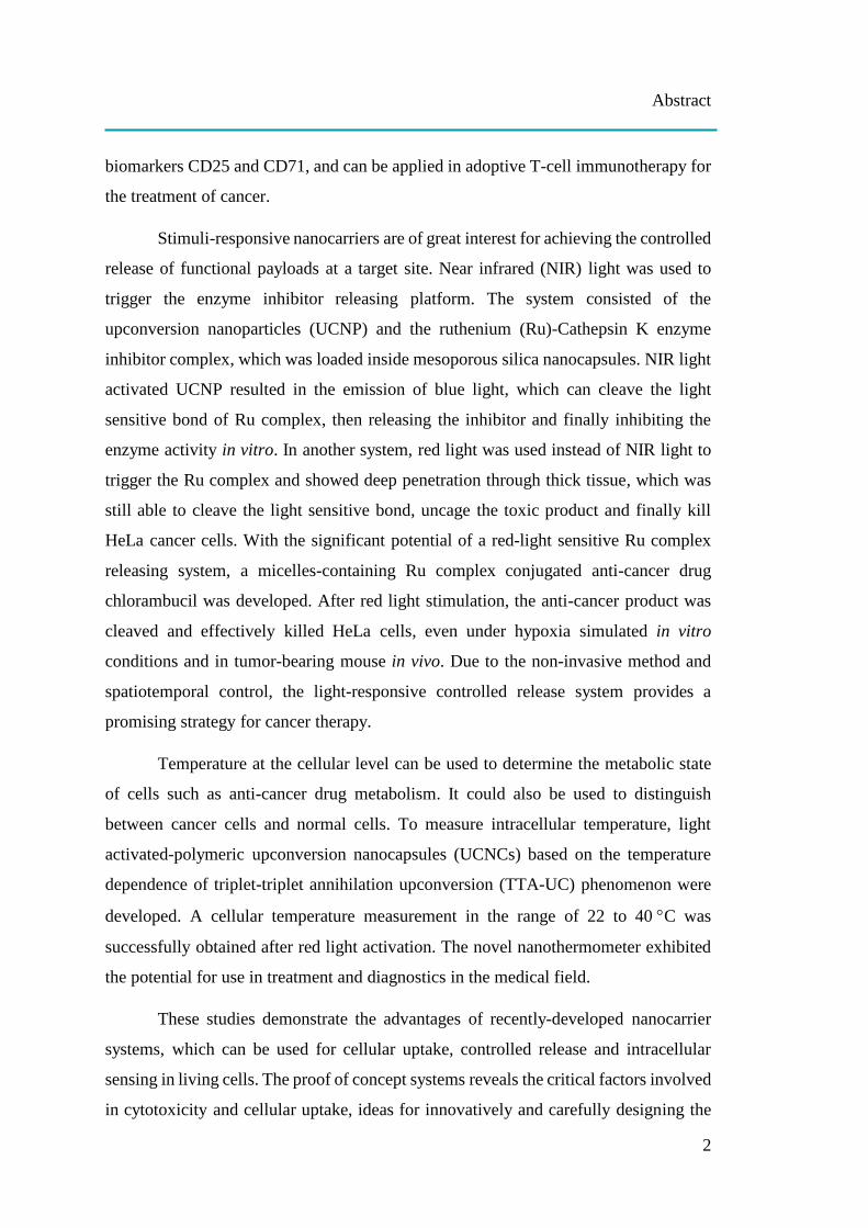
candidates for various kinds of applications.[Z-3-4,7] NPs can be categorized into organic
NPs such as polymeric nanospheres, nanocapsules, micelles, liposomes and
dendrimers, and inorganic NPs such as silica NPs, carbon nanotubes, iron oxide NPs,
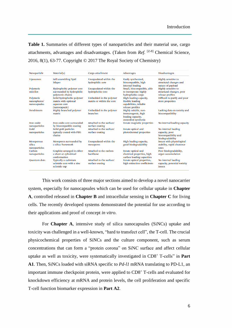
gold NPs and quantum dots, as shown in Figure 1. Summaries focused on material use,
cargo attachments, advantages and disadvantages are presented in Table 1.[Z-8]
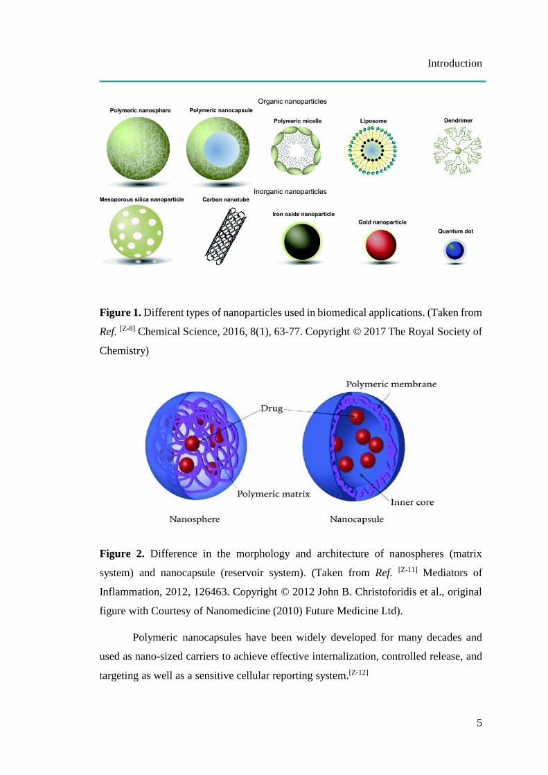
Nanocapsules and nanospheres are the two main types of polymeric
nanoparticles, which are different in their morphology and architecture (Figure 2).
Nanocapsules exhibit a typical core-shell structure consisting of a liquid core, in which
either the hydrophilic or hydrophobic cargo is entrapped inside the interior cavity
surrounded by a polymeric membrane made up of natural or synthetic polymers. Unlike
nanocapsules, nanospheres are formed by dense polymers where the drug is
homogenously dispersed in the polymeric matrix.[Z-9-11]
Introduction
5
Figure 1. Different types of nanoparticles used in biomedical applications. (Taken from
Ref. [Z-8] Chemical Science, 2016, 8(1), 63-77. Copyright © 2017 The Royal Society of
Chemistry)
Figure 2. Difference in the morphology and architecture of nanospheres (matrix
system) and nanocapsule (reservoir system). (Taken from Ref. [Z-11] Mediators of
Inflammation, 2012, 126463. Copyright © 2012 John B. Christoforidis et al., original
figure with Courtesy of Nanomedicine (2010) Future Medicine Ltd).
Polymeric nanocapsules have been widely developed for many decades and
used as nano-sized carriers to achieve effective internalization, controlled release, and
targeting as well as a sensitive cellular reporting system.[Z-12]
Introduction
6
Table 1. Summaries of different types of nanoparticles and their material use, cargo
attachments, advantages and disadvantages. (Taken from Ref. [Z-8] Chemical Science,
2016, 8(1), 63-77. Copyright © 2017 The Royal Society of Chemistry)
This work consists of three major sections aimed to develop a novel nanocarrier
system, especially for nanocapsules which can be used for cellular uptake in Chapter
A, controlled released in Chapter B and intracellular sensing in Chapter C for living
cells. The recently developed systems demonstrated the potential for use according to
their applications and proof of concept in vitro.
For Chapter A, intensive study of silica nanocapsules (SiNCs) uptake and
toxicity was challenged in a well-known, “hard to transfect cell”, the T-cell. The crucial
physicochemical properties of SiNCs and the culture component, such as serum
concentrations that can form a “protein corona” on SiNC surface and affect cellular
uptake as well as toxicity, were systematically investigated in CD8+ T-cells” in Part
A1. Then, SiNCs loaded with siRNA specific to Pd-l1 mRNA translating to PD-L1, an
important immune checkpoint protein, were applied to CD8+ T-cells and evaluated for
knockdown efficiency at mRNA and protein levels, the cell proliferation and specific
T-cell function biomarker expression in Part A2.
Introduction
7
For Chapter B, light-responsive nanocarriers draw attention for use in stimuli
responsive controlled release of functional payloads at specific target sites due to their
non-invasive method and spatiotemporal control. The efficiency of NIR light triggered
the inhibitor caging system, consisting of the upconversion nanoparticle (UCNP) and
the ruthenium (Ru)-Cathepsin K enzyme inhibitor complex, which was loaded inside
mesoporous silica nanocapsules and investigated from enzyme inhibition after NIR
irradiation without or through a piece of tissue in Publication B1. Instead of NIR light,
red light was used to activate the Ru complex and showed deep penetration through the
thickness of the tissue, which was then evaluated to answer the question of potential
depth, which remains able to uncage the toxic product and kill HeLa cells in
Publication B2. The red-light sensitive, micelles-containing Ru complex conjugated
anti-cancer drug chlorambucil was developed and investigated for the efficiency to kill
HeLa cells under hypoxia simulated in vitro and tumor-bearing mice in vivo in
Publication B3.
For Chapter C, the importance of developing a highly-sensitive, cellular
temperature nanosensor that can determine the metabolic state of cells such as anti-
cancer drug metabolism and might be used to identify cancer cells from healthy cells,
motivated this study. Red-light stimulated-polymeric upconversion nanocapsules
(UCNCs) based on the temperature dependence of triplet-triplet annihilation up-
conversion (TTA-UC) phenomenon were synthesized and evaluated for potential use
as a nanothermometer by measuring intracellular temperature in the range of 22 to
40 C inside living HeLa cells in Part C1.

Chapter A
8
CHAPTER A
NANOCAPSULE AS A NANOCARRIER FOR T-CELLS
Chapter A combines two different parts. These studies propose the application
of silica nanocapsules as suitable nanocarriers in T-cells by investigating the crucial
physicochemical characteristics of silica nanocapsules on the impact of cellular uptake
and toxicity in T-cells. They likewise demonstrate a strategy to improve T-cell survival
and the function for adoptive T-cell immunotherapy for treatment of cancer using silica
nanocapsules carrying specific siRNA-mediated gene silencing. A general introduction
concerning T-cells, adoptive T-cell immunotherapy and recent advances in nanocarriers
applied in T-cells will be described. Afterwards, each study will be presented
separately.
(Note: The literature of the introduction can be found on page 138 at the end of the dissertation.)
[A1] Raweewan Thiramanas, Shuai Jiang,* Jorge Pereira, Katharina Landfester,*
Volker Mailänder.* Silica Nanocapsule as a Suitable Nanocarrier: Uptake and
Toxicity Study in T-cells. (Manuscript in preparation).
[A2] Raweewan Thiramanas, Mengyi Li, Shuai Jiang, Katharina Landfester,*
Volker Mailänder*. Cellular Uptake of siRNA Loaded Nanocarrier to
Knockdown PD-L1: Strategies to Improve T-cell Functions. (Manuscript in
preparation).
Chapter A
9
A. Introduction
T-cells, one of the most important cells in an adaptive immune system, possess
attractive intrinsic functions that enable the killing of virus-infected cells and cancer,
which is highly impactful to the biomedical field. Many researchers have employed T-
cells properties in so-called adoptive T-cell immunotherapy for cancer treatment.[Z-13]
The treatment is based on harvesting T-cells from cancer patients, growing and /or
genetically modifying them to enhance their killing ability in terms of specificity and
efficiency before reinfusing the T-cells back into the patient.[Z-14] Compared to
traditional cancer therapies including surgery, chemotherapy, and radiation therapy,
this technique has several advantages such as being highly specific to target cancer
cells. Therefore, adverse side effects to healthy tissue can be avoided as well as
eliminate immune rejection because the therapeutic cells come from the patient, in
addition to overcoming the problems of tumor re-occurrence and metastasis.[Z-15]
However, loss of T-cells viability and function naturally occurs due to time-consuming
process and immuno-regulatory mechanism.[Z-14,16] Therefore, another field of research
in parallel to promote T-cells survival and efficacy has been intensively developped.
For this purpose, many therapeutic agents such as antibodies, cytokine, and nucleic
acids have been applied to T-cells using different methods such as systemic
administration, electroporation, and by using nanocarriers.[Z-17] With the progress in
nanotechnology, various types of nanocarriers have been designed and used for the
delivery of functional payloads into different kinds of target cells. Nanocarriers provide
protection for the cargo from surrounding conditions and facilitating internalization.[Z-
3] Among them, silica nanomaterials exhibit high potential for use in a drug delivery
system. Silica nanocarriers have many distinct advantages such as high loading
capacity, chemical and physical stability, biocompatibility and biodegradability, and
easy surface modification, making them good candidates for delivery to T-cells.[Z-18]
T-cells are well-known to be “the hard to transfect cells”, meaning they are difficult to
uptake foreign particles. In addition, they are highly sensitive and easy to exhaust,
making it a challenge to choose and design an appropriate nanocarrier.[Z-19] Lack of
study focused on nanocarrier uptake and toxicity in T-cells makes it more difficult to
Chapter A
10
understand which characteristics of nanocarriers should be carefully considered when
applied to T-cells.
In Chapter A, crucial parameters including sizes, cores, charges and surface
functionalizations of silica nanocapsules (SiNCs) that have impact on cellular uptake
and toxicity in CD8+ T-cells were systematically investigated in Part [A1] to solve this
problem. In addition, the effect of protein corona and serum in culture media on cellular
uptake and toxicity were also studied to provide more criteria to consider when applying
the nanocarriers in T-cells to achieve high uptake efficiency and low toxicity.
The uptake and toxic profiles were then used for designing the SiNCs carrying
the specific siRNA to knockdown PD-L1, a potent inhibitory protein, in CD8+ T-cells
in order to improve T-cells viability and function in Part [A2]. The knockdown
efficiency was determined at protein and mRNA levels. The effects of PD-L1
knockdown were demonstrated in terms of cell proliferation and various functional
biomarker expressions.
Here, the general background concerning the human immune system is
described. The cancer-induced mechanisms for T-cell activation and inhibition,
adoptive T-cell immunotherapy and recent advances in nanocarriers applied in T-cells
are explained as well.
A.1 Human immune system
The human immune system consists of two major subsystems: innate immunity
and adaptive immunity. Innate immunity is the first line of the defense mechanism and
is not specific to any particular pathogen in the way that adaptive immune responses
are. Although the innate immune system has evolved to quickly detect and destroy a
variety of pathogens, the pre-existing set of common pathogenic molecular patterns,
which are all established since in the germ line genome that it can recognize, is limited.
Excessive variations of antigenic structures and the immune escape-induced mutation
of pathogens are the driving forces in the evolution of the adaptive immune system or
the acquired immune system, which can be found in vertebrates including birds, fish,
amphibians, reptiles, and mammals. The slow response of adaptive immune responses
is due to their hallmarks of learning to create highly-specific response and memorizing
Chapter A
11
processes, which are mediated by white blood cells called lymphocytes. During
lymphocyte development, sets of gene segments are rearranged through a process of
somatic recombination and assembled to create genes encoding the enormously diverse
and specific antigen receptors of T and B lymphocytes to generate highly specific and
flexible immune responses capable of recognizing all specific parts of pathogens,
known as antigens. There are two major classes of adaptive immunity, humoral and
cellular immunity, which are carried out by B-cells and T-cells, respectively.[Z-20-22]
A.1.1 Adaptive humoral immunity
Adaptive humoral responses or antibody responses are mediated by antibodies,
which involve proteins called immunoglobulins produced by plasma cells derived from
B-cells. B-cells are so called because they are produced and mature in the bone marrow
then travel to the lymphatic system to circulate throughout the body. As a result of
somatic recombination, B-cells each carry a set of surface antigen-recognition
molecules or membrane-bound antibodies called B-cell receptors (BCR). When B-cells
bind an antigen that fits or matches its BCR, they will ingest, process and present
digested peptide on MHCII through T-cell receptor (TCR) binding of CD4+ T-cells.
Activated CD4+ T-cells produce stimulatory factors to help B-cells maturation and
differentiation to become either a memory B-cell to be reactivated in the future or a
plasma cell releasing large amounts of free antibodies specific to its activating-antigen.
The antibodies circulate through the bloodstream and body fluids, where they search
and bind specifically and directly to the antigens (either presented extracellularly on
infected cells or free-floating in the body) that stimulate their production. Antibody
binding neutralizes viruses and microbial toxins by blocking their ability to bind to
receptors on host cells. In addition, the binding of antibodies also provides notable
marking on invaders for targeted-extermination and facilitates engulfment by the
phagocytic cells of the innate immune system.[Z-22-24]
A.1.2 Adaptive cellular immunity
The adaptive cellular-mediated immune response is carried out by T-cells,
which are so called because they mature in the thymus. The innate immunity phagocytic
cells act by ingestion in the pathogen directly, whereas they serve as antigen-presenting
Chapter A
12
cells for adaptive immunity. Unlike the B-cells that can directly bind to antigen via
BCR, T-cells need antigen-presenting cells to process and present small peptides of the
antigen to T-cells via highly variable T-cell receptors (TCR). This receptor is generated
by randomly assorting genes providing T-cells detection and response to a wide range
of antigens. Antigen presentation results in the activation of T lymphocytes, the
initiation of the adaptive response, and finally the destruction of the target cells via
induction of apoptosis. T-cells characterized by the CD3 surface marker are categorized
into 2 subtypes: the helper cell or regulatory T lymphocyte (Treg) and the effector cell
or cytotoxic T lymphocyte (CTL), which can be called CD4+ and CD8+ T-cells
according to their unique cell surface markers, respectively. Similar to their names,
helper T-cells ‘help’ other cells of the immune system, such as the maturation of B-
cells and activation of CTLs, while CTLs kill virus-infected cells and tumors.[Z-25-26]
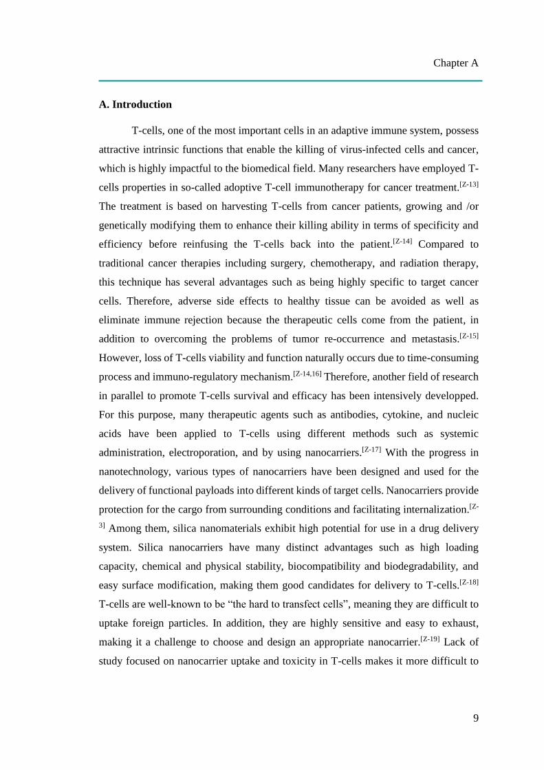
Figure A.1 Stimulatory (present in green) and inhibitory (present in red) factors in the
Cancer-Immunity Cycle. Copyright @ 2013 Elsevier Inc. Reprinted with permission
from Elsevier. Ref. [Z-27] Immunity. 2013, 39(1), 1-10.

Chapter A
13
A.2 T-cell Activation and the Cancer-Immunity Cycle
For cancer induced immune response, T-cells can be activated through a 7-
stepwise complex process called the cancer-immunity cycle, as shown in Figure A.1.
[Z-27] In step (1), cancer cell antigens are released from dead cancer cells. Antigen
presentation occurs in step (2), after which released antigens are detected, ingested and
processed into small peptides by professional antigen-presenting cells (APCs)
including dendritic cells (DCs), macrophages and B-cells. During maturation and
migration to the lymph node, DCs lose their phagocytic function, but possess high
expression of specific molecules called major histocompatibility complex class 1
(MHCI) and class 2 (MHCII) and co-stimulatory molecules such as CD40, CD80,
CD83, and CD86 on their surfaces for efficient communication with T-cells.[Z-28-30] DCs
then present the exogenous peptides on MHCI and MHCII to TCR on CD8+ (called
cross presentation) and CD4+ T-cells surfaces, respectively. As a result of mutation,
abnormal cells can present endogenous antigens on MHCI which can be detected by
CD8+ T-cells and stimulate immune response. Antigen presentation through TCR
results in step (3) the priming and activation of CTLs that respond to cancer antigens
are identified as foreign. Activated CTLs subsequently move through the blood stream
(step 4) and infiltrate to the site of the tumor (step 5). After that, CTLs recognize and
bind to the stimulating antigens present on MHCI of cancer cells through TCR (step 6),
leading to the formation of a channel in order to introduce chemicals including
granzymes (serine protease-destroying intracellular protein) and perforin
(glycoprotein-forming a pore) as well as the activation of apoptosis and finally the
killing of cancer cells (step 7). The dead cancer cells release their antigens and then
continue to begin the cycle again to generate stronger immune responses.[Z-27]
Meanwhile, repeated TCR activation also induces proapoptotic pathways via the
expression of Fas (CD95) ligand, a transmembrane protein that belongs to the tumor
necrosis factor family, on the CTL surface, which then binds to Fas receptors on the
target cell surface, triggering apoptosis.[Z-22,31-32]
A.3 T-cell Inactivation and Cancer Immune Escape
Even though T-cells can recognize and efficiently destroy tumors through the
Cancer-Immunity Cycle, in many cases the tumor cells are still able to remain,

Chapter A
14
proliferate and metastasize. These cancer resistance mechanisms are explained based
on the concept of “immunoediting”, which consists of 3 phases.[Z-33] Starting from
“immune surveillance”, the immune cells can recognize tumor cells as non-self and
progressively eliminate them. If one of the abnormal cells somehow survives immune
elimination, scarce cells can generate offspring, which might begin the second phase of
“immune equilibrium”, defined as a period during which our immune system and
tumors live in a state of balance in the body. This means although the tumors cannot be
killed, they cannot grow or metastasize. Because this phase is difficult to identify, the
driving forces behind this phase are poorly understood.[Z-34] During this state plus with
immunosuppression in tumor microenvironment such as anaerobic acidic glucose-poor
environment, tumor cells accumulate stress, metabolic changes and induced-mutation,
leading to the last phase of “immune escape”, when tumors more aggressively progress,
invade and metastasize out of control by the immune cells.[Z-35]
Many chemical factors involved in the cancer-immunity cycle can stimulate or
inhibit T-cell activation, as presented in Figure A.1.[Z-27] Stimulatory factors present in
green promote immune activity, while inhibitors present in red re-check the process and
reduce immune response and/or prevent autoimmunity. Cancer cells smartly take
advantage of these major immune checkpoint proteins, such as CTLA-4, PD-1, and PD-
L1 to escape the immune system by expression of these checkpoints in order to imitate
negative regulatory feedback inhibition when APCs communicate with T-cells.[Z-36]
Cytotoxic T-lymphocyte associated protein-4 (CTLA-4) can inhibit the T-cell
activation step (Figure A.1, step 3). Activation of T-cells not only needs the binding
between MHC of APCs to TCR, but also co-stimulation between the B7 family, CD80
(B7.1) and CD86 (B7.2), of APCs to CD28 of T-cells. CTLA-4 possesses very high
structural homology to the co-stimulatory molecule CD28 and a much higher affinity
for both CD80 and CD86 than for CD28. Therefore, its expression on activated T-cells
effectively blocks CD28-B7 interaction, resulting in the inactivation of T-cell
responses.[Z-35-37]
Like CTLA-4, programmed cell death 1 (PD-1, CD279) is expressed only in
activated T-cells. PD-1 has two ligands which also belong to B7 family, PD-L1 (B7-
H1, CD274) and PD-L2 (B7-DC, CD273). PD-L2 is predominantly expressed on APCs

Chapter A
15
as well as T-cells, whereas PD-L1 can be expressed on many non-hematopoietic and
hematopoietic cells including tumor cells, immune cells, epithelial cells, and
endothelial cells. T-cells stimulation can be inhibited in the tumor bed (Figure A.1,
step 7) through PD-1/PD-L1 interaction, in which PD-L1 on the surface of DCs bind to
PD-1 on the surface of T-cells, leading to T-cell exhaustion, which might contribute to
cancer immune evasion.[Z-35-37]
The identification of activated T-cell inhibitors, especially CTLA-4, PD-1 and
PD-L1, has brought the revolution of checkpoint blockade to a new class of cancer
immunotherapy for specifically blocking T-cell inhibition, reinforcing and potentially
propagating preexisting anticancer immune responses.[Z-27] The blockade of the PD-
1/PD-L1 pathway was thought to yield greater antitumor activity and fewer side effects
compared to CTLA-4 blockade.[Z-38] Several drugs as checkpoint blockades have been
approved and exhibited impressive responses at the clinical level, such as the anti-
CTLA-4 antibody ipilimumab, which was approved in 2011 for use against advanced
melanoma, and two anti-PD-1 antibodies, pembrolizumab and nivolumab, approved in
2014 for use against metastatic melanoma. Nowadays, these drugs are also under
intensive investigation for use in the treatment of other cancers.[Z-38]
A.4 Adoptive T-cell immunotherapy
Cancer immunotherapy is characterized by using the patient’s own immune
system to fight against cancer cells. The goal of cancer immunotherapy is to stimulate
or restimulate a potential intrinsic cycle of cancer immunity, enabling it to multiply and
function as anticancer activity under the optimal immune response to avoid unwanted
autoimmune inflammatory response.[Z-27] Cancer immunotherapy approaches include
active immunization, reversal of immunosuppression, nonspecific immune stimulation
and adoptive cell therapy (ACT). Among these approaches, ACT has achieved very
promising results in cancer clinical trials and therefore is considered to be the most
effective cancer immunotherapy.[Z-15] In ACT, tumor-specific CTLs are collected from
and infused into cancer patients with the aim of identifying and destroying tumor cells.
CTLs used for ACT can be obtained from tumor-infiltrating lymphocytes (TILs) or
peripheral blood lymphocytes, which can then be selected and used either in their
natural state or genetically modified cancer-specific T-cells, such as T-cell receptor
Chapter A
16
(TCR)- and chimeric antigen receptor (CAR)-transduced T-cells.[Z-14] The key to
success of cancer immunotherapy is mainly dependent on the identification of potential
cancer antigens that can be used for cancer vaccines, TILs activation and antigen-
specific T-cells productions in TCRs and CARs.[Z-15] TILs production requires 2 critical
steps. The first step is the collection and separation of a lymphocyte culture from cancer
tissue. This process takes up to 1 month and requires professional decisions by experts.
Thus, the automated production process is impossible. The second step is a 14-day
large-scale expansion process, which has been accomplished successfully using
automated bioreactor. However, modified TCR or CAR cells are much easier to
produce and require fewer total cells. Generally, peripheral blood lymphocytes are
harvested, transduced with the desired gene and expanded to treatment levels. Because
those T-cells are highly active, only low cell numbers are typically required for
treatment.[Z-16] TCR clones can also be produced by co-culturing peripheral blood T-
cells with antigen presenting cells that express a specific tumor antigen. To evade
immune surveillance, many cancers reduce the expression of MHC, resulting in
inefficient T-cells activation due to decreased TCR-MHC interaction. To avoid this
interaction, CAR molecules were developed. Combining the advantage of an antibody
with a TCR, the CAR structure consists of an extracellular part, which is a ligand-
binding domain containing a BCR–derived single-chain variable fragment, and a
intracellular signaling domain, which is composed of CD3 and one or more co-
stimulatory domains e.g. CD28. Thus, the function of the CAR is independent of MHC
interaction or any co-stimulatory signaling.[Z-15-16,39-40]
A.5 Recent advances in nanocarriers applied in T-cells
Because therapeutic agents including plasmid DNAs, siRNAs, antibodies,
enzymes, enzyme inhibitors, and drugs cannot spontaneously cross the cell membrane,
delivery approaches are required.
Unfortunately, many of the traditional delivery strategies used in primary
immune cells, especially in unstimulated cells and lymphocytes, have shown to be
impractical for use due to their undesirable effects either for cell viability or cellular
response.[Z-41] Conventional lipid and cationic transfection reagents result in low
transfection efficiencies and induce nonspecific inflammation in these cells because
Chapter A
17
these agents enter the cells by endocytic pathways. Hence, they trigger the immune
response. Similar results also appear when using viral vectors for the same reasons and
due to the presence of cytoplasmic viral nucleic acid sensors.[Z-41] Besides, viral vectors
also affect T-cell differentiation.[Z-42] Electroporation/nucleofection, which allows
small molecules such as plasmids, siRNAs or antibodies to pass into the cells through
temporary pore formation induced by high voltages, gains satisfactory transfection
efficiency. However, cell viability is commonly decreased after electroporation
(typically 60% survival rate), even with optimized voltages and conditions.[Z-43]
Therefore, various kinds of nanocarriers have been developed, used, and reported on as
new delivery systems for T-cells with the aim of protecting the cargo, internalizing to
the cells and releasing the functional payloads while reducing or eliminating unwanted
effects on the cells either for improved T-cells viability and functionality against virus-
infected cells or cancers, immunological network study, or for T-cells targeting.
Liu et al. [Z-44] modified single-walled carbon nanotubes (SWNT) to enhance
hydrophilicity, functionalized with cleavable disulfide bonds conjugated to siRNAs and
used to transport siRNA specific to CXCR4 chemokine receptor and CD4 to human T-
cells and primary cells with the aim of inhibiting HIV viral entry and minimizing
infection. The siRNA mediated these target gene silencing has been observed and
exhibited superior performance to that observed with conventional liposome‐ based
non-viral delivery agents.
Lee et al. [Z-45] evaluated siRNA delivery to T-cells by chitosan, a derivative of
the natural polysaccharide chitin, using nanoparticles which were chemically
conjugated to a T-cell-targeting CD7-specific single-chain antibody having a diameter
of 320 nm. Silencing of CD4 expression was observed when the Jurkat leukemic T-cell
line was incubated with these particles in vitro.
Yosef et al. [Z-41,46] established new delivery technology by culturing and
stabbing primary T-cells on vertical silicon nanowires (NWs) precoated with specific
biomolecules including siRNAs, plasmids, peptides, and proteins, which were
delivered effectively (>95%) to the cells without stimulation or alteration of viability.
Interestingly, the NWs exhibited consistent penetration to the cellular membranes
without impacting cell health or morphology. Later, potent silencing of 34 gene targets

Chapter A
18
was used to simulate a model of the dynamic regulatory network that controls Th17
differentiation.
From the intensive review of Freeley and Long[Z-19], SWNTs, chitosan
nanoparticles and vertical silicon NWs have only been used to answer basic biological
questions. However, therapeutic applications in T-cells have not yet been investigated.
Dendrimers, which are highly-branched synthetic polymers, can form with
siRNAs through electrostatic interactions. Carbosilane dendrimers by Weber et al. or
PAMAM [poly(amidoamine)] dendrimers by Zhou et al.[Z-47] have been formed with
HIV genes-specific siRNAs and shown to inhibit viral replication in T-cell lines and
PBMCs in vitro.[Z-48] A strategy to target siRNA delivery was developed by Felber et
al.[Z-49] using antibody-conjugated PAMAM dendrimers, which resulted in enhanced
uptake and more potent gene silencing in a prostate cancer cell line, suggesting that
antibody-conjugated dendrimers could be a potential T-cell specific siRNA delivery
approach.[Z-19]
The most creative and effective siRNA delivery system described to date for
targeting leucocytes and T-cells in vivo is I-tsNPs (integrin-targeted stabilized
nanoparticles), established by Peer et al.[Z-19,50] In this system, siRNAs are condensed
with the nucleic-acid binding protein protamine and subsequently encapsulated within
100 nm diameter liposome nanoparticles resulting in a ∼4000:1 ratio of siRNAs to
nanoparticles, which may have significant potential for the improvement of human T-
cell function due to their high loading capacity.
Delivery systems for the targeting of T-cells and avoiding of endocytic activities
reduction were developed by Frick et al.[Z-51] using cytokine interleukin-2 (IL-2)
coupled to the surface of hydroxyethyl starch (HES) nanocapsules. The system
demonstrated direct and specific T-cell targeting in vitro and in vivo by IL-2 receptor-
mediated internalization. The results suggest that engineering IL-2-functionalized
nanocapsules can be used for the efficient targeting of T-cell populations with different
IL-2 receptor affinities. It may also be applied to other cytokine-related targeting
systems.
Chapter A1
19
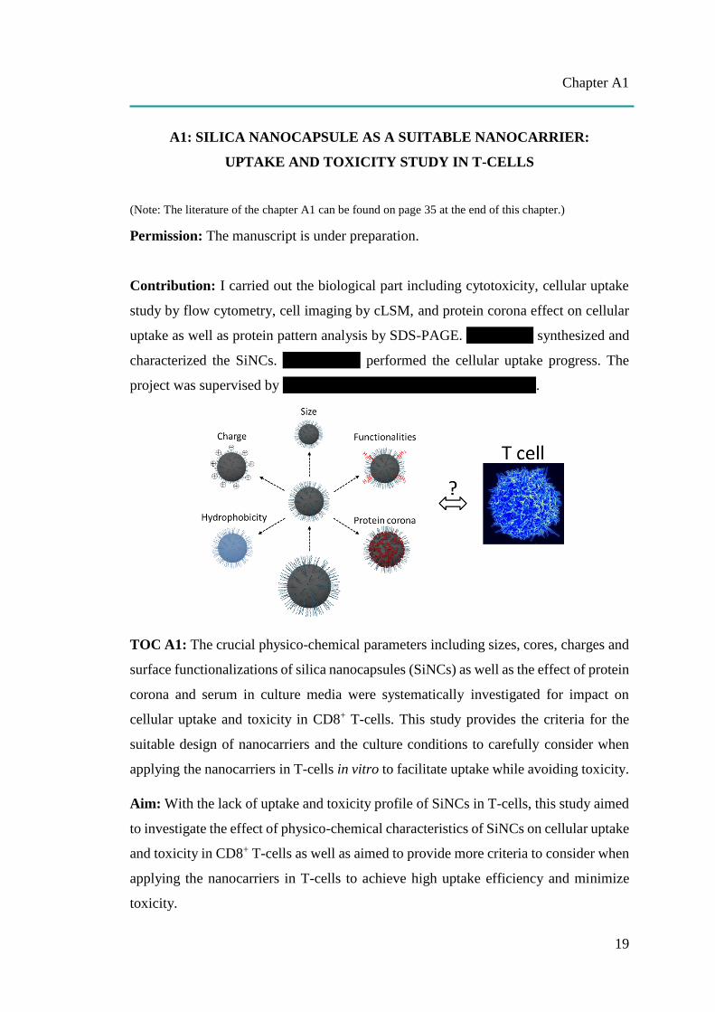
A1: SILICA NANOCAPSULE AS A SUITABLE NANOCARRIER:
UPTAKE AND TOXICITY STUDY IN T-CELLS
(Note: The literature of the chapter A1 can be found on page 35 at the end of this chapter.)
Permission: The manuscript is under preparation.
Contribution: I carried out the biological part including cytotoxicity, cellular uptake
study by flow cytometry, cell imaging by cLSM, and protein corona effect on cellular
uptake as well as protein pattern analysis by SDS-PAGE. Shuai Jiang synthesized and
characterized the SiNCs. Jorge Pereira performed the cellular uptake progress. The
project was supervised by Katharina Landfester and Volker Mailänder.
TOC A1: The crucial physico-chemical parameters including sizes, cores, charges and
surface functionalizations of silica nanocapsules (SiNCs) as well as the effect of protein
corona and serum in culture media were systematically investigated for impact on
cellular uptake and toxicity in CD8+ T-cells. This study provides the criteria for the
suitable design of nanocarriers and the culture conditions to carefully consider when
applying the nanocarriers in T-cells in vitro to facilitate uptake while avoiding toxicity.
Aim: With the lack of uptake and toxicity profile of SiNCs in T-cells, this study aimed
to investigate the effect of physico-chemical characteristics of SiNCs on cellular uptake
and toxicity in CD8+ T-cells as well as aimed to provide more criteria to consider when
applying the nanocarriers in T-cells to achieve high uptake efficiency and minimize
toxicity.
Chapter A1
20
Abstract: Adoptive T-cell immunotherapy brings hope to cancer patients. It emerges
as a powerful and promising cancer therapy so the problem regarding the immuno-
reaction between different donors and recipients can be avoided. However, a new
problem based on the nature of the T-cell itself is problematic. After long cultivation
and expansion under laboratory media conditions, T-cells start to lose their viability
and function because of the immune checkpoint proteins, leading to decreased
efficiency in killing cancer cells. A new strategy to improve T-cell survival and function
is needed. With the advantages of nanotechnology and the biocompatibility of silica-
based material, silica nanocapsules (SiNCs) provide an ideal delivery system for the
use as nanocarriers to transport therapeutic biomolecules to T-cells. Due to the lack of
uptake study concerning T-cells, various physico-chemical properties such as sizes,
charges, and surface functionalities of SiNC were systematically studied in this work
for their impact on cellular uptake and toxicity in CD8+ T-cells. The obtained results
are discussed to improve the appropriate design of nanocarriers with low toxicity and
high uptake for T-cell immunotherapy. The effect of protein corona from human serum
and the serum present in culture medium on the uptake of different SiNCs was also
investigated to provide more information to consider for suitable uptake conditions in
SiNC in CD8+ T-cells.
A1.1. Introduction
Adoptive T-cell immunotherapy (ACT) is the new era of cancer therapy. It can be
used to destroy cancer cells by using the immune cells from the patients themselves.[A1-
1] Compared to traditional cancer therapies including surgery, chemotherapy and
radiation therapy, immunotherapy has several distinct advantages including no immune
reaction (due to the same donors and recipients), high specificity, and high cancer-
killing ability (due to intrinsic functionalities of T-cells, which are able to recognize
and destroy cancer cells).[A1-2] It was mentioned that this therapy may overcome the
problem of tumor recurrence after surgery and also defeat late-stage cancer, which has
very limited options in terms of traditional cancer therapies.[A1-2] However, the problem
of this technique is that we have relied on naturally controlling the mechanism of the
immune system. After a certain time of activation, the expression of the immune check

Chapter A1
21
point proteins from either the other immune cells e.g. dendritic cells or T-cell itself will
be induced, causing T-cell inactivation and leading to a loss of T-cell survival and
function.[A1-3-5] This situation also occurs in T-cell cultivation for immunotherapy after
collection from the patient and activating in culture media, resulting in decreased
efficacy for cancer treatment.[A1-3-5] With significant achievements in ACT and a very
promising cancer therapy, research focused on the improvement of T-cell survival and
function has been intensively pursued and reported. [A1-1-2,4-5] A wide range of
therapeutic agents have been used to boost immunity including vaccines,[A1-6]
monoclonal antibodies,[A1-7] cytokines,[A1-8,9] siRNAs specific-immune checkpoint
proteins such as cytotoxic T lymphocyte-associated molecule 4 (CTLA-4), [A1-10]
Casitas B-lineage lymphoma b (Cbl-b),[A1-11] programmed cell death-1 (PD-1), [A1-12]
and their ligands (PD-L1/PD-L2).[A1-13] To achieve this goal, an efficient delivery
system loaded with specific biomolecules such as drugs, nucleotides, peptides, proteins,
and fluorescent dyes is needed in order to obtain the biological effect or track the carrier.
The criteria for an ideal delivery system require abilities to protect the payload
against culture environment and low pH in endosome/lysosome. Internalization into the
cell without toxicity and without immune response initiation as well as subsequent
release of the payload, which is still able to function inside the cell, is also required. [A1-
14] Due to the rapid development of nanotechnology, a variety of nanocarriers have been
designed and widely applied in various fields, especially for biomedical applications.
[A1-15] Nano-sized carriers provide high surface to volume ratio, protect cargo from the
biological environment before encountering the target cell, and facilitate uptake.
Among them, silica nanocapsules (SiNCs) have many great advantages including high
loading capacity, excellent colloidal and chemical stability, biocompatibility and
biodegradability.[A1-16] With their outstanding properties, silica-based nanocapsules
(SiNCs) have been widely used for drug delivery for many decades.[A1-16-17] SiNCs are
not only easy to synthesize in various well-defined controllable sizes, but also facile to
modify for different surface functionalities through the rich hydroxyl groups on their
surface. Moreover, silicon oxide has been accepted by the U.S. Food and Drug
Administration (USFDA) for use in food additives as well as medical care products.[A1-
16-17] Thus, SiNCs seem to be suitable for use as alternative nanocarriers in T-cells.
Chapter A1
22
Due to its nature, a T-cell is a type of cell that is difficult to uptake foreign particles
and is highly sensitive to the environment and dies easily.[A1-18] Few uptake studies of
silica nanomaterials in T-cells or related-cells have been reported. Silica nanoparticles
(SiNPs) were found to induce oxidative stress and the inflammation of human
peripheral blood mononuclear cells (PBMCs). Notably, smaller-sized SiNPs with
10 nm were more cytotoxic and induced more oxidative stress than the bigger SiNPs
with 100-nm.[A1-19] In addition, ultra small SiNPs (<10 nm) were also found to cause T-
cell activation by increasing the expression of CD25 and CD69 and the secretion of
IFN-.[A1-20] The toxicity of SiNPs was found to be strongly related to their physico-
chemical properties, such as size, surface area, and surface features.[A1-21] Due to the
lack of systematic study in terms of various physico-chemical parameters of the silica
nanocarriers uptake in T-cell, greater understanding is required concerning the critical
properties of SiNC which affect T-cell uptake prior to use of SiNC as a carrier for T-
cell immunotherapy.
This study aims to investigate the crucial parameters including different sizes, cores,
charges, and surface functionalizations of the SiNCs affecting cellular uptake in CD8+
T-cells. For this purpose, novel silica core-shell nanocapsules were synthesized. In
general, the therapeutic payload is encapsulated in the SiNC cavity by immersing the
SiNCs in a therapeutic solution and allowing the agent to diffuse into its cavity. Here,
we used a new one-pot synthesis to facilely incorporate the payload into the core
surrounded with silica shell, which can be beneficial for ensuring the protection of the
cargo inside the silica shell. It also provides high loading capacity for the cargo. To
avoid interfering with its surface properties, Cy5 dye was incorporated with the shell
by using fluorescently labeled silica precursors, which allowed tracking of the capsule
inside the cell by flow cytometry and was further confirmed by confocal laser scanning
microscopy. Considering in vivo uptake for systemic administration, once the
nanocarrier faces the biological fluid, the biomolecular components e.g. blood proteins
attracted to the carrier surface then create a new identity formed as protein corona,
finally impact to cellular uptake.[A1-22] Not only in vivo biological surrounding, but also
in vitro culture condition needed to be considered. Therefore, further investigation of
the effect of protein corona from human serum was carried out, while the effect of

Chapter A1
23
serum in the medium on cellular uptake was studied to provide more criteria to consider
for inhibiting or enhancing the uptake of SiNC in CD8+ T-cells. The uptake and toxicity
of human serum pre-coated SiNCs compared to the uncoated ones were determined by
flow cytometry. The SiNCs before and after performing corona were characterized by
size-zeta potential analysis. The hard protein corona was demonstrated in the corona
pattern on SDS-PAGE and could subsequently be identified by liquid chromatography–
mass spectrometry (LC-MS). A better understanding of the consequences of different
physico-chemical parameters for SiNCs on cellular uptake in CD8+ T-cells will provide
information to carefully consider when designing the SiNCs for T-cell immunotherapy.
A1.2. Results and discussion
In the following study, we investigated the effect of different characteristics
including sizes, surface charges, functional groups, and protein interactions of SiNCs
on cellular uptake and toxicity in CD8+ T-cells.
A1.2.1 The effect of size
In the first section, we studied the size effect of nanocarriers on cellular uptake in T-
cells. Silica nanocarriers (NCs) with hydrodynamic diameters ranging from 400 nm to
50 nm were synthesized by using a mini-emulsion (oil-in-water) polymerization
technique.[A1-23] Cationic surfactant cetyltrimethylammonium chloride (CTAC) was
used for stabilizing mini-emulsion droplets against coalescence. The second role of
CTAC is to serve as a template agent for confined silica condensation at the
nanodroplet-water interface via cooperative self-assembly of negatively charged silica
species and cationic CTAC. Therefore, positively charged core-shell nanocapsules
were obtained with a liquid core that provides a high loading capacity for therapeutic
agents. To compare the effect of different charges, Lutensol AT50 (LUT) was chosen
as another surfactant to generate differently charged core-shell nanocapsules. Here, we
developed a facile dialysis process for the surfactant replacement from CTAC to
nonionic surfactant LUT, which has a polyethylene chain as its hydrophilic part. The
PEGylated surface could then provide steric stabilization of the nanocapsules. The size
of the nanocapsules was tuned by varying the volume ratio of the oil phase and water
Chapter A1
24
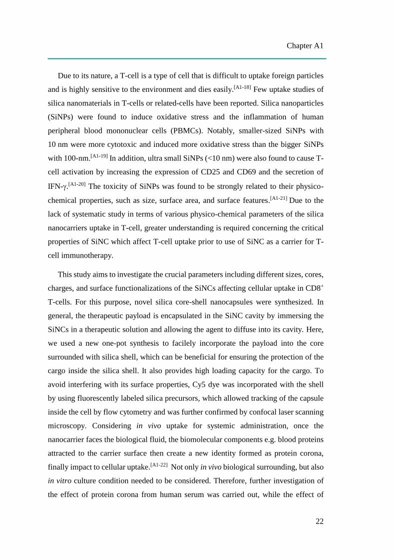
phase. NCs with average hydrodynamic diameters ranging from 400 nm (NC-400) to
50 nm (NC-50) are shown in Table A1.1. The obtained NCs were characterized in
terms of their physico-chemical properties, including hydrodynamic diameter size (Dh)
by multi-angle dynamic light scattering (DLS), surface charge by zeta-potential (ζ-
potential) measurement, and morphology by transmission electron microscopy (TEM),
as listed in Table A1.1.
Table A1.1. Characteristics of silica nanocarriers (NCs) with different sizes.
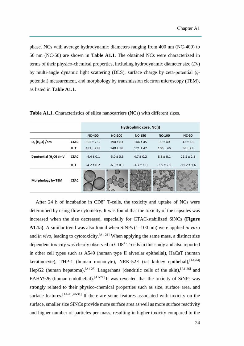
After 24 h of incubation in CD8+ T-cells, the toxicity and uptake of NCs were
determined by using flow cytometry. It was found that the toxicity of the capsules was
increased when the size decreased, especially for CTAC-stabilized SiNCs (Figure
A1.1a). A similar trend was also found when SiNPs (1–100 nm) were applied in vitro
and in vivo, leading to cytotoxicity.[A1-21] When applying the same mass, a distinct size
dependent toxicity was clearly observed in CD8+ T-cells in this study and also reported
in other cell types such as A549 (human type II alveolar epithelial), HaCaT (human
keratinocyte), THP-1 (human monocyte), NRK-52E (rat kidney epithelial),[A1-24]
HepG2 (human hepatoma),[A1-25] Langerhans (dendritic cells of the skin),[A1-26] and
EAHY926 (human endothelial).[A1-27] It was revealed that the toxicity of SiNPs was
strongly related to their physico-chemical properties such as size, surface area, and
surface features.[A1-21,28-31] If there are some features associated with toxicity on the
surface, smaller size SiNCs provide more surface area as well as more surface reactivity
and higher number of particles per mass, resulting in higher toxicity compared to the
Chapter A1
25
larger size particles, as previously described by others.[A1-24,31,32] In addition, the higher
surface area of small SiNCs can lead to a higher possibility of interaction with cellular
biomolecules such as DNA, proteins and sugars.[A1-30] Another study also demonstrated
that small SiNPs (70 nm; 10–90 μg/mL for 24 h) increased the oxidative DNA damage
(8-OH-dG levels) in HaCaT cells.[A1-33] Murugadoss et al.observed that SiNPs could
cause overproduction of reactive oxygen species (ROS) resulting in oxidative stress,
which could possibly damage the sub-cellular organelles and induce apoptosis in a size-
and dose-dependent manner.[A1-17] The mechanism of SiNPs induced toxicity is still not
fully elucidated, but it is believed to be derived from ROS-mediated toxicity.[A1-17,28,33]
This might explain the toxicity of small-sized SiNCs. Moreover, small size NPs may
cause toxicity as they pass through the cell membrane and enter the cells.[A1-29-30,34]
Some small NPs (< 50 nm) seem to enter the cells via passive diffusion, leading to
directly facing cellular components or accumulating in subcellular organelles like
mitochondria and nucleus. Another mechanism of small NPs induced toxicity when
they are entering to the cells is actin cytoskeleton disruption as a result of NP
internalization by endocytosis events, leading to cell deformation and direct cellular
injury.[A1-29-30,34] Even silica-based materials are generally accepted for their
biocompatibility and silicon oxide was approved by the USFDA. However, the
toxicology of silica nanoparticles (SiNPs) still remains to be reported.[A1-16-17,28] Besides
toxicity, the size-dependent immunological effects of SiNPs in immune cells including
monocytes and macrophages were found. It was indicated that nano-sized particles
caused a significant increase in pro-inflammatory secretion such as tumor necrosis
factor-alpha (TNF-α) compared to sub-micron and micron sized particles.[A1-17] Since
the size of SiNCs was the critical point to induce the cytotoxic effect, the SiNC size
was attempted to be controlled at sizes larger than 100 nm for subsequently experiments
to avoid toxicity due to the smaller size.
SiNCs with smaller sizes were found to not only be highly toxic, but also have been
less taken up by the cells (Figure A1.1b). In general, small-sized nanoparticles (< 100
nm) exhibited a stronger ability to be internalized inside cancer cells and were even
able to pass through the blood-brain barrier compared to the larger size
nanoparticles.[A1-35] However, small-sized SiNCs they could not be applied in this study
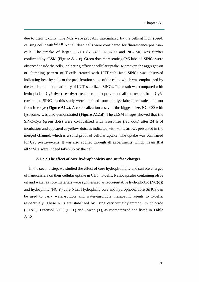
Chapter A1
26
due to their toxicity. The NCs were probably internalized by the cells at high speed,
causing cell death.[A1-24]. Not all dead cells were considered for fluorescence positive-
cells. The uptake of larger SiNCs (NC-400, NC-200 and NC-150) was further
confirmed by cLSM (Figure A1.1c). Green dots representing Cy5 labeled-SiNCs were
observed inside the cells, indicating efficient cellular uptake. Moreover, the aggregation
or clumping pattern of T-cells treated with LUT-stabilized SiNCs was observed
indicating healthy cells or the proliferation stage of the cells, which was emphasized by
the excellent biocompatibility of LUT-stabilized SiNCs. The result was compared with
hydrophobic Cy5 dye (free dye) treated cells to prove that all the results from Cy5-
covalented SiNCs in this study were obtained from the dye labeled capsules and not
from free dye (Figure A1.2). A co-localization assay of the biggest size, NC-400 with
lysosome, was also demonstrated (Figure A1.1d). The cLSM images showed that the
SiNC-Cy5 (green dots) were co-localized with lysosomes (red dots) after 24 h of
incubation and appeared as yellow dots, as indicated with white arrows presented in the
merged channel, which is a solid proof of cellular uptake. The uptake was confirmed
for Cy5 positive-cells. It was also applied through all experiments, which means that
all SiNCs were indeed taken up by the cell.
A1.2.2 The effect of core hydrophobicity and surface charges
In the second step, we studied the effect of core hydrophobicity and surface charges
of nanocarriers on their cellular uptake in CD8+ T-cells. Nanocapsules containing olive
oil and water as core materials were synthesized as representative hydrophobic (NC(o))
and hydrophilic (NC(i)) core NCs. Hydrophilic core and hydrophobic core SiNCs can
be used to carry water-soluble and water-insoluble therapeutic agents to T-cells,
respectively. These NCs are stabilized by using cetyltrimethylammonium chloride
(CTAC), Lutensol AT50 (LUT) and Tween (T), as characterized and listed in Table
A1.2.

Chapter A1
27
Figure A1.1. Cellular uptake study of SiNCs with different sizes in CD8+ T-cells. (a)
Cell viability and (b) Cy5 positive-cells of CD8+ T-cells after treatment with various
kinds of SiNCs (20 µg/mL) in the presence of 1% FBS for 24 h. Confocal laser scanning
microscopy images showing (c) the uptake of SiNCs and (d) co-localization with
lysosome in CD8+ T-cells. Co-localization assay was performed by staining lysosome
with LysoTracker® Green DND-26 (red). The membrane was stained with CellMask™
Orange (blue). The SiNC was labeled with Cy5 (green). The merged images of the three
channels demonstrated that the SiNCs were co-localized with lysosomes, as indicated
with white arrows. The scale bars represent 10 μm.
Chapter A1
28
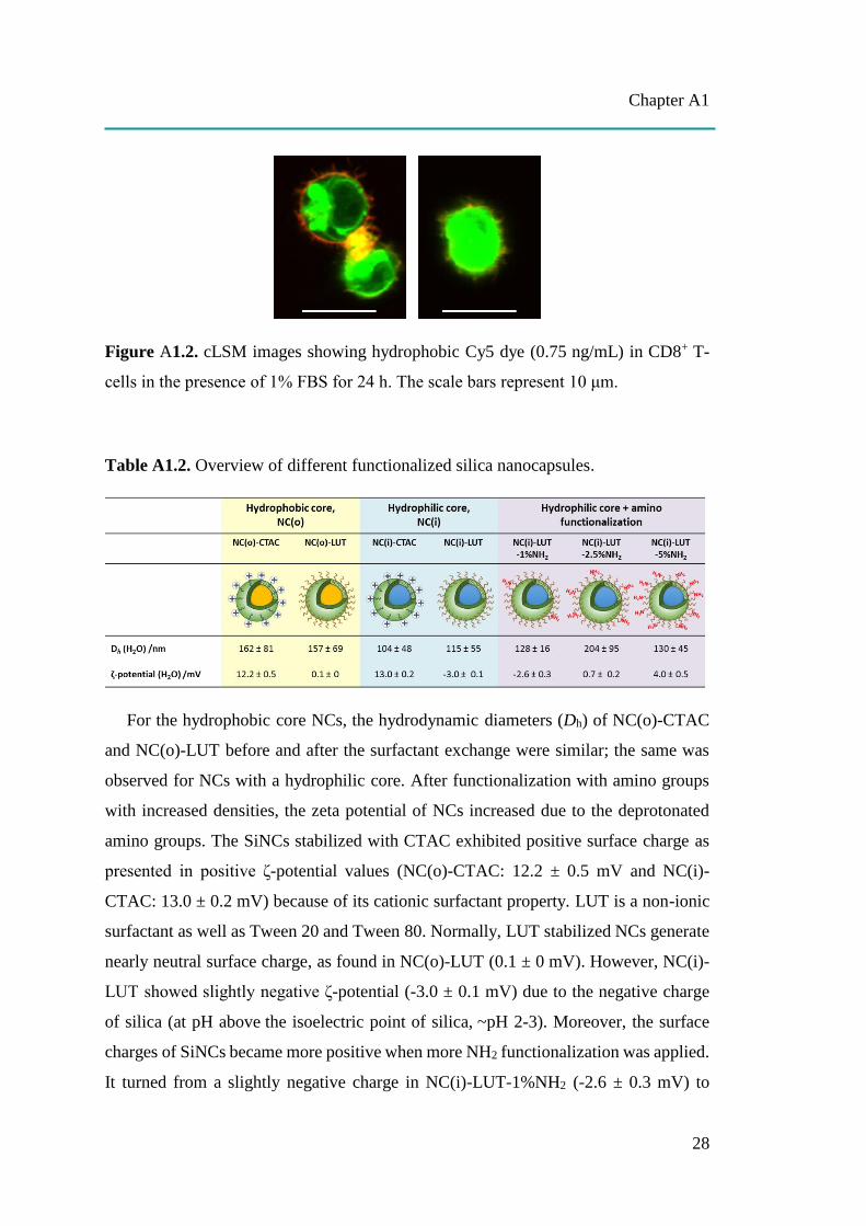
Figure A1.2. cLSM images showing hydrophobic Cy5 dye (0.75 ng/mL) in CD8+ T-
cells in the presence of 1% FBS for 24 h. The scale bars represent 10 μm.
Table A1.2. Overview of different functionalized silica nanocapsules.
For the hydrophobic core NCs, the hydrodynamic diameters (Dh) of NC(o)-CTAC
and NC(o)-LUT before and after the surfactant exchange were similar; the same was
observed for NCs with a hydrophilic core. After functionalization with amino groups
with increased densities, the zeta potential of NCs increased due to the deprotonated
amino groups. The SiNCs stabilized with CTAC exhibited positive surface charge as
presented in positive ζ-potential values (NC(o)-CTAC: 12.2 ± 0.5 mV and NC(i)-
CTAC: 13.0 ± 0.2 mV) because of its cationic surfactant property. LUT is a non-ionic
surfactant as well as Tween 20 and Tween 80. Normally, LUT stabilized NCs generate
nearly neutral surface charge, as found in NC(o)-LUT (0.1 ± 0 mV). However, NC(i)-
LUT showed slightly negative ζ-potential (-3.0 ± 0.1 mV) due to the negative charge
of silica (at pH above the isoelectric point of silica, ~pH 2-3). Moreover, the surface
charges of SiNCs became more positive when more NH2 functionalization was applied.
It turned from a slightly negative charge in NC(i)-LUT-1%NH2 (-2.6 ± 0.3 mV) to
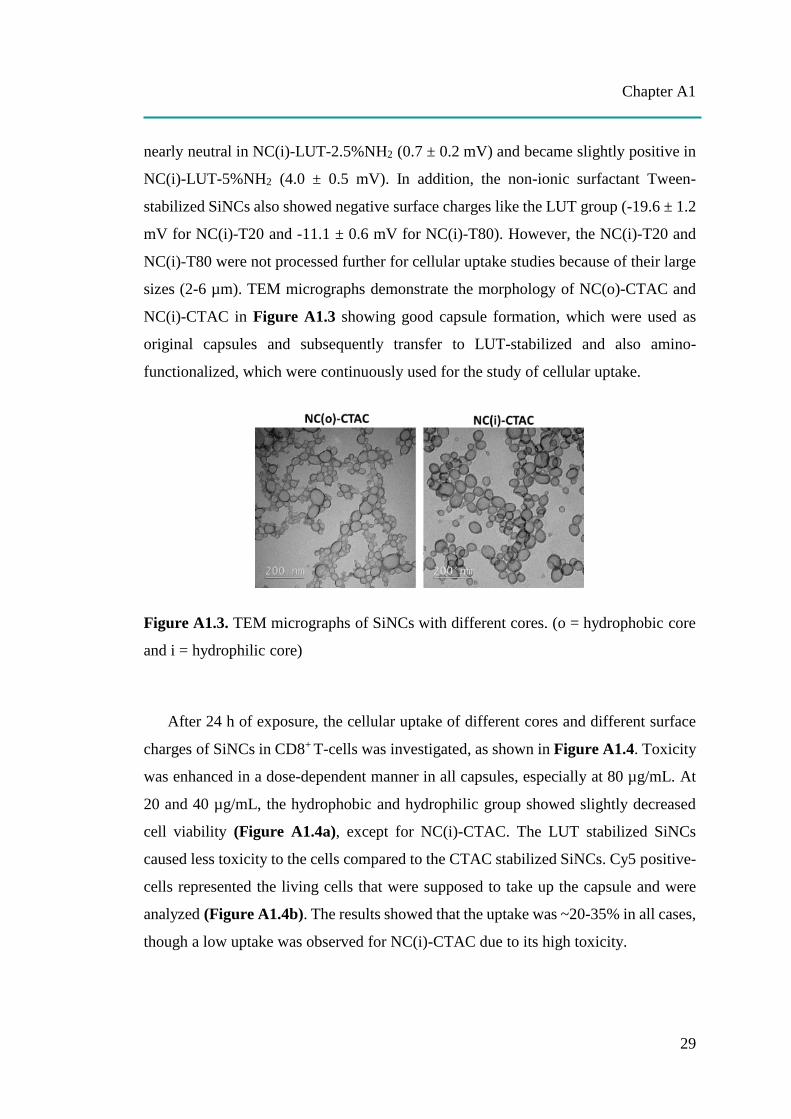
Chapter A1
29
nearly neutral in NC(i)-LUT-2.5%NH2 (0.7 ± 0.2 mV) and became slightly positive in
NC(i)-LUT-5%NH2 (4.0 ± 0.5 mV). In addition, the non-ionic surfactant Tween-
stabilized SiNCs also showed negative surface charges like the LUT group (-19.6 ± 1.2
mV for NC(i)-T20 and -11.1 ± 0.6 mV for NC(i)-T80). However, the NC(i)-T20 and
NC(i)-T80 were not processed further for cellular uptake studies because of their large
sizes (2-6 µm). TEM micrographs demonstrate the morphology of NC(o)-CTAC and
NC(i)-CTAC in Figure A1.3 showing good capsule formation, which were used as
original capsules and subsequently transfer to LUT-stabilized and also amino-
functionalized, which were continuously used for the study of cellular uptake.
Figure A1.3. TEM micrographs of SiNCs with different cores. (o = hydrophobic core
and i = hydrophilic core)
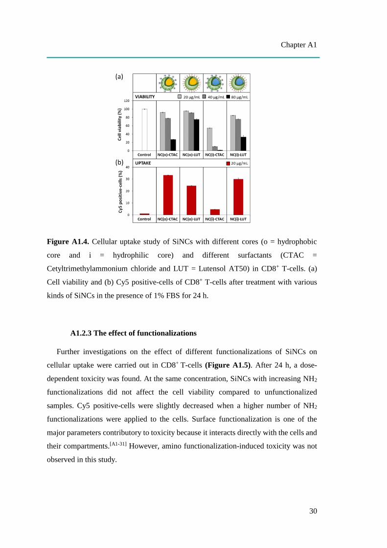
After 24 h of exposure, the cellular uptake of different cores and different surface
charges of SiNCs in CD8+ T-cells was investigated, as shown in Figure A1.4. Toxicity
was enhanced in a dose-dependent manner in all capsules, especially at 80 µg/mL. At
20 and 40 µg/mL, the hydrophobic and hydrophilic group showed slightly decreased
cell viability (Figure A1.4a), except for NC(i)-CTAC. The LUT stabilized SiNCs
caused less toxicity to the cells compared to the CTAC stabilized SiNCs. Cy5 positive-
cells represented the living cells that were supposed to take up the capsule and were
analyzed (Figure A1.4b). The results showed that the uptake was ~20-35% in all cases,
though a low uptake was observed for NC(i)-CTAC due to its high toxicity.
Chapter A1
30
Figure A1.4. Cellular uptake study of SiNCs with different cores (o = hydrophobic
core and i = hydrophilic core) and different surfactants (CTAC =
Cetyltrimethylammonium chloride and LUT = Lutensol AT50) in CD8+ T-cells. (a)
Cell viability and (b) Cy5 positive-cells of CD8+ T-cells after treatment with various
kinds of SiNCs in the presence of 1% FBS for 24 h.
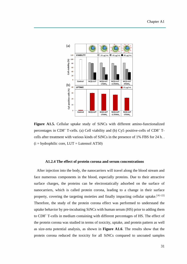
A1.2.3 The effect of functionalizations
Further investigations on the effect of different functionalizations of SiNCs on
cellular uptake were carried out in CD8+ T-cells (Figure A1.5). After 24 h, a dose-
dependent toxicity was found. At the same concentration, SiNCs with increasing NH2
functionalizations did not affect the cell viability compared to unfunctionalized
samples. Cy5 positive-cells were slightly decreased when a higher number of NH2
functionalizations were applied to the cells. Surface functionalization is one of the
major parameters contributory to toxicity because it interacts directly with the cells and
their compartments.[A1-31] However, amino functionalization-induced toxicity was not
observed in this study.
Chapter A1
31
Figure A1.5. Cellular uptake study of SiNCs with different amino-functionalized
percentages in CD8+ T-cells. (a) Cell viability and (b) Cy5 positive-cells of CD8+ T-
cells after treatment with various kinds of SiNCs in the presence of 1% FBS for 24 h. .
(i = hydrophilic core, LUT = Lutensol AT50)
A1.2.4 The effect of protein corona and serum concentrations
After injection into the body, the nanocarriers will travel along the blood stream and
face numerous components in the blood, especially proteins. Due to their attractive
surface charges, the proteins can be electrostatically adsorbed on the surface of
nanocarriers, which is called protein corona, leading to a change in their surface
property, covering the targeting moieties and finally impacting cellular uptake.[A1-22]
Therefore, the study of the protein corona effect was performed to understand the
uptake behavior by pre-incubating SiNCs with human serum (HS) prior to adding them
to CD8+ T-cells in medium containing with different percentages of HS. The effect of
the protein corona was studied in terms of toxicity, uptake, and protein pattern as well
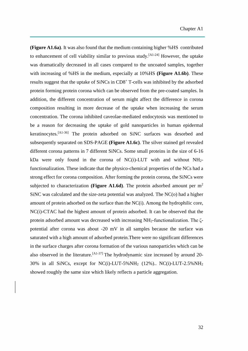
as size-zeta potential analysis, as shown in Figure A1.6. The results show that the
protein corona reduced the toxicity for all SiNCs compared to uncoated samples
Chapter A1
32
(Figure A1.6a). It was also found that the medium containing higher %HS contributed
to enhancement of cell viability similar to previous study.[A1-24] However, the uptake
was dramatically decreased in all cases compared to the uncoated samples, together
with increasing of %HS in the medium, especially at 10%HS (Figure A1.6b). These
results suggest that the uptake of SiNCs in CD8+ T-cells was inhibited by the adsorbed
protein forming protein corona which can be observed from the pre-coated samples. In
addition, the different concentration of serum might affect the difference in corona
composition resulting in more decrease of the uptake when increasing the serum
concentration. The corona inhibited caveolae-mediated endocytosis was mentioned to
be a reason for decreasing the uptake of gold nanoparticles in human epidermal
keratinocytes.[A1-36] The protein adsorbed on SiNC surfaces was desorbed and
subsequently separated on SDS-PAGE (Figure A1.6c). The silver stained gel revealed
different corona patterns in 7 different SiNCs. Some small proteins in the size of 6-16
kDa were only found in the corona of NC(i)-LUT with and without NH2-
functionalization. These indicate that the physico-chemical properties of the NCs had a
strong effect for corona composition. After forming the protein corona, the SiNCs were
subjected to characterization (Figure A1.6d). The protein adsorbed amount per m2
SiNC was calculated and the size-zeta potential was analyzed. The NC(o) had a higher
amount of protein adsorbed on the surface than the NC(i). Among the hydrophilic core,
NC(i)-CTAC had the highest amount of protein adsorbed. It can be observed that the
protein adsorbed amount was decreased with increasing NH2-functionalization. The ζ-
potential after corona was about -20 mV in all samples because the surface was
saturated with a high amount of adsorbed protein.There were no significant differences
in the surface charges after corona formation of the various nanoparticles which can be
also observed in the literature.[A1-37] The hydrodynamic size increased by around 20-
30% in all SiNCs, except for NC(i)-LUT-5%NH2 (12%).. NC(i)-LUT-2.5%NH2
showed roughly the same size which likely reflects a particle aggregation.
Chapter A1
33
Figure A1.6. Protein corona study of different SiNCs (20 µg/mL) in CD8+ T-cells. (a)
Cell viability and (b) Cy5 positive-cells of CD8+ T-cells after treatment with 1)
uncoated SiNCs in the presence of 1% FBS, 2) HS pre-coated SiNCs in the presence
of 0% HS for 6 h. After that, the HS was added to obtain 1% HS, 3) HS pre-coated
SiNCs in the presence of 1% HS, and 4) HS pre-coated SiNCs in the presence of 10%
HS. (c) Corona pattern of HS pre-coated SiNCs analyzed by SDS-PAGE with 1.5 µg
total protein loading. (d) Characterization of HS pre-coated SiNCs. (o = hydrophobic
core, i = hydrophilic core, CTAC = cetyltrimethylammonium chloride, LUT = Lutensol
AT50, FBS = fetal bovine serum and HS = human serum).
Chapter A1
34
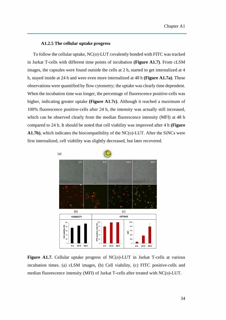
A1.2.5 The cellular uptake progress
To follow the cellular uptake, NC(o)-LUT covalently bonded with FITC was tracked
in Jurkat T-cells with different time points of incubation (Figure A1.7). From cLSM
images, the capsules were found outside the cells at 2 h, started to get internalized at 4
h, stayed inside at 24 h and were even more internalized at 48 h (Figure A1.7a). These
observations were quantified by flow cytometry; the uptake was clearly time dependent.
When the incubation time was longer, the percentage of fluorescence positive-cells was
higher, indicating greater uptake (Figure A1.7c). Although it reached a maximum of
100% fluorescence positive-cells after 24 h, the intensity was actually still increased,
which can be observed clearly from the median fluorescence intensity (MFI) at 48 h
compared to 24 h. It should be noted that cell viability was improved after 4 h (Figure
A1.7b), which indicates the biocompatibility of the NC(o)-LUT. After the SiNCs were
first internalized, cell viability was slightly decreased, but later recovered.
Figure A1.7. Cellular uptake progress of NC(o)-LUT in Jurkat T-cells at various
incubation times. (a) cLSM images, (b) Cell viability, (c) FITC positive-cells and
median fluorescence intensity (MFI) of Jurkat T-cells after treated with NC(o)-LUT.
Chapter A1
35
A1.3. Conclusion
The different physico-chemical properties of SiNCs led to different toxicities and
also affected the uptake behavior. The major impact on cellular uptake and viability
was found to be dose- and size-dependent. Smaller size SiNCs than 100 nm caused
significant toxicity to the cells, which is probably due to ROS-mediated toxicity. The
toxicity of hydrophobic core SiNCs was comparable to the hydrophilic core SiNCs. To
select the core, it should correspond to the hydrophilicity or hydrophobicity of the
desired payload. The effect of different surface charges on cellular uptake was
observed, showing the positive charges of CTAC-stabilized SiNCs decreased the cell
viability as well as the uptake more than closely neutral charges of the LUT-stabilized
SiNCs. In addition, more amino-functionalizations on SiNC surface slightly decreased
the viable cells as well as the uptake. Furthermore, the protein corona study revealed
that both protein corona and medium containing serum was able to cover the toxicity
of SiNCs, leading to improved viability; however, it inhibited the uptake. Therefore,
the concentration of serum in culture condition should be optimized carefully in order
to facilitate the uptake, but still keep the cells alive. These findings suggest the
appropriate criteria for SiNC designing according concerning their physico-chemical
properties for the impact of cellular uptake and toxicity in CD8+ T-cells. Because the
T-cell is a type of the cell that is difficult to take up foreign particles, it is highly
sensitive and not resistant in terms of toxicity.[A1-18] Therefore, the delivery system that
is suitable for use with T-cells has to meet the safety criteria with the ability to protect
and deliver the payload without causing toxicity to the T-cell. For SiNC, it is easy to
manipulate the structures and surface properties based on these findings in order to
avoid toxicity and favor uptake. Therefore, SiNCs are a promising delivery system
which can be applied as a nanocarrier in T-cell immunotherapy.
A1.4. References
[A1-1] Cohen, J. E., Merims, S., Frank, S., Engelstein, R., Peretz, T., & Lotem, M. (2017) Adoptive
cell therapy: past, present and future. Immunotherapy. 9(2), 183-196.
[A1-2] Wang, M., Yin, B., Wang, H. Y., & Wang, R. F. (2014). Current advances in T-cell-based
cancer immunotherapy. Immunotherapy, 6(12), 1265-78.
Chapter A1
36
[A1-3] Perica, K., Varela, J. C., Oelke, M., & Schneck, J. (2015). Adoptive T cell immunotherapy for
cancer. Rambam Maimonides medical journal, 6(1), e0004.
[A1-4] Sathyanarayanan, V., & Neelapu, S. S. (2015). Cancer immunotherapy: Strategies for
personalization and combinatorial approaches. Molecular oncology, 9(10), 2043-53.
[A1-5] Baruch, E. N., Berg, A. L., Besser, M. J., Schachter, J., & Markel, G. (2017), Adoptive T cell
therapy: An overview of obstacles and opportunities. Cancer, 123, 2154-2162.
[A1-6] Li, A. V., Moon, J. J., Abraham, W., Suh, H., Elkhader, J., Seidman, M. A., Yen, M., Im, E. J.,
Foley, M. H., Barouch, D. H., & Irvine, D. J. (2013). Generation of effector memory T cell-
based mucosal and systemic immunity with pulmonary nanoparticle vaccination. Science
translational medicine, 5(204), 204ra130.
[A1-7] Zitvogel, L., & Kroemer, G. (2012). Targeting PD-1/PD-L1 interactions for cancer
immunotherapy. Oncoimmunology, 1(8), 1223-1225.
[A1-8] Frick, S. U., Domogalla, M. P., Baier, G., Wurm, F. R., Mailaender, V., Landfester, K., &
Steinbrink, K. (2016). Interleukin-2 Functionalized Nanocapsules for T Cell-Based
Immunotherapy. ACS Nano, 10, 9216-9226.
[A1-9] Schmid, D., Park, C. G., Hartl, C. A., Subedi, N., Cartwright, A. N., Puerto, R. B., Zheng, Y.,
Maiarana, J., Freeman, G. J., Wucherpfennig, K. W., Irvine, D. J., & Goldberg, M. S. (2017). T
cell-targeting nanoparticles focus delivery of immunotherapy to improve antitumor
immunity. Nature communications, 8(1), 1747.
[A1-10] Li, S. Y., Liu, Y., Xu, C. F., et al. (2016). Restoring anti‐ tumor functions of T cells via
nanoparticle‐ mediated immune checkpoint modulation. J Control Release. (231), 17-28.
[A1-11] Stromnes, I. M., Blattman, J. N., Tan, X., Jeevanjee, S., Gu, H., & Greenberg, P. D. (2010).
Abrogating Cbl-b in effector CD8+ T cells improves the efficacy of adoptive therapy of leukemia
in mice. The Journal of clinical investigation, 120(10), 3722-34.
[A1-12] Ligtenberg, M. A., Pico. de Coaña Y., Shmushkovich, T., Yoshimoto, Y., Truxova, I., Yang,
Y., Betancur-Boissel, M., Eliseev, A. V., Wolfson, A. D., & Kiessling, R. (2018). Self-
Delivering RNAi Targeting PD-1 Improves Tumor-Specific T Cell Functionality for Adoptive
Cell Therapy of Malignant Melanoma. Mol Ther. 26(6), 1482-1493.
[A1-13] Iwamura, K., Kato, T., Miyahara, Y., Naota, H., Mineno, J., Ikeda, H., & Shiku, H. (2012).
siRNA-mediated silencing of PD-1 ligands enhances tumor-specific human T-cell effector
functions. Gene Therapy (19), 959–966.
[A1-14] Ding, C., Tong, L., Feng, J., & Fu, J. (2016) Recent Advances in Stimuli-Responsive Release
Function Drug Delivery Systems for Tumor Treatment. Molecules, 21, 1715.
[A1-15] Mohanraj, V. J., & Chen, Y. (2006). Nanoparticles – A Review. Tropical Journal of
Pharmaceutical Research, 5(1), 561-573.
[A1-16] Zhang, Y., Hsu, B. Y., Ren, C., Li, X., & Wang, J. (2015). Silica-based nanocapsules: synthesis,
structure control and biomedical applications. Chem. Soc. Rev.,44, 315-335.
Chapter A1
37
[A1-17] Murugadoss, S., Lison, D., Godderis, L., Van Den Brule, S., Mast, J., Brassinne, F., Sebaihi,
N., & Hoet, P. H. (2017). Toxicology of silica nanoparticles: an update. Archives of
toxicology, 91(9), 2967-3010.
[A1-18] Freeley, M., & Long, A. (2013). Advances in siRNA delivery to T-cells: potential clinical
applications for inflammatory disease, cancer and infection. Biochem J. 455(2):133-47.
[A1-19] Mendoza, A., Torres-Hernandez, J. A., Ault, J. G., Pedersen-Lane, J. H., Gao, D., & Lawrence,
D. A. (2014). Silica nanoparticles induce oxidative stress and inflammation of human peripheral
blood mononuclear cells. Cell stress & chaperones, 19(6), 777-90.
[A1-20] Vis, B., Hewitt, R. E., Faria, N., Bastos, C., Chappell, H., Pele, L., Jugdaohsingh, R., Kinrade,
S. D., & Powell, J. J. (2018). Non-Functionalized Ultrasmall Silica Nanoparticles Directly and
Size-Selectively Activate T Cells. ACS Nano. doi: 10.1021/acsnano.8b03363.
[A1-21] Napierska, D., Thomassen, L. C., Lison, D., Martens, J. A., & Hoet, P. H. (2010). The nanosilica
hazard: another variable entity. Particle and fibre toxicology, 7(1), 39.
[A1-22] Lundqvist, M., Augustsson, C., Lilja, M., Lundkvist, K., Dahlbäck, B., Linse, S., et al. (2017)
The nanoparticle protein corona formed in human blood or human blood fractions. PLoS ONE
12(4): e0175871.
[A1-23] Jiang, S., Lv, L., Li, Q., Wang, J., Landfester, K., & Crespy, D. (2016). Tailoring
nanoarchitectonics to control the release profile of payloads. Nanoscale, 8 (22), 11511-11517.
[A1-24] Hsiao, I., Gramatke, A. M., Joksimovic, R., Sokolowski, M., Gradzielski, M., et al. (2014). Size
and Cell Type Dependent Uptake of Silica Nanoparticles. J Nanomed Nanotechnol 5, 248.
[A1-25] Li, Y., Sun, L., Jin, M. H., Du, Z., Liu, X., et al. (2011). Size-dependent cytotoxicity of
amorphous silica nanoparticles in human hepatoma HepG2 cells. Toxicol Vitro 25, 1343-1352.
[A1-26] Nabeshi, H., Yoshikawa, T., Matsuyama, K., Nakazato, Y., Arimori, A., et al. (2010) Size-
dependent cytotoxic effects of amorphous silica nanoparticles on Langerhans cells. Pharmazie
65, 199-201.
[A1-27] Napierska, D., Thomassen, L. C. J., Rabolli, V., Lison, D., Gonzalez, L., et al. (2009) Size-
dependent cytotoxicity of monodisperse silica nanoparticles in human endothelial cells. Small.
5, 846-853.
[A1-28] Yildirimer, L., Thanh, T. K. N., Loizidou, M., & Seifalian, M. A. (2011). Toxicological
considerations of clinically applicable nanoparticles. Nano Today. 6, 585-607.
[A1-29] Bakand, S., & Hayes, A. (2016). Toxicological Considerations, Toxicity Assessment, and Risk
Management of Inhaled Nanoparticles. International journal of molecular sciences, 17(6), 929.
[A1-30] Huang, Y. W., Cambre, M., & Lee, H. J. (2017). The Toxicity of Nanoparticles Depends on
Multiple Molecular and Physicochemical Mechanisms. International journal of molecular
sciences, 18(12), 2702.
[A1-31] Buzea, C., Pacheco, I. I., & Robbie, K. (2007). Nanomaterials and nanoparticles: sources and
toxicity. Biointerphases. 2(4), MR17-71.

Chapter A1
38
[A1-32] Kipen, H. M., & Laskin, D. L. (2005) Smaller is not always better: nanotechnology yields
nanotoxicology. Am J Physiol-Lung Cell Mol Physiol. 289, L696-L697.
[A1-33] Nabeshi, H., Yoshikawa, T., Matsuyama, K. et al. (2011a). Amorphous nanosilica induce
endocytosis-dependent ROS generation and DNA damage in human keratinocytes. Part Fibre
Toxicol 8:1.
[A1-34] Accomasso, L., Gallina, C., Turinetto, V., & Giachino, C. (2015). Stem Cell Tracking with
Nanoparticles for Regenerative Medicine Purposes: An Overview. Stem cells
international, 2016, 7920358.
[A1-35] Betzer, O., Shilo, M., Opochinsky, R., Barnoy, E., Motiei, M., Okun, E., Yadid, G., &
Popovtzer, R. (2017). The effect of nanoparticle size on the ability to cross the blood–brain
barrier: an in vivo study. Nanomedicine, 12 (13), 1533-1546.
[A1-36] Li, Y. & Monteiro-Riviere, N. A., Mechanisms of cell uptake, inflammatory potential and
protein corona effects with gold nanoparticles. Nanomedicine 2016, 11 (24), 3185-3203.
[A1-37] Mirshafiee, V., Kim, R., Park, S., Mahmoudi, M., & Kraft, M. L. (2016). Impact of protein pre-
coating on the protein corona composition and nanoparticle cellular uptake. Biomaterials, 75,
295-304.

Chapter A2
39
A2: CELLULAR UPTAKE OF siRNA LOADED NANOCARRIER
TO KNOCKDOWN PD-L1:
STRATEGIES TO IMPROVE T-CELL FUNCTIONS
(Note: The literature of the chapter A2 can be found on page 48 at the end of this chapter.)
Permission: The manuscript is under preparation.
Contribution: I carried out the biological part including cytotoxicity, cellular uptake
study by flow cytometry, cell imaging by cLSM, PD-L1 knockdown study and
quantitative polymerase chain reaction (qPCR) analysis. Mengyi Li synthesized and
characterized the SiNCs. The project was supervised by Katharina Landfester and
Volker Mailänder.
TOC A2: The siRNA mediated PD-L1, a potent inhibitory protein, silencing was
carried out by using the silica nanocapsules (SiNCs) carrying the PD-L1 specific siRNA
to CD8+ T-cells in order to inhibit PD-L1 expression resulting in the improvement of
T-cells viability and function.
Aim: To inactivate PD-L1 expression, the silica nanocapsules (SiNCs) carrying the
PD-L1 specific siRNA were internalized to CD8+ T-cells. Knockdown efficiency was
determined at protein and mRNA levels. The effects of PD-L1 knockdown were
demonstrated in terms of cell proliferation and some functional biomarker expressions.
Chapter A2
40
Abstract: T-cells or T-lymphocytes are a type of lymphocyte (a subtype of white blood
cells) that plays a central role in cell-mediated immunity. Nowadays, adoptive T-cell
immunotherapy has been researched and developed to fight against cancer. In this
therapy, T-cells are harvested from a patient’s blood. After several weeks of expansion
in culture, tumor-specific T-cells can be reinfused into the same cancer patient. This
technique provides high efficiency for cancer treatment. However, several processes
exist to suppress anti-cancer responses of T-cells leading to the loss of their
functionality and reduction of their viability. Therefore, strategies are needed to
improve T-cell survival and functions. Here, small interfering RNA (siRNA) loaded
nanocarriers were used to knockdown PD-L1, one of the most important proteins
causing loss of functionality in T-cells. Biocompatibility and cellular uptake of siRNA
loaded silica nanocapsules (SiNCs) were investigated in CD8+ T-cells. Then, PD-L1
expression at protein and mRNA levels of treated cells were evaluated. Furthermore,
the effect of PD-L1 knockdown was observed in terms of cell proliferation and the
expression of specific biomarkers: CD25 and CD71, which are indicators of T-cell
functions. The results suggest that this siRNA loaded nanocarrier exhibits the potential
for use in delivery of siRNA into T-cells, enhanced T-cell survival by decreasing the
expression of inhibitory protein PD-L1 and can be applied in adoptive T-cell
immunotherapy for the treatment of cancer.
A2.1. Introduction
T-cells or T-lymphocytes are some of the most important cells in the immune system.
They can be divided into two main subtypes according to their surface markers, CD4+
and CD8+ T-cells. CD4+ T-cells (T-helper cells) involve the maturation of B-cells as
well as the activation of CD8+ T-cells. CD8+ T-cells (cytotoxic T-cells) play a major
role in destroying virus-infected cells and tumor cells.[A2-1-3] With the outstanding
ability to kill cancer cells, a technique called adoptive T-cell immunotherapy for cancer
treatment using CD8+ T-cells has been employed and studied intensively for a past
decade with promising results.[A2-4-5] In the first step for immunotherapy, CD8+ T-cells
are collected from patient blood, selected and activated under the culture condition to
enhance the number of specific T-cells and make them strong enough to fight against
Chapter A2
41
cancer before reinfusion to the patient’s body.[A2-6] However, T-cells start to lose their
viability and function after a long expansion and cultivation time, leading to less
efficiency to kill cancer cells.[A2-4-7] The decrease in T-cells survival and killing ability
is due to the natural mechanism of the immune system. When antigen presenting cells,
for example dendritic cells, present cancer antigen on their surface and then bind to T-
cell receptor resulting in T-cell activation, trafficking along the blood stream to the
cancer site, recognition of cancer, and finally killing the cancerous cells. After that, the
amount of specific T-cells will decrease to ensure that they are inactivated later to avoid
chronic inflammation and autoimmune disease. There are several proteins involved in
T-cell inactivation, which are called immune check point proteins such as cytotoxic T-
lymphocyte antigen 4 (CTLA-4), programmed cell death-1 (PD-1), and their cognate
ligands (PD-L1 and PD-L2).[A2-8-12] One of the well-known T-cell inhibitions occurs
through PD-1/PD-L1 interaction by the specific binding of PD-1 receptor on the surface
of T-cells through its ligand (PD-L1) on the surface of dendritic cells. Many researches
have focused on the inhibition of PD-1expression with the aim of improving T-cell
function.[A2-12] In fact, PD-L1 is also expressed on the surface of T-cells, which can also
cause PD-1/PD-L1 interaction between the T-cell population itself, which finally
blocks T-cell activation.[A2-13] To inhibit the immune check point proteins either PD-1
or PD-L1 expression, it is necessary to pay careful attention to the systemic blocking,
which can result in an undetermined effect of over immune reaction. PD-L1 is not only
expressed on the surface of hematopoietic cells, but is also found on the surface of non-
hematopoietic cells.[A2-14] As selective inhibition, small interfering RNA (siRNA)-
mediated silencing of PD-L1 was used and electroporated into tumor specific CD4+ and
CD8+ human T-cells.[A2-13] The transfected T-cells successfully increased the
interferon- production and antigen specific cytotoxicity. These findings suggest that
siRNA-mediated PD-L1 silencing is another possible approach to achieve the inhibition
of immune check point proteins, leading to enhanced T-cell function. However, it is
known that the electroporation technique is harmful to cells due to electric shock,
resulting in decreased cell viability after transfection.[A2-15] With the great advantages
of nanotechnology, nanocarriers containing payloads such as drugs, dyes and specific
biomolecules have been widely developed and used. Among the various kinds of
nanocarriers, silica-based nanocapsules have shown a high loading capacity, ease of
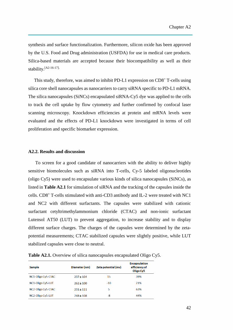
Chapter A2
42
synthesis and surface functionalization. Furthermore, silicon oxide has been approved
by the U.S. Food and Drug administration (USFDA) for use in medical care products.
Silica-based materials are accepted because their biocompatibility as well as their
stability.[A2-16-17].
This study, therefore, was aimed to inhibit PD-L1 expression on CD8+ T-cells using
silica core shell nanocapsules as nanocarriers to carry siRNA specific to PD-L1 mRNA.
The silica nanocapsules (SiNCs) encapsulated siRNA-Cy5 dye was applied to the cells
to track the cell uptake by flow cytometry and further confirmed by confocal laser
scanning microscopy. Knockdown efficiencies at protein and mRNA levels were
evaluated and the effects of PD-L1 knockdown were investigated in terms of cell
proliferation and specific biomarker expression.
A2.2. Results and discussion
To screen for a good candidate of nanocarriers with the ability to deliver highly
sensitive biomolecules such as siRNA into T-cells, Cy-5 labeled oligonucleotides
(oligo Cy5) were used to encapsulate various kinds of silica nanocapsules (SiNCs), as
listed in Table A2.1 for simulation of siRNA and the tracking of the capsules inside the
cells. CD8+ T-cells stimulated with anti-CD3 antibody and IL-2 were treated with NC1
and NC2 with different surfactants. The capsules were stabilized with cationic
surfactant cetyltrimethylammonium chloride (CTAC) and non-ionic surfactant
Lutensol AT50 (LUT) to prevent aggregation, to increase stability and to display
different surface charges. The charges of the capsules were determined by the zeta-
potential measurements; CTAC stabilized capsules were slightly positive, while LUT
stabilized capsules were close to neutral.
Table A2.1. Overview of silica nanocapsules encapsulated Oligo Cy5.
Chapter A2
43
Cellular uptake of SiNCs encapsulated oligo-Cy5 in CD8+ T-cells was studied in
terms of toxicity and uptake after 24 h of incubation. Flow cytometry revealed a slight
decrease in cell viability of CD8+ T-cells treated with SiNCs up to 80 µg/mL, indicating
their biocompatibility with CD8+ T-cells (Figure A2.1a) and an increase of Cy5
positive-cells with the increasing concentration of SiNCs (Figure A2.1b). The cellular
uptake of these SiNCs was further confirmed by confocal laser scanning microscopy
(cLSM) (Figure A2.1d). cLSM images demonstrated green dots, representing SiNC-
oligo Cy5 in the cytoplasm and indicating effective uptake by the cells. In the
experiments mentioned above, all SiNCs exhibited good potential for use as a
nanocarrier of biomolecules into CD8+ T-cells; however, the capsule NC2 showed
better capsule formation in which the shell structure was formed clearly as observed in
TEM images (Figure A2.1c). Therefore, the NC2 was chosen for use in subsequent
experiments.
To investigate the effect of different surface charges on cellular uptake, anti-CD3
antibody and IL-2 stimulated CD8+ T-cells were incubated with NC2-CTAC or -LUT
encapsulated siRNA Cy5 for 24 h. The fluorescence-tracking molecule was changed
from oligo-Cy5 to siRNA-Cy5 in order to closely imitate the real payload in this study,
which is siRNA that aimed to encapsulate inside the capsules and carry to T-cells.
Concentrations of SiNCs from 40 to 640 µg/mL were added to the cells to determine
the critical toxic concentration. Figure A2.2a showed the increase of toxicity to CD8+
T-cells in dose-dependent manner of both NC2-siRNA Cy5-CTAC and NC2-siRNA
Cy5-LUT. The half maximal effective concentration (EC50) of NC2-siRNA Cy5-CTAC
on CD8+ T-cells was 379.8 µg/mL, which was lower than that of NC2-siRNA Cy5-
LUT (406.3 µg/mL). This result suggested that the LUT stabilized NC2 was more
biocompatible with CD8+ T-cells than the CTAC stabilized NC2. The cellular uptake
of these SiNCs was determined by an increase of Cy5 positive-cells with increasing
concentration of the SiNCs using flow cytometry (Figure A2.2b) and subsequently
confirmed by cLSM (Figure A2.2c). Considering the percentages of Cy5 positive-cells,
there was no significant difference between CTAC and LUT stabilized NC2, indicating
that they are comparable in cellular uptake efficiency. It seems that the charges of the
capsule are not only involved in cellular uptake efficiency, but also other parameters

Chapter A2
44
such as the components of capsule formation. cLSM images demonstrated CD8+ T-
cells were in a clumping or aggregation form with green dots representing NC2-siRNA
Cy5 inside the cytoplasm. This result not only proves efficient uptake by the cells, but
also indicates the healthy and/or proliferation stage of the cells due to the excellent
biocompatibility of the capsules. To provide more evidence for cellular uptake, the co-
localization assay was performed. Figure A2.3 shows the NC2-siRNA Cy5-CTAC and
–LUT were co-localized with lysosome, as indicated with white arrows. This evidence
provides solid proof of cellular uptake for these SiNCs in CD8+ T-cells. Although the
EC50 was not significant difference, but the cell morphology from the cLSM image of
LUT stabilized SiNC showed more cell clusters indicating more cell healthy NC2-LUT
was then selected for use in the next steps for encapsulation of control siRNA, which
has no specific sequences to human mRNA and target siRNA containing specific
sequences to PD-L1 mRNA.
NC2-LUT encapsulated with siRNA targeted to PD-L1 mRNA (siRNA target SiNC)
was added to CD8+ T-cells. The PD-L1 knockdown and their knockdown effects were
analyzed and compared with NC2-LUT encapsulated with siRNA control, which has
no specific sequences to human mRNA (siRNA control SiNC) and cell control (without
any capsule), as shown in Figure A2.4. Relative PD-L1 protein expression profiles
were observed from day 1 to day 3 by flow cytometry analysis (Figure A2.4a).
Knockdown efficiency of siRNA target SiNC compared to siRNA control SiNC was
30%, 27% and 6%, respectively. Knockdown efficiency of siRNA target SiNC
compared to control was 35%, 48% and 40%, respectively. It seems that siRNA control
SiNC had a non-specific effect on PD-L1 expression, which was able to also decrease
PD-L1 expression. Since the maximum effect of PD-L1 knockdown of siRNA target
SiNC compared to the control was found on day 2, the total RNA was extracted. cDNA
was synthesized and subsequently used for quantitative PCR analysis. Relative PD-L1
mRNA expression (Figure A2.4b) demonstrated the knockdown efficiency at mRNA
level of siRNA target SiNC was 28% compared to siRNA control SiNC and was 61%
compared to the control. This higher knockdown efficiency at the mRNA level than at
the protein level is quite common because the siRNA targets basically direct to the
mRNA, then subsequently inhibit protein synthesis. After confirmation of PD-L1

Chapter A2
45
knockdown at both mRNA and protein levels, the effects of PD-L1 knockdown in terms
of cell survival and functions were also studied. Cell proliferation assays (Figure
A2.4c) and relative expression of some specific biomarkers for T-cell functions such as
CD25 and CD71 (Figure A2.4d) were investigated. The results showed that cell
proliferation on day 1 and day 3 of CD8+ T-cells treated with siRNA target SiNC was
increased significantly compared to the control. Furthermore, the relative expression of
CD25 and CD71 on day 2 of CD8+ T-cells treated with siRNA target SiNC was also
increased significantly compared to the control.
Figure A2.1. Cellular uptake study of SiNCs encapsulated Oligo Cy5 in CD8+ T-cells
demonstrating (a) Cell viability, (b) Cy5 positive-cells and (d) confocal laser scanning
microscopy (cLSM) images of CD8+ T-cells after treatment with SiNCs encapsulated
Oligo Cy5 for 17 h. The membrane was stained with CellMask™Orange (red). The
SiNC was labeled with Oligo Cy5 (green). The scale bars represent 10 μm. (c) TEM
micrographs of SiNCs.

Chapter A2
46
Figure A2.2. Cellular uptake study of SiNCs encapsulated siRNA Cy5 in CD8+ T-cells.
(a) Cell viability, (b) Cy5 positive-cells and (c) cLSM images of CD8+ T-cells after
treatment with SiNCs encapsulated siRNA Cy5 for 17 h. The membrane was stained
with CellMask™Orange (red). The SiNC was labeled with Oligo Cy5 (green). The
scale bars represent 10 μm. The EC50 of NC2-siRNA Cy5-CTAC and –LUT were 379.8
and 406.3 μg/mL, respectively.
Figure A2.3. cLSM images of SiNCs encapsulated siRNA Cy5 co-localized with
lysosomes in CD8+ T-cells after 17 h. Co-localization assay was performed by staining
lysosome with LysoTracker dye (red). The membrane was stained with
CellMask™Orange (blue). The SiNC was labeled with siRNA Cy5 (green). The
merged images demonstrated that SiNCs encapsulated siRNA Cy5 were co-localized
with lysosomes as indicated with white arrows. The scale bars represent 10 μm.
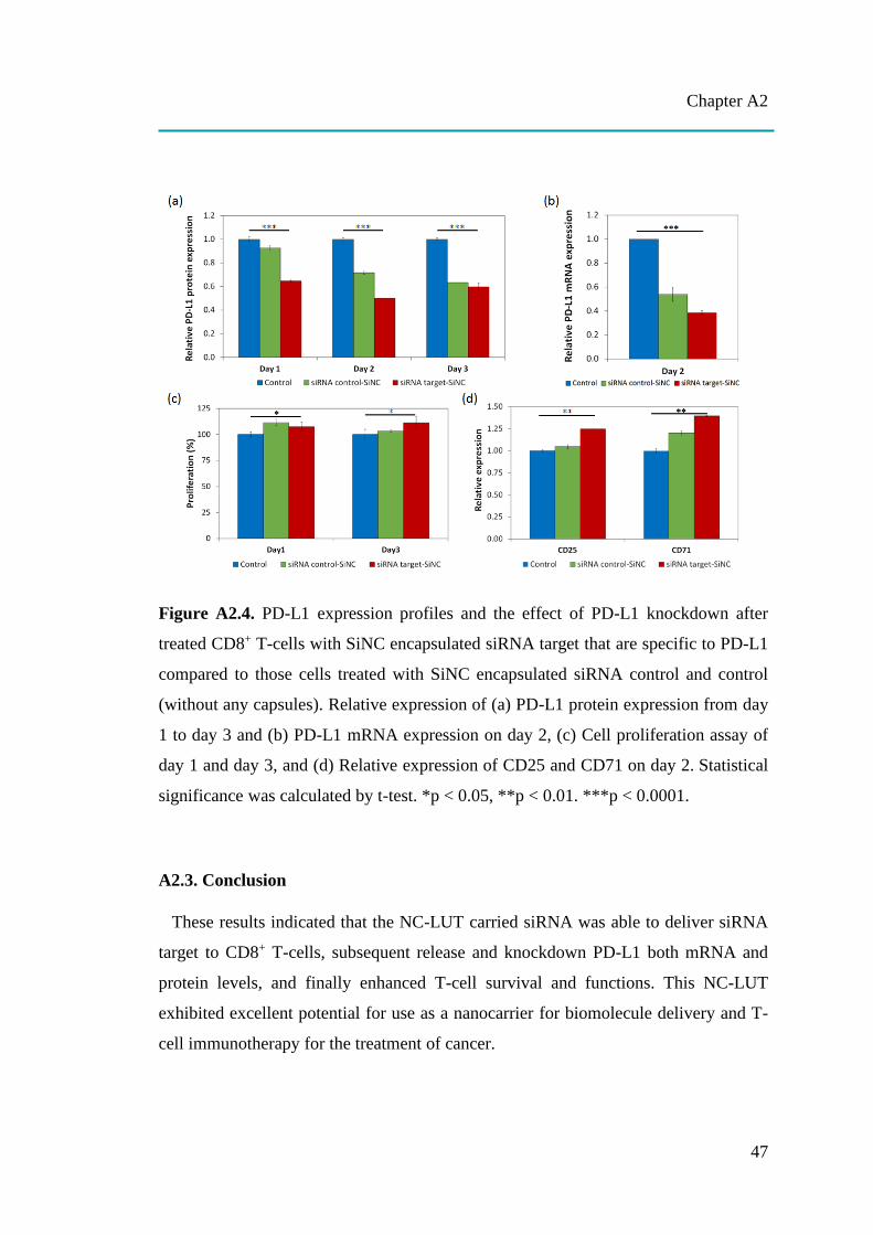
Chapter A2
47
Figure A2.4. PD-L1 expression profiles and the effect of PD-L1 knockdown after
treated CD8+ T-cells with SiNC encapsulated siRNA target that are specific to PD-L1
compared to those cells treated with SiNC encapsulated siRNA control and control
(without any capsules). Relative expression of (a) PD-L1 protein expression from day
1 to day 3 and (b) PD-L1 mRNA expression on day 2, (c) Cell proliferation assay of
day 1 and day 3, and (d) Relative expression of CD25 and CD71 on day 2. Statistical
significance was calculated by t-test. *p < 0.05, **p < 0.01. ***p < 0.0001.
A2.3. Conclusion
These results indicated that the NC-LUT carried siRNA was able to deliver siRNA
target to CD8+ T-cells, subsequent release and knockdown PD-L1 both mRNA and
protein levels, and finally enhanced T-cell survival and functions. This NC-LUT
exhibited excellent potential for use as a nanocarrier for biomolecule delivery and T-
cell immunotherapy for the treatment of cancer.
Chapter A2
48
A2.4. References
[A2-1] Wang, M., Yin, B., Wang, H. Y., & Wang, R. F. (2014). Current advances in T-cell-based
cancer immunotherapy. Immunotherapy, 6(12), 1265-78.
[A2-2] Fontenot, A. P. and P. L. Simonian (2016). 13 - Adaptive Immunity. Murray and Nadel's
Textbook of Respiratory Medicine (Sixth Edition). V. C. Broaddus, R. J. Mason, J. D. Ernst et
al. Philadelphia, W.B. Saunders: 206-224.e203.
[A2-3] Moticka, E. J. (2016). Chapter 2 - Hallmarks of the Adaptive Immune Responses. In E. J.
Moticka (Ed.), A Historical Perspective on Evidence-Based Immunology (pp. 9-19).
Amsterdam: Elsevier.
[A2-4] Perica, K., Varela, J. C., Oelke, M., & Schneck, J. (2015). Adoptive T cell immunotherapy for
cancer. Rambam Maimonides medical journal, 6(1), e0004.
[A2-5] Cohen, J. E., Merims, S., Frank, S., Engelstein, R., Peretz, T., Lotem, M. (2017) Adoptive cell
therapy: past, present and future. Immunotherapy. 9(2), 183-196.
[A2-6] Sathyanarayanan, V., & Neelapu, S. S. (2015). Cancer immunotherapy: Strategies for
personalization and combinatorial approaches. Molecular oncology, 9(10), 2043-53.
[A2-7] Baruch, E. N., A. L. Berg, M. J. Besser, J. Schachter and G. Markel (2017). "Adoptive T cell
therapy: An overview of obstacles and opportunities." Cancer 123(S11): 2154-2162.
[A2-8] Chen, D. S. and I. Mellman (2013). "Oncology meets immunology: the cancer-immunity
cycle." Immunity 39(1): 1-10.
[A2-9] Pitt, J. M., M. Vetizou, R. Daillere, M. P. Roberti, T. Yamazaki, B. Routy, P. Lepage, I. G.
Boneca, M. Chamaillard, G. Kroemer and L. Zitvogel (2016). "Resistance Mechanisms to
Immune-Checkpoint Blockade in Cancer: Tumor-Intrinsic and -Extrinsic Factors." Immunity
44(6): 1255-1269.
[A2-10] Topalian, S. L., C. G. Drake and D. M. Pardoll (2015). "Immune checkpoint blockade: a
common denominator approach to cancer therapy." Cancer Cell 27(4): 450-461.
[A2-11] Sharma, P. and J. P. Allison (2015). "Immune checkpoint targeting in cancer therapy: toward
combination strategies with curative potential." Cell 161(2): 205-214.
[A2-12] Shin, D. S. and A. Ribas (2015). "The evolution of checkpoint blockade as a cancer therapy:
what's here, what's next?" Curr Opin Immunol 33: 23-35.
[A2-13] Iwamura, K., Kato, T., Miyahara, Y., Naota, H., Mineno, J., Ikeda, H., Shiku, H. (2012). siRNA-
mediated silencing of PD-1 ligands enhances tumor-specific human T-cell effector functions.
Gene Therapy (19), 959–966.
[A2-14] Mueller, S. N., Vanguri, V. K., Ha, S. J., West, E. E., Keir, M. E., Glickman, J. N., Sharpe, A.
H., … Ahmed, R. (2010). PD-L1 has distinct functions in hematopoietic and nonhematopoietic
cells in regulating T cell responses during chronic infection in mice. The Journal of clinical
investigation, 120(7), 2508-15.
Chapter A2
49
[A2-15] McManus, M. T., B. B. Haines, C. P. Dillon, C. E. Whitehurst, L. van Parijs, J. Chen and P. A.
Sharp (2002). "Small Interfering RNA-Mediated Gene Silencing in T Lymphocytes." The
Journal of Immunology 169(10): 5754.
[A2-16] Zhang, Y., Hsu, B. Y., Ren, C., Li, X., Wang, J. (2015). Silica-based nanocapsules: synthesis,
structure control and biomedical applications. Chem. Soc. Rev.,44, 315-335.
[A2-17] Murugadoss, S., Lison, D., Godderis, L., Van Den Brule, S., Mast, J., Brassinne, F., Sebaihi,
N., Hoet, P. H. (2017). Toxicology of silica nanoparticles: an update. Archives of
toxicology, 91(9), 2967-3010.
Chapter B
50
CHAPTER B
LIGHT-TRIGGERED RELEASE FROM NANOCAPSULES
Chapter B combines three different Publications [B1-B3]. These studies
demonstrated new strategies for using nanocapsules in the release of functional
payloads for cancer therapy. General introduction concerning recent advances in the
controlled release of anti-cancer drugs are described. Afterwards, each study will be
presented separately.
(Note: The literature of the introduction can be found on page 138 at the end of the dissertation.)
[B1] Zhijun Chen, Raweewan Thiramanas, Mischa Schwendy, Chaoming Xie,
Sapun H. Parekh, Volker Mailänder,* and Si Wu*. Upconversion Nanocarriers
Encapsulated with Photoactivatable Ru Complexes for Near-Infrared Light-
Regulated Enzyme Activity. Small 2017, 13, 1700997. ( Shared first)
[B2] Wen Sun, Raweewan Thiramanas, Leonardo D. Slep, Xiaolong Zeng,
Volker Mailänder,* and Si Wu*. Photoactivation of Anticancer Ru Complexes
in Deep Tissue: How Deep Can We Go? Chem. Eur. J. 2017, 23, 10832-10837.
( Shared first)
[B3] Wen Sun, Yan Wen, Raweewan Thiramanas, Mingjia Chen, Jianxiong
Han, Ningqiang Gong, Manfred Wagner, Shuai Jiang, Michael S. Meijer,
Sylvestre Bonnet, Hans-Jürgen Butt, Volker Mailänder,* Xing-Jie Liang,* Si
Wu*. Red-Light-Controlled Release of Drug-Ru Complex Conjugates from
Metallopolymer Micelles for Phototherapy in Hypoxic Tumor Environments.
Adv. Funct. Mater. 2018, 28, 1804227. ( Shared first)
Chapter B
51
B. Introduction
Cancer is a serious medical issue that kills many people around the world every
year. Many scientists have invested significant efforts to search for a new target and
therapy. However, cancer is hard to kill and diagnose due to its ability to escape the
immune system, leading to the decrease of treatment efficacy.[Z-52-53] In addition, it is
difficult to detect cancer in the early stages. Once symptoms are detected, cancer is
mostly in the 3rd or 4th stage already.[Z-54] In addition, a tumor site which is far away
from blood supply has a unique tumor microenvironment, such as low oxygen
(hypoxia), low nutrient, and acidosis (an excessively acid condition).[Z-55-57] This
hypoxic condition hinders drugs or other methods that need oxygen to generate the
killing effect, e.g. singlet oxygen production in photodynamic therapy (PDT).[Z-58]
Nowadays, scientists continue to put effort into finding a new strategy for cancer
treatment. The main way to treat cancer is still based on surgery because the bulk of the
tumor can be eliminated. However, the disseminated part cannot be completely
removed because of difficulty in distinguishing the tumor from healthy tissue.
Moreover, the most concerning issues are the induction of tumor regrowth and
metastatic state.[Z-59] In addition to surgery, cancer therapy can be divided into two
major strategies, chemotherapy and radiotherapy.
Chemotherapy has been investigated and developed to kill cancer over the last
century and is currently the main treatment that will be considered by medical
oncologists.[Z-60] Chemotherapy involves the use of medicines and drugs to destroy
cancer cells. Many anti-cancer drugs have been approved and used for the treatment of
specific kinds of cancer. For example, doxorubicin (DOX) is effective against breast
cancer, 5-fluorouracil against colon cancer, epirubicin against stomach cancer and
paclitaxel against lung cancer.[Z-61] However, directly using these drugs via oral,
parenteral (injection) or transdermal administration can result in reduced cancer
treatment efficiency due to drug metabolism, lacking the ability to achieve site-specific
activation, and adverse side effects for healthy tissue.[Z-62] Therefore, a drug delivery
system is needed to protect the encapsulated drug from drug metabolism and deliver it
to the target area. Furthermore, a stimuli responsive delivery system is required to avoid
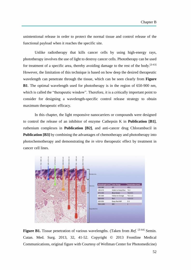
Chapter B
52
unintentional release in order to protect the normal tissue and control release of the
functional payload when it reaches the specific site.
Unlike radiotherapy that kills cancer cells by using high-energy rays,
phototherapy involves the use of light to destroy cancer cells. Phototherapy can be used
for treatment of a specific area, thereby avoiding damage to the rest of the body.[Z-63]
However, the limitation of this technique is based on how deep the desired therapeutic
wavelength can penetrate through the tissue, which can be seen clearly from Figure
B1. The optimal wavelength used for phototherapy is in the region of 650-900 nm,
which is called the “therapeutic window”. Therefore, it is a critically important point to
consider for designing a wavelength-specific control release strategy to obtain
maximum therapeutic efficacy.
In this chapter, the light responsive nanocarriers or compounds were designed
to control the release of an inhibitor of enzyme Cathepsin K in Publication [B1],
ruthenium complexes in Publication [B2], and anti-cancer drug Chlorambucil in
Publication [B3] by combining the advantages of chemotherapy and phototherapy into
photochemotherapy and demonstrating the in vitro therapeutic effect by treatment in
cancer cell lines.
Figure B1. Tissue penetration of various wavelengths. (Taken from Ref. [Z-64] Semin.
Cutan. Med. Surg. 2013, 32, 41-52. Copyright © 2013 Frontline Medical
Communications, original figure with Courtesy of Wellman Center for Photomedicine)
Chapter B
53
In Publication [B1], near-infrared (NIR) light was used to trigger the Cathepsin
K inhibitor controllable release system called “photon upconversion enzyme inhibitor
(PUEI)”. The system consists of a hollow mesoporous silica nanoparticle used as a
nanocarrier encapsulated with upconversion nanoparticles (UCNPs) and ruthenium
complex caged inhibitor (RuEI). UCNPs can convert NIR light to blue light, which
induces the cleavage of RuEI. The inhibitor is then activated and subsequently released,
resulting in the inhibition of Cathepsin K activity as demonstrated in three different
cancer cell lines: LNCaP, PC3 and SAOS-2. Cathepsin K is an important enzyme
involved in bone resorption and cancer invasion. Thus, the inhibition of this enzyme
can impede bone destruction and tumor growth. This system can be used as a model for
regulating enzyme activity in deep tissue by using the NIR light responsive control
system.
In Publication [B2], a Ru complex was investigated for possible use in
photochemotherapy for deep tissue. In fact, NIR light is able to penetrate into the tissue
not as deep compared to red light. In addition, the therapeutic window (650-900 nm) is
in the region of red light. Therefore, the 671 nm-red light was chosen to activate the Ru
complex in this Publication [B2] instead of NIR light. With the outstanding property
of being photocleavable and ruthenium being an anticancer compound, this makes the
Ru complex highly interesting. The complex is less toxic to HeLa cells in the dark
condition and becomes more toxic when irradiated with red light (671 nm) passed
through 8 mm-thick tissue. The results suggest that red light responsive Ru complex
has the potential for use in deep tissue photochemotherapy.
In Publication [B3], from a promising result in Publication [B2], red light in a
region of therapeutic window was continued for use in the activation of controlled
release of chlorambucil (anticancer drug)-Ru complex conjugates from metallopolymer
micelles in hypoxic tumor microenvironment. The hydrophilic block copolymer
conjugated with hydrophobic drug-Ru complex can be self-assembled into micelles,
thus facilitating the uptake of tumor cells. In deep solid tumors, this area has very low
physiological oxygen concentration, leading to reduced cancer treatment efficiency
when applying photodynamic therapy, which is critically dependent on local oxygen
concentration. With this new dual system using both anticancer drug, toxicity is oxygen

Chapter B
54
independent and Ru complex, which can be both oxygen dependent- and independent-
, the efficiency to kill cancer cells can be overcome, even in hypoxic condition, as
demonstrated in vitro (in HeLa cells) and in vivo (in tumor-bearing mice). This novel
red-light responsive drug-Ru complex containing micelles integrates a new powerful
system for photochemotherapy and can be applied even in hypoxic tumor
microenvironment.
Stimuli responsive drug delivery system can be recognized as on-demand drug
delivery system or smart drug delivery system. The system provides an ability to control
over the specific place, time, and amount of cargo to be released upon the activation by
specific stimuli.[Z-65] Here, an overview of various stimuli responsive mechanisms that
can be applied in the controlled release of payloads is described in general, while light-
responsiveness, which is the major controlled release strategy in these studies, is
explained in particular.
B.1 stimuli responsive mechanisms
There are various types of stimuli responsive mechanisms used for controlled
release, which can be categorized into two major types according to the source of
stimuli; intrinsic stimuli and extrinsic stimuli. Considering the huge differences
between normal cells and cancerous cells such as cellular pH, redox conditions, specific
generated biomolecules types and amounts, all these originally unique characters bring
intrinsic stimuli for a controlled release strategy and cell-specific therapy. This type of
stimuli employs the internal distinct characters of cancerous cells to switch on the
release of the payload. Meanwhile, extrinsic stimuli do not naturally exist inside the
human body. Stimuli such as heat, magnetic force and light also share an important role
as an ideal stimulus, in which site-, dose- and time-specific release are more precisely
controllable from outside of the body.
B.1.1 Intrinsic stimuli
B.1.1.1 pH-responsiveness
pH responsive stimuli is used for control release based on naturally-different pH
in various tissues/organs of the body. It generally uses acidic pH to induce drug release.
A low pH environment is found not only in cancerous cells, but also in various parts of
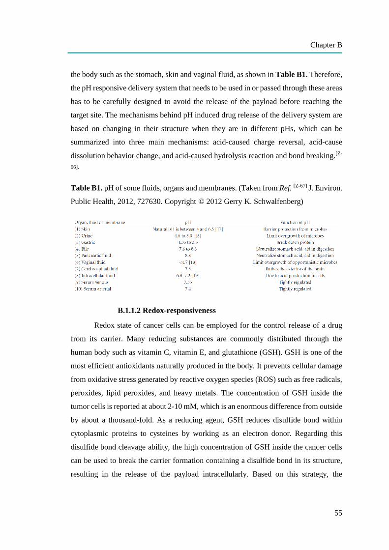
Chapter B
55
the body such as the stomach, skin and vaginal fluid, as shown in Table B1. Therefore,
the pH responsive delivery system that needs to be used in or passed through these areas
has to be carefully designed to avoid the release of the payload before reaching the
target site. The mechanisms behind pH induced drug release of the delivery system are
based on changing in their structure when they are in different pHs, which can be
summarized into three main mechanisms: acid-caused charge reversal, acid-cause
dissolution behavior change, and acid-caused hydrolysis reaction and bond breaking.[Z-
66].
Table B1. pH of some fluids, organs and membranes. (Taken from Ref. [Z-67] J. Environ.
Public Health, 2012, 727630. Copyright © 2012 Gerry K. Schwalfenberg)
B.1.1.2 Redox-responsiveness
Redox state of cancer cells can be employed for the control release of a drug
from its carrier. Many reducing substances are commonly distributed through the
human body such as vitamin C, vitamin E, and glutathione (GSH). GSH is one of the
most efficient antioxidants naturally produced in the body. It prevents cellular damage
from oxidative stress generated by reactive oxygen species (ROS) such as free radicals,
peroxides, lipid peroxides, and heavy metals. The concentration of GSH inside the
tumor cells is reported at about 2-10 mM, which is an enormous difference from outside
by about a thousand-fold. As a reducing agent, GSH reduces disulfide bond within
cytoplasmic proteins to cysteines by working as an electron donor. Regarding this
disulfide bond cleavage ability, the high concentration of GSH inside the cancer cells
can be used to break the carrier formation containing a disulfide bond in its structure,
resulting in the release of the payload intracellularly. Based on this strategy, the

Chapter B
56
functional payload will be released only when the delivery system internalizes the target
cancer cells.[Z-66]
In addition, oxidizing substances can also be used to release the cargo. It has
been found that not only GSH is overproduced in cancer cells, but also ROS, especially
hydrogen peroxide (H2O2). It is proposed that the cancer cells are considered “seeds”
that need an appropriate microenvironment considered as “soil” to grow in, develop
and metastasize systemically. Cancer cells initially produce and secrete hydrogen
peroxide that induces oxidative stress in adjacent cancer-associated fibroblasts, leading
to DNA damage, inflammation, cellular aging and cancer metabolism. As a result of
providing a paradise-like tumor microenvironment, hydrogen peroxide is considered
the “fertilizer”.[Z-68] Therefore, it is possible to design hydrogen peroxide sensitive
materials and employ intracellular hydrogen peroxide to cleave the structure and finally
release the cargo inside tumor cells.[Z-66]
B.1.1.3 Biomolecule-responsiveness
With an increase in knowledge concerning biomolecule generation and
distribution, it has become possible to creatively design responsive materials based on
specific biomolecules that accumulate higher in the target cancer cells than in normal
healthy cells. A wide variety of biological molecules can be used as targets for
biomolecule-responsive materials, including nucleic acids, saccharides, peptides, and
enzymes. Due to their specificity and excellent biocompatibility, biomolecule-
responsive polymers have been widely used in biomedical applications. Importantly, it
is also considered as a new generation of material for disease diagnosis and drug
controllable release. Because biomolecule-responsive carriers provide better targeting
ability compared to traditional extrinsic stimuli, they have gained significant interest
for development and application in vivo.[Z-69]
B.1.2 Extrinsic stimuli
B.1.2.1 Thermo-responsiveness
Using thermo-responsive stimuli for the delivery system, thermo-responsive
polymers are needed. This type of polymer has a unique characteristic in that its
structure can be reversibly transformed relating to the surrounding temperature. It can
Chapter B
57
be divided into two major types of thermo-responsive polymers: lower critical solution
temperature (LCST)-type and upper critical solution temperature (UCST)-type.[Z-65,70]
For LCST-type polymer, the polymer is more water soluble and swollen when
the temperature is below the LCST because of an attractive interaction between
polymer chains and water molecules forming hydrogen bonding, which can keep the
cargo inside the polymer matrix. In contrast, the polymer is shrunken and collapsed
when the temperature is higher than the LCST because of hydrogen bonding breaking,
leading to disassembly of the polymer matrix and release of the cargo. The most widely
used LCST-type polymers are the family of poly(N-isopropylacrylamide)s
(PNIPAAms) and poly(2-oxazoline)s (POxs), which comprise naturally derived
cellulosic polymers. The LCST of PNIPAAm is about 30-35 C, which can be
beneficial for controlled release because it is very close to the human body. Meanwhile,
the LCST of Pox ranges from 23-75 C depending on the side chain. A critical point to
carefully design thermo-responsive stimuli for the delivery system is that the LCST of
the polymer should be higher than human body temperature to avoid leakage out of the
cargo before reaching the target site. For UCST-type polymers, the behavior is opposite
to the LCST-type. When the temperature is below the UCST, the structure becomes
heterogeneous. When the temperature is above the UCST, however, its structure
becomes homogeneous, as shown in Figure B2. In general, heat itself can directly
contribute to kill the cancer cell like in phototherapy and can indirectly facilitate the
uptake of the drug carrier to cancer cells by increasing the permeability of the cell
membrane. Thus, applying thermo-responsive control release drug delivery system to
the cell can combine all these benefits.[Z-65,70]
B.1.2.2 Magnetic field-responsiveness
Because of non-invasive stimuli, the magnetic field-responsive delivery system
has been used and widely accepted as a potential carrier for specific drug targeting.[Z-
71] The system needs magnetic nanoparticles such as Fe3O4 nanoparticles. In the
beginning, nanocarriers loaded with magnetic nanoparticles and drugs are designed to
control the direction by applying external magnetic force to accumulate in the site of a
tumor. Later, the magnetic field-responsive nanocarriers have been successfully
developed based on mechanical deformation or structure rupture under a strong
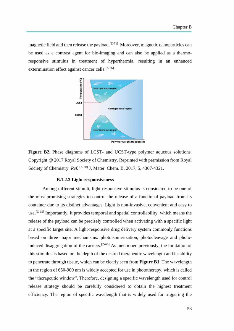
Chapter B
58
magnetic field and then release the payload.[Z-71] Moreover, magnetic nanoparticles can
be used as a contrast agent for bio-imaging and can also be applied as a thermo-
responsive stimulus in treatment of hyperthermia, resulting in an enhanced
extermination effect against cancer cells.[Z-66].
Figure B2. Phase diagrams of LCST- and UCST-type polymer aqueous solutions.
Copyright @ 2017 Royal Society of Chemistry. Reprinted with permission from Royal
Society of Chemistry. Ref. [Z-70] J. Mater. Chem. B, 2017, 5, 4307-4321.
B.1.2.3 Light-responsiveness
Among different stimuli, light-responsive stimulus is considered to be one of
the most promising strategies to control the release of a functional payload from its
container due to its distinct advantages. Light is non-invasive, convenient and easy to
use.[Z-65] Importantly, it provides temporal and spatial controllability, which means the
release of the payload can be precisely controlled when activating with a specific light
at a specific target site. A light-responsive drug delivery system commonly functions
based on three major mechanisms: photoisomerization, photocleavage and photo-
induced disaggregation of the carriers.[Z-66] As mentioned previously, the limitation of
this stimulus is based on the depth of the desired therapeutic wavelength and its ability
to penetrate through tissue, which can be clearly seen from Figure B1. The wavelength
in the region of 650-900 nm is widely accepted for use in phototherapy, which is called
the “therapeutic window”. Therefore, designing a specific wavelength used for control
release strategy should be carefully considered to obtain the highest treatment
efficiency. The region of specific wavelength that is widely used for triggering the
Chapter B
59
release includes ultraviolet (UV) light (10-400 nm), visible (Vis) light (400-700 nm),
red light (600-700 nm) and near-infrared (NIR) light (780-2526 nm).
UV-Vis light possesses high energy, which can be used to activate the release
system efficiently in vitro. Many chemicals have been used as UV-Vis light-responsive
materials such as azobenzene, spiropyran, pyrenylmethyl, o-nitrobenzyl and coumarin
family.[Z-66] However, UV-Vis light is absorbed strongly by the human tissues, leading
to low penetration ability. This make UV-Vis light inappropriate for use in deep tissue.
Moreover, UV light can cause photodamage to cells due to its relatively high energy.
[Z-72]
Nowadays, NIR has become more attractive for use in light-triggered control
release due to its safety, including no photodamage to cells, weak background
fluorescence, high detection sensitivity and high penetration ability in deep tissue.[Z-72]
NIR light has been applied for controlled release based on two-photon absorption,
which provides higher excitation and overcomes the poor penetration of UV-Vis light
sensitive system.[Z-73] In fact, however, two-photon absorption requires high-intensity
pulsed laser and only occurs at the laser focus. When the laser passes through tissue, it
defocuses, which makes the two-photon absorption strategy impractical for use in deep-
tissue. In addition, a limitation of NIR light is its low energy, which is not strong enough
to activate the photoreaction. To solve this problem, many strategies have been
developed such as triplet–triplet annihilation upconversion, upconverting
nanoparticles, and the use of NIR photosensitizers.[Z-65] The recent development of up-
conversion nanomaterials makes NIR light an alternative approach to trigger the
photosensitive drug releasing system. Upconversion materials are synthesized using
lanthanide-based substances. Lanthanide ions such as Tm3+, Er3+ and Ho3+ possess the
distinct ladder-like energy level structures. Lanthanide-doped upconverting
nanoparticles (UCNPs) can absorb NIR light and convert it into high energy in a region
of UV-Vis emission, which enables activation of the subsequent UV-light responsive
system.[Z-72] Another advantage compared to two-photon absorption strategy is that
UCNP-assisted photochemistry does not require a high-intensity pulsed laser. NIR
light-triggered photoreactions based on lanthanide-doped upconverting nanoparticles is
the controlled release strategy used in Publication [B1].
Chapter B
60
Similar to NIR light, red light is suitable for use in controlled release in
biomedical applications due to its high penetration ability in deep tissue.[Z-64] In fact,
trigger of light responsive therapeutic materials by direct irradiation with red or NIR
light via a one-photon process is more efficient to obtain a maximum therapeutic effect
than nonlinear optical processes such as two-photon absorption and photon
upconversion.[Z-65] For this purpose, photo-sensitive therapeutic agents that can be
directly activated by red or NIR light via a one-photon process are required.
Photosensitizer drugs are commonly used in photodynamic therapy (PDT) and
photoactivated chemotherapy (PACT). PDT uses specific light or laser to induce singlet
oxygen generation from photosensitizer drugs, leading to oxidative stress in cancer cells
and finally kill the cancer cells by reactive oxygen species (ROS)-mediated toxicity.
This therapy requires specific light to activate photosensitizer drugs and also
physiological oxygen to produce singlet oxygen. Meanwhile, PACT, an oxygen-
independent therapy, uses specific light or laser to release the toxic unit from
photosensitizer drugs.
A wide variety of exciting strategies employing light-responsive drug delivery
systems have been developed to achieve therapeutic efficacy at the clinical level. Many
of them have gained proof of concept successfully in vitro, while others have
demonstrated promising results in vivo. However, there has not been any report for
light-responsive drug delivery system at clinical trials. Currently, the stimuli-
responsive drug delivery systems that have been reported with high progress are the
temperature-responsive drug delivery system (ThermoDox, Doxorubicin containing
liposome) at clinical trials Phase III targeting to liver cancer and Phase II targeting to
lung cancer and redox (GSH concentration) -responsive drug delivery system (T-
DM1®, paclitaxel or docetaxel containing antibody conjugate) targeting to metastatic
breast cancer already approved by FDA.[Z-66]
This is partly due to the complex design of many stimuli-responsive drug
delivery systems, including light responsive systems. Therefore, it was suggested that
“it will be advantageous to keep the KISS principle in mind: keep it simple and
straightforward”.[Z-65] Some advantages and limitations of each stimuli-responsive drug
delivery system have been summarized in Table B2.

Chapter B
61
Table B2. Advantages and limitations of some stimuli-responsive drug delivery
systems. (Taken from Ref. [Z-65] Therapeutic delivery, 2017, 8(2), 89-107.Copyright ©
2017 Future Science Ltd.)

Chapter B1
62
B1: UPCONVERSION NANO-CARRIERS ENCAPSULATED WITH
PHOTOACTIVATABLE RU COMPLEXES FOR
NEAR-INFRARED LIGHT-REGULATED ENZYME ACTIVITY
(Note: The literature of the chapter B1 can be found on page 73 at the end of this chapter.)
Permission: The following part [B1] is based on a publication in Small 2017, 13,
170099. Presented materials are reprinted with permission from John Wiley and Sons.
Copyright © 2017, John Wiley and Sons
Contribution: Zhijun Chen and I contributed equally to this work. I carried out the
biological part including cell toxicity, Cathepsin K activity assay, Cathepsin K
inhibition assay, lysosome co-localization assay, cellular uptake study by flow
cytometry and cLSM. Zhijun Chen synthesized and characterized the Cathepsin K
inhibitor controllable release system. Mischa Schwendy performed upconversion
luminescence cell imaging. The project was supervised by Volker Mailänder and Si
Wu.
TOC B1: Photon upconversion enzyme inhibition is employed to regulate enzyme
activity using NIR light. Upconverting nanoparticles and caged enzyme inhibitors are
encapsulated in hollow mesoporous silica nanoparticles. Upconverting nanoparticles
convert near-infrared light to blue light, which activates caged enzyme inhibitors. The
activated enzyme inhibitors are released and inhibit the enzyme activity in living cells.
Aim: Cathepsin K is a key enzyme that plays an important role in bone remodeling and
cancer metastasis. Therefore, inhibition of this enzyme can prevent bone digestion and
tumor invasion. To achieve this goal, the Cathepsin K inhibitor controllable release
system responsive to the near infrared (NIR) light was developed and applied to inhibit
three different sources of Cathepsin K: purified (commercial) enzyme, lysate and live
Chapter B1
63
cells. Three different cell lines including LNCaP, PC3 cells (prostate cancer cell lines)
and SAOS-2 cells (osteosarcoma cell line) were chosen to demonstrate the enzymatic
inhibition of the new controlled release platform) due to their ability to exhibit
overexpression of Cathepsin K.
Abstract: Enzyme activity is important for metabolism, cell functions, and treating
diseases. However, remote control of enzyme activity in deep tissue remains a
challenge. Here, we demonstrate near-infrared (NIR) light-regulated enzyme activity in
living cells based on upconverting nanoparticles (UCNPs) and a photoactivatable Ru
complex. The Ru complex is a caged enzyme inhibitor that can be activated by blue
light. To prepare a nano-carrier for NIR photoinhibition of enzyme activity, a UCNP
and the caged enzyme inhibitors were encapsulated in a hollow mesoporous silica
nanoparticle. In such a nano-carrier, the UCNP can harvest NIR light and convert it into
blue light, which can activate the caged enzyme inhibitors. This photoactivation process
is feasible in deep tissue because of the tissue penetration ability of NIR light. The
nano-carrier was compatible to LNCaP, PC3, and SAOS-2 cells, which showed high
enzyme expression. NIR irradiation induced release of the inhibitors and inhibition of
enzyme activity in living cells. NIR light provides high spatiotemporal resolution to
regulate enzyme activity in deep tissue.
B1.1. Introduction
Enzymes are important macromolecular biological catalysts that enable chemical
reactions in biological systems. Controlling enzyme activity can regulate metabolism,
manipulate cell functions, kill pathogens, and treat diseases.[B1-1] Due to the great
importance of enzymes in biological systems, the use of external stimuli, such as pH,[B1-
2] temperature,[B1-3] ATP,[B1-4] cations,[B1-5] and light,[B1-6] to control enzyme activity has
been developed. Among these stimuli, light provides high spatiotemporal resolution for
controlling enzyme activity. UV or visible light can control photoresponsive
compounds, such as photocages and photoswitchable molecules.[B1-7] The
photoproducts from these photoresponsive compounds can then regulate enzyme
activities.[B1-6a, 6e, 8] However, UV light may damage biological systems. Additionally,
Chapter B1
64
both UV and visible light cannot penetrate deeply into tissue. In contrast, near-infrared
(NIR) light can penetrate deeply into tissue and causes less photodamage to biological
systems.[B1-9]
NIR light has been applied for controlling enzyme activities based on two-photon
absorption. [B1-8a, 10] However, two-photon absorption requires high-intensity pulsed
lasers (typical pulse intensity: >106 W cm−2)[B1-11] and is inefficient even when
femtosecond lasers are employed. Two-photon absorption only occurs at the laser
focus. Because the laser defocuses as it passes through tissue, the two-photon
absorption strategy is impractical for deep-tissue applications.
Recently, NIR light-triggered photoreactions based on lanthanide-doped
upconverting nanoparticles (UCNPs) have been developed.[B1-9b, 12] UCNPs can convert
NIR light into UV and visible light.[B1-13] Then, the upconverted UV or visible emission
can trigger photoreactions of traditional UV- or visible light-responsive compounds.
This process is referred to as UCNP-assisted photochemistry.[B1-9b, 14] UCNP-assisted
photochemistry can be triggered by continuous wave laser diodes with intensities that
are several orders of magnitude lower than that required for two-photon absorption.[B1-
9b, 15] UCNPs have assisted different photoreactions including photoisomerization,[B1-
14e, 16] photocleavage,[B1-17] photopolymerization,[B1-14d, 18] and photo-coupling.[B1-19]
These UCNP-assisted photoreactions have been applied to NIR-controlled drug
delivery,[B1-14c, 15b] biointerfaces,[B1-15a, 16] uncaging enzymes,[B1-17c] catalysis,[B1-20]
pH,[B1-21] and actuators.[B1-14e] However, inhibition of enzyme activity using NIR light
assisted by UCNPs has not been investigated.
Here, we use NIR light to regulate enzyme activity based on UCNP-assisted
photochemistry (Figure B1.1). We refer to this new method as photon upconversion
enzyme inhibition (PUEI). To construct a platform for PUEI, a NaYF4:TmYb@NaYF4
UCNP and caged enzyme inhibitors (RuEI) are encapsulated in a hollow mesoporous
silica nanoparticle (hmSiO2) (Figure B1.1b). RuEI can be activated by blue light to
release the enzyme inhibitor (EI). Our group[B1-9b, 15, 21-22] and others[B1-14f] have
demonstrated that NIR light can efficiently uncage Ru complexes that are similar to
RuEI with the assistance of UCNPs. With RuEI&UCNP@hmSiO2 in this study,
UCNPs can convert NIR light to blue light, which can uncage EI from RuEI (Figure

Chapter B1
65
B1.1). After NIR photoactivation, the released EI can inhibit the activity of the
cathepsin K enzyme (Figure B1.1b). Cathepsin K is an important enzyme that is critical
for osteoclastic bone resorption, macrophage invasion, and tumor growth.[B1-23]
Cathepsin K is involved in the resorption of bone tissue in osteoclasts and therefore
responsible for the remodeling of bone.[B1-24] Resorption of bone should be inhibited
under some circumstances, such as bone loss in elderly patients or during the
regeneration of new bone after a fracture. For the treatment of cancers and tumor
metastases, cathepsin K is a key factor that affects the invasiveness of tumors into
healthy tissue.[B1-25] Therefore, the inhibition of cathepsin K is important. NIR
photoinhibition of the activity of cathepsin K allows for new routes for manipulating
cell functions, regenerative medicine, and treating the spread of malignant diseases in
deep tissue. PUEI also provides a general platform for regulating enzyme activity in
deep tissue.

Chapter B1
66
Figure B1.1. Schematic illustration of photon upconversion enzyme inhibition (PUEI).
(a) Activation of the enzyme inhibitor using near-infrared (NIR) light with the
assistance of UCNPs. UCNPs convert NIR light to blue light, which induces the
cleavage of the caged inhibitor (RuEI) and activation of the enzyme inhibitor (EI). (b)
NIR irradiation of RuEI&UCNP@hmSiO2 activates EI, which inhibits enzyme activity.
B1.2. Results and discussion
B1.2.1. NIR photoinhibition of enzyme activity
RuEI&UCNP@hmSiO2 provides a platform for NIR light-induced inhibition of
enzyme activity in deep tissue (Figure B1.2a). The half maximal inhibitory
concentration (IC50) of the activated EI was only 1/6 of that of the caged inhibitor RuEI
for the inhibition of cathepsin K (Figure B1.3). Therefore, photoactivation of EI can
efficiently inhibit cathepsin K. NIR irradiation of cathepsin K in the presence of
RuEI&UCNP@hmSiO2 triggered photocleavage of RuEI. Released EI can react with
thiol moieties of cathepsin K,[B1-8b] which irreversibly inhibited its activity significantly
(Figure B1.2b, blue bars). Irradiation for 7, 15, and 30 min reduced the enzyme
activity to 30%, 17%, and 13%, respectively (Figure B1.2b, blue bars). For
comparison, control experiments in the absence of RuEI&UCNP@hmSiO2 after NIR
irradiation (Figure B1.2b, black bars) and the presence of RuEI&UCNP@hmSiO2
without NIR irradiation (Figure B1.2b, red bars) were performed. Enzyme activities
in these control samples did not decrease. These results demonstrated that the inhibition
of enzyme activity was caused by activation of the EI using NIR light. Moreover, the
feasibility of using NIR light to inhibit enzyme activity after passing through tissue was
investigated, which is important for clinical applications in the future. A piece of
chicken tissue was placed between the NIR light and the sample. The NIR light passed
through the tissue and inhibited the enzyme activity (Figure B1.2b, magenta bars).
B1.2.4. Evaluation of biocompatibility of RuEI&UCNP@hmSiO2
Before enzyme inhibition in living cells was studied, the biocompatibility of
RuEI&UCNP@hmSiO2 was studied via cellular experiments. LNCaP, PC3 cells
(prostate cancer cell lines) and SAOS-2 cells (osteosarcoma cell line) were used in our
study. These cell lines were chosen because they have a high cathepsin K expression

Chapter B1
67
rate for their invasiveness, which results in metastases in the prostate cancer cell lines
and the osteosarcoma cell line. To test the biocompatibility, RuEI&UCNP@hmSiO2
was incubated with all three cell lines for 24 h. Only a minor decrease in the cell
viability was observed in the presence of a high concentration (150 μg/mL) of
RuEI&UCNP@hmSiO2 (Figure B1.4). After NIR irradiation (1 W/cm2) on LNCaP
cells in the presence or absence of RuEI&UCNP@hmSiO2 (150 μg/mL), the cell
viability was higher than 90% (Figure B1.5a). The biocompatibility tests on PC3 and
SAOS-2 cells showed similar results to that of LNCaP cells (Figure B1.5b, c). These
results suggested that RuEI&UCNP@hmSiO2 provides a biocompatible platform for
NIR photoinhibition of enzyme activity in living cells.
Figure B1.2. (a) Schematic illustration of NIR photoinhibition of enzyme activity using
RuEI&UCNP@hmSiO2. (b) NIR photoinhibition of the activity of cathepsin K. Black
bars: activities of cathepsin K in the absence of RuEI&UCNP@hmSiO2 under NIR
irradiation; Red bars: activities of cathepsin K in the presence of
RuEI&UCNP@hmSiO2 without irradiation; Blue bars: activities of cathepsin K in the
presence of RuEI&UCNP@hmSiO2 under NIR irradiation. Magenta bars: activities of
cathepsin K in the presence of RuEI&UCNP@hmSiO2 under NIR irradiation through
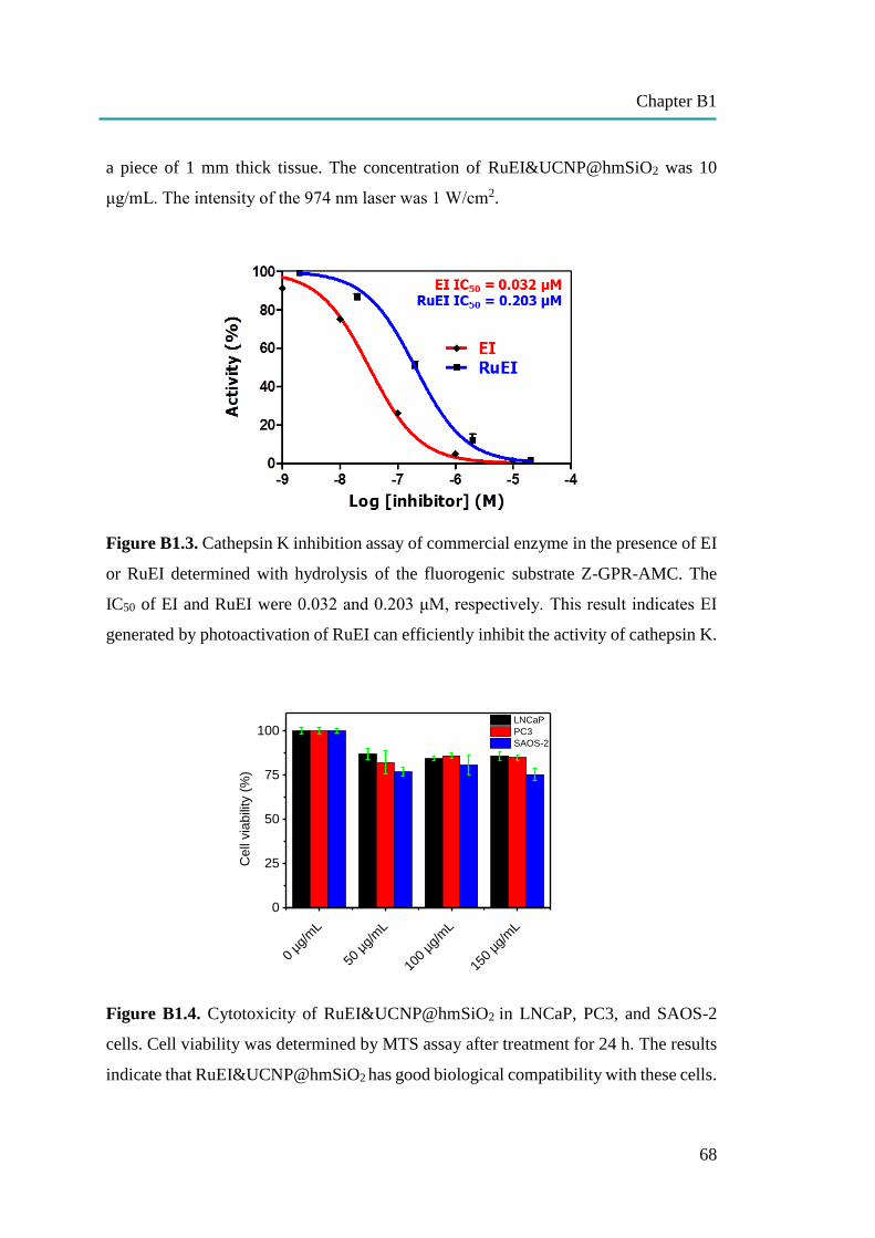
Chapter B1
68
a piece of 1 mm thick tissue. The concentration of RuEI&UCNP@hmSiO2 was 10
μg/mL. The intensity of the 974 nm laser was 1 W/cm2.
Figure B1.3. Cathepsin K inhibition assay of commercial enzyme in the presence of EI
or RuEI determined with hydrolysis of the fluorogenic substrate Z-GPR-AMC. The
IC50 of EI and RuEI were 0.032 and 0.203 μM, respectively. This result indicates EI
generated by photoactivation of RuEI can efficiently inhibit the activity of cathepsin K.
Figure B1.4. Cytotoxicity of RuEI&UCNP@hmSiO2 in LNCaP, PC3, and SAOS-2
cells. Cell viability was determined by MTS assay after treatment for 24 h. The results
indicate that RuEI&UCNP@hmSiO2 has good biological compatibility with these cells.
0 µg
/mL
50 µ
g/m
L
100
µg/m
L
150
µg/m
L
0
25
50
75
100 LNCaP
PC3
SAOS-2
Cell
via
bili
ty (
%)
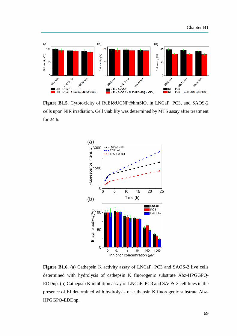
Chapter B1
69
Figure B1.5. Cytotoxicity of RuEI&UCNP@hmSiO2 in LNCaP, PC3, and SAOS-2
cells upon NIR irradiation. Cell viability was determined by MTS assay after treatment
for 24 h.
Figure B1.6. (a) Cathepsin K activity assay of LNCaP, PC3 and SAOS-2 live cells
determined with hydrolysis of cathepsin K fluorogenic substrate Abz-HPGGPQ-
EDDnp. (b) Cathepsin K inhibition assay of LNCaP, PC3 and SAOS-2 cell lines in the
presence of EI determined with hydrolysis of cathepsin K fluorogenic substrate Abz-
HPGGPQ-EDDnp.
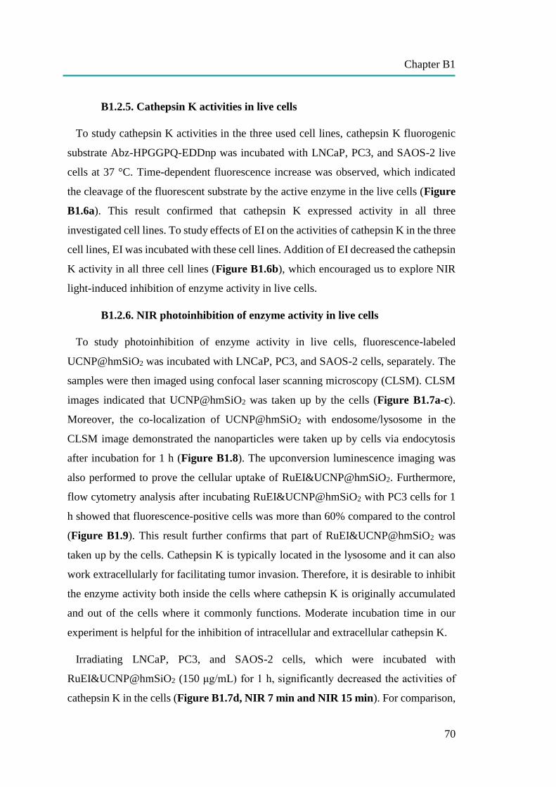
Chapter B1
70
B1.2.5. Cathepsin K activities in live cells
To study cathepsin K activities in the three used cell lines, cathepsin K fluorogenic
substrate Abz-HPGGPQ-EDDnp was incubated with LNCaP, PC3, and SAOS-2 live
cells at 37 °C. Time-dependent fluorescence increase was observed, which indicated
the cleavage of the fluorescent substrate by the active enzyme in the live cells (Figure
B1.6a). This result confirmed that cathepsin K expressed activity in all three
investigated cell lines. To study effects of EI on the activities of cathepsin K in the three
cell lines, EI was incubated with these cell lines. Addition of EI decreased the cathepsin
K activity in all three cell lines (Figure B1.6b), which encouraged us to explore NIR
light-induced inhibition of enzyme activity in live cells.
B1.2.6. NIR photoinhibition of enzyme activity in live cells
To study photoinhibition of enzyme activity in live cells, fluorescence-labeled
UCNP@hmSiO2 was incubated with LNCaP, PC3, and SAOS-2 cells, separately. The
samples were then imaged using confocal laser scanning microscopy (CLSM). CLSM
images indicated that UCNP@hmSiO2 was taken up by the cells (Figure B1.7a-c).
Moreover, the co-localization of UCNP@hmSiO2 with endosome/lysosome in the
CLSM image demonstrated the nanoparticles were taken up by cells via endocytosis
after incubation for 1 h (Figure B1.8). The upconversion luminescence imaging was
also performed to prove the cellular uptake of RuEI&UCNP@hmSiO2. Furthermore,
flow cytometry analysis after incubating RuEI&UCNP@hmSiO2 with PC3 cells for 1
h showed that fluorescence-positive cells was more than 60% compared to the control
(Figure B1.9). This result further confirms that part of RuEI&UCNP@hmSiO2 was
taken up by the cells. Cathepsin K is typically located in the lysosome and it can also
work extracellularly for facilitating tumor invasion. Therefore, it is desirable to inhibit
the enzyme activity both inside the cells where cathepsin K is originally accumulated
and out of the cells where it commonly functions. Moderate incubation time in our
experiment is helpful for the inhibition of intracellular and extracellular cathepsin K.
Irradiating LNCaP, PC3, and SAOS-2 cells, which were incubated with
RuEI&UCNP@hmSiO2 (150 μg/mL) for 1 h, significantly decreased the activities of
cathepsin K in the cells (Figure B1.7d, NIR 7 min and NIR 15 min). For comparison,
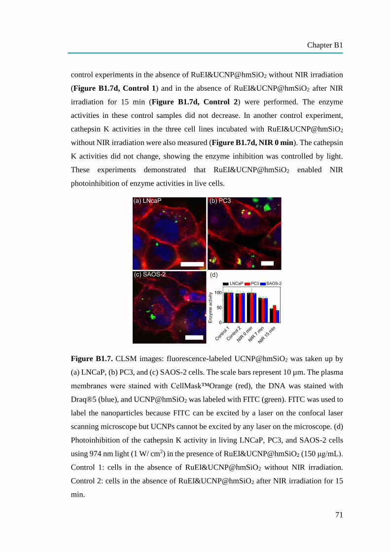
Chapter B1
71
control experiments in the absence of RuEI&UCNP@hmSiO2 without NIR irradiation
(Figure B1.7d, Control 1) and in the absence of RuEI&UCNP@hmSiO2 after NIR
irradiation for 15 min (Figure B1.7d, Control 2) were performed. The enzyme
activities in these control samples did not decrease. In another control experiment,
cathepsin K activities in the three cell lines incubated with RuEI&UCNP@hmSiO2
without NIR irradiation were also measured (Figure B1.7d, NIR 0 min). The cathepsin
K activities did not change, showing the enzyme inhibition was controlled by light.
These experiments demonstrated that RuEI&UCNP@hmSiO2 enabled NIR
photoinhibition of enzyme activities in live cells.
Figure B1.7. CLSM images: fluorescence-labeled UCNP@hmSiO2 was taken up by
(a) LNCaP, (b) PC3, and (c) SAOS-2 cells. The scale bars represent 10 µm. The plasma
membranes were stained with CellMask™Orange (red), the DNA was stained with
Draq®5 (blue), and UCNP@hmSiO2 was labeled with FITC (green). FITC was used to
label the nanoparticles because FITC can be excited by a laser on the confocal laser
scanning microscope but UCNPs cannot be excited by any laser on the microscope. (d)
Photoinhibition of the cathepsin K activity in living LNCaP, PC3, and SAOS-2 cells
using 974 nm light (1 W/ cm2) in the presence of RuEI&UCNP@hmSiO2 (150 μg/mL).
Control 1: cells in the absence of RuEI&UCNP@hmSiO2 without NIR irradiation.
Control 2: cells in the absence of RuEI&UCNP@hmSiO2 after NIR irradiation for 15
min.
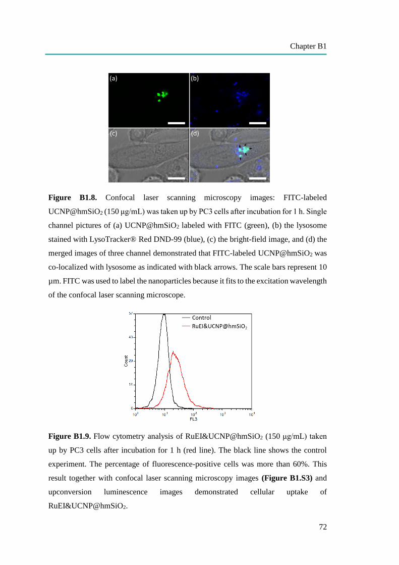
Chapter B1
72
Figure B1.8. Confocal laser scanning microscopy images: FITC-labeled
UCNP@hmSiO2 (150 μg/mL) was taken up by PC3 cells after incubation for 1 h. Single
channel pictures of (a) UCNP@hmSiO2 labeled with FITC (green), (b) the lysosome
stained with LysoTracker® Red DND-99 (blue), (c) the bright-field image, and (d) the
merged images of three channel demonstrated that FITC-labeled UCNP@hmSiO2 was
co-localized with lysosome as indicated with black arrows. The scale bars represent 10
µm. FITC was used to label the nanoparticles because it fits to the excitation wavelength
of the confocal laser scanning microscope.
Figure B1.9. Flow cytometry analysis of RuEI&UCNP@hmSiO2 (150 μg/mL) taken
up by PC3 cells after incubation for 1 h (red line). The black line shows the control
experiment. The percentage of fluorescence-positive cells was more than 60%. This
result together with confocal laser scanning microscopy images (Figure B1.S3) and
upconversion luminescence images demonstrated cellular uptake of
RuEI&UCNP@hmSiO2.
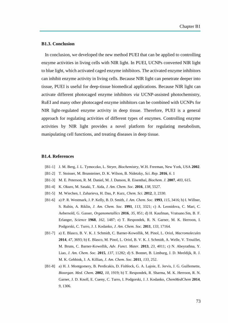
Chapter B1
73
B1.3. Conclusion
In conclusion, we developed the new method PUEI that can be applied to controlling
enzyme activities in living cells with NIR light. In PUEI, UCNPs converted NIR light
to blue light, which activated caged enzyme inhibitors. The activated enzyme inhibitors
can inhibit enzyme activity in living cells. Because NIR light can penetrate deeper into
tissue, PUEI is useful for deep-tissue biomedical applications. Because NIR light can
activate different photocaged enzyme inhibitors via UCNP-assisted photochemistry,
RuEI and many other photocaged enzyme inhibitors can be combined with UCNPs for
NIR light-regulated enzyme activity in deep tissue. Therefore, PUEI is a general
approach for regulating activities of different types of enzymes. Controlling enzyme
activities by NIR light provides a novel platform for regulating metabolism,
manipulating cell functions, and treating diseases in deep tissue.
B1.4. References
[B1-1] J. M. Berg, J. L. Tymoczko, L. Stryer, Biochemistry, W.H. Freeman, New York, USA 2002.
[B1-2] T. Stoisser, M. Brunsteiner, D. K. Wilson, B. Nidetzky, Sci. Rep. 2016, 6. 1
[B1-3] M. E. Peterson, R. M. Daniel, M. J. Danson, R. Eisenthal, Biochem. J. 2007, 403, 615.
[B1-4] K. Okuro, M. Sasaki, T. Aida, J. Am. Chem. Soc. 2016, 138, 5527.
[B1-5] M. Wiechen, I. Zaharieva, H. Dau, P. Kurz, Chem. Sci. 2012, 3, 2330.
[B1-6] a) P. R. Westmark, J. P. Kelly, B. D. Smith, J. Am. Chem. Soc. 1993, 115, 3416; b) I. Willner,
S. Rubin, A. Riklin, J. Am. Chem. Soc. 1991, 113, 3321; c) A. Leonidova, C. Mari, C.
Aebersold, G. Gasser, Organometallics 2016, 35, 851; d) H. Kaufman, Vratsano.Sm, B. F.
Erlanger, Science 1968, 162, 1487; e) T. Respondek, R. N. Garner, M. K. Herroon, I.
Podgorski, C. Turro, J. J. Kodanko, J. Am. Chem. Soc. 2011, 133, 17164.
[B1-7] a) E. Blasco, B. V. K. J. Schmidt, C. Barner-Kowollik, M. Pinol, L. Oriol, Macromolecules
2014, 47, 3693; b) E. Blasco, M. Pinol, L. Oriol, B. V. K. J. Schmidt, A. Welle, V. Trouillet,
M. Bruns, C. Barner-Kowollik, Adv. Funct. Mater. 2013, 23, 4011; c) N. Abeyrathna, Y.
Liao, J. Am. Chem. Soc. 2015, 137, 11282; d) S. Bonnet, B. Limburg, J. D. Meeldijk, R. J.
M. K. Gebbink, J. A. Killian, J. Am. Chem. Soc. 2011, 133, 252.
[B1-8] a) H. J. Montgomery, B. Perdicakis, D. Fishlock, G. A. Lajoie, E. Jervis, J. G. Guillemette,
Bioorgan. Med. Chem. 2002, 10, 1919; b) T. Respondek, R. Sharma, M. K. Herroon, R. N.
Garner, J. D. Knoll, E. Cueny, C. Turro, I. Podgorski, J. J. Kodanko, ChemMedChem 2014,
9, 1306.
Chapter B1
74
[B1-9] a) R. T. Xing, K. Liu, T. F. Jiao, N. Zhang, K. Ma, R. Y. Zhang, Q. L. Zou, G. H. Ma, X. H.
Yan, Adv. Mater. 2016, 28, 3669; b) S. Wu, H. J. Butt, Adv. Mater. 2016, 28, 1208; c) N.
Zhang, F. Zhao, Q. Zou, Y. Li, G. Ma, X. Yan, Small 2016, 12, 5936.
[B1-10] P. Anstaett, V. Pierroz, S. Ferrari, G. Gasser, Photochem. Photobiol. Sci. 2015, 14, 1821.
[B1-11] a) M. Álvarez, A. Best, S. Pradhan‐Kadam, K. Koynov, U. Jonas, M. Kreiter, Adv. Mater.
2008, 20, 4563; b) M. Álvarez, A. Best, A. Unger, J. M. Alonso, A. del Campo, M.
Schmelzeisen, K. Koynov, M. Kreiter, Adv. Funct. Mater. 2010, 20, 4265.
[B1-12] a) B. Yan, J.-C. Boyer, D. Habault, N. R. Branda, Y. Zhao, J. Am. Chem. Soc. 2012, 134,
16558; b) C.-J. Carling, J.-C. Boyer, N. R. Branda, J. Am. Chem. Soc. 2009, 131, 10838; c)
Z. Y. Cheng, J. Lin, Macromol. Rapid. Comm. 2015, 36, 790; d G. Chen, H. Qiu, P. N. Prasad,
X. Chen, Chem. Rev. 2014, 114, 5161.
[B1-13] a) G. Chen, J. Damasco, H. Qiu, W. Shao, T. Y. Ohulchanskyy, R. R. Valiev, X. Wu, G. Han,
Y. Wang, C. Yang, Nano lett. 2015, 15, 7400; b) E. M. Chan, G. Han, J. D. Goldberg, D. J.
Gargas, A. D. Ostrowski, P. J. Schuck, B. E. Cohen, D. J. Milliron, Nano lett. 2012, 12, 3839;
c) X. Wu, Y. Zhang, K. Takle, O. Bilsel, Z. Li, H. Lee, Z. Zhang, D. Li, W. Fan, C. Duan, E.
M. Chan, C. Lois, Y. Xiang, G. Han, ACS nano 2016, 10, 1060; d) K. Huang, N. M. Idris, Y.
Zhang, Small 2016, 12, 836; e) C. Li, D. Yang, P. a. Ma, Y. Chen, Y. Wu, Z. Hou, Y. Dai, J.
Zhao, C. Sui, J. Lin, Small 2013, 9, 4150; f) G. Tian, W. Ren, L. Yan, S. Jian, Z. Gu, L. Zhou,
S. Jin, W. Yin, S. Li, Y. Zhao, Small 2013, 9, 1929.
[B1-14] a) Y. M. Yang, Q. Shao, R. R. Deng, C. Wang, X. Teng, K. Cheng, Z. Cheng, L. Huang, Z.
Liu, X. G. Liu, B. G. Xing, Angew. Chem. Int. Edit. 2012, 51, 3125; b) M. L. Viger, M.
Grossman, N. Fomina, A. Almutairi, Adv. Mater. 2013, 25, 3733; c) D. Yang, P. Ma, Z. Hou,
Z. Cheng, C. Li, J. Lin, Chem. Soc. Rev. 2015, 44, 1416; d) S. Beyaʐit, S. Ambrosini, N.
Marchyk, E. Palo, V. Kale, T. Soukka, B. T. S. Bui, K. Haupt, Angew. Chem. Int. Edit. 2014,
53, 8919; e) W. Wu, L. Yao, T. Yang, R. Yin, F. Li, Y. Yu, J. Am. Chem. Soc. 2011, 133,
15810; f) E. Ruggiero, A. Habtemariam, L. Yate, J. C. Mareque-Rivas, L. Salassa, Chem.
Commun. 2014, 50, 1715; g) P. T. Burks, J. V. Garcia, R. GonzalezIrias, J. T. Tillman, M.
Niu, A. A. Mikhailovsky, J. Zhang, F. Zhang, P. C. Ford, J. Am. Chem. Soc. 2013, 135, 18145;
h) J. A. Liu, W. B. Bu, L. M. Pan, J. L. Shi, Angew. Chem. Int. Edit. 2013, 52, 4375; i) J.
Shen, G. Y. Chen, T. Y. Ohulchanskyy, S. J. Kesseli, S. Buchholz, Z. P. Li, P. N. Prasad, G.
Han, Small 2013, 9, 3213.
[B1-15] a) Z. J. Chen, S. Q. He, H. J. Butt, S. Wu, Adv. Mater. 2015, 27, 2203; b) S. Q. He, K. Krippes,
S. Ritz, Z. J. Chen, A. Best, H. J. Butt, V. Mailänder, S. Wu, Chem. Commun. 2015, 51, 431.
[B1-16] W. Li, J. Wang, J. Ren, X. Qu, J. Am. Chem. Soc. 2014, 136, 2248.
[B1-17] a) B. Yan, J.-C. Boyer, N. R. Branda, Y. Zhao, J. Am. Chem. Soc. 2011, 133, 19714; b) L. Z.
Zhao, J. J. Peng, Q. Huang, C. Y. Li, M. Chen, Y. Sun, Q. N. Lin, L. Y. Zhu, F. Y. Li, Adv.
Funct. Mater. 2014, 24, 363; c) H. D. Gao, P. Thanasekaran, C. W. Chiang, J. L. Hong, Y. C.
Liu, Y. H. Chang, H. M. Lee, ACS Nano 2015, 9, 7041.
Chapter B1
75
[B1-18] A. Stepuk, D. Mohn, R. N. Grass, M. Zehnder, K. W. Kramer, F. Pelle, A. Ferrier, W. J. Stark,
Dent. Mater. 2012, 28, 304.
[B1-19] P. Lederhose, Z. J. Chen, R. Müller, J. P. Blinco, S. Wu, C. Barner-Kowollik, Angew. Chem.
Int. Edit. 2016, 55, 12195.
[B1-20] Z. Chen, L. Zhou, W. Bing, Z. Zhang, Z. Li, J. Ren, X. Qu, J. Am. Chem. Soc. 2014, 136,
7498.
[B1-21] Z. Chen, Y. Xiong, R. Etchenique, S. Wu, Chem. Commun. 2016, 52, 13959.
[B1-22] Z. Chen, W. Sun, H. J. Butt, S. Wu, Chem. Eur. J. 2015, 21, 9165.
[B1-23] K. D. Brubaker, R. L. Vessella, L. D. True, R. Thomas, E. Corey, J. Bone. Miner. Res. 2003,
18, 222.
[B1-24] L. J. Raggatt, N. C. Partridge, J. Biol. Chem. 2010, 285, 25103.
[B1-25] M. M. Mohamed, B. F. Sloane, Nat. Rev. Cancer 2006, 6, 764.

Chapter B2
76
B2: PHOTOACTIVATION OF ANTICANCER RU COMPLEXES
IN DEEP TISSUE: HOW DEEP CAN WE GO?
(Note: The literature of the chapter B2 can be found on page 83 at the end of this chapter.)
Permission: The following part [B2] is based on a publication in Chem. Eur. J. 2017,
23, 10832 – 10837. Presented materials are reprinted with permission from John Wiley
and Sons. Copyright © 2017, John Wiley and Sons.
Contribution: Wen Sun and I contributed equally to this work. I carried out the
biological part including cell viability. Wen Sun synthesized and characterized the Ru
complex. The project was supervised by Volker Mailänder and Si Wu.
TOC B2: Anticancer ruthenium (Ru) complexes can be photoactivated by 671‐nm light
after passing through a 16‐mm‐thick tissue. The photoactivated Ru complexes can
inhibit the growth of cancer cells. These results suggest that Ru complexes are
promising anticancer agents for deep‐tissue phototherapy.
Aim: Ru complexes have gained intensive interest because of their photoactive and
anticancer properties. It is known that different wavelengths of the light are able to
penetrate different distances into the external skin. Only the light in the therapeutic
window (650-950 nm), which is in a region of red light and NIR, is optimal for use in
phototherapy. However, the important question is how deep the red light can penetrate
through the skin and still be able to activate Ru complex. Therefore, 671 nm-red light
was used to activate the Ru complex passed through the tissue with different thicknesses
to clarify this question and demonstrate the efficiency to kill cancer cells.
Chapter B2
77
Abstract : Activation of anticancer therapeutics such as ruthenium (Ru) complexes is
currently a topic of intense investigation. The success of phototherapy relies on
photoactivation of therapeutics after the light passes through skin and tissue. In this
paper, the photoactivation of anticancer Ru complexes with 671-nm red light through
tissue of different thicknesses was studied. Four photoactivatable Ru complexes with
different absorption wavelengths were synthesized. Two of them (Ru3 and Ru4) were
responsive to wavelengths in the “therapeutic window” (650–900 nm) and could be
activated using 671-nm red light after passing through tissue up to 16-mm-thick. The
other two (Ru1 and Ru2) could not be activated using red light. Additionally, activated
Ru4 caused inhibition of cancer cells. These results suggest that photoactivatable Ru
complexes are promising for applications in deep-tissue phototherapy.
B2.1. Introduction
Phototherapy based on phototherapeutic agents and light irradiation is a promising
strategy for cancer treatment.[B2-1] Phototherapeutic agents are usually non-toxic or less
toxic in the dark, until light converts them to toxic species that kill cancer cells (Figure
B2.1a). Phototherapy causes minimal side effects for normal tissues because light
provides high spatial resolution and allows activation of the phototherapeutic agents at
target sites only.[B2-1–4] Most photoresponsive materials,[B2-5, 6] such as photoactivated
platinum,[B2-7, 8] coumarin-caged[B2-9, 10] and pyrenecaged[B2-11] prodrugs are sensitive to
UV or short-wavelength visible light. However, UV or short-wavelength visible light
is problematic for biomedical applications, because these wavelengths cannot penetrate
deeply into tissue.[B2-12] Furthermore, UV light can also damage biological systems. In
contrast, red or near-infrared (NIR) light is better-suited to biomedical applications,
because red or NIR light has a deeper tissue penetration depth (Figure B2.1b).[B2-12, 13]
Simultaneous two-photon absorption is one way to activate photoresponsive materials
using NIR light.[B2-14–17] However, two photon absorption is inefficient and only occurs
at the focus of high-intensity pulsed lasers.[B2-18, 19] Considering that a laser beam will
defocus while passing through tissue, a two-photon absorption strategy is impractical
for deep-tissue applications. Another method for activating phototherapeutic agents
Chapter B2
78
using NIR light is based on photon upconversion. NIR light can be converted by
upconverting nanoparticles or some organic dyes to UV or visible light, which then
activates phototherapeutic agents.[B2-12, 20–23] This process is referred to as
upconversion-assisted photochemistry.[B2-12] Compared to simultaneous two-photon
excitation, one advantage of upconversion-assisted photochemistry is that it does not
require high-intensity pulsed lasers. However, upconversion is still a non-linear optical
process and requires high-intensity laser excitation (at least several hundred mW cm-2),
which may damage biological systems.[B2-12, 24] Furthermore, although NIR light can
penetrate into tissue deeply, passing through tissue still attenuates its intensity. Thus,
the NIR light intensity may be too low to excite upconversion after NIR light passes
through relatively thick tissue.
Activation of phototherapeutic agents by directly using red or NIR light via a one-
photon process is more efficient than nonlinear optical processes such as two-photon
absorption and photon upconversion. Some phototherapeutics that can be directly
activated by red or NIR light via a one-photon process already exist.[B2-25–29] Previous
studies have demonstrated that Ru complexes are photoresponsive molecules and have
been applied for biological applications.[B2-30–33] In particular, our group and others
showed that some Ru complexes can be directly activated by low-intensity (30–720
mW cm-2) red or NIR light.[B2-34–37] Ru complexes, analogues of platinum anticancer
drugs, are importantly proposed to also be promising anticancer agents.[B2-26, 38, 39] One
advantage of photoactivated Ru complexes is that they are usually less toxic to non-
irradiated tissues, only becoming more toxic in tumor cells through photoactivation.[B2-
14, 26, 39–47] Photoactivated Ru-containing materials have already shown anticancer
effects in a tumor-bearing mouse model.[B2-34, 37, 48] Furthermore, photoactivated Ru
complexes are promising for deep-tissue phototherapy due to their high photo-
responsiveness (e.g. several mW cm-2). However, red or NIR light will eventually be
completely attenuated while passing through thick tissue, which means photoactivation
of therapeutics via one-photon processes has a certain depth limit. The understanding
of this photoactivation of therapeutics in deep tissue will thus help provide guidelines
for phototherapy.
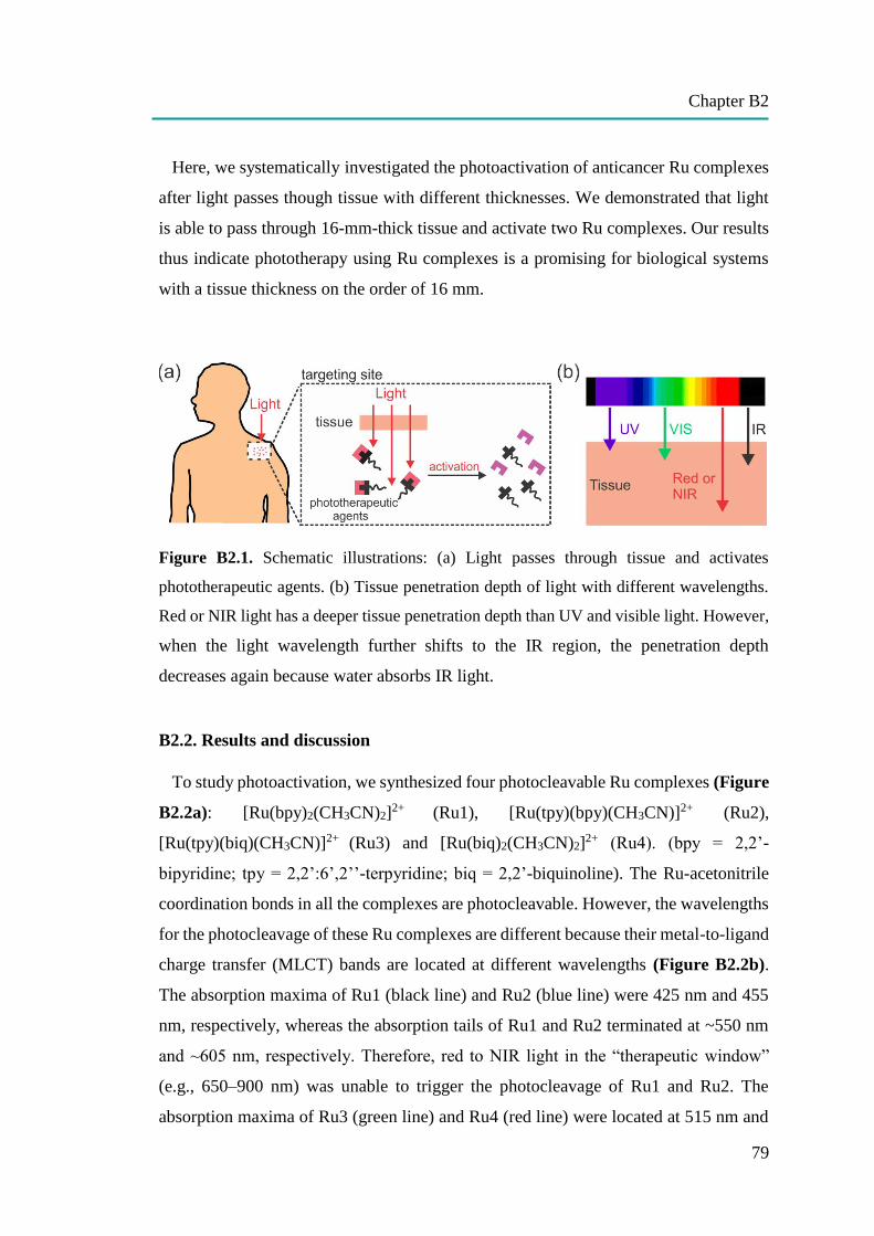
Chapter B2
79
Here, we systematically investigated the photoactivation of anticancer Ru complexes
after light passes though tissue with different thicknesses. We demonstrated that light
is able to pass through 16-mm-thick tissue and activate two Ru complexes. Our results
thus indicate phototherapy using Ru complexes is a promising for biological systems
with a tissue thickness on the order of 16 mm.
Figure B2.1. Schematic illustrations: (a) Light passes through tissue and activates
phototherapeutic agents. (b) Tissue penetration depth of light with different wavelengths.
Red or NIR light has a deeper tissue penetration depth than UV and visible light. However,
when the light wavelength further shifts to the IR region, the penetration depth
decreases again because water absorbs IR light.
B2.2. Results and discussion
To study photoactivation, we synthesized four photocleavable Ru complexes (Figure
B2.2a): [Ru(bpy)2(CH3CN)2]2+
(Ru1), [Ru(tpy)(bpy)(CH3CN)]2+ (Ru2),
[Ru(tpy)(biq)(CH3CN)]2+ (Ru3) and [Ru(biq)2(CH3CN)2]2+ (Ru4). (bpy = 2,2’-
bipyridine; tpy = 2,2’:6’,2’’-terpyridine; biq = 2,2’-biquinoline). The Ru-acetonitrile
coordination bonds in all the complexes are photocleavable. However, the wavelengths
for the photocleavage of these Ru complexes are different because their metal-to-ligand
charge transfer (MLCT) bands are located at different wavelengths (Figure B2.2b).
The absorption maxima of Ru1 (black line) and Ru2 (blue line) were 425 nm and 455
nm, respectively, whereas the absorption tails of Ru1 and Ru2 terminated at ~550 nm
and ~605 nm, respectively. Therefore, red to NIR light in the “therapeutic window”
(e.g., 650–900 nm) was unable to trigger the photocleavage of Ru1 and Ru2. The
absorption maxima of Ru3 (green line) and Ru4 (red line) were located at 515 nm and
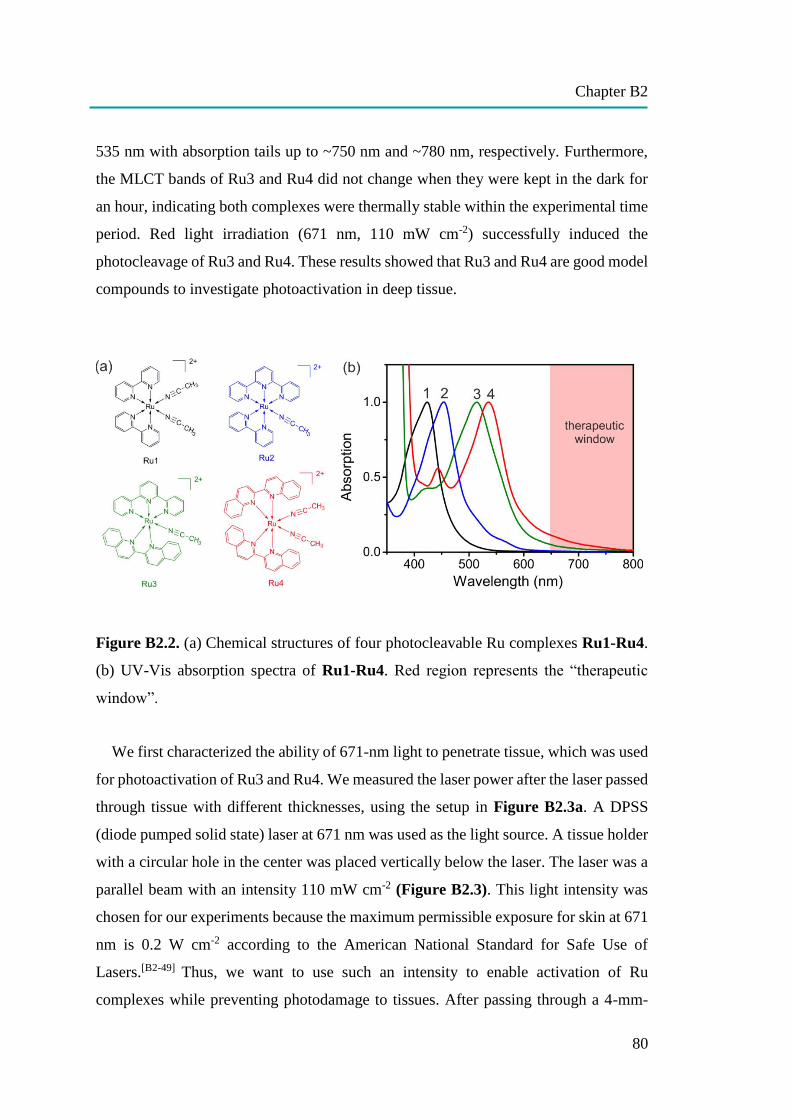
Chapter B2
80
535 nm with absorption tails up to ~750 nm and ~780 nm, respectively. Furthermore,
the MLCT bands of Ru3 and Ru4 did not change when they were kept in the dark for
an hour, indicating both complexes were thermally stable within the experimental time
period. Red light irradiation (671 nm, 110 mW cm-2) successfully induced the
photocleavage of Ru3 and Ru4. These results showed that Ru3 and Ru4 are good model
compounds to investigate photoactivation in deep tissue.
Figure B2.2. (a) Chemical structures of four photocleavable Ru complexes Ru1-Ru4.
(b) UV-Vis absorption spectra of Ru1-Ru4. Red region represents the “therapeutic
window”.
We first characterized the ability of 671-nm light to penetrate tissue, which was used
for photoactivation of Ru3 and Ru4. We measured the laser power after the laser passed
through tissue with different thicknesses, using the setup in Figure B2.3a. A DPSS
(diode pumped solid state) laser at 671 nm was used as the light source. A tissue holder
with a circular hole in the center was placed vertically below the laser. The laser was a
parallel beam with an intensity 110 mW cm-2 (Figure B2.3). This light intensity was
chosen for our experiments because the maximum permissible exposure for skin at 671
nm is 0.2 W cm-2 according to the American National Standard for Safe Use of
Lasers.[B2-49] Thus, we want to use such an intensity to enable activation of Ru
complexes while preventing photodamage to tissues. After passing through a 4-mm-
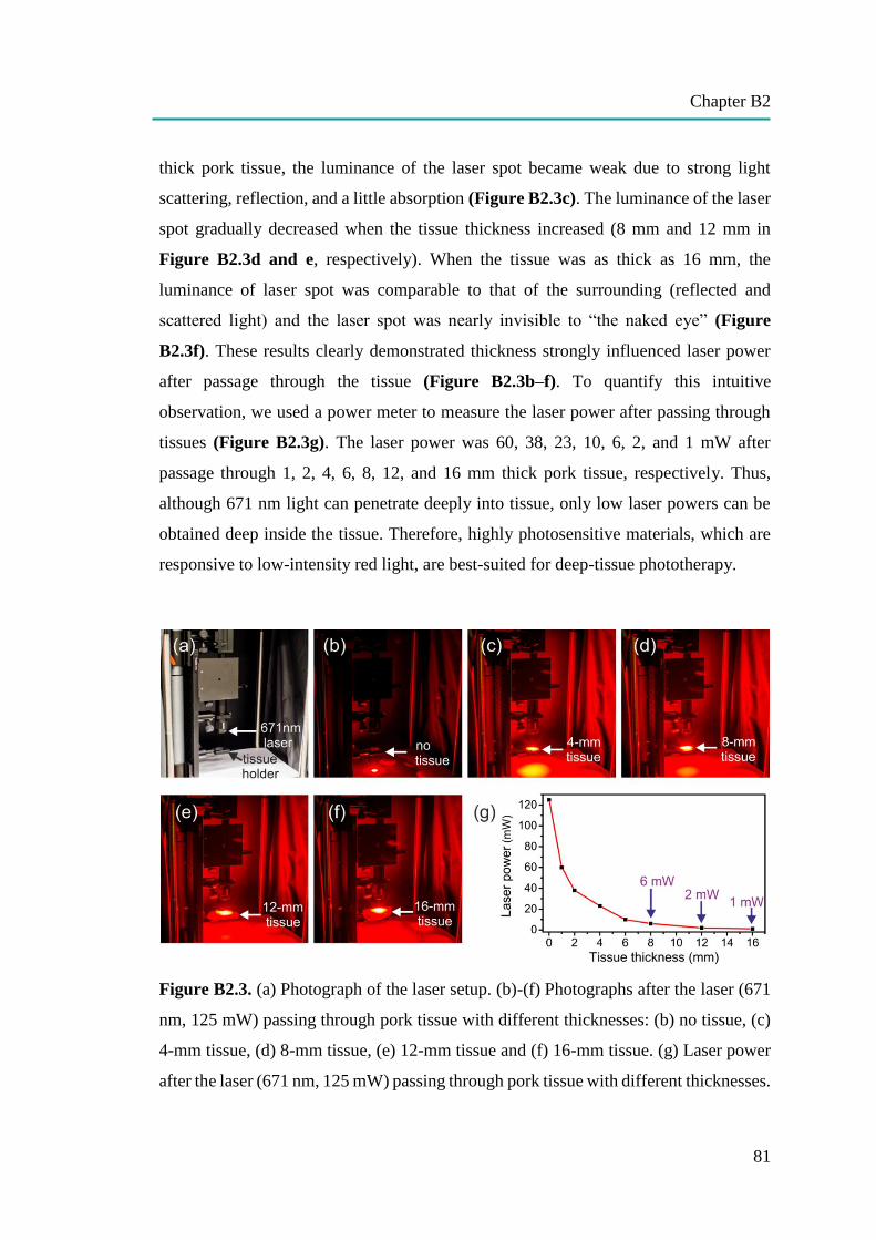
Chapter B2
81
thick pork tissue, the luminance of the laser spot became weak due to strong light
scattering, reflection, and a little absorption (Figure B2.3c). The luminance of the laser
spot gradually decreased when the tissue thickness increased (8 mm and 12 mm in
Figure B2.3d and e, respectively). When the tissue was as thick as 16 mm, the
luminance of laser spot was comparable to that of the surrounding (reflected and
scattered light) and the laser spot was nearly invisible to “the naked eye” (Figure
B2.3f). These results clearly demonstrated thickness strongly influenced laser power
after passage through the tissue (Figure B2.3b–f). To quantify this intuitive
observation, we used a power meter to measure the laser power after passing through
tissues (Figure B2.3g). The laser power was 60, 38, 23, 10, 6, 2, and 1 mW after
passage through 1, 2, 4, 6, 8, 12, and 16 mm thick pork tissue, respectively. Thus,
although 671 nm light can penetrate deeply into tissue, only low laser powers can be
obtained deep inside the tissue. Therefore, highly photosensitive materials, which are
responsive to low-intensity red light, are best-suited for deep-tissue phototherapy.
Figure B2.3. (a) Photograph of the laser setup. (b)-(f) Photographs after the laser (671
nm, 125 mW) passing through pork tissue with different thicknesses: (b) no tissue, (c)
4-mm tissue, (d) 8-mm tissue, (e) 12-mm tissue and (f) 16-mm tissue. (g) Laser power
after the laser (671 nm, 125 mW) passing through pork tissue with different thicknesses.
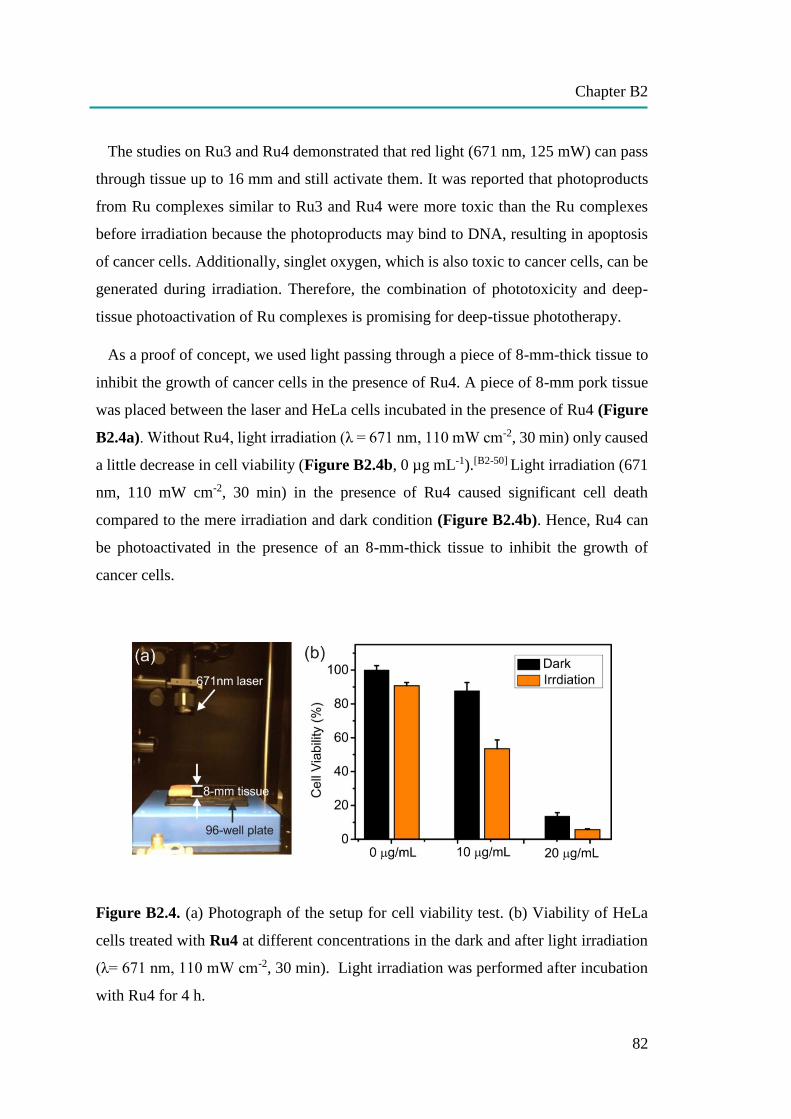
Chapter B2
82
The studies on Ru3 and Ru4 demonstrated that red light (671 nm, 125 mW) can pass
through tissue up to 16 mm and still activate them. It was reported that photoproducts
from Ru complexes similar to Ru3 and Ru4 were more toxic than the Ru complexes
before irradiation because the photoproducts may bind to DNA, resulting in apoptosis
of cancer cells. Additionally, singlet oxygen, which is also toxic to cancer cells, can be
generated during irradiation. Therefore, the combination of phototoxicity and deep-
tissue photoactivation of Ru complexes is promising for deep-tissue phototherapy.
As a proof of concept, we used light passing through a piece of 8-mm-thick tissue to
inhibit the growth of cancer cells in the presence of Ru4. A piece of 8-mm pork tissue
was placed between the laser and HeLa cells incubated in the presence of Ru4 (Figure
B2.4a). Without Ru4, light irradiation (λ = 671 nm, 110 mW cm-2, 30 min) only caused
a little decrease in cell viability (Figure B2.4b, 0 µg mL-1).[B2-50] Light irradiation (671
nm, 110 mW cm-2, 30 min) in the presence of Ru4 caused significant cell death
compared to the mere irradiation and dark condition (Figure B2.4b). Hence, Ru4 can
be photoactivated in the presence of an 8-mm-thick tissue to inhibit the growth of
cancer cells.
Figure B2.4. (a) Photograph of the setup for cell viability test. (b) Viability of HeLa
cells treated with Ru4 at different concentrations in the dark and after light irradiation
(λ= 671 nm, 110 mW cm-2, 30 min). Light irradiation was performed after incubation
with Ru4 for 4 h.
Chapter B2
83
B2.3. Conclusion
We studied the photoactivation of anticancer Ru complexes in the presence of tissues
with different thicknesses. Anticancer Ru complexes (Ru1–Ru4) were irradiated using
a 671-nm laser. Ru1 and Ru2 could not be activated by 671-nm light because their
photoresponsive wavelengths are too short. However, Ru3 and Ru4 were activated by
671-nm light due to their long responsive wavelengths. Although only 1% of the laser
power remained after passing through a 16-mm-thick tissue, the intensity of the light
was still able to activate Ru3 and Ru4. Furthermore, phototoxicity of Ru4 was
successfully induced by 671-nm light in cancer cells after passing through an 8-
mmthick tissue. Our results thus suggest that photoactivatable Ru complexes are
promising for deep-tissue biomedical applications. Further red shifting the responsive
wavelength of Ru complexes would be helpful for phototherapy in deeper tissue. Ru
complexes responsive to 800-nm light, the best wavelength for tissue penetration, are
expected to be realized in the future.
B2.4. References
[B2-1] L. Cheng, C. Wang, L. Feng, K. Yang, Z. Liu, Chem. Rev. 2014, 114, 10869-10939.
[B2-2] C. Alvarez-Lorenzo, L. Bromberg, A. Concheiro, Photochem. Photobiol. 2009, 85, 848-
860.
[B2-3] Y. Zhao, Chem. Rec. 2007, 7, 286-294.
[B2-4] P. Rai, S. Mallidi, X. Zheng, R. Rahmanzadeh, Y. Mir, S. Elrington, A. Khurshid, T. Hasan,
Adv. Drug. Deliv. Rev. 2010, 62, 1094-1124.
[B2-5] K. Hiltebrandt, K. Elies, D. R. D'Hooge, J. P. Blinco, C. Barner-Kowollik, J. Am. Chem.
Soc. 2016, 138, 7048-7054.
[B2-6] J. O. Mueller, F. G. Schmidt, J. P. Blinco, C. Barner-Kowollik, Angew. Chem. Int. Ed.
2015, 54, 10284-10288
[B2-7] P. J. Bednarski, F. S. Mackay, P. J. Sadler, Anticancer Agents Med.Chem. 2007, 7, 75-93.
[B2-8] J. A. W. Fiona S. Mackay, Pavla Heringová, Jana Kašpárková, Ana M. Pizarro, Stephen
A. Moggach, Simon Parsons, Viktor Brabec, Peter J. Sadler, Proc. Natl. Acad. Sci. USA
2007, 104, 20743-20748.
[B2-9] W. Fan, X. Tong, Q. Yan, S. Fu, Y. Zhao, Chem. Commun. 2014, 50, 13492-13494.
[B2-10] Q. Lin, C. Bao, S. Cheng, Y. Yang, W. Ji, L. Zhu, J. Am. Chem. Soc. 2012, 134, 5052-
5055.

Chapter B2
84
[B2-11] G. Yu, W. Yu, Z. Mao, C. Gao and F. Huang, Small 2015, 11, 919-925.
[B2-12] S. Wu and H. J. Butt, Adv. Mater. 2016, 28, 1208-1226.
[B2-13] R. Weissleder, V. Ntziachristos, Nature Med. 2003, 9, 123-128.
[B2-14] H. Huang, B. Yu, P. Zhang, J. Huang, Y. Chen, G. Gasser, L. Ji and H. Chao, Angew. Chem.
Int. Ed. Engl. 2015, 54, 1-5.
[B2-15] X. Zhuang, X. Ma, X. Xue, Q. Jiang, L. Song, L. Dai, C. Zhang, S. Jin, K. Yang, B. Ding,
P. C. Wang, X. J. Liang, ACS Nano 2016, 10, 3486-3495.
[B2-16] V. Nikolenko, R. Yuste, L. Zayat, L. M. Baraldo, R. Etchenique, Chem. Commun. 2005,
1752-1754.
[B2-17] R. Araya, V. Andino-Pavlovsky, R. Yuste and R. Etchenique, ACS Chem. Neurosci. 2013,
4, 1163-1167.
[B2-18] T. Furuta, S. S.-H Wang, J. L. Dantaker, T. M. Dore, W. J. Bybee, E. M. Callaway, W.
Denk, and R. T. Tsien, Proc. Natl. Acad. Sci. USA 1999, 96, 1193-1200.
[B2-19] M. Álvarez, A. Best, S. Pradhan-Kadam, K. Koynov, U. Jonas, M. Kreiter, Adv. Mater.
2008, 20, 4563-4567.
[B2-20] Y. Dai, H. Xiao, J. Liu, Q. Yuan, P. Ma, D. Yang, C. Li, Z. Cheng, Z. Hou, P. Yang, J. Lin,
J. Am. Chem. Soc. 2013, 135, 18920-18929.
[B2-21] B. Yan, J.- C. Boyer, N. R. Branda, Y. Zhao, J. Am. Chem. Soc. 2011, 133, 19714-19717.
[B2-22] F. Wang, D. Banerjee, Y. Liu, X. Chen, X. Liu, Analyst 2010, 135, 1839-1854.
[B2-23] E. Ruggiero, A. Habtemariam, L. Yate, J. C. Mareque-Rivas and L. Salassa, Chem.
Commun. 2014, 50, 1715-1718.
[B2-24] Z. Chen, W. Sun, H. J. Butt, S. Wu, Chem. Eur. J. 2015, 21, 9165-9170.
[B2-25] E. D. Anderson, A. P. Gorka and M. J. Schnermann, Nat. Commun. 2016, 7, 13378
[B2-26] C. Mari, V. Pierroz, S. Ferrari and G. Gasser, Chem. Sci. 2015, 6, 2660-2686.
[B2-27] K. Liu, R. Xing, Q. Zou, G. Ma, H. Mohwald and X. Yan, Angew. Chem. Int. Ed. Engl.
2016, 55, 3036-3039.
[B2-28] N. Zhang, F. Zhao, Q. Zou, Y. Li, G. Ma and X. Yan, Small 2016, 12, 5936-5943.
[B2-29] G. Lv, W. Guo, W. Zhang, T. Zhang, S. Li, S. Chen, A. S. Eltahan, D. Wang, Y. Wang, J.
Zhang, P. C. Wang, J. Chang, X. J. Liang, ACS Nano 2016, 10, 9637-9645.
[B2-30] D. V. Pinnick, B. Burham, Inorg. Chem. 1984, 23, 1440-1445
[B2-31] L. Zayat, C. Calero, P. Alborés, L. Baraldo, R. Etchenique, J. Am. Chem. Soc., 2003, 125,
882-883.
[B2-32] R. N. Garner, J. C. Gallucci, K. R. Dunbar, C. Turro, Inorg. Chem., 2011, 50, 9213-9215.
[B2-33] T. Respondek, R. N. Garner, M. K. Herroon, I. Podgorski, C. Turro, J. J. Kodanko, J. Am.
Chem. Soc., 2011, 133 (43), pp 17164–17167
[B2-34] W. Sun, S. Li, B. Haupler, J. Liu, S. Jin, W. Steffen, U. S. Schubert, H. J. Butt, X. J. Liang,
S. Wu, Adv. Mater. 2017, 29, 1603702.

Chapter B2
85
[B2-35] W. Sun, M. Parowatkin, W. Steffen, H. J. Butt, V. Mailander, S. Wu, Adv. Healthc. Mater.
2016, 5, 467-473.
[B2-36] E. Wachter, D. K. Heidary, B. S. Howerton, S. Parkin, E. C. Glazer, Chem. Commun. 2012,
48, 9649-9651.
[B2-37] P. Kaspler, S. Lazic, S. Forward, Y. Arenas, A. Mandel and L. Lilge, Photochem.
Photobiol. Sci. 2016, 15, 481-495.
[B2-38] R. Trondl, P. Heffeter, C. R. Kowol, M. A. Jakupec, W. Berger, B. K. Keppler, Chem. Sci.
2014, 5, 2925-2932.
[B2-39] J. D. Knoll and C. Turro, Coord. Chem. Rev., 2015, 282-283, 110-126.
[B2-40] J. K. White, R. H. Schmehl and C. Turro, Inorg. Chim. Acta 2017, 454, 7-20.
[B2-41] N. Karaoun and A. K. Renfrew, Chem. Commun. 2015, 51, 14038-14041.
[B2-42] M. A. Sgambellone, A. David, R. N. Garner, K. R. Dunbar, C. Turro, J. Am. Chem. Soc.
2013, 135, 11274-11282.
[B2-43] B. Siewert, V. H. van Rixel, E. J. van Rooden, S. L. Hopkins, M. J. Moester, F. Ariese, M.
A. Siegler, S. Bonnet, Chem. Eur. J. 2016, 22, 10960-10968
[B2-44] B. A. Albani, B. Peña, N. A. Leed, N. A. de Paula, C. Pavani, M. S. Baptista, K. R. Dunbar,
C. Turro, J. Am. Chem. Soc. 2014, 136, 17095-17101.
[B2-45] V. H. S. van Rixel, B. Siewert, S. L. Hopkins, S. H. C. Askes, A. Busemann, M. A. Siegler,
S. Bonnet, Chem. Sci. 2016, 7, 4922-4929.
[B2-46] O. Filevich, L. Zayat, L. M. Baraldo and R. Etchenique, Strut. Bond 2015, 165, 47-68.
[B2-47] L. N. Lameijer, S. L. Hopkins, T. G. Breve, S. H. Askes and S. Bonnet, Chem. Eur. J. 2016,
22, 18484-18491.
[B2-48] P. Zhang, H. Huang, J. Huang, H. Chen, J. Wang, K. Qiu, D. Zhao, L. Ji and H. Chao, ACS
Appl. Mater. Interfaces 2015, 7, 23278-23290.
[B2-49] American National Standard for Safe Use of Lasers, Laser Institute of America: Orlando,
FL . 2000.
[B2-50] A. A. Can, G. Bölükbaşiateş, H. Solmaz, ö. Kaya, M. Gülsoy, International Journal of
Oncology and Cancer Therapy 2016, 1, 7-11.
[B2-51] Y. Liu, D. B. Turner, T. N. Singh, A. M. Angeles-Boza, A. Chouai, K. R. Dunbar, C. Turro,
J. Am. Chem. Soc. 2009, 131, 26-27.
[B2-52] C. R. Hecker, P. E. Fanwick, D. R. McMillin, Inorg. Chem. 1991, 30,659–666.
[B2-53] B. A. Albani, C. B. Durr, C. Turro, J. Phys. Chem. A 2013, 117, 13885-13892.
[B2-54] V. S. Miguel, M. Álvarez, O. Filevich, R. Etchenique, A. del Campo, Langmuir 2012, 28,
1217-1221.
[B2-55] A. Petroni, L. D. Slep, R. Etchenique, Inorg. Chem. 2008, 47, 951-956.
[B2-56] J. P Marcolongo, T. Weyhermuller, L. D. Slep, Inorg. Chim. Acta 2015, 429, 174-182.
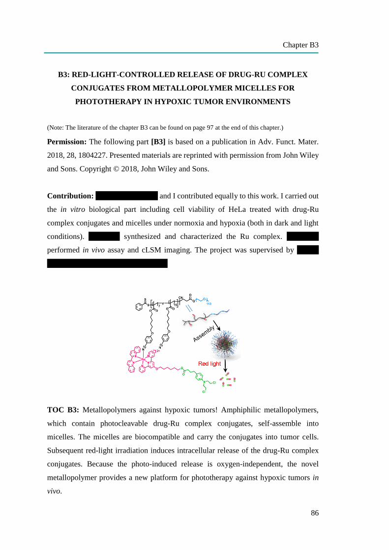
Chapter B3
86
B3: RED-LIGHT-CONTROLLED RELEASE OF DRUG-RU COMPLEX
CONJUGATES FROM METALLOPOLYMER MICELLES FOR
PHOTOTHERAPY IN HYPOXIC TUMOR ENVIRONMENTS
(Note: The literature of the chapter B3 can be found on page 97 at the end of this chapter.)
Permission: The following part [B3] is based on a publication in Adv. Funct. Mater.
2018, 28, 1804227. Presented materials are reprinted with permission from John Wiley
and Sons. Copyright © 2018, John Wiley and Sons.
Contribution: Wen Sun, Yan Wen and I contributed equally to this work. I carried out
the in vitro biological part including cell viability of HeLa treated with drug-Ru
complex conjugates and micelles under normoxia and hypoxia (both in dark and light
conditions). Wen Sun synthesized and characterized the Ru complex. Yan Wen
performed in vivo assay and cLSM imaging. The project was supervised by Volker
Mailänder, Si Wu and Xing-Jie Liang.
TOC B3: Metallopolymers against hypoxic tumors! Amphiphilic metallopolymers,
which contain photocleavable drug-Ru complex conjugates, self-assemble into
micelles. The micelles are biocompatible and carry the conjugates into tumor cells.
Subsequent red-light irradiation induces intracellular release of the drug-Ru complex
conjugates. Because the photo-induced release is oxygen-independent, the novel
metallopolymer provides a new platform for phototherapy against hypoxic tumors in
vivo.

Chapter B3
87
Aim: In a deep solid tumor, which is far away from oxygen diffusion distance supplied
by blood vessels, the area has a special tumor microenvironment comprised of low
oxygen (hypoxia), low nutrients, and acidosis (an excessively acid condition). This
hypoxic condition interferes with photodynamic therapy (PDT), which has cancer
killing ability significantly dependent on the local oxygen concentration. Therefore, this
study aimed to develop a dual-therapy system that can function under both normoxic
and hypoxic conditions as oxygen independent toxicity using anticancer drug
Chlorambucil Ru complex conjugate self-assemble into micelles. Furthermore, the
system was designed to be able to control the release of the drug-Ru complex conjugate
from micelles via red-light stimulation. The red-light control release system was proof
of concept to kill cancer cells in vitro in HeLa and further confirmed the potential to
use for phototherapy against hypoxic tumor microenvironments in vivo in tumor-
bearing mice.
Abstract: Traditional photodynamic phototherapy is not efficient for anticancer
treatment because solid tumors have a hypoxic microenvironment. The development of
photoactivated chemotherapy based on photoresponsive polymers that can be activated
by light in the “therapeutic window” would enable new approaches for basic research
and allow for anticancer phototherapy in hypoxic conditions. This work synthesizes a
novel Ru-containing block copolymer for photoactivated chemotherapy in hypoxic
tumor environment. The polymer has a hydrophilic poly(ethylene glycol) block and a
hydrophobic Ru-containing block, which contains red-light-cleavable (650–680 nm)
drug–Ru complex conjugates. The block copolymer self-assembles into micelles,
which can be efficiently taken up by cancer cells. Red light induces release of the drug–
Ru complex conjugates from the micelles and this process is oxygen independent. The
released conjugates inhibit tumor cell growth even in hypoxic tumor environment.
Furthermore, the Ru-containing polymer for photoactivated chemotherapy in a tumor-
bearing mouse model is applied. Photoactivated chemotherapy of the polymer micelles
demonstrates efficient tumor growth inhibition. In addition, the polymer micelles do
not cause any toxic side effects to mice during the treatment, demonstrating good
biocompatibility of the system to the blood and healthy tissues. The novel red-light-

Chapter B3
88
responsive Ru-containing polymer provides a new platform for phototherapy against
hypoxic tumors.
B3.1. Introduction
The success of metal complexes such as platinum (Pt) complexes for anticancer
applications encourages the development of new metallodrugs.[B3-1–5] Recent
fundamental research and clinical trials showed that ruthenium (Ru) complexes are
promising alternatives for Pt drugs.[B3-1,2] In particular, photoactivatable Ru complexes
have been proposed for anticancer phototherapy, which can increase selectivity
between tumor and healthy cells.[B3-1,2,6,7] These complexes are generally nontoxic to
nonirradiated normal cells and become highly toxic in tumor cells via selective
irradiation of tumors. Phototoxicity of Ru complexes arises from two different
mechanisms. One mechanism is singlet oxygen (1O2) sensitization using Ru complexes
for photodynamic therapy (PDT).[B3-8–16] The other mechanism is uncaging cytotoxic
ligands or Ru species via ligand photosubstitution reactions for photoactivated
chemotherapy (PACT).[B3-17–25] The therapeutic effects of PDT are critically dependent
on the local concentration of oxygen.[B3-26,27] Because solid tumors have a hypoxic
microenvironment especially in the areas far away from the blood vessels (>70 μm),[B3-
28] PDT does not work in hypoxic tumor environment. In contrast, PACT using Ru
complexes is better suited than PDT for hypoxic tumor treatment because ligand
photosubstitution is oxygen independent.[B3-29–32] Thus, PACT represents a promising
approach against hypoxic tumors. We and other groups have shown photoinhibition of
tumor cell growth using photoactivatable Ru complexes based on combined PDT and
PACT.[B3-8,33–37] True PACT in hypoxic cancer cells is difficult to achieve due to
insufficient anticancer efficiencies of uncaged ligands and Ru species. Recently,
Bonnet and co-workers reported PACT against hypoxic cancer cells in vitro using a Ru
complex caged with a cytotoxic ligand.[B3-20] The strategy was based on photo-uncaging
of cytotoxic ligands, which inhibited the growth of hypoxic cancer cells in vitro. The
next challenge is the further development of new Ru complexes for PACT in vivo.
Besides photo-uncaging of efficient anticancer ligands or Ru species, PACT in vivo
requires that photoactivatable Ru complexes have neglectable side effects to healthy
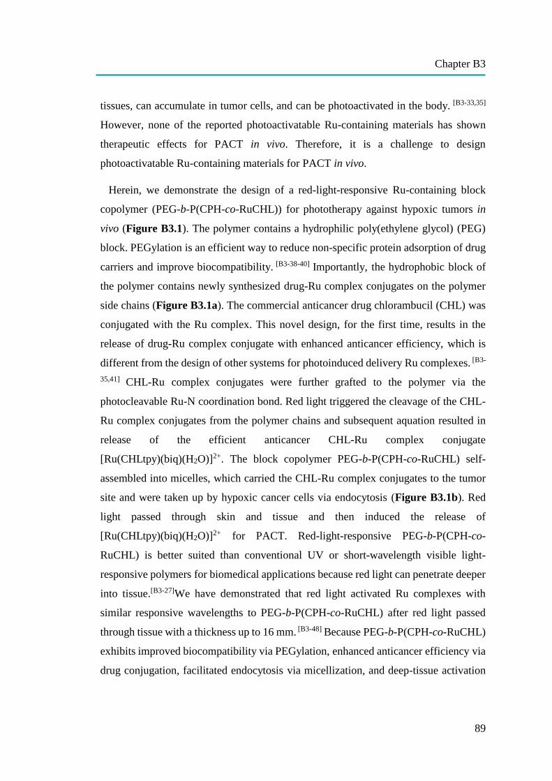
Chapter B3
89
tissues, can accumulate in tumor cells, and can be photoactivated in the body. [B3-33,35]
However, none of the reported photoactivatable Ru-containing materials has shown
therapeutic effects for PACT in vivo. Therefore, it is a challenge to design
photoactivatable Ru-containing materials for PACT in vivo.
Herein, we demonstrate the design of a red-light-responsive Ru-containing block
copolymer (PEG-b-P(CPH-co-RuCHL)) for phototherapy against hypoxic tumors in
vivo (Figure B3.1). The polymer contains a hydrophilic poly(ethylene glycol) (PEG)
block. PEGylation is an efficient way to reduce non-specific protein adsorption of drug
carriers and improve biocompatibility. [B3-38-40] Importantly, the hydrophobic block of
the polymer contains newly synthesized drug-Ru complex conjugates on the polymer
side chains (Figure B3.1a). The commercial anticancer drug chlorambucil (CHL) was
conjugated with the Ru complex. This novel design, for the first time, results in the
release of drug-Ru complex conjugate with enhanced anticancer efficiency, which is
different from the design of other systems for photoinduced delivery Ru complexes. [B3-
35,41] CHL-Ru complex conjugates were further grafted to the polymer via the
photocleavable Ru-N coordination bond. Red light triggered the cleavage of the CHL-
Ru complex conjugates from the polymer chains and subsequent aquation resulted in
release of the efficient anticancer CHL-Ru complex conjugate
[Ru(CHLtpy)(biq)(H2O)]2+. The block copolymer PEG-b-P(CPH-co-RuCHL) self-
assembled into micelles, which carried the CHL-Ru complex conjugates to the tumor
site and were taken up by hypoxic cancer cells via endocytosis (Figure B3.1b). Red
light passed through skin and tissue and then induced the release of
[Ru(CHLtpy)(biq)(H2O)]2+ for PACT. Red-light-responsive PEG-b-P(CPH-co-
RuCHL) is better suited than conventional UV or short-wavelength visible light-
responsive polymers for biomedical applications because red light can penetrate deeper
into tissue.[B3-27]We have demonstrated that red light activated Ru complexes with
similar responsive wavelengths to PEG-b-P(CPH-co-RuCHL) after red light passed
through tissue with a thickness up to 16 mm. [B3-48] Because PEG-b-P(CPH-co-RuCHL)
exhibits improved biocompatibility via PEGylation, enhanced anticancer efficiency via
drug conjugation, facilitated endocytosis via micellization, and deep-tissue activation
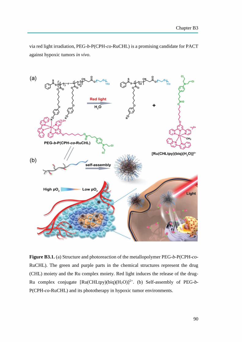
Chapter B3
90
via red light irradiation, PEG-b-P(CPH-co-RuCHL) is a promising candidate for PACT
against hypoxic tumors in vivo.
Figure B3.1. (a) Structure and photoreaction of the metallopolymer PEG-b-P(CPH-co-
RuCHL). The green and purple parts in the chemical structures represent the drug
(CHL) moiety and the Ru complex moiety. Red light induces the release of the drug-
Ru complex conjugate [Ru(CHLtpy)(biq)(H2O)]2+. (b) Self-assembly of PEG-b-
P(CPH-co-RuCHL) and its phototherapy in hypoxic tumor environments.
Chapter B3
91
B3.2. Results and discussion
We prepared block copolymer assemblies by adding water to a THF solution of PEG-
b-P(CPH-co-RuCHL). The micelle solution was collected after removing the organic
solvent via dialysis against water. Transmission electron microscopy (TEM) image
showed that PEG-b-P(CPH-co-RuCHL) formed monodisperse micelles with a diameter
of ~15 nm. Dynamic light scattering (DLS) showed that the micelles had a narrow size
distribution and the average hydrodynamic diameter of the micelles was 22 nm. The
micelles were well dispersed in water and physiological saline solution and had good
stability. To achieve in vivo applications, we used red light in the therapeutic window
(650 to 900 nm) to trigger the photoreaction. Red light irradiation (656 nm) of the
micelles red-shifted the MLCT band from 519 nm (λmax of PEG-b-P(CPH-co-
RuCHL)) to 576 nm (λmax of [Ru(CHLtpy)(biq)(H2O)]2+). This detectable spectral
change can be achieved less than 0.5 h and the change was identical to that of the
photocleavage of similar Ru complexes, suggesting [Ru(CHLtpy)(biq)(H2O)]2+ was
cleaved from the polymer.[B3-48,51] Additionally, the result from UV-Vis absorption
spectroscopy indicated that the process of the photoreaction can be precisely controlled
by the irradiation dose. Up to 84% of [Ru(CHLtpy)(biq)(H2O)]2+ was released under
light irradiation in our experimental condition. In comparison, only ≈7% of
[Ru(CHLtpy)(biq)(H2O)]2+ was released in the dark after 24 h, suggesting the good
stability of the micelles during long time incubation and the release was controlled by
light (Results are not shown here, see Publication [B3]).
The released [Ru(CHLtpy)(biq)(H2O)]2+ is a conjugate of a Ru complex and a
commercial anticancer drug CHL, both of which can inhibit cancer cell growth. Thus,
[Ru(CHLtpy)(biq)(H2O)]2+ is expected to be an efficient anticancer agent. The
cytotoxicity of [Ru(CHLtpy)(biq)(H2O)]2+ was tested using HeLa cells, a commonly
used model cancer cell line for evaluating anticancer activities of new anticancer
materials (Figure B3.1a). HeLa cells were incubated with
[Ru(CHLtpy)(biq)(H2O)](PF6)2 with a concentration from 1.25 to 50 µg mL-1 for 24 h.
Cell viability decreased as the concentration of [Ru(CHLtpy)(biq)(H2O)]2+ increased.
The half maximal effective concentration (EC50) of [Ru(CHLtpy)(biq)(H2O)]2+ to HeLa
cell was ≈13.7 µg mL-1, much lower than those of both [Ru(tpy)(biq)(H2O)] (PF6)2
Chapter B3
92
(≈30 μg mL−1, Figure B3.2) and CHL (≈150 μg mL−1).[B3-52] The result indicated that
the two anticancer moieties had synergistic effects and the conjugate
[Ru(CHLtpy)(biq)(H2O)]2+ had improved cytotoxicity toward cancer cells. The
cytotoxicity of the complex after red light irradiation (671 nm, 120 J cm−2) was also
studied (Figure B3.1a). The EC50 value for HeLa cells after irradiation was ≈10.3 μg
mL−1, which is close to that without irradiation. EC50 values under dark and light
irradiation were similar because no 1O2 was generated when
[Ru(CHLtpy)(biq)(H2O)]2+ was irradiated with light (Figure B3.1b).
Figure B3.1. (a) Viability of HeLa cells incubated with [Ru(CHLtpy)(biq)(H2O)](PF6)2
with various concentrations for 24 h. HeLa cells were kept in the dark or under light
irradiation (671 nm, 120 J cm−2) at the beginning of the incubation. (b) Emission spectra
of 1O2 (λem = 1275 nm) generated in the solutions of [Ru(CHLtpy)(biq)(H2O)] (PF6)2
and [Ru(bpy)3]Cl2 in CD3OD after excitation at λex = 450 nm. A450 nm = 0.1 (4 mm path
length) for all samples. [Ru(bpy)3]Cl2 was used as a reference sample (ΦΔ = 0.73 in
CD3OD).[B3-53]
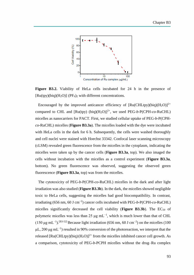
Chapter B3
93
Figure B3.2. Viability of HeLa cells incubated for 24 h in the presence of
[Ru(tpy)(biq)(H2O)] (PF6)2 with different concentrations.
Encouraged by the improved anticancer efficiency of [Ru(CHLtpy)(biq)(H2O)]2+
compared to CHL and [Ru(tpy) (biq)(H2O)]2+, we used PEG-b-P(CPH-co-RuCHL)
micelles as nanocarriers for PACT. First, we studied cellular uptake of PEG-b-P(CPH-
co-RuCHL) micelles (Figure B3.3a). The micelles loaded with the dye were incubated
with HeLa cells in the dark for 6 h. Subsequently, the cells were washed thoroughly
and cell nuclei were stained with Hoechst 33342. Confocal laser scanning microscopy
(cLSM) revealed green fluorescence from the micelles in the cytoplasm, indicating the
micelles were taken up by the cancer cells (Figure B3.3a, top). We also imaged the
cells without incubation with the micelles as a control experiment (Figure B3.3a,
bottom). No green fluorescence was observed, suggesting the observed green
fluorescence (Figure B3.3a, top) was from the micelles.
The cytotoxicity of PEG-b-P(CPH-co-RuCHL) micelles in the dark and after light
irradiation was also studied (Figure B3.3b). In the dark, the micelles showed negligible
toxic to HeLa cells, suggesting the micelles had good biocompatibility. In contrast,
irradiating (656 nm, 60 J cm−2) cancer cells incubated with PEG-b-P(CPH-co-RuCHL)
micelles significantly decreased the cell viability (Figure B3.3b). The EC50 of
polymeric micelles was less than 25 μg mL−1, which is much lower than that of CHL
(150 μg mL−1).[B3-52] Because light irradiation (656 nm, 60 J cm−2) on the micelles (100
μL, 200 μg mL−1) resulted in 90% conversion of the photoreaction, we interpret that the
released [Ru(CHLtpy)(biq) (H2O)]2+ from the micelles inhibited cancer cell growth. As
a comparison, cytotoxicity of PEG-b-PCPH micelles without the drug–Ru complex
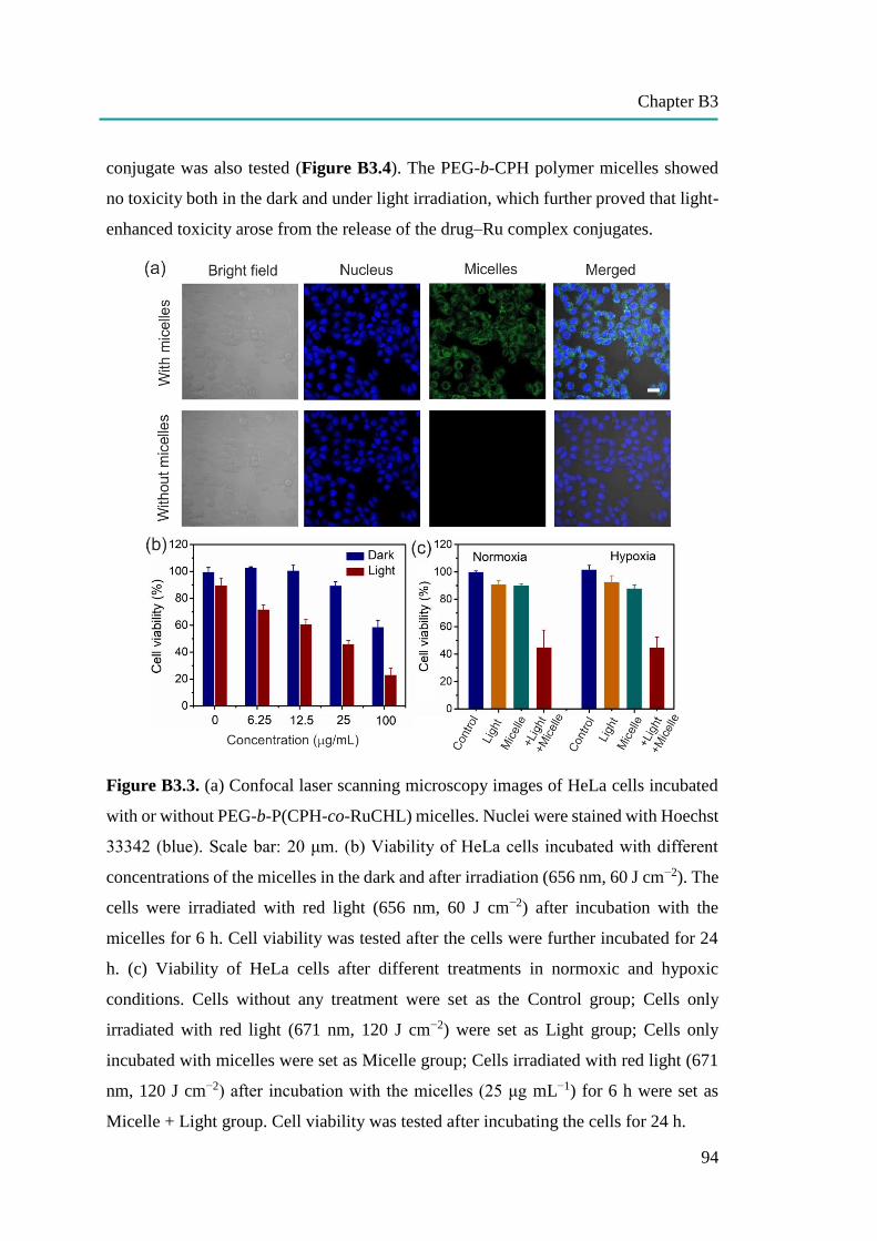
Chapter B3
94
conjugate was also tested (Figure B3.4). The PEG-b-CPH polymer micelles showed
no toxicity both in the dark and under light irradiation, which further proved that light-
enhanced toxicity arose from the release of the drug–Ru complex conjugates.
Figure B3.3. (a) Confocal laser scanning microscopy images of HeLa cells incubated
with or without PEG-b-P(CPH-co-RuCHL) micelles. Nuclei were stained with Hoechst
33342 (blue). Scale bar: 20 μm. (b) Viability of HeLa cells incubated with different
concentrations of the micelles in the dark and after irradiation (656 nm, 60 J cm−2). The
cells were irradiated with red light (656 nm, 60 J cm−2) after incubation with the
micelles for 6 h. Cell viability was tested after the cells were further incubated for 24
h. (c) Viability of HeLa cells after different treatments in normoxic and hypoxic
conditions. Cells without any treatment were set as the Control group; Cells only
irradiated with red light (671 nm, 120 J cm−2) were set as Light group; Cells only
incubated with micelles were set as Micelle group; Cells irradiated with red light (671
nm, 120 J cm−2) after incubation with the micelles (25 μg mL−1) for 6 h were set as
Micelle + Light group. Cell viability was tested after incubating the cells for 24 h.
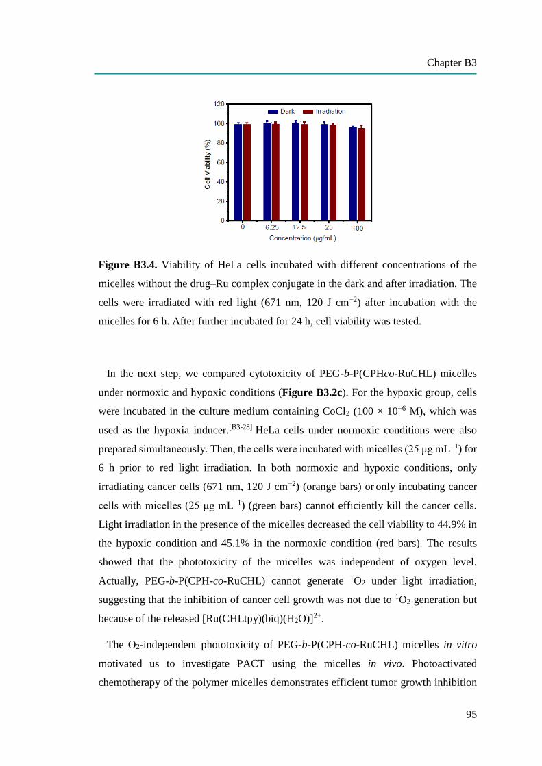
Chapter B3
95
Figure B3.4. Viability of HeLa cells incubated with different concentrations of the
micelles without the drug–Ru complex conjugate in the dark and after irradiation. The
cells were irradiated with red light (671 nm, 120 J cm−2) after incubation with the
micelles for 6 h. After further incubated for 24 h, cell viability was tested.
In the next step, we compared cytotoxicity of PEG-b-P(CPHco-RuCHL) micelles
under normoxic and hypoxic conditions (Figure B3.2c). For the hypoxic group, cells
were incubated in the culture medium containing CoCl2 (100 × 10−6 M), which was
used as the hypoxia inducer.[B3-28] HeLa cells under normoxic conditions were also
prepared simultaneously. Then, the cells were incubated with micelles (25 μg mL−1) for
6 h prior to red light irradiation. In both normoxic and hypoxic conditions, only
irradiating cancer cells (671 nm, 120 J cm−2) (orange bars) or only incubating cancer
cells with micelles (25 μg mL−1) (green bars) cannot efficiently kill the cancer cells.
Light irradiation in the presence of the micelles decreased the cell viability to 44.9% in
the hypoxic condition and 45.1% in the normoxic condition (red bars). The results
showed that the phototoxicity of the micelles was independent of oxygen level.
Actually, PEG-b-P(CPH-co-RuCHL) cannot generate 1O2 under light irradiation,
suggesting that the inhibition of cancer cell growth was not due to 1O2 generation but
because of the released [Ru(CHLtpy)(biq)(H2O)]2+.
The O2-independent phototoxicity of PEG-b-P(CPH-co-RuCHL) micelles in vitro
motivated us to investigate PACT using the micelles in vivo. Photoactivated
chemotherapy of the polymer micelles demonstrates efficient tumor growth inhibition
Chapter B3
96
in a tumor-bearing mouse. Furthermore, the polymer micelles do not cause any toxic
side effects to mice during the treatment, demonstrating their good biocompatibility to
the blood and healthy tissues (Results are not shown here, see Publication [B3]).
B3.3. Conclusion
We reported the design of a Ru-containing amphiphilic polymer for PACT in vivo.
The Ru-containing polymer, PEG-b-P(CPHco-RuCHL), contains novel red-light-
cleavable drug–Ru conjugates. The drug–Ru complex conjugates bearing a very high
content (≈45 wt%) in the polymer were carried by the polymer and can be released on-
demand via red light irradiation. The Ru-containing polymer self-assembled into
micelles and internalized into tumor cells. Light irradiation induced the release of drug–
Ru complex conjugates, which inhibited tumor cell growth. Because the photoinduced
drug release process is oxygen-independent, the polymer micelles are suited for PACT
against tumor cells with hypoxic environments. The polymer micelles were further used
for PACT in a tumor-bearing mouse model, which completely inhibited tumor growth.
Moreover, the treatment using the polymer micelles eliminated substantial systemic
toxicity in living animals. PACT based on our metallopolymers with red-light-
cleavable drug–Ru complex conjugates is a new strategy to overcome the problem of
conventional PDT in hypoxic environments. The development of polymers with drug–
Ru complex conjugates opens up an avenue for the design of polymeric therapeutics
for PACT against hypoxic tumors.
B3.4. References
[B3-1] I. Romero-Canelon, P. J. Sadler, Inorg. Chem. 2013, 52, 12276.
[B3-2] C. Mari, V. Pierroz, S. Ferrari, G. Gasser, Chem. Sci. 2015, 6, 2660.
[B3-3] K. Vellaisamy, G. Li, C.-N. Ko, H.-J. Zhong, S. Fatima, H.-Y. Kwan, C.-Y. Wong, W.-J.
Kwong, W. Tan, C.-H. Leung, D.-L. Ma, Chem. Sci. 2018, 9, 1119.
[B3-4] K. M. Boyle, J. K. Barton, J. Am. Chem. Soc. 2018, 140, 5612.
[B3-5] G. Yu, M. Zhang, M. L. Saha, Z. M. L. Saha, Z. Mao, J. Chen, Y. Yao, Z. Zhou, Y. Liu, C.
Gao, F. Huang, X. Chen, P. J. Stang. J. Am. Chem. Soc. 2017, 139, 15940.
[B3-6] C. S. Burke, A. Byrne, T. E. Keyes, J. Am. Chem. Soc. 2018, 140, 6945.
[B3-7] J. D. Knoll, C. Turro, Coord. Chem. Rev. 2015, 6, 2660.
Chapter B3
97
[B3-8] B. A. Albani, B. Pena, N. A. Leed, N. A. B. G. de Paula, C. Pavani, M. S. Baptista, K. R.
Dunbar, C. Turro, J. Am. Chem. Soc. 2014, 136, 17095.
[B3-9] H. Huang, B. Yu, P. Zhang, J. Huang, Y. Chen, G. Gasser, L. Ji, H. Chao, Angew. Chem., Int.
Ed. 2015, 54, 14049.
[B3-10] N. W. Choi, S. S. Verbridge, R. M. Williams, J. Chen, J.-Y. Kim, R. Schmehl, C. E. Farnum,
W. R. Zipfel, C. Fischbach, A. D. Stroock, Biomaterials 2012, 33, 2710.
[B3-11] T. Sainuddin, J. McCain, M. Pinto, H. Yin, J. Gibson, M. Hetu, S. A. McFarland, Inorg. Chem.
2016, 55, 83.
[B3-12] L. Kohler, L. Nease, P. Vo, J. Garofolo, D. K. Heidary, R. P. Thummel, E. C. Glazer, Inorg.
Chem. 2017, 56, 12214.
[B3-13] M. Wenzel, A. de Almeida, E. Bigaeva, P. Kavanagh, M. Picquet, P. L. Gendre, E. Bodio, A.
Casini, Inorg. Chem. 2016, 55, 2544.
[B3-14] C. Griffith, A. S. Dayoub, T. Jaranatne, N. Alatrash, A. Mohamedi, K. Abayan, Z. S. Breitbach,
D. W. Armstrong, F. M. MacDonnell, Chem. Sci. 2017, 8, 3726.
[B3-15] M. R. Gill, P. J. Jarman, S. Halder, M. G. Walker, H. K. Saeed, J. A. Thomas, C. Smythe, K.
Ramadan, K. A. Vallis, Chem. Sci. 2018, 9, 841.
[B3-16] M. Dickerson, B. Howerton, Y. Bae, E. C. Glazer, J. Mater. Chem. B 2016, 4, 394.
[B3-17] E. Wachter, D. K. Heidary, B. S. Howerton, S. Parkin, E. C. Glazer, Chem. Commun. 2012,
48, 9649.
[B3-18] B. S. Howerton, D. K. Heidary, E. C. Glazer, J. Am. Chem. Soc. 2012, 134, 8324.
[B3-19] M. A. Sgambellone, A. David, R. N. Garner, K. R. Dunbar, C. Turro, J. Am. Chem. Soc. 2013,
135, 11274.
[B3-20] L. N. Lameijer, D. Ernst, S. L. Hopkins, M. S. Meijer, S. H. C. Askes, S. E. Le Devedec, S.
Bonnet, Angew. Chem., Int. Ed. 2017, 56, 11549.
[B3-21] V. H. S. van Rixel, B. Siewert, S. L. Hopkins, S. H. C. Askes, A. Busemann, M. A. Siegler, S.
Bonnet, Chem. Sci. 2016, 7, 4922.
[B3-22] J. K. White, R. H. Schmehl, C. Turro, Inorg. Chim. Acta 2017, 454, 7.
[B3-23] N. Karaoun, A. K. Renfrew, Chem. Commun. 2015, 51, 14038.
[B3-24] A. N. Hidayatullah, E. Wachter, D. K. Heidary, S. Parkin, E. C. Glazer, Inorg. Chem. 2014,
53, 10030.
[B3-25] E. Wachter, D. Moya, S. Parkin, E. C. Glazer, Inorg. Chem. 2011, 50, 9045.
[B3-26] K. Liu, R. Xing, Q. Zou, G. Ma, H. Mohwald, X. Yan, Angew. Chem., Int. Ed. 2016, 55, 3036.
[B3-27] S. S. Lucky, K. C. Soo, Y. Zhang, Chem. Rev. 2015, 115, 1990.
[B3-28] S. Shen, C. Zhu, D. Huo, M. Yang, J. Xue, Y. Xia, Angew. Chem., Int. Ed. 2017, 56, 8801.
[B3-29] B. A. Albani, C. B. Durr, C. Turro, J. Phys. Chem. A 2013, 117, 13885.
[B3-30] R. Araya, V. Andino-Pavlovsky, R. Yuste, R. Etchenique, ACS Chem. Neurosci. 2013, 4, 1163.
[B3-31] V. Nikolenko, R. Yuste, L. Zayat, L. M. Baraldo, R. Etchenique, Chem. Commun. 2005, 1752.
Chapter B3
98
[B3-32] M. Huisman, J. K. White, V. G. Lewalski, I. Podgorski, C. Turro, J. M. Kodanko, Chem.
Commun. 2016, 52, 12590.
[B3-33] W. Sun, X. Zeng, S. Wu, Dalton Trans. 2017, 47, 283.
[B3-34] W. Sun, M. Parowatkin, W. Steffen, H. J. Butt, V. Mailander, S. Wu, Adv. Healthcare Mater.
2016, 5, 467.
[B3-35] W. Sun, S. Li, B. Haupler, J. Liu, S. Jin, W. Steffen, U. S. Schubert, H. J. Butt, X.-J. Liang, S.
Wu, Adv. Mater. 2017, 29, 1603702.
[B3-36] N. Lameijer, T. G. Breve, V. H. S. van Rixel, S. H. C. Askes, M. A. Siegler, S. Bonnet, Chem.
Eur. J. 2018, 24, 2709.
[B3-37] L. M. Loftus, J. K. White, B. A. Albani, L. Kohler, J. J. Kodanko, R. P. Thummel, K. R.
Dunbar, C. Turro, Chem. Eur. J. 2016, 22, 3704.
[B3-38] Y. Zhang, H. F. Chan, K. W. Leong, Adv. Drug Delivery Rev. 2013, 65, 104.
[B3-39] P. M. Kelly, C. Aberg, E. Polo, A. O’Connell, J. Cookman, J. Fallon, Z. Krpetić, K. A. D
awson, Nat. Nanotechnol. 2015, 10, 472.
[B3-40] F. Bertoli, D. Garry, M. P. Monopoli, A. Salvati, K. A. Dawson, ACS Nano 2016, 10, 10471.
[B3-41] G. Boeuf, G. V. Roullin, J. Moreau, L. V. Gulick, N. Z. Pineda, C. Terry, D. Ploton, M. C.
Andry, F. Chuburu, S. Dukic, M. Molinari, G. Lemercier, ChemPlusChem 2014, 79, 171.
[B3-42] M. Abbas, Q. Zou, S. Li, X. Yan, Adv. Mater. 2017, 29,1605021.
[B3-43] J. F. Gohy, Y. Zhao, Chem. Soc. Rev. 2013, 42, 7117.
[B3-44] K. Hiltebrandt, K. Elies, D. R. D’hooge D, J. P. Blinco, C. Barner-Kowollik, J. Am. Chem.
Soc. 2016, 138, 7048.
[B3-45] Y. Zhao, Macromolecules 2012, 45, 3647.
[B3-46] J. P. Menzel, B. B. Noble, A. Lauer, M. L. Coote, J. P. Blinco, C. Barner-Kowollik, J. Am.
Chem. Soc. 2017, 139, 15812.
[B3-47] C. H. Quek, K. W. Leong, Nanomaterials 2012, 2, 92.
[B3-48] W. Sun, R. Thiramanas, L. D. Slep, X. Zeng, V. Mailander, S. Wu, Chem. Eur. J. 2017, 23,
10832.
[B3-49] J. Yang, W. Liu, M. Sui, J. Tang, Y. Shen, Biomaterials 2011, 32, 9136.
[B3-50] S. Aryal, C.-M. J. Hu, L. Zhang, ACS Nano 2010, 4, 251.
[B3-51] L. Zayat, M. G. Noval, J. Campi, C. I. Calero, D. J. Calvo, R. Etchenique, ChemBioChem
2007, 8, 2035.
[B3-52] I. S. Vijayashree, P. Niranjana, G. Prabhu, V. V. Sureshbabu, J. Manjanna, J. Cluster Sci. 2017,
28, 133.
[B3-53] J.-A. Cuello-Garibo, M. S. Meijer, S. Bonnet, Chem. Commun. 2017, 53, 6768.
[B3-54] J. M. Brown, W. R. Wilson, Nat. Rev. Cancer 2004, 4, 437.

Chapter C
99
CHAPTER C
NANOCAPSULE AS A TEMPERATURE SENSOR
IN LIVING CELLS
This study developed polymeric upconversion nanocapsules for use as
nanothermometers in living cells by employing temperature dependence of triplet-
triplet annihilation upconversion (TTA-UC) phenomenon. A general introduction about
human body temperature, the effects of temperature change and current
nanotechnology used for temperature measurement is given. Afterwards, the study is
presented.
(Note: The literature of the introduction can be found on page 138 at the end of the dissertation.)
[C1] Banu Iyisan, Raweewan Thiramanas, Nadya Nazarova, Yuri Avlasevich,
Volker Mailänder*, Stanislav Balouchev*, Katharina Landfester*. Temperature
Sensing in Cells by Polymeric Upconversion Nanocapsules. (Manuscript in
preparation).
C. Introduction
Human body temperature is normally around 35-37 C, which is commonly
measured by using a mercury thermometer. The internal core body temperature (T(c))
can be monitored from different sites of the body including oral, rectal and oesophageal
temperatures. However, these measurement sites and the methods used to measure T(c)
are not convenient and invasive, often causing irritation and discomfort.[Z-74] Rectal
temperature is not appropriate for use in many conditions due to its labor- and time-
consuming response. However, rectal temperature is still considered to be the most
correctly available method for measuring T(c) in thermal illness and also an indicator
of systemic arterial temperature during whole body hyperthermia.[Z-74-75] In fact, not
only does the tissue temperature involve heterogeneity, but the temperature in tumors
can be variable. Evidence indicates that most parts of the body can show similar
Chapter C
100
temperatures to rectal temperature, or T(c); however, higher and lower temperatures
than T(c) can be observed in some parts of tumors.[Z-75]
In cancer cells, a major temperature and pH change within the tumor
microenvironment could easily affect them, which is not usually the case in normal
cells as they have an effective buffering system to protect cells. There is about 1-2
degrees difference between the cancerous and normal cells, as typically mentioned in
literature. It has been well known for at least 50 years that the tumor cells are slightly
more sensitive to heat than normal cells, especially at temperatures between 42 and 45
C. Thus, cancer cells could be killed by this range of temperature about one degree
lower, causing irreversible damage to cancer cells but not affecting healthy cells. This
fact motivated the establishment of hyperthermia as cancer therapy.[Z-76]
Accumulating evidence indicates that temperature can play an important role in
cancer therapy. Increased survival rate and more efficient chemotherapy after the
cancer patient getting fever - resulting in T(c) increase - from infection have been found
in many cases. Furthermore, temperature increase in tumor site can enhance
permeability of therapeutic agents to cancer cells and can stimulate immune response.
[Z-77] Zhu et al.[Z-78] found that culturing cancer cells at 39 °C slightly prevented cell
growth by arresting the cells at the G1 phase of the cell cycle and by hampering of cell-
cell interaction, and efficiently enhanced the killing effect of chemotherapy which can
be long-lasting even after finishing of the treatment. Moreover, the morphology of the
cancer cells was discovered to be temperature-dependent; when the temperature
increased, the cell surface became smoother and cell height became higher, indicating
that temperature can cause the alteration of cellular structures (such as cell surface and
cell height), which may then lead to changes in the cellular mechanical properties. [Z-79]
As the temperature changes inside cancer cells, from intra-metabolism or from
external treatment, which have a significant impact on cancer therapy as mentioned
above, the motivation to establish a new method to monitor temperature at the cellular
level has increased intensively. This Part [C1] reported the development of light
triggered-polymeric upconversion nanocapsules based on triplet-triplet annihilation
upconversion (TTA-UC), which is a temperature dependent system for measurement
Chapter C
101
of cellular temperature in living cancerous cells. Here, recent advances in
nanotechnology used for temperature measurement are described.
Considering the size of the cells, only nanoscale size of thermometer could
overcome the intracellular temperature sensing and mapping, whereas a conventional
thermometer could not achieve this. Development of nano-optic electronic device
brought the thermocouples to precisely detect and reach the cellular temperature in
nanoscale. However, this technique can interrupt cellular activity and morphology
when coming into contact with the cells. Temperature sensing fluorescence microscopy
can override the contacting issue and provide high spatiotemporal resolution. However,
a limitation of this method is based on the properties of fluorophores such as
cytotoxicity, photobleaching, and cellular environmental sensitiveness.
C.1 Fluorescent polymeric nano-thermometer
Polymers can be used to protect fluorescent probes from intracellular
environment. Some of them have thermo-sensitive properties, which behave differently
according to their critical solution temperatures such as the family of poly(N-
isopropylacrylamide)s (PNIPAAms) and poly(2-oxazoline)s (POxs). Combined with
fluorescent probes, fluorescent polymeric thermometers (FPT) have drawn significant
interest because they provide accurate tracking of intracellular temperature with high
spatial resolution. Instead of using thermo-responsive polymers for the controlled
release of the drug, as mentioned in Chapter B, FPT uses temperature dependent
morphology of the polymer to control fluorescence probes as a temperature reporter.
For lower critical solution temperature (LCST)-type polymer, at a temperature below
the LCST, the fluorescent polymer appears in coil state; at a temperature above the
LCST, the fluorescent polymer turns from coil to globule state, allowing simple and
fast detection of the fluorescent signal that is continuous, reversible and reusable.[Z-80-
81]
Gong et al.[Z-82] developed a water soluble, visible excited and simple-structured
copolymer hydrogel, poly(NIPAM-co-BODIPY-AA), containing N-
isopropylacrylamide (NIPAM) and derivative of 4,4-difluoro-4-bora-3a,4a-diaza-s-
indacene (BODIPY) units as thermo-responsive and polarity sensitive fluorescent
Chapter C
102
signaling parts, respectively. A linear increase of fluorescence intensity with increasing
temperature could be obtained in a range of 37-42 C in aqueous solution. The cLSM
images showed the uptake of the copolymer inside BHK cells and exhibited
temperature dependent fluorescent intensity at 35 and 42 C.
C.2 DNA fluorescent thermometers
Thermo-sensitive DNA has several advantages including biocompatibility, a
well-defined structure, nanoscale size and tunable temperature sensing properties.
Therefore, it was applied for development of DNA fluorescent thermometers. The most
common temperature sensitive-DNA based nanostructure is the molecular beacon
(MB), which is a hairpin-shaped oligonucleotide containing a single-stranded loop and
a double-stranded stem with end-labeled quencher and reporter fluorophores. MBs are
highly sensitive, sequence specific, and are commonly used for sequence detection in
qPCR. MBs are considered a smart temperature sensing material because the
conformation of MBs can reversibly change between the hairpin shape and random coil
state upon exposure to various temperatures. The relative distance changes between the
fluorophore and quencher result in the quenching or emitting of fluorescence. The
melting point (Tm) of MBs is defined as the temperature at which half of the MBs
structure is open. It is also independent of concentration. Due to its fixed Tm for each
MB, this led to a new design of multiplex MBs nanoassemblies for fluorescence
thermometry with the resolution <0.5 C for a wide range of temperatures from 15 to
60C. MBs are widely used in fluorescence thermometry, including thermal mapping
and temperature sensing in some living organisms.[Z-83-84]
C.3 Gold nanoparticles
Gold nanoparticles (AuNPs) have several distinct properties such as high
absorption coefficient, luminescence and conductivity, quenched or enhanced
fluorescence, catalyzed reactions, chemically inert and excellent biocompatibility,
which makes them more attractive for use as bio-nanosensors.[Z-85] Shang et al.
developed a gold nanocluster (AuNC)‐ based fluorescent nanothermometer introduced
into HeLa cells by simple endocytosis and localized mainly in endosomes/lysosomes.
The fluorescent lifetime of the AuNCs exhibited clearly excellent temperature
Chapter C
103
dependence. Interestingly, simultaneous intracellular temperature sensing and imaging
of cells using fluorescence life time imaging microscopy (FLIM) was clearly shown
and confirmed highly sensitive temperature dependent fluorescent emission.[Z-86-87]
C.4 Nanodiamonds
Kucsko et al.[Z-88] demonstrated a new approach for the nanothermometer by
using coherent manipulation of the electronic spin associated with nitrogen-vacancy
color centers in diamond nanocrystals (nanodiamond). The ground state of
nanodiamonds is split into two energy levels. The energy gap between the two levels is
temperature dependent. This technique uses nanowire to introduce nanodiamonds for
local temperature sensing and AuNPs for heat transfer to the cells and achieves the
detection of temperature fluctuations in living cells as small as 1.8 mK in an ultrapure
bulk diamond sample at a small length scale as short as 200 nm. Using this technique,
temperature-gradient control and mapping at the subcellular level in a human
embryonic fibroblast were demonstrated. The adequate spatial resolution of this
approach allows compatibility with tracking intracellular metabolic reaction in single
cells. Although it is difficult and invasive to localize the nanodiamonds into the cells or
subcellular organelles by microinjection or electroporation and this method involves
laser which can cause photodamage to the cell, the incredible detection sensitivity is
the highest among any thermometers developed so far.[Z-89-90]
C.5 Quantum dots (QDs)
A quantum dot is a tiny inorganic nanoparticle with a typical size less than 10
nm. The unique optical properties are derived from the confinement of the electrons
and produce changes in the electronic structure, leading to fluorescence enhancement
with better brightness for detection, a broader excitation profile providing tunability,
and greater photostability compared with organic dyes.[Z-87] The well-known QDs
include CdSe@ZnS (the @ symbol indicates a CdSe core surrounded by a ZnS shell)
commonly used as fluorescent probes. They possess narrow emission lines with particle
size dependent wavelengths. However, disadvantages such as potential cytotoxicity and
fluorescence blinking can be problematic.[Z-91]
Chapter C
104
Yang et al.[Z-92] reported the cellular temperature response inside single living
NIH/3T3 murine fibroblast cells upon external chemical and physical stimuli following
Ca2+ stress and cold shock using quantum dots as nano-thermometers. The obtained
photoluminescence spectral shifts used for intracellular heat generation mapping
revealed inhomogeneous intracellular temperature progression, which supports the
concept of subcellular temperature gradient signaling and regulation in cells.
C.6 Magnetic nanoparticles
Iron oxide nanoparticles (IONPs) and superparamagnetic iron oxide
nanoparticles (SPIONs) have gained much attention in many bio-medical fields due to
their intrinsic properties, such as internal magnetism, visualization by magnetic
resonance (MR) imaging, biocompatibility, guidance to target sites by means of an
external magnetic field, heating to provide hyperthermia for cancer therapy and
degradation into nontoxic iron ions in vivo. SPIONs are composed of an inner magnetic
particle core (usually magnetite, Fe3O4, or maghemite, γ-Fe2O3) and a hydrophilic
coating of polymers, such as polysaccharide, poly(ethylene glycol) (PEG), and
poly(vinyl alcohol). The coating prevents agglomerations and protects them from the
surrounding environment. They can also be modified by targeting ligands.[Z-22,93]
Zhong et al.[Z-94] reported a new non-invasive approach for remote temperature
probing that provides accuracy as good as 0.017 C (0.0055% accuracy) by measuring
the magnetization curve of a commercial oleic acid coated magnetic nanoparticles with
a size around 10 nm. This method provides great significance in the potential
establishment of safer and non-invasive protocols for hyperthermia therapy and
intracellular temperature sensing. However, experimental evidence inside the living
cells has not been proven so far.
Wang et al.[Z-95] developed a magnetic fluorescent nano-thermometer consisting
of magnetic nanoparticles (Fe3O4) encapsulated inside a silica layer. Then a poly(N-
isopropylacrylamide)(pNIPAM) copolymer shell with Rhodamine B isothiocyanate
(RhBITC) embedded inside was further coated. Finally, gold nanoparticles were
introduced onto the copolymer shell by in-situ growth method and the nano-
thermometer (denoted as Fe3O4@SiO2@(pNIPAM-co-RhBITC)/Au) was obtained.
Chapter C
105
The dual responses to both magnetism and temperature nanothermometer, in which the
fluorescence intensity of the nano-thermometer decreases as the temperature increases
was demonstrated in HeLa at 26-41 C with a sensitivity of -4.84% C-1.

Chapter C1
106
C1: TEMPERATURE SENSING IN CELLS
BY POLYMERIC UPCONVERSION NANOCAPSULES
(Note: The literature of the chapter C1 can be found on page 116 at the end of this chapter.)
Permission: The manuscript is under preparation.
Contribution: I carried out the biological part including cell preparation, cell toxicity,
cellular uptake study by flow cytometry and cell imaging by cLSM. Banu Iyisan
synthesized and characterized the UCNCs. Nadya Nazarova performed upconversion
measurements. Yuri Avlasevich prepared the dyes. The project was supervised by
Volker Mailänder, Stanislav Balouchev and Katharina Landfester.
TOC C1: Polymeric upconversion nanocapsules (UCNCs) utilized the temperature
dependence of triplet-triplet annihilation upconversion (TTA-UC) phenomenon was
developed for use as nanothermometers in living cells. The UCNCs consisted of a
PMMA shell and a core containing oxygen scavenger rice bran oil with the PdTBP as
sensitizer and perylene as emitter dyes, enabling the conversion of low energy photons
(λex = 633 nm) into high energy photons (λem = 520 nm). The UCNCs exhibited
biocompatibility, efficient cellular uptake, and temperature dependent luminescence
intensity in a range of 22-40 °C inside HeLa cells, which can be used for local
temperature monitoring and is promising for metabolic study, therapy and diagnostics.
Aim: With the importance of local temperature monitoring, this study aimed to develop
light-triggered polymeric upconversion nanocapsules based on triplet-triplet

Chapter C1
107
annihilation upconversion (TTA-UC), which is a temperature dependent system for the
measurement of cellular temperature in living HeLa cells.
Abstract: Monitoring local temperature in cells is crucial to interpret their biological
activities since the enhanced cellular metabolism leads to higher heat productions as
commonly correlated with the presence of diseases like cancer. For this purpose, herein,
we reported polymeric upconversion nanocapsules that utilized temperature
dependence of triplet-triplet annihilation upconversion (TTA-UC) phenomenon for use
as nanothermometers in cells. The nanocapsules (NC) were synthesized by
miniemulsion solvent evaporation technique that comprised a PMMA-shell and a liquid
core of rice bran oil, hosting TTA-UC active dyes (sensitizer and emitter). The TTA-
UC enables the conversion of lower energy photons (λex = 633 nm, low intensity laser
excitation) into higher energy ones (with central emission wavelength of λem = 520 nm),
depends remarkably on the environment temperature. The sensitivity of the TTA-UC
on the local oxygen concentration was overcome by oxygen reduction ability of the rice
bran oil core and properly chosen temporal registration scheme. Therefore, TTA-UC
process could be performed at different levels of oxygen amount including the ambient
environment. By this, sustainable calibration curves, linking the local temperature
(inside the NC, in vitro, for a temperature range T ~ 22 °C up to 40 °C) and the
emission parameters of the TTA-UC signals, were obtained when UCNCs were uptaken
by HeLa cells with a good cellular viability. Thus, the HeLa-cell temperatures
approaching to their enhanced metabolic activity range led to a significant increase in
the delayed fluorescence spectrum of the UCNCs that is promising for developing novel
treatment and diagnostic tools in medicine.
C1.1. Introduction
Gathering information about local temperature in cells is useful for understanding
their biological activities. It is well known that cellular events like enzyme reactions
and cell division are strongly correlated with temperature changes.[C1-1-3] For instance,
continuously growing cancerous cells produce higher levels of heat in comparison with
healthy cells due to their increased metabolism.[C1-4, 5] Therefore, the determination of
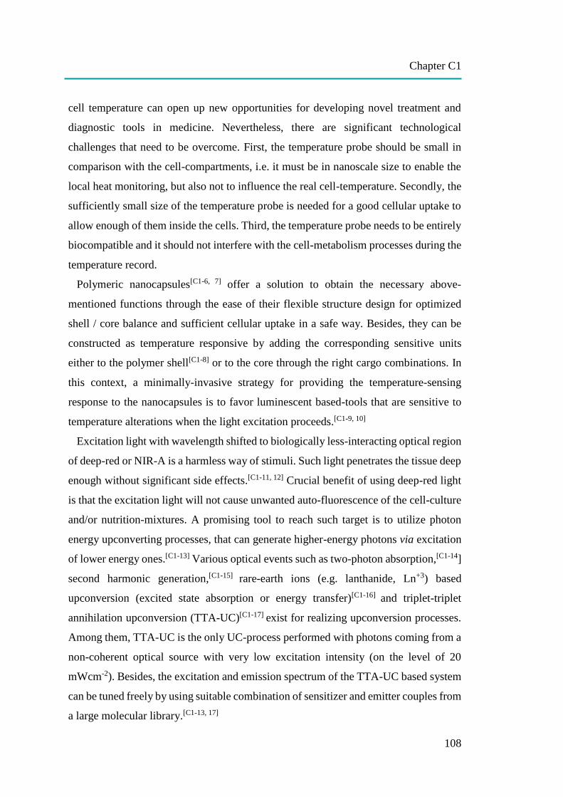
Chapter C1
108
cell temperature can open up new opportunities for developing novel treatment and
diagnostic tools in medicine. Nevertheless, there are significant technological
challenges that need to be overcome. First, the temperature probe should be small in
comparison with the cell-compartments, i.e. it must be in nanoscale size to enable the
local heat monitoring, but also not to influence the real cell-temperature. Secondly, the
sufficiently small size of the temperature probe is needed for a good cellular uptake to
allow enough of them inside the cells. Third, the temperature probe needs to be entirely
biocompatible and it should not interfere with the cell-metabolism processes during the
temperature record.
Polymeric nanocapsules[C1-6, 7] offer a solution to obtain the necessary above-
mentioned functions through the ease of their flexible structure design for optimized
shell / core balance and sufficient cellular uptake in a safe way. Besides, they can be
constructed as temperature responsive by adding the corresponding sensitive units
either to the polymer shell[C1-8] or to the core through the right cargo combinations. In
this context, a minimally-invasive strategy for providing the temperature-sensing
response to the nanocapsules is to favor luminescent based-tools that are sensitive to
temperature alterations when the light excitation proceeds.[C1-9, 10]
Excitation light with wavelength shifted to biologically less-interacting optical region
of deep-red or NIR-A is a harmless way of stimuli. Such light penetrates the tissue deep
enough without significant side effects.[C1-11, 12] Crucial benefit of using deep-red light
is that the excitation light will not cause unwanted auto-fluorescence of the cell-culture
and/or nutrition-mixtures. A promising tool to reach such target is to utilize photon
energy upconverting processes, that can generate higher-energy photons via excitation
of lower energy ones.[C1-13] Various optical events such as two-photon absorption,[C1-14]
second harmonic generation,[C1-15] rare-earth ions (e.g. lanthanide, Ln+3) based
upconversion (excited state absorption or energy transfer)[C1-16] and triplet-triplet
annihilation upconversion (TTA-UC)[C1-17] exist for realizing upconversion processes.
Among them, TTA-UC is the only UC-process performed with photons coming from a
non-coherent optical source with very low excitation intensity (on the level of 20
mWcm-2). Besides, the excitation and emission spectrum of the TTA-UC based system
can be tuned freely by using suitable combination of sensitizer and emitter couples from
a large molecular library.[C1-13, 17]

Chapter C1
109
The vast majority of the reported TTA-UC systems in the literature are hydrophobic;
therefore, the transfer of the annihilation upconversion process into water environment
is an unavoidable requirement when applications in the field of life-science are planned.
This has been partially achieved by incorporating the UC-dyes into dendrimers,[C1-18]
micelles,[C1-19] polymer,[C1-20-23] silica[C1-24] or protein[C1-25] based nanoparticles.
However, since the oxygen molecules penetrates into the nano-sized objects (even if
the penetration rate is slowed-down), oxygen quenching in the process of TTA-UC
optically excited triplet states still exists.[C1-26] In this regard, core/shell structured
nanoparticles (i.e. nanocapsules) enable an opportunity as their hydrophobic core can
be enriched by incorporating oxygen-scavenging substances. At presence of molecular
oxygen, the TTA-UC process is unavoidably related to generation of singlet oxygen,
known as very aggressive species. If the rate of oxygen “consumption“ (i.e. the rate of
covalent bonding of the singlet oxygen with the introduced singlet oxygen scavenging
moiety) is higher than the rate of oxygen penetration through the NC-shell, it is possible
to mimic “oxygen-free conditions”. So far, some examples of TTA-UC processing in
the presence of oxygen have been shown[C1-25, 27-30] and it should be further extended in
order to utilize this technique in medical applications like the construction of
nanothermometers.
To date, most reported luminescent nanothermometers are developed by use of
lanthanide based upconversion nanoparticles (UCNPs).[C1-31-33] In contrast, TTA-UC
based nanocapsules are mainly fabricated for bio-imaging applications[C1-22-25, 34] and
the construction of solar cell devices.[C1-29, 35] However, the intense developments of
polymeric nanocapsules and high knowledge of their biosafety can be combined with
the advantages of TTA-UC process for further advancement in this field. Thus, in this
paper, we demonstrate the synthesis of polymeric upconversion nanocapsules and use
them for temperature dependent in vitro TTA-UC process in HeLa cancer cells exposed
to ambient environment for applicability as nanothermometer. The shell of the
nanocapsules was selected as a biologically safe poly (methyl methacrylate) (PMMA)
and the NC-core was rice bran oil (RBO), which serves as efficient singlet oxygen
quencher. Scheme C1 illustrates the general approach towards in vitro temperature-
sensing in HeLa cells via TTA-UC, together with the chemical structures of the
encapsulated sensitizer and emitter couple.
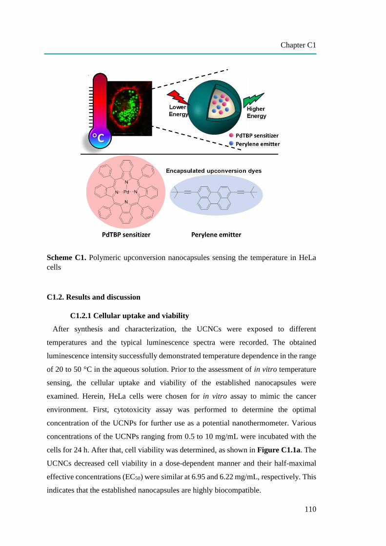
Chapter C1
110
Scheme C1. Polymeric upconversion nanocapsules sensing the temperature in HeLa
cells
C1.2. Results and discussion
C1.2.1 Cellular uptake and viability
After synthesis and characterization, the UCNCs were exposed to different
temperatures and the typical luminescence spectra were recorded. The obtained
luminescence intensity successfully demonstrated temperature dependence in the range
of 20 to 50 °C in the aqueous solution. Prior to the assessment of in vitro temperature
sensing, the cellular uptake and viability of the established nanocapsules were
examined. Herein, HeLa cells were chosen for in vitro assay to mimic the cancer
environment. First, cytotoxicity assay was performed to determine the optimal
concentration of the UCNPs for further use as a potential nanothermometer. Various
concentrations of the UCNPs ranging from 0.5 to 10 mg/mL were incubated with the
cells for 24 h. After that, cell viability was determined, as shown in Figure C1.1a. The
UCNCs decreased cell viability in a dose-dependent manner and their half-maximal
effective concentrations (EC50) were similar at 6.95 and 6.22 mg/mL, respectively. This
indicates that the established nanocapsules are highly biocompatible.

Chapter C1
111
Figure C1.1. (a) Cell viability assay of HeLa cells after treatment with the UCNCs for
24 h. (b) Confocal laser scanning microscopy (cLSM) images of HeLa cells incubated
with UCNC2 at 6 mg/mL for various incubation times. The cell membrane was stained
with CellMask™Orange and pseudocolored in red. The direct fluorescence emission
from encapsulated perylene dye was detected and pseudocolored in green. The scale
bar is 10 µm. Flow cytometry analysis showing (c) the percentage of fluorescence-
positive cells and (d) median fluorescence intensity (MFI) obtained from the direct
fluorescence emission of encapsulated perylene dye in UCNC1 and UCNC2 incubated
in HeLa cells after 24 h.
24 h 48 h1 h 8 h
1 2 3 4 5 6 7 8 9 10 110
20
40
60
80
100
120
Ce
ll V
iab
ility
(%
)
UCNCs Concentration (mg/mL)
UCNC1 UCNC2
0 0.5 1 2 2.5 3 4 5 6 8 10
1 2 3 4 5 6
0
5000
10000
15000
20000
25000
30000
Me
dia
n F
luo
resce
nce
in
ten
sity
UCNCs Concentration (mg/mL)
UCNC1 UCNC2
0 1 2 4 6 81 2 3 4 5 60
20
40
60
80
100
120
Flu
ore
sce
nce
po
sitiv
e c
ells
(%
)
UCNCs Concentration (mg/mL)
UCNC1 UCNC2
0 1 2 4 6 8
(a)
(b)
(c) (d)
Chapter C1
112
The next step was to study the cellular uptake of the corresponding nanocapsules
to select the sufficient loading amount for a good upconversion signal from the HeLa
cells. For this purpose, flow cytometry analysis was performed by following the direct
fluorescence emission of BDMBP dye as already present in UCNPs that was taken up
by the cells after 24 h. Figure C1.1c showed 100% of fluorescence positive-cells when
the concentration of the UCNPs has reached to 1 mg/mL. However, the median
fluorescence intensity (MFI) obtained from the cells treated with UCNC2 (containing
more amount of BDMBP dye) was higher than those from UCNC1 at the same
concentration of the nanocapsules as shown in Figure C1.1d. This stronger signal
exhibits the high potential of those nanocapsules to be used as a reporter in
physiological temperature measurement. Moreover, the MFI value was reached its
maximum level at 6 mg/mL which was not increased more in the case of higher
concentrations as observed for 8 mg/mL in Figure C1.1d. Therefore, the maximum
concentration that gave the highest fluorescence signal and still less toxic to the cell at
6 mg/mL for both UCNC1 and UCNC2 was selected for further temperature sensing
investigations in HeLa cells. Nevertheless, before proceeding to that final part, the
cellular uptake of UCNC2 at 6 mg/mL in HeLa cells was further investigated by
imaging with confocal laser scanning microscopy (cLSM) at different times of
incubation. Figure C1.1b showed the progress of cellular uptake process. At 1 h of
incubation, small amount of UCNPs surrounded the HeLa cells. Later at 8 h, more
nanocapsules were co-localized with the membrane and some of them were already
inside of the HeLa cells. After 24 h, high amount of the nanocapsules were taken up by
every cell and this status was still stable even after 48 h of incubation. Thus, this
confirmed not only the higher efficiency for the cellular uptake of the nanocapsules,
but also supported their biocompatibility to the cells.
C1.2.2 Temperature Sensing in HeLa Cells
After the confirmation of good cellular uptake and biocompatibility to the cells, we
have continued with the in vitro TTA-UC measurements to evaluate the potential of our
PMMA nanocapsules as a thermometer. Thus, the first investigation was to ensure the
sufficient upconversion efficiency at mild excitation power for acquiring the
temperature-sensing response when UCNCs are inside the HeLa cells. As mentioned
previously, 6 mg/mL of the UCNC1 and UCNC2 nanocapsules were uptaken by the
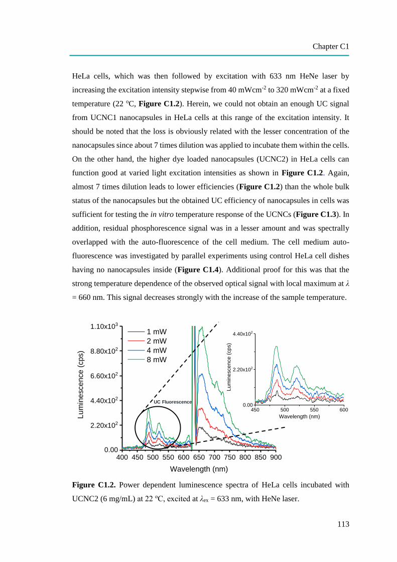
Chapter C1
113
HeLa cells, which was then followed by excitation with 633 nm HeNe laser by
increasing the excitation intensity stepwise from 40 mWcm-2 to 320 mWcm-2 at a fixed
temperature (22 oC, Figure C1.2). Herein, we could not obtain an enough UC signal
from UCNC1 nanocapsules in HeLa cells at this range of the excitation intensity. It
should be noted that the loss is obviously related with the lesser concentration of the
nanocapsules since about 7 times dilution was applied to incubate them within the cells.
On the other hand, the higher dye loaded nanocapsules (UCNC2) in HeLa cells can
function good at varied light excitation intensities as shown in Figure C1.2. Again,
almost 7 times dilution leads to lower efficiencies (Figure C1.2) than the whole bulk
status of the nanocapsules but the obtained UC efficiency of nanocapsules in cells was
sufficient for testing the in vitro temperature response of the UCNCs (Figure C1.3). In
addition, residual phosphorescence signal was in a lesser amount and was spectrally
overlapped with the auto-fluorescence of the cell medium. The cell medium auto-
fluorescence was investigated by parallel experiments using control HeLa cell dishes
having no nanocapsules inside (Figure C1.4). Additional proof for this was that the
strong temperature dependence of the observed optical signal with local maximum at λ
= 660 nm. This signal decreases strongly with the increase of the sample temperature.
Figure C1.2. Power dependent luminescence spectra of HeLa cells incubated with
UCNC2 (6 mg/mL) at 22 oC, excited at λex = 633 nm, with HeNe laser.
400 450 500 550 600 650 700 750 800 850 9000.00
2.20x10²
4.40x10²
6.60x10²
8.80x10²
1.10x10³
Lu
min
escen
ce
(cps)
Wavelength (nm)
1 mW
2 mW
4 mW
8 mW
UC Fluorescence
450 500 550 6000.00
2.20x10²
4.40x10²
Lum
inesce
nce (
cps)
Wavelength (nm)
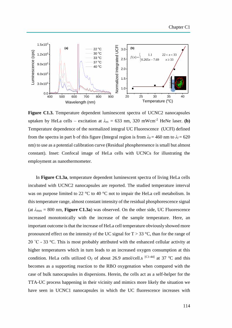
Chapter C1
114
Figure C1.3. Temperature dependent luminescent spectra of UCNC2 nanocapsules
uptaken by HeLa cells – excitation at λex = 633 nm, 320 mWcm-2 HeNe laser. (b)
Temperature dependence of the normalized integral UC Fluorescence (UCFl) defined
from the spectra in part b of this figure (Integral region is from λ0 = 460 nm to λf = 620
nm) to use as a potential calibration curve (Residual phospheresence is small but almost
constant). Inset: Confocal image of HeLa cells with UCNCs for illustrating the
employment as nanothermometer.
In Figure C1.3a, temperature dependent luminescent spectra of living HeLa cells
incubated with UCNC2 nanocapsules are reported. The studied temperature interval
was on purpose limited to 22 °C to 40 °C not to impair the HeLa cell metabolism. In
this temperature range, almost constant intensity of the residual phosphorescence signal
(at λmax = 800 nm, Figure C1.3a) was observed. On the other side, UC Fluorescence
increased monotonically with the increase of the sample temperature. Here, an
important outcome is that the increase of HeLa cell temperature obviously showed more
pronounced effect on the intensity of the UC signal for T > 33 °C, than for the range of
20 ˚C - 33 °C. This is most probably attributed with the enhanced cellular activity at
higher temperatures which in turn leads to an increased oxygen consumption at this
condition. HeLa cells utilized O2 of about 26.9 amol/cell.s [C1-44] at 37 oC and this
becomes as a supporting reaction to the RBO oxygenation when compared with the
case of bulk nanocapsules in dispersions. Herein, the cells act as a self-helper for the
TTA-UC process happening in their vicinity and mimics more likely the situation we
have seen in UCNC1 nanocapsules in which the UC fluorescence increases with
oC
400 500 600 700 800 9000.0
3.0x10²
6.0x10²
9.0x10²
1.2x10³
1.5x10³
Lu
min
esce
nce
(cp
s)
Wavelength (nm)
22 oC
30 oC
33 oC
37 oC
40 oC
(a) (b)
20 25 30 35 40
1.0
1.5
2.0
2.5
3.0
No
rma
lize
d I
nte
gra
ted U
CF
l
Temperature (oC)
1.1 22 33( )
0.265 7.69 33
xf x
x x
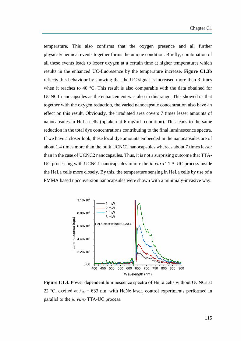
Chapter C1
115
temperature. This also confirms that the oxygen presence and all further
physical/chemical events together forms the unique condition. Briefly, combination of
all these events leads to lesser oxygen at a certain time at higher temperatures which
results in the enhanced UC-fluoresence by the temperature increase. Figure C1.3b
reflects this behaviour by showing that the UC signal is increased more than 3 times
when it reaches to 40 °C. This result is also comparable with the data obtained for
UCNC1 nanocapsules as the enhancement was also in this range. This showed us that
together with the oxygen reduction, the varied nanocapsule concentration also have an
effect on this result. Obviously, the irradiated area covers 7 times lesser amounts of
nanocapsules in HeLa cells (uptaken at 6 mg/mL condition). This leads to the same
reduction in the total dye concentrations contributing to the final luminescence spectra.
If we have a closer look, these local dye amounts embeeded in the nanocapsules are of
about 1.4 times more than the bulk UCNC1 nanocapsules whereas about 7 times lesser
than in the case of UCNC2 nanocapsules. Thus, it is not a surprising outcome that TTA-
UC processing with UCNC1 nanocapsules mimic the in vitro TTA-UC process inside
the HeLa cells more closely. By this, the temperature sensing in HeLa cells by use of a
PMMA based upconversion nanocapsules were shown with a minimaly-invasive way.
Figure C1.4. Power dependent luminescence spectra of HeLa cells without UCNCs at
22 oC, excited at λex = 633 nm, with HeNe laser, control experiments performed in
parallel to the in vitro TTA-UC process.
400 450 500 550 600 650 700 750 800 850 900
0.00
2.20x10²
4.40x10²
6.60x10²
8.80x10²
1.10x10³
1 mW
2 mW
4 mW
8 mW
Lum
inescence (
cps)
Wavelength (nm)
HeLa cells without UCNCS
Chapter C1
116
C1.3. Conclusion
In the present work, we have demonstrated polymeric upconversion nanocapsules that
utilized TTA-UC based temperature responsivity for potential use as minimally-
invasive nanothermometers in living cells. The established nanocapsules consist of a
PMMA shell and a RBO core hosting the PdTBP sensitizer and BDMBP emitter.
Different amounts of dye encapsulation were shown to perform TTA-UC at aqueous
medium and for further applications in temperature probing of a biological
environment. The UCNCs can function at different levels of oxygen presence including
the fully air saturated condition that supports the antioxidant capacity with the aid of an
unsaturated fatty acids in the RBO core. In addition, the good cellular uptake and
biocompatibility towards HeLa cancer cells and further temperature sensing through in
vitro TTA-UC process with a mild excitation intensity by red light opens up new
opportunities for the design of nanothermometers to gain information about the cellular
activities. This exhibits further advances in medical applications for especially early
diagnosis and treatment of the diseases like cancer.
C1.4. References
[C1-1] Hildebrandt, B.; Wust, P.; Ahlers, O.; Dieing, A.; Sreenivasa, G.; Kerner, T.; Felix, R.; Riess,
H., The cellular and molecular basis of hyperthermia. Critical Reviews in
Oncology/Hematology 2002, 43, 33-56. Lowell, B. B.; Spiegelman, B. M., Towards a molecular
understanding of adaptive thermogenesis. Nature 2000, 404, 652.
[C1-2] Lowell, B. B.; Spiegelman, B. M., Towards a molecular understanding of adaptive
thermogenesis. Nature 2000, 404, 652.
[C1-3] Rao, P. N.; Engelberg, J., HeLa Cells: Effects of Temperature on the Life Cycle. Science 1965,
148, 1092-1094.
[C1-4] Monti, M.; Brandt, L.; Ikomi-Kumm, J.; Olsson, H., Microcalorimetric investigation of cell
metabolism in tumour cells from patients with non-Hodgkin lymphoma (NHL). Scandinavian
Journal of Haematology 1986, 36, 353-357.
[C1-5] Karnebogen, M.; Singer, D.; Kallerhoff, M.; Ringert, R. H., Microcalorimetric investigations
on isolated tumorous and non-tumorous tissue samples. Thermochim. Acta 1993, 229, 147-155.
[C1-6] Cui, J.; van Koeverden, M. P.; Müllner, M.; Kempe, K.; Caruso, F., Emerging methods for the
fabrication of polymer capsules. Adv. Colloid Interface Sci. 2014, 207, 14-31.
[C1-7] Musyanovych, A.; Landfester, K., Polymer Micro- and Nanocapsules as Biological Carriers
with Multifunctional Properties. Macromolecular Bioscience 2014, 14, 458-477.
Chapter C1
117
[C1-8] Schmaljohann, D., Thermo- and pH-responsive polymers in drug delivery. Advanced Drug
Delivery Reviews 2006, 58, 1655-1670.
[C1-9] Jaque, D.; Rosal, B. d.; Rodríguez, E. M.; Maestro, L. M.; Haro-González, P.; Solé, J. G.,
Fluorescent nanothermometers for intracellular thermal sensing. Nanomedicine 2014, 9, 1047-
1062.
[C1-10] Jaque, D.; Vetrone, F., Luminescence nanothermometry. Nanoscale 2012, 4, 4301-4326.
[C1-11] Frangioni, J. V., In vivo near-infrared fluorescence imaging. Curr. Opin. Chem. Biol. 2003, 7,
626-634.
[C1-12] Stolik, S.; Delgado, J. A.; Pérez, A.; Anasagasti, L., Measurement of the penetration depths of
red and near infrared light in human “ex vivo” tissues. Journal of Photochemistry and
Photobiology B: Biology 2000, 57, 90-93.
[C1-13] Zhou, J.; Liu, Q.; Feng, W.; Sun, Y.; Li, F., Upconversion Luminescent Materials: Advances
and Applications. Chem. Rev. 2015, 115, 395-465.
[C1-14] Denk, W.; Strickler, J.; Webb, W., Two-photon laser scanning fluorescence microscopy.
Science 1990, 248, 73-76.
[C1-15] Clark, H. A.; Campagnola, P. J.; Wuskell, J. P.; Lewis, A.; Loew, L. M., Second Harmonic
Generation Properties of Fluorescent Polymer-Encapsulated Gold Nanoparticles. J. Am. Chem.
Soc. 2000, 122, 10234-10235.
[C1-16] Chen, G.; Qiu, H.; Prasad, P. N.; Chen, X., Upconversion Nanoparticles: Design,
Nanochemistry, and Applications in Theranostics. Chem. Rev. 2014, 114, 5161-5214.
[C1-17] Simon, Y. C.; Weder, C., Low-power photon upconversion through triplet–triplet annihilation
in polymers. J. Mater. Chem. 2012, 22, 20817-20830.
[C1-18] Roussakis, E.; Li, Z.; Nowell, N. H.; Nichols, A. J.; Evans, C. L., Bright, “Clickable” Porphyrins
for the Visualization of Oxygenation under Ambient Light. Angew. Chem. Int. Ed. 2015, 54,
14728-14731.
[C1-19] Andrey, T.; Dmitry, B.; Stanislav, B.; Tzenka, M.; Katharina, L., Micellar carrier for triplet–
triplet annihilation-assisted photon energy upconversion in a water environment. New Journal
of Physics 2011, 13, 083035.
[C1-20] Monguzzi, A.; Frigoli, M.; Larpent, C.; Tubino, R.; Meinardi, F., Low-Power-Photon Up-
Conversion in Dual-Dye-Loaded Polymer Nanoparticles. Adv. Funct. Mater. 2012, 22, 139-
143.
[C1-21] Thévenaz, D. C.; Monguzzi, A.; Vanhecke, D.; Vadrucci, R.; Meinardi, F.; Simon, Y. C.;
Weder, C., Thermoresponsive low-power light upconverting polymer nanoparticles. Materials
Horizons 2016, 3, 602-607.
[C1-22] Wohnhaas, C.; Turshatov, A.; Mailänder, V.; Lorenz, S.; Baluschev, S.; Miteva, T.; Landfester,
K., Annihilation Upconversion in Cells by Embedding the Dye System in Polymeric
Nanocapsules. Macromolecular Bioscience 2011, 11, 772-778.
Chapter C1
118
[C1-23] Wohnhaas, C.; Mailänder, V.; Dröge, M.; Filatov, M. A.; Busko, D.; Avlasevich, Y.; Baluschev,
S.; Miteva, T.; Landfester, K.; Turshatov, A., Triplet–Triplet Annihilation Upconversion Based
Nanocapsules for Bioimaging Under Excitation by Red and Deep-Red Light. Macromolecular
Bioscience 2013, 13, 1422-1430.
[C1-24] Kwon, O. S.; Song, H. S.; Conde, J.; Kim, H.-i.; Artzi, N.; Kim, J.-H., Dual-Color Emissive
Upconversion Nanocapsules for Differential Cancer Bioimaging In Vivo. ACS Nano 2016, 10,
1512-1521.
[C1-25] Liu, Q.; Yin, B.; Yang, T.; Yang, Y.; Shen, Z.; Yao, P.; Li, F., A General Strategy for
Biocompatible, High-Effective Upconversion Nanocapsules Based on Triplet–Triplet
Annihilation. J. Am. Chem. Soc. 2013, 135, 5029-5037.
[C1-26] Baluschev, S.; Katta, K.; Avlasevich, Y.; Landfester, K., Annihilation upconversion in
nanoconfinement: solving the oxygen quenching problem. Materials Horizons 2016, 3, 478-
486.
[C1-27] Kim, J.-H.; Kim, J.-H., Encapsulated Triplet–Triplet Annihilation-Based Upconversion in the
Aqueous Phase for Sub-Band-Gap Semiconductor Photocatalysis. J. Am. Chem. Soc. 2012,
134, 17478-17481.
[C1-28] Ye, C.; Wang, B.; Hao, R.; Wang, X.; Ding, P.; Tao, X.; Chen, Z.; Liang, Z.; Zhou, Y., Oil-in-
water microemulsion: an effective medium for triplet–triplet annihilated upconversion with
efficient triplet acceptors. Journal of Materials Chemistry C 2014, 2, 8507-8514.
[C1-29] Svagan, A. J.; Busko, D.; Avlasevich, Y.; Glasser, G.; Baluschev, S.; Landfester, K., Photon
Energy Upconverting Nanopaper: A Bioinspired Oxygen Protection Strategy. ACS Nano 2014,
8, 8198-8207.
[C1-30] Marsico, F.; Turshatov, A.; Peköz, R.; Avlasevich, Y.; Wagner, M.; Weber, K.; Donadio, D.;
Landfester, K.; Baluschev, S.; Wurm, F. R., Hyperbranched Unsaturated Polyphosphates as a
Protective Matrix for Long-Term Photon Upconversion in Air. J. Am. Chem. Soc. 2014, 136,
11057-11064.
[C1-31] Vetrone, F.; Naccache, R.; Zamarrón, A.; Juarranz de la Fuente, A.; Sanz-Rodríguez, F.;
Martinez Maestro, L.; Martín Rodriguez, E.; Jaque, D.; García Solé, J.; Capobianco, J. A.,
Temperature Sensing Using Fluorescent Nanothermometers. ACS Nano 2010, 4, 3254-3258.
[C1-32] Sedlmeier, A.; Achatz, D. E.; Fischer, L. H.; Gorris, H. H.; Wolfbeis, O. S., Photon
upconverting nanoparticles for luminescent sensing of temperature. Nanoscale 2012, 4, 7090-
7096.
[C1-33] Fischer, L. H.; Harms, G. S.; Wolfbeis, O. S., Upconverting Nanoparticles for Nanoscale
Thermometry. Angew. Chem. Int. Ed. 2011, 50, 4546-4551.
[C1-34] Tian, B.; Wang, Q.; Su, Q.; Feng, W.; Li, F., In vivo biodistribution and toxicity assessment of
triplet-triplet annihilation-based upconversion nanocapsules. Biomaterials 2017, 112, 10-19.
Chapter C1
119
[C1-35] Wohnhaas, C.; Friedemann, K.; Busko, D.; Landfester, K.; Baluschev, S.; Crespy, D.;
Turshatov, A., All Organic Nanofibers As Ultralight Versatile Support for Triplet–Triplet
Annihilation Upconversion. ACS Macro Letters 2013, 2, 446-450.
[C1-36] Ju, Y.-H.; Vali, S. R., Rice bran oil as a potential resource for biodiesel: a review. 2005.
[C1-37] Bettencourt, A.; Almeida, A. J., Poly(methyl methacrylate) particulate carriers in drug delivery.
J. Microencapsulation 2012, 29, 353-367.
[C1-38] Keivanidis, P. E.; Baluschev, S.; Lieser, G.; Wegner, G., Inherent Photon Energy Recycling
Effects in the Up-Converted Delayed Luminescence Dynamics of Poly(fluorene)–PtIIoctaethyl
Porphyrin Blends. ChemPhysChem 2009, 10, 2316-2326.
[C1-39] Nazarova, N. V.; Avlasevich, Y. S.; Landfester, K.; Baluschev, S., Stimuli-responsive
protection of optically excited triplet ensembles against deactivation by molecular oxygen.
Dalton Transactions 2018, 47, 8605-8610.
[C1-40] Stich, M. I. J.; Fischer, L. H.; Wolfbeis, O. S., Multiple fluorescent chemical sensing and
imaging. Chem. Soc. Rev. 2010, 39, 3102-3114.
[C1-41] Filatov, M. A.; Heinrich, E.; Busko, D.; Ilieva, I. Z.; Landfester, K.; Baluschev, S., Reversible
oxygen addition on a triplet sensitizer molecule: protection from excited state depopulation.
PCCP 2015, 17, 6501-6510.
[C1-42] Pope, M.; Swenberg, C. E., Electronic processes in organic crystals and polymers. Oxford
University Press New York: 1999; Vol. 2.
[C1-43] Duan, P.; Yanai, N.; Nagatomi, H.; Kimizuka, N., Photon Upconversion in Supramolecular Gel
Matrixes: Spontaneous Accumulation of Light-Harvesting Donor–Acceptor Arrays in
Nanofibers and Acquired Air Stability. J. Am. Chem. Soc. 2015, 137, 1887-1894.
[C1-44] Wagner, B. A.; Venkataraman, S.; Buettner, G. R., The rate of oxygen utilization by cells. Free
Radical Biol. Med. 2011, 51, 700-712.
[C1-45] Filatov, M. A.; Baluschev, S.; Ilieva, I. Z.; Enkelmann, V.; Miteva, T.; Landfester, K.;
Aleshchenkov, S. E.; Cheprakov, A. V., Tetraaryltetraanthra[2,3]porphyrins: Synthesis,
Structure, and Optical Properties. The Journal of Organic Chemistry 2012, 77, 11119-11131.
[C1-46] Cheprakov, A. V.; Filatov, M. A., The dihydroisoindole approach to linearly annelated π-
extended porphyrins. J. Porphyrins Phthalocyanines 2009, 13, 291-303.

Materials and Methods
120
Materials and Methods
General protocols applied in all chapters are combined and specific protocols
needed for each chapter are separately described. The other experimental methods that
was done by the collaborators can be found in the original online publications.
GENERAL PROTOCOLS FOR ALL CHAPTERS
1. Adherent Cell Culture
Adherent cells including HeLa, LNCaP, PC3 and SAOS-2 were obtained from
DSMZ (Deutsche Sammlung von Mikroorganismen und Zellkulturen, Germany). They
were cultured in Dulbecco's Modified Eagle's Medium (DMEM, Gibco, USA)
complete medium containing 10% fetal bovine serum (FBS, Gibco, USA), 1% L-
glutamine (Gibco, USA) and 1% penicillin/streptomycin (Gibco, USA) and incubated
at 37 oC in CO2-incubator with 95% humidity and 5% CO2 (C200, Labotect, Germany).
To dissociate adherent cells, the cells were trypsinized with 0.25% trypsin (Gibco,
USA) for 3 min as general procedure. The cell pellet was collected by centrifugation at
130 g for 3 min, resuspended in DMEM complete medium and used for further assays.
Viable cells were determined by trypan blue exclusion method and counted by using
TC10™ automated cell counter (Bio-Rad, USA).
For uptake and toxicity study, adherent cells resuspended in DMEM complete
medium were seeded at a density of 6,400 cells per well in 96-well plate or 8 × 10⁴ cells
per well in 24-well plate for 48 h. For cLSM imaging, adherent cells were seeded at a
density of 2 x 10⁴ cells per well in µ–Slide 8 well with a glass coverslip bottom (Ibidi,
Germany) and cultured for 24 h in DMEM complete medium (Phenol red free).
2. Cell Viability Assay determined by CellTiter-Glo® Luminescent Assay
After cell preparation, the cells were treated with various concentrations of the
corresponding samples for 24 h. Sample without any treatment was used as a negative
control and calculated as 100% cell viability, while 20% DMSO added sample was used
as a positive control. After cell treatment, cell viability was evaluated by using the
CellTiter-Glo® luminescent cell viability assay (Promega, USA) according to the
manufacturer’s protocol. This assay is based on the amount of ATP present, which
Materials and Methods
121
reflect the presence of metabolically active cells. Luminescence was recorded 10
minutes after reagent addition using plate reader (Infinite® M1000, Tecan, Germany).
The half maximal effective concentration (EC50) values were calculated by fitting a
curve with non-linear regression using GraphPad Program.
3. Cellular Uptake Study by Flow Cytometry
After cell treatment, the cells were washed and resuspended in PBS. Flow
cytometry measurements were performed on a Attune™ NxT Flow Cytometer
(Invitrogen, USA). Zombie Aqua™ dye (BioLegend, USA) was used for live cell
gating with the excitation of the violet laser (405 nm) and detected in channel VL-2.
All fluorophores labeling nanocapsules and dyes conjugating antibodies are excited and
recorded according to their excitation and emission profiles. Data analysis was
performed using Attune™ NxT software (Invitrogen, USA) by selecting the cells on a
forward/sideward scatter plot, thereby excluding cell debris. These gated events were
shown by the histogram of fluorescent signal. Percentages of cell viability were
recorded from living cells (Zombie Aqua negative-cells) compared to dead cells
(Zombie Aqua positive-cells). After gating for living cells, percentages of fluorescence
positive-cells or their median fluorescence intensities (MFI) were reported.
4. Cell Imaging by Confocal Laser Scanning Microscopy (cLSM)
After cell treatment, adherent cells were washed twice with DMEM to remove
the remaining nanocapsules outside of the cells. T-cells were collected from the plate,
washed and transferred to µ–Slide 8 well with a glass coverslip bottom (Ibidi,
Germany). Then the cells were stained subcellular organs with fluorescent dyes and
finally suspended in DMEM. Live cell images were taken with a commercial setup
(LSM SP5 STED Leica Laser Scanning Confocal Microscope, Leica, Germany),
consisting of an inverse fluorescence microscope DMI 6000 CS equipped with a multi-
laser combination, five detectors operating in the range of 400-800 nm. A HCX PL
APO CS 63 x 1.4 oil objective was used in this study. The excitation and detection
conditions in a sequential mode were described as follows: green fluorescence labeling
such as FITC or Perylene dye were excited with an Ar laser (488 nm), detected at 510-
540 nm; The cell membrane was stained with CellMask™Orange (5 µg/mL, Life

Materials and Methods
122
technologies, USA), excited with a DPSS laser (561 nm), detected at 570-600 nm; The
cell nucleus was stained with DraQ®5 (5 µM, Cell signaling technology, USA) or red
fluorescence labeling such as Cy5, excited with a HeNe laser (633 nm), detected at 650-
710 nm.
5. Co-Localization Assay with Lysosome visualized by cLSM
To provide more evidences for cellular uptake, co-localization assay of
nanocapsules and lysosome was investigated. After cell treatment, LysoTracker®
Green DND-26 or LysoTracker® Red DND-99 (50 nM diluted in DMEM, Life
Technologies, USA) was used to stain lysosome for 1 h. Then the subcellular organs
were stained with fluorescent dyes and live cell images were taken as described above.
SPECIFIC PROTOCOLS FOR CHAPTER A
A.1. T-cell Isolation and Culture
Peripheral blood mononuclear cells (PBMCs) were collected from blood of
healthy donors by using lymphocyte separation medium (Histopaque®-1077, Sigma-
Aldrich, USA). Human PBMCs were co-stimulated with immobilized 0.1 µg/mL of
anti-human CD3 (Clone OKT3, functional grade, eBioscience, Germany) and 100
U/mL recombinant human IL-2 (Novartis, Switzerland) in Roswell Park Memorial
Institute (RPMI, Gibco, USA) complete medium containing 10% fetal bovine serum
(FBS, Gibco, USA), 1% L-Glutamine (Gibco, USA) and 1% penicillin/streptomycin
(Gibco, USA) according to T-cell activation, in vitro protocol from eBioscience. After
cell aggregation was formed on the bottom of the flask, CD8+ T-cells were harvested
from the culture by CD8 MicroBead and separated through immunomagnetically
MACS separation column (Miltenyi Biotec, Germany). CD8+ T-cells were cultured in
RPMI complete medium and restimulated with anti-CD3 and IL-2 as mentioned above
and incubated at 37oC in CO2-incubator (C200, Labotect, Germany). The cell pellet was
collected by centrifugation at 500 g for 5 min, resuspended in RPMI medium and used
for further assays. Viable cells were determined by trypan blue exclusion method and
counted by using TC10™ automated cell counter (Bio-Rad, USA).
Materials and Methods
123
For uptake and toxicity study, CD8+ T-cells resuspended in RPMI medium
containing 1% FBS and 100 U/mL IL-2 without antibiotics were added into 0.1 µg/mL
of anti-CD3 immobilized 24-well plate at a density of 200,000 cells per well. Then,
various concentrations of SiNCs were incubated with the cells for 24 h. Then, the
samples were analyzed by flow cytometer and further confirmed by cLSM.
SPECIFIC PROTOCOLS FOR CHAPTER A1
A1.1. Synthesis of silica nanocapsules (synthesized by Shuai Jiang)
Silica nanocapsules were synthesized in an oil-in-water miniemulsion by using
the surface of oil nanodroplets as template for the hydrolysis and condensation
of alkoxysilanes. Specifically, 2.0 g (9.6 mmol) of TEOS was first mixed with 125 mg
of hexadecane and 1 g of organic solvent (olive oil or chloroform) to form the oil phase
for the synthesis of NCs with hydrodynamic diameter of ~150 nm. In order to tune the
size of NCs, 400 vol%, 200 vol%, 50 vol%, or 25 vol% of the above solution was used
as oil phase. In the second step, 30 mL of 0.77 mg·mL-1 aqueous solution of CTMA-Cl
was poured into the oil mixture under stirring. After a pre-emulsification step by stirring
at 1000 rpm for 1 h, the obtained emulsion was sonicated by using a Branson 450
W sonifier with a 1/2’’ tip at 70% amplitude for 180 s (30 s of sonication, 10 s of pause)
with ice cooling. The resulting miniemulsion was stirred at 1000 rpm for 12 h at room
temperature to obtain an aqueous dispersion of SiNCs. In the case
of nanocapsules containing chloroform as core liquid, the dispersion was further stirred
at air for 24 h to evaporate the chloroform, during which the water diffuses in for filling
the core.
To obtain NCs stabilized with nonionic surfactant Lutensol AT50, 40 mg of
Lutensol AT50 was added to 2 mL of NC dispersion. The mixture was stirred at 1000
rpm for 2 h and then dialyzed against water within a dialysis tube with MWCO of 1000
g/mol. In this case, CTMA-Cl (Mw = 320 g/mol) could diffuse through the dialysis
membrane into the aqueous dialysis medium while Lutensol AT50 (Mw = 2460 g/mol)
was kept inside.
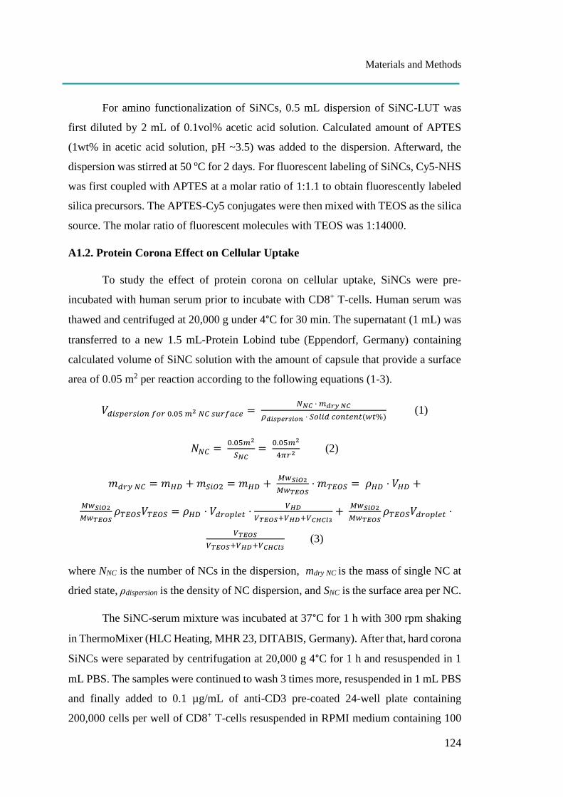
Materials and Methods
124
For amino functionalization of SiNCs, 0.5 mL dispersion of SiNC-LUT was
first diluted by 2 mL of 0.1vol% acetic acid solution. Calculated amount of APTES
(1wt% in acetic acid solution, pH ~3.5) was added to the dispersion. Afterward, the
dispersion was stirred at 50 oC for 2 days. For fluorescent labeling of SiNCs, Cy5-NHS
was first coupled with APTES at a molar ratio of 1:1.1 to obtain fluorescently labeled
silica precursors. The APTES-Cy5 conjugates were then mixed with TEOS as the silica
source. The molar ratio of fluorescent molecules with TEOS was 1:14000.
A1.2. Protein Corona Effect on Cellular Uptake
To study the effect of protein corona on cellular uptake, SiNCs were pre-
incubated with human serum prior to incubate with CD8+ T-cells. Human serum was
thawed and centrifuged at 20,000 g under 4°C for 30 min. The supernatant (1 mL) was
transferred to a new 1.5 mL-Protein Lobind tube (Eppendorf, Germany) containing
calculated volume of SiNC solution with the amount of capsule that provide a surface
area of 0.05 m2 per reaction according to the following equations (1-3).
𝑉𝑑𝑖𝑠𝑝𝑒𝑟𝑠𝑖𝑜𝑛 𝑓𝑜𝑟 0.05 𝑚2 𝑁𝐶 𝑠𝑢𝑟𝑓𝑎𝑐𝑒 = 𝑁𝑁𝐶 · 𝑚𝑑𝑟𝑦 𝑁𝐶
𝜌𝑑𝑖𝑠𝑝𝑒𝑟𝑠𝑖𝑜𝑛 · 𝑆𝑜𝑙𝑖𝑑 𝑐𝑜𝑛𝑡𝑒𝑛𝑡(𝑤𝑡%) (1)
𝑁𝑁𝐶 = 0.05𝑚2
𝑆𝑁𝐶=
0.05𝑚2
4𝜋𝑟2 (2)
𝑚𝑑𝑟𝑦 𝑁𝐶 = 𝑚𝐻𝐷 + 𝑚𝑆𝑖𝑂2 = 𝑚𝐻𝐷 + 𝑀𝑤𝑆𝑖𝑂2
𝑀𝑤𝑇𝐸𝑂𝑆· 𝑚𝑇𝐸𝑂𝑆 = 𝜌𝐻𝐷 · 𝑉𝐻𝐷 +
𝑀𝑤𝑆𝑖𝑂2
𝑀𝑤𝑇𝐸𝑂𝑆𝜌𝑇𝐸𝑂𝑆𝑉𝑇𝐸𝑂𝑆 = 𝜌𝐻𝐷 · 𝑉𝑑𝑟𝑜𝑝𝑙𝑒𝑡 ·
𝑉𝐻𝐷
𝑉𝑇𝐸𝑂𝑆+𝑉𝐻𝐷+𝑉𝐶𝐻𝐶𝑙3+
𝑀𝑤𝑆𝑖𝑂2
𝑀𝑤𝑇𝐸𝑂𝑆𝜌𝑇𝐸𝑂𝑆𝑉𝑑𝑟𝑜𝑝𝑙𝑒𝑡 ·
𝑉𝑇𝐸𝑂𝑆
𝑉𝑇𝐸𝑂𝑆+𝑉𝐻𝐷+𝑉𝐶𝐻𝐶𝑙3 (3)
where NNC is the number of NCs in the dispersion, mdry NC is the mass of single NC at
dried state, ρdispersion is the density of NC dispersion, and SNC is the surface area per NC.
The SiNC-serum mixture was incubated at 37°C for 1 h with 300 rpm shaking
in ThermoMixer (HLC Heating, MHR 23, DITABIS, Germany). After that, hard corona
SiNCs were separated by centrifugation at 20,000 g 4°C for 1 h and resuspended in 1
mL PBS. The samples were continued to wash 3 times more, resuspended in 1 mL PBS
and finally added to 0.1 µg/mL of anti-CD3 pre-coated 24-well plate containing
200,000 cells per well of CD8+ T-cells resuspended in RPMI medium containing 100
Materials and Methods
125
U/mL IL-2 without antibiotics in the presence of different concentrations of human
serum at 0%, 1% and 10%. To keep the cells in a good condition, the human serum was
added to the group of 0% to obtain 1% final concentration after 6 h. The uptake of
uncoated SiNC in the presence of 1% FBS was used to compare with the pre-coated
SiNCs uptake in the presence of 0%, 1% and 10% human serum. After 24 h of
incubation, the cells were collected and determined for cell viability and Cy5 positive-
cells by flow cytometer as described above.
A1.3. Protein Pattern Analysis by SDS-PAGE
After separating and 3 times washing of hard corona SiNCs, the capsule pellet
was resuspended in 100 µL of desorption buffer containing 2% SDS, 62.5 mM Tris-
HCl and incubated at 95°C for 5 min. After centrifugation at 20,000 g 4°C for 1 h, the
supernatant containing the protein absorbed on the surface of the capsules was collected
and kept at -20°C until used. The protein concentration was determined by Pierce™
660nm Protein Assay (Pierce, USA) according to the manufacturer’s instruction. The
total amount of protein at 1.5 µg of each sample was loaded onto pre-cast Bolt™ 10%
Bis-Tris Plus Gel (Invitrogen, USA) and separated in MES SDS running buffer
(Invitrogen, USA) at 100 volts for 1 h 15 min. Then, the gel was stained with
SilverQuest™ Silver Staining Kit (Invitrogen, USA) according to the manufacturer’s
protocol.
SPECIFIC PROTOCOLS FOR CHAPTER A2
A2.1. Synthesis of silica nanocapsules (synthesized by Mengyi Li)
The silica nanocapsules were synthesized in a water-in-oil miniemulsion. For
NC-1, 7 mg of NaCl and 20 µL of 25 wt% CTMA-Cl aqueous solution were dissolved
in 0.5 mL of milli-Q water as dispersed (water) phase. For continuous (oil) phase, 5 mg
of dodecylamine and 35 mg of P(E/B-b-EO) were dissolved in 7.5 mL of cyclohexane
and the mixture was added to the aqueous phase under stirring at 500 rpm for 10 min
at room temperature. The emulsion was then sonicated under ice cooling for 180 s at
70% amplitude in a pulse regime (20 s sonication, 10 s pause) using a Branson 450 W
Materials and Methods
126
sonifier and a 1/2′′ tip. A cyclohexane solution containing 10 mg of P(E/B-b-EO) and
250 µL TEOS in 5 mL of cyclohexane was then added dropwise over a period of 20
min to the miniemulsion. The mixture was stirred at 25 °C for 24 h. For NC-2, an
aqueous solution (0.5 mL) with pH 10.3 tuned by ammonia and containing 0.5 mg/mL
of NaCl was used as the dispersed phase. A cyclohexane solution (6 mL) containing
160 mg PGPR surfactant was added to the water phase under stirring at 500 rpm for 10
min. The following sonication process for NC-2 is the same as that for NC-1.
The synthesized nanocapsules were purified by repetitive centrifugation (3 times for
20 min, RCF 1664) and redispersion in cyclohexane to remove the excess amount of
surfactant. For transferring the nanocapsules into aqueous media, 600 µL of
nanocapsule dispersion in cyclohexane were added dropwise to 5 mL of Lutensol AT50
aqueous solution (1 wt%) under mechanical stirring, and the samples were placed in an
ultrasound bath for 3 min at 25 °C (25 kHz). Subsequently, the samples were stirred
openly at 25 °C for 24 h to evaporate cyclohexane.
A2.2. Materials
All siRNAs and primers were purchased from Sigma. The sequences were as
follows: PD-L1 siRNAs (sense: 5-GGAUAAGAACAUUAUUCAA[dT][dT]-3,
antisense: 5-UUGAAUAAUGUUCUUAUCC[dT][dT]-3), PD-L1 primers (forward:
5-TATGGTGGTGCCGACTACAA-3, reverse: 5-TGCTTGTCCAGATGACTTCG -
3 ) and actin primers (forward: 5-TTGCCGACAGGATGCAGAA -3 , reverse: 5-
GCCGATCCACACGGAGTACT-3 ). MISSION® siRNA Universal Negative
Control #1 was used as siRNA control. All antibodies were purchased from BioLegend
(USA): anti-CD274-APC (anti-PD-L1, clone 29E.2A3), anti-CD25-FITC (clone
BC96,) and anti-CD71-APC (clone CY1G4).
A2.3. PD-L1 Knockdown Study by Flow Cytometry
After T-cells preparation as mentioned above, the cells were treated with SiNCs
encapsulated with siRNA target that specific to PD-L1 mRNA (siRNA target SiNC) or
SiNCs encapsulated with siRNA control which has no specific sequences to human
mRNA (siRNA control SiNC) compared to the cell control (without any capsule). After
Materials and Methods
127
24 h, FBS was added to the culture to obtain a final concentration at 20%. The cells
were daily collected from the plate, washed and stained with Zombie Aqua™ for live
cell gating and anti-CD274-APC or anti-CD25-FITC or anti-CD71-APC antibodies at
4°C for 30 min. Subsequently, the cells were washed, resuspended in PBS and analyzed
by flow cytometer as described above. After gating for living cells, their median
fluorescence intensities were used to calculate relative expression of each cell surface
marker.
A2.4. Quantitative Polymerase Chain Reaction (qPCR) Analysis
After 2 days of cell treatment with SiNCs containing siRNA as described earlier,
the cells were collected and total RNA was extracted using RNeasy Mini Kit (Qiagen,
USA). RNA concentration of extracted RNA samples was measure by Nanodrop
(NanoDrop™ 8000 Spectrophotometer, Thermo Fisher Scientific, USA). RT-PCR
grade water (Invitrogen, USA) was used throughout the experiment. Then, cDNA was
synthesized using iScript™ cDNA Synthesis Kit (Bio-Rad, USA) and qPCR reaction
was performed using iQ™ SYBR® Green Supermix (Bio-Rad, USA) in a Thermal
cycler (C1000, CFX96 Real-time PCR detection system, Bio-Rad,USA). The qPCR
data was analyzed using 2(-delta delta C(T)) method. Results are expressed as relative
expression of two independent experiments compared to the cell control.
A2.5. Cell Proliferation Assay
For cell proliferation assay, CD8+ T-cells treated with siRNA target SiNC or
siRNA control SiNC was compared to the cell control to follow the proliferation of the
cells at day 1 and day 3 using the same protocol for CellTiter-Glo® Luminescent Cell
Viability Assay as described above. Sample without any treatment in each day was used
as a control and calculated as 100% cell proliferation.
SPECIFIC PROTOCOLS FOR CHAPTER B1
B1.1. Cell Viability after NIR Light Treatment
To study effects of NIR irradiation, the cells were exposed to 974 nm light at 1
W/cm2 (L4-9897510-100M, JDS Uniphase Corporation) for 0, 15 and 30 min with and
Materials and Methods
128
without RuEI&UCNP@hmSiO2 (150 µg/mL) and then incubated at 37 °C in a CO2
incubator for 24 h. The samples without NIR irradiation were covered with a black
plate. For each cell type, all control and treatments were performed in the same plate.
After that, cell viability was evaluated by MTS assay using CellTiter 96® aqueous one
solution cell proliferation assay (Promega, USA) according to the manufacturer’s
protocol. This assay is based on NADPH or NADH produced by dehydrogenase
enzymes in metabolically active cells that can reduce MTS tetrazolium compound [3-
(4,5-dimethylthiazol-2-yl)-5-(3-carboxymethoxyphenyl)-2-(4-sulfophenyl)-2H-
tetrazolium,inner salt] into a soluble-colored formazan product which can be detected
at 490 nm by plate reader (Infinite® M1000, Tecan, Germany).
B1.2. Cathepsin K Inhibition Assay with EI and RuEI
The cathepsin K activity assay was performed in a 96-well plate at a total
volume of 100 µL and incubated at 37 °C. Recombinant human cathepsin K (Enzo Life
Sciences, USA) with a final concentration of 2 nM was used to study the catalytic
activity of 100 µM Z-Gly-Pro-Arg(GPR)-AMC, which is a fluorogenic substrate
specific for cathepsin K, (Enzo Life Sciences, USA) with or without EI. Commercial
cathepsin K was diluted from a stock solution to 8 nM as the working concentration
with enzymatic assay buffer containing 100 mM sodium acetate buffer (pH 5.5), 8 mM
DTT, 4 mM EDTA, 0.01% Triton X-100, and 1% DMSO. The enzyme solution (25
µL) and additional buffer (15 µL) were activated at 37 °C for 15 min prior to the
addition of the substrate and/or EI. EI or RuEI at concentrations ranging from 0.001 to
10 µM were prepared in a reaction buffer (50 mM sodium acetate buffer pH 5.5, 2.5
mM DTT, 2.5 mM EDTA) containing 10% DMSO. Then, the EI solution (10 µL) was
added to the activated enzyme solution to obtain a final DMSO concentration of 1%.
Finally, the Z-GPR-AMC substrate, which was dissolved in the enzymatic assay buffer
(200 µM, 50 µL), was added to the mixture and gently mixed. The fluorescent signals
were kinetically measured every 3 min for 30 min by a plate reader with excitation at
365 nm and emission at 440 nm. The fluorescent intensities were converted to the
percentage of enzyme activity by setting the signal obtained from the control
experiment without EI as 100%. The IC50 values were calculated at a reaction time of
15 min by fitting a curve with non-linear regression using the GraphPad program.
Materials and Methods
129
B1.3. NIR Photoinhibition of Cathepsin K
RuEI&UCNP@hmSiO2 was prepared in a reaction buffer containing 10%
DMSO. Then, the RuEI&UCNP@hmSiO2 suspension (10 µL) was added to the
activated enzyme solution to obtain a final concentration of 10 µg/mL. The samples
were irradiated with an NIR laser at 974 nm (1 W/cm2) without or through a slice of
chicken tissue (1 mm thick) at various exposure times (0, 7, 15, and 30 min). The plate
was placed in a cold-water bath during irradiation. Finally, the Z-GPR-AMC substrate
(200 µM, 50 µL) was added, and the fluorescent signal was kinetically measured every
3 min for 30 min as previously mentioned. The enzyme activity is expressed as
percentage of the activity. The percentage of the activity at 100% was calculated from
the control experiment without RuEI&UCNP@hmSiO2 and NIR treatment, which were
performed in parallel.
B1.4. Cathepsin K Activity Assay of Live Cells
100 µL of live cells (2-days culture starting from approximately 106 cells/mL
per well) were incubated with 5 µM of Abz-HPGGPQ-EDDnp, a highly cathepsin K
specific fluorogenic substrate (Anaspec) at 37 °C in a 96-well plate. Abz-HPGGPQ-
EDDnp substrate was dissolved in dimethylformamide at a concentration of 1 mM
stock solution, kept at -20 °C until used and diluted to 0.1 mM working substrate
solution in the reaction buffer. The fluorescent signals of enzymatic hydrolysis were
monitored until 24 h. Cathepsin K inhibition assay with EI in live cells: All cell lines
resuspended in DMEM complete medium were seeded at a density of 5,000 cell per
well in a 96-well plate for 48 h. After medium removal, the reaction was carried out in
the absence or presence of EI at concentration in a range of 0.1-1000 µM in the reaction
buffer containing 1 % DMSO. The inhibition was allowed to occur for 1 h at 37 °C
before adding Abz-HPGGPQ-EDDnp substrate at final concentration of 5 µM. After
that, the fluorescent signals were measured at 5 h of incubation. The IC50 values were
calculated at 15 min of reaction time by fitting a curve with non-linear regression using
GraphPad Program.
Materials and Methods
130
B1.5. NIR Photoinhibition of Cathepsin K in Live Cells
To perform NIR-controlled inhibition assay in cells, all cell lines resuspended
in DMEM complete medium were seeded at a density of 5,000 cells per well in a 96-
well plate for 72 h. After medium removal, the reaction was carried out in the absence
or presence of RuEI&UCNP@hmSiO2 (150 μg/mL). RuEI&UCNP@hmSiO2 and cells
were incubated for 1 h at 37 °C prior to treatment with NIR at 0, 7, and 15 min. The
inhibition was then allowed to occur for 1 h at 37 °C before adding Abz-HPGGPQ-
EDDnp substrate at final concentration of 5 µM. The fluorescent signals were measured
at 3 h after incubation. Enzyme activity was exhibited as percentage of activity. The
percentage of activity at 100% was calculated from the control experiment without the
NPs and NIR treatment which were performed in parallel.
SPECIFIC PROTOCOLS FOR CHAPTER B2
B2.1. Cell Viability after Red Light Treatment
After cell preparation, the cells were treated with Ru4 solutions at final
concentration of 10 and 20 µg/mL for 4 h prior to irradiation with laser (671 nm)
through a sliced piece of 8 mm-pork tissue for 30 min and further incubated at 37oC in
CO2-incubator for 24 h. Samples without light treatment were covered with aluminium
foil and performed parallelly. After that, cell viability was evaluated by using the
CellTiter-Glo® luminescent cell viability assay (Promega, USA) as described above.
SPECIFIC PROTOCOLS FOR CHAPTER B3
B3.1. Cell Viability after Red Light Treatment in Hypoxic condition
After cell preparation, the cells were incubated with various concentrations of
Ru complex from 1.25–50 μg mL−1 for 4 h before irradiation or with various
concentrations of micelles from 6.25 to 150 μg mL−1 for 6 h before irradiation. To
investigate the toxic effect of micelles under hypoxia, the cells were prepared as
described above except for using the medium containing 100 × 10−6 M of cobalt
chloride (CoCl2) to switch on hypoxic cellular response for 24 h. Then, the cells were
treated with 25 μg mL−1 of micelles for 6 h before irradiation. After exposed to the light,
Materials and Methods
131
the cells were incubated at 37 °C for 24 h. Samples without being treated with light
were covered with a piece of Al foil and performed in parallel. After that, cell viability
was evaluated by using the CellTiter-Glo® luminescent assay as described above.
SPECIFIC PROTOCOLS FOR CHAPTER C1
C1.1. Cell Preparation
For the in vitro TTA-UC measurements, HeLa cells were seeded at a density of
6 x 10⁴ cells in 35 mm Petri-dish with a glass coverslip bottom (µ–Dish, 7 mm walls,
Ibidi, Germany) and cultured for 24 h in DMEM complete medium. After that, the cells
were further incubated for 24 h with the nanocapsules at 6 mg/mL (UCNC1, UCNC2).
Then, the cells were washed 5 times with 500 µL of DMEM to remove the remaining
nanocapsules outside of the cells and finally suspended in 1 mL DMEM. Subsequently,
the HeLa cells incubated with UCNCs were placed into temperature-controlled sample
holder and the variation of the temperature starting from 22 oC to 40 oC was controlled.

Summary and Outlook
132
Summary and Outlook
Advances in nanotechnology have brought together different scientific fields to
join and create a wide variety of nanocapsules, which can be beneficially applied in
many bio-applications including diagnostics, therapy, imaging and sensors. However,
the broad ranges of either commercial or lab-made nanocapsules do not match every
kind of application and also fail to match every type of cell. Therefore, nanocapsules
firstly need to be tested for their safe use in each application and for various cell types.
Subsequently, internalization will be the second basic biological question that needs to
be answered if they show proven biocompatibility. The rest of the experiments are
mainly performed to support their potential use in vitro or in vivo according to specific
purposes.
Chapter A points out the importance of uptake studies in the very well-known
“hard to transfect cell – T-cell”. Starting from Part [A1], the study emphasizes
investigation of the crucial physicochemical properties of the silica nanocapsules
(SiNCs) that affect cellular uptake and toxicity in T-cells. The major impact of cellular
uptake and viability was found to be dose and size dependence. The findings suggest
the critical criteria have to be carefully taken into account for SiNC design and also in
vitro culture conditions, especially serum concentrations that are able to maintain cell
viability and facilitate uptake. Applying all knowledge to the next Part [A2], this study
used optimally designed SiNCs as nanocarriers containing siRNA to knockdown PD-
L1, a potent T-cell exhausting factor. The PD-L1 knockdown efficiency in T-cells at
mRNA level was higher than at the protein level due to the background protein.
However, the knockdown effect was strong enough to increase cell proliferation and
cell function biomarkers. This result can be improved by blocking the pre-existing PD-
L1 using anti-PD-L1 antibody, either directly applied in the culture or attached to the
SiNCs surface, which provides a double inhibition system both at the mRNA level by
siRNA and the protein level by antibody. Because T-cells are highly sensitive immune
cells, many factors can affect not only their cell viability, but also their immune
responses e.g. cytokine secretion, specific biomarker expression and activation state. It
is worth noting that the nanocarriers used in T-cells have to be considered carefully in
Summary and Outlook
133
terms of the possible immune reaction consequences before applying in vivo T-cell
immunotherapy.
Chapters B and C employ the advantage of light as a responsive stimulus to
trigger the light sensitive system for different purposes: control the release of enzyme
inhibitors or anti-cancer drugs in Chapter B; report cellular temperature in Chapter
C.
In Publication [B1], NIR light was used to activate the upconversion
nanoparticle, which was loaded inside mesoporous silica nanocapsules resulting in the
emission of blue light, which is able to cleave the light-sensitive bond of ruthenium
(Ru) complex, then uncaging the Cathepsin K enzyme inhibitor and finally inhibiting
enzyme activity. Red light was used instead of NIR light in Publications [B2] and [B3]
because the wavelength of red light is in the region of the therapeutic window and
stronger than NIR light. Red light was used to trigger the Ru complex in Publication
[B2] and showed deep penetration through the thickness of the tissue, which is still able
to cleave the light sensitive bond, uncage the toxic product and finally kill cancer cells.
In Publication [B3], the Ru complex conjugated anti-cancer drug chlorambucil was
released from micelles by red light stimulation and efficiently killed cancer cells, even
under hypoxia simulated in vitro and tumor-bearing mouse in vivo. All light sensitive
releasing systems here exhibited promising results that could be beneficially applied in
various kinds of therapeutic agents and cancer cells for cancer therapy through a non-
invasive strategy and spatiotemporal control.
Similar to Publication [B1], light triggered-polymeric upconversion
nanocapsules (UCNCs) were developed in Part [C1]. The system utilized the
temperature dependence of triplet-triplet annihilation upconversion (TTA-UC)
phenomenon for use as a nanothermometer in living cells, which was successfully
obtained in the range of 22 to 40C. At high temperatures in the range of 40-45C, a
difference in cellular temperature of only 1C is enough to kill cancer cells while not
affecting healthy cells. Therefore, it is of great interest to develop a highly sensitive
real-time temperature detection system at the cellular level that could be used to track
the metabolic reaction before and after drug treatment or distinguish cancer cells from

Summary and Outlook
134
non-cancerous cells. This has led to the development of novel therapy and diagnostic
tools in medicine.
Zusammenfassung
135
Zusammenfassung
Durch Fortschritte in der Nanotechnologie werden verschiedene
wissenschaftliche Bereiche zusammengebracht. Eine Vielzahl von Nanokapseln
werden in unterschiedlichen Bioapplikationen u.a. in der medizinischen Diagnostik, der
Therapie, dem Imaging oder Sensoring angewendet. Jedoch kann nicht jede
Nanokapsel für jede Anwendung und jeden Zelltyp eingesetzt werden. Daher müssen
die Nanokapseln für jede spezielle Anwendung und jeden Zelltyp zunächst hinsichtlich
ihrer biologische Verträglichkeit untersucht werden. Wenn diese biokompatibel sind,
ist die Internalisierung der Nanokapseln in verschiedene Zelltypen die zweite
grundlegende biologische Frage.
In Kapitel A wurden Aufnahmestudie in die sehr bekannten ´´schwer zu
transfizierenden´´ T-Zellen untersucht. In der Teil [A1] wurden die physikochemischen
Eigenschaften von Siliciumdioxid-Nanokapseln (SiNCs) und deren Einfluss auf die
zelluläre Aufnahme und Toxizität in T-Zellen analysiert. Es zeigte sich, dass sowohl
die Dosis als auch die Größe die Zellaufnahme und Toxizität erheblich beeinflussen.
Diese Ergebnisse beschreiben kritische Faktoren, die für die Entwicklung von SiNC
sorgfältig berücksichtigen werden sollten. Weiterhin, wurde in diesen Versuchen der
Einfluss von Serum auf die Zellinteraktion in vitro untersucht. Es zeigte sich, dass
Serum einen signifikanten Einfluss auf die Zellaufnahme und Toxizität hat. In der Teil
[A2] wurden siRNA beladene SiNC verwendet, wodurch ein Knock-Down von PD-L1,
einem T-Zell-Erschöpfungsmarker, hervorgerufen werden sollte. Die PD-L1-
Knockdown-Effizienz in T-Zellen war auf mRNA-Ebene höher als auf Proteinebene.
Der Knockdown Effekt war jedoch ausreichend um die Zellproliferation und
Zellfunktion zu erhöhen. Diese Ergebnisse könnten durch das Blocken des PD-L1
Rezeptors (bsp. in Form von anti-PD-L1-Antikörpern) verbessert werden. Die
Antikörper können entweder direkt in der Zellkultur zugesetzt werden oder könnten auf
die Oberfläche der SiNCs angebracht werden. Dadurch entsteht ein
Doppelinhibitionssystem auf mRNA-Ebene (siRNA) und Protein-Ebene (Antikörper).
Da T-Zellen hochempfindliche Immunzellen sind, können viele Faktoren nicht nur ihre
Lebensfähigkeit beeinflussen, sondern auch ihre Immunantworten z.B.
Zytokinsekretion, Biomarker-Expression und ihren Aktivierungszustand. Deshalb ist
Zusammenfassung
136
es von großer Bedeutung, dass bevor Nanocarrier in vivo für die T-Zell-Immuntherapie
eingesetzt werden können, diese erst hinsichtlich ihrer immunologischen Reaktion
untersucht werden.
In den Kapiteln B und C wurden lichtempfindliche nanodimensonale
Trägersystem in verschiedenen Anwendungen wie z.B für die lichtgesteuerte
Freisetzung von Enzymhemmern und Krebsmedikamenten oder für das Monitoring der
Zelltemperatur (Kapitel C) verwendet.
In Publikation [B1] wurden mesoporöse Silica-Nanokapseln mit so genannten
´´Upconversion-Nanopartikeln´´ beladen. Durch NIR-Licht konnten diese aktiviert
werden, wodurch es zur Emission von blauem Licht kam. Dadurch wurde die Bindung
an den Ruthenium (Ru)-Komplex gespalten und es konnte der Cathepsin-K-Enzym
Inhibitor freigesetzt werden, welcher die Enzymaktivität hemmte. Rotes Licht wurde
in Publikationen [B2] und [B3] anstelle von NIR-Licht verwendet, da die Wellenlänge
des roten Lichts in einem therapeutisch relevanten Bereich liegt und stärker ist als das
NIR-Licht. Rotes Licht wurde als Trigger für das Anregen des Ru-Komplexes in
Publikation [B2] verwendet. Durch rotes Licht kam es zum Eindringen in dickes
Gewebe. Auch dort konnte die lichtempfindliche Bindung gespalten werden, wodurch
das toxische Produkt freigesetzt wurde und die Krebszelle schließlich abgetötet werden
konnte. In Publikation [B3] wurde das mit Ru-Komplex konjugierte
Antikrebsmedikament Chlorambucil durch Rotlichtstimulation aus Mizellen freigesetzt
und tötete Krebszellen selbst unter Hypoxie in vitro, und auch intumortragenden
Mäusen in vivo. Alle lichtempfindlichen Freisetzungssysteme zeigten hier
vielversprechende Ergebnisse, die aufgrund der nichtinvasiven Anwendung und der
räumlich-zeitlichen Kontrolle in verschiedenen Arten von Therapeutika und für die
Krebstherapie eingesetzt werden können.
Ähnlich wie in der Publikation [B1] wurden in der Teil [C1] lichtgesteuerte
polymere Upconversion-Nanokapseln (UCNCs) entwickelt. Das System nutzte die
Temperaturabhängigkeit des Phänomens der Triplett-Triplett-Annihilations-
Aufwärtskonvertierung (TTA-UC) zur Verwendung von Nanokapseln als
Nanothermometer in lebenden Zellen in einem Bereich von 22°C bis 40°C. Bei höheren
Temperaturen in einem Bereich von 40°C bis 45°C reichte bereits ein Unterschied von
Zusammenfassung
137
1°C in der Zelltemperatur aus, um Krebszellen abzutöten, nicht jedoch gesunde Zellen.
Daher ist es von großem Interesse, ein hochempfindliches Echtzeit-
Temperaturerfassungssystem auf zellulärer Ebene zu entwickeln, das zum Beispiel die
Stoffwechselreaktion vor und nach der Medikamentenbehandlung verfolgt oder
Krebszellen von gesunden Zellen unterscheidet. Dies könnte Fortschritte in
Krebstherapie bringen oder auch als Diagnosewerkzeuge in der Medizin genutzt
werden.
Literature
138
Literature
Combined references from the general introduction (page 4-7) and the
individual introduction parts in chapter A, B and C.
[Z-1] Vert, M., Doi, Y., Hellwich, K.-H., Hess, M., Hodge, P., Kubisa, P, Rinaudo,
M., & Schué, F. (2012). Terminology for biorelated polymers and applications
(IUPAC Recommendations 2012) (Vol. 84). Pure Appl. Chem. 84, 377–410.
[Z-2] Lee, B. K., Yun, Y. H., & Park, K. (2015). Smart nanoparticles for drug
delivery: boundaries and opportunities. Chem Eng Sci., 125, 158-164.
[Z-3] Mohanraj, V.J. and Chen, Y. (2007). Nanoparticles - A review. Trop J Pharm
Res. 5 (1), 561-573.
[Z-4] Jeevanandam, J., Barhoum, A., Chan, Y. S., Dufresne, A., & Danquah, M. K.
(2018). Review on nanoparticles and nanostructured materials: history, sources,
toxicity and regulations. Beilstein J Nanotechnol, 9, 1050-1074.
[Z-5] Klaessig, F., Marrapese, M., & Abe, S. (2011). Chapter 2 - Current Perspectives
in Nanotechnology Terminology and Nomenclature. In V. Murashov and J.
Howard (Eds.), Nanotechnology Standards, Nanostructure Science and
Technology (pp. 21-52). Pennsylvania: Springer Science+Business Media.
[Z-6] House of Lords Science and Technology Committee: Nanotechnologies and
Food, vol. I and II. (2010). Available from:
http://www.publications.parliament.uk/pa/ld200910/ldselect/ldsctech/22/22i.p
df. Accessed 27 Jan 2019.
[Z-7] Yu, X., Valmikinathan, C. M., Rogers, A., & Wang, J. (2007). Chapter 5 -
Nanotechnology and Drug Delivery. In K. E. Gonsalves, C. R. Halberstadt, C.
T. Laurencin & L. S. Nair (Eds.), Biomedical Nanostructures (pp. 93-113). New
Jersey: John Wiley & Sons
[Z-8] Richards, D. A., Maruani, A., & Chudasama, V. (2017). Antibody fragments as
nanoparticle targeting ligands: a step in the right direction. Chem Sci, 8(1), 63-
77.
[Z-9] Baghaban-Eslaminejad, M., Oryan, A., Kamali, A., & Moshiri, A. (2017).
Chapter 25 - The role of nanomedicine, nanotechnology, and nanostructures on
oral bone healing, modeling, and remodeling. In E. Andronescu & A. M.
Grumezescu (Eds.), Nanostructures for Oral Medicine (pp. 777-832).
Amsterdam: Elsevier.
[Z-10] Kothamasu, P., Kanumur, H., Ravur, N., Maddu, C., Parasuramrajam, R., &
Thangavel, S. (2012). Nanocapsules: the weapons for novel drug delivery
systems. Bioimpacts, 2(2), 71-81.
Literature
139
[Z-11] Christoforidis, J. B., Chang, S., Jiang, A., Wang, J., & Cebulla, C. M. (2012).
Intravitreal devices for the treatment of vitreous inflammation. Mediators
Inflamm, 2012, 126463.
[Z-12] Zhu, X., Anquillare, E. L. B., Farokhzad, O. C., & Shi, J. (2014). Chapter 22 -
Polymer- and Protein-Based Nanotechnologies for Cancer Theranostics. In X.
Chen & S. Wong (Eds.), Cancer Theranostics (pp. 419-436). Amsterdam:
Elsevier.
[Z-13] Houot, R., Schultz, L. M., Marabelle, A., & Kohrt, H. (2015). T-cell–based
immunotherapy: adoptive cell transfer and checkpoint inhibition. Cancer
Immunol Res., 3(10), 1115-1122.
[Z-14] Perica, K., Varela, J. C., Oelke, M., & Schneck, J. (2015). Adoptive T cell
immunotherapy for cancer. Rambam Maimonides Med J, 6(1), e0004.
[Z-15] Wang, M., Yin, B., Wang, H. Y., & Wang, R.-F. (2014). Current advances in
T-cell-based cancer immunotherapy. Immunotherapy, 6(12), 1265-1278.
[Z-16] Baruch, E. N., Berg, A. L., Besser, M. J., Schachter, J., & Markel, G. (2017).
Adoptive T cell therapy: An overview of obstacles and opportunities. Cancer,
123(S11), 2154-2162.
[Z-17] Li, S. Y., Liu, Y., Xu, C. F., Shen, S., Sun, R., Du, X. J., Xia, J. X., Zhu Y. H.,
& Wang, J. (2016). Restoring anti-tumor functions of T cells via nanoparticle-
mediated immune checkpoint modulation. J Control Release, 231, 17-28.
[Z-18] Zhang, Y., Hsu, B. Y., Ren, C., Li, X., & Wang, J. (2015). Silica-based
nanocapsules: synthesis, structure control and biomedical applications. Chem
Soc Rev, 44(1), 315-335.
[Z-19] Freeley, M., & Long, A. (2013). Advances in siRNA delivery to T-cells:
potential clinical applications for inflammatory disease, cancer and infection.
Biochem J, 455(2), 133-147.
[Z-20] Alberts, B., Johnson, A., Lewis, J., Raff, M., Roberts, K., & Walter, P. (2002).
Chapter 25 - Pathogens, Infection, and Innate Immunity. In Molecular Biology
of the Cell. (Forth Edition). New York: Garland Science. Available from:
https://www.ncbi.nlm.nih.gov/books/NBK26846/
[Z-21] Porcelli, S. A. (2017). Chapter 17 - Innate Immunity. In G. S. Firestein, R. C.
Budd, S. E. Gabriel, I. B. McInnes, & J. R. O'Dell (Eds.), Kelley and Firestein's
Textbook of Rheumatology (Tenth Edition) (pp. 274-287): Elsevier.
[Z-22] Bonilla, F. A., & Oettgen, H. C. (2010). Adaptive immunity. J Allergy Clin
Immunol, 125(2 Suppl 2), S33-40.
[Z-23] Merlo, L. M. F., & Mandik-Nayak, L. (2013). Chapter 3 - Adaptive Immunity:
B Cells and Antibodies. In G. C. Prendergast & E. M. Jaffee (Eds.), Cancer
Immunotherapy (Second Edition) (pp. 25-40). San Diego: Academic Press.
[Z-24] Alberts, B., Johnson, A., Lewis, J., Raff, M., Roberts, K., & Walter, P. (2002).
Chapter 24 - The Adaptive Immune System. In Molecular Biology of the Cell.
Literature
140
(Forth Edition). New York: Garland Science. Available from:
https://www.ncbi.nlm.nih.gov/books/NBK21070/
[Z-25] Fontenot, A. P., & Simonian, P. L. (2016). 13 - Adaptive Immunity. In V. C.
Broaddus, R. J. Mason, J. D. Ernst, T. E. King, S. C. Lazarus, J. F. Murray, J.
A. Nadel, A. S. Slutsky, & M. B. Gotway (Eds.), Murray and Nadel's Textbook
of Respiratory Medicine (Sixth Edition) (pp. 206-224.e203). Philadelphia: W.B.
Saunders.
[Z-26] Moticka, E. J. (2016). Chapter 2 - Hallmarks of the Adaptive Immune
Responses. In E. J. Moticka (Ed.), A Historical Perspective on Evidence-Based
Immunology (pp. 9-19). Amsterdam: Elsevier.
[Z-27] Chen, D. S., & Mellman, I. (2013). Oncology meets immunology: the cancer-
immunity cycle. Immunity, 39(1), 1-10.
[Z-28] Larsson, M., Messmer, D., Somersan, S., Fonteneau, J.-F., Donahoe, S. M., Lee,
M., Dunbar, P. R., Cerundolo, V., Julkunen, I., Nixon, D. F. & Bhardwaj, N.
(2000). Requirement of Mature Dendritic Cells for Efficient Activation of
Influenza A-Specific Memory CD8+ T Cells. J Immunol, 165(3), 1182-1190.
[Z-29] Albert, M. L., Jegathesan, M., & Darnell, R. B. (2001). Dendritic cell maturation
is required for the cross-tolerization of CD8+ T cells. Nat Immunol, 2(11), 1010-
1017.
[Z-30] Nagai, T. (2017). Difference between Immature Dendritic Cells (imDCs) and
Mature Dendritic Cells (mDCs) Derived from Human Monocytes. J Immunol,
198(1 Supplement), 201.16.
[Z-31] Peter, M. E., Hadji, A., Murmann, A. E., Brockway, S., Putzbach, W.,
Pattanayak, A., & Ceppi, P. (2015). The role of CD95 and CD95 ligand in
cancer. Cell Death Differ, 22(4), 549-559.
[Z-32] Volpe, E., Sambucci, M., Battistini, L., & Borsellino, G. (2016). Fas-Fas ligand:
checkpoint of t cell functions in multiple sclerosis. Front Immunol, 7, 382.
[Z-33] Messerschmidt, J. L., Prendergast, G. C., & Messerschmidt, G. L. (2016). How
cancers escape immune destruction and mechanisms of action for the new
significantly active immune therapies: helping nonimmunologists decipher
recent advances. Oncologist, 21(2), 233-243.
[Z-34] Bhatia, A., & Kumar, Y. (2011). Cancer-immune equilibrium: questions
unanswered. Cancer Microenviron, 4(2), 209-217.
[Z-35] Pitt, J. M., Vetizou, M., Daillere, R., Roberti, M. P., Yamazaki, T., Routy, B.,
Lepage, P., Boneca, I. G., Chamaillard, M., Kroemer G., & Zitvogel, L. (2016).
Resistance mechanisms to immune-checkpoint blockade in cancer: tumor-
intrinsic and -extrinsic factors. Immunity, 44(6), 1255-1269.
[Z-36] Topalian, S. L., Drake, C. G., & Pardoll, D. M. (2015). Immune checkpoint
blockade: a common denominator approach to cancer therapy. Cancer Cell,
27(4), 450-461.
Literature
141
[Z-37] Sharma, P., & Allison, J. P. (2015). Immune checkpoint targeting in cancer
therapy: toward combination strategies with curative potential. Cell, 161(2),
205-214.
[Z-38] Shin, D. S., & Ribas, A. (2015). The evolution of checkpoint blockade as a
cancer therapy: what's here, what's next? Curr Opin Immunol, 33, 23-35.
[Z-39] Cohen, J. E., Merims, S., Frank, S., Engelstein, R., Peretz, T., & Lotem, M.
(2017). Adoptive cell therapy: past, present and future. Immunotherapy, 9(2),
183-196.
[Z-40] Maus, M. V., Fraietta, J. A., Levine, B. L., Kalos, M., Zhao, Y., & June, C. H.
(2014). Adoptive immunotherapy for cancer or viruses. Annu Rev Immunol,
32, 189-225.
[Z-41] Shalek, A. K., Gaublomme, J. T., Wang, L., Yosef, N., Chevrier, N., Andersen,
M. S., Robinson, J. T., Pochet, N., Neuberg, D., Gertner, R. S., Amit, I., Brown,
J. R., Hacohen, N., Regev, A., Wu C. J., & Park, H. (2012). Nanowire-mediated
delivery enables functional interrogation of primary immune cells: application
to the analysis of chronic lymphocytic leukemia. Nano Lett, 12(12), 6498-6504.
[Z-42] Dardalhon, V., Herpers, B., Noraz, N., Pflumio, F., Guetard, D., Leveau, C.,
Dubart-Kupperschmitt, A., Charneau, P., & Taylor, N. (2001). Lentivirus-
mediated gene transfer in primary T cells is enhanced by a central DNA flap.
Gene Therapy, 8, 190-198.
[Z-43] McManus, M. T., Haines, B. B., Dillon, C. P., Whitehurst, C. E., van Parijs, L.,
Chen, J., & Sharp, P. A. (2002). Small interfering RNA-mediated gene silencing
in T lymphocytes. J Immunol, 169(10), 5754-5760.
[Z-44] Liu, Z., Winters, M., Holodniy, M., & Dai, H. (2007). siRNA delivery into
human T cells and primary cells with carbon-nanotube transporters. Angew
Chem Int Ed Engl, 46(12), 2023-2027.
[Z-45] Lee, J., Yun, K. S., Choi, C. S., Shin, S. H., Ban, H. S., Rhim, T., Lee, S. K., &
Lee, K. Y. (2012). T cell-specific siRNA delivery using antibody-conjugated
chitosan nanoparticles. Bioconjug Chem, 23(6), 1174-1180.
[Z-46] Yosef, N., Shalek, A. K., Gaublomme, J. T., Jin, H., Lee, Y., Awasthi, A., Wu,
C., Karwacz, K., Xiao, S., Jorgolli, M., Gennert, D., Satija,R., Shakya, A., Lu,
D. Y., Trombetta, J. J., Pillai, M. R., Ratcliffe, P. J., Coleman, M. L., Bix, M.,
Tantin, D., Park, H., Kuchroo, V. K., & Regev, A. (2013). Dynamic regulatory
network controlling TH17 cell differentiation. Nature, 496(7446), 461-468.
[Z-47] Weber, N., Ortega, P., Clemente, M. I., Shcharbin, D., Bryszewska, M., de la
Mata, F. J., Gomez, R., & Munoz-Fernandez, M. A. (2008). Characterization of
carbosilane dendrimers as effective carriers of siRNA to HIV-infected
lymphocytes. J Control Release, 132(1), 55-64.
[Z-48] Zhou, J., Neff, C. P., Liu, X., Zhang, J., Li, H., Smith, D. D., Swiderski, P.,
Aboellail, T., Huang, Y., Du, Q., Liang, Z., Peng, L., Akkina R., & Rossi, J. J.
(2011). Systemic administration of combinatorial dsiRNAs via nanoparticles
Literature
142
efficiently suppresses HIV-1 infection in humanized mice. Mol Ther, 19(12),
2228-2238.
[Z-49] Felber, A. E., Castagner, B., Elsabahy, M., Deleavey, G. F., Damha, M. J., &
Leroux, J. C. (2011). siRNA nanocarriers based on methacrylic acid
copolymers. J Control Release, 152(1), 159-167.
[Z-50] Peer, D., Park, E. J., Morishita, Y., Carman, C. V., & Shimaoka, M. (2008).
Systemic leukocyte-directed siRNA delivery revealing cyclin D1 as an anti-
inflammatory target. Science (New York, N.Y.), 319(5863), 627-630.
[Z-51] Frick, S. U., Domogalla, M. P., Baier, G., Wurm, F. R., Mailänder, V.,
Landfester, K., & Steinbrink, K. (2016). Interleukin-2 functionalized
nanocapsules for T cell-based immunotherapy. ACS Nano, 10(10), 9216-9226.
[Z-52] Chakraborty, S., & Rahman, T. (2012). The difficulties in cancer treatment.
Ecancermedicalscience, 6, ed16.
[Z-53] Vinay, D. S., Ryan, E. P., Pawelec, G., Talib, W. H., Stagg, J., Elkord, E.,
Lichtor, T., Decker, W. K., Whelan, R. L., Kumara, H. M. C. S., Signori, E.,
Honoki, K., Georgakilas, A. G., Amin, A., Helferich, W. G., Boosani, C. S.,
Guha, G., Ciriolo, M. R., Chen, S., Mohammed, S. I., Azmi, A. S., Keith, W.
N., Bilsland, A., Bhakta, D., Halicka, D., Fujii, H., Aquilano, K., Ashraf, S. S.,
Nowsheen, S., Yang, X., Choi, B. K.,,Kwon, B.S. (2015). Immune evasion in
cancer: Mechanistic basis and therapeutic strategies. Semin Cancer Biol, 35
Suppl, S185-S198.
[Z-54] Lyratzopoulos, G., Wardle, J., & Rubin, G. (2014). Rethinking diagnostic delay
in cancer: how difficult is the diagnosis? BMJ, 349, g7400.
[Z-55] Dhani, N., Fyles, A., Hedley, D., & Milosevic, M. (2015). The clinical
significance of hypoxia in human cancers. Semin Nucl Med, 45(2), 110-121.
[Z-56] Patel, A., & Sant, S. (2016). Hypoxic tumor microenvironment: Opportunities
to develop targeted therapies. Biotechnol Adv, 34(5), 803-812.
[Z-57] Petrova, V., Annicchiarico-Petruzzelli, M., Melino, G., & Amelio, I. (2018).
The hypoxic tumour microenvironment. Oncogenesis, 7(1), 10.
[Z-58] Shen, S., Zhu, C., Huo, D., Yang, M., Xue, J., & Xia, Y. (2017). A hybrid
nanomaterial for the controlled generation of free radicals and oxidative
destruction of hypoxic cancer cells. Angew Chem Int Ed Engl, 56(30), 8801-
8804.
[Z-59] Hu, Q., Sun, W., Wang, C., & Gu, Z. (2016). Recent advances of cocktail
chemotherapy by combination drug delivery systems. Adv Drug Deliv Rev, 98,
19-34.
[Z-60] Liu, B., Ezeogu, L., Zellmer, L., Yu, B., Xu, N., & Joshua Liao, D. (2015).
Protecting the normal in order to better kill the cancer. Cancer Med, 4(9), 1394-
1403.
[Z-61] McDermott, M., Eustace, A. J., Busschots, S., Breen, L., Crown, J., Clynes, M.,
O'Donovan N., & Stordal, B. (2014). In vitro development of chemotherapy and
Literature
143
targeted therapy drug-resistant cancer cell lines: a practical guide with case
studies. Front Oncol, 4, 40.
[Z-62] Kushwaha, S. K. S., Rastogi, A., Rai, A. K. & Singh, S. (2012). Novel drug
delivery system for anticancer drug: A review. Int J Pharmtech Res. 4. 542-553.
[Z-63] Cheng, L., Wang, C., Feng, L., Yang, K., & Liu, Z. (2014). Functional
nanomaterials for phototherapies of cancer. Chem Rev, 114(21), 10869-10939.
[Z-64] Avci, P., Gupta, A., Sadasivam, M., Vecchio, D., Pam, Z., Pam, N., & Hamblin,
M. R. (2013). Low-level laser (light) therapy (LLLT) in skin: stimulating,
healing, restoring. Seminars in cutaneous medicine and surgery, 32(1), 41-52.
[Z-65] Linsley, C. S., & Wu, B. M. (2017). Recent advances in light-responsive on-
demand drug-delivery systems. Ther deliv, 8(2), 89-107.
[Z-66] Ding, C., Tong, L., Feng, J., & Fu, J. (2016). Recent advances in stimuli-
responsive release function drug delivery systems for tumor treatment.
Molecules, 21, 1715.
[Z-67] Schwalfenberg, G. K. (2012). The alkaline diet: is there evidence that an
alkaline pH diet benefits health? J Environ Public Health, 2012, 727630.
[Z-68] Lisanti, M. P., Martinez-Outschoorn, U. E., Lin, Z., Pavlides, S., Whitaker-
Menezes, D., Pestell, R. G., Howell, A., & Sotgia, F. (2011). Hydrogen peroxide
fuels aging, inflammation, cancer metabolism and metastasis: the seed and soil
also needs "fertilizer". Cell Cycle, 10(15), 2440-2449.
[Z-69] Sharifzadeh, G., & Hosseinkhani, H. (2017). Biomolecule-responsive hydrogels
in medicine. Adv Healthc Mater, 6(24), 1700801.
[Z-70] Kim, Y.-J., & Matsunaga, Y. T. (2017). Thermo-responsive polymers and their
application as smart biomaterials. J Mater Chem B, 5(23), 4307-4321.
[Z-71] Senapati, S., Mahanta, A. K., Kumar, S., & Maiti, P. (2018). Controlled drug
delivery vehicles for cancer treatment and their performance. Signal Transduct
Target Ther, 3, 7.
[Z-72] Raza, A., Hayat, U., Rasheed, T., Bilal, M., & Iqbal, H. M. N. (2018). “Smart”
materials-based near-infrared light-responsive drug delivery systems for cancer
treatment: A review. J Mater Res Technol.
doi:https://doi.org/10.1016/j.jmrt.2018.03.007
[Z-73] Cui, L., Zhang, F., Wang, Q., Lin, H., Yang, C., Zhang, T., Tong, R., An, N., &
Qu, F. (2015). NIR light responsive core–shell nanocontainers for drug delivery.
Journal of Materials Chemistry B, 3(35), 7046-7054.
[Z-74] Moran, D. S., & Mendal, L. (2002). Core temperature measurement. Sports
Medicine, 32(14), 879-885.
[Z-75] Thrall, D. E., Page, R. L., Dewhirst, M. W., Meyer, R. E., Hoopes, P. J., &
Kornegay, J. N. (1986). Temperature Measurements in Normal and Tumor
Tissue of Dogs Undergoing Whole Body Hyperthermia. Cancer Research,
46(12 Part 1), 6229.
Literature
144
[Z-76] Storm, F. K., & Morton, D. L. (1982) Localized hyperthermia in the treatment
of cancer. Int Adv Surg Oncol. 5: 261-275.
[Z-77] Repasky, E. A., Evans, S. S., & Dewhirst, M. W. (2013). Temperature matters!
And why it should matter to tumor immunologists. Cancer Immunol Res, 1(4),
210-216.
[Z-78] Zhu, S., Wang, J., Xie, B., Luo, Z., Lin, X., & Liao, D. J. (2015). Culture at a
higher temperature mildly inhibits cancer cell growth but enhances
chemotherapeutic effects by inhibiting cell-cell collaboration. PLoS One,
10(10), e0137042.
[Z-79] Li, M., Liu, L., Xi, N., Wang, Y., Xiao, X., & Zhang, W. (2015). Effects of
temperature and cellular interactions on the mechanics and morphology of
human cancer cells investigated by atomic force microscopy. Sci China Life
Sci, 58(9), 889-901.
[Z-80] Qiao, J., Mu, X., & Qi, L. (2016). Construction of fluorescent polymeric nano-
thermometers for intracellular temperature imaging: A review. Biosens
Bioelectron, 85, 403-413.
[Z-81] Kim, Y.-J., & Matsunaga, Y. T. (2017). Thermo-responsive polymers and their
application as smart biomaterials. J Mater Chem B, 5(23), 4307-4321.
[Z-82] Gong, D., Cao, T., Han, S.-C., Zhu, X., Iqbal, A., Liu, W., Qin, W., & Guo, H.
(2017). Fluorescence enhancement thermoresponsive polymer luminescent
sensors based on BODIPY for intracellular temperature. Sensors and Actuators
B: Chemical, 252, 577-583.
[Z-83] Qin, T., Liu, B., Zhu, K., Luo, Z., Huang, Y., Pan, C., & Wang, L. (2018).
Organic fluorescent thermometers: Highlights from 2013 to 2017. 102, 259-
271.
[Z-84] Ebrahimi, S., Akhlaghi, Y., Kompany-Zareh, M., & Rinnan, Å. (2014). Nucleic
Acid Based Fluorescent Nanothermometers. ACS Nano, 8(10), 10372-10382.
[Z-85] Maysinger, D., Ji, J., Hutter, E., & Cooper, E. (2015). Nanoparticle-based and
bioengineered probes and sensors to detect physiological and pathological
biomarkers in neural cells. Front Neurosci, 9, 480.
[Z-86] Shang, L., Stockmar, F., Azadfar, N., & Nienhaus, G. U. (2013). Intracellular
thermometry by using fluorescent gold nanoclusters. Angew Chem Int Ed Engl,
52(42), 11154-11157.
[Z-87] Nakano, M., & Nagai, T. (2017). Thermometers for monitoring cellular
temperature. Journal of Photochemistry and Photobiology C: Photochemistry
Reviews, 30, 2-9.
[Z-88] Kucsko, G., Maurer, P. C., Yao, N. Y., Kubo, M., Noh, H. J., Lo, P. K., Park,
H., & Lukin, M. D. (2013). Nanometre-scale thermometry in a living cell.
Nature, 500(7460), 54-58.

Literature
145
[Z-89] Wang, D., Zhao, X., & Gu, Z. (2017). Advanced optoelectronic nanodevices
and nanomaterials for sensing inside single living cell. Optics Communications,
395, 3-15.
[Z-90] Sakaguchi, R., Kiyonaka, S., & Mori, Y. (2015). Fluorescent sensors reveal
subcellular thermal changes. Curr Opin Biotechnol, 31, 57-64.
[Z-91] Dowd, A., Pissuwan, D., & Cortie, M. B. (2014). Optical readout of the
intracellular environment using nanoparticle transducers. Trends Biotechnol,
32(11), 571-577.
[Z-92] Yang, J.-M., Yang, H., & Lin, L. (2011). Quantum dot nano thermometers
reveal heterogeneous local thermogenesis in living cells. ACS Nano, 5(6),
5067-5071.
[Z-93] Manatunga, D. C., de Silva, R. M., de Silva, K. M. N., de Silva, N., Bhandari,
S., Yap, Y. K., & Costha, N. P. (2017). pH responsive controlled release of anti-
cancer hydrophobic drugs from sodium alginate and hydroxyapatite bi-coated
iron oxide nanoparticles. Eur J Pharm Biopharm, 117, 29-38.
[Z-94] Zhong, J., Liu, W., Kong, L., & Morais, P. C. (2014). A new approach for highly
accurate, remote temperature probing using magnetic nanoparticles. Sci Rep, 4,
6338.
[Z-95] Wang, Z., Ma, X., Zong, S., Wang, Y., Chen, H., & Cui, Y. (2015). Preparation
of a magnetofluorescent nano-thermometer and its targeted temperature sensing
applications in living cells. Talanta, 131, 259-265.
Declaration
146
Declaration
I hereby declare that I wrote the dissertation submitted without any unauthorized
external assistance and used only sources acknowledged in this work. All textual
passages which are appropriate verbatim or paraphrased from published and
unpublished texts, as well as all information obtained from oral sources, are duly
indicated and listed in accordance with bibliographical rules. In carrying out this
research, I complied with the rules of standard scientific practice as formulated in the
statutes of Johannes Gutenberg-University Mainz to insure standard scientific practice.
……………………………
(Raweewan Thiramanas)

Curriculum Vitae
147
Curriculum Vitae
1. Name: Miss Raweewan Thiramanas
2. Personal details
Sex: Female Date of birth: November 12, 1980
Nationality: Thai Place of birth: Bangkok
Religion: Buddhism Marital status: Single
3. Contact details
3.1 Address (at work):
National Nanotechnology Center (NANOTEC)
National Science and Technology Development Agency (NSTDA)
111 Thailand Science Park, Phahonyothin Road
Khlong Nueng, Khlong Luang, Pathum Thani 12120 THAILAND
Telephone number: 0-2117-6582
Fax number: 0-2564-6985
E-mail address: [email protected]
3.2 Address (at home):
276 Soi Chalermsuk (Ratchadaphisek 42), Phahonyothin Road
Khwaeng Chantharakasem, Chatuchak District, Bangkok 10900 THAILAND
Telephone number: 0-2511-1215
Mobile phone number: 08-1513-2151
E-mail address: [email protected]
4. Education:
2015-present Doctoral candidate at Max Planck Institute for Polymer Research
& Faculty of Biology, Johannes Gutenberg University Mainz,
Germany
(Prof. Dr. Katharina Landfester / Prof. Dr. Volker Mailänder)
Thesis title: Nanocapsules for Uptake, Release and Sensing in Cells
2002-2006 Master of Science (GPA 3.91)
Major in Biopharmaceutical Sciences (Microbiology)
Faculty of Pharmacy, Mahidol University
Thesis title: Molecular Cloning of Hemolysin BL Encoding Genes
from Bacillus cereus A6
1998-2002 Bachelor of Science, Second class honours (GPA 3.44)
Major in Biotechnology
Faculty of Science and Technology, Thammasat University
Special problem title: Optimization of Single Strand Conformation
Polymorphism (SSCP) to detect mutation of Blast Resistance Gene
(PIB)

Curriculum Vitae
148
5. Research skill:
Molecular biology DNA and RNA extraction, gene cloning, PCR primer and probe
design, PCR amplification, protein expression and purification,
gene hybridization and blotting, bioinformatics tools
Microbiology Enumeration and identification of bacteria by conventional
standard plate count technique, biochemical test and molecular
assay
Bionanotechnology Research and development of bacterial detection test kit by
nanoparticles based assay (Magneto-PCR, Lateral flow assay,
Latex agglutination test), nanomaterial characterization and
functionalization, enzymatic assay
Cellular biology PBMCs separation, primary cell culture, cytotoxicity, cellular
uptake by flow cytometry and cLSM
6. Work experience:
April - May 2001 Trainee in Protein-Ligand Engineering and Molecular Biology
Laboratory at Biotec-Yothi Research Unit of National Center for
Genetic Engineering and Biotechnology (BIOTEC)
Jul 2006 - Sep 2008 Research Assistant at Department of Microbiology,
Faculty of Pharmacy, Mahidol University
Oct 2008 - Sep 2015 Research Assistant at Functional Nanomaterials and Interfaces
(FNI) Laboratory, National Nanotechnology Center
(NANOTEC)
Oct 2015 - Present Doctoral candidate at Max Planck Institute for Polymer Research
7. Scholarship:
2015 – Present Full PhD scholarship from Ministry of Science and Technology
under Royal Thai Government
Publication list
149
Publication List
[1] Zhijun Chen, Raweewan Thiramanas, Mischa Schwendy, Chaoming Xie,
Sapun H. Parekh, Volker Mailänder,* and Si Wu*. Upconversion Nanocarriers
Encapsulated with Photoactivatable Ru Complexes for Near-Infrared Light-
Regulated Enzyme Activity. Small 2017, 13, 1700997. ( Shared first)
[2] Wen Sun, Raweewan Thiramanas, Leonardo D. Slep, Xiaolong Zeng,
Volker Mailänder,* and Si Wu*. Photoactivation of Anticancer Ru Complexes
in Deep Tissue: How Deep Can We Go? Chem. Eur. J. 2017, 23, 10832-10837.
( Shared first)
[3] Wen Sun, Yan Wen, Raweewan Thiramanas, Mingjia Chen, Jianxiong
Han, Ningqiang Gong, Manfred Wagner, Shuai Jiang, Michael S. Meijer,
Sylvestre Bonnet, Hans-Jürgen Butt, Volker Mailänder,* Xing-Jie Liang,* Si
Wu*. Red-Light-Controlled Release of Drug-Ru Complex Conjugates from
Metallopolymer Micelles for Phototherapy in Hypoxic Tumor Environments.
Adv. Funct. Mater. 2018, 28, 1804227. ( Shared first)
[4] Raweewan Thiramanas, Shuai Jiang,* Jorge Pereira, Katharina Landfester,*
Volker Mailänder.* Silica Nanocapsule as a Suitable Nanocarrier: Uptake and
Toxicity Study in T-cell. (Manuscript in preparation).
[5] Raweewan Thiramanas, Mengyi Li, Shuai Jiang, Katharina Landfester,*
Volker Mailänder*. Cellular Uptake of siRNA Loaded Nanocarrier to
Knockdown PD-L1: Strategies to Improve T-cell Functions. (Manuscript in
preparation).
[6] Banu Iyisan, Raweewan Thiramanas, Nadya Nazarova, Yuri Avlasevich,
Volker Mailänder*, Stanislav Balouchev*, Katharina Landfester*. Temperature
Sensing in Cells by Polymeric Upconversion Nanocapsules. (Manuscript in
preparation).

Acknowledgement
150
Acknowledgement
I would like to express my sincere gratitude and deep appreciation to my supervisor,
Prof. Dr. Katharina Landfester and Prof. Dr. Volker Mailänder for their valuable
discussion, guidance, kindness, encouragement, supervision, extensive support
throughout this research and giving me a great opportunity to be a part of their group.
In addition, I would like to express my sincere gratitude and deep appreciation to my
researcher Dr. Duangporn Polpanich for always supporting and encouraging me, and
NANOTEC Thailand for funding me a full PhD scholarship to study here in Germany.
I would like to thank my lovely Mensa team, Coffee gang and Bio-Group: Dr. Maria
Parowatkin, Ulrike Kintzel, Ernesta heinrich, Jorge Pereira, Dr. Artur Ghazaryan, Jonas
Reinholz, Dr. Johanna Simon, Dr. Manuel Tonigold, Dr. Marleen Willig, Dr. Carole
Champanhac, Maximilian Brückner, Richard da Costa Marques and Philip Knaff for
their warm friendship, encouragement, supporting and sincere guidance during my
study. I am also very happy to be surrounded with all members in AK Landfester, thank
you for providing friendly working environment.
I would like to say many thanks to my hard-working collaborators: Dr. Zhijun Chen,
Dr. Wen Sun, Dr. Shuai Jiang and Dr. Banu Iyisan. It is my pleasure to work with you.
Without your kindliness, beginning of my PhD life might not run smoothly like this,
thanks a lot Dr. Doungporn Yiamsawas.
I also greatly appreciate my best friends, Miss Varissaporn Mayen and Dr. Tawin
Iempridee for their everlasting friendship, helpful comments and warm encouragement
from the past, throughout my study and forever.
Finally, my sincere and deepest gratitude is extended to my father who is always on my
mind, my mother and my sister for their unconditional love, supporting, understanding
and sincere caring for all of my life. They truly share a part of my success.
Raweewan Thiramanas