murgocil is a highly bioactive staphylococcal-specific inhibitor of the peptidoglycan...
TRANSCRIPT

Subscriber access provided by b-on / UNIV NOVA DE LISBOA
ACS Chemical Biology is published by the American Chemical Society. 1155 SixteenthStreet N.W., Washington, DC 20036Published by American Chemical Society. Copyright © American Chemical Society.However, no copyright claim is made to original U.S. Government works, or worksproduced by employees of any Commonwealth realm Crown government in the courseof their duties.
Article
Murgocil is a Highly Bioactive Staphylococcal-specific Inhibitorof the Peptidoglycan Glycosyltransferase Enzyme MurG
Paul A Mann, Anna Müller, Li Xiao, Pedro M. Pereira, Christine Yang, Sang Ho Lee, HaoWang, Joanna Trzeciak, Jonathan Schneeweis, Margarida Moreira dos Santos, Nicholas
Murgolo, Xinwei She, Charles Gill, Carl J Balibar, Marc Labroli, Jing Su, Amy Flattery,Brad Sherborne, Richard Maier, Christopher M. Tan, Todd Black, Kamil Önder, StaciaKargman, Frederick J Monsma, Mariana G. Pinho, Tanja Schneider, and Terry Roemer
ACS Chem. Biol., Just Accepted Manuscript • DOI: 10.1021/cb400487f • Publication Date (Web): 19 Aug 2013
Downloaded from http://pubs.acs.org on August 20, 2013
Just Accepted
“Just Accepted” manuscripts have been peer-reviewed and accepted for publication. They are postedonline prior to technical editing, formatting for publication and author proofing. The American ChemicalSociety provides “Just Accepted” as a free service to the research community to expedite thedissemination of scientific material as soon as possible after acceptance. “Just Accepted” manuscriptsappear in full in PDF format accompanied by an HTML abstract. “Just Accepted” manuscripts have beenfully peer reviewed, but should not be considered the official version of record. They are accessible to allreaders and citable by the Digital Object Identifier (DOI®). “Just Accepted” is an optional service offeredto authors. Therefore, the “Just Accepted” Web site may not include all articles that will be publishedin the journal. After a manuscript is technically edited and formatted, it will be removed from the “JustAccepted” Web site and published as an ASAP article. Note that technical editing may introduce minorchanges to the manuscript text and/or graphics which could affect content, and all legal disclaimersand ethical guidelines that apply to the journal pertain. ACS cannot be held responsible for errorsor consequences arising from the use of information contained in these “Just Accepted” manuscripts.

�
�
Page 1 of 27
ACS Paragon Plus Environment
ACS Chemical Biology
123456789101112131415161718192021222324252627282930313233343536373839404142434445464748495051525354555657585960

Murgocil is a Highly Bioactive Staphylococcal-specific Inhibitor of the Peptidoglycan
Glycosyltransferase Enzyme MurG
Paul A. Mann1, Anna Müller2, Li Xiao3, Pedro M. Pereira4, Christine Yang5, Sang Ho Lee1, Hao Wang1,
Joanna Trzeciak1, Jonathan Schneeweis6, Margarida Moreira dos Santos4, Nicholas Murgolo7, Xinwei
She8, Charles Gill9, Carl J. Balibar1, Marc Labroli5, Jing Su5, Amy Flattery9, Brad Sherborne3, Richard
Maier10,11 Christopher M. Tan1, Todd Black1, Kamil Önder10,11, Stacia Kargman6, Frederick J Monsma
Jr.6, Mariana G. Pinho4, Tanja Schneider2, 12, Terry Roemer1+
1 Infectious Disease Research, Merck Research Laboratories, Kenilworth NJ, 07033, USA
2 Institute of Medical Microbiology, Immunology and Parasitology – Pharmaceutical Microbiology
Section, University of Bonn, Bonn, Germany
3 Computational Chemistry, Global Structure Chemistry, Merck Research Laboratories, Kenilworth NJ,
07033, USA
4 Laboratory of Bacterial Cell Biology, Instituto de Tecnologia Química e Biológica, Universidade Nova de
Lisboa, Avenida da República, 2781-901 Oeiras, Portugal.
5 Medicinal Chemistry, Merck Research Laboratories, Kenilworth NJ, 07033, USA
6 In vitro Pharmacology, Merck Research Laboratories, Kenilworth NJ, 07033, USA
7 Research Solutions, Bioinformatics, Merck Research Laboratories, Kenilworth NJ, 07033, USA
8 Research Solutions, Bioinformatics, Merck Research Laboratories, Boston MA, 02115, USA
9 In vivo Pharmacology, Merck Research Laboratories, Kenilworth NJ, 07033, USA
10 Procomcure Biotech GmbH, Krems a.d. Donau, Austria
11 Division of Molecular Dermatology, Department of Dermatology, Paracelsus Medical University,
Salzburg, Austria
12 German Centre for Infection Research (DZIF), partner site Bonn-Cologne, Cologne, Germany
Corresponding author: Terry Roemer, Merck Research Laboratories, Kenilworth NJ, 07033, USA
Phone: 908-740-2480, Email: [email protected]
Page 2 of 27
ACS Paragon Plus Environment
ACS Chemical Biology
123456789101112131415161718192021222324252627282930313233343536373839404142434445464748495051525354555657585960

Key Words: MurG, murgocil, Staphylococcus aureus, MRSA β-lactam synergy, peptidoglycan, chemical
biology
ABSTRACT
Modern medicine is founded on the discovery of penicillin and subsequent small molecules that
inhibit bacterial peptidoglycan (PG) and cell wall synthesis. However, the discovery of new chemically
and mechanistically distinct classes of PG inhibitors has become exceedingly rare, prompting
speculation that intracellular enzymes involved in PG precursor synthesis are not ‘druggable’ targets.
Here, we describe a ββββ-lactam potentiation screen to identify small molecules which augment the
activity of ββββ-lactams against methicillin-resistant Staphylococcus aureus (MRSA) and mechanistically
characterize a compound resulting from this screen, which we have named murgocil. We provide
extensive genetic, biochemical, and structural modeling data demonstrating both in vitro and in
whole cells that murgocil specifically inhibits the intracellular membrane -associated
glycosyltransferase, MurG, which synthesizes the lipid II PG substrate that penicillin binding proteins
(PBPs) polymerize and crosslink into the cell wall. Further we demonstrate that the chemical synergy
and cidality achieved between murgocil and the ββββ-lactam imipenem is mediated through MurG
dependent localization of PBP2 to the division septum. Collectively, these data validate our approach
to rationally identify new target-specific bioactive ββββ-lactam potentiation agents and demonstrate that
murgocil now serves as a highly selective and potent chemical probe to assist our understanding of PG
biosynthesis and cell wall biogenesis across Staphylococcal species.
INTRODUCTION
Peptidoglycan (PG) is an essential carbohydrate polymer of the bacterial cell wall, providing strength to
resist osmotic pressure, maintaining cell shape, and coordinating cell division and septation. PG is
composed of chains of a repeating disaccharide unit comprising N-acetyl-glucosamine (GlcNAc) and N-
acetyl-muramic acid (MurNAc) that are cross-linked by short peptides (1). The PG monomer is
synthesized within the cytoplasm in a multistep process, starting with the conversion of UDP-GlcNAc to
UDP-MurNAc through the activities of MurA and MurB. Subsequently, UDP-MurNAc is modified by a
series of Mur ligases (MurC-F) which sequentially synthesize a pentapeptide comprising L-Ala-D-Glu-L-
Lys (or DAP)-D-Ala-D-Ala. This soluble PG precursor is then linked to a lipid carrier (undecaprenyl-
phosphate; C55-P) by the membrane protein MraY (resulting in undecaprenyl-P-P-MurNAc-pentapeptide
and referred to as lipid I). Completion of PG monomer synthesis is catalyzed by MurG, a membrane-
Page 3 of 27
ACS Paragon Plus Environment
ACS Chemical Biology
123456789101112131415161718192021222324252627282930313233343536373839404142434445464748495051525354555657585960

associated nucleotide-sugar glycosyltransferase, which utilizes UDP-GlcNAc substrate to form
undecaprenyl-P-P-GlcNAc-MurNAc-pentapeptide, or lipid II (1,2). Following species-specific
modifications and export to the cell surface, lipid II serves as the substrate for a family of penicillin
binding proteins (PBPs) that, depending on their specific activity, polymerize linear chains of the PG
polymer via transglycosylation (TG) of disaccharide units and/or cross-link adjacent PG polymers by
transpeptidation (TP) of their peptide side chains (1-3). β-lactam antibiotics such as penicillins,
cephalosporins, and carbapenems act as irreversible competitive inhibitors of PBP enzymes, thereby
blocking PG synthesis and cross-linking leading to cell lysis (4).
Staphylococcus aureus, a leading cause of serious Gram-positive bacterial infections, has become
alarmingly resistant to β-lactams and now reflects over 50% of clinical isolates reported in the USA and
throughout the world (5,6). S. aureus has four PBPs (PBP1-4), each susceptible to β-lactam inhibition (3).
Methicillin-resistant S. aureus (MRSA) strains are broadly resistant to β-lactams due to the acquisition of
the mecA gene, encoding an additional PBP (PBP2A) with intrinsically low binding affinity to β-lactams
(7-11). MRSA evades the action of β-lactams by the cooperative function of PBP2 providing TG activity
and PBP2A providing the necessary TP/cross-linking activity to maintain PG synthesis (3,9,12,13).
However, in addition to PBP2 and PBP2A, a large number of accessory factors are required for the full
expression of β-lactam resistance of MRSA and many of these are in fact PG synthesis enzymes
responsible for lipid II synthesis and modification (14-18). Cognate inhibitors to such targets are
particularly attractive from the perspective of developing combination agents, which if paired with β-
lactams are synergistic and restore the activity of β-lactams against MRSA (17-22).
Surprisingly, no other clinically successful agents targeting non-PBP enzymes involved in PG biosynthesis
have been developed, although fosfomycin, which targets MurA activity, has limited clinical utility (23).
In fact, even the discovery of small molecules displaying whole cell bioactivity and which clearly target
specific enzymes involved in intracellular steps of PG monomer biosynthesis has been exceedingly rare,
and is limited to liposidomycin targeting MraY (24-26) and D-cycloserine, which inhibits alanine
racemase and D-Ala-D-Ala ligase enzymes that catalyze the synthesis of MurF substrate (23).
Noteworthy, all PG inhibitors described are derived from natural products and conspicuously absent are
reports of synthetic compounds with potent whole cell activity and rigorous mechanism-of-action
(MOA) evidence describing target-specific inhibition of any enzyme involved in PG lipid II synthesis.
Indeed the paucity of bioactive target-specific inhibitors to the PG pathway has left many to question
whether enzymes involved in lipid II synthesis are druggable (23,27).
Page 4 of 27
ACS Paragon Plus Environment
ACS Chemical Biology
123456789101112131415161718192021222324252627282930313233343536373839404142434445464748495051525354555657585960

Here we describe a phenotypic screen for synthetic small molecules that potentiate the activity of β-
lactam antibiotics against MRSA. We identify the steroid-like compound we have named murgocil and
demonstrate it to be a highly selective inhibitor of MurG, abolishing the conversion of lipid I to lipid II in
vitro and impairing PG synthesis in drug treated cells. Further, murgocilR mutations isolated in MRSA and
Staphylococcus epidermidis map exclusively to murG, revealing the MOA of murgocil in a whole cell
context across β-lactam susceptible and resistant Staphylococci spp and demonstrating that these
mutations are sufficient to confer MurG-based murgocilR in vitro. Docking studies using a homology
model of S. aureus MurG based on the Escherichia coli MurG crystal structure (28,29) and
Staphylococcal murgocilR MurG amino acid substitution mutations predict that murgocil binds to a deep
cleft within the MurG protein, resulting in a competition with UDP-GlcNAc substrate as well as
potentially locking the enzyme into a rigid, inactive form. Finally, we demonstrate murgocil is synergistic
and cidal in combination with β-lactam antibiotics against MRSA and that mechanistically this is
mediated in part by murgocil-dependent delocalization of PBP2 from the division septum and where PG
and cell wall biosynthesis normally occurs.
RESULTS AND DISCUSSION
Phenotypic screen for ββββ-lactam synergy identifies murgocil. Genetic studies have identified a growing
number of auxiliary factors required for MRSA β-lactam resistance since gene deletion or depletion in
their expression levels restores MRSA susceptibility to diverse β-lactams (14-18,30,31). With few
exceptions, these factors participate in the coordinated process of cell division and cell wall synthesis,
the latter of which involves the synthesis of PG as well as other cell surface polymers, including wall
teichoic acid (32,33) and lipoteichoic acid (34). We and others have demonstrated that small molecule
inhibitors of several of these factors are synergistic with β-lactams against MRSA strains (17-22,30-35).
Based on these findings, we initiated a phenotypic screen to empirically identify small molecule
inhibitors with synergistic activity in combination with imipenem (IPM), with the expectation that such
chemotypes would reflect inhibitors to auxiliary factors involved in MRSA β-lactam resistance.
Previously, we have reported a 20,000 compound library of synthetic small molecules (19)
demonstrated to display growth inhibitory activity against MRSA when tested at 8 uM in combination
with ertapenem (EPM) at a sub-minimal inhibitory concentration (4 �g/ml; note this is referred to as the
EPM drug resistance breakpoint concentration to which the drug is defined as clinically ineffective
against MRSA). EPM was selected instead of IPM (see below) at this stage due to its superior stability in
Page 5 of 27
ACS Paragon Plus Environment
ACS Chemical Biology
123456789101112131415161718192021222324252627282930313233343536373839404142434445464748495051525354555657585960
liquid medium. However, as this primary assay was performed at a single drug screening concentration
in liquid medium, it was not possible to discern whether compounds in the library are bioactive or non-
bioactive alone or conclude whether such compounds comprising this bioactive library exhibit additivity
or synergy in combination with EPM. To address this issue, we performed a secondary phenotypic
screen where MRSA strain COL was seeded into LB agar plates containing either the sub-minimal
inhibitory breakpoint concentration (4 �g/ml) of IPM versus 32 �g/ml MIC of IPM against COL), or mock
treatment (2% DMSO) and each plate pair robotically spotted using 1 mM stock solutions of focused
library compounds (250 nl compound/spot; 384 compounds/plate) onto the surface of the agar plate.
IPM was selected over EPM for performing this secondary screen as it fulfills the preferred potency and
spectrum of a desired β-lactam in our combination agent strategy. After 18-24hr incubation, visually
comparing the zone of growth inhibition attributed to each spotted compound between +/- IPM
supplement provided a simple assessment of each possible chemotype (Supplementary Figure 1).
Murgocil inhibits PG Biosynthesis. Based on their minimum inhibitory concentration (MIC) against
MRSA COL in the absence or presence of IPM (4 �g/ml), physicochemical properties, and compound
availability, 134 compounds were evaluated further for possible PG inhibitory effects by performing
macromolecular labeling (MML) of drug-treated cells. MML was performed in S. aureus strain RN4220
by measuring the relative incorporation of radiolabeled precursors of PG versus DNA, RNA, protein, or
phospholipid across a range of concentrations. Two potent PG inhibitory compounds were identified. L-
108 is a structural analog of CDFI (Supplementary Figure 2 panel f), a previously described PG synthesis
inhibitor targeting the S. aureus cell wall assembly protein, SAV1754 (19) (and which serves as a positive
control for the screen) and murgocil, a steroid-like synthetic compound (Figure 1 panel a). Interestingly,
murgocil inhibition was highly selective for PG synthesis; all other macromolecular pathways assayed
were unaffected at concentrations up to 20-fold the MIC of murgocil. Further, the murgocil EC50 value
(1.5 �g/ml) for inhibiting PG synthesis approaches very closely its MIC against RN4220 (MIC=2-4 �g/ml;
Supplementary Table 1). Corroborating this inhibitory effect on PG synthesis, HPLC analysis of murgocil-
treated S. aureus revealed a dose-dependent accumulation of UDP-MurNAc-pentapeptide paralleling
the inhibitory effect of vancomycin-treated cells (Figure 1 panel c and d and Supplementary Table 2).
Importantly, murgocil also displayed synergistic activity in combination with IPM against MRSA COL both
by the standard checkerboard assay (Supplementary Figure 3) as well as by the more sensitive
population analysis profiling (PAP) assay, with synergy approaching that of the PBP transglycosylation
inhibitor, moenomycin (12) (Figure 1 panel b). Further, murgocil displayed a modest cidal (1.5 log kill)
Page 6 of 27
ACS Paragon Plus Environment
ACS Chemical Biology
123456789101112131415161718192021222324252627282930313233343536373839404142434445464748495051525354555657585960

effect alone at 8X MIC in a standard S. aureus kill curve assay and in combination with IPM at 4 �g/ml,
cidality approached that of the bactericidal agent, levofloxacin, with ~3 log kill achieved within 24 hr
(Supplementary Figure 3 panel b).
To specifically link murgocil to its cognate target within the PG pathway, 14 MRSA COL strains (see
Supplementary Figure 4), each maintaining a unique xylose-inducible antisense (AS) interference
plasmid targeting a specific gene involved in PG biosynthesis (17) were assayed for murgocil
hypersensitivity under conditions in which the target is partially depleted. Accordingly, any gene-specific
AS-mediated depletion strain displaying marked hypersensitivity to murgocil is a candidate for the target
of the compound. One AS strain corresponding to a murG knock down uniquely demonstrated a
dramatic murgocil hypersensitivity (Figure 1 panel e and Supplementary Figure 4) not observed against a
broad assortment of antibiotics and bioactive molecules previously tested (17,36). To corroborate MurG
as the target of murgocil, we also observed that MRSA COL maintaining an autonomously replicating
murG plasmid, robustly overexpressing MurG (Supplementary Figure 5) was noticeably resistant to the
inhibitor (Figure 1 panel f), resulting in an 8-fold higher MIC versus wild type and control strains
(Supplementary Figure 5). Conversely, MurG overexpression provided no noticeable resistance to all
control antibiotics tested (Supplementary Figure 5). These gene dosage effects and highly specific
susceptibility changes to murgocil strongly implicate a direct mechanistic relationship between MurG
and murgocil.
MurG amino acid substitutions confer Staphylococcal target-based murgocil resistance. Drug
resistance mutations provide a genome-wide survey of small molecule MOA in a whole cell context and
mapping of such mutations by Next Generation Sequencing (NGS) methods has proven highly successful
in discerning between target-based and by-pass or compensatory drug resistance mutations (20,37).
Therefore to independently test whether murgocil is the cognate inhibitor of MurG, we isolated 12
stable murgocilR mutants in MRSA COL, each demonstrating a MIC increase to >64 �g/ml (versus MIC=4
�g/ml against wild type MRSA COL) and performed NGS analysis. In all cases, a single missense mutation
in murG was identified in murgocilR isolates and, in all but two instances, no additional non-synonymous
mutations were found elsewhere in their genome (Figure 2). S. aureus MurG amino acid substitution
mutations map to three regions of the protein; SaMurG-M45I, SaMurG-M45V, SaMurG-R166L, SaMurG-
D168A, SaMurG-D168G, SaMurG-D168N, and SaMurG-F242L. No substantial growth defect or altered
antibiotic susceptibility phenotype were observed for any of the mutants. Consistent with their robust
Page 7 of 27
ACS Paragon Plus Environment
ACS Chemical Biology
123456789101112131415161718192021222324252627282930313233343536373839404142434445464748495051525354555657585960

enzymatic activity when tested in vitro (see below), none of the examined MurG mutants exhibited an
attenuated virulence phenotype in a murine infection model (Supplementary Figure 6).
We also isolated 5 independent murgocilR mutants in the methicillin-resistant Staphylococcus
epidermidis (MRSE) strain CLB26329 (Figure 2), which represents a second clinically relevant
Staphylococcus species. MRSE murgocilR mutants similarly exhibited substantial (16-32-fold) increases in
MIC to murgocil as observed in S. aureus. Following whole genome sequencing of these isolates, non-
synonymous mutations again uniquely mapped to murG (note S. aureus and S. epidermidis MurG are
>98% identical; see Supplementary Figure 7), with MRSE murgocilR mutants being either identical to
those identified in MRSA COL (e.g. SeMurG-D168N) or new amino acid substitutions at position 168 (e.g.
SeMurG-D168Y or SeMurG-D168H). These data strongly implicate MurG as the direct cellular drug target
of murgocil and that murG point mutations are causal for target-based murgocil resistance across
Staphylococci spp.
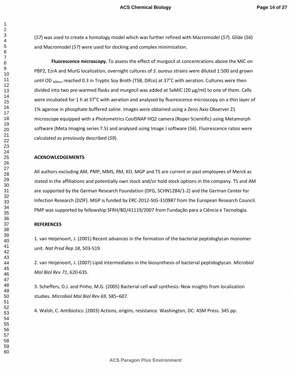
Murgocil inhibits MurG enzymatic activity. The cytosolic membrane-associated glycotransferase MurG
synthesizes lipid II by catalyzing the transfer of N-acetyl glucosamine (GlcNAc) from UDP-N-acetyl
glucosamine (UDP-GlcNAc) to the C4 hydroxyl of undecaprenyl-pyrophosphate-N-acetylmuramoyl-
pentapeptide (lipid I). Reconstitution of the SaMurG-catalyzed reaction in vitro, using purified
recombinant enzyme and substrates, lipid I and (14
C)-UDP-GlcNAc , respectively, revealed a full
conversion to the synthesis product lipid II which migrates slower on the TLC compared to lipid I (38)
(Figure 3 panel a). Testing murgocil in this system showed that lipid II synthesis is strongly impaired in a
dose-dependent fashion (20-400 µM; Figure 3 panel a). An almost complete inhibition of MurG activity
was observed in the presence of 3000 µM murgocil and an IC50 of approximately 115 μM was
determined under the conditions tested (Figure 3 panel b; See Summary and Implications).
To investigate the impact of MurG amino acid substitutions on enzymatic activity and resistance to
murgocil, site directed mutagenesis was used to generate recombinant proteins MurG_M45I,
MurG_D168N and MurG_D168G. In line with the in vivo situation, amino acid substitutions did not
affect MurG enzymatic activity and full conversion to lipid II was achieved in vitro (Figure 3 panel c). In
contrast to wild type SaMurG, the Lipid II synthesizing activity of all individual MurG variants was
unaffected in the presence of 200 µM murgocil (Figure 3 panel c), further confirming that these single
amino acid substitutions are causal for murgocilR phenotypes and that they prevent a murgocil:MurG
interaction without affecting binding of the substrate UDP-GlcNAc.
Page 8 of 27
ACS Paragon Plus Environment
ACS Chemical Biology
123456789101112131415161718192021222324252627282930313233343536373839404142434445464748495051525354555657585960
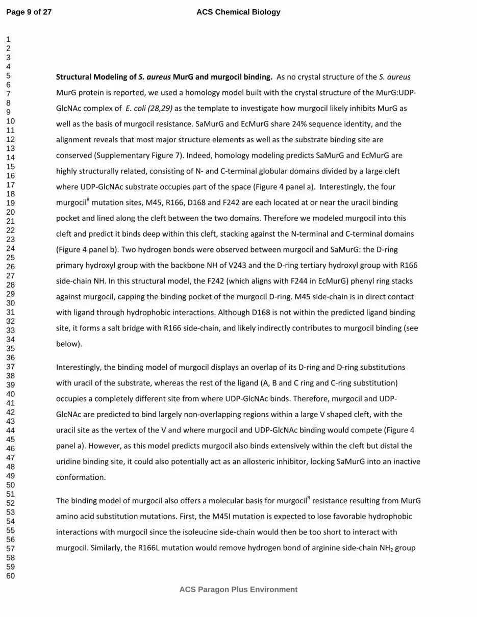
Structural Modeling of S. aureus MurG and murgocil binding. As no crystal structure of the S. aureus
MurG protein is reported, we used a homology model built with the crystal structure of the MurG:UDP-
GlcNAc complex of E. coli (28,29) as the template to investigate how murgocil likely inhibits MurG as
well as the basis of murgocil resistance. SaMurG and EcMurG share 24% sequence identity, and the
alignment reveals that most major structure elements as well as the substrate binding site are
conserved (Supplementary Figure 7). Indeed, homology modeling predicts SaMurG and EcMurG are
highly structurally related, consisting of N- and C-terminal globular domains divided by a large cleft
where UDP-GlcNAc substrate occupies part of the space (Figure 4 panel a). Interestingly, the four
murgocilR mutation sites, M45, R166, D168 and F242 are each located at or near the uracil binding
pocket and lined along the cleft between the two domains. Therefore we modeled murgocil into this
cleft and predict it binds deep within this cleft, stacking against the N-terminal and C-terminal domains
(Figure 4 panel b). Two hydrogen bonds were observed between murgocil and SaMurG: the D-ring
primary hydroxyl group with the backbone NH of V243 and the D-ring tertiary hydroxyl group with R166
side-chain NH. In this structural model, the F242 (which aligns with F244 in EcMurG) phenyl ring stacks
against murgocil, capping the binding pocket of the murgocil D-ring. M45 side-chain is in direct contact
with ligand through hydrophobic interactions. Although D168 is not within the predicted ligand binding
site, it forms a salt bridge with R166 side-chain, and likely indirectly contributes to murgocil binding (see
below).
Interestingly, the binding model of murgocil displays an overlap of its D-ring and D-ring substitutions
with uracil of the substrate, whereas the rest of the ligand (A, B and C ring and C-ring substitution)
occupies a completely different site from where UDP-GlcNAc binds. Therefore, murgocil and UDP-
GlcNAc are predicted to bind largely non-overlapping regions within a large V shaped cleft, with the
uracil site as the vertex of the V and where murgocil and UDP-GlcNAc binding would compete (Figure 4
panel a). However, as this model predicts murgocil also binds extensively within the cleft but distal the
uridine binding site, it could also potentially act as an allosteric inhibitor, locking SaMurG into an inactive
conformation.
The binding model of murgocil also offers a molecular basis for murgocilR resistance resulting from MurG
amino acid substitution mutations. First, the M45I mutation is expected to lose favorable hydrophobic
interactions with murgocil since the isoleucine side-chain would then be too short to interact with
murgocil. Similarly, the R166L mutation would remove hydrogen bond of arginine side-chain NH2 group
Page 9 of 27
ACS Paragon Plus Environment
ACS Chemical Biology
123456789101112131415161718192021222324252627282930313233343536373839404142434445464748495051525354555657585960

with the ligand and the F242L mutation would alter hydrophobic interactions as well as removal of the
pi-pi stacking between F242 phenyl ring and ligand D-ring substitution groups. Although D168 is
predicted not to directly contact murgocil, the salt bridge between D168 and R166 is likely disrupted by
the D168G or D168N mutation resulting in the movement of R166 side-chain toward D246 to form a
new salt bridge (Figure 4 panel c). This altered side-chain of R166 points into the predicted murgocil
binding pocket and would cause VDW conflict with the inhibitor.
PBP2 is delocalized in murgocil-treated cells. Previously, it has been demonstrated that lipid II is
required for proper localization of PBP2 to the division septum where active PG synthesis occurs in S.
aureus (3,39). Therefore, the simplest explanation for the extreme hypersensitivity of the murG
antisense strain to β-lactams and the observed synergy between murgocil and β-lactams is that
murgocil-specific inhibition of MurG leads to a depletion of lipid II and consequently PBP2 is delocalized
from the septum. To test this, we performed localization studies of murgocil-treated cells maintaining a
previously described functional superfolder green fluorescent protein (sGFP)-PBP2 fusion gene
integrated into the pbp2 locus of MRSA COL (20). sGFP-PBP2 correctly localized to the division septum in
mock-treated cells as previously reported (20). However, sGFP-PBP2 was highly delocalized among cells
treated with murgocil at 5X MIC for 1 hr, with discrete patches of sGFP-PBP2 localized throughout the
plasma membrane (Figure 5 panel a) and verified by quantification of fluorescence signal at the septum
versus fluorescence at the lateral wall under the two conditions tested (Figure 5 panel a and b). To
determine if PBP2 delocalization was a specific effect of the presence of murgocil and consequent
decrease of its substrate, and not part of general disassembly of the cell division machinery (40) that
could be caused by the presence of the antibiotic, we determined the localization of the FtsZ ring
(20,41), a bacterial ortholog of tubulin, using as a reporter the FtsZ-associated protein EzrA (42). MRSA
COL cells expressing a functional copy of EzrA-mCherry showed septal rings both in the absence and in
the presence of murgocil (Figure 5 panel a and b), indicating that murgocil-dependent delocalization of
PBP2 is not due to disassembly of the divisome.
Fluorescence localization studies of MRSA COL expressing a functional copy of MurG-GFP integrated into
the murG locus were also unaffected by murgocil treatment, with the fusion protein localized at the
plasma membrane, as indicated by patches of strong fluorescence, similar to untreated cells (Figure 5
panel a). Therefore, murgocil appears unlikely to interfere with the normal membrane-association of
MurG, an observation consistent with the fact that murgocilR mutations do not map to the proposed
membrane-association interface of MurG (28). We conclude that murgocil-based delocalization of PBP2
Page 10 of 27
ACS Paragon Plus Environment
ACS Chemical Biology
123456789101112131415161718192021222324252627282930313233343536373839404142434445464748495051525354555657585960

involves depletion of lipid II rather than disruption of the Z ring. In addition, we propose that the synergy
between murgocil and β-lactams involves (at least in part) murgocil-mediated depletion of lipid II
substrate, which is required for both PG synthesis and proper localization of PBP2, and that the
concomitant mislocalization of PBP2 under these conditions renders cells susceptible to reduced β-
lactam concentrations that are now sufficient to inactivate the residual functionally competent PBP2
activity remaining at the septum.
Summary and Implications. Here we describe a MRSA whole cell β-lactam potentiation screen resulting
in the identification of the highly specific PG inhibitor, murgocil, targeting the broadly conserved and
essential glycosyltransferase, MurG. Murgocil is demonstrated to inhibit PG synthesis based on MML
profiling as well as HPLC analysis revealing the accumulation of UDP-MurNAc-pentapeptide in drug-
treated cells. Mechanistically, a series of indirect and direct evidence provide compelling demonstration
of MurG as the drug target of murgocil; (a) antisense-based depletion of MurG produces a specific
hypersensitization to the compound and reciprocally MurG overexpression confers murgocil resistance,
(b) multiple independently derived murgocilR mutations isolated in both MRSA and MRSE map to
specific residues of MurG, (c) murgocil inhibits MurG-mediated PG lipid II synthesis in a cell-free in vitro
enzyme assay, and (d) murgocil inhibitory activity is abolished in vitro when tested against purified MurG
variants containing murgocilR amino acid substitution mutations. Further, docking models using the E.
coli MurG crystal structure and Staphylococcal murgocilR MurG mutations suggest murgocil binds to the
cleft separating N and C-terminal domains, thereby potentially competing with UDP-GlcNAc substrate
binding to this site as well as possibly locking SaMurG into a rigid, non-active form. Finally, we
demonstrate that the synergistic and cidal activity of murgocil in combination with imipenem is
achieved, at least in part, by mislocalization of PBP2 from the division septum, thus likely reducing β-
lactam drug levels necessary to inhibit the residual amount of functionally active PBP2 under murgocil
drug treatment conditions.
Despite murgocil targeting a broadly conserved PG biosynthetic enzyme, its microbiological activity is
uniquely restricted to Staphylococci, lacking bioactivity against other Gram-positive (e.g. Bacillus subtilis
and Streptococcus pyogenes) or Gram-negative bacteria tested, including E. coli and P. aeruginosa
strains defective in efflux pumps (Supplementary Table 1). Consistent with murgocil bioactivity
spectrum, the proposed S. epidermidis MurG murgocil binding site is 100% identical to SaMurG, while B.
subtilis MurG is significantly different, including intrinsic amino acid differences at M45 and D168 (I45
and E168, respectively) (Supplementary Figure 7). Similarly, EcMurG contains a M45L change and P.
Page 11 of 27
ACS Paragon Plus Environment
ACS Chemical Biology
123456789101112131415161718192021222324252627282930313233343536373839404142434445464748495051525354555657585960

aeruginosa MurG possesses M45L and D168G alterations. These changes alone, being similar (and in the
case of D168G, identical) to those found in SaMurG murgocilR mutants, are predicted to interfere with
murgocil binding and abolish its activity against these orthologous enzymes. Therefore, the narrow
spectrum of murgocil appears likely to be MurG-mechanism based. However, further in vitro studies
using purified MurG from E. coli and other recalcitrant bacteria as well as co-crystal structures of
murgocil in complex with SaMurG and (ideally) other MurG enzymes are required to fully address this
issue. Murgocil also exhibits an unfavorably high rate of spontaneous resistance, in the range of ~1 x10⁻⁷
at 4X MIC versus standard antibiotics whose frequency of resistance is more typically in the range of ~1
x10-8
or lower (23, 27). Therefore, although these issues significantly compromise the antibiotic
development potential of murgocil itself, the compound and MurG drugR mutations described here now
provide important structural modeling-based in silico screening opportunities for new MurG inhibitory
series with potentially broader activity, reduced susceptibility to resistance, and improved synergistic
and cidal activity when paired with β-lactam antibiotics.
Despite historically intensive efforts in identifying target-specific bioactive inhibitors to intracellular
steps of PG biosynthesis, it is startling how few molecules have been discovered that meet these basic
criteria for an antibiotic drug lead (23,27,43,44). Generally, these compounds either lack bioactivity
and/or solely possess in vitro activity against specific Mur enzymes (in which case the inhibitor is either
unable to penetrate cells or is efficiently effluxed from cells) and/or their whole cell activity has not
been genetically linked to the same enzyme used to screen and identify such inhibitors (23,27,43).
Moreover, in the few exceptions to this generalization, such inhibitors instead act to sequester lipid II PG
substrate (e.g. vancomycin, ramoplanin), undecaprenyl-P- (e.g. friulimicin) or undecaprenyl-PP (e.g.
bacitracin) rather than directly inhibiting a Mur enzyme (45-47). Indeed, we are unaware of any report
rigorously describing a target-specific bioactive inhibitor to MurG despite attempts using a variety of
target or pathway-specific in vitro or whole cell screening approaches (48-52). The potency of murgocil
against Staphylococci (MIC 2-4 �g/ml) is, however, considerably lower than the in vitro IC50 (115 �g/ml)
of the compound against purified S. aureus His-tagged MurG protein. We believe this likely reflects the
inability to mimic the in vivo complexity of the entire PG synthesizing machinery in vitro as well as the
difficulty in optimizing an extensive set of conditions tested to effectively solubilize murgocil while still
maintaining recombinant MurG in vitro enzyme activity. Notwithstanding these technical issues, we
provide compelling mechanistic evidence demonstrating that murgocil is both a highly selective and
potent inhibitor of endogenous MurG in whole cells. Moreover, murgocil serves as an important
Page 12 of 27
ACS Paragon Plus Environment
ACS Chemical Biology
123456789101112131415161718192021222324252627282930313233343536373839404142434445464748495051525354555657585960

addition to the suite of chemical probes (53) to study MurG function, PG synthesis and its coordinated
assembly with other cell wall polymers as well as cytoskeletal and cell division processes in
Staphylococci. Importantly, the demonstration that combining phenotypic screens, drug resistant
mutant isolation and mapping by NGS can collectively be used successfully to uncover target-specific
bioactive inhibitors of PG biosynthesis provides optimism that additional antibacterial lead series
targeting this pathway are expected to be discovered.
METHODS
See Supporting Information for Supplementary Methods for general microbiology, strains and plasmid
construction methods.
Accumulation of UDP-N-acetyl-muramoyl-pentapeptide. S. aureus SG511 was grown in
Mueller-Hinton broth to an OD600 of 0.5 and incubated with 130 µg/ml of chloramphenicol for 15 min.
Vancomycin (10x MIC – 5 µg/ml) and murgocil (2.5x, 5x and 10x MIC – 5, 10, 20 µg/ml) were added and
incubated for another 30 min. Cells were harvested and extracted with boiling water. The cell extract
was centrifuged and the supernatant lyophilized. Nucleotide-linked cell wall precursors were analyzed
by HPLC (54) and corresponding fractions were confirmed by mass spectrometry.
MurG-His6 in vitro assay activity. MurG activity was assayed in a final volume of 30 μl containing
2.5 nmol purified lipid I, 5 nmol UDP-GlcNAc or [14
C]UDP-GlcNAc in 60 mM Tris-HCl pH 7.5, 5 mM MgCl2,
and 0.5% Triton X-100. Conversion to lipid II was initiated by addition of 0.2 μg of purified MurG-
His6 enzyme. The reaction mixture was incubated for 60 min at 30°C. Lipid intermediates were extracted
from the reaction mixture with n-butanol-pyridine acetate (2:1, vol/vol), pH 4.2, and analyzed by TLC
(silica plates, 60F254; Merck) using chloroform-methanol-water-ammonia (88:48:10:1) as the solvent.
Spots were visualized by PMA staining reagent A (55). Radiolabeled spots were visualized and quantified
using a phosphor storage screen and a Storm 820 optical scanner (GE Healthcare, Munich, Germany).
Murgocil (molecular mass: 447.582 Da) was added in concentrations ranging from 10 to 500 µM.
Computer modeling of SaMurG:murgocil complex. The compound murgocil 3D conformation
was sampled and selected from a set of 3D conformations generated by LigPrep (56). A steroid bound X-
ray structure from PDB was taken to guide the final 3D selection. The X-ray crystal structure of UDP-
GlcNAc:EcMurG complex 1NLM.pdb (29) was chosen as the template for the homology model of
SaMurG. The alignment from MOE (MOE, 2012) based on sequence similarity was manually adjusted at
several regions, in particular moving a five-residue deletion at F242 out of the uracil binding site. MOE
Page 13 of 27
ACS Paragon Plus Environment
ACS Chemical Biology
123456789101112131415161718192021222324252627282930313233343536373839404142434445464748495051525354555657585960
(57) was used to create a homology model which was further refined with Macromodel (57). Glide (56)
and Macromodel (57) were used for docking and complex minimization.
Fluorescence microscopy. To assess the effect of murgocil at concentrations above the MIC on
PBP2, EzrA and MurG localization, overnight cultures of S. aureus strains were diluted 1:500 and grown
until OD 600nm reached 0.3 in Tryptic Soy Broth (TSB, Difco) at 37oC with aeration. Cultures were then
divided into two pre-warmed flasks and murgocil was added at 5xMIC (20 µg/ml) to one of them. Cells
were incubated for 1 h at 37oC with aeration and analysed by fluorescence microscopy on a thin layer of
1% agarose in phosphate buffered saline. Images were obtained using a Zeiss Axio Observer Z1
microscope equipped with a Photometrics CoolSNAP HQ2 camera (Roper Scientific) using Metamorph
software (Meta Imaging series 7.5) and analysed using Image J software (56). Fluorescence ratios were
calculated as previously described (59).
ACKNOWLEDGEMENTS
All authors excluding AM, PMP, MMS, RM, KO, MGP and TS are current or past employees of Merck as
stated in the affiliations and potentially own stock and/or hold stock options in the company. TS and AM
are supported by the German Research Foundation (DFG, SCHN1284/1-2) and the German Center for
Infection Research (DZIF). MGP is funded by ERC-2012-StG-310987 from the European Research Council.
PMP was supported by fellowship SFRH/BD/41119/2007 from Fundação para a Ciência e Tecnologia.
REFERENCES
1. van Heijenoort, J. (2001) Recent advances in the formation of the bacterial peptidoglycan monomer
unit. Nat Prod Rep 18, 503-519.
2. van Heijenoort, J. (2007) Lipid intermediates in the biosynthesis of bacterial peptidoglycan. Microbiol
Mol Biol Rev 71, 620-635.
3. Scheffers, D.J. and Pinho, M.G. (2005) Bacterial cell wall synthesis: New insights from localization
studies. Microbiol Mol Biol Rev 69, 585–607.
4. Walsh, C. Antibiotics: (2003) Actions, origins, resistance. Washington, DC: ASM Press. 345 pp.
Page 14 of 27
ACS Paragon Plus Environment
ACS Chemical Biology
123456789101112131415161718192021222324252627282930313233343536373839404142434445464748495051525354555657585960

5. Klevens, R.M., Morrison, M.A., Nadle, J., Petit, S., Gershman, K., Ray, S., Harrison, L.H., Lynfield, R.,
Dumyati, G., Townes, J.M., Craig, A.S., Zell, E.R., Fosheim, G.E., McDougal, L.K., Carey, R.B., and Fridkin,
S.K. (2007) Active Bacterial Core surveillance (ABCs) MRSA Investigators. JAMA 298, 1763-1771.
6. Boucher, H.W., Talbot, G.H., Bradley, J.S., Edwards, J.E., Gilbert, D., Rice, L.B., Scheld, M., Spellberg,
B., and Bartlett, J. (2009) Bad bugs, no drugs: No ESKAPE! An update from the Infectious Diseases
Society of America. Clin Infect Dis 48, 1–12.
7. Couto, I., de Lencastre, H., Severina, E., Kloos, W., Webster, J.A., Hubner, R.J., Sanches, I.S., and
Tomasz, A. (1996) Ubiquitous presence of a mecA homologue in natural isolates of Staphylococcus sciuri.
Microb Drug Resist 2, 377-391.
8. de Lencastre, H., Oliveira, D., and Tomasz, A. (2007) Antibiotic resistant Staphylococcus aureus: a
paradigm of adaptive power. Curr Opin Microbiol 10, 428-435.
9. Fuda, C.C., Fisher, J.F., and Mobashery, S. (2005) Beta-lactam resistance in Staphylococcus aureus: the
adaptive resistance of a plastic genome. Cell Mol Life Sci 62, 2617-2633.
10. Fuda, C., Suvorov, M., Vakulenko, S.B., and Mobashery, S. (2004) The basis for resistance to beta-
lactam antibiotics by penicillin-binding protein 2a of methicillin-resistant Staphylococcus aureus. J Biol
Chem 279, 40802-40806.
11. Llarrull, L.I., Fisher, J.F., and Mobashery, S. (2009) Molecular basis and phenotype of methicillin
resistance in Staphylococcus aureus and insights into new beta-lactams that meet the challenge.
Antimicrob Agents Chemother 53, 4051-4063.
12. Pinho, M.G., de Lencastre, H., and Tomasz, A. (2001) An acquired and a native penicillin binding
protein cooperate in building the cell wall of drug-resistant staphylococci. Proc Natl Acad Sci USA 98,
10886–10891.
13. Pinho, M.G., Filipe, S.R., de Lencastre, H., and Tomasz, A. (2001) Complementation of the essential
peptidoglycan transpeptidase function of penicillin-binding protein 2 (PBP2) by the drug resistance
protein PBP2A in Staphylococcus aureus. J Bacteriol 183, 6525-6531.
14. de Lencastre, H., and Tomasz, A. (1994) Reassessment of the number of auxiliary genes essential for
expression of high-level methicillin resistance in Staphylococcus aureus. Antimicrob Agents Chemother
38, 2590-2598.
Page 15 of 27
ACS Paragon Plus Environment
ACS Chemical Biology
123456789101112131415161718192021222324252627282930313233343536373839404142434445464748495051525354555657585960
15. de Lencastre, H., Wu, S.W., Pinho, M.G., Ludovice, A.M., Filipe, S., Gardete, S., Sobral ,R., Gill, S.,
Chung, M., and Tomasz, A. (1999) Antibiotic resistance as a stress response: Complete sequencing of a
large number of chromosomal loci in Staphylococcus aureus strain COL that impact on the expression of
resistance to methicillin. Microb Drug Resist 5, 163–175.
16. Berger-Bächi, B. and Rohrer, S. (2002) Factors influencing methicillin resistance in staphylococci.
Arch Microbiol 178, 165–171.
17. Lee, S.H. Wang-Jarantow, L., Wang, H., Sillaots, S., Cheng, H., Meredith, T.C., Thompson, J., and
Roemer, T. (2011) Antagonism of chemical genetic interaction networks resensitize MRSA to β-lactam
antibiotics. Chem Biol 18, 1379–1389.
18. Roemer, T., Schneider, T. and Pinho, M.G. (2013) Accessory Factors: A chink in the armor of MRSA
resistance to β-lactam antibiotics. Curr Opin Microbiol (in press).
19. Huber, J. Donald, R.G., Lee, S.H., Jarantow, L.W., Salvatore, M.J., Meng, X., Painter, R., Onishi, R.H.,
Occi, J., Dorso, K., Young, K., Park, Y.W., Skwish, S., Szymonifka, M.J., Waddell, T.S., Miesel, L., Phillips,
J.W., and Roemer, T. (2009) Chemical genetic identification of peptidoglycan inhibitors potentiating
carbapenem activity against methicillin-resistant Staphylococcus aureus. Chem Biol 16, 837–848.
20. Tan, C.M., Therien, A.G., Lu, J., Lee, S.H., Caron, A., Gill, C.J., Lebeau-Jacob, C., Benton-Perdomo, L.,
Monteiro, J.M., Pereira, P.M., Elsen, N.L., Wu, J., Deschamps, K., Petcu, M., Wong, S., Daigneault, E.,
Kramer, S., Liang, L., Maxwell, E., Claveau, D., Vaillancourt, J., Skorey, K., Tam, J., Wang, H., Meredith,
T.C., Sillaots, S., Wang-Jarantow, L., Ramtohul, Y., Langlois, E., Landry, F., Reid, J.C., Parthasarathy, G.,
Sharma, S., Baryshnikova, A., Lumb, K.J., Pinho, M.G., Soisson, S.M., and Roemer, T. (2012) Restoring
methicillin-resistant Staphylococcus aureus susceptibility to β-lactam antibiotics. Sci Transl Med 4,
126ra35.
21. Koyama, N., Tokura, Y., Münch, D., Sahl, H.G., Schneider, T., Shibagaki, Y., Ikeda, H., and Tomoda, H.
(2012) The nonantibiotic small molecule cyslabdan enhances the potency of β-lactams against MRSA by
inhibiting pentaglycine interpeptide bridge synthesis. PLoS One 7, e48981.
22. Therien, A.G., Huber, J.L., Wilson, K.E., Beaulieu, P., Caron, A., Claveau, D., Deschamps, K., Donald,
R.G., Galgoci, A.M., Gallant, M., Gu, X., Kevin, N.J., Lafleur, J., Leavitt, P.S., Lebeau-Jacob, C., Lee, S.S., Lin,
M.M., Michels, A.A., Ogawa, A.M,, Painter, R.E., Parish, C.A., Park, Y.W., Benton-Perdomo, L., Petcu, M.,
Page 16 of 27
ACS Paragon Plus Environment
ACS Chemical Biology
123456789101112131415161718192021222324252627282930313233343536373839404142434445464748495051525354555657585960

Phillips, J.W., Powles, M.A., Skorey, K.I., Tam, J., Tan, C.M., Young, K., Wong, S., Waddell, S.T., and
Miesel, L. (2012) Broadening the spectrum of β-lactam antibiotics through inhibition of signal peptidase
type I. Antimicrob Agents Chemother 56, 4662-4670.
23. Silver, L.L. (2013) Viable screening targets related to the bacterial cell wall. Ann N Y Acad Sci 277, 29-
53.
24. Isono, K., Uramoto, M., Kusakabe, H., Kimura, K., Isaki, K., Nelson, C.C., and McCloskey, J.A.
(1985) Liposidomycins: novel nucleoside antibiotics which inhibit bacterial peptidoglycan synthesis. J
Antibiot 38, 1617–1621.
25. Kimura, K-I., Miyata, N., Kawanishi, G., Kamio, Y., Izaki, K., and Isono, and K. (1989) Liposidomycin C
inhibits phospho-N-acetylmuramyl-pentapeptide transferase in peptidoglycan synthesis of Escherichia
coli Y-10. Agric Biol Chem 53, 1811–1815.
26. Brandish, P.E., Burnham, M.K., Lonsdale, J.T., Southgate, R., Inukai, M., and Bugg, T.D. (1996) Slow-
binding inhibition of phospho-N-acetylmuramyl-pentapeptide translocase (Escherichia coli) by
mureidomycin A. J Biol Chem 271, 7609–7614.
27. Silver, L.L. (2011) Challenges of antibacterial discovery. Clin Microbiol Rev 24, 71-109.
28. Ha, S., Walker, D., Shi, Y., and Walker, S. (2000) The 1.9 A crystal structure of Escherichia coli MurG, a
membrane-associated glycosyltransferase involved in peptidoglycan biosynthesis. Protein Sci 9, 1045-
1052.
29. Hu, Y., Chen, L., Ha, S., Gross, B., Falcone, B., Walker, D., Mokhtarzadeh, M., and Walker, S. (2003)
Crystal structure of the MurG:UDP-GlcNAc complex reveals common structural principles of a
superfamily of glycosyltransferases. Proc Natl Acad Sci USA 100, 845-849.
30. Campbell, J., Singh, A.K., Santa Maria, J.P., Kim, Y., Brown, S., Swoboda, J.G., Mylonakis, E.,
Wilkinson, B., and Walker, S. (2011) Synthetic lethal compound combinations reveal a fundamental
connection between wall teichoic acid and peptidoglycan biosyntheses in Staphylococcus aureus. ACS
Chem Biol 6, 106–116.
Page 17 of 27
ACS Paragon Plus Environment
ACS Chemical Biology
123456789101112131415161718192021222324252627282930313233343536373839404142434445464748495051525354555657585960

31. Brown, S., Xia, G., Luhachack, L.G., Campbell, J., Meredith, T.C., Chen, C., Winstel, V., Gekeler, C.,
Irazoqui, J.E., Peschel, A., and Walker, S. (2012) Methicillin resistance in Staphylococcus aureus requires
glycosylated wall teichoic acids. Proc Natl Acad Sci USA 109, 18909-18914.
32. Weidenmaier, C. and Peschel, A. (2008) Teichoic acids and related cell-wall glycopolymers in Gram-
positive physiology and host interactions. Nat Rev Microbiol 6, 276-287.
33. Swoboda, J.G., Campbell ,J., Meredith, T.C., and Walker, S. (2010) Wall teichoic acid function,
biosynthesis, and inhibition. Chembiochem 11, 35-45.
34. Meredith, T.C., Wang, H., Beaulieu, P., Gründling, A., and Roemer, T. (2012) Harnessing the power of
transposon mutagenesis for antibacterial target identification and evaluation. Mob Genet Elements. 2,
171-178.
35. Farha, M.A., Leung, A., Sewell, E.W., D'Elia, M.A., Allison, S.E., Ejim, L., Pereira, P.M., Pinho, M.G.,
Wright, G.D., and Brown, E.D. (2013) Inhibition of WTA synthesis blocks the cooperative action of PBPs
and sensitizes MRSA to β-lactams. ACS Chem Biol. 8, 226-233.
36. Donald, R.G., Skwish, S., Forsyth, R.A., Anderson, J.W., Zhong, T., Burns, C., Lee, S., Meng, X.,
LoCastro, L., Jarantow, L.W., Martin, J., Lee, S.H., Taylor, I., Robbins, D., Malone, C., Wang, L., Zamudio,
C.S., Youngman, P.J., and Phillips, J.W. (2009) A Staphylococcus aureus fitness test platform for
mechanism-based profiling of antibacterial compounds. Chem Biol 16, 826-836.
37. Wang, H., Gill, C.J., Lee, S.H., Mann, P., Zuck, P., Meredith, T.C., Murgolo, N., She, X., Kales, S., Liang,
L., Liu, J., Wu, J., Santa Maria, J., Su, J., Pan, J., Hailey, J., Mcguinness, D., Tan, C.M., Flattery, A., Walker,
S., Black, T., and Roemer, T. (2013) Discovery of novel wall teichoic acid inhibitors as effective anti-MRSA
β-lactam combination agents. Chem Biol 20, 272-284.
38. Schneider, T., Kruse, T., Wimmer,R., Wiedemann, I., Sass, V., Pag, U., Jansen, A., Nielsen, A.K.,
Mygind, P.H., Raventós, D.S., Neve, S., Ravn, B., Bonvin, A.M., De Maria, L., Andersen, A.S.,
Gammelgaard, L.K., Sahl, H.G., and Kristensen, H.H. (2010) Plectasin, a fungal defensin, targets the
bacterial cell wall precursor Lipid II. Science 328, 1168-1172.
Page 18 of 27
ACS Paragon Plus Environment
ACS Chemical Biology
123456789101112131415161718192021222324252627282930313233343536373839404142434445464748495051525354555657585960

39. Pinho, M.G. and Errington, J. (2005) Recruitment of penicillin-binding protein PBP2 to the division
site of Staphylococcus aureus is dependent on its transpeptidation substrates. Mol Microbiol 55, 799–
807.
40. Adams, D.W. and Errington, J. (2009) Bacterial cell division: assembly, maintenance and disassembly
of the Z ring. Nat Rev Microbiol 7, 642-653.
41. Pinho, M.G. and Errington, J. (2003) Dispersed mode of Staphylococcus aureus cell wall synthesis in
the absence of the division machinery. Mol Microbiol 50, 871–881.
42. Jorge, A.M., Hoiczyk, E., Gomes, J.P., and Pinho, M.G. (2011) EzrA contributes to the regulation of cell
size in Staphylococcus aureus. PLoS One 6, e27542.
43. Silver, L.L. (2003) Novel inhibitors of bacterial cell wall synthesis. Curr Opin Microbiol 6, 431-438.
44. Payne, D.J., Gwynn, M.N., Holmes, D.J., and Pompliano, D.L. (2007) Drugs for bad bugs: confronting
the challenges of antibacterial discovery. Nat Rev Drug Discov 6, 29-40.
45. Hu, Y., Helm, J.S., Chen, L., Ye, X.Y., and Walker, S. (2003) Ramoplanin inhibits bacterial
transglycosylases by binding as a dimer to lipid II. J Am Chem Soc 125, 8736-8737.
46. Silver, L.L. (2006) Does the cell wall of bacteria remain a viable source of targets for novel
antibiotics? Biochem Pharmacol 71, 996-1005.
47. Schneider, T., Gries, K., Josten, M., Wiedemann, I., Pelzer, S., Labischinski, H., and Sahl, H.G. (2009)
The lipopeptide antibiotic Friulimicin B inhibits cell wall biosynthesis through complex formation with
bactoprenol phosphate. Antimicrob Agents Chemother 53, 1610–1618.
48. Helm, J.S., Hu, Y., Chen, L., Gross, B., and Walker, S. (2003) Identification of active-site inhibitors of
MurG using a generalizable, high-throughput glycosyltransferase screen. J Am Chem Soc 125, 11168-
11169.
49. Hu, Y., Helm, J.S., Chen, L., Ginsberg, C., Gross, B., Kraybill, B., Tiyanont, K., Fang, X., Wu, T., and
Walker, S. (2004) Identification of selective inhibitors for the glycosyltransferase MurG via high-
throughput screening. Chem Biol 11, 703-711.
Page 19 of 27
ACS Paragon Plus Environment
ACS Chemical Biology
123456789101112131415161718192021222324252627282930313233343536373839404142434445464748495051525354555657585960

50. Barbosa, M.D., Ross, H.O., Hillman, M.C., Meade, R.P., Kurilla, M.G., and Pompliano, D.L. (2002) A
multitarget assay for inhibitors of membrane-associated steps of peptidoglycan biosynthesis. Anal
Biochem 306, 17-22.
51. Barbosa, M.D., Yang, G., Fang, J., Kurilla, M.G., and Pompliano, D.L. (2002) Development of a whole-
cell assay for peptidoglycan biosynthesis inhibitors. Antimicrob Agents Chemother 46, 943-946.
52. DeCenzo, M., Kuranda, M., Cohen, S., Babiak, J., Jiang, Z.D., Su, D., Hickey, M., Sancheti, P., Bradford,
P.A., Youngman, P., Projan, S., and Rothstein, D.M. (2002) Identification of compounds that inhibit late
steps of peptidoglycan synthesis in bacteria. J Antibiot 55, 288-295.
53. Foss, M.H., Eun, Y.J., and Weibel, D.B. (2011) Chemical-biological studies of subcellular organization
in bacteria. Biochemistry. 50, 7719-7734.
54. Rubinchik, E., Schneider, T., Elliott, M., Scott, W.R., Pan, J., Anklin, C., Yang, H., Dugourd, D., Müller,
A., Gries, K., Straus, S.K., Sahl, H.G., and Hancock, R.E. (2011) Mechanism of Action and Limited Cross-
Resistance of NewLipopeptide MX-2401. Antimicrob Agents Chemother 55, 2743-2754.
55. Schneider, T., Senn, M.M., Berger-Bächi, B., Tossi ,A., Sahl, H.G, and Wiedemann, I. (2004) In vitro
assembly of a complete, pentaglycine interpeptide bridge containing cell wall precursor (lipid II-Gly5) of
Staphylococcus aureus. Mol Microbiol 53, 675–685.
56. Schrödinger, Inc., New York, NY, 2012
57. MOE, Molecular Operating Environment, 2012.10; Chemical Computing Group Inc., 1010 Sherbooke
St. West, Suite #910, Montreal, QC, Canada, H3A 2R7.
58. Abràmoff, M.D., Magalhães, P.J., and Ram, S.J. (2004) Image processing with ImageJ. Biophotonics
Int 11, 36-42.
59. Pereira, P.M., Filipe, S.R., Tomasz, A., and Pinho, M.G. (2007) Fluorescence ratio imaging
microscopy shows decreased access of vancomycin to cell wall synthetic sites in Vancomycin-Resistant
Staphylococcus aureus. Antimicrob Agents and Chemother 51, 3627-3633.
Page 20 of 27
ACS Paragon Plus Environment
ACS Chemical Biology
123456789101112131415161718192021222324252627282930313233343536373839404142434445464748495051525354555657585960

Supporting Information Available: general microbiology, strains and plasmid construction methods, and murgocil chemical synthesis methods. This material is available free of charge via the Internet at http://pubs.acs.org. Figure Legends:
Figure 1. Murgocil inhibits PG biosynthesis. (a) Macromolecular labeling of S. aureus strain RN4220 cells
treated with murgocil specifically produces a dose dependent depletion of PG synthesis. Chemical
structure of murgocil is shown. (b) PAP analysis of MRSA COL treated cells with ¼ MIC of murgocil,
moenomycin, or mock control (DMSO at 2% final concentration) in combination with a range (0-64
�g/ml) of imipenem (IPM). Chemical synergy is scored by enumerating colony forming units (CFU) post
drug treatment. (c,d) S. aureus murgocil-treated cells (2.5X, 5X and 10X MIC; 5, 10, 20 µg/ml ) display a
dose dependent accumulation of the PG intermediate, UDP-N-acetyl-muramoyl-pentapeptide (UPD-
MurNAc-pentapetide) comparable to vancomycin-treated cells at 10X MIC (5 µg/ml). (e) Antisense-
mediated depletion of murG expression by 50 mM xylose supplementation (bottom panel) induces
MRSA COL hypersensitivity to murgocil versus vector control (top panel) treated cells. (f) MRSA COL
maintaining overexpression plasmid ptuf-MurG (bottom panel) displays murgocil dose dependent
resistance versus control (top panel) treated cells.
Figure 2. murG missense mutations confer murgocil resistance. Heatmap summary of all non-
synonymous mutations identified by illumina-based whole genome sequencing (>100X genome
coverage) of 17 independently isolated murgocil drug resistant mutants in MRSA COL and MRSE strain
CLB26329. Red, non-synonymous mutation which maps to murG; yellow additional non-synonymous
mutation identified by whole genome sequencing; black, no change versus parental genome sequence.
Genome position, base change, and resulting amino acid substitution are shown. Murgocil� mutants
Sa80-96 and Se1-5 refer to S. aureus and S. epidermidis isolates, respectively. Note: additional
substitutions (yellow) are presumably unlinked to drug resistance as each strain also contains a MurG
amino acid substitution.
Figure 3. Murgocil inhibits S. aureus MurG-catalyzed lipid II synthesis in vitro. (a) Murgocil dose
dependent inhibition of purified MurG-His6 enzyme activity. Lipid intermediates were extracted from
the reaction mixture, separated by TLC and visualized by PMA staining. Murgocil inhibitory effect was
tested at concentrations ranging from 0-400 µM. (b) Graphical summary of murgocil dose dependent
Page 21 of 27
ACS Paragon Plus Environment
ACS Chemical Biology
123456789101112131415161718192021222324252627282930313233343536373839404142434445464748495051525354555657585960
inhibition of MurG-His6 mediated lipid II formation. (c) S. aureus MurG-His6 enzymes containing M45I,
D168N, and D168G murgocilR mutations are active but refractory to murgocil-mediated inhibition of
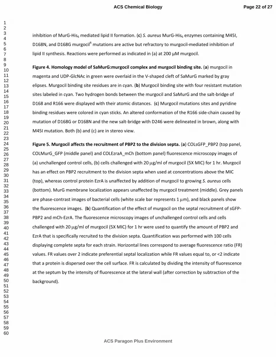
lipid II synthesis. Reactions were performed as indicated in (a) at 200 µM murgocil.
Figure 4. Homology model of SaMurG:murgocil complex and murgocil binding site. (a) murgocil in
magenta and UDP-GlcNAc in green were overlaid in the V-shaped cleft of SaMurG marked by gray
elipses. Murgocil binding site residues are in cyan. (b) Murgocil binding site with four resistant mutation
sites labeled in cyan. Two hydrogen bonds between the murgocil and SaMurG and the salt-bridge of
D168 and R166 were displayed with their atomic distances. (c) Murgocil mutations sites and pyridine
binding residues were colored in cyan sticks. An altered conformation of the R166 side-chain caused by
mutation of D168G or D168N and the new salt-bridge with D246 were delineated in brown, along with
M45I mutation. Both (b) and (c) are in stereo view.
Figure 5. Murgocil affects the recruitment of PBP2 to the division septa. (a) COLsGFP_PBP2 (top panel,
COLMurG_GFP (middle panel) and COLEzraA_mCh (bottom panel) fluorescence microscopy images of
(a) unchallenged control cells, (b) cells challenged with 20 �g/ml of murgocil (5X MIC) for 1 hr. Murgocil
has an effect on PBP2 recruitment to the division septa when used at concentrations above the MIC
(top), whereas control protein EzrA is unaffected by addition of murgocil to growing S. aureus cells
(bottom). MurG membrane localization appears unaffected by murgocil treatment (middle). Grey panels
are phase-contrast images of bacterial cells (white scale bar represents 1 �m), and black panels show
the fluorescence images. (b) Quantification of the effect of murgocil on the septal recruitment of sGFP-
PBP2 and mCh-EzrA. The fluorescence microscopy images of unchallenged control cells and cells
challenged with 20 �g/ml of murgocil (5X MIC) for 1 hr were used to quantify the amount of PBP2 and
EzrA that is specifically recruited to the division septa. Quantification was performed with 100 cells
displaying complete septa for each strain. Horizontal lines correspond to average fluorescence ratio (FR)
values. FR values over 2 indicate preferential septal localization while FR values equal to, or <2 indicate
that a protein is dispersed over the cell surface. FR is calculated by dividing the intensity of fluorescence
at the septum by the intensity of fluorescence at the lateral wall (after correction by subtraction of the
background).
Page 22 of 27
ACS Paragon Plus Environment
ACS Chemical Biology
123456789101112131415161718192021222324252627282930313233343536373839404142434445464748495051525354555657585960

a b
e f
Figure 1
vector
murG-AS
c d
OH
OHO
CH3
N
N
N
CH3
A B
C D
ptuf-murG
vector
0
1
2
3
4
5
6
7
8
9
10
0 0.06 0.125 0.25 0.5 1 2 4 8 16 32 64
Log
10
CF
U/m
l
IPM concentration, µg/ml
DMSO
Moenomycin
Murgocil
0 mM xylose 50 mM xylose Murgocil
Vancomycin
Murgocil
Vancomycin
overexpression
0.00 5.00 10.00 15.00 20.00 25.00 30.00
0
0.1
0.05
UDP-MurNAc-
pentapeptide
10x MIC
5x MIC
2.5x MIC
min
A260
0.00 5.00 10.00 15.00 20.00 25.00 30.00
0
0.1
0.05
UDP-MurNAc-
pentapeptide
vancomycin
min
A260
control
Page 23 of 27
ACS Paragon Plus Environment
ACS Chemical Biology
12345678910111213141516171819202122232425262728293031323334353637383940414243444546474849505152535455565758
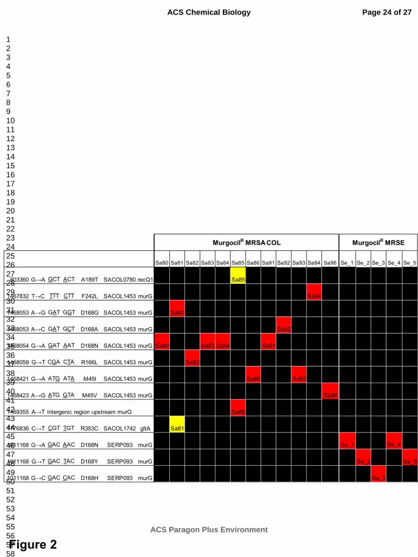
Figure 2
Sa80 Sa81 Sa82 Sa83 Sa84 Sa85 Sa86 Sa91 Sa92 Sa93 Sa94 Sa96 Se_1 Se_2 Se_3 Se_4 Se_5
803360 G→A GCT ACT A189T SACOL0780 recQ1 Sa85
1467832 T→C TTT CTT F242L SACOL1453 murG Sa94
1468053 A→G GAT GGT D168G SACOL1453 murG Sa81
1468053 A→C GAT GCT D168A SACOL1453 murG Sa92
1468054 G→A GAT AAT D168N SACOL1453 murG Sa80 Sa83 Sa84 Sa91
1468059 G→T CGA CTA R166L SACOL1453 murG Sa82
1468421 G→A ATG ATA M45I SACOL1453 murG Sa86 Sa93
1468423 A→G ATG GTA M45V SACOL1453 murG Sa96
1469355 A→T intergenic region upstream murG Sa85
1776836 C→T CGT TGT R353C SACOL1742 gltA Sa81
1011168 G→A GAC AAC D168N SERP093 murG Se_1 Se_4
1011168 G→T GAC TAC D168Y SERP093 murG Se_2 Se_5
1011168 G→C GAC CAC D168H SERP093 murG Se_3
MurgocilR MRSA COL MurgocilR MRSE
Page 24 of 27
ACS Paragon Plus Environment
ACS Chemical Biology
12345678910111213141516171819202122232425262728293031323334353637383940414243444546474849505152535455565758

������������� ����������������� ��������������� �������� ��� (a) Murgocil dose dependent inhibition of purified MurG�His6 enzyme activity. Lipid intermediates were extracted from the reaction
mixture, separated by TLC and visualized by PMA staining. Murgocil inhibitory effect was tested at
concentrations ranging from 0�400 )M. (b) Graphical summary of murgocil dose dependent inhibition of MurG�His6 mediated lipid II formation. (c) ��������� MurG�His6 enzymes containing M45I, D168N, and
D168G murgocilR mutations are active but refractory to murgocil�mediated inhibition of lipid II synthesis. Reactions were performed as indicated in (a) at 200 )M murgocil.
190x254mm (96 x 96 DPI)
Page 25 of 27
ACS Paragon Plus Environment
ACS Chemical Biology
123456789101112131415161718192021222324252627282930313233343536373839404142434445464748495051525354555657585960

��
�
�
������������������ ��������������������������������������������� (a) �������� ����� ���� ������������ ����� ��������������� ����������������������� �������!���"#����#��������$� ������"� �� ��������������������� �#� $��"�� ������"� �� ����������������������� �������� ���������"������ �#� $�
%���#���� �" ���"����� ������������� ���� ����� �����������"���������&'(�� ��)&''������������#������������������������ ��$����� ������������ ��������� ���#���� ��"� �� ���������������������� �#� ����!�$�� ��������� ������ �������)&''��������� �������"#������� ����&'(�����&'(��� ������ ��������"������������*+'���������� ������� �"�� ,��� ������� +-.������� $�/����"��
� ���������� �����������$��*-+0&12����1'�0�1'���.���
�
�
Page 26 of 27
ACS Paragon Plus Environment
ACS Chemical Biology
123456789101112131415161718192021222324252627282930313233343536373839404142434445464748495051525354555657585960

��
�
�
������������ � �������� ��� ������ � ������������� ������������� ��������������������������������������������������������� �������������!������"��"����"���"��#����$����!�����
��"������$���"�������"������� ��"�����"������$���%�����&��$'����!����$�"����()��*���!���+���,����$�"����������
�!!�"������ �����"������������������-������������%������������"��"������������ �-�������*���������%�������"��������������������������!!�"���� #�����������!����$�"������$��%��$�����������"������ ������,�����
��� �������"���������������������!!�"���� #����$�"���������������������,���#�����������������."��������
���$����!� �"�������"������%������"���� ��������������+�/�������� ��"0�����������%�����!������"��"�����$��,��� ��1�����!�"�������!������!!�"���!����$�"�������������������"����������!���.� ����������.����,�2���!������"��"����"���"��#����$����!���"������$���"�������"���������"�����"������$���%�����&�/$'����!�
���$�"����()��*���!���+����%������������3�����!#�������������!�� �����������������������"�!�"���#���"����������������-�����������,�1�����!�"������%������!������%����+&&�"�����������#��$�"��������������!�����"��������,�4����������������"��������������-���$��!������"��"���������5��-�����,�5�-�������-���������"�������!������������������"����������%�����5�-�������3�����������6������"���������������������������������-�������"�������!�"�,�
Page 27 of 27
ACS Paragon Plus Environment
ACS Chemical Biology
123456789101112131415161718192021222324252627282930313233343536373839404142434445464748495051525354555657585960

5����"��"������� #���-����$�������������#��!�!������"��"���������������� #�������������#��!�!������"��"�����������������%������!����"����"����� #��� ���"������!����� �"0$������,��
+7&8�(9����7:�8�7:�;�*���
�
�
Page 28 of 27
ACS Paragon Plus Environment
ACS Chemical Biology
123456789101112131415161718192021222324252627282930313233343536373839404142434445464748495051525354555657585960

�
����������������� ����
Page 29 of 27
ACS Paragon Plus Environment
ACS Chemical Biology
123456789101112131415161718192021222324252627282930313233343536373839404142434445464748495051525354555657585960