microbiological and physicochemical quality of - edo poly
TRANSCRIPT

1
MICROBIOLOGICAL AND PHYSICOCHEMICAL QUALITY OF
AGBO: A HERBAL MIXTURE SOLD IN MUNICIPAL MARKETS IN
BENIN CITY
BY
IKECHUKWU INNOCENT INOMA
PG/LSC0308580
B. Sc (Hons.) Benin
UNIVERSITY OF BENIN
BENIN CITY
NIGERIA.
JULY, 2017

2
MICROBIOLOGICAL AND PHYSICOCHEMICAL QUALITY OF
AGBO: A HERBAL MIXTURE SOLD IN MUNICIPAL MARKETS IN
BENIN CITY
BY
IKECHUKWU INNOCENT INOMA
PG/LSC0308580
B. Sc (Hons.) Benin
A THESIS SUBMITTED TO THE SCHOOL OF POSTGRADUATE
STUDIES, UNIVERSITY OF BENIN, IN PARTIAL FULFILMENT OF
THE REQUIREMENT FOR THE AWARD OF MASTER OF SCIENCE
(M. Sc.) DEGREE IN ENVIRONMENTAL AND PUBLIC HEALTH
MICROBIOLOGY
3
CERTIFICATION
I certify that this research project was carried out by Ikechukwu Innocent Inoma
with matriculation number PG/LSC0308580 in the Department of Microbiology, Faculty of
Life Sciences, University of Benin, Benin City under my supervision.
--------------------------------- ----------------------------
Prof. M. J. Ikenebomeh Date
(Supervisor)
4
ANTI-PLAGIARISM
We the undersigned declare that the project work of Ikechukwu Innocent Inoma has
successfully passed the anti-plagiarism test and do not violate any copyright regulation.
------------------------------------ ---------------------------
Prof. M. J. Ikenebomeh Date
(Supervisor)
----------------------------------- ----------------------------
Dr. (Mrs.) F.E. Oviasogie Date
(Head of Department)
5
APPROVAL
This is to certify that this project work was approved in partial fulfillment of the requirements
for the award of Masters of Science (M.Sc.) degree in Environmental and Public Health
Microbiology
____________________________ _________________
Prof. V. E. Omozuwa Date
(Dean, School of post-Graduate studies)

6
DEDICATION
I dedicate this work to God Almighty for the inspiration and His infiniteness
7
ACKNOWLEDGEMENT
I, with a grateful heart express my gratitude to God Almighty. His loving kindness and
provision through this research work are simply incalculable.
I really appreciate my supervisor, Prof. M.J. Ikenebomeh for his immense contributions and
constructive ideas that aided this work.
I want to say a big thank you to the Dean of Life Sciences, Prof. (Mrs.) O.I. Enabulele; the
HOD of Microbiology Department, Dr. (Mrs.) F. E Oviasogie and to all my lecturers
including Prof. N.O Eghafona, Prof. E.I. Atuanya, Prof. A.O. Emoghene, Prof. S.E.
Omonigho, Dr. (Mrs.) I. S. Obuekwe, Dr. B.A Omogbai, Dr C.E. Oshoma, and others, you
have been wonderful.
A song of appreciation to my dad, Mr. Olisa Inoma (blessed memory); Mum, Deaconess
Ngozi Imaghodor, my darling wife, Inoma Osariemen Annabel, my lovely daughter, Inoma
Keziah Chimamanda and my special aunty, Deaconess Chigo Ononye. I also want to
appreciate my friends who stood by me during this program. God bless you all.

8
TABLE OF CONTENTS
Title page - - - - - - - - - - i
Certification - - - - - -- - - - - iii
Certification of Thesis and Desertification of plagiarism-- - - - iv
Approval- - - - - - - - - - v
Dedication- - - - - - - - - - vi
Acknowledgement- - - - - - -- - - vii
Table of contents- - - - - - - - - viii
List of tables - - - - - - - -- - xiii
List of plates - - - - - - - - - - xiv
Abstract- - - - - - - - - - - xv
CHAPTER ONE
1.1 Introduction - - - - - - - - 1
1.2 Aim and Objectives- - - - -- - - - 3
CHAPTER TWO
Literature Review- - - - - - - - - 4
2.1 Traditional Medicine- - - - - - - - - 4
2.1.1 Historical Background of Traditional Herbal Medicine - - - - 6

9
2.1.2 Herbal Medicine- - - - - - - - - 6
2.1.3 Criteria for the selection of medicinal plants for drug discovery- - - 14
2.2 Medicinal Plants Used in Herbal Mixtures (Agbo)- - - - - 14
2.2.1 Names of herbal medicine preparation in Nigeria- - - - - 15
2.2.2 Description and Uses of some Medicinal Plants - - - - - 18
2.3 Physicochemical and proximate composition of medicinal plants- - - 23
2.3.1 Secondary metabolites from higher plants with antimicrobial activity - - 33
2.3.2 Solvent Extraction - - -- - - - - - - 34
2.3.3 Plant – derived Antimicrobial Agents - - - - - - 34
2.3.4 Major groups of Antimicrobial Phytochemical Compounds from plants - - 35
2.3.5 Phenolics and Polyphenols - - - - - - - - 36
2.3.6 Simple phenols and phenolic acids - - - - - - - 36
2.3.6.1 Quinones - - - - - - - - - - 37
2.3.6.2 Tannins- - - - - - - - - - 38
2.3.6.3 Flavonoids - - - - - - - - - - 38
2.3.6.4 Terpenoids and Essential Oils - - - - - - - 40
2.3.6.5 Alkaloids - - - - - - - - - - 44
2.4 Antioxidants and free radical scavenging property of medicinal plants- - 45
2.5 Antimicrobial effects of herbal mixtures- - - - - - 47
2.6 Sources of heavy metal contamination of herbal products- - - - 50
2.6.1 Factors that Result in the Contamination of Polyherbals by Heavy Metals- - 55
2.6.2 Effects of Heavy Metals- - - - - - - - 56
2.6.3 Radioactive contamination - - - - - - - - 58
2.6.4 Pesticide residues - - - - - - - - - 59
10
2.7 Microbial and aflatoxin contamination of commonly consumed polyherbal- 59
2.8 Multidrug resistant bacterial isolates from herbal mixtures- - - - 63
2.9 Safety evaluation of nutri-medicinal plants: the need for toxicity testing
of herbal extracts - - - - - - - - 64
2.9.1 Quality control and standardization of herbal medicines - - - - 66
2.9.2 Good agricultural/Manufacturing practices - - - - - - 70
2.9.3 Contaminants of herbal ingredients - - - - - - - 70
2.9.4 Labelling of herbal products - - - - - - - - 72
CHAPTER THREE
Materials and Methods- - - - - - - - 73
3.1 Collection of Sample- - - - - - - - - 73
3.2 Preparation of culture media- - - - - - - - 73
3.2.1 Nutrient agar- - - - - - - - - - 73
3.2.2 Potato dextrose agar- - - - - - - - - 73
3.3 Isolation of Microorganisms- - - - - - - - 73
3.4 Sub-culturing of bacterial isolates- - - - - - - 74
3.5 Characterization of Isolates- - - - - - - - 74
3.5.1 Gram Staining of the Isolates- - - - - - - 74
3.5.2 Motility Test- - - - - - - - - - 74
3.5.3 Spore stain- - - - - - - - - - 75
3.6 Biochemical Tests for Identification of Bacteria- - - - - 75
3.7 Fungal identification- - - - - - - - - 78
3.8 Molecular identification of the bacterial isolates - - -- - - 78
3.8.1 Genomic DNA extraction /Concentration- - - - - - 78
3.8.2 Preparation of Primers- - - - - - - - 78
11
3.8.3 Preparation of PCR Master Mix - - - - - - - 79
3.8.4 PCR amplification - - - - - - - - 79
3.8.5 Agarose gel electrophoresis - - - - - - - - 79
3.8.6 DNA sequencing and bacterial identification- - - - - 80
3.9 Molecular Detection of Fungal Isolates- - - - - - 80
3.9.1 DNA Extraction - - - - - - - - - 80
3.9.2 PCR Amplification of the Fungal ITS gene- - - - - - 80
3.10 Antibiotic Susceptibility pattern of the isolates:- - - - - 81
3.11 Plasmid Isolation:- - - - - - - - - 81
3.12 Plasmid Curing:- - - - - - - - - 82
3.13 Phytochemical Screening of Agbo Herbal Mixture- - - - - 82
3.13.1 Determination of total phenols by spectrophotometric method- - - 82
3.13.2 Alkaloid determination using - - - - - - - 83
3.13.3 Flavanoid determination - - - - - - - - 83
3.13.4 Saponin determination- - -- - - - - - 83
3.14 pH and Heavy Metal determination in Agbo Mixture-- - - - 84
3.14.1 pH determination- - - - - - - - - 84
3.14.2 Atomic absorption spectrophometer.- - - - - - - 84
CHAPTER FOUR
Results- - - - - - - - - - - 85
CHAPTER FIVE
12
Discussion - - - - - - - - - - 97
Conclusion - - - - - - - - - 104
References - - - - - - - - - - 105
Appendix - - - - - - - - - - 118

13
LIST OF TABLES
Table
Pages
1. Total microbial counts in agbo herbal mixture- - - - - 87
2. Distribution of bacterial isolates among different samples- - - - 90
3. Distribution of fungal isolates among different samples- - - - 91
4. Antibiotic susceptibility pattern of bacterial isolates before curing - - - 92
5. Antibiotic susceptibility pattern of bacterial isolates after curing - - - 94
6. Physicochemical parameters of agbo herbal mixture - - - - 95
7. Phytochemical constituents of agbo herbal mixture - - - - 96

14
LIST OF PLATES
Plate Page
1. PCR product of 16S rRNA on 1% Agarose Gel- - - - 88
2. PCR amplicons of the 18S rRNA genes of the fungal isolates- - 89
3. Plasmid profile of multiple drug resistance bacterial isolates analyzed
with 0.8% agarose gel electrophoresis, stained with ethidium bromide. - 93
ABSTRACT
15
Herbal mixture such as agbo, has been used in recent years to treat various sicknesses
including malaria, typhoid, dysentery and cholera. However, the microbiological and
chemical safety is of paramount importance. Hence this study was conducted to investigate
the microbial and physicochemical analysis of agbo herbal preparations.
Agbo herbal mixtures were purchased from five different markets (Uselu, New Benin, Oba,
Santana and Ogida Markets) in Benin City. Microbiological analysis was carried out using
pour plate isolation method. Identification of isolated microorganisms was based on their
cultural, morphological, biochemical and molecular techniques. Antibiotic sensitivity pattern
was carried out using disk diffusion method. Antibiotics used included septrin, sparfloxacin,
ciprofloxacin, amoxicillin, augmentin, pefloxacin, ofloxacin, streptomycin, gentamicin,
rocephin, zinnacef, erythromycin and ampicillin. The plasmid profile of multiple drug
resistance bacterial genes isolated was also analysed. Phytochemical analysis of agbo mixture
was carried out using appropriate method. The pH of agbo herbal mixture was measured
using pH meter while determination of heavy metals concentration such as, copper, lead,
nickel, iron, chromium and zinc, was carried out using atomic absorption spectrophotometry
methods.
Microbiological analyses showed that the total bacterial counts (TBC) of all the test herbal
samples obtained from the various markets ranged from 0.04 x 10⁴ to 1.13 x 10⁴cfu/ml and
the total fungal count in agbo herbal mixture had a range of 0.70±0.40x10⁴cfu/ml to
1.00±0.60x10⁴cfu/ml. Eight bacterial species were identified and they include; Bacillus
cereus, Bacillus subtilis, Escherichia coli, Lactobacillus casei, Serratia marcescens,
Micrococcus varians, Pseudomonas aeruginosa and Staphylococcus aureus. The least
occurring bacterial isolates were Serratia marcescens and Pseudomonas aeruginosa (5.26%)
while the highest occurring was Bacillus cereus (21.05%). Six fungal isolates were identified
and they include Aspergillus flavus, Aspergillus niger, Penicillium chrysogenum, Penicillium
16
italicum, Mucor mucedo and Rhizopus stolonifer. Mucor mucedo was the least occurring
fungal isolate (9.10%) while Aspergillus flavus was the most occurring fungal isolate
(22.75%). Escherichia coli and Pseudomonas aeruginosa showed resistance to all but two
(pefloxacin and ofloxacin) of the antibiotics tested. Serratia marcescens was resistant to
septrin (SXT), sparfloxacin (SP), ciprofloxacin (CPX), and gentamicin (CN) but was
sensitive to augmentin (AU), pefloxacin (PEF) and ofloxacin (OFX). Bacillus subtilis was
sensitive to almost all antibiotics tested except Ampicillin (APX). Bacillus cereus was also
sensitive to most antibiotics tested but showed resistance to ampicillin and amoxicillin.
Plasmid profile revealed presence of plasmid genes in the bacterial isolates. Physicochemical
analysis of agbo revealed the presence of Iron (Fe), Lead (Pb), Nickel (Ni), Chromium (Cr),
Copper (Cu) and Zinc (Zn). The samples were all acidic with pH range of 4.53±0.05 to
5.37±0.14. Phytochemical tests revealed the presence of tannin, flavonoid, saponin, alkaloids
and phenols in the various samples. Since applications of herbal medicines for curative
purposes is on the increase, there is a need for risk assessment of microbial load of the
medicinal plants at critical control points during processing.
17
CHAPTER ONE
INTRODUCTION
During the past decade, there has been increasing public interest and acceptance of natural
therapies in both developing and developed countries. Due to poverty and limited access to
modern medicine, about 80% of the world’s population, especially in the developing
countries use herbal medicine as their source of primary healthcare (Bodeker et al., 2005). In
these communities, traditional medical practice is often viewed as an integral part of their
culture. People are attracted to herbal therapies for many reasons, the most important reason
being that, like our ancestors, it is believed they will help us live healthier lives. Herbal
medicines are often viewed as a balanced and moderate approach to healing. Individuals who
use them as home remedies and over-the-counter drugs spend billions of dollars on herbal
products. As such, they represent a substantial proportion of the global drug market (WHO,
2005).
The use of herbs as medicine is the oldest form of healthcare known to humanity and has
been used in all cultures throughout history (Barnes et al., 2007). Early humans recognized
their dependence on nature for a healthy life and since that time humanity has depended on
the diversity of plant resources for food, clothing, shelter, and medicine to cure myriads of
ailments. Led by instinct, taste, and experience, primitive men and women treated illness by
using plants, animal parts, and minerals that were not part of their usual diet. Primitive people
learned by trial and error to distinguish useful plants with beneficial effects from those that
were toxic or inactive, and also which combinations or processing methods had to be used to
gain consistent and optimal results. Even in ancient cultures, tribal people methodically
collected information on herbs and developed well-defined herbal pharmacopeias. Physical
evidence of the use of herbal remedies some sixty thousand years ago has been found in a
burial site of a Neanderthal man uncovered in 1960 in a cave in northern Iraq (Solecki, 1975).

18
Indeed, well into the twentieth century, much of the pharmacopeia of scientific medicine was
derived from the herbal lore of native people. The knowledge of plant-based drugs developed
gradually and was passed on, thus, laying the foundation for many systems of traditional
medicine all over the world. In some communities herbal medicine is still a central part of
their medical system. According to WHO, traditional medicine is the sum total of the
knowledge, skills, and practices based on the theories, beliefs, and experiences indigenous to
different cultures, whether explicable or not, used in the maintenance of health as well as in
the prevention, diagnosis, improvement or treatment of physical and mental illness. It is a
holistic approach, that is, processes of the physical body, mind, emotions and spirit working
together in determining good health or ill health (Mandel, 2009). The equation of good health
or ill health also includes the interaction and relationship between nature, the cosmos and
human beings (Mandel, 2009).
Medicinal plants are widely distributed throughout the world but most abundantly in tropical
countries. It is estimated that about 25% of all modern medicines are directly or indirectly
derived from higher plants (WHO, 2005; De Smet, 1995). Thus, herbal medicine has led to
the discovery of a number of new drugs, and non-drug substances.
In order to effectively research whether herbal medicine is effective or even safe, we need to
detect all the active chemicals that exist in a medicinal plant, but also evaluate their effects on
humans individually and together. Plants have been used in traditional medicine for several
thousand years. The secondary metabolites of the plants are the major sources of
pharmaceutical, food additives and fragrances. Although it has many medicinal properties, it
particularly contain numerous active constituents of immense therapeutic value. In the
present era of drug development and discovery of newer drug molecules many plant products
are evaluated on the basis of their traditional uses (Pandey et al., 2014).

19
Herbal medicine (HM) is derived from whole plants or part of plants. Therefore, herbal
medicine include crude plant materials such as leaves, flowers, fruit, seed, stems, roots,
rhizomes or other plant parts which may be whole or portioned (WHO, 2000). These herbs
may be dried or made into a tincture or herbs that have been powdered to make tablets or
capsules.
1.2 AIM AND OBJECTIVES
The aim of this study is to investigate the microbiological and physicochemical properties of
Agbo, a traditional herbal mixture compounded from pytoelements of some selected plants.
The specific objectives include:
1. to isolate, enumerate and characterize microorganisms from agbo herbal mixture
2. to evaluate the antibiotic sensitivity pattern of bacterial isolates
3. to determine the plasmid profile of multi-drug resistant bacterial isolates
4. to subject the multi-drug resistant isolates to plasmid curing
5. to determine the antibiotic sensitivity pattern of cured bacterial isolates
6. to evaluate the physico-chemical parameters of agbo herbal mixture
20
CHAPTER TWO
LITERATURE REVIEW
2.1 Traditional Medicine
Traditional medicine refers to any ancient, culturally based healthcare practice different from
orthodox medicine. It is commonly regarded as indigenous, unorthodox, alternative or folk
medicine and a largely orally transmitted practice used by communities with different
cultures (Lulekal et al., 2008). WHO (2003) defined traditional medicine as health practices,
approaches, knowledge and beliefs incorporating plant, animal and mineral based medicines.
It also involves spiritual therapies, manual techniques and exercises applied to treat, diagnose
and prevent illnesses or maintain wellbeing.
Traditional medicine is the sum total of the knowledge, skills, and practices based on the
theories, beliefs, and experiences indigenous to different cultures, whether explicable or not,
used in the maintenance of health as well as in the prevention, diagnosis, improvement or
treatment of physical and mental illness (WHO, 2002b). Traditional knowledge (TK) of
medicinal plants and their use by indigenous cultures is not only useful for conservation of
cultural traditions and biodiversity, but also for community healthcare and drug development
for present and future generations (Pei, 2001).
Herbal medicine is an integral part of “traditional medicine” (TM). TM has a broad range of
characteristics and elements which earned it the working definition from the World Health
Organization (WHO). Traditional medicines are diverse health practices, approaches,
knowledge and beliefs that incorporate plant, animal and/or mineral based medicines,
spiritual therapies, manual techniques and exercises which are applied singularly or in
combination to maintain well-being, as well as to treat, diagnose or prevent illness. In the
developed countries, TM has been adapted outside its indigenous culture as
21
“Complementary” or “Alternative” medicine. Globally, people developed unique indigenous
healing traditions adapted and defined by their culture, beliefs and environment, which
satisfied the health needs of their communities over centuries. The increasing widespread use
of TM has prompted the WHO to promote the integration of TM and CAM into the national
health care systems of some countries and to encourage the development of national policy
and regulations as essential indicators of the level of integration of such medicine within a
national health care system (Oreagba et al., 2011).
The pharmacological treatment of diseases began long ago with the use of herbs. Methods of
folk healing throughout the world used herbs as part of their tradition (Schulz et al., 2001).
Traditional medicine has demonstrated its contribution to health through reduction of
excessive mortality, morbidity and disability due to diseases such as HIV/AIDS, malaria,
tuberculosis, diabetes, sickle cell anemia and mental disorders. The devastating effects of
these diseases coupled with the severe shortage of health personnel have compelled patients
to develop coping mechanisms by adopting alternative sources of primary health care. One of
the sources is the use of herbal therapies because they are easily accessible and affordable
especially in rural settings (Adjanohoun et al., 1996).
Higher plants as sources of medicinal compounds have continued to play a dominant role in
the maintenance of human health since ancient times (Nair et al., 2005). Plants produce a
diverse range of bioactive molecules, making them a rich source of different types of
medicines. Over 50% of all modern clinical drugs are of natural product origin and natural
products play an important role in drug development programs in the pharmaceutical industry
(Farombi, 2003; Nair et al., 2005). There has been a revival of interest in herbal medicines,
partly due to increased awareness of the limited ability of synthetic pharmaceutical products
to control major diseases, the relatively lower incidence of adverse reactions to plant
22
preparations compared to modern conventional pharmaceuticals and their reduced cost
(Lulekal et al., 2008).
2.1.1 Historical Background of Traditional Herbal Medicine
Medicinal plants are the oldest known healthcare products and their use is well established
and widely acknowledged to be safe and effective (WHO, 2012). The importance of
medicinal plants is still growing, although it varies, depending on the ethnological, medical
and historical background of each country. Scientists began to purify active extracts from
medicinal plants as early as the nineteenth century (Kong et al, 2003). For example, Friedrich
Serturner isolated morphine from the opium poppy in 1806 (Maoela, 2005). Medicinal plants
are also important for pharmacological research and drug development, not only when the
plant constituents are used directly as therapeutic agents but also when they are used as
templates for the synthesis of drugs or as models for pharmacologically active compounds
(Maoela, 2005). A good example is aspirin; the lead compound in the development of this
drug is salicyclic acid which is isolated from the bark of willow tree (Salix alba). Throat
lozenges, nasal sprays containing menthol, are isolated from the herb mint.
Renewed interest in traditional pharmacopoeias has meant that researchers are concerned not
only with determining the scientific rationale for the plants usage, but also discovery of novel
compounds of pharmaceutical value (Fennell et al, 2004). Hence, Bodeker and Kronenberg
(2002) suggest that there is a renewed interest in anything natural. This interest in the natural
has led to an increase in markets for herbal products, thus leading to new economic
possibilities, research and business interests.
2.1.2 Herbal Medicine
An herb is a plant or part of a plant valued for its medicinal, aromatic, or savoury
qualities. Herbs can beviewed as biosynthetic chemical laboratories, producing a number of
chemical compounds. Herbal remedies or medicines consist of portions of plants or

23
unpurified plant extracts containing several constituents, which often work together
synergistically. Herbal medicine or herbalism is the use of herbs or herbal products for their
therapeutic or medicinal value. They may come from any part of the plant but are most
commonly made from leaves, roots, bark seeds, and flowers. They are eaten, swallowed,
drunk, inhaled, or applied topically to the skin Herbal products often contain a variety of
naturally occuring biochemicals from plants, many of which contribute to the plant’s
medicinal benefits. Chemicals known to have medicinal benefits are referred to as “active
ingredients” or “active principles” and their presence depends on a number of factors
including the plant species, the time and season of harvest, the type of soil, the way the herb
is prepared, etc.
Herbal medicines, also called botanical medicines orphytomedicines, refer to herbs,
herbal materials, herbal preparations, and finished herbal products that contain parts of plants
or other plant materials as active ingredients (WHO, 2007). The plant materials include seeds,
berries, roots, leaves, bark or flowers (Ehrlich, 2010). Many drugs used in conventional
medicine were originally derived from plants. Salicylic acid is a precursor of aspirin that was
originally derived from white willow bark and the meadowsweet plant (Filipendula ulmaria
(L.) Maxim.) (Raskin, 1992). Quinine and Artemesinin are antimalarial drugs derived from
Cinchona pubescens Vahl bark and Artemisia annua L. plant, respectively (covello, 2008).
Vincristine is an anticancer drug derived from periwinkle (Cantharnthus rosues Linn. G.
Donn.). Morphine, codeine, and paregoric, derived from the opium poppy (Papaver
somniferum L.), are used in the treatment of diarrhea and pain relief (Elhardalou, 2008).
Digitalis is a cardiac glycoside derived from foxglove plant (Digitalis purpurea L.); an herb
in use since 1775.
Herbal medicine is most often polyherbal, being prepared from mixtures of different plant
parts obtained from various plant species and families and may contain multiple bioactive
24
constituents that could be difficult to characterize (Ogbonnia et al., 2010). The bioactive
principles in most herbal preparations are not always known and there could be possibilities
of interaction with each other in solution. The quality as well as the safety criteria for herbal
drugs may be based, therefore, on a clear scientific definition of the raw materials used for
such preparations. Also herbal medicine may have multiple physiological activities and could
be used in the treatment of a variety of disease conditions. It could be administered in most
disease states over a long period of time without proper dosage monitoring and consideration
of toxic effects that might result from such prolonged usage (Ogbonnia et al 2010). The
danger associated with the potential toxicity of herbal therapies employed over a long period
of time demand that the practitioners be kept abreast of the reported incidence of renal and
hepatic toxicity resulting from the ingestion of medicinal herbs. Ade and Ade antidiabetic
formulation is one of such popular polyherbal formulation used in the treatment of diabetes.
It is prepared with Ocimum gratissimum, Citrullus lanatus, Momordica charantia,
Chrysophyllum delevoyi and Uncaria tomentosa leaves.
Plants and herbs derived medicines are popularly known as “Herbal medicine” and are
generally regarded as safe; based on their long-standing use in various cultures. Herbal
medicines have been employed since prehistoric era by the traditional medical practitioners
for the treatment of various diseases. They remain the main stay of health care system in the
developing countries and are gaining increasing popularity in the developed countries where
orthodox medicines are predominantly used. Herbal medicines are currently being employed
in the management of diabetes mellitus and other diseases that could not be effectively
managed with orthodox medicines (Ogbonnia, et al., 2013).
The World Health Organization (WHO) estimates that 80% of the world population use
herbal medicines as their primary health care intervention. This is prevalent in the developing
countries and has been attributed to better cultural acceptability, better compatibility with

25
human body and lesser side effects (Kamboj, 1999). The use of herbal and traditional
medicines raises concerns in relation to their safety and there is a wide misconception that
‘natural’ means ‘safe’ (WHO, 2002).
The World Health Organization (WHO) recognizes traditional medicine, particularly plant
medicine, as an important alternative healthcare delivery system for most of the world’s
population. In Nigeria and other West African countries, traditional medicine, especially
plant medicine, provide many citizens with affordable healthcare services. (Nyarko et. al.,
2005). Since prehistoric times man has used plants for various purposes and he will continue
to do so as long as life continues on this planet. (Abbiw, 1990). Man’s symbiotic relationship
over time with plants has given the world many invaluable benefits. Apart from the raw
materials that go to form our variety of foods, the most important plant products are
medicines, cosmetic and flavour products, as well as other pharmaceuticals. (Sofowora,
1996). Even in an age of substitute man-made materials, plants and plant products are still in
great demand. The living world depends on plant life. Plants purify the air we breathe and
serve as food for both man and beast; they are a source of fuel for cooking, lighting, heating
and provide materials for building and construction. (Abbiw, 1990). It was estimated in 1987
by Anon that, more than two thirds of the world’s population relied on plant derived drugs.
(Anon, 1987). It is estimated that local communities have used about ten percent (10%) of all
flowering plants on Earth to treat various infections, although only one percent (1%) have
gained recognition by modern scientist. (Kafaru, 1994). The Centre for Research into Plant
Medicine has identified one thousand medicinal plants in Ghana and forty (40) of them are
used in treatments of thirty-three diseases such as: malaria, jaundice, asthma, diabetes,
epilepsy, typhoid fever, hypertension and anaemia. (Yidana et al., 2002).
Many medicinal plants have other economic uses, supplying fruits, and vegetables,
browse for livestock and timber for fuel and tool handles (Abbiw, 1990). Medicinal plants

26
therefore have a high potential of contributing to enhanced rural health care and in poverty
reduction from sale of processed products from herbal plants. Unfortunately, supply of
medicinal plants is entirely dependent on wild sources. (Yidana et al., 2002). In the rural
areas of Ghana, elderly people and herbalists apply their knowledge of plant medicine as a
responsibility to household and community members. (Yidana et al., 2002). The use of plants
and their extracts for healing by fetish priests, native doctors, and other specialists was the
main method of treating various illness before the advent of Western medicine. The practice
continues still, especially among rural communities who, in any case, may not have access to
a hospital or health post. (Abbiw, 1990).
The skill of healing with herbs is acquired informally and improved upon with practice. The
ingredients or constituents of a particular prescription, and its preparation, are usually the
herbalists‟ copyright which is secretly and jealously guarded. (Abbiw, 1990). Illiterate
herbalists die, regrettably, with this wealth of secret knowledge. The efficacy or otherwise of
herbal medicine depends on the active part or parts in it and their pharmacological effect.
(Abbiw, 1990).
The usage of herbs as medicines is determined mostly by the community and
environment in which one grows up. Addo (2007) carried out a study to determine the socio-
demographic characteristics and pattern of use of herbal medicines by women admitted to the
Obstetrics and Gynaecology Department in the Komfo Anokye Teaching Hospital (KATH), a
teaching hospital serving the Northern part of Ghana and made the following observations:
More than fifty percent (50%) of patients used herbal medicines which were mostly unknown
to the attending health workers. The less educated as well as the unskilled/ semi-skilled used
herbal medicines more frequently compared to their more skilled and educated counterparts.
Herbal medicine use is thus more prevalent in the groups who usually have poor socio-
27
economic facilities and carry most of the burden of social deprivation. It is possible that their
disease conditions may be adversely affected.
To achieve the desired benefit from herbal preparations, an individual must take the required
dose over a certain length of time. Although it is generally believed that most herbal
preparations are safe for consumption, some herbs like most biologically active substances
could be toxic with undesirable side effects (Bisset, 1994).
The variability of the constituents in herbs or herbal preparations due to genetic, cultural and
environmental factors has made the use of herbal medicines more challenging than it would
necessarily have been. For instance, the availability and quality of the raw materials are
frequently problematic, the active principles are diverse and may be unknown, and quality of
different batches of preparation may be difficult to control and ascertain. In most countries,
herbal products are launched into the market without proper scientific evaluation, and without
any mandatory safety and toxicological studies. There is no effective machinery to regulate
manufacturing practices and quality standards. Consumers can buy herbal products without a
prescription and might not recognize the potential hazards in an inferior product. A well-
defined and constant composition of the drug is therefore, one of the most important
prerequisites for the production of a quality drug. Given the nature of products of plant
origin, which are not usually constant and are dependent on and influenced by many factors,
ensuring consistent quality of products is vital for the survival and success of the industry
(Bauer, 1998).
In folklore medicine in Nigeria Rauwolfia vomitoria (Afzel) is used for treating hypertension,
stroke, insomnia and convulsion (Amole et al., 2009) and Ocimum gratissimum L. is used for
treating diarrheal diseases (Ilori et al., 1996) the seeds of Citrus parasidi Macfad. are
effective in treating urinary tract infections that are resistant to the conventional antibiotics
(oyelami et al., 2005); pure honey healed infected wounds faster than eusol (Okeniyi et al.,

28
2005); dried seeds of Carica papaya L. is effective in the treatment of intestinal parasitosis
(Okeniyi et al., 2007); the analgesic and inflammatory effects of Garcinia kola Heckel is
known to enhance its use for osteoarthritis treatment (Adegbehingbe et al., 2008); and Aloe
vera Mill. Gel is as effective as benzyl benzoate in the treatment of scabies (Oyelami et al.,
2009). Similarly, in South Africa, plant extracts with muscle relaxant properties are used by
traditional birth attendants (TBAs) to assist in child deliveries (Veale et al., 1992). Over 80%
of the populations in some Asian and African countries depend on traditional medicine for
primary health care (WHO, 2011). The WHO estimates that in many developed countries,
70% to 80% of the population has used some form of alternative or complementary medicine
including Ayurvedic, homeopathic, naturopathic, traditional oriental, and Native American
Indian medicine (WHO, 2011). It is also recognised by the WHO that herbal medicines are
the most popular form of traditional medicine, and are highly lucrative in the international
medicine market. Annual revenues in Western Europe were estimated as US $5 billion in
2003-2004, in China the revenue was estimated as US $14 billion in 2005, and in Brazil it
was US $160 million in 2007 (WHO, 2011).
Herbal drugs are often promoted as “natural” and “safe” and these claims may especially
attract pregnant women who are often concerned about their unborn child‟s well-being.
Media liberalization, especially of the airwaves has provided avenues for widespread
advertising of herbal medicines. It is common to hear advertisements on the numerous FM
(Frequency Modulation) Radio stations, whose broadcasts cover large areas of the country,
about herbal preparations which can “melt” fibroids and treat various diseases including
cancers and infertility. Concluding on a positive note, Addo (2007) ended that, there are
encouraging strategies to make the use of herbal medicines safe. The Ministry of Health in
Ghana has produced a manual to harmonize procedures for assessing the safety, efficacy and
quality of plant medicines (Addo, 2007).
29
Despite the widespread use of herbal medicines globallyand their reported benefits, they are
not completely harmless. The indiscriminate, irresponsible or non-regulated use of several
herbal medicines may put the health of their users at risk of toxicity. Also, there is limited
scientific evidence from studies done to evaluate the safety and effectiveness of traditional
medicine products and practices (WHO, 2011). Adverse reactions have been reported to
herbal medicines when used alone (Oshikoya et al., 2007) or concurrently with conventional
or orthodox medicines (Langlois-Klassen et al., 2007). Despite the international diversity and
adoption of TM in different cultures and regions, there is no parallel advance in international
standards and methods for its evaluation (WHO, 2011). National policies and regulations also
are lacking for TM in many countries and where these are available; it is difficult to fully
regulate TM products, practices and practitioners due to variations in definitions and
categorizations of TM therapies (WHO, 2005). Lack of knowledge of how to sustain and
preserve the plant populations and how to use them for medicinal purposes is a potential
threat to TM sustenance. Previous studies of herbal medicine use in Nigeria were focused on
adults with various forms of chronic illnesses (Danesi and Adetunji, 1994; Amira and
Okubadejo, 2007; Ogbera et al., 2010), pregnant women (Faleye et al., 2010) and children
with chronic illnesses (Oshikoya et al., 2008). The use of herbal medicines among a general
population without chronic health conditions has never been evaluated in Nigeria or other
African countries.
A study aimed at assessing the extent of use and the general knowledge of the benefits and
safety of herbal medicines among residents in Surulere Local Government Area (LGA) in
Lagos, Nigeria. A total of 12 herbal medicines (crude or refined) were used by the
respondents, either alone or incombination with other herbal medicines. Herbal medicines
were reportedly used by 259 (66.8%) respondents. ‘Agbo jedi-jedi’ (35%) was the most
frequently used herbal medicine preparation, followed by ‘agbo-iba’ (27.5%) and Oroki

30
herbal mixture (9%). Family and friends had a marked influence on 78.4% of the respondents
who used herbal medicine preparations. Herbal medicines were considered safe by half of the
respondents despite 20.8% of those who experienced mild to moderate adverse effects
(Oreagba et al., 2011).
2.1.3 Criteria for the selection of medicinal plants for drug discovery
Fabricant and Farnsworth (2001) described four standard approaches for selecting plants: (1)
random selection followed by chemical screening, (2) random selection followed by
antimicrobial assays, (3) follow-up of antimicrobial activity reports and (4) follow-up of
ethnomedical or traditional uses of plants against infectious diseases. The first, so-called
phytochemical approach searches for classes of secondary metabolites containing various
antimicrobial substances (e.g. alkaloids, flavonoids, etc). This approach is still very popular
in developing countries because the tests are easy to perform. In the second approach, all
available plant parts are collected, irrespective of prior knowledge and experience (Fabricant
and Farnsworth, 2001). This methodology is expensive and laborious and depends heavily on
the panel of test pathogens and the ‘activity’ criteria used. The third approach exploits the
vast resource of published reports on antimicrobial activities (Cos et al., 2006). However,
critical evaluation of sometimes contradictory test results is warranted and prior confirmation
of the published results remains prerequisite. In the ethnomedical approach, oral or written
information on the medicinal use of a plant forms the basis for selection and focused
evaluation. Information from organised traditional medical systems, herbalism and folklore
can be acquired from various sources, such as books, herbals, review articles and computer
databases (Cos et al., 2006).
2.2 Medicinal Plants Used in Herbal Mixtures (Agbo)
Agbo is a native herbal drug that consists mainly of roots gotten from specific trees. The herb
is very affordable and it is said to have various benefits. The drug is popularly called Agbo or
31
Agbo Jedi Jedi by the Yoruba tribe of Nigeria and it is hawked mostly in the rural areas of the
country. Women who sell Agbo are referred to in Yoruba language as ‘EleweOmo’.
Traditionalists who make Agbo, seek for purest roots and barks of specific trees. They are
then thoroughly boiled and soaked for days in a bottle before it can be used with prescriptions
given by the expert. Agbo is sometimes mixed with alcohol for the youths and elderly, this
method is mostly preferred (Wikipedia, 2015).
Medicinal plants are plants which one or more of its organcontain substance that can be used
for therapeutic purposes or whichare precursors for the synthesis of useful drugs. According
to producers and vendors, Agboherb is used in thetreatment of malaria, typhoid, cough, and
convulsions. The plants used are Enantia chlorantha (bark), Anogiessus leiocarpus (stem),
Khaya grandifoliola (stem bark) and Nauclealatifolia (bark). Agbo is well known among the
Yoruba people in the western part of Nigeria. The name Agbo is the local name of thisherbal
preparation (Adeyemi et al., 2005).
2.2.1 Names of herbal medicine preparation in Nigeria
'Agbo jedi-jedi’: components are ccented-leaves (Pelargonium zonale (L.) L'Hér.), grapefruit
(Citrus paradisi Macfad.) juice extracts, bitter leaf (Vernonia amygdalina Delile), Sorghum
(Sorghum bicolour Moench) leaves, naphthalene tablets, garlic (Allium sativum L.).
'Agbo iba’: components are bark of pineapple (Ananas comosus (L.) Merr.) fruit, paw paw
(Carica papaya L.) leaves and seeds, 'Dongoyaro' (Azadirachta indica A. Juss.) leaves, lime
juice, lemon grass (Cymbopogon citrates Stapf.) leaves, guava (Psidium guajava L.) leaves,
scented- leaves (Pelargonium zonale (L.) L'Hér.)
Oroki herbal mixture: Stem bark of African mahogany (Khaya ivorensis A. Chev.) tree,
pattern wood (Alstonia congensis Engl.), mango (Mangifera indica L.) leaves, Sorghum
(Sorghum bicolour Moench)
32
Herbal tooth paste: Aloe vera (Aloe barbadensis Mill.)
Ajase poki-poki: Tobacco (Nicotiana L.) leaves, stem bark of coconut (Cocos nucifera L.),
seeds and coat of alligator pepper (Aframomum melegueta K. Schum.)
Yoyo bitter: Bitter leaf (Vernonia amygdalina Delile), ginger (Zingiber officinale Roscoe),
scented- leaves (Pelargonium zonale (L.) L'Hér.)
'Ijebu-ode' mixture drink: Mushroom (Ganoderma lucidum), Coconut (Cocos nucifera L.) oil
and roots (Oreagba et al., 2011).
Splina: Splina (Bucataria corpului), natural honey.
Omega root: Coconut (Cocos nucifera L.) oil
Jobelyn: Sorghum (Sorghum bicolour Moench) leaves
Dudu-Osun soap: Palm kernel (Elaeis guineensis A. Chev.) oil
Alomo bitter: African breadfruit (Treculia Africana Decne. Ex Trécul), stem bark of African
mahogany (Khaya ivorensis A. Chev.) (Oreagba et al., 2011).
Many drugs used in conventional medicine were originally derived from plants. Salicylic acid
is a precursor of aspirin that was originally derived from white willow (Salix albaL.) bark and
the meadowsweet (Filipendula ulmaria (L.) Maxim.) plant (Raskin, 1992). Quinine and
Artemesinin are antimalarial drugs derived from Cinchona pubescens Vahl bark and
Artemisia annua L. plant, respectively (Covello, 2008). Vincristine is an anticancer drug
derived from periwinkle (Cantharnthus rosues Linn. G. Donn.) (Arcamone et al., 1980).
Morphine, codeine, and paregoric, derived from the opium poppy (Papaver somniferum L.),
are used in the treatment of diarrhea and pain (Elhardallou, 2011). Digitalis is a cardiac
33
glycoside derived from foxglove plant (Digitalis purpurea L.); an herb in use since 1775
(Hollman, 1985). In Nigerian folklore medicine, Rauwolfia vomitoria (Afzel) is used for
treating hypertension, stroke, insomnia and convulsion (Amole et al., 2009); Ocimum
gratissimum L. is used for treating diarrheal diseases (Ilori et al., 1996). Clinical trial studies
in Nigeria have shown that the seeds of Citrus paradiseMacfad. are effective in treating
urinary tract infections that are resistant to the conventional antibiotics (Oyelami et al.,
2005); pure honey healed infected wounds faster than eusol (Okeniyi et al., 2005); dried
seeds of Caricapapaya L. are effective in the treatment of intestinal parasitosis (Okeniyi et
al., 2007); the analgesic and inflammatory effects of Garcinia kola Heckel is known to
enhance its use for osteoarthritis treatment (Adegbehingbeet al., 2008); and Aloe vera Mill.
gel is as effective as benzyl benzoate in the treatment of scabies (Oyelami et al., 2009).
Similarly, in South African folklore medicine, plant extracts with muscle relaxant properties
are used by traditional birth attendants (TBAs) to assist in child deliveries (Veale et al.,
1992). Over 80% of the populations in some Asian and African countries depend on
traditional medicine for primary health care (WHO, 2011). The WHO estimated that in many
developed countries, 70% to 80% of the population has used some form of alternative or
complementary medicine including Ayurvedic, homeopathic, naturopathic, traditional
oriental, and Native American Indian medicine. Also, it is recognised by the WHO that
herbal medicines are the most popular form of traditional medicine, and are highly lucrative
in the international medicine market. Annual revenues in Western Europe were estimated as
US$ 5 billion in 2003-2004, in China the revenue was estimated as US$ 14 billion in 2005,
and in Brazil it was US$ 160 million in 2007 (WHO, 2011)
Pavetta crassipes is a low shrub of the savannah. In Nigeria, the leaves are eaten by some
native tribes pounded with other foods, or boiled in the slightly fermented water in which
cereals have been left to steep, and mixed with pap. The leaves of this plant are used
34
medicinally in themanagement of respiratory infections andabdominal disorders. In Central
Africa, the acid infusion of the leaves is taken as a cough remedy. The P. crassipes leaves
extract are effective agents against infectious diseases and other diseases such as cancers,
diabetes, cardio-vascular, neurological, respiratory disorders. The leaves have content of
selected minerals, vitamins and essential amino acids which are used as a preventive measure
against diseases and other infection as well as nourishment of the body. Extract of Alkaloids
from the leaves has been shown to have significant anti-malaria activities. Bello et al. (2011)
reported that Pavetta crassipes leaves showed activity against some pathogenic
microorganisms which included Streptococcus pyogenes, Corynebacterium ulcerans,
Klebsiellapneumoniae, Neisseria gonorrhoeae, Pseudomonas aeruginosa, and
Escherichiacoli at a concentration < 50 mg/mL (Alakali et al., 2016).
2.2.2 Description and Uses of some Medicinal Plants
Anogiessus leiocarpus
Plant taxonomy
Binomial name: - Anogeissus leiocarpus (DC.)
Family: - Combretaceae
English name: African birch,
Anogeissus leiocarpus is a deciduous tree species that can grow up to 15–18 m of height and
measure up to 1 m diameter. Bark greyish, scaly. Branchesoften drooping and slender, leaves
alternate, ovate –lanceolate in shape,2-8 cm long and 1.3-5 cm across. The leaves are acute at
the apex and attenuate at the base, pubescent beneath. Inflorescence globose heads, 2cm
across, yellow; the flowers are bisexual, petals absent. Fruits areglobose cone like heads; each
fruit is broadly winged, dark grey, 3cm across. It canreproduce by seeds as well as vegetative
propagation.

35
Many traditional uses have been reported for the plant. In Sudanese traditional medicine, the
decoction of the barks isused against cough. Rural populations of Nigeria use sticks for
orodental hygiene, the end of the sticks are chewedinto fibrous brush which is rubbed against
teeth and gum. Ivory Coast traditional practitioners use the plant for parasitic disease such as
Malaria, Trypansomiasis, Helminthasis and dysenteric syndrome. In Togolese traditional
medicine it is used against fungal infections such as dermatitis and Mycosis, also
thedecoction of leaves is used against stomach infections9. The plant is also used for the
treatment of diabetic ulcers, general body pain, blood clots, asthma, coughing and
tuberculosis (Ahmad, 2014).
Khaya grandifoliola
Khaya is a genus of seven speciesof trees in the mahogany family Meliaceae, native to
tropical Africa and Madagascar. All species become big trees 30–35 m tall, rarely 45 m, with
a trunk over 1 m trunk diameter, often buttressed at the base. The leaves are pinnate, with 4-6
pairs of leaflets, the terminal leaflet absent; each leaflet is 10–15 cm long abruptly rounded
toward the apex but often with an acuminate tip. The leaves can be either deciduous or
evergreen depending on the species. The flowers are produced in loose inflorescences, each
flower small, with four or five yellowish petals and ten stamens. The fruit is a globose four or
five-valved capsule 5–8 cm diameter, containing numerous winged seeds.The timber of
Khaya is called African mahogany, and is generally regarded as the closest mahogany to
genuine mahogany which is of the genus Swietenia. Khaya senegalensis, also known as the
African dry zone mahogany or Mubaba in the Shona language is also used for its herbaceous
parts. In west Africa, Fulani herdmen prune the tree during the dry season to feed cattle. In
addition, the bark of K. senegalensis is often harvested from natural populations as well as
plantations and used to treat many diseases. The seeds of K. senegalensis have an oil content
of 52.5%, consisting of 21% palmitic acid, 10% stearic acid, 65% oleic acid, and 4%
36
"unidentifiable acid" (Joffe, 2007).The durable reddish-brown wood of K. anthotheca is used
for dug-out canoes or makoros and as a general beam, door frame and shelving timber which
is termite and borer resistant.Some drum companies, as Premier, used Khaya wood for
making their drums in the mid-70s. However, it was too expensive, so they switched to using
other materials such as maple and birch.
Nauclea latifolia
Nauclea is a genus of flowering plants in the Rubiaceaefamily. The species are evergreen
trees or shrubs that are native to the paleotropics. The terminal vegetative buds are usually
strongly flattened.The generic name is derived from the Ancient Greek words naus, meaning
"ship" and kleio, meaning "to close".It refers to the resemblance of the cells of the capsule to
a ship's hull.Nauclea diderrichii is a large tree from West Africa that is widely cultivated
elsewhere. Its wood is resistant to borers and is used at harbours and in other places where
wood is in constant contact with water. In 2013, researchers reported that samples of Nauclea
latifolia were found to contain the opioidanalgesictramadol (Boumendjelet al., 2013).
However, the presence of the compound's mammalian metabolites in the tree and surrounding
bodies of water suggests that this is a consequence of environmental accumulation of
tramadol after it is given to local livestock and that the tree is not synthesizing the compound.
Enantia chlorantha
Enantia chlorantha is widely distributed along the coasts of West and Central Africa. It is
also very common in the forest regions of Nigeria. It is an ornamental tree which may grow
up to 30 m high, with dense foliage and spreading crown. The outer bark which is thin and
dark brown is fissured geometrically while the inner bark is brown above and pale cream
37
beneath. The stem is fluted and aromatic while the elliptic leaves are about 0.14–0.15 m long
and 0.05–0.14 m broad (Iwu, 1993).
Studies have reported the possible use of the plant in conditions such as rickettsia fever,
cough and wounds, typhoid fever and infective hepatitis or jaundice (Gill, 1992). It has also
been revealed that the plant possesses antipyretic (Agbaje and Onabanjo, 1998) as well as
antimicrobial and antimalarial activities (Adesokan etal.,2007; Fasola et al.,2011). In
Cameroon, stem bark extract of E. chlorantha is used to treat jaundice and urinary tract
infections (Adjanohoun et al., 1996).
Bryophyllum pinnatum
Bryophyllum pinnatum (Kalanchoe pinnata; Lamarch Crassulaceae) is an erect, succulent,
perennial shrub that grows about 1.5m tall and reproduces through seeds and also
vegetatively from leaf bubils. It has a tall hollow stem, freshly dark green leaves that are
distinctively scalloped and trimmed in red and dark bell-like pendulous flowers. Bryophyllum
pinnatumcan easily be propagated through stems or leaf cutting. It is an introduced
ornamental plant that is now growing as a weed around plantation crop. Bryophyllum
pinnatum is used in ethnomedicine for the treatment of earache, burns, abscesses, ulcers,
insect bites, whitlow, diarrhoea and cithiasis. In Southeastern Nigeria, this herb is used to
facilitate the dropping ofthe placenta of new born baby. The lightly roasted leaves are used
externally for skin fungus and inflammations. The leaf infusions are an internal remedy for
fever (Okwu and Nnamdi, 2011).
Khaya Senegalensis
It is a tree that belongs to the family Meliaceae. There are about five species and four (K.
anthoteia, K. senegalensisK. ivoriensis and K. grandifolia) are found in West Africa.The
West Africa species are known as Africa mahogany. The wood of the plant has oleo-resisn in

38
their vessels and this makes it resistant to insect attack. The bark is commonly used in
traditional medicine in West Africa mainly for the treatment of fever, lumbago, cough,
rheumatism, stomach ache and gastric pain (Kercharo and Banques, 1950) in humans.
Citrus aurantifolia (Lime)
Lime requires tropical climate and it probably originated from southwest Asia, where many
more related species grow widely. It has different names in different languages. These
include Ma: nao (Thai), Tatli limoh B (Turkish), Limette, Limone (German) and many
others. The fruits are almost always picked and consumed before it reaches the ripe state. The
juice is sour as lemon juice but more aromatic. Lime pericarp contains essential oil (7%)
whose main components are citral limonene, B pinene and fenchone (15%). Other aromatic
compounds are terpincol, basabolence and some terpenoids.
Vernonia amygdalina (Bitter leaf)
It is a shrub or small tree of between 2 and 5 cm in height with petiolate leaf of about 6 mm
in diameter which is elliptic in shape. The leaves are green with characteristics odour and a
bitter taste (Anonymous, 2000). The leaves are used for human consumption as vegetable
after washing. It stimulates digestive system as well as helps in reducing fever, it is used
locally against leech. It is also used in making beer in Nigeria (Anonymous, 2000). The bitter
taste of the leave is due to the presence of some antinutritional factors like alkaloids, saponin,
tannins and glycosides (Bansu and Rastogi, 1967).
Ocimum gratissimum (Scent leaf)
It is a perennial herb which is woody at base. The stem is between 1 and 3 m long. The leaves
are broadly to narrow ovate in shape which are usually between 5 - 13cm long, 3 - 9 cm wide
with both surfaces being copiouslyglandular punctate. The upper surface is glabrate to
sparley puberulent while the lower surface is puberulent on veins. The margins are serrate
while the apex is acuminate with cuneate base. The petiole is between 1- 6 cm long. The
39
plant is mostly a weed of roadsides and wasteland but is also vital in pastures. The plant
prefers moist and fertile soils during growth but will tolerate drought at flowering.
Allium sativum (Garlic)
It is part of the lily family and is closely related to shallots. The bulb is made of a series of
bulblets known as cloves. The bulb has a papery exterior skin that varies in colour from white
to purple. There are many varieties with thesativum or soft neck being the most common
variety. Garlic medicinal uses include digestive stimulants, diuretic and antispasmodic. Its
use in the prevention of cancer is well documented (Mercola, 2001). Garlic utilization was
found to kill pathogenic bacteria, rotavirus infection as well as protozoa (Cryptospordium
parvum). Garlic was also found to be active against Helicobacterpylori. The presence of
allicin in garlic helps in the disruption of cell membrane biosynthesis. It inhibits DNA
polymerases and inhibits RNA synthesis, and as such disrupts the whole enzyme system that
is responsible for cell replication. Allicin also destroys the SH groups in proteins. Presently,
there are no resistant pathogens that have developed resistant to allicin found in garlic
(Mercola, 2001).
Zingiber officinale (Ginger)
Ginger is a perennial herb which grows from underground rhizomes. The rhizome has thick
lobes coloured from tan to white. Fresh ginger contains “gingerols” and when exposed to air
and heat changes to “shogaols”. The nutritional content of ginger includes protein, lipids,
carbohydrates, minerals and vitamins plus trace nutrients. Ginger also has capsaicin,
curcumin and limonene as well as proteolytic enzymes. Additionally, it is one of the best
carrier herbs and it could help in digestive absorption by up to 200% (Belewu, 2006).
2.3 PHYSICOCHEMICAL AND PROXIMATE COMPOSITION OF MEDICINAL
PLANTS

40
Moringa oleífera commonly called Moringa, is a valuable tree whose fruits, roots and leaves
have been advocated for traditional, medicinal and industrial uses. The nutritional properties
of the dried leaf powder of M. oleifera used as nutraceuticals, dietary supplements, functional
foods or a source of vegetable in meal preparation were investigated in a study by Isitua et al.
(2015) in Ecuador, to scientifically provide an empirical evidence for its use and benefits.
The physico-chemical analysis using standard official methods and gas chromatography
revealed the following nutrients; proteins (24.31%), carbohydrate (55.97%), ashes (11.50%),
crude fiber (10.28%), total fat (9.22%), moisture (6.12 %), caloric value (404.10 Kcal/100g)
and saturated fatty acids (3.77 %), unsaturated fatty acids (5.45 %), monounsaturated fatty
acids (0.87 %), polyunsaturated fatty acids (4.58 %) and Trans fatty acid (0.00 %) for fatty
acid profile. Using acid hydrolysis and ion-exchange chromatography, the amino acid
analysis report showed the presence of essential and semi essential amino acids in varying
amounts with a total of 27.16 nmol at 570nm and proline was 1.432 nmol at 440nm. These
findings have far reaching nutritional importance in the healthcare system of this country and
will help to address undernutrition in acost effective manner. Thus, the use of M. oleifera
leaves as nutrients should be encouraged and sustained in Ecuador and other countries (Isitua
et al., 2015).
The leaf-extract of Cymbopogon citratus was evaluated for nutritional and anti-nutritional
compositions. The results revealed that the plant leaves contained appreciable amounts of
phytochemicals (alkaloids, glucosides, phenols, saponins, flavonoids and tannins), proximate
compositions (proteins, carbohydrates, fats, crude fibre, ash and moisture), vitamins (A, C, E,
B1, B2 and B9) and trace elements (Fe, Zn, Mn, Cu, Na, K, Ca and Co) in varying degrees.
These chemical compositions obtained may be responsible for the nutritional and therapeutic
uses. The proximate, vitamin and mineral compositions obtained suggested that the leaves
may serve as cheap sources of vitamin A, C, E, B1, B2 and B9 as well as other macro- and
41
micro-nutrients, and could be incorporated into human diets to meet-up with their
recommended daily dietary allowances. The content of flavonoids, vitamin A, C and E in the
leaf extract also suggests possible anti-oxidant effects of the plant leaves (Uraku et al., 2015).
The Phytochemical and Nutrient evaluation of the leaves and fruits of Naulcea latifolia
(Uvuru-ilu) was undertaken because of the wide application of the plant in ethnomedicine by
Eze and Obinwa (2014). Ethanolic extracts of the plant parts were analysed for their
phytochemicals, proximate composition including minerals and some vitamins using standard
methods. The phytochemical analysis revealed the presence of bioactive compounds in the
leaves and fruit samples. The leaves of Nauclea latifolia contained tannins 0.374%, alkaloid
2.387%, 0.373% flavonoid, 1.25% saponins, 0.377% phytate and 16.897mg/kg of HCN. The
fruit also revealed the presence of 0.214%, 1.407% 0.433%, 0.833%, 0.377% and
9.270mg/kg for tannins, alkaloids, flavonoids, saponins, phytates and cyanogenic glycosides
respectively. The proximate analyses of the leaves and fruits revealed that Nauclea latifolia is
rich in proteins 12.51%, fats 1.49%, fibre 34.82%, ash 5.46%, carbohydrates 46.69%,
moisture 68.93% and dry matter 31.07% in the leaves while the fruit should 15.42%, 1.74%,
35.88%, 8.19%, 38.79%, 44.72% and 55.28% of proteins, fat, fibre, ash, carbohydrates,
moisture and dry matter respectively. The analysis also show that the leaves and fruit contain
essential minerals such as Ca 52.104, Mg 3.17, K 427.50, P 457.83 in mg/100g w/w basis for
the leaves as well as 85.51 Ca, 4.50 Mg, 368.67 K, and 429.86 P. Vitamin A and C analysis
for the leaves gave 17.65 mg/100g and 56.74 mg/100g respectively while we got 36.22 and
67.47 respectively from the fruits on a mg/100g basis. The phytochemical analysis supports
the extensive use of the leaves and fruits of Nauclea latifolia in ethnomedicine in many parts
of Africa and the proximate analysis showed that its use in the feeding of ruminants and
human consumption of the fruits is a good practice (Eze and Obinwa, 2014).

42
Preliminary phytochemical screening of the Anogeissus leiocarpus stem bark for the major
secondary constituents showed that the plant was rich in tannins and having appreciable
quantities offlavonoids, terpenes and saponins, however it was devoid of alkaloidsand
anthraquinones. Polyphenolic compounds such as 3,3,4-tri-Omethylflavellagic acid, 3,3,4-tri-
O-methylflavellagic acid-4--Dglucoside, gentisic, protocatechuic, gallicacids, chebulagic
acid, chebulinic acid and ellagic acid were isolated. Flavogallonic acid bislactone, castalagin
and ellagic acid were isolated from the bark.Eight flavonoids, namely, cathecin, quercetin,
isoquercetin, rutin, vitexin, kaempferol , and procyanidin B2 were isolated from the leaves of
the plant Five triterpens and triterpene glycosides were isolated, namely sericoside, its related
aglyconesericic acid, rachelosperoside; Its related aglyconerachelosperogenin, and arjungenin
(Ahmad, 2014).
Aspilia africana (Pers) C. D. Adams and Tithonia diversifolia (Hemsl.) A. Gray belong to the
family of Asteraceae. The leaves of A. africana and T. diversifolia were investigated for their
phytochemical constituents, quantitative evaluation, nutritional values and extractive values.
Phytochemical screening revealed the presence of saponins, tannins, flavonoids, and cardiac
glycosides in A. africana and T. diversifolia leaves. Alkaloids was absent in both plant leaves
probably due to the geographical position and other environmental factors. Quantitative
evaluation shows moisture content (8% and 10%), total ash (11.33% and 11.00%), sulfated
ash (4.10% and 2.10%), acid-insoluble ash (4.33% and 1.33%) respectively for A. africana
and T. diversifolia leaves. Nutritional analysis revealed protein content (6.13% and 10.30%),
Fats (1.90% and 1.90%), Fibre (17.34% and 5.80%) and carbohydrate content (55.30% and
61%) respectively for A. africana and T. diversifolia leaves. Extractives determination
revealed water-soluble (5% and 5%), diluted Alcohol-soluble (7.5% and 5.0%), Non-volatile
ether-soluble (10.0% and 2.5%) and volatile ether-soluble (2.5% and 5.0%) for A. africana
and T. diversifolia leaves respectively. The results of the study further confirm the use of A.
43
africana and T. diversifolia leaves traditionally for the treatment of different ailments (Uduak
and Nodeley, 2013).
The leaf of the Guinea corn plant (Sorghum vulgare) was analyzed for the proximate,
mineral and antinutritional compositions to determine the distribution of nutrients and
antinutrients in the leaf using standard methods. Proximate composition (%) shows
carbohydrate (63.76+ 3.26) as the most concentrated nutrient and crude fiber (3.07+0.13) as
the least concentrated. Calcium (30.33+ 9.44mg/100g) was the most abundant mineral in the
leaf. Selenium (14.74+4.57mg/100g) and manganese (6.13+0.54) were also present in
appreciable quantities. Antinutrients such as phytate (235.63+0.01/100g), tannin
(7.60+1.00%TAE), flavonoid (0.02+0.00) and cyanide (0.01 + 0.00mg/100g) were present in
the leaf. The calculated [ca]/[phytate] molar ratio for the leaf was below the critical value.
The foregoing shows Sorghum vulgare leaf as an additional source of food nutrients and
phytochemicals with antioxidant properties which hold promise as source of food and herbal
medicine in the developing world (Oyetayo and Ogunrotimi, 2012).
Pterocarpus santalinus L., (Family: Leguminaceae) is an important medicinal tree grows on
dry, hilly, often rocky ground of India. It has been used in almost all the traditional system of
medicine, ayurveda, unani, and sidha from the ancient time. It serves as a folk medicine in
traditional uses. Its aqueous extracts were screened. The Physio-Chemical parameters of
Pterocarpus santalinus such as moisture content, total ash content, acid insoluble ash content,
and solvent extractive values were determined. Qualitative phytochemicals revealed the
presence of alkaloids, saponins, flavonoids and glycosides in each extract (Pandey et al.,
2014).
Sample of Tetracarpidium conophorum root (Nigerian walnut) was analysed for
phytochemical composition, Vitamins and Mineral constituents. Phytochemical screening and
subsequent quantification revealed the presence of bioactive compounds. Tannin,0.545mg/g
44
Saponin,10.705mg/g, Alkaloid,0.41mg/g, Oxalate,0.895mg/g, Phenols, 0.215mg/g. The
mineral analysis revealed K,0.002mg/g, Ca,0.004mg/g, Na,0.002, Mg,0.105mg/g,
Fe,0.004mg/g, Zn,0.000045, Mn, 0.000021mg/g, Cu, 0.00009mg/g, Cr,0.000029mg/g.
Vitamin composition results showed that the plant roots contained Thiamine (B1) 0.002mg/g,
Ascorbic acid (C)4.1mg/g, Riboflavin (B2) 0.004mg/g, Niacin,0.004mg/g, Cyano-cobalamin
(B12) 0.001mg/g. The results proved that Tetracarpidium conophorum root could be a
potential source of useful drugs formulation (Ayoola et al., 2011).
Analysis of the leaves of Chromolaena odorata by Nwinuka et al, 2009, indicated that the
leaves contained Carbohydrate (1.10±1.14%), Protein (24.08±0.08%), Lipid (14.00±0.01%),
Fiber (50.26±0.01%), Ash (10.98± 2.00%) and Moisture content of 5.65±0.02%. An energy
content of 220.20 kcal was recorded. The leaves also constituted a rich source of mineral
elements such as Ca, Na, K, Fe, Mn, Zn, Cu, P, and Mg (Kigigha and Zige, 2013).
Physicochemical determinations, including proximate analysis were carried out on extracts of
Picralima nitida seeds, Detarium microcarpum stem bark, Aframomum melagueta seeds,
Terminalia catappa leaves, Acacia nilotica pods, and Morinda lucida stem bark. No harsh
sensory effects, such as lacrimation, were detectedin any of the extracts. Total ash ranged
from 3.79 – 20.68 %w/w, while acid insoluble ash values were below detection. The extracts
yielded reproducible chromatograms on normal silica plates developed with various solvent
systems. Copper, present at 0.16 - 0.58 mg/100g, was the lowest occurring micro-element
while calcium content was highest, at 41 - 216 mg/100g. The level of lead, a heavy metal was
0.05 - 0.22 mg/100g (Ameh et al., 2010).
Tea-like product (green tea) was developed using ginger (Zingiber officinale, Rose) and
Pavetta crassipes k. schum blends. Samples were blended in the following ratios
(ginger/pavetta): 100/0(sample A), 80/ 20 (sample B), 60/40 (sample C), 40/60 (sample D)
and 20/80 (sample E). The physicochemical, phytochemical, antinutritional and sensory
45
properties of the formulations were investigated. Results showed that increase in Pavetta
crassipes level in the formulation significantly (P < 0.05) increased protein (8.35 - 10.67), fat
(4.6 – 6.31) and carbohydrate (17.99 – 47.38) contents. However, moisture content, ash
content and crude fibre significantly decreased (p ≤ 0.05) from 8.72 – 7.54, 1.96 – 1.67 and
58.13 – 26.43 respectively. The micronutrients including Ca increased significantly while Mg
decreased with increased Pavetta crassipes. Vitamin C content also increases significantly.
The supplementation of Pavetta crassipes leaf powder also decreased significantly (P < 0.05)
the level of anti-nutrients including oxalates, total phenol and alkaloids while phytates
content increased significantly (P < 0.05). Na2CO3, K2CO3 alkalinity and acid in soluble ash
decreased significantly from 7.66 – 6.21, 11.23 – 8.32 and 57.93 – 27.36 respectively
(Alakali et al., 2016). Some Saudi herbs and spices were analyzed. The results indicated that
mustard, black cumin, and cress seeds contain high amount of fat 38.45%, 31.95% and
23.19%, respectively, as compared to clove (16.63%), black pepper (5.34%) and fenugreek
(4.51%) seeds. Cress, mustard, black cumin and black pepper contain higher protein contents
ranging from 26.61 to 25.45%, as compared to fenugreek (12.91%) and clove (6.9%). Crude
fiber and ash content ranged from 6.36 to 23.6% and from 3.57 to 7.1%, respectively. All
seeds contain high levels of potassium (ranging from 383 to 823 mg/100g), followed by
calcium (ranging from 75 to 270 mg/100g), Magnesium (ranged from 42 to 102 mg/100g)
and iron (ranged from 20.5 to 65mg/100g). However, zinc, manganese and copper were
found at low levels. The major fatty acids in cress and mustard were linolenic acid (48.43%)
and erucic acid (29.81%), respectively. The lenoleic acid was the major fatty acid in black
cumin, fenugreek, black pepper and clove oils being 68.07%, 34.85%, 33.03% and 44.73%,
respectively. Total unsaturated fatty acids were 83.24, 95.62, 86.46, 92.99, 81.34 and 87.82%
for cress, mustard, black cumin, fenugreek, black pepper and clove, respectively. The
differences in the results obtained are due to environmental factors, production areas,
46
cultivars used to produce seeds and also due to the different methods used to prepare these
local spices (Fahad and Mohammed, 2012).
Piper umbellatum L. is a tropical shrub with many medicinal and nutritional values in
different parts of Nigeria. The leaves of P. umbellatum were obtained from Amaku Igbodo in
Etche local local government area of Rivers State, Nigeria. The leaves were processed and
analyzed for phytochemical properties and proximate composition to ascertain its importance
in medicinal and culinary purposes, using standard analytical procedures. The phytochemical
results revealed a very high amount of steroid (more than 95%), little traces of tannin and
alkaloid. saponin and phenol were slightly above 10% each, and flavonoid (less that 10%).
Proximate analysis demonstrated the presence of protein (20. 56%), ash (17%), high amount
of fibre (55.6%), moisture (less than 10%) and small amounts of carbohydrate and lipid. The
presence of these substances accounts for its local use in herbal medicine and nutritional
purposes (Nwauzoma et al., 2013).
Sida acuta, a shrub belonging to Malvacea family, is widely distributed in pan tropical areas
and it has many folk medicine applications that varies from one region to another. The
proximate, phytochemical and micronutrient (minerals and vitamins) composition of Sida
acuta leaves were determined and quantified in a study using standard analytical methods.
The result for proximate composition (%) was 9.03+0.06, 19.13+0.15 0.67+0.06, 6.33+0.06,
9.50+0.01 and 55.30+0.10 for moisture, protein, fat, ash, fibre and carbohydrate respectively
and values obtained for the phytochemicals were 125.0+0.00mg/100g tannin,
406.67+2.89mg/100g saponin, 1751.67+2.89mg/100g alkaloid, 1255.0±0.0 mg/100g
flavonoids, 85.0+0.0mg/100g terpenoids and 90.0+0.0mg/100g phenolics. The result of
micronutrient analysis gave 22.43± 0.21 mg/100g, 0.33±0.06mg/100g, 0.10±0.0mg/100g,
0.02±mg/100g and 925±0.0mg/100g for ascorbic acid, niacin, thiamin, riboflavin and β-
carotene, respectively, and the values for calcium, iron, phosphorus, sodium and magnesium
47
were 85.0+0.0mg/100g, 4.867+0.06mg/100g, 65.0+0.0mg/100g, 110+0.0mg/100g and
24.5±0.0mg/100g respectively. The phytochemical composition of Sida acuta are in
significant quantities to confer diverse therapeutic effects while the values for proximate and
micronutrient composition indicate that Sida acuta would provide beneficial nutrients (Raimi
et al., 2014).
Chemical composition and physicochemical properties of pumpkin seeds and fatty acidsof
their oil were determined. It was found that the seeds contained 41.59% oil and 25.4%
protein. Moisture, crude fiber, total ash, and carbohydrate contents were 5.2%, 5.34%, 2.49%,
and 25.19%, respectively. The specific gravity, dynamic viscosity, and refractive index of the
extracted pumpkin seed oil were 0.915, 93.659 cP, and 1.4662, respectively. Acid value (mg
KOH/g oil), peroxide value (meq O2/kg oil), iodine value (g I2/100 g oil), saponification
number (mg KOH/ g oil), and unsaponifiable matter content (%) of the extracted oil from
pumpkin seeds were 0.78, 0.39, 10.85, 104.36, 190.69, and 5.73, respectively. Total
phenolics compounds (mg gallic acid/kg oil), total tocopherols (mg α-tocopherol/kg oil), total
sterols (%), and waxes (%) were 66.27, 882.65, 1.86, and 1.58, respectively. Specific
extinctions at two wavelengths of 232 nm (K232) and 270 nm (K270) and R-value
(K232/K270) were 3.80, 3.52 and 0.74, respectively. Gas chromatographic analysis of the
pumpkin seed oil showed that the linoleic (39.84%), oleic (38.42%), palmitic (10.68%) and
stearic (8.67%) acids were the major fatty acids (Ardabili et al., 2011).
A study was conducted to investigate qualities of benoil (Moringa oleifera), melon, water
melon (Citrullus lanatus L.), pear and pawpaw (Carica papaya) seeds’ flours with a view to
harnessing them for consumption and possible industrial usage. All the seeds were manually
separated from fruit pulps / pod, cleaned, washed with distilled water, air dried, shelled
manually, sun dried and then grinded to flours. Chemical contents and functional properties
of the resulting seeds’flours were determined using standard methods. The results of the
48
proximate and mineral composition indicated that all the seeds’ flours contained considerable
amounts of protein, fat, carbohydrate, ash, crude fibre, Ca, Na, Fe and P which made them
potentials food supplements/food processing especially benoil (Moringa oleifera) and
pawpaw seeds’ flours. The result shows that watermelon seed flour ranked the highest in
terms of all the phytochemical contents determined except its saponin content which was low.
Melon seed’s flour was low in saponin and tannin contents while its flavonoids and alkaloids
contents were high. Saponin content of benoil seed’s flours was also high compare to other
seeds’ flours tested. The tannin and flavonoid contents of benoil seed flour were low while
the alkaloids content shows to be low in melon seed flour. However, the cyanide content of
all the seeds’ flours examined were low generally which shows the seeds’ flours are expected
to be save for consumption. There were significant (p > 0.05) differences between the
samples for all the phytochemical content determined. The seeds’ containing all these
phytochemicals show that they are highly medicinal and is good for human consumption
especially watermelon and benoil (Moringa oleifera) seeds’ flours. Also, the investigation
showed that all the flours are characterized with good functional properties which mean they
could be incorporated into food or use for industrial purpose most especially in infant food
formulation (Olorode et al., 2014).
The leaf of the Guinea corn plant (Sorghum vulgare) was analyzed for the proximate,
mineral and antinutritional compositions to determine the distribution of nutrients and
antinutrients in the leaf using standard methods. Proximate composition (%) shows
carbohydrate (63.76+ 3.26) as the most concentrated nutrient and crude fiber (3.07+0.13) as
the least concentrated. Calcium (30.33+ 9.44mg/100g) was the most abundant mineral in the
leaf. Selenium (14.74+4.57mg/100g) and manganese (6.13+0.54) were also present in
appreciable quantities. Antinutrients such as phytate (235.63+0.01/100g), tannin
(7.60+1.00%TAE), flavonoid (0.02+0.00) and cyanide (0.01 + 0.00mg/100g) were present in
49
the leaf. The calculated [ca]/[phytate] molar ratio for the leaf was below the critical value.
The foregoing shows Sorghum vulgare leaf as an additional source of food nutrients and
phytochemicals with antioxidant properties which hold promise as source of food and herbal
medicine in the developing world (Oyetayo and Ogunrotimi, 2012).
The phytochemical contents and medicinal values of Dacryodes edulis exudates were
investigated. Phytochemical screening of the plant showed that it contains the presence of
bioactive compounds comprising saponins (2.08–3.98mg 100g−1), alkaloids (0.28–0.49 mg
100g−1), tannins (0.47–0.72 mg 100g−1), flavonoids (0.26–0.39 mg 100g−1), and phenolic
compounds (0.01–0.05 mg 100g−1). The carbohydrates, lipids and protein content were
77.42–78.90%, 2.02–4.185% and 16.63–18.38% respectively. The exudate is a good source
of water soluble vitamins; ascorbic acid (7.04–26.40 mg 100g−1), niacin (3.12–4.00 mg
100g−1), riboflavin (0.14–0.54 mg 100g−1) and thiamine (0.15–0.22 mg 100g−1),). The
plant exudate is a good source of minerals such as Ca, Mg, P, Fe, Zn, Cu and Mn while Cr
and Co were in trace values (Ikhuoria and Maliki, 2007).
Raw seed flour of wild Corchorous olitorius was evaluated for its proximate composition and
mineral content using standard procedures. The mean values of parameters from proximate
composition (%) were: moisture, (5.32±0.3), crude fibre (6.60±0.1), carbohydrate (by
difference) (21.99±0.1) and the calculated energy (kg/100g) (1892.3). Minerals (mg/100g)
included; Na (25.8±0.4), K (37.2±0.1), Ca (28.9±0.5), Fe (0.9±0.1), P (19.5±0.2) and Mn
(1.4±0.2). The results from the study showed that the seed flour of wild Corchorous
olitorious is a good source of energy, protein and minerals which proved the flour to be used
as food fortifier (Oloye et al., 2013).
2.3.1 SECONDARY METABOLITES FROM HIGHER PLANTS WITH
ANTIMICROBIAL ACTIVITY
50
Antimicrobials of plant origin have enormous therapeutic potential. They are effective in the
treatment of infectious diseases while simultaneously mitigating many of the side effects that
are often associated with synthetic antimicrobials. The beneficial medicinal effects of plant
materials typically result from the combinations of secondary products present in the plant. In
plants, these compounds are mostly secondary metabolites such as alkaloids, steroids,
tannins, and phenol compounds, flavonoids, resins fatty acids gums which are capable of
producing definite physiological action on body. (Joshi et al., 2009).
Plants with anti-bacterial effect are rich in polyphenolic substances such as tannins, catechins,
alkaloids, steroids and polyphenolic acids. The anti-bacterial activity also could be due to
various chemical components and the presence of essential oils in adequate concentrations,
which damage microorganism. The insolubility of essential oils and non-polar extracts make
it very difficult for them to be used in an aqueous medium during the study of anti-microbial
activity (Samy et al., 2008). A great number of factors can influence the results such as the
extraction method, volume of media, culture composition and incubation temperature (Samy
et al., 2008).
2.3.2 Solvent Extraction
Soxhlet extraction, a method of separation, relies on the solubility characteristics of the
particular species involved. In this method, the grinding process assists the penetration of the
solvent to the cellular structure of the plant tissues, thereby helping to dissolve the secondary
metabolites and increase the yields of extraction. Generally, the smaller the particle sizes of
the plant material the more efficient the extraction (Silva et al., 1998). Also in this
procedure, ether is often used as a solvent for extraction due to its low boiling point,
relatively non-toxic nature (when compared to chloroform or methanol), and of course
because it is quite non-polar (Bergeron and Benning, 2010). Petroleum ether is more non-
polar, cheaper and less flammable than diethyl ether. Due to its greater non-polarity,
51
petroleum ether will yield a more specific extract than diethyl ether (Bergeron and Benning,
2010). Diethyl ether is rarely used for plant extractions because of its volatility, flammability,
toxicity, as well as its tendency to form explosive peroxides (Silva et al., 1998). From the
scheme of fractionation i.e. from Mistcher et al. (1987), (using the soxhlet extraction) all the
dry powdered plant samples were first extracted with a non-polar solvent thus petroleum
ether 40 – 60oC. The residue was subsequently extracted with 70% ethanol and finally the
residue was extracted with water.
According to Silva et al. (1998), alcoholic solvents efficiently penetrate cell membranes,
permitting the extraction of high amounts of endocellular components but in contrast, less
polar solvent such as chloroform, etc may wash out mostly extracellular material. Petroleum
ether being non-polar solvent is believed to have extracted the extracellular materials of the
leaves plus any other non-polar components present.
2.3.3 Plant – derived Antimicrobial Agents
Traditional medicinal plants have an almost maximum ability to synthesize aromatic
substances, most of which are phenols or their oxygen-substituted derivatives. Most of these
are secondary metabolites, of which 12,000 plant-derived agents have been isolated in the
recent past (Samy et al., 2008).
2.3.4 Major groups of Antimicrobial Phytochemical Compounds from plants
Plants have an almost limitless ability to synthesise secondary metabolites, of which at least
12,000 have been isolated (Cowan, 1999). Many of these substances serve as plant defense
mechanism against invasion by micro-organisms, insects and herbivores. Some of the plants
substances such as terpenoids are responsible for odour (quinines and tannins) plus pigment
of the plant. Many compounds are responsible for plant flavour (e.g. the terpenoid capsaicin
from chili pepper), and some of the same herbs and spices used by humans to season food
yield useful medicinal compounds (Samy et al., 2008). The useful major groups of
52
antimicrobial Phytochemicals can be divided into several categories that include: alkaloids,
flavonoids (flavones, flavonols, Quinones), essential oils, lectins, polypeptides, phenolics,
polyphenols, tannins and terpenoids (Samy et al., 2008).
Moringa oleífera commonly called Moringa, is a valuable tree whose fruits, roots and leaves
have been advocated for traditional, medicinal and industrial uses. The phytochemical
properties of the dried leaf powder of M. oleifera were investigated in a study by Isitua et al.
(2015) to scientifically provide an empirical evidence for its use and benefits. Using standard
phytochemical screening procedure, the phytochemicals identified were tannins, saponins,
alkaloids, flavonoids, cardiac glycosides and reducing sugars. (Isitua et al., 2015).
The chemical compounds available in plants is called phytochemicals. Phytochemicals
constitute one of the most numerous and widely distributed groups of substances in the plant
kingdom. Plants produce chemicals known as secondary metabolites that are not directly
involved in the process of growth but acts as deterrents to insects and microbial attack.
Alkaloids, cyanogenic glycosides, flavonoids, terpenoids and phenolic compounds all fit in
this category. Phytochemicals that possess many ecological and physiological roles are
widely distributed as plant constituents. Citrus plants synthesize and accumulate in their cells
a great variety of phytochemicals including low molecular phenolic (hydroxy benzoic and
hydroxycinnamic acids, acetophenones, terpenoids, flavonoids, stilbenes and condensed
tannins. There are about 40 limonoids in citrus with limonin and nomilin being the principal
ones. These compounds, which occur in high concentration in grapefruit (C. vitis) and orange
juice (C. sinensis) partly provide the bitter taste in citrus (Okwu, 2008).
2.3.5 Phenolics and Polyphenols
The term phenolic compounds embrace a wide range of plant substances which possess in
common aromatic ring bearing one or more hydroxyl substituents. Phenolic substances tend
53
to be water-soluble, since they frequently occur combined with sugar as glycosides and they
are usually located in the cell vacuole (Harborne, 1984).
2.3.6 Simple phenols and phenolic acids
Some of the simplest bioactive phytochemicals consist of a single substituted phenolic ring.
Cinnamic and caffeic acids are common representatives of a wide group of phenylpropane-
derived compounds which are in the highest oxidation state (Cowan, 1999). The common
herbs tarragon and thyme both contain caffeic acid, which is effective against viruses,
bacteria, and fungi (Cowan, 1999).
Catechol and pyrogallol both are hydroxylated phenols, shown to be toxic to microorganisms.
Catechol has two (2) OH groups, and pyrogallol has three. The site(s) and number of
hydroxyl groups on the phenol group are thought to be related to their relative toxicity to
microorganisms, with evidence that increased hydroxylation results in increased toxicity. In
addition, some authors have found that more highly oxidized phenols are more inhibitory.
The mechanisms thought to be responsible for phenolic toxicity to microorganisms include
enzyme inhibition by the oxidized compounds, possibly through reaction with sulfhydryl
groups or through more nonspecific interactions with the proteins (Cowan, 1999).
Phenolic compounds possessing a C3 side chain at a lower level of oxidation and containing
no oxygen are classified as essential oils and often cited as antimicrobial as well. Eugenol is a
well-characterized representative found in clove oil. Eugenol is considered bacteriostatic
against both fungi and bacteria (Cowan, 1999).
2.3.6.1 Quinones
Quinones are aromatic rings with two ketone substitutions (Figure 1). They are ubiquitous in
nature and are characteristically highly reactive. These compounds, being colored, are
responsible for the browning reaction in cut or injured fruits and vegetables and are an
intermediate in the melanin synthesis pathway in human skin. Their presence in henna gives
54
that material its dyeing properties (Cowan, 1999). The switch between diphenol (or
hydroquinone) and diketone (or quinone) occurs easily through oxidation and reduction
reactions. The individual redox potential of the particular quinone- hydroquinone pair is very
important in many biological systems; witness the role of ubiquinone (coenzyme Q) in
mammalian electron transport systems. Vitamin K is a complex naphthoquinone. Its
antihemorrhagic activity may be related to its ease of oxidation in body tissues. Hydroxylated
amino acids may be made into quinones in the presence of suitable enzymes, such as a
polyphenoloxidase (Cowan, 1999). In addition to providing a source of stable free radicals,
quinones are known to complex irreversibly with nucleophilic amino acids in proteins
(Cowan, 1999), often leading to inactivation of the protein and loss of function. For that
reason, the potential range of quinone antimicrobial effects is great. Probable targets in the
microbial cell are surface-exposed adhesins, cell wall polypeptides, and membrane-bound
enzymes. Quinones may also render substrates unavailable to the microorganism. As with all
plant-derived antimicrobials, the possible toxic effects of quinones must be thoroughly
examined. Kazmi et.al. (1994) described an anthraquinone from Cassia italica, a Pakistani
tree, which was bacteriostatic for Bacillus anthracis, Corynebacterium pseudodiphthericum,
and Pseudomonas aeruginosa and bactericidal for Pseudomonas pseudomalliae. Hypericin,
an anthraquinone from St. John‟s wort (Hypericum perforatum), has received much attention
in the popular press lately as an antidepressant, and has been reported that it had general
antimicrobial properties (Cowan, 1999).
2.3.6.2 Tannins
Tannin is a general descriptive name for a group of polymeric phenolic substances capable of
tanning leather or precipitating gelatin from solution, a property known as astringency
(Cowan, 1999). They are found in almost every plant part (Scalbert, 1991). They are divided
into two groups, hydrolysable and condensed tannins. Hydrolysable tannins are based on

55
gallic acid, usually as multiple esters with D-glucose; while the more numerous condensed
tannins (often called proanthocyanidins) are derived from flavonoid monomers (Serafini et
al., 1994). This group of compounds has received a great deal of attention in recent years,
since it was suggested that the consumption of tannin-containing beverages, especially green
teas and red wines, can cure or prevent a variety of ills (Serafini et al., 1994). A review on the
antimicrobial properties of tannins showed that tannins can be toxic to filamentous fungi,
yeasts, and bacteria (Scalbert, 1991).
2.3.6.3 Flavonoids
Flavonoids are hydroxylated phenolic substances that occur as a C6-C3 unit linked to an
aromatic ring. Since they are known to be synthesized by plants in response to microbial
infection, it should not be surprising that they have been found in vitro to be effective
antimicrobial substances against a wide array of microorganisms (Cowan, 1999).
Their activity is probably due to their ability to complex with extracellular and soluble
proteins and to complex with bacterial cell walls, as described above for quinones. More
lipophilic flavonoids may also disrupt microbial membranes (Cowan, 1999).
The leaves of guava are rich in flavonoids, in particular, quercetin. Much of guava's
therapeutic activity is attributed to these flavonoids. The flavonoids have demonstrated
antibacterial activity. Quercetin is thought to contribute to the anti-diarrhea effect of guava; it
is able to relax intestinal smooth muscle and inhibit bowel contractions (Taylor, 2005).
Catechins, the most reduced form of the C3 unit in flavonoid compounds, deserve special
mention. These flavonoids have been extensively researched due to their occurrence in
Oolong green teas. It was noticed some time ago that teas exerted antimicrobial activity and
that they contain a mixture of catechin compounds. These compounds inhibited in vitro
Vibrio cholerae O1, Streptococcus mutans, Shigella, and other bacteria and microorganisms.
The catechins inactivated cholera toxin in Vibrio and inhibited isolated bacterial
56
glucosyltransferases in S. mutans, possibly due to complexing activities described for
quinones above. This latter activity was borne out in in vivo tests of conventional rats. When
the rats were fed a diet containing 0.1% tea catechins, fissure caries (caused by S. mutans)
was reduced by 40% (Cowan, 1999).
Flavonoid compounds exhibit inhibitory effects against multiple viruses. Numerous studies
have documented the effectiveness of flavonoids such as swertifrancheside, glycyrrhizin
(from licorice), and chrysin against HIV. More than one study has found that flavone
derivatives are inhibitory to respiratory syncytial virus (RSV). Kaul et al. (1985) provide a
summary of the activities and modes of action of quercetin, naringin, hesperetin, and catechin
in in vitro cell culture monolayers. While naringin was not inhibitory to herpes simplex virus
type 1 (HSV-1), poliovirus type 1, parainfluenza virus type 3, or RSV, the other three
flavonoids were effective in various ways. Hesperetin reduced intracellular replication of all
four viruses; catechin inhibited infectivity but not intracellular replication of RSV and HSV-
1; and quercetin was universally effective in reducing infectivity. The authors propose that
small structural differences in the compounds are critical to their activity and pointed out
another advantage of many plant derivatives: their low toxic potential (Cowan, 1999).
An isoflavone found in a West African legume, alpinumisoflavone, prevents schistosomal
infection when applied topically. Phloretin, found in certain serovars of apples, may have
activity against a variety of microorganisms. Galangin (3,5,7-trihydroxyflavone), derived
from the perennial herb Helichrysum aureonitens, seems to be a particularly useful
compound, since it has shown activity against a wide range of gram-positive bacteria as well
as fungi and viruses, in particular HSV-1 and coxsackie B virus type 1 (Cowan, 1999).
Delineation of the possible mechanism of action of flavones and flavonoids is hampered by
conflicting findings. Flavonoids lacking hydroxyl groups on their b-rings are more active
against microorganisms than are those with the 2OH groups; this finding supports the idea

57
that their microbial target is the membrane. Lipophilic compounds would be more disruptive
of this structure. However, several authors have also found the opposite effect; i.e., the more
hydroxylation, the greater the antimicrobial activity. This latter finding reflects the similar
result for simple phenolics. It is safe to say that there is no clear predictability for the degree
of hydroxylation and toxicity to microorganisms (Cowan, 1999).
Anti-microbial flavonoids have been reported from E. latissima. Dimethoxyflavone and
bonducellin were isolated from the aerial parts of Caesalpinia pulcherrima. Isobonducellin
was found to be a homoisoflavanoid containing a cis (Z)-double bond possessing anti-
microbial activity. Compounds of C. pulcherrima with anti-viral activities were derived from
the flavonoid of quercetin. Moreover, the flavonoids, acacetin-7-o-b-D-galactopyranoside of
C. morifolium was found to be active as towards HIV. A wide variety of flavonoids,
sesquiterpenoid alcohols, triterpenoids and quinic acid caffeates product from plants may also
be useful as anti-microbials (Samy et al., 2008).
2.3.6.4 Terpenoids and Essential Oils
An enormous range of plant substances are covered by the word „terpenoid‟, a term which is
used to indicate that all such substances have a common biosynthetic origin. Thus, terpenoids
are all based on the isoprene molecule and their carbon skeletons are built up from the union
of two or more of these C5 units. Chemically, terpenoids are generally lipid-soluble and are
located in the cytoplasm of the plant cell. Essential oils sometimes occur in special glandular
cells on the leaf surface, whilst carotenoids are especially associated with chloroplast in the
leaf and with chromoplasts in the petal. Terpenoids are normally extracted from plant tissues
with light petroleum, ether or chloroform and can be separated by chromatography on silica
gel or alumina using the same solvents (Harborne, 1984).
The mainly terpenoids essential oils comprise the volatile steam-distillable fraction
responsible for the characteristic scent, odor or smell found in many plants. They are

58
commercially important as the basis of natural perfumes and also spices and flavorings in the
food industry. Plant families particularly rich in essential oils include the Compositae,
Matricaria, Labiatae, e.g. the mints, Mentha spp., Myrtaceae, Eucalyptus, Pinaceae, Pinus,
Rosaceae, „altar‟ of roses, Rutaceae, Cirus oils and Umbelliferae, anise, caraway, cumin, dill,
etc. Chemically, the terpene essential oils can be divided into two classes, the mono- and
sesquiterpenes, C10 and C15 isoprenoids, which differ in their boiling point range
(monoterpenes b.p. 140 – 180oC; sesquiterpenes b.p. >200oC). (Harborne, 1984).
Essential oils are steam-volatile or organic-solvent extracts of plants used traditionally by
man for many centuries for the pleasant odour of the essence, its flavour or its antiseptic
and/or preservative properties. (Wallace, 2004). The anti-microbial properties of aromatic
volatile oils from medicinal, as well as other edible, plants have been recognized since
antiquity (Samy et al., 2008).
The fragrance of plants is carried in the so called quinta essentia, or essential oil fraction.
These oils are secondary metabolites that are highly enriched in compounds based on an
isoprene structure. They are called terpenes, and they occur as diterpenes, triterpenes, and
tetraterpenes (C20, C30, and C40), as well as hemiterpenes (C5) and sesquiterpenes (C15).
When the compounds contain additional elements, usually oxygen, they are termed
terpenoids (Cowan, 1999).
Terpenoids are synthesized from acetate units, and as such they share their origins with fatty
acids. They differ from fatty acids in that they contain extensive branching and are cyclized.
Useful effects of essential oils have been demonstrated against pathogenic bacteria (Cowan,
1999).
Oils from Cinnamomum osmophloeum have been shown to possess antibacterial activity
against Escherichia coli, Enterococcus faecalis, and Staphylococcus aureus (including the
clinically problematic methicillin-resistant S. aureus), Salmonella sp. and Vibrio

59
parahemolyticus; cinnamaldehyde is the main antibacterial component of the mixture (Chang
et al., 2001).
Examples of common terpenoids are menthol and camphor (monoterpenes) and farnesol and
artemisin (sesquiterpenoids). Artemisin and its derivative a-arteether, also known by the
name qinghaosu, find current use as antimalarials (Cowan, 1999).
The seeds of Nigella sativa Linn. (Ranunculaceae) contain active constituents, e.g. volatile oil
and thymoquinone showed protection against nephrotoxicity and hepatotoxicity induced by
either disease or chemicals. The seed oil has anti-inflammatory, analgesic, anti-pyretic, anti-
microbial and anti-neoplastic activity. Petroleum ether extract of Melicope indica afforded
two unusual pentacyclic triterpenes and the ubiquitous steroids, stigmasterol and sitosterol
(Samy et al., 2008). Pentacyclic tritepenes were isolated from Combretum imberbe that are
novel glycosidic derivatives (hydroxyimberbic acid). Terminalia stuhlmannii Engl. Stem bark
yielded two glycosides of hydroxyimberbic acid, several of which had anti-bacterial activity.
Imberbic acid showed potent activity against Mycobacterium fortuitum and Staphylococcus
aureus. New cycloartane-type triterpenes isolated from the aerial parts of Acalypha communis
exhibited moderate anti-microbial activity (MIC 8 and 32 mg/ml) against vancomycin-
resistant enterococci (Samy et al., 2008). In 1977, it was reported that 60% of essential oil
derivatives examined to that date were inhibitory to fungi while 30% inhibited bacteria
(Cowan, 1999). The triterpenoid betulinic acid is just one of several terpenoids which have
been shown to inhibit HIV. The mechanism of action of terpenes is not fully understood but
is speculated to involve membrane disruption by the lipophilic compounds. Accordingly,
Mendoza et al. (1997) found that increasing the hydrophilicity of kaurene diterpenoids by
addition of a methyl group drastically reduced their antimicrobial activity. Food scientists
have found the terpenoids present in essential oils of plants to be useful in the control of
Listeria monocytogenes. Oil of basil, a commercially available herbal, was found to be as
60
effective as 125 ppm chlorine in disinfecting lettuce leaves. Chile peppers are a food item
found nearly ubiquitously in many Mesoamerican cultures. Their use may reflect more than a
desire to flavor foods. Many essential nutrients, such as vitamin C, provitamins A and E, and
several B vitamins, are found in chiles (Cowan, 1999).
A terpenoid constituent, capsaicin, has a wide range of biological activities in humans,
affecting the nervous, cardiovascular, and digestive systems as well as finding use as an
analgesic. The evidence for its antimicrobial activity is mixed. Recently, Cichewicz and
Thorpe (Cichewicz et al., 1996) found that capsaicin might enhance the growth of Candida
albicans but that it clearly inhibited various bacteria to differing extents. Although possibly
detrimental to the human gastric mucosa, capsaicin is also bactericidal to Helicobacter pylori.
Another hot-tasting diterpene, aframodial, from a Cameroonian spice, is a broad-spectrum
antifungal. The ethanol-soluble fraction of purple prairie clover yields a terpenoid called
petalostemumol, which showed excellent activity against Bacillus subtilis and
Staphylococcus aureus and lesser activity against gram-negative bacteria as well as Candida
albicans. When it was observed that residents of Mali used the bark of a tree called Ptelopsis
suberosa for the treatment of gastric ulcers, investigators tested terpenoid-containing
fractions in 10 rats before and after the rats had ulcers chemically induced. They found that
the terpenoids prevented the formation of ulcers and diminished the severity of existent ulcers
(Cowan, 1999). Whether this activity was due to antimicrobial action or to protection of the
gastric mucosa is not clear (Cowan, 1999).
2.3.6.5 Alkaloids
There is no one definition of the term „alkaloid‟ which is completely satisfactory, but
alkaloids generally include “those basic substances which contain one or more nitrogen
atoms, usually in combination as part of a cyclic system.” Alkaloids are often toxic to man
and many have dramatic physiological activities; hence their wide use in medicine. They are
61
usually colourless, often optically active substances; most are crystalline but a few (e.g.
nicotine) are liquids at room temperatures. A simple but no means infallible test for alkaloids
in fresh leaf or fruit material is the bitter taste they often impart to the tongue (Harborne,
1984).
The first medically useful example of an alkaloid was morphine, isolated in 1805 from the
opium poppy Papaver somniferum; the name morphine comes from the Greek Morpheus,
god of dreams. Codeine and heroin are both derivatives of morphine. Diterpenoid alkaloids,
commonly isolated from the plants of the Ranunculaceae, or buttercup family, are commonly
found to have antimicrobial properties. Solamargine, a glycoalkaloid from the berries of
Solanum khasianum, and other alkaloids may be useful against HIV infection as well as
intestinal infections associated with AIDS (Cowan, 1999).
Berberine is an important representative of the alkaloid group. It is potentially effective
against trypanosomes and plasmodia. The mechanism of action of highly aromatic planar
quaternary alkaloids such as berberine and harmane is attributed to their ability to intercalate
with DNA (Cowan, 1999).
Bioassay-guided isolation studies done on the root extract of Polyalthia longifolia shows that
it possesses significant anti-bacterial activity led to the isolation of three new alkaloids
pendulamine A, pendulamine B and penduline along with stigmasterol 3-O-beta-D-glucoside,
allantoin, the known diterpenoid kolavenic acid and the azafluorene alkaloid isoursuline.
Compound pendulamine A and pendulamine B were found to be active. Micro-organism
inhibition concentrations, abbreviated as MICs, are 0.02–20 mg against bacteria (Samy et al.,
2008). The seed pods of Erythrina latissima yielded erysotrine, erysodine, syringaresinol,
vanillic acid and a new erythrina alkaloid, (+)-10, 11-dioxoerysotrine that was lethal to brine
shrimp. 2-(50-Hydroxy-30-methoxy phenyl)-6-hydroxy-5-methoxybenzofuran has strong
anti-microbial activity against yeast spores (Samy et al., 2008). Ethanol extracts of the

62
Guatteria multivenia root have furnished known alkaloids such as liriodenine, lysicamine,
lanuginosine, guadiscine and O-methylpallidine. Lanuginosine possesses weak inhibitory
effects against fungi and liriodenine was found to have anti-microbial activity against both
bacteria and Candida albicans (Samy et al. 2008).
2.4 Antioxidants and free radical scavenging property of medicinal plants
A free radical may be defined as a molecule containing one or more unpaired electrons in its
outermost atomic or molecular orbital and is capable of independent existence (Miller and
Rice Evans, 1997). Reactive oxygen species (ROS) describes free radicals such as
superoxide, hydroxyl, peroxyl, hydroperoxyl and non-radicals like hydrogen peroxide.
Antioxidants are substances that delay or inhibit oxidative damage to a target molecule
(Yildirim et al., 2001). Antioxidant molecules can react with single free radicals and
neutralise them by donating one of their own electrons, ending the carbon stealing reaction.
There is considerable evidence that free radicals induce oxidative damage to biomolecules
(lipids, proteins and nucleic acids), which eventually causes atherosclerosis, ageing, cancer,
diabetes mellitus, inflammation, AIDS and several degenerative diseases in humans (Choi et
al.,2002). Hence, antioxidants of plant origin with free-radical scavenging properties could
have great importance as therapeutic agents in several diseases that are related to oxidative
stress. Plant extracts and phytoconstituents have been shown to be effective radical
scavengers and inhibitors of lipid peroxidation (Dash et al., 2007). Many synthetic
antioxidant compounds have shown toxic and/or mutagenic effects, which observations
stimulated the interest of many investigators to search for natural antioxidants. The reliable
method to determine radical scavenging potential involves the measurement of the
disappearance of free radicals, such as 2,2-azino-bis (3- ethylbenzenthiazoline-6-sulphonic)
acid radical (ABTS+), the 2,2-diphenyl-1-picrylhydrazyl radical (DPPH+) or other coloured
radicals, with a spectrophotometer (Choi et al., 2002).
63
Methanol and ethyl acetate extracts of Anogeissus leiocarpus were investigated for their1,1-
diphenyl-2- picryl hydrazyl(DPPH) free radical scavenging activity and Ferric reducing
antioxidant power (FRAP). The results revealed that plants exhibited scavenging ability and
strong reducing ability. Additionally, the methanol extract of the stem bark of the plant was
reported to have strong in vivo antioxidant, hepatoprotective and ameliorative actvities on
hepatocellular injury following pre-treatment or post-treatment with carbon tetrachloride
(CCl4). Therefore, it may have a protective effect on human carcinogenesis, diabetes, asthma,
atherosclerosis, and other degenerative diseases that are associated free radicals. This activity
can be attributed to flavonoids, phenolic acids, and tannins (Ahmad, 2014).
The use of traditional herbal remedies popularly known as “agbo” over the years especially
among the local populace in the South west, Nigeria has been seen as an alternative approach
to orthodox health care delivery in Nigeria for various reasons. Apparently, daily health
issues of Nigerians are partly taken care of by these herbal remedies and their use are on the
increase in Lagos metropolis which raises safety concerns. The study was carried out to
estimate the total antioxidant activity, total reducing antioxidant power, total phenolic
content, tannin, total flavonoids and % DPPH+ scavenging activity of some of these locally
prepared herbal samples on sale in Lagos Metropolis using standard methods.
Physicochemical parameters such as pH, total dissolved solids and odour were also
determined. The phytochemical screening revealed the presence of tannin, saponin, alkaloids
and flavonoids in the herbal remedy samples. The samples showed wide range of values:
Tannin (380.44 ± 15.1-1538.90 ± 43.53 mgGAE/ml), flavonoids (69.50 ± 11.00- 26382.98 ±
69 mgGAE/ml) phenols (30.23 ± 4.0-104.07 ± 8.3 mgGAE/ml) and %DPPH activity (24.82 ±
4.7%- 84.68 ± 12.3%). Physicochemical parameters analysis showed that the samples
contained dissolved particles with “agbo atosi” having the highest value of 1149.20 ± 34
mg/L, “agbo jedi” having the lowest value of 205.33 ± 28 mg/L at 26.2°C. The samples were
64
acidic with pH range of 5.39 – 6.75 while the odour associated with the “agbo atosi” was
offensive in nature. This might not be unconnected with various unnatural ingredients
probably contained in these preparations which are largely marketed by young and apparently
ignorant female youths unfamiliar with traditional herbal formulations. Based on these
findings, it is probable that these preparations though potential sources of natural antioxidants
may be harmful to human health. There is also a need for standardization of dosage regimens
and close scrutiny of pedigree of the peddlers of these herbal remedies by appropriate
government agencies (Akande et al., 2012).
2.5 ANTIMICROBIAL EFFECTS OH HERBAL MIXTURES
A local herbal preparation called ‘agbo’ in Nigeria (a mixture of the extracts of the barks of
Enantia chlorantha and Nauclea latifolia, stem of Anogiessus leiocarpus and the stem bark of
Khaya grandifoliola) are highly patronized cough medicines in Makurdi, Benue State,
Nigeria. A total of 36 Agbo samples were analysed for antimicrobial activity. The spectrum
of antimicrobial activity of Agbo at 5 mg/dl and 10 mg/dl concentrations on Staphylococcus
aureus, Pseudomonas aeruginosa, Escherichia coli, Klebsiella pneumonia, Candida albicans
and Aspergillus flavus using ciprofloxacin and distilled water as positive and negative
controls respectively were also determined. Mixture of the ethanolic extracts of the Agbo
plants have antibacterial activity at 5 mg/dl and 10 mg/dl, with less antifungal activity. At the
same concentration, the mixture of the aqueous extracts also has antibacterial activity but no
antifungal activity. The zone of inhibition of the ethanolic extracts was slightly greater than
that of the aqueous extracts on the bacteria spp. at both concentrations. At the lower
concentration, the zone of inhibition was seen to be higher for both the ethanolic and aqueous
extracts. Greater inhibition of A. flavus, was seen at both concentrations compared to C.
albicans (Agbulu et al., 2016).
65
Imaga et al., (2009) and Indran et al., (2008) have proven that Carica papaya leaf extract is a
potential anti sickling agent and has protective effect against gastric ulcer in rats. Carica
papaya flowers have antibacterial activities (Zakaria et al., 2006). Oral administration of the
seed extract could induce reversible male infertility and could be used for pharmaceutical
development of a male contraceptive (Udoh et al., 2005).
Various aqueous concentrations of (200mg/ml, 100mg/ml, 50mg/ml, 25mg/ml and
12.5mg/ml) anogeissus leiocarpus leaf extract (AL), carica papaya leaf extract (CP) and
mangifera indica stem bark extract (MI) were separately tested for antibacterial activities
against salmonella typhi (the causative agent of typhoid fever) and six other bacteria using
punch hole diffusion method. These three plants extract showed varying degrees of
antibacterial activities as seen in the zones of inhibition of bacterial growth. Equal quantities
of the plant extracts were mixed together as follows: AL+ CP, AL+MI, CP+MI and
AL+CP+MI to get four herbal preparations. These herbal preparations were separately
reconstituted in sterile distilled water to get concentrations of 200mg/ml, 100mg/ml,
50mg/ml, 25mg/ml and 12.5mg/ml. These were again tested against Salmonella typhi (S.
typhi) and six other bacteria by agar gel diffusion method and the zones of inhibition
recorded. It was noted that the antibacterial activities of the mixture of the three plant extracts
was greater than those of the individual extract and the herbal mixture that does not contain
anogeissus leiocarpus. This inferred that anogeissus leiocarpus aqueous leaf extract had a
potentiating effect on the antibacterial activities of the other two plant extracts (Chidozie and
Adoga, 2014).
Phytochemical screenings of Bryophyllum pinnatum have yielded alkaloids, triterpenes,
glycosides, flavonoids, steroids, butadienolides, lipids, and organic acids, Phenol and tannis,
free amino acid and terpenoids. Arachidic acid, astragal in, behenic acid, beta amyrin,
benzenoids, bersaldegenin, beta-sit sterol, bryophollenone, bryophollone, bryophyllin, caffeic
66
acid, ferulic acid, quercetin, steroids, taraxerol have also been found from extracts of
Brophyllum pinnatum (Joshi and Chauhan, 2013). Two novel flavanoids; 5 methyl 4,5,7
trihydroxyl flavones and 4,3,5,7 tetrahydroxy 5 methyl 5 propenamine anthocyanidines
showed potential antimicrobial activities against Pseudomonas aeruginosa, Klebsiella
pneumonia, E. coli, Staphylococcus aureus, Candida albicans and Aspergillus niger. When
60% methanolic extract of Bryophyllum pinnatum leaf used to inhibits the growth bacteria, at
a concentration of 25 mg/ml it showed good antibacterial effects. Further the Plant is
effective in the treatment of typhoid fever and other bacterial infections, particularly those
caused by S. aureus, E. coli, B. subtilis, P. aeruginosa, K. aerogenes, K. pneumonia and S.
typhi due to the presence of phenolic compounds (Afzal et al., 2012).
Telfaria occidentals is popularly used in soup and folk medicine preparation in the
management of various diseases such as diabetics, anaemia and gastrointestinal disorder
(Obohet al., 2006). A study has shown that the ethanol root extract of T. occidentalis possess
antiplasmodial potential (Okonko et al., 2007) and an inhibitory effect on some
enterobacteriacae (Odoemena and Onyeneke, 1998) while Oluwoleet al., (2003), reported
Telfaria occidentalis anti-inflammatory activities (Kayode and Kayode, 2011).
The antibacterial activity of the leaf of Telfairia occidentalis (fluted pumpkins) against
selected intestinal pathogens was investigated using the agar diffusion technique. The extract
showed a higher antibacterial activity against E. coli, S. faecalis and S. typhi. MIC was 0.5,
5.0 and 500mg/ml for E. coli, S. typhi and S. fecalis, respectively. Similarly, the ethanolic leaf
extract had a higher inhibitory effect on some of the commonly encountered
Enterobacterioceae in Nigeria, namely Escherichia coli (4.0 nm), Pseudomonads aeruginosa
(8.0 nm) and Proteus sp (4.0 nm), except Salmonella typhii (2.0 nm). The aqueous extracts
had a higher inhibition of the growth. The crude extract inhibited the growth of 93.1 % of the
tested microorganisms and showed synergistic effects at MIC/2 andMIC/5 with seven of the

67
tested antibiotics on more than 70 % of the tested bacteria. The extracts of the plant caused
concentration dependentparalysis and death of organisms, with the aqueous extracts showing
higher inhibitory and destructive activities compared to the methanol extracts (Oboh et al.,
2010).
2.6 SOURCES OF HEAVY METAL CONTAMINATION OF HERBAL PRODUCTS
Contamination by toxic metals can either be accidental or intentional. Contamination by
heavy metals such as mercury, lead, copper, cadmium, and arsenic in herbal remedies can be
attributed to many causes, including environmental pollution, and can pose clinically relevant
dangers for the health of the user and should therefore be limited (AOAC, 2005; De Smet,
1992). The potential intake of the toxic metal can be estimated on the basis of the level of its
presence in the product and the recommended or estimated dosage of the product. This
potential exposure can then be put into a toxicological perspective by comparison with the so-
called Provisional Tolerable Weekly Intake values (PTWI) for toxic metals, which have been
established by the Food and Agriculture Organization of the World Health Organization
(FAO-WHO) (De Smet, 1999; WHO, 1981). A simple, straightforward determination of
heavy metals can be found in many pharmacopoeias and is based on colour reactions with
special reagents such as thioacetamide or diethyldithiocarbamate, and the amount present is
estimated by comparison with a standard. Instrumental analyses have to be employed when
the metals are present in trace quantities, in admixture, or when the analyses have to be
quantitative. Generally, the main methods commonly used are atomic absorption
spectrophotometry (AAS), inductively coupled plasma (ICP) and neutron activation analysis
(NAA) (Watson, 1999).
Heavy metals are often defined as a group of metals whose atomic density is greater
than 5 g/cm3 (Gadd, 1992).A toxic heavy metal is any relatively dense metal or metalloid

68
that is noted for its potential toxicity, especially in environmental contexts. The term has
particular application to cadmium, mercury, lead and arsenic, all of which appear in the
World Health Organisation's list of 10 chemicals of major public concern. Other examples
include manganese, chromium, cobalt, nickel, copper, zinc, selenium, silver, antimony and
thallium. Toxic heavy metals are found naturally in the earth, and become concentrated as a
result of human activities. They enter plant, animal and human tissues via inhalation, diet and
manual handling, and can bind to, and interfere with the functioning of vital cellular
components. The toxic effects of arsenic, mercury and lead were known to the ancients but
methodical studies of the toxicity of some heavy metals appear to date from only 1868. In
humans, heavy metal poisoning is generally treated by the administration of chelating agents.
Some elements regarded as toxic heavy metals are essential, in small quantities, for human
health.
The contamination of herbal remedies with heavy metals due to soil and atmospheric
contamination poses a threat to its quality and safety. Apart from occupational exposure,
heavy metals gain access to human body through diets and inhalation of suspended heavy
metal particles. Regulation of heavy metal emission from several industrial sources has not
been effective in many parts of the world especially in the developing countries.
Cases of poisoning with toxic heavy metals from herbal products are well documented.
Medicinal plants are normally contaminated with toxic metals during growth, development
and processing. Indeed, the issues of safety, efficacy and quality of these medicines have
been an important concern for health authorities and health professionals. This could be due
to lack of standards for herbal products. To maximize the potential of African traditional
medicines as a source of healthcare, the safety, efficacy and quality need to be assessed
(Ngari, et al., 2013).

69
A study was carried out to assess the level of heavy metal concentration in four selected
medicinal plants locally consumed in Kura Local Government Area, Kano State Nigeria. The
concentration in mg/Kg of heavy metals (Ni, Pb, Zn, Co, Fe, Cr and Cu) were determined in
Guiera Senegalensis (Sabara), Boswellia papyrifera (Ararrabi), Bolsamina momordica
(Garafuni) and Cassia occcidentalis (Raidore) samples using the Atomic Absorption
Spectrometry. The mean concentration of the metals in the selected medicinal plants were
found to be 0.0177mg/Kg Zn, 0.0385mg/Kg Ni, 0.0136 mg/Kg Pb, 0.0192 mg/Kg Co,
0.0185mg/Kg Fe, 0.0364mg/Kg Cu and 0.0011mg/Kg Cr. The mean concentration of the
heavy metals in the plant samples was within the permissible limit of the recommended range
by WHO/FAO. The result indicates no potential heavy metal risk as a result of the
consumption of the mentioned traditional medicine in the study area (Adama, et al., 2014).
The Zn, Cd and Pb content of selected ready-to-use herbal remedies in Southeast Nigeria
were determined by Nwoko and Mgbeahuruike (2011). The concentration levels of Pb, Cd
were generally high and above the safe limits set by WHO/FAO. Only Tunya B.fil, Virgy
virgy worm expeller and Sekin powder had Zn concentrations above international safe limits
representing 20% of the tested herbal remedies. The consumers of these herbal products are
inadvertently exposed to heavy metal poisoning. It is important that regulators should
intensify efforts to minimize human exposure risk.
The increasing prevalence of environmental pollution, especially soil contamination with
heavy metals has led to their uptake in the human food chains through plant parts.
Accumulation and magnification of heavy metals in human tissues through consumption of
herbal remedies can cause hazardous impacts on health. Therefore, chemical profiling of nine
heavy metals (Mn, Cr, Pb, Fe, Cd, Co, Zn, Ni and Hg) was undertaken in stem and leaf
samples of ten medicinal plants (Acacia nilotica, Bacopa monnieri, Commiphora wightii,
Ficus religiosa, Glycyrrhiza glabra, Hemidesmus indicus, Salvadora oleoides, Terminalia
70
bellirica, Terminalia chebula and Withania somnifera) collected from environmentally
diverse regions of Haryana and Rajasthan states in North-Western India. Concentration of all
heavy metals, except Cr, was within permissible limits in the tested stem and leaf samples.
Leaf samples had consistently more Cr compared to respective stem samples with highest
concentration in leaf samples of Bacopa monnieri (13.19 ± 0.0480 ppm) and stem samples of
Withania somnifera (4.93 ± 0.0185 ppm) both collected from Bahadurgarh (heavy industrial
area), Haryana. This amount was beyond the permissible limit of 2.0 ppm defined by WHO
for raw herbal material. Other two most perilous metals Pb (2.64 ± 0.0260) and Cd (0.04 ±
0.0274) were also recorded in Bahadurgarh region, although below permissible limits.
Concentration of Hg remained below detectable levels in all the leaf and stem samples tested.
These results suggested that cultivation of medicinal plants and other dietary herbs should be
curtailed near environmentally polluted especially industrial areas for avoidance of health
hazards (Kulhari, et al., 2013).
The contents of heavy metals in T. vulgaris, T. serpyllum, and S. officinalis were determined
in the ranges of 1.26-32.03, 0.47-23.85, 7.66-13.23, 14.7-44.0, 15.8-114.91, 141.3-
756.17ppm for Pb, Ni, Cu, Mn, Zn, Fe, respectively. Cobalt has been detected only in T.
serpyllum, while Cd and Cr were not detectable in all studied samples. The highest Pb, Ni
and Cu content has been detected in T. vulgaris (32.03ppm, 23.85ppm and 13.23ppm,
respectively. S. officinalis had the highest Mn, Zn and Fe content 44.0ppm, 114.91ppm and
756.17 ppm, respectively (Abu-Darwish, 2009).
Twenty four different Nigerian herbal remedies (NHR) in two types of pharmaceutical
dosage forms-liquid and capsules, were sampled using basket market protocol in the Niger
Delta, Nigeria. The NHR were ashed before digestion using concentrated aqua regia HCl:
HNO3 (3:1) and arsenic, cadmium, chromium, cobalt, lead and nickel were assayed with
Atomic Absorption Spectrophotometer. Arsenic, cadmium, chromium, cobalt, lead and nickel
71
contents were compared with the recommended limits of the World Health Organization
(WHO), European Union (EU) and United States Environmental Protection Agency
(USEPA). The highest concentrations of the heavy metals were found in the solid dosage
forms whereas the lowest concentrations were found in the liquid preparations. The study
showed the percentage violation of the WHO and EU limits for the six metals were arsenic
(0%); cadmium (58.3%), chromium (4.16%); lead (54.1%). The study highlights the need for
pharmaco-vigilance especially with respect to metalo-toxicity of Nigerian herbal remedies
and the need for in-depth risk assessment to understand the extent of the problem (Igweze, et
al., 2012).
The contents of 22 heavy and toxic metals in 14 herbal drugs collected from the local markets
of the Western province of Saudi Arabia have been determined. All investigated elements
were detected using inductively coupled plasma atomic emission spectrometer. The levels of
the most dangerous heavy metals Cd and Pb in the samples were below the maximum
permitted levels reported by World Health Organization (WHO) standards. K and Ca were
present at high levels in Chamomile and Becham respectively. Ca and Mg were the most
abundant mineral elements in all herbal samples. Moreover, it is observed that the
concentrations of most of the tested toxic metals in the investigated herbal plants are found
below the permitted levels reported by the international regulatory standards of the medicinal
plants (Maghrabi, 2014).
According to Kalagbor, et al., 2014, the presence of Cr, Mn, Ni, Co, Cu, Cd, Zn and Pb were
investigated in four of the most commonly consumed vegetables in the Southern part of
Nigeria. These vegetables are fluted pumpkin (Telfairia occidentalis), Bitter leaf (Vernonia
amygdalina), Scent leaf (Ocimum gratissimum) and Water leaf (Talinum triangulaire). The
metal analysis results showed the concentrations (mg/kg) as follows; Cr (1.50-10.25), Mn
(9.75-62.75), Ni (15.75-19.25), Co (1.75-3.00), Cu (7.75-11.00), Cd (1.25-1.50), Zn (79.75-
72
186.95) and Pb (6.25-8.00). The concentrations of the metals are in the order of
Zn>Mn>Ni>Cu>Pb>Cr>Co>Cd.
2.6.1 Factors that Result in the Contamination of Polyherbals by Heavy Metals
Toxic heavy metals enter plant, animal and human tissues via air inhalation, diet and manual
handling. Motor vehicle emissions are a major source of airborne contaminants including
arsenic, cadmium, cobalt, nickel, lead, antimony, vanadium, zinc, platinum, palladium and
rhodium. Water sources (groundwater, lakes, streams and rivers) can be polluted by toxic
heavy metals leaching from industrial and consumer waste; acid rain can exacerbate this
process by releasing toxic heavy metals trapped in soils. Plants are exposed to toxic heavy
metals through the uptake of water; animals eat these plants; ingestion of plant- and animal-
based foods are the largest sources of toxic heavy metals in humans. Absorption through skin
contact, for example from contact with soil, is another potential source of toxic heavy metal
contamination. Toxic heavy metals can bioaccumulate in organisms as they are hard to
metabolize.
The uptake and bioaccumulation of heavy metals in herbs/plants are influenced by a number
of factors such as climate, atmospheric depositions, the concentrations of heavy metals in
soil, the nature of soil on which the herbs are grown and the degree of maturity of the plant at
the time of harvest. Elevated levels of heavy metals in plants are reported from the areas
having long-term uses of treated or untreated wastewater, plants growing along heavy traffic
ways and previous dumpsites (Nwachukwu et al., 2010). Other anthropogenic sources of
heavy metals include the addition of manures, sewage sludge, fertilizers and pesticides,
which may affect the uptake of heavy metals by modifying the physico-chemical properties
of the soil such as pH, organic matter and bioavailability of heavy metals in the soil. Farm

73
lands near heavy traffic high ways are exposed to atmospheric pollution in the form of metal
containing aerosols. These aerosols can be deposited on soil and are absorbed by plants
and/or deposited on leaves, barks and fruits. Voutsa et al. (1996) have reported high
accumulation of Pb, Cr and Cd in leafy vegetables due to atmospheric depositions. In
Nigeria, most Tradomedicine practitioners’ sell their products along busy traffic urban
centres. Herbs, barks and roots used for various ailments are displayed outside their stores
thereby exposing them to air-borne heavy metals contamination. Machine blended herbs are
also exposed to heavy metal contamination. Urban activity may significantly contribute to
elevated heavy metal loads in atmospheric deposits and consequently contaminate ready-to-
use herbal products (Nwoko and Mgbeahuruike, 2011).
Heavy metals concentrations in the soil are associated with biological and geochemical cycles
which are influenced by anthropogenic activities such as agricultural practices and waste
disposal methods.
2.6.2 Effects of Heavy Metals
Heavy metals contamination affects the nutritive values of agricultural products and erases
the benefits required from consuming them. Cr, Cu, Ni and Zn are beneficial to man at lower
and standard concentrations. Cr, Ni and Zn have been suggested as essential trace elements in
nutrition. Their functions include regulation of apoptosis, activation of depressed immune
system and as co factors for metaloenzymes (Gbaruko and Friday, 2007). Cr (III) is an
essential element required for normal sugar and fat metabolism. It is effective to the
management of diabetes and it is a cofactor with insulin. Cr (III) and its compounds are not
considered a health hazard, while the toxicity and carcinogenic properties of Cr (VI) have
been known for a long time. High concentrations can be found in the liver, kidney, spleen and
bones. Cr (VI) is not beneficial to man and it is the one most prevalent in the environment.
The concentration range is 1.50 mg/kg (bitter leaf) to 5.75 mg/kg (water leaf). This is 72

74
times higher than the acceptable limit for FAO (0.08 mg/kg). The acute toxicity of Cr (VI) is
due to its strong oxidative properties. In the blood stream, it damages the kidneys, the liver
and blood cells through, oxidation reactions. Haemolysis, renal and liver failure are the
results of these damages.
Mn is an important element responsible for the function of the pituitary gland and promotes
hepatorenal functions. However, it is capable of causing brain and nerve damage,
forgetfulness and other health problems when present in high concentrations.
Ni is involved in fat metabolism and acid in fat deposition. It also plays some role in body
function including enzyme functions and occurs naturally more in plants than in animal flesh.
It activates some enzymes systems in trace amount but its toxicity at higher levels is more
prominent (Divrikli et al., 2006). It functions as a biocatalyst required for body pigmentation
in addition to iron, maintains a healthy central nervous system, prevents anaemia and
interrelates with the function of Zn and Fe in the body. Some Ni metal dust and soluble
compounds are believed to be carcinogenic.
Cu is an essential authentic micronutrient which functions as a biocatalyst required for body
pigmentation in addition to iron. It helps maintain a healthy central nervous system, prevents
anaemia and interrelated with the functions of Zn and Fe in the body. High concentration of
Cu may be linked to liver cancer and brain tumors. Cu does not break down from the
environment therefore it accumulates in plants. Co is an intergral component of the vitamin
B12
molecule. It is required in the manufacture of red blood cells and in preventing anaemia.
An excessive intake of cobalt may cause the overproduction of red blood cells. Though Co is
an essential element in minute quantities at higher levels of exposure it shows mutagenic and
carcinogenic effects similar to Ni.
Cd is a non-essential element and very actually displaces Zn in some of its important
enzymatic and organ functions. Thus the Zn-Cd ratio is very important as Cd toxicity and
75
storage are greatly increased with Zn deficiency. It accumulates principally in the kidneys
and liver (Divrikli et al., 2006). Cd and several Cd compounds are known carcinogens and
can induce many types of cancer. Saplakogelu and Iscan, (1997) reported that long-term
intake of Cd caused renal, prostrate and ovarian cancers. Long period of exposure may lead
to kidney failure and permanent lung damage.
Zinc is one of the important metals needed by the body for normal growth and development
of the sexual organs. It stimulates the activity of vitamin formation of red and white
corpuscles. Zinc facilitates the process of wound healing. High levels of Zn can lead to
urinary tract infection, kidney stones and even kidney failure. The source of Zn is attributed
to domestic refuse, construction materials, motor vehicle emissions and motor vehicle wear.
Large quantities of Zn may cause anaemia, nervous system disorders, damage to the pancreas
and low levels of “good” cholesterol.
Pb is toxic to the body and is not required even in the smallest quantity. It accumulates in the
bones and teeth causing weakness in the wrist and joints leading to brittle bones. It affects the
central nervous system, kidney and liver (Kalagbor et al., 2013).
2.6.3 Radioactive contamination
Dangerous contamination, however, may be the consequence of a nuclear accident. The
WHO, in close cooperation with several other international organizations, has developed
guidelines in the event of a wide spread contamination by radionuclides resulting from major
nuclear accidents. These publications emphasize that the health risk, in general, due to
radioactive contamination from naturally occurring radio nuclides is not a real concern, but
those arising from major nuclear accidents such as the nuclear accident in Chernobyl and
Fukushima may be serious and depend on the specific radionuclide, the level of
contamination, and the quantity of the contaminant consumed. Taking into account the
quantity of herbal medicine normally consumed by an individual, is unlikely to be a health
76
risk. Therefore, at present, no limits are proposed for radioactive contamination (AOAC,
2005; WHO, 2000; De Smet, 1992).
2.6.4 Pesticide residues
Even though there are no serious reports of toxicity due to the presence of pesticides and
fumigants, it is important that herbs and herbal products are free of these chemicals or at least
are controlled for the absence of unsafe levels (De Smet, 1992). Herbal drugs are liable to
contain pesticide residues, which accumulate from agricultural practices, such as spraying,
treatment of soils during cultivation, and administering of fumigants during storage.
However, it may be desirable to test herbal drugs for broad groups in general, rather than for
individual pesticides. Many pesticides contain chlorine in the molecule, which, for example,
can be measured by analysis of total organic chlorine. In an analogous way, insecticides
containing phosphate can be detected by measuring total organic phosphorus.
Samples of herbal material are extracted by a standard procedure, impurities are removed by
partition and/or adsorption, and individual pesticides are measured by GC, MS, or GC-MS.
Some simple procedures have been published by the WHO and the European
Pharamacopoeia has laid down general limits for pesticide residues in medicine (WHO, 2000;
De Smet, 1999; AOAC, 2005).
2.7 MICROBIAL AND AFLATOXIN CONTAMINATION OF COMMONLY
CONSUMED POLYHERBAL
Medicinal plants may be associated with a broad variety of microbial contaminants,
represented by bacteria, fungi, and viruses. Inevitably, this microbiological background
depends on several environmental factors and exerts an important impact on the overall
quality of herbal products and preparations. Risk assessment of the microbial load of
77
medicinal plants has therefore become an important subject in the establishment of modern
Hazard Analysis and Critical Control Point (HACCP) schemes.
Herbal drugs normally carry a number of bacteria and molds, often originating in the soil.
Poor methods of harvesting, cleaning, drying, handling, and storage may also cause
additional contamination, as may be the case with Escherichia coli or Salmonella spp. while a
large range of bacteria and fungi are from naturally occurring microflora, aerobic spore-
forming bacteria that frequently predominate. Laboratory procedures investigating microbial
contaminations are laid down in the well-known pharmacopeias, as well as, in the WHO
guidelines (WHO, 2000). Generally, a complete procedure consists of determining the total
aerobic microbial count, the total fungal count, and the total Entero-bacteriaceae count,
together with tests for the presence of Escherichia coli, Staphylococcus aureus, Shigella, and
Pseudomonas aeruginosa and Salmonella spp. The European Pharmacopoeia also specifies
that E. coli and Salmonella spp. should be absent from herbal preparations. Materials of
vegetable origin tend to show much higher levels of microbial contamination than synthetic
products and the requirements for microbial contamination in the European Pharmacopoeia
allow higher levels of microbial contamination in herbal remedies than in synthetic
pharmaceuticals. The allowed contamination level may also depend on the method of
processing of the drug. For example, higher contamination levels are permitted if the final
herbal preparation involves boiling with water.
The presence of fungi should be carefully investigated and/or monitored, since some common
species produce toxins, especially aflatoxins. Aflatoxins in herbal drugs can be dangerous to
health even if they are absorbed in minute amounts (WHO, 2000). Aflatoxin-producing fungi
sometimes build up during storage (De Smet, 1992). Procedures for the determination of
aflatoxin contamination in herbal drugs are published by the WHO (2000). After a thorough
clean-up procedure, TLC is used for confirmation. In addition to the risk of bacterial and viral
78
contamination, herbal remedies may also be contaminated with microbial toxins, and as such,
bacterial endotoxins and mycotoxins, at times may also be an issue (De Smet, 1992). There is
evidence that medicinal plants from some countries may be contaminated with toxigenic
fungi (Aspergillus, Fusarium). Certain plant constituents are susceptible to chemical
transformation by contaminating micro-organisms. Withering leads to enhanced enzymic
activity, transforming some of the constituents to other metabolites not initially found in the
herb. These newly formed constituent(s) along with the molds such as Penicillium nigricans
and P. jensi may then have adverse effects (De Smet, 1992).
Microbial contamination refers to the presence of undesired microbes or their metabolites,
which may be pathogenic or merely causes spoilage or degradation in an environment. Raw
materials account for a high proportion of microorganisms introduced into the products
during manufacturing hence selection of materials of a good microbiological quality aids in
the control of contamination of products (Underwood, 1992).
Medicinal plant materials normally carry a large number of microbes originating from the
soil. Microorganisms of various kinds are normally adhered to leaves, stem, flowers, seeds,
and roots. Additional contaminants may also be introduced during harvesting, handling, and
production of various herbal remedies since no conscious efforts are made to decontaminate
the herbs other than by washing them.
Herbal medicines are widely perceived as being natural and free from side effects.
Nevertheless, it is now well established that a number of these agents have potential to
produce minor or major safety problems. Some reported adverse effects following the use of
herbal medicine have been associated with contamination with microorganisms and heavy
metals. The commonly used herbal materials include chewing sticks, herbal pastes, powders,
herbal mixtures and suspensions. Most of these herbal materials are prepared and sold under
unhygienic conditions. A number of oral health care materials are hawked when not packaged
79
and this raises the possibility of contamination. Most of these materials are used directly
without further processing (for example chewing sticks) thereby increasing the risk of disease
transmission. Herbal preparations are used in different forms and may carry a large number
of microbes originating from soil usually adhering to various parts of herb. The contaminants
that present serious health hazard are pathogenic bacteria such as Salmonella, Escherichia
coli, Staphylococcus aureus, Shigella species and other Gram positive and Gram negative
strains of bacteria (Ngari, et al., 2013).
An investigation was conducted on the microbial contamination of some herbal products
hawked in Ado-Ekiti metropolis. Eight samples were randomly collected from herbal
medicine hawkers and were subjected to bacteriological examination. Seven bacteria namely:
Escherichia coli, Salmonella typhi, Pseudomonas aeruginosa, Staphylococcus aureus,
Serratia marcescens, Klebsiella pneumonia and Proteus mirabilis were isolated from one two
samples. Mean total bacterial counts ranged from 4.0 x 10⁴ – 1.7 x 10⁷ cfu/ml. The total
counts for E. coli, Staphylococcus aureus, and Pseudomonas aeruginosa ranged from 3.7 x
104 – 2.8 x 10⁵, 1.0 x 10⁴ – 2.8 x 10⁴ and 2.0 x 10⁴ – 8.0 x 10⁴cfu/ml respectively. Fungal
counts ranged from 3.0 x 101 and 4.0 x 10⁴spore forming unit per millilitres (Oluyege and
Adelabu, 2010).
Biological contamination refers to contamination of herbaceous plants by microorganisms
such as bacteria, fungi 9molds), viruses, protozoa, insects (eggs and larvae) and other
organisms. The presence of pathogenic microorganisms in herbs might pose a risk to public
health and affects the quality of the products. Salmonella spp, Escherichia coli, Listeria
Monocytogenes and spore-forming microorganisms such as Bacillus cereus and Clostridium
perfringens. Enterobacteriaceae spp and Pseudomonas spp are the two groups of bacteria
found commonly on the harvested plant surface and these can rise to the problem of damage
80
and deterioration in the quality of food. Thus, it’s important to monitor the bacteria
contamination in any herbal product (Zin, et al., 2013).
The microbial contaminants of herbal medicinal preparations marketed in Freetown, Liberia
were investigated. It was found that 80% of the samples contained mean bacterial and fungal
counts ranging from 1.47 x 10⁸ to 9.375 x 10⁸cfu/ml and3.45x108 to 1.6x10⁹cfu/ml,
respectively. The bacterial contaminants were predominantly Gram-positive organisms of the
genera Bacillus and Staphylococcus. Escherichia coli, Salmonella spp. and Shigella spp. were
among the isolated pathogens. Aspergillus spp., Trichoderma harzianum, Candida albicans
and Cryptococcus neoformans were the predominant fungal contaminants. Two of the herbal
samples from which no contaminants were recovered inhibited test organisms while the
tested preservative system consisting of a mixture of methyl- and propyl-para hydroxyl
benzoic acid in the ratio 2:1 and a use concentration of 0.2%w/v completely inhibited growth
in tested samples (Kanu et al., 2015).
The following bacteria were isolated from different Agbo concoctions varying from 2.6x10²
to 2.5x10³cfu per mL: Escherichia coli, Klebsiella pneumoniae, Proteus vulgaris, P. rettgeri,
Enterobacter aerogenes, Citrobacter freundii, Bacillus subtilis, B. coagulans, B.cereus,
Corynebacterium sp., Micrococcus varians, M.luteus, Staphylococcus aureus, and Erwinia
sp.Yeast strains encountered in large numbers included Saccharomyces cerevisiae,
Kluyveromyces sp., Torulopsissp., Rhodotorula sp., Candida sp., and Geotrichum sp., while
the fungal isolates were Aspergillus fumigatus, A. niger, A. flavus, and Rhizopus stolonifer
(Adeyemi, 2005).
2.8 MULTIDRUG RESISTANT BACTERIAL ISOLATES FROM HERBAL
MIXTURES
An investigation was conducted on the microbial contamination of some herbal products
hawked in Ado-Ekiti metropolis. Eight samples were randomly collected from herbal

81
medicine hawkers and were subjected to bacteriological examination and phytochemical
analysis. Seven bacteria namely: Escherichia coli, Salmonella typhi, Pseudomonas
aeruginosa, Staphylococcus aureus, Serratia marcescens, Klebsiella pneumonia and Proteus
mirabilis were isolated from one two samples. Mean total bacterial counts ranged from 4.0 x
104 – 1.7 x 10⁷ cfu/ml. The total counts for E. coli, Staphylococcus aureus, and Pseudomonas
aeruginosa ranged from 3.7 x 10⁴ – 2.8 x 10⁵, 1.0 x 10⁴ – 2.8 x 10⁴ and 2.0 x 10⁴ – 8.0 x
104cfu/ml respectively. Fungal counts ranged from 3.0 x 101 and 4.0 x 10⁴ spore forming unit
per millilitres. Phytochemical analysis of the products examined revealed the occurrence of
alkaloids, saponins, phenols and tannin. Overall resistance pattern by the bacterial isolates to
standard antibiotics were erythromycin (100%), ampicillin (100%), pencillin (100%),
clindamycin (100%), tetracycline (75%), cotrimoxazole (72%), nalidixic acid (72%),
nitrofuratoin (63%) colistin (50%), gentamicin (40%) and ciprofloxacin (0%). The result of
this piece of work revealed the need for adequate quality control measure to be put in place
for herbal preparations for commercial purpose in order to safeguard the health of the
populace especially in Nigeria (Oluyege and Adelabu, 2010).
2.9 SAFETY EVALUATION OF NUTRI-MEDICINAL PLANTS: THE NEED FOR
TOXICITY TESTING OF HERBAL EXTRACTS
Toxicity studies show how potent a chemical is by measuring the concentration that will
affect the test organisms, thereby assessing the potential health risk in humans caused by
intrinsic adverse effects of chemical compounds present in plant extracts. Plants have been
used for medicinal purposes for centuries and are usually promoted as being ‘natural’ and
‘safe alternatives’ to conventional medicines, but many contain useful as well as toxic
constituents (Adewunmi and Ojewole, 2004). Traditionally, people think that medicinal herbs
being natural are safe and free from undesirable effects failing to recognize that herbs are
composed of chemical compounds some of which may be toxic. For instance, Amaranthus

82
species contain high levels of oxalic acid, which has the ability to bind metals like calcium
and magnesium thereby interfering with their metabolism (Soetan and Aiyelaagbe, 2009).
Although more than 80% of people today depend on herbal medication as a component of
their primary health care according to world health organization, there is still great concern
about the safety and efficacy of herbal use. Care must be taken not to consume harmful plants
or high doses of plant extracts that could have deleterious effects on vital body organs either
in short or long term. Apart from efficacy, safety of herbal medicines is of paramount
importance as there is limited scientific evidence to establish the safety and efficacy of most
herbal products used in traditional medicine. Although there are no systemic side-effects
reported for humans in the literature (Jon and Ted, 2008), many different side effects have
been reported owing to active ingredients, contaminants or interactions with drugs (Stephen,
2008). Herbal medicines have stood a test of time for their safety, efficacy, cultural
acceptability and lesser side effects. The chemical constituents present in them are a part of
the physiological functions of living flora and hence they are believed to have better
compatibility with the human body. This scenario withstanding, it is important to determine
phyto-extract toxicological effects for safety. Biochemical tests have immense benefits in the
diagnosis and monitoring of liver diseases (Vasudevan and Sreekumari, 2007). The liver
being the major organ responsible for metabolism of toxic substances entering the body, its
functions can be altered by liver injury following acute or chronic exposure of toxicants.
Damage to the liver is associated with cellular necrosis and increase in serum levels of
biochemical parameters like Alanine aminotransferase, bilirubin and aspartate
aminotransferase (Wolf, 1999). Evaluation of hematological parameters can also be used to
determine the extent of deleterious effect of xenobiotics (such as saponins, alkaloids and
tannins) on the blood of an animal. Such analysis is relevant to risk evaluation as changes in
the hematological system have higher predictive value for humans when the data are
83
translated from animal studies. The adverse effects may manifest in form of alterations in
levels of biomolecules such as enzymes and metabolic products, normal functioning and
histomorphology of the organs (Ashafa et al., 2009). Hence, there is need to study the effects
of these plants which have potential therapeutic benefits to ascertain their safety in animals
using biochemical and haematogical indices. Several studies have indicated the possibility
that using plant extracts in high doses could lead to toxic injury to the kidneys which interfere
with renal tubular functions and induce acute renal failure (Bwititi et al., 2000; Ijeh and
Agbo, 2006). Research shows that most of the existing data on traditional medicine in Africa
deal only with medicinal plants and their uses, ignoring chemical and pharmacological
studies. The administration of herbal preparations without any standard dosage coupled with
inadequate scientific studies of their safety has raised concerns on their toxicity.
2.9.1 QUALITY CONTROL AND STANDARDIZATION OF HERBAL MEDICINES
Generally, all medicines, whether synthetic or of plant origin, should fulfil the basic
requirements of being safe and effective (EMEA, 2005). The term “herbal drugs” denotes
plants or plant parts that have been converted into phytopharmaceuticals by means of simple
processes involving harvesting, drying, and storage. Hence they are capable of variation. This
variability is also caused by differences in growth, geographical location, and time of
harvesting.
Standardization of herbal medicines is the process of prescribing a set of standards or
inherent characteristics, constant parameters, definitive qualitative and quantitative values
that carry an assurance of quality, efficacy, safety and reproducibility. It is the process of
developing and agreeing upon technical standards. Specific standards are worked out by
experimentation and observations, which would lead to the process of prescribing a set of
characteristics exhibited by the particular herbal medicine. Hence standardization is a tool in
the quality control process (Addo 2007).
84
Several problems not applicable to synthetic drugs often influence the quality of herbal drugs.
For instance:
1. Herbal drugs are usually mixtures of many constituents.
2. The active principle(s) is (are), in most cases unknown.
3. Selective analytical methods or reference compounds may not be available commercially.
4. Plant materials are chemically and naturally variable.
5. Chemo-varieties and chemo cultivars exist.
6. The source and quality of the raw material are variable.
The methods of harvesting, drying, storage, transportation, and processing (for example,
mode of extraction and polarity of the extracting solvent, instability of constituents,) also
affect herbal quality (Adewunmi and Ojewole, 2004).
At present no official standards are available for herbal preparations. Manufacturers, who are
currently doing some testing for their formulations, have their own parameters, many of
which are very preliminary in nature. Presently it is very difficult to identify the presence of
all the ingredients as claimed in a formulation. Hence the first important task is to evolve
such parameter by which the presence of the entire ingredient can be identified, various
chromatographic and spectrophotometric methods and evaluation of physicochemical
properties can be tried to evolve pattern for identifying the presence of different ingredient.
Wherever possible these methods can be applied for quantitative estimation of bioactive
group of compounds like alkaloids, flavonoids, polyphenolic components or estimation of
particular compound (Wani, 2007). In the global perspective, there is a shift towards the use
of medicine of herbal origin, as the dangers and the shortcoming of modern medicine are
getting more apparent.
It is the cardinal responsibility of the regulatory authorities to ensure that consumers get the
medication, which guarantees purity, safety, potency and efficacy. The regulatory authorities
85
rigidly follow various standards of quality prescribed for raw materials and finished products
in pharmacopoeias, formularies and manufacturing operation through statutory imposed good
manufacturing practices. These procedures logically would apply to all types of medication
whether included in modern system of medicine or one of the traditional systems.
Though herbal products have become increasingly popular throughout the world, one of the
impediments in its acceptance is the lack of standard quality control profile. The quality of
herbal medicine that is, the profile of the constituents in the final product has implications in
efficacy and safety. However, due to the complex nature and inherent variability of the
constituents of plant-based drugs, it is difficult to establish quality control parameter though
modern analytical technique are expected to help in circumventing this problem.
Furthermore, the constituents responsible for the claimed therapeutic effects are frequently
unknown or only partly explained. This is further complicated by the use of combination of
herbal ingredients as being used in traditional practice. It is common to have as many as five
different herbal ingredients in one product. Thus batch to batch variation starts from the
collection of raw material itself in the absence of any reference standard for identification.
These variations multiply during storage and further processing. Hence for herbal drugs and
products, standardization should encompass the entire field of study from cultivation of
medicinal plant to its clinical application. Plant materials and herbal remedies derived from
them represent substantial portion of global market and in this respect internationally
recognized guidelines for their quality assessment and quality control are necessary.
Standardization and quality control of herbal crude drugs – Processes and procedures
According to WHO (1996), standardization and quality control of herbals is the process
involved in the physicochemical evaluation of crude drug covering aspects, such as selection
and handling of crude material, safety, efficacy and stability assessment of finished product,
86
documentation of safety and risk based on experience, provision of product information to
consumer and product promotion. Attention is normally paid to such quality indices such as:
1. Macro and microscopic examination: For Identification of right variety and search of
adulterants.
2. Foreign organic matter: This involves removal of matter other than source plant to get the
drug in pure form.
3. Ash values: These are criteria to judge the identity and purity of crude drug – Total ash,
sulphated ash, water soluble ash and acid insoluble ash etc.
4. Moisture content: Checking moisture content helps reduce errors in the estimation of the
actual weight of drug material. Low moisture suggests better stability against degradation of
product.
5. Extractive values: These are indicative weights of the extractable chemical constituents of
crude drug under different solvents environment.
6. Crude fibre: This helps to determine the woody material component, and it is a criterion for
judging purity.
7. Qualitative chemical evaluation: This covers identification and characterization of crude
drug with respect to phytochemical constituent. It employs different analytical technique to
detect and isolate the active constituents. Phytochemical screening techniques involve
botanical identification, extraction with suitable solvents, purification, and characterization of
the active constituents of pharmaceutical importance.
8. Chromatographic examination: Include identification of crude drug based on the use of
major chemical constituents as markers.
9. Quantitative chemical evaluation: To estimate the amount of the major classes of
constituents.
87
10. Toxicological studies: This helps to determine the pesticide residues, potentially toxic
elements, safety studies in animals like LD50 and Microbial assay to establish the absence or
presence of potentially harmful microorganisms.
The processes mentioned above involves wide array of scientific investigations, which
include physical, chemical and biological evaluation employing various analytical methods
and tools. The specific aims of such investigation in assuring herbal quality are as varied as
the processes employed.
2.9.2 Good agricultural/Manufacturing practices
Quality control and the standardization of herbal medicines also involve several other steps
like source and quality of raw materials, good agricultural practices and good manufacturing
practices. These practices play a pivotal role in guaranteeing the quality and stability of
herbal preparations (WHO, 2004, Blumenthal et al., 1998). The quality of a plant product is
determined by the prevailing conditions during growth, and accepted Good Agricultural
Practices (GAP) can control this. These include seed selection, growth conditions, fertilizers
application, harvesting, drying and storage. In fact, GAP procedures are integral part of
quality control.
Factors such as the use of fresh plants, age and part of plant collected, period, time and
method of collection, temperature of processing, exposure to light, availability of water,
nutrients, drying, packing, transportation of raw material and storage, can greatly affect the
quality, and hence the therapeutic value of herbal medicines. Apart from these criteria, factors
such as the method of extraction, contamination with microorganisms, heavy metals, and
pesticides can alter the quality, safety, and efficacy of herbal drugs. Using cultivated plants
undercontrolled conditions instead of those collected from the wild can minimize most of
88
these factors (Eskinazi et al., 1999; Blumenthal et al., 1998; Bauer, 1998). Sometimes, the
active principles are destroyed by enzymic processes that continue for long periods from
collection to marketing, resulting in a variation of composition. Thus, proper standardization
and quality control of both the raw material and the herbal preparations should be conducted.
2.9.3 Contaminants of herbal ingredients
Herbal ingredients of high quality should be free from insects, animal matter and excreta. It is
usually not possible to remove completely all contaminants; hence specifications should be
set in order to limit them:
1. Ash values: Incineration of herbal ingredient produces ash which constitutes inorganic
matter. Treatment of the ash with hydrochloric acid results in acid-insoluble ash which
consists mainly of silica and may be used to act as a measure of soil present. Limits may be
set for ash and acid-insoluble ash of herbal ingredients.
2. Foreign organic matter: It is not possible to collect herbal ingredient without small
amounts of related parts of plant or other plants. Standards should be set in order to limit the
percentage of such unwanted plant contaminants.
3. Microbial contamination: Aerobic bacteria and fungi are normally present in plant material
and may increase due to faulty growing, harvesting, storage or processing. Herbal
ingredients, particularly those with high starch content, may be prone to increased microbial
growth. Pathogenic organisms including Enterobacter, Enterococcus, Clostridium,
Pseudomonas, Shigella and Streptococcus have been shown to contaminate herbal
ingredients. It is essential that limits be set for microbial contamination and the European
Pharmacopoeia now gives non-mandatory guidance on acceptable limits (Barnes et al., 2007).
4. Pesticides: Herbal ingredients, particularly those grown as cultivated crops, may be
contaminated by DDT (dichlorodiphenyltrichloroethane) or other chlorinated hydrocarbons,
organophosphates, carbamates or polychlorinated biphenyls. Limit tests are necessary for
89
acceptable levels of pesticide contamination of herbal ingredients. The European
Pharmacopoeia includes details of test methods together with mandatory limits for 34
potential pesticide residues (Barnes et al., 2007).
5. Fumigants: Ethylene oxide, methyl bromide and phosphine have been used to control pests
which contaminate herbal ingredients. The use of ethylene oxide as a fumigant with herbal
drugs is no longer permitted in Europe (Barnes et al., 2007).
6. Toxic metals: Lead, cadmium, mercury, thallium and arsenic have been shown to be
contaminants of some herbal ingredients. Limit tests for such toxic metals are essential for
herbal ingredients.
7. Radioactive contamination: There are many sources of ionization radiation, including
radionuclides, occurring in the environment. Hence, a certain degree of exposure is
inevitable. (AOAC, 2005; WHO, 2000; De Smet, 1992).
8. Other contaminants: As standards increase for the quality of herbal ingredients it is
possible that tests to limit other contaminants such as endotoxins and mycotoxins will be
utilized to ensure high quality for medicinal purposes (Barnes et al., 2007).
2.9.4 Labelling of herbal products
The quality of consumer information about the product is as important as the finished herbal
product. Warnings on the packet or label will help to reduce the risk of inappropriate uses and
adverse reactions (De Smet et al., 1997). The primary source of information on herbal
products is the product label. Currently, there is no organization or government body that
certifies herb or a supplement as being labelled correctly. It has been found that herbal
remedy labels often cannot be trusted to reveal what is in the container. Studies of herbal
products have shown that consumers have less than a 50% chance of actually getting what is
listed on the label, and published analyses of herbal supplements have found significant
differences between what is listed on the label and what is in the bottle. The word

90
“standardized” on a product label is no guarantee of higher product quality, since there is no
legal definition of the word “standardized.” Consumers are often left on their own to decide
what is safe and effective for them and the lack of consistent labelling on herbal products can
be a source of consumer frustration. Certain information such as “the product has been
manufactured according to Pharmacopoeia standards,” listing of active ingredients and
amounts, directions such as serving quantity (dosage) and frequency of intake of the drug,
must be in the label (Kunle et al., 2012).
CHAPTER THREE
MATERIALS AND METHODS
3.1 Collection of Sample
Commercially sold agbo herbal mixture was purchased from five different markets (Uselu,
New Benin, Oba, Santana and Ogida Market) in Benin City. Two samples were purchased
from each market and were immediately transported to the laboratory for microbiological and
physicochemical analysis
3.2 Preparation of culture media
The media used were prepared according to the manufacturer’s instructions. They were
nutrient agar (NA) and potato dextrose agar (PDA).
3.2.1 Nutrient agar
Twenty-eight gram (28 g) of nutrient agar (NA) powder was dissolved in 1 litre of distilled
water in a conical flask covered with cotton wool and aluminium foil paper. It was mixed
thoroughly and sterilized by autoclaving at 121OC for 15 minutes. The medium was cooled to
45-50OC and then 20 ml was dispensed aseptically into sterile Petri dishes.
3.2.2 Potato dextrose agar
91
Thirty-nine grams (39 g) of potato dextrose agar (PDA) powder was dissolved in 1 litre of
distilled water in a conical flask covered with cotton wool and aluminium foil paper. It was
mixed thoroughly and sterilized by autoclaving at 121OC for 15 minutes. The medium was
cooled to 45-50OC and then dispensed aseptically into sterile Petri dishes.
3.3 Isolation of Microorganisms
One millilitre (1ml) of each sample was measured into sterile test tube containing 9ml of
sterilized distilled water. The 10-1 suspension was serially diluted using tenfold serial dilution
up to 10-3. Aliquot of 1ml of the appropriate dilution was plated in nutrient agar for isolation
of bacteria while Potato dextrose agar was used for fungi isolation. The inoculated nutrient
agar plates were incubated at 37oC for 24-48 hours, while PDA plates were incubated at room
temperature (28 oC) for 3-5 days. After incubation, the number of discrete colonies was
counted in terms of colony forming units. The viable counts were obtained by reference to
the serial dilution used.
3.4 Sub-culturing of bacterial isolates
The colonies were collected with a sterile wire loop and were streaked on already solidified
medium to obtain pure culture. Each pure culture was then sub-cultured into agar slants in
bijou bottles and kept as stock culture.
3.5 Characterization of Isolates
3.5.1 Gram Staining of the Isolates
The Gram staining divides bacteria into two groups which are Gram positive (purple) and
Gram negative (pink or reddish in color). The Gram staining was conducted as follows: A
smear of culture was prepared on clean slide by emulsifying a little quantity of the growth on
a drop of normal saline. The smear was allowed to air dry and was heat fixed. Crystal violet
was then added as a primary stain for 1 minute and then drained off with distilled water.
Lugol’s iodine was added and allowed to react for 1 minute and immediately it was washed
92
off with distilled water. Acetone alcohol was also added and immediately was equally
washed off with distilled water. The smear was counter stained with safranin for 1min and
was washed with distilled water. The smear was allowed to air dry. A drop of oil immersion
was placed on the stained smear and viewed with a high objective microscope.
3.5.2 Motility Test
This test was carried out to determine the presence of or absence of flagella as organelle of
movement in the bacteria isolates. Discrete colonies of overnight culture were placed on
microscopic slides containing a drop of peptone water and covered with cover slip after a
minute. It was viewed microscopically with high power objective. Motile organisms were
seen swimming around indicating a positive reaction while non motile organism indicated
negative reaction.
3.5.3 Spore stain
Malachite green staining method was used. Smears of pure isolates were made on grease free
glass slide and heat fixed. The slides were flooded with 5% w/v malachite green solution and
slides were flamed in such a way that the stain steamed. The slides were allowed to steam for
5 min. The stain was washed out under running tap water. The smears were counter stained
with safranin for 30 sec., washed off and were blotted, dried and examined under the oil
immersion objective. The spores stained green while vegetative cells stained red.
3.6 Biochemical Tests for Identification of Bacteria
Catalase Test
The test demonstrates the presence of catalase which is an enzyme that catalyses the release
of oxygen from hydrogen peroxide (H2O2). A colony of 24 hours old culture was picked
using a sterile loop and then emulsified in a few drops of hydrogen peroxide on a clean slide.
Presence of effervescence indicated catalase positive reaction whereas negative reaction
showed no effervescence.

93
Oxidase Test
A piece of filter paper was soaked in 1% solution of oxidase reagent (tetramethyl-p-
phenylenediamine- dihydrochloride) which was prepared by standard procedure. Sample of
growth from the nutrient agar slant was obtained using sterilized platinum wire loop and
smeared on the moistened piece of paper. Development of an intense purple colour by the
cells within 30 seconds indicates a positive oxidase test (Aneja, 2003).
Coagulase Test
The slide method described by Cheesbrough (2005) was used for the test. A drop of normal
saline was placed on each end of a slide. An 18-24 hours old culture of test organism was
emulsified in each of the drops to make two thick suspensions. Thereafter, a drop of human
plasma was added to one of the suspensions. The mixture was stirred for about 5 seconds.
Clumping of the organism within 10 seconds is a positive test. No plasma was added to the
second suspension which is the control to differentiate any granular appearance of the
organism from true coagulase clumping.
Indole Test
Some organisms have tryptophanase enzyme which helps them to hydrolyze the amino acid;
“tryptophan”. Sterile wire loop was used to inoculate organism in a test tube containing 5 ml
of peptone water (medium) and incubated for 48 h at 37oC (). After incubation, 0.5 ml of
Kovac’s reagent was added into the tube and allowed to stand for 15 min. A rose spank color
indicated positive reaction.
Urease test
Isolates were inoculated into liquid urea agar supplemented with urea and aseptically
dispensed into sterile bijou bottles, and slanted to solidify. They were incubated at 370C for

94
24-48 hours. Development of bright pink or red color indicates positive urea reaction
(Cheesbrough, 2005).
Citrate Utilization Test:
Simmon‟s citrate agar was prepared based on manufacturer‟s instruction, sterilized, poured
and allowed to solidify at slant angle of 450C. The test organism was streaked on the surface
of the agar slant and then incubated at 370C for 48 hours. A change in the colour of agar
medium from green to blue following growth of the organism on the slant indicated a positive
test, while no colour change indicated a negative test.
Methyl Red Test Two (2) ml of sterile glucose phosphate peptone water was inoculated with
the bacterial isolates and incubated at 370C for 48hours, thereafter, 3-5drops of methyl red
indicator was added, mixed and read immediately. A bright red colour was read as positive
test indicating acidity, while yellow colour indicated a negative test.
Voges-Proskauer Test Two (2) ml of sterile glucose phosphate peptone water was
inoculated with bacteria culture and incubated at 370C for 48 h. Measured 1ml of 40%
potassium hydroxide was added followed by 3 ml of 5% alcoholic alpha-naphthol. The test
tube was mixed very well and observed for colour change. A pink-red colour within 2-5 min
shows a positive test.
Sugar Fermentation
Measured 10 ml of peptone water was introduced into 4 sterile test tubes respectively. One
gram of respective carbohydrate such as glucose, lactose and mannitol were added into each
of the test tubes that contain the peptone water and labelled accordingly. They were stirred to
dissolve completely over a Bunsen burner after which 3 drops of phenol red were added into
each of the test tubes. The tubes were plugged with cotton wool and sealed with foil before
sterilization in autoclave at 115oC for 15 min. After the sterilization of medium, the isolated
organisms were inoculated into each of the tubes respectively and Durham’s tubes were

95
inserted in an inverted position into each of the tubes and incubated at 37oC for 24 h. A
change in coloration of medium after 24 h from purple to yellow indicated acid production
due to the fermentation of sugar by the organism while retention of the purple colour
indicated a negative reaction. Gas production was shown by the presence of bubbles inside
the Durham tube and upward movement of the inverted durhams’s tubes.
3.7 Fungal identification
The fungal isolates were identified microscopically using lactophenol cotton blue test. The
identification was achieved by placing a drop of the stain on clean slide with the aid of a wire
loop. A small portion of mycelium from fungal culture was removed and placed in a drop of
lactophenol. The mycelium was spread on a slide with the aid of the wire loop. A cover slip
was gently applied with little pressure to eliminate air bubbles. The slide was then mounted
and observed with x10 and x40 objective lenses respectively.
3.8 Molecular identification of the bacterial isolates
3.8.1 Genomic DNA extraction /Concentration
Bacterial isolates from an overnight culture were resuspended in 200µl of sterile deionized
water, boiled for 15 min and centrifuged for 5 min at 12 000 ×g. The supernatant was stored
at -20oC for further use as genomic DNA template for PCR. DNA quality was first confirmed
by agarose gel electrophoresis with 5µl of each DNA preparation on a 0.7% Tris-Acetate-
EDTA agarose gel using 0.5µg/ml ethidium bromide solution and DNA was visualized with
ultraviolet transilluminator. The DNA concentration was estimated using a spectrophotometer
by diluting it in distilled sterile water (1:100) and reading the absorbance at 260 nm (A260)
96
and 280 nm (A280). The concentration of DNA was estimated by the A260 considering that 1
absorbance unit equals 50 µg/ml of double stranded DNA. The quality was also evaluated by
the A260/A280 ratio.
3.8.2 Preparation of Primers
Polymerase chain reaction (PCR) was determined using the forward primer 63F (5'-
CAGGCCTAACACATGCAAGTC-3') and reverse primer 1387R (5'-GGGCGG
GTGTACAAGGC-3'). The primers were dissolved in milliQ water to a concentration of 100
μM and were diluted to a final concentration of 10 μM with milliQ water. The PCR buffer
was prepared mixing 5× Green GoTaq Flexi Buffer (Promega) with Magnesium Chloride
(MgCl2 at 25mM, Promega) in a 10:3, v:v ratio. Deoxynucleotide triphosphate (dNTP) at 2.5
mM was prepared by adding 10 μL 100 mM dCTP (Promega), 10 μL 100 mM dATP
(Promega), 10 μL 100 mM dGTP (Promega) and 10 μL 100 mM dTTP (Promega) to 1160 μL
autoclaved milliQ water on an autoclaved Eppendorf tube. Taq Polymerase (Promega) was
the enzyme used for PCR and was placed on ice prior to adding the PCR Master Mix.
3.8.3 Preparation of PCR Master Mix
The PCR Master Mix was prepared in an autoclaved Eppendorf tube kept on ice, by mixing
10 μL 5× PCR buffer, 4 μL dNTP (2.5 mM), 1 μL 63F (10 μM), 1 μL 1387R (10 μM),
0.25μL Taq polymerase and 33.75 μL autoclaved MilliQ water.
3.8.4 PCR amplification
The microfuge vials were placed on a PCR thermal cycler. Amplification was performed by
an initial denaturation at 95 °C for 4 min, followed by 35 cycles of 95 °C for 1 min, 50 °C for
1 min and 72 °C for 1 min, and with a final extension cycle of 72 °C for 5 min. After the PCR
procedure, samples were kept at 4 °C.
3.8.5 Agarose gel electrophoresis
97
After PCR the amplification was confirmed by agarose gel electrophoreses. The gel was
prepared with 1× Tris-Acetate EDTA-Buffer. To every 100 ml 1× TAE, 1 g Agarose (Sigma)
was added in an Erlenmeyer flask and heated for 5 min in a microwave at 650 Watts. After
cooling at approximate 50°C every 100 ml of this mixture received 1 μL of 10 mg ml-1
Ethidium Bromide. This solution was poured into an electrophoresis tray and left in rest for
30 min until the gel had solidified. The gel was subsequently transferred to the gel
electrophoresis chamber filled with 1× TAE solution. From every PCR sample obtained, 5 μL
were taken and transferred into the wells of the agarose gel. In the first slot of the agarose gel
the 1 kb DNA Ladder (Promega) was loaded. After transferring all samples to the gel, the
electrophoresis analysis was performed for 60 min at 120 V. To visualize and analyze the
DNA bands, the gel was placed on ultraviolet transilluminator.
3.8.6 DNA sequencing and bacterial identification
The PCR products were sequenced (Inqaba Biotech). Sequences were compared in the
GenBank database (http://www.ncbi.nlm.nih.gov/BLAST) to identify the bacterial isolate
using BLAST algorithm analysis.
3.9 Molecular Detection of Fungal Isolates
3.9.1 DNA Extraction
DNA extraction was carried out on the isolates using the Zymo ZR Fungi DNA miniprep Kit
(Zymo Scientific, USA).
3.9.2 PCR Amplification of the Fungal ITS gene
Polymerase chain reaction was carried out to amplify the ITS gene of fungi using the primer
pair ITS-1 (TCCGTAGGTGAACCTGCGG) and ITS-4(TCCTCCGCTTATTGATATGCC).
The PCR reaction was carried out using the Solis Biodyne 5X HOT FIREPol Blend Master
mix. PCR was performed in 25 µl of a reaction mixture, and the reaction concentration was
98
brought down from 5x concentration to 1X concentration containing 1X Blend Master mix
bufferBuffer (Solis Biodyne), 1.5mM MgCl2, 200µM of each deoxynucleotide triphosphates
(dNTP) (Solis Biodyne), 25pMol of each primer (BIOMERS, Germany), 2 unit of Hot
FIREPol DNApolymerase (Solis Biodyne), Proofreading Enzyme, 2 µl of the extracted DNA,
and sterile distilled water was used to make up the reaction mixture(Mohammed et al., 2006).
Thermal cycling was conducted in an Eppendorf Vapo protect thermal cycler (Nexus Series)
for an initial denaturation of 95 °C for 15 min followed by 35 amplification cycles of 30 sec
at 95°C; 1 min at 55 °C and 1 min at 72 °C. This was followed by a final extension step of 10
min at 72 °C. The amplification product was separated on a 1.5% agarose gel and
electrophoresis was carried out at 80 V for 1 hr 30 minutes. After electrophoresis, DNA
bands were visualized by ethidium bromide staining. A 100 bp DNA ladder was used as
DNA molecular weight standard. The ITS genes were sequenced to get the nucleotide
arrangement and then seeded in the Genbank using Basic Local Allignment Search Tool
(BLAST).
3.10 Antibiotic Susceptibility pattern of the isolates:
The organism was inoculated into nutrient broth in test tube and incubated for 24 hours.
Measured 0.1 ml of liquid culture was added to solidify nutrient agar in Petri dish and a
spreader was used to evenly distribute the liquid on the agar. The plates were allowed to dry
for 5-10 minutes, after which standard antibiotics disks was layered on the inoculated agar.
The plates were incubated at 37oC for 24 hours. Clear zones around each disk was measured
and interpreted as either resistance or susceptibility of the organisms to the particular
antibiotic under investigation. Antimicrobial disc tests were performed on the isolates using
the following antibiotic discs: septrin, chloramphenicol, sperfloxacin, ciprofloxacin,
amoxicillin, augmentin, gentamicin, perfloxacin, ofloxacin, streptomycin, ampicillin,
zinnacef, rocephin and erythromycin
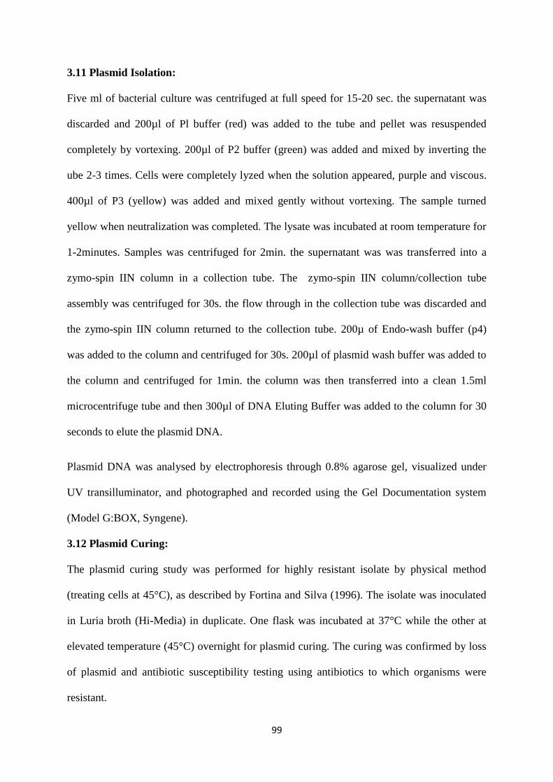
99
3.11 Plasmid Isolation:
Five ml of bacterial culture was centrifuged at full speed for 15-20 sec. the supernatant was
discarded and 200µl of Pl buffer (red) was added to the tube and pellet was resuspended
completely by vortexing. 200µl of P2 buffer (green) was added and mixed by inverting the
ube 2-3 times. Cells were completely lyzed when the solution appeared, purple and viscous.
400µl of P3 (yellow) was added and mixed gently without vortexing. The sample turned
yellow when neutralization was completed. The lysate was incubated at room temperature for
1-2minutes. Samples was centrifuged for 2min. the supernatant was was transferred into a
zymo-spin IIN column in a collection tube. The zymo-spin IIN column/collection tube
assembly was centrifuged for 30s. the flow through in the collection tube was discarded and
the zymo-spin IIN column returned to the collection tube. 200µ of Endo-wash buffer (p4)
was added to the column and centrifuged for 30s. 200µl of plasmid wash buffer was added to
the column and centrifuged for 1min. the column was then transferred into a clean 1.5ml
microcentrifuge tube and then 300µl of DNA Eluting Buffer was added to the column for 30
seconds to elute the plasmid DNA.
Plasmid DNA was analysed by electrophoresis through 0.8% agarose gel, visualized under
UV transilluminator, and photographed and recorded using the Gel Documentation system
(Model G:BOX, Syngene).
3.12 Plasmid Curing:
The plasmid curing study was performed for highly resistant isolate by physical method
(treating cells at 45°C), as described by Fortina and Silva (1996). The isolate was inoculated
in Luria broth (Hi-Media) in duplicate. One flask was incubated at 37°C while the other at
elevated temperature (45°C) overnight for plasmid curing. The curing was confirmed by loss
of plasmid and antibiotic susceptibility testing using antibiotics to which organisms were
resistant.

100
3.13 Phytochemical Screening of Agbo Herbal Mixture
Quantitative phytochemical content of agbo herbal mixture was carried out using method
described by Chionyedua et al. (2015)
3.13.1 Determination of total phenols by spectrophotometric method
Five (5) ml of the extract was pipetted into a 50ml flask, then 10ml of distilled water was
added. 2ml of ammonium hydroxide solution and 5 ml of concentrated amyl alcohol were
also added. The samples were made up to mark and left to react for 30 min for colour
development. This was measured at 505nm using spectrophotometer.
3.13.2 Alkaloid determination using
Five (5) ml of the sample was weighed into a 250 ml beaker and 200 ml of 10% acetic acid in
ethanol was added and covered and allowed to stand for 4 h. This was filtered and the extract
was concentrated on a water bath to one quarter of the original volume. Concentrated
ammoniumhydroxide was added drop wise to the extract until the precipitation was complete.
The whole solution was allowed to settle and the precipitated was collected and washed with
dilute ammonium hydroxide and then filtered. The residue is the alkaloid, which was dried
and weighed.
3.13.3 Flavanoid determination
Ten (10) ml of the agbo sample was extracted repeatedly with100 ml of 80% aqueous
methanol at room temperature.The whole solution was filtered through whatman filter paper.
The filtrate was later transferred into a crucible and evaporated into dryness over a waterbath
and weighed to a constant weight.
3.13.4 Saponin determination
Twenty (20) ml of agbo sample was dispersed in 200 ml of 20 %ethanol. The suspension was
heated over a hot waterbath for 4 h with continuous stirring at about 55ºC. The mixture was
filtered and the residue re-extracted with another 200 ml of 20% ethanol. The combined
101
extracts were reduced to 40 ml over water bath at about 90ºC. The concentrate was
transferred into a 250 ml separating funnel and 20 ml of diethyl ether was added and shaken
vigorously. The aqueous layer was recovered while the ether layer was discarded. The
purification process was repeated. Sixty (60) ml of normal butanol extracts were washed
twice with 10 ml of 5% aqueous sodium chloride. The remaining solution was heated in a
water bath. After evaporation the sample were dried in the oven into a constant weight. The
saponin content was calculated in percentage.
3.14 pH and Heavy Metal determination in Agbo Mixture
3.14.1pH determination
An 11751 SensorE-5000 pH meter (Garden Grove scientific, USA) was used for pH
determination. The pH were determined directly without the addition of demineralised water.
3.14.2 Atomic absorption spectrophometer.
A VGP model 210 (Bulk scientific, USA) was employed for the determination of the heavy
metals in the samples. Buck scientific hallow cathode lamps for Pb, Cu, Ni, Fe, Cr and Zn
were used as recitation sources.Under optimum operating conditions, the metals were
measured using an air-acetylene flame. The absorbance of each metal in the sample solution
was obtained and the calibration curve prepared to obtain the concentration.

102
CHAPTER FOUR
RESULTS
Total bacterial counts ranged from 0.04±0.002 x10⁴ cfu/ml in agbo samples purchased from
Santana market to 1.13±0.7 x10⁴ cfu/ml in samples from Uselu market. Total fungal counts
ranged from 0.70±0.40 x10⁴ cfu/ml in samples from Oba market to 1.00±0.60 x10⁴ cfu/ml in
samples from Uselu markets (Table 1)
Bacterial isolates recovered from agbo herbal mixture included Bacillus cereus, Escherichia
coli, Serratia marcescens, Lactobacillus casei, Bacillus subtilis, Micrococcus varians,
Pseudomonas aeruginosa and Staphylococcus aureus while fungal isolates included
Penicillium italicum, Aspergillus flavus, Aspergillus niger, Rhizopus stolonifer, Penicillium
chrysogenum and Mucor mucedo (plates 1 and 2).
The most occurring bacterial isolate was Bacillus cereus with percentage occurrence of
21.05% while the lease occurring bacterium were Serratia marcescens and Pseudomonas
aeruginosa (5.26%) (Table 2).
103
The most prevalent fungal isolate was Aspergillus flavus (22.75%) while the least prevalent
fungal isolate was Mucor mucedo (9.10%) table 3
Table 4 shows antibiotics sensitivity pattern of bacterial isolates. Escherichia coli and
Pseudomonas aeruginosa showed resistance to all but two (pefloxacin and ofloxacin) of the
antibiotics tested. Serratia marcescens was resistant to septrin (SXT), sparfloxacin (SP),
ciprofloxacin (CPX), and gentamicin (CN) but was sensitive to augmentin (AU), pefloxacin
(PEF) and ofloxacin (OFX). Bacillus subtilis was sensitive to almost all antibiotics tested
except Ampicillin (APX). Bacillus cereus was also sensitive to most antibiotics tested but
showed resistance to ampicillin and amoxicillin. Plasmid profile revealed presence of plasmid
genes in the bacterial isolates.
Physicochemical analysis of agbo revealed the presence of Iron (Fe), Lead (Pb), Nickel (Ni),
Chromium (Cr), Copper (Cu) and Zinc (Zn). pH values ranged from 4.53±0.05 to 5.37±0.14
a shown in table 5.
Phytochemical tests revealed the presence of tannin, flavonoid, saponin, alkaloids and
phenols in the various samples (Table 6).
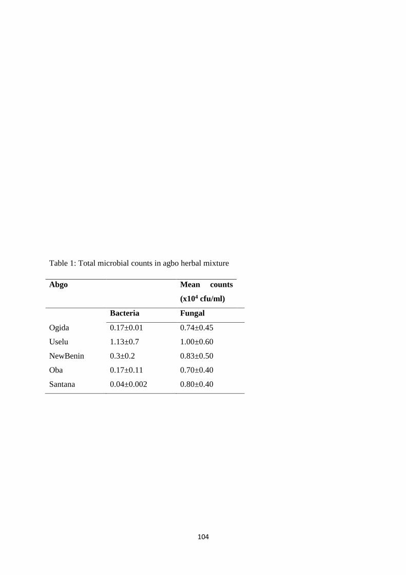
104
Table 1: Total microbial counts in agbo herbal mixture
Abgo Mean counts
(x104 cfu/ml)
Bacteria Fungal
Ogida 0.17±0.01 0.74±0.45
Uselu 1.13±0.7 1.00±0.60
NewBenin 0.3±0.2 0.83±0.50
Oba 0.17±0.11 0.70±0.40
Santana 0.04±0.002 0.80±0.40

105
Plate 1: PCR product of 16SrRNA on 1% Agarose Gel
B1 B2 B3 B4 B5 B6 B7 B8 M

106
Lane M = molecular size marker, B1=Bacillus cereus, B2=Escherichia coli, B3= Serratia
marcescens,B4=Lactobacillus casei, B5=Bacillus subtilis, B6=Micrococcus varians,
B7=Pseudomonas aeruginosa,B8= Staphylococcus aureus
Plate 2: PCR amplicons of the 18S rRNA genes of the fungal isolates.lane M: Molecular
weight marker, Lane F1=Penicillium italicum F2=Aspergillus flavus, F3=Aspergillus niger,
F4=Rhizopus stolonifer, F5=Penicillium chrysogenum, F6=Mucor mucedo
M F1 F2 F3 F4 F5 F6
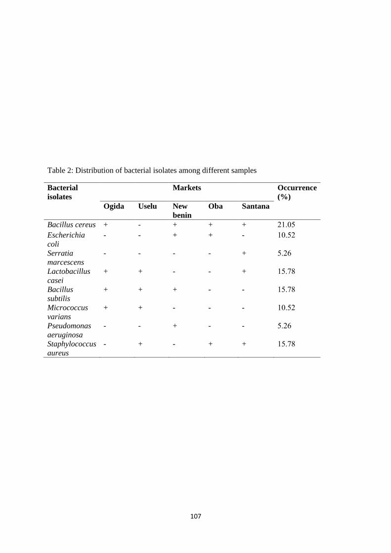
107
Table 2: Distribution of bacterial isolates among different samples
Bacterial
isolates
Markets Occurrence
(%)
Ogida Uselu New
benin
Oba Santana
Bacillus cereus + - + + + 21.05
Escherichia
coli
- - + + - 10.52
Serratia
marcescens
- - - - + 5.26
Lactobacillus
casei
+ + - - + 15.78
Bacillus
subtilis
+ + + - - 15.78
Micrococcus
varians
+ + - - - 10.52
Pseudomonas
aeruginosa
- - + - - 5.26
Staphylococcus
aureus
- + - + + 15.78
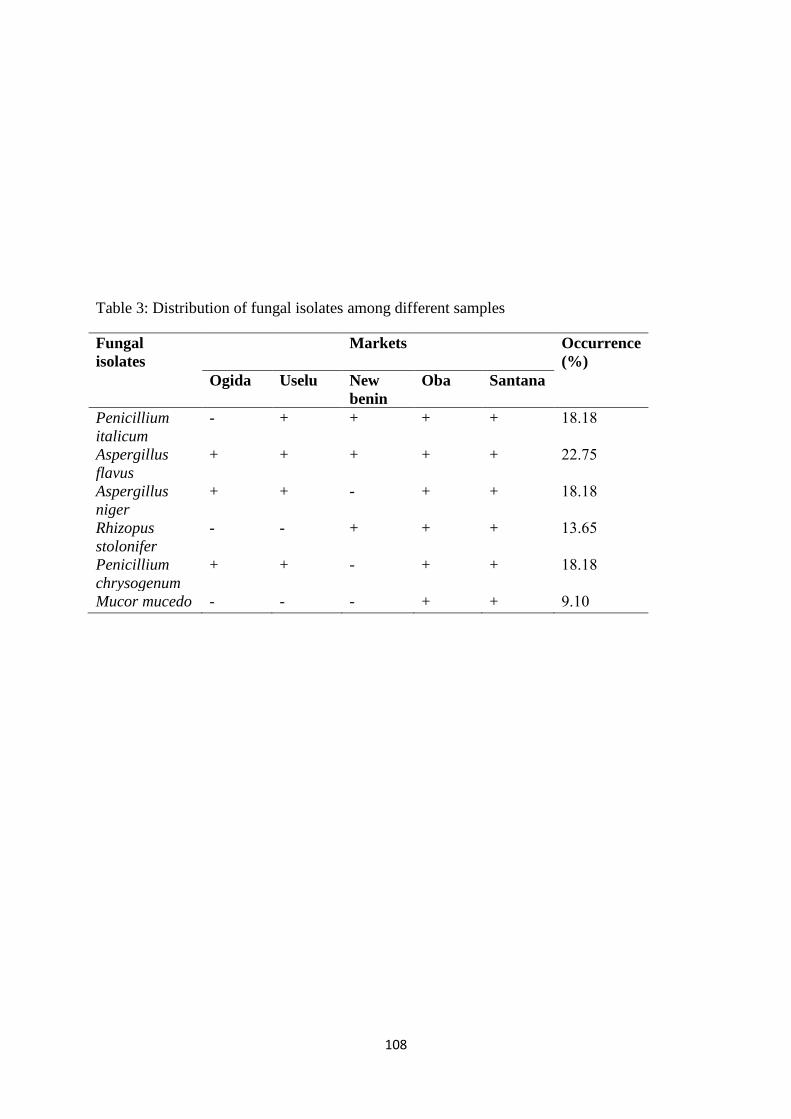
108
Table 3: Distribution of fungal isolates among different samples
Fungal
isolates
Markets Occurrence
(%)
Ogida Uselu New
benin
Oba Santana
Penicillium
italicum
- + + + + 18.18
Aspergillus
flavus
+ + + + + 22.75
Aspergillus
niger
+ + - + + 18.18
Rhizopus
stolonifer
- - + + + 13.65
Penicillium
chrysogenum
+ + - + + 18.18
Mucor mucedo - - - + + 9.10

109
Table 4: Antibiotic susceptibility pattern of bacterial isolates before curing
G-ve SXT CH SP CPX AM AU CN PEF OFX St
Escherichia coli 0.0 (R) 0.0 (R) 0.0 (R) 0.0 (R) 0.0 (R) 0.0 (R) 0.0 (R) 11.0 (I) 15.0 (I) 10.0 (R)
Serratia marcescens 10.0 (R) 0.0 (R) 20.0 (S) 10.0 (R) 15.0 (I) 25.0 (S) 10.0 (R) 20.0 (S) 10.0 (S) 11.0 (I)
Pseudomonas aeruginosa 0.0 (R) 9.0 (R) 0.0 (R) 10.0 (R) 0.0 (R) 0.0 (R) 0.0 (R) 11.0 (I) 15.0 (I) 10.0 (R)
G+ve PEF CN APX Z AM Ro CPX St SXT E
Bacillus subtilis 20.0 (S) 20.0 (S) 10.0 (R) 20.0 (S) 0.0 (R) 0.0 (R) 22.0 (S) 20.0 (S) 20.0 (S) 20.0 (S)
Micrococcus varians 15.0 (I) 18.0 (S) 0.0 (R) 11.0 (I) 0.0 (R) 10.0 (R) 21.0 (S) 14.0 (I) 10.0 (R) 16.0 (I)
Lactobacillus casei 20.0 (S) 20.0 (S) 0.0 (R) 20.0 (S) 0.0 (R) 15.0 (I) 20.0 (S) 20.0 (S) 10.0 (R) 20.0 (S)
Bacillus cereus 20.0 (S) 17.0 (I) 0.0 (R) 29.0 (S) 0.0 (R) 20.0 (S) 20.0 (S) 20.0 (S) 20.0 (S) 20.0 (S)
Staphylococcus aureus 0.0 (R) 10.0 (R) 17.0 (S) 15.0 (I) 10.0 (R) 0.0 (R) 15.0 (I) 0.0 (R) 15.0 (I) 10.0 (S)
Note
SXT = septrin, SP = sparfloxacin, CPX = ciprofloxacin, AM = amoxicillin, AU = augmentin, PEF = pefloxacin, OFX = ofloxacin, S =
streptomycin, CN = gentamicin, R = rocephin, Z = zinnacef, E = erythromycin, APX = ampicillin
I= Intermediate R= Resistant S= Sensitive
Resistance (R) = ≤10 mm. Intermediate (I) = 11-17 mm. Sensitivity (S) ≥ 18mm

110
Plate 3: Plasmid profile of multiple drug resistance bacterial isolates analyzed with 0.8%
agarose gel electrophoresis, stained with ethidium bromide. L is 0.5kb-48.5kb ladder
(molecular marker). Samples 1, 2, 3, 4 and 5 (E. coli, P. aeruginosa, Micrococcus virians,
Staphylococcus aureus and Serratia marcescens) are positive for plasmid genes. NC is a no
plasmid DNA template control.
L NC 1 2 3 4 5
48.5kb
0.5kb
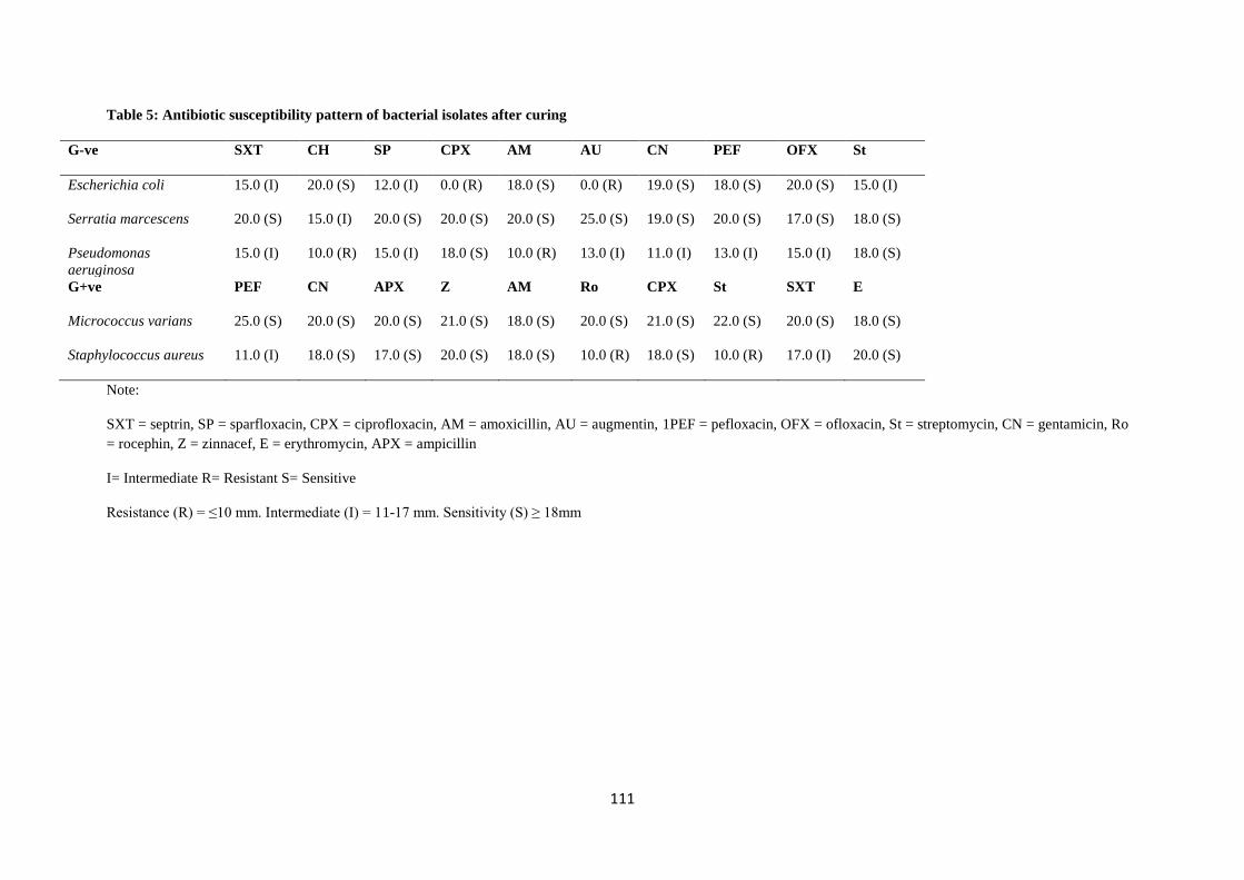
111
Table 5: Antibiotic susceptibility pattern of bacterial isolates after curing
G-ve SXT CH SP CPX AM AU CN PEF OFX St
Escherichia coli 15.0 (I) 20.0 (S) 12.0 (I) 0.0 (R) 18.0 (S) 0.0 (R) 19.0 (S) 18.0 (S) 20.0 (S) 15.0 (I)
Serratia marcescens 20.0 (S) 15.0 (I) 20.0 (S) 20.0 (S) 20.0 (S) 25.0 (S) 19.0 (S) 20.0 (S) 17.0 (S) 18.0 (S)
Pseudomonas
aeruginosa
15.0 (I) 10.0 (R) 15.0 (I) 18.0 (S) 10.0 (R) 13.0 (I) 11.0 (I) 13.0 (I) 15.0 (I) 18.0 (S)
G+ve PEF CN APX Z AM Ro CPX St SXT E
Micrococcus varians 25.0 (S) 20.0 (S) 20.0 (S) 21.0 (S) 18.0 (S) 20.0 (S) 21.0 (S) 22.0 (S) 20.0 (S) 18.0 (S)
Staphylococcus aureus 11.0 (I) 18.0 (S) 17.0 (S) 20.0 (S) 18.0 (S) 10.0 (R) 18.0 (S) 10.0 (R) 17.0 (I) 20.0 (S)
Note:
SXT = septrin, SP = sparfloxacin, CPX = ciprofloxacin, AM = amoxicillin, AU = augmentin, 1PEF = pefloxacin, OFX = ofloxacin, St = streptomycin, CN = gentamicin, Ro
= rocephin, Z = zinnacef, E = erythromycin, APX = ampicillin
I= Intermediate R= Resistant S= Sensitive
Resistance (R) = ≤10 mm. Intermediate (I) = 11-17 mm. Sensitivity (S) ≥ 18mm
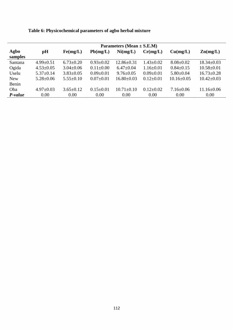
112
Table 6: Physicochemical parameters of agbo herbal mixture
Agbo
samples
Parameters (Mean ± S.E.M)
pH Fe(mg/L) Pb(mg/L) Ni(mg/L) Cr(mg/L) Cu(mg/L) Zn(mg/L)
Santana 4.99±0.51 6.73±0.20 0.93±0.02 12.86±0.31 1.43±0.02 8.08±0.02 18.34±0.03 Ogida 4.53±0.05 3.04±0.06 0.11±0.00 6.47±0.04 1.16±0.01 0.84±0.15 10.58±0.01 Uselu 5.37±0.14 3.83±0.05 0.09±0.01 9.76±0.05 0.09±0.01 5.80±0.04 16.73±0.28 New
Benin
5.28±0.06 5.55±0.10 0.07±0.01 16.80±0.03 0.12±0.01 10.16±0.05 10.42±0.03
Oba 4.97±0.03 3.65±0.12 0.15±0.01 10.71±0.10 0.12±0.02 7.16±0.06 11.16±0.06 P-value 0.00 0.00 0.00 0.00 0.00 0.00 0.00
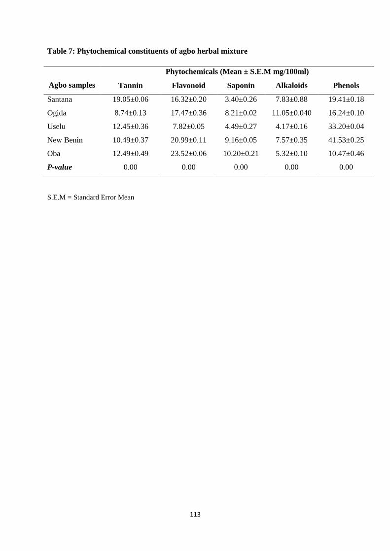
113
Table 7: Phytochemical constituents of agbo herbal mixture
Agbo samples
Phytochemicals (Mean ± S.E.M mg/100ml)
Tannin Flavonoid Saponin Alkaloids Phenols
Santana 19.05±0.06 16.32±0.20 3.40±0.26 7.83±0.88 19.41±0.18
Ogida 8.74±0.13 17.47±0.36 8.21±0.02 11.05±0.040 16.24±0.10
Uselu 12.45±0.36 7.82±0.05 4.49±0.27 4.17±0.16 33.20±0.04
New Benin 10.49±0.37 20.99±0.11 9.16±0.05 7.57±0.35 41.53±0.25
Oba 12.49±0.49 23.52±0.06 10.20±0.21 5.32±0.10 10.47±0.46
P-value 0.00 0.00 0.00 0.00 0.00
S.E.M = Standard Error Mean
114
CHAPTER FIVE
DISCUSSION
The microbiological background of herbal medicines depends on several environmental
factors and exerts an important impact on the overall quality of the herbal products and
preparations (Akerele, 1993). Agbo herbal mixtures were purchased from five different
markets (Uselu, New Benin, Oba, Santana and Ogida Market) in Benin City with different
levels of sanitation. Result of the microbial load in agbo mixture is in consonance with the
observation by Agbulu et al. (2016) who reported the microbiological quality of cough syrups
and herbal solutions in Markudi, Benue State. The result is slightly different from that of
Oluyege and Adelabu (2010) who reported a higher bacterial count of 4.0 x 10⁴ to 1.7 x
106cfu/ml in hawked herbal products in Ado-Ekiti. The difference could be attributed to the
fact that the Ado-Ekiti samples were hawked around in the streets, exposing the products to
different microorganisms. Also, the source of water and level of hygiene of the producers and
vendors could impact on microbial load. This is also true for the differences in microbial
counts observed from samples obtained from the different markets in Benin City.
Herbal medicines harbour various pathogenic microorganisms. This is because herbs are
made from trees and these plants have microorganisms adhered to their stems, barks, leaves,
flowers, fruits and roots. Though these microorganisms exist in their natural environment,
and are normal flora of the tree, they could be sources of infection, when in contact with
human body.
Microorganisms isolated from agbo mixtures were identified as Bacillus cereus, Escherichia
coli, Serratia marcescens, Lactobacillus casei, Bacillus subtilis, Micrococcus varians,
Pseudomonas aeruginosa and Staphylococcus aureus. The least occurring bacterial isolates
were Serratia marcescens and Pseudomonas aeruginosa (5.26%) while the highest occurring
was Bacillus cereus (21.05%). Fungi are common environmental contaminants of herbs and
115
contamination chiefly occurs due to a slow drying process, inadequate drying or during post-
harvest storage if relative humidity is high and temperatures are favourable (Sharma, 1990).
Six fungal isolates were identified and they include Penicillium italicum, Aspergillus flavus,
Aspergillus niger, Rhizopus stolonifer, Penicillium chrysogenum and Mucor mucedo. All
isolates were further confirmed using the polymerase chain reaction. Mucor mucedo was the
least occurring fungal isolate (9.10%) while Aspergillus flavus was the most occurring fungal
isolate (22.75%). The result is in agreement with reports by Agbulu et al. 2016; Abdulahi et
al. 2015; Oluyege and Adelabu 2010. Interestingly, Adeleye et al. (2005) reported the
presence of Klebsiella pneumoniae, Proteus vulgaris, P. rettgeri, Enterobacter aerogenes,
Citrobacter freundii, Bacillus subtilis, B. coagulans, B. cereus, Corynebacterium sp.,
Micrococcus varians and M. luteus in herbal preparations in Lagos, Nigeria. This could be as
a result of the different constituents used in herbal formulations with their respective flora,
the level of hygiene during preparation, packaging and storage of these products.
Apart from the unacceptable microbial loads observed in the samples, the presence of
contaminants considered to be completely unacceptable in herbal preparations was
demonstrated. The most common isolates in the tested samples were Gram-positive
organisms belonging to the genera Bacillus (21.05%) and Staphylococcus (15.78%).
Staphylococcus aureus are normal commensals of the mammalian skin, hands and mucous
membranes. Upon the consideration of the extent of human contact involved in the
preparation of herbal medicinal samples, it is most likely that sources of the contaminating
Staphylococcus spp. are the producers of the Agbo. This suggests that the level of hygiene of
persons involved in the preparation of the tested samples may be low. Similar studies carried
out on herbal samples include work by Chomnawang et al. (2003); Odedera and
Memuletiwon, (2014); Oluyege et al. (2010); Abba et al. (2009); Okunlola et al. (2007);
Adeleye et al. (2005) have all reported that the pathogens frequently isolated in herbal
116
products were S. aureus, E. coli and Pseudomonas aeruginosa. This work varies by reporting
a higher count of Bacillus cereus. Contamination by Bacillus cereus (21.05%) could have
arisen during growth of the herbs as the bacterium is commonly found in soils. This finding is
in contrast with the report of Odedera and Mumuletiwon (2014) who found more of
Escherichia coli and Penicillium notatum as herbal contaminants in Abeokuta, Nigeria and
this could be as a result of differences in the hygiene level of the producers. Escherichia coli,
a major faecal coliform may have been introduced from the water used during processing of
the herbs.
Microbial contamination of agbo herbal mixture as shown in this study, may also be as a
result of the plant materials, utensils used during preparation, poor hygiene of the
manufacturer or even the packaging vessel after processing. Microorganisms are present
everywhere and can easily contaminate any substrate. Considering the packaging materials, it
is worthy of note that this contributes greatly to microbial contamination as the final stage of
the processing is packaging. Most of the packaging cans used are not sterilized and are
usually picked up where they are found littered along the road or in public places and barely
washed before being used to package finished agbo products. It was observed that fungal
growth was more than bacterial growth and this is attributable to the low pH value of all the
agbo samples which is favourable for fungal growth.
The high microbial load and presence of specific pathogens in the tested Agbo heave serious
clinical as well as pharmaceutical implications. Clinically, consumers of any of these
products are at risk of contracting infections by the different pathogens which may be of great
consequence if not identified and treated appropriately. Presence of Eschericha coli in the
sample indicates poor hygiene practices and lack of adequate handling of the products.
According to the European pharmacopoeia 2007, no Salmonella sp or Eschericha coli strain
should be present in samples (Oluyege and Adelabu, 2010). The intake of a high
117
concentration of accumulated toxins produced by organisms such as Bacillus cereus,
Staphylococcus aureus and Aspergillus flavus may lead to undesirable reactions in
consumers. Oyetayo (2008) reported that the presence of Bacillus spp. in herbal preparations
is an indication that the water used in the preparation of the products is not from a good
source. The risk is greater if the consumer is a young child with undeveloped immunity, an
elderly with diminished immunity or the immunocompromised patients. Incidentally, these
groups of consumers are the most in need of herbal medicines for the treatment of many
diseases to which they are susceptible. Pharmaceutically, the presence of microbial
contaminants may lead to the spoilage of the products, thus reducing their shelf life.
Antibiotic resistance of microorganisms is an area of growing concern because antibiotic
resistant strains can be detrimental as they are capable of transferring the resistance gene to
pathogenic bacteria (some of which are part of the human microflora) when consumed. There
is therefore the necessity to study the susceptibility patterns of the isolates from herbal
mixtures before and after curing. Thirteen (13) selected antibiotics were used in this study
with each possessing a unique mechanism of action. Escherichia coli and Pseudomonas
aeruginosa showed resistance to all but two (perfloxacin and ofloxacin) of the antibiotics
tested. Serratia marcescens was resistant to septrin (SXT), sparfloxacin (SP), ciprofloxacin
(CPX), and gentamicin (CN) but was sensitive to augmentin (AU), pefloxacin (PEF) and
ofloxacin (OFX). This is in agreement with the report of Oluyege and Adelabu (2010).
Bacillus subtilis was sensitive to almost all antibiotics tested. It however was resistant to
Ampicillin (APX). Bacillus cereus was also sensitive to most antibiotics tested but showed
resistance to ampicillin and amoxicillin. It is noteworthy that all Gram positive organisms
tested (except Staphylococcus aureus) were resistant to ampicillin, and amoxicillin (except
Bacillus subtilis). This could be attributed to the widespread use of these drugs. People
purchase these drugs over the counter without recourse to the doctors’ prescription. In fact
118
they are now regular drugs in most households. The ability of some of the antibiotics applied
in the sensitivity tests to resist the growth of opportunistic pathogens such as B. cereus and E.
coli indicate the potency of these orthodox medicine against such bacteria and might be
resorted to by herbal consumers in case of probable infections (Odedera and Memuletiwon,
2014). The plasmid profile of multiple drug resistance bacterial genes isolated was also
analysed. The electropherogram was positive for the respective plasmid genes of the isolates
showing the contributions of the plasmid genes to drug resistance.
Lead, cadmium, mercury, thallium and arsenic have been shown to be contaminants of
different herbal ingredients and limit tests for such toxic metals are essential for herbal
ingredients (Kunle et al., 2012). Physicochemical analysis of agbo revealed the presence of
iron (Fe), lead (Pb), nickel (Ni), chromium (Cr), copper (Cu) and zinc (Zn). The samples
were all acidic with pH range of 4.53±0.05 to 5.37±0.14. Ogida samples had the lowest pH
(4.53±0.05) while the highest was recorded for Uselu samples (5.37±0.14). The difference
could be as a result of the water used in preparation as some water sources are more acidic
than others. The pH observed is slightly lower than that reported by Akande et al. (2012) who
gave the pH range to be 5.30-6.80 among locally prepared herbal remedies in Lagos
Metropolis. This could be attributed to differences in soil acidity for cultivation and water
used in processing.
The lowest iron concentration (3.04±0.06 mg/l) was recorded for Ogida samples while
Santana market samples had the highest levels of iron (6.73±0.20 mg/l). The concentration of
lead observed ranged from 0.07±0.01 mg/l to 0.15±0.01 mg/l. Cr, Cu, Ni and Zn are beneficial
to man at lower or standard levels, since they are integral parts of important physiologic
compound such as Zn, Cr, Ni and Cu in certain enzymes, where it is essential for their
activity. Cr, Ni, and Zn have been suggested as essential trace elements in nutrition, which
functions include regulation of apoptosis, activation of depressed immune system, and as
119
cofactors for metalloenzymes. Ni is involved in fat metabolism and aid in fat deposition
(Goyer, 1995). The levels of Cu, Ni and Zn in the herbal mixtures are generally higher than
those of, Pb, and Cr. Cu, Ni, and Zn and are of no toxicological significance, except for cases
of bioaccumulation as seen in hypercupremia which could lead to neurological/behavioural
alterations. This finding is in line with the observation of Gbaruko and Friday (2007). From
the results, it seems all the samples analyzed have very high concentration of the heavy
metals investigated. The implication is that consumers of these drugs may be exposed to
heavy metals due to regular consumption.
The high levels of Pb and Cr in the herbal mixtures are of public health concern. The levels
are far beyond the tolerable level of 0.001 μg/g set by WHO. Though these herbal drugs are
processed before consumption, the effect of processing could be minimal, since the heavy
metals are non-degradable. This is in agreement with the report of Gbaruko and Friday
(2007) who found high concentrations of these heavy metals in the fauna and flora in the Ijaw
area of Niger Delta of Nigeria.
There is growing concern about the physiological, biochemical and behavioural effects of
environmental trace metal in human population. The toxicity of lead (Pb) at high level of
exposure is well known, but the concern of today is the possibility that continual exposure
may result to gradual accumulation of these metals in the human system and may lead to
adverse health effects.
The preliminary phytochemical tests showed the presence of tannin, flavonoid, saponin,
alkaloids and phenols in the various samples. Santana market samples recorded the highest
tannin levels (19.05±0.06mg/100ml) while Ogida market samples had the lowest
(8.74±0.13mg/100ml). Phenols content was generally higher across all samples with Oba
Market samples having the lowest (10.47±0.46mg/100ml) and New Benin samples recording
the highest (41.53±0.25mg/100ml). Flavonoid content was lowest in Uselu market samples
120
(7.82±0.05mg/100ml) and highest in Oba Market samples (23.52±0.06mg/100ml). Saponin
concentration also varied depending on the market with Santana market sample having the
lowest (3.40±0.26mg/100ml) and Oba market sample recording the highest
(10.20±0.21mg/100ml). Uselu market samples also recorded the lowest concentration of
alkaloids (4.17±0.16mg/100ml) while Ogida market samples had the highest concentration
(11.05±0.040mg/100ml). Raimi et al. (2014) presented a similar report from their
phytochemical investigation of Sida acuta; a widely used shrub in folk medicine.
The result of the phytochemical screening is in consonance with the report of Akande et al.
(2012) who evaluated the biochemical constituents of locally prepared herbal remedies in
high demand in Lagos metropolis, Nigeria. However, Pandey et al. (2014) observed only the
presence of two phytochemicals (saponin and tannin) in Pterocarpus santalinus extracts. This
could be due to the fact that the herbal remedies used for this research were polyherbals
(mixtures of different plant constituents used as medicine) rather than a single plant as used
by them.
121
CONCLUSION
The results of this study show contamination of Agbo by microbes and heavy metals. Since
applications of herbal medicines for curative purposes is on the increase, there is a need for
risk assessment of microbial load of the medicinal plants at critical control points during
processing. Nigerian government also need to introduce some standards that must be met by
every herbal processor and seller. There is a need for good sanitation training of all herbal
processors so as to safeguard the health of Agbo consumers.

122
REFERENCES
Abdulahi, K., Igbeneghu, O.A. and Orafidiya, L.O. (2015). The Organoleptic and Microbial
Quality Of Some Herbal Medicinal Products Marketed In Freetown, Sierra Leone.
African Journal of Traditional and Complement Alternative Medicine 12(4):1-8
Abbiw, D.K. (1990). Useful Plants of Ghana: West African Uses of Wild and Cultivated
Plants. Intermediate Technology Publication and the Royal Botanic Gardens,
England.P. 102.
Abu-Darwish, S.M. (2009). Essential Oils Yield and Heavy Metals Content of Some
Aromatic Medicinal Plants Grown in Ash-Shoubak Region, South of Jordan.
Advances in Environmental Biology 3(3): 296-301.
Adamu, A.U., Mohammed, G.A., Faruruwa, Y. and Garba, M.D. (2014). Determination of
Heavy Metals in Some Medicinal Plants Commonly Used in Kura Local Government
Area of Kano State, Nigeria.Global Advanced Research Journal of Agricultural
Science 3(8): 256-258.
Adegbehingbe, O.O., Adesanya, S.A., Idowu, T.O., Okimi, O.C., Oyelami, O.A. and
Iwalewa, E.O. (2008). Clinical effects of Garcinia kola in knee osteoarthritis.Journal
of Orthopedic Surgery Research 3:34.
Adesokan, A.A., Akanji, M.A. and Yakubu, M.T. (2007). Antibacterial potentials of aqueous
extract of Enantia chlorantha stem bark. African Journal of Biotechnology
6(22):2502–2505.
Adewunmi, C.O. and Ojewole, J.A.O. (2004). Safety of Traditional medicines,
Complementary and Alternative Medicines in Africa.Africa Journal of Traditional,
Complementary and Alternative Medicine 1: 1-3.
Addo, V.N. (2007). Herbal Medicines: Socio-Demographic Characteristics and Pattern of
Use by Patients in a Tertiary Obstetrics and Gynaecology Unit. Journal of Science
and Technology 27(3): 149-155.
Adjanohoun, J.F., Aboubakar, N., Dramane, K., Ebot, M.E., Ekpere, J.A. and Enow-Orock,
E.G. (1996). Traditional Medecine and Pharmacopoeia: Contribution to Ethnobotanic
and Floristic Studies in Cameroon; Organization of African Unity Scientific,

123
Technical and Research Commission; Porto-Novo. Benin: Centre Nationale de
Production des Manuels Scolaires.p. 63.
Afzal, M., kazmi, I., Singh, R., Chauhan, M., Bisht, T. and Anwar, F. (2012). Bryophyllum
pinnatum: A review. International Journal of Research in Biological Sciences 2(4):
143-149.
Agbaje, E.O. and Onabanjo, A.O. (1998). Analgesic and antipyretic actions of Enantia
chlorantha extract in some laboratory animals. Nigerian Journal of Natural Products
Medicine 2:24–25.
Agbulu, C.O., Ameh, E.A. and Ocharifu, S.A. (2016). Journal of Microbiology and
Technology 1(1): 1-6.
Ahmad, H.A. (2014). Review on Anogeissus leiocarpus: A PotentAfrican Traditional Drug.
International journal of research in pharmacy and chemistry 4(3): 496-500.
Akande, I.S., Adewoyin, O.A., Njoku, U.F. and Awosika, S.O. (2012). Biochemical
Evaluation of Some Locally Prepared Herbal Remedies (Agbo) Currently On High
Demand in Lagos Metropolis, Nigeria. Journal of Drug Metabolism and Toxicology
3:3-9.
Alisi, C.S. and Onyeze, G.O.C. (2008). Nitric oxide scavenging ability of ethyl acetate
fraction of methanolic leaf extracts of Chromolaena odorata. Africa Journal of
Biochemistry Research 2 145-150.
Alakali, J.S., Ismaila, A.R., Alaka, I.C., Faasema, J. and Yaji, T.A. (2016). Quality
Evaluation of Herbal Tea Blends from Gingerand Pavetta crassipes. European
Journal of Medicinal Plants 12(4): 1-8
Ameh, S.J., Tarfa, F., Abdulkareem, T.M., Ibe, M.C., Onanuga, C. and Obodozie, O.O.
(2010). Physicochemical Analysis of the Aqueous Extracts ofSix Nigerian Medicinal
Plants.Tropical Journal of Pharmaceutical Research 9(2): 119-125.
Amira, C.O. and Okubadejo, N.U. (2007). Frequency of complementary and alternative
medicine utilization in hypertensive patients attending an urban tertiary care centre in
Nigeria.Complementary and Alternative Medicine 7:30-38.
Amole, O.O., Yemitan, O.K. and Oshikoya, K.A. (2009). Anticonvulsant activity of
Rauwolfia vomitoria (Afzel). African Journal of Pharmacy and Pharmacology 3:
319-322.
Anonymous (2000) Ethnoveterinary Medicine in Kenya.A field manualof traditional animal
health care pratices ITDG/IIRR, Nairobi, Kenya. P. 42
124
AOAC (2005). Official Methods of Analysis of AOAC International, 18th edn. AOAC
International, Gaithersburg, MD. 79p.
Arcamone, F., Cassinelli, G. and Casazza, A.M. (1980). New antitumor drugs from plants.
Journal of Ethnopharmacology 2: 149-160.
Ardabili, A.G., Farhoosh, R. and Khodaparast, M.H.H. (2011). Chemical Composition and
Physicochemical Properties ofPumpkin Seeds (Cucurbita pepo Subsp. pepo Var.
Styriaka) Grown in Iran.Journal of Agricultural Science and Technology 13: 1053-
1063.
Ashafa, A.O.T., Yakubu, M.T., Grierson, A.J. and Afolayan, A.J. (2009). Toxicological
evaluation of the aqueous extract of Felicia muricatta Thunb leaves in Wistar rats.
African Journal of Biotechnology 6: 949-954.
Ayoola, P.B., Adeyeye, A., Onawumi, O.O. and Faboya, O.O.P. (2011). Phytochemical and
nutrient evaluation of Tetracarpidium Conophorum (Nigerian Walnut)
Root.International Journal of Recent Research and Applied Studies7(2): 1-6.
Bansu, N. and Rastogi, R.P. (1967). Triterpenoin saponin and sapogenine. Phtochemistry 6:
1249-1270.
Barnes J, Anderson LA, Phillipson JD (2007). Herbal medicine. 3rd Edition, Pharmaceutical
Press, London. pp 1-23. Bauer, R. (1998). Quality criteria and standardization of phytopharmaceuticals: Can
acceptable drug standards be achieved? Drug Information Journal 32: 101–110.
Belewu, M.A., Olarunde, O.A. and Giwa, T.A. (2009). Underutilized medicinal plants and
spices: Chemicalcomposition and phytochemical properties. Journal of Medicinal
Plants Research 3(12): 1099-1103.
Bello, I.A., Ndukwe, G.I., Audu, O.T. and Habila, J.D. (2011). A bioactive flavonoid from
Pavettacrassipes K. Schum.Organic and Medicinal Chemistry Letters 1:14-19.
Bisset, N.G. (1994). Herbal Drugs and Phytopharmaceuticals. CRC Press, Boca Raton. 82p.
Bloomfield, S.F. (1991). Methods for Assessing Antimicrobial Activity. In: Denyer, S.P. and
Hugo, W. B. Mechanisms of Action of Chemical Biocides. Society for Applied
Bacteriology Technical series 27, Blackwell Scientific Publication. 122p.
Blumenthal, M., Brusse, W.R., Goldberg, A., Gruenwald, J., Hall, T., Riggins, C.W. and
Rister, R.S. (1998). The Complete German Commission E Monographs.Therapeutic
Guide to Herbal Medicines.The American Botanical Council, Austin, TX. 91p

125
Bodeker, C., Bodeker, G., Ong, C.K., Grundy, C.K., Burford, G. and Shein, K. (2005). WHO
Global Atlas of Traditional, Complementary and Alternative Medicine. World Health
Organization, Geneva. P. 56
Boumendjel, A., Sotoing, T.G., Ngo, B.E., Chabrol, T., Beney, C., Sinniger, V., Haudecoeur,
R., Marcourt, L., Challal, S., Ferreira, Q.E., Souard, F., Le Borgne, M., Lomberget,
T., Depaulis, A., Lavaud, C., Robins, R., Wolfender, J.L., Bonaz, B., De Waard, M.
and Sotoing T. (2013). Occurrence of the synthetic analgesic tramadol in an African
medicinal plant". Journal of Pharmacology 52: 11780–11784.
Briskin, D.P. (2000). Medicinal plants and phytomedicines. Linking plant biochemistry and
physiology to human health. Plant Physiology 24: 507–514.
Bwititi, P., Musabayane. C. T. and Nhachi, C.F. (2000). Effects of Opuntia megacantha on
blood glucose and kidney function in streptozotocin diabetic rats. Journal of
Ethnopharmacology 69: 247-252.
Chang, S.T., Chen, P.F. and Chang, S.C. (2001). Antibacterial activity of leaf essential oils
and their constituents from Cinnamomum osmophloeum. In: Wallace, R.J. (2004).
Antimicrobial Properties of plant secondary metabolites. Proceedings of the Nutrition
Society 63: 621 – 629.
Choi, C.W, Kim, S.C., Hwang, S.S., Choi, B.K., Ahn, H.J., Lee, M.Y., Park, S.H. and Kim,
S.K. (2002).Antioxidant activity and free radical scavenging capacity between
Korean medicinal plants and flavonoids by assay-guided comparison.Plant Science
163: 1161–1168.
Cichewicz, R. H., and Thorpe, P. A. (1996). The antimicrobial properties of chile peppers
(Capsicum species) and their uses in Mayan medicine. Clinical Microbiology Reviews
12: 121-129
Chidozie, V.N. and Adoga, G.I. (2014). Potentiating effect of aqueous leaf extract of
Anogeissus leiocarpus on Carica papaya aqueous leaf extract and Mangifera indica
aqueous stem bark extract - A herbal product used against typhoid fever in Nigeria.
International Journal of Current Microbiology and Applied Science 3(10) 1046-1062.
Chionyedua, T.O., Agbo, N. and Isiaka, A.O. (2015). Levels of potentially toxic metals in
selected herbal medicinesin Lagos, Nigeria. Journal of Natural Sciences Research
5(2): 148-156
Covello, P.S. (2008). Making artemisinin. Phytochemistry 69:2881-2885.
126
Cowan, M.M. (1999). Plants Products as Antimicrobial Agents.Clinical Microbiology
Reviews 18: 89-94.
Cos, P., Vlietinck, A.J., Berghe, D.V. and Maes, L. (2006). Anti-infective potential of natural
products: how to develop a stronger in vitro proof-of concept‘. Journal of
Ethnopharmacology 106: 290–302.
Danesi, M.A. and Adetunji, J.B. (1994). Use of alternative medicine by patients with
epilepsy: a survey of 265 epileptic patients in a developing country. Epilepsia
35:344-51.
De Smet, P.A.G.M. (1995). Health risks of herbal remedies. Drug Safety 13: 81-93.
De Smet, P.A.G.M., Keller, K., Hansel, R. and Chandler, R.F. (1997). Adverse Effects of
Herbal Drugs.Journal of Ethnopharmacology 21(3): 137-145.
De Smet, P.A.G.M. (1999). Overview of herbal quality control. Drug Information Journal
33: 717-724.
Divrikli, U., Saracoglu, S., Soylak, M. and Elci, L. (2006). Determination of trace heavy
metalcontents of green vegetable samples from Kayseri-Turkey by flame atomic
absorption spectrometry. Fresenius Environmental Bullettin 12:1123-1125
EMEA (2005). Guidelines on Quality of Herbal Medicinal Products/Traditional Medicinal
Products, EMEA/CVMP/814OO Review.European Agency for the Evaluation of
Medicinal Products (EMEA), London. 88pp Elhardallou, S.B. (2011). Cytotoxicity and biological activity of selected Sudanese medicinal
plants. Research Journal of Medicinal Plant 5:201-229.
Eskinazi, D., Blumenthal, M., Farnsworth, N. and Riggins, C.W. (1999). Botanical Medicine:
Efficacy, Quality, Assurance, and Regulation. Mary Ann Liebert, New York. 157pp
Eze, S.O. and Obinwa, E. (2014). Phytochemical and Nutrient Evaluation of the Leaves and
Fruits of Nauclea Latifolia (Uvuru-ilu). Communications in Applied Sciences 2(1): 8-
24.
Fakeye, T.O., Adisa, R., Musa, I.E. (2009). Attitude and use of herbal medicines among
pregnant women in Nigeria.Complementary and Alternative Medicine9:53-60.
Fasola, T.R., Adeyemo, F.A., Adeniji, J.A. and Okonko, I.O. (2011). Antiviral potentials of
Enantia chlorantha extracts on yellow fever virus. Natural Science 9: 99-105.
Fahad, M.A. and Mohammed, S.A. (2012). Chemical Composition and Fatty Acid Content of
Some Spicesand Herbs under Saudi Arabia Conditions.The ScientificWorld Journal
4: 1-5.
127
Farombi, EO. (2003). African indigenous plants with chemotherapeutic potentials and
biotechnological approach to the production of bioactive prophylactic agents. African
Journal of Biotechnology 2: 662–671.
Gbaruko, B.C. and Friday, O.U. (2007). Bioaccumulation of heavy metals in some fauna and
flora. International Journal of Environmental Science and Technology 4 (2): 197-202.
Gullece, M., Aslan, A., Sokmen, M., Sahin, F., Adiguzel, A., Agar, G. and Sokmen, A.
(2006). Screening the antioxidant and antimicrobial properties of the lichens Parmelia
saxatilis, Platismatiaglauca, Ramalina pollinaria, Ramalina polymorpha and
Umbilicaria nylanderian. Phytomedicine 13: 515–521.
Harborne, J.B. (1984). Phytochemical Methods: A Guide to Modern Technique of Plant
Analysis (2nd ed.), Chapman and Hall, Hong Kong, pp. 37, 84, 101, 103 – 104, 192.
Hollman, A. (1985). Plants and cardiac glycosides. British Heart Journal 54: 258-261.
Hugo, W.B. and Bloomfield, S.F. (1971). Studies of the mode of action of the phenolic
antibacterial agent Fentichlor against Staphylococcus aureus and Escherichia coli.1.
The adsorption of Fentichlor by the bacterial cell and its antibacterial. Journal of
Applied Bacteriology34: 557 – 567.
Igweze, Z.N., Orisakwe, O.E. and Obianime, A.W. (2012). Nigerian herbal remedies and
heavy metals: violation of standard recommended guidelines. Asian Pacific Journal
of Tropical Biomedcine 11: 1423-1430.
Ijeh, I.I. and Agbo, C.A. (2006). Body organ weight changes following the administration of
aqueous extracts of Ficus exasperate Vahl. Journal of Animal and Vetinary
Advance5: 277-279.
Ikhuoria, E., U. and Maliki, M. (2007). Characterization of avocado pear (Persea americana)
and African pear extracts. African Journal of Biotechnology 6 (7): 950-952.
Ilori, M., Sheteolu, A.O., Omonigbehin, E.A. and Adeneye, A.A. (1996). Antidiarrhoeal
activities of Ocimum gratissimum (Lamiaceae). Journal of Diarrhoeal Disease
Research 4:283-285.
Imaga, N.O.A., Gbenle, G.O., Okochi V.I., Akanbi S.O., Edeoghon S.O., Oigbochie V.,
Kehinde, M.O. and Bamiro S.B. (2009). Antisickling property of Caricapapaya leaf
extract. African Journal of Biochemistry Research 3(4): 102-106.
Indran, M., Mahmood, A.A. and Kuppusamy, U.R. (2008). Protective effect of Carica
papaya L leaf extract against alcohol induced acute gastric damage and blood
oxidative stress in rats. West Indian Medical Journal 57(4): 323.
128
Isitua, C.C., Lozano, M.J.S., Jaramillo, C.J. and Dutan, F. (2015). Phytochemical and
nutritional properties of dried leaf powder of Moringaoleifera Lam. from machala el
oro province of Ecuador. Asian Journal of Plant Science and Research 5(2):8-16.
Iwu, M.M. (1993). LL.C. USA: RC Press; Handbook of African Medicinal plants. pp. 1–13.
Joffe, P. (2007). Indigenous Plants of South Africa. Briza Publications. Pp. 123
Jon, C.T. and Ted, J.K. (2008). Herbal medicine research and global health: an ethical
analysis. Bulletin of the World Health Organization 86: 594–599.
Joshi, B., Lekhak, S. and Sharma, A. (2009). Antibacterial Property of Different Medicinal
Plants: Ocimum sanctum, Cinnamomum zeylanicum, Xanthoxylum armatum and
Origanum majorana. Kathmandu University Journal of Science, Engineering and
Technology 5(1): 143 – 150.
Joshi, A. and Chauhan, R.S. (2013). Phytochemical analysis and Cytotoxicity studies of
Bryophyllum calycium in BHK-21 cells A. Scholars Academic Journal of Pharmacy 2
(3): 190-194.
Kazmi, M. H., Malik, A., Hameed, S., Akhtar, N. and Noor A. S. (1994). An anthraquinone
derivative from Cassia italica. In: Cowan, M.M. (1999). Plants Products as
Antimicrobial Agents. American Society for Microbiology (ed.) Clinical
Microbiology Reviews 12(4): 121-129.
Kafaru, E. (1994). Immense help formative workshop. In: Khan, R., Islam, B., Akram, M.,
Shakil, S., Ahmad, A., Ali, M.S., Sadiqui, M., Khan, A.U. (2008). Antimicrobial
Activity of Five Herbal Extracts against Multi Drug Resistant (MDR) strains of
Bacteria and Fungus of Clinical Origin. Molecules 13:97-103.
Kalagbor, I.A., Barisere, V., Barivule, G., Barile, S. and Bassey, C. (2014). Investigation of
the Presence of Some Heavy Metals in Four Edible Vegetables, Bitter Leaf (Vernonia
amygdalina), Scent Leaf (Ocimum gratissimum), Water Leaf (Talinum triangulare)
and Fluted Pumpkin (Telfairia occidentalis) from a Cottage Farm in Port Harcourt.
Research Journal of Environmental and Earth Sciences 6(1): 18-24.
Kaul, T. N., Middletown, E. and Ogra, P. L. (1985). Antiviral effect of flavonoids on human
viruses. In: Cowan, M.M. (1999). Plants Products as Antimicrobial Agents.Clinical
Microbiology Reviews 12(4): 100-108.
Kayode, A. A. A. and Kayode, O. T. (2011). Some medicinal values of Telfaria occidentalis:
A review. American Journal of Biochemistry and Molecular Biology 1: 30-38.
129
Kigigha, L.T. and Zige, D.V. (2013). Activity of Chromolaena Odorata on Enteric and
Superficial Etiologic Bacterial Agents.American Journal of Research and
Communication 1 (11): 1-11.
Kong, J., Goh, N.. Chia, L. and Chia, T. (2003). Recent Advances in Traditional Plant Drugs
and Orchids.Acta Pharmacology 24(1):7-21.
Kunle, O.F., Egharevba, H.O. and Ahmadu, P.O. (2012). Standardization of herbal medicines
- A review. International Journal of Biodiversity and Conservation4 (3): 101-112.
Kulhari, A., Sheorayan, A., Bojar, S., Sarkar, S., Chauldhury, A. and Kalia, A.R. (2013).
Investigation of heavy metals in frequently utilized medicinal plants collected from
environmentally diverse locations of North Western India. SpringerPlus2:676-683.
Langlois-Klassen, D., Kipp, W., Jhangri, G.S. and Rubaale, T. (2007). Use of traditional
herbal medicine by AIDS patients in Kabarole district, Western Uganda. American
Tropical and Medical Hygiene 77:757-763.
Lulekal, E., Kelbess, E., Bekele, T. and Yineger, H. (2008). An ethnobotanical study of
medicinal plants in Mana Angetu District, Southeastern Ethiopia.Journal of
Ethnobiolology and Ethnomedicine 4(10): 1746–1752.
Maghrabi, A.I. (2014). Determination of Some Minerals and Heavy Metals in Saudi Arabia
Popular Herbal Drugs Using Modern Techniques.African Journal of Pharmacy and
Pharmacology 8(36): 893-898.
Maregesi, S.M., Pieters, L., Ngassapa, O.D., Apers, S., Vingerhoets, R., Cos, P., Berghe, D.
and Vlietinck, A. (2008). Screening of some Tanzanian medicinal plants from Bunda
district for antibacterial, antifungal and antiviral activities. Journal of
Ethnopharmacology 119: 58–66.
Mendoza, L., Wilkens, M. and Urzua, A. (1997). Antimicrobial study of the resinous
exudates and of diterpenoids and flavonoids isolated from some Chilean
Pseudognaphalium (Asteraceae). In: Cowan, M.M. (1999). Plants Products as
Antimicrobial Agents.Clinical Microbiology Reviews 12(4): 121-129.
Miller, N.J. and Rice-Evans, C.A. (1997). Factors influencing the antioxidant activity
determined by the ABTS radical cation assay. Free Radical Research 26: 195–199.
Mandel, I.S. (2009). Understanding Differences between Holistic, Alternative and
Complementary Medicine. Student Pulse 1(10):1-6.
Maoela, M.S. (2005). Studies on Some Biologically Active Natural Products from Tulbaghia
Alliacea.Masters Thesis. Belville: University of the Western Cape. 157pp.

130
Nair, R., Kalariya, T. and Chanda, S. (2005). Antibacterial activity of some selected Indian
medicinal flora. Turkish Journal of Biology 29: 41–47.
Ngari, W.F., Gikonyo, K.N., Wanjau, R.N. and Njagi, E.N.M. (2013). Investigation of
Selected Pathogenic Microorganisms and Toxic Elements in Herbal Materials Used in
Management of Oral Health in Nairobi County, Kenya.Journal of Applied
Environmental and Biological Sciences 3(12): 1-7.
Nwachukwu, A.M., Feng, H. and Achilike, K. (2010). Integrated study for automobile wastes
management and environmentally friendly mechanic villages in the Imo River Basin,
Nigeria. African Journal of Environmental Science and Technology 4(4): 234 - 294. Nwauzoma, A.B. and Dawari, S.L. (2013). Studies on the Phytochemical Properties and
Proximate Analysis of Piper Umbellatum (Linn) from Nigeria.American Journal of
Research Communication 1 (7): 164-177.
Nwoko, O.C. and Mgbeahuruike, L. (2011). Heavy Metal Contamination of Ready-to-use
Herbal Remedies in South Eastern Nigeria.Pakistan Journal of Nutrition 10(10):
959-964.
Nyarko, K.N., Aseidu-Gyekye, I.J. and Sittie, A.A. (2005). A Manuel of Harmonized
Procedures for Assessing the Safety, Efficacy and Quality of Plant Medicines in
Ghana, Ministry of Health: The Ghana National Drugs Programme. 202pp.
Oboh, G. (2006). Antioxidant and Antimicrobial Properties of Ethanolic Extract of Ocimum
gratissimum Leaves. Journal of Pharmacology and Toxicology 1(1): 47-53.
Oboh G, Nwanna E, Elusiyan C. (2010). Antioxidant and Antimicrobial Properties of
Telfairia occidentalis (Fluted pumpkin) Leaf Extracts. Journal of Pharmacology and
Toxicology 5(8): 539-547.
Ogbeibu, A.E. (2015) Biostatistics: a practical approach to research and data handling
(2nded.). Mindex publishing company, Lagos, Nigeria. pp: 17-22
Ogbera, A.O., Dada, O., Adeyeye, F. and Jewo, P.I. (2010). Complementary and alternative
medicine use in diabetes mellitus. West African Journal of Medicine 29:158-162.
Ogbonnia, S.O., Mbaka, G.O., Emordi J., Nkemhule, F., Joshua, P., Usman, A., Odusanya, P.
and Ota, D. (2013). Antimicrobial evaluation and acute and sub-acute toxicity
studies on a commercial polyherbal formulation, “Ade & Ade Antidiabetic®” used
131
in the treatment of diabetes in Southwestern Nigerian. Journal of Medicine and
Medical Sciences 4 (11): 423-432.
Okeniyi, J.A., Olubanjo, O.O., Ogunlesi, T.A. and Oyelami, O.A. (2005). Comparison of
healing of incised abscess wounds with honey and EUSOL dressing. Journal
Alternative and Complementary Medicine 11:511-513.
Okeniyi, J.A., Ogunlesi, T.A., Oyelami, O.A. and Adeyemi, L.A. (2007). Effectiveness of
dried Carica papaya seeds against human intestinal parasitosis: a pilot study. Journal
of Medicine and Food 10:194-196.
Okwu, D.E. and Nnamdi, F.U. (2011). Two novel flavonoids from Bryophyllum pinnatum
and theirantimicrobial Activity.Journal of Chemistry and Pharmacy Researc 3(2):1-
10.
Okwu, D.E. (2008). Citrus Fruits: A Rich Source of Phytochemicals and their Roles in
Human Health. International Journal of Chemical Sciences 6(2): 451-471.
Olorode, O.O., Idowu, M.A., Bamgbose, A. and Ayano, A.E. (2014). Chemical,
phytochemical and functional properties ofselected seeds’ flours. International
Journal of Nutrition and Food Sciences 3(6): 572-578.
Oloye, D.A., Odeja, O.T., Faboya, E.T. and Ibrahim, T.A. (2013). Proximate and Mineral
Composition of Wild Corchorous olitorius Seed Flour.Journal of Agriculture and
Allied sciences 3 (1): 1-4.
Oreagba, I.A., Oshikoya, K.A. and Amachree, M. (2011). Herbal medicine use among urban
residents in Lagos, Nigeria.BMC Complementary and Alternative Medicine 11:117-
125.
Oshikoya, K.A., Njokanma, O.F., Chukwura, H.A. and Ojo, O.I. (2007). Adverse drug
reactions in Nigerian children. Paediatric and Perinatal Drug Therapy 8:81-88.
Oshikoya, K.A., Senbanjo, I.O., Njokanma, O.F. and Soipe, A. (2008). Use of
complementary and alternative medicines for children with chronic health conditions
in Lagos, Nigeria.Complementary and Alternative Medicine 8:66-72.
Oyelami, O.A., Agbakwuru, E.A., Adeyemi, L.A. and Adedeji, G.B. (2005). The
effectiveness of grapefruit (Citrus paradisi) seeds in treating urinary tract infections.
Journal of Alternative and Complementary Medicine 11:369-371.
Oyelami, O.A., Onayemi, A., Oyedeji, O.A. and Adeyemi, L.A. (2009). Preliminary study of
effectiveness of Aloe vera in scabies treatment. Phytotherapy Research 23:1482-
1484.
132
Oyetayo, F.L. and Ogunrotimi, A.I. (2012). Guinea Corn (Sorghum vulgare) Leaf, a Potential
Source of Nutrients and Phytochemicals.Food and Public Health 2(6): 228-230.
Pandey, B., Gangrale, D., Upadhyay, N. and Tiwari, P. (2014). Physiochemical Analysis of
Pterocarpus Santalinus L. Extracts. Indian Journal of Scientific Research 4(1):201-
204.
Pei, S.J. (2001). Ethnobotanical approaches of traditional medicine studies: Some experiences
from Asia. Pharmaceutical Biology 39: 74-79.
Raimi, M.O., Oyekanmi, A.M. and Adegoke, B.M. (2014). Proximate, Phytochemical and
Micronutrient Composition of Sida acuta. Journal of Applied Chemistry 7 (2): 93-98.
Raskin, I. (1992). Role of Salicylic Acid in Plants.Annual Review of Plant Physiology and
Plant Molecular Biology 43:439-463.
Saplakogelu, U. and Iscan, M. (1997). DNA single-strand breakage in rat lung, liver and
kidney after single and combined treatments of nickel and Cadium.Mutation Research
394(1): 133-140.
Scalbert, A. 1991.Antimicrobial properties of tannins. Phytochemistry 30: 3875–3883.
Schulz, V., Hänsel, R., Tyler, V. E. (2001). Rational Phytotherapy.A Physician’s Guide to
Herbal Medicine, 4th ed. Springer-Verlag, Berlin. P. 125
Serafini, M., Ghiselli, A. and Ferro-Luzzi, A. (1994). Red wine, tea and antioxidants. Lancet
344:626-631.
Soetan, K.O. and Aiyelaagbe, O.O. (2009). The need for bioactivity-safety evaluation and
conservation of medicinal plants - A review. Journal of Medicinal Plants Research 3:
324-328.
Sofowora, A. (1996). Research on Medicinal Plants and Traditional Medicine in Africa.
Journal of Alternative and Complementary Medicine 2(3):365-372.
Solecki, R.S. (1975). Standardized product as well as the quality of the consumer information
on the herbal remedy. hanidar IV. Science 190: 880-885. Stephen, B. (2008). Herbal Medicine in the United States: Review of Efficacy, Safety, and
Regulation.Journal of General Internal Medicine 23: 854-859.
Suleiman, M.M., McGaw, L.J., Naidoo, V. and Eloff, J.N. (2010). Evaluation of several tree
species for activity against the animal fungal pathogen Aspergillus fumigatus. South
African Journal of Botany76: 64–71.
Veale, D.J.H., Furman, K.I. and Oliver, D.W. (1992). South African traditional
herbalmedicines used during pregnancy and childbirth. Journal of
Ethnopharmacology 36: 185-190.
133
Underwood, E. (1992). Eclogy of microorganism as it affects pharmaceutical industries. In:
Hugo WB, Russel AD (Eds). Pharmaceutical Microbology (5thedn). Blackwell
Scientific Publication, London.pp. 353.
Udoh, F.V. (2005) Activity of Alkaloid Extract of Carica papaya Seeds on Reproductive
Functions in Male Wistar Rats. Summary. Pharmaceutical Biology 43 (6): 563-567.
Uduak, E. and Nodeley, U. (2013). Comparative Phytochemical and Physicochemical
Properties of Aspilia africana and Tithonia diversifolia Leaves. International Journal
of Modern Biology and Medicine 3(3): 113-122.
Uraku, A.J., Onuoha, S.C., Nzube, E., Ezeani, N., Ogbanshi, M.E., Ezeali, C., Nwali, B.U.
and Ominyi, M.C. (2015). Nutritional and Anti-Nutritional Quantification Assessment
of Cymbopopgon citratus Leaf.Pharmacology and Pharmacy 6: 401-410.
Vasudevan, D.M. and Sreekumari, S. (2007). Textbook of Biochemistry for Medical
Students.Jaypee Brothers Medical Publishers, New Delhi.p. 712
Voutsa, D., A. Grimanis and Samara, C. (1996). Trace elements in vegetables grown in
industrial areas in relation to soil and air particulate matter. EnvironmentalPollution
94: 325-335.
Wani, M.S. (2007). Herbal medicine and its standardization. Pharmacy information 1: 6-12. Wallace, R.J. (2004). Antimicrobial Properties of plant secondary metabolites. Proceedings
of the Nutrition Society 63: 621-629.
WHO (1981). The International Pharmacopeia, Quality Specifications, 3rd edn. World Health
Organization, Geneva. p. 41 WHO (1996). Quality Assurance of Pharmaceuticals: A Compendum of Guidelines and
Related Materials, Good Manufacturing Practices and Inspection. World Health
Organization, Geneva. p. 76
WHO (2004). Guidelines on Good Agricultural and Collection Practices (GACP) for
Medicinal Plants. World Health Organization, Geneva. p. 94 WHO Media Centre: Traditional Medicine. Fact sheet N°134 2008 [http://
www.who.int/mediacentre/factsheets/fs134/en/], (Accessed June 2011).
WHO, (2002). World Health Organisation Traditional Medicine Strategy 2002–2005,
Geneva. p. 101
WHO guidelines for assessing quality of herbal medicines with reference to
contaminants and residues (2007).
WHO (2005). WHO Global Atlas of Traditional, Complementary and Alternative Medicine.
World Health Organization, Geneva. p.76
134
Wolf, P.L. (1999). Biochemical diagnosis of Liver diseases. Indian Journal of Clinical
Biochemistry14: 59-70.
Yidana, J.A. and Bayorbor, T. (2002). The Multi-Purpose Value of Medicinal Plants and the
Potential for Cultivation.Journal of the Ghana Science Association 4(2): 146- 158.
Yildirim, A., Oktay, M. and Bulaloulu, V. (2001). The antioxidant activity of the leaves of
Cydonia vulgaris. Turkish Journal of Medical Sciences 31: 23–27.
Zakaria, Z.A., Jais, A.M., Sulaiman, M.R., Mohamed, S.S.P. and Riffin, S. (2006). The in
vitro Antibacterial of Methanol and Ethanol Extracts of Carica papayaFlowers and
Mangifera indica Leaves. Pharmacology and Toxicology 1(3): 278-283.
Zin, N.M., Chit, Y.M., Bakar, N.F.A. (2013). Commercial Herbal SlimingProducts: Concern
for the Presence of Heavy Metals and Bacteria. Parkistan Journal of Biological
Sciences 2(1): 1-9.

135
APPENDIX I
DNA Sequences of Bacterial Isolates
Isolate
Code
Sequence blast Sequence
identity
B1
GGAATTATTGGGCGTAAAGCGCGCGCAGGTGGTTTCTTAAGTCTGATGTGAAAGCCCACGGCTC
AACCGTGGAGGGTCATTGGAAACTGGGAGACTTGAGTGCAGAAGAGGAAAGTGGAATTCCATGT
GTAGCGGTGAAATGCGTAGAGATATGGAGGAACACCAGTGGCGAAGGCGACTTTCTGGTCTGTA
ACTGACACTGAGGCGCGAAAGCGTGGGGAGCAAACAGGATTAGATACCCTGGTAGTCCACGCCG
TAAACGATGAGTGCTAAGTGTTAGAGGGTTTCCGCCCTTTAGTGCTGAAGTTAACGCATTAAGCA
CTCCGCCTGGGGAGTACGGCCGCAAGGCTGAAACTCAAAGGAATTGACGGGGGCCCGCACAAGC
GGTGGAGCATGTGGTTTAATTCGAAGCAACGCGAAGAACCTTACCAGGTCTTGACATCCTCTGAA
AACTCTAGAGATAGAGCTTCTCCTTCGGGAGCAGAGTGACAGGTGGTGCATGGTTGTCGTCAGCT
CGTGTCGTGAGATGTTGGGTTAAGTCCCGCAACGAGCGCAACCCTTGATCTTAGTTGCCATCATT
AAGTTGGGCACTCTAAGGTGACTGCCGGTGACAAACCGGAGGAAGGTGGGGATGACGTCAAATC
ATCATGCCCCTTATGACCTGGGCTACACACGTGCTACAATGGACGGTACAAAGAGCTGCAAGAC
CGCGAGGTGGAGCTAATCTCATAAAACCGTTCTCAGTTCGGATTGTAGGCTGCAACTCGCCTACA
TGAAGCTGGAATCGCTAGTAATCGCGGATCAGCATGCCGCGGTGAATACGTTCCCGGGCCTTGTA
CACACCGCCCGTCACACCACGAGAGTTTGTAACACCCGAAGTCGGTGGGGTAACCTTTATGGAG
CCAGCCGCCTAAGGTGGGACAGATGATTGGGGTGAA
Bacillus cereus
B2
GAATTATTGGGCGTAAAGGGCTCGCAGGCGGTTTCTTAAGTCTGATGTGAAAGCCCCCGGCTCAA
CCGGGGAGGGTCATTGGAAACTGGGGAACTTGAGTGCAGAAGAGGAGAGTGGAATTCCACGTGT
AGCGGTGAAATGCGTAGAGATGTGGAGGAACACCAGTGGCGAAGGCGACTCTCTGGTCTGTAAC
TGACGCTGAGGAGCGAAAGCGTGGGGAGCGAACAGGATTAGATACCCTGGTAGTCCACGCCGTA
AACGATGAGTGCTAAGTGTTAGGGGGTTTCCGCCCCTTAGTGCTGCAGCTAACGCATTAAGCACT
CCGCCTGGGGAGTACGGTCGCAAGACTGAAACTCAAAGGAATTGACGGGGGCCCGCACAAGCG
GTGGAGCATGTGGTTTAATTCGAAGCAACGCGAAGAACCTTACCAGGTCTTGACATCCTCTGACA
ATCCTAGAGATAGGACGTCCCCTTCGGGGGCAGAGTGACAGGTGGTGCATGGTTGTCGTCAGCT
CGTGTCGTGAGATGTTGGGTTAAGTCCCGCAACGAGCGCAACCCTTGATCTTAGTTGCCAGCATT
TAGTTGGGCACTCTAAGGTGACTGCCGGTGACAAACCGGAGGAAGGTGGGGATGACGTCAAATC
ATCATGCCCCTTATGACCTGGGCTACACACGTGCTACAATGGGCAGAACAAAGGGCAGCGAAAC
CGCGAGGTTAAGCCAATCCCACAAATCTGTTCTCAGTTCGGATCGCAGTCTGCAACTCGACTGCG
TGAAGCTGGAATCGCTAGTAATCGCGGATCAGCATGCCGCGGTGAATACGTTCCCGGGCCTTGTA
CACACCGCCCGTCACACCACGAGAGTTTGTAACACCCGAAGTCGGTGAGGTAACCTTTTTGGAGC
CAGCCGCCGAAGGTGGGACAGATGATTGGGGTGAAGTC
Escherichia coli
B3
GGAATTACTGGGCGTAAAGCGCACGCAGGCGGTCTGTCAAGTCGGATGTGAAATCCCCGGGCTC
AACCTGGGAACTGCATTCGAAACTGGCAGGCTAGAGTCTTGTAGAGGGGGGTAGAATTCCAGGT
GTAGCGGTGAAATGCGTAGAGATCTGGAGGAATACCGGTGGCGAAGGCGGCCCCCTGGACAAA
GACTGACGCTCAGGTGCGAAAGCGTGGGGAGCAAACAGGATTAGATACCCTGGTAGTCCACGCC
Serratia
marcescens
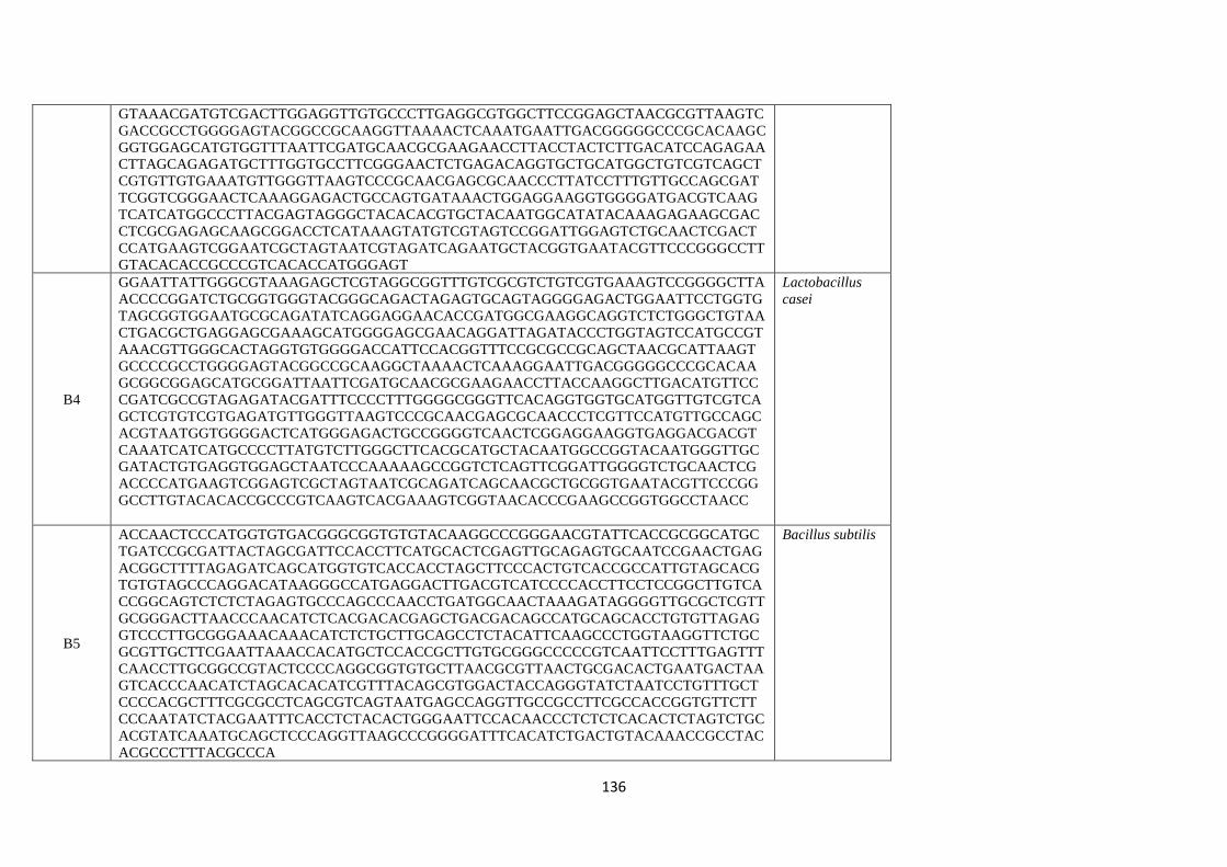
136
GTAAACGATGTCGACTTGGAGGTTGTGCCCTTGAGGCGTGGCTTCCGGAGCTAACGCGTTAAGTC
GACCGCCTGGGGAGTACGGCCGCAAGGTTAAAACTCAAATGAATTGACGGGGGCCCGCACAAGC
GGTGGAGCATGTGGTTTAATTCGATGCAACGCGAAGAACCTTACCTACTCTTGACATCCAGAGAA
CTTAGCAGAGATGCTTTGGTGCCTTCGGGAACTCTGAGACAGGTGCTGCATGGCTGTCGTCAGCT
CGTGTTGTGAAATGTTGGGTTAAGTCCCGCAACGAGCGCAACCCTTATCCTTTGTTGCCAGCGAT
TCGGTCGGGAACTCAAAGGAGACTGCCAGTGATAAACTGGAGGAAGGTGGGGATGACGTCAAG
TCATCATGGCCCTTACGAGTAGGGCTACACACGTGCTACAATGGCATATACAAAGAGAAGCGAC
CTCGCGAGAGCAAGCGGACCTCATAAAGTATGTCGTAGTCCGGATTGGAGTCTGCAACTCGACT
CCATGAAGTCGGAATCGCTAGTAATCGTAGATCAGAATGCTACGGTGAATACGTTCCCGGGCCTT
GTACACACCGCCCGTCACACCATGGGAGT
B4
GGAATTATTGGGCGTAAAGAGCTCGTAGGCGGTTTGTCGCGTCTGTCGTGAAAGTCCGGGGCTTA
ACCCCGGATCTGCGGTGGGTACGGGCAGACTAGAGTGCAGTAGGGGAGACTGGAATTCCTGGTG
TAGCGGTGGAATGCGCAGATATCAGGAGGAACACCGATGGCGAAGGCAGGTCTCTGGGCTGTAA
CTGACGCTGAGGAGCGAAAGCATGGGGAGCGAACAGGATTAGATACCCTGGTAGTCCATGCCGT
AAACGTTGGGCACTAGGTGTGGGGACCATTCCACGGTTTCCGCGCCGCAGCTAACGCATTAAGT
GCCCCGCCTGGGGAGTACGGCCGCAAGGCTAAAACTCAAAGGAATTGACGGGGGCCCGCACAA
GCGGCGGAGCATGCGGATTAATTCGATGCAACGCGAAGAACCTTACCAAGGCTTGACATGTTCC
CGATCGCCGTAGAGATACGATTTCCCCTTTGGGGCGGGTTCACAGGTGGTGCATGGTTGTCGTCA
GCTCGTGTCGTGAGATGTTGGGTTAAGTCCCGCAACGAGCGCAACCCTCGTTCCATGTTGCCAGC
ACGTAATGGTGGGGACTCATGGGAGACTGCCGGGGTCAACTCGGAGGAAGGTGAGGACGACGT
CAAATCATCATGCCCCTTATGTCTTGGGCTTCACGCATGCTACAATGGCCGGTACAATGGGTTGC
GATACTGTGAGGTGGAGCTAATCCCAAAAAGCCGGTCTCAGTTCGGATTGGGGTCTGCAACTCG
ACCCCATGAAGTCGGAGTCGCTAGTAATCGCAGATCAGCAACGCTGCGGTGAATACGTTCCCGG
GCCTTGTACACACCGCCCGTCAAGTCACGAAAGTCGGTAACACCCGAAGCCGGTGGCCTAACC
Lactobacillus
casei
B5
ACCAACTCCCATGGTGTGACGGGCGGTGTGTACAAGGCCCGGGAACGTATTCACCGCGGCATGC
TGATCCGCGATTACTAGCGATTCCACCTTCATGCACTCGAGTTGCAGAGTGCAATCCGAACTGAG
ACGGCTTTTAGAGATCAGCATGGTGTCACCACCTAGCTTCCCACTGTCACCGCCATTGTAGCACG
TGTGTAGCCCAGGACATAAGGGCCATGAGGACTTGACGTCATCCCCACCTTCCTCCGGCTTGTCA
CCGGCAGTCTCTCTAGAGTGCCCAGCCCAACCTGATGGCAACTAAAGATAGGGGTTGCGCTCGTT
GCGGGACTTAACCCAACATCTCACGACACGAGCTGACGACAGCCATGCAGCACCTGTGTTAGAG
GTCCCTTGCGGGAAACAAACATCTCTGCTTGCAGCCTCTACATTCAAGCCCTGGTAAGGTTCTGC
GCGTTGCTTCGAATTAAACCACATGCTCCACCGCTTGTGCGGGCCCCCGTCAATTCCTTTGAGTTT
CAACCTTGCGGCCGTACTCCCCAGGCGGTGTGCTTAACGCGTTAACTGCGACACTGAATGACTAA
GTCACCCAACATCTAGCACACATCGTTTACAGCGTGGACTACCAGGGTATCTAATCCTGTTTGCT
CCCCACGCTTTCGCGCCTCAGCGTCAGTAATGAGCCAGGTTGCCGCCTTCGCCACCGGTGTTCTT
CCCAATATCTACGAATTTCACCTCTACACTGGGAATTCCACAACCCTCTCTCACACTCTAGTCTGC
ACGTATCAAATGCAGCTCCCAGGTTAAGCCCGGGGATTTCACATCTGACTGTACAAACCGCCTAC
ACGCCCTTTACGCCCA
Bacillus subtilis
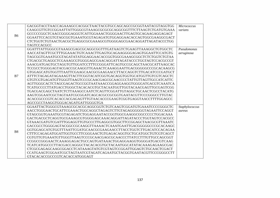
137
B6
GACGGTACCTAACCAGAAAGCCACGGCTAACTACGTGCCAGCAGCCGCGGTAATACGTAGGTGG
CAAGCGTTGTCCGGAATTATTGGGCGTAAAGCGCGCGCAGGCGGTTTCTTAAGTCTGATGTGAAA
GCCCCCGGCTCAACCGGGGAGGGTCATTGGAAACTGGGGAACTTGAGTGCAGAAGAGGAGAGT
GGAATTCCACGTGTAGCGGTGAAATGCGTAGAGATGTGGAGGAACACCAGTGGCGAAGGCGACT
CTCTGGTCTGTAACTGACGCTGAGGCGCGAAAGCGTGGGGAGCGAACAGGATTAGATACCCTGG
TAGTCCACGCC
Micrococcus
varians
B7
GGATTTATTGGGCGTAAAGCGAGCGCAGGCGGTTTGATAAGTCTGAAGTTAAAGGCTGTGGCTC
AACCATAGTTCGCTTTGGAAACTGTCAAACTTGAGTGCAGAAGGGGAGAGTGGAATTCCATGTG
TAGCGGTGAAATGCGTAGATATATGGAGGAACACCGGTGGCGAAAGCGGCTCTCTGGTCTGTAA
CTGACGCTGAGGCTCGAAAGCGTGGGGAGCGAACAGGATTAGATACCCTGGTAGTCCACGCCGT
AAACGATGAGTGCTAGGTGTTGGATCCTTTCCGGGATTCAGTGCCGCAGCTAACGCATTAAGCAC
TCCGCCTGGGGAGTACGACCGCAAGGTTGAAACTCAAAGGAATTGACGGGGGCCCGCACAAGCG
GTGGAGCATGTGGTTTAATTCGAAGCAACGCGAAGAACCTTACCAGGTCTTGACATCCCGATGCT
ATTTCTAGAGATAGAAAGTTACTTCGGTACATCGGTGACAGGTGGTGCATGGTTGTCGTCAGCTC
GTGTCGTGAGATGTTGGGTTAAGTCCCGCAACGAGCGCAACCCCTATTGTTAGTTGCCATCATTC
AGTTGGGCACTCTAGCGAGACTGCCGGTAATAAACCGGAGGAAGGTGGGGATGACGTCAAATCA
TCATGCCCCTTATGACCTGGGCTACACACGTGCTACAATGGTTGGTACAACGAGTTGCGAGTCGG
TGACGACGAGCTAATCTCTTAAAGCCAATCTCAGTTCGGATTGTAGGCTGCAACTCGCCTACATG
AAGTCGGAATCGCTAGTAATCGCGGATCAGCACGCCGCGGTGAATACGTTCCCGGGCCTTGTAC
ACACCGCCCGTCACACCACGAGAGTTTGTAACACCCGAAGTCGGTGAGGTAACCTTTTGGAGCC
AGCCGCCTAAGGTGGGACAGATGATTGGGGTGA
Pseudomonas
aeruginosa
B8
GGAATTACTGGGCGTAAAGCGCACGCAGGCGGTCTGTCAAGTCGGATGTGAAATCCCCGGGCTC
AACCTGGGAACTGCATTCGAAACTGGCAGGCTAGAGTCTTGTAGAGGGGGGTAGAATTCCAGGT
GTAGCGGTGAAATGCGTAGAGATCTGGAGGAATACCGGTGGCGAAGGCGGCCCCCTGGACAAA
GACTGACGCTCAGGTGCGAAAGCGTGGGGAGCAAACAGGATTAGATACCCTGGTAGTCCACGCC
GTAAACGATGTCGATTTGGAGGTTGTGCCCTTGAGGCGTGGCTTCCGGAGCTAACGCGTTAAATC
GACCGCCTGGGGAGTACGGCCGCAAGGTTAAAACTCAAATGAATTGACGGGGGCCCGCACAAGC
GGTGGAGCATGTGGTTTAATTCGATGCAACGCGAAGAACCTTACCTGGTCTTGACATCCACAGAA
CTTTCCAGAGATGGATTGGTGCCTTCGGGAACTGTGAGACAGGTGCTGCATGGCTGTCGTCAGCT
CGTGTTGTGAAATGTTGGGTTAAGTCCCGCAACGAGCGCAACCCTTATCCTTTGTTGCCAGCGGT
CCGGCCGGGAACTCAAAGGAGACTGCCAGTGATAAACTGGAGGAAGGTGGGGATGACGTCAAG
TCATCATGGCCCTTACGACCAGGGCTACACACGTGCTACAATGGCATATACAAAGAGAAGCGAC
CTCGCGAGAGCAAGCGGACCTCATAAAGTATGTCGTAGTCCGGATTGGAGTCTGCAACTCGACT
CCATGAAGTCGGAATCGCTAGTAATCGTAGATCAGAATGCTACGGTGAATACGTTCCCGGGCCTT
GTACACACCGCCCGTCACACCATGGGAGT
Staphylococcus
aureus
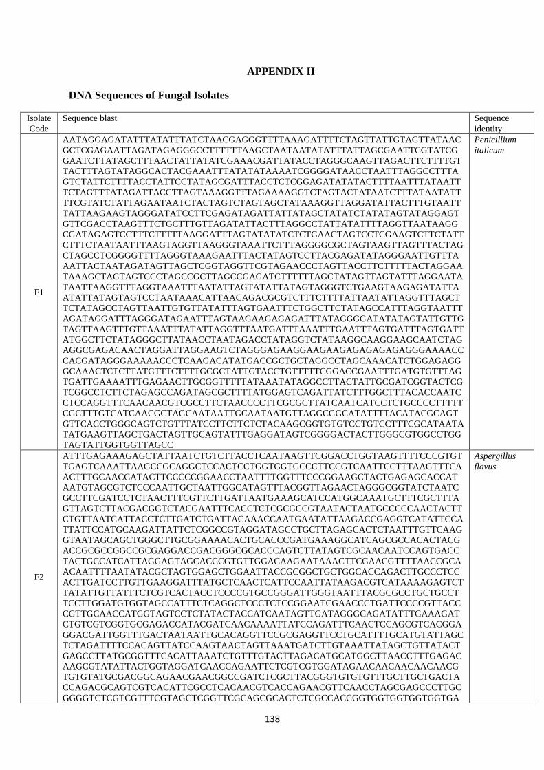
138
APPENDIX II
DNA Sequences of Fungal Isolates
Isolate
Code
Sequence blast Sequence
identity
F1
AATAGGAGATATTTATATTTATCTAACGAGGGTTTTAAAGATTTTCTAGTTATTGTAGTTATAAC
GCTCGAGAATTAGATAGAGGGCCTTTTTTAAGCTAATAATATATTTATTAGCGAATTCGTATCG
GAATCTTATAGCTTTAACTATTATATCGAAACGATTATACCTAGGGCAAGTTAGACTTCTTTTGT
TACTTTAGTATAGGCACTACGAAATTTATATATAAAATCGGGGATAACCTAATTTAGGCCTTTA
GTCTATTCTTTTACCTATTCCTATAGCGATTTACCTCTCGGAGATATATACTTTTAATTTATAATT
TCTAGTTTATAGATTACCTTAGTAAAGGTTTAGAAAAGGTCTAGTACTATAATCTTTATAATATT
TTCGTATCTATTAGAATAATCTACTAGTCTAGTAGCTATAAAGGTTAGGATATTACTTTGTAATT
TATTAAGAAGTAGGGATATCCTTCGAGATAGATTATTATAGCTATATCTATATAGTATAGGAGT
GTTCGACCTAAGTTTCTGCTTTGTTAGATATTACTTTAGGCCTATTATATTTTAGGTTAATAAGG
CGATAGAGTCCTTTCTTTTTAAGGATTTAGTATATATCTCTGAACTAGTCCTCGAAGTCTTCTATT
CTTTCTAATAATTTAAGTAGGTTAAGGGTAAATTCTTTAGGGGCGCTAGTAAGTTAGTTTACTAG
CTAGCCTCGGGGTTTTAGGGTAAAGAATTTACTATAGTCCTTACGAGATATAGGGAATTGTTTA
AATTACTAATAGATAGTTAGCTCGGTAGGTTCGTAGAACCCTAGTTACCTTCTTTTTACTAGGAA
TAAAGCTAGTAGTCCCTAGCCGCTTAGCCGAGATCTTTTTTAGCTATAGTTAGTATTTAGGAATA
TAATTAAGGTTTAGGTAAATTTAATATTAGTATATTATAGTAGGGTCTGAAGTAAGAGATATTA
ATATTATAGTAGTCCTAATAAACATTAACAGACGCGTCTTTCTTTTATTAATATTAGGTTTAGCT
TCTATAGCCTAGTTAATTGTGTTATATTTAGTGAATTTCTGGCTTCTATAGCCATTTAGGTAATTT
AGATAGGATTTAGGGATAGAATTTAGTAAGAAGAGAGATTTATAGGGGATATATAGTATTGTTG
TAGTTAAGTTTGTTAAATTTATATTAGGTTTAATGATTTAAATTTGAATTTAGTGATTTAGTGATT
ATGGCTTCTATAGGGCTTATAACCTAATAGACCTATAGGTCTATAAGGCAAGGAAGCAATCTAG
AGGCGAGACAACTAGGATTAGGAAGTCTAGGGAGAAGGAAGAAGAGAGAGAGAGGGAAAACC
CACGATAGGGAAAAACCCTCAAGACATATGACCGCTGCTAGGCCTAGCAAACATCTGGAGAGG
GCAAACTCTCTTATGTTTCTTTTGCGCTATTGTACCTGTTTTTCGGACCGAATTTGATGTGTTTAG
TGATTGAAAATTTGAGAACTTGCGGTTTTTATAAATATAGGCCTTACTATTGCGATCGGTACTCG
TCGGCCTCTTCTAGAGCCAGATAGCGCTTTTATGGAGTCAGATTATCTTTGGCTTTACACCAATC
CTCCAGGTTTCAACAACGTCGCCTTCTAACCCCTTCGCGCTTATCAATCATCCTCTGCCCCTTTTT
CGCTTTGTCATCAACGCTAGCAATAATTGCAATAATGTTAGGCGGCATATTTTACATACGCAGT
GTTCACCTGGGCAGTCTGTTTATCCTTCTTCTCTACAAGCGGTGTGTCCTGTCCTTTCGCATAATA
TATGAAGTTAGCTGACTAGTTGCAGTATTTGAGGATAGTCGGGGACTACTTGGGCGTGGCCTGG
TAGTATTGGTGGTTAGCC
Penicillium
italicum
F2
ATTTGAGAAAGAGCTATTAATCTGTCTTACCTCAATAAGTTCGGACCTGGTAAGTTTTCCCGTGT
TGAGTCAAATTAAGCCGCAGGCTCCACTCCTGGTGGTGCCCTTCCGTCAATTCCTTTAAGTTTCA
ACTTTGCAACCATACTTCCCCCGGAACCTAATTTTGGTTTCCCGGAAGCTACTGAGAGCACCAT
AATGTAGCGTCTCCCAATTGCTAATTGGCATAGTTTACGGTTAGAACTAGGGCGGTATCTAATC
GCCTTCGATCCTCTAACTTTCGTTCTTGATTAATGAAAGCATCCATGGCAAATGCTTTCGCTTTA
GTTAGTCTTACGACGGTCTACGAATTTCACCTCTCGCGCCGTAATACTAATGCCCCCAACTACTT
CTGTTAATCATTACCTCTTGATCTGATTACAAACCAATGAATATTAAGACCGAGGTCATATTCCA
TTATTCCATGCAAGATTATTCTCGGCCGTAGGATAGCCTGCTTAGAGCACTCTAATTTGTTCAAG
GTAATAGCAGCTGGGCTTGCGGAAAACACTGCACCCGATGAAAGGCATCAGCGCCACACTACG
ACCGCGCCGGCCGCGAGGACCGACGGGCGCACCCAGTCTTATAGTCGCAACAATCCAGTGACC
TACTGCCATCATTAGGAGTAGCACCCGTGTTGGACAAGAATAAACTTCGAACGTTTTAACCGCA
ACAATTTTAATATACGCTAGTGGAGCTGGAATTACCGCGGCTGCTGGCACCAGACTTGCCCTCC
ACTTGATCCTTGTTGAAGGATTTATGCTCAACTCATTCCAATTATAAGACGTCATAAAAGAGTCT
TATATTGTTATTTCTCGTCACTACCTCCCCGTGCCGGGATTGGGTAATTTACGCGCCTGCTGCCT
TCCTTGGATGTGGTAGCCATTTCTCAGGCTCCCTCTCCGGAATCGAACCCTGATTCCCCGTTACC
CGTTGCAACCATGGTAGTCCTCTATACTACCATCAATAGTTGATAGGGCAGATATTTGAAAGAT
CTGTCGTCGGTGCGAGACCATACGATCAACAAAATTATCCAGATTTCAACTCCAGCGTCACGGA
GGACGATTGGTTTGACTAATAATTGCACAGGTTCCGCGAGGTTCCTGCATTTTGCATGTATTAGC
TCTAGATTTTCCACAGTTATCCAAGTAACTAGTTAAATGATCTTGTAAATTATAGCTGTTATACT
GAGCCTTATGCGGTTTCACATTAAATCTGTTTGTACTTAGACATGCATGGCTTAACCTTTGAGAC
AAGCGTATATTACTGGTAGGATCAACCAGAATTCTCGTCGTGGATAGAACAACAACAACAACG
TGTGTATGCGACGGCAGAACGAACGGCCGATCTCGCTTACGGGTGTGTGTTTGCTTGCTGACTA
CCAGACGCAGTCGTCACATTCGCCTCACAACGTCACCAGAACGTTCAACCTAGCGAGCCCTTGC
GGGGTCTCGTCGTTTCGTAGCTCGGTTCGCAGCGCACTCTCGCCACCGGTGGTGGTGGTGGTGA
Aspergillus
flavus

139
TTGTGCAGCAGTAAACTCGGCAAAAGCTTCCCATCGGTTTTTATTTTGAATTTCGGGGTCATAAT
ACTTGCTACTGTCGCTATCCTTCCATCATAGCGTGGTAGCCGTATGACATTCATGTACGGCCCTC
GGTTCGGGAGACGACACGCCTGCACTGCGCCCTGCTCTCCGTGCTTTCGGTGGGAGCGCGCGGT
GCAAACGTCTCGTGCCGGTCTGGTGCTGGTTGCTGGTTGGACTTTGCTGCTGCGAAAAGCGTGA
GCCACTGGT
F3
GATCTTATCCGTGGACCCGAAGAACTATGCGGATGCGCCGACTGAGGCAATCCGTCGCGTCTTA
GAGCAATACTATGGTTCTCCTATCCCGCGTGGAACAGAGCTCGACCTGAGAGACGTCGGTATGA
TTCTGGGATTCACCCAACAGCTTGAGACTTCTATCAGCACTAACTCCTTCGCTAACAGAATGGA
TCCGTATGGGCACAACAGTTGCAACAAATGCGTTACTCGAGAGAAAAGGAGAGAAGACAGCCT
TACTGATCACCGAAGGCTTCAAGGATGTTCTTCAAATCGGAACTCAGAGCCGGCCCCATATGTT
TGACTTGACAATCCGGCGGCCGATACCCTTGTATTCAAAAACCTTTGAAGTTCGAGAGCGAGTG
ACTGTGCAAAATTGCAGCGACTCAGATCTCCGCAATATTCATTTGTCCTCTCCAGAGCCTGTGG
ATTCCGTCATTGCAGCATCAGGGGAGATAATTCAAGTACTCCAGCCCCTGGATACGGCCAGAAC
AAAAGTTCAACTCCAAGGTATTTACGAAGAAGGGTTTCGCACCCTGGCAGTGTGCCTGATGCAC
TCGTACAGCTTCCCAAAGCACGAACTAGAAATCAGAGAAATGGCACTCGAAATTGGGTTTGAA
AATGTATCACTTTCGCATGAGATATCTTCAAGACCAAAGCTCGTCCCCCGTGGTAACTCCACGG
TAGTTGATGCATATTTGACACCGAGTATCAAACAATATCTTGAAAGATTCTCCAAAAGCTTCCC
CAATATAGGTAATTCTCGGACGCGACTAGAATTCATGCAATCAGACGGAGGCCTGGTGCCTTCG
TCTAGTCTTTCAGGGCTTCGCTCTATCCTCTCAGGGCCAGCTGGAGGCGTTATTGGCTTTTCTCG
AACATGCTTCGATACTGAAACTCGAGCTCCGGTGATTGGTTTTGATATGGGAGGAACCAGCACT
GATGTTAGTCGATATGACGGCGAACTGGATCATATCTTCGAGACAATAACTGCTGGGATCACTA
TCCACGCTCCACAGCTGAATGTCAATACAATTGCCG
Aspergillus
niger
F4
TACTGATGAATAGAAACGGTTAGGGGCATTTGTATTTGGTCGCTAGAGGTGAAATTCTTGGATT
GACCGAAGAAAAACTACTGCGAAAGCATTTGACCCGGGACGTTTTCATTGATCAAGGTCTAATG
TTAAGGGATCGAAGACGATTAGATACCGTCGTAGTCTTAACCACAAACTATGCCGACTAGAGAT
TGGGCGTGTTTATTATGACTCGCTCAGCATCTTAGCGAAAGTAAAGATTTTGGGTTCTGGGGGG
AGTATGGGACGCAAGGCTGAACCTTAAAGGAATTGACGGAAGGGCACCACCAGGAGTGGAGCC
TGCGGCTTAATTTGACTCAACACGGGGAAACTCACCAGGTCCAGACATAGTAATGATTGACAGA
TTGAAAGCTCTTTCTAGATTCTATGGGTGGTGGTGCATGGCCGTTCTTAGTTCGTGGAGTGATTT
GTCTGGTTA
Rhizopus
stolonifer
F5
GGGATAACCTAATTTAGGCCTTTAGTCTATTCTTTTACCTATTCCTATAGCGATTTACCTCTCGG
AGATATATACTTTAATAGGAGATATTTATATTTATCTAACGAGGGTTTTAAAGATTTTCTAGTTA
TTGTAGTTATAACGCTCGAGAATTAGTAATTTATAATTTCTAGTTTATAGATTACCTTAGTAAAG
GTTTAGAAAAGGTCTAGTACTATAATCTTTATAATATTTTCGTATCTATTAGAATAATCTACTAG
TCTAGTAGCTATAAAGGTTAGGATATTACTTTGTAATTTATTAAGAAGTAGGGAAACGATTATA
CCTAGGGCAAGTTAGACTTCTTTTGTTACTTTAGTATAGGCACTACGAAATTTATATATAAAATC
GATTACTAATAGATAGTTAGCTCGGTAGGTTCGTAGAACCCTAGTTACCTTCTTTTTACTAGGAA
TAAAGCTAGTAGTATAGAGGGCCTTTTTTAAGCTAATAATATATTTATTAGCGAATTCGTATCGG
AATCTTATAGCTTTAACTATTATATCGATATCCTTCGAGATAGATTATTATAGCTATATCTATAT
AGTATAGGAGTGTTCGACCTAAGTTTCTGCTTTGTTAGATATTACTTTAGGCCTATTATATTTTA
GGTTAATAAGGCGATAGAGTCCTTTCTTTTTAAGGATTTAGTATATATCTCTGAACTAGTCCTCG
AAGTCTTCTATTCTTTCTAATAATTTAAGTAGGTTAAGGGTAAATTCTTTAGGGGCGCTAGTAAG
TTAGTTTACTAGCTAGCCTCGGGGTTTTAGGGTAAAGAATTTACTATAGTCCTTACGAGATATAG
GGAATTGTTTAACCCTAGCCGCTTAGCCGAGATCTTTTTTAGCTATAGTTAGTATTTAGGAATAT
AATTAAGGTTTAGGTAAATTTAATAATTTAGGTAATTTAGATAGGATTTAGGGATAGAATTTAG
TAAGAAGAGAGATTTATAGGGGATATATAGTATTGTTGTTAGTATATTATAGTAGGGTCTGAAG
TAAGAGATATTAATATTATAGTAGTCCTAATAAACATTAACAGACGCGTCTTTCTTTTATTAATA
TTAGGTTTAGCTTCTATAGCCTAGTTAATTGTGTTATATTTAGTGAATTTCTGGCTTCTATAGCCT
AGTTAAGTTTGTTAAATTTATATTAGGTTTAATGATTTAAATTTGAATTTAGTGATTTAGTGATT
ATGGCTTCTATAGCCGAATTTGATGTGTTTAGTGATTGAAAATTTGAGAACTTGCGGTTTTTATA
AATATAGGCCTTACTATTGCGATCGGGCTTATAACCTAATAGACCTATAGGTCTATAAGGCAAG
GAAGCAATCTAGAGGCGAGACAACTAGGATTAGGAAGTCTAGGGAGAAGGAAGAAGAGAGAG
AGAGGGAAAACCCACGATAGGGAAAAACCCTCAAGACATATGACCGCTGCTAGGCCTAGCAAA
CATCTGGAGAGGGCAAACTCTCTTATGTTTCTTTTGCGCTATTGTACCTGTTTTTCGGAGTACTC
GTCGGCCTCTTCTAGAGCCAGATAGCGCTTTTATGGAGTCAGATTATCTTTGGCTTTACACCAAT
CCTCCCTTCTCTACAAGCGGTGTGTCCTGTCCTTTCGCATAATATATGAAGTTAGCTGACTAGTT
GCAGTATTTGAGGATAAGGTTTCAACAACGTCGCCTTCTAACCCCTTCGCGCTTATCAATCATCC
TCTGCCCCTTTTTCGCTTTGTCATCAACGCTAGCAATAATTGCAATAATGTTAGGCGGCATATTT
TACATACGCAGTGTTCACCTGGGCAGTCTGTTTATCCTTGTCGGGGACTACTTGGGCGTGGCCTG
Penicillium
chrysogenum
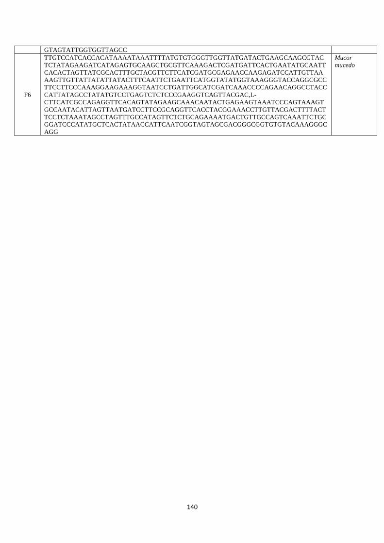
140
GTAGTATTGGTGGTTAGCC
F6
TTGTCCATCACCACATAAAATAAATTTTATGTGTGGGTTGGTTATGATACTGAAGCAAGCGTAC
TCTATAGAAGATCATAGAGTGCAAGCTGCGTTCAAAGACTCGATGATTCACTGAATATGCAATT
CACACTAGTTATCGCACTTTGCTACGTTCTTCATCGATGCGAGAACCAAGAGATCCATTGTTAA
AAGTTGTTATTATATTATACTTTCAATTCTGAATTCATGGTATATGGTAAAGGGTACCAGGCGCC
TTCCTTCCCAAAGGAAGAAAGGTAATCCTGATTGGCATCGATCAAACCCCAGAACAGGCCTACC
CATTATAGCCTATATGTCCTGAGTCTCTCCCGAAGGTCAGTTACGAC,L-
CTTCATCGCCAGAGGTTCACAGTATAGAAGCAAACAATACTGAGAAGTAAATCCCAGTAAAGT
GCCAATACATTAGTTAATGATCCTTCCGCAGGTTCACCTACGGAAACCTTGTTACGACTTTTACT
TCCTCTAAATAGCCTAGTTTGCCATAGTTCTCTGCAGAAAATGACTGTTGCCAGTCAAATTCTGC
GGATCCCATATGCTCACTATAACCATTCAATCGGTAGTAGCGACGGGCGGTGTGTACAAAGGGC
AGG
Mucor
mucedo
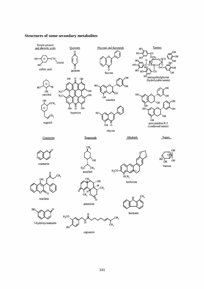
141
Structures of some secondary metabolites

142
The conversion of tyrosine to quinone