comparison of microbiological, histological, and - plos
TRANSCRIPT
Comparison of Microbiological, Histological, andImmunomodulatory Parameters in Response toTreatment with Either Combination Therapy withPrednisone and Metronidazole or Probiotic VSL#3Strains in Dogs with Idiopathic Inflammatory BowelDiseaseGiacomo Rossi1, Graziano Pengo2, Marco Caldin3, Angela Palumbo Piccionello1, Jorg M. Steiner4,
Noah D. Cohen5, Albert E. Jergens6, Jan S. Suchodolski4*
1 School of Veterinary Medical Sciences, University of Camerino, Camerino, Italy, 2 Clinic ‘‘St. Antonio’’, Cremona, Italy, 3 San Marco Laboratories, Padova, Italy,
4 Gastrointestinal Laboratory, Department of Small Animal Clinical Sciences, College of Veterinary Medicine and Biomedical Sciences, Texas A&M University, College
Station, Texas, United States of America, 5 Department of Large Animal Clinical Sciences, College of Veterinary Medicine and Biomedical Sciences, Texas A&M University,
College Station, Texas, United States of America, 6 Department of Veterinary Clinical Sciences, College of Veterinary Medicine, Iowa State University, Ames, Iowa, United
States of America
Abstract
Background: Idiopathic inflammatory bowel disease (IBD) is a common chronic enteropathy in dogs. There are nopublished studies regarding the use of probiotics in the treatment of canine IBD. The objectives were to compare responsesto treatment with either combination therapy (prednisone and metronidazole) or probiotic strains (VSL#3) in dogs with IBD.
Methodology and Principal Findings: Twenty pet dogs with a diagnosis of IBD, ten healthy pet dogs, and archived controlintestinal tissues from three euthanized dogs were used in this open label study. Dogs with IBD were randomized to receiveeither probiotic (D-VSL#3, n = 10) or combination drug therapy (D-CT, n = 10). Dogs were monitored for 60 days (duringtreatment) and re-evaluated 30 days after completing treatment. The CIBDAI (P,0.001), duodenal histology scores (P,0.001), and CD3+ cells decreased post-treatment in both treatment groups. FoxP3+ cells (p,0.002) increased in the D-VSL#3 group after treatment but not in the D-CT group. TGF-b+ cells increased in both groups after treatment (P = 0.0043)with the magnitude of this increase being significantly greater for dogs in the D-VSL#3 group compared to the D-CT group.Changes in apical junction complex molecules occludin and claudin-2 differed depending on treatment. Faecalibacteriumand Turicibacter were significantly decreased in dogs with IBD at T0, with a significant increase in Faecalibacteriumabundance observed in the animals treated with VSL#3 strains.
Conclusions: A protective effect of VSL#3 strains was observed in dogs with IBD, with a significant decrease in clinical andhistological scores and a decrease in CD3+ T-cell infiltration. Protection was associated with an enhancement of regulatoryT-cell markers (FoxP3+ and TGF-b+), specifically observed in the probiotic-treated group and not in animals receivingcombination therapy. A normalization of dysbiosis after long-term therapy was observed in the probiotic group. Largerscale studies are warranted to evaluate the clinical efficacy of VSL#3 in canine IBD.
Citation: Rossi G, Pengo G, Caldin M, Palumbo Piccionello A, Steiner JM, et al. (2014) Comparison of Microbiological, Histological, and ImmunomodulatoryParameters in Response to Treatment with Either Combination Therapy with Prednisone and Metronidazole or Probiotic VSL#3 Strains in Dogs with IdiopathicInflammatory Bowel Disease. PLoS ONE 9(4): e94699. doi:10.1371/journal.pone.0094699
Editor: Mathias Chamaillard, INSERM, France
Received October 25, 2013; Accepted March 19, 2014; Published April 10, 2014
Copyright: � 2014 Rossi et al. This is an open-access article distributed under the terms of the Creative Commons Attribution License, which permitsunrestricted use, distribution, and reproduction in any medium, provided the original author and source are credited.
Funding: There was no external funding support for this research. VSL Pharmaceuticals supplied the probiotic strains used in this study, but otherwise did notprovide any financial support and had no role in study design, data collection, analysis, interpretation or preparation of the manuscript.
Competing Interests: VSL Pharmaceuticals supplied the probiotic strains used in this study. Graziano Pengo is employed by Clinic ‘‘St. Antonio’’ and MarcoCaldin by San Marco Laboratories. There are no further patents, products in development or marketed products to declare. Jan Suchodolski is a member of thePLOS ONE Editorial Board. This does not alter the authors’ adherence to all the PLOS ONE policies on sharing data and materials, as detailed online in the guide forauthors.
* E-mail: [email protected]
Introduction
Similar to human inflammatory bowel disease (IBD), three main
factors are considered to be fundamental in the pathogenesis of
canine idiopathic IBD: the interactions between the mucosal
immune system, host genetic susceptibility, and environmental
factors (e.g., microbiota, nutrition) [1–3]. Experimental evidence
supports a role for commensal bacteria in the pathogenesis of IBD;
PLOS ONE | www.plosone.org 1 April 2014 | Volume 9 | Issue 4 | e94699
for example, spontaneous colitis develops in mice deficient in
interleukin (IL)-2 [4] and IL-10 [5] when colonized with a
complex microbiota, but not in mice raised under germ-free
conditions. Recent studies suggest involvement of the intestinal
microbiota in the pathogenesis of canine and feline IBD [2,6–9].
Also, antibiotics such as metronidazole are useful in the treatment
of IBD in humans [10] and dogs [11], and there is evidence that
children with IBD respond to probiotic administration [12].
Collectively, these findings suggest that the intestinal microbiota
plays a crucial role in the pathogenesis of IBD and modulation of
intestinal microbiota may be beneficial in the treatment of mucosal
inflammation. While probiotics are used frequently in small animal
practice, there are only few published studies regarding their
efficacy in dogs with chronic enteropathies. In one investigation, a
probiotic cocktail was shown to reduce clinical severity in a
prospective, placebo-controlled trial in dogs with food-responsive
diarrhea treated with an elimination diet [13], but studies
evaluating idiopathic IBD have not been reported.
VSL#3 is a high-dose, multi-strain probiotic product contain-
ing viable lyophilized bacteria consisting of 4 strains of Lactobacillus
(L. casei, L. plantarum. L. acidophilus, and L. delbrueckii subsp.
bulgaricus), 3 strains of Bifidobacterium (B. longum, B. breve, and B.
infantis), and 1 strain of Streptococcus sulivarius subsp thermophilus. The
VSL#3 strains have shown efficacy in humans for the prevention,
treatment, and maintenance of remission of both pouchitis and
ulcerative colitis in adults and children [12,14,15].
The purpose of the present study was to perform a randomized
open-label trial to compare the microbiological, histological, and
immunomodulatory effects between the commercial multi-strain
probiotic SIVOY, a probiotic product formulated with VSL#3
strains for pets (VSL Pharmaceuticals, Inc., Gaithersburg, MD,
USA) and combination therapy with prednisone and metronida-
zole in canine IBD.
Our results suggest a protective effect of the probiotic mixture in
dogs with IBD, with a significant decrease in clinical and
histological scores, and a decrease in CD3+ T-cell infiltration.
Protection was associated with an enhancement of regulatory T-
cell markers (FoxP3+ and TGF-b+), specifically observed in the
probiotic-treated group and not in animals receiving combination
therapy. The protective effect of the probiotic VSL#3 strains was
also associated with normalization of dysbiosis, specifically
increases in Faecalibacterium spp.
Materials and Methods
AnimalsThe study was approved by the Camerino University Institu-
tional Animal Care and Use Committee protocol and all owners of
the IBD dogs gave informed written consent before enrollment.
Twenty pet dogs (Table 1) with a long-time diagnosis of IBD
according to published criteria [16] were evaluated at the
Veterinary Teaching Hospital, Camerino University, for chronic
gastroenteritis. Inclusion criteria included recurrence of clinical
signs and absence of any immunomodulating drug therapy (e.g.,
corticosteroids, metronidazole, and sulfasalazine) within a month
before referral. Diagnostic criteria for IBD included: persistent (.3
weeks) gastrointestinal signs, failed responses to dietary (hydroly-
sate or commercial intact protein elimination diet) or symptomatic
therapies (anthelminthics, antibiotics, anticholinergics, gastrointes-
tinal protectants) alone, a thorough diagnostic evaluation with
failure to document other causes for gastroenteritis, and
histopathologic evidence of intestinal inflammation. The mini-
mum diagnostic evaluation in all dogs included a complete blood
count, serum biochemistry, urinalysis, direct (wet mount) and
indirect (flotation) examination of feces for endoparasites, and
survey abdominal radiographs. In some instances, additional tests
including contrast radiography, abdominal ultrasound (performed
in 16 of the 20 dogs) and measurement of serum concentrations of
trypsin-like immunoreactivity and/or folate and cobalamin were
performed. Additional inclusion criteria were the absence of extra-
alimentary tract inflammation based on results obtained from
initial diagnostic testing. Dogs with hypoproteinemia or a
suspicion of intestinal lymphangiectasia were excluded from the
study.
Ten pet dogs (Table 1), living in home environments and free of
gastrointestinal signs for at least four months, were enrolled as
control group (D–H) for comparison of fecal microbiota between
healthy dogs and dogs with IBD. Control dogs were judged to be
healthy based on normal results on physical examination,
complete blood count, serum biochemistry, urinalysis, repeated
fecal examinations, and dirofilarial antigen assay.
Study designThe trial was a 90 day open-label evaluation to compare the
effects of VSL#3 strains versus combination drug therapy on
histological, microbiological, and immunological markers. Dogs
were randomized into two groups using a computer-generated
randomization list. The VSL#3 group (D-VSL#3; n = 10)
received between 112 and 225 billion (112 to 2256109) lyophilized
bacteria per 10 kg daily for 60 consecutive days; the D-CT group
(n = 10) received a combination protocol of metronidazole at
20 mg/Kg q12 h and prednisone at 1 mg/kg body weight/day.
The clinical disease activity (CIBDAI score) was assessed at
baseline (T0) and after 90 days (T1) of enrollment, which was 30
days following completion of either treatment. The CIBDAI is
based on 6 criteria, each scored on a scale from 0–3: attitude/
activity, appetite, vomiting, stool consistency, stool frequency, and
weight loss. After summation, the total composite score is
determined to be clinically insignificant (score 0–3), mild (score
4–5), moderate (score 6–8) or severe (score 9 or greater) [17].
Fecal samples were also collected at each visit then immediately
stored at 280uC, until microbiota analysis. The evaluation time
point 30 days post-treatment was chosen to determine whether
individual dogs would relapse within 30 days following completion
of either treatment regimen.
Tissue samplingAfter enrollment (time point T0) and after 90 days (T1), multiple
(10–15 specimens) mucosal biopsy specimens were procured
endoscopically from the small and/or large intestine of all dogs
with IBD (n = 20, 10 dogs per treatment group). Fifteen dogs
having predominantly upper gastrointestinal signs (i.e., vomiting,
small bowel diarrhea, anorexia, and/or weight loss) underwent
esophagogastroduodenoscopy, whereas upper and lower endo-
scopic examinations were performed in 5 dogs having mixed signs
of enterocolitis (i.e., GI signs associated with tenesmus, hemato-
chezia, mucoid feces, and/or frequent defecation). Biopsy
specimens were obtained directly from mucosal lesions of
increased granularity, friability, or erosions as well as areas of
normal-appearing mucosa. Tissues for histopathology were placed
in 10% neutral buffered formalin, then paraffin embedded and
serial 3 mm thick sections were prepared. For ethical consider-
ations, no endoscopic examinations were performed in healthy
dogs. Histopathology was performed by a single pathologist, who
was blinded regarding history, clinical signs, or endoscopic
observations. A severity score was assigned for each dog, by using
a standardized and previously described histologic grading system,
based on the extent of architectural disruption and mucosal
Probiotic Treatment Response in Canine IBD
PLOS ONE | www.plosone.org 2 April 2014 | Volume 9 | Issue 4 | e94699
epithelial changes [17,18], as recently been proposed by the
WSAVA for diagnosis of gastrointestinal inflammation [19].
Tissues were also evaluated for expression patterns of apical
junction complex (AJC) molecules in both dog groups after end of
the therapy. To obtain control tissue from healthy dogs for this
analysis, archived formalin-fixed and paraffin–embedded colonic
tissues from three male dogs with no clinical signs of intestinal
disease were retrieved from the University of Camerino Veterinary
Pathology Unit archives. These samples had been obtained
immediately post-mortem from dogs that were presented for
euthanasia (euthanized dogs, ED) for old age (n = 1), nasal
carcinoma (n = 1), or splenic haemangiosarcoma (n = 1). Ages
ranged from 7 years to 14 years and histopathological examination
of full-thickness intestinal biopsies was normal in all these ED
cases.
Immunohistochemical evaluationParaffin sections were rehydrated and neutralized for endoge-
nous peroxidases with 3% hydrogen peroxide for 5 minutes
followed by rinsing for 5 minutes in distilled water. For antigen
retrieval, slides were incubated in three antigen retrieval solutions:
citrate buffer (pH 6.0) for TGF-b, EDTA (pH 8.0) for CD3 and
FoxP3, and 0.01 M Tris-EDTA buffer (pH 9.0) for claudin 2,
occludin and E-cadherin in a steamer (Black & Decker, Towson,
MD, USA) for 20 minutes. Non-specific binding was blocked by
incubation of slides for 10 minutes with a protein-blocking agent
(Protein-blocking agent, Dako, Carpinteria, CA, USA) before
application of the primary antibody. Slides were incubated
overnight in a moist-chamber with the following primary
antibodies: monoclonal (mAb) rat anti-human CD3 (Monoclonal
rat anti-human CD3 clone MCA1477, Serotec abD, Biorad
Laboratories, Hercules, CA, USA) diluted 1:50, mAb anti-mouse/
rat FoxP3 antibodies (Monoclonal anti-mouse/rat FoxP3 antibod-
ies clone FJK-16s, eBioscience, San Diego, CA, USA) diluted
1:400, and mAb mouse anti-TGF-b (Monoclonal mouse anti-
TGF-b, clone 1D11, Serotec abD, Biorad Laboratories, Hercules,
CA, USA) diluted 1:25 [19,20]. Polyclonal rabbit anti-claudin-2
(Polyclonal rabbit anti-claudin-2 (PAD: MH44), Invitrogen Ltd.,
Paisley, UK) and anti-occludin (anti-occludin PAD: Z-T22,
Invitrogen Ltd., Paisley, UK) antibodies and monoclonal mouse
anti-E-cadherin IgG2a (Monoclonal mouse anti-E-cadherin
IgG2a (clone: 36), BD Biosciences, Oxford, UK) were used as
described previously [21].
The immunoreaction with streptavidin–immunoperoxidase
(Streptavidin–immunoperoxidase, Black & Decker, Towson,
MD, USA) was visualized with 3,39-diaminobenzidine substrate
(3,39-diaminobenzidine substrate, Vector, Burlingame, UK).
Tissues were counterstained with Mayer’s hematoxylin. For
negative immunohistochemical controls the primary antibodies
were omitted. Sections of canine spleen and tonsil served as
positive control tissues for CD3 and FoxP3 cell staining and
sections of canine placenta for that of TGF-b expression. Positive
control tissues for claudin/occludin and E-cadherin staining
consisted of canine lung and kidney sections, respectively.
For scoring of intestinal CD3+ T-lymphocytes, FoxP3+ cells,
and TGF-b+ cells, these cells were quantified in select compart-
ments of the GI tract (small intestine: villi, basal crypt area, villus-
crypt junction; large intestine: apical crypt area, basal crypt area).
All cellular types were evaluated using a light microscope (Carl
Zeiss, Jena, Germany), a640 objective, a610 eyepiece, and a
square eyepiece graticule (10610 squares, having a total area of
62,500 mm2). Ten appropriate fields were chosen for each
compartment and arithmetic means were calculated for each
intestinal region. Results were expressed as IHC positive cells per
62,500 mm2. For all parameters, cells on the margins of the tissue
sections were not considered for evaluation to avoid inflation of
positive cell numbers.
For the evaluation of different lymphocytes subsets in the same
histological sections, consecutive 3-mm-thick bioptic cross sections
were cut. Sections were placed consecutively on each of eight
separate slides, after which the ninth section was placed on the first
slide, next to the first section, continuing for 48 sections. A single
slide, upon which were six bioptic cross sections from each dog,
was analyzed for any given immunostain. Numbers of CD3+ T-
lymphocytes, FoxP3+ cells, and TGF-b+ cells, were quantified by
using an image-analysis system consisting of a light microscope
Table 1. Summary characteristics of enrolled dogs.
Treatment groups
VSL#3 (n = 10) CT (n = 10) Healthy Control (n = 10)
Breed Golden Retriever, Husky, Boxer,Rottweiler, Jack Russell Terrier,WHW Terrier, German shepherd(2), Shih Tzu, Yorkshire Terrier
Golden retriever (2), CockerSpaniel, Boxer, Bull Terrier,Carlino, WHW Terrier, Germanshepherd, Shar Pei, Yorkshire Terrier
Golden Retriever, Epagneul Breton, ChowChow, Rottweiler, Border collie, Germanshepherd, Bolognese, Miniature Schnauzer,Yorkshire Terrier (2)
Sex m = 5, mn = 1, f = 1, fs = 3 m = 5, f = 2,fs = 3 m = 5, f = 5
Median age(range) in years
5.8 (2.5–11) 5.5 (1.5–9) 6.5 (1–12)
Body weight(range) in kg
18.9 (2–36) 18.7 (1.5–30) 20.6 (2.8–45)
Median (range) timeto remission (days)
10.6 (5–15) 4.8 (2.5–7) n/a
m = male, mn = neutered male, f = female, fs = spayed female; CT = combination therapy;n/a-not applicable.doi:10.1371/journal.pone.0094699.t001
Probiotic Treatment Response in Canine IBD
PLOS ONE | www.plosone.org 3 April 2014 | Volume 9 | Issue 4 | e94699
(Carl Zeiss, Jena, Germany) attached to a Javelin JE3462 high-
resolution camera and a personal computer equipped with a
Coreco-Oculus OC-TCX frame grabber and high-resolution
monitor. Computerized color-image analysis was performed by
using Image-Pro Plus software (Media Cybernetics). The area of
each biopsy in all six cross sections in every dog was recorded, as
was the total number of T-lymphocytes determined by immuno-
staining as previously described. For each dog, the total bioptic
area was calculated as the sum of the areas of all fields in all six
bioptic cross sections on one slide. CD3+ T-lymphocytes, FoxP3+cells, and TGF-b+ cells were counted per section, and stained cell
densities were expressed as the number of lymphocytes/cells per
square millimeter of analyzed bioptic area [22].
To assess AJC expression (claudin-2, occludin, and E-cadherin)
in biopsies sampled after treatment in both groups (at T1 for D-
VSL#3 and D-CT), and to compare data to the AJC expression in
non-IBD control dogs (ED group), stained tissue sections were
evaluated at 6200 and 6630 (oil immersion) magnification to
identify areas of consistent staining and acceptable orientation.
Immunostaining was evaluated along the length of multiple
enteric/colonic crypts and in areas of intact luminal epithelium.
Stain intensity was subjectively graded as absent (2), weak (+),
moderate (++), or strong (+++), and the localization and
distribution of chromogen were noted. For evaluation, intestinal
epithelium was divided into luminal, proximal, and distal gland/
crypt regions, and the intercellular junction was divided into apical
and basolateral compartments. Finally, the scoring of intestinal
AJC molecules expression was calculated as previously described
for CD3+ T-cells, FoxP3+ cells, and TGF-b+ cells. The AJC
molecules were assessd only at T1 (following treatment interven-
tion), because at T0 all dogs had endoscopically visible lesions of
intestinal inflammation including erosions, friability, and increased
mucosal granularity. Also, dogs had histopathologic lesions of
intestinal inflammation of varying severity. Intestinal inflammation
was associated with different degrees of epithelial infiltration by
lymphocytes (i.e., intraepithelial lymphocytes) in all dogs of both
groups. In these instances, it was not considered useful to evaluate
AJCs as they were assumed to be altered, but instead AJC
molecules were evaluated at T1 when the previously observed
endoscopic lesions of inflammation had resolved.
Plasma citrullinePlasma concentrations of citrulline were measured in the D-
VSL#3 treated group only. Plasma samples were taken at baseline
(T0) and after 90 days (T1) and stored at 280uC until evaluation.
Samples were precipitated with organic solvents and quantified by
MS/MS mass spectrometer equipped with electrospray ionization
(ESI) interface in positive ion mode (Waters TQ Detector, Water
Corp., Milford, MA, USA). All assays were performed in duplicate
fashion. All data collected in centroid mode were processed using
Table 2. Oligonucleotides primers/probes used in this study.
qPCR primers/probe Sequence (59-39) Target Annealing (6C) Reference
Forward CCGGAWTYATTGGGTTTAAAGGG Bacteroidetes 60 [45]
Reverse GGTAAGGTTCCTCGCGTA
Forward GAAGGCGGCCTACTGGGCAC Faecalibacterium 60 [47]
Reverse GTGCAGGCGAGTTGCAGCCT
Forward ACTGAGAGGTTGAACGGCCA Family Ruminococcaceae 59 [47]
Reverse CCTTTACACCCAGTAAWTCCGGA
Forward CGCATAACGTTGAAAGATGG
Reverse CCTTGGTAGGCCGTTACCC C. perfringens 16S 58 [48]
Probe TCATCATTCAACCAAAGGAGCAATCC
Forward KGGGCTCAACMCMGTATTGCGT Fusobacteria 51 [9]
Reverse TCGCGTTAGCTTGGGCGCTG
Forward TCTGATGTGAAAGGCTGGGGCTTA Blautia 56 [9]
Reverse GGCTTAGCCACCCGACACCTA
Forward CCTACGGGAGGCAGCAGT Universal Bacteria 59 [49]
Reverse ATTACCGCGGCTGCTGG
Forward CAGACGGGGACAACGATTGGA Turicibacter 63 [9]
Reverse TACGCATCGTCGCCTTGGTA
Forward TCGCGTCCGGTGTGAAAG Bifidobacterium 60 [50]
Reverse CCACATCCAGCATCCAC
Forward CCCTTATTGTTAGTTGCCATCATT Enterococcus 61 [46]
Reverse ACTCGTTGTACTTCCCATTGT
Forward AGCAGTAGGGAATCTTCCAa Lactobacillus 58 [46]
Reverse CACCGCTACACATGGAGb
Forward TTATTTGAAAGGGGCAATTGCT Streptococcus 54 [51]
Reverse GTGAACTTTCCACTCTCACAC
aOriginally described by [52].bOriginally described by [53].doi:10.1371/journal.pone.0094699.t002
Probiotic Treatment Response in Canine IBD
PLOS ONE | www.plosone.org 4 April 2014 | Volume 9 | Issue 4 | e94699
commercial software (MassLynx 4.1 software, Water Corp.,
Milford, Ma, USA).
Microbiota analysisFresh naturally voided samples were collected from all 20
diseased dogs (at T0 and T1) and 10 healthy dogs (one time point),
flash-frozen in liquid nitrogen and stored at 280uC. DNA was
extracted using a bead-beating method (PowerSoil DNA Isolation
Kit, MoBio Laboratories, Carlsbad, CA) according to the
manufacturer’s protocol. Selected bacterial groups within the
fecal microbiota were analyzed by quantitative PCR (qPCR)
assays as described previously for canine fecal samples (Table 2)
[9,23]. Amplified DNA from each bacterial group was normalized
for total amplified bacterial DNA (log10 amplified DNA for each
bacterial group divided by log10 of amplified bacterial DNA) as
described previously [23].
Data analysis (statistics)To evaluate differences at baseline as well as post-treatment
between both treatment groups, a combined statistical analysis
model was used. This model takes into account differences
between the treatment groups at T0 as well as post-treatment at
T1. The effects of time (i.e., T0 or T1), treatment, and their 2-way
interaction on the various outcome parameters were measured
(viz., histology, CIBDAI, TGF-b+, CD3+, and FoxP3+ cells). Dog
was modeled as a random effect to account for repeated measures
(before and after treatment) for individual dogs; time, treatment,
and their 2-way interaction were modeled as fixed, categorical
variables. Datasets for TGF-b and FoxP3+ were log10-transformed
to meet distributional assumptions underlying the statistical
modeling. Confidence intervals (CIs) were estimated using
maximum likelihood methods. The correlation structure for
mixed-effects modeling was that of compound symmetry. Model
fit was assessed visually by examining plots of standardized
residuals versus fitted values, and by examining the AIC and BIC
values for models. A significance level of P,0.05 was used for all
analyses (S-PLUS, Version 8.2, TIBCO, Inc., Palo Alto, CA,
USA). Changes in plasma citrulline concentrations were compared
in the D-VSL#3 group between T0 and T1 using a Wilcoxon
matched pairs test. The expression of AJC molecules expression
(claudin-2, occludin, and E-cadherin) were compared at T1
between the dogs in the D-VSL#3, the D-CT, and the ED group
using a Kruskal-Wallis-Test. The microbiota data obtained by
qPCR were compared between healthy dogs and both treatment
groups at T0 using an ANOVA or Kruskal-Wallis test where
appropriate after evaluating for normal distribution using the
Kolmogorov–Smirnov test. Changes in bacterial groups between
T0 and T1 were compared using Wilcoxon matched pairs tests.
Resulting P-values were corrected for multiple comparisons using
the false discovery rate as described by Benjamini & Hochberg,
and a P,0.05 was considered significant [9].
Results
Table 1 summarizes the signalment of the dogs enrolled into the
study. No significant differences for age, sex, or body weights were
identified (P.0.05 for each) between the dog groups. Table 3 and
Figure 1 summarize the changes in histology scores, CIBDAI, and
TGF-b+, FoxP3+, and CD3+ T-cell expression in both treatment
groups.
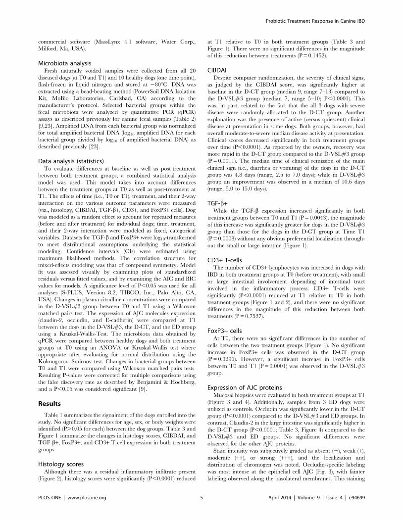
Histology scoresAlthough there was a residual inflammatory infiltrate present
(Figure 2), histology scores were significantly (P,0.0001) reduced
at T1 relative to T0 in both treatment groups (Table 3 and
Figure 1). There were no significant differences in the magnitude
of this reduction between treatments (P = 0.1452).
CIBDAIDespite computer randomization, the severity of clinical signs,
as judged by the CIBDAI score, was significantly higher at
baseline in the D-CT group (median 9, range 7–13) compared to
the D-VSL#3 group (median 7, range 5–10; P,0.0001). This
was, in part, related to the fact that the all 3 dogs with severe
disease were randomly allocated to the D-CT group. Another
explanation was the presence of active (versus quiescent) clinical
disease at presentation in some dogs. Both groups, however, had
overall moderate-to-severe median disease activity at presentation.
Clinical scores decreased significantly in both treatment groups
over time (P,0.0001). As reported by the owners, recovery was
more rapid in the D-CT group compared to the D-VSL#3 group
(P = 0.0011). The median time of clinical remission of the main
clinical sign (i.e., diarrhea or vomiting) of the dogs in the D-CT
group was 4.8 days (range, 2.5 to 7.0 days); while in D-VSL#3
group an improvement was observed in a median of 10.6 days
(range, 5.0 to 15.0 days).
TGF-b+While the TGF-b expression increased significantly in both
treatment groups between T0 and T1 (P = 0.0043), the magnitude
of this increase was significantly greater for dogs in the D-VSL#3
group than those for the dogs in the D-CT group at Time T1
(P = 0.0008) without any obvious preferential localization through-
out the small or large intestine (Figure 1).
CD3+ T-cellsThe number of CD3+ lymphocytes was increased in dogs with
IBD in both treatment groups at T0 (before treatment), with small
or large intestinal involvement depending of intestinal tract
involved in the inflammatory process. CD3+ T-cells were
significantly (P,0.0001) reduced at T1 relative to T0 in both
treatment groups (Figure 1 and 2), and there were no significant
differences in the magnitude of this reduction between both
treatments (P = 0.7527).
FoxP3+ cellsAt T0, there were no significant differences in the number of
cells between the two treatment groups (Figure 1). No significant
increase in FoxP3+ cells was observed in the D-CT group
(P = 0.3296). However, a significant increase in FoxP3+ cells
between T0 and T1 (P = 0.0001) was observed in the D-VSL#3
group.
Expression of AJC proteinsMucosal biopsies were evaluated in both treatment groups at T1
(Figure 3 and 4). Additionally, samples from 3 ED dogs were
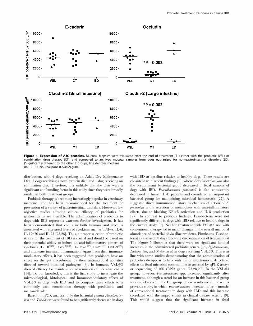
utilized as controls. Occludin was significantly lower in the D-CT
group (P,0.0001) compared to the D-VSL#3 and ED groups. In
contrast, Claudin-2 in the large intestine was significantly higher in
the D-CT group (P,0.0001; Table 3, Figure 4) compared to the
D-VSL#3 and ED groups. No significant differences were
observed for the other AJC proteins.
Stain intensity was subjectively graded as absent (2), weak (+),
moderate (++), or strong (+++), and the localization and
distribution of chromogen was noted. Occludin-specific labeling
was most intense at the epithelial cell AJC (Fig. 3), with fainter
labeling observed along the basolateral membranes. This staining
Probiotic Treatment Response in Canine IBD
PLOS ONE | www.plosone.org 5 April 2014 | Volume 9 | Issue 4 | e94699
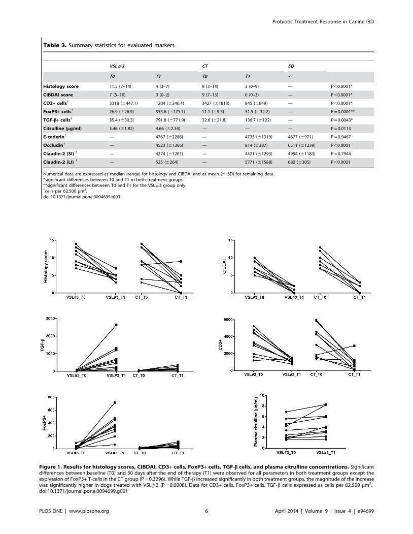
Figure 1. Results for histology scores, CIBDAI, CD3+ cells, FoxP3+ cells, TGF-b cells, and plasma citrulline concentrations. Significantdifferences between baseline (T0) and 30 days after the end of therapy (T1) were observed for all parameters in both treatment groups except theexpression of FoxP3+ T-cells in the CT group (P = 0.3296). While TGF-b increased significantly in both treatment groups, the magnitude of the increasewas significantly higher in dogs treated with VSL#3 (P = 0.0008). Data for CD3+ cells, FoxP3+ cells, TGF-b cells expressed as cells per 62,500 mm2.doi:10.1371/journal.pone.0094699.g001
Table 3. Summary statistics for evaluated markers.
VSL#3 CT ED
T0 T1 T0 T1 -
Histology score 11.5 (7–14) 4 (3–7) 9 (3–14) 3 (0–9) — P,0.0001*
CIBDAI score 7 (5–10) 0 (0–2) 9 (7–13) 0 (0–3) — P,0.0001*
CD3+ cells{ 3318 (6447.1) 1204 (6240.4) 3427 (61813) 845 (6849) — P,0.0001*
FoxP3+ cells{ 26.9 (626.9) 353.6 (6175.1) 11.1 (69.5) 51.5 (632.2) — P = 0.0001**
TGF-b+ cells{ 35.4 (630.3) 791.8 (6771.9) 32.6 (621.8) 136.7 (6122) — P = 0.0043*
Citrulline (mg/ml) 3.46 (61.82) 4.66 (62.34) — — — P = 0.0113
E-caderin{ — 4767 (62288) — 4735 (61319) 4877 (6971) P = 0.9467
Occludin{ — 4523 (61366) — 814 (6387) 6511 (61239) P,0.0001
Claudin-2 (SI) { — 4274 (61201) — 4421 (61293) 4994 (61183) P = 0.7944
Claudin-2 (LI) { — 525 (6264) — 5771 (61588) 680 (6305) P,0.0001
Numerical data are expressed as median (range) for histology and CIBDAI and as mean (6 SD) for remaining data.*significant differences between T0 and T1 in both treatment groups.**significant differences between T0 and T1 for the VSL#3 group only.{cells per 62,500 mm2.doi:10.1371/journal.pone.0094699.t003
Probiotic Treatment Response in Canine IBD
PLOS ONE | www.plosone.org 6 April 2014 | Volume 9 | Issue 4 | e94699
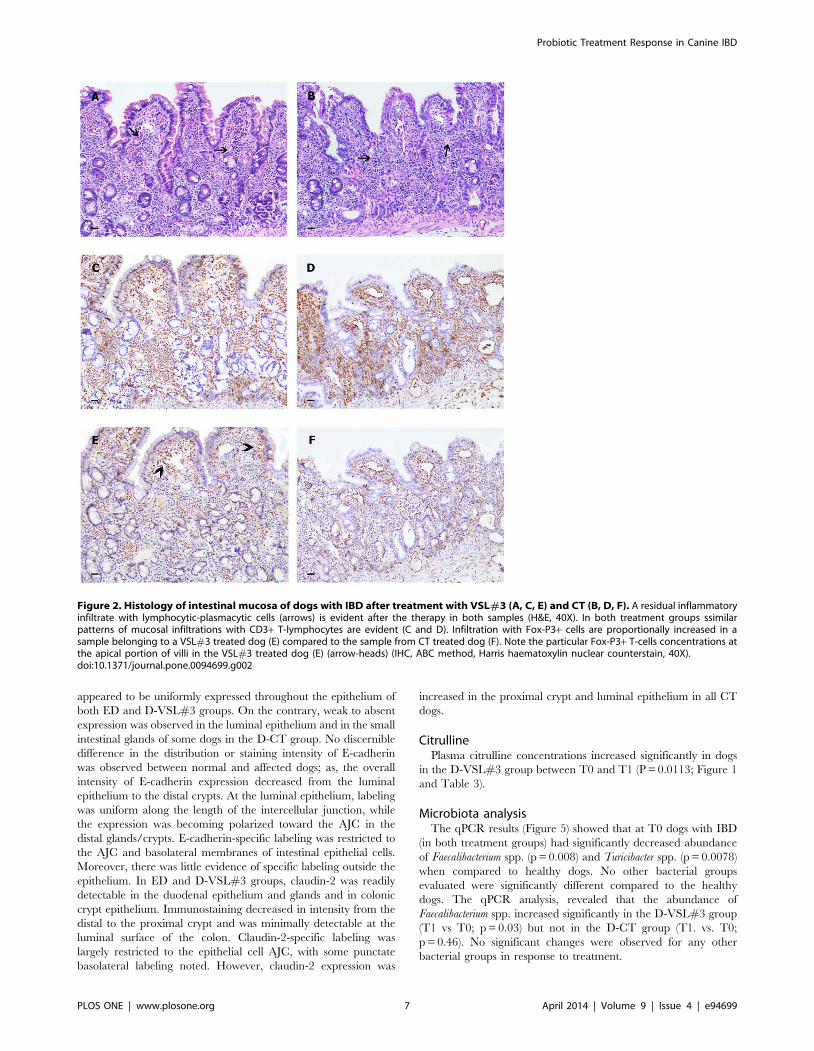
appeared to be uniformly expressed throughout the epithelium of
both ED and D-VSL#3 groups. On the contrary, weak to absent
expression was observed in the luminal epithelium and in the small
intestinal glands of some dogs in the D-CT group. No discernible
difference in the distribution or staining intensity of E-cadherin
was observed between normal and affected dogs; as, the overall
intensity of E-cadherin expression decreased from the luminal
epithelium to the distal crypts. At the luminal epithelium, labeling
was uniform along the length of the intercellular junction, while
the expression was becoming polarized toward the AJC in the
distal glands/crypts. E-cadherin-specific labeling was restricted to
the AJC and basolateral membranes of intestinal epithelial cells.
Moreover, there was little evidence of specific labeling outside the
epithelium. In ED and D-VSL#3 groups, claudin-2 was readily
detectable in the duodenal epithelium and glands and in colonic
crypt epithelium. Immunostaining decreased in intensity from the
distal to the proximal crypt and was minimally detectable at the
luminal surface of the colon. Claudin-2-specific labeling was
largely restricted to the epithelial cell AJC, with some punctate
basolateral labeling noted. However, claudin-2 expression was
increased in the proximal crypt and luminal epithelium in all CT
dogs.
CitrullinePlasma citrulline concentrations increased significantly in dogs
in the D-VSL#3 group between T0 and T1 (P = 0.0113; Figure 1
and Table 3).
Microbiota analysisThe qPCR results (Figure 5) showed that at T0 dogs with IBD
(in both treatment groups) had significantly decreased abundance
of Faecalibacterium spp. (p = 0.008) and Turicibacter spp. (p = 0.0078)
when compared to healthy dogs. No other bacterial groups
evaluated were significantly different compared to the healthy
dogs. The qPCR analysis, revealed that the abundance of
Faecalibacterium spp. increased significantly in the D-VSL#3 group
(T1 vs T0; p = 0.03) but not in the D-CT group (T1. vs. T0;
p = 0.46). No significant changes were observed for any other
bacterial groups in response to treatment.
Figure 2. Histology of intestinal mucosa of dogs with IBD after treatment with VSL#3 (A, C, E) and CT (B, D, F). A residual inflammatoryinfiltrate with lymphocytic-plasmacytic cells (arrows) is evident after the therapy in both samples (H&E, 40X). In both treatment groups ssimilarpatterns of mucosal infiltrations with CD3+ T-lymphocytes are evident (C and D). Infiltration with Fox-P3+ cells are proportionally increased in asample belonging to a VSL#3 treated dog (E) compared to the sample from CT treated dog (F). Note the particular Fox-P3+ T-cells concentrations atthe apical portion of villi in the VSL#3 treated dog (E) (arrow-heads) (IHC, ABC method, Harris haematoxylin nuclear counterstain, 40X).doi:10.1371/journal.pone.0094699.g002
Probiotic Treatment Response in Canine IBD
PLOS ONE | www.plosone.org 7 April 2014 | Volume 9 | Issue 4 | e94699
Discussion
In this study, 20 dogs with long standing IBD were randomized
to receive either a probiotic containing VSL#3 strains (SIVOY) or
a combination therapy of prednisone and metronidazole. Using a
statistical analysis that takes into account differences between the
treatment groups at enrollment (T0) as well as post-treatment (T1),
we observed differences in some of the evaluated variables
depending on the treatment regimen. Histology scores, CIBDAI,
and infiltration with mucosal CD3+ T-cells decreased significantly
in both treatment groups, and there was no significant effect
between the two treatments. FoxP3+ T-cells increased in dogs
treated with VSL#3 but not in the D-CT group. While TGF-b+cells increased significantly in both treatment groups, the
magnitude of the increase was significantly greater in dogs treated
with VSL#3. The expression of occludin and claudin-2 was also
significantly different between dogs treated with probiotic VSL#3
compared to combination therapy.
Although the etiology of canine IBD is poorly understood, there
is evidence from clinical observations, studies in humans, and
animal models to incriminate the intestinal microbiota as one
factor influencing aberrant host responses. Evidence for the role of
enteric microbiota in the pathogenesis of IBD in humans is
supported by clinical responses to fecal stream diversion in patients
with Crohn’s disease (CD) and antimicrobial therapy in both CD
and ulcerative colitis (UC) patients [3,10]. Furthermore, genetic
mutations in NOD2/CARD15 and TLR-4 (Toll-like-receptor-4)
in IBD patients make them less able to respond to bacterial
components, resulting in defective innate immune responses to
enteric microbiota [24]. Dietary factors also appear to play a role
in mediating mucosal inflammation in dogs based on the beneficial
clinical response to elimination or ‘‘hypoallergenic’’ diets in many
of these animals [16]. All 20 patients enrolled in this study were
diagnosed as having long-standing idiopathic IBD, and in the past
had undergone unsuccessful dietary trials (e.g., elimination diets to
exclude adverse food events). During the study period, all dogs
remained on their pre-trial diets, and no dietary changes were
performed as part of the here presented study. These diets were
similar in nutritional composition across both treatment groups. In
the D-VSL#3 group, 4 dogs were on an Adult Dry Maintenance
Diet, 4 dogs were on a novel protein diet, and 2 dogs were on an
elimination diet. The dogs in the D-CT group had a similar diet
Figure 3. Expression of AJC proteins in the intestinal mucosa of control dogs (ED group) (A, D, G) and dogs treated with VSL#3 (B,E, H) or CT (C, F, I). No discernible differences in the distribution or staining intensity of E-cadherin are observed between normal mucosa (A) andIBD samples (B and C); the overall intensity of E-cadherin staining decreased from the luminal epithelium to the distal crypts. Occludin-specificlabelling is most intense at the epithelial cell AJC (arrows) of the luminal epithelium covering the apical portion of villi in ED (D) and VSL#3 (E); a weakto absent expression is observed in the luminal epithelium and in some intestinal glands of the small intestine of the CT sample (F). In colonicsamples belonging to ED (G) and VSL#3 (H) groups, claudin-2 is readily detectable only in the colonic crypt epithelium, decreasing in intensity fromthe distal to the proximal crypt and becoming barely detectable at the luminal surface of the colon. In contrast, claudin-2 expression is increased inthe proximal crypt and luminal epithelium of all samples from CT dogs (I).doi:10.1371/journal.pone.0094699.g003
Probiotic Treatment Response in Canine IBD
PLOS ONE | www.plosone.org 8 April 2014 | Volume 9 | Issue 4 | e94699
distribution, with 4 dogs receiving an Adult Dry Maintenance
Diet, 5 dogs receiving a novel protein diet, and 1 dog receiving an
elimination diet. Therefore, it is unlikely that the diets were a
significant confounding factor in this study since they were broadly
similar in both treatment groups.
Probiotic therapy is becoming increasingly popular in veterinary
medicine, and has been recommended for the treatment or
prevention of a variety of gastrointestinal disorders. However, few
objective studies attesting clinical efficacy of probiotics for
gastroenteritis are available. The administration of probiotics to
dogs with IBD represents warrants further investigation. It has
been demonstrated that colitis in both humans and mice is
associated with increased levels of cytokines such as TNF-a, IL-6,
IL-12p70 and IL-23 [25,26]. Thus, a proper selection of probiotic
strains for the treatment of IBD is crucial and should be based on
their potential ability to induce an anti-inflammatory pattern of
cytokines (IL- 10high, TGF-bhigh, IL-12p70low, IL-23low, TNF-alow)
and attenuate intestinal inflammation. Apart from their immuno-
modulatory effects, it has been suggested that probiotics have an
effect on the gut microbiome by their antimicrobial activities
directed toward intestinal pathogens [3]. In humans, VSL#3
showed efficacy for maintenance of remission of ulcerative colitis
[14]. To our knowledge, this is the first study to investigate the
microbiological, histological, and immunomodulatory effects of
VSL#3 in dogs with IBD and to compare these effects to a
commonly used combination therapy with prednisone and
metronidazole.
Based on qPCR analysis, only the bacterial genera Faecalibacter-
ium and Turicibacter were found to be significantly decreased in dogs
with IBD at baseline relative to healthy dogs. These results are
consistent with recent findings [9], where Faecalibacterium was also
the predominant bacterial group decreased in fecal samples of
dogs with IBD. Faecalibacterium prausnitzii is also consistently
decreased in human IBD patients and considered an important
bacterial group for maintaining microbial homeostasis [27]. A
suggested direct immunomodulatory mechanism of action of F.
prausnitzii is the secretion of metabolites with anti-inflammatory
effects, due to blocking NF-kB activation and IL-8 production
[27]. In contrast to previous findings, Fusobacteria were not
significantly different in dogs with IBD relative to healthy dogs in
the current study [9]. Neither treatment with VSL#3 nor with
conventional therapy led to major changes in the overall microbial
abundance of bacterial phyla (Bacteroidetes, Firmicutes, Fusobac-
teria) as assessed 30 days following discontinuation of treatment (at
T1). Figure 5 illustrates that there were no significant luminal
increases in the administered probiotic genera (i.e., Bifidobacterium,
Lactobacillus, and Streptococcus) in dogs receiving VSL#3. This is in
line with some studies demonstrating that the administration of
probiotics do appear to have only minor and transient detectable
effects on fecal microbial communities as assessed by qPCR assays
or sequencing of 16S rRNA genes [23,28,29]. In the VSL#3
group, however, Faecalibacterium spp. increased significantly after
treatment, although a trend for an increase in this bacterial group
was also observed in the CT group. These results are in line with a
previous study, in which Faecalibacterium increased after 4 months
of conventional treatment in dogs with IBD and this increase
correlated with the improvement in clinical disease activity [9].
This would suggest that the significant increase in fecal
Figure 4. Expression of AJC proteins. Mucosal biopsies were evaluated after the end of treatment (T1) either with the probiotic (VSL) orcombination drug therapy (CT), and compared to archived mucosal samples from dogs euthanized for non-gastrointestinal disorders (ED).(*significantly different to the other 2 groups; line denotes median).doi:10.1371/journal.pone.0094699.g004
Probiotic Treatment Response in Canine IBD
PLOS ONE | www.plosone.org 9 April 2014 | Volume 9 | Issue 4 | e94699
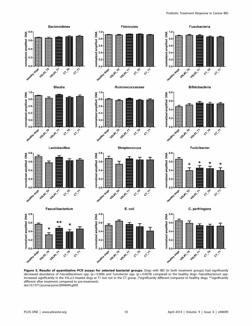
Figure 5. Results of quantitative PCR assays for selected bacterial groups. Dogs with IBD (in both treatment groups) had significantlydecreased abundance of Faecalibacterium spp. (p = 0.008) and Turicibacter spp. (p = 0.0078) compared to the healthy dogs. Faecalibacterium spp.increased significantly in the VSL#3 treated dogs at T1 but not in the CT group. (*significantly different compared to healthy dogs; **significantlydifferent after treatment compared to pre-treatment).doi:10.1371/journal.pone.0094699.g005
Probiotic Treatment Response in Canine IBD
PLOS ONE | www.plosone.org 10 April 2014 | Volume 9 | Issue 4 | e94699
Faecalibacterium is not necessarily specific for the probiotic
treatment, but may be a general indicator for normalization of
fecal dysbiosis after long-term therapy. The Faecalibacterium–
Subdoligranulum group is a major bacterial group in the canine
gastrointestinal tract, comprising 16% of total bacterial counts in
feces of healthy dogs and is believed to be of importance in canine
gastrointestinal health [30]. Therefore, more-in depth studies
evaluating the functional properties of canine Faecalibacterium
strains are warranted. Some limitation of the microbiota analysis
performed in this study need to be noted. Analyzing the fecal
microbiota using sequencing of 16S rRNA genes may have
revealed potential changes either in microbial diversity indices or
in bacterial groups that were not covered by our qPCR assays. For
technical reasons, a sequencing approach was not possible in this
study. However, we have utilized qPCR assays targeting the
microbiota on various phylogenetic levels and also targeting
bacterial groups that are major bacterial groups in the canine
intestine and that have been shown to be important in canine IBD
[9]. Furthermore, in the current study, only fecal samples were
analyzed, and the potential impact of treatment on the compo-
sition of the small intestinal mucosa-associated microbiota may
have been missed. Previous studies have revealed that dogs with
IBD have significant differences in small intestinal microbiota
compared to controls, and future studies should evaluate the effect
of probiotics on the small intestinal microbiota of these dogs [8].
Also, in this study we assessed the fecal microbiota 30 days after
the discontinuation of therapy, and it is possible that a transient
change in the fecal microbiota during the administration period
may have remained undetected and/or changed during the 30
days post-treatment.
It has been speculated that IBD is associated with a loss of
intestinal barrier function, as multiple genes encoding for proteins
responsible for maintenance of intestinal barrier function (i.e.,
those encoding for claudin-8, metallothionein, and matrix
metalloproteinases) were down-regulated in dogs with IBD in a
previous study [31]. The observation that the expression and
distribution of occludin and claudin-2 in the large intestine were
not significantly different between dogs treated with VSL#3 and
the non-IBD control dogs (ED group), but were significantly
different compared to the D-CT group, suggests potential effects of
VSL#3 on intestinal barrier function, warranting further studies
[32]. Similar changes in the distribution of claudin-2 expression
have been observed in humans with active UC, where claudin-2
was detected at the surface epithelium [33]. Similarly, down
regulation of occludin has been observed in the intestinal mucosa
of patients with both UC and CD [34]. Here we compared the
expression patterns of AJC proteins between healthy dogs
(euthanized dogs; group ED) and dogs with IBD after the two
different types of treatment (VSL # 3 or CT treated dogs). The
expression pattern of AJC proteins in the ED group was similar to
that described by Ohta et al. in healthy dogs [35]. In contrast,
based on our results it seems that dogs in the CT-group had a
greater deviation from the physiological conditions in expression of
Claudin-2 in the colon. This particular expression pattern
resembles that observed in samples from the colon of dogs with
colitis [21]. While we cannot conclusively state that there was an
improvement in the expression pattern after probiotic treatment,
as samples were not evaluated at T0, we speculate that the
expression pattern of AJC proteins in dogs treated with VSL#3
appears to resembles more the physiological state as observed in
healthy dogs [35]. Future studies are warranted to confirm this
observation. At this point it remains also unclear why claudin-2 is
increased in the large intestine of dogs treated with drug therapy,
and further work is needed to elucidate the mechanism behind this
increased expression of claudin-2.
Dogs treated with VSL#3 showed significantly increased
plasma citrulline concentrations 30 days after end of administra-
tion, suggesting restitution of the mucosal barrier. Plasma citrulline
concentrations are a marker of global enterocyte mass in humans,
rodents, and pigs [36], and have recently been shown to reflect
intestinal mucosal recovery in response to severe injury in dogs
[37]. Unfortunately, we were able to statistically evaluate the blood
levels of citrulline only in the D-VSL#3 group, as plasma citrulline
concentrations were not available for all dogs in the D-CT group.
Because of the small samples size in the D-CT group, we decided
not to perform any statistical analysis to compare plasma citrulline
concentrations between treatments. Therefore, it is currently
unknown whether the observed increase in plasma citrulline
concentrations was specific for the treatment with VSL#3 strains,
or would also be present in dogs treated with conventional
therapy.
The immunohistochemical results showed cross-reactivity for
canine tissues of all antibodies used in this study. This is in line
with results from previous studies which have shown that these
antibodies are useful for immunohistochemical assessment of
canine tissues. In particular, cross-reactivity of the rat anti-human
CD3 antigen, clone MCA1477, for canine CD3 positive T-
lymphocytes has been shown previously on gastric tissue of dogs
[20]. Cross-reactivity of the clone FJK-16s used to stain canine
FoxP3-lymphocytes has been reported in another study [38].
Similarly, other authors have successfully used the monoclonal
antibody against TGF-b positive dog lymphocytes (clone 1D11)
[39]. Finally, the specificities of the antibodies used for canine AJC
proteins (i.e., pAb anti-claudin-2 (PAD: MH44), anti-occludin
(PAD: Z-T22), and mAb anti-E-cadherin (IgG2a, clone: 36) were,
similarly to our study, also reported on sections of intestinal tissue
in dogs with IBD [21].
The evaluation of immunomorphological variables suggests a
potential anti-inflammatory effect of VSL#3 strains, as decreased
mucosal CD3+ T-lymphocytes, and increased FoxP3+ and TGF-
b+ positive cells were observed 30 days after the end of
administration. Immunohistochemistry results showed a difference
in the predominant immunophenotype of infiltrating cells in
intestinal lamina propria of biopsies from VSL#3 treated dogs.
More specifically, the VSL#3 treated dogs showed increases in
CD3+/FoxP3+ cells (Figure 2) in the intestinal mucosa, while dogs
treated with prednisone and metronidazole displayed an overall
decrease in all inflammatory cell populations that was accompa-
nied by a decrease of FoxP3+ lymphocytes and TGF-b expressing
cells (Figure 2). These findings are consistent with a previous study
in a mouse model, where VSL#3 also led to increased FoxP3+expressing T-cells in intestinal lymphoid follicles [40]. In clinical
studies with human IBD patients as well as studies on rodent
models of IBD, VSL#3 has shown various other anti-inflamma-
tory mechanisms. For example, VSL#3 was shown to induce
heat-shock-proteins in intestinal epithelial cells (IEC) [41] or
enhance proliferation of IL-10-dependent TGF-b-bearing regula-
tory T-cells in Th1-dependent murine colitis [42]. These variables
have not been examined in the current study, and it would be
useful to evaluate these markers in future clinical studies.
Furthermore, qPCR quantification of both pro-inflammatory
(i.e., TNF-a, IL1-b, IL-8) as well as regulatory genes (FoxP3, IL-
10) would have been useful to perform since canine probes have
already been published [43] and these studies showed increases in
IL-8 in colorectal inflammation [44].
As limitations to this study it should be noted that only a small
number of dogs was evaluated, and the power to detect differences
Probiotic Treatment Response in Canine IBD
PLOS ONE | www.plosone.org 11 April 2014 | Volume 9 | Issue 4 | e94699
in some of the evaluated variables may have been insufficient to
detect differences between treatment groups. Furthermore, this
was an open-label study and no placebo group was included.
Ideally, the clinical effect of the treatment with probiotic strains
should be evaluated in a double-blinded placebo controlled trial
and compared to a non-treated group. However, in the case of
chronic IBD, it is difficult to enroll a non-treated group as these
dogs show chronic signs of disease, and therefore we chose in this
study to compare the effects of VSL#3 strains to the commonly
used combination therapy with prednisone and metronidazole.
Our study results suggest that probiotic treatment induces
differential anti-inflammatory immune responses when compared
to routine combination therapy as evidenced by significant
increases in FoxP3+ cells and a significantly larger increase in
TGF-b. The findings lay the foundation for future larger scale
placebo controlled clinical studies to evaluate clinical benefits of
probiotic VSL#3 strains in the treatment of dogs with IBD.
Author Contributions
Conceived and designed the experiments: GR GP AEJ JSS. Performed the
experiments: GR GP MC APP JSS. Analyzed the data: GR NDC AEJ
JMS JSS. Contributed reagents/materials/analysis tools: GR GP MC
APP. Wrote the paper: GR JMS NDC AEJ JSS.
References
1. Suchodolski JS (2011) Companion animals symposium: Microbes and
gastrointestinal health of dogs and cats. J Anim Sci 89: 1520–1530.
2. Allenspach K, House A, Smith K, McNeill FM, Hendricks A, et al. (2010)
Evaluation of mucosal bacteria and histopathology, clinical disease activity and
expression of Toll-like receptors in German shepherd dogs with chronic
enteropathies. Vet Microbiol 146: 326–335.
3. Rioux KP, Madsen KL, Fedorak RN (2005) The role of enteric microflora in
inflammatory bowel disease: Human and animal studies with probiotics and
prebiotics. Gastroenterol Clin North Am 34: 465–482.
4. Contractor NV, Bassiri H, Reya T, Park AY, Baumgart DC, et al. (1998)
Lymphoid hyperplasia, autoimmunity, and compromised intestinal intraepithe-
lial lymphocyte development in colitis-free gnotobiotic IL-2-deficient mice.
J Immunol 160: 385–394.
5. Sellon RK, Tonkonogy S, Schultz M, Dieleman LA, Grenther W, et al. (1998)
Resident enteric bacteria are necessary for development of spontaneous colitis
and immune system activation in interleukin-10-deficient mice. Infect Immun
66: 5224–5231.
6. Xenoulis PG, Palculict B, Allenspach K, Steiner JM, Van House AM, et al.
(2008) Molecular-phylogenetic characterization of microbial communities
imbalances in the small intestine of dogs with inflammatory bowel disease.
FEMS Microbiol Ecol 66: 579–589.
7. Suchodolski JS, Xenoulis PG, Paddock CG, Steiner JM, Jergens AE (2010)
Molecular analysis of the bacterial microbiota in duodenal biopsies from dogs
with idiopathic inflammatory bowel disease. Vet Microbiol 142: 394–400.
8. Suchodolski JS, Dowd SE, Wilke V, Steiner JM, Jergens AE (2012) 16S rRNA
Gene Pyrosequencing Reveals Bacterial Dysbiosis in the Duodenum of Dogs
with Idiopathic Inflammatory Bowel Disease. Plos ONE 7: e39333.
9. Suchodolski JS, Markel ME, Garcia-Mazcorro JF, Unterer S, Heilmann RM, et
al. (2012) The fecal microbiome in dogs with acute diarrhea and idiopathic
inflammatory bowel disease. Plos ONE 7: e51907.
10. Sutherland L, Singleton J, Sessions J, Hanauer S, Krawitt E, et al. (1991)
Double-Blind, Placebo Controlled Trial of Metronidazole in Crohns-Disease.
Gut 32: 1071–1075.
11. Jergens AE, Crandell J, Morrison JA, Deitz K, Pressel M, et al. (2010)
Comparison of oral prednisone and prednisone combined with metronidazole
for induction therapy of canine inflammatory bowel disease: a randomized-
controlled trial. J Vet Intern Med 24: 269–277.
12. Turner D, Levine A, Escher JC, Griffiths AM, Russell RK, et al. (2012)
Management of pediatric ulcerative colitis: joint ECCO and ESPGHAN
evidence-based consensus guidelines. J Pediatr Gastroenterol Nutr 55: 340–361.
13. Sauter SN, Benyacoub J, Allenspach K, Gaschen F, Ontsouka E, et al. (2006)
Effects of probiotic bacteria in dogs with food responsive diarrhoea treated with
an elimination diet. J Anim Physiol Anim Nutr (Berl) 90: 269–277.
14. Bibiloni R, Fedorak RN, Tannock GW, Madsen KL, Gionchetti P, et al. (2005)
VSL#3 probiotic-mixture induces remission in patients with active ulcerative
colitis. Am J Gastroenterol 100: 1539–1546.
15. Tursi A, Brandimarte G, Papa A, Giglio A, Elisei W, et al. (2010) Treatment of
Relapsing Mild-to-Moderate Ulcerative Colitis With the Probiotic VSL#3 as
Adjunctive to a Standard Pharmaceutical Treatment: A Double-Blind,
Randomized, Placebo-Controlled Study. Am J Gastroenterol: 2218–2227.
16. Simpson KW, Jergens AE (2011) Pitfalls and progress in the diagnosis and
management of canine inflammatory bowel disease. Vet Clin North Am Small
Anim Pract 41: 381–398.
17. Jergens AE, Schreiner CA, Frank DE, Niyo Y, Ahrens FE, et al. (2003) A scoring
index for disease activity in canine inflammatory bowel disease. J Vet Intern
Med 17: 291–297.
18. German AJ, Helps CR, Hall EJ, Day MJ (2000) Cytokine mRNA expression in
mucosal biopsies from German Shepherd dogs with small intestinal enteropa-
thies. Dig Dis Sci 45: 7–17.
19. Day MJ, Bilzer T, Mansell J, Wilcock B, Hall EJ, et al. (2008) Histopathological
Standards for the Diagnosis of Gastrointestinal Inflammation in Endoscopic
Biopsy Samples from the Dog and Cat: A Report from the World Small Animal
Veterinary Association Gastrointestinal Standardization Group. J Comp Pathol
138 S1–S43.
20. Rossi G, Fortuna D, Pancotto L, Renzoni G, Taccini E, et al. (2000)
Immunohistochemical study of lymphocyte populations infiltrating the gastric
mucosa of beagle dogs experimentally infected with Helicobacter pylori. InfectImmun 68: 4769–4772.
21. Ridyard AE, Brown JK, Rhind SM, Else RW, Simpson JW, et al. (2007) Apical
junction complex protein expression in the canine colon: differential expressionof claudin-2 in the colonic mucosa in dogs with idiopathic colitis. J Histochem
Cytochem 55: 1049–1058.
22. Engel AG, Arahata K (1986) Mononuclear cells in myopathies: quantitation offunctionally distinct subsets, recognition of antigen-specific cell-mediated
cytotoxicity in some diseases, and implications for the pathogenesis of the
different inflammatory myopathies. Hum Pathol 17: 704–721.
23. Garcia-Mazcorro JF, Lanerie DJ, Dowd SE, Paddock CG, Grutzner N, et al.(2011) Effect of a multi-species synbiotic formulation on fecal bacterial
microbiota of healthy cats and dogs as evaluated by pyrosequencing. FEMSMicrobiol Ecol 78: 542–554.
24. Franchimont D, Vermeire S, El Housni H, Pierik M, Van Steen K, et al. (2004)
Deficient host-bacteria interactions in inflammatory bowel disease? The toll-likereceptor (TLR)-4 Asp299gly polymorphism is associated with Crohn’s disease
and ulcerative colitis. Gut 53: 987–992.
25. Becker C, Dornhoff H, Neufert C, Fantini MC, Wirtz S, et al. (2006) Cutting
edge: IL-23 cross-regulates IL-12 production in T cell-dependent experimentalcolitis. J Immunol 177: 2760–2764.
26. Fuss IJ, Becker C, Yang Z, Groden C, Hornung RL, et al. (2006) Both IL-12p70
and IL-23 are synthesized during active Crohn’s disease and are down-regulatedby treatment with anti-IL-12 p40 monoclonal antibody. Inflamm Bowel Dis 12:
9–15.
27. Sokol H, Pigneur B, Watterlot L, Lakhdari O, Bermudez-Humaran LG, et al.(2008) Faecalibacterium prausnitzii is an anti-inflammatory commensal
bacterium identified by gut microbiota analysis of Crohn disease patients.
PNAS 105: 16731–16736.
28. Vitali B, Ndagijimana M, Cruciani F, Carnevali P, Candela M, et al. (2010)Impact of a synbiotic food on the gut microbial ecology and metabolic profiles.
BMC Microbiol 10: 4.
29. Larsen N, Vogensen FK, Gobel R, Michaelsen KF, Abu Al-Soud W, et al.(2011) Predominant genera of fecal microbiota in children with atopic dermatitis
are not altered by intake of probiotic bacteria Lactobacillus acidophilus NCFMand Bifidobacterium animalis subsp. lactis Bi-07. FEMS Microbiol Ecol 75: 482–
496.
30. Garcia-Mazcorro JF, Dowd SE, Poulsen J, Steiner JM, Suchodolski JS (2012)
Abundance and short-term temporal variability of fecal microbiota in healthydogs. MicrobiologyOpen 1: 340–347.
31. Wilke VL, Nettleton D, Wymore MJ, Gallup JM, Demirkale CY, et al. (2012)
Gene expression in intestinal mucosal biopsy specimens obtained from dogs withchronic enteropathy. Am J Vet Res 73: 1219–1229.
32. Madsen K, Cornish A, Soper P, McKaigney C, Jijon H, et al. (2001) Probiotic
bacteria enhance murine and human intestinal epithelial barrier function.Gastroenterol 121: 580–591.
33. Prasad S, Mingrino R, Kaukinen K, Hayes KL, Powell RM, et al. (2005)
Inflammatory processes have differential effects on claudins 2, 3 and 4 in colonic
epithelial cells. Lab Invest 85: 1139–1162.
34. Gassler N, Rohr C, Schneider A, Kartenbeck J, Bach A, et al. (2001)
Inflammatory bowel disease is associated with changes of enterocytic junctions.
Am J Physiol Gastrointest Liver Physiol 281: G216–228.
35. Ohta H, Yamaguchi T, Rajapakshage BK, Murakami M, Sasaki N, et al. (2011)Expression and subcellular localization of apical junction proteins in canine
duodenal and colonic mucosa. Am J Vet Res 72: 1046–1051.
36. Curis E, Nicolis I, Osowska S, Zerrouk N, Benazeth S, et al. (2005) Almost allabout citrullin in mammals. Amino Acids 29: 177–205.
37. Dossin O, Rupassara SI, Weng HY, Williams DA, Garlick PJ, et al. (2011) Effect
of Parvoviral Enteritis on Plasma Citrulline Concentration in Dogs. J Vet InternMed 25: 215–221.
38. Pinheiro D, Singh Y, Grant CR, Appleton RC, Sacchini F, et al. (2011)
Phenotypic and functional characterization of a CD4(+) CD25(high) FOX-
P3(high) regulatory T-cell population in the dog. Immunol 132: 111–122.
Probiotic Treatment Response in Canine IBD
PLOS ONE | www.plosone.org 12 April 2014 | Volume 9 | Issue 4 | e94699
39. Colitz CM, Malarkey D, Dykstra MJ, McGahan MC, Davidson MG (2000)
Histologic and immunohistochemical characterization of lens capsular plaquesin dogs with cataracts. Am J Vet Res 61: 139–143.
40. Bassaganya-Riera J, Viladomiu M, Pedragosa M, De Simone C, Carbo A, et al.
(2012) Probiotic bacteria produce conjugated linoleic acid locally in the gut thattargets macrophage PPAR gamma to suppress colitis. Plos ONE 7: e31238.
41. Petrof EO, Kojima K, Ropeleski MJ, Musch MW, Tao Y, et al. (2004)Probiotics inhibit nuclear factor-kappaB and induce heat shock proteins in
colonic epithelial cells through proteasome inhibition. Gastroenterol 127: 1474–
1487.42. Di Giacinto C, Marinaro M, Sanchez M, Strober W, Boirivant M (2005)
Probiotics ameliorate recurrent Th1-mediated murine colitis by inducing IL-10and IL-10-dependent TGF-beta-bearing regulatory cells. J Immunol 174: 3237–
3246.43. Ohta H, Takada K, Torisu S, Yuki M, Tamura Y, et al. (2013) Expression of
CD4+ T cell cytokine genes in the colorectal mucosa of inflammatory colorectal
polyps in miniature dachshunds. Vet Immunol Immunopathol 155: 259–263.44. Tamura Y, Ohta H, Torisu S, Yuki M, Yokoyama N, et al. (2013) Markedly
increased expression of interleukin-8 in the colorectal mucosa of inflammatorycolorectal polyps in miniature dachshunds. Vet Immunol Immunopathol 156:
32–42.
45. Muhling M, Woolven-Allen J, Murrell JC, Joint I (2008) Improved group-specific PCR primers for denaturing gradient gel electrophoresis analysis of the
genetic diversity of complex microbial communities. ISME J 2: 379–392.46. Malinen E, Rinttila T, Kajander K, Matto J, Kassinen A, et al. (2005) Analysis
of the fecal microbiota of irritable bowel syndrome patients and healthy controlswith real-time PCR. Am J Gastroenterol 100: 373–382.
47. Garcia-Mazcorro JF, Suchodolski JS, Jones KR, Clark-Price SC, Dowd SE, et
al. (2012) Effect of the proton pump inhibitor omeprazole on the gastrointestinal
bacterial microbiota of healthy dogs. FEMS Microbiol Ecol 80: 624–636.
48. Wise MG, Siragusa GR (2005) Quantitative detection of Clostridium perfringens
in the broiler fowl gastrointestinal tract by real-time PCR. Appl Environ
Microbiol 71: 3911–3916.
49. Lubbs DC, Vester BM, Fastinger ND, Swanson KS (2009) Dietary protein
concentration affects intestinal microbiota of adult cats: a study using DGGE
and qPCR to evaluate differences in microbial populations in the feline
gastrointestinal tract. J Anim Physiol Anim Nutr (Berl) 93: 113–121.
50. Rinttila T, Kassinen A, Malinen E, Krogius L, Palva A (2004) Development of
an extensive set of 16S rDNA-targeted primers for quantification of pathogenic
and indigenous bacteria in faecal samples by real-time PCR. J Appl Microbiol
97: 1166–1177.
51. Furet JP, Quenee P, Tailliez P (2004) Molecular quantification of lactic acid
bacteria in fermented milk products using real-time quantitative PCR. Int J Food
Microbiol 97: 197–207.
52. Walter J, Hertel C, Tannock GW, Lis CM, Munro K, et al. (2001) Detection of
Lactobacillus, Pediococcus, Leuconostoc, and Weissella species in human feces
by using group-specific PCR primers and denaturing gradient gel electropho-
resis. Appl Environ Microbiol 67: 2578–2585.
53. Heilig HG, Zoetendal EG, Vaughan EE, Marteau P, Akkermans AD, et al.
(2002) Molecular diversity of Lactobacillus spp. and other lactic acid bacteria in
the human intestine as determined by specific amplification of 16S ribosomal
DNA. Appl Environ Microbiol 68: 114–123.
Probiotic Treatment Response in Canine IBD
PLOS ONE | www.plosone.org 13 April 2014 | Volume 9 | Issue 4 | e94699