recombinant expression, purification and biochemical ... - plos
TRANSCRIPT
RESEARCH ARTICLE
Recombinant expression, purification and
biochemical characterization of kievitone
hydratase from Nectria haematococca
Matthias Engleder1,2☯, Melissa Horvat1,2☯, Anita Emmerstorfer-Augustin1,
Tamara Wriessnegger1, Stefanie Gabriel2, Gernot Strohmeier1,3, Hansjorg Weber3,
Monika Muller4¤, Iwona Kaluzna4¤, Daniel Mink4¤, Martin Schurmann4¤, Harald Pichler1,2*
1 acib—Austrian Centre of Industrial Biotechnology, Graz, Austria, 2 Institute of Molecular Biotechnology,
Graz University of Technology, NAWI Graz, BioTechMed Graz, Graz, Austria, 3 Institute of Organic
Chemistry, Graz University of Technology, NAWI Graz, Graz, Austria, 4 DSM Ahead R&D—Innovative
Synthesis, Geleen, The Netherlands
☯ These authors contributed equally to this work.
¤ Current Address: InnoSyn B.V., Geleen, The Netherlands
Abstract
Kievitone hydratase catalyzes the addition of water to the double bond of the prenyl moiety
of plant isoflavonoid kievitone and, thereby, forms the tertiary alcohol hydroxy-kievitone.
In nature, this conversion is associated with a defense mechanism of fungal pathogens
against phytoalexins generated by host plants after infection. As of today, a gene sequence
coding for kievitone hydratase activity has only been identified and characterized in Fusar-
ium solani f. sp. phaseoli. Here, we report on the identification of a putative kievitone hydra-
tase sequence in Nectria haematococca (NhKHS), the teleomorph state of F. solani, based
on in silico sequence analyses. After heterologous expression of the enzyme in the methylo-
trophic yeast Pichia pastoris, we have confirmed its kievitone hydration activity and have
assessed its biochemical properties and substrate specificity. Purified recombinant NhKHS
is obviously a homodimeric glycoprotein. Due to its good activity for the readily available
chalcone derivative xanthohumol (XN), this compound was selected as a model substrate
for biochemical studies. The optimal pH and temperature for hydratase activity were 6.0 and
35˚C, respectively, and apparent Vmax and Km values for hydration of XN were 7.16 μmol
min-1 mg-1 and 0.98 ± 0.13 mM, respectively. Due to its catalytic properties and apparent
substrate promiscuity, NhKHS is a promising enzyme for the biocatalytic production of ter-
tiary alcohols.
Introduction
The production of enantiopure tertiary alcohols is a major challenge of organic synthesis as
these functional groups are widely applicable for the generation of pharmaceuticals or other
bioactive compounds [1,2]. However, synthesis of tertiary alcohols is still a demanding task for
synthetic organic chemistry due to issues such as low yields, poor selectivity or harsh reaction
PLOS ONE | https://doi.org/10.1371/journal.pone.0192653 February 8, 2018 1 / 19
a1111111111
a1111111111
a1111111111
a1111111111
a1111111111
OPENACCESS
Citation: Engleder M, Horvat M, Emmerstorfer-
Augustin A, Wriessnegger T, Gabriel S, Strohmeier
G, et al. (2018) Recombinant expression,
purification and biochemical characterization of
kievitone hydratase from Nectria haematococca.
PLoS ONE 13(2): e0192653. https://doi.org/
10.1371/journal.pone.0192653
Editor: Israel Silman, Weizmann Institute of
Science, ISRAEL
Received: November 28, 2017
Accepted: January 26, 2018
Published: February 8, 2018
Copyright: © 2018 Engleder et al. This is an open
access article distributed under the terms of the
Creative Commons Attribution License, which
permits unrestricted use, distribution, and
reproduction in any medium, provided the original
author and source are credited.
Data Availability Statement: All relevant data are
within the paper and its Supporting Information
files.
Funding: This work has been supported by Das
Land Steiermark, the Federal Ministry of Science,
Research and Economy (BMWFW), the Federal
Ministry of Traffic, Innovation and Technology
(bmvit), the Styrian Business Promotion Agency
SFG, the Standortagentur Tirol, the Government of
Lower Austria and Business Agency Vienna

conditions. Therefore, sustainable biocatalytic processes that rely on enzymatic transforma-
tions are highly desirable [2,3]. The most extensively applied enzymes for synthesis of optically
active, tertiary alcohols belong to the class of hydrolases [4,5], where especially lipases and
esterases have been used for their kinetic resolution [1,2]. In contrast, members of other
enzyme classes are markedly underrepresented, even given the fact that some of them would
offer great possibilities for applications. This holds particularly true for hydro-lyases (EC 4.2.1.
X), which are able to catalyze the highly selective, reversible addition of water to non-activated
carbon-carbon double bonds and, thereby, generate primary, secondary or tertiary alcohols
[6,7]. Aside from cofactor dependent hydro-lyases, a number of enzymes catalyze the addition
of water cofactor-independently, which increases the potential of this enzyme group for indus-
trial applications. However, although more than 100 hydro-lyases have been discovered to
date, only a very limited number has been applied industrially. The most prominent examples
include nitrile hydratase and fumarase for the production of acrylamide on a 30,000 t a-1 scale,
or the production of (S)-malic acid (2,500 t a-1), respectively [7]. Another enzyme group that
has recently received increasing attention of both academic and industrial research are oleate
hydratases. Representatives of this class of hydratases have been applied for the production of
10-hydroxystearic acid from oleic acid, as well as α,ω-dicarboxylic acids, ω-hydroxycarboxylic
acids and γ-dodecalactones in multistep enzymatic reaction systems [8–10]. Compounds
obtained from oleate hydratase reactions are applied widespread in the production of a large
variety of chemicals and intermediates, such as polymers, coatings, lubricants, personal care,
perfumes and food additives [11].
An attractive member of the hydro-lyase enzyme group for the biosynthesis of tertiary alco-
hols is kievitone hydratase (KHS; EC 4.2.1.95). This enzyme was first described by Kuhn and
Smith in 1979 by analysis of liquid cultures of the fungal plant pathogen Fusarium solani f. sp.
phaseoli [12]. It catalyzes the detoxification of the plant phytoalexin kievitone (KV) produced
by Phaseolus vulgaris (French bean) after a microbial infection. Hydration of the isolated car-
bon-carbon double bond of KV results in formation of the less toxic tertiary alcohol hydroxy–
kievitone (HO-KV). Thereby, KHS plays an important role in the pathogenicity of the fungus
for its plant host. The first investigations of F. solani f. sp. phaseoli KHS (FsKHS) were per-
formed by Cleveland and Smith upon partial purification from cell free culture filtrates [13].
FsKHS is an extracellular glycoprotein with inferred activity not only for the plant isoflavanon
KV, but also for the pterocarpan phaseollidin. However, additional studies showed that in F.
solani f. sp. phaseoli, kievitone hydratase and phaseollidin hydratase activities are most likely
conferred by two different enzymes [14]. In 1995, the complete nucleotide sequence of FsKHS
was published [15]. It could be shown that secretion of the enzyme is most likely mediated by
an N-terminal signal peptide that was not present in the mature enzyme. Using low-stringency
Southern blot hybridization, several sequences homologous to FsKHS were identified in differ-
ent members of the F. solani species complex. This included, among others, Nectria haemato-cocca, the teleomorphic state of F. solani. This corroborated an earlier study that detected
HO-KV in cell free culture filtrates of N. haematococca upon infecting P. vulgaris [16]. How-
ever, to date no gene or protein sequence could be unambiguously assigned to kievitone hydra-
tase activity in N. haematococca.
In order to elucidate potential hydro-lyases for the synthesis of tertiary alcohols, we have
investigated different gene sequences from the F. solani species complex on the basis of in-
formation from previous studies [14–16]. After identifying a putative kievitone hydratase
sequence in N. haematococca MP VI, we have heterologously expressed the gene in P. pastorisand confirmed its activity for KV and other flavonoids. Subsequently, a biochemical character-
ization provided detailed insight into the potential of this enzyme group for biocatalytic
applications.
Kievitone hydratase from Nectria haematococca
PLOS ONE | https://doi.org/10.1371/journal.pone.0192653 February 8, 2018 2 / 19
through the COMET–Funding Program managed
by the Austrian Research Promotion Agency FFG.
The co-funder, InnoSyn B.V. provided support in
the form of salaries for authors [MM, IK, DM, MS],
but did not have any additional role in the study
design, data collection and analysis, decision to
publish, or preparation of the manuscript. All co-
authors with industrial affiliation, i.e. MM, IK, DM,
MS, approved of the publication, like all the other
authors. The specific roles of all authors are
articulated in the ‘author contributions’ section. The
industrial affiliation of MM, IK, DM, MS, did not
have any influence whatsoever on the scientific
layout of this manuscript.
Competing interests: Martin Schuermann, Daniel
Mink, Iwona Kaluzna and Monika Mueller have
commercial affiliations (InnoSyn B.V., Geleen, The
Netherlands). This does not alter the authors’
adherence to all PLOS ONE policies on sharing data
and materials.

Materials and methods
Chemicals and media components
Standard laboratory reagents were obtained from Sigma-Aldrich (Vienna, Austria) or Carl
Roth GmbH & Co. KG (Karlsruhe, Germany) with the highest purity available. KV and
HO-KV were obtained from InnoSyn B.V. (Geleen, The Netherlands). 8-Prenylnaringenin
was purchased from Sigma-Aldrich (Vienna, Austria), and Isoxanthohumol was a kind gift of
Prof. Michael Murkovic, Institute of Biochemistry, Graz University of Technology. Hop
extract capsules were purchased from Allcura (Wertheim, Germany). Restriction enzymes
were acquired from Thermo Scientific (St. Leon-Rot, Germany). Bacto™ peptone, Bacto™ yeast
extract and Difco™ yeast nitrogen base w/o amino acids (YNB) were obtained from Becton,
Dickinson and Company (Schwechat, Austria). ZeocinTM was purchased from InvivoGen
(Vienna, Austria). Sterile water was acquired from Fresenius Kabi, Graz, Austria.
P. pastoris cultures were routinely grown in buffered glycerol-complex medium BMGY (1%
yeast extract, 2% peptone, 100 mM potassium phosphate, pH 6.0, 1.34% YNB, 4 × 10⁻⁵% bio-
tin, 1% glycerol). Buffered methanol-complex medium, BMMY (1% yeast extract, 2% peptone,
100 mM potassium phosphate, pH 6.0, 1.34% YNB, 4 × 10⁻⁵% biotin, 1% methanol) was used
as induction medium.
Vector and P. pastoris KHS strain construction
The P. pastoris (Komagataella phaffii) strain CBS7435 (NRRL Y-11430) (Sturmberger et al.
2016, Kuberl et al. 2011) was used as wild type host strain for expression of kievitone hydra-
tases from N. haematococca (NhKHS; GenBank number: EEU35471.1) and F. solani (FsKHS;
GenBank number: L39639.1). Codon-harmonized gene variants of KHSs were designed man-
ually by applying the P. pastoris codon usage. The synthetic KHS genes with a C-terminal
His10-tag and EcoRI/NotI restriction sites for cloning into P. pastoris expression vectors were
purchased from GeneArt1. The P. pastoris vectors pPpT4_S and pPpT4_Alpha_S [17] were
used for intracellular or secrectory protein production. Primers used for cloning of the KHS
genes from N. haematococca and F. solani into the respective vectors are given in (S1 Table).
The correct nucleotide sequence of each construct was checked by sequencing the expression
cassette. Expression vectors were linearized with SmiI for integration into the genome of P.
pastoris generating the expression strain PpKHSAlpha. Routinely, electrocompetent P. pastoriscells were transformed with 3 μg of linearized plasmids according to the protocol of Lin-Cere-
ghino [18]. Aliquots of transformed cells were plated on YPD containing 100 mg L-1
ZeocinTM.
In silico analysis of KHS sequences
DNA and protein sequences similar to FsKHS(15) were identified with the Basic Local
Alignment Search Tool (BLAST) [19] using default settings. In order to find putative KHS
sequences in different N. haematococca MPs, the pBLAST algorithm with N. haematococcaMPs I, IV, V, and VI as search set was applied. BLASTn and tBLASTn options were selected
with the nucleotide collection (nr/nt). A multiple sequence alignment of selected proteins
was performed with the Clustal Omega sequence alignment tool. The amino acid sequences
of (putative) KHS enzymes from F. solani (AAA87627.1), N. haematococca. (XP_003041184),
Aspergillus terreus (XP_001217367) and Aspergillus nidulans (XP_682503) were compared.
In addition, the putative KHS sequence from N. haematococca was subjected to analysis with
SignalP to predict the presence and location of a supposed signal peptide and its cleavage
site, respectively.
Kievitone hydratase from Nectria haematococca
PLOS ONE | https://doi.org/10.1371/journal.pone.0192653 February 8, 2018 3 / 19
Shake flask cultivations
P. pastorismain cultures were grown in 25 mL of BMGY, inoculated to a final OD600 of 0.1
with a pre-culture grown for 48 h in BMGY, and the cells were cultivated at 130 rpm and 28˚C
for 24 h. Then, 25 mL of BMMY with 1% of methanol were added yielding a final concentra-
tion of 0.5% of methanol in the culture medium. Methanol was added every 12 h to a final con-
centration of 0.5% for 48 h of induction. For cultivation of cells, 300 mL wide necked, baffled
shake flasks covered with two layers of cotton cloth were used. For purification of recombi-
nantly expressed His10-tagged NhKHS, cultures were scaled-up to 400 mL in 2 L baffled shake
flasks. Cells were harvested by centrifugation after 48 h of protein expression. Recombinant
NhKHS recovered from the culture supernatant was used in all further analyses.
Purification of recombinant protein from P. pastoris culture supernatant
Prior to purification, the culture supernatant containing secreted NhKHS was filtered through
0.22 μm filters (Millipore, Bedford, MA). His10-tagged NhKHS was purified with Ni-NTA affin-
ity chromatography using self-packed columns (GE Healthcare, UK). Ni-sepharose beads were
prepared and regenerated according to the GE Healthcare manual. After equilibration with 50
mM NaH2PO4, pH 7.0, containing 300 mM NaCl and 10 mM imidazole, the filtered culture
supernatant was loaded. Afterwards, the column was washed twice with 5 column volumes
(CV) of the same buffer containing 10 mM imidazole in the first and 50 mM imidazole in the
second step, respectively. Recombinant NhKHS was eluted with 3 CV of 50 mM NaH2PO4, pH
7.0, containing 300 mM NaCl and 250 mM imidazole. The eluate was collected and, immedi-
ately, buffer was exchanged with 50 mM sodium citrate, pH 6.0, using disposable PD-10 desalt-
ing columns (GE Healthcare, UK) according to the manufacturer’s recommendation.
The apparent relative molecular mass of the native enzyme was determined by gel filtration
on a Superdex 200 HiLoad 16/60 column (GE Healthcare, UK) in 50 mM sodium citrate, pH
6.0, and comparison of the size of the eluted protein with standard proteins. A calibration
curve was generated with the standard proteins conalbumin (75 kDa), ovalbumin (44 kDa),
carbonic anhydrase (29 kDa), ribonuclease A (13.7 kDa) and aprotinin (6.5 kDa).
Fed-batch cultivation in 2 L bioreactor
For up-scaling of NhKHS protein production in P. pastoris, a DASGIP parallel bioreactor sys-
tem was used (DS1500TPSS; DASGIP AG, Julich, Germany). The bioreactor cultivation was
performed according to the protocol described by Wriessnegger et al. [20], applying a single
instead of a two-phase cultivation process. PpCBS7435 wild type and PpKHSAlpha strains
were cultivated in parallel and the methanol induction was carried out for 123 h with a flow
rate of 5 mL h-1. Biomass concentration in the cultivation broth was determined gravimetri-
cally as cell dry weight (CDW). Every 24 h, 1 mL samples of the cell culture were transferred to
pre-weighed 1.5 mL tubes and centrifuged for 5 min at 16,000 x g. The pellets were dried at
100˚C in an oven for at least 48 h to constant weight. The supernatants were transferred into a
new tube and were used for subsequent analysis of protein concentration, protein expression
levels by SDS-PAGE and NhKHS activity.
Analysis of recombinant protein in the P. pastoris culture supernatant
Proteins from culture supernatant were precipitated using the chloroform/methanol method.
Therefore, 400 μL of cell broth were harvested by centrifugation and the supernatant was
transferred into 2.0 mL reaction tubes. A mixture of 480 μL of methanol, 160 μL of CHCl3
and 640 μL of ddH2O were added to the culture supernatants and briefly mixed. After
Kievitone hydratase from Nectria haematococca
PLOS ONE | https://doi.org/10.1371/journal.pone.0192653 February 8, 2018 4 / 19
centrifugation at full speed in a table top centrifuge for 5 min, the precipitated protein was
found at the interphase. The upper aqueous layer was carefully removed and 300 μL of metha-
nol were added to the organic phase. After centrifugation at full speed for 30 min at 4˚C, the
supernatant was removed and the pellet was dried at 65˚C for 5 min. The dried pellet was dis-
solved in 30 μL of NuPAGE1 loading dye at 70˚C for 10 min and loaded onto a NuPAGE1
SDS-gel. SDS-PAGE was performed according to the manual of the NuPAGE1 SDS-PAGE
System (life technologies, Vienna, Austria).
UV-Vis spectroscopy
UV-Vis absorption spectra of His10-tag purified KHS were recorded from 250 nm to 1,000 nm
with a Specord 205 double-beam spectrophotometer (Analytik Jena, Germany) in semi-micro
quartz cuvettes with a path length of 1 cm. Spectral measurements were performed in 50 mM
sodium citrate, pH 6.0. The concentration of purified protein was estimated with an ε280 of
83,310 M-1 cm-1.
Determination of KHS activity
The activity of the NhKHS protein was determined by applying in vitro assays with either cul-
ture supernatant containing secreted NhKHS or His10-tag purified NhKHS. For this task,
98 μL of culture supernatant (~ 0.6 mg mL-1 of NhKHS) were incubated with 2 mM of sub-
strate (KV, xanthohumol (XN), isoxanthohumol or 8-prenylnaringenin) in Pyrex reaction
tubes shaken for 3 h at 150 rpm and 35˚C. The reaction was stopped by adding 300 μL of meth-
anol and formic acid to 1% final concentration. After centrifugation at 13,200 rpm for 5 min,
the assay reaction was analyzed by HPLC-MS. In standard in vitro assays with purified
NhKHS, One hundred μg of protein were incubated with 0.5 mM of XN in 100 μl of 50 mM
sodium citrate, pH 6.0. The reaction was incubated for 10 min at 150 rpm and 35˚C, and the
protein was precipitated with methanol as described.
Enzyme kinetics
Kinetic parameters of NhKHS for conversion of XN were determined using 0.05 mg mL-1 of
purified enzyme. Substrate concentrations ranging from 0.125 mM to 3.0 mM were applied.
The assays were incubated in Pyrex tubes in a total volume of 100 μL with 2% v/v ethanol at
150 rpm and 35˚C for 2 min. Afterwards, the protein was precipitated with 300 μL of methanol
and formic acid to 1%. Conversions were analysed via HPLC-MS in triplicates. The specific
activity was determined and the data was plotted with SigmaPlot.
Effects of reaction conditions and N-glycosylation on NhKHS activity
In order to investigate the effect of reaction conditions on NhKHS activity, in vitro assays with
100 μg of purified protein and 0.5 mM XN in a final assay volume of 100 μL were performed.
Unless otherwise mentioned, the assays were conducted on a shaker at 150 rpm and 28˚C for
10 min. The effect of pH on NhKHS activity was determined by performing in vitro reactions
in buffers with varying pH values. The buffer systems used were 50 mM sodium citrate for pH
4.0–6.0, 50 mM potassium phosphate for pH 7.0–8.0, and 50 mM Tris-HCl for pH 9.0. The
optimal reaction temperature was investigated by performing conversions from 15˚C to 40˚C
in 5˚C steps. The influence of N-glycosylation on NhKHS activity was examined by in vitrodeglycosylation of the enzyme with EndoHf (Thermo Scientific, Austria). Therefore, 50 μL of
NhKHS with a concentration of 11 mg mL-1 were mixed with 0.5 μL of EndoHf and 5.5 μL of
G5 buffer (10x). Reactions were incubated for 0.5 to 2.5 h at 37˚C. Control reactions contained
Kievitone hydratase from Nectria haematococca
PLOS ONE | https://doi.org/10.1371/journal.pone.0192653 February 8, 2018 5 / 19
50 mM sodium citrate, pH 6.0, instead of EndoHf. SDS-PAGE was conducted by loading
2.5 μg of NhKHS on the gel. In order to test for differences in stability and activity of (de-) gly-
cosylated NhKHS, thermal shift assays and in vitro activity assays with 20 μg and 100 μg of
NhKHS, respectively, were performed.
To detect the effect of organic solvents on the NhKHS reaction, in vitro reactions with 0.05
mg mL-1 of purified enzyme and 2 mM XN were performed in the presence of 1%; 5%; 10%
and 30% v/v of ethanol, DMSO, chloroform, dodecane and n-hexane, respectively. Conver-
sions were incubated for 3 h at 35˚C and 150 rpm.
Thermal shift assay
The temperature stability of purifiedNhKHS was determined by monitoring the fluorescence of a
solvatochromic dye (ThermoFluor1) [21,22]. Therefore, SYPRO Orange (Thermo Scientific,
Austria) was used as fluorescence indicator. Samples were prepared in 50 mM sodium citrate,
sodium phosphate or Tris-HCl, ranging from pH 4.0 to 9.0 with a final concentration of 20 mM
of either glycosylated or deglycosylatedNhKHS and addition of 2 μL of a SYPRO Orange solution
(diluted 1:5000). Measurements were performed in triplicates with a CFX Connect Real-Time
PCR system (BioRad, Hercules, CA). Samples were pre-heated to 25˚C for 60 s before raising the
temperature to 95˚C in a 1˚C min-1 ramp. The fluorescence emission of the SYPRO Orange dye
was determined with a HEX fluorescence emission filter to monitor unfolding of the protein.
HPLC analysis
Analyses were performed on an Agilent 1200 HPLC instrument equipped with MS and VWD
detectors and a Poroshell HPLC column, (RP-18e 5 μm, 180 x 4.6 mm) maintained at 30˚C.
The elution system consisted of ddH2O with 0.1% formic acid (A) and acetonitrile with 0.1%
formic acid (B). The gradient was set as follows: 0 min (40% B); 7 min (98% B); 8 min (5% B)
at a flow rate of 0.7 mL min-1. KV, isoxanthohumol and 8-prenylnaringenin, as well as the
respective reaction products were detected at a wavelength of 371 nm. XN was detected at
355.4 m/z and 371 nm and OH-XN at 373.4 m/z and 371 nm.
Purification of XN from hop extract
XN was extracted from 600 g of hop extract (Hop extract capsules, Allcura, Germany) in three
consecutive batches by adding 500 mL of methanol and 1 mL of formic acid to 200 g of hops
extract. The mixture was heated to 60˚C for 30 min. The insoluble solid was filtered off with a
Buchner funnel and was washed four times with 75 mL each of hot methanol until the filtrate
became virtually colorless. The combined solvent was removed under reduced pressure and
the residue was stored at 4˚C in the presence of nitrogen until continuing the work-up. The
combined yield of extracts was 158.4 g. TLC analysis (cyclohexane/ethyl acetate 1:1) of the
crude material yielded the following Rf values: 0.03–0.16 (most of contamination); 0.39 (XN);
0.79 and 0.85 (unknown compounds). For further identification of purity of the extract, a dilu-
tion of 1:100 was measured via HPLC UV 2.1 as described above. To remove polar contami-
nants from the extract, consecutive solid-phase extractions were conducted. The crude residue
was treated with 400 mL of hot cyclohexane/ ethyl acetate (3:1) and the mixture was filtered
through a 4 cm plug of approximately 50 g silica gel in a glass frit. Elution with cyclohexane/
ethyl acetate (3:1) was continued until all XN was recovered. After removal of the volatiles
under reduced pressure, 102.923 g of crude product were obtained. Then, 300 mL of hot
acetone and 200 g silica gel were added and the solvent was removed to obtain the silica gel
deposited crude material. This mass was placed on a flash chromatography column filled with
250 g silica gel pretreated with cyclohexane/ethyl acetate (4:1). Elution was started with 1 L
Kievitone hydratase from Nectria haematococca
PLOS ONE | https://doi.org/10.1371/journal.pone.0192653 February 8, 2018 6 / 19

cyclohexane/ethyl acetate (4:1) and then continued with cyclohexane/ethyl acetate (3:1). All frac-
tions containing XN were combined and evaporated to dryness, leaving 32.985 g of crude prod-
uct. This material was subjected to flash chromatography purification on 120 g silica gel using
cyclohexane/ethyl acetate mixtures starting from 6:1 to 1:1 for elution. After removal of all vola-
tiles from the fractions containing XN, 23.225 g of crude product was obtained. This product was
sufficiently pure to continue with a recrystallization from approximately 45 mL of ethyl acetate/
cyclohexane. Following a second recrystallization from 20 mL of ethyl acetate and 50 mL of cyclo-
hexane, 4.23 g of pure XN were obtained (~ 99% purity; rp-HPLC at 210 nm). The mother liquors
were combined and purified by flash chromatography on 200 g silica gel using dichloromethane/
acetone (50:1) for elution. After pooling the product-containing fractions and removal of the vola-
tiles under reduced pressure, 6.175 g of fairly pure XN were obtained. The material was recrystal-
lized from 25 mL of ethyl acetate and 50 mL of cyclohexane leading to 4.938 g of pure XN (~ 99%
purity; rp-HPLC at 210 nm). The overall yield of pure XN was 9.168 g.
NMR analysis of the reaction product from XN
For confirmation of HO-XN formation from XN by 1H-NMR and 13C-NMR, in vitro conver-
sions with a total amount of 5 mg of substrate were performed. One hundred μL of purified
NhKHS with a concentration of 1 mg mL-1 in 880 μL of 50 mM sodium citrate, pH 6.0, were
incubated with 20 μL of 100 mM XN dissolved in ethanol. Seven bioconversion reactions were
incubated in Pyrex reaction tubes in parallel on a shaker at 150 rpm and 35˚C for 16 h. After
addition of 3 mL of acetonitrile, the samples were centrifuged at 2,936 x g for 15 min and the
product was extracted, dried and then dissolved in 4 mL of CD3OD for NMR measurement.1H- and 13C- NMR spectra of XN isolated from hop extract and HO-XN obtained from invitro conversions were measured on a Varian Inova-500 spectrometer (1H: 500 MHz; 13C: 125
MHz). Chemical shift values are reported in ppm (δ), and coupling constants (J values) are
given in Hertz. Solvent was used as an internal standard. Abbreviations for 1H-NMR signals
are as follows: s, singlet; d, doublet; t, triplet; dd, doublet of doublets; and m, multiplet. After
NMR analysis, HO-XN was extracted and solidified.
XN:1H-NMR (499.8 MHz, CD3OD): δ = 7.81 (d, J = 15.5 Hz, 1H, H-13), 7.69 (d, J = 15.5 Hz,
1H, H-14), 7.52 (d, J = 8.6 Hz, 2H, H-16), 6.85 (d, J = 8.6 Hz, 2H, H-17), 6.04 (s, 1H, H-7), 5.22
(t, J = 7.1 Hz, 1H, H-3), 3.92 (s, 3H, H-11), 3.25 (d, J = 7.1 Hz, 2H, H-4), 1.78 (s, 3H, H-1b),
1.67 (s, 3H, H-1a).13C-NMR (125.7 MHz, CD3OD): δ = 192.7 (C-12), 164.8 (C-8), 162.3 (C-6), 161.0 (C-10),
159.6 (C-18), 141.9 (C-14), 130.0 (C-2), 129.8 (C-16), 127.1 (C-15), 124.5 (C-13), 122.9 (C-3),
115.5 (C-17), 108.0 (C-5), 105.2 (C-9), 90.3 (C-7), 54.8 (C-11), 24.5 (C-1a), 20.9 (C-4), 16.5 (C-
1b).HO-XN:1H-NMR (499.8 MHz, CD3OD): δ = 7.82 (d, J = 15.5 Hz, 1H, H-13), 7.71, (d, J = 15.5 Hz,
1H, H-14), 7.53 (d, J = 8.6 Hz, 2H, H-16), 6.93 (d, J = 8.6 Hz, 2H, H-17), 6.08 (s, 1H, H-7), 3.96
(s, 3H, H-11), 2.62 (t, J = 8.4 Hz, 2H, H-3), 1.66 (t, J = 8.4 Hz, 2H, H-4), 1.23 (s, 6H, H-1).13C-NMR (125.7 MHz, CD3OD): δ = 192.8 (C-12), 164.8 (C-8), 162.4 (C-6), 161.0 (C-10),
159.6 (C-18), 142.0 (C-14), 129.8 (C-16), 127.1 (C-15), 124.5 (C-13), 115.5 (C-17), 108.7 (C-5),
105.2 (C-9), 90.4 (C-7), 70.4 (C-2), 54.8 (C-11), 42.0 (C-4), 27.6 (C-1), 17.1 (C-3).
Results and discussion
Searching for fungal kievitone hydratase sequences
To date, several studies have shown that detoxification of the plant isoflavonoid phytoalexins
KV and phaseollidin by F. solani f. sp. phaseoli is conferred by at least two different enzymatic
Kievitone hydratase from Nectria haematococca
PLOS ONE | https://doi.org/10.1371/journal.pone.0192653 February 8, 2018 7 / 19
activities: Kievitone hydratase and phaseollidin hydratase [14]. Kievitone hydratase activity
was also detected for other members of the F. solani species complex, such as F. oxysporumandN. haematococca by showing production of HO-KV from KV upon infection of the French
bean [16]. Li et al. (1995) later detected sequences homologous to FsKHS in different N. hae-matococca mating population (MP) VI isolates by Southern blot hybridization upon low strin-
gency washing steps [15]. However, as of now no study could assign the enzymatic activity of
N. haematococca to a specific gene or protein sequence. Therefore, we decided to search for
sequences similar to FsKHS in the four N. haematococca MPs that are listed in the BLAST suite
on the NCBI web page (N. haematococca MP I, IV, V and VI) by using the pBLAST algorithm.
Sequences with significant similarities were only found in N. haematococca MP VI 77-13-4,
possibly due to the fact that it is one of the most extensively studied Nectriaceae, and the only
one with a readily available genome sequence [23]. Upon search, five sequences produced sig-
nificant alignments. Each represented a hypothetical protein with predicted hydroxyneuros-
porene synthase (CrtC) or peptidase activity without indication for kievitone hydratase
activity. However, CrtC activity would, similar to a kievitone hydratase, imply the formation of
a tertiary alcohol by addition of water to the non-activated terminal double bond of the
3-methyl-2-buten-1-yl moiety of the carotenoid neurosporene [24,25]. The DNA sequence
(XM_003041138.1) corresponding to the hypothetical protein with the highest identity (58%)
to FsKHS was subsequently selected for heterologous expression in P. pastoris and E. coli.The putative kievitone hydratase gene from N. haematococca (NhKHS) codes for a peptide
with 348 amino acid residues. Comparison of the protein and gene sequences with genome
and nucleotide datasets did not reveal significant relationships to known proteins and genes
from other organisms. A tblastn [19] analysis identified hypothetical proteins from Aspergillusterreus and Aspergillus nidulans as the closest relatives, sharing a sequence identity of only 43%
and 35%, respectively, with FsKHS. Despite an overall low sequence identity between the
tblastn alignments of the four proteins with the highest similarity, a Clustal Omega alignment
revealed a number of conserved amino acid residues among the selected sequences (S1 Fig).
Interestingly, the N-terminal region of the alignment showed a noticeable variation in length
and contained only few fully or strongly conserved residues. However, in the middle and C-
terminal parts, a number of conserved residues frequently appeared over the whole segment of
the sequence. Provided that all four amino acid sequences show kievitone hydratase activity, it
can be argued that highly conserved residues located in these regions may be of high impor-
tance for either substrate binding or the catalytic mechanism.
A 19 amino acid long signal peptide absent from the predominantly secreted mature
enzyme was identified for FsKHS with a cleavage site between alanine 19 and serine 20 [15,26].
Similarly, according to SignalP, the cleavage site of the putative N-terminal signal peptide of
NhKHS is predicted between alanine 19 and lysine 20 [27]. While the N-terminal part of
NhKHS is in accordance with the rules proposed by van Heijne [28] the signal sequence of
FsKHS does not include basic amino acids in its N-terminal region [23].
Further analyses did not indicate the presence of any nucleotide cofactor due to the absence
of a conserved nucleotide binding motif [29,30]. However, aside from the cleavage of a putative
N-terminal signal sequence, the protein was expected to be N-glycosylated, as four typical con-
sensus sequences for N-linked glycosylation were found [31]. Extensive glycosylation was
shown previously for kievitone and phaseollidin hydratases from F. solani f. sp. phaseoli [13,14].
Expression and purification of NhKHS
Protein expression studies were conducted in E. coli and P. pastoris. Using E. coli as a host for
expression of NhKHS after codon-optimization, extensive formation of inclusion bodies
Kievitone hydratase from Nectria haematococca
PLOS ONE | https://doi.org/10.1371/journal.pone.0192653 February 8, 2018 8 / 19
without yielding notable amounts of soluble protein was detected by SDS-PAGE and Western
blotting. Therefore, E. coli was not further regarded as a host for expression of NhKHS.
Subsequently, the C-terminally His10-tagged NhKHS gene was expressed in the methylo-
trophic yeast P. pastoris. The gene was codon-harmonized and cloned into the expression vec-
tors pPpT4_S and pPpT4_Alpha_S [17] via EcoRI and HindIII restriction sites (S2 Fig). Initial
analyses showed the best secretory expression using the vector pPpT4_Alpha_S. With the
pPpT4_S vector, secretory expression was notably worse, whereas NhKHS expression was even
more severely affected when lacking the N-terminal part coding for the putative signal se-
quence. Simultaneously, the gene coding for C-terminally His10-tagged FsKHSwas cloned into
pPpT4_S and pPpT4_Alpha_S and expressed under the same conditions as NhKHS. However,
upon recombinant expression in P. pastoris, protein levels of FsKHS in the culture supernatant
were clearly lower than those obtained for the strain expressing NhKHS. Therefore, all further
studies were performed using PpKHSAlpha. Only a minor fraction of cell-associated NhKHS
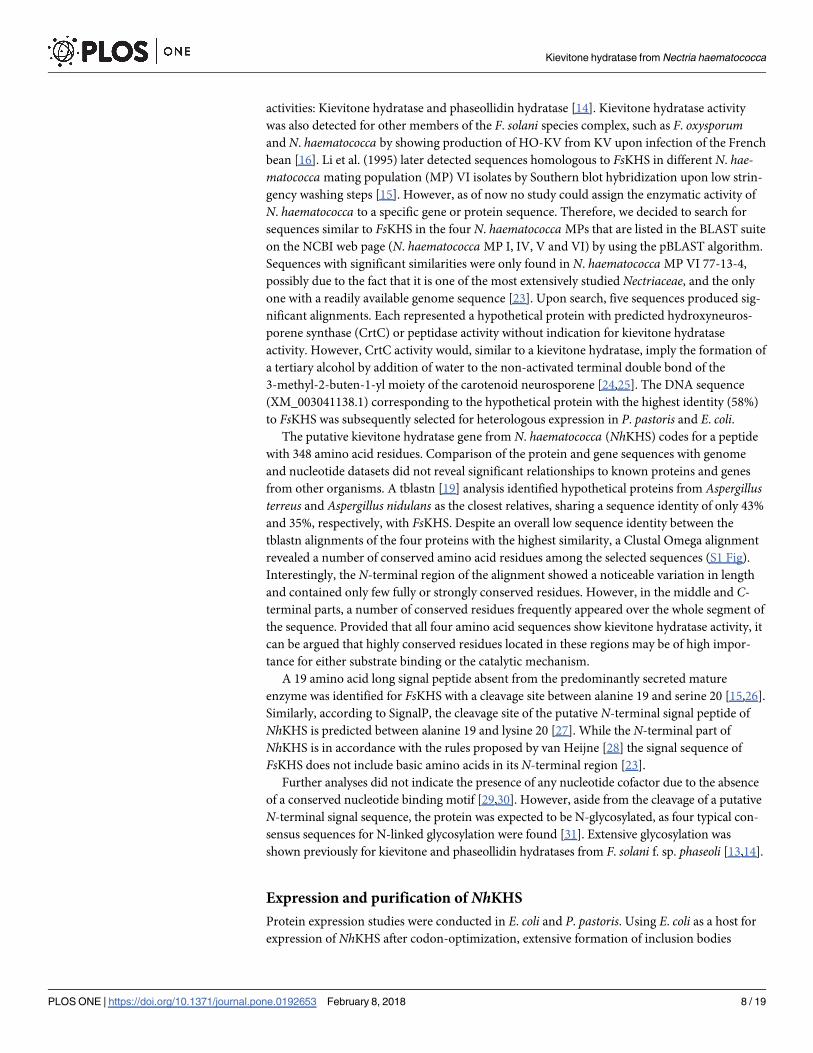
was detected by Western blotting, indicating highly efficient secretion of the enzyme by P. pas-toris. Analysis of protein levels in the culture supernatant after precipitation with the chloro-
form/methanol method revealed a band at an estimated size of ~ 48 kDa on SDS-PAGE. This
indicated that the protein was notably larger than the calculated size of 38 kDa of the mature
polypeptide, which was attributed to glycosylation at one or several of the four N-glycosylation
sites [13,14].
Due to the low levels of secreted endogenous proteins in P. pastoris, NhKHS could already
be partially purified to a good extent by secretion of the enzyme into the culture supernatant.
His10-tagged NhKHS was further purified from the culture supernatant using self-packed Ni-
NTA columns. Affinity chromatography resulted in isolation of highly pure NhKHS that was
used for further biochemical analyses (Fig 1A). Upon in vitro deglycosylation with EndoHf, the
apparent molecular weight was matching the calculated size of the non-glycosylated enzyme of
38 kDa (Fig 1B). Gel filtration of His10-tag purified NhKHS on a Superdex 200 HiLoad 16/60
column resulted in detection of one single peak at an elution volume of 77 mL (S3 Fig). On the
basis of a calibration with standard proteins, a molecular weight of approx. 75 kDa was deter-
mined for NhKHS. This observation suggests that the native form of the enzyme in solution is
most likely a homodimer. UV-Vis absorption spectral analyses of the purified enzyme did not
Fig 1. Analysis of secreted protein and purification of NhKHS from P. pastoris culture supernatant (A). Proteins were precipitated from the culture supernatant of P.
pastoriswild type strain (lane 1) and PpKHSAlpha strain (lane 2) with the chloroform/methanol method. Four hundred μL of cell culture were precipitated. KHS was
purified via Ni-NTA affinity chromatography. Ten μL of flow through (lane 4), washing fractions with 10 mM imidazole (lanes 5 and 6) and 50 mM imidazole (lanes 7 and
8), as well as 4 μL of concentrated, pooled elution fractions (lane 9) were loaded onto the gel. PageRulerTM Prestained Protein Ladder was used as molecular weight
standard (Lane 3). In vitro deglycosylation of purified KHS with EndoHf (B). Deglycosylation reactions were incubated for 0.5–2.5 h.
https://doi.org/10.1371/journal.pone.0192653.g001
Kievitone hydratase from Nectria haematococca
PLOS ONE | https://doi.org/10.1371/journal.pone.0192653 February 8, 2018 9 / 19
indicate the presence of any chromophores in the range from 300 to 1,000 nm, suggesting the
absence of any prosthetic group.
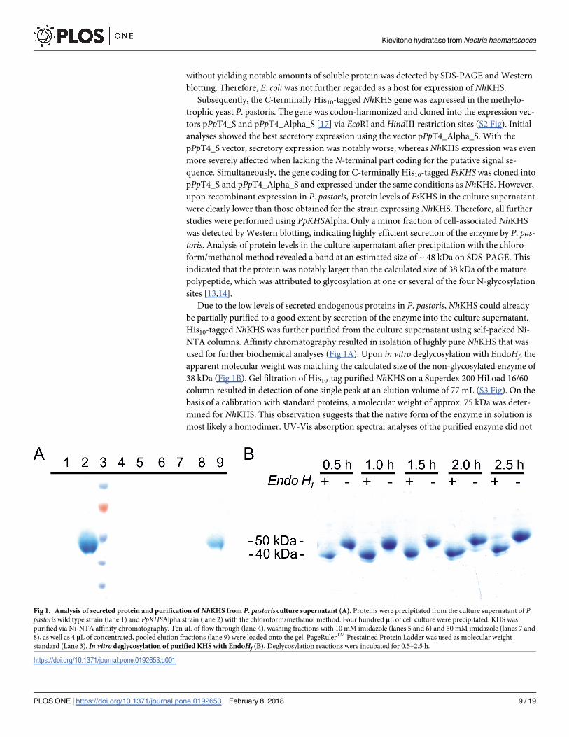
In vitro conversion of KV with recombinantly expressed NhKHS
Functional expression of NhKHS was validated by hydratase activity measurements with
P. pastoris culture supernatant containing ~ 0.6 mg mL-1 of recombinantly expressed protein.
In order to test whether the putative KHS from N. haematococca showed activity for the isofla-
vanon KV, NhKHS was incubated with 2 mM KV in 2% ethanol (v/v) for 3 h. In vitro reactions
were analyzed by HPLC-UV. Retention times and mass fragmentation patterns of substrate
and product in conversions were compared with authentic KV and HO-KV standards (Fig 2A
Fig 2. Formation of HO-KV from KV catalyzed by NhKHS. P. pastoris culture supernatants were incubated with KV for 3 h at 35˚C and product formation was
analyzed via HPLC-UV at 371 nm. Insets represent mass spectra of selected peaks at respective retention times determined by HPLC-MS in positive SIM mode. The
retention time of the substrate was determined by analysis of a 2 mM authentic KV standard (A). No product formation was observed using the culture supernatant of a
P. pastorisWT control (B). Inset of (A & B) represent the mass spectrum of the KV peak at a retention time of 3.1 min. The retention time of the product was determined
by analysis of a 0.5 mM authentic HO-KV standard (C). HO-KV was formed using the culture supernatant of PpKHSAlpha expressing NhKHS (D). Insets of (C & D)
represent the mass spectrum of the product peak at a retention time of 1.4 min.
https://doi.org/10.1371/journal.pone.0192653.g002
Kievitone hydratase from Nectria haematococca
PLOS ONE | https://doi.org/10.1371/journal.pone.0192653 February 8, 2018 10 / 19
and 2C). In vitro conversions with PpKHSAlpha and KV as substrate resulted in formation of
one additional peak, as well as the consumption of approx. 91% of the substrate (Fig 2D). No
putative product peak was formed in the control reaction with P. pastorisWT culture superna-
tant (Fig 2B). Furthermore, mass fragmentation patterns of substrate and product were identi-
cal with the ones obtained for the authentic standards (Fig 2, insets). Thus, formation of
HO-KV was confirmed and the enzyme from N. haematococca did indeed show KHS activity.
Additionally, FsKHS in P. pastoris culture supernatant was used to convert KV in vitro. Despite
a markedly lower protein expression level than NhKHS, approx. 89% of the substrate was con-
verted by FsKHS in the culture supernatant within 3 h of incubation.
Substrate scope of NhKHS for different flavonoids
Since KV and phaseollidin are commercially not/rarely available, further biochemical and
functional studies were initially restricted. Therefore, identification of alternative substrates
for further analyses was highly desirable. The more easily accessible flavonoids 8-prenylnarin-
genin and isoxanthohumol, as well as the chalcone derivative xanthohumol (XN) were pur-
chased and used for conversion reactions with NhKHS (Fig 3). Selection criteria for potential
alternative substrates were a close similarity of their prenylated A-ring core structure with KV,
as well as differently attached B-rings on the pyran-moiety of the C15 flavan scaffold in order
to assess potential effects thereof on enzyme activity. Whereas the B-ring of the isoflavanon
derivative KV is attached at C3, the B-ring of the prenylated flavanons 8-prenylnaringenin and
isoxanthohumol is attached at C2. In contrast, XN lacks the typical flavonoid structure due to
its chalconoid skeleton. Furthermore, the selected compounds differed in their hydroxyl- and
methoxy-substituents, respectively.
All tested flavonoids (Fig 3) were converted by recombinantly expressed and secreted
NhKHS as described in Materials and Methods. The highest conversion was obtained for kievi-
tone (91.5 ± 0.6%), followed by XN (56.1 ± 1.4%, see Fig 4), isoxanthohumol (38.2 ± 0.2%) and
8-prenylnaringenin (3.5 ± 2.2%). These results underscore that the enzyme is indeed a KHS
with a substantial degree of substrate promiscuity. The lower activity for 8-prenylnaringenin
and isoxanthohumol compared to XN may be a consequence of the differently attached B-ring
in comparison to KV, resulting in a more pronounced steric hindrance of substrate binding in
the cavity. This restriction may be less of a problem in the case of XN, since the chalconoid
scaffold resembles the shape of the isoflavanon KV more closely. Since the only common moi-
eties of all tested compounds are the aromatic A-ring and the hydroxyl-substituent at C7, it
may be speculated that substrate binding to the active site is mediated by either π-π stacking of
the A-ring with aromatic amino acid residues of NhKHS, or interaction of polar side chains
with the hydroxyl group at C7, or both.
Isolation of XN from hop extract and NMR analysis of the reaction product
Owing to the good conversion and easy access to XN, we selected it as a model substrate for
biochemical analysis of NhKHS. Sufficient amounts of XN were obtained by extraction from
commercially available hop extract and subsequent purification. The final yield of XN was
approximately 9.2 g with a purity of 99% as shown by HPLC (Fig 4B). NMR analysis of isolated
XN and the reaction product after incubation of XN with 1 mg mL-1 of purified NhKHS unam-
biguously confirmed hydration of XN [32] to HO-XN [33] (S4 Fig).
Enzyme kinetics
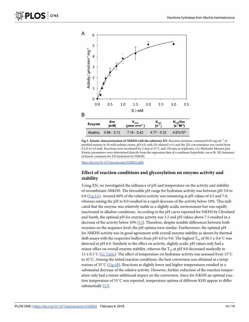
Kinetic assays were performed in vitro under optimized reaction conditions. Kinetic parame-
ters were determined with XN concentrations ranging from 0.125 to 3.0 mM. Due to the low
Kievitone hydratase from Nectria haematococca
PLOS ONE | https://doi.org/10.1371/journal.pone.0192653 February 8, 2018 11 / 19
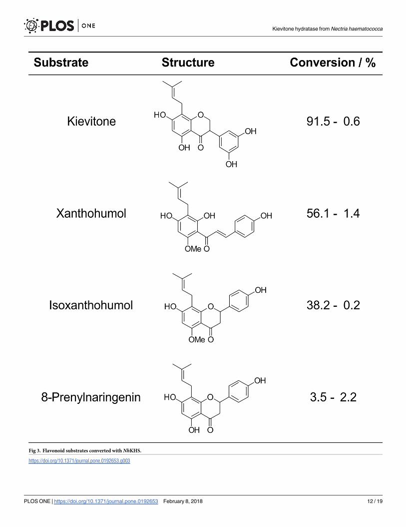
Fig 3. Flavonoid substrates converted with NhKHS.
https://doi.org/10.1371/journal.pone.0192653.g003
Kievitone hydratase from Nectria haematococca
PLOS ONE | https://doi.org/10.1371/journal.pone.0192653 February 8, 2018 12 / 19

solubility of XN in the aqueous buffer system, even upon addition of 2% ethanol (v/v) as co-
solvent, only apparent values for the substrate concentration, as well as Vmax and Km can be
stated. Results of kinetic measurements are shown in a Michaelis-Menten plot as the initial
reaction rate versus the apparent substrate concentration (Fig 5). Km,app and Vmax,app of
recombinant NhKHS were calculated as 0.98 ± 0.13 mM and 7.16 ± 0.42 μmol min-1 mg-1,
respectively, with a kcat of 4.77 ± 0.33 s-1 and a catalytic efficiency of 4.87 x 10³ s-1 M-1. Our
data represent the first enzyme kinetics for the activity of a KHS for a non-physiological
substrate. Until now, only FsKHS was kinetically characterized, with values for Vmax,app and
Km,app for kievitone of 4.92 nmol min-1 mL-1 (0.21 μmol min-1 mg-1) and 0.0175 mM, respec-
tively [13].
Fig 4. Formation of HO-XN from XN catalyzed by NhKHS. P. pastoris culture supernatants were incubated with XN for 3 h at 35˚C and product formation was
analyzed via HPLC-UV at 371 nm. Insets represent mass spectra of selected peaks at respective retention times determined by HPLC-MS in positive SIM mode. The
retention time of the substrate was determined by analysis of a 2 mM authentic XN standard (A). No product formation was observed using the culture supernatant of a P.
pastorisWT control (B). Insets of (A & B) represent the mass spectrum of the XN peak at a retention time of 4.3 min. The retention time of the product was determined by
analysis of a 0.5 mM authentic HO-XN standard (C). HO-XN was formed using the culture supernatant of PpKHSAlpha expressingNhKHS (D). Insets of (C & D)
represent the mass spectrum of the product peak at a retention time of 2.8 min.
https://doi.org/10.1371/journal.pone.0192653.g004
Kievitone hydratase from Nectria haematococca
PLOS ONE | https://doi.org/10.1371/journal.pone.0192653 February 8, 2018 13 / 19
Effect of reaction conditions and glycosylation on enzyme activity and
stability
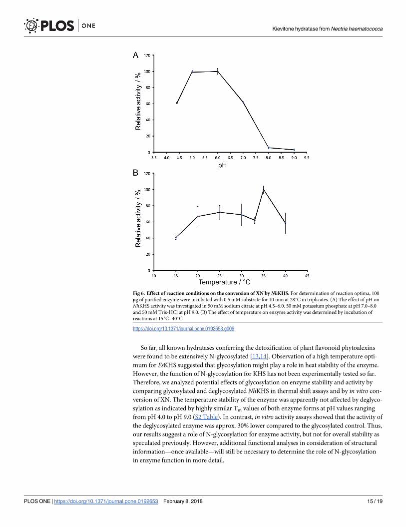
Using XN, we investigated the influence of pH and temperature on the activity and stability
of recombinant NhKHS. The favorable pH range for hydratase activity was between pH 5.0 to
6.0 (Fig 6A). Around 60% of the relative activity was remaining at pH values of 4.5 and 7.0,
whereas raising the pH to 8.0 resulted in a rapid decrease of the activity below 10%. This indi-
cated that the enzyme was relatively stable in a slightly acidic environment but was rapidly
inactivated in alkaline conditions. According to the pH curve reported for FsKHS by Cleveland
and Smith, the optimal pH for enzyme activity was 5.5 and pH values above 7.5 resulted in a
decrease of the activity below 10% [13]. Therefore, despite notable differences between both
enzymes on the sequence level, the pH optima were similar. Furthermore, the optimal pH
for NhKHS activity was in good agreement with overall enzyme stability as shown by thermal
shift assays with the respective buffers from pH 4.0 to 9.0. The highest Tm of 50.3 ± 0.6˚C was
detected at pH 6.0. Similarly to the effect on activity, slightly acidic pH values only had a
minor effect on overall enzyme stability, whereas the Tm at pH 8.0 decreased markedly to
15 ± 0.1˚C (S2 Table). The effect of temperature on hydratase activity was assessed from 15˚C
to 45˚C. Among the tested reaction conditions, the best conversion was obtained at a temp-
erature of 35˚C (Fig 6B). Reactions at slightly lower and higher temperatures resulted in a
substantial decrease of the relative activity. However, further reduction of the reaction temper-
ature only had a minor additional impact on the conversion. Since for FsKHS an optimal reac-
tion temperature of 55˚C was reported, temperature optima of different KHS appear to differ
substantially [13].
Fig 5. Kinetic characterization of NhKHS with the substrate XN. Reaction mixtures contained 0.05 mg mL-1 of
purified enzyme in 50 mM sodium citrate, pH 6.0, with 2% ethanol (v/v) and the XN concentration was varied from
0.125 to 3.0 mM. Reactions were incubated for 2 min at 35˚C and 150 rpm in triplicates. (A) Michaelis-Menten plot.
Kinetic parameters were determined directly from the regression data of a nonlinear hyperbolic curve fit. (B) Summary
of kinetic constants for XN hydration by NhKHS.
https://doi.org/10.1371/journal.pone.0192653.g005
Kievitone hydratase from Nectria haematococca
PLOS ONE | https://doi.org/10.1371/journal.pone.0192653 February 8, 2018 14 / 19
So far, all known hydratases conferring the detoxification of plant flavonoid phytoalexins
were found to be extensively N-glycosylated [13,14]. Observation of a high temperature opti-
mum for FsKHS suggested that glycosylation might play a role in heat stability of the enzyme.
However, the function of N-glycosylation for KHS has not been experimentally tested so far.
Therefore, we analyzed potential effects of glycosylation on enzyme stability and activity by
comparing glycosylated and deglycosylated NhKHS in thermal shift assays and by in vitro con-
version of XN. The temperature stability of the enzyme was apparently not affected by deglyco-
sylation as indicated by highly similar Tm values of both enzyme forms at pH values ranging
from pH 4.0 to pH 9.0 (S2 Table). In contrast, in vitro activity assays showed that the activity of
the deglycosylated enzyme was approx. 30% lower compared to the glycosylated control. Thus,
our results suggest a role of N-glycosylation for enzyme activity, but not for overall stability as
speculated previously. However, additional functional analyses in consideration of structural
information—once available—will still be necessary to determine the role of N-glycosylation
in enzyme function in more detail.
Fig 6. Effect of reaction conditions on the conversion of XN by NhKHS. For determination of reaction optima, 100
μg of purified enzyme were incubated with 0.5 mM substrate for 10 min at 28˚C in triplicates. (A) The effect of pH on
NhKHS activity was investigated in 50 mM sodium citrate at pH 4.5–6.0, 50 mM potassium phosphate at pH 7.0–8.0
and 50 mM Tris-HCl at pH 9.0. (B) The effect of temperature on enzyme activity was determined by incubation of
reactions at 15˚C- 40˚C.
https://doi.org/10.1371/journal.pone.0192653.g006
Kievitone hydratase from Nectria haematococca
PLOS ONE | https://doi.org/10.1371/journal.pone.0192653 February 8, 2018 15 / 19

The effect of organic solvents on enzyme activity was investigated by incubation of purified
NhKHS with XN in the presence of 1–30% v/v of different solvents. After 3 h of incubation,
the highest activity was retained with hexane and n-dodecane. In these cases, addition of up to
30% v/v of solvent to the reactions did not result in notably reduced product formation com-
pared to the control (S5 Fig). However, product formation was already considerably affected
in the presence of 10% v/v of ethanol, DMSO or chloroform. This indicates that the polarity of
the organic solvent is a determining factor for its effect on NhKHS activity, with increasing sol-
ubility in water resulting in a more distinct impact. This might be caused by an enhanced per-
turbing effect on the hydration shell of the enzyme. However, the compatibility of NhKHS
with nonpolar organic solvents will be beneficial for a number of biocatalytic applications, e.g.
in general for conversion of hydrophobic substrates.
Bioreactor cultivation
An outstanding feature of the P. pastoris host system for recombinant protein expression is its
ability to grow to high cell densities of more than 150 g CDW L-1 in controlled fed-batch culti-
vations [34]. Since this often results in high yields of recombinant protein, we tested whether
scaling up protein expression from shake flask to lab scale fed-batch fermentation is a feasible
strategy to increase the production of recombinant NhKHS. Therefore, PpKHSAlpha and P.
pastoris wild type strains were cultivated in parallel in 2 L bioreactors (DASGIP, Julich, Ger-
many). At different time points of methanol induction, culture aliquots were withdrawn for
determination of CDW, protein concentration, expression level and enzyme activity. At the
end of induction, CDWs of the different strains were similar, reaching values in the range of
80–90 g L-1 of culture. SDS-PAGE confirmed that protein levels increased steadily during
induction (S6 Fig). At the end of the cultivation, 2.61 g L-1 of secreted protein was obtained
from 2 L of fermentation broth. Comparison of CDW and protein levels in the culture super-
natant of bioreactor (89 g L-1 CDW; 2.3 g L-1 of recombinant NhKHS) and shake flask cultiva-
tion (9.6 g L-1 CDW; 0.6 g L-1 of recombinant NhKHS) under standard conditions, i.e. 400 mL
of medium, 48 h of methanol induction, indicated that the notably higher yield of NhKHS in
the bioreactor cultivation indeed resulted from the higher cell density in the bioreactor. Over-
all, P. pastoris is a most appropriate host for producing high amounts of recombinant NhKHS.
In summary, our study reports on the identification and heterologous expression of KHS
from N. haematococca MP VI that catalyzes the formation of a tertiary alcohol by hydration of
the phytoalexin KV. After investigating into the substrate spectrum of KHS, the first thorough
biochemical characterization of a KHS was performed by using XN as the model substrate.
Since NhKHS showed good activity for a range of different non-physiological substrates as
well as stability at broad and suitable temperature and pH ranges, it is a promising candidate
for the biosynthesis of industrially relevant tertiary alcohols.
Supporting information
S1 Fig. Clustal Omega alignment of putative KHS enzymes. Protein sequences from A. nidu-lans, A. terreus, F. solani and N. haematococca are shown.
(PDF)
S2 Fig. Coding sequence of codon-harmonized putative kievitone hydratases from N. hae-matococca (NhKHS) and from F. solani (FsKHS) for expression in P. pastoris.
(PDF)
S3 Fig. Gel filtration profile of NhKHS on a Superdex 200 HiLoad 16/60 column (GE
Healthcare, UK) using 50 mM sodium citrate, pH 6.0, as buffer system. Inset: Calibration
Kievitone hydratase from Nectria haematococca
PLOS ONE | https://doi.org/10.1371/journal.pone.0192653 February 8, 2018 16 / 19
curve generated with the standard proteins conalbumin (75 kDa), ovalbumin (44 kDa), car-
bonic anhydrase (29 kDa), ribonuclease A (13.7 kDa) and aprotinin (6.5 kDa).
(PDF)
S4 Fig. 1H- and 13C-NMR spectra of XN and HO-XN in CD3OD. After incubation of XN
with purified NhKHS, the reaction products were extracted and prepared for NMR analysis as
described. (A) 1H-NMR spectrum of XN at 499.8 MHz. (B) 13C-NMR spectrum of XN at 125.7
MHz. (C) 1H-NMR spectrum of HO-XN at 499.8 MHz. (D) 13C-NMR spectrum of HO-XN at
125.7 MHz. HO-XN could be unambiguously identified as the reaction product upon hydra-
tion of XN by NhKHS, and substrate and product were independently confirmed by compari-
son with 1H- and 13C-NMR spectra provided in other work [1,2].
(PDF)
S5 Fig. Influence of different organic solvents on the activity of NhKHS. The standard
enzyme assay was performed in the presence of respective organic solvents at concentrations
ranging from 0.5 to 30%. One mM of XN and 0.05 mg mL-1 of enzyme were incubated for 3 h.
Amounts of XN-hydrate in mM were obtained via HPLC-MS measurements. Biological tripli-
cates were analyzed.
(PDF)
S6 Fig. Bioreactor cultivation of strain PpKHSAlpha. Results of activity assays using 3 μL of
supernatant of the fermentation broth from strain PpKHSAlpha at indicated time points of
induction (A). Volumetric activity of strain PpKHSAlpha (B). Activity assays and HPLC analy-
ses were performed in triplicates. NhKHS levels in culture supernatants were monitored by
SDS-PAGE (C) and compared to purified protein (KHS pur.).
(PDF)
S1 Table. Primers used for cloning of NhKHS and FsKHS into the vector pPpT4_Alpha_S.
(PDF)
S2 Table. Thermostability of glycosylated and deglycosylated NhKHS at different pH val-
ues. Purified enzyme was deglycosylated with EndoHf enzyme mix for 0.5 h at 37˚C.
(PDF)
Acknowledgments
We would like to thank Prof. Michael Murkovic (Institute of Biochemistry, Graz University of
Technology) for kindly providing isoxanthohumol and Eva Baumhackl for excellent technical
assistance.
Author Contributions
Conceptualization: Matthias Engleder, Anita Emmerstorfer-Augustin, Tamara Wriessnegger,
Gernot Strohmeier, Monika Muller, Daniel Mink, Martin Schurmann, Harald Pichler.
Data curation: Matthias Engleder, Anita Emmerstorfer-Augustin, Gernot Strohmeier, Martin
Schurmann, Harald Pichler.
Formal analysis: Hansjorg Weber, Harald Pichler.
Funding acquisition: Daniel Mink, Martin Schurmann, Harald Pichler.
Investigation: Matthias Engleder, Melissa Horvat, Anita Emmerstorfer-Augustin, Tamara
Wriessnegger, Stefanie Gabriel, Gernot Strohmeier, Hansjorg Weber.
Kievitone hydratase from Nectria haematococca
PLOS ONE | https://doi.org/10.1371/journal.pone.0192653 February 8, 2018 17 / 19

Methodology: Matthias Engleder, Anita Emmerstorfer-Augustin, Tamara Wriessnegger, Ger-
not Strohmeier, Iwona Kaluzna, Daniel Mink, Martin Schurmann, Harald Pichler.
Project administration: Anita Emmerstorfer-Augustin, Tamara Wriessnegger, Daniel Mink,
Martin Schurmann, Harald Pichler.
Resources: Harald Pichler.
Supervision: Anita Emmerstorfer-Augustin, Tamara Wriessnegger, Martin Schurmann, Har-
ald Pichler.
Validation: Matthias Engleder, Gernot Strohmeier, Monika Muller.
Visualization: Tamara Wriessnegger.
Writing – original draft: Matthias Engleder, Harald Pichler.
Writing – review & editing: Matthias Engleder, Anita Emmerstorfer-Augustin, Tamara
Wriessnegger, Gernot Strohmeier, Martin Schurmann, Harald Pichler.
References1. Kourist R, Bornscheuer UT. Biocatalytic synthesis of optically active tertiary alcohols. Appl Microbiol
Biotechnol. 2011; 91: 505–517. https://doi.org/10.1007/s00253-011-3418-9 PMID: 21691783
2. Muller M. Enzymatic Synthesis of Tertiary Alcohols. ChemBioEng Rev. 2014; 1: 14–26.
3. Kourist R, de Maria PD, Bornschuer UT. Enzymatic synthesis of optically active teritiary alcohols:
Expanding the biocatalysis toolbox. ChemBioChem. 2008; 9: 491–498. https://doi.org/10.1002/cbic.
200700688 PMID: 18232040
4. Liese A, Seelbach K, Wandrey C. Industrial Biotransformations. 2nd ed. Wiley-VCH; 2006.
5. Faber K. Biotransformations in Organic Chemistry. 6th ed. Springer; 2011.
6. Resch V, Hanefeld U. The selective addition of water. Catal Sci Technol. 2015; 5: 1385–1399.
7. Jin J, Hanefeld U. The selective addition of water to C = C bonds; enzymes are the best chemists.
Chem Commun. 2011; 47: 2502–2510.
8. Engleder M, Pavkov-Keller T, Emmerstorfer A, Hromic A, Schrempf S, Steinkellner G, et al. Structure-
Based Mechanism of Oleate Hydratase from Elizabethkingia meningoseptica. ChemBioChem. 2015;
16: 1730–1734. https://doi.org/10.1002/cbic.201500269 PMID: 26077980
9. Song J-W, Jeon E-Y, Song D-H, Jang HY, Bornscheuer UT, Oh D-K, et al. Multistep enzymatic synthe-
sis of long-chain α,ω-Dicarboxylic andω-hydroxycarboxylic acids from renewable fatty acids and plant
oils. Angew Chem Int Ed. 2013; 52: 2534–2537.
10. Jo Y-S, An J-U, Oh D-K. γ-Dodecelactone production from safflower oil via 10-hydroxy-12(Z)-octadece-
noic acid intermediate by whole cells of Candida boidinii and Stenotrophomonas nitritireducens. J Agric
Food Chem. 2014; 62: 6736–6745. https://doi.org/10.1021/jf501081z PMID: 24967938
11. Hou CT. Biotechnology for fats and oils: new oxygenated fatty acids. N Biotechnol. 2009; 26: 2–10.
https://doi.org/10.1016/j.nbt.2009.05.001 PMID: 19447212
12. Kuhn PJ, Smith DA. Isolation from Fusarium solani f. sp. phaseoli of an enzymic system responsible for
kievitone and phaseollidin detoxification. Physiol Plant Pathol. 1979; 14: 179–190.
13. Cleveland TE, Smith DA. Partial purification, and further characterization, of kievitone hydratase from
cell-free culture filtrates of Fusarium solani f. sp. phaseoli. Physiol Plant Pathol. 1983; 22: 129–142.
14. Turbek CS, Smith DA, Schardl CL. An extracellular enzyme from Fusarium solani f. sp. phaseoli which
catalyses hydration of the isoflavonoid phytoalexin, phaseollidin. FEMS Microbiol Lett. 1992; 94: 187–190.
15. Li D, Chung K-R, Smith DA, Schardl CL. The Fusarium solani gene encoding kievitone hydratase, a
secreted enzyme that catalyzes detoxification of a bean phytoalexin. Mol Plant Microbe. 1995; 8: 388–
397.
16. Smith DA, Harrer JM, Cleveland TE. Relation between production of extracellular kievitone hydratase
by isolates of Fusarium and their pathogenicity on Phaseolus vulgaris. Phytopathology. 1982; 72:
1319–1323.
17. Naatsaari L, Mistlberger B, Ruth C, Hajek T, Hartner FS, Glieder A. Deletion of the Pichia pastoris ku70
homologue facilitates platform strain generation for gene expression and synthetic biology. PLoS One.
2012; 7: 7:e39720.
Kievitone hydratase from Nectria haematococca
PLOS ONE | https://doi.org/10.1371/journal.pone.0192653 February 8, 2018 18 / 19

18. Lin-Cereghino J, Wong WW, Xiong S, Giang W, Luong LT, Vu J, et al. Condensed protocol for compe-
tent cell preparation and transformation of the methylotrophic yeast Pichia pastoris. Biotechniques.
2005; 38: 44–48. PMID: 15679083
19. Altschul SF, Madden TL, Schaffer AA, Zhang J, Zhang Z, Miller W, et al. Gapped BLAST and PSI-
BLAST: a new generation of protein database search programs. Nucleic Acids Res. 1997; 25:3389–
3402. PMID: 9254694
20. Wriessnegger T, Augustin P, Engleder M, Leitner E, Muller M, Kaluzna I, et al. Production of the sesqui-
terpenoid (+)-nootkatone by metabolic engineering of Pichia pastoris. Metab Eng. 2014; 24: 18–29.
https://doi.org/10.1016/j.ymben.2014.04.001 PMID: 24747046
21. Lavinder JJ, Hari SB, Sullivan BJ, Magliery TJ. High-throughput thermal scanning: a general, rapid dye-
binding thermal shift screen for protein engineering. J Am Chem Soc. 2009; 131(11): 3794–3795.
https://doi.org/10.1021/ja8049063 PMID: 19292479
22. Ericsson UB, Hallberg BM, DeTitta GT, Dekker N, Nordlund P. Thermofluor-based high-throughput sta-
bility optimization of proteins for structural studies. Anal Biochem. 2006; 357: 289–298. https://doi.org/
10.1016/j.ab.2006.07.027 PMID: 16962548
23. Coleman JJ, Rounsley SD, Rodriguez-Carres M, Kuo A, Wasmann CC, Grimwood J, et al. The genome
of Nectria haematococca: Contribution of supernumerary chromosomes to gene expansion. PLoS
Genet. 2009; 5: e1000618. https://doi.org/10.1371/journal.pgen.1000618 PMID: 19714214
24. Albrecht M, Takaichi S, Misawa N, Schnurr G, Boger P, Sandmann G. Synthesis of atypical cyclic and
acyclic hydroxy carotenoids in Escherichia coli transformants. J Biotechnol. 1997; 58: 177–185. PMID:
9470222
25. Sandmann G. Carotenoid biosynthesis in microorganisms and plants. Eur J Biochem. 1994; 223: 7–24.
PMID: 8033911
26. Turbek CS, Li D, Choi GH, Schardl CL, Smith DA. Induction and purification of kievitone hydratase from
Fusarium solani f. sp. phaseoli. Phytochemistry. 1990; 29: 2841–2846. PMID: 1366757
27. Petersen TN, Brunak S, von Heijne G, Nielsen H. SignalP 4.0: discriminating signal peptides from trans-
membrane regions. Nat Methods. 2011; 8: 785–786. https://doi.org/10.1038/nmeth.1701 PMID:
21959131
28. von Heijne G. Towards a comparative anatomy of N-terminal topogenic protein sequences. J Mol Biol.
1986; 189: 239–242. PMID: 3783674
29. Wierenga RK, Terpstra P, Hol WGJ. Prediction of the occurrence of the ADP-binding βαβ-fold in pro-
teins, using an amino acid sequence fingerprint. J Mol Biol. 1986; 187: 101–107. PMID: 3959077
30. Kleiger G, Eisenberg D. GXXXG and GXXXA Motifs Stabilize FAD and NAD(P)-binding Rossmann
Folds Through Cα–H���O Hydrogen Bonds and van der Waals Interactions. J Mol Biol. 2002; 323: 69–
76. PMID: 12368099
31. Blom N, Sicheritz-Ponten T, Gupta R, Gammeltoft S, Brunak S. Prediction of post-translational glycosyl-
ation and phosphorylation of proteins from the amino acid sequence. Proteomics. 2004; 4: 1633–1649.
https://doi.org/10.1002/pmic.200300771 PMID: 15174133
32. Gerhauser C, Alt A, Heiss E, Gamal-Eldeen A, Klimo K, Knauft J, et al. Cancer chemopreventive activity
of Xanthohumol, a natural product derived from hop. Mol Cancer Ther. 2002; 1: 959–969. PMID:
12481418
33. Tronina T, Bartmańska A, Filip-Psurska B, Wietrzyk J, Popłoński J, Huszcza E. Fungal metabolites of
xanthohumol with potent antiproliferative activity on human cancer cell lines in vitro. Bioorg Med Chem.
2013; 21: 2001–2006. https://doi.org/10.1016/j.bmc.2013.01.026 PMID: 23434138
34. Jahic M, Veide A, Charoenrat T, Teeri T, Enfors SO. Process technology for production and recovery of
heterologous proteins with Pichia pastoris. Biotechnol Prog. 2006; 22: 1465–1473. https://doi.org/10.
1021/bp060171t PMID: 17137292
Kievitone hydratase from Nectria haematococca
PLOS ONE | https://doi.org/10.1371/journal.pone.0192653 February 8, 2018 19 / 19